27th Annual Meeting Poster Abstract Book
122
www.sitcancer.org 2 SITC 0 12 27 th Annual Meeting Poster Abstract Book October 26-28, 2012 • North Bethesda, MD Poster Abstract Book Printing Supported By:
-
Upload
khangminh22 -
Category
Documents
-
view
0 -
download
0
Transcript of 27th Annual Meeting Poster Abstract Book

www.sitcancer.org
2S I TC0
1 2
27th Annual Meeting Poster Abstract BookOctober 26-28, 2012 • North Bethesda, MD
Poster Abstract Book Printing Supported By:

SITC 27th Annual Meeting Poster Abstract Book • October 26-28, 2012 sitcancer.org
Poster Information
Poster Hall locationMain Level, Grand Ballroom Salons A-D
Poster set-UP HoUrsFriday, October 26, 6:30 am – 10:00 am
Poster tear Down HoUrsSaturday, October 27, 8:00 pm – 9:30 pm
Poster Hall HoUrsFriday, October 26, 10:00 am – 8:00 pmSaturday, October 27, 10:00 am – 8:00 pm
Poster PresentationsOdd Number Posters (authors are present)Friday, October 26, 12:30 pm – 1:30 pm and 6:30 pm – 7:15 pm
Even Number Posters (authors are present)Friday, October 26, 7:15 pm – 8:00 pm and Saturday, October 27 from 1:00 pm – 2:00 pm
Poster nUmbersAdoptive T Cell Transfer and Cell Therapy as Cancer Immunotherapy (CARS) . . . . . . . . . . . . . . . . . . . . . . . . . . . . . . . . . . . . . . . . . . . 1-23Combining Immunotherapy and Other Therapies . . . . . . . . . . . . . . . . . . . . . . . . . . . . . . . . . . . . . . . . . . . . . . . . . . . . . . . . . . . . . . . . . . . 24-50DC Subsets/Cancer Vaccines . . . . . . . . . . . . . . . . . . . . . . . . . . . . . . . . . . . . . . . . . . . . . . . . . . . . . . . . . . . . . . . . . . . . . . . . . . . . . . . . . . . . . . 51-81Immunity of Oncolytic Viruses . . . . . . . . . . . . . . . . . . . . . . . . . . . . . . . . . . . . . . . . . . . . . . . . . . . . . . . . . . . . . . . . . . . . . . . . . . . . . . . . . . . . 82-83Immunotherapy Combinations . . . . . . . . . . . . . . . . . . . . . . . . . . . . . . . . . . . . . . . . . . . . . . . . . . . . . . . . . . . . . . . . . . . . . . . . . . . . . . . . . . 84-100Innate Immunity in Cancer . . . . . . . . . . . . . . . . . . . . . . . . . . . . . . . . . . . . . . . . . . . . . . . . . . . . . . . . . . . . . . . . . . . . . . . . . . . . . . . . . . . . .101-113Single Cell High Throughput Technologies Immune Monitoring . . . . . . . . . . . . . . . . . . . . . . . . . . . . . . . . . . . . . . . . . . . . . . . . . . . .114-121T Cell Manufacture and Potency Testing . . . . . . . . . . . . . . . . . . . . . . . . . . . . . . . . . . . . . . . . . . . . . . . . . . . . . . . . . . . . . . . . . . . . . . . . . .122-124T Cell Modulating Strategies . . . . . . . . . . . . . . . . . . . . . . . . . . . . . . . . . . . . . . . . . . . . . . . . . . . . . . . . . . . . . . . . . . . . . . . . . . . . . . . . . . . .125-129Targeted Therapies and Anti-Tumor Immunity . . . . . . . . . . . . . . . . . . . . . . . . . . . . . . . . . . . . . . . . . . . . . . . . . . . . . . . . . . . . . . . . . . . .130-157Targeting Immune Suppression . . . . . . . . . . . . . . . . . . . . . . . . . . . . . . . . . . . . . . . . . . . . . . . . . . . . . . . . . . . . . . . . . . . . . . . . . . . . . . . . .158-175Therapeutic Monoclonal Antibodies in Cancer . . . . . . . . . . . . . . . . . . . . . . . . . . . . . . . . . . . . . . . . . . . . . . . . . . . . . . . . . . . . . . . . . . . .176-182Tumor Microenvironment . . . . . . . . . . . . . . . . . . . . . . . . . . . . . . . . . . . . . . . . . . . . . . . . . . . . . . . . . . . . . . . . . . . . . . . . . . . . . . . . . . . . . .183-202Tumor Vasculature, Chemokines and Lymphocyte Trafficking to the Tumor . . . . . . . . . . . . . . . . . . . . . . . . . . . . . . . . . . . . . . . . . . .203-206

SITC 27th Annual Meeting Poster Abstract Book • October 26-28, 2012 1sitcancer.org
Table of Contents
Author Index . . . . . . . . . . . . . . . . . . . . . . . . . . . . . . . . . . . . . . . . . . . . . . . . . . . . . . . . . . . . . . . . . . . . . . . . . . . . . . . . . . . . . . . . . . . . . . . . . . . . . . .2
Keyword Index . . . . . . . . . . . . . . . . . . . . . . . . . . . . . . . . . . . . . . . . . . . . . . . . . . . . . . . . . . . . . . . . . . . . . . . . . . . . . . . . . . . . . . . . . . . . . . . . . . . .13
Abstract Listings . . . . . . . . . . . . . . . . . . . . . . . . . . . . . . . . . . . . . . . . . . . . . . . . . . . . . . . . . . . . . . . . . . . . . . . . . . . . . . . . . . . . . . . . . . . . . . . . . . .15
Notes . . . . . . . . . . . . . . . . . . . . . . . . . . . . . . . . . . . . . . . . . . . . . . . . . . . . . . . . . . . . . . . . . . . . . . . . . . . . . . . . . . . . . . . . . . . . . . . . . . . . . . . . . . . 118

SITC 27th Annual Meeting Poster Abstract Book • October 26-28, 2012 2 sitcancer.org
author A poster number
Aarts-Riemens, Tineke . . . . . . . . . . . . . . . . . . . . . . . . . . . . . . . . . 184Abdullah, Shaad . . . . . . . . . . . . . . . . . . . . . . . . . . . . . . . . . . . . . . 144AbouShousha, Seham . . . . . . . . . . . . . . . . . . . . . . . . . . . . . . . . . 186Ackerman, Allison . . . . . . . . . . . . . . . . . . . . . . . . . . . . . . . . . . . . . 24Adams, Katherine . . . . . . . . . . . . . . . . . . . . . . . . . . . . . . . . .133, 143Adams, Richard . . . . . . . . . . . . . . . . . . . . . . . . . . . . . . . . . . . . . . . 72Adams, Sylvia . . . . . . . . . . . . . . . . . . . . . . . . . . . . . . . . . . . . . . . . . 32Afanasiev, O. . . . . . . . . . . . . . . . . . . . . . . . . . . . . . . . . . . . . . . . . . 183Aguilar, Richard J. . . . . . . . . . . . . . . . . . . . . . . . . . . . . . . . . . . . . . . 82Al-Banna, Nadia . . . . . . . . . . . . . . . . . . . . . . . . . . . . . . . . . . . . . . 147Al-Hadidi, Abeer . . . . . . . . . . . . . . . . . . . . . . . . . . . . . . . . . . . . . . 186Albarello, Luca . . . . . . . . . . . . . . . . . . . . . . . . . . . . . . . . . . . . . . . 164Alegre, Maria-Luisa . . . . . . . . . . . . . . . . . . . . . . . . . . . . . . . . . . . 125Alvarado, Gladys . . . . . . . . . . . . . . . . . . . . . . . . . . . . . . . . . . . . . . . . 1Amadori, Dino . . . . . . . . . . . . . . . . . . . . . . . . . . . . . . . . . . . . . . . 122Ambrosino, Elena . . . . . . . . . . . . . . . . . . . . . . . . . . . . . . . . . . . . . 191Ameen, Zeenath . . . . . . . . . . . . . . . . . . . . . . . . . . . . . . . . . . . . . . 195Amendolara, Benjamin . . . . . . . . . . . . . . . . . . . . . . . . . . . . . . . . 182Ancarani, Valentina . . . . . . . . . . . . . . . . . . . . . . . . . . . . . . . . . . . 122Andersen, Mads H. . . . . . . . . . . . . . . . . . . . . . . . . . . . . . . . . . . . . . . 3Ando, Jun . . . . . . . . . . . . . . . . . . . . . . . . . . . . . . . . . . . . . . . . . . . . . 23Andreotti, Alessia . . . . . . . . . . . . . . . . . . . . . . . . . . . . . . . . . . . . . . 21Ang, Sonny . . . . . . . . . . . . . . . . . . . . . . . . . . . . . . . . . . . . . . . . . . . . 2Angell, Helen K. . . . . . . . . . . . . . . . . . . . . . . . . . . . . . . . . . . . . . . 120Annels, Nicola E. . . . . . . . . . . . . . . . . . . . . . . . . . . . . . . . . . .131, 158Ansaldi, Filippo . . . . . . . . . . . . . . . . . . . . . . . . . . . . . . . . . . . . . . . . 84Antignani, Antonella . . . . . . . . . . . . . . . . . . . . . . . . . . . . . . . . . . . 31Archer, Rich . . . . . . . . . . . . . . . . . . . . . . . . . . . . . . . . . . . . . . . . . . 176Arns, Katherine . . . . . . . . . . . . . . . . . . . . . . . . . . . . . . . . . . . . . . . . 51Aryankalayil, Joseph . . . . . . . . . . . . . . . . . . . . . . . . . . . . . . . . . . 168Ascarateil, Stephane . . . . . . . . . . . . . . . . . . . . . . . . . . . . . . . . . . . . 52Asch, Adam S. . . . . . . . . . . . . . . . . . . . . . . . . . . . . . . . . . . . . . . . . . 48Ascierto, Maria Libera . . . . . . . . . . . . . . 82, 84, 101, 103, 162, 200Ascierto, Paolo A. . . . . . . . . . . . . . . . . . . . . . . . . . . . . . . . . .101, 115Ashdown, Martin L. . . . . . . . . . . . . . . . . . . . . . . . . . . . . . . . .29, 159Ashfield, Rebecca . . . . . . . . . . . . . . . . . . . . . . . . . . . . . . . . .133, 143Atkins, Michael B. . . . . . . . . . . . . . . . . . . . . . . . . . . . . . . . . . . . . . 24Auci, Dominick . . . . . . . . . . . . . . . . . . . . . . . . . . . . . . . .53, 118, 127Aung, Sandra . . . . . . . . . . . . . . . . . . . . . . . . . . . . . . . . . . . . . . 76, 86Ayala, Victor I. . . . . . . . . . . . . . . . . . . . . . . . . . . . . . . . . . . . . . . . . 49
BBack, Tim . . . . . . . . . . . . . . . . . . . . . . . . . . . . . . . . . . . . . . . . . . 94, 96Bahjat, Keith S. . . . . . . . . . . . . . . . . . . . . . . . . . . . . . . . . . . . . . 43, 54Baker, D. . . . . . . . . . . . . . . . . . . . . . . . . . . . . . . . . . . . . . . . . . . . . . 143Balducci, Anthony . . . . . . . . . . . . . . . . . . . . . . . . . . . . . . . . . . 9, 190Balint, Klara . . . . . . . . . . . . . . . . . . . . . . . . . . . . . . . . . . . . . . . . . . . . 4Bambina, Shelly . . . . . . . . . . . . . . . . . . . . . . . . . . . . . . . . . . . . . . . 54Bandman, Olga . . . . . . . . . . . . . . . . . . . . . . . . . . . . . . . . . . . . . 63, 64Barat, N. C. . . . . . . . . . . . . . . . . . . . . . . . . . . . . . . . . . . . . . . . . . . . . 74Barcellos-Hoff, Mary-Helen . . . . . . . . . . . . . . . . . . . . . . . . . .46, 139Barker, Christopher A. . . . . . . . . . . . . . . . . . . . . . . . . . . . . . . . . . . 39
Author Index
Barrett, David . . . . . . . . . . . . . . . . . . . . . . . . . . . . . . . . . . . . . . . . . 20Bartlett, David L. . . . . . . . . . . . . . . . . . . . . . . . . . . . . . . . . . . . . . . 205Bassett, Jennifer D. . . . . . . . . . . . . . . . . . . . . . . . . . . . . . . . . .22, 126Bastiaannet, Esther . . . . . . . . . . . . . . . . . . . . . . . . . . . . . . . . . . . . 104Baston, Emma . . . . . . . . . . . . . . . . . . . . . . . . . . . . . . . . . . . . . . . . 133Baumgarth, Nicole . . . . . . . . . . . . . . . . . . . . . . . . . . . . . . . . . . . . 100Baumhof, Patrick . . . . . . . . . . . . . . . . . . . . . . . . . . . . . . . . . . . . . 142Bear, Harry D. . . . . . . . . . . . . . . . . . . . . . . . . . . . . . . . . . . . . . . . . . 18Beard, Brian . . . . . . . . . . . . . . . . . . . . . . . . . . . . . . . . . . . . . . . . . . . 27Beck, Barbara . . . . . . . . . . . . . . . . . . . . . . . . . . . . . . . . . . . . . . . . . 55Becke, Sabine . . . . . . . . . . . . . . . . . . . . . . . . . . . . . . . . . . . . . . . . 151Beckhove, Philipp . . . . . . . . . . . . . . . . . . . . . . . . . . . . . . . . . . . . . . 25Bedognetti, Davide . . . . . . . . . . . . . . . . . . . . 84, 101, 103, 162, 200Beilhack, Andreas . . . . . . . . . . . . . . . . . . . . . . . . . . . . . . . . . . . . . . 49Bekaii-Saab, Tanios . . . . . . . . . . . . . . . . . . . . . . . . . . . . . . . . . . . . 195Belani, Chandra P. . . . . . . . . . . . . . . . . . . . . . . . . . . . . . . . . . . . . . 105Bender, James G. . . . . . . . . . . . . . . . . . . . . . . . . . . . . . . . . . . . . . . . 80Bengala, Carmelo . . . . . . . . . . . . . . . . . . . . . . . . . . . . . . . . . . . . . . 21Bentley, James . . . . . . . . . . . . . . . . . . . . . . . . . . . . . . . . . . . . . . . . 147Berger, Carolina . . . . . . . . . . . . . . . . . . . . . . . . . . . . . . . . . . . . . . . 27Berger, Michael . . . . . . . . . . . . . . . . . . . . . . . . . . . . . . . . . . . . . . . . 27Berk, Erik . . . . . . . . . . . . . . . . . . . . . . . . . . . . . . . . . . . . . . . .205, 206Bernal-Estévez, David A. . . . . . . . . . . . . . . . . . . . . . . . . . . . . . . . . 56Bernatchez, Chantale . . . . . . . . . . . . . . . . . . . . . . . . . . . . . . . . . . 1, 8Berry, J. S. . . . . . . . . . . . . . . . . . . . . . . . . . . . . . . . . . . . . . . . . . . . . 132Berzofsky, Jay A. . . . . . . . . . . . . . . . . . . . . . . . . . . . . . . . . . . .58, 191Bhaiwala, Zahra . . . . . . . . . . . . . . . . . . . . . . . . . . . . . . . . . . . . . . 117Bhardwaj, Nina . . . . . . . . . . . . . . . . . . . . . . . . . . . . . . . . . . . . . . . . 61Bhatia, Ravi . . . . . . . . . . . . . . . . . . . . . . . . . . . . . . . . . . . . . . . . 5, 148Bhatia, Shailender . . . . . . . . . . . . . . . . . . . . . . . . . . . . . . . . . . . . 183Bhattacharya, Bhaskar . . . . . . . . . . . . . . . . . . . . . . . . . . . . . . . . . 150Bianchi, Frayne . . . . . . . . . . . . . . . . . . . . . . . . . . . . . . . . . . . . . . . 133Bianchi, Marco . . . . . . . . . . . . . . . . . . . . . . . . . . . . . . . . . . . . . . . . 33Bielas, Jason . . . . . . . . . . . . . . . . . . . . . . . . . . . . . . . . . . . . . . . . . . 199Bigalke, Iris . . . . . . . . . . . . . . . . . . . . . . . . . . . . . . . . . . . . . . . . 55, 57Black, Keith . . . . . . . . . . . . . . . . . . . . . . . . . . . . . . . . . . . . . . . . . . . 80Blair, S. . . . . . . . . . . . . . . . . . . . . . . . . . . . . . . . . . . . . . . . . . . . . . . 135Blank, Christian U. . . . . . . . . . . . . . . . . . . . . . . . . . . . . . . . . . . . . . 28Blom, A. . . . . . . . . . . . . . . . . . . . . . . . . . . . . . . . . . . . . . . . . . . . . . 183Bloomston, Mark . . . . . . . . . . . . . . . . . . . . . . . . . . . . . . . . . . . . . 195Bokaee, Shadi . . . . . . . . . . . . . . . . . . . . . . . . . . . . . . . . . . . .131, 158Bonanno, Giuseppina . . . . . . . . . . . . . . . . . . . . . . . . . . . . . . . . . . 169Boon, Louis . . . . . . . . . . . . . . . . . . . . . . . . . . . . . . . . . . . . . . . . . . 175Bossi, Giovanna . . . . . . . . . . . . . . . . . . . . . . . . . . . . . . . . . . .133, 143Bouchlaka, Myriam N. . . . . . . . . . . . . . . . . . . . . . . . . . . . . . .89, 173Bouquet, Sophie . . . . . . . . . . . . . . . . . . . . . . . . . . . . . . . . . . . . . . . 46Bovenschen, Niels . . . . . . . . . . . . . . . . . . . . . . . . . . . . . . . . . . . . . 184Brahmer, Julie R. . . . . . . . . . . . . . . . . . . . . . . . . . . . . . . . . . . . . . . 129Bramson, Jonathan L. . . . . . . . . . . . . . . . . . . . . . . . . . . . . . . .22, 126Brech, Dorothee . . . . . . . . . . . . . . . . . . . . . . . . . . . . . . . . . . . . . . 196Britten, Cedrik M. . . . . . . . . . . . . . . . . . . . . . . . . . . . . . . . . . . . . . 119Bronte, Vincenzo . . . . . . . . . . . . . . . . . . . . . . . . . . . . . . . . . . . . . . 72Brown, Christine E. . . . . . . . . . . . . . . . . . . . . . . . . . . . . . . . . . . . . . 5Brown, Krysten . . . . . . . . . . . . . . . . . . . . . . . . . . . . . . . . . . . . . . . . 61

SITC 27th Annual Meeting Poster Abstract Book • October 26-28, 2012 3sitcancer.org
Bruce, Jeffrey N. . . . . . . . . . . . . . . . . . . . . . . . . . . . . . . . . . .117, 182Bryson, Campbell . . . . . . . . . . . . . . . . . . . . . . . . . . . . . . . . . . . . . 172Budhu, Sadna . . . . . . . . . . . . . . . . . . . . . . . . . . . . . . . . . . . . . . . . 172Buhrman, Jonathan D. . . . . . . . . . . . . . . . . . . . . . . . . . . . . . . . . . . 73Buisson, Sandrine . . . . . . . . . . . . . . . . . . . . . . . . . . . . . . . . . . . . . 133Bukur, Juergen . . . . . . . . . . . . . . . . . . . . . . . . . . . . . . . . . . . . . . . 153Bulur, Peggy . . . . . . . . . . . . . . . . . . . . . . . . . . . . . . . . . . . . . . . . . . 116Burcus, Niculina . . . . . . . . . . . . . . . . . . . . . . . . . . . . . . . . . . . . . . 154Butterfield, Lisa H. . . . . . . . . . . . . . . . . . . . . . . . . . . . .68, 69, 70, 77Byrd, D. . . . . . . . . . . . . . . . . . . . . . . . . . . . . . . . . . . . . . . . . . . . . . . 183
CCafarelli, Luigi . . . . . . . . . . . . . . . . . . . . . . . . . . . . . . . . . . . . . . . . . 21Cailhier, Jean-François . . . . . . . . . . . . . . . . . . . . . . . . . . . . . . . . . 189Caldrer, Sara . . . . . . . . . . . . . . . . . . . . . . . . . . . . . . . . . . . . . . . . . . 21Callejo, Bernadette . . . . . . . . . . . . . . . . . . . . . . . . . . . . . . . . . . 63, 64Camacho, Luis H. . . . . . . . . . . . . . . . . . . . . . . . . . . . . . . . . . . . . . 129Cameron, Brian . . . . . . . . . . . . . . . . . . . . . . . . . . . . . . . . . . .133, 143Canoll, Peter D. . . . . . . . . . . . . . . . . . . . . . . . . . . . . . . . . . . .117, 182Carbone, Michele . . . . . . . . . . . . . . . . . . . . . . . . . . . . . . . . . . . 33, 40Carbonell, Denysha . . . . . . . . . . . . . . . . . . . . . . . . . . . . . . . . . . . . 59Caruso, Roberta . . . . . . . . . . . . . . . . . . . . . . . . . . . . . . . . . . . . . . 170Carvajal, Richard D. . . . . . . . . . . . . . . . . . . . . . . . . . . . . . . . . . . . . 60Castiello, Luciano . . . . . . . . . . . . . . . . . . . . . . . . . . . . . . . . . . . . . . 58Cesano, Alessandra . . . . . . . . . . . . . . . . . . . . . . . . . . . . . . . . . . . . 115Chacon, Jessica Ann . . . . . . . . . . . . . . . . . . . . . . . . . . . . . . . . . . . . . 1Chakraborty, Bhaswat . . . . . . . . . . . . . . . . . . . . . . . . . . . . . . . . . 106Chan, Michael D. . . . . . . . . . . . . . . . . . . . . . . . . . . . . . . . . . . . . . . . 68Chang, Alfred E. . . . . . . . . . . . . . . . . . . . . . . . . . . . . . . . . . . . . 65, 95Chang, Wen-Chung . . . . . . . . . . . . . . . . . . . . . . . . . . . . . . . . . . . . . 5Chen, Mingyi . . . . . . . . . . . . . . . . . . . . . . . . . . . . . . . . . . . . . . . . . . 89Chen, Nanhai G. . . . . . . . . . . . . . . . . . . . . . . . . . . . . . . . . . . . . . . . 82Chen, Xin . . . . . . . . . . . . . . . . . . . . . . . . . . . . . . . . . . . . . . . . . . . . . 96Cheng, Dongmei . . . . . . . . . . . . . . . . . . . . . . . . . . . . . . . . . . . . . . 171Cheung, Alex . . . . . . . . . . . . . . . . . . . . . . . . . . . . . . . . . . . . . . . 63, 64Chiaramonte, Raffaella . . . . . . . . . . . . . . . . . . . . . . . . . . . . . . . . 134Childs, Jennifer . . . . . . . . . . . . . . . . . . . . . . . . . . . . . . . . . . . . . . . . 53Childs, Richard . . . . . . . . . . . . . . . . . . . . . . . . . . . . . . . . . . . . . . . 108Chiriva-Internati, Maurizio . . . . . . . . . . . . . . . . . . . . . . . . . . . . . 134Chisholm, Lana . . . . . . . . . . . . . . . . . . . . . . . . . . . . . . . . . . . . . . . 203Cho, Daniel C. . . . . . . . . . . . . . . . . . . . . . . . . . . . . . . . . . . . . . . . . . 24Chopra, Martin . . . . . . . . . . . . . . . . . . . . . . . . . . . . . . . . . . . . . . . . 49Chow, Laura Q. . . . . . . . . . . . . . . . . . . . . . . . . . . . . . . . . . . . . . . . 129Chu, Ray . . . . . . . . . . . . . . . . . . . . . . . . . . . . . . . . . . . . . . . . . . . . . . 80Chuang, Ellen . . . . . . . . . . . . . . . . . . . . . . . . . . . . . . . . . . . . . . . . . 60Civini, Sara . . . . . . . . . . . . . . . . . . . . . . . . . . . . . . . . . . . . . .193, 200Clark, R. A. . . . . . . . . . . . . . . . . . . . . . . . . . . . . . . . . . . . . . . . . . . . . 74Clifton, G. T. . . . . . . . . . . . . . . . . . . . . . . . . . . . . . . . . . . . . . . . . . . 132Cobos, Everardo . . . . . . . . . . . . . . . . . . . . . . . . . . . . . . . . . . . . . . 134Cohn, Zachary . . . . . . . . . . . . . . . . . . . . . . . . . . . . . . . . . . . . . . . . . 71Cole, David . . . . . . . . . . . . . . . . . . . . . . . . . . . . . . . . . . . . . . . . . . . 16Coletti, Valentina . . . . . . . . . . . . . . . . . . . . . . . . . . . . . . . . . . . . . 170Collins, Amy . . . . . . . . . . . . . . . . . . . . . . . . . . . . . . . . . . . . . . . . . 195Conroy, Andy . . . . . . . . . . . . . . . . . . . . . . . . . . . . . . . . . . . . . . . . 115
Conte, Pierfranco . . . . . . . . . . . . . . . . . . . . . . . . . . . . . . . . . . . . . . 21Cooper, Laurence J. . . . . . . . . . . . . . . . . . . . . . . . . . . . . . . .2, 13, 124Cornforth, Andrew N. . . . . . . . . . . . . . . . . . . . . . . . . . . . . . . . . . . 59Corrales, Leticia . . . . . . . . . . . . . . . . . . . . . . . . . . . . . . . . . . . . . . 102Cote, Joseph . . . . . . . . . . . . . . . . . . . . . . . . . . . . . . . . . . . . . .136, 137Cottam, Benjamin . . . . . . . . . . . . . . . . . . . . . . . . . . . . . . .30, 43, 110Coukos, George . . . . . . . . . . . . . . . . . . . . . . . . . . . . . . . . . .4, 85, 160Coveler, Andrew . . . . . . . . . . . . . . . . . . . . . . . . . . . . . . . . . . . . . . . 53Coventry, Brendon J. . . . . . . . . . . . . . . . . . . . . . . . . . . . . . . .29, 159Crittenden, Marka R. . . . . . . . . . . . . . . . . . . . . . . . . . . . .30, 43, 110Crocker, Andrea . . . . . . . . . . . . . . . . . . . . . . . . . . . . . . . . . . . . . . . 60Cruz, Crystal . . . . . . . . . . . . . . . . . . . . . . . . . . . . . . . . . . . . . . . 32, 61Cumberbatch, Marie . . . . . . . . . . . . . . . . . . . . . . . . . . . . . . . . . . 120Curti, Brendan . . . . . . . . . . . . . . . . . . . . . . . . . . . . . . . . . . . . . . . . . 30Curvietto, Marcello . . . . . . . . . . . . . . . . . . . . . . . . . . . . . . . . . . . . 115
DD’Cunha, Nicholas . . . . . . . . . . . . . . . . . . . . . . . . . . . . . . . . . . . . 134Da Silva, Diane . . . . . . . . . . . . . . . . . . . . . . . . . . . . . . . . . . . . . . . . 99Dai, Cuilian . . . . . . . . . . . . . . . . . . . . . . . . . . . . . . . . . . . . . . . . . . 162Dalgleish, Angus . . . . . . . . . . . . . . . . . . . . . . . . . . . . . . . . . . . . . . . 36Dang, Yushe . . . . . . . . . . . . . . . . . . . . . . . . . . . . . . . . . . . . . . . . . . 197Daud, A. . . . . . . . . . . . . . . . . . . . . . . . . . . . . . . . . . . . . . . . . . . . . . 183Davis, Thomas . . . . . . . . . . . . . . . . . . . . . . . . . . . . . . . . . . . . . . . . . 60De Angelis, Robert A. . . . . . . . . . . . . . . . . . . . . . . . . . . . . . . . . . . . . 4De Cristofaro, Raimondo . . . . . . . . . . . . . . . . . . . . . . . . . . .169, 170De Giorgi, Valeria . . . . . . . . . . . . . . . . . . . . . . . . . . . . . . . . .103, 200de Gruijl, Tanja D. . . . . . . . . . . . . . . . . . . . . . . . . . . . . . . . . . . . . . . 76de Haart, Sanne J. . . . . . . . . . . . . . . . . . . . . . . . . . . . . . . . . . . . . . 184de Kruijf, Esther . . . . . . . . . . . . . . . . . . . . . . . . . . . . . . . . . . . . . . 104De Maria, Andrea . . . . . . . . . . . . . . . . . . . . . . . . . . . . . . . . . .84, 101de Rosa, Francesco . . . . . . . . . . . . . . . . . . . . . . . . . . . . . . . . . . . . 122De Sanctis, Francesco . . . . . . . . . . . . . . . . . . . . . . . . . . . . . . . . . 4, 85De Vleeschouwer, Steven . . . . . . . . . . . . . . . . . . . . . . . . . . . . . . 175de Vries, Christiaan R. . . . . . . . . . . . . . . . . . . . . . . . . . . . . . . . . . 185Dees, Craig . . . . . . . . . . . . . . . . . . . . . . . . . . . . . . . . . . . . . . . . . . . 135Del Bufalo, Alessandra . . . . . . . . . . . . . . . . . . . . . . . . . . . . . . . . . 170dela Cruz, Tracy . . . . . . . . . . . . . . . . . . . . . . . . . . . . . . . . . .136, 137Delcayre, Alain . . . . . . . . . . . . . . . . . . . . . . . . . . . . 63, 64, 136, 137Demaria, Sandra . . . . . . . . . . . . . . . . . . . . . . . . . . . . . . .46, 139, 168Demian, Soheir R. . . . . . . . . . . . . . . . . . . . . . . . . . . . . . . . . . . . . 186Dengjel, Jörn . . . . . . . . . . . . . . . . . . . . . . . . . . . . . . . . . . . . . . . . . . 72Deniger, Drew C. . . . . . . . . . . . . . . . . . . . . . . . . . . . . . . . . . . . . . . . . 2Denisova, Galina F. . . . . . . . . . . . . . . . . . . . . . . . . . . . . . . . . .22, 126Denyer, Mick . . . . . . . . . . . . . . . . . . . . . . . . . . . . . . . . . . . . .131, 158Desmarais, Cindy . . . . . . . . . . . . . . . . . . . . . . . . . . . . . . . . . . . . . 199Devarakonda, Srinivas S. . . . . . . . . . . . . . . . . . . . . . . . . . . . . . . . 177Dhodapkar, Madhav . . . . . . . . . . . . . . . . . . . . . . . . . . . . . . . . . . . . 60Diep, T. . . . . . . . . . . . . . . . . . . . . . . . . . . . . . . . . . . . . . . . . . . . . . . 183Dietsch, Gregory N. . . . . . . . . . . . . . . . . . . . . . . . . . . . . . . . . . . . 107Dietz, Allan B. . . . . . . . . . . . . . . . . . . . . . . . . . . . . . . . . . . . .116, 201Dillman, Robert O. . . . . . . . . . . . . . . . . . . . . . . . . . . . . . . . . . . . . . 59Dimitrov, Dimiter S. . . . . . . . . . . . . . . . . . . . . . . . . . . . . . . . . . . . . . 7Disis, Mary (Nora) L. . . . . . . . . . . . . 51, 53, 92, 107, 118, 127, 197
Author Index
SITC 27th Annual Meeting Poster Abstract Book • October 26-28, 2012 4 sitcancer.org
Divanji, Payal . . . . . . . . . . . . . . . . . . . . . . . . . . . . . . . . . . . . . . . . . 129Doglioni, Claudio . . . . . . . . . . . . . . . . . . . . . . . . . . . . . . . . . . . . . 164Dominici, Massimo . . . . . . . . . . . . . . . . . . . . . . . . . . . . . . . . . . . . 21Donia, Marco . . . . . . . . . . . . . . . . . . . . . . . . . . . . . . . . . . . . . . . . . . . 3Dos Santos, Cedric . . . . . . . . . . . . . . . . . . . . . . . . . . . . . . . . . . . . . . 5Dou, Ping Q. . . . . . . . . . . . . . . . . . . . . . . . . . . . . . . . . . . . . . . . . . . 38Drent, Esther . . . . . . . . . . . . . . . . . . . . . . . . . . . . . . . . . . . . . . . . . . . 6Drescher, Charles . . . . . . . . . . . . . . . . . . . . . . . . . . . . . . . . . . . . . 199Drucker, Arik . . . . . . . . . . . . . . . . . . . . . . . . . . . . . . . . . . . . . . . . . 147Dubay, Christopher . . . . . . . . . . . . . . . . . . . . . . . . . . . . . . . . . . . . 86Dubey, Seema . . . . . . . . . . . . . . . . . . . . . . . . . . . . . . . . . . . . . . . . 146Duffin, Paul . . . . . . . . . . . . . . . . . . . . . . . . . . . . . . . . . . . . . . . . . . 166Dunn, Ian S. . . . . . . . . . . . . . . . . . . . . . . . . . . . . . . . . . . . . . . . . . . . 34Duraiswamy, Jaikumar . . . . . . . . . . . . . . . . . . . . . . . . . . . . . . . . . 160D’Arcy, Padraig . . . . . . . . . . . . . . . . . . . . . . . . . . . . . . . . . . . . . . . . 41
EEckl, Judith . . . . . . . . . . . . . . . . . . . . . . . . . . . . . . . . . . . . . . . . . . 196Edwards, Robert P. . . . . . . . . . . . . . . . . . . . . . . . . . . . . . . . . . . . . 206Egenti, Martin . . . . . . . . . . . . . . . . . . . . . . . . . . . . . . . . . . . . . . 65, 95Ellebaek, Eva . . . . . . . . . . . . . . . . . . . . . . . . . . . . . . . . . . . . . . . . . . . 3Emmelot, Maarten E. . . . . . . . . . . . . . . . . . . . . . . . . . . . . . . . . . . 149Engell-Noerregaard, Lotte . . . . . . . . . . . . . . . . . . . . . . . . . . . . . . . . 3Engels, Charla . . . . . . . . . . . . . . . . . . . . . . . . . . . . . . . . . . . . . . . . 104Englund, Janet A. . . . . . . . . . . . . . . . . . . . . . . . . . . . . . . . . . . . . . . 51Enstrom, Amanda . . . . . . . . . . . . . . . . . . . . . . . . . . . . . . . . . . 63, 64Epstein, Alan L. . . . . . . . . . . . . . . . . . . . . . . . . . . . . . . . . . . . . . . . 145Escobar, Thelma . . . . . . . . . . . . . . . . . . . . . . . . . . . . . . . . . . . . . . . 11Esposito, Assunta . . . . . . . . . . . . . . . . . . . . . . . . . . . . . . . . . . . . . 115Evaristo, Cesar . . . . . . . . . . . . . . . . . . . . . . . . . . . . . . . . . . . . . . . . 125Evelegh, Carole . . . . . . . . . . . . . . . . . . . . . . . . . . . . . . . . . . . .22, 126Evensen, Erik . . . . . . . . . . . . . . . . . . . . . . . . . . . . . . . . . . . . . . . . 115Eyrich, Matthias . . . . . . . . . . . . . . . . . . . . . . . . . . . . . . . . . . . . . . . 49
FFacciabene, Andrea . . . . . . . . . . . . . . . . . . . . . . . . . . . . . . . . . . 4, 85Facciponte, John . . . . . . . . . . . . . . . . . . . . . . . . . . . . . . . . . . . . . . . . 4Fahldieck, Corinna . . . . . . . . . . . . . . . . . . . . . . . . . . . . . . . . . . . . 152Falgari, Giulia . . . . . . . . . . . . . . . . . . . . . . . . . . . . . . . . . . . . . . . . 131Fan, Xuemo . . . . . . . . . . . . . . . . . . . . . . . . . . . . . . . . . . . . . . . . . . . 80Farazi, Michelle . . . . . . . . . . . . . . . . . . . . . . . . . . . . . . . . . . . . . . . . 71Fathers, Lorraine . . . . . . . . . . . . . . . . . . . . . . . . . . . . . . . . . . . . . . . 97Feely, William . . . . . . . . . . . . . . . . . . . . . . . . . . . . . . . . . . . . . . . . 129Feng, Yaping . . . . . . . . . . . . . . . . . . . . . . . . . . . . . . . . . . . . . . . . . 117Ferris, Robert L. . . . . . . . . . . . . . . . . . . . . . . . . . . . . . 180, 181, 192Ferrone, Soldano . . . . . . . . . . . . . . . . . . . . . . . . . . . . . . . . . . .40, 181Ferrone, Soldao . . . . . . . . . . . . . . . . . . . . . . . . . . . . . . . . . . . . . . . 180Fiammenghi, Laura . . . . . . . . . . . . . . . . . . . . . . . . . . . . . . . . . . . 122Finkel, Toren . . . . . . . . . . . . . . . . . . . . . . . . . . . . . . . . . . . . . . . . . 128Fitzgerald, David . . . . . . . . . . . . . . . . . . . . . . . . . . . . . . . . . . . . . 7, 31Flaherty, Keith T. . . . . . . . . . . . . . . . . . . . . . . . . . . . . . . . . . . . . . . 24Flask, Chris . . . . . . . . . . . . . . . . . . . . . . . . . . . . . . . . . . . . . . . . . . . . 9Folgiero, Valentina . . . . . . . . . . . . . . . . . . . . . . . . . . . . . . . .169, 170Forman, Stephen J. . . . . . . . . . . . . . . . . . . . . . . . . . . . . . . . . . . . . . . 5
Formenti, Silvia C. . . . . . . . . . . . . . . . . . . . . . . . . 46, 138, 139, 168Fotin-Mleczek, Mariola . . . . . . . . . . . . . . . . . . . . . . . . . . . . . . . . 142Fowler, Daniel . . . . . . . . . . . . . . . . . . . . . . . . . . . . . . . . . . . . . . . . . 36Fox, Bernard A. . . . . . . . . . . . . . . . . . . . . . . . . . . 30, 75, 76, 86, 150Fox, Patricia S. . . . . . . . . . . . . . . . . . . . . . . . . . . . . . . . . . . . . . . . . . . 1Foy, Susan P. . . . . . . . . . . . . . . . . . . . . . . . . . . . . . . . . . . . . . . . . . 136Franco, Zulmarie . . . . . . . . . . . . . . . . . . . . . . . . . . . . . . . . . . . . . . 11Freeman, Gordon J. . . . . . . . . . . . . . . . . . . . . . . . . . . . . . . . . . . . . 160Freimark, Bruce . . . . . . . . . . . . . . . . . . . . . . . . . . . . . . . . . . . . . . 176Fuertes, Mercedes . . . . . . . . . . . . . . . . . . . . . . . . . . . . . . . . . . . . . . 79Fujisawa, Toshio . . . . . . . . . . . . . . . . . . . . . . . . . . . . . . . . . . . . . . 187Fujita, Mitsugu . . . . . . . . . . . . . . . . . . . . . . . . . . . . . . . . . . . . . . . 167Fujiwara, Yasuhiro . . . . . . . . . . . . . . . . . . . . . . . . . . . . . . . . . . . . 179Fulbright, Orenthial J. . . . . . . . . . . . . . . . . . . . . . . . . . . . . . . . . . . . 1Furdyna, Michael . . . . . . . . . . . . . . . . . . . . . . . . . . . . . . . . . . . . . . 79
GGad, Ekram . . . . . . . . . . . . . . . . . . . . . . . . . . . . . . . . . . . . . . . . . . 197Gadiot, Jules . . . . . . . . . . . . . . . . . . . . . . . . . . . . . . . . . . . . . . . . . . . 28Gahl, Wiiliam A. . . . . . . . . . . . . . . . . . . . . . . . . . . . . . . . . . . . . . . 187Gajewski, Thomas F. . . . . . . . . . . . . . . . . . . . 45, 79, 102, 121, 125Gallagher, Kevin M. . . . . . . . . . . . . . . . . . . . . . . . . . . . . . . . . . . . 141Gamrekelashvili, Jaba . . . . . . . . . . . . . . . . . . . . . . . . . . . . . . . . . . 163Gardner, Sharon . . . . . . . . . . . . . . . . . . . . . . . . . . . . . . . . . . . . . . . 61Garnett-Benson, Charlie . . . . . . . . . . . . . . . . . . . . . . . . . . . . . . . . . 26Gastineau, Dennis . . . . . . . . . . . . . . . . . . . . . . . . . . . . . . . . . . . . . 116Gattinoni, Luca . . . . . . . . . . . . . . . . . . . . . . . . . . . . . . . . . . . .11, 128Gattoni-Celli, Sebastiano . . . . . . . . . . . . . . . . . . . . . . . . . . . . . . . . 62Gaudernack, Gustav . . . . . . . . . . . . . . . . . . . . . . . . . . . . . . . . . . . . 10Gaudino, Giovanni . . . . . . . . . . . . . . . . . . . . . . . . . . . . . . . . . . 33, 40Geiger, Chritstiane . . . . . . . . . . . . . . . . . . . . . . . . . . . . . . . . . . . . . 55Gelmini, Roberta . . . . . . . . . . . . . . . . . . . . . . . . . . . . . . . . . . . . . . . 21Gentilcore, Giusy . . . . . . . . . . . . . . . . . . . . . . . . . . . . . . . . . . . . . 115Gentili, Giorgia . . . . . . . . . . . . . . . . . . . . . . . . . . . . . . . . . . . . . . . 122Gershan, Jill A. . . . . . . . . . . . . . . . . . . . . . . . . . . . . . . . . . . . . . . . 188Ghosh, Arundhati . . . . . . . . . . . . . . . . . . . . . . . . . . . . . . . . . . . . . 111Gibson, Sandra P. . . . . . . . . . . . . . . . . . . . . . . . . . . . . . . . . .180, 192Giffon, Thierry . . . . . . . . . . . . . . . . . . . . . . . . . . . . . . . . . . .136, 137Gil, Eun-Young . . . . . . . . . . . . . . . . . . . . . . . . . . . . . . . . . . . . . . . . 92Giorgi, Valeria D. . . . . . . . . . . . . . . . . . . . . . . . . . . . . . . . . . . . . . . 162Gobor, Laszlo . . . . . . . . . . . . . . . . . . . . . . . . . . . . . . . . . . . . . . . . . 194Godeny, Maria . . . . . . . . . . . . . . . . . . . . . . . . . . . . . . . . . . . . . . . . 194Godfrey, Wayne . . . . . . . . . . . . . . . . . . . . . . . . . . . . . . . . . . . . 63, 64Goldberg, Stacie M. . . . . . . . . . . . . . . . . . . . . . . . . . . . . . . . . . . . 129Golden, Encouse B. . . . . . . . . . . . . . . . . . . . . . . . . . . . . . . . .138, 139Goldufsky, Josef . . . . . . . . . . . . . . . . . . . . . . . . . . . . . . . . . . .71, 140Gong, Jian . . . . . . . . . . . . . . . . . . . . . . . . . . . . . . . . . . . . . . . . . . . 176Gonzalez, Audrey M. . . . . . . . . . . . . . . . . . . . . . . . . . . . . . . . . . . . . 1Gordon, Evan . . . . . . . . . . . . . . . . . . . . . . . . . . . . . . . . . . . . . . . . . 136Gough, Michael J. . . . . . . . . . . . . . . . . . . . . . . . . . . . . . . .30, 43, 110Gouttefangeas, Cecile . . . . . . . . . . . . . . . . . . . . . . . . . . . . . . . . . . 119Granato, Anna Maria . . . . . . . . . . . . . . . . . . . . . . . . . . . . . . . . . . 122Grange, Cécile . . . . . . . . . . . . . . . . . . . . . . . . . . . . . . . . . . . . . . . . 189Gray, Neil . . . . . . . . . . . . . . . . . . . . . . . . . . . . . . . . . . . . . . . . . . . . 120
Author Index

SITC 27th Annual Meeting Poster Abstract Book • October 26-28, 2012 5sitcancer.org
Greten, Tim F. . . . . . . . . . . . . . . . . . . . . . . . . . . . . . . . . . . . . . . . . 163Grupp, Stephan . . . . . . . . . . . . . . . . . . . . . . . . . . . . . . . . . . . . . . . . 20Gründer, Cordula . . . . . . . . . . . . . . . . . . . . . . . . . . . . . . . . . . . 6, 151Guenthoer, Jamie . . . . . . . . . . . . . . . . . . . . . . . . . . . . . . . . . . . . . 199Guidoboni, Massimo . . . . . . . . . . . . . . . . . . . . . . . . . . . . . . . . . . 122Gunturi, Anasuya . . . . . . . . . . . . . . . . . . . . . . . . . . . . . . . . . . . . . . 24Gurajada, Deepthy . . . . . . . . . . . . . . . . . . . . . . . . . . . . . . . . . . . . . 41Gustafson, Michael P. . . . . . . . . . . . . . . . . . . . . . . . . . . . . . .116, 201
HHaakenstad, Hege . . . . . . . . . . . . . . . . . . . . . . . . . . . . . . . . . . . . . . 57Haddad, Philip A. . . . . . . . . . . . . . . . . . . . . . . . . . . . . 141, 177, 178Haggerty, Timothy J. . . . . . . . . . . . . . . . . . . . . . . . . . . . . . . . . . . . 34Hale, D. F. . . . . . . . . . . . . . . . . . . . . . . . . . . . . . . . . . . . . . . . . . . . . 132Hall, Charles E. . . . . . . . . . . . . . . . . . . . . . . . . . . . . . . . . . . . . . . . . 18Hamana, Hiroshi . . . . . . . . . . . . . . . . . . . . . . . . . . . . . . . . . . . . . 123Hamid, Omid . . . . . . . . . . . . . . . . . . . . . . . . . . . . . . . . . . . . . . . . 129Hamilton, Ronald L. . . . . . . . . . . . . . . . . . . . . . . . . . . . . . . . . . . . . 68Hammill, Chet . . . . . . . . . . . . . . . . . . . . . . . . . . . . . . . . . . . .110, 203Hammill, Joanne A. . . . . . . . . . . . . . . . . . . . . . . . . . . . . . . . .22, 126Han, Jing . . . . . . . . . . . . . . . . . . . . . . . . . . . . . . . . . . . . . . . . . . . . 150Hansen, Paul . . . . . . . . . . . . . . . . . . . . . . . . . . . . . . . . . . . . .110, 203Harasymczuk, Malgorzata . . . . . . . . . . . . . . . . . . . . . . . . . . . . . 165Hardwicke, Fred . . . . . . . . . . . . . . . . . . . . . . . . . . . . . . . . . . . . . . 134Harper, Jane . . . . . . . . . . . . . . . . . . . . . . . . . . . . . . . . . . . . . .133, 143Hasan, Farah . . . . . . . . . . . . . . . . . . . . . . . . . . . . . . . . . . . . . . . 32, 61Haso, Waleed . . . . . . . . . . . . . . . . . . . . . . . . . . . . . . . . . . . . . . . . 7, 14Hassan, Ezzat M. . . . . . . . . . . . . . . . . . . . . . . . . . . . . . . . . . . . . . 186Hassan, Namir . . . . . . . . . . . . . . . . . . . . . . . . . . . . . . . . . . . .133, 143Hatton, C. . . . . . . . . . . . . . . . . . . . . . . . . . . . . . . . . . . . . . . . . . . . . 143Hawkins, Elma S. . . . . . . . . . . . . . . . . . . . . . . . . . . . . . . . . . . . . . . 80Hawtin, Rachael . . . . . . . . . . . . . . . . . . . . . . . . . . . . . . . . . . . . . . 115Haymaker, Cara . . . . . . . . . . . . . . . . . . . . . . . . . . . . . . . . . . . . . . . . 8Heemskerk, Mirjam H. . . . . . . . . . . . . . . . . . . . . . . . . . . . . . . . . . 55Heidenreich, Regina . . . . . . . . . . . . . . . . . . . . . . . . . . . . . . . . . . . 142Heijhuurs, Sabine . . . . . . . . . . . . . . . . . . . . . . . . . . . . . . . . . . . 6, 151Helfer, Brooke . . . . . . . . . . . . . . . . . . . . . . . . . . . . . . . . . . . . . . 9, 190Heller, Richard . . . . . . . . . . . . . . . . . . . . . . . . . . . . . . . . . . .154, 183Hepburne-Scott, Dr. Henry . . . . . . . . . . . . . . . . . . . . . . . . . . . . . 114Hersey, Peter . . . . . . . . . . . . . . . . . . . . . . . . . . . . . . . . . . . . . . . . . . 29Hershberg, Robert M. . . . . . . . . . . . . . . . . . . . . . . . . . . . . . . . . . 107Hersperger, Adam R. . . . . . . . . . . . . . . . . . . . . . . . . . . . . . . . . . . . 15Hibbert, Linda . . . . . . . . . . . . . . . . . . . . . . . . . . . . . . . . . . . . . . . . 143Hiebl, Bernhard . . . . . . . . . . . . . . . . . . . . . . . . . . . . . . . . . . . . . . 153Higgins, Doreen . . . . . . . . . . . . . . . . . . . . . . . . . . . . . . . . . . . . . . . 53Highfill, Steven L. . . . . . . . . . . . . . . . . . . . . . . . . . . . . . . . . . .14, 161Hijaz, Adonis . . . . . . . . . . . . . . . . . . . . . . . . . . . . . . . . . . . . . . . . . . . 9Hilf, Norbert . . . . . . . . . . . . . . . . . . . . . . . . . . . . . . . . . . . . . . . . . . 72Hilton, Traci L. . . . . . . . . . . . . . . . . . . . . . . . . . . . . . . . . . . . . . 76, 86Hitchens, Kevin . . . . . . . . . . . . . . . . . . . . . . . . . . . . . . . . . . . . . . 190Hitre, Erika . . . . . . . . . . . . . . . . . . . . . . . . . . . . . . . . . . . . . . . . . . . 72Hodi, F. Stephen . . . . . . . . . . . . . . . . . . . . . . . . . . . . . . . . . . . . . . . 24Hol, Samantha . . . . . . . . . . . . . . . . . . . . . . . . . . . . . . . . . . . . . 6, 151Hollevoet, Kevin . . . . . . . . . . . . . . . . . . . . . . . . . . . . . . . . . . . . . . . 31
Holmes, J. P. . . . . . . . . . . . . . . . . . . . . . . . . . . . . . . . . . . . . . . . . . . 132Hong, Chang-Sook . . . . . . . . . . . . . . . . . . . . . . . . . . . . . . . . . . . . 171Hooijkaas, Anna . . . . . . . . . . . . . . . . . . . . . . . . . . . . . . . . . . . . . . . 28Hoppes, Rieuwert . . . . . . . . . . . . . . . . . . . . . . . . . . . . . . . . . . . . . 149Hossain, Sakib D M. . . . . . . . . . . . . . . . . . . . . . . . . . . . . . . . . . . . 148Hotson, Drew . . . . . . . . . . . . . . . . . . . . . . . . . . . . . . . . . . . . . . . . 115Houghton, Alan . . . . . . . . . . . . . . . . . . . . . . . . . . . . . . . . . . . . . . 172Howard, O. M. Zack . . . . . . . . . . . . . . . . . . . . . . . . . . . . . . . . . . . . 78Hsiao, Hui-Hua . . . . . . . . . . . . . . . . . . . . . . . . . . . . . . . . . . . .89, 173Hu, Hong-Ming . . . . . . . . . . . . . . . . . . . . . . . . . . . . . . . . . .75, 76, 86Hu, Peisheng . . . . . . . . . . . . . . . . . . . . . . . . . . . . . . . . . . . . . . . . . 145Huan Yap, Xiu . . . . . . . . . . . . . . . . . . . . . . . . . . . . . . . . . . . . . . . . 120Huang, Andrew . . . . . . . . . . . . . . . . . . . . . . . . . . . . . . . . . . . . . . . 38Huang, Xianming . . . . . . . . . . . . . . . . . . . . . . . . . . . . . . . . . . . . . 176Huber, Peter . . . . . . . . . . . . . . . . . . . . . . . . . . . . . . . . . . . . . . . . . . . 25Huff, Logan W. . . . . . . . . . . . . . . . . . . . . . . . . . . . . . . . . . . . . . 12, 16Hughes, Christopher . . . . . . . . . . . . . . . . . . . . . . . . . . . . . . . . . . 176Huls, Helen . . . . . . . . . . . . . . . . . . . . . . . . . . . . . . . . . . . . . . . . . . . . 2Hwang, Olivia . . . . . . . . . . . . . . . . . . . . . . . . . . . . . . . . . . . . . . 63, 64Hwu, Patrick . . . . . . . . . . . . . . . . . . . . . . . . . . . . 1, 8, 129, 157, 174Hwu, Wen-Jen . . . . . . . . . . . . . . . . . . . . . . . . . . . . . . . . . . . . . . . . 129Hyun-bae, Jie . . . . . . . . . . . . . . . . . . . . . . . . . . . . . . . . . . . . .180, 181Hämmerling, Günther . . . . . . . . . . . . . . . . . . . . . . . . . . . . . . . . . . 25Höhler, Thomas . . . . . . . . . . . . . . . . . . . . . . . . . . . . . . . . . . . . . . . 72
IIbrahim, Nageatte . . . . . . . . . . . . . . . . . . . . . . . . . . . . . . . . . . . . . . 24Ibrani, D. . . . . . . . . . . . . . . . . . . . . . . . . . . . . . . . . . . . . . . . . . . . . 183Idowu, Michael O. . . . . . . . . . . . . . . . . . . . . . . . . . . . . . . . . . . . . 101Ikeura, Maki . . . . . . . . . . . . . . . . . . . . . . . . . . . . . . . . . . . . . . . . . 111Imbault, Heloise . . . . . . . . . . . . . . . . . . . . . . . . . . . . . . . . . . . . . . . 52Inderberg-Suso, Else M. . . . . . . . . . . . . . . . . . . . . . . . . . . . . . . . . . 10Itoh, Kyogo . . . . . . . . . . . . . . . . . . . . . . . . . . . . . . . . . . . . . . . . . . . 42Iversen, Trine Z. . . . . . . . . . . . . . . . . . . . . . . . . . . . . . . . . . . . . . . . . 3Iyer, J. . . . . . . . . . . . . . . . . . . . . . . . . . . . . . . . . . . . . . . . . . . . . . . . 183Izhak, Liat . . . . . . . . . . . . . . . . . . . . . . . . . . . . . . . . . . . . . . . . . . . 191
JJackson, Edwin K. . . . . . . . . . . . . . . . . . . . . . . . . . . . . . . . . .165, 171Jadus, Martin . . . . . . . . . . . . . . . . . . . . . . . . . . . . . . . . . . . . . . . . . . 61Jakacki, Regina . . . . . . . . . . . . . . . . . . . . . . . . . . . . . . . . . . . . . . . . 70Jakobsen, Bent K. . . . . . . . . . . . . . . . . . . . . . . . . . . . . . . . . .133, 143Jang-Wu, Julie K. . . . . . . . . . . . . . . . . . . . . . . . . . . . . . . . . . . . . . . 145Janjic, Jelena . . . . . . . . . . . . . . . . . . . . . . . . . . . . . . . . . . . . . . . . . 190Janosky, Maxwell . . . . . . . . . . . . . . . . . . . . . . . . . . . . . . . . . . . . . . 32Jena, Bipulendu . . . . . . . . . . . . . . . . . . . . . . . . . . . . . . . . . . . . . . . 124Jensen, Michael C. . . . . . . . . . . . . . . . . . . . . . . . . . . . . . . . . . . . . . . 5Ji, Jianfei . . . . . . . . . . . . . . . . . . . . . . . . . . . . . . . . . . . . . . . . . . . . . . 80Ji, Yun . . . . . . . . . . . . . . . . . . . . . . . . . . . . . . . . . . . . . . . . . . . .11, 128Jiang, Shuguang . . . . . . . . . . . . . . . . . . . . . . . . . . . . . . . . . . . . . . . 20Jie, Hyun-Bae . . . . . . . . . . . . . . . . . . . . . . . . . . . . . . . . . . . . . . . . . 192Jin, Aishun . . . . . . . . . . . . . . . . . . . . . . . . . . . . . . . . . . . . . . . . . . . 123Jin, Lei . . . . . . . . . . . . . . . . . . . . . . . . . . . . . . . . . . . . . . . . . . . . . . 174Jin, Ping . . . . . . . . . . . . . . . . . . . . . . . . . . . . . . . . . . . . . . . . . . . . . 193
Author Index
SITC 27th Annual Meeting Poster Abstract Book • October 26-28, 2012 6 sitcancer.org
Jin S., Im . . . . . . . . . . . . . . . . . . . . . . . . . . . . . . . . . . . . . . . . . . . . 144Jo, Uk Hyun . . . . . . . . . . . . . . . . . . . . . . . . . . . . . . . . . . . . . . . . . . . 92Johnson, Andrew . . . . . . . . . . . . . . . . . . . . . . . . . . . . . . . . .133, 143Johnson, Bryon D. . . . . . . . . . . . . . . . . . . . . . . . . . . . . . . . . . . . . . 188Joo, Jinsoo . . . . . . . . . . . . . . . . . . . . . . . . . . . . . . . . . . . . . . . . . . . . 63Jordan, Kimberly R. . . . . . . . . . . . . . . . . . . . . . . . . . . . . . . . . . . . . 73Joshi, Bharat H. . . . . . . . . . . . . . . . . . . . . . . . . . . . . . . . . . . .150, 187Joyce, James A. . . . . . . . . . . . . . . . . . . . . . . . . . . . . . . . . . . . . . . . 166Jube, Sandro . . . . . . . . . . . . . . . . . . . . . . . . . . . . . . . . . . . . . . . 33, 40June, Carl . . . . . . . . . . . . . . . . . . . . . . . . . . . . . . . . . . . . .20, 133, 143Junecko, Beth Fallert . . . . . . . . . . . . . . . . . . . . . . . . . . . . . . . . . . 205Junker, Niels . . . . . . . . . . . . . . . . . . . . . . . . . . . . . . . . . . . . . . . . . . . 3
KKadry, Hend . . . . . . . . . . . . . . . . . . . . . . . . . . . . . . . . . . . . . . . . . 186Kalich, Jana . . . . . . . . . . . . . . . . . . . . . . . . . . . . . . . . . . . . . . . . . . 152Kalinski, Pawel . . . . . . . . . . . . . . . . . . . . . . . . . . . . . . . . . . .205, 206Kallen, Karl Josef . . . . . . . . . . . . . . . . . . . . . . . . . . . . . . . . . . . . . 142Kalos, Michael . . . . . . . . . . . . . . . . . . . . . . . . . . . . . . . . . . . .133, 143Kamdar, Manali K. . . . . . . . . . . . . . . . . . . . . . . . . . . . . . . . . . . . . . 48Kaneko, Shuichi . . . . . . . . . . . . . . . . . . . . . . . . . . . . . . . . . . . . . . 123Kanodia, Shreya . . . . . . . . . . . . . . . . . . . . . . . . . . . . . . . . . . . . . . . 99Kapanadze, Tamar . . . . . . . . . . . . . . . . . . . . . . . . . . . . . . . . . . . . 163Karakousis, Giorgos . . . . . . . . . . . . . . . . . . . . . . . . . . . . . . . . . . . 133Karan, Dev . . . . . . . . . . . . . . . . . . . . . . . . . . . . . . . . . . . . . . . . . . . 146Karkada, Mohan . . . . . . . . . . . . . . . . . . . . . . . . . . . . . . . . . . .67, 147Kasler, Miklos . . . . . . . . . . . . . . . . . . . . . . . . . . . . . . . . . . . . . . . . 194Kast, W. Martin . . . . . . . . . . . . . . . . . . . . . . . . . . . . . . . . . . . . . . . . 99Kasten, Julitta . . . . . . . . . . . . . . . . . . . . . . . . . . . . . . . . . . . . . . . . . 57Kato, Yoichi . . . . . . . . . . . . . . . . . . . . . . . . . . . . . . . . . . . . . . . . . . . 66Kaufman, Howard L. . . . . . . . . . . . . . . . . . . . . . . . . .71, 87, 98, 140Kauh, John . . . . . . . . . . . . . . . . . . . . . . . . . . . . . . . . . . . . . . . . . . . 129Kefford, Richard . . . . . . . . . . . . . . . . . . . . . . . . . . . . . . . . . . . . . . . 24Keler, Tibor . . . . . . . . . . . . . . . . . . . . . . . . . . . . . . . . . . . . . . . . . . . 60Kemmler, Charles B. . . . . . . . . . . . . . . . . . . . . . . . . . . . . . . . . . . . . 73Keohan, Mary . . . . . . . . . . . . . . . . . . . . . . . . . . . . . . . . . . . . . . . . . 60Kersting, Sabina . . . . . . . . . . . . . . . . . . . . . . . . . . . . . . . . . . . . . . 151Khamar, Bakulesh M. . . . . . . . . . . . . . . . . . . . . . . . . . . . . . .105, 106Khan, Shaheer A. . . . . . . . . . . . . . . . . . . . . . . . . . . . . . . . . . . . . . . 39Khawli, Leslie A. . . . . . . . . . . . . . . . . . . . . . . . . . . . . . . . . . . . . . . 145Khuu, Hanh . . . . . . . . . . . . . . . . . . . . . . . . . . . . . . . . . . . . . . . . . . . 58Kiem, Hans-Peter . . . . . . . . . . . . . . . . . . . . . . . . . . . . . . . . . . . . . . 27Kim, In Sun . . . . . . . . . . . . . . . . . . . . . . . . . . . . . . . . . . . . . . . . . . . 92Kindler, Hedy . . . . . . . . . . . . . . . . . . . . . . . . . . . . . . . . . . . . . . . . 121Kirkwood, John M. . . . . . . . . . . . . . . . . . . . . . . . . . . . . . . . . . . . . . 77Kishi, Hiroyuki . . . . . . . . . . . . . . . . . . . . . . . . . . . . . . . . . . . . . . . 123Kiss, Robert . . . . . . . . . . . . . . . . . . . . . . . . . . . . . . . . . . . . . . . . . . 175Kitt, E. . . . . . . . . . . . . . . . . . . . . . . . . . . . . . . . . . . . . . . . . . . . . . . . 183Klebanoff, Christopher . . . . . . . . . . . . . . . . . . . . . . . . . . . . .11, 128Klein, Oliver . . . . . . . . . . . . . . . . . . . . . . . . . . . . . . . . . . . . . . . . . . 24Klug, Felix . . . . . . . . . . . . . . . . . . . . . . . . . . . . . . . . . . . . . . . . . . . . 25Kluger, Harriet . . . . . . . . . . . . . . . . . . . . . . . . . . . . . . . . . . . . . . . . 60Kobayashi, Eiji . . . . . . . . . . . . . . . . . . . . . . . . . . . . . . . . . . . . . . . 123Kodera, Yasuo . . . . . . . . . . . . . . . . . . . . . . . . . . . . . . . . . . . . . . . . 179
Author Index
Kodumudi, Krithika N. . . . . . . . . . . . . . . . . . . . . . . . . . . . . . . . . . 88Koehnlein, Marlese . . . . . . . . . . . . . . . . . . . . . . . . . . . . . . . . . . . 197Kohanbash, Gary . . . . . . . . . . . . . . . . . . . . . . . . . . . . . . . . . . . . . 167Koizumi, Fumiaki . . . . . . . . . . . . . . . . . . . . . . . . . . . . . . . . . . . . . 179Komatsu, Nobukazu . . . . . . . . . . . . . . . . . . . . . . . . . . . . . . . . . . . . 42Korangy, Firouzeh . . . . . . . . . . . . . . . . . . . . . . . . . . . . . . . . . . . . 163Korman, Alan . . . . . . . . . . . . . . . . . . . . . . . . . . . . . . . . . . . . . . . . 129Kortylewski, Marcin . . . . . . . . . . . . . . . . . . . . . . . . . . . . . . . . . . . 148Kosaka, Akemi . . . . . . . . . . . . . . . . . . . . . . . . . . . . . . . . . . . . . . . 111Kotlan, Beatrix . . . . . . . . . . . . . . . . . . . . . . . . . . . . . . . . . . . . . . . 194Kovacsovics-Bankowski, Magdalena . . . . . . . . . . . . . . . . . . . . . 203Kowolik, Claudia . . . . . . . . . . . . . . . . . . . . . . . . . . . . . . . . . . . . . 148Kozlowska, Anna . . . . . . . . . . . . . . . . . . . . . . . . . . . . . . . . . . . . . 148Kramps, Thomas . . . . . . . . . . . . . . . . . . . . . . . . . . . . . . . . . . . . . 142Kranz, David M. . . . . . . . . . . . . . . . . . . . . . . . . . . . . . . . . . . . . . . . 83Kremer, Veronika . . . . . . . . . . . . . . . . . . . . . . . . . . . . . . . . . . . . . 108Kuan, Ling-Yu . . . . . . . . . . . . . . . . . . . . . . . . . . . . . . . . . . . . . . . . 127Kuball, Jürgen . . . . . . . . . . . . . . . . . . . . . . . . . . . . . . . . . . . . . . 6, 151Kumari, Anita . . . . . . . . . . . . . . . . . . . . . . . . . . . . . . . . . . . . . . . . . 26Kundimi, Sreenath . . . . . . . . . . . . . . . . . . . . . . . . . . . . . . . . . . 12, 16Kuo, Ya-Huei . . . . . . . . . . . . . . . . . . . . . . . . . . . . . . . . . . . . . . . . . 148Kuppen, Peter . . . . . . . . . . . . . . . . . . . . . . . . . . . . . . . . .97, 104, 112Kurnick, James T. . . . . . . . . . . . . . . . . . . . . . . . . . . . . . . . . . . . . . . 34Kutscher, Sarah . . . . . . . . . . . . . . . . . . . . . . . . . . . . . . . . . . . . . . . . 72Kuttruff, Sabrina . . . . . . . . . . . . . . . . . . . . . . . . . . . . . . . . . . . . . . 72Kvalheim, Gunnar . . . . . . . . . . . . . . . . . . . . . . . . . . . . . . . . . . 10, 57
LLa Monica, Nicola . . . . . . . . . . . . . . . . . . . . . . . . . . . . . . . . . . . . . . 85Lacey, Carol L. . . . . . . . . . . . . . . . . . . . . . . . . . . . . . . . . . . . . . . . . 174Lambris, John D. . . . . . . . . . . . . . . . . . . . . . . . . . . . . . . . . . . . . . . . . 4Lang, Stephan . . . . . . . . . . . . . . . . . . . . . . . . . . . . . . . . . . . . . . . . 165Lapointe, Réjean . . . . . . . . . . . . . . . . . . . . . . . . . . . . . . . . . . . . . . 189Lattime, Edmund C. . . . . . . . . . . . . . . . . . . . . . . . . . . . . . . . . . . . 185Laus, Reiner . . . . . . . . . . . . . . . . . . . . . . . . . . . . . . . 63, 64, 136, 137Lawrence, Donald P. . . . . . . . . . . . . . . . . . . . . . . . . . . . . . . . . . . . . 24Lee, Daniel W. . . . . . . . . . . . . . . . . . . . . . . . . . . . . . . . . . . . . . . . 7, 14Lee, Dean A. . . . . . . . . . . . . . . . . . . . . . . . . . . . . . . . . . . . . .2, 13, 124Lee, In-Kyung . . . . . . . . . . . . . . . . . . . . . . . . . . . . . . . . . . . . . . . . . 35Lee, Shin-Wha . . . . . . . . . . . . . . . . . . . . . . . . . . . . . . . . . . . . . . . . . 35Leen, Ann . . . . . . . . . . . . . . . . . . . . . . . . . . . . . . . . . . . . . . . . . . . . 23Lefranc, Florence . . . . . . . . . . . . . . . . . . . . . . . . . . . . . . . . . . . . . 175Legault, Genevieve . . . . . . . . . . . . . . . . . . . . . . . . . . . . . . . . . . . . . 61Legrand, Fatema A. . . . . . . . . . . . . . . . . . . . . . . . . . . . . . . . . . 63, 64Leisz, Sandra . . . . . . . . . . . . . . . . . . . . . . . . . . . . . . . . . . . . . . . . . 153Leonardi, Anthony . . . . . . . . . . . . . . . . . . . . . . . . . . . . . . . . . . . . . 11Lesinski, Gregory B. . . . . . . . . . . . . . . . . . . . . . . . . . . . . . . . . . . . 195Leung, Michael . . . . . . . . . . . . . . . . . . . . . . . . . . . . . . . . . . . . . 45, 79Leung, Richard . . . . . . . . . . . . . . . . . . . . . . . . . . . . . . . . . . . . . . . 182Li, Fanying . . . . . . . . . . . . . . . . . . . . . . . . . . . . . . . . . . . . . . . . . . . 154Li, Qiao . . . . . . . . . . . . . . . . . . . . . . . . . . . . . . . . . . . . . . . . . . . . 65, 95Li, Yi . . . . . . . . . . . . . . . . . . . . . . . . . . . . . . . . . . . . . . . . . . . . . . . . 133Li, Zhongjun . . . . . . . . . . . . . . . . . . . . . . . . . . . . . . . . . . . . . . . . . 145Lichtenegger, Felix S. . . . . . . . . . . . . . . . . . . . . . . . . . . . . . . . . . . . 55

SITC 27th Annual Meeting Poster Abstract Book • October 26-28, 2012 7sitcancer.org
Author Index
Liddy, Nathaniel . . . . . . . . . . . . . . . . . . . . . . . . . . . . . . . . . .133, 143Lieberman, Frank S. . . . . . . . . . . . . . . . . . . . . . . . . . . . . . . . . . . . . 68Liebes, Leonard . . . . . . . . . . . . . . . . . . . . . . . . . . . . . . . . . . . . . . . 139Liefers, Gerrit-Jan . . . . . . . . . . . . . . . . . . . . . . . . . . . . . . . . .104, 112Lin, Yi . . . . . . . . . . . . . . . . . . . . . . . . . . . . . . . . . . . . . . . . . . . . . . . 116Linder, Stig . . . . . . . . . . . . . . . . . . . . . . . . . . . . . . . . . . . . . . . . . . . 41Lindner, Juha . . . . . . . . . . . . . . . . . . . . . . . . . . . . . . . . . . . . . . . . . . 72Linette, Gerry . . . . . . . . . . . . . . . . . . . . . . . . . . . . . . . . . . . . . . . . 133Linnane, Stephanie . . . . . . . . . . . . . . . . . . . . . . . . . . . . . . . . . . . . 71Liszkay, Gabriella . . . . . . . . . . . . . . . . . . . . . . . . . . . . . . . . . . . . . 194Liu, Cailan . . . . . . . . . . . . . . . . . . . . . . . . . . . . . . . . . . . . . . . . . . . 172Liu, Chengwen . . . . . . . . . . . . . . . . . . . . . . . . . . . . . . . . . . . . . . . 157Liu, Hongjun . . . . . . . . . . . . . . . . . . . . . . . . . . . . . . . . . . . . . . . . . 148Liu, Jia . . . . . . . . . . . . . . . . . . . . . . . . . . . . . . . . . . . . . . . . . . . . . . . 83Liu, Jie . . . . . . . . . . . . . . . . . . . . . . . . . . . . . . . . . . . . . . . . . . . . . . 128Liu, Qiuzhen . . . . . . . . . . . . . . . . . . . . . . . . . . . . . . . . 103, 162, 200Liu, Wai . . . . . . . . . . . . . . . . . . . . . . . . . . . . . . . . . . . . . . . . . . . . . . 36Liu, Xiaojun . . . . . . . . . . . . . . . . . . . . . . . . . . . . . . . . . . . . . . . . . . . 20Liu, Yin . . . . . . . . . . . . . . . . . . . . . . . . . . . . . . . . . . . . . . . . . . . . . . . 13Lively, Mark O. . . . . . . . . . . . . . . . . . . . . . . . . . . . . . . . . . . . . . . . . 68Lizee, Gregory . . . . . . . . . . . . . . . . . . . . . . . . . . . . . . . . . . . . . . 1, 157Locatelli, Franco . . . . . . . . . . . . . . . . . . . . . . . . . . . . . . . . . .169, 170Lokhorst, Henk M. . . . . . . . . . . . . . . . . . . . . . . . . . . . . . . . .149, 184Lombardo, John R. . . . . . . . . . . . . . . . . . . . . . . . . . . . . . . . .136, 137Long, Adrienne H. . . . . . . . . . . . . . . . . . . . . . . . . . . . . . . . . . . . . . 14Long, Georgina V. . . . . . . . . . . . . . . . . . . . . . . . . . . . . . . . . . . . . . . 24Lopez-Albaitero, Andres . . . . . . . . . . . . . . . . . . . . . . . . . . . . . . . 180Lowder, James N. . . . . . . . . . . . . . . . . . . . . . . . . . . . . . . . . . . . 87, 98Lu, Hailing . . . . . . . . . . . . . . . . . . . . . . . . . . . . . . . . . . . . . . . . . . . 107Lu, Lin . . . . . . . . . . . . . . . . . . . . . . . . . . . . . . . . . . . . . . . . . . . . 65, 95Luckman, Tijani . . . . . . . . . . . . . . . . . . . . . . . . . . . . . . . . . . . . . . 134Ludwig, Jörg . . . . . . . . . . . . . . . . . . . . . . . . . . . . . . . . . . . . . . . . . . 72Lundberg, Cathryn . . . . . . . . . . . . . . . . . . . . . . . . . . . . . . . . . . . . 154Lundby, Marianne . . . . . . . . . . . . . . . . . . . . . . . . . . . . . . . . . . . . . 57Lundqvist, Andreas . . . . . . . . . . . . . . . . . . . . . . . . . . . . . . . .41, 108Luong, Quang T. . . . . . . . . . . . . . . . . . . . . . . . . . . . . . . . . . . . . . . . 81Lutzky, Jose . . . . . . . . . . . . . . . . . . . . . . . . . . . . . . . . . . . . . . . . . . . 60
MMa, Chi . . . . . . . . . . . . . . . . . . . . . . . . . . . . . . . . . . . . . . . . . . . . . . 163Ma, Jun . . . . . . . . . . . . . . . . . . . . . . . . . . . . . . . . . . . . . . . . . . . . . . 203Maccalli, Cristina . . . . . . . . . . . . . . . . . . . . . . . . . . . . . . . . . . . . . 164Mace, Thomas A. . . . . . . . . . . . . . . . . . . . . . . . . . . . . . . . . . . . . . 195Mackall, Crystal L. . . . . . . . . . . . . . . . . . . . . . . . . . . . . . . .7, 14, 161Mackay, Amy . . . . . . . . . . . . . . . . . . . . . . . . . . . . . . . . . . . . . . . . . . 88MacNeill, Amy L. . . . . . . . . . . . . . . . . . . . . . . . . . . . . . . . . . . . . . . 83Macu-Malina, Victoria . . . . . . . . . . . . . . . . . . . . . . . . . . . . . . . . . 151Magee, Michael S. . . . . . . . . . . . . . . . . . . . . . . . . . . . . . . . . . . . . . . 15Magotti, Paola . . . . . . . . . . . . . . . . . . . . . . . . . . . . . . . . . . . . . . . . . . 4Mahoney, Sandy L. . . . . . . . . . . . . . . . . . . . . . . . . . . . . . . . . . . . . . . 1Mair, Markus . . . . . . . . . . . . . . . . . . . . . . . . . . . . . . . . . . . . . . . . . 195Maiti, Sourindra . . . . . . . . . . . . . . . . . . . . . . . . . . . . . . . . . . . . . . . . 2Malandro, Nicole . . . . . . . . . . . . . . . . . . . . . . . . . . . . . . . . . . . . . 172Malinina, Inga . . . . . . . . . . . . . . . . . . . . . . . . . . . . . . . . . . . . . . . . 155
Mandalaparty, Chandramouli . . . . . . . . . . . . . . . . . . . . . . . . . . . . 38Mandapathil, Magis . . . . . . . . . . . . . . . . . . . . . . . . . . . . . . . . . . . 165Mandl, Stefanie J. . . . . . . . . . . . . . . . . . . . . . . . . . . . . . . . . .136, 137Manjili, Masoud H. . . . . . . . . . . . . . . . . . . . . . . . . . . . . . . . . .18, 101Manns, Michael P. . . . . . . . . . . . . . . . . . . . . . . . . . . . . . . . . . . . . . 163Mansaray, Rahmatu . . . . . . . . . . . . . . . . . . . . . . . . . . . . . . . . . . . . . 1Mansour, Marc . . . . . . . . . . . . . . . . . . . . . . . . . . . . . . . . . . . .67, 147Mardiros, Armen . . . . . . . . . . . . . . . . . . . . . . . . . . . . . . . . . . . . . . . 5Marincola, Francesco M. . . . . . . . . . . . . .82, 84, 93, 101, 103, 122, 162, 164, 193, 194, 200Mariotti, Andrea . . . . . . . . . . . . . . . . . . . . . . . . . . . . . . . . . . . . . . 169Marleau, Annette M. . . . . . . . . . . . . . . . . . . . . . . . . . . . . . . . . . . 166Marszalowicz, Glen . . . . . . . . . . . . . . . . . . . . . . . . . . . . . . . . . . . . 15Martens, Anton . . . . . . . . . . . . . . . . . . . . . . . . . . . . . . . . . . . . . . . . . 6Martin-Orozco, Natalia . . . . . . . . . . . . . . . . . . . . . . . . . . . . . . . . 174Masini, Cristina . . . . . . . . . . . . . . . . . . . . . . . . . . . . . . . . . . . . . . . 21Matsueda, Satoko . . . . . . . . . . . . . . . . . . . . . . . . . . . . . . . . . . . . . . 42Matthews, Maura A. . . . . . . . . . . . . . . . . . . . . . . . . . . . . . . . . . . . 107Maughan, Tim . . . . . . . . . . . . . . . . . . . . . . . . . . . . . . . . . . . . . . . . . 72Mauldin, Ileana S. . . . . . . . . . . . . . . . . . . . . . . . . . . . . . . . . . . . . 204Maurer, Dominik . . . . . . . . . . . . . . . . . . . . . . . . . . . . . . . . . . . . . . 72Maverakis, Emanual . . . . . . . . . . . . . . . . . . . . . . . . . . . . . . . . . . . . 89Mayer, Andrea . . . . . . . . . . . . . . . . . . . . . . . . . . . . . . . . . . . . . . . . . 72Mayer, Frank . . . . . . . . . . . . . . . . . . . . . . . . . . . . . . . . . . . . . . . . . . 72Mazer, Mia . . . . . . . . . . . . . . . . . . . . . . . . . . . . . . . . . . . . . . . . . . . . 80Mbofung, Rina M. . . . . . . . . . . . . . . . . . . . . . . . . . . . . . . . . . . . . 157McCall, S. . . . . . . . . . . . . . . . . . . . . . . . . . . . . . . . . . . . . . . . . . . . . 132McDermott, David F. . . . . . . . . . . . . . . . . . . . . . . . . . . . . .24, 87, 98McDonald, Tinisha . . . . . . . . . . . . . . . . . . . . . . . . . . . . . . . . . . . . . . 5McFadden, Grant . . . . . . . . . . . . . . . . . . . . . . . . . . . . . . . . . . . . . . 83McGary, Michael . . . . . . . . . . . . . . . . . . . . . . . . . . . . . . . . . . . . . . . 59McGrath, Yvonne . . . . . . . . . . . . . . . . . . . . . . . . . . . . . . . . .133, 143McKaveney, Kayla . . . . . . . . . . . . . . . . . . . . . . . . . . . . . . . . . . . . 167McLoughlin, Jackie . . . . . . . . . . . . . . . . . . . . . . . . . . . . . . . . . . . . . 59Melief, Cornelis J. . . . . . . . . . . . . . . . . . . . . . . . . . . . . . . . . . . . . . . 97Mendler, Anna N. . . . . . . . . . . . . . . . . . . . . . . . . . . . . . . . . . . . . . 196Meng, Wilson . . . . . . . . . . . . . . . . . . . . . . . . . . . . . . . . . . . . . . . . 190Mennuni, Carmela . . . . . . . . . . . . . . . . . . . . . . . . . . . . . . . . . . . . . 85Menzies, Alexander M. . . . . . . . . . . . . . . . . . . . . . . . . . . . . . . . . . 24Merghoub, Taha . . . . . . . . . . . . . . . . . . . . . . . . . . . . . . . . . . . . . . 172Merrill, G. A. . . . . . . . . . . . . . . . . . . . . . . . . . . . . . . . . . . . . . . . . . 132Mi, Tiejuan . . . . . . . . . . . . . . . . . . . . . . . . . . . . . . . . . . . . . . . . . . . . 2Migliori, Giuseppe . . . . . . . . . . . . . . . . . . . . . . . . . . . . . . . . . . . . 122Minnema, Monique C. . . . . . . . . . . . . . . . . . . . . . . . . . . . . .149, 184Mirandola, Leonardo . . . . . . . . . . . . . . . . . . . . . . . . . . . . . . . . . . 134Mirsoian, Annie . . . . . . . . . . . . . . . . . . . . . . . . . . . . . . . .37, 89, 173Mitsiades, Constantine S. . . . . . . . . . . . . . . . . . . . . . . . . . . . . . . 184Mitsuhashi, Masato . . . . . . . . . . . . . . . . . . . . . . . . . . . . .66, 67, 179Mittal, Sandeep . . . . . . . . . . . . . . . . . . . . . . . . . . . . . . . . . . . . . . . . 38Mittendorf, E. A. . . . . . . . . . . . . . . . . . . . . . . . . . . . . . . . . . . . . . . 132Mizukoshi, Eishiro . . . . . . . . . . . . . . . . . . . . . . . . . . . . . . . . . . . . 123Molinero, Luciana . . . . . . . . . . . . . . . . . . . . . . . . . . . . . . . . . . . . . 125Mollik, Md. Ariful Haque . . . . . . . . . . . . . . . . . . . . . . . . . . .91, 109Monjazeb, Arta M. . . . . . . . . . . . . . . . . . . . . . . . . . . . . . .37, 89, 173

SITC 27th Annual Meeting Poster Abstract Book • October 26-28, 2012 8 sitcancer.org
Monken, Claude E. . . . . . . . . . . . . . . . . . . . . . . . . . . . . . . . . . . . . 185Montler, Ryan . . . . . . . . . . . . . . . . . . . . . . . . . . . . . . . . . . . . . . . . . 54Morgan, Richard . . . . . . . . . . . . . . . . . . . . . . . . . . . . . . . . . . . . . . 158Morishima, Chihiro . . . . . . . . . . . . . . . . . . . . . . . . . . . . . . . . . . . . 51Morse, Michael . . . . . . . . . . . . . . . . . . . . . . . . . . . . . . . . . . . . . 87, 98Moudgil, Tarsem . . . . . . . . . . . . . . . . . . . . . . . . . . . . . . . . . . . . . . 150Muljo, Stefan . . . . . . . . . . . . . . . . . . . . . . . . . . . . . . . . . . . . . . . . . . 11Muraguchi, Atsushi . . . . . . . . . . . . . . . . . . . . . . . . . . . . . . . . . . . 123Murphy, William J. . . . . . . . . . . . . . . . . . . . . . . 37, 89, 96, 100, 173Murtas, Daniela . . . . . . . . . . . . . . . . . . . . . . . . . . . . . . . . . . . . . . . 162Muthuswamy, Ravikumar . . . . . . . . . . . . . . . . . . . . . . . . . .205, 206Mutis, Tuna . . . . . . . . . . . . . . . . . . . . . . . . . . . . . . . . . . . . . .149, 184Myhre, Marit R. . . . . . . . . . . . . . . . . . . . . . . . . . . . . . . . . . . . . . . . 10
NNagai, Terumi . . . . . . . . . . . . . . . . . . . . . . . . . . . . . . . . . . . . . . . . 123Nakagawa, Hidetoshi . . . . . . . . . . . . . . . . . . . . . . . . . . . . . . . . . . 123Nanni, Oriana . . . . . . . . . . . . . . . . . . . . . . . . . . . . . . . . . . . . . . . . 122Nannipieri, Fabrizio . . . . . . . . . . . . . . . . . . . . . . . . . . . . . . . . . . . . 21Naszados, Gyorgy . . . . . . . . . . . . . . . . . . . . . . . . . . . . . . . . . . . . . 194Natale, Daniela . . . . . . . . . . . . . . . . . . . . . . . . . . . . . . . . . . .169, 170Nechaev, Sergey . . . . . . . . . . . . . . . . . . . . . . . . . . . . . . . . . . . . . . 148Nelson, Michelle H. . . . . . . . . . . . . . . . . . . . . . . . . . . . . . . . . . 12, 16Newell, Philippa . . . . . . . . . . . . . . . . . . . . . . . . . . . . . . .43, 110, 203Newton, Estelle E. . . . . . . . . . . . . . . . . . . . . . . . . . . . . . . . . . . . . . . 34Nghiem, P. . . . . . . . . . . . . . . . . . . . . . . . . . . . . . . . . . . . . . . . . . . . 183Ngo, Minhtran . . . . . . . . . . . . . . . . . . . . . . . . . . . . . . . . . . . . . . . . 23Nguyen, David H. . . . . . . . . . . . . . . . . . . . . . . . . . . . . . . . . . . . . . . 81Nguyen, Diane D. . . . . . . . . . . . . . . . . . . . . . . . . . . . . . . . . . . . . . 134Nguyen, Justine . . . . . . . . . . . . . . . . . . . . . . . . . . . . . . . . . . . . . . . . 71Nguyen, Van . . . . . . . . . . . . . . . . . . . . . . . . . . . . . . . . . . . . . . . . . 176Noessner, Elfriede . . . . . . . . . . . . . . . . . . . . . . . . . . . . . . . . . . . . . 196Nolin, Jess . . . . . . . . . . . . . . . . . . . . . . . . . . . . . . . . . . . . . . . . . 63, 64Nolte, Sara . . . . . . . . . . . . . . . . . . . . . . . . . . . . . . . . . . . . . . . . . . . 126Normolle, Daniel . . . . . . . . . . . . . . . . . . . . . . . . . . . . . . . . . . . . . . 205Nowara, Elzbieta . . . . . . . . . . . . . . . . . . . . . . . . . . . . . . . . . . . . . . . 72Nuno, Miriam . . . . . . . . . . . . . . . . . . . . . . . . . . . . . . . . . . . . . . . . . 80
OO’Donnell, Michael . . . . . . . . . . . . . . . . . . . . . . . . . . . . . . . .105, 106O’Donoghue, Elizabeth . . . . . . . . . . . . . . . . . . . . . . . . . . . . . . . . 118O’Konek, Jessica J. . . . . . . . . . . . . . . . . . . . . . . . . . . . . . . . . . . . . . 191Obara, Kazuhiro . . . . . . . . . . . . . . . . . . . . . . . . . . . . . . . . . . . . . . 179Odunsi, Kunle . . . . . . . . . . . . . . . . . . . . . . . . . . . . . . . . . . . . . . . . 129Ogura, Mieko . . . . . . . . . . . . . . . . . . . . . . . . . . . . . . . . . . . . . . . . . . 67Ohkuri, Takayuki . . . . . . . . . . . . . . . . . . . . . . . . . . . . . . . . . . . . . 111Ohlen, Claes . . . . . . . . . . . . . . . . . . . . . . . . . . . . . . . . . . . . . . . . . . . 49Okada, Hideho . . . . . . . . . . . . . . . . . . . . . . . . . . . . 68, 70, 111, 167Olivares, Simon . . . . . . . . . . . . . . . . . . . . . . . . . . . . . . . . . . . . . 2, 124Olweus, Johanna . . . . . . . . . . . . . . . . . . . . . . . . . . . . . . . . . . . . . . . 10Oostendorp, Jaap . . . . . . . . . . . . . . . . . . . . . . . . . . . . . . . . . . . . . . 97Oostvogels, Rimke . . . . . . . . . . . . . . . . . . . . . . . . . . . . . . . .149, 184Oppenheim, Joost J. . . . . . . . . . . . . . . . . . . . . . . . . . . . . . . . . . . . . 78Orentas, Rimas J. . . . . . . . . . . . . . . . . . . . . . . . . . . . . . . . . . . . . 7, 14
Ortaldo, John . . . . . . . . . . . . . . . . . . . . . . . . . . . . . . . . . . . . . . . . . 198Ostberg, Julie R. . . . . . . . . . . . . . . . . . . . . . . . . . . . . . . . . . . . . . . . . 5Ottensmeier, Christian H. . . . . . . . . . . . . . . . . . . . . . . . . . . . . . . 119Ovaa, Huib . . . . . . . . . . . . . . . . . . . . . . . . . . . . . . . . . . . . . . . . . . . 149Overwijk, Willem W. . . . . . . . . . . . . . . . . . . . . . . . . . . . . . . . . . . 157Owen, Rachel . . . . . . . . . . . . . . . . . . . . . . . . . . . . . . . . . . . . . . 63, 64Ozawa, Tatsuhiko . . . . . . . . . . . . . . . . . . . . . . . . . . . . . . . . . . . . . 123
PPagano, Ian . . . . . . . . . . . . . . . . . . . . . . . . . . . . . . . . . . . . . . . . . . . 33Pai, Steven . . . . . . . . . . . . . . . . . . . . . . . . . . . . . . . . . . . . . . . . . . . . 37Palen, Katie A. . . . . . . . . . . . . . . . . . . . . . . . . . . . . . . . . . . . . . . . . 188Palmer, Douglas . . . . . . . . . . . . . . . . . . . . . . . . . . . . . . . . . . . . . . . 11Pancisi, Elena . . . . . . . . . . . . . . . . . . . . . . . . . . . . . . . . . . . . . . . . 122Pandha, Hardev . . . . . . . . . . . . . . . . . . . . . . . . . . . . . . . . . .131, 158Pandolfi, Franco . . . . . . . . . . . . . . . . . . . . . . . . . . . . . . . . . . . . . . . 34Parajuli, Prahlad . . . . . . . . . . . . . . . . . . . . . . . . . . . . . . . . . . . . . . . 38Paranjpe, Gayatri . . . . . . . . . . . . . . . . . . . . . . . . . . . . . . . . . . . 63, 64Pardee, Angela D. . . . . . . . . . . . . . . . . . . . . . . . . . . . . . . . . . . . . . . 69Pardoll, Drew M. . . . . . . . . . . . . . . . . . . . . . . . . . . . . . . . . . . . . . . 129Parish, Stanley T. . . . . . . . . . . . . . . . . . . . . . . . . . . . . . . . . . . . . . 191Park, Kyong Hwa . . . . . . . . . . . . . . . . . . . . . . . . . . . . . . . . . . . . . . 92Park, Yong . . . . . . . . . . . . . . . . . . . . . . . . . . . . . . . . . . . . . . . . . . . . 92Parker, Susan M. . . . . . . . . . . . . . . . . . . . . . . . . . . . . . . . . . . . . . . 129Parmiani, Giorgio . . . . . . . . . . . . . . . . . . . . . . . . . . . . . . . . . . . . . 164Parra-López, Carlos A. . . . . . . . . . . . . . . . . . . . . . . . . . . . . . . . . . . 56Parvathaneni, U. . . . . . . . . . . . . . . . . . . . . . . . . . . . . . . . . . . . . . . 183Pass, Harvey . . . . . . . . . . . . . . . . . . . . . . . . . . . . . . . . . . . . . . . . . . 33Pastan, Ira H. . . . . . . . . . . . . . . . . . . . . . . . . . . . . . . . . . . . . . . . . 7, 31Paston, Samantha . . . . . . . . . . . . . . . . . . . . . . . . . . . . . . . . .133, 143Patel, Prabhudas S. . . . . . . . . . . . . . . . . . . . . . . . . . . . . . . . . . . . . 187Patil, Chirag G. . . . . . . . . . . . . . . . . . . . . . . . . . . . . . . . . . . . . . . . . 80Patil, R. . . . . . . . . . . . . . . . . . . . . . . . . . . . . . . . . . . . . . . . . . . . . . . 132Patterson, John R. . . . . . . . . . . . . . . . . . . . . . . . . . . . . . . . . . . . . . 202Paulos, Chrystal M. . . . . . . . . . . . . . . . . . . . . . . . . . . . . . . . . . . 12, 16Pawlowski, Nina . . . . . . . . . . . . . . . . . . . . . . . . . . . . . . . . . . . . . . . 72Payne, Kyle K. . . . . . . . . . . . . . . . . . . . . . . . . . . . . . . . . . . . . . . . . . 18Peng, Weiyi . . . . . . . . . . . . . . . . . . . . . . . . . . . . . . . . . . . . . . . . . . 157Peoples, G. E. . . . . . . . . . . . . . . . . . . . . . . . . . . . . . . . . . . . . . . . . . 132Perez-soler, Roman . . . . . . . . . . . . . . . . . . . . . . . . . . . . . . . . . . . . 144Petrini, Massimiliano . . . . . . . . . . . . . . . . . . . . . . . . . . . . . . . . . . 122Pfischke, Christina . . . . . . . . . . . . . . . . . . . . . . . . . . . . . . . . . . . . . 25Phillips, Aaron A. . . . . . . . . . . . . . . . . . . . . . . . . . . . . . . . . . . . . . 188Phuphanich, Surasak . . . . . . . . . . . . . . . . . . . . . . . . . . . . . . . . . . . 80Pietersma, Floor . . . . . . . . . . . . . . . . . . . . . . . . . . . . . . . . . . . . . . 151Pilon-Thomas, Shari . . . . . . . . . . . . . . . . . . . . . . . . . . . . . . . . . . . . 88Pilones, Karsten A. . . . . . . . . . . . . . . . . . . . . . . . . . . . . . . . . .46, 168Pitot, Henry C. . . . . . . . . . . . . . . . . . . . . . . . . . . . . . . . . . . . . . . . 129Plachter, Bodo . . . . . . . . . . . . . . . . . . . . . . . . . . . . . . . . . . . . . . . . 151Plotar, Vanda . . . . . . . . . . . . . . . . . . . . . . . . . . . . . . . . . . . . . . . . . 194Poirier, Françoise . . . . . . . . . . . . . . . . . . . . . . . . . . . . . . . . . . . . . 175Pollack, Ian . . . . . . . . . . . . . . . . . . . . . . . . . . . . . . . . . . . . . . . . . . . 70Ponniah, S. . . . . . . . . . . . . . . . . . . . . . . . . . . . . . . . . . . . . . . . . . . . 132Ponugupati, John P. . . . . . . . . . . . . . . . . . . . . . . . . . . . . . . . . . . . 178
Author Index

SITC 27th Annual Meeting Poster Abstract Book • October 26-28, 2012 9sitcancer.org
Porcelli, Steven A. . . . . . . . . . . . . . . . . . . . . . . . . . . . . . . . . . . . . . 144Pos, Zoltan . . . . . . . . . . . . . . . . . . . . . . . . . . . . . . . . . . . . . . . . . . . 162Postow, Michael A. . . . . . . . . . . . . . . . . . . . . . . . . . . . . . . . . . . . . . 39Potter, Douglas M. . . . . . . . . . . . . . . . . . . . . . . . . . . . . . . . . . . . . . 68Powderly, John . . . . . . . . . . . . . . . . . . . . . . . . . . . . . . . . . . . . . . . . 60Powers, Amy . . . . . . . . . . . . . . . . . . . . . . . . . . . . . . . . . . . . . . . . . . 33Prakash, Hriyadesh . . . . . . . . . . . . . . . . . . . . . . . . . . . . . . . . . . . . 25Prat, Maria . . . . . . . . . . . . . . . . . . . . . . . . . . . . . . . . . . . . . . . . . . . . 40Price, David A. . . . . . . . . . . . . . . . . . . . . . . . . . . . . . . . . . . . . . . . . 133Prinz, Petra U. . . . . . . . . . . . . . . . . . . . . . . . . . . . . . . . . . . . . . . . . 196Procoli, Annabella . . . . . . . . . . . . . . . . . . . . . . . . . . . . . . . . . . . . 169Puri, Raj K. . . . . . . . . . . . . . . . . . . . . . . . . . . . . . . . . . . . . . .150, 187Puri, Sachin . . . . . . . . . . . . . . . . . . . . . . . . . . . . . . . . . . . . . . . . . . 150
QQuinton, Tara . . . . . . . . . . . . . . . . . . . . . . . . . . . . . . . . . . . . . . . . 147
RRabinovich, Brian . . . . . . . . . . . . . . . . . . . . . . . . . . . . . . . . . .22, 126Rabinovich, Peter M. . . . . . . . . . . . . . . . . . . . . . . . . . . . . . . . . . . . 19Radvanyi, Laszlo . . . . . . . . . . . . . . . . . . . . . . . . . . . . . . . . . . 1, 8, 174Rajkumar, Vincent . . . . . . . . . . . . . . . . . . . . . . . . . . . . . . . . . . . . 116Ramachandran, Renjith . . . . . . . . . . . . . . . . . . . . . . . . . . . . . . . . . . 1Ramakrishna, Venky . . . . . . . . . . . . . . . . . . . . . . . . . . . . . . . . . . . 60Ramos, Kimberly . . . . . . . . . . . . . . . . . . . . . . . . . . . . . . . . . . . . . 155Rasini, Valeria . . . . . . . . . . . . . . . . . . . . . . . . . . . . . . . . . . . . . . . . . 21Rastetter, Lauren R. . . . . . . . . . . . . . . . . . . . . . . . . . . . . . . . . . . . 197Rayson, Daniel . . . . . . . . . . . . . . . . . . . . . . . . . . . . . . . . . . . . . . . 147Recktenwald, Christian V. . . . . . . . . . . . . . . . . . . . . . . . . . . . . . . 153Redmond, William L. . . . . . . . . . . . . . . . . . . . . . . . . . . . . . . . . . . . 43Reimer, Ulf . . . . . . . . . . . . . . . . . . . . . . . . . . . . . . . . . . . . . . . . . . . 64Reimers, Marlies . . . . . . . . . . . . . . . . . . . . . . . . . . . . . . . . . . . . . . 112Reinboth, Jennifer . . . . . . . . . . . . . . . . . . . . . . . . . . . . .82, 162, 200Reinhardt, Carsten . . . . . . . . . . . . . . . . . . . . . . . . . . . . . . . . . . . . . 72Reinhart, Todd A. . . . . . . . . . . . . . . . . . . . . . . . . . . . . . . . . . . . . . 205Ren, Jiaqiang . . . . . . . . . . . . . . . . . . . . . . . . . . . . . . . . . . . . . . . . . 193Ress, Marie L. . . . . . . . . . . . . . . . . . . . . . . . . . . . . . . . . . . . . . . . . . 49Restifo, Nicholas . . . . . . . . . . . . . . . . . . . . . . . . . . . . . . . . . . .11, 128Riccobon, Angela . . . . . . . . . . . . . . . . . . . . . . . . . . . . . . . . . . . . . 122Richardson, Jaime E. . . . . . . . . . . . . . . . . . . . . . . . . . . . . . . . . . . . 80Riddell, Stanley R. . . . . . . . . . . . . . . . . . . . . . . . . . . . . . . . . . . . . . 27Ridolfi, Laura . . . . . . . . . . . . . . . . . . . . . . . . . . . . . . . . . . . . . . . . . 122Ridolfi, Ruggero . . . . . . . . . . . . . . . . . . . . . . . . . . . . . . . . . . . . . . 122Riemann, Dagmar . . . . . . . . . . . . . . . . . . . . . . . . . . . . . . . . . . . . 152Riley, Catherine . . . . . . . . . . . . . . . . . . . . . . . . . . . . . . . . . . .131, 158Rivera, Zeyana S. . . . . . . . . . . . . . . . . . . . . . . . . . . . . . . . . . . . 33, 40Robert, Wiltrout H. . . . . . . . . . . . . . . . . . . . . . . . . . . . . . . . . . . . . 94Roberts, P. . . . . . . . . . . . . . . . . . . . . . . . . . . . . . . . . . . . . . . . . . . . 143Robins, Harlan . . . . . . . . . . . . . . . . . . . . . . . . . . . . . . . . . . . . . . . 199Robinson, John . . . . . . . . . . . . . . . . . . . . . . . . . . . . . . . . . . . . . . . . 88Rogers, Carolyn E. . . . . . . . . . . . . . . . . . . . . . . . . . . . . . . . . . . 12, 16Rooney, Cliona . . . . . . . . . . . . . . . . . . . . . . . . . . . . . . . . . . . . . . . . 23Rose, Lenora B. . . . . . . . . . . . . . . . . . . . . . . . . . . . . . . . . . . . . . . . . 34Rossi, John J. . . . . . . . . . . . . . . . . . . . . . . . . . . . . . . . . . . . . . . . . . 148
Rountree, Ryan B. . . . . . . . . . . . . . . . . . . . . . . . . . . . . . . . . .136, 137Roy, Edward J. . . . . . . . . . . . . . . . . . . . . . . . . . . . . . . . . . . . . . . . . . 83Roychoudhuri, Rahul . . . . . . . . . . . . . . . . . . . . . . . . . . . . . . .11, 128Rubin, Benjamin . . . . . . . . . . . . . . . . . . . . . . . . . . . . . . . . . . . . . . 187Rubinstein, Mark . . . . . . . . . . . . . . . . . . . . . . . . . . . . . . . . . . . . . . 16Ruby, Carl E. . . . . . . . . . . . . . . . . . . . . . . . . . . . . . . . . . . . . . .71, 140Rudnick, Jeremy . . . . . . . . . . . . . . . . . . . . . . . . . . . . . . . . . . . . . . . 80Rueter, Jens . . . . . . . . . . . . . . . . . . . . . . . . . . . . . . . . . . . . . . . . . . . 85Rushworth, David . . . . . . . . . . . . . . . . . . . . . . . . . . . . . . . . . . . . . 124Rutella, Sergio . . . . . . . . . . . . . . . . . . . . . . . . . . . . . . . . . . . .169, 170Rössler, Bernhard . . . . . . . . . . . . . . . . . . . . . . . . . . . . . . . . . . . . . . 72
SSaadatmand, Sepideh . . . . . . . . . . . . . . . . . . . . . . . . . . . . . . . . . . 97Sabado, Rachel . . . . . . . . . . . . . . . . . . . . . . . . . . . . . . . . . . . . . 32, 61Sabatino, Marianna . . . . . . . . . . . . . . . . . . . . . . . . . . . . . . . .58, 193Saboe-Larssen, Stein . . . . . . . . . . . . . . . . . . . . . . . . . . . . . . . . . . . 55Sadeghi, Zhina . . . . . . . . . . . . . . . . . . . . . . . . . . . . . . . . . . . . . . . . . 9Sakaki, Masashi . . . . . . . . . . . . . . . . . . . . . . . . . . . . . . . . . . . . . . 167Salazar, Andres M. . . . . . . . . . . . . . . . . . . . . . . . . . . . . . . . . . . . . . 68Salazar, Lupe . . . . . . . . . . . . . . . . . . . . . . . . . . . . . . . . . . . . . . . . . . 53Salerno, Elise P. . . . . . . . . . . . . . . . . . . . . . . . . . . . . . . . . . . . . . . . . 93Sanborn, Rachel . . . . . . . . . . . . . . . . . . . . . . . . . . . . . . . . . . . . . . . 60Sanders, Deborah L. . . . . . . . . . . . . . . . . . . . . . . . . . . . . . . . . . . . 174Sanvito, Francesca . . . . . . . . . . . . . . . . . . . . . . . . . . . . . . . . . . . . 164Sarhan, Dhifaf . . . . . . . . . . . . . . . . . . . . . . . . . . . . . . . . . . . . . . . . . 41Sarkar, Saumendra N. . . . . . . . . . . . . . . . . . . . . . . . . . . . . .111, 205Sasada, Tetsuro . . . . . . . . . . . . . . . . . . . . . . . . . . . . . . . . . . . . . . . . 42Sasadeusz, Kevin . . . . . . . . . . . . . . . . . . . . . . . . . . . . . . . . . . . . . 110Savage, Talicia . . . . . . . . . . . . . . . . . . . . . . . . . . . . . . . . . . . . .43, 110Sayers, Thomas J. . . . . . . . . . . . . . . . . . . . . . . . . . . . . . . . . . . . . . . 96Saze, Zenichiro . . . . . . . . . . . . . . . . . . . . . . . . . . . . . . . . . . . . . . . 171Scarselli, Elisa . . . . . . . . . . . . . . . . . . . . . . . . . . . . . . . . . . . . . . . . . 85Scarzello, Anthony . . . . . . . . . . . . . . . . . . . . . . . . . . . . . . . . .94, 198Schaer, David . . . . . . . . . . . . . . . . . . . . . . . . . . . . . . . . . . . . . . . . . 172Scheel, Birgit . . . . . . . . . . . . . . . . . . . . . . . . . . . . . . . . . . . . . . . . . 142Schendel, Dolores J. . . . . . . . . . . . . . . . . . . . . . . . . . . . . . . . . . 55, 57Scheper, Wouter . . . . . . . . . . . . . . . . . . . . . . . . . . . . . . . . . . . . 6, 151Schlegel, Paul G. . . . . . . . . . . . . . . . . . . . . . . . . . . . . . . . . . . . . . . . 49Schmitz-Winnenthal, Hubertus . . . . . . . . . . . . . . . . . . . . . . . . . . 25Scholten, Kirsten . . . . . . . . . . . . . . . . . . . . . . . . . . . . . . . . . . . . . . . 6Schoor, Oliver . . . . . . . . . . . . . . . . . . . . . . . . . . . . . . . . . . . . . . . . . 72Schreiber, Susanne C. . . . . . . . . . . . . . . . . . . . . . . . . . . . . . . . . . . 49Schuler, Patrick J. . . . . . . . . . . . . . . . . . . . . . . . . . . . . . . . . . . . . . 171Schulz, Kristin . . . . . . . . . . . . . . . . . . . . . . . . . . . . . . . . . . . . . . . 152Schwinn, Stefanie . . . . . . . . . . . . . . . . . . . . . . . . . . . . . . . . . . . . . 49Sckisel, Gail D. . . . . . . . . . . . . . . . . . . . . . . . . . . . . . 37, 89, 100, 173Scott, T. . . . . . . . . . . . . . . . . . . . . . . . . . . . . . . . . . . . . . . . . . . . . . . 135Sears, A. K. . . . . . . . . . . . . . . . . . . . . . . . . . . . . . . . . . . . . . . . . . . . 132Sebestyen, Zsolt . . . . . . . . . . . . . . . . . . . . . . . . . . . . . . . . . . . . 6, 151Seeger, Robert C. . . . . . . . . . . . . . . . . . . . . . . . . . . . . . . . . . . . 13, 50Seibel, Tobias . . . . . . . . . . . . . . . . . . . . . . . . . . . . . . . . . . . . . . . . . 25Seigel, Jessica . . . . . . . . . . . . . . . . . . . . . . . . . . . . . . . . . . . . . . . . . 88Selby, Mark . . . . . . . . . . . . . . . . . . . . . . . . . . . . . . . . . . . . . . . . . . 129
Author Index

SITC 27th Annual Meeting Poster Abstract Book • October 26-28, 2012 10 sitcancer.org
Seliger, Barbara . . . . . . . . . . . . . . . . . . . . . . . . . . . . . . . . . . .152, 153Sertoli, Mario Roberto . . . . . . . . . . . . . . . . . . . . . . . . . . . . . . . . . . 84Seung, Steven . . . . . . . . . . . . . . . . . . . . . . . . . . . . . . . . . . . . . . . . . 30Sewell, Andrew K. . . . . . . . . . . . . . . . . . . . . . . . . . . . . . . . . . . . . 133Shafikhani, Sasha . . . . . . . . . . . . . . . . . . . . . . . . . . . . . . . . .140, 156Shanker, Anil . . . . . . . . . . . . . . . . . . . . . . . . . . . . . . . . . . . . . . . . . 113Shaw, Edward G. . . . . . . . . . . . . . . . . . . . . . . . . . . . . . . . . . . . . . . . 68Sheard, Michael A. . . . . . . . . . . . . . . . . . . . . . . . . . . . . . . . . . . . . . 13Shen, Yufeng . . . . . . . . . . . . . . . . . . . . . . . . . . . . . . . . . . . . . . . . . 117Sherwood, Anna . . . . . . . . . . . . . . . . . . . . . . . . . . . . . . . . . . . . . . 199Shirley, Shawna . . . . . . . . . . . . . . . . . . . . . . . . . . . . . . . . . . . . . . 154Shugart, Jessica A. . . . . . . . . . . . . . . . . . . . . . . . . . . . . . . . . . . . . . 54Shumway, N. M. . . . . . . . . . . . . . . . . . . . . . . . . . . . . . . . . . . . . . . 132Sim, Geok Choo . . . . . . . . . . . . . . . . . . . . . . . . . . . . . . . . . . . . . . 174Simeone, Ester . . . . . . . . . . . . . . . . . . . . . . . . . . . . . . . . . . . . . . . 115Simpson, Guy R. . . . . . . . . . . . . . . . . . . . . . . . . . . . . . . . . . .131, 158Sims, Jennifer S. . . . . . . . . . . . . . . . . . . . . . . . . . . . . . . . . . .117, 182Sims, Peter A. . . . . . . . . . . . . . . . . . . . . . . . . . . . . . . . . . . . . . . . . 117Singh, Harjeet . . . . . . . . . . . . . . . . . . . . . . . . . . . . . . . . . . . . . . . . . . 2Singh, Harpeet . . . . . . . . . . . . . . . . . . . . . . . . . . . . . . . . . . . . . . . . 72Singh, Nathan . . . . . . . . . . . . . . . . . . . . . . . . . . . . . . . . . . . . . . . . . 20Singh, Reshma . . . . . . . . . . . . . . . . . . . . . . . . . . . . . . . . . . . . . . . 155Sivaramakrishnan, Gayathri . . . . . . . . . . . . . . . . . . . . . . . . . . . . 156Slansky, Jill E. . . . . . . . . . . . . . . . . . . . . . . . . . . . . . . . . . . . . . . . . . 73Slingluff, Craig L. . . . . . . . . . . . . . . . . . . . . . . . . . . . . . . . . . .93, 204Slota, Meredith . . . . . . . . . . . . . . . . . . . . . . . . . . . 53, 118, 127, 197Sluijter, Berbel J. . . . . . . . . . . . . . . . . . . . . . . . . . . . . . . . . . . . . . . . 76Smit, Vincent . . . . . . . . . . . . . . . . . . . . . . . . . . . . . . . . . . . . . . . . 104Smith, Sean G. . . . . . . . . . . . . . . . . . . . . . . . . . . . . . . . . . . . . . . . . . 44Snook, Adam E. . . . . . . . . . . . . . . . . . . . . . . . . . . . . . . . . . . . . . . . 15Soldati, Valentina . . . . . . . . . . . . . . . . . . . . . . . . . . . . . . . . . . . . . 122Somanchi, Srinivas S. . . . . . . . . . . . . . . . . . . . . . . . . . . . . . . . . . . 13Song, Wenchao C. . . . . . . . . . . . . . . . . . . . . . . . . . . . . . . . . . . . . . . . 4Songer, Emily A. . . . . . . . . . . . . . . . . . . . . . . . . . . . . . . . . . . . . . . 202Spaapen, Robbert M. . . . . . . . . . . . . . . . . . . . . . . . . . . . . . . . . . . 149Speetjens, Frank . . . . . . . . . . . . . . . . . . . . . . . . . . . . . . . . . . . . . . . 97Spivey, Tara . . . . . . . . . . . . . . . . . . . . . . . . . . . . . . . . . . . . . . . . . . 162Sposto, Richard . . . . . . . . . . . . . . . . . . . . . . . . . . . . . . . . . . . . . . . . 13Sprague, Jonathan . . . . . . . . . . . . . . . . . . . . . . . . . . . . . . . . . . . . . 73Spranger, Stefani . . . . . . . . . . . . . . . . . . . . . . . . . . . . . . . . . . . . . . 45Srinivasan, Mohan . . . . . . . . . . . . . . . . . . . . . . . . . . . . . . . . . . . . 129Srivastava, Raghvendra M. . . . . . . . . . . . . . . . . . . . . 180, 181, 192Stauffer, Jim . . . . . . . . . . . . . . . . . . . . . . . . . . . . . . . . . . . . . . . . . 198Stehle, Franziska . . . . . . . . . . . . . . . . . . . . . . . . . . . . . . . . . . . . . . 152Sternieri, Rita . . . . . . . . . . . . . . . . . . . . . . . . . . . . . . . . . . . . . . . . . 21Steven, André . . . . . . . . . . . . . . . . . . . . . . . . . . . . . . . . . . . . . . . . 153Stonewall, Keven J. . . . . . . . . . . . . . . . . . . . . . . . . . . . . . . . . . . . . . 71Storkus, Walter J. . . . . . . . . . . . . . . . . . . . . . . . . . . . . . . . . . . . . . . 77Stroncek, David F. . . . . . . . . . . . . . . . . . . . . . . . . . . . . . . . . . .58, 193Strong, Roland . . . . . . . . . . . . . . . . . . . . . . . . . . . . . . . . . . . . . . . . . 6Stynenbosch, Linda F. . . . . . . . . . . . . . . . . . . . . . . . . . . . . . . . . . . 97Su, Joy . . . . . . . . . . . . . . . . . . . . . . . . . . . . . . . . . . . . . . . . . . . . 63, 64Subklewe, Marion . . . . . . . . . . . . . . . . . . . . . . . . . . . . . . . . . . . . . 55Subleski, Jeff J. . . . . . . . . . . . . . . . . . . . . . . . . . . . . . . . . .94, 96, 198
Sukumar, Madhusudhanan . . . . . . . . . . . . . . . . . . . . . . . . .11, 128Sullivan, Ryan J. . . . . . . . . . . . . . . . . . . . . . . . . . . . . . . . . . . . . . . . 24Sun, Jiali . . . . . . . . . . . . . . . . . . . . . . . . . . . . . . . . . . . . . . . . . . . . . . 23Sun, Jianping . . . . . . . . . . . . . . . . . . . . . . . . . . . . . . . . . . . . . . . . . . 50Suzuki, Akiko . . . . . . . . . . . . . . . . . . . . . . . . . . . . . . . . . . . .150, 187Svane, Inge Marie . . . . . . . . . . . . . . . . . . . . . . . . . . . . . . . . . . . . . . . 3Swiderski, Piotr . . . . . . . . . . . . . . . . . . . . . . . . . . . . . . . . . . . . . . 148Switzer, Kirsten . . . . . . . . . . . . . . . . . . . . . . . . . . . . . . . . . . . . . . . . 2Szalay, Aladar A. . . . . . . . . . . . . . . . . . . . . . . . . . . . . . . . . . . . . . . . 82Szczepanski, Miroslaw J. . . . . . . . . . . . . . . . . . . . . . . . . . . . . . . . 165Sznol, Mario . . . . . . . . . . . . . . . . . . . . . . . . . . . . . . . . . . . . . . . . . . 60Szollar, Andras . . . . . . . . . . . . . . . . . . . . . . . . . . . . . . . . . . . . . . . 194Sæbøe-Larssen, Stein . . . . . . . . . . . . . . . . . . . . . . . . . . . . . . . . . . . 57
TTaguchi, Fumiko . . . . . . . . . . . . . . . . . . . . . . . . . . . . . . . . . . . . . . 179Tamura, Kenji . . . . . . . . . . . . . . . . . . . . . . . . . . . . . . . . . . . . . . . . 179Tanaka-Yanagisawa, Miyuki . . . . . . . . . . . . . . . . . . . . . . . . . . . . . 23Tao, Huimin . . . . . . . . . . . . . . . . . . . . . . . . . . . . . . . . . . . . . . . . 65, 95Tate, David J. . . . . . . . . . . . . . . . . . . . . . . . . . . . . . . . . . . . . . . . . . 202Taub, Dennis . . . . . . . . . . . . . . . . . . . . . . . . . . . . . . . . . . . . . . . . . . 89Taylor, Douglas D. . . . . . . . . . . . . . . . . . . . . . . . . . . . . . . . . . . . . . 166Teague, J. . . . . . . . . . . . . . . . . . . . . . . . . . . . . . . . . . . . . . . . . . . . . . 74Tejwani, Sheela . . . . . . . . . . . . . . . . . . . . . . . . . . . . . . . . . . . . . . . . 60Terabe, Masaki . . . . . . . . . . . . . . . . . . . . . . . . . . . . . . . . . . . .58, 191Tewari, Muneesh . . . . . . . . . . . . . . . . . . . . . . . . . . . . . . . . . . . . . 199Tewary, Poonam . . . . . . . . . . . . . . . . . . . . . . . . . . . . . . . . . . . . . . . 78Thiesing, Tyler . . . . . . . . . . . . . . . . . . . . . . . . . . . . . . . . . . . . . . . 110Thomas, Diana L. . . . . . . . . . . . . . . . . . . . . . . . . . . . . . . . . . . . . . . 83Thomas, Jaime . . . . . . . . . . . . . . . . . . . . . . . . . . . . . . . . . . . . . . . 162thor Straten, Per . . . . . . . . . . . . . . . . . . . . . . . . . . . . . . . . . . . . . . . . 3Thorpe, Philip . . . . . . . . . . . . . . . . . . . . . . . . . . . . . . . . . . . . . . . . 176Thudium, Kent . . . . . . . . . . . . . . . . . . . . . . . . . . . . . . . . . . . . . . . 129Tietze, Julia K. . . . . . . . . . . . . . . . . . . . . . . . . . . . . . . . . . . . . . . . . 100Timke, Karin . . . . . . . . . . . . . . . . . . . . . . . . . . . . . . . . . . . . . . . . . . 25Todorov, Penio . . . . . . . . . . . . . . . . . . . . . . . . . . . . . . . . . . . . . . . . 133Toelen, Jaan . . . . . . . . . . . . . . . . . . . . . . . . . . . . . . . . . . . . . . . . . . 175Tomei, Sara . . . . . . . . . . . . . . . . . . . . . . . . . . . . . 101, 103, 162, 200Toor, Amir A. . . . . . . . . . . . . . . . . . . . . . . . . . . . . . . . . . . . . . . . . . . 18Topalian, Suzanne . . . . . . . . . . . . . . . . . . . . . . . . . . . . . . . . . . . . 129Torday, László . . . . . . . . . . . . . . . . . . . . . . . . . . . . . . . . . . . . . . . . . 72Torikai, Hiroki . . . . . . . . . . . . . . . . . . . . . . . . . . . . . . . . . . . . . . . . 124Tosic, Vesna . . . . . . . . . . . . . . . . . . . . . . . . . . . . . . . . . . . . . . . . . . . 83Toth, Christopher L. . . . . . . . . . . . . . . . . . . . . . . . . . . . . . . . . . . . . . 1Toth, Erika . . . . . . . . . . . . . . . . . . . . . . . . . . . . . . . . . . . . . . . . . . . 194Toth, Laszlo . . . . . . . . . . . . . . . . . . . . . . . . . . . . . . . . . . . . . . . . . . 194Tovar, Vivian . . . . . . . . . . . . . . . . . . . . . . . . . . . . . . . . . . . . . . . . . . 67Trachsel, Sissel . . . . . . . . . . . . . . . . . . . . . . . . . . . . . . . . . . . . . . . . 10Trappey, A. F. . . . . . . . . . . . . . . . . . . . . . . . . . . . . . . . . . . . . . . . . . 132Trasciatti, Slvia . . . . . . . . . . . . . . . . . . . . . . . . . . . . . . . . . . . . . . . . 21Trautwein, Claudia . . . . . . . . . . . . . . . . . . . . . . . . . . . . . . . . . . . . . 72Trent, Erica . . . . . . . . . . . . . . . . . . . . . . . . . . . . . . . . . . . . . .136, 137Treuting, Piper M. . . . . . . . . . . . . . . . . . . . . . . . . . . . . . . . . . . . . 197Trimble, C. L. . . . . . . . . . . . . . . . . . . . . . . . . . . . . . . . . . . . . . . . . . . 74
Author Index

SITC 27th Annual Meeting Poster Abstract Book • October 26-28, 2012 11sitcancer.org
Tseng, Paul . . . . . . . . . . . . . . . . . . . . . . . . . . . . . . . . . . . . . . . . . . . 203Tsvankin, Vadim . . . . . . . . . . . . . . . . . . . . . . . . . . . . . . . . . . . . . . 182Tullis, Richard H. . . . . . . . . . . . . . . . . . . . . . . . . . . . . . . . . . . . . . 166Twitty, Christopher G. . . . . . . . . . . . . . . . . . . . . . . . . . . . . . . . 75, 86Tykodi, Scott S. . . . . . . . . . . . . . . . . . . . . . . . . . . . . . . . . . . . . . . . 129
UU’Ren, Lance . . . . . . . . . . . . . . . . . . . . . . . . . . . . . . . . . . . . . . . . . . 73Uccellini, Lorenzo . . . . . . . . . . . . . . . . . . . . . . . . . . . . 103, 162, 200Ugel, Stefano . . . . . . . . . . . . . . . . . . . . . . . . . . . . . . . . . . . . . . . . . . 85Urba, Walter . . . . . . . . . . . . . . . . . . . . . . . . . . . . . . . . . . . . . . . . . . 30Uzhachenko, Roman V. . . . . . . . . . . . . . . . . . . . . . . . . . . . . . . . . 113
VValentijn, Rob . . . . . . . . . . . . . . . . . . . . . . . . . . . . . . . . . . . . . . . . . 97Valmorri, Linda . . . . . . . . . . . . . . . . . . . . . . . . . . . . . . . . . . . . . . . 122van Baarle, Debbie . . . . . . . . . . . . . . . . . . . . . . . . . . . . . . . . . . . . 151van de Donk, Niels W. . . . . . . . . . . . . . . . . . . . . . . . . . . . . . . . . . 184van de Velde, Cornelis . . . . . . . . . . . . . . . . . . . . . . . . . .97, 104, 112van de Ven, Rieneke . . . . . . . . . . . . . . . . . . . . . . . . . . . . . . . . . . . . 76van de Water, Willemien . . . . . . . . . . . . . . . . . . . . . . . . . . . . . . . 104van den Hout, Mari F. . . . . . . . . . . . . . . . . . . . . . . . . . . . . . . . . . . 76van der Burg, Sjoerd H. . . . . . . . . . . . . . . . . . . . . . . . . . . . . .97, 119van Dorp, Suzanne . . . . . . . . . . . . . . . . . . . . . . . . . . . . . . . . . . 6, 151van Elk, Maureen . . . . . . . . . . . . . . . . . . . . . . . . . . . . . . . . . . . . . 149Van Gool, Stefaan . . . . . . . . . . . . . . . . . . . . . . . . . . . . . . . . . . . . . 175Vanpouille-Box, Claire . . . . . . . . . . . . . . . . . . . . . . . . . . . . . . . . . . 46VanSeggelen, Heather . . . . . . . . . . . . . . . . . . . . . . . . . . . . . .22, 126Vass, Verona . . . . . . . . . . . . . . . . . . . . . . . . . . . . . . . . . . . . . . . . . . 72Vence, Luis M. . . . . . . . . . . . . . . . . . . . . . . . . . . . . . . . . . . . . . . . . 174Vengco, Isabelita . . . . . . . . . . . . . . . . . . . . . . . . . . . . . . . . . . . . 32, 61Venzon, David . . . . . . . . . . . . . . . . . . . . . . . . . . . . . . . . . . . . . . . . 191Vercellini, Jonna . . . . . . . . . . . . . . . . . . . . . . . . . . . . . . . . . . . . . . 203Verma, Bhavna . . . . . . . . . . . . . . . . . . . . . . . . . . . . . . . . . . . . . . . . 99Verschuere, Tina . . . . . . . . . . . . . . . . . . . . . . . . . . . . . . . . . . . . . . 175Vinti, Luciana . . . . . . . . . . . . . . . . . . . . . . . . . . . . . . . . . . . . . . . . 170Vitale, Laura . . . . . . . . . . . . . . . . . . . . . . . . . . . . . . . . . . . . . . . . . . 60Vo, Jimmy . . . . . . . . . . . . . . . . . . . . . . . . . . . . . . . . . . . . . . . . . . . . 47Volontè, Andrea . . . . . . . . . . . . . . . . . . . . . . . . . . . . . . . . . . . . . . 164Voss, Ralf H. . . . . . . . . . . . . . . . . . . . . . . . . . . . . . . . . . . . . . . . . . . 25Voss, Söhnke . . . . . . . . . . . . . . . . . . . . . . . . . . . . . . . . . . . . . . . . . 142Vreeland, T. J. . . . . . . . . . . . . . . . . . . . . . . . . . . . . . . . . . . . . . . . . 132Vuidepot, Annelise . . . . . . . . . . . . . . . . . . . . . . . . . . . . . . . .133, 143Vujanovic, Lazar . . . . . . . . . . . . . . . . . . . . . . . . . . . . . . . . . . . . . . . 77
WWachter, E. . . . . . . . . . . . . . . . . . . . . . . . . . . . . . . . . . . . . . . . . . . . 135Waldman, Scott A. . . . . . . . . . . . . . . . . . . . . . . . . . . . . . . . . . . . . . 15Walker, Joshua . . . . . . . . . . . . . . . . . . . . . . . . . . . . . . . . . . . . . . . . 30Walker, Paul R. . . . . . . . . . . . . . . . . . . . . . . . . . . . . . . . . . . . . . . . . 48Wallecha, Anu . . . . . . . . . . . . . . . . . . . . . . . . . . . . . . . . . . . . . . . . 155Walter, Steffen . . . . . . . . . . . . . . . . . . . . . . . . . . . . . . . . . . . .72, 119Wan, Zesheng . . . . . . . . . . . . . . . . . . . . . . . . . . . . . . . . . . . . . . . . . 50
Wang, Changyu . . . . . . . . . . . . . . . . . . . . . . . . . . . . . . . . . . . . . . 129Wang, Ding . . . . . . . . . . . . . . . . . . . . . . . . . . . . . . . . . . . . . . . . . . . 60Wang, Ena . . . . . . . . . . . . .33, 82, 93, 101, 103, 162, 164, 193, 200Wang, Heidi . . . . . . . . . . . . . . . . . . . . . . . . . . . . . . . . . . . . . . . . . 193Wang, Hong Q. . . . . . . . . . . . . . . . . . . . . . . . . . . . . . . . . . . . . . . . . 80Wang, Meng Yu . . . . . . . . . . . . . . . . . . . . . . . . . . . . . . . . . . . . . . . 10Wang, Xiang-Yang . . . . . . . . . . . . . . . . . . . . . . . . . . . . . . . . . . . . . 18Wang, Xinhui . . . . . . . . . . . . . . . . . . . . . . . . . . . . . . . . . . . . . . . . . 40Wang, Xiuli . . . . . . . . . . . . . . . . . . . . . . . . . . . . . . . . . . . . . . . . . . . . 5Wang, Yijun . . . . . . . . . . . . . . . . . . . . . . . . . . . . . . . . . . . . . . . . . . 174Warad, Deepti . . . . . . . . . . . . . . . . . . . . . . . . . . . . . . . . . . . . . . . . 201Wardell, Seth . . . . . . . . . . . . . . . . . . . . . . . . . . . . . . . . . . . . . . . . . . . 1Washington, Edwina W. . . . . . . . . . . . . . . . . . . . . . . . . . . . . . . . 174Watkins, Simon C. . . . . . . . . . . . . . . . . . . . . . . . . . . . . . . . . . . . . 111Watkins, Stephanie K. . . . . . . . . . . . . . . . . . . . . . . . . . . . . . . . . . . 96Waziri, Allen . . . . . . . . . . . . . . . . . . . . . . . . . . . . . . . . . . . . . . . . . 182Wei, Feng . . . . . . . . . . . . . . . . . . . . . . . . . . . . . . . . . . . . . . . . . . . . . 78Weinberg, Andrew D. . . . . . . . . . . . . . . . . . . . . . . . . . . . . . .71, 203Weinschenk, Toni . . . . . . . . . . . . . . . . . . . . . . . . . . . . . . . . . . . . . . 72Weiss, Jonathan M. . . . . . . . . . . . . . . . . . . . . . . . . . . . . . .94, 96, 198Weissman, Sherman M. . . . . . . . . . . . . . . . . . . . . . . . . . . . . . . . . 19Weitz, Jürgen . . . . . . . . . . . . . . . . . . . . . . . . . . . . . . . . . . . . . . . . . . 25Welters, Marij J. . . . . . . . . . . . . . . . . . . . . . . . . . . . . . . . . . . .97, 119Wen, Yi . . . . . . . . . . . . . . . . . . . . . . . . . . . . . . . . . . . . . . . . . . . . . 190Wennerberg, Erik . . . . . . . . . . . . . . . . . . . . . . . . . . . . . . . . . .41, 108Wenschuh, Holger . . . . . . . . . . . . . . . . . . . . . . . . . . . . . . . . . . . . . 64Wesa, Amy . . . . . . . . . . . . . . . . . . . . . . . . . . . . . . . . . . . . . . . . . 9, 190Wheeler, Christopher . . . . . . . . . . . . . . . . . . . . . . . . . . . . . . . . . . . 80Whiteside, Theresa L. . . . . . . . . . . . . . . . . . . . . . . . . . . . . .165, 171Wicha, Max S. . . . . . . . . . . . . . . . . . . . . . . . . . . . . . . . . . . . . . . . . . 65Wilkinson, Robert W. . . . . . . . . . . . . . . . . . . . . . . . . . . . . . . . . . 120Williams, Daniel . . . . . . . . . . . . . . . . . . . . . . . . . . . . . . . . . .133, 143Wiltrout, Robert H. . . . . . . . . . . . . . . . . . . . . . . . . . . . . . . . .96, 198Wojcik, Sylwia . . . . . . . . . . . . . . . . . . . . . . . . . . . . . . . . . . . . . . . 195Wolchok, Jedd D. . . . . . . . . . . . . . . . . . . . . . . . . . . . . . . . . . .39, 172Wolf, Ronald . . . . . . . . . . . . . . . . . . . . . . . . . . . . . . . . . . . . .110, 203Womack, Christopher . . . . . . . . . . . . . . . . . . . . . . . . . . . . . . . . . 120Wong, Jeffrey L. . . . . . . . . . . . . . . . . . . . . . . . . . . . . . . . . . . . . . . 206Wong, Michael K. . . . . . . . . . . . . . . . . . . . . . . . . . . . . . . . . . . . 87, 98Woo, Seng-Ryong . . . . . . . . . . . . . . . . . . . . . . . . . . . . . . . . . .79, 102Wood, Lauren V. . . . . . . . . . . . . . . . . . . . . . . . . . . . . . . . . . . . . . . . 58Wood, Lori . . . . . . . . . . . . . . . . . . . . . . . . . . . . . . . . . . . . . . . . . . . 147Wood, Stephen . . . . . . . . . . . . . . . . . . . . . . . . . . . . . . . . . . .140, 156Woodman, Scott E. . . . . . . . . . . . . . . . . . . . . . . . . . . . . . . . . . . . . 157Worschech, Andrea . . . . . . . . . . . . . . . . . . . . . . . . . . . . . . . . . . . . 82Wu, Cheng-Han . . . . . . . . . . . . . . . . . . . . . . . . . . . . . . . . . . . . . . . . 1Wu, Hong-wei . . . . . . . . . . . . . . . . . . . . . . . . . . . . . . . . . . . . . . . . . 13Wu, Richard . . . . . . . . . . . . . . . . . . . . . . . . . . . . . . . . . . . . . . . . . . . 8Wu, Sheng . . . . . . . . . . . . . . . . . . . . . . . . . . . . . . . . . . . . . . . . . . . 174Wu, T. C. . . . . . . . . . . . . . . . . . . . . . . . . . . . . . . . . . . . . . . . . . . . . . . 74Wunderlich, John . . . . . . . . . . . . . . . . . . . . . . . . . . . . . . . . . . . . . 103Wälchli, Sébastien . . . . . . . . . . . . . . . . . . . . . . . . . . . . . . . . . . . . . 10Wölfl, Matthias . . . . . . . . . . . . . . . . . . . . . . . . . . . . . . . . . . . . . . . . 49
Author Index

SITC 27th Annual Meeting Poster Abstract Book • October 26-28, 2012 12 sitcancer.org
XXia, Zheng . . . . . . . . . . . . . . . . . . . . . . . . . . . . . . . . . . . . . . . . . . . 191Xu, Le . . . . . . . . . . . . . . . . . . . . . . . . . . . . . . . . . . . . . . . . . . . . . . . . 81Xu, Yibing . . . . . . . . . . . . . . . . . . . . . . . . . . . . . . . . . . . . . . . . . . . . 50
YYagita, Hideo . . . . . . . . . . . . . . . . . . . . . . . . . . . . . . . . . . . . . . . . . . 96Yan, Lisa . . . . . . . . . . . . . . . . . . . . . . . . . . . . . . . . . . . . . . . . . . . . . . 99Yang, De . . . . . . . . . . . . . . . . . . . . . . . . . . . . . . . . . . . . . . . . . . . . . . 78Yang, Haining . . . . . . . . . . . . . . . . . . . . . . . . . . . . . . . . . . . . . . 33, 40Yang, Lirong . . . . . . . . . . . . . . . . . . . . . . . . . . . . . . . . . . . . . . . 44, 47Yang, Weiwen . . . . . . . . . . . . . . . . . . . . . . . . . . . . . . . . . . . . . . . . . 10Yang, Yan . . . . . . . . . . . . . . . . . . . . . . . . . . . . . . . . . . . . . . . .157, 174Yang, Yi . . . . . . . . . . . . . . . . . . . . . . . . . . . . . . . . . . . . . . . . . . . . . 107Yellin, Michael . . . . . . . . . . . . . . . . . . . . . . . . . . . . . . . . . . . . . . . . 60Yin, Yi . . . . . . . . . . . . . . . . . . . . . . . . . . . . . . . . . . . . . . . . . . . . . . 176Yoo, Young-Eun . . . . . . . . . . . . . . . . . . . . . . . . . . . . . . . . . . . . . . . . 49Yu, John S. . . . . . . . . . . . . . . . . . . . . . . . . . . . . . . . . . . . . . . . . . . . . 80Yu, S. . . . . . . . . . . . . . . . . . . . . . . . . . . . . . . . . . . . . . . . . . . . . . . . . 183Yu, Yong A. . . . . . . . . . . . . . . . . . . . . . . . . . . . . . . . . . . . . . . . . . . . 82Yu, Yuefei . . . . . . . . . . . . . . . . . . . . . . . . . . . . . . . . . . . . . . . . . . . 134Yu, Zhiya . . . . . . . . . . . . . . . . . . . . . . . . . . . . . . . . . . . . . . . . .11, 128Yunokawa, Mayu . . . . . . . . . . . . . . . . . . . . . . . . . . . . . . . . . . . . . 179Yutani, Shigeru . . . . . . . . . . . . . . . . . . . . . . . . . . . . . . . . . . . . . . . . 42
ZZagzag, David . . . . . . . . . . . . . . . . . . . . . . . . . . . . . . . . . . . . . . . . . 61Zaharoff, David . . . . . . . . . . . . . . . . . . . . . . . . . . . . . . . . . . . . . 44, 47Zamora, Anthony E. . . . . . . . . . . . . . . . . . . . . . . . . . . . . .37, 89, 100Zavadil, Jiri . . . . . . . . . . . . . . . . . . . . . . . . . . . . . . . . . . . . . . . . . . . 46Zea, Arnold H. . . . . . . . . . . . . . . . . . . . . . . . . . . . . . . . . . . . . . . . . 202Zeestraten, Eliane . . . . . . . . . . . . . . . . . . . . . . . . . . . . . . . . . .97, 112Zeh, Herbert J. . . . . . . . . . . . . . . . . . . . . . . . . . . . . . . . . . . . . . . . . 205Zeng, Gang . . . . . . . . . . . . . . . . . . . . . . . . . . . . . . . . . . . . . . . . . . . . 81Zha, Yuanyuan . . . . . . . . . . . . . . . . . . . . . . . . . . . . . . . . . . . . . . . 121Zhang, Minying . . . . . . . . . . . . . . . . . . . . . . . . . . . . . . . . . . . . . . . . 1Zhang, Qian . . . . . . . . . . . . . . . . . . . . . . . . . . . . . . . . . . . . . . . . . . . 82Zhang, Qifang . . . . . . . . . . . . . . . . . . . . . . . . . . . . . . . . . . . . . . . . 148Zhang, Yang . . . . . . . . . . . . . . . . . . . . . . . . . . . . . . . . . . . . . . . . . . 190Zhao, Biwei . . . . . . . . . . . . . . . . . . . . . . . . . . . . . . . . . . . . . . . . . . . 60Zhao, Yangbing . . . . . . . . . . . . . . . . . . . . . . . . . . . . . . . . . . . . . . . . 20Zhao, Yingdong . . . . . . . . . . . . . . . . . . . . . . . . . . . . . . . . . . .82, 101Zhu, Jianzhong . . . . . . . . . . . . . . . . . . . . . . . . . . . . . . . . . . . . . . . 111Zoppoli, Gabriele . . . . . . . . . . . . . . . . . . . . . . . . . . . . . . . . . . . . . . 84Zureikat, Amer H. . . . . . . . . . . . . . . . . . . . . . . . . . . . . . . . . . . . . . 205
Author Index

SITC 27th Annual Meeting Poster Abstract Book • October 26-28, 2012 13sitcancer.org
Keyword Index
keyword poster number
Abscopal . . . . . . . . . . . . . . . . . . . . . . . . . . . . . . . . . . 37, 39, 138, 139Active immunotherapy . . . . . . . . . . . 54, 61, 62, 92, 134, 146, 175Acute myeloid leukemia . . . . . . . . . . . . . . . . . . . . . . . . . . . . . . 5, 148ADCC . . . . . . . . . . . . . . . . . . . . . . . . . . . . . . . . . . . . . . . . . . . .179, 180Adjuvant . . . . . . . . . . . . . . . . . . . . . . . . . . . . . . 52, 78, 137, 142, 145Adoptive immunotherapy . . . . . . . . . . . 1, 5, 6, 7, 8, 10, 11, 12, 14; 15, 16, 18, 19, 22, 23, 41, 95; 108, 113, 124, 126, 128, 149Adoptive therapy . . . . . . . . . . . . . . . . . . . . . . . . . . . . . . .3, 20, 21, 45Advanced cancer . . . . . . . . . . . . . . . . . . . . . . . . . . . . . . . . . . .66, 159Advanced cancer immune response . . . . . . . . . . . . . . . . . . .93, 159Anaphylatoxin . . . . . . . . . . . . . . . . . . . . . . . . . . . . . . . . . . . . . . . . . . 4Animal model . . . . . . . . . . . . . . 10, 83, 91, 136, 137, 146, 187, 197Anti-tumor immunity . . . . . . . . . . . . . . . . . . . . . . . . . . . . . . . . . . 144Antibody response . . . . . . . . . . . . . . . . . . . . . . . . . 64, 92, 177, 178Antitumor activity . . . . . . . . . . . . . . . . . . . . . . . . . . . . . . . . . . . . . 129Apoptosis . . . . . . . . . . . . . . . . . . . . . . . . . . . . . . . . . . . . .31, 135, 156Autophagosome-enriched vaccine . . . . . . . . . . . . . . . . . . . . . . . . 76B cell . . . . . . . . . . . . . . . . . . . . . . . . . . . . . 84, 95, 141, 171, 194, 198B-ALL . . . . . . . . . . . . . . . . . . . . . . . . . . . . . . . . . . . . . . . . . . . . . . . . . . 7BCG . . . . . . . . . . . . . . . . . . . . . . . . . . . . . . . . . . . . . . . . . . . . . . . . . . 44BDCA3 . . . . . . . . . . . . . . . . . . . . . . . . . . . . . . . . . . . . . . . . . . . . . . . . 76Biomarker . . . . . . . . . . . . . . . . . . 58, 66, 67, 104, 112, 118, 119, 120Breast cancer . . . . . . . . . . . . . 18, 46, 47, 53, 75, 92, 104, 109, 132; 138, 139, 155, 168, 186, 188, 197Cancer immunotherapy . . . . . . . . . . . . .1, 2, 14, 22, 31, 32, 35, 56; 60, 70, 74, 75, 77, 97, 105, 106; 109, 118, 119, 123, 136, 137, 142; 146, 153, 165, 169, 173, 180Cancer vaccine(s) . . . . . . . . . . . . . 42, 51, 52, 53, 57, 61, 62, 63, 64; 65, 68, 70, 72, 73, 76, 78, 80, 85; 127, 131, 132, 135, 147, 155, 185CD4+ T cells . . . . . . . . . . . . . . . . . . . . . . . . . . . . . . . . . . . . . . . . . . . 91CD8+ T cells . . . . . . . . . . . . . . . . . . . . . . . 8, 11, 16, 21, 77, 147, 203Cell trafficking . . . . . . . . . . . . . . . . . . . . . . . . . . . . . . . . . . . . . . 9, 190Cellular immunity . . . . . . . . . . . . . . . . . . . . 51, 63, 67, 97, 116, 119Chemokines . . . . . . . . . . . . . . . . . . . . . . . . . . 50, 108, 204, 205, 206Chemotherapy . . . . . . . . . . . . . . . . . . . . . . . . . . 36, 42, 50, 140, 156Chimeric receptors . . . . . . . . . . . . . 2, 5, 7, 14, 15, 20, 22, 124, 126Clinical efficacy . . . . . . . . . . . . . . . . . . . . . . . . . . . . . . . . . . . . . . . . 29Colorectal cancer . . . . . . . . . . . . . . . . . . . . . . 15, 26, 42, 88, 97, 112Combination immunotherapy . . . . . . . . . . .28, 32, 34, 38, 49, 62; 88, 89, 91, 93, 96Complement . . . . . . . . . . . . . . . . . . . . . . . . . . . . . . . . . . . . . . . . . . . . 4CSPG4 . . . . . . . . . . . . . . . . . . . . . . . . . . . . . . . . . . . . . . . . . . . . . . . . 40CTLA-4 . . . . . . . . . . . . . . . . . . . . . . . . . . . . . . . . . . . . . . . .39, 45, 115Cytokine . . . . . . . . . . . . . . . . . . . . . . . . . . . . . . . . . . . . . . . . . .63, 183Cytotoxic lymphocytes . . . . . . . . . . . . . . . . . . . . . . . . . . . . . . . . . . 26Dasatinib . . . . . . . . . . . . . . . . . . . . . . . . . . . . . . . . . . . . . . . . . . . . . 157DC-based vaccine . . . . . . . . . . . . . 55, 56, 58, 59, 60, 66, 69, 80, 122Dendritic cell(s) . . . . . . . . .49, 56, 57, 65, 69, 71, 79, 102, 145, 206Dendritic-cell surface receptor . . . . . . . . . . . . . . . . . . . . . . . . . . . 81
EBV . . . . . . . . . . . . . . . . . . . . . . . . . . . . . . . . . . . . . . . . . . . . . . . . . 123EGFR inhibitors . . . . . . . . . . . . . . . . . . . . . . . . . . . . . . . . . . . . . . . 180EGFR targeted therapy . . . . . . . . . . . . . . . . . . . . . . . . . . . . . . . . . 144Engineering . . . . . . . . . . . . . . . . . . . . . . . . . . . . . . . . . . . . .11, 67, 82Ethyl pyruvate . . . . . . . . . . . . . . . . . . . . . . . . . . . . . . . . . . . . . . . . . 33Flow cytometry . . . . . . . . . . . . . . . . . . . . . . . . . . . . . . . . . . . . . . . . 59Gene profiling . . . . . . . . . . . . . . . . . . . . . . . . . . . . . . . . . . . . . . . . . 86Genital tumors . . . . . . . . . . . . . . . . . . . . . . . . . . . . . . . . . . . .105, 106GITR . . . . . . . . . . . . . . . . . . . . . . . . . . . . . . . . . . . . . . . . . . . . . . . . 172Glioblastoma . . . . . . . . .38, 61, 68, 70, 80, 111, 117, 167, 175, 182GM-CSF . . . . . . . . . . . . . . . . . . . . . . . . . . . . . . . . . . . . . . . . . . . . . . 167High-throughput sequencing . . . . . . . . . . . . . . . . . . . . . . . . . . . 117HMGB1 . . . . . . . . . . . . . . . . . . . . . . . . . . . . . . . . . . . . . . . . . . . . . . . 33HPV . . . . . . . . . . . . . . . . . . . . . . . . . . . . . . . . . . . . . . . . . .74, 142, 192Humanized mouse model . . . . . . . . . . . . . . . . . . . . . . . . . . . . . . . 20IFNalpha . . . . . . . . . . . . . . . . . . . . . . . . . . . . . . . . . . . . . . . . . . . . . 162IL-12 . . . . . . . . . . . . . . . . . . . . . . . . . . . . . . . . . . . 44, 47, 94, 154, 174Immune escape . . . . . . . . . . .34, 104, 112, 113, 153, 169, 175, 181Immune-mediated tumor rejection . . . . . . . . . . . . . . . . . . . . . . 128Immunization . . . . . . . . . . . . . . . . . . . . . . . . . . . . . . . . . . . . . . 51, 54Immuno chemothearpy . . . . . . . . . . . . . . . . . . . . . . . . . . . . . . . . . 85Immunogenic cell death . . . . . . . . . . . . . . . . . . . . 79, 138, 139, 140Immunomodulation . . . . . . . . . . . . . . . . . . . . .28, 38, 78, 103, 107; 143, 157, 159, 164, 193Immunosuppression . . . . . . . . . . . . . 153, 158, 160, 162, 166, 168; 171, 173, 188, 191, 195, 201Immunotherapy . . . . . . . . . . . . . . . . . . . .13, 27, 29, 40, 46, 50, 53; 57, 83, 84, 86, 87, 98, 114; 122, 127, 129, 154, 155, 168Immunovisibility . . . . . . . . . . . . . . . . . . . . . . . . . . . . . . . . . . . . . . . 36Indoleamine 2,3-dioxygenase 1 . . . . . . . . . . . . . . 37, 164, 169, 170Infiltrating lymphocytes . . . . . . . . . . . . . . . . . . . . . . . . . . . . . . . 196Innate immunity . . . . . . . . . . . . . . . . . 79, 101, 102, 105, 106, 107; 109, 110, 111, 113, 151Interleukin-15 . . . . . . . . . . . . . . . . . . . . . . . . . . . . . . . . . . .27, 83, 94Interleukin-2 . . . . . . . . . . . . . . . . . . . . . . . . . . . . . . . . .30, 48, 87, 98Ionizing radiation . . . . . . . . . . . . . . . . . . . . . . . . . . . . . . . . . . . . . . 26Leukemia . . . . . . . . . . . . . . . . . . . . . . . . . . . . . . . . . . . . .48, 151, 170Low dose irradiation . . . . . . . . . . . . . . . . . . . . . . . . . . . . . . . . . . . . 25Lymph node . . . . . . . . . . . . . . . . . . . . . . . . . . . . . . . . . . . . . . . . . . . 71Lymphoma . . . . . . . . . . . . . . . . . . . . . . . . . . . . 19, 84, 141, 177, 178Macrophages . . . . . . . . . . . . . . . . . . . . . . . . . . . . . . 43, 89, 110, 190MDSC . . . . . . . . . . . . . . . . . . . . . . . . . . . . . . . . . . . 18, 147, 161, 167Melanoma . . . . . . . . . . . . . . . . . . . . .1, 3, 8, 16, 19, 30, 82, 103, 125; 140, 154, 162, 174, 194, 200, 204Melanoma immunotherapy . . . . . . . . 12, 24, 39, 59, 115, 133, 172Memory CD8+ T cells . . . . . . . . . . . . . . . . . . . . . . . . . . .54, 100, 128Merkel cell carcinoma . . . . . . . . . . . . . . . . . . . . . . . . . . . . . . . . . . 183Mesothelioma . . . . . . . . . . . . . . . . . . . . . . . . . . . . . . . . . . . . . . 33, 40Metastases . . . . . . . . . . . . . . . . . . . . . . . . . . . . . . . 47, 187, 194, 197Microenvironment . . . . . . . . . . . . . . . . . . . . . . . . . . . . . . . . .43, 101Monoclonal antibody . . . . . . . . . . . . . . . . . . . . . . . . . . . . . . . . . . 176Multiple myeloma . . . . . . . . . . . . . . . . . . . . . . . . . . . . 116, 134, 184Myeloid derived suppressor cell . . . . . . . . . . . . . . . . . .72, 163, 185

SITC 27th Annual Meeting Poster Abstract Book • October 26-28, 2012 14 sitcancer.org
Naive T cells . . . . . . . . . . . . . . . . . . . . . . . . . . . . . . . . . . . . . . . . . . 173Neuroblastoma . . . . . . . . . . . . . . . . . . . . . . . . . . . . . . . . . . . . . . . . . 13Nitric oxide . . . . . . . . . . . . . . . . . . . . . . . . . . . . . . . . . . . . . . . . . . . 202NK cells . . . . . . . . . . . . . . . . . . . . . . . . . . . . . . . . . . 13, 107, 108, 206Ovarian cancer . . . . . . . . . . . . . . . . . . . . . . . . . . . . 21, 160, 166, 199Pancreatic carcinoma . . . . . . . . . . . . . . . . . . . . . . . . . . . . . . . . . . . 25PD-1 . . . . . . . . . . . . . . . . . . . . . . . . . . . . . . . . . . . . . . . . . .88, 160, 161PD-L1 . . . . . . . . . . . . . . . . . . . . . . . . . . . . . . . . . . . . . . . . . . . . . . . . 129Phase II . . . . . . . . . . . . . . . . . . . . . . . . . . . . . . . . . . . . . . . . . . . . . . 183PKM2 . . . . . . . . . . . . . . . . . . . . . . . . . . . . . . . . . . . . . . . . . . . . . . . 150Prostate cancer . . . . . . . . . . . . . . . . . . . . . . . . . . . . . . . . . . . . .58, 136Radiotherapy . . . . . . . . . . . . . . . . . . . . . . . . . . . . . . . . .30, 37, 43, 46Regulatory T cells . . . . . . . . . . . . . . . . . . . . . . . . . . . . 158, 165, 191Renal cell carcinoma . . . . . . . . . . . . . . . . . . . . . . . . . . . . . . . . . . . 196Skin rash . . . . . . . . . . . . . . . . . . . . . . . . . . . . . . . . . . . . . . . . . . . . . 144STAT3 . . . . . . . . . . . . . . . . . . . . . . . . . . . . . . . . . . . . . . . . . . . . . . . 148T cell receptor . . . . . . . . . . . . . . . . . . . . . . . . . . . . . . . . . . . . . . . . 117T cells . . . . . . . . . . . . . . . .2, 6, 27, 72, 124, 125, 126, 149, 151, 184Targeted therapeutics . . . . . . . . . . . . . . . . . .9, 24, 31, 49, 133, 143; 145, 152, 177, 178Targeted therapy . . . . . . . . . . . . . . . . . . . . . . . . . . . . . . . . . . . . . . 150TCR . . . . . . . . . . . . . . . . . . . . . . . . . . . . . . . . 6, 10, 73, 115, 123, 143TCR deep sequencing . . . . . . . . . . . . . . . . . . . . . . . . . . . . . . . . . . 121Th1/Th2 polarization . . . . . . . . . . . . . . . . . . . . . . . . . . . . . . . . . . . 68Therapeutic vaccine . . . . . . . . . . . . . . . . . . . . . . . .52, 60, 75, 82, 86Timing . . . . . . . . . . . . . . . . . . . . . . . . . . . . . . . . . . . . . . . . . . . . . . . . 85TLR9 . . . . . . . . . . . . . . . . . . . . . . . . . . . . . . . . . . . . . . . . . . . . . . . . 148Toll-like receptor . . . . . . . . . . . . . . . . . . . . . . . . . . . . . . . . . . . . . . . 81Topical immunostimulation . . . . . . . . . . . . . . . . . . . . . . . . . . . . . 32Toxicity . . . . . . . . . . . . . . . . . . . . . . . . . . . . . . . . . . . . . . . . . . . . 89, 94Treg cells . . . . . . . . . . . . . . . . . . . . . . . . . . 96, 99, 172, 174, 182, 203Tumor antigen . . . . . . . . . . . . . . . . . . . . . . . . . . . . . . . . . . . . .73, 150Tumor associated antigen . . . . . . . . . . . . . . . . . . 34, 64, 69, 77, 99; 114, 127, 134, 181Tumor immunity . . . . . . . . . . . . 12, 44, 45, 71, 101, 102, 103, 114; 116, 135, 141, 164, 166, 170, 181, 191Tumor infiltration lymphocytes . . . . . . . . .4, 28, 74, 99, 120, 189; 192, 199, 203, 205Tumor microenvironment . . . . . . . . . . 93, 96, 111, 120, 156, 157; 165, 184, 185, 187, 188, 192, 193; 195, 196, 198, 199, 201, 204, 205Tumor milieu . . . . . . . . . . . . . . . . . . . . . . . . . . . . . . . . . . . . . . . . . 110Tumor stromal cells . . . . . . . . . . . . . . . . . . . . . . . . . . . . . . .193, 195Tumor-associated antigen . . . . . . . . . . . . . . . . . . . . . . . . . . . . . . . 81Tumor-specific antigens . . . . . . . . . . . . . . . . . . . . . . . . . . . . . . . . 149Tumour supernatants . . . . . . . . . . . . . . . . . . . . . . . . . . . . . . . . . . . 36Vaccine . . . . . . . . . . . . . . . . . . . . . . . . . . . . . . . . . . . . . . . . . . . . 23, 29
Keyword Index

SITC 27th Annual Meeting Poster Abstract Book • October 26-28, 2012 15sitcancer.org
Presenting author underlined; Primary author in italics.
Adoptive T Cell Transfer and Cell Therapy as Cancer Immunotherapy (CARS)
-1-ADOPTIVE CELL THERAPY USING EXPANDED AU-TOLOGOUS TUMOR-INFILTRATING LYMPHOCYTES IN METASTATIC MELANOMA PATIENTS: ROLE OF SPECIFIC LYMPHOCYTE SUBSETSChantale Bernatchez1, Minying Zhang1, Patricia S. Fox3, Jessica Ann Chacon1, Cheng-Han Wu1, Gregory Lizee1, Sandy L. Ma-honey1, Gladys Alvarado1, Rahmatu Mansaray1,2, Orenthial J. Ful-bright1,2, Christopher L. Toth1,2, Renjith Ramachandran1,2, Seth Wardell1,2, Audrey M. Gonzalez1,2, Patrick Hwu1, Laszlo Radvanyi1
1Melanoma Medical Oncology, MD Anderson Cancer Center, Houston, TX; 2Stem Cell Transplantation & Cellular Therapy, MD Anderson Cancer Center, Houston, TX; 3Biostatistics, MD Ander-son Cancer Center, Houston, TX
Purpose: Adoptive cell therapy (ACT) using autologous tumor-infiltrating lymphocytes (TIL) is a promising treatment for metastatic melanoma unresponsive to conventional therapies. We report here on the results of an ongoing Phase II clinical trial testing the efficacy of ACT using TIL in metastatic mela-noma patients and the association of specific patient clinical characteristics and the phenotypic attributes of the infused TIL with clinical response.
Experimental Design: Altogether, 31 transiently lymphode-pleted patients were treated with their expanded TIL followed by two cycles of high-dose (HD) IL-2 therapy. Persistence of infused TIL was tracked in the blood of patients at various time points after infusion using TCR Vβ cloning and CDR3 sequenc-ing. The effects of patient clinical features and the phenotypes of the T-cells infused on clinical response were determined.
Results: Overall, 15/31 (48.4%) patients had an objective clinical response using immune-related response criteria (irRC), with two patients (6.5%) having a complete response. Progression-free survival of >12 months was observed for 9/15 (60%) of the responding patients. Discrete T cell clones from the infusion product were found at high frequency in the blood of responder patients up to 22 months post infusion. Factors significantly associated with objective tumor regression included a higher number of TIL infused, a higher proportion of CD8+ T-cells in the infusion product, a more differentiated effector phenotype of the CD8+ population and a higher frequency of CD8+ T-cells co-expressing the negative costimulation molecule “B- and T-lymphocyte attenuator” (BTLA). In an accompanying abstract presented at this meeting (Haymaker et al.), evidence for the en-hanced functional capacities of CD8+BTLA+ TIL is also shown.
Conclusion: These results indicate that immunotherapy with expanded autologous TIL can achieve durable clinical responses in metastatic melanoma patients. Infused T cells are capable of long term persistence post infusion. CD8+ T-cells in the infused TIL, particularly differentiated effectors cells and cells express-ing BTLA, are associated with tumor regression.
Key Word: Cancer immunotherapy, Adoptive immunotherapy, Melanoma.
-2-GAMMA DELTA T CELLS: NATURAL TUMOR KILLERS AMPLIFIED BY CHIMERIC ANTIGEN RECEPTORSDrew C. Deniger, Sourindra Maiti, Kirsten Switzer, Tiejuan Mi, Simon Olivares, Harjeet Singh, Sonny Ang, Helen Huls, Dean A. Lee, Laurence J. Cooper
Division of Pediatrics, The University of Texas MD Anderson Can-cer Center, Houston, TX
Chimeric Antigen Receptors (CARs) and gamma delta T-cells have demonstrated clinical efficacy as cancer therapies indepen-dently of one another. CAR binding directly to tumor antigens (Ag), e.g. CD19 on B-cell leukemia, activates CAR intracellular domains leading to tumor killing and growth of CAR+ T cells independent of their T-cell Receptor (TCR) specificity. Gamma delta T-cells can be identified by the variable (V) region of their TCR, where V(delta)1 and V(delta)2 subsets have independently demonstrated anti-tumor immunity, but adoptive T-cell therapy is currently limited to V(delta)2 because of limited expansion methods. We hypothesized that a CAR would expand both V(delta)1 and V(delta)2 gamma delta T-cells independent of their TCR and would thus re-direct their killing abilities to Ag+ tu-mors. The ability of gamma delta T-cells to grow on Ag+ artificial antigen presenting cells (aAPC) without the CAR was first evalu-ated. Co-cultures were set up with aAPC, exogenous administra-tion of interleukin-2 and -21 (IL-2 and IL-21), and paramagnetic bead-sorted gamma delta T-cells from peripheral blood mono-nuclear cells (PBMC). Unexpectedly, gamma delta T-cells pro-liferated on aAPC at a rate of greater than 10-fold increases per weekly stimulation, and this phenomenon was cytokine (IL-21) and CD86/41BBL co-stimulation dependent. Expanded gamma delta T-cells were comprised of V(delta)1 and V(delta)2 subsets, which displayed broad anti-tumor capabilities against a number of tumor cell lines from leukemia (B- and T-cell), colon, pancreat-ic, and ovarian cancer but did not lyse normal allogeneic B-cells. Killing abilities of these gamma delta T-cells was then re-directed with CD19-specific CAR. Sleeping Beauty transposase and a CAR transposon were electroporated into PBMC to establish stable CAR expression in T-cells, and paramagnetic bead sorting was used to isolate gamma delta T-cells the day after electroporation. CAR+ T-cells were propagated on CD19+ aAPC and yielded over 109 CAR+ T-cells (>103 fold change) after a month of culture. Both V(delta)1 and V(delta)2 subsets were present at high frequencies in CAR+ T-cells, which displayed enhanced killing of Ag+ tumor cell lines in vitro compared to gamma delta T-cells not express-ing CAR. Tumor xenografts in immunocompromised mice were significantly eliminated when treated with CAR+ gamma delta T-cells compared to mock treated mice. In sum, both V(delta)1 and V(delta)2 gamma delta T-cells expanded robustly on aAPC and their inherent killing abilities could be amplified through CAR expression. This study bridged two adoptive T-cell therapy approaches that had shown efficacy separately and portrays potential for clinical translation.
Key Word: T cells, Cancer immunotherapy, Chimeric receptors.
SITC 27th Annual Meeting Poster Abstract Book • October 26-28, 2012 16 sitcancer.org
Presenting author underlined; Primary author in italics.
-3-CLINICAL RESPONSE IN MELANOMA PATIENTS TREAT-ED WITH ADOPTIVE CELL THERAPY USING LOW OR INTERMEDIATE DOSES OF INTERLEUKIN-2Eva Ellebaek1,2, Trine Z. Iversen1,2, Niels Junker1,2, Marco Do-nia1, Lotte Engell-Noerregaard1,2, Mads H. Andersen1, Per thor Straten1, Inge Marie Svane1,2
1Center for Cancer ImmuneTherapy (CCIT), Copenhagen Univer-sity Hospital, Herlev, Herlev, Denmark; 2Department of Oncology, Copenhagen University Hospital, Herlev, Herlev, Denmark
Background: Adoptive cell therapy may be based on isolation of tumour-specific T cells, e.g. autologous tumour infiltrating lymphocytes (TIL), in vitro activation and expansion and the reinfusion of these cells into patients upon chemotherapy induced lymphodepletion. Together with high-dose interleukin (IL)-2 this treatment has been given to patients with advanced malignant melanoma and impressive response rates but also significant IL-2 associated toxicity have been observed. Here we report the experience from a Danish Translational Research Centre using low to intermediate doses of IL-2.
Methods: A phase I feasibility study including patients with progressive metastatic melanoma, PS ≤1, age <70, measurable disease according to RECIST, at least one resectable metastasis and no CNS involvement. Twelve patients were treated with lymphodepleting chemotherapy, TIL infusion, and IL-2, 2 MIU/day subcutaneously in 14 days (6 patients) or an intravenous decrescendo regimen (6 patients).
Intracellular cytokine staining was used for immune evaluation.
Results: Twelve patients have been treated and eleven of these are evaluable. The lower doses of IL-2 considerably decreased the toxicity of the treatment and no intervention from intensive care unit was necessary. Clinical evaluations with PET/CT scans have shown five objective responders, three complete respond-ers (34+, 12+ and 9+ months) and two partial responders (6+ and 3+ months), all ongoing. Three patients had stable disease for a shorter period and three patients progressed shortly after treatment. One patient awaits evaluation.
Tumour-reactivity of the infused cells and peripheral blood monocytes before and after therapy were analyzed. The abso-lute number of tumour-reactive T cells in the infusion products were significantly correlated to clinical response (p=0.017). Also, an induction of peripheral tumour-reactive T cells was observed for all but one of the responding patients, this was not seen among the patients not responding to treatment (p=0.048).
Conclusions: Complete and durable responses are induced after treatment with adoptive cell transfer in combination with lower doses of IL-2 which reduced the toxicity of treatment. A ran-domized trial is needed in order to finally evaluate the clinical efficacy of lower doses of IL-2 compared to standard high-dose IL-2 in combination with ACT.
Key Word: Adoptive therapy, Melanoma.
-4-LOCAL COMPLEMENT ACTIVATION ABROGATES THE TUMOR-ENDOTHELIAL BARRIER AND MEDIATES T CELL HOMING AND TUMOR IMMUNE ATTACKAndrea Facciabene1, Francesco De Sanctis1, Klara Balint1, Paola Magotti2, John Facciponte1, Robert A. De Angelis2, Wenchao C. Song3, John D. Lambris2, George Coukos1
1Ovarian Cancer Research Center and Department of Obstetrics and Gynecology, University of Pennsylvania, Philadelphia, PA; 2Department of Pathology & Laboratory Medicine, University of Pennsylvania, Philadelphia, PA; 3Institute for Translational Medicine and Therapeutics and Department of Pharmacology, University of Pennsylvania, Philadelphia, PA
Background and Objectives: Cancer immune therapy does not only depend on the ability of the patients to mount a specific tumor immune response but also to the capability of the effector cells to infiltrate tumors through the endothelial barrier. In Ovar-ian Cancer the endothelial barrier mediated by the endothelin-B receptor (ETBR) as we previously described. For this reason we investigated if the endothelial barrier was an innate and irrevers-ible characteristic of certain tumors that allows them to resist immune attacks, or whether it could be overcome or reversed.
Methodology: CD3+ T cells harvested from W.T. C57BL/6, HPV16 E6-E7 DNA vaccinated mice were adoptively transferred in tumor bearing: 1. W.T. C57BL/6 +/- C5a Receptor inhibitor, 2. complement component C3 (C3KO) deficient mice, 3. Decay accelerating factor 1 deficient mice (DAF-KO), C5a receptor (C5aRKO) deficient mice. E7 specific CD8+ T cells infiltration was correlated with tumor growth and with complement activa-tion on tumor endothelial surface.
Results: We demonstrated that an anti-tumor specific E7-CD8+ T cells infiltration required in vivo local endothelial complement activation and anaphylatoxin release. Moreover T cell-released Th1 cytokines in vitro induced an endothelial C3 expression that promoted T cell adhesion to endothelium up-regulating both ICAM-1 and VCAM-1 surface protein by complement activation (C3a and C5a anaphylatoxin release).
Conclusions: This data suggests that initial T cell recognition of cognate tumor antigen by a sufficient number of tumor-reactive T cells can lead, via local complement activation and anaphyla-toxins, to the up regulation of endothelial adhesion molecules. Therefor facilitating homing of additional tumor-specific effec-tor T cells to the tumors, thereby establishing a positive feed-back loop that tunes T cell infiltration.
Key Word: Tumor infiltration lymphocytes, Complement, Anaphylatoxin.
Adoptive T Cell Transfer and Cell Therapy as Cancer Immunotherapy (CARS)

SITC 27th Annual Meeting Poster Abstract Book • October 26-28, 2012 17sitcancer.org
Presenting author underlined; Primary author in italics.
-5-CD123-SPECIFIC CAR REDIRECTED T CELLS EXHIBIT PO-TENT EFFECTOR ACTIVITY AGAINST ACUTE MYELOID LEUKEMIA WITHOUT ALTERING NORMAL HEMATOPOI-ETIC COLONY FORMATION IN VITROArmen Mardiros1, Cedric Dos Santos1, Tinisha McDonald1, Christine E. Brown1, Xiuli Wang1, Wen-Chung Chang1, Julie R. Ostberg1, Ravi Bhatia1, Michael C. Jensen2, Stephen J. Forman1
1Hematology and Hematopoietic Cell Transplantation, BRI, COHNMC, Duarte, CA; 2Program in Immunology, Fred Hutchin-son Cancer Research Center, Seattle, WA
Current treatment regimes for acute myeloid leukemia (AML) achieve complete remissions in a subset of individuals, and most adult patients will relapse within 5 years, emphasizing the need for novel treatment alternatives. One such therapy may be the administration of T cells engineered to express chimeric antigen receptors (CAR) specific for AML-associated antigens. The inter-leukin 3 receptor α chain (CD123) is over-expressed on multiple hematologic malignancies including AML, but is not expressed on all CD34+/CD38- hematopoietic stem cells and is restricted to cells of the myeloid lineage, making CD123 an attractive target for CAR+ T cell therapy. Therefore, we generated two novel CD123-specific CAR (CD123R) using scFvs from previously char-acterized antibodies, designated 26292 and 32716, which bind two distinct epitopes on CD123. Here we demonstrate that T cells expressing CD123R derived from either scFv effectively re-direct T cell specificity against CD123+ cells. In 4 hr 51Cr-release assays, healthy donor T cells engineered to express CD123R efficiently lyse CD123+ cell lines LCL and KG1a, while spar-ing CD123-negative K562 cells. Additionally, both CD123R+ T cells produced similar levels of IFN-γ and TNF-α, and displayed comparable antigen-dependent proliferation upon co-culture with CD123+ cell lines. CD123R+ T cells, but not donor-matched CD19-specific CAR (CD19R) T cells, also robustly lysed a panel of primary AML samples (n=6: 3 persistent, 1 relapsed, 2 un-treated), and both CD4+ and CD8+ T cell subsets exhibited mul-tiple effector functions (i.e., CD107a degranulation, IFN-γ/TNF-α production, and antigen-specific proliferation) when co-cultured with primary AML samples (n=3: 2 relapsed, 1 persistent). To examine the effect of our CD123-specific T cells on normal and leukemic progenitor cells, we co-cultured CD123R+ T cells, or donor-matched CD19R+ T cells, with either CD34-enriched cord blood (CB, n=3) or primary AML samples (n=3: 2 relapsed, 1 untreated) for 4 hr (E:T 25:1) prior to plating in a semisolid methylcellulose progenitor culture. CD123R+ T cells did not significantly reduce the number of CFU-GM or BFU-E colonies from CB when compared to controls. Finally, while CD19R+ T cells had little impact on leukemic colony formation of primary AML samples, CD123R+ T cells significantly reduced leukemic colony formation in vitro. Thus, CD123R+ T cells are a promis-ing candidate for future immunotherapy of AML.
Key Word: Adoptive immunotherapy, Chimeric receptors, Acute myeloid leukemia.
-6-γ9- AND δ2-CDR3 DOMAINS REGULATE FUNCTIONAL AVIDITY OF T CELLS HARBORING γ9δ2T CELL RECEP-TORSCordula Gründer1, Suzanne van Dorp1, Samantha Hol1, Esther Drent1, Kirsten Scholten1, Sabine Heijhuurs1, Wouter Scheper1, Zsolt Sebestyen1, Anton Martens1,2, Roland Strong3, Jürgen Kuball1
1Hematology and Immunology, UMC Utrecht, Utrecht, Nether-lands; 2Cell Biology, UMC Utrecht, Utrecht, Netherlands; 3Basic Sciences, FHCRC, Seattle, WA
Immunotherapy with innate immune cells has recently evoked a broad interest as a novel treatment option for cancer patients. γ9δ2T cells are an emerging innate cell population with strong anti-tumor reactivity, which makes them a promising candidate for immune interventions. γ9δ2T cell receptors (TCR) in par-ticular recognize a broad panel of tumor cells with high avidity, and are therefore clinically attractive, since αβT cells can be efficiently redirected against a variety of tumor cells by intro-ducing a γ9δ2TCR. Here we demonstrate that distinct γ9δ2TCRs mediate different functional avidities, and present the concept of combinatorial-γδTCR-chain-exchange (CTE) as an efficient method to create γ9δ2TCRs that mediate strong anti-tumor-re-sponses. In this way, γ9- and δ2-chains derived from individual γ9δ2T cell clones are newly combined, allowing the design of γ9δ2TCRs that mediate a significant higher functional avid-ity against a broad tumor cell panel in vitro and in vivo when compared to a reference γ9δ2TCR. In addition, we demonstrate that this phenomenon is selectively caused by differences in the CDR3 domains of γ9- and δ2-chain. Accordingly, an alanine-scanning-mutagenesis was performed to elucidate important residues within the CDR3 sequence and the impact of the CDR3 length for optimal γ9δ2TCR function. While length and sequence seem to both play critical roles in δ2-CDR3, selectively the γ9-CDR3 sequence but not the length is a crucial factor. To summarize, structurally and functionally important residues within the CDR3 domains of a γ9δ2TCR were identified, sug-gesting a thus far underestimated role of δ2-CDR3 in particu-lar in antigen-recognition. This knowledge allowed improved tumor control by using engineered T cells, not only in vitro, but also in vivo in a humanized mouse model.
Key Word: TCR, T cells, Adoptive immunotherapy.
Adoptive T Cell Transfer and Cell Therapy as Cancer Immunotherapy (CARS)

SITC 27th Annual Meeting Poster Abstract Book • October 26-28, 2012 18 sitcancer.org
Presenting author underlined; Primary author in italics.
-7-TARGETING CD22 EXPRESSING B CELL LEUKEMIA WITH CHIMERIC ANTIGEN RECEPTORS (CAR): ENGINEERING MEMBRANE PROXIMITY AND SECOND SIGNALING MO-TIFS FOR OPTIMAL ACTIVITYWaleed Haso1, Daniel W. Lee1, Ira H. Pastan2, Dimiter S. Dimi-trov3, David Fitzgerald2, Crystal L. Mackall1, Rimas J. Orentas1
11Pediatric Oncology Branch, NCI, CCR, NIH, Bethesda, MD; 22Laboratory of Molecular Biology,NCI, CCR, NIH, Frederick, MD; 3Protein Interactions Group, CCRNP, 2BRP, SAIC-Frederick, Inc., NCI-Frederick, NIH, Frederick, MD
CD22 is expressed on the surface of B cell hematologic malig-nancies such as acute lymphoblastic leukemia (ALL) and also expressed on normal B cells. CD22 is a Siglec family lectin pres-ent on B cells, starting at the pre-B cell stage of development, but is not expressed on plasma cells. CD22 consists of 7 extracellular Ig domains and is found in 2 isoforms one of which is missing the second and third N-terminal Ig domains. We generated CAR modified T cells containing anti-CD22 extracellular binding motifs and intracellular signaling domains for T cells activation (CD3 zeta) or costimulation (CD28 or 4-1BB). To find the optimal CD22 CAR we investigated 2 scFvs against CD22. One binding domain is derived from the HA22 immunotoxin (Moxetumomab pasudodtox) and binds the third Ig domain of CD22. We have also developed CD22 CARs that bind a membrane proximal CD22 domain (binding between domains 5-7), derived from the m972 fully human high affinity monoclonal antibody gener-ated by phage display (Xiao et. al mAbs. 2009). To evaluate the potency of activation and persistence of CD22 CARs we gener-ated second-generation constructs (CD28 and CD3 zeta, or, 4-1BB and CD3 zeta) and a third generation construct (containing CD28, 4-1BB and CD3 zeta domains). In vitro cellular cytotoxic-ity experiments with 4 B cell-ALL cell lines revealed that second generation CD22 CARs expressing CD28 or 4-1BB were signifi-cantly better in lytic assays than third generation vectors. In vitro proliferation experiments are currently being evaluated using different methods of initial T-cell activation (OKT3 or anti-CD3 anti-CD28 beads) in order to determine which CAR-express-ing T cells better expand in an antigen-dependent manner upon subsequent re-stimulation. We are evaluating anti-CD22 CAR activity in vivo using a pre-B-ALL xenograft mouse model, i.e. the NALM6-GL cell line, which stably expresses luciferase. NSG mice were injected i.v. with 0.5E6 NALM6-GL, three days later the mice were treated with 1E7 CAR+ T cells, and then followed by bioluminescent imaging to measure disease burden. Prelimi-nary data indicate that the HA22 second generation with CD28 is more potent at tumor clearance than 4-1BB, and that both are more potent than third generation vectors. Further definition of CAR-CD22 interactions and of T cell activation mediated by differing CAR signaling formats will guide future pre-clinical models for anti-leukemia immunotherapy.
Key Word: Adoptive immunotherapy, B-ALL, Chimeric receptors.
-8-BTLA: NEW BIOMARKER FOR A HIGHLY PROLIFERA-TIVE CD8+ TIL SUBSET ASSOCIATED WITH MELANOMA REGRESSION DURING ADOPTIVE CELL THERAPYCara Haymaker, Richard Wu, Chantale Bernatchez, Patrick Hwu, Laszlo Radvanyi
Melanoma Medical Oncology, MD Anderson Cancer Center, Houston, TX
Adoptive T cell therapy using tumor-infiltrating lymphocytes (TIL) expanded ex vivo with high-dose IL-2 is a promising ap-proach for the treatment of metastatic melanoma. Recently, our lab demonstrated the importance of CD8+ T cells expressing B and T lymphocyte attentuator (BTLA) with a positive clinical response. This suggests that functional differences may exist between CD8+BTLA+ and CD8+BTLA- cells resulting in the differential therapeutic potency of TIL in treated patients. Here, we isolated BTLA+ and BTLA- CD8 T cell subsets from expand-ed TIL from metastatic melanoma patients accrued in a Phase II clinical trial at MD Anderson and performed functional assays measuring proliferation, apoptosis, cytokine production, and CTL activity as well as differences in global gene expression profiles between the subsets using microarray analysis. Func-tional analysis revealed that CD8+BTLA+ TIL exhibited superior proliferative capacity compared to CD8+BTLA- TIL in response to IL-2 and anti-CD3/CD28 stimulation correlating with a higher degree of IL-2-induced STAT5 activation. CD8+BTLA+ TIL also had increased baseline levels of major cell cycle proteins such as cyclin B1 and CDK1 by reverse phase protein array analysis. Notably, we also found that CD25 was mainly expressed in CD8+BTLA+ T cells lacking PD-1 expression. CD8+BTLA+ TIL produced higher levels of IFN-γ, TNF-α, IL-2, and MIP-1β but did not have higher levels of CTL activity against OKT3-loaded targets or autologous tumor lines. However, ligation of BTLA with HVEM-Fc/anti-CD3 did reduce CD8+BTLA+ proliferation. Finally, our microarray analysis revealed significant differences in gene expression between CD8+BTLA+ and CD8+BTLA- TIL, including higher expression of CD28 and IL-7R in the BTLA+ subset. Interestingly, NK-associated markers in the KIR family were highly enriched in the BTLA- subset suggesting a more late-stage phenotype. These results suggest that BTLA is a new and powerful biomarker during T cell therapy for metastatic melanoma. Overall, BTLA appears to be a critical marker distin-guishing a less differentiated, more highly active and polyfunc-tional CD8+ T cell subset with high responsiveness to IL-2. As BTLA ligation by its binding partner HVEM (which is expressed on melanoma cells) results in a decreased proliferative respon-siveness to anti-CD3 stimulation, paradoxically, BTLA signaling however may still serve as a negative co-inhibitory molecule. Our results also underscore that increased expression of T cell co-inhibitory molecules may not be necessarily markers of “exhaustion”, but instead be markers of more highly-activated, responsive T cells susceptible to negative regulation.
Key Word: Adoptive immunotherapy, CD8+ T cells, Melanoma.
Adoptive T Cell Transfer and Cell Therapy as Cancer Immunotherapy (CARS)

SITC 27th Annual Meeting Poster Abstract Book • October 26-28, 2012 19sitcancer.org
Presenting author underlined; Primary author in italics.
-9-HEMATOPOIETIC STEM CELL CHARACTERIZATION WITH A 19F TRACER AGENT, THE ABILITY TO EVALUATE CELLULAR PERSISTENCEBrooke Helfer1, Anthony Balducci1, Zhina Sadeghi2, Adonis Hi-jaz2, Chris Flask2, Amy Wesa1
1Celsense, Inc, Pittsburgh, PA; 2Case Western Reserve University, Cleveland, OH
Hematopoietic stem cells (HSC) have numerous applications including immune reconstitution, enzyme replacement, re-generative medicine and immunomodulation. The trafficking and persistence of these cells after administration is a question fundamental to the therapeutic applications of HSC. Here we describe the labeling of human CD34+ HSC with a perfluoro-carbon (19F) tracer agent and address their detection in vivo. A comparison of unlabeled and 19F-labeled human CD34+ bone marrow isolates demonstrates the maintenance of therapeutic efficacy in both hematopoietic reconstitution and self-renewal studies. The lack of interference in these highly complex biological processes both in vitro and in vivo following 19F labeling provides strong evidence that the therapeutic potential of the HSC is likely to be maintained. Pilot studies to visual-ize HSC application address both intramuscular injection and cellular scaffold supporting implantation and demonstrate the importance of cellular persistence studies. Direct injection of cells resulted in a dissipation of the cells that proved difficult to analyze. Implementing a cellular scaffold allowed for the persis-tence of the HSC application. Migration and homing studies are currently being addressed. These data support the safety and utility of using PFC tracers for clinical applications of HSC and the assessment of cellular persistence.
Key Word: Cell trafficking, Targeted therapeutics.
-10-TARGETING MICROSATELLITE INSTABLE COLON CANCER WITH ADOPTIVELY TRANSFERRED REDIRECTED T CELLSElse M. Inderberg-Suso1, Sébastien Wälchli1, Marit R. Myhre1, Weiwen Yang1, Sissel Trachsel1, Johanna Olweus1, Meng Yu Wang2, Gunnar Kvalheim2, Gustav Gaudernack1
1Section for Immunology, Oslo University Hospital-Norwegian Radium Hospital, Oslo, Norway; 2Section for Cell Therapy, Oslo University Hospital-Norwegian Radium Hospital, Oslo, Norway
Adoptive transfer of genetically engineered T cells is a promis-ing immunotherapeutic approach for the treatment of cancer. However, recent findings both in the clinic and in pre-clinical mouse models indicate that careful consideration of the target antigen should be made to avoid on-target toxicity. Rather than targeting tumour-associated auto-antigens, it may be safer di-recting engineered T cells against mutated proteins such as fre-quently occurring frameshift mutations. These mutations result in polypeptides which are not otherwise available for antigen processing and are thus truly tumour specific. The exquisite
tumour specificity of such mutations also avoids the problem of low affinity TCRs due to central tolerance. One such example is the transforming growth factor β Receptor II (TGFβRII) frameshift mutation found in hereditary non-polyposis colorec-tal cancers (HNPCC) and around 15% of sporadic colorectal and gastric cancers displaying microsatellite instability (MSI). The -1A mutation in an adenine stretch of the TGFβRII gene gives rise to immunogenic peptides which have previously been used for vaccination of MSI+ colorectal cancer patients in a Phase I clinical trial. In one of these patients, we identified and cloned a novel HLA-A2-restricted TGFβRII frameshift mutation-specific TCR (TGFβRII-TCR) from a CD8-/CD4- CTL clone.
Cloning and expression of TGFβRII-TCR in Jurkat cells showed that this TCR was co-receptor independent, but HLA-A2/pep-tide-specific. Consequently, electroporation of mRNA encoding the TGFβRII-TCR into polyclonal, in vitro expanded human T cells showed that both CD8+ and CD4+ T could be redirected against HLA-A2/TGFβRII peptide expressing cells. Indeed, cells expressing the TGFβRII-TCR were functional following recogni-tion of peptide-loaded HLA-A2 positive target cells as well as colon carcinoma cell lines harbouring the mutation.
Transient TCR expression may also be a safer alternative com-pared with stable gene expression for the first evaluation of a novel TCR in the clinic, but requires multiple T-cell infusions to compensate for the short-lasting transgene expression.
We performed pilot studies in a murine model of MSI+ colorec-tal cancer which indicated that the colon cancer cell line HCT-116 engrafted well and that adoptively transferred redirected TGFβRII-T cells homed to the tumour and displayed anti-tu-mour activity. Alloreactivity in our control, mock-electroporated T cells complicated the interpretation of the results and we are now further optimising in vivo experiments to demonstrate the therapeutic potential of TGFβRII-TCR expressing T cells.
Key Word: TCR, Adoptive immunotherapy, Animal model.
-11-MIR155 AUGMENTS THE ANTI-TUMOR ACTIVITY OF CD8+ T CELLS BY ENHANCING RESPONSIVENESS TO HOMEOSTATIC CYTOKINES IN THE ABSENCE OF LYM-PHODEPLETIONYun Ji1, Thelma Escobar2, Christopher Klebanoff1, Zhiya Yu1, Madhusudhanan Sukumar1, Zulmarie Franco1, Douglas Palmer1, Rahul Roychoudhuri1, Anthony Leonardi1, Stefan Muljo2, Nicho-las Restifo1, Luca Gattinoni1
1Surgery Branch, NCI, Bethesda, MD; 2Laboratory of Immunology, NIAID, Bethesda, MD
Lymphodepleting preconditioning regimens are routinely employed to remove endogenous cellular sinks for homeostatic cytokines thus augmenting the engraftment and anti-tumor ef-ficacy of transferred tumor-reactive T cells. We found that self/tumor-specific CD8+ T cells constitutively expressing miR155, a microRNA highly expressed in effector and memory T cells,
Adoptive T Cell Transfer and Cell Therapy as Cancer Immunotherapy (CARS)

SITC 27th Annual Meeting Poster Abstract Book • October 26-28, 2012 20 sitcancer.org
Presenting author underlined; Primary author in italics.
displayed enhanced proliferation and anti-tumor function in the absence of lymphodepletion. The benefit of miR155-trans-duced T cells compared to control cells expressing a scrambled miR was minimized in irradiated or genetically lymphodepleted hosts, suggesting that miR155 enhances T cell activity only under conditions of limited homeostatic cytokines availability. Conversely, the increased functionality of miR155 overexpress-ing CD8+ T cells was virtually abrogated in mice deficient in the homeostatic cytokine interleukin-15. We found that miR155 inhibited the expression of several negative regulators of signal transducer and activator of transcription (STAT) signaling, including the protein tyrosine phosphatase Ptpn2. Consistently, STAT5 phosphorylation in response to homeostatic cytokine stimulation was enhanced in miR155-transduced CD8+ T cells while expression of a constitutive active Stat5a variant reca-pitulated the functional advantage conferred by miR155. Thus, miR155 augments the anti-tumor activity of CD8+ T cells by act-ing as a STAT5 rheostat to facilitate signaling when homeostat-ic cytokines are limiting. These findings indicate that miR155 might be employed to enhance the effectiveness of adoptive immunotherapies in a cell intrinsic manner without the need of potentially life-threatening, lymphodepleting maneuvers.
Key Word: Engineering, Adoptive immunotherapy, CD8+ T cells.
-12-CYTOKINES IN THE IL-12 FAMILY DISTINCTLY IMPACT THE FUNCTIONAL FATE AND ANTI-TUMOR ACTIVITY OF TC17 CELLSSreenath Kundimi, Michelle H. Nelson, Logan W. Huff, Carolyn E. Rogers, Chrystal M. Paulos
Microbiology and Immunology, Medical University of South Carolina, Charleston, SC
IL-17 secreting CD8+ T (Tc17) cells play an important role in regulating infectious diseases, cancer and autoimmune disor-ders, but how distinct inflammatory cytokines regulate their long-term function and persistence remains unknown. Given that cytokines in the IL-12 family, such as IL-12, IL-23, and IL-27, impact the function of Tc17 cells in vitro, we sought to investigate how these IL-12 family cytokines regulate Tc17 cell-mediated tumor immunity. To address this question, Tc17-polarized Pmel-1 CD8+ T cells specific for melanoma-associated antigen, glycoprotein (gp)100 were primed with IL-12 (Tc17+IL-12), IL-23 (Tc17+IL-23) or IL-27 (Tc17+IL-27). Intracellular cytokine staining and ELISA revealed that Tc17 cells primed with IL-12 have increased polyfunctionality and produced heightened levels of IL-17, IFN-γ, TNF-α and IL-10 compared to Tc17 cells primed with IL-23 or IL-27. In contrast, no differences in function by Tc1 cells were observed when they were primed with IL-12, IL-23 or IL-27. Tc17 cells primed with IL-12 also expressed elevated levels of CD25 and ICOS. To understand how these functional and phenotypic differences affect the anti-tumor activity of the Tc17 cells, we adoptively transferred Tc17 cells primed with IL-12, IL-23 or IL-27 into a
lymphodepleted C57BL/6 host bearing B16F10 melanoma. In-terestingly, we found that Tc17 cells primed with IL-12 or IL-27 mediated superior tumor regression compared to those primed with IL-23. Furthermore, IL-12 primed Tc17 cells persisted in vivo superior to Tc17 cells primed with IL-23 or IL-27. Thus, IL-12, IL-23 and IL-27 differentially regulate the functional fate and anti-tumor activity of Tc17 cells. Unraveling how IL-12 and IL-27 endow Tc17 cells with abiding memory to tumors will have immediate relevance for the treatment of cancer patients. As the proposed studies focus on determining the specific means by which cytokines in the IL-12 family augment Tc17 cell-mediated tumor immunity, this work will have broad clinical significance for understanding and harnessing Tc17 (and Th17) cell memory to tumor antigens expressed on many advanced malignancies.
Key Word: Melanoma immunotherapy, Tumor immunity, Adoptive im-munotherapy.
-13-GROWTH AND ACTIVATION OF NATURAL KILLER CELLS EX VIVO FROM CHILDREN WITH NEUROBLASTOMA FOR ADOPTIVE CELL THERAPYYin Liu1,2, Hong-wei Wu1, Michael A. Sheard1, Richard Sposto1,3, Srinivas S. Somanchi4, Laurence J. Cooper4, Dean A. Lee4, Robert C . Seeger1
1Hematology/Oncology, Children’s Hospital Los Angeles, Los Angeles, CA; 2Hematology/Oncology, Shanghai Children’s Medi-cal Center, Shanghai, China; 3Preventive Medicine, Keck School of Medicine, University of Southern California, Los Angeles, CA; 4Pediatrics, MD Anderson Cancer Center, University of Texas, Houston, TX
Purpose: Adoptive transfer of natural killer (NK) cells com-bined with tumor-specific monoclonal antibodies (mAbs) has therapeutic potential for malignancies. We determined if large numbers of activated NK (aNK) cells can be grown ex vivo from peripheral blood mononuclear cells (PBMC) of children with high-risk neuroblastoma using artificial antigen-presenting cells (aAPC).
Experimental Design: Irradiated K562-derived Clone 9.mbIL21 aAPC were co-cultured with PBMC, and propagated NK cells were characterized with flow cytometry, cytotoxicity assays, Lu-minex® multi-cytokine assays, and a NOD/SCID mouse model of disseminated neuroblastoma.
Results: Co-culturing patient PBMC with aAPC for 14 days induced 2,363±443-fold expansion of CD56+CD3-CD14- NK cells with 83±4% purity (n=10). Results were similar with PBMC from normal donors (n=5). Expression of DNAM-1, NKG2D, FcγRIII/CD16 and CD56 increased 6±3, 10±2, 21±20, and 18±3-fold respectively on day 14 of co-culture compared to day 0, demonstrating activation of NK cells. In vitro, aNK cells were highly cytotoxic against neuroblastoma cell lines, and killing was enhanced with GD2-specific monoclonal antibody
Adoptive T Cell Transfer and Cell Therapy as Cancer Immunotherapy (CARS)

SITC 27th Annual Meeting Poster Abstract Book • October 26-28, 2012 21sitcancer.org
Presenting author underlined; Primary author in italics.
ch14.18. When mediating cytotoxicity with ch14.18, release of TNFα, GM-CSF, IFNγ, sCD40L, CCL2/MCP-1, CXCL9/MIG, and CXCL11/I-TAC by aNK cells increased 4-, 5- 6-, 15-, 265-, 917- and 363-fold (151-9,121 pg/mL), respectively, compared to aNK cells alone. Survival of NOD/SCID mice bearing dissemi-nated neuroblastoma improved when treated with thawed and immediately intravenously infused cryopreserved aNK cells compared to un-treated mice and was further improved when ch14.18 was added.
Conclusion: Propagation of large numbers of aNK cells that maintain potent anti-neuroblastoma activities when cryopre-served supports clinical testing of adoptive cell therapy with ch14.18.
Key Word: Neuroblastoma, NK cells, Immunotherapy.
-14-EVALUATING THE SUSCEPTIBILITY OF SOLID TUMORS TO CHIMERIC ANTIGEN RECEPTOR MODIFIED T CELL THERAPIESAdrienne H. Long1,2, Waleed Haso1, Daniel Lee1, Steven Highfill1, Rimas Orentas1, Crystal Mackall1
1Pediatric Oncology Branch, National Institutes of Health, Bethes-da, MD; 2Department of Microbiology-Immunology, Northwestern University, Feinberg School of Medicine, Chicago, IL
Adoptive cell therapy with tumor infiltrating lymphocytes (TIL) has been successfully used as a treatment in metastatic mela-noma. However, TIL are not present in the majority of solid cancers. To overcome this barrier, T cells can be transduced with chimeric antigen receptors (CARs) to produce tumor spe-cific T cells for adoptive immunotherapy. Anti-CD19 CAR T cell therapies have been effectively used to treat B cell hematologic malignancies in both pre-clinical and clinical studies, but we have observed less success when targeting solid tumors with CAR therapies in preclinical models. We hypothesize that this relates to a more hostile microenvironment within solid tumors compared to liquid tumors. To normalize for potential differ-ences in tumor antigens, we created a CD19 expressing osteo-sarcoma cell line, thus allowing use of the well-characterized CD19 CAR to explore relative susceptibilities of solid versus hematologic malignancies to adoptive immunotherapy.
The 143B human osteosarcoma cell line was transfected to express CD19 (143B-CD19). In vitro 51-Cr-release assays dem-onstrated that CD19 CAR T cells had strong cytolytic activity against 143B-CD19. In vivo susceptibility of 143B-CD19 ver-sus NALM6 (a CD19+ B-ALL line) was then assessed using a xenograft model. NSG mice were injected IM with 5e5 143B, 143B-CD19, or luciferase expressing NALM6. On day 3, mice were treated IV with 5e6 CD19-CAR or mock T cells. In four of five IM-injected NALM6 mice, CD19 CAR T cell treatment cleared all evidence of tumor as assessed by bioluminescence, and provided a survival benefit exceeding 60 days compared to controls (p<0.01). However in 143B-CD19 injected mice, CD19
CAR T cell treatment failed to eradicate any tumors and only prolonged survival by an average of 23 days (p<0.01). Further, we observed that human solid tumors expand murine myeloid derived suppressor cells (MDSCs) in this system, which bear a hallmark phenotype and suppress human T cells. Our ongoing work focuses on assessing the in vivo contribution of MDSCs to solid tumor resistance to CAR therapy. We conclude that solid tumors are less susceptible to CAR based therapy, even when expressing optimal tumor antigens, and hypothesize that MDSCs may be in part responsible for immune evasion of solid tumors in this model system.
Key Word: Cancer immunotherapy, Adoptive immunotherapy, Chimeric receptors.
-15-CYTOTOXIC T CELLS EXPRESSING GUCY2C-SPECIFIC CHIMERIC ANTIGEN RECEPTOR AS TARGETED THERA-PY FOR METASTATIC COLORECTAL CANCERMichael S. Magee1, Adam E. Snook1, Adam R. Hersperger2, Glen Marszalowicz3, Scott A. Waldman1
1Pharmacology and Experimental Therapeutics, Thomas Jefferson University, Philadelphia, PA; 2Microbiology and Immunology, Thomas Jefferson University, Philadelphia, PA; 3School of Bio-medical Engineering, Drexel University, Philadelphia, PA
Adoptive T cell therapy (ACT) has been successful in early phase clinical trials for the treatment of metastatic melanoma and B cell leukemia. In that context, advances in genetic engi-neering enable introduction of antigen-specific receptors into peripheral blood immune cells to generate large numbers of cytotoxic T cells ex vivo that are specifically targeted to tumors. Moreover, chimeric antigen receptors (CARs) that possess an antigen recognition domain derived from antibody variable re-gions coupled to cytoplasmic T cell receptor signaling domains produce cytotoxic immune cells that recognize and destroy their targets independently of T cell receptor-MHC interactions. Colorectal cancer is the fourth most commonly diagnosed and second leading cause of cancer related deaths in the United States. Mortality reflects metastatic disease associated with advanced stage: five-year survival rates drop from 90% in local-ized disease to 12% in patients with distant metastases. To date, ACT in colorectal cancer has been limited by antigen-targeted toxicities resulting in severe colitis and one patient death using cells targeting CEA and Her2 respectively, demonstrating the unmet need for antigens that better discriminate tumor from normal tissues. Guanylyl cyclase C (GUCY2C) is a membrane-bound cyclase expressed selectively on apical surfaces of intes-tinal epithelial cells. Further, GUCY2C expression is maintained throughout colorectal tumorigenesis, with universal (>95%) expression by metastatic colorectal cancer. Luminal expres-sion by epithelial cells sequesters GUCY2C from the systemic compartment by tight junctions forming the mucosal barrier, providing a unique opportunity to target systemic metastases without damaging GUCY2C-expressing intestinal epithelium.
Adoptive T Cell Transfer and Cell Therapy as Cancer Immunotherapy (CARS)
SITC 27th Annual Meeting Poster Abstract Book • October 26-28, 2012 22 sitcancer.org
Presenting author underlined; Primary author in italics.
Here, we describe the application of retroviral transduction to express GUCY2C-specific CARs in mouse T cells. In vitro these CARs induce polyfunctional T cell cytokine production and direct anti-tumor cytolysis in an antigen-dependent manner. Moreover, GUCY2C-specific CAR T cells effectively eliminate pulmonary metastases of colorectal cancer in mice in vivo. These data suggest that cytotoxic T cells expressing GUCY2C-specific CARs may offer a novel immunotherapeutic approach to patients with metastatic colorectal cancer.
Key Word: Colorectal cancer, Adoptive immunotherapy, Chimeric recep-tors.
-16-INDUCIBLE COSTIMULATOR (ICOS) AUGMENTS THE AN-TITUMOR ACTIVITY OF TC17 CELLSMichelle Nelson, Sreenath Kundimi, Carolyn Rogers, Logan Huff, David Cole, Mark Rubinstein, Chrystal Paulos
Medical University of South Carolina, Charleston, SC
IL-17-producing CD8+ T cells, called Tc17 cells, have been identified in both mice and humans, but their role in regulating immunity to tumor tissue remain incompletely elucidated. Tc17 cells have recently been discovered to exhibit potent antitumor immunity in mice and have been shown to possess enhanced memory properties. The cytokines that program CD8+ T cells towards a Tc17 cell phenotype have been identified, but the costimulatory molecules important for regulating their function and phenotypic fate remain unknown. We discovered that the inducible costimulator ICOS (CD278) is critical for augment-ing the antitumor activity IL-17-producing CD8+ T cells . In this study, we examined the role of ICOS using a clinically relevant pmel-1 adoptive cell transfer therapy model for murine mela-noma. We found that adoptively transferred ICOS+ Tc17 cells secreted significantly more IL-17 on day 3 post-transfer com-pared to ICOS- Tc17 cells. In comparison to ICOS+ Tc1 cells, ICOS+ Tc17 cells secreted a similar amount of IFN-γ on day 3, but ICOS+ Tc17 cells continued to secrete heightened amounts of IFN-γ at day 28. Additionally, blocking ICOS signaling by using ICOSL-/- mice or by using an anti-ICOS antibody (20μg/ml days 2, 4, and 6 in culture) dramatically impaired Tc17 cell-me-diated tumor destruction. Conversely, activating Tc17 cells with an ICOS agonist augmented their polyfunctionality, thereby improving their capacity to eradicate large tumors. This was as-sociated with increased expression of ICOS, IL-2Rα, and IL-7Rα expression on Tc17 cells. To uncover the ideal signal(s) to gener-ate human Tc17 cells for clinical use, antigen-specific human Tc17 cells were expanded with K562 artificial APCs (aAPCs). We used aAPCs expressing ligands for the T cell receptor (CD3), ICOS and/or CD28 to expand human Tc17 cells. Interestingly, ICOS stimulation endowed Tc17 cells with superior multifunc-tionality and improved antigen-specific lytic ability compared to those stimulated with CD28. Our data shows that ICOS bolsters the function and persistence of murine Tc17 cells in a mouse model of melanoma and also potentiates the function
and lytic capacity of human Th17 cell function. Collectively, our data reveal that targeting the ICOS/ICOS ligand pathway may have therapeutic merit for cellular therapy for patients with advanced malignancies and have broad clinical implications for the design of next generation vaccine and cellular therapies. Methods: Recipient mice, bearing established B16F10 mela-noma, were pretreated with 5Gy TBI. Mice then received 1e7 pmel-1 CD8+ in vitro-vaccinated Tc1 or Tc17 cells in conjunction with bolus IL-2. 5-8 mice were used per treatment group and all experiments were repeated. Human studies are representative of three experiments.
Key Word: Adoptive immunotherapy, CD8+ T cells, Melanoma.
-17-WITHDRAWN BY AUTHOR
-18-IMMUNOTHERAPY OF CANCER: REPROGRAMMING TUMOR-IMMUNE CROSSTALKKyle K. Payne1, Charles E. Hall1, Amir A. Toor2, Harry D. Bear3, Xiang-Yang Wang4, Masoud H. Manjili1
1Microbiology & Immunology, Virginia Commonwealth Univer-sity - Massey Cancer Center, Richmond, VA; 2Internal Medicine, Virginia Commonwealth University - Massey Cancer Center, Richmond, VA; 3Surgery, Virginia Commonwealth University - Massey Cancer Center, Richmond, VA; 4Human & Molecular Genetics, Virginia Commonwealth University - Massey Cancer Center, Richmond, VA
The inability of the host’s immune response to eliminate tumor cells results from: i) the expression of weakly immunogenic tumor antigens coupled with a low frequency and low affinity of T cells, and ii) the suppression of anti-tumor immune re-sponses. Therefore, we propose to induce fundamental changes in the tumor as well as immune cells in order to establish a new tumor/immune interaction that dominates the tumor with an effective immune response. We hypothesized that epigenetic modulation of tumor cells in situ using decitabine (Dec), a DNA demethylating agent, can render breast cancer more immu-nogenic by inducing the expression of cancer/testis antigens (CTA). However, the accumulation of myeloid-derived suppres-sor cells (MDSCs) may inhibit rejection of established tumors mediated by CTA-reactive immune cells. Therefore, we devel-oped the ability to re-program CTA-sensitized immune cells by pharmacologic activation using bryostatin1/ionomycin (B/I) and common γ-chain cytokines (IL-2, IL-7, IL-15) in order to gener-ate a more effective phenotype of T cells and NK/NKT cells that function together to overcome immune suppression. Herein we report, utilizing qRT-PCR, that breast cancer patients who have remained relapse-free after conventional therapies displayed significant expression in 9/10 CTA transcripts examined in the tumor, whereas those who relapsed with metastatic breast cancer showed no CTA expression, such data demonstrate
Adoptive T Cell Transfer and Cell Therapy as Cancer Immunotherapy (CARS)

SITC 27th Annual Meeting Poster Abstract Book • October 26-28, 2012 23sitcancer.org
Presenting author underlined; Primary author in italics.
the rational in epigenetic modulation to induce CTA expres-sion as a therapeutic approach. Further, we demonstrate that upon treatment of human and murine mammary tumor cells with 3uM Dec for 3days in vitro, 8/10 and 4/5 CTA transcripts were induced, respectively. We also show that tumor-bearing FVBN202 mice that received injections of Dec also demonstrat-ed induction of CTA expression (5/5) in the tumor. Importantly, upon re-programming CTA-sensitized immune cells harvested from these animals, we observed more robust tumor-specific T cell responses, as demonstrated by a 2-fold increase in the production of IFN-γ. Adoptive cellular therapy (ACT) using ex vivo re-programmed CTA-reactive T cells resulted in significant inhibition of established mammary carcinoma in FVBN202 transgenic mice that had been pre-conditioned with Dec injec-tions. These data, therefore, suggest that traditional barriers in the anti-tumor efficacy of immunotherapy against advanced carcinoma can be overcome by re-programming the crosstalk between tumor and immune cells.
Key Word: Breast cancer, Adoptive immunotherapy, MDSC.
-19-MRNA MEDIATED T CELL REPROGRAMMING FOR ADOPTIVE IMMUNOTHERAPYPeter M. Rabinovich, Sherman M. Weissman
Genetics, Yale School of Medicine, New Haven, CT
mRNA transfection results in essentially rapid and uniform gene expression in cells and permits simultaneous co-expres-sion of several transgenes. We developed an approach for T cell reprogramming with mRNA coding for chimeric receptors against hematological and solid malignancies.
Autologous T cells can be isolated from peripheral blood of the patient, quickly activated ex vivo and reprogrammed with mRNAs against the tumors. This approach does not dependent on T cell cloning and can be applied to the entire population of T cells .
The efficiency of the method was demonstrated in vivo in Xenogeneic murine models against B cell lymphoma and mela-noma tumors.
The introduction of additional chemokine mRNAs can be useful in modulation of lymphocyte activity. We demonstrated in vitro that mRNA coding IL2 can dramatically increase T cell viability in IL2 deficient medium.
Modification of mRNA to increase the duration of its expres-sion in cytoplasm can further improve the applicability of the method.
Scaling up the protocols for mRNA synthesis and lymphocytes transfection will make this approach applicable for clinical use. This will make it possible to: 1) use combinatorial lymphocyte programming to increase the efficiency of blood malignancy targeting, 2) eliminate the risk of graft versus host disease that is a possibility with allogeneic lymphocytes, 3) bypass regula-
tory constraints for DNA transduction, and facilitate transition to clinical trials .
Key Word: Adoptive immunotherapy, Lymphoma, Melanoma.
-20-CARS FOR CHILDHOOD CANCER: DEVELOPMENT AND COMPARISON OF PERMANENTLY AND TRANSIENTLY-MODIFIED T-CELLS TARGETING ALL AND NEUROBLAS-TOMANathan Singh1, David Barrett2, Xiaojun Liu3, Shuguang Jiang3, Yangbing Zhao3, Carl June1,3, Stephan Grupp1,2
1Perelman School of Medicine, University of Pennsylvania, Phila-delphia, PA; 2Division of Oncology, Children’s Hospital of Phila-delphia, Philadelphia, PA; 3Abramson Family Cancer Research Institute, University of Pennsylvania, Philadelphia, PA
Acute lymphocytic leukemia (ALL) and neuroblastoma (NB) account for ~40% of pediatric cancer deaths. While there have been remarkable advances in treatment, the last decade has seen a plateau in survival, suggesting that novel approaches are needed. We have previously demonstrated great clinical success using CD19-directed CAR T-cells (CART19) in adults with CLL. In this study we compare permanently (lenti) and transiently-modified (RNA) CART19 cells in a xenograft model of ALL, as well as the development and comparison of CAR T-cells directed against the NB antigen GD2.
To follow disease progression in vivo, we made the CD19+ ALL cell line Nalm-6 and the GD2+ NB cell line SY5Y biolumines-cent. We used our established xenograft model of ALL, and developed xenograft models of NB. All cell lines and T-cells were human in origin.
Mice with established Nalm-6 were given either 1 dose of lenti or 3 doses of RNA CART19 cells, with lymphodepleting Cytox-an (CTX) between doses. Mice treated with lenti T-cells quickly cleared their ALL and remained disease-free. Mice given RNA T-cells showed disease reduction/control and a significant survival benefit, including long-term disease control in some animals.
To test GD2-directed CAR T-cells, NB cells were injected SC and given 15d to establish disease, followed by 3 doses of RNA GD2 CAR T-cells with intervening CTX. Intratumoral injection of cells resulted extensive tumor necrosis within 5d of the first treatment, and significant reduction in tumor volume. We then developed a disseminated model of NB, modeling the clinical circumstances in which adoptive therapy would be used. NB cells were injected IV, reproducibly resulting in liver and bone mar-row disease, both relevant metastatic sites. Lenti GD2 CAR T-cells eradicated disease and prevented recurrence long-term. RNA GD2 CAR T-cells significantly slowed the progression of disease, and demonstrated massive expansion within tumor sites.
These data demonstrate the development of transiently-mod-ified CAR T-cells to treat pediatric cancers, highlighting the importance of lymphodepletion and optimized dosing sched-
Adoptive T Cell Transfer and Cell Therapy as Cancer Immunotherapy (CARS)
SITC 27th Annual Meeting Poster Abstract Book • October 26-28, 2012 24 sitcancer.org
Presenting author underlined; Primary author in italics.
ules. Our pilot NB data suggest that CAR therapy can mediate successful anti-tumor responses in both flank and disseminated models of NB. For antigens such as GD2, use of these transient-ly-modified T-cells may provide a greater degree of safety than permanently-modified cells, given the very high degree of clini-cal activity we have observed using CAR-modified cells.
Key Word: Humanized mouse model, Adoptive therapy, Chimeric recep-tors.
-21-PHASE I STUDY OF INTRAPERITONEAL ADOPTIVE CELL THERAPY WITH MHC NON-RESTRICTED TALL-104 CELLS IN PATIENTS WITH PERITONEAL CARCINOMATOSISCarmelo Bengala1, Valeria Rasini1, Rita Sternieri1, Massimo Dominici1, Alessia Andreotti2, Roberta Gelmini2, Luigi Cafarelli1, Sara Caldrer1, Cristina Masini1, Fabrizio Nannipieri3, Silvia Trasciatti3, Pierfranco Conte1
1Division of Medical Oncology, Department of Oncology, Hema-tology and Respiratory Diseases, University Hospital, Modena, Italy; 2Division of Surgery 1, Department of Surgery, University Hospital, Modena, Italy; 3R&D, Galileo Research, Pisa, Italy
Purpose. TALL-104 is a human leukemic T cell line (CD3+, CD4- CD8+, CD56+, CD16-, CD161+) grown in IL-2, that has the ability to kill tumor cells in preclinical models in a MHC unrestricted way. In this Phase I study safety, immunological effects and pharmacodynamics of TALL-104 cells given as intraperitoneal (IP) infusion in patients with peritoneal carcinomatosis were assessed .
Experimental design. Fifteen patients with peritoneal carcino-matosis from gastrointestinal (GI, 7 patients) or ovarian cancer (OC, 8 patients) not amenable for surgery, age between 18 and 75 years, performance status (PS) ≤2 (ECOG scale) and a life expectancy > 6 months were included in the study. Irradiated TALL-104 cells were administered, as IP infusions, on day 1, 3, 5, 15 and 30 using a cell escalation design. Starting dose was 1x10⁸ cells/infusion, subsequent dose levels were 5x10⁸ and 2.5x10⁹ cells/infusion. Primary study objective was safety, sec-ondary objectives were the kinetics of TALL-104 cells in ascitic fluid (AF) and peripheral blood, levels of cytokines dosed in AF and serum, immunological monitoring and clinical outcome.
Results: Five patients have been treated at each dose level for a total of fifteen patients: 8 with OC and 7 with GI cancer. No treatment-related serious adverse events were observed and no significant toxicity was associated with TALL-104 infusions. The presence of TALL-104 in AF was detected at 24 and 48 hrs after infusion in 12 and 3 samples respectively. Cytotoxicity of autologous mononuclear cells showed a mean increase up to 5 % at day 3 in patients treated with 1st and 2nd dose level, HLA-DR+/CD14+ cells showed a mean increase up to 5 % at day 3 through day 15 in all patients. IL-10, sICAM and sIL-2R in se-rum, showed a remarkable decrease at 2nd dose level. In AF an
overall decrease of HGF, TGF-β, IL-10, sICAM1, sIL-2R and an acute increase of TNF-α at the 2nd cell dose level was observed. Six and 5 patients had a confirmed stable disease at day 45 and 90 respectively, with a median duration of 44 days (12-210).
Conclusion: TALL 104 cells administered by IP route showed a very good safety profile and doses above 5x10⁸ cell/infusion are likely the recommended doses for a phase II trial. Cytokine levels, immunological parameters, and preliminary clinical findings suggest a potential antitumor effect of TALL 104 as adjunctive therapy.
Key Word: Adoptive therapy, CD8+ T cells, Ovarian cancer.
-22-DUAL-SPECIFIC T CELLS: COMBINING CAR-ENGINEERED T CELLS WITH ONCOLYTIC VIROTHERAPYHeather VanSeggelen1, Joanne A. Hammill1, Jennifer D. Bassett1, Carole Evelegh1, Galina F. Denisova1, Brian Rabinovich2, Jona-than L. Bramson1
1Pathology and Molecular Medicine, McMaster University, Hamil-ton, ON, Canada; 2MD Anderson Cancer Center, Houston, TX
Engineering T cells with chimeric antigen receptors (CARs) to direct them against tumor cells is emerging as a viable option for adoptive T cell therapy. The effectiveness of adoptive T cell transfer is limited by the efficiency of T cell engraftment, degree of tumor infiltration and the immunosuppressive na-ture of the tumor microenvironment. Oncolytic viruses (OVs) display important biological properties that directly address those limiting factors. Furthermore, OVs can serve as remark-ably effective booster vaccines. In an effort to capitalize on the robust boosting properties of the OVs, we have combined OV therapy with CAR-engineered, OV-specific T cells which express a TCR that is specific for the OV and CAR that is specific for the tumor. In mouse tumor models, our experiments have revealed that infusion of vesiular stomatitis virus (VSV), a prototypic OV, following adoptive transfer into tumor-bearing hosts can dramatically boost the CAR-T cells, both CD8+ and CD4+, lead-ing to a marked increase in tumor infiltration. The VSV boost also dramatically affected the distribution of the CAR-T cells. In the absence of the VSV boost, T cells were largely localized to the lymphoid tissues and the tumors. Following the VSV boost, the T cells could be found throughout the body. We have confirmed that the boosting effect requires cognate interac-tion between the CAR-T cells and the OV-derived antigens. Our current experiments seek to examine the relative benefits of boosting CAR-T cells with with either rhabovirus- or vaccinia virus-based OVs. We are also employing non-oncolytic variants of VSV to determine whether the oncolytic properties of the boosting agent provide a therapeutic advantage over traditional, non-oncolytic booster vaccines. Given the promising clinical outcomes with both these treatment platforms (CAR-T and OV), we believe that our combination approach provides a clinically
Adoptive T Cell Transfer and Cell Therapy as Cancer Immunotherapy (CARS)

SITC 27th Annual Meeting Poster Abstract Book • October 26-28, 2012 25sitcancer.org
Presenting author underlined; Primary author in italics.
feasible strategy to maximize the therapeutic potential of these two modalities.
Key Word: Cancer immunotherapy, Adoptive immunotherapy, Chimeric receptors.
-23-VACCINATION TO ENHANCE THE IN VIVO PROLIFERA-TION OF VZV-SPECIFIC T CELLS REDIRECTED TO GD2+ TUMORSMiyuki Tanaka-Yanagisawa1, Minhtran Ngo1,2, Jun Ando1, Jiali Sun1, Ann Leen1,3, Cliona Rooney1,3,4
1Center for Cell and Gene Therapy, Baylor College of Medicine, Houston, TX; 2Translational Biology and Molecular Medicine, Baylor College of Medicine, Houston, TX; 3Department of Pedi-atrics, Baylor College of Medicine, Houston, TX; 4Department of Molecular Virology and Microbiology, Baylor College of Medi-cine, Houston, TX
Background:Substantial in vivo proliferation of adoptively-transferred tumor-specific T cells has emerged as a crucial requirement for successful T cell therapy, since each T cell may have to kill thousands of tumor cells and must persist for long enough to prevent late tumor relapses. Complete tumor responses can be obtained when in vivo T cell expansion is induced by lymphodepletion or stem cell transplantation or when in vivo stimulation is provided by endogenous viruses such as EBV, but lymphodepletion is associated with severe toxicities and stimulation by endogenous viruses cannot be controlled. We plan to ensure in vivo proliferation of adoptive-ly-transferred neuroblastoma (NB) specific T cells by engrafting the GD2.CAR onto T cells specific for the varicella zoster virus (VZV) for which their exists a potent vaccine that increases the in vivo proliferation of VZV-specific T cells via their TCRs.
Methods:Five VZV proteins (gE, ORF10, IE61, IE62 and IE63) that are expressed in the virion or early after infection were chosen based on their likely ability to induce protective T-cell responses. 1)To ensure these T-cells increased in vivo in response to vaccination, PBMCs drawn before and after VZV (Zostavax) vaccination of healthy seropositive adults were stimulated with peptide libraries (PepMixes) spanning the en-tire protein sequences of the 5 selected antigens in the presence of cytokines, and responses were measured in a γ-IFN Elispot assay. 2)For efficient transduction, PBMCs were stimulated with DCs pulsed with PepMixes. On day 2 they are transduced with retrovirus vector encoding a GD2.CAR. VZV-specific T cells were then expanded by restimulation with irradiated autolo-gous activated T cells (AATCs) pulsed with PepMixes in the presence of irradiated costimulatory cells.
Results:1)Circulating VZV antigen-specific T cells increased in response to booster vaccination. 2)VZV-specific T cells from both healthy seropositive donors (n=9) and from donors (n=5) who were vaccinated but never naturally infected could be activated and expanded in vitro in response to PepMixes. Moreover, these T cells can be transduced with high efficiency (46.1-55.0%, n=3) and showed cytotoxicity both to LAN-1 NB cells (GD2 positive) and PepMix pulsed AATCs.
Conclusions:We propose that VZV-specific T cells expressing CARs may represent a new source of effector cells that can be induced to expand, persist and function long-term after their transfer to cancer patients by repeat vaccination.
Key Word: Adoptive immunotherapy, Vaccine.
Adoptive T Cell Transfer and Cell Therapy as Cancer Immunotherapy (CARS)
SITC 27th Annual Meeting Poster Abstract Book • October 26-28, 2012 26 sitcancer.org
Presenting author underlined; Primary author in italics.
-24-OUTCOMES OF PATIENTS WITH MALIGNANT MELANO-MA TREATED WITH IMMUNOTHERAPY PRIOR TO OR AFTER BRAF TARGETED INHIBITORSAllison Ackerman1, Oliver Klein2, David F. McDermott1, Donald P . Lawrence3, Anasuya Gunturi1, Keith T. Flaherty3, F. Stephen Hodi4, Nageatte Ibrahim4, Richard Kefford3,6,5, Alexander M. Menzies6, Michael B. Atkins1,7, Daniel C. Cho1, Georgina V. Long3,6,5, Ryan J. Sullivan3
1Beth Israel Deaconess Medical Center, Boston, MA; 2Westmead Hospital, Sydney, NSW, Australia; 3Massachusetts General Hospi-tal, Boston, MA; 4Dana Farber Cancer Institute, Boston, MA; 5Uni-versity of Sydney, Sydney, NSW, Australia; 6Melanoma Institute Australia, North Sydney, NSW, Australia; 7Georgetown Lombardi Comprehensive Cancer Center, Washington, DC
Background: We previously reported on outcomes of patient (pts) who received immunotherapy (IT) prior to or after single agent BRAF inhibitor (BRAFi) therapy in a cohort of 43 meta-static melanoma (MM) pts. For pts receiving IT following BRAFi, no treatment responses were seen and survival was dismal. We now report an update and expansion on this analy-sis which includes a larger cohort of pts, treated with either a BRAFi or the combination of a BRAFi and a MEK inhibitor (BRAFi/MEKi).
Methods: A cohort of 295 BRAF mutant MM pts, treated in the US and Australia with either BRAFi or BRAFi/MEKi from 2009-2012,was retrospectively identified. RR was evaluated us-ing RECIST criteria. OS and PFS were calculated using Kaplan-Meier estimates in months (mo). A multivariate analysis will be performed in the complete cohort, including prognostic variables LDH and stage. All variables will be evaluated for the entire cohort, subdivided by immunotherapy (IT) prior to or after BRAF-directed therapy (BRAF-DT), with additional subset analysis for BRAFi vs BRAFi/MEKi. RR, OS and PFS were also determined for a cohort receiving IT after progression on BRAF-DT. A preliminary analysis has been performed on the 103 US pts, though data from all pts will be presented.
Results: The final analysis includes 295 patients treated with BRAF-DT and 45 pts treated with IT following BRAF-DT. In the preliminary analysis of 103 pts, the RR, PFS, and OS to BRAF-DT following IT initially were 57%, 6.7 mo. and 19.6 mo. respectively, whereas for BRAF-DT initially were 60%, 5.5 mo., and 12.1 mo. Results are similar when controlled for prognostic variables. For the 71 pts who discontinued BRAF-DT, the re-maining OS was 4.4 mo. from day of BRAF-DT discontinuation. Of this subgroup, 27 pts subsequently received IT with PFS 1.8 mo and OS 5.0 mo.
Conclusions: In a preliminary analysis of 103 pts, prior treat-ment with IT does not negatively influence response to BRAF-DT and response to IT following BRAF-DT discontinuation is poor. The final analysis of a larger cohort is awaited to deter-mine if these preliminary findings are statistically significant.
Our current data suggests that in appropriately selected pts, IT should be considered prior to BRAF targeted inhibitors.
Key Word: Melanoma immunotherapy, Targeted therapeutics.
-25-LOCAL LOW DOSE IRRADIATION PROMOTES NORMAL-IZATION OF ABERRANT TUMOR VASCULATURE AND TUMOR IMMUNE REJECTION THROUGH INDUCTION OF INOS+ INTRAEPITHELIAL MACROPHAGESFelix Klug1, Hriyadesh Prakash1, Tobias Seibel1, Christina Pfischke1, Ralf H. Voss4, Karin Timke2, Hubertus Schmitz-Win-nenthal3, Jürgen Weitz3, Günther Hämmerling5, Peter Huber3, Philipp Beckhove1
1Translational Immunology Division, German Cancer Research Center, Heidelberg, Germany; 2Department of Radiology, German Cancer Research Center, Heidelberg, Germany; 3Department of Visceral Surgery, University Medical Center, Heidelberg, Ger-many; 4Department of Hematology, University Hospital, Mainz, Germany; 5Division of Molecular Immunology, German Cancer Research Center, Heidelberg, Germany
Inefficient immigration of effector T cells into tumors is a ma-jor limitation of cancer immunotherapy. Targeted activation of the tumor microenvironment may overcome this barrier. By an-alyzing syngeneic and xenotransplant tumor mouse models and advanced human pancreatic carcinomas we here demonstrate that single administration of local gamma irradiation at low, subtherapeutic doses of smaller than or equal to 2Gy results in normalization of aberrant vasculature, efficient recruitment of adoptively transferred tumor specific CD4 and CD8 T cells, T cell mediated tumor rejection and prolonged survival.
Vascular normalization and T cell recruitment required the presence of tumor specific CD8+ T cells and were governed by irradiated macrophages which accumulated within intraepi-thelial tumor areas. Irradiation of other components of the tumor microenvironment did not contribute to T cell immigra-tion as demonstrated by the capacity of transferred irradiated macrophages to fully restore normal vessel structure and T cell recruitment into non irradiated tumors. In contrast, adoptive transfer of activated tumor specific CD8 T cells into unirradi-ated tumors did not result in efficient T cell infiltration and tumor immune destruction but instead triggered the produc-tion of a plethora of cytokines and growth factors in the tumor microenvironment, most of them known to promote immune suppression, establishment of an aberrant tumor vasculature and tumor cell growth.
Therapeutic effects of low dose irradiation depended on radia-tion induced NO secretion by tumor infiltrating macrophages which selectively inhibited the production of T cell induced angiogenic, immune suppressive and tumor growth promoting factors in the tumor microenvironment while expression of T cell induced anti-angiogenic factors and TH1 cell chemokines were not inhibited or further increased .
Combining Immunotherapy and Other Therapies
SITC 27th Annual Meeting Poster Abstract Book • October 26-28, 2012 27sitcancer.org
Presenting author underlined; Primary author in italics.
Demonstrating an essential, hitherto unexpected, role of INOS positive tumor associated macrophages in the joint regulation of T cell recruitment and angiogenesis in tumors our find-ings have implications for adoptive T cell therapy and open new options for the development of cancer immunotherapies utilizing activated tumor associated macrophages for selective enrichment of immune effector cells and inhibition of tumor angiogenesis .
Key Word: Low dose irradiation, Pancreatic carcinoma.
-26-SUB-LETHAL IRRADIATION OF HUMAN COLORECTAL CARCINOMA CELLS IMPARTS ENHANCED AND SUS-TAINED EXPRESSION OF IMPORTANT MODULATORS OF EFFECTOR CTL ACTIVITYAnita Kumari, Charlie Garnett-Benson
Cellular Molecular Biology and Physiology, Georgia State Univer-sity, Atlanta, GA
Sub-lethal doses of radiation can alter the phenotype of target tissue by modulating gene products that may make tumor cells more susceptible to T-cell-mediated immune attack. Previously, we demonstrated that colorectal cancer lines responded to radiation by up-regulating surface expression of CTL relevant proteins including numerous death receptors, cell adhesion molecules and tumor-associated antigens. The present study was designed to determine the extent of CTL relevant changes induced by radiation in human carcinoma cells. Here, several tumor cell lines (SW620, HCT116, Caco-2, Colo205 and WiDr) were examined for their response to various sub-lethal doses of radiation (0-15 Gy). Experiments quantified changes in the ex-pression of genes that could result in enhanced effector CTL ac-tivity against irradiated tumor cells. One to seven d post-irradia-tion, changes in expression of effector costimulatory molecules (OX40L and 41BBL), as well as expression of other molecules in-volved in effector T-cell activity against target cells (ICOSL and CD70) was examined. All cell lines altered expression of one or more of these molecules post-irradiation. Increased expression could be observed as long as 7-days post-irradiation. In some tumor cell lines, altered expression of these gene products cor-related with enhanced killing of irradiated tumor cells by both CEA-specific and MUC-specific CTLs in an in vitro cytotoxicity assay. This was observable as early as 24h post-IR and as late as 5 days post-IR. Furthermore we saw enhanced survival of CTLs exposed to irradiated tumor cells detected by decreased num-bers of Annexin-V and/or 7AAD positive T-cells. Overall, the results of this study suggest that non-lethal doses of radiation can be used to make human tumors more amenable to immune system attack even in the absence of innate immune response to ‘danger’ from dying cells.
Key Word: Colorectal cancer, Cytotoxic lymphocytes, Ionizing radiation.
-27-IN VIVO ADMINISTRATION OF INTERLEUKIN 15 (IL15) DOES NOT AUGMENT TRANSFER OF CD8+ T EFFECTOR CELLS IN NON-HUMAN PRIMATES (NHP)Carolina Berger, Michael Berger, Brian Beard, Hans-Peter Kiem, Stanley R. Riddell
Clinical Research, Fred Hutchinson Cancer Research Center, Seattle, WA
The adoptive transfer of antigen-specific effector T cells (TE) is
a promising treatment for human malignancies and infections. Persistence of transferred T cells has been identified as a pre-requisite for efficacy, but reliably achieving a high magnitude and durable T cell response has proven challenging. Altering the host by administering chemotherapy to induce lymphodepletion improves the survival of transferred T cells in part by removing sinks for homeostatic cytokines such as IL15 and IL7, but has toxicity. Our prior work in a macaque model of T cell transfer showed that deriving CD8+ T
E from CD62L+ central memory cells
(TCM/E
) for adoptive transfer provided T cells that when admin-istered to lymphoreplete animals, migrated to memory niches, reverted to long-lived memory T (T
M) cells, and responded to
antigen. IL15 is a γc-cytokine that maintains T
M survival, and the
survival and proliferation of TCM/E
cells is enhanced by IL15 in vitro. This motivated us to determine if administering IL15 could further augment the persistence of transferred T
CM/E cells in NHP.
Methods: We identified an intermittent 3-week regimen of subcutaneous (s.c.) IL15 (10μg/kg) that safely increased endoge-nous CD8+ T
M without boosting CD4+FoxP3+ regulatory cells. We
then treated 7 lymphoreplete macaques with sequential infu-sions of clonal or polyclonal CD8+ T
CM/E (5x10⁸/kg) ± IL15. Gene-
marking of the TCM/E
cells with CD19 or CD20 was used to track migration, persistence, phenotype, and expression of Ki-67 by flow cytometry and/or qPCR after transfer. Retroviral integra-tion was assessed using massively paralleled pyrosequencing.
Results: Intermittent s.c. IL15 yielded plasma levels of >0.5 ng/ml and enhanced expression of Ki-67 by endogenous and trans-ferred T cells. Despite enhancing proliferation of the infused T cells in vivo, the dose of IL15 used failed to consistently augment the peak level or magnitude of the transferred T cell response compared to that achieved without IL15. Transferred Ki-67high T cells displayed increased signatures of apoptosis in vivo suggest-ing IL15-induced cycling was balanced by cell death. IL15 did not interfere with the migration of transferred T cells to lymph nodes or bone marrow, or with their formation of long-lived (>4 years) T
M subsets, including a CD122high T
M subset . Persisting transferred
T cells contained diverse retroviral integration sites. The results demonstrate that increased T cell division induced by IL15 is linked to apoptosis, thus limiting the ability to augment a trans-ferred T cell response by administering this cytokine to lymphore-plete hosts. It is possible that higher or sustained doses of IL15 alone or with antigen challenge would improve T cell transfer.
Key Word: T cells, Interleukin-15, Immunotherapy.
Combining Immunotherapy and Other Therapies

SITC 27th Annual Meeting Poster Abstract Book • October 26-28, 2012 28 sitcancer.org
Presenting author underlined; Primary author in italics.
-28-SELECTIVE BRAF INHIBITION IMPAIRS REVERSIBLE TIL INFILTRATION IN BRAFV600E/PTEN-/- MOUSE MODEL OF HUMAN MELANOMAAnna Hooijkaas, Jules Gadiot, Christian U. Blank
Dept Medical Oncology & Div Immunology, The Netherlands Cancer Institute (NKI-AVL), Amsterdam, Netherlands
Purpose of the study: The targeting of the BRAFV600E muta-tion in human melanoma by small molecule treatment has revealed high response rates, but at short duration. In contrast, immunologically-based approaches, and in particular the block-ade of co-inhibitory molecules CTLA-4 or PD-1/PD-L1, induce long-term responses, although overall response rates are rela-tively low. Consolidation of high response rates may potentially be expected by combining both approaches, in which small molecule treatment-induced antigen release would enhance the efficacy of T cell checkpoint blockade by mAb. Importantly, the optimal timing and drug combinations for such dual treatment have not been established.
Methods/summarized description of the project: To rapidly identify the most promising combinations/treatment sched-ules for clinical testing we have: 1) crossed Tyr::CreERT2 with conditional BrafV600E and PTEN mutant mice. These mice develop melanomas with high penetrance that can be treated by targeting the BRAF pathway, 2) backcrossed these mice onto the C57BL/6 background, allowing the evaluation of therapy-in-duced T cell responses, 3) set up the analysis of tumor-infiltrat-ing lymphocytes (TIL) within this model to accurately quantify therapy-induced changes in local immune function.
Results: We found that selective BRAF inhibition impaired TIL infiltration into the tumor environment. This TIL impairment is target-specific as selective BRAF inhibition in BRAFwt tumors did not alter TIL infiltration. Synchronous T cell checkpoint blockade by using CTLA-4 or PD-L1 blocking antibodies in ad-dition to the selective BRAF inhibition could not restore TIL tu-mor infiltration. Stopping BRAF inhibition restored TIL tumor infiltration within days .
Conclusion: Selective BRAF inhibition and T cell checkpoint blockade might not synergize when applied synchronously, but sequential application might be more promising. Confirmation in human melanoma patients, identification of subpopulations well benefiting, and definition of promising therapy sequence are warranted for clinical implementation.
Key Word: Immunomodulation, Combination immunotherapy, Tumor infiltration lymphocytes.
-29-REPETITIVE IMMUNOTHERAPY IMPROVES COMPLETE RESPONSE RATES AND SURVIVAL FOR ADVANCED MELANOMA PATIENTSBrendon J. Coventry1, Peter Hersey2, Martin L. Ashdown3
1Surgery, University of Adelaide, Adelaide, SA, Australia; 2Medi-cal Oncology, University of Sydney, Sydney, NSW, Australia; 3Medicine, University of Melbourne, Melbourne, VIC, Australia
Introduction: The Complete Response (CR) rates and over-all survival from any standard chemotherapy for advanced Stage IV and IIIc melanoma remains dismal. Newer B-raf and CTLA-4 inhibitor therapies appear hopeful, but unfortunately these have not translated into cures. Repetitive vaccine therapy dosing was investigated using Vaccinia Melanoma Cell Lysate (VMCL), either alone, or combined with chemotherapy.
Methods: 54 patients (48 Stage IV [M1a(6), b(14), c(28)] and 6 Stage IIIc) were studied using repeated intra-dermal VMCL vac-cine therapy. If disease progressed, vaccine was continued with standard chemotherapy (DTIC and/or Fotemustine). Overall survival was assessed and clinical responses were also recorded.
Results: From vaccine commencement, median overall follow-up was 76.5 months. Median (mean) overall survival was 62.5 (22.6) months, overall survivals at 1, 2, 3 and 5 years were 29.6%, 18.5%, 11.1% and 9.3% respectively. CR occurred in 16.7% (9) and PR in 18.5% (10) patients, Complete durable regression beyond 18 months (Alive + CR) occurred in 7 (13%) patients, these were so far durable for up to 10.1 years. Stable disease was noted in a further 26 patients (48%). In 12 patients (22.2%) no response to therapy was apparent.
Conclusions: Repeated vaccinations with or without chemo-therapy produced strong, durable clinical responses with overall survival >23 months occurring in nearly 29.6% (16) of advanced melanoma patients. The overall response rate (CR, PR and SD) was 83.2%, including almost 17% CR rates in very advanced cases. These results for patients with advanced meta-static melanoma indicate that repeated vaccination strategies appear to be a particularly valuable approach for cancer control, essentially without toxicity. The mechanism for inducing ef-fectiveness using the repeated vaccine approach is likely to be similar to that observed for allergic desensitisation, tolerance induction and development of responsiveness from vaccines against infections, namely successive sequential immunomodu-lation or immunoapproximation.
Repeated immunotherapy approaches appear clinically effec-tive and notably under-explored, warranting further closer analysis .
Key Word: Immunotherapy, Vaccine, Clinical Efficacy.
Combining Immunotherapy and Other Therapies

SITC 27th Annual Meeting Poster Abstract Book • October 26-28, 2012 29sitcancer.org
Presenting author underlined; Primary author in italics.
-30-DAMPENED SERUM INFLAMMATORY RESPONSE IN PA-TIENTS RECEIVING COMBINED IL-2 AND RADIATIONMarka Crittenden1,2, Michael Gough1, Bernard Fox1, Steven Seung1,2, Ben Cottam1, Joshua Walker3, Walter Urba1, Brendan Curti1
1Earle A. Chiles Research Institute, Providence Cancer Center, Portland, OR; 2The Oregon Clinic, Portland, OR; 3Oregon Health Science University, Portland, OR
Introduction: We completed a Phase I study combining high-dose per fraction radiation therapy (SBRT) to metastatic lesions in the lung and liver of patients undergoing high dose IL-2 treatment for stage IV melanoma or renal cell carcinoma. This study demonstrated a response rate of 66%. We have an ongo-ing randomized Phase II study of SBRT followed by IL-2 versus IL-2 alone in patients with metastatic melanoma. To understand the higher response rate associated with radiation therapy we undertook analysis of serum markers of inflammation, tumor lysis and adjuvant release.
Methods: We analyzed the serum of all 11 patients from our Phase I study and the first 16 patients enrolled in our Phase II study. Multiplex bead array assays were used to analyze serum levels of a panel of cytokines, chemokines and heat-shock pro-teins at baseline, post SBRT pre-IL-2, day 2 following IL-2 and day 8 following IL-2. Circulating levels of tumor lysis markers including uric acid, phosphate, and LDH as well as pro-calcito-nin, a marker of inflammation, were measured.
Results: A significant increase in circulating uric acid was seen only on day 8 in patients receiving combined therapy compared with IL-2 alone (11.05 mg/dL vs 7.133mg/dL, p=0.0277). Con-versely, 24 hours following IL-2 administration patients receiv-ing IL-2 alone showed a significantly higher level of circulating pro-calcitonin levels when compared to patients receiving com-bined therapy (12.23 ng/ml vs 4.159 ng/ml, p=0.0314). Patients receiving IL-2 alone show significantly higher levels of circulat-ing IL-12 and IL-15 (398.74 pg/ml vs 171.51 pg/ml p=0.0007 and 1115.56 pg/ml vs 776.17pg/ml p=0.0374 respectively) when compared to levels in patients receiving combined therapy. On day 8 circulating RANTES was significantly higher (1638.65 pg/ml vs. 992.68 pg/ml p= 0.0037) in patients receiving combined therapy compared to patients receiving IL-2 alone.
Conclusions: Radiation therapy in combination with IL-2 is as-sociated with an increase in tumor lysis as indicated by elevated circulating levels of uric acid. Yet, combined therapy results in a dampening of systemic inflammatory markers as shown by cir-culating levels of pro-calcitonin, IL-12 and IL-15. We hypothesize that this reflects a change in location of the immune response trafficking from the blood to the periphery and is supported by the higher circulating RANTES level in patients receiving com-bined treatment. Further studies will correlate these data with cellular populations and response as clinical data matures.
Key Word: Radiotherapy, Interleukin-2, Melanoma.
-31-COMBINING THE RECOMBINANT IMMUNOTOXIN SS1P WITH THE BH3-MIMETIC ABT-737 INDUCES CELL DEATH IN PANCREATIC CANCER CELLSKevin Hollevoet1, Antonella Antignani2, David Fitzgerald2, Ira Pastan1
1Moleculary Biology Section at Laboratory of Molecular Biology, NCI, Bethesda, MD; 2Biotherapy Section at Laboratory of Molecu-lar Biology, NCI, Bethesda, MD
Background: Our laboratory focuses on the development of recombinant immunotoxins (RITs) for cancer treatment. RITs are antibody-toxin fusion proteins composed of an antigen-binding Fv fused to a 38-kDa portion of Pseudomonas exotoxin A (PE38). SS1P is a mesothelin-targeting RIT that currently is in clinical tri-als for malignant mesothelioma. In normal tissues, mesothelin is only expressed in mesothelial cells llining the pleura, peritoneum, and pericardium. However, in several malignancies, including mesothelioma and pancreatic ductal adenocarcinoma (PDA), me-sothelin is overexpressed. PDA responds poorly to most chemo-therapeutic agents, and alternative treatments are clearly needed. We previously reported that, in vitro, mesothelin-expressing PDA cell lines were resistant to SS1P. Despite the inhibition of protein synthesis, cells managed to escape apoptosis due to an aberration in the intrinsic apoptotic pathway. This finding fuelled current efforts to combine our immunotherapy with drugs that can tip the balance in favor of cell death. ABT-737, a BH3-mimetic, binds with high affinity to Bcl-2, Bcl-xl and Blc-w, but not to Mcl-1. High expression of Mcl-1 therefore is a source of resistance to ABT-737. Since RITs inhibit protein synthesis, thereby downregulating Mcl-1, we examined whether combinations of SS1P and ABT-737 could induce cell death in PDA.
Methods: Apoptosis of several mesothelin-expressing PDA cell lines was evaluated with FACS (7-AAD and PE Annexin V stain-ing). Protein synthesis inhibition was quantified via 3H-leucine incorporation. Protein levels were evaluated with Western Blot-ting. SS1P was manufactured at Advanced BioScience Laborato-ries, Inc. (Kensington, MD).
Results: Treatment with various concentrations of SS1P or ABT-737 separately for up to 72hrs did not induce substantial apop-tosis. Combining both compounds, however, led to a significant increase in cell death, either additive or synergetic depending on the cell line, in as little as 24hrs. 3H-leucine incorporation data showed that combining SS1P with ABT-737 significantly enhanced protein synthesis inhibition. This was also obvious by the enhanced speed at which ABT-737 and SS1P down-regulated Mcl-1.
Conclusion: Combining SS1P with ABT-737 induces cell death in vitro in several PDA cell lines. However, the different re-sponses to this combination require additional research. Our in vitro findings will be further evaluated in vivo using a recently established xenograft model.
Key Word: Cancer immunotherapy, Apoptosis, Targeted therapeutics.
Combining Immunotherapy and Other Therapies
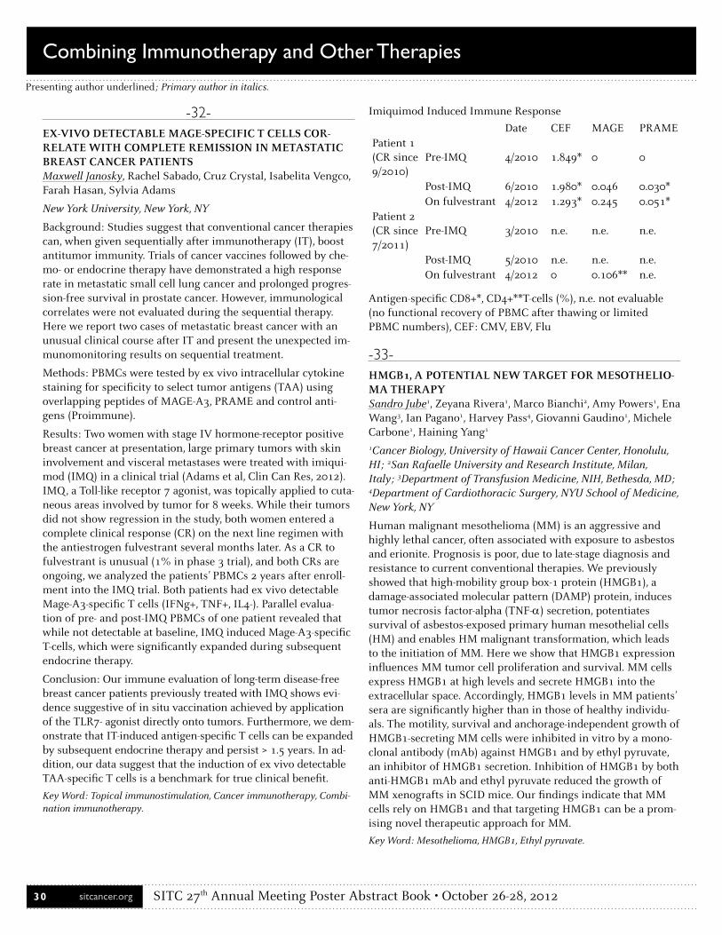
SITC 27th Annual Meeting Poster Abstract Book • October 26-28, 2012 30 sitcancer.org
Presenting author underlined; Primary author in italics.
-32-EX-VIVO DETECTABLE MAGE-SPECIFIC T CELLS COR-RELATE WITH COMPLETE REMISSION IN METASTATIC BREAST CANCER PATIENTSMaxwell Janosky, Rachel Sabado, Cruz Crystal, Isabelita Vengco, Farah Hasan, Sylvia Adams
New York University, New York, NY
Background: Studies suggest that conventional cancer therapies can, when given sequentially after immunotherapy (IT), boost antitumor immunity. Trials of cancer vaccines followed by che-mo- or endocrine therapy have demonstrated a high response rate in metastatic small cell lung cancer and prolonged progres-sion-free survival in prostate cancer. However, immunological correlates were not evaluated during the sequential therapy. Here we report two cases of metastatic breast cancer with an unusual clinical course after IT and present the unexpected im-munomonitoring results on sequential treatment.
Methods: PBMCs were tested by ex vivo intracellular cytokine staining for specificity to select tumor antigens (TAA) using overlapping peptides of MAGE-A3, PRAME and control anti-gens (Proimmune).
Results: Two women with stage IV hormone-receptor positive breast cancer at presentation, large primary tumors with skin involvement and visceral metastases were treated with imiqui-mod (IMQ) in a clinical trial (Adams et al, Clin Can Res, 2012). IMQ, a Toll-like receptor 7 agonist, was topically applied to cuta-neous areas involved by tumor for 8 weeks. While their tumors did not show regression in the study, both women entered a complete clinical response (CR) on the next line regimen with the antiestrogen fulvestrant several months later. As a CR to fulvestrant is unusual (1% in phase 3 trial), and both CRs are ongoing, we analyzed the patients’ PBMCs 2 years after enroll-ment into the IMQ trial. Both patients had ex vivo detectable Mage-A3-specific T cells (IFNg+, TNF+, IL4-). Parallel evalua-tion of pre- and post-IMQ PBMCs of one patient revealed that while not detectable at baseline, IMQ induced Mage-A3-specific T-cells, which were significantly expanded during subsequent endocrine therapy.
Conclusion: Our immune evaluation of long-term disease-free breast cancer patients previously treated with IMQ shows evi-dence suggestive of in situ vaccination achieved by application of the TLR7- agonist directly onto tumors. Furthermore, we dem-onstrate that IT-induced antigen-specific T cells can be expanded by subsequent endocrine therapy and persist > 1.5 years. In ad-dition, our data suggest that the induction of ex vivo detectable TAA-specific T cells is a benchmark for true clinical benefit.
Key Word: Topical immunostimulation, Cancer immunotherapy, Combi-nation immunotherapy.
Imiquimod Induced Immune Response
Date CEF MAGE PRAMEPatient 1 (CR since 9/2010)
Pre-IMQ 4/2010 1.849* 0 0
Post-IMQ 6/2010 1.980* 0.046 0.030*On fulvestrant 4/2012 1.293* 0.245 0.051*
Patient 2 (CR since 7/2011)
Pre-IMQ 3/2010 n .e . n .e . n .e .
Post-IMQ 5/2010 n .e . n .e . n .e .On fulvestrant 4/2012 0 0.106** n .e .
Antigen-specific CD8+*, CD4+**T-cells (%), n.e. not evaluable (no functional recovery of PBMC after thawing or limited PBMC numbers), CEF: CMV, EBV, Flu
-33-HMGB1, A POTENTIAL NEW TARGET FOR MESOTHELIO-MA THERAPYSandro Jube1, Zeyana Rivera1, Marco Bianchi2, Amy Powers1, Ena Wang3, Ian Pagano1, Harvey Pass4, Giovanni Gaudino1, Michele Carbone1, Haining Yang1
1Cancer Biology, University of Hawaii Cancer Center, Honolulu, HI; 2San Rafaelle University and Research Institute, Milan, Italy; 3Department of Transfusion Medicine, NIH, Bethesda, MD; 4Department of Cardiothoracic Surgery, NYU School of Medicine, New York, NY
Human malignant mesothelioma (MM) is an aggressive and highly lethal cancer, often associated with exposure to asbestos and erionite. Prognosis is poor, due to late-stage diagnosis and resistance to current conventional therapies. We previously showed that high-mobility group box-1 protein (HMGB1), a damage-associated molecular pattern (DAMP) protein, induces tumor necrosis factor-alpha (TNF-α) secretion, potentiates survival of asbestos-exposed primary human mesothelial cells (HM) and enables HM malignant transformation, which leads to the initiation of MM. Here we show that HMGB1 expression influences MM tumor cell proliferation and survival. MM cells express HMGB1 at high levels and secrete HMGB1 into the extracellular space. Accordingly, HMGB1 levels in MM patients’ sera are significantly higher than in those of healthy individu-als. The motility, survival and anchorage-independent growth of HMGB1-secreting MM cells were inhibited in vitro by a mono-clonal antibody (mAb) against HMGB1 and by ethyl pyruvate, an inhibitor of HMGB1 secretion. Inhibition of HMGB1 by both anti-HMGB1 mAb and ethyl pyruvate reduced the growth of MM xenografts in SCID mice. Our findings indicate that MM cells rely on HMGB1 and that targeting HMGB1 can be a prom-ising novel therapeutic approach for MM.
Key Word: Mesothelioma, HMGB1, Ethyl pyruvate.
Combining Immunotherapy and Other Therapies

SITC 27th Annual Meeting Poster Abstract Book • October 26-28, 2012 31sitcancer.org
Presenting author underlined; Primary author in italics.
-34-COMBINATION THERAPY WITH HSP90 INHIBITORS AND INTERFERONS SYNERGISTICALLY INCREASES MHC EXPRESSION LEADING TO ENHANCED TUMOR CELL RECOGNITION, IMPLICATIONS FOR IMMUNOTHERAPYJames T. Kurnick3,1, Timothy J. Haggerty1, Lenora B. Rose1, Ian S. Dunn1, Estelle E. Newton1, Franco Pandolfi2
1CytoCure LLC, Beverly, MA; 2Medicine, Catholic University Col. of Med., Rome, Italy; 3Pathology, Massachusetts Gen. Hosp., Boston, MA
In an effort to enhance antigen expression on human tumors, making them more susceptible to immune recognition and de-struction, we found that Hsp90 inhibitors (iHsp90) can enhance both differentiation antigens and MHC Class I in melanomas and gliomas. An additional series of tumors, including cancers of the breast, cervix, osteosarcoma and lymphomas were treated with a combination of iHsp90 and interferon to assess induc-tion of both Class I and II MHC. Class I MHC was enhanced by iHsp90 in most tumors, but induction of MHC Class II antigens was NOT induced in MHC Class II-negative tumors. Since both-Types I and II interferon’s (IFN) are known to enhance MHC antigen expression, we assessed their ability to synergize with iHsp90s. Importantly, since iHsp90s can inhibit IFN-signaling, we staggered the drug exposure of the tumor cells to the IFNs and iHsp90s to determine if we could circumvent the coun-teracting effects. While IFN-gamma stimulated strong Class II MHC expression, the simultaneous addition of iHSP90 largely prevented MHC Class II expression. Notably, pretreatment for 24 hrs with IFN-gamma allowed iHsp90 to further enhance MHC Class II expression. Similarly, synergistic enhancement of MHC Class I expression was noted with both IFN-beta or IFN-gamma when either was added prior to addition of iHsp90. Staggering treatment with interferon’s and iHsp90s yielded strong induction of both Class I and II MHC antigens. In con-trast to previous reports showing enhanced T cell recognition of tumors after iHsp90 treatment, the antigens we are assess-ing are not client proteins of Hsp90, and there is an increase in mRNA levels, cytoplasmic and surface protein expression, and increased induction of gene promoters. While there is an impact on pMEK and pERK expression following iHsp90 treat-ment, antigen induction is seen on a variety of tumors that vary in their expression of BRAF and NRAS activating mutations. Thus, there is a potential to utilize iHsp90 on a broad array of tumor types and tumor associated antigens that are impacted by Hsp90 expression, although they may not be client proteins of this important chaperon molecule. As both IFN and iHsp90 increased differentiation antigens and MHC, the further in-crease in MHC expression from the drug combination could be particularly useful for cancer immunotherapy.
Key Word: Immune escape, Combination immunotherapy, Tumor associ-ated antigen.
-35-EFFECTIVE ACTIVATION OF HIGHLY PURIFIED NK CELLS EXPANDED EX VIVO FOR THE ERADICATION OF EPITHELIAL OVARIAN CANCER CELLSIn-Kyung Lee1, Shin-Wha Lee2
1Department of Obstetrics and Gynecology, Asan Institute for Life Science, Seoul, Republic of Korea; 2Department of Obstetrics and Gynecology, Asan Medical Center, Seoul, Republic of Korea
Purpose: Ovarian cancer is the 7th most common malignancy and the 5th most common cause of death from cancers in wom-en. While many patients initially respond to surgery and che-motherapy, the long-term prognosis is generally unfavorable, with recurrence and development of drug-resistant disease. Therefore, new strategies for ovarian cancers, for example, im-munotherapy or targeted therapy, are needed. For that reason, this study evaluated the efficacy and safety of activated NK cells on ovarian cancer cells.
Methods: The patients were diagnosed with endometrioid ad-enocarcinoma (n = 2), papillary serous adenocarcinoma (n = 2). And two established human ovarian epithelial carcinoma cell lines (SKOV3 and OVCAR3) were used for all experiments. NK cells from Green Cross Corp. (Korea) were obtained and used as effector cells. MTT assay for cell viability, LDH release assay for the cytotoxic activity, and ELISA for the secretion levels of cytokines were measured for the capacity of activated NK cells.
Results:Firstly, an MTT assay was performed to determine the effect of NK cells on the proliferation of cancer cells. NK cells caused the loss of cancer cell viability and proliferation. Secondly, to investigate whether NK cells could have a cytotoxic effect on cancer cells, an LDH assay was performed. SKOV3 showed higher cytotoxicity than OVCAR3 in cancer cell lines and cytotoxicity of papillary serous adenocarcinoma cells was much more pronounced than all other cell types.Thirdly, to ana-lyze the release of cytokines, the levels of IFN-γ, TNF-α and IL-12 were examined. IFN-γ and TNF-α were significantly released at all ratios in all cell types.
Conclusion:Highly purified and activated NK cells can pro-duce various cytokines and many cytokines stimulate NK cell cytotoxicity in vitro. Generally, elevated levels of cytokines are related to cancer, and cytokines appear to play an important role in the progression of ovarian cancers due to their multi-functional roles. Also, highly purified and activated NK cells effectively increased cell cytotoxicity and proliferation, indicat-ing that activated NK cells could effectively eradicate cancer cells. Therefore, highly purified and activated NK cell-based treatment could be an alternative immunotherapy for ovarian cancer patients.
Key Word: Cancer immunotherapy.
Combining Immunotherapy and Other Therapies

SITC 27th Annual Meeting Poster Abstract Book • October 26-28, 2012 32 sitcancer.org
Presenting author underlined; Primary author in italics.
-36-THE COMBINATION OF CHEMOTHERAPY AND MY-COBACTERIUM ENHANCES THE ANTIGENICITY OF TUMOURS AND STIMULATE ALLOGENEIC T-CELL RE-SPONSESWai Liu, Daniel Fowler, Angus Dalgleish
Division of Clinical Sciences (Oncology), St George’s University of London, London, United Kingdom
Reduced expression of class 1 human leucocyte antigens (HLA1) is often a mechanism by which tumours evade surveillance by the host immune system. Consequently, strategies are currently being developed that restore immune-visibility, such as the use of mycobacterium. There is now an increasing body of evidence to suggest that standard chemotherapy drugs can also serve this purpose. Therefore, we have studied in a panel of tumour cell lines the expression of HLA1 before and after the chemotherapy agents cyclophosphamide, oxaliplatin or gemcitabine (GEM). Similarly, we have explored the effects of Bacillus Calmette-Guerin (BCG) on HLA1 expression on these cell lines through its capability to modify the cytokine output of leucocytes. We have also investigated the role that exudates produced by tumours play on the carcinogenic process, and have studied the impact that chemotherapies may have on their constitution. Our results show that some chemotherapy can increase HLA1 on tumour cells. For example, culturing with GEM caused significant increases in HCT116, A549 and MCF7 cells (MFI cf. untreated controls: 132 ± 30 vs. 33 ± 7.8, 0.23 ± 2.3 vs. 10 ± 0.67, and 45 ± 11 vs. 18 ± 3.7, respectively, p<0.01). Similarly, the cytokines found in supernatants from leucocytes treated with BCG were Th1-predominant, and capable of increasing tumour HLA1 expression. For example, MFI in MCF7 cells treated with these supernatants was 760 vs. 290 in those cells cultured with control supernatants (p<0.02). Increases in HLA1 were associated with increased cytolytic T-cell activities, which were negated by HLA1-blockade. We also showed that treating tumour cells with some chemotherapy could alter the pro-carcinogenic form of the exudates derived from these cells. Specifically, the extent of tubule formation by HUVECs was significantly reduced if super-natants from chemotherapy-treated A549 cells were used instead on supernatants from untreated tumours (1362 ± 192 vs. 2806 ± 225, p=0.006). Furthermore, this chemotherapy also altered the composition of tumour-exudates, which made them capable of augmenting DC maturation and T-cell function.
These studies highlight a secondary immunological effect of chemotherapy that renders it supportive of DC maturation and T-cell responsiveness. Also, these studies reaffirm the role of BCG as a putative immunotherapy through their cytokine-modifying effects on leucocytes and their capacity to enhance tumour visibility. Finally, as this facet of immune surveillance can also be restored by using appropriate chemotherapy agents, combining these treatments is clinically attractive.
Key Word: Chemotherapy, Tumour supernatants, Immunovisibility.
-37-COMBINED RADIOTHERAPY AND IMMUNOTHERAPY USING CPG OLIGODEOXYNUCLEOTIDES AND INDOL-AMINE 2,3 DIOXYGENASE (IDO) BLOCKADE IN MURINE 4T-1 MAMMARY CARCINOMA RESULTS IN GREATER ANTI-TUMOR EFFECTSArta M. Monjazeb1, Gail Sckisel2, Annie Mirsoian2, Steven Pai3, Anthony Zamora2, William J. Murphy4
1Radiation Oncology, UC Davis, Sacramento, CA; 2Immunology, UC Davis, Sacramento, CA; 3Comparative Pathology, UC Davis, Sacramento, CA; 4Dermatology, UC Davis, Sacramento, CA
Introduction: In rare instances localized radiotherapy can induce a systemic anti-tumor immune response known as the abscopal effect. Preclinical and clinical data suggest that the immunomodulatory effects of radiotherapy can be enhanced by combining it with immunotherapy. Two clinical trials have demonstrated the effectiveness of combining local tumor ir-radiation and local intratumoral injection of CpG immunother-apy. This local therapy induced a systemic immune response, as measured by regression of disease at distant, untreated sites, in approximately 27% of patients. There is preclinical data to suggest that the effectiveness of CpG immunotherapy may be limited by induction of the immunosuppressive enzyme, IDO. In this study we examine the effectiveness of combining local radiotherapy and CpG immunotherapy with systemic admin-istration of the IDO inhibitor, 1-Methyl D-Tryptophan (1-MT). We hypothesized that this combination therapy will enhance dendritic cell and T-cell effects on tumor growth and survival.
Methods: Mice with orthotopic 4T-1 tumor were divided into three groups and treated with sham radiotherapy (Group 1), local radiotherapy (Group 2), or local radiotherapy + Immuno-therapy (Group 3). Immunotherapy consisted of intratumoral CpG injection and 1-MT administered in the drinking water at 2mg/ml. Effects of therapy on tumor growth and survival were assessed and flow cytometric analysis was used for immunolog-ic profiling of the tumor, draining lymph nodes, and periphery.
Results: Compared to groups 1 and 2, the combination of ra-diotherapy and immunotherapy significantly improved overall survival (p=0.02). Likewise, mean tumor growth was decreased in group 3 compared to groups 1 and 2. Flow cytometric analy-sis revealed increased number of activated dendritic cells in the draining lymph nodes of mice receiving combined therapy. No treatment related toxicities were observed.
Conclusions: Combining radiotherapy and immunotherapy us-ing CpG oligodeoxynucleotides and IDO blockade is superior to radiotherapy alone in the treatment of murine 4T-1 mammary carcinoma and no toxicities were observed with this therapy. Combination therapy increases the activated dendritic cells in tumor draining lymph nodes. Further study is needed to deter-mine the immunologic effects and mechanism of this therapy.
Key Word: Indoleamine 2,3-dioxygenase 1, Abscopal, Radiotherapy.
Combining Immunotherapy and Other Therapies

SITC 27th Annual Meeting Poster Abstract Book • October 26-28, 2012 33sitcancer.org
Presenting author underlined; Primary author in italics.
-38-IMMUNO-SENSITIZATION OF MALIGNANT GLIOMAS BY NATURAL POLYPHENOLSChandramouli Mandalaparty1, Andrew Huang1, Sandeep Mit-tal1, Ping Q. Dou2, Prahlad Parajuli1
1Neurosurgery, Wayne State University & Karmanos Cancer Insti-tute, Detroit, MI; 2Oncology, Wayne State University & Karmanos Cancer Institute, Detroit, MI
Our lab has been investigating the adjuvant therapeutic poten-tial and associated molecular mechanisms of natural polyphe-nols in malignant gliomas. Pterostilbene is a methoxylated, natural analog of Resveratrol, found predominantly in blueber-ries and grapes. Recent studies have demonstrated the efficacy of Pterostilbene in extracranial tumor models. Pterostilbene was bioavailable in the brain even after low dose oral administra-tion and there was no toxicity to normal tissue. On the contrary, it was found to be neuroprotective in Alzheimer’s disease mod-els. Mechanistically, Pterostilbene modulates NF-κB, STAT3, Wnt/β-catenin signaling and also inhibits anti-apoptotic protein Bcl-xl. While these molecules are critical in imparting chemo-resistance, they are also pivotal in inducing immune-resistance and enhancing pro-tumor immune infiltration in gliomas. We hypothesize that Pterostilbene enhances the efficacy of immu-notherapy by inhibiting immune-suppression and -resistance in malignant gliomas via overlapping molecular mechanisms. A primary human glioma (pGl), a human glioma cell line (U87-MG) and a mouse glioma cell line (GL261) were used in our studies. The dose of Pterostilbene for 50% inhibition of prolif-eration (IC50), as determined by MTT assay, was observed to be between 10-20 μg/ml. Pterostilbene induced apoptosis in glio-mas in a dose-dependent manner, as detected by flow cytom-etry. Western blot analysis of glioma cells revealed a significant inhibition of Bcl-2, Bcl-xl and STAT3 as early as 1 hour after treatment with Pterostilbene (10 μg/ml). Importantly, 24 hour treatment with Pterostilbene (10 μg/ml) significantly enhanced the expression of TRAIL-R2 and MIC A/B in gliomas as deter-mined by flow cytometry. Pre-treatment of glioma cells with Pterostilbene significantly enhanced their cytolysis by CTLs and NK cells in an LDH-release assay, following 12-hour co-culture. Cytolysis was partially inhibited by anti-TRAIL-R and MIC A/B antibodies. Studies are underway to investigate combination of DC-based immunotherapy with Pterostilbene for the treatment of malignant gliomas using syngeneic, orthotopic mouse glioma models. The results provide valuable mechanistic insight into immune modulation by a natural polyphenol in gliomas. This study can have significant impact in developing a novel adju-vant therapy using Pterostilbene along with a DC-based vaccine to improve survival in patients with malignant gliomas.
Key Word: Glioblastoma, Immunomodulation, Combination immuno-therapy.
-39-EVALUATING THE SAFETY OF COMBINED IPILIMUMAB AND RADIOTHERAPY FOR THE TREATMENT OF META-STATIC MELANOMAMichael A. Postow, Christopher A. Barker, Shaheer A. Khan, Jedd D. Wolchok
Memorial Sloan-Kettering Cancer Center, New York, NY
Background: Ipilimumab (ipi) improves overall survival in patients (pts) with metastatic melanoma (MM) but only a subset benefit. Preclinical and early clinical evidence suggest radiotherapy (RT) may increase the efficacy of immunotherapy. Little is known about the toxicity of administering RT with ipi. We assessed the safety of concomitant palliative RT and ipi and preliminarily examined efficacy.
Methods: After IRB approval, medical records of 17 pts with MM treated with non-brain RT during ipi induction were reviewed. CT-CAE v4.0 was used to grade immune-related adverse events (irAEs) occurring after the initiation of RT and ≤ 3 months of the last ipi induction dose. Efficacy was determined by clinician’s assessment of improved pt symptoms within the irradiated field and by radio-graphic assessment of tumors outside of the irradiated field.
Results: From 2005-2011, 17 pts received 19 courses of RT (10 bone metastases, 6 lymph nodes, 1 skin metastasis, 1 perirectal tumor, and 1 periorbital tumor). RT ranged from 20-62.5 Gy total delivered over 1-25 fractions (median 30 Gy in 5 fractions). For 12 pts receiving RT with 3mg/kg of ipi: 4 had grade (G) 1 irAEs (5 events: 2 diarrhea, 2 rash, 1 hypophysitis), 1 had G2 rash, and 1 had G3 diarrhea. Using a one-tailed Fisher’s exact test, the propor-tion of pts with G3/4 toxicity in this small cohort (1/12, 8.3%) did not exceed the rate of G3/4 irAEs (19/131, 14.5%) in a prior phase III trial of pts receiving ipi at 3mg/kg (p=0.4759). (Hodi 2010) For 5 pts receiving RT with 10mg/kg of ipi: 1 had G1 pruritis, 2 had G2 irAEs (rash, cytokine release syndrome), and 2 had G3 irAEs (3 events: 2 hepatitis and 1 rash). Numbers were very small, but the proportion of pts with G3/4 toxicity (2/5, 40%) was higher than the rate of G3/4 irAEs (18/71, 25.4%) in a prior phase II trial of pts treated with ipi at 10mg/kg,(Wolchok 2009) though no statistical difference was seen (p=0.3956). No pts had G4 events. 3 pts had local inflammation in the RT field (proctitis, cystitis, and localized dermatitis). Within 12 weeks of ipi initiation, 11 pts had improvements within the irradiated field: less pain (5), better vi-sion (1), reduced bleeding (3), and decreased tumor size (2). 3 pts had tumor sites that regressed outside of the irradiated field and 1 pt had overall stable disease. 2 pts died of disease.
Conclusions: This is an early description of the safety of combining RT with ipi induction for MM. RT does not seem to increase the toxicity of ipi at 3mg/kg. There were likely too few pts treated with RT during ipi induction at 10mg/kg to assess safety. RT appeared locally effective in palliating sites of MM. Ongoing prospective evaluation is necessary to assess improve-ments in efficacy compared to ipi alone.
Key Word: Melanoma immunotherapy, Abscopal, CTLA-4.
Combining Immunotherapy and Other Therapies
cstroh
Text Box
WITHDRAWN BY AUTHOR

SITC 27th Annual Meeting Poster Abstract Book • October 26-28, 2012 34 sitcancer.org
Presenting author underlined; Primary author in italics.
-40-CSPG4 AS A COMBINATORIAL TARGET OF ANTIBODY-BASED IMMUNOTHERAPY FOR MALIGNANT MESOTHE-LIOMAZeyana S. Rivera1, Soldano Ferrone2, Xinhui Wang2, Sandro Jube1, Maria Prat3, Haining Yang1, Giovanni Gaudino1, Michele Carbone1
1Cancer Biology, University of Hawaii Cancer Center, Honolulu, HI; 2Immunology, Hillmann Cancer Center, Honolulu, PA; 3Bio-medical Sciences, University of Piemonte Orientale, Novara, Italy
Malignant mesothelioma (MM) is an aggressive tumor, resistant to conventional therapies, with median survival of 1 year from diagnosis. The lack of effective therapies available prompted us to implement antibody-based immunotherapy against novel tar-gets and to investigate possible synergies with chemotherapy on MM. Chondroitin sulphate proteoglycan 4 (CSPG4) is involved in the onset and progression of melanoma and other tumors, as well as in neo-angiogenesis. CSPG4-specific monoclonal anti-bodies (mAb) have shown anti-tumor effects in both preclinical and clinical trials of melanoma. We hypothesized that target-ing CSPG4 with a CSPG4-specific mAb would have therapeutic efficacy against MM. We found that CSPG4 was aberrantly expressed in 6 out of 8 MM cell lines and in 24 out of 40 MM biopsies, with minimal expression in surrounding nonmalignant cells. The expression of CSPG4 in MM was higher upon engage-ment of extracellular matrix components (ECM) and was associ-ated with increased MM cell viability and motility. Silencing of CSPG4 expression by siRNAs supported this functional signifi-cance of CSPG4 in MM. Consistently, the CSPG4-specific mAb TP41.2 inhibited MM cell attachment to the ECM, resulting in decreased phosphorylation of FAK and AKT, decreased expres-sion of cyclin D1 and apoptosis. Moreover, TP41.2 significantly reduced MM cell motility, migration and invasiveness, and inhibited MM growth in soft agar. TP41.2 prevented the out-growth of human MM tumors in SCID mice and inhibited the growth of established MM xenografts, resulting in significantly extended survival of tumor-bearing mice. These results represent a novel approach for CSPG4 mAb-based immunotherapy of MM. Combinatorial strategies with chemotherapeutic agents and with mAbs targeting c-Met related receptor are ongoing, aimed at translating CSPG4-specific mAb into clinical setting.
Key Word: Immunotherapy, Mesothelioma, CSPG4.
-41-INHIBITION OF PROTEASOME DEUBIQUITINATING AC-TIVITY RENDERS TUMOR CELLS SENSITIVE TO TRAIL-MEDIATED APOPTOSIS BY NATURAL KILLER CELLS AND T CELLSDhifaf Sarhan, Erik Wennerberg, Padraig D’Arcy, Deepthy Gura-jada, Stig Linder, Andreas Lundqvist
Oncology and Pathology, Karolinska Institutet, Stockholm, Sweden
The proteasome inhibitor bortezomib simultaneously renders tumor cells sensitive to killing by natural killer (NK) cells and
resistant to killing by tumor-specific T cells. Here we show that inhibition of proteasome deubiquitinating activity by treat-ment with b-AP15 sensitizes tumors to both NK and T cell-mediated killing. Exposure to b-AP15 significantly increased the susceptibility in 17 and 6 tumor cell lines of various origin to NK (p≤0.001) and T cell (p=0.01) -mediated cytotoxicity respec-tively Tumors sensitized to NK or T cell-mediated cytotoxicity showed a significant increase in surface expression of TRAIL [tumor necrosis factor-related apoptosis-inducing ligand] recep-tor-2 (p=0.01). In tumor-bearing SCID/Beige mice, treatment with b-AP15 followed by infusion of either human NK cells or tumor-specific T cells resulted in a significantly delayed tumor progression compared to mice treated with NK (p=0.006) or T cells (p≤0.001) cells alone. Furthermore, combined infusion of NK and T cells in tumor-bearing BALB/c mice following treatment with b-AP15 resulted in a significantly prolonged long-term survival compared to mice treated with b-AP15 and NK or T cells (p≤0.005). Our findings show that b-AP15-induced sensitization to TRAIL could be used as a novel strategy to potentiate the anticancer effects of adoptively infused NK and T cells in patients with cancer.
Key Word: Adoptive immunotherapy.
-42-PERSONALIZED PEPTIDE VACCINE IN COMBINATION WITH CHEMOTHERAPY FOR PATIENTS WITH AD-VANCED COLORECTAL CANCERTetsuro Sasada, Shigeru Yutani, Satoko Matsueda, Nobukazu Komatsu, Kyogo Itoh
Department of Immunology and Immunotherapy, Kurume Uni-versity School of Medicine, Kurume, Japan
Background: Despite recent development of various types of chemotherapeutic and molecular targeted agents, the prognosis of advanced colorectal cancer remains poor. Therefore, novel therapeutic approaches, including cancer vaccine, need to be established. We conducted a phase II study of personalized peptide vaccine (PPV) in combination with chemotherapy for advanced colorectal cancer patients.
Methods: Sixty-one advanced colorectal cancer patients (Stage IV, 27, recurrent, 34) who failed at least the first line chemo-therapy were enrolled. Median duration of chemotherapy prior to the enrollment was 526 days. A maximum of four HLA-matched vaccine peptides showing higher antigen-specific im-mune responses were selected based on the titers of IgG against each of 31 different peptide candidates. The selected peptides were subcutaneously administered in combination with che-motherapeutic and/or molecular targeted agents. In order to identify potentially prognostic biomarkers for overall survival (OS), pre-vaccination clinical findings and laboratory data were statistically evaluated.
Results: Median OS from the first vaccination was 355 days with 1-year and 2-year survival rates of 47.5% and 25.0%, respec-
Combining Immunotherapy and Other Therapies

SITC 27th Annual Meeting Poster Abstract Book • October 26-28, 2012 35sitcancer.org
Presenting author underlined; Primary author in italics.
tively. Notably, median OS from the first line chemotherapy was 1127 days with 3-year and 5-year survival rates of 53.6% and 22.8%, respectively. The main toxicity of PPV was skin reactions at injection sites, but no vaccine-related serious adverse events were observed. Among the pre-vaccination factors examined, high levels of inflammatory factors, including IL-6, CRP, and se-rum amyloid A (SAA), were significantly unfavorable for OS (P < 0.0001, P = 0.0005, and P=0.0001, respectively). The numbers of previous chemotherapy regimens and serum albumin levels were also associated with OS (P = 0.0174 and P = 0.0444).
Conclusions: Even in advanced colorectal cancer patients, combined treatment with PPV and chemotherapeutic and/or molecular targeted agents might improve prognosis. To inhibit the inflammatory factors that might negatively affect immune responses to cancer vaccines, a new clinical trial of PPV in com-bination with anti-IL-6 receptor antibody is currently underway for advanced colorectal cancer patients.
Key Word: Cancer vaccine, Colorectal cancer, Chemotherapy.
-43-TUMOR-DIRECTED MYELOID EXPANSION IS REVERSED BY RADIATION THERAPYTalicia Savage1, Michael J. Gough1, Benjamin Cottam1, Pippa Newell1,2, William L. Redmond1, Keith S. Bahjat1, Marka R. Crit-tenden1,2
1Earle A Chiles Research Institute, Providence Cancer Center, Portland, OR; 2The Oregon Clinic, Portland, OR
The expansion of myeloid cell lineages has been correlated with cancer progression. Myeloid cells are associated with immune suppression, angiogenesis, metastasis, and have been shown to limit the response to therapy. In the MMTV-PyMT spontane-ous mammary carcinoma mouse model, we demonstrated that tumor progression was associated with myeloid expansion in the peripheral blood and this expansion correlated with increased numbers of macrophages in the tumor. A number of published studies have demonstrated that tumor-associated myeloid cells suppress T cell responses in vitro and in vivo. Reducing the tumor burden through surgical resection and chemotherapy has been shown to reduce myeloid numbers. However, chemo-therapies that reduce tumor burden also have direct effects on myeloid cells, and surgical trauma has been shown to mobilize myeloid cells. We propose that focal radiation therapy of the tu-mor is an appropriate model to test the hypothesis that myeloid expansion is directly related to tumor burden. To test the conse-quence of radiation therapy, we treated the 4T1 mammary car-cinoma with 3x20Gy focal radiation in a hind limb model that allows focal radiation of the primary tumor with minimal dos-age to radiosensitive regions of the torso. Fresh whole blood was collected from mice over the course of treatment and myeloid numbers were calculated by quantitative flow cytometry. While 4T1 tumor growth caused a dramatic expansion of myeloid cells in peripheral blood, we demonstrate that treatment with focal
radiation resulted in a myeloid contraction and tumor control. However, we demonstrate that residual metastatic disease can prevent myeloid numbers returning to baseline levels observed in tumor-free mice. We also demonstrate that tumor recurrence from residual disease correlated with a return of myeloid expan-sion and there remained a close correlation between gross tumor weight and myeloid numbers regardless of the route of tumor challenge and the presence or absence of radiation therapy. Interestingly, while T cell numbers did not change with radia-tion treatment, we demonstrated an increase in CD3+CD8+ and CD3+CD4+FoxP3- proliferation following radiation that corre-lated with the reduction in myeloid numbers. Our data dem-onstrates that reduction in tumor burden by radiation therapy causes a significant decrease in an immunosuppressive cell population and a corresponding increase in T cell proliferation. We propose that these data indicates a window of opportunity following radiation therapy for adjuvant immunotherapy.
Key Word: Micrenvironment, Radiotherapy, Macrophages.
-44-INTRAVESICAL TREATMENT OF ORTHOTOPIC BLADDER CANCER WITH CHITOSAN/IL-12 INDUCES SYSTEMIC TUMOR-SPECIFIC IMMUNITYSean G. Smith, Lirong Yang, David Zaharoff
Biomedical Engineering, University of Arkansas, Fayetteville, AR
Bladder cancer is the sixth most common non-cutaneous can-cer diagnosis in the U.S. with an estimated 73,510 new cases and 14,880 deaths in 2012. Although 70-80% of patients are diagnosed early with superficial (non-invasive) disease, bladder cancer has a recurrence rate of approximately 65%. For nearly three decades, Mycobacterium bovis bacillus Calmette-Guerin (BCG) has been the standard of care intravesical immuno-therapy for superficial bladder cancer. While touted as the most effective cancer immunotherapy to date, 20-30% of patients who undergo BCG therapy fail initial treatment while 30-50% of BCG responders develop recurrent tumors within 5 years. Furthermore, a significant limitation of intravesical BCG is its inability to generate adaptive tumor-specific immunity leading to the need for long-term maintenance and continuous surveil-lance of bladder cancer patients. These limitations combine to justify the search for more effective therapies.
Interleukin-12 (IL-12) is under investigation as a candidate to replace BCG. IL-12 has demonstrated remarkable anti-tumor activity against a range of malignancies in preclinical studies. However, schedule-dependent toxicities associated with sys-temic administrations have limited its clinical use. The intraves-ical route of administration offers an opportunity to minimize systemic exposure to IL-12 and its associate adverse events. Our previous studies demonstrated that we can enhance intravesical delivery of IL-12 through co-formulation with a solution of chi-tosan. Chitosan is an abundant, natural polysaccharide derived primarily from the exoskeletons of crustaceans. It is a non-toxic,
Combining Immunotherapy and Other Therapies

SITC 27th Annual Meeting Poster Abstract Book • October 26-28, 2012 36 sitcancer.org
Presenting author underlined; Primary author in italics.
biodegradable, non-immunogenic unbranched copolymer of glucosamine and N-acetylglucosamine units linked by β(1-4) glycosidic bonds. Three or four weekly intravesical treatments with IL-12 in chitosan solution (chitosan/IL-12) cures 80-100% of mice bearing orthotopic MB49 bladder tumors. BCG, on the other hand, is ineffective in this aggressive model.
Our recent studies demonstrate for the first time that mice cured following intravesical chitosan/IL-12 immunotherapy rejected a distant flank tumor challenge in a tumor-specific manner, 100% of previously cured mice were protected from s.c. MB49 rechallenge, but not s.c. B16 challenge. Protective im-munity was found to be dependent on CD4+ and CD8+ subsets as their depletion abrogated protection. These data are the first to demonstrate that an intravesical IL-12-based immunotherapy for bladder cancer can induce a systemic tumor-specific im-mune response. This is particularly important for patients with highly recurrent bladder cancer as standard-of-care BCG does not generate tumor-specific immunity.
Key Word: Tumor immunity, IL-12, BCG.
-45-SYNERGISTIC THERAPEUTIC EFFECT OF BLOCKING TWO IMMUNE INHIBITORY PATHWAYS CONCUR-RENTLY IN THE CONTEXT OF A TOLEROGENIC TUMOR MICROENVIRONMENT IN VIVOStefani Spranger1, Michael Leung1, Thomas F. Gajewski1,2
1Pathology, University of Chicago, Chicago, IL; 2Medicine, Univer-sity of Chicago, Chicago, IL
Over the last two decades, numerous mechanisms have been identified that contribute to immune suppression in the context of a growing tumor. The first therapeutic approach to reach clinical practice is the blockade of CLTA-4 using the anti-CTLA-4 mAb ipilimumab. Despite this therapeutic advance, this regimen is effective in only a minority of patients, and therefore further treatment refinements are required. Based on these notions, we have initiated studies of rational combina-tions of agents in preclinical models to target two independent inhibitory pathways concurrently, in a logical fashion. For this we intend to uncouple inhibitory pathways largely operational in lymph nodes (CTLA-4) and in the tumor (PDL1-PD1 interac-tions) combined with treatments that affect T cell anergy (IL-7, LAG-3, 4-1BB), metabolic dysregulation (IDO) or regulatory T cells (Tregs, using CD25-depletion). The first combinations tested have utilized anti-CTLA-4 mAb as an anchor. Using the well-characterized tumor model of B16-SIY cells pre-established in syngeneic mice in vivo, we found a major synergistic effect of combining anti-CTLA-4 with either an IDO inhibitor, with anti-PD-L1 mAb, or with CD25-depletion. The other combina-tions are being explored similarly. This synergistic therapeutic effect already seen with strategies that have similar agents
available for clinical testing points towards logical combination studies to prioritize for clinical trials in cancer patients.
Key Word: Tumor immunity, Adoptive therapy, CTLA-4.
-46-TGFβ IS A MASTER REGULATOR OF THE PRO-IMMUNO-GENIC EFFECTS OF RADIOTHERAPYClaire Vanpouille-Box1, Karsten A. Pilones1, Sophie Bouquet2, Jiri Zavadil3, Silvia Formenti2, Mary-Helen Barcellos-Hoff2, Sandra Demaria1
1Pathology, NYU School of Medicine, New York, NY; 2Radiation Oncology, NYU School of Medicine, New York, NY; 3Pathology, NYU Cancer Institute and NYU Center for Health Informatics and Bioinformatics, New York, NY
Radiation therapy has the potential to convert the tumor into an in situ individualized vaccine by inducing immunogenic can-cer cell death and pro-inflammatory cytokines and chemokines, however this potential is rarely realized by irradiation alone. We hypothesized that radiation-induced immunosuppressive factors may hinder its pro-immunogenic effects. Transforming growth factor β (TGFβ) has immunosuppressive function for dendritic cells and T cells and is activated by radiation. Here we tested the hypothesis that inhibiting TGFβ during radiation treatment would induce an immunogenic response.
Poorly immunogenic, highly metastatic 4T1 carcinoma cells were injected s.c. in syngeneic BALB/c mice (day 0). TGFβ neutralizing 1D11 or isotype control 13C4 monoclonal antibodies were given i.p. (200μg/mouse) every other day from day 12 to 28. Tumors were irradiated with 6 Gy on five consecutive days beginning on day 13. Tumor growth was measured consecutively. Mice were euthanized at day 21 for analysis, at day 28 for enumeration of lung metastases, or followed for survival. Gene expression profiles were obtained using Affymetrix mouse genome 430 2.0 array.
Tumor growth rates and the frequency of lung metastases were similar in mice receiving control antibody or 1D11 alone. Radia-tion treatment caused significant (p=0.0065) tumor growth delay but did not inhibit lung metastases. In contrast, mice treated with both 1D11 and radiation exhibited significantly greater tumor growth control and reduced lung metastases (p<0.0001), and significantly prolonged survival (p<0.005). As expected, TGFβ sig-nalling was inhibited with 1D11 as measured in CD4+ and CD8+ T cells from tumor-draining lymph nodes at day 21. CD8+ T cells producing IFNγ in response to a tumor-specific antigen were de-tected only in mice treated with 1D11 and radiation. Expression profiles showed that genes associated with immune response and T cell activation were upregulated in irradiated tumors of mice treated with 1D11 compared to other treatment groups. In vivo depletion experiments demonstrated that T cells were essential for the improved tumor control and inhibition of lung metastases of mice treated with 1D11 and radiation.
Combining Immunotherapy and Other Therapies

SITC 27th Annual Meeting Poster Abstract Book • October 26-28, 2012 37sitcancer.org
Presenting author underlined; Primary author in italics.
These data support a critical role for TGFβ as a regulator of the pro-immunogenic effects of local tumor radiotherapy. Inhibi-tion of TGFβ during radiotherapy may promote self-immuniza-tion and achieve systemic control of metastatic disease.
Supported by DOD BCRP grant BC100481P2.
Key Word: Breast cancer, Radiotherapy, Immunotherapy.
-47-INTRATUMORAL CHITOSAN/IL-12 NEOADJUVANT TO TUMOR RESECTION REDUCES BREAST CANCER METAS-TASISJimmy Vo, Lirong Yang, David Zaharoff
Department of Biomedical Engineering, University of Arkansas, Fayetteville, AR
Interleukin-12 (IL-12) is a potent antitumor cytokine that exhibits significant clinical toxicities after systemic adminis-tration. Local delivery strategies capable of maintaining high concentrations of IL-12 in the tumor microenvironment while minimizing systemic exposure are under investigation. We have previously shown that intratumoral injections of IL-12 co-formulated with a solution of chitosan (chitosan/IL-12) can eliminate established primary tumors. Chitosan is an abundant, natural polysaccharide derived primarily from the exoskeletons of crustaceans. Chitosan has been shown to maintain high lo-cal concentrations of protein antigens and cytokines through viscous, electrostatic and bioadhesive interactions.
Because of IL-12’s well documented ability to generate tumor-specific cell-mediated immunity, we have recently begun to explore the anti-metastatic potential of intratumoral chitosan/IL-12 immunotherapy in a highly metastatic model of breast cancer (4T1). Thus far, we have found that intratumoral chitosan/IL-12 neoadjuvant prior to tumor resection confers a long-term survival benefit (Figure 1). Specifically, mice bear-ing flank 4T1 tumors were treated intratumorally with saline, IL-12, chitosan, or chitosan/IL-12 on days 6 and 12 after tumor implantation. All primary tumors were resected on day 15. Mice treated with either saline or chitosan alone died within 33 days after resection. 2/13 mice (~15%) treated with IL-12 alone remain alive more than 100 days after resection. 8/14 mice (~57%) treated with chitosan/IL-12 remain alive more than 100 days after resection. Furthermore, splenocytes from long term survivors demonstrated anti-tumor immunity in a cytotoxic T lymphocyte (CTL) killing assay. Specifically, after 1 week of in vitro stimulation, splenocytes were found to lyse approximately 40% of target tumor cells at an effector:target ratio of 50:1. Based on data obtained thus far, intratumoral chitosan/IL-12 shows promising potential as a neoadjuvant immunotherapy to reduce metastatic disease.
Key Word: Breast cancer, IL-12, Metastases.
Figure 1: Chitosan/IL-12 immunotherapy improves survival fol-lowing breast tumor resection. Mice bearing 4T1 tumors were given i.t. treatments with saline (filled square), chitosan (filled triangle), IL-12 (filled circle) or chitosan/IL-12 (open circle) prior to resection .
-48-PHASE II TRIAL DAILY PULSE INTERLEUKIN-2 THERAPY DURING MARROW/LYMPHOCYTE RECOVERY IN ACUTE MYELOID LEUKEMIA NCT#01289678Paul R. Walker, Manali K. Kamdar, Adam S. Asch
Oncology, Leo Jenkins Cancer Center, Brody School of Medicine at East Carolina University, Greenville, NC
Background: Therapeutic advances in the first line treatment of acute myelogenous leukemia (AML) have yet to step beyond high-dose cytarabine. Allogeneic stem cell transplant (Allo-SCT) can cure patients with relapsed and high-risk AML with a graft versus leukemia immune effect. Growth of clonogenic leukemic blasts can be abrogated following preincubation with lympho-kine activated killer (LAK) cells. (Lotzova et al Leuk Res 1987, Lista et al Eur J Haematol 1989) Interleukin-2 (IL-2) will gener-ate LAK cells in peripheral blood and bone marrow. (Foa et al Cancer Res 1991) High-dose IL-2 alone in relapsed AML has achieved complete remissions. (Meloni et al Blood 1994, Mara-inichi et al Blood 1991) Early absolute lymphocyte recovery after induction chemotherapy predicts a superior survival in AML suggesting a critical role of early immune recovery. (Behl et al Leukemia 2006) Even with an Allo-SCT, slow absolute lymphocyte recovery at day 29 is associated with a higher risk of relapse. (Powles et al Blood 1998) Stimulated LAK cells by IL-2 during this critical early lymphocyte recovery period may enhance a graft versus leukemia immune effect and potentially improve the outcome of first-line AML treatment.
Methods: 9 patients (range ages 33-72) with de novo or second-ary AML received IL-2 during induction and/or post-remission treatment marrow nadir recovery. Eligibility criteria included
Combining Immunotherapy and Other Therapies

SITC 27th Annual Meeting Poster Abstract Book • October 26-28, 2012 38 sitcancer.org
Presenting author underlined; Primary author in italics.
a rising WBC count above 500mm3, supported platelet count above 20,000 and hemodynamic stability. IL-2 18 million IU/m2 was administered over 15-30 minutes daily for 5 days within 7 days of a WBC rising above 500mm3.
Results: 3 patients received IL-2 during induction recovery, 3 during re-induction recovery, 3 during consolidation recovery. 1 patient subsequently underwent an Allo-SCT in remission. Median OS 25 months. With a median followup of 36 months (range 12-60 months), 5 of 9 (55%) treated patients and 4 of 8 (50%) patients without an Allo-SCT, are alive and leukemia free. Of the 5 alive, 3 are over the age of 60 and 2 had complex cyto-genetics. Expected toxicities with IL-2 included grade 3 fever/chills (77%) but no respiratory or hemodynamic complications requiring ventilator support.
Conclusions: IL-2 therapy administered during marrow/lym-phocyte recovery after standard induction/consolidation che-motherapy in first-line AML treatment achieved a 50% 3-year overall and leukemia free survival without an Allo-SCT.
Key Word: Interleukin-2, Leukemia.
-49-DASATINIB PRIMES HUMAN CELLS OF MYELOID ORI-GIN TO TLR-INDUCED IL12 SYNTHESISMatthias Wölfl1, Stefanie Schwinn1, Young-Eun Yoo1, Marie L. Ress1, Martin Chopra2, Susanne C. Schreiber1, Victor I. Ayala3, Claes Ohlen3, Matthias Eyrich1, Andreas Beilhack2, Paul G. Schlegel1
1Children’s Hospital, University of Würzburg, Würzburg, Ger-many; 2Department of Medicine II, University of Würzburg, Würzburg, Germany; 3AIDS and Cancer Virus Program, Frederick National Laboratory for Cancer Research, Frederick, MD
Dasatinib is a dual bcr/abl/src-kinase inhibitor used for the treatment of bcr/abl+ leukemias. Originally it was designed as immunosuppressant and has been shown to inhibit effector T-cell activation. The clinically observed increase of CD8+ T-cells and NK-cells with an LGL-phenotype, associated with colitis and pleuritis, is poorly understood.
We studied the effect of dasatinib on the antigen-specific priming of naïve, human CD8+ T-cells by dendritic cells, using a previously validated priming system (Wölfl et al. CII, 2011). This system allows the quantitative and qualitative evaluation of the T-cell response against the melanosomal antigen MART1 after only 10 days of in vitro expansion. Dasatinib was added either at the time of the initial DC/TC interaction, or during maturation of the DC. Furthermore, purified myeloid subsets (dendritic cells, CD14+ monocytes, slan+ DC, CD1c+ DC) as well as DC from C57/B6-mice and rhesus macaques were evaluated functionally in response to different maturation stimuli in com-bination with dasatinib .
In striking contrast to the strong suppression observed when dasatinib was present during DC/T-cell interaction, T-cell responses were clearly enhanced, when only the DC had been pre-incubated with dasatinib. Analyzing IL-12p40 production in DC from 10 donors revealed increased responsiveness to LPS (Median% of IL12p40+DC: LPS/IFNg: 11.6%, LPS/IFNg/Dasat-inib: 24.6%, p<.0001). Subsequently, higher IL-12 production ac-counted for T-cells with better TCR avidity leading to increased tumor recognition. This effect was seen in human cells but not in mouse and macaques DC. We identified several src-kinase inhibitors, some of which already in clinical use, with similar effects on IL-12 production in DC. When tested directly ex vivo, the sensitizing effect of dasatinib to TLR-mediated activation was observed in various cell types of the myeloid compartment (monocytes, slan+ DC, CD1c+ DC). Furthermore, biochemical analysis revealed involvement of src-kinases in the upregulation of IL12-production upon dasatinib treatment, suggesting inhibi-tion of central regulatory pathways of DC activation.
Our findings suggest that increased CD8+ and NK-cell counts observed clinically may be the result of dasatinib-mediated effects on stimulated myeloid cells. Moreover targeting the involved regulatory pathways in DC may greatly improve their function. Such increased functionality of antigen-presenting cells is critical for immunotherapy approaches as well as for the development of a molecularly designed adjuvant.
Key Word: Combination immunotherapy, Dendritic cell, Targeted thera-peutics.
-50-INHIBITING MONOCYTE/MACROPHAGE-NEUROBLASTO-MA CELL INTERACTIONS WITH LENALIDOMIDE IN-CREASES TUMOR CELL RESPONSE TO CYCLOPHOSPHA-MIDE AND TOPOTECANYibing Xu1, Jianping Sun1, Zesheng Wan1, Robert Seeger1,2
1Center for Cancer and Blood Diseases and Saban Research Institute, Childrens Hospital Los Angeles, Los Angeles, CA; 2Keck School of Medicine, University of Southern California, Los Ange-les, CA
Background: New therapeutic strategies based upon under-standing the tumor microenvironment may improve the ef-ficacy of chemotherapy and, as a result, long-term survival for patients with high-risk neuroblastoma. We showed that tumor associated monocytes/macrophages (TAMs) and cytokines (IL-6/IL-6R, IL-7, IL-10, IL-8, VEGF, TNFα, and TGFβ1) are prominent in high-risk neuroblastomas. We hypothesize that the milieu created byTAM-neuroblastoma interactions promotes tumor growth and interferes with the efficacy of targeted and non-targeted chemotherapeutic agents. Our specific aims were to 1) characterize TAM-neuroblastoma interactions, and 2) determine if lenalidomide, an immunomodulating drug, inhibits these
Combining Immunotherapy and Other Therapies

SITC 27th Annual Meeting Poster Abstract Book • October 26-28, 2012 39sitcancer.org
Presenting author underlined; Primary author in italics.
interactions and improves efficacy of cyclophosphamide-topo-tecan chemotherapy. Methods: The effect of TAMs upon tumor cell growth was determined in vitro with co-cultures (Boyden chamber, 15 cell lines tested) and in vivo with subcutaneous co-injection of monocytes and tumor cells into NOD/SCID mice (3 cell lines tested). Using co-cultures, tumor cell DNA synthesis was determined by flow cytometry, pathway activation by West-ern blotting, and cytokine secretion by Luminex® multiplex assays. The NOD/SCID model was used to determine effects oflenalidomide and cyclophosphamide-topotecan upon growth of luciferase expressing neuroblastoma cell lines. Results: Co-culture and co-injection into NOD/SCID mice of monocytes with neuroblastoma cells uniformly and markedly enhanced tu-mor cell growth. Co-culture increased tumor cell DNA synthesis, phosphorylation of STAT3, PI3K (110α, 110γ, p85, p55), AKT, mTOR, S6, 4E-BP1, and IKKβ in both tumor cells and TAMs and secretion of G-CSF, IL-10, IL-1RA, IL-6, IL-7, CCL5, TNFα, and VEGF>10-fold. Under these same conditions, lenalido-mide significantly inhibited tumor cell growth, DNA synthesis, protein phosphorylation, and cytokine secretion. Tumor cells co-injected with monocytes were most effectively eliminated by lenalidomide with cyclophosphamide-topotecan compared to lenalidomide or cyclophosphamide-topotecan alone (p=0.01). Conclusions: Chemotherapy with cyclophosphamide-topotecan is improved by lenalidomide, which inhibits pro-tumor interac-tions of TAMS and neuroblastoma cells.
Key Word: Chemokines, Chemotherapy, Immunotherapy.
Combining Immunotherapy and Other Therapies
SITC 27th Annual Meeting Poster Abstract Book • October 26-28, 2012 40 sitcancer.org
Presenting author underlined; Primary author in italics.
-51-SELECTION OF A CONTROL INFECTIOUS DISEASE VAC-CINE FOR CANCER IMMUNOTHERAPY TRIALSKatherine Arns1, Janet A. Englund3, Mary L. Disis1, Chihiro Mor-ishima1,2
1Tumor Vaccine Group, University of Washington, Seattle, WA; 2Department of Laboratory Medicine, University of Washington, Seattle, WA; 3Department of Pediatrics, University of Washing-ton, Seattle, WA
An increasing number of clinical trials are being conducted to test vaccines directed against tumor-associated antigens for the treatment of various malignancies. To fulfill the promise of the widespread dissemination of immunotherapies for cancer, it will be critical to employ best practices in the design and con-duct of these clinical trials. One significant concern is the ability to induce robust tumor antigen-specific cellular immunity among populations of patients who are older, ill from advanced disease, and potentially lymphopenic as a result of preceding courses of standard-of-care chemotherapy. Thus, the utilization of a vaccine control demonstrating that these patients are capa-ble of responding to immunizing protocols would be beneficial. We have conducted a thorough review of commercially avail-able, licensed infectious disease vaccines in order to identify the most suitable vaccine for this purpose. An ideal control vaccine would have demonstrated ability to induce anamnestic antigen-specific T cell responses that could be consistently measured. Other desirable characteristics would include minimal adverse effects, a relatively low prevalence of natural immunity and/or vaccination rates in an older adult population, and a pos-sibility of clinical benefit, making the vaccine acceptable to all trial enrollees. We eliminated potential vaccines if they were contraindicated for immunocompromised patients, had substantial adverse effects in any population including healthy patients, were not licensed for older adults, were only approved for high risk subjects, were no longer commercially available, induced only T cell-independent responses, or were associated with potentially high levels of pre-existing immunity due to exposure from childhood and/or subsequent booster vaccina-tion. Of the remaining possible vaccine candidates, we suggest that hepatitis A virus vaccine may have the best combination of features including minimal adverse effects, relatively low natu-ral exposure and vaccination rates, and robust cellular immune responses after a single dose that can be readily measured. We will discuss the advantages and disadvantages of this and other potential choices.
Key Word: Cancer vaccine, Immunization, Cellular immunity.
-52-MONTANIDE™ ISA 51VG AND MONTANIDE™ ISA 720VG, ADJUVANTS DEDICATED TO HUMAN THERAPEUTIC VACCINESStephane Ascarateil1, Heloise Imbault2
1AVI, SEPPIC, Puteaux, France; 2SEPPIC, Inc, Fairfield, NJ
Montanide™ ISA 51 VG and Montanide™ ISA 720 VG are adjuvants rendering stable water in oil (W/O) emulsions when mixed with antigenic media. Montanide™ ISA 51VG is based on blend of mannide monooleate derivative surfactant and min-eral oil, whereas Montanide™ ISA 720 VG uses a non mineral oil. These adjuvant when mixed with antigenic media into W/O emulsion formulations are allowing a sustained release of the antigen and a prolonged stimulation of the immune system. They are strong inducers of danger signal (signal 0) through proinflammatory environment and enhancement of interac-tion between antigen and dencritic cells (DC). Their way of action, independent of MyD88 and Toll Like Recpetors (TLR), is based on Pattern Recognition Receptor (PRR) activation, as NOD- like receptor, specifically, NOD2 protein, leading to nuclear factor kappa-light-chain-enhancer (NF-kB) translocation. This pro-inflammatory response creates a local immunocompe-tent environment and triggers DCs recruitment before it helps for maturation and migration of these cells . This mechanism induces then an important specific antibody and Interferon gamma. TH1 polarized response, and potent response cytotoxic T lymphocytes (CTL). W/O emulsified vaccines act as substitute of danger signals, but they provide this information in a safe manner through prolonged half-life of the antigen, enhanced in-nate cell infiltration into the site of injection, improved antigen presentation by antigen-presenting cells and increased produc-tion of immunomodulatory cytokines and chemokines.
Montanide ISA 51 VG and Montanide ISA 720 VG are included in numerous clinical trials up to phase III in the world, and Montanide ISA 51 VG is a component of a therapeutic vaccine against NSCLC (CIMAVax EGF) registered in two countries.
Key Word: Therapeutic vaccine, Cancer vaccine, Adjuvant.
-53-EFFECT OF AGE ON IMMUNITY AND RESPONSES TO BREAST CANCER VACCINATIONDominick Auci, Meredith Slota, Doreen Higgins, Jennifer Childs, Lupe Salazar, Andrew Coveler, Mary (Nora) L. Disis
University of Washington, Seattle, WA
Background: Elderly patients (aged > 65) suffer nearly half of all malignancies in the United States and nearly two thirds of all malignancy-related deaths. By the end of the current decade, most cancers will be diagnosed in elderly patients. Yet the clini-cal impact of immune senescence, which is marked by T cell unresponsiveness, on cancer vaccines remains largely unknown. No published studies have systematically compared cancer vac-
DC Subsets/Cancer Vaccines

SITC 27th Annual Meeting Poster Abstract Book • October 26-28, 2012 41sitcancer.org
Presenting author underlined; Primary author in italics.
cines in younger and older patients. As vaccinated patients are typically middle-aged or elderly, we sought to determine if age might impact vaccine efficacy.
Methods: We conducted a meta-analysis of immune monitor-ing data and clinical outcomes from three clinical studies of breast cancer vaccines conducted by the Tumor Vaccine group at the University of Washington since 2004. Two studies tested a HER2/neu-based peptide vaccine (n = 22 and n = 38, respec-tively) while a third tested a HER2/neu-based plasmid DNA vac-cine in a dose-escalation study (n = 22 in each arm, n = 66 total). These studies enrolled a total of 126 patients with stage III/IV breast cancer, with all patients receiving a tetanus vaccine on the same day as the study vaccine. Immune responses were moni-tored throughout the studies using the IFNγ ELISPOT assay to measure the frequency of antigen-specific cytokine-secreting cells in circulating PBMC. We correlated maximum immune response with patient age and clinical outcome using Pearson’s coefficient of correlation .
Results: The average patient age across the three studies was 50.6 +/- 9.3 years and ranged from 30 to 77 years. For patients with matched ELISPOT results for multiple time points (n = 99), we found detectable baseline responses to tetanus in 80% (n = 79) and boosted responses post-vaccination in 79% (n = 78), indicating that these patients are able to respond to vaccination, but the magnitude of response varied. Age and maximal antigen-specific IFNγ responses to tetanus were inversely correlated (R = -0.2159, p = 0.0301) across all three studies. In the first peptide vaccine study, which correlated the development of post-vaccina-tion intra-molecular epitope-spreading to long-term survival, we found that age was also inversely correlated with epitope-spread-ing (p = 0.0527, n = 20). For 3 out of the 9 HER2/neu epitopes analyzed, maximal antigen-specific post-vaccination responses showed significant (p < 0.05) negative correlation with age.
Conclusion: Long-term follow-up and analyses remain in prog-ress. However, these observations indicate that age may impact cancer vaccine efficacy and suggests that mitigation of age-associated immune defects may be important to optimize and potentially enable successful breast cancer vaccines and maxi-mize their clinical impact.
Key Word: Breast cancer, Cancer vaccine, Immunotherapy.
-54-MEMORY CD8+ T CELLS, SECONDARY EXPANSION AND SELF-HELP: HOW HETEROLOGOUS PRIME-BOOST VAC-CINATION CIRCUMVENTS THE NEED FOR CD4+ T CELL HELP AND CD40-CD40L-SIGNALINGJessica A. Shugart, Shelly Bambina, Ryan Montler, Keith S. Bahjat
Earle A. Chiles Research Institute, Providence Cancer Center, Portland, OR
Heterologous prime-boost vaccination regimens are appreciated to elicit a more potent antigen-specific CD8+ T cell response than multiple immunizations using the same vaccine vector.
Yet aside from avoidance of vector-neutralizing antibodies, the mechanisms responsible for the superiority of heterologous prime-boost immunization remain unclear. We investigated the requirement for CD4+ T cell help, CD40-CD40L signaling and systemic inflammation for the secondary expansion of memory CD8+ T cells. Following either homologous or heterolo-gous prime-boost vaccination, memory CD8+ T cell secondary expansion was independent of CD4+ T cell help. Alternatively, CD40L signaling was required to maximize CD8+ secondary expansion following homologous, but not heterologous, prime-boost vaccination. Dependence on CD40 signaling correlated with accelerated vaccine clearance and decreased inflammation following homologous secondary immunization. Antibiotic treatment during heterologous boost recapitulated the CD40-CD40L dependence observed after homologous boost. Together, our studies reveal a distinct population of CD40L-expressing memory CD8+ T cells that are essential for maximizing expan-sion of the antigen-specific memory CD8+ T cell pool when the innate inflammatory response is limited. Conversely, boosting with a heterologous vaccine vector prolongs the duration and magnitude of the inflammatory response and promotes memo-ry CD8+ T cell secondary expansion independent of CD4+ T cell help or CD40-CD40L signaling.
Key Word: Memory CD8+ T cells, Immunization, Active immunotherapy.
-55-3-DAY DENDRITIC CELLS FOR POSTREMISSION VACCI-NATION IN AML: CHARACTERIZATION OF TLR-AGONIST MATURED DCS EXPRESSING THE LEUKEMIA-ASSOCIAT-ED ANTIGENS WT1 AND PRAMEBarbara Beck1, Chritstiane Geiger2, Iris Bigalke3, Felix S. Lichte-negger1, Mirjam H. Heemskerk4, Stein Saboe-Larssen5, Marion Subklewe1, Dolores J. Schendel2,3
1Department of Internal Medicine III, University of Munich, Campus Grosshadern, Munich, Germany; 2Institute of Molecular Immunology, Helmholtz Zentrum München, Munich, Germany; 3GMP Unit, Helmholtz Zentrum München, Munich, Germany; 4Department of Hematology, Leiden University Medical Center, Leiden, Netherlands; 5Department of Cellular Therapy, Oslo Uni-versity Hospital, Oslo, Norway
We have designed a new generation of dendritic cells (DCs) optimized for the use in cell-based immunotherapy of cancer patients. Our goal was to tailor these DCs to be used for vaccina-tion in acute myeloid leukemia (AML) patients with a high risk of relapse after intense induction/consolidation therapy in order to eradicate minimal residual disease. A three-day manufacturing protocol was established using a cytokine cocktail containing a synthetic TLR7/8-agonist for generation of monocyte-derived ma-ture DCs with improved immunogenicity. For induction of a spe-cific T cell-based anti-AML response against residual tumor cells, our DCs were loaded with RNA encoding the leukemia-associated antigens WT1 and PRAME. Additionally, DCs transfected with RNA encoding CMV-pp65 serve as a helper and surrogate antigen
DC Subsets/Cancer Vaccines

SITC 27th Annual Meeting Poster Abstract Book • October 26-28, 2012 42 sitcancer.org
Presenting author underlined; Primary author in italics.
in our proposed clinical phase I/II trial. In this study, we pres-ent the careful evaluation of our 3d DCs generated from healthy donors and AML patients after consolidation therapy. Following RNA electroporation and cryopreservation, we could ensure a fully functional phenotype of the autologous dendritic cells. Our studies demonstrated high and controlled protein expression of all three antigens following RNA transfection, which was also stably detected after cryopreservation (WT1: 90-93%, PRAME: 35-80%, CMV-pp65: 78-93%, n≥3). Additionally, expression of common DC surface markers was not altered by these process-ing steps. To ensure functional integrity of our DCs, the ability to secrete the critical cytokine IL12p70 upon T cell encounter - an important characteristic of our TLR matured DCs - was analyzed in a signal-3 assay with CD40 ligand transfected fibroblasts. We observed a high IL12p70 secretion of RNA transfected DCs even after thawing as compared to IL10 (n≥3 for each antigen). We therefore can conclude that electroporation and cryopreserva-tion did not alter this capacity. Furthermore, cryopreserved DCs expressing the different antigens also displayed a high capacity both for reactivation of antigen-specific pre-primed effector cells and for priming of naïve T cells in vitro, showing proper process-ing and presentation of the introduced antigens. These studies thereby demonstrate that our manufacturing protocol yield improved DCs with a high potential to initiate long-lasting anti-leukemic responses in patients with AML.
Key Word: DC-based vaccine.
-56-CHARACTERIZATION OF TWO-DAY DERIVED ALPHA DENDRITIC CELLS SUITABLE FOR CANCER IMMUNO-THERAPYDavid A. Bernal-Estévez2,1, Carlos A. Parra-López1,2
1Department of Microbiology and Immunology, Universidad Nacional de Colombia, Bogotá, Colombia; 2Immunology and Clinical Oncology Research Group (GIIOC), Fundación Salud de los Andes, Bogotá, Colombia
Rational: The exposure of immature dendritic cells (iDCs) to pro-inflammatory cytokines (IFN-α, IFN-γ, Poly I:C, TNF-α, IL-6 and IL-1β) generates in 7 days a Type I polarized Alpha-DC (aDCs) that produce significant levels of IL-12 required for efficient stimulation of anti-tumor CD8+ T-cells. For this reason aDCs are an important alternative for cancer immunotherapy. Similar to what has been described for mature DCs (mDCs) ob-tained by culturing iDCs in IL-1β, IL-6, TNF-α and PGE2 for 24 hours - named hereinafter Fast-Standard DC (Fast-Std-DCs) - we consider that a reduction in seven to two days in the genera-tion time of aDCs would be advantageous for its clinical use. In this work, we present the phenotypic and functional analysis of aDCs produced after two days in culture (Fast-aDCs).
Methods: Adherent monocytes were induced to iDCs with GM-CSF and IL-4 for 24h and mDCs were induced with the respective maturation cocktail for additional 24h. DCs pheno-
type was measured by flow cytometry using CD80, CD83, CCR7, CD123, CD11c, HLA-DR, CD209 and CD14 antibodies. Secreted cytokines were detected in culture supernatant by CBA. CD8+ T-cells from a breast cancer patient and CD4+ T-cells from an in-dividual with latent tuberculosis were stimulated in vitro with fresh or freeze-thawed Fast-Std-DCs and Fast-Std-DCs mDCs pulsed with HER-2 (369-377) peptide or a pool of M. tubercu-losis peptide-antigens respectively. The proficiency of mDCs to stimulate T-cells was assessed by measuring effector and memory T-cells sub-populations that produce IFN-γ and TNF-α upon in vitro stimulation with antigen pulsed mDCs.
Results and Conclusions: In response to maturation, both Fast-Std-DCs and Fast-aDCs up regulate CD80, CD83, HLA-DR and down regulate CD209 and CD14 markers. In contrast, CCR7 was expressed only by Fast-Std-DCs and IL-12 was only secreted by Fast-aDCs. Functional assays showed that both types of peptide pulsed DCs induced the production of IFN-γ and TNF-α by CD4+ T-cells. That similar level of activation was obtained despite the use of fresh vs. freeze-thaw peptide pulsed mDCs suggest that after thawing, peptide pulsed mDCs that had been stored frozen maintain intact its capacity to efficiently present antigens and stimulate CD4+T cells. Finally Fast-aDCs were more efficient than Fast-Std-DCs to stimulate IFN-γ production by HER-2 tetra-mer specific CD8+ effector cells T-cells. Altogether, these results led us to argue that Fast-aDCs obtained from monocytes in two-day protocol lead to mDCs suitable for cancer immunotherapy.
Key Word: Cancer immunotherapy, DC-based vaccine, Dendritic cell.
-57-LARGE SCALE (GMP) PRODUCTION AND FIRST CLINICAL EXPERIENCE WITH A NEW GENERATION OF FAST DCS TRANSFECTED WITH MRNA HTERT AND SURVIVINIris Bigalke1, Marianne Lundby1, Julitta Kasten2, Stein Sæbøe-Larssen1, Hege Haakenstad1, Dolores J. Schendel2, Gunnar Kvalheim1
1Dept. of Cellular Therapy, Oslo University Hospital, Oslo, Nor-way; 2Institute of Molecular Immunology, Helmholtz Zentrum Muenchen, Muenchen, Germany
At our hospital we have treated more than 160 patients with dendritic cells (DCs) transfected with antigen specific mRNA using standard “Jonuleit maturation cocktail” and 7 day culture.
50% of the patients mounted specific T-cell responses which were also related to clinical responses.
It has previously been shown in experimental settings that a new generation of fast DCs, using a maturation cocktail con-taining IL-1β, TNFα, IFNγ, PGE2 and the TLR7/8 ligand R848 was more efficient than the standard 7 days DCs. To establish this DC method in our GMP- facility, we cultivated monocytes from 2 volunteer healthy donors. On day 3, the above described maturation cocktail was added. mDCs were harvested and elec-troporated on day 4 and frozen after a recovery period. The fol-lowing parameters were investigated: expression of the surface
DC Subsets/Cancer Vaccines
DC Subsets/Cancer Vaccines

SITC 27th Annual Meeting Poster Abstract Book • October 26-28, 2012 43sitcancer.org
Presenting author underlined; Primary author in italics.
markers CD14, CD80, CD83, CD86, CD40, HLA-DR, CCR7 and CD274 using FACS-analysis, the capability to secrete IL-10 and IL-12p70 during coculture with CD40L-mouse fibroblasts and the migratory capacity using a CCL19 gradient. In both prepara-tions CD14 showed a complete negative shift in mean fluores-cence intensity after maturation while all other markers showed a complete positive shift. All tested cells produced IL-12p70 in the range from 138 pg/ml to 216 pg/ml. The IL-10 production ranged from 14 pg/ml to 60 pg/ml. All mDCs showed increased migration towards CCL19 compared to controls.
After those test runs two autologous DC vaccines transfected with mRNA hTERT and Survivin were produced for “compas-sionate use”. No difference in expression of surface markers could be seen when compared to preparations from healthy donors. IL-12p70 production was somewhat lower and IL-10 production was similar compared to mDCs from healthy do-nors. All cells showed nice migratory capacity.
Of the two patients vaccinated, one suffered from metastatic lung cancer with a brain metastasis. Following the diagnosis in June 2011 she was treated with local radiotherapy against the brain metastasis and with chemotherapy until November 2011. From December 2011 she has been vaccinated with the DCs and CT/MRI examination in August showed a partial response in the lung and no further disease development in the brain. The DTH tests following DC vaccination were positive and the patient has flu-like symptoms 2-4 days following vaccination. The second patient receiving DC vaccination has a hormone resistant prostate cancer, but he is still too early to evaluate.
Altogether, our results show that our new generation of DC vaccines can be a potentially useful immunotherapy for cancer patients.
Key Word: Immunotherapy, Dendritic Cells, Cancer Vaccine.
-58-TRANSCRIPTIONAL SIGNATURES IN DENDRITIC CELLS: CORRELATION OF PATIENT VARIABILITY WITH CLINI-CAL OUTCOMELuciano Castiello1, David F. Stroncek1, Masaki Terabe2, Hanh Khuu1, Lauren V. Wood2, Jay A. Berzofsky2, Marianna Sabatino1
1Department of Transfusion Medicine, Clinical Center, National Institutes of Health, Bethesda, MD; 2Center for Cancer Research, National Cancer Institute, National Institutes of Health, Bethes-da, MD
In previous preclinical studies we have defined factors affecting monocyte derived dendritic cells (DCs) variability and hypoth-esized inter-patient variability (IPV) as a major affecter of DCs functionality and potency in clinical settings. Therefore, we tested our hypothesis by analyzing gene expression profiles of 74 TARP-antigens pulsed DCs vaccines administered to 14 pa-tients with biochemically relapsed prostate cancer in the context of a phase I clinical trial at the NIH. In particular, we sought to correlate the profiles with clinical outcome (measured as an
increase in PSA doubling time) with the intention to identify potential biomarkers of biological activity correlating with clini-cal effectiveness. First, we tested gene expression profiles for the presence of a shared signature of responsiveness. Even though a signature able to distinguish responders vs non responders was found (t-test with p-value < 0.001, 655 genes differentially expressed), the evaluation of IPV dependent effects suggested that most of the discovered signature might be reflecting IPV and not clinical response. Based on this observation we focused more on IPV and found that genes showing the highest IPV were enriched in inflammatory response, antigen binding and chemotaxis related genes, suggesting the presence of functional differences among DCs generated from different patients. Next, we tried to detect unique features shown by non responders DCs by analyzing the expression level of genes showing the high-est IPV. Such an approach revealed that DCs of non responders showed a downregulation in at least one of the following gene ontology families: cell death of immune cells, maturation of DCs, and differentiation of monocytes gene. In conclusion, IPV could be a major factor affecting DCs function and has to be rigorously characterized in order to develop assays for the evaluation of the quality and potency of DCs used in cancer vaccines.
Key Word: DC-based vaccine, Biomarker, Prostate cancer.
-59-MELANOMA CANCER STEM CELLS OF MESENCHYMAL AND NEURAL CREST ORIGIN USED IN AUTOLOGOUS DENDRITIC CELL-BASED IMMUNOTHERAPY RESULT IN 50% 5-YEAR SURVIVAL RATESAndrew N. Cornforth1, Denysha Carbonell1, Michael McGary1, Jackie McLoughlin1, Robert O. Dillman2
1California Stem Cell, Irvine, CA; 2Hoag Memorial Hospital, New-port Beach, CA
The ideal source of antigen in an immunotherapeutic ap-proach to cancer is to use the patient’s own tumor products. Bulk preparations lack large amounts of antigen from the most aggressive phenotypes, namely tumor initiating or cancer stem cells. Our latest approach uses specific media formulations to isolate and propagate putative cancer stem cells from patient tu-mor samples to quantities necessary for loading dendritic cells. Previously, our standard approach using non-cancer stem cell specific media was labor intensive and lengthy with an average production time of 3.8 months (range 0.6 to 22.3 months, me-dian 3.1). This resulted in delayed time to treatment with only 29% of the patients who submitted a sample receiving therapy. Frequently, over growth of normal fibroblast required extensive manipulation by skilled technicians which made the process expensive. However, characterization of these cell lines by flow-cytometry demonstrated the enrichment for cells of mesenchy-mal and neural crest origin (CD146 and CD271, respectively) which have been described as melanoma stem cell markers. Comparison of these cell lines versus the original bulk enzyme digest samples demonstrated that they were enriched for either
DC Subsets/Cancer Vaccines
DC Subsets/Cancer Vaccines

SITC 27th Annual Meeting Poster Abstract Book • October 26-28, 2012 44 sitcancer.org
Presenting author underlined; Primary author in italics.
CD146 and/or CD271 (78.5 ± 8.3% versus 26.9 ± 5.8%) after purification and expansion. Examination of 35/42 cell lines used in a randomized phase II clinical trial revealed consistent expression these markers in the purified tumor cell lines (35.2 ± 3.9% CD146+/CD271-, 41.5 ± 4.3% CD146+/CD271+, 16.9 ± 4.0% CD146-/CD271-, and 6.4 ± 1.9% CD146-/CD271+) however no associations could be made with survival and expression of one or both markers. But using these cells as the antigen source in an autologous dendritic cell therapy resulted in 50% 5-year survival in patients with stage IV melanoma (n = 54). Using ex-cess cryopreserved samples and our proprietary media formula-tion, we were able to reduce the production time to 2 months and increase the success rate to 80%. This process also resulted in increasing the purity of the cancer stem cells from ~70% to >90% based on these known cancer stem cell markers. In addi-tion, contaminating fibroblasts were eliminated with minimal skilled manipulations. Lastly, the approach is now being opti-mized for a more closed and uniform system that may make it feasible for automation and scalability which will reduce the cost of delivering the patient-specific therapy.
Key Word: Melanoma immunotherapy, Flow cytometry, DC-based vaccine.
-60-A PHASE 1 TRIAL OF A NOVEL VACCINE TARGETING NY-ESO-1 TO THE DENDRITIC CELL RECEPTOR DEC-205 IN COMBINATION WITH TOLL-LIKE RECEPTOR AGONISTSMadhav Dhodapkar1, Biwei Zhao2, Ding Wang3, Richard D. Carvajal4, Mary Keohan4, Ellen Chuang5, Rachel Sanborn6, Jose Lutzky7, John Powderly8, Harriet Kluger1, Mario Sznol1, Sheela Tejwani3, Andrea Crocker2, Laura Vitale2, Venky Ramakrishna2, Michael Yellin2, Thomas Davis2, Tibor Keler2
1Yale University, New Haven, CT; 2Celldex Therapeutics, Inc., Needham, MA, MA; 3Henry Ford Health System, Detroit, MI; 4Memorial Sloan Kettering Cancer Center, New York, NY; 5Cornell University, New York, NY; 6Providence Portland Medical Center, Portland, OR; 7Mt. Sinai Medical Center, Miami Beach, FL; 8Caro-lina BioOncology Institute, Huntersville, NC
Targeting protein antigens to the DEC-205 receptor on dendritic cells was pioneered by Ralph Steinman and shown to induce potent and broad immunity in preclinical models. CDX-1401 vaccine consists of a human mAb specific for DEC-205 fused to full-length tumor antigen NY-ESO-1. A phase 1 trial assessed the safety, immunogenicity and clinical activity of escalating doses of CDX-1401 plus the toll-like receptor (TLR) agonists resiquimod and/or Poly ICLC. 45 treated patients (pts) had advanced malignancies that had progressed after any available curative/salvage therapies (21 melanoma, 6 ovarian, 5 sarcoma, 4 NSCLC, 4 colorectal, 5 other), 87% had distant metastases at entry, 60% had NY-ESO-1+ tumors as per central analysis of archived tumor, median age = 64 years, 51% were male. 41 completed ≥1 treatment cycle (4 CDX-1401 doses plus adjuvant over 6 weeks), 10 were retreated (median [range] = 10 [6 to 20] doses). 13 pts had stable disease (median [range] = 5.6 [2.4+ to
13.4] months), and 4 pts had measurable tumor shrinkage (-2, -8, -20 & -21%). 7 pts completed 2 years on study, 11 remain in follow up. Treatment was well tolerated without dose limiting toxicities, treatment-related toxicities (all ≤ Grade 2) included injection site reaction (69%), fatigue (22%), and nausea (9%). Significant anti-NY-ESO-1 titers (up to 1:800,000) occurred in 80% of pts. ~60% of pts with NY-ESO-1+ tumors had signifi-cant anti-NY-ESO-1 titers at baseline and most increased after vaccination. Humoral responses were elicited in both NY-ESO-1 + and - pts. NY-ESO-1-specific T cell response (IFN-γ ELISPOT) was absent or low at baseline, but increased post-vaccination in ~50% of pts. Evidence of NY-ESO-1 specific CD4 or CD8 T cell responses were observed using intracellular cytokine secretion and pentamer analysis in selected samples. Similar immune re-sponses were observed with adjuvants, alone or in combination, with no clear dose response in these small cohorts. This is the first study documenting that dendritic cell targeting through DEC-205 can safely lead to robust humoral and cellular immu-nity when combined with TLR agonists in cancer patients.
Key Word: Therapeutic vaccine, Cancer immunotherapy, DC-based vac-cine.
-61-PHASE I PEPTIDE VACCINE WITH MONTANIDE ISA-51 VG IN CHILDREN WITH REFRACTORY CENTRAL NERVOUS SYSTEM (CNS) TUMORSSharon Gardner1, Rachel Sabado1, Genevieve Legault1, David Zagzag2, Krysten Brown1, Crystal Cruz1, Farah Hasan1, Martin Jadus3, Isabelita Vengco1, Nina Bhardwaj1
1NYU Cancer Institute, New York University Langone Medical Center, New York, NY; 2Pathology, New York University Langone Medical Center, New York, NY; 3Pathology and Laboratory Medi-cine Service, VA Long Beach Healthcare System, Long Beach, CA
Background: CNS tumors are the second most common cancer in children and the leading cause of mortality due to disease. New therapies are desperately needed.
Specific Aims: The primary aim of this phase I study was to determine the safety and feasibility of administering HLA-A2 restricted, tumor associated antigenic peptides with Montanide ISA-51 VG to children with refractory CNS tumors. Secondary aims were to evaluate immune response to the vaccine and tumor response.
Methods: Each vaccine consisted of HLA-A2 restricted peptides targeting epitopes on the tumor associated antigens Her2, Trp2, EphA2 and gp100 mixed with Montanide ISA-51 VG as the immune adjuvant. The neoantigen KLH was given with the first vaccine as a control. Patients received the vaccines divided into 2 subcutaneous injections on weeks 1, 4 and 7. Immune responses induced by the vaccine were evaluated by tetramer and intracellular cytokine staining .
Results: 15 patients, females=8, median age 12 years (range 7-20 years) were treated between August 2009 and May 2012. Diagno-
DC Subsets/Cancer Vaccines
SITC 27th Annual Meeting Poster Abstract Book • October 26-28, 2012 45sitcancer.org
Presenting author underlined; Primary author in italics.
ses included pilocytic astrocytoma=1, low grade glioneuronal tu-mor=1, pilocytic/pilomyxoid tumor=2, anaplastic astrocytoma=3, DIPG=2, radiation-induced glioblastoma=1, and ependymoma=3. Two patients had unbiopsied presumed low grade astrocytomas. One patient with an ependymoma was removed after only 2 im-munizations because of progressive disease. 14 pts received all 3 vaccines. Several patients had grade 1 local skin reactions at the injection sites. No patients had grade 2 or higher adverse reac-tions related to the vaccine. Analysis of immune response shows induction of T cell responses to the tumor associated antigens. Impressively, most patients evaluated so far had detectable T cell responses to gp100 and Her-2 post vaccination. Furthermore, both antibody and T cell responses to the control antigen KLH were detected in most patients. Five of 6 patients with low grade astrocytomas have had stable disease for a median of 24 months (range 6-36 months). Three patients with anaplastic astrocyto-mas have stable disease for 16, 24, and 24 months.
Conclusions: Vaccine therapy using tumor associated antigenic peptides with Montanide ISA-51 VG was well tolerated. Despite being heavily pre-treated, these children were able to mount both humoral and adaptive immune response. Stable disease was seen in children with refractory low grade and high grade gliomas.
Key Word: Glioblastoma, Cancer vaccine, Active immunotherapy.
-62-SEMI-ALLOGENEIC CANCER VACCINESSebastiano Gattoni-Celli1Radiation Oncology, Medical University of South Carolina, Charleston, SC; 2Ralph H. Johnson VA Medical Center, Charles-ton, SC
Experimental results from studies with inbred mice and their syngeneic tumors indicated that the inoculation of semi-alloge-neic cell hybrids (derived from the fusion between syngeneic tumor cells and an allogeneic cell line) protects the animal host from a subsequent lethal challenge with unmodified syngeneic tumor cells. This approach appears to increase the immunoge-nicity of a tumor and is called heterogenization, which can be achieved by fusing patient-derived tumor cells with designated allogeneic cells. The purpose of heterogenization is to force the host immune response to recognize tumor-associated antigens in the context of allogeneic HLA-I or II molecules or in proximity of strong non-self antigens. The allogeneic/non-self antigen would provide a strong co-stimulatory signal to enhance anti-tumor immune responses. We reported on the use of semi-allogeneic vaccines as stimulators of HIV-envelope-specific cytotoxic T lym-phocytes (CTL) and we proposed that semi-allogeneic cell hybrids functionally mimic antigen-presenting cells (APC) by concomi-tantly stimulating alloantigen-specific T helper (Th-1) cells via allogeneic HLA, and antigen-specific CTL precursors via antigen presentation through self-HLA. We also proposed that the Th-1 cytokine response, induced through alloantigen-specific help, acti-vates more efficiently antigen-specific CTL and that the cytokine-
rich microenvironment of allograft rejection is crucial to attract-ing dendritic APC. Using this approach, we were allowed by the Food and Drug Administration to conduct two Phase I studies in patients with disseminated melanoma and metastatic adenocarci-noma. We determined that treatment of cancer patients with irra-diated semi-allogeneic vaccines is associated with minimal or no toxicity and can induce a specific anti-tumor immune response, measured by a positive delayed-type hypersensitivity (DTH) to irradiated autologous tumor cells injected intra-dermally. We can generate unlimited amounts of tailor-made semi-allogeneic vaccines for melanoma patients using a single blood draw. This approach can readily be translated into a Phase II randomized clinical trial as an adjunct to standard therapy.
Key Word: Cancer vaccine, Active immunotherapy, Combination immunotherapy.
-63-ASSESSMENT OF CELLULAR IMMUNE RESPONSES FOL-LOWING POXVIRUS BASED CANCER IMMUNOTHERA-PIESFatema Legrand, Rachel Owen, Amanda Enstrom, Olivia Hwang, Gayatri Paranjpe, Jinsoo Joo, Joy Su, Bernadette Callejo, Alex Cheung, Jess Nolin, Olga Bandman, Wayne Godfrey, Reiner Laus, Alain Delcayre
BN ImmunoTherapeutics, Mountain View, CA
BN ImmunoTherapeutics Inc. is developing cancer immuno-therapies using poxvirus based vectors that encode heterologous cancer antigens. The MVA-BN®-vector is a highly attenuated vac-cinia virus that is non-replicating in humans and has been shown to be an immunogenic vaccine. MVA-BN®-based immunotherapy vectors have been tested in proof-of-concept animal models as well as in early stage clinical settings. The MVA-BN®-vector has been utilized to express tumor specific proteins for breast cancer (MVA-BN®-HER2) and prostate cancer (MVA-BN®-PRO).
MVA-BN®-HER2 utilizes a poxviral vector that encodes a modi-fied and truncated form of the HER-2 epidermal growth factor receptor (HER-2 extracellular domain plus 2 tetanus toxoid peptide epitopes), and has been tested in metastatic and adju-vant breast cancer settings. MVA-BN®-PRO has been engineered to encode prostate specific antigen (PSA) and prostate acid phosphatase (PAP) proteins and was evaluated in an open-label multi-center dosing evaluation clinical trial in non-metastatic castration resistant prostate cancer (CRPC).
Immune evaluation of patients treated with MVA-BN®-HER2 and MVA-BN®-PRO enrolled in phase I clinical trials was performed to determine the relevant immune parameters that correlate with clinical benefit. A variety of cellular immune response assays were performed. These included the IFN-γ ELISPOT assay, the CFSE-based proliferation assay, as well as a flow cytometry based intracellular cytokine secretion and degranulation assay. Immune monitoring revealed the induc-tion of cellular immune responses specific to the transgene
DC Subsets/Cancer Vaccines

SITC 27th Annual Meeting Poster Abstract Book • October 26-28, 2012 46 sitcancer.org
Presenting author underlined; Primary author in italics.
encoded by the vector in the majority of patients treated with MVA-BN®-HER2 and MVA-BN®-PRO. The presence of a pre-existing immune response to MVA did not impair the induction of transgene specific immune responses. Additionally, vaccine-induced determinant spreading was evident in tumor-bearing patients treated with MVA-BN®-HER2 and MVA-BN®-PRO. The therapies were well tolerated with no dose-limiting toxicities or serious adverse events reported. The data suggest that MVA-BN®-HER2 and MVA-BN®-PRO are well-tolerated, immunogen-ic, and support going forward with larger efficacy trials.
Key Word: Cancer vaccine, Cellular immunity, Cytokine.
-64-EVALUATION OF HER-2 SPECIFIC HUMORAL IMMUNE RESPONSES IN BREAST CANCER PATIENTS TREATED WITH MVA-BN®-HER2Fatema A. Legrand1, Rachel Owen1, Amanda Enstrom1, Olivia Hwang1, Gayatri Paranjpe1, Joy Su1, Bernadette Callejo1, Alex Cheung1, Jess Nolin1, Olga Bandman1, Ulf Reimer2, Holger Wen-schuh2, Reiner Laus1, Wayne Godfrey1, Alain Delcayre1
1BN ImmunoTherapeutics, Mountain View, CA; 2JPT Peptide Tech-nologies GmbH, Berlin, Germany
MVA-BN®-HER2 is a poxviral vector that encodes the extracel-lular domain of human HER-2 as well as two universal tetanus toxin T cell epitopes. Preclinical data have demonstrated MVA-BN®-HER2 to be immunogenic, inducing strong anti-tumor ac-tivity (Mandl et al., iSBTC 2010). MVA-BN®-HER2 has also been evaluated in various phase I safety and immunogenicity trials, with 30 HER-2-positive breast cancer patients being tested in the metastatic setting and 15 patients following adjuvant therapy. Previous immunological monitoring of MVA-BN®-HER2 treated patient samples revealed that treatment was able to break toler-ance against HER-2 in the adjuvant and metastatic settings, inducing humoral and/or T-cell responses in the majority of the patients (Legrand et al., iSBTC 2010 and Owen et al., SITC 2011).
Extended analysis of humoral responses was performed in patients receiving MVA-BN®-HER2 to determine the relevant immune parameters that correlate with clinical benefit. The generation of HER-2 transgene and MVA vector specific anti-body responses was assessed with the ELISA IgG titer assay. The breadth of the anti-tumor response was determined using a peptide array comprised of 7590 peptides derived from 46 breast cancer tumor associated antigens (TAA) including HER-2. In addition, the role of vaccine induced HER-2 specific antibod-ies in eliciting functional anti-tumor activity is being evaluated.
Overall, it was observed that qualitatively different anti-HER-2 antibody responses were induced in patients treated with MVA-BN®-HER2. The peptide array assay revealed that repeated treatment was accompanied by a broadening of the anti-HER-2 humoral response as well as epitope spreading to other TAAs. Strong responses to 15 TAA proteins were detected in at least 12 out of the 30 tested patients. In addition, 42 out of the 7590
total evaluated peptides were identified as being immuno-dominant. Importantly, the presence of a pre-existing immune response to the MVA vector did not impair the induction of transgene specific immune responses. The broadening of im-mune responses to non-HER-2 TAAs suggests that the MVA-BN®-HER2-mediated immune activation results in anti-tumor activity. Taken together, these data support MVA-BN®-HER2 treatment to be a potent activator of humoral immune respons-es in both the metastatic and adjuvant settings.
Key Word: Cancer vaccine, Antibody response, Tumor associated antigen.
-65-THERAPEUTIC EFFECT OF CANCER STEM CELL-BASED VACCINELin Lu, Huimin Tao, Martin Egenti, Max S. Wicha, Alfred E. Chang, Qiao Li
University of Michigan, Ann Arbor, MI
The isolation of human cancer stem cells (CSC) represents a new paradigm for the development of cancer treatments. So far, the majority of the stem cell studies have been confined to hu-man tumors inoculated into severely immunosuppressed hosts (e.g. SCID mice), where adaptive immune responses are absent, and such hosts are therefore not suitable for the immunologic evaluation of CSCs. The lack of immunocompetent syngeneic animal tumor models where stem cells can be isolated has been the primary obstacle to evaluating the immunogenicity of CSCs. We recently reported the identification of CSC-enriched populations using ALDEFLUOR/ALDH as a marker in murine melanoma D5 and squamous cell cancer SCC7, and evaluated their immunogenicity in two genetically distinct syngeneic immunocompetent hosts. Enriched CSCs are immunogenic and significantly more effective as an antigen source in inducing protective anti-tumor immunity than unsorted tumor cells or purified non-CSCs. We further found that selective targeting of CSCs by CSC-primed antibodies and T cells represents the mechanisms involved in CSC vaccine-conferred protective im-munity. If a CSC vaccine is to be clinically relevant, it needs to be evaluated in a therapeutic model. In this study, we exam-ined two models for this purpose. The first model involves the treatment of micrometastatic disease. We initiated vaccination 24 hrs after s.c. inoculation of D5 tumor cells in the syngeneic immunocompetent host (B6 mice), and repeated the vaccination one week later. Comparison was made among the group with PBS-injected controls, mice vaccinated with dendritic cells (DC) pulsed with the lysate of ALDHhigh D5 cells (CSC-TPDC) vs. ALDHlow D5 cells (ALDHlow -TPDC). We found that CSC-TPDC inhibited the tumor growth more than ALDHlow-TPDC and PBS controls, and significantly prolonged the survival of the tumor-bearing mice. The second model involves the treatment of established tumors using CSC-TPDC vaccine as an additional strategy to radiation therapy (RT). Day 7 D5 sc tumors were treated with localized RT with repeat treatments on D8. Vaccine therapy commenced on D8. This combination therapy was re-
DC Subsets/Cancer Vaccines
DC Subsets/Cancer Vaccines

SITC 27th Annual Meeting Poster Abstract Book • October 26-28, 2012 47sitcancer.org
Presenting author underlined; Primary author in italics.
peated twice with one week apart. CSC-TPDC plus RT mediated D5 tumor regression more than ALDHlow-TPDC + RT, RT alone and PBS controls, and prolonged the survival of the tumor-bearing mice. Importantly, we observed metastasis of the D5 tumors to the lung in all the groups except for the one treated with CSC-TPDC plus RT. These results provide a rationale for the development of immunological approaches for the therapy of cancer by targeting cancer stem cells. The findings may help develop novel immunological approaches for cancer treatment by utilizing an autologous cancer stem cell-based vaccine.
Key Word: Cancer vaccine, Dendritic cell.
-66-DISCOVERY OF UNIQUE BIOMARKERS WHICH PREDICT CLINICAL RESPONSES TO DENDRITIC CELL VACCINEMasato Mitsuhashi1, Yoichi Kato2
1Hitachi Chemical Research Center, Inc., Irvine, CA; 2Shin-Yoko-hama Kato Clinic, Yokohama, Japan
Dendritic cell (DC) vaccine therapy is a new weapon against cancer, however, it requires labor-intensive cell preparation procedures, leading to one of expensive therapy options. If patient likely respond to DC vaccine were predicted, this therapy might be considered as a standard care. We demonstrated our preliminary results last year, where 14 different leukocyte-function-associated (IFNG, TNFSF1, 2, and 5, IL2, 8, and 10, TGFB, CTLA4, PDCD1, FOXP3, GMCSF, VEGF, and CXCL3) and 2 control mRNAs (ACTB, B2M) were quantified after ex vivo stimulation with 8 different agents (phytohemagglutinin (PHA), heat aggregated IgG (HAG), zymosan A (ZA), recombinant hu-man IL2 (rIL2) and interferon (rIFNα2b), monoclonal antibody against α/β chain of T cell receptor (TCR), picibanil, and phos-phate buffered saline (PBS). Ex vivo stimulation was performed at 37 degreeC for only 4 hours using heparinized whole blood obtained from patients before DC vaccine therapy. Since each re-action used 0.06 mL of whole blood, the volume needed for this assay was as small as 1.5 mL, even in triplicate. This year, we added 21 more advanced cancer patients (total 47 patients) with a variety of cancer types, and the clinical outcome (PD, SD, and PR) was determined by the RECIST criteria, without knowing mRNA data. The number of mRNA preparation/cDNA synthesis was 1,128 (8 stimulants x 3 (triplicate) x 47 (patients). The fold increase (FI) was calculated using the values of PBS. FI of ACTB and B2M was not different among 3 groups, and all subjects showed at least 1 mRNA induction, suggesting that the assay condition was appropriate and functionality of blood samples was maintained. Significant difference between PD (n=21) and PR (n=11) were found in PHA-induced CTLA4, PDCD1, IL8, HAG-induced IL8, CXCL3, Picibanil-induced IFNG, anti-TCR-induced IL8, and rIFNα2b-induced IL10 (p<0.05). When SD and PR were combined, only PHA- and HAG-induced IL8 were significant (p=0.03, 0.04, respectively). More importantly, using multivariate discriminant analysis, various prediction formulas were developed. When 18 parameters were used, which were
derived from 9 PHA-, 5 ZA-, 1 each of HAG- and rIL2-, and 2 TCR-induced mRNAs, the prediction of PD and SD+PR were 100% and 93% in both the first (n=26) and the second set (n=21) of patients, respectively. In order to achieve such high prediction rate, single gene or single stimulant was not enough, and the combinations of multiple immune components were required. This is reasonable, because clinical outcome is dependent on the balance among various immune functions in each patient at the time of DC vaccine therapy. This formula will be used to identify appropriate DC vaccine candidates and non-candidates in the future .
Key Word: Advanced cancer, DC-based vaccine, Biomarker.
-67-NOVEL CLINICAL RESEARCH IMMUNE MONITORING MODEL OF EX VIVO INDUCTION OF MRNAMasato Mitsuhashi1, Mieko Ogura1, Vivian Tovar1, Marc Man-sour2, Mohan Karkada2
1Hitachi Chemical Research Center, Inc., Irvine, CA; 2Immunovac-cine Inc., Halifax, NS, Canada
Immune monitoring is an important first step for the analy-sis of immune modifying drugs as well as immunotherapy. Although the monitoring of humoral immunity can be easily performed by quantifying the titer of specific IgG in serum, the analysis of cell-mediated immunity is not yet standardized, due mainly to the complexity of cellular immunity and cor-relates of clinical efficacy as well as technical variations of cell isolation/culture and stability/functionality of blood samples. To address the latter technical problems, we first developed stimulant-containing heparin-coated vacuum blood collection tubes. Examples of such stimulants are phytohemagglutinin-L (PHA), heat aggregated IgG (immune complex), lipopolysaccha-ride (LPS) (toll-like receptor agonist), recombinant human IL2 (rIL2), mouse monoclonal antibody against human α/β chain of T cell receptor, peptide vaccine antigen(s) itself and others, with or without combinations of various immune-modifying drugs. Since each tube takes approximately 1.5 mL of whole blood, only 12 mL of blood is enough for 7 stimulants and 1 control (total 8 tubes). Moreover, blood cells are immediately stimulated with these agents without any time lag, and without cell isolation procedures. We then developed a unique transpor-tation box, where blood samples were maintained at 37degreeC for 4 hours, then refrigerated overnight. Thus, incubation can be done during transportation. Moreover, because the box con-tains a temperature logger, post-blood draw temperature profile can be assessed in each case . The blood was then used to quan-tify various immune function-associated mRNA by the method we reported previously (J Immunol Methods. 363:95-100, 2010). Since whole blood contains multiple types of leukocytes, specific population of cells can be analyzed for functionality by carefully choosing right combinations of stimulant(s) and mRNAs. After phlebotomists were trained appropriately, the system was successfully implemented as an exploratory first
DC Subsets/Cancer Vaccines
DC Subsets/Cancer Vaccines

SITC 27th Annual Meeting Poster Abstract Book • October 26-28, 2012 48 sitcancer.org
Presenting author underlined; Primary author in italics.
step in a multicenter clinical trial of cancer peptide vaccine, and target mRNAs were quantified. Understanding of cellular immunology is evolving very rapidly, and various potential bio-markers are identified that can be used as correlates of clinical efficacy during immunotherapy. In order to validate clinical ap-plications of such new knowledge, the system described in this study is expected to be a leading research model in the future.
Key Word: Engineering, Biomarker, Cellular immunity.
-68-A BI-INSTITUTIONAL PILOT STUDY OF PEPTIDE-BASED VACCINES IN COMBINATION WITH POLY ICLC IN PA-TIENTS WITH WHO GRADE 2 LOW-GRADE GLIOMAHideho Okada1, Lisa H. Butterfield1, Ronald L. Hamilton1, Mark O. Lively2, Michael D. Chan2, Andres M. Salazar3, Douglas M. Potter1, Edward G. Shaw2, Frank S. Lieberman1
1University of Pittsburgh Cancer Institute, Pittsburgh, PA; 2Wake Forest University School of Medicine, Winston-Salem, NC; 3Onco-vir, Inc, Washington, DC
Adult patients with WHO grade 2 low-grade glioma (LGG) have a significant risk of tumor progression despite treatment with surgery or surgery followed by radiation therapy (RT) and /or chemotherapy, and most patients eventually die of the disease. Because patients with LGGs are likely not to be as immuno-compromised as patients with high-grade glioma (HGG), they may exhibit greater immunological response to and benefit from the vaccines. Further, the generally mild toxicity of vaccines may help maintain a higher quality of life than is experienced with current cancer therapy. Based on promising data from our phase I/II study targeting multiple glioma-associated antigen (GAA) epitopes in patients with recurrent HGGs, we initiated a pilot study of subcutaneous vaccinations with synthetic peptides for GAA epitopes emulsified in Montanide-ISA-51 every 3 weeks for 8 courses as well as intramuscular administration of poly-ICLC in HLA-A2+ patients with: newly diagnosed high-risk WHO grade 2 LGG without prior RT (Cohort 1), newly diagnosed high-risk LGG with prior RT (Cohort 2), or recurrent LGG (Cohort 3). Primary endpoints were safety and CD8+ T-cell responses against vaccine-targeted GAAs, assessed by ELISPOT assays. Treatment response was evaluated clinically and by MRI. GAAs for these peptides are IL-13Rα2, EphA2, WT1, and Survivin. A pan-HLA-DR tetanus toxoid peptide (TetA830) was included to enhance general helper CD4+ T-cell response. To date, 12, 1, and 10 patients have been enrolled in Cohorts 1, 2, and 3, respec-tively. No regimen-limiting toxicity has been encountered except for one case with Grade 3 fever (Cohort 1). ELISPOT assays, completed in 7 and 1 patients in Cohorts 1 and 2, respectively, demonstrated robust and sustained IFN-γ (type-1) responses against at least 3 of the GAA epitopes in all cases, while IL-5 (type-2) responses were absent or transient in all cases. The mag-nitude of the IFN-γ ELISPOT responses in this study is signifi-cantly higher than that observed in our previous phase I/II study in HGG patients. One case demonstrated evidence of epitope-
spreading based on the post-vaccine induction of IFN-γ ELISPOT responses against GAAs that were not included in the vaccines. Currently, 5 of 10, 1 of 1, and 2 of 8 patients in Cohorts 1, 2, and 3, respectively, who received all 8 vaccinations are stable (me-dian follow-up of 16.2 months). Our preliminary results demon-strate the regimen in these patients is well tolerated and induces a robust type-1 anti-GAA T-cell response.
Key Word: Glioblastoma, Cancer vaccine, Th1/Th2 polarization.
-69-TUMOR-DERIVED ALPHA-FETOPROTEIN IMPAIRS THE DIFFERENTIATION OF HUMAN DENDRITIC CELLSAngela D. Pardee1, Lisa H. Butterfield1,2,3
1Department of Medicine, University of Pittsburgh School of Medicine, Pittsburgh, PA; 2Department of Surgery, University of Pittsburgh School of Medicine, Pittsburgh, PA; 3Department of Immunology, University of Pittsburgh School of Medicine, Pitts-burgh, PA
Within the past thirty years, the incidence and mortality rates for hepatocellular carcinoma (HCC) have tripled in the United States. Numerous immune suppressive mechanisms are thought to drive HCC development and therapeutic resistance. In order to improve clinical outcomes for HCC patients, identi-fying and counteracting these factors will therefore be crucial . It has been proposed that alpha-fetoprotein (AFP), an oncofetal antigen that is the most abundant serum protein in the fetus and is re-expressed by HCC tumors, plays an immunoregulatory role. In this study, we investigated the effect of AFP of dendritic cell (DC) differentiation, maturation, and function. Human pe-ripheral blood monocytes were cultured with GM-CSF and IL-4 for five days in the presence or absence of cord blood-derived normal AFP (nAFP) or HCC tumor-derived AFP (tAFP). Al-though the nAFP and tAFP isoforms only differ at one carbohy-drate group, we unexpectedly observed that tAFP, but not nAFP, interferes with the differentiation of monocytes into dendritic cells (DC) in a dose-dependent manner. Despite high viability, these tAFP-conditioned DC expressed diminished levels of several DC maturation markers and retained a monocyte-like morphology. Moreover, this effect could not be abrogated by simultaneous culture with nAFP, heat inactivation of tAFP, or subsequent maturation of cells with IFN-gamma and LPS. On-going studies are addressing the allostimulatory function and cytokine profile of tAFP-conditioned DC, as well as the ability of tAFP+ HCC patient serum or cell culture supernatants to inhibit DC differentiation. These data suggest that elevated levels of serum tAFP in HCC patients may inhibit endogenous DC dif-ferentiation, thereby supporting clinical observations that the peripheral blood of these patients contains reduced frequencies of myeloid DC. Novel therapeutic approaches that antagonize or circumvent tAFP and other regulatory circuits will be critical to optimizing clinical outcomes for HCC patients.
Key Word: DC-based vaccine, Dendritic cell, Tumor associated antigen.
DC Subsets/Cancer Vaccines

SITC 27th Annual Meeting Poster Abstract Book • October 26-28, 2012 49sitcancer.org
Presenting author underlined; Primary author in italics.
-70-PEPTIDE VACCINE THERAPY FOR CHILDHOOD GLIO-MAS: INTERIM RESULTS OF A PILOT STUDYIan Pollack1, Regina Jakacki1, Lisa Butterfield2, Hideho Okada2
1Children’s Hospital of Pittsburgh, Pittsburgh, PA; 2University of Pittsburgh Cancer Institute, Pittsburgh, PA
Diffuse brainstem gliomas, other malignant astrocytomas and multiply recurrent low-grade gliomas carry a poor prognosis with current treatments. We initiated a pilot trial of subcutane-ous vaccinations with peptides for glioma association antigen (GAA) epitopes emulsified in Montanide-ISA-51 given every 3 weeks for 8 courses along with intramuscular injections of poly-ICLC in HLA-A2+ children with newly diagnosed brainstem gliomas, high-grade gliomas, or recurrent gliomas. GAAs were EphA2, IL13Rα2, and survivin, three proteins that we demon-strated were overexpressed in childhood gliomas. Primary end-points were safety and T cell responses against vaccine-targeted GAAs. 28 children have been enrolled and assessed for response to date, 16 with newly diagnosed BSG, 5 with newly diagnosed HGG, 4 with recurrent low-grade gliomas and 3 with recurrent HGGs. No dose-limiting non-CNS toxicity has been encoun-tered. Seven children had immunological pseudoprogression, which was symptomatic in 6, but responsive to corticosteroids. Among 24 patients evaluable for response, 3 had rapidly progressive disease, 16 had stable disease for > 2 cycles, 3 had PRs, 1 had an MR, and 1 had prolonged disease-free status after surgery. ELISPOT analysis, completed in 13 children, showed GAA responses in 11, most commonly to IL13Rα2. Peptide vaccination in children with gliomas is generally well tolerated, although distinguishing pseudoprogression from true progres-sion can be challenging. Immunological and clinical evidence of activity has been obtained. More extensive analyses of efficacy in a multi-institutional context are warranted.
Key Word: Glioblastoma, Cancer vaccine, Cancer immunotherapy.
-71-IDENTIFICATION OF A NOVEL AGE-RELATED DENDRITIC CELL DEFICIENCY AND ITS IMPACT ON TUMOR IMMU-NITYJosef Goldufsky2,3, Michelle Farazi2, Zachary Cohn3, Keven J. Stonewall3, Stephanie Linnane3, Justine Nguyen3, Howard Kaufman1,2,3, Andrew D. Weinberg4, Carl E. Ruby1,2,3
1General Surgery, Rush University Medical Center, Chicago, IL; 2Immunology/Microbiology, Rush University Medical Center, Chicago, IL; 3Rush University Cancer Center, Rush University Medical Center, Chicago, IL; 4Earle A Chiles Research Center, Providence Portland Medical Center, Portland, OR
Immune responses progressively wane during aging (i.e. im-mune senescence), posing significant challenges to protect and treat a growing elderly population from diseases, including cancer. Our previous work demonstrated that older animals exhibited impaired anti-tumor immune responses and dimin-
ished CD4 T cell effector differentiation in the context of OX40 costimulation. Furthermore, these impaired immune responses were attributed to age-related deficiencies in the host environ-ment. Signals from dendritic cells (DCs) strongly influence im-mune responses, therefore, we hypothesized that the observed age-impaired immune responses were the result of deficient DCs in the priming microenvironment. We assessed a num-ber of various DC subsets found in the LN following soluble antigen challenge in the context of OX40 costimulation. There was no significant age-related difference in the number and function of CD11c+ DCs within 18 hours of immunization. We, however, observed a significant decrease in the number of a migratory inflammatory DC population (CD11c+CD11b+Ly6C+) found in the draining LNs of older animals starting 24 hours after immunization. Additional experiments determined that this inflammatory DC subset was critical for the effector dif-ferentiation of antigen-specific CD4 T cells. Finally, in a tumor model, older tumor-bearing mice experienced a decrease in the numbers of this inflammatory DC population in the tumor-draining LNs shortly after tumor challenge that correlated with a decrease in anti-tumor immunity. Thus, we have identified a novel age-related deficiency in the recruitment or retention of an inflammatory DC population, shown to produce IL-12 and induce Th1 responses. These finding could have implications in the effectiveness of immunotherapies, such as tumor vaccines, in elderly cancer patients.
Key Word: Tumor immunity, Lymph node, Dendritic cell.
-72-IMA910, A NOVEL MULTI-PEPTIDE CANCER VACCINE FOR ADVANCED COLORECTAL CANCER, INDUCES MUL-TIPLE CD8+ AND CD4+ T-CELL RESPONSES ASSOCIATED WITH IMPROVED SURVIVALSabrina Kuttruff1, Sarah Kutscher1, Andrea Mayer1, Oliver Schoor1, Jörg Ludwig1, Frank Mayer2, Erika Hitre3, Elzbieta Nowara4, László Torday5, Thomas Höhler6, Vincenzo Bronte7, Tim Maughan8, Richard Adams9, Bernhard Rössler1, Dominik Maurer1, Verona Vass1, Juha Lindner1, Nina Pawlowski1, Claudia Trautwein1, Jörn Dengjel1, Norbert Hilf1, Toni Weinschenk1, Carsten Reinhardt1, Harpreet Singh1, Steffen Walter1
1immatics biotechnologies, Tübingen, Germany; 2University of Tübingen, Tübingen, Germany, 3National Institute of Oncol-ogy, Budapest, Hungary; 4Centrum Onkologii Instytut im. Marii Sklodowskiej-Curie, Gliwice, Poland; 5University of Szeged, Sze-ged, Hungary; 6Prosper Hospital Recklinghausen, Recklinghau-sen, Germany; 7University of Verona, Verona, Italy; 8University of Oxford, Oxford, United Kingdom; 9Velindre Hospital, Cardiff, United Kingdom
IMA910 is a novel vaccine consisting of 10 HLA-A*02 and 3 HLA-DR binding peptides identified based on natural presenta-tion on human colorectal cancer (CRC). IMA910 was developed similarly to its sister product IMA901, which has completed phase I and II trials (Walter et al., Nat Med 2012).
DC Subsets/Cancer Vaccines

SITC 27th Annual Meeting Poster Abstract Book • October 26-28, 2012 50 sitcancer.org
Presenting author underlined; Primary author in italics.
IMA910 was characterized in a phase I/II trial in 92 advanced CRC patients with stable or responding disease after 12 weeks of first-line oxaliplatin-based therapy. After immunomodula-tion with single-dose cyclophosphamide (300 mg/m2), patients were immunized intradermally with IMA910 in combination with GM-CSF in two cohorts without (n=66) or with (n=26) topically applied imiquimod. T-cell responses to individual IMA910 peptides were analyzed by HLA-multimer and intracel-lular cytokine staining (ICS) assays pre- and post-vaccination. Myeloid derived suppressor cells (MDSCs) were analyzed prior to immunotherapy.
IMA910 was immunogenic in 73/81 (90%) patients, with 43% and 65% of patients mounting multiple CD8+ and CD4+ T-cell responses, respectively. Imiquimod induced more multiple CD8+ responses as detected by ICS and a trend to increased response magnitudes by HLA-multimer analysis.
Patients responding to multiple class I and class II binding antigens showed significantly increased disease control rate, time to progression and progression free survival and a trend for prolonged OS compared to other patients. In a prospectively defined, blinded matched-pair analysis with patients in arm C of the COIN trial, multi-peptide responder patients showed a significantly prolonged survival vs. corresponding COIN pa-tients, while OS of non-multi responders was comparable.
Levels of MDSCs were significantly increased in CRC patients compared to matched controls. High MDSC levels were associat-ed with decreased CD3zeta expression, fewer immune responses and shorter OS. These results are in line with data from renal cell cancer patients, implying a more general role for MDSCs.
Key Word: T cells, Cancer vaccine, Myeloid derived suppressor cell.
-73-AUGMENTING ANTITUMOR T CELL RESPONSES TO MIMOTOPE VACCINATION BY BOOSTING WITH NATIVE TUMOR ANTIGENSJonathan D. Buhrman, Kimberly R. Jordan, Lance U’Ren, Jona-than Sprague, Charles B. Kemmler, Jill E. Slansky
Immunology, University of Colorado School of Medicine, Denver, CO
Vaccination with mimotopes, or peptide mimics of tumor anti-gens, is a promising strategy to improve antitumor immunity. Mi-motopes function by eliciting increased numbers of T cells that cross-react with the native tumor antigen. Unfortunately, mimo-topes also elicit cells that do not cross-react or have low affinity for tumor antigen. We previously showed that one such mimo-tope for the dominant tumor antigen (AH1) of a mouse colon car-cinoma (CT26) stimulates a tumor-specific T cell clone and elicits antigen-specific cells in vivo, yet protects poorly against tumor growth. We hypothesized that boosting the mimotope vaccine with the native tumor antigen would focus the T cell response elicited by the mimotope towards high affinity, tumor-specific T cells and improve antitumor immunity. Similar approaches have
been examined in clinical trials. We showed that priming T cells with the mimotope, followed by a native tumor-antigen boost improves tumor immunity, compared to the same prime with a mimotope boost. Our data suggest that the improved tumor immunity results from the preferential expansion of mimotope-elicited tumor-specific T cells that have increased binding affinity and functional avidity for the tumor antigen, and are phenotypi-cally distinct. The TCRs of the T cells identified after the AH1 boost are also found after the mimotope boost, although more frequently. These results suggest that incorporation of native antigen into clinical mimotope vaccine regimens may enhance the efficacy of antitumor T cell responses.
Key Word: TCR, Cancer vaccines, Tumor antigens.
-74-THERAPEUTIC SYSTEMIC VACCINATION IN SUBJECTS WITH PREINVASIVE HPV DISEASE IS ASSOCIATED WITH CHANGES IN THE IMMUNE CELLS INFILTRATING THE TARGET TISSUEC. L. Trimble1, J. Teague2, T. C. Wu1, N. C. Barat1, R. A. Clark2
1Johns Hopkins Medical Institutions, Baltimore, MD; 2Harvard Medical School, Boston, MA
In women with preinvasive cervical lesions (high grade dys-plasia, CIN2/3), nearly all caused by human papillomavirus (HPV), systemic T cell responses to HPV antigens are modest, requiring ex vivo manipulation to be detected. However, in the cervical mucosa, immune cell infiltrates do predict regression. In persistent lesions, T cell infiltrates are restricted to lesional stroma, while lesion regression is associated with intraepithelial CD8 infiltrates. Intraepithelial HPV lesions are associated with a shift in the composition of tissue immune cell subsets. Whereas normal cervix T cells exhibit a effector memory (TEM) pheno-type, and have a CD8:Treg ratio of approximately 6:1, T cells in persistent dysplasia are comprised of a greater number and percentage of cells with a central memory (TCM) phenotype, and accumulation of many more Treg cells (CD8:Treg 3:1). We have been enrolling healthy subjects with HPV16+ CIN2/3 on a prospective trial testing priming vaccinations with a DNA con-struct targeting HPV16 E7 (wk 0 and 4), and boost vaccination with a recombinant vaccinia construct targeting HPV16 and 18 E6 and E7 (TA-HPV) (wk8), to enhance the host response to the lesions, which persistently express the E6 and E7 antigens. In-dividuals are being analyzed pre- and post-vaccination, not only for systemic responses, but also for changes in the lesions, by comparing diagnostic biopsies obtained pre-intervention to post-vaccination resection specimens at week 15. Within-subject com-parisons of pre- and post-vaccination tissue show a shift back towards TEM cells, and towards normalization of the CD8:Treg ratio. In contrast to an observational cohort of unvaccinated sub-jects with CIN2/3 followed over the same study window, tissue lymphocytes in vaccinated subjects are Ki67+, consistent with proliferation via activation via cognate antigen. Within-subject comparisons show increases in CD8+ infiltrates in lesional
DC Subsets/Cancer Vaccines

SITC 27th Annual Meeting Poster Abstract Book • October 26-28, 2012 51sitcancer.org
Presenting author underlined; Primary author in italics.
mucosa significantly greater in vaccinated than in unvaccinated subjects. Stromal lymphocytes in vaccinated subjects are often organized in either lymphoid aggregates, or in frank germinal centers. These findings indicate that systemic vaccination with a heterologous DNA prime, recombinant vaccinia boost regimen is followed by accumulation of proliferating TEM in the target le-sion. Studies to quantitate T cell receptor diversity are ongoing. We have been pursuing strategies to enhance access to lesional epithelium, using local application of TLR agonists. Our find-ings support future trial designs that incorporate strategies to enhance the access of effector T cells into lesional epithelium.
Key Word: Cancer immunotherapy, HPV, Tumor infiltration lymphocytes.
-75-TUMOR AUTOPHAGOSOME-BASED CANCER VACCINE COMBINED IMMUNOTHERAPY WITH ANTI-OX40 PRO-VIDES THERAPEUTIC IMMUNITY AGAINST ESTAB-LISHED BREAST CANCERChristopher G. Twitty1, Hong-Ming Hu1, Bernard A. Fox1,2
1Earle A Chiles Research Institute, Portland, OR; 2Molecular Mi-crobiology & Immunology, Oregon Health & Science University, Portland, OR
Our group has shown that tumor macroautophagy is critical for antigen delivery to professional APCs and for the generation of an effective anti-tumor immune response. Exploiting these observations, we showed that vaccination with tumor-derived autophagosomes (DRibbles) provides cross-protection against a panel of syngeneic MCA sarcomas while irradiated whole tu-mor vaccine was ineffective, breaking a 50-year paradigm. Data supports that this vaccine contains a broad repertoire of antigen as well as the abundance of both damage-associated molecular pattern molecules and ligands for CLEC9A to promote crosspre-sentation. In a recent set of three independent experiments, an intranodal 4T1 DRibble vaccine provided a significant increase in the survival of mice bearing 5-day orthotopic 4T1 tumors (p<0.05 n=15 mice/group) that did not occur with a whole cell 4T1 vaccine. Based on previous studies and the difficulty of treating mice in this therapeutic model, anti-OX40 was com-bined to augment the vaccine-induced T cell response. While anti-OX40 alone had the same impact on the survival of mice bearing 5-day tumors as the 4T1 DRibble vaccine, combination of the 4T1 DRibble vaccine and anti-OX40 significantly en-hanced the survival of mice compared to vaccine alone (P<0.05 n=15 mice/group) which correlated with a >50% increase in the ratio of proliferating CD8+T cells to CD4+Treg cells (2 indepen-dent experiments n=6 mice/group).
In all of the preceding studies, the vaccines were syngeneic to the host . Since the clinical translation of these findings would be accelerated if the therapeutic effect could be replicated in an allogeneic setting, we utilized three breast tumor cell lines on three different H2 backgrounds (H2b, H2d and H2q) to test the therapeutic potential of a DRibble vaccine in a 5-9
day established tumor model using a criss-cross experimental design. Whole cell vaccination with irradiated “allogeneic” or “syngeneic” tumors failed to provide significant therapeutic efficacy against 5-, 9- or 7-day established 4T1, FAT or C57mg tumors in BALB/C, FVB/N or C57BL/6 mice respectively (data from 9 independent experiments). In striking contrast, vaccina-tion with either the syngeneic DRibble vaccine or one of the two allogeneic DRibble vaccines provided therapeutic effects in all combinations studied (p<0.05 n=12-24/group). These results demonstrated a shift in our understanding of how to prime an effective anti-tumor immune response and provide insights into novel immunotherapy strategies that might be employed to more effectively treat patients with cancer.
Supported by ACS #PF-12-243-01 and Providence Medical Foundation
Key Word: Therapeutic vaccine, Breast cancer, Cancer immunotherapy.
-76-DRIBBLES, AN AUTOPHAGOSOME-ENRICHED VACCINE, INDUCE PRO-INFLAMMATORY BDCA3 EXPRESSING MONOCYTES IN HUMAN PBMCRieneke van de Ven1,2, Traci L. Hilton3, Mari F. van den Hout4, Berbel J. Sluijter4, Tanja D. de Gruijl4, Bernard A. Fox1, Hong Ming Hu2, Sandra Aung3
1Laboratory of Molecular and Tumor Immunology, Earle A. Chiles Research Institute at Providence Cancer Center, Portland, OR; 2Laboratory of Cancer Immunobiology, Earle A. Chiles Research Institute at Providence Cancer Center, Portland, OR; 3UbiVac, Earle A. Chiles Research Institute at Providence Cancer Center, Portland, OR; 4Immunotherapy Laboratory, VU University Medi-cal Center/Cancer Center Amsterdam, Amsterdam, Netherlands
DRibbles, a tumor-derived autophagosome-enriched vaccine, are produced by tumor cells upon combined inhibition of proteo-somal degradation and lysosome-mediated proteolysis and con-tain a variety of tumor antigens as well as damage-associated molecular patterns (DAMP) and Toll-like receptor (TLR) ligands. DRibbles are efficiently taken up by murine CLEC9A+ cross-presenting dendritic cells (DC) and capable to elicit therapeutic immunity to established murine tumors. Here we set out to un-derstand the interactions and effects DRibbles have on human myeloid cells using PBMC and single cell suspensions from lymph nodes (LN). DRibbles were efficiently taken up by blood CD14+BDCA3- monocytes. Interaction with DRibble resulted in a dose-dependent induction of the blood DC marker BDCA3 on monocytes. BDCA3 upregulation on monocytes could be par-tially blocked by TLR9 blockade using blocking ODN. In clinical samples, melanoma patients vaccinated with a combination of GM-CSF and CpG displayed increased levels of CD14+BDCA3+ cells in their tumor-draining LN. Similar to DRibbles, in vitro stimulation with GM-CSF and CpG or CpG alone led to BDCA3 induction both on monocytes and CD11c+CD14- DC. In ad-dition to blood monocytes, DRibbles also increased BDCA3
DC Subsets/Cancer Vaccines

SITC 27th Annual Meeting Poster Abstract Book • October 26-28, 2012 52 sitcancer.org
Presenting author underlined; Primary author in italics.
expression on CD14+CD11c+ cells present in LN single cell suspensions of melanoma patients. In these samples, BDCA3 induction correlated with enhanced levels of secreted IL8 in the supernatant (R2 = 0.76, p<0.01). Indeed sorted DRibble-induced CD14+BDCA3+ cells from PBMC produced higher levels of IL-8 compared to CD14+BDCA3- cells from the same culture or sorted vehicle-treated BDCA3- monocytes. While IL10-induced CD14+BDCA3+ tissue DC have been described in literature to be tolerogenic and suppress T cell responses, we found DRibble-induced CD14+BDCA3+ monocytes to stimulate CD4 and CD8 T cell proliferation in a concentration-dependent manner in the presence of a suboptimal dose of anti-CD3 antibody. CD4 and CD8 T cells in these co-cultures produced high levels of the Th1 cytokines IFNγ and TNFα, the latter correlating with prolifera-tion (R2 = 0.97, p<0.001), suggestive of a pro-inflammatory role for DRibble-activated monocytes. Cross-presenting abilities of DRibble-induced BDCA3+ monocytes are now being compared to blood CLEC9A+BDCA3+ cross-presenting DC and BDCA3- monocytes.
Key Word: Cancer Vaccine, BDCA3, Autophagosome-enriched vaccine.
-77-INDUCTION OF MAGE-A6-SPECIFIC CD8+ T-CELL RE-SPONSES USING MAGE-A6 AND MYCOPLASMA PEN-ETRANS HF-2 PERMEASE-DERIVED PEPTIDESLazar Vujanovic1,2, John M. Kirkwood1,2, Walter J. Storkus1,3,4, Lisa H. Butterfield1,2,3
1University of Pittsburgh Cancer Institute, Pittsburgh, PA; 2Medi-cine, University of Pittsburgh School of Medicine, Pittsburgh, PA; 3Immunology, University of Pittsburgh School of Medicine, Pittsburgh, PA; 4Dermatology, University of Pittsburgh School of Medicine, Pittsburgh, PA
A promising vaccine strategy for treatment of cancer involves the use of synthetic peptides derived from a tumor-associated antigen, and which encompass multiple epitopes capable of stimulating both CD4+ and CD8+ anti-tumor T-cell responses. Previously, we have reported that a MAGE-A6-derived peptide (MAGE-A6.172-187) and its highly immunogenic and cross-reactive homologue derived from Mycoplasma penetrans HF-2 permease (HF-2.216-229) are promiscuously presented by many HLA-DR alleles, and are capable of stimulating MAGE-A6-specific CD4+ T-cell responses in the majority of healthy donors and melanoma patients tested. Here, we investigated whether the same peptides could also stimulate MAGE-A6-specific CD8+ T cell responses. Using cells isolated from HLA-A*0201 (HLA-A2)+ healthy individuals and melanoma patients, we showed that MAGE-A6.172-187 and, particularly, HF-2.216-229 induced cross-reactive memory CD8+ T-cell responses capable of recognizing HLA-matched, MAGE-A6+ tumor targets. Their shared immunogenicity was partially attributed to internal MAGE-A6.176-185 and HF-2.220-229 epitopes within MAGE-A6.172-187 and HF-2.216-229, respectively, which were targets for MAGE-A6.172-187 and HF-2.216-229-stimulated CD8+ T-cell
responders. The two peptides induced a number of different CD8+ T cell clones, as shown by T-cell receptor Vβ segment staining. Some of the clones were uniquely reactive to one pep-tide and some were reactive to both peptides. These data indi-cate that the MAGE-A6.172-187 and HF-2.216-229 homology is extended beyond HLA-DR promiscuity and CD4+ T cell stimu-lation, and that they are also capable of inducing MAGE-A6-specific polyclonal CD8+ T cell responses. This newly-described property of these peptides further confirms their potential in patient vaccination and/or monitoring of cancer patients.
Key Word: Cancer immunotherapy, CD8+ T cells, Tumor associated antigen.
-78-ALARMIN HMGN1 PROMOTES ANTITUMOR IMMUNITYFeng Wei1, De Yang1,2, Poonam Tewary1, O. M. Zack Howard1, Joost J. Oppenheim1
1Laboratory of Molecular Immunoregulation, Frederick National Laboratory for Cancer Research (FNLCR), Frederick, MD; 2Basic Research Program, SAIC-Frederick, Inc., Frederick, MD
Alarmins are endogenous mediators that rapidly become avail-able in peripheral tissues in response to danger signals and are capable of enhancing the induction of innate and adaptive immune responses by promoting the recruitment and matura-tion of antigen presenting cells (APCs). We have previously shown that high-mobility group nucleosome-binding protein 1 (HMGN1) is an alarmin that contributes to the development of antigen-specific immune responses. Interestingly, administra-tion of exogenous HMGN1 with an antigen selectively promotes the development of antigen-specific Th1 immune response. In this study, we investigated whether HMGN1 played an impor-tant role in the generation of antitumor immunity. Inoculation of EG7, a mouse thymoma transfected to overexpress OVA, into Hmgn1-/- and littermate-matched Hmgn1+/+ mice revealed that the tumor grew much faster in Hmgn1-/- mice than in Hmgn1+/+ mice. In addition, EG7-bearing Hmgn1-/- mice had fewer splenic OVA-specific CD8 cells, suggesting that endog-enous HMGN1 contributed to the development of antitumor immune responses. To determine whether exogenous HMGN1 could also enhance antitumor defense, we inoculated EG7-N1, an HMGN1-expressing EG7 tumor cell line, and parental EG7 into C57BL/6 mice. Monitoring the growth of implanted tumors showed that EG7-N1 tumor grew slower than EG7 tumors in the mice, while both cell lines proliferated equally in vitro, suggest-ing that the murine immune system could be “awakened” by the HMGN1 expressed by the tumors. These results indicate that HMGN1 contributes to the development of antitumor immunity. To verify the capability of the alarmin HMGN1 to enhance antitumor immune responses, we constructed a series of eukaryotic expressing plasmids encoding the genes of HMGN1, gp100 (a murine melanoma-associated antigens), or HMGN1-gp100 fusion protein, and used these plasmids as DNA vaccines to determine whether vaccination with HMGN1-gp100
DC Subsets/Cancer Vaccines

SITC 27th Annual Meeting Poster Abstract Book • October 26-28, 2012 53sitcancer.org
Presenting author underlined; Primary author in italics.
fusion gene could promote the generation of anti-melanoma immunity. C57BL/6 mice vaccinated by genegun with various plasmids were subcutaneously implanted with B16F1 mela-noma (2×10⁴/mouse) and monitored for tumor growth for 4 weeks. A significant tumor growth inhibition was observed in mice vaccinated with HMGN1-gp100 plasmid, whereas control mice developed tumors. Further analysis revealed that T cells from mice immunized with HMGN1-gp100 plasmid generated a stronger gp100-specific cytotoxic activity. Overall, the data il-lustrate that HMGN1 contributes to the generation of antitumor immunity and suggest that the alarmin HMGN1 may be used as an effective tumor vaccine adjuvant.
Key Word: Cancer vaccine, Immunomodulation, Adjuvant.
-79-AUTOPHAGY IN TUMOR CELLS IS CRITICAL FOR IN-NATE IMMUNE SENSING OF A GROWING TUMOR AND BRIDGING TO AN ADAPTIVE IMMUNE RESPONSESeng-Ryong Woo1, Mercedes Fuertes1, Michael Leung1, Michael Furdyna1, Thomas F. Gajewski1,2
1Pathology, University of Chicago, Chicago, IL; 2Medicine, Univer-sity of Chicago, Chicago, IL
Adaptive T cell responses are required for effective anti-tumor activity, and spontaneous T cell responses against tumors occur frequently. However, the mechanisms by which innate immune responses become induced in response to cancer, and how they can bridge to T cell priming against tumor antigens, are poorly defined. Our laboratory has recently shown that CD11c+ cells produce IFN-β after tumor implantation and this IFN-β plays a critical role in the cross-priming of host CD8+ T cells in vivo. However, the sensing mechanism that mediates production of IFN-β by host DCs in response to tumor-derived products has remained unclear. Using specific gene targeted mice and in vitro experiments, we observed that tumor-derived DNA could trigger production of IFN-β by APCs, and mice deficient in the molecule STING (stimulator of IFN gene) were severely deficient in IFN-β production and T cell priming against tumors. Based on these observations, it has become critical to determine the mechanism by which tumor DNA might become delivered into the cytosol of DCs for cytosolic sensing of DNA and induction of IFN-β production. We hypothesized that the autophagosome might support delivery of tumor DNA into DCs by protecting it against degradation by DNase I and DNase II. To address this question, we inhibited autophagy of tumor cells either using a chemical approach (3-MA) or a genetic approach (shRNA knockdown of Beclin1). In fact, both 3-MA treatment and Beclin1 knockdown in B16.SIY melanoma cells led to significantly reduced anti-tumor T cell priming in vivo. These data suggest that autophagy induction in tumor cells might be linked to innate immune sensing of tumors. To evaluate wheth-er tumor DNA transfer to host APCs could be detected in vivo, we stained tumor cells with the DNA-specific fluorescent dye DRAQ5 and implanted them into mice. Subsequently, tumor-
infiltrating CD45+CD11c+ were analyzed by flow cytometry and using the Amnis ImageStream instrument. In fact, most these cells were positive for uptake of tumor DNA in a diffuse pattern, supporting the notion that tumor-derived DNA can end up in the appropriate cellular compartment for innate immune priming in vivo. These data suggest a possible mechanism by which tumor DNA might be acquired by host APCs in vivo and thereby lead to an adaptive T cell response against tumor-derived antigens in vivo.
Key Word: Immunogenic cell death, Dendritic cell, Innate immunity.
-80-PHASE I TRIAL OF A MULTI-EPITOPE PULSED DENDRIT-IC CELL VACCINE TARGETING CANCER STEM CELLS IN PATIENTS WITH NEWLY DIAGNOSED GLIOBLASTOMAJohn S. Yu1,2, Surasak Phuphanich1, Christopher Wheeler1, Jeremy Rudnick1, Mia Mazer1, Hong Q. Wang1, Miriam Nuno1, Jaime E. Richardson1, Xuemo Fan1, Jianfei Ji1, Ray Chu1, James G. Bender2, Elma S. Hawkins2, Chirag G. Patil1, Keith Black1
1Neuro-Oncology Program, Cedars-Sinai Medical Center, Los An-geles, CA; 2Immunocellular Therapeutics Ltd, Woodland Hills, CA
This study evaluated the safety and immune responses to an autologous dendritic cell vaccine pulsed with class I peptides from tumor associated antigens (TAA) expressed on gliomas and overexpressed in their cancer stem cell population (ICT-107). TAA epitopes included HER2, TRP-2, gp100, MAGE-1, IL13Rα2, and AIM-2. HLA-A1 and/or HLA-A2 positive patients with glioblastoma (GBM) were eligible. Mononuclear cells from leukapheresis were differentiated into dendritic cells, pulsed with TAA peptides, and administered intradermally three times at two-week intervals. Twenty-one patients were enrolled with 17 newly diagnosed (ND-GBM) and three recurrent GBM pa-tients and one brainstem glioma. TAA expression by qRT-PCR from fresh frozen tumor samples showed all patient tumors expressed at least three TAA with 75% expressing all six. CD8+ peptide specific CTL lines induced in vitro from normal donors to the HLA-A2 peptides, TRP2, gp100, HER2, and IL-13Ra2 showed killing of a CD133+ HLA-A2+ GBM neurosphere line (BTSC5). Correlations of increased PFS and OS with quantita-tive expression of MAGE1, AIM-2 were observed and a trend for longer survival was observed with gp100 and HER2 antigens . Target antigens gp100, HER1 and IL13Rα2 were down regulat-ed in recurrent tumors from 4 HLA-A2+ patients. A decrease or absence of CD133 expression was seen in five patients who un-derwent a second resection. Immune response data on 15 newly diagnosed patients showed 33% responders. At a median follow up of 40.1 months, six of 16 ND-GBM patients showed no evidence of tumor recurrence. Median PFS in newly diagnosed patients was 16.9 months and median OS was 38.4 months. Expression of four ICT-107 targeted antigens in the pre-vaccine tumors correlated with prolonged overall survival and PFS in ND-GBM patients. The goal of targeting tumor antigens highly expressed on glioblastoma cancer stem cells is supported by
DC Subsets/Cancer Vaccines

SITC 27th Annual Meeting Poster Abstract Book • October 26-28, 2012 54 sitcancer.org
Presenting author underlined; Primary author in italics.
the observation of decreased or absent CD133 expression in the recurrent areas of gadolinium-enhanced tumors.
Key Word: Glioblastoma, Cancer vaccine, DC-based vaccine.
-81-ENHANCING WHOLE TUMOR CELL VACCINATION BY TARGETING DENDRITIC CELLS THROUGH NY-ESO-1/CRT/TLR4 INTERACTIONSGang Zeng1, Le Xu1, David H. Nguyen1, Quang T. Luong2
1Urology, UCLA, Los Angeles, CA; 2Medicine, UCLA, Los Angeles, CA
In most cancer patients, immune suppression are the dominant effects in the tumor microenvironment. However, this does not exclude the existence of cancer-derived intrinsic factors that may have a powerful activation effect to the immune system. In search of the mechanisms governing the distinctive strong im-munogenicity of a cancer/germline antigen NY-ESO-1, we have found that NY-ESO-1 is capable of engaging innate immune system through dendritic cell (DC)-surface receptors including calreticulin (CRT) and Toll-like Receptor-4 (TLR4) (Zeng, G. et al 2006 J Immunol 177:3582-3589, Liu, Y. et al 2011 J Biol Chem 286:37077-37084). NY-ESO-1 may thus represent a tumor-as-sociated antigen (TAA) that also serves as a damage-associated molecular pattern (DAMP) (Liu, Y. et al 2012 J Immunother 35:299-308).
We hypothesized that ectopic cell surface expression of the cytoplasmic NY-ESO-1 in a tumor cell amplified such an endog-enous DAMP as a “danger signal” to the innate immune system, leading to robust antitumor responses.
Such hypothesis was tested in a whole tumor cell vaccine strat-egy in vivo. Our results showed that forced cell-surface expres-sion of NY-ESO-1 reduced the tumor growth of Renca renal cell carcinoma in BALB/c mice, although the modification did not alter cell proliferation rate in vitro. Directly engaging the innate immune system through NY-ESO-1 facilitated the interaction of tumor cells with immature DC, leading to enhanced DC activa-tion and subsequent tumor-specific T cell priming. When used as a therapeutic cellular vaccine, Renca cells with NY-ESO-1 on the surface mediated stronger antitumor effects on tumor growth and metastasis compared with parental Renca or Renca cells expressing a control protein GFP on the surface. Augment-ed anti-tumor efficacy correlated with increased CD8+ T cell infiltration into tumors and decreased myeloid-derived suppres-sor cells (MDSC) and regulatory T cells (Treg) in the spleen.
As a TAA and a DC-targeting ligand, NY-ESO-1 offers an novel opportunity for improving whole tumor cell vaccination.
Key Word: Dendritic-cell surface receptor, Tumor-associated antigen, Toll-like receptor.
DC Subsets / Cancer Vaccines

SITC 27th Annual Meeting Poster Abstract Book • October 26-28, 2012 55sitcancer.org
Presenting author underlined; Primary author in italics.
-82-CORRELATION BETWEEN HUMAN AND ONCOLYTIC VACCINIA VIRUS TRANSCRIPTIONAL PROFILEJennifer Reinboth1,2,3, Maria L. Ascierto2,4, Nanhai G. Chen1,5, Qian Zhang1,5, Yong A. Yu1,5, Richard J. Aguilar1, Andrea Worschech6, Yingdong Zhao7, Ena Wang2, Francesco M. Marincola2, Aladar A. Szalay1,3,5
1Genelux Corporation, San Diego Science Center, San Diego, CA; 2Infectious Disease and Immunogenetics Section, Department of Transfusion Medicine, CC, and trans-NIH Center for Hu-man Immunology, National Institutes of Health, Bethesda, MD; 3Department of Biochemistry, University of Wuerzburg, Wuerz-burg, Germany; 4Department of Health Sciences and Center of Excellence for Biomedical Research, University of Genoa, Genoa, Italy; 5Department of Radiation Oncology, Rebecca and John Moores Comprehensive Cancer Center, University of California, San Diego, CA; 6Department of Internal Medicine II, University of Wuerzburg, Wuerzburg, Germany; 7Biometric Research Branch, Division of Cancer Treatment and Diagnosis, National Cancer Institute, National Institutes of Health, Bethesda, MD
Introduction: Oncolytic viruses such as vaccinia virus (VACV) have emerged as an attractive strategy for cancer treatment. VACV replication efficiency is associated with increased cytotox-icity in vitro and with an improved therapeutic effect in mice. However, little is known about the influence of host factors on viral replication efficiency and permissiveness of a host cell line to infection and oncolysis. In this work, gene expression and replication efficiency of the recombinant VACV GLV-1h68 and wild type VACV isolates was determined in two autologous human melanoma cell lines. One major aim was to identify host genes that may modulate viral replication and thus permissive-ness of cancer cell lines to oncolytic VACV therapy.
Methods: Host gene expression and viral gene expression in infected and uninfected cells were evaluated via a 36k whole genome human array platform as well as a custom-made VACV array. Viral replication was determined by plaque assay analysis.
Results: The results demonstrate a probable correlation between VACV replication, viral early gene expression and the respective host response and thus a possible involvement of human host factors in viral early replication. Further we identified a set of human candidate genes as possible predictors for viral replica-tion in an independent dataset.
Conclusion: Taken together our data suggest a probable correla-tion between viral replication, early gene expression and the respective host response. The identification of host factors that may play a role in viral replication could provide important information regarding host cell permissiveness to oncolytic vi-rotherapy and thus facilitate the development of novel recombi-nant virus strains with improved therapeutic features.
Key Word: Engineering, Therapeutic vaccine, Melanoma.
-83-ONCOLYIC MYXOMA VIRUS DELIVERY OF IMMUNO-THERAPEUTIC GENES TO BRAIN TUMORSVesna Tosic1, Diana L. Thomas2, David M. Kranz3, Jia Liu5, Grant McFadden5, Amy L. MacNeill4, Edward J. Roy1,2
1Molecular and Integrative Physiology, University of Illinois at Urbana-Champaign, Urbana, IL; 2Neuroscience Program, Univer-sity of Illinois at Urbana-Champaign, Urbana, IL; 3Biochemistry, University of Illinois at Urbana-Champaign, Urbana, IL; 4Patho-biology at College of Veterinary Medicine, University of Illinois at Urbana-Champaign, Urbana, IL; 5Molecular Genetics and Microbiology, University of Florida, Gainesville, FL
Myxoma virus, a rabbit poxvirus, can efficiently infect and kill several mouse and human cancer cell lines. Recombinant vi-ruses were previously engineered to express tdTomatoRed fluo-rescent protein (vMyx-tdTr) and mouse interleukin-15 (vMyx-IL15-tdTr). IL15 is a pro-inflamatory cytokine with a great potential for stimulating T lymphocytes and NK cells against cancer. It has been shown that coexpression of IL15 with the α subunit of IL15 receptor (IL15Rα) greatly enhances IL15 stabil-ity and function in vivo. Our previous studies have shown that earlier generation recombinant myxoma viruses (vMyx-tdTr and vMyx-IL15-tdTr), productively infect cancer cells in vitro, but have limited effect on tumors in vivo. In order to improve oncolytic efficacy of the virus in vivo, we engineered a new recombinant myxoma virus (vMyx-IL15Ra-tdTr), which express-es IL15Rα-IL15 fusion protein and tdTomatoRed fluorescent protein. Multi-step growth curves show productive infection of various cancer cell lines tested. Melanoma cell lines (B16-F10 and B16.SIY) are as permissive as the control cell line RK-13. Glioma cell lines (GL261 and GL261.SIY) are less permissive to myxoma infection. RK-13 cells infected with vMyx-IL15Ra-tdTr (MOI=5) express and secrete the IL15Rα-IL15 fusion protein, as shown by Western blot analysis. Preliminary in vivo experi-ments, in which B16.SIY intracranial tumors were treated with 5x10^6 ffu vMyx-IL15Ra-tdTr i.t., showed a statistically signifi-cant survival benefit for the treated group compared to the PBS control (median survival of 18 vs. 12.5 days, respectively). We are currently continuing the in vitro evaluation of the novel recombinant vMyx-IL15Ra-tdTr and further testing it as a treat-ment for murine brain tumors in vivo. We hypothesize that the three virotherapeutic effects of the virus (oncolysis, delivery of IL15Rα-IL15, and immune activation from Toll like receptor-mediated inflammation) will augment the immunotherapeutic effects of T cell mediated tumor cell killing.
Key Word: Interleukin-15, Animal model, Immunotherapy.
Immunity of Oncolytic Viruses

SITC 27th Annual Meeting Poster Abstract Book • October 26-28, 2012 56 sitcancer.org
Presenting author underlined; Primary author in italics.
-84-ATTENUATED HUMORAL RESPONSE TO SEASONAL AND PANDEMIC (A/H1N1 2009) VIROSOMAL AND MF-59 ADJU-VATED INFLUENZA VACCINES IN COMPLETE REMISSION NON-HODGKIN LYMPHOMA PATIENTS PREVIOUSLY TREATED WITH RITUXIMAB CONTAINING REGIMENSDavide Bedognetti1, Gabriele Zoppoli2, Mario Roberto Sertoli2, Maria Libera Ascierto1, Francesco Marincola1, Filippo Ansaldi2, Andrea De Maria2
1Department of Transfusion Medicine, National Institutes of Health, Bethesda, MD; 2IRCCS San Martino-IST, Genova, Italy
Influenza vaccination is generally recommended in Lymphoma patients, but patients treated with rituximab (anti-CD20 mAb) seem to be unable to mount an adequate humoral response to naïve/recall antigens during the peri-treatment time. Here, we analyzed the humoral response (hemagglutinin inhibition assay) to seasonal influenza vaccination in two consecutive influenza seasons (2008/2009 and 2009/2010) as well as the response to naïve influenza antigen (pandemic H1N1/2009) during season 2009/2010 in complete remission NHL for at least 6 months after the last rituximab administration. An intramus-cular virosomal inactivated vaccine was used during the season 2008/2009 (RIT-01 study) and intramuscular MF-59 adjuvated vaccines were used during the seasons 2009/2010 (RIT-02 study. The CHMP/EMEA immunogenicity criteria were assessed: seroconversion rate (SC), seroprotection rate (SP), and mean fold increase of antibody titer were determined. Median time after rituximab administration was 29 months in the RIT-01 study and 33 months in the RIT-02 study. In the RIT-01 study, the response to one dose of seasonal vaccine in 31 NHL patients was compared to that of 34 age matched healthy volunteers (HV). In the RIT-02 study, the response to pandemic vaccine (two doses) followed by single-shot seasonal influenza vaccine in 14 NHL patients was compared to that of two cohorts of 14 age-matched HV vaccinated with the same seasonal or pandemic vaccination schedule. In both the studies patients had a strongly attenuated but not completely suppressed response to seasonal and pan-demic influenza vaccine. According to the CHMP/EMEA criteria patients in the RIT-01 study do not appear sufficiently protected. Patient response to pandemic vaccine (RIT-02 study) was weak but it was boosted by the second dose (reaching levels similar to those observed in HV after one dose). Fifty-five % of patients enrolled in the RIT-01 study and 43% of patients enrolled in the RIT-02 had either IgG, IgA or IgM serum level below the normal rage. CD27+ memory B-cell populations were signifi-cantly depleted in the patients in both the studies (p<0.001). In conclusion, patients treated with rituximab-congaing regimens have a significant lack of humoral response to influenza vaccine compared with healthy controls, even long time after treatment administration, associated with depletion of CD27+ memory B cells and hypogammaglobulinaemia. Nevertheless, influenza vac-cination should be still recommended/offered in this setting.
Key Word: B cell, Lymphoma, Immunotherapy.
-85-MTERT GENETIC VACCINE COMBINED WITH CHEMO-THERAPY AUGMENTS ANTIGEN-SPECIFIC IMMUNE RESPONSE AND CONFERS TUMOR PROTECTION IN TIME DEPENDED FASHIONStefano Ugel1, Jens Rueter1, Francesco De Sanctis1, Elisa Scar-selli2, Carmela Mennuni2, Nicola La Monica2, George Coukos1, Andrea Facciabene1
1Ovarian Cancer Research Center and Department of Obstetrics and Gynecology, University of Pennsylvania, Philadelphia, PA; 2Cancer Therapy, Istituto Ricerche Biologia Molecolare (IRBM), Roma, Italy
Successful cancer chemotherapy relies on the comprehensive elimination of tumor cells. However, at clinically tolerated doses, chemotherapeutic drugs usually fail to kill all tumors cells in vivo. Theoretically, to achieve complete eradication, partial tumor killing by chemotherapy should be accompanied by a “bystander effect” in which the immune system recognizes, attacks and eradicates residual tumor cells. Doxorubicin is an anthracycline antibiotic that intercalates with DNA, inhibiting its replication. It has been shown that treatment of dendritic cells with Doxorubicin increases their ability to present anti-gen to Ag-specific T cells in vitro and it is accompanied by up regulation of the Ag-processing machinery (AMP) proteins and costimulatory molecules. The telomerase reverse transcriptase , TERT, is an attractive target antigen for cancer vaccine because its expression is reactivated in a conspicuous fraction of human tumors. Critical to the success of combination therapy using TERT vaccine and chemotherapy drugs is a positive (synergis-tic) interaction between chemotherapy and immune effectors mechanisms. Indeed, activation of anti tumor immune response by vaccine could be antagonized by chemotherapy, if the se-lected drug exerts immune antagonistic effects. Understanding the dose and the schedule of administration of Doxil (pegylated Doxorubicin) is critical for the design of rational combinations of vaccine and chemotherapy. To test the interactions between TERT vaccine and Doxil, we performed a series of experiments using five different therapeutic schedules of Doxil/TERT ad-ministration. Interestingly, only two of the Doxil/TERT vaccine combinations resulted in a significant ovarian tumor growth inhibition which was accompanied by an increased TERT-specific T cell response. This data unveil new aspects of “Chemo immune adjuvancy” of Doxorubicin and demonstrate how the in vivo efficacy of this combination is impacted by the dosing schedule .
Key Word: Cancer Vaccine, Immuno chemotherapy, Timing.
Immunotherapy Combinations

SITC 27th Annual Meeting Poster Abstract Book • October 26-28, 2012 57sitcancer.org
Presenting author underlined; Primary author in italics.
-86-A NOVEL AUTOPHAGOSOME CANCER VACCINE DE-RIVED FROM NON-SMALL CELL LUNG CANCER (NSCLC) CONTAINS AT LEAST SIX NCI PRIORITIZED CANCER ANTIGENS AND LIGANDS FOR TLR 2, 3, 4, 7 AND 9Traci L. Hilton1, Christopher Dubay2, Chris G. Twitty2, Bernard A. Fox2, Hong-Ming Hu3, Sandra Aung1
1UbiVac, Portland, OR; 2Laboratory of Molecular and Tumor Immunology, Earle A. Chiles Research Institute at Providence Cancer Center, Portland, OR; 3Laboratory of Cancer Immunobi-ology, Earle A. Chiles Research Institute at Providence Cancer Center, Portland, OR
Cancer-derived autophagosome-enriched vaccines, “DRibbles”, sequester a complex mixture of proteins including relevant can-cer antigens and damage-associated molecular pattern molecules (DAMPs). In preclinical studies, using MCA sarcoma models where the specificity of a unique dominant tumor antigen only protects against a homologous tumor challenge we demon-strated that DRibble vaccines provided cross-protection against syngeneic tumors where whole tumor cell vaccines could not. Further, DRibble vaccines, unlike the intact tumor from which they are derived, provided therapeutic immunity to established murine breast tumors even when derived from allogeneic tumor cells. This raises the possibility that DRibbles from one or two NSCLC tumor cell lines might be an effective vaccine for all NSCLC and possibly other cancers. UbiVac has produced allogeneic DRibble vaccines from two NSCLC tumor cell lines, herein referred to as UbLT3 (non-specific histopathology) and UbLT6 (adenocarcinoma-like), for a Phase II clinical trial. Based on their disparate gene expression profiles (8213 of 19935 genes are significantly differentially expressed), we examined whether UbLT3 and UbLT6 shared upregulated genes common to other cancer types. Microarray analysis of UbLT3 and UbLT6 tumor cells before and after treatment with bortezomib and NH4Cl (protein degradation inhibitors) were compared to normal lung tissue. Comparisons to the Cancer Genome Atlas (TCGA) datasets showed that many genes up-regulated in the vaccine cell lines are also up-regulated in other cancers, including head and neck squamous cell carcinoma (HNSCC). SDS-PAGE analy-ses show that at least 9 proteins reported to be overexpressed in oral cancers (OSCC) are confirmed to be in our vaccine. In addition, 6 OSCC cancer antigens in DRibbles are on the NCI’s list of prioritized cancer antigens. Thus, UbLT3 DRibbles could be an effective vaccine for patients with OSCC. While TCGA data for several cancers show similar profiles of up-regulated genes in common with UbLT3 and UbLT6, other cancers do not (e.g. glioblastoma). In addition to expressing relevant cancer antigens, our microarray data shows several DAMPs are highly up-regulated, including heat shock proteins Hsp70 and Hsp90. SDS-PAGE analyses confirms that DAMPs, including calreticulin, HMGB1, HSP70 are HSP90 are present in this vaccine.
Key Word: Therapeutic cancer vaccine, Immunotherapy, Gene profiling.
-87-THE PROCLAIMSM (PROLEUKIN® OBSERVATIONAL STUDY TO EVALUATE TREATMENT PATTERNS AND CLINICAL RESPONSE IN MALIGNANCY) STUDY: THE RESPONSE RATES FOR HIGH DOSE INTERLEUKIN-2 (HD IL-2) THERAPYHoward Kaufman1, David McDermott2, Michael Morse3, James Lowder4, Michael Wong5
1Rush University, Chicago, IL; 2Harvard University, Boston, MA; 3Duke University, Durham, NC; 4Prometheus Laboratories Inc, San Diego, CA; 5University of Southern California, Los Angeles, CA
The primary aim of the PROCLAIMSM Registry is to establish a standardized source of observational data that can be used to report and query patient care patterns, clinical outcomes and trends from HD IL-2 therapy in treating metastatic melanoma (MM), renal cell carcinoma (RCC). As part of the registry ret-rospective data were collected from 268 patients. Consecutive patients were entered at each of the 13 participating sites.
The primary aim of the PROCLAIM Registry is to establish a standardized source of observational data that can be used to report and query patient care patterns, clinical outcomes and trends from HD IL-2 therapy in treating metastatic melanoma (MM), renal cell carcinoma (RCC). As part of the registry ret-rospective data were collected from 268 patients. Consecutive patients were entered at each of the 13 participating sites.
Key Word: Interleukin-2, Immunotherapy.
Demographics
Age (mean ± SD) 53.2 yrs ± 11.5 Gender 178 M, 90 F
Diagnosis 169 MM, 99 RCC Number of metastatic sites
(median, min, max) 2 (1,6) (n=248)
ECOG Status at baseline (n=255)
0-74%, 1-24%, 2-1%
LDH at baseline(median, min, max)
178 (74,1282) IU/L (n=139)
Physician Reported Response Assessment (N=243)
Melanoma% (n) Renal Cell% (n) Complete Response 1 (2) 2 (2)
Partial Response 16 (23) 18 (17) Stable Disease 28 (41) 35 (33)
Progressive Disease 56 (88) 45 (43) Total 148 95
Immunotherapy Combinations

SITC 27th Annual Meeting Poster Abstract Book • October 26-28, 2012 58 sitcancer.org
Presenting author underlined; Primary author in italics.
-88-PD-L1 BLOCKADE IMPROVES THE EFFICACY OF ADOP-TIVELY TRANSFERRED TUMOR INFILTRATING LYMPHO-CYTES IN A COLON CARCINOMA MODELKrithika N. Kodumudi, Amy Mackay, Jessica Seigel, John Robin-son, Shari Pilon-Thomas
Immunology, H. Lee Moffitt Cancer Center, Tampa, FL
Colon cancer is the fourth most common cause of cancer death worldwide, with 141,000 new cases diagnosed in the United States in 2011. The presence of tumor-infiltrating lymphocytes (TIL) has been associated with improved survival in patients with colon cancer. Within the tumor environment, multiple factors, including regulatory T cells, myeloid derived sup-pressor cells, and expression of co-inhibitory signals such as CTLA-4, PD-1 and PD-L1, can lead to the inactivation of TIL. Hence, there is a need to develop strategies that disrupt these negative regulators in the tumor microenvironment in order to achieve robust anti-tumor immune responses. In this study, we treated mice bearing the MC-38 colon carcinoma with anti-PD-L1 antibodies and evaluated the reactivity of TIL. In vitro expanded TIL from tumor-bearing mice treated with anti-PD-L1 antibodies demonstrated a significant increase in cytotoxic T cell responses (28% lysis) compared to TIL from mice treated with normal rat IgG (rIgG) antibodies (17% lysis, p<0.01). We measured increased numbers of CD4+ and CD8+ T cells infil-trating the tumors of PD-L1 antibody-treated mice compared to mice treated with rIgG antibodies (p<0.01). Finally, adoptive transfer of in vitro expanded TIL purified from the tumors of PD-L1 antibody-treated mice in combination with dendritic cell vaccination led to a delay in MC-38 tumor growth (55 mm2) compared to mice that received TIL from rIgG-treated mice (100 mm2, p<0.05). These findings suggest that blockade of PD-L1 increases T cell infiltration into tumors and adoptive transfer of these T cells enhances anti-tumor immunity. This strategy may lead to improvements in the treatment of patients with colon carcinoma.
Key Word: Colorectal cancer, Combination immunotherapy, PD-1.
-89-AGING RESULTS IN ACUTE TNF-α-DEPENDENT LETHAL-ITY FOLLOWING SYSTEMIC IMMUNOTHERAPY: IMPACT OF BODY FAT CONTENTMyriam N. Bouchlaka1, Gail D. Sckisel2, Mingyi Chen4, Annie Mirsoian2, Anthony E. Zamora2, Emanual Maverakis6, Hui-Hua Hsiao2, Arta M. Monjazeb3, William J. Murphy5, Dennis Taub7
1Microbiology and Immunology, University of Nevada School of Medicine, Reno, CA; 2Dermatology, UC Davis, Sacramento, CA; 3Oncology, UC Davis, Sacramento, CA; 4Pathology and Labora-tory Medicine, UC Davis, Sacramento, CA; 5Dermatology and Internal Medicine, UC Davis, Sacramento, CA; 6VA, Northern CA Health Care System, Sacramento, CA; 7NIA-IRP, NIH Biomedical Research Center, Baltimore, MD
Aging is associated with an increased distribution of visceral fat and loss of lean body mass. Emerging data has recently associated obesity with increased induction of proinflamma-tory cytokines and immune activation. Specifically, adipocytes have been shown to release adipokines, such as TNFα, which can directly induce T cell and macrophage responses towards chronic inflammatory states. Conversely, caloric restriction has been shown to enhance metabolic efficiency, increase life-span, and increase resistance to oxidative stress. Previous studies comparing aged and young mice treated with agonistic αCD40/IL-2 immunotherapy (IT) resulted in aged mice under-going rapid lethality within two days of treatment as opposed to young mice, whom are able to tolerate IT treatment. Aged mice exhibited lethal toxicities via increased pathological levels of serum proinflammatory cytokines (TNFα, IL-6, IFNγ), and increased multi-organ damage to the liver, lung and gut. Given recent published evidence that fat plays a role in inflammation, we sought to determine if fat contributes to immunotherapy induced toxicities. We assessed the impact of IT treatment on young obese (ob/ob) mice in comparison to aged caloric-restrict-ed (CR) and age-matched control (WT) mice. Young ob/ob mice showed greater serum levels of TNFα, IL-6 and IFNγ after IT, similar to aged mice, in comparison to age-matched controls. Aged-CR mice had reduced proinflammatory cytokine levels and displayed less pathology in response to IT compared to aged ad libitum fed mice. In vivo depletion of macrophages in aged mice resulted in lesser cytokine levels and protection from pathology. Both TNF-α-deficient mice and in vivo TNF blockade in tumor-bearing aged mice resulted in increased survival due to protection from toxicity and anti-tumor effects. The data demonstrate an intricate relationship between TNFα, macro-phages and body fat as factors in the age-associated pathologic responses to systemic IT.
Key Word: Toxicity, Combination immunotherapy, Macrophages.
Immunotherapy Combinations

SITC 27th Annual Meeting Poster Abstract Book • October 26-28, 2012 59sitcancer.org
Presenting author underlined; Primary author in italics.
-90-WITHDRAWN BY AUTHOR
-91-PHARMACOLOGICAL CHARACTERISTICS OF PEPERO-MIA PELLUCIDA (L.) KUNTH AS WELL AS FICUS PUMILA L. AND ITS APPLICATION IN IMMUNOLOGICAL CELLS OF WISTAR RATSMd. Ariful Haque Mollik1Biological Sciences, Biotech Concern, Mirpur, Bangladesh; 2Biological Sciences, Peoples Integrated Alliance, Mirpur, Ban-gladesh; 3Research and Development, Prescience Trust Funds, Phoenixville, PA
Plants are vital for existence of life on earth. The plants around the habitats of the world population not only provide food for living organisms, but also produce different chemicals neces-sary for human health. Extracts of some plants have been reported to play a contributory role in enhancing immune function. The studies were evaluated and compared the effects of single and combined oral administration of fresh aqueous Peperomia pellucida (L.) Kunth, and Ficus pumila L. extracts at different concentrations on some immunological determi-nants in Wistar rats. Cluster of differentiation 4 (CD4) cells of the Wistar rats were estimated using Partec flow cytometric technique, while total and differential white blood cell (WBC) counts were estimated using the automated haematology analyzing technique. The studies revealed that, CD4 cells and total WBC counts were significantly increased (P≤0.05) in a dose-dependent manner in both Peperomia pellucida (L.) Kunth (250mg/Kg/d:350±12cell/μl and 2.75±0.15X10³cell/l, 500mg/Kg/d:390±11cells/μl and 3.05±0.10X10³cell/l, 750mg/Kg/d:600±10cell/μl and 3.25±0.05X10³cells/l), and Ficus pumila L. (250mg/Kg/d:450±12cell/μl and 2.85±0.15X10³cell/l, 500mg/Kg/d:495±33cells/μl and 3.30±0.10X10³cell/l, 750mg/Kg/d:685±10cell/μl and 3.55±0.05X10³cells/l) treated Wistar rats when compared to the zero control (200±10cells/μl and 1.55±0.05X10³cells/l, respectively). Extract of Ficus pumila L. at 750mg/Kg/d had significantly increased the CD4 cells and total white blood cell count when compared to other concentrations (P≤0.05). But no significant effect was observed on these param-eters when extracts were combined (250mg/Kg/d:259±24cell/μl and 1.85±0.15X10³cells/l, 500mg/Kg/d:325±21cells/μl and 2.15±0.10X10³cells/l, 750mg/Kg/d:369±10cells/μl and 2.35±0.05X10³cells/l respectively), the differential WBC count showed a significant increase in the proportion of cell types (lymphocytes, neutophils, and monocytes) P≤0.05. The out-comes from the studies revealed the immune boosting capabili-ties of Peperomia pellucida (L.) Kunth, and Ficus pumila L., but underscored their synergistic activities. Proper scientific studies conducted on the above plants may lead to discovery of more effective drugs than in use at present.
Key Word: Animal model, CD4+ T cells, Combination immunotherapy.
-92-HER-2 PEPTIDE VACCINATION SUPPRESSES SPONTANE-OUS TUMORIGENESIS AND TUMOR STEM CELL EXPAN-SION IN MMTV-PYVT TRANSGENIC MOUSE MODELYong Park1, Eun-Young Gil1, In Sun Kim2, Mary L. Disis3, Uk Hyun Jo1, Kyong Hwa Park1
1Internal Medicine, Korea University College of Medicine, Seoul, Republic of Korea; 2Pathology, Korea University College of Medi-cine, Seoul, Republic of Korea; 3Medicine, University of Washing-ton, Seattle, WA
Immunization targeting HER-2 could have considerable thera-peutic potential by controlling growth and metastasis of highly aggressive tumor cells in the earlier preclinical and clinical studies. Just a few studies have examined preventive potential of HER -2 vaccines in preclinical studies. However, animal model systems used in the previous studies were tumor transplantation or neu-transgenic mouse, which were not relevant to human HER-2 positive breast tumorigenesis. In this study, active im-munotherapy against tumor antigen HER -2/neu for primary prevention of breast cancer was tested using FVB/N-Tg (MMTV-PyVT) transgenic mice model. Mice were grouped to receive either HER -2 peptide vaccine, immune adjuvant only, tetanus toxoid, or PBS every 2 weeks for 3 times and monthly thereaf-ter. The MMTV-PyVT transgenic mice in control groups (PBS, immune adjuvant only, or tetanus toxoid peptide) developed spontaneous mammary adenocarcinomas in 12 to 15 weeks, but vaccination against HER-2 strongly suppressed tumor formation by 30 weeks of observation. Further pathologic examination showed complete prevention of tumorigenesis was observed in HER-2 vaccinated mice, whereas the mice in control groups de-veloped highly aggressive HER-2 overexpressing tumors similar to human breast cancer. The tumor protective effect of peptide vaccination was associated with induction of HER-2-specific humoral immune responses as well as T cell responses. Tu-mors from HER-2 peptide vaccine group showed a significantly higher level of CD8 T cell infiltration. Additionally, role of signal through HER-2 pathway and the relationship with stemness of cancer cells were determined by Aldefluor assay, mammosphere formation assay using Mouse mammary carcinoma (MMC) cells in vitro. Further analysis of mammosphere formation capac-ity of MMC cells using immune sera showed that sera from HER-2 vaccinated mice had a significant inhibitory effect on mammosphere formation in HER-2 overexpressing MMC cells. These results suggest that HER-2 targeting by cancer vaccination might be useful adjuvant to standard therapy, helping to prevent relapse in patients with HER-2-overexpressing tumors by sup-pressing stem/progenitor cell population.
Key Word: Breast cancer, Antibody response, Active immunotherapy.
Immunotherapy Combinations

SITC 27th Annual Meeting Poster Abstract Book • October 26-28, 2012 60 sitcancer.org
Presenting author underlined; Primary author in italics.
-93-TOPICAL IMIQUIMOD INDUCES IMMUNE ACTIVATION AND REGRESSIONS OF CUTANEOUS MELANOMA ME-TASTASESElise P. Salerno1, Ena Wang2, Franco Marincola2, Craig L. Sling-luff1
1Division of Surgical Oncology, Department of Surgery, University of Virginia, Charlottesville, VA; 2Infectious Disease and Immuno-genetics Section, Department of Transfusion Medicine, Clinical Center and Center for Human Immunology, NIH, Bethesda, MD
Introduction: Topical imiquimod, a toll-like-receptor 7 agonist, may induce regression of some cutaneous melanoma. However, its role and potential mechanism of effect have not been
addressed systematically in metatases. Toward this end, we examined two hypotheses: 1) Topical imiquimod monotherapy may induce regressions of intransit metastases of melanoma, 2) Combination immunotherapy of topical imiquimod with cutaneous peptide vaccination may induce immune signatures in the tumor microenvironment.
Methods: Historical evaluation of imiquimod monotherapy: From 2005-2009, 9 evaluable patients were identified from our melanoma database with cutaneous metastases-in-transit treated with imiquimod. Clinical outcomes were recorded. Kaplan-Meier curves were generated with IBM SPSS v.20, and significance evaluated by the log rank test. Assessing intratu-moral effects of topical imiquimod combined with cutaneous peptide vaccination: 14 patients are being enrolled in a pilot study of topical imiquimod plus multipeptide melanoma vac-cine (NCT01264731). Initial data are available from the first 3 patients, who underwent tumor biopsies pre-treatment (d1), af-ter 3 vaccines + 21days topical imiquimod (d22) and after a to-tal of 4 vaccines and 42 days imiquimod (d43). 12 biopsies have been obtained: 3 d1, 5 d22 (3 treated, 2 untreated), and 4 d43 (3 treated, 1 untreated). RNA was isolated from each specimen, and microarray analysis performed with Partek Genomics Suite and Ingenuity Pathway Analysis software using probe intensity data derived from the Affymetrix Human Gene 1.0 ST Array.
Results: Of 9 patients treated with topical imiquimod mono-therapy from 2005-2009, 2 (22%) had complete cutaneous responses, 3 (33%) had mixed response of different lesions and 4 (44%) had progression of all lesions. Gene expression analy-sis, with unchaperoned clustering at p<0.005, yielded a distinct separation between treated and untreated tumors, with similar changes on day 22 and day 43. Functional gene analysis of treated tumors (day 22 + day 43) compared to day 1 at p<0.01 overwhelmingly revealed upregulated immune signatures in treated tumors, with highly significant increases in canonical pathways including T-cell receptor signaling, TLR signaling, interferon signaling and dendritic cell maturation.
Conclusion: Topical imiquimod for cutaneous metastases of melanoma can induce control of skin metastases of melanoma, and induces immunologic signatures in the tumor microenvi-ronment. Imiquimod has promise for treatment of melanoma metastases as part of combination immunotherapy.
Key Word: Advanced cancer immune response, Combination immuno-therapy, Tumor microenvironment.
-94-BLOCKING IL-18 DURING IL-12+IL-15 THERAPY AMELIO-RATES TOXICITY AND ERADICATES MALIGNANCYJeff Subleski, Tim Back, Anthony Scarzello, Jonathan Weiss, Wiltrout H. Robert
Frederick National Laboratory for Cancer Research, National Cancer Institute, Frederick, MD
IL-12 induces tumor regression through the activation of T cells, but is limited as an anti-tumor therapeutic because IL-12 is prone to cause rapid tachyphylaxis. IL-15 is a promising cy-tokine to help overcome IL-12 induced tachyphylaxis because it induces NK cell activation and proliferation, protects NK and T cells from apoptosis and maintains CD8 memory T cells. How-ever, IL-15 in combination with IL-12 caused significant toxicity in mice limiting its usefulness as a therapy. Because IL-15 induces IL-18, which also causes severe toxicity in combination with IL-12, we investigated whether much of the IL-15 induced toxicity could be ascribed to IL-18 and whether IL-18 blockade could alleviate toxicity while maintaining antitumor activity when given with IL-15+IL-12. To block IL-18 we hydrodynami-cally delivered a plasmid encoding the naturally occurring IL-18 binding protein (IL-18bpa), which caused sustained serum lev-els of IL-18bpa out to 21 days. Using an escalating dose of IL-15 in combination with IL-12, we examined the ability of IL-18bpa to alleviate the toxicity initiated by IL-15 +IL-12 treatment of mice. Treatment with IL-18bpa was able to block mouse mor-bidity at all doses tested, while vector control-treated mice be-came moribund at higher doses of IL-15. Additionally, IL-18bpa prevented the IL-15-induced increase in blood and peritoneal neutrophils often associated with toxicity, while not affecting the increase in beneficial NK and CD8 T cells. Next, we exam-ined the anti-tumor potential of IL-18 blockade in combination with IL-15+IL-12. The anti-tumor activity of IL-15+IL-12 therapy against mouse renal cancer (Renca) growing in the peritoneal cavity was actually enhanced in the presence of IL-18bpa as compared to those mice that did not receive IL-18bpa. Thus IL-18bpa enhances the anti-tumor therapeutic potential of IL-15+IL-12 therapy and alleviates treatment toxicity. These stud-ies support the notion that treatment with IL-18bpa might offer clinical applications in combination cytokine therapies that are toxic due to induction of IL-18.
Key Word: IL-12, Interleukin-15, Toxicity.
Immunotherapy Combinations

SITC 27th Annual Meeting Poster Abstract Book • October 26-28, 2012 61sitcancer.org
Presenting author underlined; Primary author in italics.
-95-UTILIZING EFFECTOR B CELLS IN CANCER ADOPTIVE IMMUNOTHERAPYHuimin Tao, Lin Lu, Martin Egenti, Alfred E. Chang, Qiao Li
University of Michigan, Ann Arbor, MI
We hypothesize that successful anti-cancer treatment will, in the final analysis, have to appropriately stimulate both hu-moral as well as cellular immunity. To this end, we reported that simultaneous targeting of CD3 on T cells and CD40 on B cells augmented the antitumor reactivity of tumor-primed LN cells. These studies established a role for engaging CD40 on tumor-draining lymph node (TDLN) B cells in the generation of effector cells. We also reported that IL-21 augmented the efficacy of T cell therapy by eliciting concurrent cellular and humoral responses. This study confirmed an interactive role between tumor-specific humoral responses related to IL-21 administration and adoptively transferred effector T cells. In recent study, we identified TDLN B cells as effector cells in an adoptive immunotherapy model. In vivo primed and in vitro activated TDLN B cells alone mediated effective (p<0.05) tumor regression after adoptive transfer into two histologically dis-tinct murine tumor models. B cell plus T cell transfers resulted in substantially more efficient antitumor responses than B cells or T cells alone (p<0.05). Activated TDLN B cells produced IgM, IgG and IgG2b, which bound specifically to tumor cells and led to specific tumor cell lysis in the presence of complement. In a third tumor model, the 4T1 breast cancer spontaneous metastases model, we found that adoptive transfer of activated 4T1 TDLN B cells alone mediated significant inhibition of spontaneous metastases of the 4T1 cells from the injection site (the mammary fat pad) to the lung. Examination of the host revealed that the adoptive transfer of these B cells resulted in the induction of tumor specific T cell immunity as measured by cytotoxicity and cytokine (IFNγ and GM-CSF) production. Importantly, we found that the 4T1 TDLN effector B cells could directly and specifically kill the 4T1 tumor cells in the absence of antibody. More mechanistic studies revealed that adoptively transferred IL-10-/- TDLN B cells mediated significantly more ef-fective antitumor immunity than equal numbers of WT TDLN B cells (p<0.05). Adoptively transferred IL-10-/- 4T1TDLN B cells increased the production of IgG which bound to 4T1 tumor cells and significantly increased CTL activity mediated by host B cells as well as T cells in an immunologically specific fashion. While the role played by B cells in the host immune response to cancer is complex and controversial, our results indicate that in vivo primed and in vitro activated TDLN B cells can function as effector cells in cancer adoptive immunotherapy, and removal of IL-10-producing B cell subsets may represent an effective strategy to augment the therapeutic efficacy of the TDLN effec-tor B cells .
Key Word: Adoptive immunotherapy, B cell.
-96-REGULATORY T CELLS AND MYELOID-DERIVED SUP-PRESSOR CELLS IN THE TUMOR MICROENVIRONMENT UNDERGO FAS-DEPENDENT CELL DEATH DURING IL-2/ACD40 THERAPYJonathan M. Weiss1, Jeff J. Subleski1, Tim Back1, Xin Chen2, Stephanie K. Watkins1, Hideo Yagita3, Thomas J. Sayers2, Wil-liam J. Murphy4, Robert H. Wiltrout1
1Cancer and Inflammation Program, Frederick National Labora-tory for Cancer Research, Frederick, MD; 2Basic Science Program, SAIC Frederick, Frederick, MD; 3Immunology, Juntendo Univer-sity School of Medicine, Tokyo, Japan; 4Dermatology, University of California, Davis, CA
The presence of immunoregulatory T cells (Tregs) and myeloid-derived suppressor cells (MDSC) within the tumor microenvi-ronment represent major obstacles for cancer immunotherapy. A combination strategy which couples activation of anti-tumor immune responses with the removal of Tregs and MDSC should increase the durability of anti-tumor responses. Previously, we showed that systemic administration of Interleukin (IL)-2 and agonistic CD40 antibody (aCD40) elicited synergistic anti-tumor responses. This process was associated with the infiltration of tumors by effector CD8+ T cells, M1 macrophages and NK cells concomitant with the dramatic removal of Tregs and MDSC. In this study, we demonstrate that the loss of Tregs and MDSC from the tumor microenvironment after IL-2/aCD40 occurs through a Fas-dependent pathway. Among tumor-infiltrating leukocytes, CD8+ T cells, neutrophils and immature myeloid cells expressed Fas ligand. Furthermore, Fas was expressed by tumor-associated Tregs and immature myeloid cells, including MDSC. Tregs and MDSC in the tumor microenvironment also expressed activated caspases following IL-2/aCD40 treatment. In contrast to effector T cells, Tregs significantly downregulated Bcl-2 expression after IL-2/aCD40 therapy. Tregs and MDSC in the spleen also expressed activated caspases, however this was overcompensated for by the dramatic proliferation of these cells, as evidenced by BrDU uptake by both splenic Tregs and MDSC. Finally, the adoptive transfer of Fas-deficient Tregs into wildtype, Treg-depleted hosts, resulted in the persistence of a Treg population in the tumor microenvironment which was resistant to the IL-2/aCD40 and Fas-mediated apoptosis. The efficacy of IL-2/aCD40 was abrogated in these recipient mice, demonstrating the critical importance of Fas-mediated Treg removal towards the anti-tumor efficacy of this combination therapy. Our results suggest that immunotherapeutic strate-gies, such as IL-2/aCD40, that target the susceptibility of Tregs and MDSC to Fas-mediated cell death hold promise for further development as cancer treatment strategies.
Key Word: Combination immunotherapy, Treg cells, Tumor microenvi-ronment.
Immunotherapy Combinations
SITC 27th Annual Meeting Poster Abstract Book • October 26-28, 2012 62 sitcancer.org
Presenting author underlined; Primary author in italics.
-97-IMMUNOGENICITY OF P53 SYNTHETIC LONG PEPTIDE VACCINATION WITH INTERFERON-α RESULTS IN IN-CREASED P53-SPECIFIC POLYFUNCTIONAL T-CELL FRE-QUENCIESMarij J. Welters1, Frank Speetjens2, Eliane Zeestraten2, Sepi-deh Saadatmand2, Linda F. Stynenbosch1, Rob Valentijn3, Jaap Oostendorp3, Lorraine Fathers3, Cornelis van de Velde2, Peter Kuppen2, Sjoerd H. van der Burg1, Cornelis J. Melief4
1Clinical Oncology, Leiden University Medical Center, Leiden, Netherlands; 2Surgery, Leiden University Medical Center, Leiden, Netherlands; 3Clinical Pharmacy and Toxicology, Leiden Univer-sity Medical Center, Leiden, Netherlands; 4ISA Pharmaceuticals, Bilthoven, Netherlands
We previously established safety and immunogenicity of our vaccine, consisting of p53 synthetic long peptides (p53-SLP®) emulsified in Montanide ISA-51 VG. Upon 2 vaccinations p53-specific CD4+ T-cell responses were induced in 9 out of 10 colorectal cancer patients as measured by IFNγ-Elispot and pro-liferation. However, these T cells were not optimally polarized. In the current trial we investigated whether combination of Interferon-alpha (IFN-α) with p53-SLP® is both safe and able to improve the induced p53-specific IFN-γ response.
Eleven colorectal cancer patients successfully treated for meta-static disease were enrolled in this study. Nine patients com-pleted follow up after 2 injections with p53-SLP® together with IFN-α. Safety and p53-specific immune responses were deter-mined before and after vaccination. Cryopreserved PBMCs of the current trial were head-to-head compared to those obtained in our previous trial (p53-SLP® only). Of note, there were no overt clinicopathological differences between the 2 patient groups.
Toxicity of p53-SLP® vaccination combined with IFN-α was limited to grade 1 or 2, with predominantly small ongoing swellings at the vaccination site. Most patients (6/7) showed p53-specific IgG antibodies, while this humoral response was less broadly observed in patients with p53-SLP® only, which indicates an overall better p53-specific T-helper response. Ad-dition of IFN-α to p53-SLP® vaccination also significantly im-proved the frequency of p53-specific T cells in IFN-γ ELISPOT (p=0.02). Moreover, in the current trial, p53-specific T cells were detectable in blood samples of all patients in a direct ex vivo multiparameter flowcytometric assay (in which cells were stained for CD3, CD4, CD8, CD154, CD137, IFNγ and IL-2), op-posed to only 2 out of 10 patients vaccinated with p53-SLP® only. Finally, the migratory capacity of the circulating p53-specific T cells was assessed by their presence in a biopsy of the second vaccination site. In the current trial all 5 successfully cultured skin biopsies harbored p53-specific T cells, while this was only the case in 2 out of 4 biopsies from the previous trial.
In conclusion, our study shows that p53-SLP® vaccination combined with IFN-α injection is safe and capable of inducing
p53-specific immunity. When compared to a similar trial with p53-SLP® vaccination alone the combination was found to induce significantly more IFN-γ producing p53-specific T-cells.
Key Word: Colorectal cancer, Cancer immunotherapy, Cellular immu-nity.
-98-THE TRANSIENT NATURE OF SIGNIFICANT TOXICITIES ASSOCIATED WITH HIGH DOSE INTERLEUKIN (HD IL-2): PRELIMINARY DATA FROM THE PROCLAIMSM STUDYMichael K. Wong1, Howard L. Kaufman2, Michael Morse3, David F. McDermott4, James N. Lowder5
1Medicine, University of Southern California, Los Angeles, CA; 2Surgery, Rush University, Chicago, IL; 3Medicine, Duke Univer-sity, Durham, NC; 4Medicine, Harvard University, Boston, MA; 5Clinical, Prometheus Laboratories Inc, San Diego, CA
Treatment with HD IL-2 has long been associated with life-threatening adverse events such as capillary-leak syndrome. However, minimal data exists detailing the duration of these events. The PROCLAIMSM registry was established to generate a database from which the use of HD IL-2 may be better under-stood and refined. Data from a cohort of 268 patients from 13 sites in the US, with either metastatic melanoma (n=169) or renal cell carcinoma (n=99), has been entered. Each site retro-spectively entered consecutive patients. Two sites administered HD IL-2 in their ICU, all other sites treated patients in either a step-down unit or on the oncology floor. All patients received at least one cycle of HD IL-2, 249 two cycles, 87 three cycles, 80 four cycles, 28 five cycles and 26 received six or more cycles. The most frequent dose-limiting toxicities for cycles 1 and 2 were hypotension (29%, 43%), renal toxicity (30%, 41%), thrombocytopenia (26%, 17%), neurologic disorders (19%, 18%) and GI disorders (16%, 18%). In cycle 1, 29% pts required pressor support, this number increasing to 43% by cycle 2. Colitis, hepatitis, autoimmune phenomena such as acute or persistent arthralgia or endocrinopathy other than hypothyroid-ism were not observed. No infections or deaths were reported in this cohort .
Importantly, values for blood and biochemistry variables universally returned to pre-dose values prior to the first dose of the next cycle, usually within 10 days. This demonstrates the transient nature of these toxicities and suggests the potential to explore HD IL-2 in combination or sequence with other therapy.
Key Word: Interleukin-2, Immunotherapy.
Immunotherapy Combinations

SITC 27th Annual Meeting Poster Abstract Book • October 26-28, 2012 63sitcancer.org
Presenting author underlined; Primary author in italics.
Serum Creatinine (mg/dL)
Bilirubin (mg/dL)
Platelet Count (x109/L)
Pre-Cycle 1 Mean 1.01 0.61 243 SD 0.31 0.51 81 N 251 247 253 Nadir or Peak Cycle 1 Mean 2 .27 3.32 84.6 SD 1.62 2.08 54 N 243 232 210 Pre-Cycle 2 Mean 1.02 0.84 366 SD 0.33 0.50 157 N 228 229 229
-99-FORCED LIGHT EXPRESSION IN TRAMP TUMORS IN-DUCES PROSTATE CANCER SPECIFIC IMMUNITY AND REDUCES TUMOR VOLUMELisa Yan1, Diane Da Silva1, Shreya Kanodia2, Bhavna Verma1, W. Martin Kast1
1Molecular Microbiology and Immunology, University of South-ern California, Los Angeles, CA; 2Cancer Center, University of Hawaii, Honolulu, HI
The forced expression of LIGHT molecules (a ligand for the lymphotoxin beta receptor) in an HPV induced cervical cancer tumor model has been shown in our lab to cause the recruit-ment of naïve T cells into the tumor-microenvironment and induce HPV-specific immunity leading to increased survival. Forced LIGHT expression has not been studied in a prostate cancer setting where tolerance to self-antigens exists. Here we tested the hypothesis that forced expression of LIGHT in prostate tumors would result in tumor regression by inducing prostate cancer-specific immunity and/or inhibiting the sup-pressive activity of Tregs. Quantitative real-time PCR and flow cytometric data showed that prostate cancer TRAMP-C2 cells can successfully express LIGHT molecules via infection with an adenovirus vector in vitro. In addition, intra-tumoral injections of LIGHT into TRAMP-C2 tumors in vivo caused an increase in tumor infiltrating lymphocytes and a decrease in Treg cells. The suppressive function of these Tregs was also compromised as shown by their decreased release of suppressive cytokines and their decreased suppressive capacity towards effector cells. In addition our data showed that the intra-tumoral delivery of LIGHT encoding adenoviral vectors into TRAMP-C2 tumor bearing mice can delay tumor growth while inducing prostate stem cell antigen specific IFN-gamma releasing CD8+ T cells. In conclusion, treatment of prostate cancer through forced LIGHT expression induces prostate cancer specific immunity, reduces tumor induced immune suppression and results in an increased survival.
Key Word: Treg cells, Tumor associated antigen, Tumor infiltration lymphocytes.
LIGHT treatment delays prostate tumor growth and increases survival time.
Regulatory T cell suppression is lost subsequent to Ad-LIGHT treatment.
Immunotherapy Combinations

SITC 27th Annual Meeting Poster Abstract Book • October 26-28, 2012 64 sitcancer.org
Presenting author underlined; Primary author in italics.
-100-PHENOTYPIC AND FUNCTIONAL PARALLELS BETWEEN ANTIGEN-NONSPECIFIC CD8+ MEMORY T CELLS FOL-LOWING CANCER IMMUNOTHERAPY AND INFLUENZA INFECTIONAnthony E. Zamora1,2, Gail D. Sckisel1,2, Julia K. Tietze1, Nicole Baumgarth3, William J. Murphy1,4
1Department of Dermatology, School of Medicine, University of California, Davis, Sacramento, CA; 2Graduate Group in Immu-nology, University of California, Davis, Davis, CA; 3Center for Comparative Medicine, University of California, Davis, Davis, CA; 4Department of Internal Medicine, School of Medicine, University of California, Davis, Sacramento, CA
One of the hallmarks of the adaptive immune system is the development of specificity towards antigen by immune cells. Memory T cells are an important component of acquired immu-nity, providing the host a great deal of protection from infec-tious agents due to their antigen specificity, rapid acquisition of effector function and low requirement for co-stimulation. Acute viral infection leads to activation and proliferation of antigen-specific CD8+ T cells, which play a critical role in viral clear-ance. In addition to the expansion of antigen-specific T cells, the presence of bystander activated CD8+ T cells has been well documented in various pathogenic infections, autoimmunity, and immunotherapeutic regimens for cancer. The phenotypic differences between antigen-specific versus antigen-nonspecific memory CD8+ T cells are consistent with TCR dependent (anti-CD3/anti-CD28) or independent (cytokine based) conditions, re-spectively. In previous studies, we have shown that anti-tumor immunotherapy with agonistic anti-CD40/IL-2 induced marked expansion of memory CD8+ T cells displaying increased expres-sion of NKG2D but not of CD25 on the CD44highCD8+ T cells, indicative of bystander activation. Likewise, infection of naïve mice with two different strains of influenza resulted in rapid expansion of memory CD8+ T cells with this same bystander phenotype. Immunotherapy-based stimulation of memory CD8+ T cells also resulted in antigen-nonspecific proliferation and activation in the absence of TCR ligation, with NKG2D ligation inducing MHC-unrestricted cytotoxicity and leading to anti-tumor effects. Furthermore, NKG2D blockade resulted in increased tumor volume in immunotherapy treated mice bearing subcutaneous Renca tumors. Likewise, influenza infec-tion with the concurrent administration of intranasal NKG2D blockade resulted in a significant increase in viral replication during early stages of infection. These anti-tumor and anti-viral effects may be attributed to the upregulation of various NKG2D ligands on tumors and virally infected cells and may predispose these cells to control by bystander CD8+ T cells via primary effector mechanisms. Thus, our data suggest a common mechanism whereby antigen-nonspecific CD8+ T cells play an important, NKG2D-mediated, role in both anti-cancer immune responses and viral clearance.
Key Word: Memory CD8+ T cells.
Immunotherapy Combinations

SITC 27th Annual Meeting Poster Abstract Book • October 26-28, 2012 65sitcancer.org
Presenting author underlined; Primary author in italics.
-101-NK CELL MOLECULAR SIGNATURES ARE PREDICTIVE OF RELAPSE FREE SURVIVAL OF FAVORABLE PROGNO-SIS OF BREAST CANCER PATIENTSMaria Libera Ascierto1,3, Michael O. Idowu2, Yingdong Zhao1, Davide Bedognetti1, Sara Tomei1, Paolo A. Ascierto4, Ena Wang1, Francesco Marincola1, Andrea De Maria3, Masoud Manjili2
1National Institute of Health, Bethesda, MD; 2Virginia Com-mowealth University, Richmond, VA; 3University of Genoa, Genoa, Italy; 4Istituto Tumori Fondazione” G. Pascale”, Naples, Italy
An “immune response” signature has been associated with improved outcomes in several tumor types. Particularly, such im-mune signature has been linked to increased activity of adaptive immune effector cells recognizing antigens expressed by tumor cells. However, experimental as well as clinical observations suggest that immune-mediated tissue destruction is dependent upon coordinate activation of immune effector genes expressed by cells of the innate and adaptive immune systems. Among cells of the innate immune system, natural killer (NK) cells can play a major role in resisting tumor progression. Recognition of tumor cells by NK cells is mediated by the interaction of activating receptors with ligands expressed on the tumor target cells which fail to express ligands specific for NK cell inhibitory receptors. NK cells also express adhesion molecules, thereby interacting with tumor cells and DCs mediating their disruption or activation, respectively. Here, we investigated whether the coordinate expression of NK activating receptors and adhesion molecules could provide a signature to segregate breast cancer patients into relapse and relapse-free outcomes. Gene expression profiling, RT-PCR screening and survival analysis were per-formed on RNA extracted from primary breast cancers. Tumors were obtained from patients experiencing either 5-9 years relapse-free survival or tumor relapse within 1-6 years following initial treatment. Based on differential expression of CD56 and CD16, NK cells did not vary between relapse free and progress-ing groups.However, tumors from patients with no recurrence were characterized by up-regulation of Natural Cytotoxicity Receptors (NCRs), NKG2D, CD226 (DNAM-1) and CD96 . Indeed, expression of NK cells inhibitory receptors transcripts includ-ing KIR2DL3 and KIR3DL3 was not significantly different in patients with widely diverging outcomes and their expression was negatively correlated with NCRs expression. This confirmed previous observations suggesting that the balance between in-hibitory and activating pathways in NK cells are finely regulated at the transcriptional level and activating pathways are central modulators of NK function in the tumor microenvironment.The NK cells parameters identified in this study, together with the prognostic B and T cell signatures previously reported by us, highlight the effector cooperation between the innate and adaptive immune components within the tumor microenviron-ment and represent a powerful tool for predicting breast cancer outcome which might be easily introduced in clinical practice.
Key Word: Microenvironment, Tumor immunity, Innate immunity.
-102-THE AIM2 INFLAMMASOME INHIBITS IFN-β PRODUC-TION TRIGGERED BY TUMOR-DERIVED DNALeticia Corrales1, Seng-Ryong Woo1, Thomas F. Gajewski1,2
1Pathology, University of Chicago, Chicago, IL; 2Medicine, Univer-sity of Chicago, Chicago, IL
Spontaneously T cell responses against a growing tumor mass frequently occur, despite the fact that most tumors lack an obvi-ous infectious etiology. This observation raises the question of which endogenous adjuvants provide activation signals for an-tigen presenting cells (APCs) that leads to productive T cell ac-tivation and differentiation. Our group has identified a critical role for host IFN-β production as a bridge to adaptive immunity against tumors. Recent work has indicated that tumor-derived DNA may be the ultimate ligand, which triggers a pathway in-volving the adapter STING to drive IFN-β production by APCs. Cytosolic DNA also can activate the AIM2 inflammasome, that leads to the activation of ASC and Caspase-1 and consequently the production of mature IL-1β. We therefore have studied a potential functional role of the AIM2 inflammasome in IFN-β production in response to tumor DNA. We observed that macro-phages or DCs from mice deficient in AIM2, ASC or Caspase-1 produce markedly higher amounts of IFN-β after DNA stimula-tion. This observation suggests that the AIM2 inflammasome is inhibitory for the STING/IFN-β pathway. The mechanism of this inhibitory effect is being dissected. The possibility that inflammasome-deficient cells might be less susceptible to cell death was ruled out, as induction of apoptosis after tumor DNA loading was similar in WT and knockout macrophages. Another possibility is that AIM2 inflammasome could induce the release or activation of a soluble factor that might feedback on the cells and inhibit the STING pathway. However, there was no inhibi-tion of IFN-β production when supernatants collected from IRF3-deficient macrophages was added to stimulated AIM2-/- or ASC-/- cells. Similar results were obtained when IRF3-/- and inflammasome-deficient macrophages were cocultured but separated by transwells. Our results indicate that the AIM2 inflammasome is inhibitory for the STING/IFN-β pathway in a cell-intrinsic mechanism. The crosslink between the STING and the AIM2 inflammasome pathways may have a regulatory effect in the immunological response against tumors which should be explored in vivo.
Key Word: Tumor immunity, Dendritic cell, Innate immunity.
Innate Immunity in Cancer
SITC 27th Annual Meeting Poster Abstract Book • October 26-28, 2012 66 sitcancer.org
Presenting author underlined; Primary author in italics.
-103-GENOMIC CHARACTERIZATION OF MELANOMA CELL LINESValeria De Giorgi1, Sara Tomei1, Qiuzhen Liu1, John Wun-derlich2, Lorenzo Uccellini1, Maria L. Ascierto1, Davide Bedog-netti1, Ena Wang1, Franco M. Marincola1
1Infectious Disease and Immunogenetics Section (IDIS), Depart-ment of Transfusion Medicine, National Institutes of Health, Bethesda, MD; 2Surgery Branch, NCI, National Institutes of Health, Bethesda, MD
Background: It is assumed that transcriptional signatures dis-playing a status of immune activation of cancer reflect genes ex-pressed by infiltrating immune cells. However, it is possible that part of the immune signature is due to constitutive activation of cancer cells. We recently confirmed this observation by assessing the transcriptional profiles of 113 melanoma metastases accord-ing with the expression of Interferon regulatory factor (IRF1) with enhancement of Interferon IFNG type signature. In several cases, the in vivo activation observed in tissues corresponded to constitutive activation of cell lines in vitro. Thus, it is possible that the immune signatures expressed by cancer tissues are partially due to the activation of immune mechanisms intrinsic to the tumor cell biology. The present study focused on genom-ics characterization of melanoma cells lines with up regulated IFNG related genes and their relationship with immune activa-tion in the parental tumors, in particular the relation between gene expression level, variation of copy number and mutational status of activated genes. Methods: Transcriptional pattern, copy number variation and mutational status was correlated with con-stitutive levels of intracellular STATs and their relative activation (pSTAT) assessed by flow cytometry analysis. This information was correlated with the metastatic parental tissues. Results: Class comparison (Student’s T-test p <0.05) identified more the 2,000 genes differentially expressed by IRF1 positive compared to IRF1 negative cell lines. 154 genes IFNG related were found significantly differentially expressed between two groups (JAK1, JAK2, IFNGR1, IFNGR2, IL 15, IDO1). Also comparing the gene profiling data with the protein data we found that the samples with strong up-regulation of IFNG related genes have pSTAT1 constitutively activated. The analysis of copy number of the same genes identifies amplification for some of these genes. The constitutive activation of these transcriptional factors in vitro suggested that part of the profile observed in vivo could be related to the intrinsic biology of cancer cells rather than being limited to the activation of infiltrating immune cells. Conclu-sion: This novel approach comparing genomic data with func-tional data for cell lines and their parental tissues may provide insights about the intrinsic genetic alterations of cancer cells driving the immune phenotype observed in some melanomas and other cancers. These preliminary observations support the notion that immune activation of some tumors is at least in part determined by the intrinsic biology of cancer cells.
Key Word: Tumor immunity, Immunomodulation, Melanoma.
-104-TUMOR IMMUNE SUBTYPES DISTINGUISH TUMOR SUBCLASSES WITH CLINICAL IMPLICATIONS IN BREAST CANCER PATIENTSEsther de Kruijf1, Charla Engels1, Willemien van de Water1,2, Esther Bastiaannet1,2, Vincent Smit3, Cornelis van de Velde1, Gerrit-Jan Liefers1, Peter Kuppen1
1Surgery, Leiden University Medical Center, Leiden, Netherlands; 2Gerontology & Geriatrics, Leiden University Medical Center, Leiden, Netherlands; 3Pathology, Leiden University Medical Cen-ter, Leiden, the Netherlands
Introduction: Nowadays decisions regarding systemic therapy are largely based on classic prognostic and predictive factors, being nodal stage, tumor size, hormone and HER2 receptor status, which do not provide optimal risk-stratification.
There is strong evidence that the host’s immune response is able to control cancer progression and therefore has mayor prognostic and predictive implications. By determining markers of immune surveillance and immune evasion present in the tu-mor, we will define clinical applicable tumor immune subtypes in breast cancer and objectify the prognostic value hereof in a large cohort of breast cancer patients, with tailored systemic therapy in the near future being a primary focus of this study.
Patients and Methods: The study population (n=677) consisted of all early stage breast cancer patients primarily treated with surgery in our center between 1985 and 1994. Sections of available formalin-fixed paraffin embedded tumor tissue (85%, 574/677) were immunohistochemically stained (IHC) for CD8 (CTL) and PEN5 (NK cells) to determine tumor infiltrating immune cells. Tumor expression of classical and non-classical HLA class I and tumor infiltrating Tregs (FoxP3) were also determined by IHC. Tumor immune subtypes were constructed based on underlying biology and quantification of the presence or absence of these markers.
Results: Patients with data available for all mentioned immune markers (59%, 336/574) were analyzed for tumor immune subtype determination. High, intermediate and low immune susceptible tumor immune subtypes were found in respectively 20% (68/336), 62% (208/336) and 18% (60/336) of patients. Tumor immune subtypes showed to be statistically significant prognostic factor for relapse free period (RFP) (p<0.001, inter-mediate versus high immune susceptible: hazard ratio (HR) 1.92, 95% confidence interval (CI) 1.14-3.24, low versus high immune susceptible HR 4.00, 95% CI 2.26-7.08) and relative survival (RS) (p<0.001, low versus high immune susceptible HR 4.58, 95% CI 2.02-10.36, intermediate versus high immune susceptible: HR 2.69, 95% CI 1.24-5.81), independent of known clinicopathological parameters and with high discriminative power. A validation study is currently underway.
Innate Immunity in Cancer

SITC 27th Annual Meeting Poster Abstract Book • October 26-28, 2012 67sitcancer.org
Presenting author underlined; Primary author in italics.
Conclusion: The breast cancer tumor immune subtypes that we define here represent a prognostic profile with solid underlying biological rationale and with high discriminative power. Tumor immune subtype profiling is a promising method for progno-sis prediction and may aid in achieving tailored treatment for breast cancer patients.
Key Word: Breast cancer, Biomarker, Immune escape.
-105-INTRADERMAL TOLL LIKE RECEPTOR-2 (TLR2) AGONIST MYCOBACTERIUM W (CADI-05) IN THE TREATMENT OF BCG REFRACTORY NON MUSCLE INVASIVE TRANSI-TIONAL CELL CARCINOMA OF BLADDERBakulesh M. Khamar1, Michael O’Donnell2, Chandra P. Belani3
1Research & Development, Cadila Pharmaceuticals Limited, Ahmedabad, India; 2Urology, University of Iowa Hospitals & Clin-ics, Iowa, IA; 3Penn State Hershey Cancer Institute, Hershey, PA
Background: TLR2 agonist, mycobacterium w (Cadi-05), a non-specific immunomodulator induces pure Th 1 response leading to tumor regression, following intradermal administra-tion . It is well tolerated and has been found to enhance the efficacy of standard chemotherapeutic agents including but not limited to platinum compounds and taxanes ( J. Clin. Onco 29:2011(suppl;abstr 7501)). This study was designed to evaluate the efficacy and toxicity of cadi-05 in patients (pts.) with BCG refractory non muscle invasive bladder cancer.
Methods: Twenty two patients with BCG refractory non muscle invasive bladder cancer received 0.1 ml Cadi-05 intradermally on each deltoid following TUR. This was followed by 0.1 ml Cadi-05 every fourteen days for 12 weeks, then every four weeks for 24 weeks and thereafter every eight weeks for 24 weeks. None of the patients received any intravesical or other systemic therapy. All were followed up with cystoscopy, cytol-ogy and biopsy at weeks 10,22,34,46 and 58.
Results: There were 11 patients each with Stages T1 and Ta at the start of treatment. At 22 weeks visit 11 patients (50%) were found to be disease free. Seven patients (out of these eleven patients) remained disease- free at the end of 58 weeks (study period). Of remaining patients with recurrence of disease prior to 22 weeks, disease recurred in less than 10 weeks in five. In none of the patients with recurrence was disease progres-sion observed at time of recurrence. Systemic side effects seen include fever (5 patients, 8 episodes), body pain (2patients,3 episodes), frequent urination (1 patient , 1episode).
Conclusion: Intradermal Cadi-05 achieves and maintains com-plete remission in 32% beyond 58 weeks. Overall intradermal Cadi-05 treatment is fairly well tolerated.
Key Word: Genital tumors, Cancer immunotherapy, Innate immunity.
Recurrence over time
Visit Week Recurrence Free No. (%) 6 10 16(77%)9 22 11(50%)
12 34 09(41%) 14 46 07(32%) 16 58 07(32%)
-106-OPEN-LABEL, RANDOMIZED MULTICENTER PHASE II CLINICAL TRIAL OF AN INTRADERMAL TOLL-LIKE RE-CEPTOR-2 (TLR2) AGONIST MYCOBACTERIUM W (CADI-05) VERSUS INTRAVESICAL BCG IN NEWLY DIAGNOSED NON MUSCLE INVASIVE TRANSITIONAL CELL CARCI-NOMA OF BLADDERBakulesh M. Khamar1, Michael O’Donnell2, Bhaswat Chakraborty1
1Research & Development, Cadila Pharmaceuticals Limited, Ahmedabad, India; 2Urology, University of Iowa Hospitals & Clin-ics, IA
Background: TLR2 agonist, mycobacterium w (Cadi-05), a non-specific immunomodulator induces pure Th 1 response, leading to tumor regression following intradermal admin-istration . It is well tolerated and has been found to enhance the efficacy of standard chemotherapeutic agents including but not limited to platinum compounds and taxanes ( J. Clin. Onco 29:2011(suppl;abstr 7501)). This study was designed to compare the efficacy and toxicity of Cadi-05 intradermally to intravesical BCG in patients with newly diagnosed non muscle invasive transitional cell carcinoma of the bladder with high probability of recurrence.
Methods: One hundred and twenty treatment naïve patients (pts) with stage T1(69),Ta (47), CIS(4) have been randomized to receive intradermal Cadi-05 (59)or intravesical BCG(61). Cadi-05 was administered intradermally, 0.1 ml on each deltoid follow-ing TUR followed by 0.1 ml Cadi-05 every fourteen days for 12 weeks, 0.1 ml Cadi-05 every four weeks for 24 weeks and 0.1 ml Cadi-05 every eight weeks for 24 weeks. The primary endpoint of the study is recurrence rate over two years as evaluated by sonography, cystoscopy and cytology.
Results: Enrollment completed in January 2012 and the median follow up is 63 months. The median age is 57 and 59 years for Cadi-05 and BCG arm respectively. 36 of 69 patients with T1 stage, 21 of 47 patients with Ta stage and 3 of 4 patients with CIS stage received Cadi-05.Of the 120 recruited patients, to date 42 have completed one year follow up without recurrence (21 intradermal Cadi-05 and 21 intravesical BCG).11 of these have completed 100 week follow up (end of study period) and are found to be disease free (6 intradermal cadi-05 and 5 intravesi-cal BCG). Serious adverse events have been seen in two patients receiving intravesical BCG and none in Cadi-05 arm.
Innate Immunity in Cancer

SITC 27th Annual Meeting Poster Abstract Book • October 26-28, 2012 68 sitcancer.org
Presenting author underlined; Primary author in italics.
Conclusions: These early results (63 weeks median follow-up vs 100 weeks planned) suggest that intradermal Cadi-05 may provide a safe therapeutic option to intravesical BCG in the management of non muscle invasive bladder cancer.
Key Word: Genital tumors, Cancer immunotherapy, Innate immunity.
-107-TLR8 AGONIST VTX-2337 ENHANCES HUMAN NK CELL FUNCTION VIA INFLAMMASOME ACTIVATION AND IL-18 INDUCTIONHailing Lu1, Gregory N. Dietsch2, Maura A. Matthews2, Yi Yang1, Mary L. Disis1, Robert M. Hershberg2
1University of Washington, Seattle, WA; 2VentiRx Pharmaceuti-cals, Seattle, WA
The important role for NK cells in tumor immune surveillance is well established, and supported by reports of NK cell defi-ciencies in cancer patients. The current study was undertaken to assess the activity of VTX-2337, a potent and selective TLR8 agonist, on NK cell function. NK cells from 10 healthy donors were stimulated with VTX-2337 alone or VTX-2337 followed by subsequent CD16 or NKG2D crosslinking. VTX-2337 by itself induced IFN-γ production and CD107a degranulation in NK cells. Furthermore, pre-incubation with VTX-2337 significantly enhanced IFN-γ production and CD107a degranulation induced by CD16 and NKG2D crosslinking. The increase in percentages of IFN-γ+ and CD107a+ cells was observed in both CD56bright and CD56dim NK cells. The percentage of dual functional NK cells (IFN-γ+ and CD107a+) was also significantly increased. Mi-croRNA-155, which has been recently shown to be an important regulator of IFN-γ production in NK cells, was significantly in-duced by VTX-2337. IL-18, a cytokine that is known to enhance NK cell function, was found to be induced by VTX-2337. While the induction of pro-IL-18 by TLR8 activation was expected, the formation of mature, secreted IL-18 is dependent on inflamma-some activation. Therefore, we examined the potential effect of VTX-2337 on inflammasome. Results showed that NLRP3 inflammasome activation is involved in the effect of VTX-2337. Collectively, results from our study showed that TLR8 agonist VTX-2337 can enhance NK cell function (IFN-γ+ production and CD107a degranulation) via NLRP3 inflammasome activa-tion and induction of IL-18. Thus, VTX-2337 has the potential to enhance NK mediated anti-tumor activities in cancer patients.
Key Word: Immunomodulation, Innate immunity, NK cells.
-108-IFN-GAMMA-INDUCIBLE PROTEIN-10 STIMULATES MIGRATION OF ACTIVATED NATURAL KILLER CELLS TOWARD MELANOMA TUMORSErik Wennerberg1, Veronika Kremer1, Richard Childs2, Andreas Lundqvist1,2
1Oncology-Pathology, Karolinska Institutet, Stockholm, Sweden; 2Hematology Branch, National Heart Lung and Blood Institute, National Institutes of Health, Bethesda, MD
Tumor infiltration by natural killer (NK) cells is associated with improved prognosis in patients with several different solid can-cers. In mice, NK cell accumulation is dependent on production of IFN-gamma-inducible protein-10 (IP-10) by tumor cells. Here we investigated whether infusion of human NK cells would result in increased migration toward melanoma in a xenograft model. Upon activation with IL-2, expression of CXCR3 was up-regulated on human NK cells. Following activation, NK cells had a significantly increased ability to migrate toward the che-mokines MIG, IP-10 and I-TAC, as well as toward IP-10 produc-ing melanoma tumor cells in vitro (p=0.03). Infusion of acti-vated NK cells accumulated in IP-10 positive melanoma tumors whereas little or no NK cell infiltration was observed in IP-10 negative tumors (p=0.02). Significantly reduced tumor burden and prolonged survival were observed in IP-10 positive tumor-bearing mice compared to IP-10 negative tumor-bearing mice following infusion of activated NK cells (p=0.03). These results demonstrate specificity in the interaction of IP-10 and CXCR3 expression on NK cells and define IP-10 as a distinct chemokine in accumulation of adoptively infused activated human NK cells in melanoma tumors.
Key Word: Adoptive immunotherapy, Chemokines, NK cells.
-109-CLINICAL EVIDENCE FOR THE EFFECTIVENESS OF TRA-DITIONAL NATUROPATHIC MEDICINES TREATMENT FOR DIFFERENT TYPES OF CANCER IN THE SAN FRAN-CISCO OF CALIFORNIA UNITED STATES OF AMERICAMd. Ariful Haque Mollik1Research and Development, Prescience Trust Funds, Phoenixville, PA; 2Biological Sciences, Peoples Integrated Alliance, Mirpur, Bangladesh; 3Health and Nutrition, Biogene Life Care, Paltan, Bangladesh
Naturopathic medicines are in great demand in both developed and developing countries in primary health care because of their great efficacy and no side effects . Today according to the World Health Organization, as many as 80% of the world’s natives depend on naturopathic medicines for their primary health care needs. San Francisco is the largest city in the state of California, and the second-most densely populated large city in the United States of America, and the naturopathic healing sys-tems are still popular here. The studies were conducted during
Innate Immunity in Cancer

SITC 27th Annual Meeting Poster Abstract Book • October 26-28, 2012 69sitcancer.org
Presenting author underlined; Primary author in italics.
July 2010 to June 2012 using semi-structured questionnaires, open-ended questionnaires, informal interviews, and group dis-cussions with neighborhood naturopathic physicians as well as residents having thorough knowledge about medicinal plants. The data such as local name of medicinal plants, plant parts used, application etc. were collected. The voucher samples of the medicinal plants collected during the studies were properly identified with help of floras. The studies were includes infor-mation on 36 medicinal plants used for wide range of different types of cancer. Of these 13 medicinal plants are used against malignant tumor, 11 medicinal plants are used against leukemia cancer, 08 medicinal plants are used against breast cancer, 06 medicinal plants are used against colorectal cancer, 05 medici-nal plants are used against lung cancer, 03 medicinal plants are used against lymphoma cancer, 03 medicinal plants are used against pancreas cancer, and one medicinal plant is used for blastoma cancer. An assessment of the scientific literatures re-vealed that preliminary studies conducted on some of the above medicinal plants justify their use to treat specific ailments as practiced by the naturopathic physicians as well as residents. There is enough scope of the amalgamation of these medicinal plants in the main stream of prenatal medicines suggest today after the medicinal plants drug are subjected to the phyto-chemical and biological screening, together with clinical trials. The studies were to interact with neighborhood naturopathic physicians as well as residents and document their knowledge on medicinal plants, their usage, and the types of cancer treated etc.. The formulations mostly contained single medicinal plant instead of multiple medicinal plants. The knowledge evolved for along time through trial and error.
Key Word: Breast cancer, Cancer immunotherapy, Innate immunity.
-110-MYELOID CELL POPULATIONS IN PATIENTS WITH HEPATOCELLULAR CARCINOMA: CORRELATION WITH LIVER-DIRECTED THERAPYPippa Newell1, Talicia Savage1, Ben Cottam1, Ron Wolf2, Chet Hammill2, Kevin Sasadeusz3, Tyler Thiesing3, Marka Critten-den2, Paul Hansen2, Michael Gough1
1Earle A. Chiles Research Institute, Providence Cancer Center, Portland, OR; 2The Oregon Clinic, Portland, OR; 3Interventional Radiology, Providence Cancer Center, Portland, OR
HCC arises in an environment of chronic injury. Tumor macrophages can drive neoangiogenesis, fibrosis and immune suppression, mimicking normal wound healing in a response termed inflammatory resolution. Treatments for HCC may vary in their potential for immune stimulation: resection influ-ences growth factors, ablation causes rapid release of tumor antigen, endoluminal therapy such as intra-arterial TACE and Yttrium-90 produce tumor hypoxia. We quantified monocytes and granulocytes, as well as T-lymphocytes, in peripheral blood of 35 patients with HCC before and 3 weeks after treatment with resection, ablation, or endoluminal therapy, and in pa-
tients with liver disease but no HCC (n=9). Macrophages were quantified by immunostaining with CD68, whereas CD163 was used to identify M2 polarized macrophages in 6 patients who underwent resection. CD8 immunostaining was performed to quantify cytotoxic T cells. Staining was quantified within tumor and nontumor tissue using NIH Image. We found no signifi-cant differences in peripheral blood monocyte and granulocyte counts between patients according to size or number of tumors treated, vascular invasion, BCLC or MELD scores, or recurrence rates among the 46 patients studied. There was significantly greater expansion of the granulocyte population following oper-ative procedures for HCC such as resection and laparoscopic ab-lation than after endoluminal treatments. However, granulocyte expansion was not seen after resection for benign liver masses (n=3). The small number of patients with more advanced liver disease (Child-Pugh score of B or C) tended to have a greater expansion of monocytes following treatment (ns). There there was not a significant difference in the number of CD163+ staining macrophages or CD8+ cytotoxic T cells within tumor when compared to nontumor. These data provide preliminary evidence of greater pro-inflammatory destructive response after ablation as compared to endoluminal therapy, suggesting that endoluminal therapies may need additional adjuvant stimula-tion to engender a more active anti-tumor immune response.
Key Word: Tumor milieu, Innate immunity, Macrophages.
-111-TYPE I INTERFERON SIGNALING IN A MOUSE DE NOVO GLIOMA MODELTakayuki Ohkuri, Arundhati Ghosh, Akemi Kosaka, Jianzhong Zhu, Maki Ikeura, Simon C. Watkins, Hideho Okada, Saumen-dra N . Sarkar
University of Pittsburgh, Cancer Institute, Pittsburgh, PA
Glioma accounts for approximately 40% of all primary brain tumors and are responsible for approximately 13,000 cancer-related deaths in the US each year. We have previously demon-strated the protective role of type I interferon (IFN) signaling against glioma progression using a clinically relevant de novo mouse glioma model, and identified single nucleotide poly-morphisms (SNPs) in human IFNA genes associated with the prognosis of patients with glioma (Fujita et al . Clin . Cancer Res. 16, 3409-3419, 2010). We have also demonstrated that the A-allele in the rs12553612 SNP, which is associated with better patient survival, allows for IFNA8 transcription via Oct-1 bind-ing, which is absent in patients with the C allele and suggests a molecular mechanism for IFNA8-mediated immune-surveil-lance against glioma (Kohanbash G. et al. Oncoimmunology. 1, 487-492, 2012). However, the molecular factors responsible for inducing the IFN response (“sterile infection”) remain elusive. Here we report our initial findings on in vivo induction of IFN and its effects on tumor microenvironment. To find the cells producing type I IFN, we induced de novo glioma by intraven-tricular transfection of plasmids: pT2/C-Luc//PGK-SB1.a3, Sleep-
Innate Immunity in Cancer

SITC 27th Annual Meeting Poster Abstract Book • October 26-28, 2012 70 sitcancer.org
Presenting author underlined; Primary author in italics.
ing Beauty transposon (SB)-flanked pT/CAG-NRas and pT/shp53 in neonatal mice transgenic for IFNβ promoter-driven yellow fluorescence protein (YFP). We detected robust YFP signals in the brain as early as day 30 post induction, when the glioma is still in a dormant or slow growing phase. Our analysis of brain infiltrating leukocytes indicates that CD11b+ myeloid cells are at least partially contributing to the IFN production. Furthermore, our in vitro experiments using mouse CD11b+ myeloid cells sug-gest that glioma-derived genomic DNA and STING (stimulator of IFN genes) in myeloid cells play key roles in the IFN induc-tion. To determine which cells receive the type I IFN signals in the glioma microenvironment, we employed mice transgenic for loxP-tomato-loxP-EGFP/Mx1-Cre. When cells in these mice sense type I IFN signals, Mx1-Cre is activated through STAT1, thereby turning from tomato+ to EGFP+ cells. Following the in-duction of de novo gliomas in these mice, we observed approxi-mately 50% of glioma-infiltrating CD11b+ cells as well as CD4+ and CD8+ T-cells turning EGFP+. Further, the level of tomato+ to EGFP+ conversion appeared to be associated with spontane-ous regression of gliomas. Our results demonstrate that use of the SB-induced de novo glioma model in these transgenic mice allow us to determine how type I IFN signals operate in the microenvironment of glioma development. Furthermore, these models will likely contribute to development of IFN-enhancing therapeutic strategies for gliomas.
Key Word: Glioblastoma, Innate immunity, Tumor microenvironment.
-112-PROGNOSTIC AND PREDICTIVE RELEVANCE OF IMMU-NOLOGICAL BIOMARKERS IN COLORECTAL CANCERMarlies Reimers, Eliane Zeestraten, Gerrit-Jan Liefers, Cornelis van de Velde, Peter Kuppen
Surgery, Leiden University Medical Center, Leiden, Netherlands
Introduction: The host’s immune response may be able to con-trol tumor growth. Down regulation of HLA class I, infiltration of Foxp3+ cells (Tregs) and up regulation of HLA-E and HLA-G are important mechanisms by which immune surveillance can be avoided. The goals of this study were to determine the prognostic and predictive value of these markers in colorectal cancer, define clinical applicable tumor immune subtypes and to define differences between colon - and rectal cancer.
Patients and Methods: The study population consisted of two cohorts. One with 470 Stage I-IV, mainly colon cancer patients all treated for their primary tumor in the LUMC between 1991 and 2001 and one with 532 rectal cancer patients from the Dutch TME trial. TMAs of the colon cancer patients were im-munohistochemically stained for HLA Class I tumor expression, for Foxp3+ cells and for HLA-E and HLA-G tumor expression. Tumor immune subtypes were constructed based on underlying biology and quantification of the presence and/or absence of these markers, creating a profile of high, intermediate and low immune susceptible.
Results: In the first colon cohort, loss of HLA class I tumor ex-pression was significantly associated with a better OS and DFS (logrank p=0.005 and p=0.014). The same was true for higher numbers of Foxp3+ infiltrating cells (logrank p=0.012 and 0.039). HLA expression was an independent prognostic factor for survival in multivariate analysis (OS p=0.01, DFS p=0.03). HLA E and HLA G expression were not associated with sur-vival in colon cancer patients. Interestingly, absence of HLA G expression and absence of HLA EG was significantly related to a better OS in patients with loss of HLA class 1 expression with microsatellite stable tumors (p=0.022). High immune suscep-tible patients were significantly related to a better OS (p=0.040) in patients with microsatellite stable tumors.
In rectal cancer patients the results were different, with pres-ence of HLA G and HLA EG related to a better OS and DFS. (HLA G p-value for OS 0.023 and for DFS 0.024, HLA EG p-value for OS 0.048 and for DFS 0.040)
Conclusion: The immune system plays an important role in carcinogenesis. Combinations of the immune markers will give more insight in the different immune escape mechanisms and their prognostic role. Tumor immune subtyping can be a promising method for the prediction of prognosis in colorectal cancer. However, preliminary results in our study indicate dif-ferences between colon cancer and rectal cancer in interaction with the immune system.
Key Word: Colorectal cancer, Biomarker, Immune escape.
-113-T CELL-DEPENDENT ANTI-TUMOR ACTIVITY OF NK CELLSAnil Shanker1,2, Roman V. Uzhachenko1
1Biochemistry and Cancer Biology, Meharry Medical College School of Medicine, Nashville, TN; 2Cancer Biology, Vanderbilt University, Vanderbilt-Ingram Comprehensive Cancer Center, Nashville, TN
Immune rejection of cancer takes place by the concerted activ-ity of innate and adaptive cell populations. While mechanistic details are available for the innate instruction of the adaptive immune responses, little is known for an equally important adaptive control of the innate immunity. Recent studies in vari-ous pathophysiological models of immune rejection, including ours in a mouse cancer model, yield new insights into how an ongoing antigen-specific adaptive immune response can promote an innate response at the site of tissue antigen. We demonstrated in a mouse model of P815 mastocytoma tumors expressing a self “cancer-testis” antigen P1A that effector CD8+ T cells provided a necessary “help” to dormant natural killer (NK) cells in eliciting their antitumor effector function. Live bioluminescence imaging of P815 tumors following adoptive transfer of P1A-specific CD8+ T cells in RAG-/- and RAG-/- gam-ma chain-/- mice show that NK cell anti-tumor activity requires cytolytic T cells, whereas T cells can function independent of
Innate Immunity in Cancer

SITC 27th Annual Meeting Poster Abstract Book • October 26-28, 2012 71sitcancer.org
Presenting author underlined; Primary author in italics.
NK cells. Moreover, the concerted function of T cell and NK cell effector cells operating cooperatively leads to complete tumor regression, including prevention of the development of antigen-deficient tumor escape variants. Thus, promotion of the func-tional cooperativity of T cell and NK cell effector duets may provide novel therapeutic avenues, with potential to regulate the tumor-associated inflammation that usually overrides the protective antitumor functions of adoptive T cell transfers.
Key Word: Adoptive immunotherapy, Immune escape, Innate immunity.
Innate Immunity in Cancer

SITC 27th Annual Meeting Poster Abstract Book • October 26-28, 2012 72 sitcancer.org
Presenting author underlined; Primary author in italics.
-114-USE OF IMMUNE MONITORING ASSAYS IN THE CLINI-CAL DEVELOPMENT OF IMMUNOTHERAPIESDr. Henry Hepburne-Scott
Serametrix Corporation, Carlsbad, CA
Effective monitoring of anti-cancer immunity is proving vital to the clinical development of immunotherapies. Recently devel-oped assays have the potential not only to measure the effects of therapy on patient immune responses but also to identify patients most likely to benefit from treatment. The prospect of patient selection for personalized medicine is particularly important because immunotherapies typically benefit only a subset of patients, have potentially serious adverse effects and are often costly. We have developed a number of immune moni-toring assays to measure humoral and cellular immunity in cancer patients.
Our serum profiling assays measure antibodies to proprietary panels of tumor antigens before and after immunotherapy. A patient’s trend with respect to serum antibody can be reveal-ing: patients with increasing levels of humoral immunity to key tumor antigens exhibit longer survival times than those whose antibody levels decrease or are static following treatment. Cru-cially, however, baseline antibody signatures have been shown to be predictive of clinical outcome.
Our assays for measuring cellular immunity in cancer patients include tetramer staining and counts of myeloid derived sup-pressor cells (MDSC). We have developed processes for routine measurement of T-cell activation to key panels of tumor anti-gens to support clinical trials for immunotherapy. In addition the measurement of MDSC is increasingly of interest since it has been shown that abundant levels of this cell type, which is known to suppress the proliferation of T-cells in cancer pa-tients, is predictive for poor clinical outcomes.
In this presentation the various methods for monitoring anti-cancer immunity will be reviewed and we will present evidence for their effectiveness in improving clinical development of immunotherapies.
Key Word: Tumor immunity, Tumor associated antigen, Immunotherapy.
-115-CTLA-4 DEFINES DISTINCT T CELL SIGNALING POPULA-TIONS IN HEALTHY DONORS AND METASTATIC MELA-NOMA PATIENTSDrew Hotson1, Andy Conroy1, Erik Evensen1, Giusy Gentilcore2, Ester Simeone2, Assunta Esposito2, Marcello Curvietto2, Alessan-dra Cesano1, Rachael Hawtin1, Paolo Ascierto2
1Nodality, Inc, South San Francisco, CA; 2G. Pascale National Tumor Foundation Institute, Naples, Italy
Background: Ipilimumab, an anti-CTLA-4 monoclonal antibody, is approved for treatment of unresectable/metastatic melanoma. CTLA-4 is an immune regulator expressed by regulatory (Tregs) and activated T cells. Ipiliumab treatment benefits only a subset of patients and has been associated with significant side effects and cost. Biomarkers are therefore needed to identify patients most likely to respond. Single cell network profiling (SCNP) is a multiparametric flow cytometry-based assay that quantitatively measures both extracellular phenotypic markers and changes in intracellular signaling proteins in response to ex vivo modula-tion (Kornblau et al. Clin Cancer Res 2010) enabling analysis of cell signaling networks and inter-cellular cross-talk.
Objectives: Examine CD4+ T cell signaling in the context of CTLA-4 expression:a.Between healthy donors and melanoma patient samplesb.Between samples from melanoma patients who experienced stable versus progressive disease following ipilimumab admin-istration .
Methods: 10 melanoma pre-treatment samples (4 from pts with stable disease, 5 with progressive disease, and 1 non-assessable) and 3 healthy samples were examined. SCNP analysis of cytokine and TCR signaling nodes focused on the CD4+ T cell subsets defined by intracellular staining of CTLA-4 and Foxp3. Metrics included Equivalent number of Reference Fluorophores (ERF, median fluorescence intensity calibrated per plate), and Uu (the proportion of cells responding relative to basal activity).
Results: Lymphocyte viability was >90% in healthy donors, but only 7/10 melanoma met the >60% viability cut-off for analysis. Treg frequencies did not differ between healthy and melanoma samples. Anti-CD3 induced p-CD3-zeta in CD4+ T cells (Table 1). CD4+ T cells from melanoma patients had reduced Uu and ERF compared to CD4+ T cells from healthy subjects. Within CD4+ T cells, melanoma samples signaled highest in the Treg cell subset while signaling magnitude in healthy donor cells was greatest CTLA-4- T cells. Due to low sample numbers, comparison of sig-naling between responders and non-responders was not feasible.
Conclusions: Signal transduction activities differed between CTLA-4 defined CD4+ subsets, and between healthy and mela-noma samples. A planned expansion study will confirm these data, expand upon the biology reported in this study, and assess associations of Ipilimumab response with signaling differences.
Key Word: TCR, Melanoma immunotherapy, CTLA-4.
Single Cell High Throughput Technologies Immune Monitoring

SITC 27th Annual Meeting Poster Abstract Book • October 26-28, 2012 73sitcancer.org
Presenting author underlined; Primary author in italics.
Table 1. Anti-CD3 → p-CD3-zeta signaling in CD4+ T cell sub-sets
Sample Metric CTLA-4- Foxp3-
CTLA-4+ Foxp3-
CTLA-4+ Foxp3+ (Treg)
Healthy ERF 7877 ± 507 6410 ± 937 6142 ± 653 Melanoma ERF 2011 ± 1332 1855 ± 1587 3604 ± 1885 Healthy Uu 0.92 ± 0.02 0.82 ± 0.04 0.97 ± 0.01 Melanoma Uu 0.66 ± 0.08 0.59 ± 0.07 0.78 ± 0.10
-116-ALTERED MONOCYTE PHENOTYPE DISTRIBUTION PRE-DICTS SURVIVAL IN MULTIPLE MYELOMA PATIENTSYi Lin, Peggy Bulur, Michael Gustafson, Dennis Gastineau, Allan Dietz, Vincent Rajkumar
Mayo Clinic, Rochester, MN
Background: Recent advances in the development of prognostic biomarkers for multiple myeloma (MM) have focused on tumor-specific characteristics. Patient-specific characteristics are also important. We have begun to characterize patient peripheral blood immune profiles to identify immune biomarkers. Here we describe our immune profiling approach and provide evidence that certain monocyte subsets can be prognostic for overall survival. Methods: Sixty-nine patients with newly diagnosed MM had peripheral blood mononuclear cells (PBMC) collected prior to treatment. Thirty-two of these patients had PBMC collected after first-line treatments. PBMC were examined by flow cytometry for twenty cellular immune markers among monocytes and lympho-cytes. PBMC from healthy controls (NL, n=10) were processed and analyzed in the same manner for comparison. Results: Patient demographics and clinical outcome of this cohort were comparable to those treated at Mayo Clinic over the same time period. Examination of pre-treatment phenotype predictor of sur-vival by univariate analysis identified changes in Treg, CD28+ T-cells, CD4/CD8 ratio, and intermediate monocyte (CD14+CD16+) populations. After multivariate analysis adjusting for ISS, age, gender, and treatment groups, intermediate monocytes were the only cell population that remained statistically significant as a predictor of OS (intermediate monocytes≥8%, median OS 4.6 yr, <8%, median OS 8.7 yr, p=0.02). HR was 2.26 (p=0.01). Monocyte subset phenotypes were altered in MM compared to healthy con-trols. The proportions of intermediate and non-classical (CD14-CD16+) monocytes were increased pre-treatment (intermediate NL 3.3±1.9%, MM 8.5±4.8%, p<0.001, non-classical NL 9.3±3.1%, MM 14.3±7.5%, p<0.001). Post-treatment, only the intermediate monocytes remain elevated while the other subsets were not different from controls. Looking at the magnitude of change in relation to survival we found that found that patients with a low proportion of non-classical monocytes (≤median) that decreased further with treatments had a prolonged PFS (post-treatment 4.86±4.1%, n=12, median PFS 4.1 yr) compared to all others (post-treatment 9.5±5.3%, n=20, median PFS 2.3 yr, p=0.04). HR by multivariate analysis was 0.35 (p=0.03). There was a sugges-
tion of improvement in OS, although this did not reach statistical significance in this limited sample population. Conclusions: An elevated intermediate monocyte population is an independent pre-treatment predictor of OS in MM patients. Declining propor-tions of non-classical monocyte during treatment likely improves PFS, and potentially OS. This study supports further study in the role of monocyte subsets in pathobiology of MM and its potential impact on immunotherapy.
Key Word: Tumor immunity, Cellular immunity, Multiple myeloma.
-117-GLOBAL AND INTRATUMORAL DIVERSITY OF THE TCR REPERTOIRE IN GLIOBLASTOMA MULTIFORME AND LOW-GRADE GLIOMAJennifer S. Sims1, Zahra Bhaiwala1, Yaping Feng3, Peter A. Sims3, Yufeng Shen3, Peter D. Canoll2, Jeffrey N. Bruce1
1Neurological Surgery, Columbia University, New York, NY; 2Pathology and Cell Biology, Columbia University, New York, NJ; 3Columbia Initiative for Systems Biology, Columbia University, New York, NY
Despite ongoing improvement of surgical approaches, glio-blastoma multiforme (GBM) remains the most aggressive and prognostically devastating brain tumor, diverse in cellular phenotype and persistent in spite of varied chemotherapeutic and radiological treatment. The distinct immunosuppressive character of the GBM intratumoral environment correlates with disease progression. Late-stage GBM patients display significant changes in functional T cell subsets in peripheral blood, and the tumor itself amasses regulatory T cells, amid suppressive cyto-kines such as IL-10 and TGFβ. Low-grade gliomas (LGG), many of which progress slowly, can also represent a developmental precursor of GBM. As LGGs are largely asymptomatic, how progression from low grade to high grade is linked to immuno-logical involvement promises unique diagnostic and therapeu-tic opportunities. High-throughput sequencing, coupled with recent advances in amplification of expressed T cell receptor (TCR) alpha and beta chains, offers unprecedented depth and sensitivity in studying TCR diversity. To assess the effects of GBM on global TCR diversity as well as tumor-specific subsets, and the evolution of these effects in lower-grade tumors, we collected tumor tissue and peripheral blood from GBM and LGG resections performed at New York Presbyterian Hospital, as well as peripheral blood from healthy individuals. We have optimized the iRepertoire Human V-J protocol for our samples, and sequenced the resulting libraries of expressed TCRs using the Illumina MiSeq, identifying TCR sequences present both in brain tumor tissue of each stage tumor and peripheral blood.
In addition, we have conducted the same sequencing ex-periment in our previously reported mouse model, in which gliomas are induced de novo by retroviral manipulation of oncogenes in glial progenitors. This model mimics progression from low- to high-grade glioma, allowing both longitudinal
Single Cell High Throughput Technologies Immune Monitoring

SITC 27th Annual Meeting Poster Abstract Book • October 26-28, 2012 74 sitcancer.org
Presenting author underlined; Primary author in italics.
sampling and experimental therapy. Our pipeline for sample procurement and TCR library preparation along with our initial experiments provide the groundwork for ongoing study of TCR repertoire diversity in the context of tumor progression and eventual application the T cell repertoire diversity during the progression toward GBM, with the hope of including samples from therapeutic and vaccine trials.
Key Word: Glioblastoma, T cell receptor, High-throughput sequencing.
-118-ELISA TOOL: AN OPEN-SOURCE RELATIONAL DATABASE MODULE FOR ELISA EXPERIMENTAL DATAElizabeth O’Donoghue, Meredith Slota, Dominick Auci, Mary (Nora) L. Disis
University of Washington, Seattle, WA
Background: The enzyme-linked immunosorbent assay (ELISA) is widely used in developmental research and clinical immune monitoring due to its flexibility and low cost. The standardized 96-well format and easy-of-execution make it appropriate for high-throughput applications. However, as with other high-throughput techniques, analyzing, auditing, and sharing assay data via spreadsheets or laboratory notebooks can be challeng-ing and error-prone. Additionally, annotating experimental results with relevant clinical information and patient charac-teristics is tedious and risks confidentiality. We have developed a database tool using Microsoft Access to store and analyze ELISA data in a standardized and detailed format to address these data management issues.
Methods: The ELISA tool was developed within our existing database framework. This open-source tool handles ELISA assay data via linked data tables and SQL queries and allows simultaneous data entry by multiple users. A modular data entry system maintains data on three levels: ‘Batch’ stores as-say details (assay date, standard operating protocol, reagents, operator, etc.), ‘Plate’ stores plate name and comments and links the batch data to raw results, and ‘Results’ stores data and an-notations (samples, controls, standard curve values) exported directly from the ELISA plate reader. Assay results are captured in both raw and analyzed formats, including dilution series and replicates for each experiment. Key entry fields (antigen, patient, sample ID) are constrained to match entries in related data modules to ensure data integrity. Relational links connect these key entry fields to other data, including sample processing and storage information, de-identified patient demographics, and assay data from other experiments.
Results: The ELISA tool aggregates data from multiple sources within the database. A powerful set of reports and queries orga-nizes and analyzes raw assay data and links experimental data with assay annotations. Results are cross-referenced by key data fields which allow data mining by antigen, patient, and sample ID, links to data from other assays allow sensitivity and specific-ity calculations for the ELISA assay using matched Western blot
data . Standard quality control checks are built into the tool to assess assay performance across multiple experimental vari-ables such as date, operator, or antigen.
Conclusion: Consistently formatted and widely accessible data in a customizable, reliable, and stable database means that our group can use the same information at the same time without concerns over multiple copies of files or incorrect calculations. Annotated raw data can be imported directly, minimizing errors from too much data manipulation, and overall data quality is improved.
Key Word: Cancer immunotherapy, Biomarker.
-119-HARMONIZATION OF CELLULAR IMMUNOLOGICAL BIOMARKERS BY AN INTERNATIONAL NETWORK: THE CANCER IMMUNOTHERAPY IMMUNOGUIDING PRO-GRAM (CIP)Marij J. Welters1, Cecile Gouttefangeas2, Christian H. Ottensmei-er3, Steffen Walter4, Cedrik M. Britten5, Sjoerd H. van der Burg1
1Clinical Oncology, Leiden University Medical Center, Leiden, Netherlands; 2Immunology, Eberhard-Karls University, Tuebin-gen, Germany; 3Cancer Sciences, Southampton University Hospi-tals, Southampton, United Kingdom; 4immatics biotechnologies GmbH, Tuebingen, Germany; 5TRON GmbH, Mainz, Germany
Immunomonitoring is an essential aspect in the development of immunotherapies. However, data of cellular immune response assays across laboratories shows high variability and therefore limited comparability The CIMT Immunoguiding Program (CIP), which was founded in 2005 as a working group under the aegis of the Cancer Immunotherapy Association (CIMT), has pioneered the concept of assay harmonization to overcome this hurdle. Currently this network comprises 52 academic and industry participants in Europe and the USA. Through its program of proficiency panels, CIP supports technical validation of in vitro immune assays, and strives to establish high-quality immunologi-cal biomarker assessment for guiding the development of new and efficient immunotherapeutics. CIP organizes regular service panels for quality control purposes and exploratory panels for ad-dressing new aspects or techniques. Moreover, CIP has developed and provides cellular reference samples to be used for inter-assay comparibility. In large-scale proficiency panels, CIP has followed a two-step approach to identify and confirm parameters critical for assay sensitivity, specificity, precision and accuracy. These panels also deliver direct feed-back to individual laboratories on assay performance as compared to that of other participants and to internal benchmarks. CIP is sharing its results and data-driven guidelines by peer review publications and on its homep-age (www.cimt.eu/workgroups/CIP). Results of the most recent proficiency panels will be presented, as well as an update on the activities of the group. While primary effort was focused on the measurement of antigen-specific CD8+ T cells, CIP is extending its area of interest to other immune cells which may play a role
Single Cell High Throughput Technologies Immune Monitoring
SITC 27th Annual Meeting Poster Abstract Book • October 26-28, 2012 75sitcancer.org
Presenting author underlined; Primary author in italics.
in the efficacy of immunotherapy, in particular immunosuppres-sive cells such as regulatory T cells (Tregs) and myeloid-derived suppressive cells (MDSCs). Strategies for harmonizing the moni-toring of these subsets will be discussed. In conclusion, assay har-monization guidelines obtained by inter-laboratory comparisons (proficiency panels) improve assay performance and reduces as-say variability. Moreover, it is easy to implement by laboratories without standardization for not yet validated biomarkers.
Key Word: Cancer immunotherapy, Biomarker, Cellular immunity.
-120-ANALYSIS OF HOST IMMUNE CELL INFILTRATION OF IMMUNOSTAINED HUMAN TUMORS USING A COMPUT-ER-ASSISTED PATTERN RECOGNITION IMAGE ANALYSIS (PRIA) APPROACHMarie Cumberbatch1, Helen K. Angell1,2, Xiu Huan Yap1,3, Neil Gray1, Christopher Womack1, Robert W. Wilkinson1
1Innovative Medicines Oncology, AstraZeneca, Macclesfield, United Kingdom; 2Immune Modulation Research Group, Univer-sity of Nottingham, Nottingham, United Kingdom; 3Department of Pharmacy and Pharmacology, University of Bath, Bath, United Kingdom
Understanding the role of the host immune system in tumori-genesis and how immune modulatory approaches (such as anti-bodies, vaccines) can be used treat cancer patients is becoming an increasingly important therapeutic area in oncology treat-ment. However, translation of immunotherapeutic approaches in the clinic will require the development of reliable and robust immune-monitoring strategies for the identification of relevant biomarkers. We have built a platform of immunohistochemistry based assays for the detection of innate and adaptive immune cells in formalin fixed paraffin embedded human tumours, which have been combined with a computer-assisted pattern recognition image analysis (PRIA) approach for the quantifica-tion and localization of infiltrating immune cell populations.
We studied the intratumoral immune infiltrates in a range of cancers including colorectal and head and neck squamous cell carcinomas (HNSCC). In the HNSCC, histological sec-tions were immunostained for the presence of CD8+ cytotoxic T cells, FoxP3+ regulatory T cells and CD45+ haematopoietic cells. Tumor, stromal and necrotic components were separated in digitally acquired images using GenieTM pattern recogni-tion software, and Aperio image analysis algorithms were applied to quantify infiltrating immune cells in each tissue component. Due to the varied morphology of HNSCC, differ-ent GenieTM classifiers were required for each tumor and for each immune marker to accurately segment tumors. Despite this, accurate quantification of inter-individual variation in tumor:stroma:necrosis composition was achieved. Furthermore, immune cell infiltrates predominated in the stroma with a low frequency of immune cells infiltrating tumor cell regions. Analysis of FoxP3:CD8 ratios revealed an immunosuppressive
phenotype in the stroma, with elevated FoxP3+ regulatory T cells compared with CD8+ cytotoxic T cells being evident.
These data demonstrate that immune cell infiltrates can be local-ised and quantified accurately in human tumors using a digital imaging approach enabling assessment of inter- and intra- patient variability in baseline immune cell frequencies and the objective assessment of potential changes upon treatment. The conclusion drawn is that this approach could contribute to biomarker strate-gies for cancer therapies that modulate the immune system.
Key Word: Biomarker, Tumor infiltration lymphocytes, Tumor microen-vironment.
-121-PREDOMINANT USE OF A SINGLE TCR BY CEA-SPECIFIC T CELLS IN INDEPENDENT PANCREATIC CANCER PA-TIENTS TREATED WITH CEA PEPTIDE (CAP1-6D)/MON-TANIDE/GM-CSF VACCINATIONYuanyuan Zha1, Hedy Kindler2, Thomas Gajewski1,2
1Human Immunologic Monitoring Facility, University of Chicago, Chicago, IL; 2Department of Medicine, Section of Hematology/Oncology, University of Chicago, Chicago, IL
In a randomized phase II trial, HLA-A2+ pancreatic cancer (PC) patients with CEA+ tumors were immunized with a modified CEA (CAP1-6D) peptide/Montanide/GM-CSF vaccine at two-week intervals for maximum one year or until disease progres-sion. After peptide vaccine treatment, the CEA-specific CD8+ CTL responses in PBMCs were detected by IFN-γ ELISPOT. In order to analyze the TCR utilization by CEA-specific CD8+ T cells in different patients, we performed a 10 day in vitro expansion with antigen, sorted using a CEA/HLA-A2 dextramer, and performed TCR β deep sequencing. Dextramer-negative CD8+ T cells, and pre-expansion CD8+ T cells, were compared as controls. From 2 independent patients with a favorable clinical outcome, we found dominant use of TCRVβ9 and Jβ1.2 (73.74% in patient 1 and 57.68% in patient 2. In contrast, the pre-expansion CD8+ T cells and the dextramer-negative CD8+ T cells had much broader repertoires with no dominant Vβ usage . These results indicate that this vaccination may favor induc-tion of CEA-specific CD8+ T cells that have similar TCRs across different patients. The association with clinical benefit warrants continued characterization and molecular identification of both the α and β chains of these receptors, with the ultimate goal of generating CEA-specific TCRs with therapeutic potential.
Key Word: TCR deep sequencing.
Single Cell High Throughput Technologies Immune Monitoring
SITC 27th Annual Meeting Poster Abstract Book • October 26-28, 2012 76 sitcancer.org
Presenting author underlined; Primary author in italics.
-122-DC VACCINE POTENCY CORRELATES WITH EFFICIENT INDUCTION OF TUMOR-SPECIFIC IMMUNE RESPONSES AFTER VACCINATION OF ADVANCED MELANOMA PATIENTS: PRELIMINARY DATA WITH AN IN VITRO FUNCTIONAL ASSAYAngela Riccobon1, Valentina Ancarani1, Elena Pancisi1, Mas-similiano Petrini1, Laura Fiammenghi1, Anna Maria Granato1, Valentina Soldati1, Laura Ridolfi1, Francesco de Rosa1, Linda Valmorri2, Giorgia Gentili2, Oriana Nanni2, Giuseppe Migliori4, Dino Amadori3, Francesco M. Marincola5, Ruggero Ridolfi1, Mas-simo Guidoboni1,5
1Immunotherapy and Somatic Cell Therapy, IRCCS-IRST, Meldola (FC), Italy; 2Biostatistics and Clinical Trial Unit, IRCCS-IRST, Meldola (FC), Italy; 3Medical Oncology, IRCCS-IRST, Meldola (FC), Italy; 4Blood Transfusion Unit, Morgagni-Pierantoni Hospi-tal, Forli’ (FC), Italy; 5IDIS/DTM, NIH, Bethesda, MD
DC-based vaccines have been increasingly used in cancer therapy and regulatory agencies, both in USA and Europe, are pressingly requiring the full characterization of the biological activity of cellular therapies (i.e. potency).
Potency assay for DCs should estimate their ability to induce tumor-specific T lymphocytes, however, in vitro stimulation assays are time-consuming and do not actually provide a real measure of their activity of the vaccine in vivo. Allostimulatory capacity in mixed lymphocyte reaction (MLR) have been largely used as a surrogate measure of potency, although it does not separate stimulation due to presentation of alloantigens from that related to actual costimulatory activity of DCs, of note, this latter property has been indicated by the European regulatory agency EMA as a direct measure of DC vaccine potency. In addition, standardized methods able to measure costimulatory activity of DC in a GMP setting have still not fully developed.
To test the equipotency of cryopreserved and freshly made DC vaccine, produced in a GMP setting and utilized in patients with advanced melanoma, we developed a modified COSTIM assay, originally reported by Shankar et al.
In this assay, DCs are cocultured for 24 hrs with frozen batches of responder allogeneic T cells prestimulated with very low doses of anti-CD3 antibody OKT3, thus, DCs provide to alloge-neic responder T cells the costimulatory signals able to trigger their activation. In the original assay, proliferation testing was used as a read-out system, however, proliferation alone does not allow to discriminate DC-induced activation of T cell subsets that can hamper an efficient induction of antitumor immune response by the vaccine (i.e. Tregs).
To avoid this, we utilized IFN-gamma ELISPOT, which can allow to selectively measure a Th1-biased costimulatory ability of DC.
Our data, yet preliminary, showed a positive correlation be-tween vaccine potency and in vivo immunological activity (as assessed by DTH and ELISPOT testings). In addition, freeze/thaw of DC vaccine does not reduce potency up to 4 months of cryopreservation but rather, in 3 of the 4 patients evaluated, po-tency was even in higher frozen/thawed vaccine than observed in the fresh product.
Key Word: DC-based vaccine, Immunotherapy.
-123-A NOVEL HUMAN TCR EFFICIENT CLONING SYSTEM CONFERS CANDIDATE FOR TCR GENE THERAPY WITHIN 10 DAYSEiji Kobayashi1, Eishiro Mizukoshi2, Hiroyuki Kishi1, Hiroshi Hamana1, Terumi Nagai1, Tatsuhiko Ozawa1, Hidetoshi Nakaga-wa2, Aishun Jin1, Shuichi Kaneko2, Atsushi Muraguchi1
1Department of Immunology, University of Toyama, Toyama, Japan; 2Department of Gastroenterology, Kanazawa University, Kanazawa, Japan
Introduction: Antigen (Ag)-specific T-cell therapy or T-cell receptor (TCR) gene therapy is a promising immunotherapy for infectious diseases as well as cancers. High throughput screen-ing system of Ag-specific T-cells and TCR repertoire is requisite for controlling infectious diseases and cancers. Either TCR beta chain or alpha chain repertoire is currently analyzed, however, the availability of a suitable screening system for analyzing both Ag-specific TCR alpha/beta pairs from single T cell is limited. Here, we report an efficient cloning and functional evaluation system of TCR cDNA derived from a single Ag-specific human T cell by which we can obtain TCR cDNAs and determine their an-tigen specificity within 10 days. We designated this system the hTEC10 system (human TCR efficient cloning within 10 days)
Method: In hTEC10 system, human antigen-specific T cells are detected by staining with antigen-specific MHC tetramers and single cells are obtained by FACS. TCR cDNA is amplified from single cells, cloned into an expression vector, and transduced into the TCR-negative T cell line TG40. The antigen specificity of the TCR is then assessed by staining the transduced TG40 with MHC tetramers and analyzing CD69 expression. This entire process can be performed within 10 days.
Results and Discussion: To evaluate this system, we cloned and analyzed 379 Epstein-Barr virus-specific TCRs from 10 latent healthy donors and showed their CTL activity for antigenic peptide-bearing target cells. In addition, we applied the hTEC10 system to detect and retrieve TCR α/β cDNA pairs from cyto-kine-secreting CD8+ T cells that were stimulated with a specific peptide. The TCRs obtained from IFN-γ-secreting cells that were stimulated with a specific peptide corresponded with the TCRs recovered from the MHC/peptide tetramer staining of cells
T Cell Manufacture and Potency Testing

SITC 27th Annual Meeting Poster Abstract Book • October 26-28, 2012 77sitcancer.org
Presenting author underlined; Primary author in italics.
from the same donor. Taken together with the CD69 induction assay results, we can use this system with cytokine-secreting CD8+ T cells stimulated with specific peptides without the need to stain with a MHC/peptide multimer. This system may pro-vide a faster and powerful approach for TCR gene therapy for infectious diseases and cancers .
Key Word: TCR, Cancer immunotherapy, EBV.
-124-A HLA-NULL CELL-BASED SYSTEM FOR THE RAPID AND SPECIFIC EXPANSION OF ANY CAR+ T CELL INDEPEN-DENT OF ANTIGEN-SPECIFIC STIMULATIONDavid Rushworth, Bipulendu Jena, Simon Olivares, Hiroki Tori-kai, Dean Lee, Laurence Cooper
Pediatrics, MD Anderson Cancer Center, Houston, TX
A novel and promising form of cancer therapy genetically mod-ifies a patient’s own T cells in peripheral blood to target and kill their cancer using a chimeric antigen receptor (CAR) directed against a tumor associated antigen (TAA). Growing a therapeu-tic number of CAR+ T cells to treat cancer patients requires an artificial antigen presenting cell (aAPC) to express the TAA which the CAR+ T cell targets. Antigen specific growth using a cell based system requires at least a month of intensive moni-toring for outgrowth of unwanted cell types. Here we describe a system that decreases the high cost and time associated with manufacturing a different aAPC for each TAA targeted and decreases the time and effort needed to develop, validate, and manufacture large numbers of CAR+ T-cells. A HLA null K562 (HnK) was modified to surface express CAR specific to a do-main present on all other CARs produced by our lab, which we call G4CAR. When peripheral blood mononuclear cells (PBMC) are genetically modified to express any CAR containing this do-main, those CAR+ PBMC can be co-cultured with G4CAR+ HnK (G4HnK) to specifically grow CAR+ T cells independent of TAA. Utilizing G4HnKs to grow CAR+ T cells generates a sufficient number of CAR+ T cells for therapy in half the amount of time typically needed utilizing antigen-specific aAPC. The T cells consistently grow without competition to a pure population within 2 weeks (>80% CAR+ T cells), and this level of infusion product purity is rarely achieved using current aAPC before one month of T cell co-culture with standard aAPC. Also unique to this system is the capacity to expand antigen non-specific CAR+ T cells as a means of antibody independent T cell growth with-out tissue targeting. This provides a novel method of expand-ing T cells containing important transgenes (e.g. iCaspase9). In conclusion, we have made a single aAPC cell line capable of ex-panding any CAR+ T cell independent of CAR-specific antigen.
Key Word: T cells, Adoptive immunotherapy, Chimeric receptors.
T Cell Manufacture and Potency Testing

SITC 27th Annual Meeting Poster Abstract Book • October 26-28, 2012 78 sitcancer.org
Presenting author underlined; Primary author in italics.
-125-FORCING NF-κB IN T CELLS PROMOTES TUMOR REJEC-TIONCesar Evaristo, Luciana Molinero, Thomas Gajewski, Maria-Luisa Alegre
University of Chicago, Chicago, IL
T cells play an important role in the elimination of tumors. Tu-mor-specific T cells can be found in cancer patients despite tumor growth. However, in tumor-bearing hosts, tumor-specific T cells can have reduced viability, be intrinsically anergized, extrinsically suppressed, or lack sufficient effector function to successfully reject tumors. Therapeutic strategies aimed at promoting T cell survival and amplifying T cell differentiation/effector function would be extremely desirable as novel cancer therapies.
NF-κB activity has been reported to be reduced in T cells from tumor-bearing hosts. Our previous results indicate that reduced NF-κB activation results in impaired survival of T cells, decreased Th1 and Th17 differentiation and increased iTreg differentiation. Mice with reduced T cell-NF-κB activity fail to reject cardiac and pancreatic islet allografts in the absence of any pharmacological treatment. We hypothesize that forced activation of NF-κB in T cells should have the opposite effect and promote T cell survival, facilitate Th1/Th17 differentiation and prevent iTreg differentia-tion, which would be beneficial to reject tumors.
We generated mice expressing a constitutively active form of IKKβ (CA-IKKβ) in T cells. Ectopic expression of CA-IKKβ result-ed in phosphorylation of NF-κB. Transgene expression was lim-ited to CD4+, CD8+ and NKT cells and T cells showed increased NF-κB activation and nuclear translocation. T cell numbers were comparable to littermate controls, but CA-IKKβ mice had fewer Tregs and increased frequency of activated T cells that produced IFNγ upon re-stimulation. When B16-SIY melanoma cells were injected subcutaneously, tumors grew progressively in control littermates, whereas they were rejected by mice expressing CA-IKKβ in T cells. CA-IKKβ expressing T cells were necessary for tumor control, as shown by antibody-mediated depletion of CD4+ and CD8+ T cells. Furthermore, adoptive transfer of CA-IKKβ-expressing, but not wild-type, T cells into immune-compromised (RAG-deficient) hosts prior to inoculation of tumor cells was sufficient for tumor control. Tumor control was associated with a massive increase in the number of tumor-specific IFNg-produc-ing CD8+ T cells and IKKβ-CA+ CD8+ T cells were able to control tumor growth in the absence of CD4+ T cell help. Interestingly, on the other hand, IKKβ-CA+ CD4+ T cell help was sufficient to induce tumor control by WT CD8+ T cells. Finally, enhanced tumor control was observed in immune-competent mice when fewer than 5% of T cells expressed CA-IKKβ .
Our results demonstrate NF-κB to be at the cross-roads of major T cell fate decisions that uniquely synergize for control of tumor growth and may be translatable to the clinic.
Key Word: T cells, Melanoma.
-126-RAPAMYCIN TREATMENT ENDOWS CAR-ENGINEERED CD8+ EFFECTOR T CELLS WITH MEMORY-LIKE PROPER-TIES RESULTING IN ENHANCED IN VIVO ENGRAFTMENTJoanne A. Hammill1, Heather VanSeggelen1, Jennifer D. Bassett1, Sara Nolte1, Galina F. Denisova1, Carole Evelegh1, Brian Rabinov-ich2, Jonathan L. Bramson1
1Pathology and Molecular Medicine, McMaster University, Hamil-ton, ON, Canada; 2M.D. Anderson Cancer Center, Houston, TX
Adoptive transfer of tumor-specific T cells is proving to be an effective method for treating established tumors. However, naturally occurring tumor-specific T cells are rare in cancer patients and require extensive ex vivo manipulation to generate a sufficient bolus of cells for treatment. To facilitate the produc-tion of tumor-specific T cells, we have engineered bulk T cells to express chimeric antigen receptors (CARs) that are specific for tumor antigens. CARs confer tumor specificity via an extracel-lular antigen recognition domain, often a single-chain antibody, and trigger T cell effector function via intracellular signaling domains derived from the T cell activating proteins CD3zeta and CD28. Several reports have suggested that T cells with a memory phenotype may be preferential for adoptive transfer because they demonstrate enhanced engraftment, prolifera-tive capacity and subsequent anti-tumor activity in vivo when compared to T cells with an effector phenotype. Signaling via mTOR has been shown to suppress memory development. Therefore, to generate CAR-engineered T cells with a memory phenotype, we included the mTOR inhibitor, rapamycin, during retroviral transduction of murine splenocytes with a CAR. The resultant population manifested a unique phenotype. The cells expressed characteristics of memory cells (elevated CD62L, eomes expression, and suppressed cytokine production) while also displaying hallmarks of effector cells (elevated granzyme B and t-bet expression). Rapamycin treatment of CAR-engineered CD8+ T cells prior to adoptive transfer resulted in enhanced engraftment in tumor-bearing animals in comparison to cells cultured without rapamycin. In vivo impacts of rapamycin treated CAR T cells on tumor growth are currently being evalu-ated. These experiments investigate a novel method for cultur-ing CAR-engineered T cells, with the goal of enhancing in vivo engraftment and functionality, and thus provide further insight into adoptive transfer as a cancer immunotherapy.
Key Word: T cells, Adoptive immunotherapy, Chimeric receptors.
T Cell Modulating Strategies
SITC 27th Annual Meeting Poster Abstract Book • October 26-28, 2012 79sitcancer.org
Presenting author underlined; Primary author in italics.
-127-IDENTIFICATION OF IMMUNOGENIC EPITOPES FROM CANCER STEM CELL ANTIGENS FOR THE DESIGN OF MULTI-EPITOPE TH1 CD4+ T CELL VACCINES AGAINST BREAST CANCERMeredith Slota, Ling-Yu Kuan, Dominick Auci, Mary (Nora) L. Disis
University of Washington, Seattle, WA
Background: Cancer stem cells may drive the initiation and maintenance of malignancy and metastasis. They express epithelial-to-mesenchymal transition (EMT) proteins that are potential targets for immunotherapy. Previous work from our group has demonstrated that vaccination with immunogenic epitopes of tumor-associated antigens such as HER2/neu can generate persistent antigen-specific immunity which may in turn provide durable protection against relapse. Proteins as-sociated with breast cancer stem cells (bCSC) and EMT may be ideal candidates for breast cancer vaccines if they are properly immunogenic. We have devised an efficient method for identi-fying putative promiscuous high affinity binding MHC class II epitopes from bCSC/EMT expected to be immunogenic across a wide range of individuals (i.e. universal epitopes).
Methods: We conducted a systematic literature review to select bCSC antigens with the following characteristics: (1) over-expres-sion in breast cancer, (2) association with EMT, (3) association with breast cancer cells, and (4) independent poor prognostic indicators via univariate or multivariate analysis. Fifteen candi-date antigens (including cell surface molecules, transcription fac-tors, and intracellular signaling molecules) were analyzed using an in silico epitope prediction algorithm to identify high-affinity epitopes. Epitopes were synthesized as 12-26mer peptides and tested for immunogenicity in vitro using the ELISPOT assay to measure antigen-specific cytokine (IFNγ and IL-10) secreting cells in PBMC from 20 breast cancer donors and 20 healthy controls.
Results: We present here the results from 49 peptides identi-fied from the first 6 bCSC/EMT antigens. The majority (45/49) of the peptides elicited IFNγ ELISPOT responses in either healthy or cancer donor PBMCs, while 3/49 elicited only IL-10 ELISPOT responses and 1/49 elicited no detectable responses. The majority (32/49) elicited both IFNγ and IL-10 responses. All peptides were evaluated for IFNγ/IL-10 cytokine profiles across all donors. Epitopes which generated the broadest-based (highest incidence) and most potent (greatest magnitude) IFNγ responses with low or no immunosuppressive IL-10 responses remain candidates for inclusion in the final vaccine product.
Conclusions: Our results demonstrate the feasibility of selecting immunogenic peptides (via in silico and in vitro methods) that preferentially elicit antigen-specific IFNγ responses in both nor-mal and cancer PBMC. These responses may be protective when generated in a vaccine setting given the initial criteria for antigen selection. We will test the selected universal epitopes of bCSC/EMT antigens in mouse models to further validate this hypothesis.
Key Word: Cancer vaccine, Tumor associated antigen, Immunotherapy.
-128-INHIBITION OF GLYCOLYTIC FLUX ENHANCES CD8+ T CELL MEMORY, STEMNESS AND ANTI-TUMOR FUNCTIONMadhusudhanan Sukumar1, Jie Liu2, Yun Ji1, Rahul Roychoud-huri1, Zhiya Yu1, Christopher Klebanoff1, Toren Finkel2, Nicholas Restifo1, Luca Gattinoni1
1Center for Cancer Research, National Cancer Institute, National Institutes of Health, Bethesda, MD; 2Center for Molecular Medi-cine, National Heart, Lung and Blood Institute, National Insti-tutes of Health, Bethesda, MD
The ability of tumor-reactive T cells to eradicate tumors follow-ing adoptive transfer correlates with their capacity to robustly proliferate and persist for a long period of time. These quali-ties are predominantly found in naïve and less differentiated memory cells such as memory stem cells (TSCM) and central memory cells (TCM) but the determinants of these attributes are poorly understood. While numerous transcriptional and epigenetic changes have been implicated in the generation and maintenance of various T cell subsets, it remains unclear whether changes in cellular metabolism have an influence on T cell fate and function. We found that naïve T cells, which rely on fatty acid oxidation as a primary source for ATP generation, dramatically shifted to a glucose metabolism following antigen stimulation and effector differentiation. Sorting effector cells based on their glucose uptake revealed that cells incorporating less glucose had an enhanced ability to engraft and establish long-term memory following adoptive transfer suggesting that glucose metabolism might determine T cell fate decisions. Specific blockade of glycolysis during T cell priming by the hexokinase inhibitor, 2-deoxyglucose (2-DG) prevented effector differentiation resulting in the generation of memory CD8+ T cells. Furthermore, we found that genes that transduce Wnt β-catenin signaling that are related to T cell stemness such as T cell factor 7 (Tcf7) and lymphoid enhancer binding-factor 1 (Lef1) were dramatically increased in CD8+ T cells sorted for low glucose and also in 2DG treated CD8+ T cells compared to untreated controls. Most importantly, we observed a 100-fold increase in the frequency of secondary memory CD8+ T cells detected in lymphoid and non-lymphoid organs and an enrich-ment of TCM over senescent KLRG1+ T cells upon adoptive transfer of 2DG-treated cells compared to controls. In tumor-bearing mice, 2-DG treated cells exhibited increased tumor-in-filtration, cytokine functionality, and resulted in the regression of large-vascularized tumors. 2-DG treatment led to sustained activation of Foxo1, a transcription factor that promotes T cell memory, through inhibition of the mTOR pathway. These findings identify glycolysis as a key metabolic pathway that limits T cells from entering into the memory pool and provide a basis for the rational design of new adoptive immunotherapies through the specific modulation of glucose metabolism.
Key Word: Adoptive immunotherapy, Memory CD8+ T cells, Immune-mediated tumor rejection.
T Cell Modulating Strategies

SITC 27th Annual Meeting Poster Abstract Book • October 26-28, 2012 80 sitcancer.org
Presenting author underlined; Primary author in italics.
-129-SAFETY AND CLINICAL ACTIVITY OF THE ANTI-PD-L1 AN-TIBODY BMS-936559 IN PATIENTS WITH SOLID TUMORSScott S. Tykodi1, Julie R. Brahmer2, Wen-Jen Hwu3, Laura Q. Chow1, Suzanne Topalian2, Patrick Hwu3, Kunle Odunsi4, Luis H. Camacho5, John Kauh6, Henry C. Pitot7, Omid Hamid8, Drew M . Pardoll2, William Feely9, Payal Divanji9, Susan M. Parker9, Changyu Wang9, Kent Thudium9, Mohan Srinivasan9, Mark Selby9, Alan Korman9, Stacie M. Goldberg9
1University of Washington and Fred Hutchinson Cancer Research Center, Seattle, WA; 2Sidney Kimmel Comprehensive Cancer Center at Johns Hopkins, Baltimore, MD; 3MD Anderson Cancer Center, Houston, TX; 4Roswell Park Cancer Institute, Buffalo, NY; 5St. Luke’s Episcopal Hospital, Houston, TX; 6The Winship Cancer Institute of Emory University, Atlanta, GA; 7Mayo Clinic College of Medicine, Rochester, MN; 8The Angeles Clinic, Santa Monica, CA; 9Bristol-Myers Squibb, Princeton, NJ
Background: Blocking the interaction between the inhibitory co-receptor programmed death-1 (PD-1) and its ligand PD-L1 may potentiate antitumor T-cell responses. We describe preclin-ical characterization, interim safety and activity of the anti-PD-L1 human IgG4 monoclonal antibody (mAb) BMS-936559 in previously treated patients (pts) with advanced solid tumors.
Methods: BMS-936559 (0.3−10.0 mg/kg) was given Q2wk IV (6 wk cycles: max.16 cycles) during dose escalation/cohort expansion.
Results: BMS-936559 demonstrates specific high affinity binding to human PD-L1 (K
D=3.7 nM), inhibits PD-L1 binding to PD-1 and
B7-1, augments T-cell proliferation and effector functions and reverses inhibition by T-reg cells in vitro. A murine PD-L1 mAb homolog inhibits tumor growth in the MC38 adenocarcinoma model. As of Feb. 2012, 207 pts with non-small-cell lung (NSCLC, n=75), melanoma (MEL, n=55), colorectal (n=18), renal cell (RCC, n=17), ovarian (OV, n=17), pancreatic (n=14), gastric (n=7) and breast (n=4) cancer were treated. Drug-related adverse events (AEs) were observed in 61% of pts (9% grade 3/4), the most com-mon AEs were fatigue, infusion reactions, diarrhea, arthralgia, rash and pruritus, with no drug-related deaths. AEs of special interest in 39% of pts (5% grade 3/4) included rash, hypothyroid-ism, hepatitis and single cases of sarcoidosis, endophthalmitis, diabetes and myasthenia gravis. Objective responses (ORs) in 160 evaluable patients occurred in MEL (17%), NSCLC (10%, squa-mous and nonsquamous histologies), RCC (12%) and OV (6%). Durable ORs were observed: 8/16 ORs occurring ≥1 yr before data analysis lasted ≥1 yr. Stable disease ≥24 wks occurred in 30 pts. For pts with OR or SD, PFS (wks) ranged from 8.3-108.3+ for MEL, 2.1+-78.1+ for NSCLC, 18-98.4+ for RCC and 11.4-35.4 for OV.
Conclusions: BMS-936559 was generally well tolerated and active in treatment-refractory pts with MEL, NSCLC and RCC. This study supports the importance of the PD-1/PD-L1 pathway for cancer immunotherapy and the further clinical develop-ment of anti-PD-L1 directed therapy.
Key Word: Immunotherapy, PD-L1, Antitumor activity.
T Cell Modulating Strategies

SITC 27th Annual Meeting Poster Abstract Book • October 26-28, 2012 81sitcancer.org
Presenting author underlined; Primary author in italics.
-130-WITHDRAWN BY AUTHOR
-131-EN2: A CANDIDATE ANTIGEN FOR THE DEVELOPMENT OF TARGETED THERAPIES IN BREAST CANCERNicola E. Annels, Giulia Falgari, Shadi Bokaee, Catherine Riley, Mick Denyer, Guy Simpson, Hardev Pandha
Faculty of Health and Medical Sciences, University of Surrey, Guildford, United Kingdom
Engrailed (En) is a homeodomain-containing transcription factor with a multifunctional role in neural development. In vertebrates there are two engrailed genes, En-1 and En-2, each with their own specific functions. A unique feature of these proteins is their ability to regulate both transcription and translation at different stages of normal development. EN2 is normally involved in brain development in the embryo then silenced in adulthood. However, initial studies have shown that EN2 is over-expressed in a variety of cancers including prostate, ovarian and colon carcinomas. Furthermore, EN2 has been characterised as an oncogene in breast cancer due to its aber-rant expression and its tumor-promoting role in human breast cancer. The oncogenic nature of EN2 and its over-expression specifically in tumours makes it an ideal immunotherapeutic target. To date we have confirmed by immunohistochemistry on high density tissue arrays that EN2 protein is expressed in >90% of early and late-stage breast cancer including triple nega-tive breast cancers. In addition, we have shown that EN2 pro-tein is immunogenic. Cell-mediated immune responses to EN2 have been generated in vitro from HLA-A2 positive healthy donors and the EN2 epitopes inducing the response identified. Importantly, these EN2 specific T cells were able to recognise and kill breast cancer cell lines in an HLA-restricted manner. Preliminary work has also demonstrated an antibody response to EN2 in a significant proportion of breast cancer patients de-rived from three independent cohorts. To our knowledge this is the first study demonstrating the human immune reactivity to this protein EN2. The key role of EN2 in breast cancer develop-ment, its over-expression specifically in breast tumours and its immunogenicity makes it an interesting antigen to exploit as a novel target for breast cancer immunotherapy.
Key Word: Cancer vaccine.
-132-TRENDS IN CIRCULATING TUMOR CELLS (CTCS) IN MULTIPLE ADJUVANT (ADJ) TRIALS OF HER2-SPECIFIC PEPTIDE VACCINES (PV) IN BREAST CANCER (BRCA) PTSJ. S. Berry1, T. J. Vreeland1, A. F. Trappey1, D. F. Hale1, G. T. Clif-ton1, A. K. Sears1, R. Patil2, N. M. Shumway1, J. P. Holmes3, S. Mc-Call1, G. A. Merrill1, S. Ponniah4, E. A. Mittendorf5, G. E. Peoples1
1SAMMC, San Antonio, TX; 2RPCI, Buffalo, NY; 3RRMG, Santa Rosa, CA; 4USUHS, Bethesda, MD; 5MDACC, Houston, TX
Background: CTCs are an independent prognostic factor of overall survival in metastatic BrCa and data suggests a role for CTCs predicting recurrence in pts with non-metastatic BrCa. We are conducting phase II trials evaluating 3 HER2-specific vaccines (E75, AE37, GP2) in the adj setting and have previ-ously published “proof of principle” data suggesting a potential role for CTCs as a marker of response to adj immunotherapy. This study was undertaken to evaluate updated data on CTCs in these trials .
Methods: Node+ or high-risk node-, disease-free BrCa pts with any level of HER2 expression were enrolled after standard treat-ments. In the AE37 & GP2 trials, pts were randomized to either peptide+GMCSF(vaccine group,VG) or GMCSF alone(control group,CG). In the E75 trial, HLA-A2/A3+ pts were assigned to the VG and HLA-A2/A3- pts were followed prospectively as a CG. VG pts in all trials received six, monthly intradermal inocu-lations in the primary vaccine series(PVS) followed by booster inoculations every 6 months(B1-B8). CTCs were enumerated from blood samples using the CellSearch(Veridex). After establishing baseline CTCs, those with ≥1 CTC had subsequent measurements taken at R3, R6 and with each booster. Pts with multiple data points were divided into those with increased/stable(I/S) or decreased(D) CTCs. Immunologic response in the E75 trial was measured in vivo with delayed-type hypersensitiv-ity (DTH) and in vitro with dimer assays.
Results: Combining all trials, CTCs were measured in 96 pts(74VG, 22CG) with 56 (57%) having ≥1 baseline CTC. 44(39VG, 5CG)/56 had >1 CTC data point. VG pts were more likely to have a decrease in CTCs than were CG pts(59% v 20%, p=0.16). Analyzing E75 pts alone, 26 had >1 data point. 16/26 pts had decreased CTCs with an av-erage decrease of 3.06±0.93(SEM). The average number of CTCs decreased from R0 to R6 and all post-PVS time points, but rarely to 0(R0:4 v R6:0.51, preB1:1.6;postB1:0.4, p=.005;preB2:0.72, p=.03;postB2:0.42, preB3:1.37;postB3:0.75, preB4:0.57;postB4:0.67, preB5:1;postB5:0.50;preB6:0.50;postB6:1.33;preB7:1;postB7:0.75;preB8:1;postB8:1). Compared to I/S pts, D pts had increased post-PVS DTH and dimer response(DTH I/S:12.5vD:24, p=0.03 ;dimer I/S:0.21vD:0.81,p=0.07).
Conclusions: Adj BrCa vaccines may decrease the number of CTCs and our data suggests a correlation with standard im-munologic response assays. CTCs rarely disappeared, lending
Targeted Therapies and Anti-Tumor Immunity
SITC 27th Annual Meeting Poster Abstract Book • October 26-28, 2012 82 sitcancer.org
Presenting author underlined; Primary author in italics.
credence to the theory that BrCa is a chronic disease . Monitor-ing CTC trends may be clinically useful in the adj setting as a surrogate for response to PVs.
Key Word: Breast cancer, Cancer vaccine.
-133-IMCGP100: A NOVEL BI-SPECIFIC BIOLOGIC IMMUNO-THERAPY FOR THE TREATMENT OF MALIGNANT MELA-NOMAGiovanna Bossi1, Namir Hassan1, Katherine Adams2, Jane Harp-er1, Sandrine Buisson1, Samantha Paston1, Nathaniel Liddy1, Rebecca Ashfield1, Frayne Bianchi1, Emma Baston1, Andrew K . Sewell3, Yi Li1, Brian Cameron1, Andrew Johnson1, Annelise Vuidepot1, Penio Todorov1, Michael Kalos4, Carl June4, Giorgos Karakousis4, Gerry Linette5, David A. Price5, Daniel Williams2, Yvonne McGrath1, Bent K. Jakobsen1
1Immunocore Ltd, Abingdon, United Kingdom; 2Adaptimmune Ltd, Abingdon, United Kingdom; 3Cardiff University, Cardiff, United Kingdom; 4University of Pennsylvania, Philadelphia, PA; 5Washington University School of Medicine, St. Louis, MO
In recent years significant advances in the treatment of meta-static melanoma have emerged. Small molecule drugs provide potent short-term responses for a significant proportion of the patient population, for a minority of patients, immunotherapy has elicited long-term responses with the promise of a cure. Despite these advances, long-term remission for the majority of patients remains elusive and much effort is focussed on com-bination therapies attempting to bring together the potency of small molecule drugs with the durability of immunotherapy.
IMCgp100 is a novel bi-specific immunotherapy comprising a soluble, affinity- enhanced, T cell receptor (TCR) specific for the melanoma-associated antigen gp100, fused to an anti-CD3 specific antibody fragment (scfv). The engineered TCR portion of the drug targets the gp100 peptide 280-288 antigen, which is over- expressed and presented by HLA-A2 on the surface of melanoma cells, thereby effectively coating these cells with CD3-specific antibody fragments. The anti-CD3 scfv portion captures and redirects any T cells in physical contact with the melanoma cell to kill it. Within the T cells immune repertoire, T effector memory cells and Temra T cell subpopulations are the most potent in eliminating the melanoma cells while T central memory and naïve cells are induced to proliferate and to become effector cells capable of potent killing. In vitro, IMCgp100 potently redirects T cells from the blood of late stage cancer patients to target melanoma cells exhibiting substantial HLA-down regulation, even in the presence of high numbers of regulatory T cells. The killing of multiple targets by a single T cell is observed within hours, and is associated with the release of pro-inflammatory cytokines. Apoptotic melanoma cells are promptly phagocytosed by dendritic cells that cross-present gp100 and other melanoma antigens to the immune repertoire. Thus, IMCgp100 demonstrates the potential to elicit potent
short-term responses and trigger longer-term anti melanoma durability in vivo.
IMCgp100 is currently under investigation as part of a Phase 1 dose-finding study in patients with unresectable Stage III/Stage IV malignant melanoma. We have also performed a Phase 0 trial in which IMCgp100 is injected directly into tumours to assess pharmacodynamic activity in human lesions. The Phase 1 study is actively enrolling and preliminary clinical data will be presented.
Key Word: Melanoma immunotherapy, Targeted therapeutics.
-134-THE CANCER/TESTIS ANTIGENS ROPPORIN AND AKAP-4 ARE NOVEL TARGETS FOR MULTIPLE MYELOMA IMMU-NOTHERAPYLeonardo Mirandola1,2, Raffaella Chiaramonte1,2, Yuefei Yu2, Fred Hardwicke2, Nicholas D’Cunha2, Tijani Luckman2, Diane D. Nguyen2, Everardo Cobos2, Maurizio Chiriva-Internati2
1Heatlh Sciences, Università degli Studi di Milano, Milano, Italy; 2Internal Medicine, Texas Tech University Health Sciences Center, Lubbock, TX
Multiple myeloma (MM) is an incurable malignancy caused by malignant plasma cells accumulating in the bone marrow. Despite recent improvements in standard pharmacologic treat-ments of MM, immunotherapy may prove to be more effective due to its higher specificity and lower toxicity. Ropporin and AKAP-4 are testis-specific proteins localized in the sperm fla-gella. Comparing Ropporin and AKAP-4 expression in healthy and MM samples, we did not detect expression in the normal tissues, but positive signals were found in the majority of the MM primary samples. Ropporin/AKAP-4 immunogenicity was confirmed by the presence of specific antibodies detected by enzyme-linked immunosorbent assay in patients’ sera. We suggest that Ropporin and AKAP-4 are promising targets for MM immunotherapy, as we were able to generate Ropporin and AKAP-4-specific human leukocyte antigen class I-restricted cytotoxic lymphocytes able to kill autologous MM cells.
Key Word: Active immunotherapy, Multiple myeloma, Tumor associated antigen.
-135-GENERATION OF AN ANTITUMOR RESPONSE AND IM-MUNITY USING A SMALL MOLECULE DRUG (PV-10)Craig Dees, S. Blair, J. Harkins, T. Scott, E. Wachter
Provectus Pharmaceuticals, Knoxville, TN
PV-10 (10% Rose Bengal in 0.9% w/v NaCl solution) recently has been used to chemoablate a wide variety of tumors in human clinical trials and in animal patients when delivered by intratumoral injection. PV-10 exhibits antitumoral activ-ity targeted only on diseased tissue while sparring normal tissue. Additionally, PV-10 stimulated the removal of remote
Targeted Therapies and Anti-Tumor Immunity
SITC 27th Annual Meeting Poster Abstract Book • October 26-28, 2012 83sitcancer.org
Presenting author underlined; Primary author in italics.
untreated tumors by immune-mediated antitumor responses including brain and lung metastases . Therefore, we investi-gated the mechanism by which this remote “bystander “ effect was produced using immunocompetent and incompetent mice. Tumor models examined include: hepatocellular carcinoma, melanoma, pancreatic and colon adenocarcinomas. PV-10 was found to chemoablate all tumors tested with no apparent side effects. No tumors resolved in control mice injected with dilu-ent only. Durable immunity was produced to the same tumor that was ablated. However, tumors of different origin could be established in treated mice. Immunity to establishment of a tumor could be transferred using spleen cells from mice whose tumors had been previously chemoablated. Remote untreated tumors in the opposite flank of mice were removed or reduced in size in imunocompetent mice (e.g. HCC 8/9). No remote tumors have ever been observed to resolve in nude mice when a second tumor was treated by PV-10 chemoablation. In conclu-sion, remote removal of untreated tumors is dependent on an immune mechanism requiring T-cells. We hypothesize that production of an immunotherapy/vaccine like response using a small molecule drug is thought to require: 1) intratumoral route of injection that generates rapid massive tumor killing, 2) rapid clearance of drug from normal tissue, 3) antitumor effects tar-geted only to tumor tissue, 4) reduction of tumors via produc-tion of autophagy/apoptosis.
Key Word: Tumor immunity, Cancer vaccine, Apoptosis.
-136-ACTIVE IMMUNOTHERAPY WITH PROSTVAC® DEMON-STRATES POTENT ANTI-TUMOR EFFICACY IN A MOUSE MODEL OF PROSTATE CANCERRyan B. Rountree, Stefanie J. Mandl, Joseph Cote, Tracy dela Cruz, Thierry Giffon, Evan Gordon, Susan P. Foy, John R. Lom-bardo, Erica Trent, Reiner Laus, Alain Delcayre
BN ImmunoTherapeutics, Mountain View, CA
BN ImmunoTherapeutics (BNIT) specializes in developing novel active immunotherapies for cancer. These therapies use recombinant poxviruses engineered to express tumor-associated antigens (TAAs), with the intent of generating ef-fective immune responses against the patients’ cancer. PROS-TVAC® is a candidate product for the treatment of prostate cancer for which a global Phase III clinical trial (PROSPECT) was recently initiated. This product is composed of two dif-ferent viral vectors derived from a recombinant vaccinia virus (PROSTVAC™-V) and a recombinant fowlpox virus (PROSTVAC™-F). Both vectors contain transgenes encoding prostate-specific antigen (PSA) and a triad of costimulatory molecules (B7-1, ICAM-1, and LFA-3), designated as TRICOM™. Patients are immunized using a prime-boost strategy consisting of an initial treatment with PROSTVAC™-V followed by re-peated boosting with PROSTVAC™-F to maximize the immune responses against the PSA tumor-antigen.
Here we show preclinical data characterizing PROSTVAC® activity in mice. Treatment with either PROSTVAC™-V or PROSTVAC™-F induced PSA-specific antibody and T cell responses, however, PSA-specific responses were further in-creased by the prime/boost strategy, particularly with respect to the frequency of responding CD8 T cells. These CD8 T cells pro-duced IFN-gamma and degranulated in an antigen-specific man-ner. Furthermore, PROSTVAC® treatment resulted in strong efficacy in a mouse model of prostate cancer. In this model, treatment with PROSTVAC resulted in anti-tumor efficacy ac-companied by a Th1-biased response against PSA. In contrast, growth of tumors in control mice induced only non-protective PSA-specific responses with strong Th2 bias. To improve anti-tumor efficacy in established tumors, combination therapy with anti-CTLA-4 blockade was also explored.
Overall, these animal studies help define the activity and mechanism of action of PROSTVAC® which is currently being evaluated in the clinic.
* S.J. Mandl and R.B. Rountree contributed equally to this work
Key Word: Cancer immunotherapy, Animal model, Prostate cancer.
-137-INCREASED CD4 T CELL AND ANTIBODY RESPONSES BY ADDITION OF RECOMBINANT HER2 PROTEIN TO MVA-BN®-HER2Stefanie J. Mandl, Ryan B. Rountree, Joseph Cote, Tracy dela Cruz, Thierry Giffon, John R. Lombardo, Erica Trent, Reiner Laus, Alain Delcayre
BN ImmunoTherapeutics, Mountain View, CA
BN ImmunoTherapeutics (BNIT) specializes in developing novel active immunotherapies for cancer. These therapies use recombinant poxviruses engineered to express tumor associated antigens (TAAs), with the intent of generating effective immune responses against the patients’ cancer. MVA-BN®-HER2, is in Phase I clinical trials for the treatment of HER-2-positive breast cancer. This immunotherapy is derived from a clonal isolate of the highly attenuated Modified Vaccinia Ankara (MVA) virus stock known as MVA-BN®. The attenuated phenotype of MVA-BN® provides additional safety when given to immunocompro-mised patients, while providing a strong adjuvant activity to transgenic antigens that triggers adaptive and innate immunity. MVA-BN®-HER2 expresses a modified form of human epidermal growth factor receptor 2 (HER-2) that includes two universal T cell epitopes from tetanus toxin to facilitate the induction of effective immune responses against HER-2. Our Phase I clinical results show that HER-2-specific antibody and T cell responses were induced in patients treated with MVA-BN®-HER2.
Previous preclinical studies showed that anti-tumor activ-ity of MVA-BN®-HER2 was characterized by the induction of Th1-biased antigen-specific immune responses in preclinical HER-2-specific tumor models. Tumor efficacy was accompanied by increased infiltration of tumors with highly activated, HER-
Targeted Therapies and Anti-Tumor Immunity

SITC 27th Annual Meeting Poster Abstract Book • October 26-28, 2012 84 sitcancer.org
Presenting author underlined; Primary author in italics.
2-specific T cells and a decrease in the frequency of regulatory T cells (Treg) (Mandl et al., CII, 2012 Jan, 61(1):19-29).
To further characterize the mechanism of action of MVA-BN®-HER2 the immunologic function of the MVA-BN® vector as an adjuvant was explored. Here we describe preclinical experiments comparing the immune responses and anti-tumor efficacy of MVA-BN® or MVA-BN®-HER2 as adjuvants when mixed with recombinant HER2 protein. Our data demonstrate that MVA-BN® has potent adjuvant activity and requires live virus. HER-2 specific immune responses and anti-tumor efficacy were in-duced, however, expressing the HER2 protein directly from the vector as in MVA-BN®-HER2 was superior. Addition of protein to MVA-BN®-HER2 further increased HER-2 specific immune responses particularly with respect to CD4 T cell and antibody responses. This resulted in improved anti-tumor efficacy in the TUBO breast cancer model in which anti-tumor efficacy has been described as being antibody dependent. These data show that the anti-tumor activity of MVA-BN®-HER2 could potentially be increased by adding HER2 protein to the recombinant vector.
*S.J. Mandl and R.B. Rountree contributed equally to this work
Key Word: Cancer immunotherapy, Adjuvant, Animal model.
-138-A NOVEL ASSAY TO MEASURE THE IMMUNOGENICITY OF DIFFERENT IONIZING RADIATION (IR) REGIMENSEncouse B. Golden, Silvia C. Formenti
Radiation Oncology, New York University, New York, NY
Purpose: Recent evidence suggests that IR induces immune-me-diated systemic effects (Formenti, Lancet Oncology 2009) that were described as abscopal (Demaria, IJROBP 2004).
Immunogenic cell death (ICD), characterized from standard therapy by Kroemer and Zitvogel, is orchestrated through three indispensible pathways: 1) surface translocation of calreticulin (CRT, an ER resident protein) and the extracellular release of 2) high-mobility group protein B1 (HMGB1, a non-histone nuclear protein) and 3) ATP.
We developed an assay to rapidly detect IRs contribution to ICD with engineered cell lines from parental TSA cells (BALB/c murine mammary cancer).
Methods: CRT fusion protein was detected by fluorescent microscopy in TSA cells transfected with the pEZ-M02 vector containing the CRT-HaloTag-KDEL construct (Figure A). ER and membranous localization of CRT fusion protein was validated with incubation of membrane permeable R110Direct ligand or membrane impermeable alexa fluor 488 ligand, respectively.
HMGB1-GFP and HMGB1-RFP nuclear localization was detected by fluorescent microscopy in TSA cells transfected with the pCMV6-AC-GFP or pCMV6-AN-RFP vectors with an HMGB1 construct (Figure B).
Pericellular ATP was detected in TSA cells after transfection with the pGen2.1 vector and a DNA construct that expresses a cell membrane targeted firefly luciferase fused to a folate recep-tor N-terminal leader sequence and a C-terminal GPI anchor (Figure C). ATP detection was validated by measuring the lumi-nescence from the plasma membrane luciferase (pmeLUC) in the presence of ATP, MgSO4, and luciferin substrates.
Results: Cell lines to detect each arm of ICD were established and validated. To understand the kinetics of CRT redistribution, and release of HMGB1 and ATP, each cell line was exposed to IR at various times, doses, and fractionation schedules.
Conclusions: Genetically engineered cell lines can report the ac-tivities of CRT, HMGB1, and ATP. Future studies include screen-ing for IR based regimens that potently stimulate all three arms of ICD with the potential for clinical translation.
Key Word: Breast cancer, Immunogenic Cell Death, Abscopal.
Immunogenic cell death assay for radioimmunogenicity
Targeted Therapies and Anti-Tumor Immunity

SITC 27th Annual Meeting Poster Abstract Book • October 26-28, 2012 85sitcancer.org
Presenting author underlined; Primary author in italics.
-139-RAPID ASSESSMENT OF TIME, DOSE, AND FRACTION DE-PENDENT RELEASE OF EXTRACELLULAR HMGB1 AFTER IONIZING RADIATION (IR)Encouse B. Golden1, Leonard Liebes3, Sandra Demaria2, Mary Helen Barcellos-Hoff1, Silvia C. Formenti1
1Radiation Oncology, New York University, New York, NY; 2Pa-thology, New York University, New York, NY; 3Medicine, New York University, New York, NY
Purpose: Immunogenic cell death (ICD) induced by standard therapies is described by Kroemer and Zitvogel as being orchestrated through three indispensible pathways: 1) surface translocation of calreticulin and extracellular release of 2) high-mobility group protein B1 (HMGB1) and 3) ATP.
HMGB1 promotes inflammation upon extracellular release of dying tumor cells. We applied an assay from engineered TSA cells (a BALB/c murine mammary cancer) to rapidly measure HMGB1 release in response to IR.
Methods: To investigate HMGB1 release kinetics, TSA cells were transfected with a pCMV6-AC-GFP or pCMV6-AN-RFP expression vectors containing an HMGB1 construct (Figure A). HMGB1 nuclear localization was confirmed via fluorescent microscopy (Figure B). Cells were seeded in 6-well plates and treated with IR. Media was collected and transferred to 96-well plates for extracellular HMGB1 spectrophotometric analysis.
Results: The fold increases (+/-SD) of HMGB1-GFP were de-tected in the media at various times, doses, and fractionations. Controls were standardized to 1.0. The fold increase in HMB-GB1-GFP release at 0, 24, 48, and 72 hrs in untreated cells were 1.0+/-0.04, 1.05+/-0.03, 1.11+/-0.06, and 1.23+/-0.05, respectively. In cells treated with IR 20 Gy, the fold increases at 0, 24, 48, and 72 hrs were 0.92+/-0.02, 0.96+/-0.04, 1.42+/-0.04, and 1.85+/-0.07, respectively (Figure C). The fold increases of HMGB1-GFP re-lease after 72 hrs of treatment with IR 0, 2, 10, and 20 Gy were 1.0+/-0.08, 1.12+/-0.11, 1.47+/-0.09, and 1.54+/-0.11, respectively (Figure D). Finally, the fold increases of HMGB1-GFP release in untreated cells, cells exposed to 20 Gy x1, and 8 Gy x3 were 1.0+/-0.11, 1.45+/-0.07, and 1.41+/-0.06, respectively (Figure E).
Conclusions: HMGB1 transfected cells permit the rapid assess-ment of HMGB1 release in response to IR in a time, dose, and fraction dependent manner. Future studies include screening for IR based regimens that potently stimulate ICD.
Key Word: Breast cancer, Immunogenic cell death, Abscopal.
HMGB1 release after IR exposure.
-140-THE UTILIZATION OF PSEUDOMONAS AERUGINOSA EXOTOXIN T AS A POTENTIAL CHEMOTHERAPEUTIC AGENT FOR SOLID TUMORSJoe Goldufsky1,2, Stephen Wood1, Howard L. Kaufman1,2,3, Sasha Shafikhani1,2, Carl Ruby1,2,3
1Immmunology/Microbiology, Rush University Medical Center, Chicago, IL; 2Cancer Center, Rush University Medical Center, Chicago, IL; 3General Surgery, Rush University Medical Center, Chicago, IL
Despite significant advancements in the treatment of metastatic melanoma, approved therapies fall short of providing long-term control, especially in patients with advanced disease, which are associated with 5-year survival rates of 10-15%. Therapeu-tic agents are needed to induce potent cell death by targeting multiple cellular pathways and elicit a robust anticancer im-mune response. Pseudomonas aeruginosa exotoxin T (ExoT) is known to kill with different modalities of cytotoxicity and induce a proinflammatory environment, thus we hypothesized that this exotoxin could be an ideal therapy for melanoma and other malignancies. To demonstrate that this toxin is effective at killing melanomas and other carcinoma lines, we co-cultured cancer cells with ExoT-expressing Pseudomonas aeruginosa. Compared with the chemoreagent cisplatin, ExoT induced more potent cytotoxicity and faster kinetics of killing in B16 melanoma cells. To determine if ExoT was sufficient to kill melanoma, we delivered this bacterial exotoxin to B16 cells via transient transfection of a plasmid containing a direct fusion of EGFP to ExoT. We examined the extent of ExoT-specific killing in the context of transfection efficiency by flow cytometry and by time-lapse video microscopy. Finally, to further examine the potential of ExoT as a valuable therapeutic in the treatment
Targeted Therapies and Anti-Tumor Immunity

SITC 27th Annual Meeting Poster Abstract Book • October 26-28, 2012 86 sitcancer.org
Presenting author underlined; Primary author in italics.
of solid tumors, we examined its cytotoxic effects in vivo by delivering ExoT to B16 melanoma tumors in mice. We found that this toxin was able to induce potent cytotoxicity in a wide variety of cancer cell lines, demonstrating enhanced killing compared with known chemotherapeutic drugs, like cisplatin. Together, our data suggests that P. aeruginosa ExoT may be an effective new therapy for treating not only melanoma, but also many different solid-tumor malignancies. Our future studies will include a detailed analysis of exotoxin-mediated immuno-genicity in cancer .
Key Word: Immunogenic cell death, Chemotherapy, Melanoma.
-141-PRALATREXATE (FOLOTYN TM) MAY EXERT AN ANTI-MARGINAL ZONE LYMPHOMA (MZL) EFFECT THROUGH AN IMMUNE MEDIATED MECHANISMKevin M. Gallagher1, Philip A. Haddad1,2
1LSUHSC, Feist-Weiller Cancer Center, Shreveport, LA; 2Overton Brooks VAMC, Shreveport, LA
MZL are subtypes of indolent B-cell non-Hodgkin’s Lymphoma (NHL) some of which are characterized by T cell-dependent B-cell activation and deficient cytotoxic control of B-cell growth. Pralatrexate (P) is a folate analogue metabolic inhibitor indi-cated for the treatment of patients with relapsed or refractory peripheral T-cell lymphoma (PTCL). While P has notable single agent activity in PTCL, its single agent activity in B-cell NHL subtypes has been modest per early clinical reports however systematic clinical trials are ongoing to formally characterize its role in such disease entities .
We present a unique case of MZL that shed a light on a possible Pralatrexate related immune mediated anti-MZL effect. Our patient is 53 year old male who was diagnosed with relapsed MZL after he presented with generalized lymphadenopathy (LN) and subcutaneous biopsy proven MZL nodules. The patient was enrolled on one of the Pralatrexate trials in B-cell lymphoma where he received P at 30mg/m2 IVP weekly for 3 consecutive weeks every 28 days. Initially the patient’s subcuta-neous nodules (SN) responded with a notable decrease in size and numbers. CT scans obtained after 2 cycles revealed stable disease per IWG criteria. He went on to receive his 3rd cycle per protocol but presented soon after with what seemed to be rapid progression of his generalized SN and LN with a notable increase in size and numbers on physical exam and CT/PET imaging. The patient was deemed to be a treatment failure and P was discontinued. A biopsy of his largest axillary lymph node revealed significant reactive component with residual MZL. At that time patient declined immediate therapy opting to defer it till symptomatic progression. Surprisingly, his SN and LN stabilized and almost completely resolved by the 4th and 7th month respectively. Moreover, he remains to be in CRu a year after he was deemed to have progressed though he received no further therapy. The appearance of pseudo-progression prior to
his durable CRu point to an immune mediated mechanism of action. Given the significant anti-T cell activity of P, we propose that P may act through an immune mediated pathway by elimi-nating T cell-dependent B-cell activation and restoring anti-MZL cytotoxic activity.
Key Word: Tumor immunity, B cell, Lymphoma.
-142-RNADJUVANT® COMBINED WITH ANTIGEN INDUCES SUPERIOR ANTI-TUMOR ACTIVITY COMPARED TO POLY I:C AND HAS A VERY FAVOURABLE SAFETY PROFILERegina Heidenreich, Mariola Fotin-Mleczek, Patrick Baumhof, Birgit Scheel, Söhnke Voss, Thomas Kramps, Karl Josef Kallen
CureVac GmbH, Tübingen, Germany
Most peptide or protein based tumor antigens are weakly im-munogenic. Strong adjuvants with good safety profiles are thus required to induce potent and persistent immune responses against cancer. RNAdjuvant® is an adjuvant based on a non-coding RNA developed by us, which is protected against degra-dation and can be easily combined with relevant antigens.
RNAdjuvant® leads to a strong activation of antigen presenting cells in in vitro studies on human peripheral blood monocytes with increased expression of activation markers and secretion of TH1-type cytokines. In vivo, vaccination with RNAdjuvanted antigens induces balanced immune responses, comprising humoral IgG1 and IgG2a responses as well as induction of anti-gen-specific effector T-cells and, importantly, memory T-cells.
Combination of RNAdjuvant® with both, ovalbumin or the ovalbumin-derived SIINFEKL epitope, triggers a strong anti-gen-specific cytotoxic T-cell response that is barely observed after vaccination with ovalbumin or SIINFEKL alone. Only the response induced by vaccination with the RNAdjuvant®/ovalbumin combination translated into potent prophylactic and therapeutic anti-tumor activity, whereas the T-cells elicited against SIINFEKL were irrelevant for the anti-tumor effect.
RNAdjuvant® also induced strongly enhanced cytotoxic T-cell responses against a long-chain peptide of the human papillo-mavirus (HPV)-16 derived E7 protein compared to the standard adjuvant PolyI:C. Immunization with RNAdjuvanted E7 peptide largely protected mice against challenge with the tumor model cell line TC-1. Remarkably, RNAdjuvanted® E7 peptide showed a much superior anti-tumor activity in a therapeutic TC-1 model compared to Poly I:C. RNAdjuvant® even impairs the growth of extremely large tumors in this model.
A toxicology study performed with a relevant tumor antigen revealed no conspicuous findings.
Our data suggest that RNAdjuvant® has an extremely favour-able risk/ benefit profile that could help to propel forward the field of cancer vaccines.
Key Word: Cancer immunotherapy, Adjuvant, HPV.
Targeted Therapies and Anti-Tumor Immunity

SITC 27th Annual Meeting Poster Abstract Book • October 26-28, 2012 87sitcancer.org
Presenting author underlined; Primary author in italics.
-143-IMCMAGE1: A NOVEL BI-SPECIFIC BIOLOGIC RE-DIRECT-ING T CELLS TO KILL MAGE-A3/A6 PRESENTING CANCERSLinda Hibbert1, N. Hassan1, D. Baker1, J. Harper1, K. Adams1, G. Bossi1, N. Liddy1, S. Paston1, R. Ashfield1, Y. McGrath1, D. Wil-liams1, B. Cameron1, A. Johnson1, A. Vuidepot1, P. Roberts2, C. Hatton2, M. Kalos3, C. June3, B. K. Jakobsen1
1Immunocore Ltd., Abingdon, United Kingdom; 2Clinical Haema-tology, Churchill Hospital, Oxford, United Kingdom; 3University of Pennsylvania, Philadelphia, PA
In a minority of cancer patients immunotherapy has shown the capacity to eradicate tumours leading to clinical remission and the promise of a cure. In the majority of patients however, a cure re-mains elusive due to immune evasion by cancers, HLA-down-reg-ulation and immunosuppression are two mechanisms adopted by cancers to promote their survival and proliferation. To overcome these challenges we have developed bi-specific soluble biologics termed ImmTACs (Immune mobilising mTCR against cancer) to re-direct the immune system to recognise and kill cancers.
Antigenic peptide fragments presented by HLA molecules on the surface of cancer cells constitute the largest class of cancer associated targets. T cells scan the HLA-peptide (pHLA) antigens being presented, sufficient recognition by the harboured T Cell Receptor (TCR) will result in T cell activation and killing of the antigen presenting cell. In cancer patients this process is inef-ficient partly due to the low affinity TCRs expressed by tumour specific T cells and the low abundance of pHLA on cancers. ImmTACs comprise a soluble TCR with an enhanced affinity for cancer associated pHLA (targeting end) fused to an anti-CD3 scFv (effector end), enabling potent T cell re-direction. Our pipe-line constitutes a number of ImmTACs targeting various antigen pHLA complexes relevant to numerous cancer indications.
IMCmage1 is a novel ImmTAC targeting MAGE-A3168-176 in the context of HLA-A1. MAGE-A3 is a well validated cancer testis antigen expressed in a variety of cancers including myeloma, NSCLC, prostate cancer, melanoma, bladder cancer, oesophageal cancer and others. IMCmage1 re-directs T cells from cancer pa-tients or healthy donors to kill a range of MAGE positive cell-lines in vitro, this activity is observed against cells presenting as few as 40 epitopes per cell and is coupled with the release of pro-inflam-matory cytokines including IFNγ, TNFα, IL-2, and MIP1β. We also demonstrate that IMCmage1 specifically targets and kills the my-eloma associated population within CD138+ cells extracted from the marrow of a stage III myeloma patient. IMCmage1 activity is not influenced by the presence of bone stromal cells, which are known to maintain survival of myeloma cells. IMCmage1 specific-ity was confirmed by exposure to a panel of HLA-A1 MAGE nega-tive primary cells derived from various organs including heart, skin, lung and others, no significant activity was detected. A phase I clinical trial in multiple myeloma to assess tolerability and estab-lish a maximum tolerated dose is planned to commence in 2012.
Key Word: TCR, Immunomodulation, Targeted therapeutics.
-144-IMMUNE-MODULATION BY EPIDERMAL GROWTH FAC-TOR RECEPTOR SIGNALING PATHWAY IN THE PATHO-GENESIS OF SKIN RASH AND ANTI-TUMOR IMMUNITYIm Jin S.1,3, Shaad Abdullah1, Steven A. Porcelli2, Roman Perez-soler1
1Medicine, Montefiore Medical Center/Albert Einstein College of Medicine, Bronx, NY; 2Microbiology and Immunology, Albert Einstein College of Medicine, Bronx, NY; 3Cancer Medicine, The University of Texas MD Anderson Cancer Center, Houston, TX
Epidermal Growth Factor Receptor (EGFR) signaling pathways play essential roles in tumorigenesis via regulation of prolifera-tion, migration, differentiation, and apoptosis of tumor cells. Activating mutations of EGFR have been observed in several malignancies such as head and neck cancer, colorectal cancer, squamous cell carcinoma of the lung and pancreatic cancer, and are thought to promote tumorigenesis. The inhibition of altered EGFR signaling has become therapeutic strategy for such malig-nancies. Skin rash is the major adverse effect of targeted thera-py to EGFR inhibition, and occurs in majority of patients with the varying degrees of severity. The recent study has shown that the severity of skin rash is associated wtih the better clinical responses, implying that a common mechanism may contribute to both skin rash and tumor regression. Thus, we hypothesize that the ablation of EGFR pathway may induce inflammatory immune responses in skin and enhance anti-tumor immunity in the microenvironment of tumor in parallel. In this study, we investigated various immune responses elicited by skin kerati-nocytes and tumor cell lines derived from non-small cell lung cancer after treatment with EGFR inhibitors. We found that EGFR inhibition upregulates the production of proinflamma-tory cytokines such as IL-6, and certain chemokines including CCL-2, Gro-alpha, CXCL2, and IL-8 in both keratinocytes and lung cancer cell lines. In addition, EGFR inhibition potentiates innate immune response through TNF-alpha and toll like recep-tor signaling in production of inflammatory responses by both keratinocytes and tumor cell lines. Lastly, we found that EGFR inhibitors upregulate the expression of Major Histocompat-ibility Complex on both keratinocytes and tumor cell lines. Our results suggest that the inhibition of EGFR signaling pathway can induce the inflammatory immune responses both in skin and the tumor microenvironment, and may contribute to the pathogenesis of skin rash as well as anti-tumor immunity
Key Word: EGFR targeted therapy, Anti-tumor immunity, Skin rash.
Targeted Therapies and Anti-Tumor Immunity

SITC 27th Annual Meeting Poster Abstract Book • October 26-28, 2012 88 sitcancer.org
Presenting author underlined; Primary author in italics.
-145-ANTIBODY-DIRECTED CPG TARGETS TUMOR MICROEN-VIRONMENT AND PROVIDES ACTIVE IMMUNOTHERAPYJulie K. Jang-Wu1, Peisheng Hu1, Zhongjun Li1, Leslie A. Khawli2, Alan L. Epstein1
1Pathology, Keck School of Medicine of USC, Los Angeles, CA; 2Genentech Inc., South San Francisco, CA
An obstacle in the immunotherapy of cancer is the cancer’s abil-ity to escape detection by the immune system. Immune adju-vants, such as toll-like receptor (TLR) agonists, delivered to the tumor microenvironment may be able to block this escape by activating an innate response, which can lead to a subsequent adaptive response. Unmethylated CpG oligodeoxynucleotides (ODNs) are TLR-9 agonists known to stimulate dendritic cells and have been used primarily as an adjuvant injected locally at the site of interest. However, for systemic diseases such as can-cer, the local administration of CpG would limit its potential to the site of injection. To address this, we have chemically linked active CpG motif analogues to chTNT-3, a monoclonal antibody which targets tumor necrosis, a site rich in tumor antigens. This approach to cancer immunotherapy is novel in concept and may yield important information regarding the effectiveness of innate immunity in the treatment of cancer. Our findings vali-date the usefulness of this approach and show that, compared with the parental antibody, the CpG immunoconjugate is able to target tumor and produce a 50% greater reduction in tumor growth. We are currently addressing the mechanism for this observation by characterizing the tumor-infiltrating immune cells in CpG-treated, tumor-bearing mice. It is anticipated that the antibody-directed CpG will enable research to optimize the effectiveness of CpG, as well as other TLR-agonists, especially in combination with other forms of immunotherapy, for the suc-cessful treatment of solid tumors.
Key Word: Adjuvant, Dendritic cell, Targeted therapeutics.
-146-ANALYSIS OF PSA-SPECIFIC T CELL RESPONSE IN PSA-TRANSGENIC MOUSE: A USEFUL MODEL TO STUDY PROSTATE CANCER IMMUNOTHERAPYSeema Dubey, Dev Karan
Department of Urology, The University of Kansas Medical Center, Kansas City, KS
The idea of manipulating immune system for cancer treatment represents one of the most valuable approaches. However, the development of immunotherapy approach for the treatment of human cancer using cancer-specific antigens is dependent on the ability to overcome immune tolerance to the antigen. While various approaches are in developmental stages, the use of adeno-virus vector (Ad) as an immunotherapy agent to deliver tumor-associated antigens is an attractive and versatile vector system. In this study we tested the utility of Ad-vector-based prostate cancer
vaccine inducing anti-PSA T cell response in PSA-tg (prostate-spe-cific antigen-transgenic) mouse model. PSA-tg mouse expresses human PSA in the prostate, analyzed at the mRNA (RT-PCR) as well as protein (immunohistochemical staining) levels. The overall intensity and the amount of PSA secretion is associated with increasing age (higher in 15-month-old mice), and the secre-tion of PSA is confined to the lumen. To analyze the ability of Ad-vector-based prostate cancer vaccine (Ad/PSA+PSCA) induc-ing PSA-specific T cell response, PSA-tg mice were immunized subcutaneously in the collagen matrix. Single immunization with Ad-vaccine (108pfu) expressing prostate-specific antigens resulted PSA-specific T cell response almost parallel to Ad-LacZ control immunization. However, homologous prime-boost im-munizations at an interval of three weeks apart induces strong anti-PSA T cell response as illustrated by ELISPOT assay and intracellular cytokine staining for IFN-gamma by CD8+ T cells, and ELISA assay for IFN-gamma. These observations suggest that the use of collagen matrix with Ad-vector-based vaccine induces strong immune T cell response against the self-antigen as well as circumvent the pre-existing anti-Ad immunity. This finding is important in that most humans have pre-existing levels of anti-adenovirus antibodies due to prior natural exposure to the virus. Thus, PSA-tg mouse may represent a useful model to understand the mechanistic regulation of immune response against the self-antigen to break immunological tolerance.
Key Word: Cancer immunotherapy, Active immunotherapy, Animal model.
-147-PRE-EXISTING IMMUNITY TO CANCER OVER-EXPRESSED ANTIGENS INVERSELY CORRELATE WITH THE LEVEL OF MYELOID SUPPRESSOR CELLS IN CIRCULATIONMohan Karkada1,2, Nadia Al-Banna1, Tara Quinton1, Lori Wood3, Arik Drucker3, James Bentley3, Marc Mansour1, Daniel Rayson3
1Immunovaccine Inc, Halifax, NS, Canada; 2Microbiology/Im-munology, Dalhousie University, Halifax, NS, Canada; 3Medicine/Oncology, Dalhousie University, Halifax, NS, Canada
Cancer immunotherapy remains a promising approach in cancer therapeutics. Immunogenicity of tumors is highly variable and it can be challenging to determine why robust immune responses fail to be induced in vivo, despite recog-nition of tumor-associated/over-expressed antigens (TAA). The extent of immune induction may depend on the balance between immunogenicity of TAA and the immunosuppressive mechanism(s) at play at different stages of cancer progression. We have developed two therapeutic cancer vaccines incorporat-ing peptides derived from TAAs, DPX-0907 containing seven antigens designed for breast, ovarian and prostate cancers and DPX-Survivac targeting the survivin protein which is over-expressed in several cancer types. The present study examines the pre-existing immunity to these 8 TAA in metastatic cancer patients not on concurrent chemotherapy by immunofluores-cence using MHC-multimers that bind TAA-specific CD8 T cells and attempts to correlate it with myeloid suppressor cells
Targeted Therapies and Anti-Tumor Immunity

SITC 27th Annual Meeting Poster Abstract Book • October 26-28, 2012 89sitcancer.org
Presenting author underlined; Primary author in italics.
(MDSC) in blood. Among the 44 HLA-A2 positive patients stud-ied, samples from 7/14 (50%) breast, 6/15 (40%) ovarian and 2/15 (13%) prostate cancer patients showed antigen-specific CD8 T cells to at least one of the TAA. Ovarian cancer patients had T cells against 6 of 8 TAA, representing the cohort with the broadest immune response, while breast and prostate cancer patients had specific T cells for 3 of 8 TAA. For the entire cohort (n=44), 18% (n=8) of patients had pre-existing immunity to at least one TAA, 14% (n=6) to 2 TAA and 2% (n=1) to ≥3 TAA. Interestingly, three of the TAA (TACE, EDDR1 and survivin) had more frequently detectable specific T cells than other TAA. Frequency of CD11b+CD33+HLA-DR- MDSC among all gated leukocytes was significantly higher in prostate cancer patients (0.83±0.19. p<0.01) compared to breast/ovarian cancer patients (0.2±0.05/0.48±0.1). The absolute MDSC count in the peripheral blood was also significantly higher in these patients compared to other two cancer types. Our findings suggest that patients with metastatic prostate cancer may have higher levels of circulating immunosuppressive MDSC hindering the establish-ment of anti-cancer immunity. A lower frequency of MDSC was observed in patients who had TAA-specific immunity compared to those without detectable immunity suggesting that patient selection for immunotherapy may be optimized by targeting those where specific vaccines may boost pre-existing immunity in conditions of less severe immunosuppression.
Key Word: Cancer vaccine, MDSC, CD8+ T cells.
-148-TWO-PRONGED APPROACH TO IMMUNOTHERAPY OF ACUTE MYELOID LEUKEMIA USING CPG-STAT3 SIRNASakib D M. Hossain1, Qifang Zhang1, Sergey Nechaev1, Anna Kozlowska1, Hongjun Liu4, Piotr Swiderski2, Claudia Kowolik2, John J. Rossi3, Ravi Bhatia4, Ya-Huei Kuo4, Marcin Kortylewski1
1Cancer Immunotherapeutics and Tumor Immunology, Beckman Researck Institute at City of Hope, Duarte, CA; 2Molecular Medi-cine, Beckman Researck Institute at City of Hope, Duarte, CA; 3Molecular Biology, Beckman Researck Institute at City of Hope, Duarte, CA; 4Hematopoietic Stem Cell and Leukemia Research, Beckman Researck Institute at City of Hope, Duarte, CA
STAT3 operates in both cancer cells and tumor-associated im-mune cells to promote cancer progression. As a transcription factor, it is a highly desirable but difficult target for pharmaco-logical inhibition. We have recently shown that Toll-like recep-tor 9 (TLR9) agonists, CpG oligonucleotides, can be used for targeted siRNA delivery to mouse immune cells. Now, we dem-onstrate that similar strategy can target both normal and malig-nant human TLR9-positive hematopoietic cells in vivo. The new human cell-specific CpG-STAT3 siRNA are capable of TLR9-mediated target gene silencing, which results in potent activa-tion of primary immune cells, such as myeloid DCs, plasmacy-toid DCs and B cells. TLR9 is also expressed by various human hematologic malignancies, including acute myeloid leukemia (AML). We further show that CpG-siRNA conjugates allow for
in vivo targeting of TLR9-positive hematologic tumors. Local as well as systemic administration of STAT3- or BCL-XL-specific CpG-siRNAs (5 mg/kg) resulted in RNAi-mediated target gene knock down on mRNA and protein levels. Furthermore, re-peated administration of both CpG-siRNAs induced tumor cell death and inhibited growth of xenotransplanted MV4-11 and MonoMac6 AML tumors in NOD/SCID/IL2rγKO mice. To assess antitumor effect of CpG-STAT3 siRNA in immunocompetent mice, we utilized syngeneic Cbfb/MYH11 model of AML closely mimicking human disease. Our results in 129Sv as well as in C57BL/6 mice suggest that simultaneous targeting of STAT3 in both tumor and immune cells augments the overall antitumor effect. Intravenous injections of CpG-STAT3 siRNA stimulated differentiation of AML blasts and normal dendritic cells, while eliminating tolerogenic populations of regulatory T cells and myeloid-derived suppressor cells. STAT3 targeting using CpG-siRNA reduced numbers of blood and bone marrow AML cells as well as their leukemia-initiating potential. The secondary implantation of AML blasts was significantly delayed by earlier in vivo treatment with CpG-STAT3 siRNA but not with control conjugate (P = 0.0002 by logrank test). Our findings indicate the potential of utilizing tumoricidal and immunostimulatory CpG-STAT3 siRNA oligonucleotides as a novel two-pronged therapeutic strategy for acute myeloid leukemia and potentially other hematologic malignancies.
Key Word: STAT3, TLR9, Acute Myeloid Leukemia.
-149-IMPROVING THE IMMUNOGENICITY OF IMMUNOTHER-APEUTIC MINOR HISTOCOMPATIBILITY ANTIGEN UTA2-1Rimke Oostvogels1,2, Rieuwert Hoppes3, Monique C. Minnema1, Maarten E. Emmelot2, Maureen van Elk2, Robbert M. Spaapen2,3, Henk M. Lokhorst1, Huib Ovaa3, Tuna Mutis2
1Hematology, University Medical Center Utrecht, Utrecht, Neth-erlands, 2Clinical Chemistry and Hematology, University Medical Center Utrecht, Utrecht, Netherlands; 3Cell Biology, Dutch Cancer Institute, Amsterdam, Netherlands
Donor T-cells directed at hematopoietic system-specific minor histocompatibility antigens (mHags) are generally considered to be important cellular tools to induce therapeutic graft-versus-tu-mor (GvT) effects after allogeneic stem cell transplantation (allo-SCT) in the treatment of various hematological malignancies. To enable a timely evaluation of this novel concept in clinical trials, and to finally enable its broad clinical implementation, we have used a directed approach for the identification of immuno-therapeutic mHags. Using this strategy, we recently discovered a new HLA-A*0201-restricted, hematopoietic-specific and broadly applicable mHag, that we designated UTA2-1. Currently we are investigating the feasibility, safety and efficacy of dendritic cell (DC) vaccinations loaded with UTA2-1 and other mHag peptides to improve the GvT effect of donor lymphocyte infusions (DLI) in a phase I/II trial. In addition, we explore novel and conve-nient approaches to improve the immunogenicity of mHag
Targeted Therapies and Anti-Tumor Immunity

SITC 27th Annual Meeting Poster Abstract Book • October 26-28, 2012 90 sitcancer.org
Presenting author underlined; Primary author in italics.
peptides, since initial clinical trials of adoptive T-cell transfer and peptide loaded-DC vaccinations postulate the necessity for better strategies for efficient ex vivo as well as in vivo genera-tion of tumor specific CTLs. To this end, we recently substituted one or several amino acids of the nonameric UTA2-1 peptide by synthetic amino acids. We demonstrate that certain synthetic modifications to the UTA2-1 peptide significantly increase not only its affinity to, but most importantly make stable interac-tions with the HLA-A*0201 molecule, which is considered highly important for the immunogenicity. Futhermore, a number of the modified peptides have led to an improved activation of a UTA2-1 specific CTL clone in functional assays. When loaded on DCs, these peptides were also capable of generating UTA2-1 specific CTL responses from an UTA2-1 negative healthy donor, indicat-ing the biological relevancy of the approach.
These results confirm the feasibility of improving HLA-binding and thereby increasing the immunogenicity of mHag peptides by synthetic substitution of natural amino acids in the mHag peptide sequences. Experiments exploring the in vivo capacity of modified peptides to effectively generate biologically relevant UTA2-1 specific CTL responses in an HLA-A2 transgenic murine model are ongoing. These findings may ultimately improve the overall efficacy of mHag-based adoptive immunotherapy.
Key Word: Adoptive immunotherapy, T-cells, Tumor-specific antigens.
-150-SEARCH FOR COMMON NON-SMALL CELL LUNG CAN-CER (NSCLC) TUMOR ANTIGEN IDENTIFIES A GLYCOLIC PATHWAY ENZYME, PKM2, AS A POTENTIAL DRUG TAR-GET, COMMON TO 8 NSCLC CELL LINES TESTEDSachin Puri1, Bharat H. Joshi2, Akiko Suzuki2, Bhaskar Bhattacha-rya2, Tarsem Moudgil1, Jing Han2, Raj K. Puri2, Bernard Fox1,3
1Molecular & Tumor Immunology, Earle A Chiles Research Insti-tute, Portland, OR; 2Center for Biologics Evaluation & Research, Food Drug Adminsitration, Bethesda, MD; 3Molecular Microbiol-ogy and Immunology, Oregon Health and Science University, Portland, OR
NSCLC is the most common cause of cancer related mortality worldwide and in need of improved treatment options. In order to identify over-expressed genes that might serve as vaccine targets common to NSCLC, we performed gene expression profiling of eight primary lung cancer cell cultures using 70-mer oligonucleotide arrays. We found over-expression of a total of 97 genes with statistical significance of p < 0.001 and log2 ratio ≥ 1 in NSCLC compared to normal lung tissues. The gene expression results were confirmed by real-time PCR and IHC techniques. Among highly expressed genes, we focused our attention to pyru-vate kinase isoform M2 of the glycolytic enzyme pyruvate kinase (PKM2), a key metabolic enzyme necessary for aerobic glycolysis and cell proliferation. PKM2 mRNA expression was >7 times higher in NSCLC cell lines compared to normal cell lines. By indi-rect immunofluorescence assay, PKM2 was highly overexpressed
at the protein level (>80% cells positive in NSCLC compared to <10% in three normal lung cell lines). Consistent with the expres-sion of PKM2, all 8 NSCLC cell lines had >10 times higher PKM2 enzyme activity than 3 normal lung cell lines. Since most normal cells including normal lung cell lines express an isoform of pyruvate kinase other than PKM2, we focused on PKM2. While initiating studies aimed at evaluating whether patients make immune responses against PKM2, we also examined the effect of a PKM2 inhibitor on NSCLC cell lines and observed a signifi-cant concentration-dependent inhibition of not only mRNA and protein levels of PKM2 but also PKM2 enzyme activity by a small molecule inhibitor in all 8 NSCLC cell lines but not in normal lung cell lines. Most importantly, PKM2 inhibitor reduced cell viability of NSCLC cells, but not normal lung cell lines, in a con-centration dependent manner. These results suggest that NSCLC is dependent on aerobic glycolysis for glucose metabolism as inhibition of glycolysis leads to inhibition of cell proliferation. Thus our search for a vaccine target also identified a novel target that may be useful in the treatment of NSCLC. Ongoing studies are investigating the role of PKM2 inhibitor in antitumor effects in an animal model of lung cancer.
SP and BHJ made equal contribution.
Key Word: PKM2, Tumor antigen, Targeted therapy.
-151-INDUCTION OF MULTIPOTENT Vδ2-NEGATIVE γδT-CELLS AFTER CMV-REACTIVATION IN ALLOGENEIC STEM CELL TRANSPLANTATIONWouter Scheper1, Suzanne van Dorp1, Sabina Kersting1, Floor Pietersma1, Samantha Hol1, Zsolt Sebestyen1, Sabine Heijhuurs1, Victoria Macu-Malina1, Cordula Grunder1, Sabine Becke3, Bodo Plachter3, Debbie van Baarle1,2, Jurgen Kuball1
1Hematology & Immunology, UMC Utrecht, Utrecht, Netherlands; 2Internal Medicine & Infectious Diseases, UMC Utrecht, Utrecht, Netherlands; 3Institute for Virology, UMC of the Johannes Gutenberg-University, Mainz, Germany
Human cytomegalovirus (CMV) infections and relapse of dis-ease remain major problems after allogeneic stem cell trans-plantation (allo-SCT), in particular in combination with CMV-negative donors or cordblood-transplantations. Expansion of Vδ2-negative γδT-cells after CMV-infection in healthy individu-als and after transplantation has been reported, and provides great promise as therapeutic tool. However, the contribution of distinct γδT-cell-subsets expanding during CMV-infection, including specificity and molecular interaction with their target remains unclear. We report, contrary to previous observations, that γδT-cell expansions after CMV-infections during allo-SCT with conventional and cordblood-donors precede expansions of αβT-cells, and that elicited γδT-cells have diverse functions: they react not only to CMV-infected fibroblasts but also primary leu-kemic blasts, and mediate maturation of dendritic cells (DCs). CMV- and leukemia-reactivity were restricted to the same clonal
Targeted Therapies and Anti-Tumor Immunity
SITC 27th Annual Meeting Poster Abstract Book • October 26-28, 2012 91sitcancer.org
Presenting author underlined; Primary author in italics.
population, whereas other Vδ2neg T-cells had DC-maturing ca-pacities. Moreover, Vδ2neg-TCRs mediated DC-maturation and leukemia-reactivity, but surprisingly not CMV-reactivity. Finally, signalling through selected leukemia-reactive γδTCRs depended on CD8αα, demonstrating a co-stimulatory role of human CD8αα for distinct γδTCRs. In summary, our data support a so far underestimated diverse role of γδT-cells elicited dur-ing CMV-reactivation in shaping an immune response, either directly by attacking CMV-infected cells and leukemic blasts or indirectly by facilitating adaptive immune responses.
Key Word: T cells, Innate immunity, Leukemia.
-152-DIFFERENTIAL EFFECTS OF THE TYROSINE KINASE INHIBITORS ON T CELL GROWTH PROPERTIES AND ACTIVITYFranziska Stehle, Corinna Fahldieck, Jana Kalich, Kristin Schulz, Dagmar Riemann, Barbara Seliger
Institute of Medical Immunology, Martin Luther University Halle-Wittenberg, Halle, Germany
Tyrosine kinase inhibitors (TKI) have been successfully imple-mented as first-line therapy for the treatment of malignant tu-mors, including renal cell carcinoma (RCC). Treatment of RCC with TKI results in significant objective clinical responses and a longer progression-free survival of patients by the inhibition of cell growth, angiogenesis and the induction of apoptosis. There is also evidence that different TKI are able to modulate the immune response. Concerning sorafenib and sunitinib, effects on the frequency and function of T cell subpopulations, DCs as well as MDSC have been reported. Thus, for the optimized clinical use of these inhibitors, a better understanding of their effects on the anti-tumor specific immune response as well as against opportunistic infections is required. Although immuno-modulatory effects of sunitinib and sorafenib have been report-ed, little is yet known about axitinib. Therefore, T cells obtained from malignant hematopoietic cells as well as peripheral blood lymphocytes from healthy donors were exposed to different TKI to monitor the effect on the growth properties and the in-duction of apoptosis. All three TKI (sunitinib, sorafenib and ax-itinib) dramatically reduced the T cell proliferation rate, which was at least partially associated with an induction of apoptosis as determined by an altered annexin V expression, caspase activity as well as disruption of the mitochondrial potential. In contrast to sunitinib or sorafenib, the axitinib-mediated growth inhibition was biphasic in Jurkat cells, and hardly no effect of axitinib on the viability of stimulated PBMC was detected. Cell cycle arrest in the G2/M phase could only be detected in the presence of axitinib, but not for sunitinib or sorafenib. Based on the comparative analysis of apoptosis induction in Jurkat cells, these substances exhibit distinct apoptotic mechanisms.
Whereas treatment with axitinib resulted in a slight up-regulation of the early activation marker CD69 in Jurkat cells,
treatment with sunitinib or sorafenib led to a strong down-reg-ulation of CD69. In addition, TKI-induced alterations within the protein expression profiles of activated and unstimulated Jurkat cells in response to TKI-treatment were further characterized by 2D-based proteomic analysis. So far, Jurkat cells revealed > 20 differentially expressed protein spots. Functional analysis of TKI-regulated proteins are still ongoing, but will likely shed light into the biologic activity of these TKI in T cells.
Key Word: Targeted therapeutics.
-153-IMMUNE SUPPRESSIVE ACTIVITY MEDIATED BY ONCO-GENES AND LOSS OF TUMOR SUPPRESSOR GENE ACTIV-ITYJuergen Bukur1, Sandra Leisz1, André Steven1, Christian V. Reck-tenwald1, Bernhard Hiebl2, Barbara Seliger1
1Institute of Medical Immunology, Martin Luther University Halle-Wittenberg, Halle, Germany; 2Center of Basic Medical Sci-ence, Martin Luther University Halle-Wittenberg, Halle, Germany
Modulation of MHC class I surface antigens represents a major mechanism of murine and human tumor cells to escape T cell-mediated immune responses, which is associated with deficiencies in molecules of the MHC class I antigen processing and presentation machinery (APM). The underlying molecular mechanisms of these abnormalities are mainly due to deregu-lations at different levels rather than structural alterations of APM components. Using HER-2/neu-transformed cells, a re-duced MHC class I surface expression was demonstrated, which was mainly mediated by a transcriptional downregulation of the expression of various APM components, such as peptide transporter subunits, the low molecular weight subunits and tapasin. Site-directed mutagenesis of transcription factor bind-ing sites in the APM promoters showed for first time that E2F1, p300 and CREB play a key role in the transcriptional repres-sion of TAP, tapasin and LMP subunits, which was associated with a reduced transcription initiation. The link between APM component, E2F1 as well as CREB expression was confirmed by (i) shRNA-mediated inhibition of E2F1 and CREB in E2F1- and CREB-overexpressing cells and (ii) overexpression of E2F1 and CREB in non-malignant cells. CREB silencing in HER-2/neu-transformed cells was associated with an enhanced MHC class I and APM component expression as well as a reduced tumor formation. There exists recent evidence that not only oncogenes, but also tumor suppressor genes are able to modu-late MHC class I surface expression. Biglycan (Bgn), a member of the small leucine rich proteoglycan family, has been shown to be downregulated upon oncogenic transformation. This was directly linked to MHC class I deficiencies, while reconstitution of Bgn leads to an induction of components of the MHC class I pathway and loss of tumorigenicity. In addition, blocking of the HER-2/neu-controlled down-stream signaling pathways by distinct inhibitors was also able to restore the immunogenicity of HER-2/neu-overexpressing tumor cells suggesting that the
Targeted Therapies and Anti-Tumor Immunity

SITC 27th Annual Meeting Poster Abstract Book • October 26-28, 2012 92 sitcancer.org
Presenting author underlined; Primary author in italics.
modulation of tumor suppressor and oncogene activity as well as their signal transduction pathways might lead to the develop-ment or design of novel therapies.
Key Word: Immunosuppression, Cancer immunotherapy, Immune escape.
-154-SUCCESSFUL TREATMENT OF ESTABLISHED MOUSE MELANOMA WITH IL-12 ELECTROTRANSFER IS DEPEN-DENT ON THE DELIVERY PARAMETERS USEDShawna Shirley, Cathryn Lundberg, Fanying Li, Niculina Burcus, Richard Heller
Old Dominion University, Norfolk, VA
Electrotransfer (ET), a reliable physical method of deliver-ing plasmid DNA (pDNA) directly to tumors, has been used in a number of clinical trials for melanoma, squamous cell carcinoma and basal cell carcinoma. ET of interleukin 12 (IL-12) directly to tumors has been shown to generate a local and systemic anti-tumor effect in both preclinical and clini-cal studies. It is important to achieve the appropriate balance between transgene expression and tissue damage in order to stimulate the host immune response to reach the clinically desired outcome. Here we examine the effects of varying the ET parameters, electrodes and how the resultant expression levels of pDNA influences the outcome of IL-12 ET therapy.
Plasmid DNA was injected into established tumors of C57BL/6J mice and electric pulses applied. This was done a total of three times on days 0, 4 and 7 to complete the treatment protocol. The plasmids used were an empty vector control (pUMVC3), a low expresser (pUMVC3-mIL12) and a higher expresser (pAG250-mIL12) of murine IL-12. A caliper applicator consisting of two metal plates or a circular applicator comprised of six penetrat-ing electrodes was used to deliver the pulses. The pulses applied were either high voltage, short duration (HVSD) or low voltage, long duration (LDLV). These conditions generate electric fields that mediate different efficiencies of gene transfer. The tumor volumes were measured for nine weeks after which the surviv-ing mice were challenged by subcutaneous injection of B16.F10 melanoma cells on the opposite flank. Gene expression was mea-sured by ELISA after a single treatment. Tissue sections were collected for histology at 24 hours after a single treatment.
At least 89 percent of the mice treated with ET and pUMVC3-mIL12 showed tumor regression and were visibly tumor free at the end of nine weeks. This was not the case with mice treated with ET and pAG250-mIL12. Mice treated with pUMVC3-mIL12 and HVSD pulses had the highest survival rates of all the treated groups. They also had the lowest levels of IL-12 expression. H& E staining revealed more damage to tumors treated with LVLD pulses than HVSD pulses. Tumor infiltrating lymphocytes were present in most of the ET treated tumors but there were more CD4+ and CD8a+ cells in the tumors treated with pUMVC3-mIL12. These results indicate low levels of IL-12 expression in tumors treated with pIL-12 and ET are best for generating
local and systemic anti-tumor response that correspond with a more successful outcome. This finding is important in order to improve ET-based therapies for melanoma patients.
Key Word: IL-12, Melanoma, Immunotherapy.
-155-DEVELOPMENT OF BIVALENT LISTERIA MONOCYTO-GENES -LLO IMMUNOTHERAPY THAT CONCOMITANTLY TARGETS TUMOR CELLS AND ANGIOGENESISAnu Wallecha, Kimberly Ramos, Inga Malinina, Reshma Singh
Research and Development, Advaxis Inc., Princeton, NJ
Numerous published reports show that recombinant Listeria monocytogenes (Lm-LLO)-based immunotherapy expressing either tumor associated antigens (TAA) or angiogenesis associ-ated antigens fused to an immunogenic fragment of listeriolysin O (LLO) demonstrate therapeutic efficacy in different mouse tumor models such as lung, breast, prostate or melanoma. Over-expression of tumor associated antigens (TAA) such as HER2/neu and high molecular weight melanoma associated antigen (HMW-MAA) are associated with aggressive high-grade tumors leading to disease progression and reduced survival. HMW-MAA has been reported as a TAA in triple negative breast tumors and is also expressed at high levels both by activated- and tumor angiogenic-pericytes associated with neovascularization in vivo . The Lm-LLO-cHER2 immunotherapy developed using a chimeric HER2/neu (cHER2) was found to regress tumors, elicit a strong T cell immune response and break immune tolerance towards the HER2/neu self-antigen in experimental animals. The Lm-LLO-HMW-MAA immunotherapy has been shown to eradicate established breast tumors, reduce microvascular density and pro-tect against tumor recurrence. Therefore, we hypothesized that bivalent Lm-LLO immunotherapy capable of delivering two dif-ferent antigens would likely have a synergistic effect on decreas-ing tumor growth by targeting two independent mechanisms that support tumor growth, 1) tumor angiogenesis, and 2) tumor cell surface marker, thus improving the therapeutic efficacy of the agent. In addition, a bivalent construct creates a flexible platform for future use. A bivalent Lm-LLO immunotherapy (BV-168) was created that expresses and secretes both the cHER2 and HMW-MAA antigens as LLO-based proteins and is based on a highly attenuated strain Lm △ dal dat actA, which is cleared 48 hours post-injection in wild type- and interferon gamma knockout-mice. Initial characterization of the BV-168 indicates that the two antigens cHER2 and HMW-MAA are stably ex-pressed and secreted after two in vivo mouse passages. Currently, we are evaluating the anti-tumor effects and antigen specific immune responses generated by BV-168 in both transplantable and transgenic mouse models. If successful, BV-168 may offer a new immunotherapy for the treatment of HER2 overexpressing cancers including breast, GI, CNS and others.
Key Word: Breast cancer, Cancer vaccine, Immunotherapy.
Targeted Therapies and Anti-Tumor Immunity

SITC 27th Annual Meeting Poster Abstract Book • October 26-28, 2012 93sitcancer.org
Presenting author underlined; Primary author in italics.
-156-PSEUDOMONAS AERUGINOSA EXOTOXIN T INDUCES CYTOTOXICITY AND BLOCKS APOPTOTIC COMPENSA-TORY PROLIFERATION SIGNALINGStephen Wood, Sasha Shafikhani, Gayathri Sivaramakrishnan
Microbiology/Immunology, Rush University Medical Center, Chicago, IL
Most cancer therapies induce apoptosis in cancer cells, however, tumors frequently become resistant to therapy. While resistance can result from many different factors, one mechanism that has not been well studied is the apoptotic compensatory prolifera-tion. For several decades it has been postulated that dying cells can induce compensatory proliferation in neighboring cells to maintain tissue homeostasis. The ability of dying cells to induce compensatory proliferation could limit the effectiveness of cancer therapies that induce apoptosis, however, the molecu-lar components of compensatory proliferation have remained unknown. We have previously shown that the Pseudomonas ae-ruginosa virulence factor Exotoxin T causes potent apoptosis in HeLa cells. During the investigation of how ExoT induces cyto-toxicity we identified the adapter protein Crk as a component of apoptotic compensatory proliferation. We have found that apop-totic cells, prior to their demise, form and release specialized complexes, which induce proliferation in bystander cells upon contact. We refer to these complexes as apoptotic compensa-tory proliferation complexes (ACPC). Importantly, ExoT targets Crk for ADP-ribosylation, which blocks apoptotic compensatory proliferation signaling while inducing potent apoptosis. This finding indicates that apoptotic compensatory proliferation sig-naling and apoptotic programmed cell death are distinct cellular processes which can be uncoupled from each other. We propose that the induction of apoptotic compensatory proliferation is one of the main mechanisms for tumor resistance to cancer ther-apy. Further understanding of the compensatory proliferation pathway could greatly enhance our knowledge of cancer biology. ExoT is capable of inducing immunogenic cytotoxicity and also uncouples apoptotic compensatory proliferation signaling from apoptotic cell death. Therefore, we believe ExoT could be used as a promising new therapy for cancer.
Key Word: Apoptosis, Chemotherapy, Tumor microenvironment.
-157-THE ANTI-TUMOR T CELL RESPONSE PLAYS A CRITICAL ROLE IN THE THERAPEUTIC EFFECT OF DASATINIB ON C-KIT MUTANT MASTOCYTOMA AND CAN BE POTENTI-ATED BY ANTI-OX40 ANTIBODYYan Yang, Chengwen Liu, Weiyi Peng, Rina M. Mbofung, Grego-ry Lizee, Willem W. Overwijk, Scott E. Woodman, Patrick Hwu
Melanoma Medical Oncology, M. D. Anderson Cancer Center, Houston, TX
The therapeutic effects of molecular targeted drugs are believed to be primarily dependent on direct effect on tumor cells. However, using a c-kit mutant mastocytoma model P815, we show that the underlying T cell-mediated anti-tumor immunity contributes substantially to the therapeutic effect of a c-kit in-hibitor dasatinib, and this therapeutic effect can be potentiated by combining with a costimulatory antibody anti-OX40. We ob-served that 3 days of dasatinib treatment significantly decreased the tumor volumes and slightly prolonged the survival of the mice. However, depletion of CD4+ or CD8+ T cells effectively abrogated the anti-tumor effect and survival benefit provided by dasatinib, suggesting that the therapeutic effect of dasatinib on P815 is crucially dependent on the presence of a T cell-mediated anti-tumor immune response. P1A tetramer staining and IFN-γ intracellular staining of PBMC showed that dasatinib treatment significantly enhanced the tumor antigen-specific CTL response. Since we also found that 3 days of dasatinib treatment aug-mented CD8+ T cell response in a tumor-free vaccine model, we speculated that the enhanced effector T cell response might be caused by decreased levels of Treg cells after dasatinib treat-ment and direct inhibitory effect of dasatinib on Treg function. Addition of anti-OX40 antibody further improved the therapeu-tic effect of dasatinib resulting in the cure of most mice. Flow cytometry analysis of tumor-infiltrating lymphocytes showed that anti-OX40 alone enhanced the overall infiltration of CD8+ effector T cells but not the infiltration of tumor-specific T cells. While, with dasatinib increasing tumor-specific CTL level in circulation, the combined regimen led to significantly increased intratumoral infiltration of tumor-specific CTL and more robust therapeutic effect. Realtime PCR showed that this combination significantly up-regulated the IFN-γ-induced Th1 chemokines CXCL9,10 and 11 in the tumor microenvironment, suggesting that combining anti-OX40 with dasatinib leads to the formation of a positive feed-back loop composed of CTL,IFN-γ and Th1 chemokines in situ. This study shows that the development of anti-tumor immune response is an important underlying con-tributory factor to the therapeutic effect of targeted therapy and describes a complementary mechanism by which molecular tar-geted drug and immune-boosting antibody could be combined to improve anti-tumor efficacy.
Key Word: Dasatinib, Immunomodulation, Tumor microenvironment.
Targeted Therapies and Anti-Tumor Immunity

SITC 27th Annual Meeting Poster Abstract Book • October 26-28, 2012 94 sitcancer.org
Presenting author underlined; Primary author in italics.
-158-MODULATION OF REGULATORY T CELLS BY TARGETING THE NFAT-FOXP3 PROTEIN:PROTEIN INTERACTIONNicola E. Annels, Guy R. Simpson, Shadi Bokaee, Catherine Riley, Mick Denyer, Hardev Pandha, Richard Morgan
Faculty of Health and Medical Sciences, University of Surrey, Guildford, United Kingdom
Cancer vaccines often generate elevated numbers of tumour-specific T-cells however these are generally insufficient to con-trol disease. Considerable evidence suggests that CD4+CD25+ regulatory T-cells (Treg) are largely responsible for preventing effective anti-tumour immune responses. Thus the develop-ment of novel strategies to manipulate the suppressive activity of Treg remains an important goal for cancer immunotherapy. Agents targeting Treg in the clinic have shown variable efficacy and considerable toxicity, e.g. the use of anti-CTLA4 antibod-ies has achieved some significant successes in clinical trials for several malignancies. Although effective, these antibodies have a relatively long clearance time which is thought to promote aggressive autoimmune responses.
CD4+CD25+ Treg cells are characterized by the transcription factor FOXP3 which is a master regulator of the function and development of Tregs. FOXP3 functions through the obligatory interaction with another transcription factor NFAT (nuclear fac-tor of activated T cells) resulting in repression of cytokine gene expression as well as the activation of the Treg marker genes CTLA4 and CD25. We have taken a novel approach to targeting Treg by developing a peptide (HWFT) that disrupts the interac-tion between the Treg specific transcription factor, FOXP3 and its obligatory co-factor NFAT. In mice HWFT triggers apoptosis specifically in Treg in vitro, whilst in humans it inhibits their sup-pressive capacity without killing. At the molecular level we have shown that the DNA binding ability of FOXP3 is abolished in the presence of HWFT. In order to evaluate the effect of HWFT treatment in mouse tumour models, the CD4+CD25+ subset in peripheral blood, spleen lymphocytes and tumour-infiltrating lymphocytes from HWFT-treated compared to untreated CT26 colon-carcinoma-bearing BALB/c mice will be analyzed by flow cytometry. The findings from these animal experiments will also be presented. This novel approach of targeting the FOXP3/NFAT complex may provide an additional strategy for abrogating local immune suppression in tumours exerted by Treg.
Key Word: Immunosuppression, Regulatory T cells.
-159-DRIVING ANTI-CANCER IMMUNE RESPONSES IN THE CORRECT DIRECTION: IMPORTANT CLINICAL FACTS LOST IN TRANSLATIONBrendon J. Coventry1, Martin L. Ashdown2
1Surgery & Immunotherapy, University of Adelaide, Adelaide, SA, Australia; 2Medicine, University of Melbourne, Melbourne, VIC, Australia
Introduction: The immune system recognises cancer cell surface ‘aberration’ via protein, carbohydrate and lipid antigen molecules to induce immune ‘recognition’ of cancer by innate and T-cell receptor mechanisms. However, desired ‘responsive-ness’ may be replaced by ‘tolerance’, which facilitates malignant cell growth. We aimed to investigate this paradox.
Methods: Search databases were used to find studies associated with complete clinical responses and survival. Search terms in-cluded cancer, effector, regulatory, T-cells, tolerance, responsive-ness, inhibition, immune response, survival, complete response.
Results: Numerous studies of solid cancers and systemic thera-pies reported tolerant/ suppressive, or responsive/ activated states in patients. Immune therapies, notably Interleukin-2 and CTLA-4 antibodies, did not supply any antigen, but generated durable complete responses, implying endogenous, pre-existing immune responses already occur in the cancer patient before therapy, which when augmented caused successful clinical re-sponses. Partial clinical responses suggest the immune response was forced partially in the correct direction for clinical efficacy, but not efficiently enough. However, of concern, some patients experienced rapid progression of cancer growth, which might be due to unintended tolerance induction from T-regulatory im-mune stimulation.
Conclusions: Few explanations adequately explain the paradox of the same therapy driving the immune response in either a responsive clinically effective direction, or a tolerant ineffective direction. Susceptibility to immunomodulatory agents ap-peared to be critically governed by the basic immune reactiv-ity occurring at the time of therapy. Moreover, the detection of ‘when’ the correct time was for stimulation of the immune system, therefore appeared absolutely critical for determin-ing the direction the immune response was finally driven and thereby the resultant clinical effect of the treatment. The time when treatment was/ is administered is currently not being adequately considered nor determined clinically before therapy is given. This implies that treatment is in essence ‘random’ with respect to its application, despite evidence from the mouse data suggesting an optimum time for dosing actually exists. If we could accurately decide ‘when’ therapy should be applied, many existing and experimental therapies would likely become much more effective clinically. In summary, some near-immediate translational approaches can be readily applied for determina-
Targeting Immune Suppression

SITC 27th Annual Meeting Poster Abstract Book • October 26-28, 2012 95sitcancer.org
Presenting author underlined; Primary author in italics.
tion of the correct or optimal timing of therapeutic manipula-tion of the immune response in the cancer patient for maxi-mum clinical benefit.
Key Word: Immunomodulation, Advanced cancer immune response, Advanced cancer.
-160-OVARIAN TUMOR-INFILTRATING T CELLS AND MY-ELOID CELLS MEDIATE IMMUNE SUPPRESSION THROUGH PD-1/PD-L1 PATHWAYJaikumar Duraiswamy1, Gordon J. Freeman2, George Coukos1
1Ovarian Cancer Research Center and Dept of Obstetrics & Gynecology, University of Pennsylvania, Philadelphia, PA; 2Dana Farber Cancer Institute, Harvard Medical School, Boston, MA
Tumor microenvironment mediates induction of immunosup-pressive molecules, such as PD-1 on infiltrating T cells in tumor (TIL), and PD-L1 on tumor cells as well as tumor-derived my-eloid cells (TAMs, tolerogenic DC and MDSC). Using a syngeneic mouse model of epithelial ovarian cancer (ID-8 and ID8-VEGF), we assessed the relative contribution of these immunosuppres-sive molecules in modulating essential TIL function in tumor and ascites. By systematically blocking PD-1 mediated pathways (PD1:PD-L1, PD-1:PD-L2, and PD-L1:B7.1), we found that the level of TIL exhaustion was proportional to the amount of PD-1 ligands expressed by the tumor cells as well as tumor-derived myeloid cells (r=0.07525, p=0.0083). ID-8 ovarian tumor vaccines genetically engineered to express GM-CSF (ID-8-Gvax) or Flt3-ligand (ID-8-Fvax) improved antigen presentation by DC and in combination with PD-1 blockade further increased polyfunction-al T cell responses (IFN-γ, TNF-α, IL-2, phospho-T-bet, phospho-Eomes, p<0.01). In addition, in vivo ablation of Treg cells using IL-2DT before tumor inoculation further added value to thera-peutic PD-1 blockade (p<0.05). Furthermore, immune activation using anti-4-1BB or CpG-ODN (TLR9 agonist) provided addition-al antitumor effects. Interestingly, we found an additional role of PD-1 in enhancing Treg cell-mediated suppression in the tumor environment. Hence an effective immune response requires modulation of both suppressive and stimulatory signals.
We thank Drs. James Allison and Michael Curran (GM-CSF and Flt3L plasmids), Dr. Rafi Ahmed and Dr. John Altman (tetramers).
Key Word: Immunosuppression, Ovarian cancer, PD-1.
-161-OVERCOMING TUMOR-INDUCED NEGATIVE REGULATO-RY PATHWAYS IN MURINE MODELS OF RHABDOMYO-SARCOMASteven L. Highfill, Crystal L. Mackall
Pediatric Oncology Branch, NCI, Bethesda, MD
Rhabdomyosarcoma (RMS) is the most common soft tissue sar-coma in children. Here, we employ mouse models of embryonal RMS (eRMS) to evaluate the effectiveness of targeting negative
regulatory pathways utilized by the tumor to augment immune escape. One of the most critical immune inhibitory checkpoints occurs when Programmed death 1 (PD1) expressed by T-cells binds to its receptor, PDL1. We observe that murine RMS has high surface expression of PDL1 and also induces the expression of PD1 on T-cells in vivo during tumor progression. We find that when we administer anti-PD1 blocking antibodies at the time of tumor inoculation (prevention model) we see 100% long term survival with no sign of tumor formation. When we use a more clinically relevant therapeutic model where anti-PD1 therapy is started at day 7 post tumor inoculation, we observe that much of the beneficial effect of this therapy has diminished. We discovered that RMS tumor-bearing mice have a robust expan-sion of granulocytic myeloid-derived suppressor cells (gMDSC, CD11b+Ly6G+Ly6Clo) that expressed high levels of the che-mokine receptor CXCR2. We hypothesized that the weakened efficacy of anti-PD1 in our therapeutic model may be due to the accumulation of these gMDSC at the tumor site and that CXCR2 mediated their migration. Indeed, we show that when we block this chemokine axis in vitro using anti-CXCR2 or anti-CXCL1/2, we see a significant decrease in ability of gMDSC to migrate toward RMS cell lines. In accordance with this, we find that there are virtually no granulocytic MDSC within the tumors of CXCR2 knock-out mice. In this context, we are able to delay anti-PD1 therapy to day 12 post tumor inoculation and still observe a significant improvement in survival and tumor growth over un-treated mice. Importantly, anti-PD1 therapy in wild type mice at this time point proves to be ineffective. Our results demonstrate the efficacy of anti-PD1 immune therapy for eRMS and also the strong influence that tumor-induced MDSCs have against this therapy. Taken together, these data support the notion that multiple negative regulatory pathways may need to be overcome before an effective anti-tumor response can be observed.
Key Word: MDSC, PD-1.
-162-NOS1 OVEREXPRESSION BY MELANOMA CELLS CON-TRIBUTES TO TYPE I IFNα SIGNAL DYSFUNCTION IN IMMUNE CELLSQiuzhen Liu1,2, Sara Tomei1, Maria L. Ascierto1, Valeria D. Giorgi1, Cuilian Dai3, Lorenzo Uccellini1, Tara Spivey1, Zoltan Pos1, Jaime Thomas1, Jennifer Reinboth1, Daniela Murtas1, Davide Bedog-netti1, Ena Wang1, Francesco M. Marincola1
1Infectious Disease and Immunogenetics Section (IDIS), Depart-ment of Transfusion Medicine, Clinical Center and trans-NIH Center for Human Immunology (CHI), National Institutes of Health, Bethesda, MD; 2Cancer Research Institute, Southern Med-ical University, Guangzhou, China; 3Department of Cardiology, The Affiliated Hospital of Zunyi Medical College, Zunyi, China
Dysfunction in type I interferon (IFNs) signaling occurs often in patients with stage II or more advanced cancer and affects responsiveness to IFNα therapy. A marker of such dysfunction is the level of phosphorylation of signal transduction and acti-
Targeting Immune Suppression
SITC 27th Annual Meeting Poster Abstract Book • October 26-28, 2012 96 sitcancer.org
Presenting author underlined; Primary author in italics.
vator transcription (STAT-1) in peripheral blood mononuclear cells (PBMCs) exposed to IFNα. Such alterations have been recently correlated to predictive and/or prognostic signifi-cance. Hypothesizing that this suppression could be partly due to soluble factors released by cancer cells, we screened in a transwell system the effects of a panel of 12 melanoma cell lines on PBMCs obtained from healthy volunteers. After 7 days of co-culture, PBMCs were separated from the melanoma cells and stimulated with IFNα. All but one cell line induced depres-sion of pSTAT-1. Two groups could be identified one induc-ing stronger suppression (pSTAT-1 low group) than the other one (pSTAT-1 high group). Class comparison between the two groups based on comparative genomic hybridization (CGH) identified a consistent amplification of 12q24 in the pSTAT1 low group. This corresponded to higher transcription of the NOS1 gene included in this genomic region. Administration of NOS donor induced depression of pSTAT-1 levels following IFNα stimulation that was reversed by scavenger experiments. NOS inhibitors also reversed the suppression of pSTAT1. This study suggests that NOS1 expression by melanoma cells con-tributes to type I IFN signal dysfunction in cancer patients and establishes a link between the genetics of individual cancers and a circulating biomarker of potential clinical significance.
Key Word: Immunosuppression, IFNalpha, Melanoma.
-163-ANTI-GR-1 ANTIBODY DEPLETION FAILS TO ELIMINATE HEPATIC MYELOID DERIVED SUPPRESSOR CELLS IN TUMOR BEARING MICEChi Ma1, Tamar Kapanadze1,2, Jaba Gamrekelashvili1,2, Michael P. Manns2, Firouzeh Korangy1, Tim F. Greten1
1Medical Oncology Branch, National Cancer Institute/NIH, Bethesda, MD; 2Department of Gastroenterology, Hepatology and Endocrinology, Hannover Medical School, Hannover, Germany
Recent studies show that liver is a preferred organ for the ac-cumulation of myeloid derived suppressor cells. In this study, we examined the effect of systemic RB6-8C5 treatment on hepatic MDSC in tumor bearing mice. EL4-tumor-bearing mice were injected i.p. with RB6-8C5, and hepatic, splenic and blood MDSC were analyzed by flow cytometry. Unexpectedly, hepatic MDSC remained in the liver although RB6-8C5 completely eliminated them in spleen and peripheral blood 24 hours after treatment. Secondary antibody staining confirmed the pres-ence of RB6-8C5-bound MDSC in the liver of mice with subcu-taneous tumors. Similar observations were made in two other (colon and melanoma) tumor models. While RB6-8C5 injection induced cell death of hepatic MDSC as shown by AnnexinV/7-AAD staining, these cells were immediately replaced leading to a constant increased frequency of hepatic MDSC. Finally, hepatic MDSC remained immunosuppressive despite RB6-8C5 injection. Our study demonstrates that RB6-8C5 is not suitable for depletion of hepatic MDSC and analysis of their function.
Key Word: Myeloid derived suppressor cell.
-164-CANCER STEM CELLS ISOLATED FROM SOLID TUMORS CAN DISPLAY IMMUNOMODULATORY ACTIVITY FOR T CELL RESPONSESCristina Maccalli1, Andrea Volontè1, Ena Wang2, Francesca San-vito3, Luca Albarello3, Claudio Doglioni3, Francesco M. Marin-cola2, Giorgio Parmiani1
1Molecular Oncology, San Raffaele Scientific Institute, Milan, Italy; 2Transfusion Medicine, Clinical Center, and Center for Hu-man Immunology, National Institutes of Health, Bethesda, MD; 3Unit of Pathology, San Raffaele Scientific Institute, Milan, Italy
We have previously documented the low immunogenic profile associated with glioblastoma multiforme (GBM)-derived cancer stem cells (CSCs) compared to their FBS-cultured non-CSC (FBS tumor cells) pairs (Di Tomaso et al, 2010). Similar results were obtained for CSCs derived from colorectal cancer (CRC) that we have recently isolated. We could identify two main candidate of negative immunoregulatory pathways associated with CSCs. First, the indoleamine 2,3-dioxygenase (IDO) that was found, by RT-PCR and a colorimetric functional assay, to be up-regulated after IFN-γ treatment preferentially in both GBM- and CRC-derived CSCs vs. their FBS tumor cell pairs (7 out of 10 cases). Interestingly, IDO-mediated activity was inhibited by treatment of these cells with the specific inhibitor 1-Methyl Tryptophane (1-MT) or with curcumin. Furthermore, by blocking IDO in GBM CSCs we could both recover T cell proliferation during the co-culture with allogeneic peripheral blood mononuclear cells (PBMCs) from healthy donors and induce TH1 type responses in autologous settings .
Second, we found an over-expression of IL-4 and, though at lower extent, of IL-4R in CRC CSCs compared to autologous FBS tumor cells. We could demonstrate the negative immuno-regulatory activity of IL-4 by blocking this cytokine with a neu-tralizing antibody leading to an efficient in vitro induction of TH1 type responses and recovering of their proliferation both on fresh PBMCs and in the co-culture with autologous CSCs.
We are currently exploiting the microRNA signature associated with GBM CSCs to identify microRNA with immunoregulatory functions .
Altogether, these results allowed to identify CSC-associated im-munomodulatory agents and to demonstrate that their blocking can affect the efficiency of in vitro anti-CSCs T cell responses.
Key Word: Tumor immunity, Indoleamine 2,3-dioxygenase 1, Immuno-modulation.
Targeting Immune Suppression

SITC 27th Annual Meeting Poster Abstract Book • October 26-28, 2012 97sitcancer.org
Presenting author underlined; Primary author in italics.
-165-TUMOR-DERIVED ADENOSINE ENHANCES GENERATION AND SUPPRESSIVE FUNCTIONS OF HUMAN ADAPTIVE REGULATORY T CELLSMagis Mandapathil1, Malgorzata Harasymczuk4, Miroslaw J. Szczepanski4, Edwin K. Jackson3, Stephan Lang2, Theresa L. Whiteside3
1Otorhinolaryngology, University of Marburg, Marburg, Germany; 2Otorhinolaryngology, University of Duisburg-Essen, Essen, Germany; 3Pathology, University of Pittsburgh, Pittsburgh, PA; 4Otorhinolaryngology, University of Poznan, Poznan, Poland
Adaptive regulatory T cells (Tr1) are induced in the periphery by environmental stimuli. CD73 expression and adenosine (ADO) production by tumor cells may influence Tr1 generation and their immunosuppressive activity.
Tr1 were generated in co-cultures of CD4+CD25neg T cells (RC), autologous immature dendritic cells and irradiated ADO-producing CD73+ or non-producing CD73neg breast cancer (BrCa) cell lines (TU). Expression of ectonucleotidases and other surface markers on Tr1 was determined by flow cytometry. Tr1-mediated suppression of RC proliferation was evaluated in CFSE-based assays. Luciferase-based ATP-detection assays and mass spectrometry were used to measure ATP hydrolysis and ADO levels. Cytokine levels were measured by ELISA or Lu-minex. CD73 expression on tumor cells or T cells in TU tissues was assessed by immunofluorescence.
CD73+ TU induced higher numbers of Tr1 cells than CD73neg TU (p<0.01). Tr1TU73+ hydrolyzed more exogenous ATP, produced more ADO and mediated higher suppression than Tr1TU73neg (p<0.05). ARL67156, an ectonucleotidase inhibitor, and ZM241385, A2A receptor antagonist, reduced suppression of proliferation mediated by Tr1TU73+ cells (p<0.01). Basal-like BrCa cells expressed higher levels of ectonucleotidases and induced more Tr1 than less aggressive luminal-like BrCa.
BrCa producing ADO (CD73+ TU) favor the induction of Tr1 which express CD39 and CD73, hydrolyse ATP to ADO and effectively suppress anti-tumor immunity. In conclusion, ADO emerges as an important novel target to consider in immuno-therapeutic approaches in the treatment of BrCa in the future.
Key Word: Cancer immunotherapy, Regulatory T cells, Tumor microen-vironment.
-166-THERAPEUTIC EXOSOME REMOVAL TO TARGET TU-MOR-MEDIATED IMMUNE SUPPRESSIONAnnette M. Marleau1, Paul Duffin1, Douglas D. Taylor2, James A. Joyce1, Richard H. Tullis1
1Aethlon Medical, San Diego, CA; 2University of Louisville, Louis-ville, KY
Exosomes are 30-100 nm membrane vesicles released by many cells types during normal physiological processes. There is
increasing evidence that tumors secrete large quantities of exo-somes, which are responsible for the systemic transport of RNAs and immunosuppressive proteins that support tumor growth and metastasis. Using an enzyme-linked lectin-specific assay, we have demonstrated the sensitivity of the lectin Galanthus nivalis agglutinin (GNA), as a binding agent for detection and quantifi-cation of tumor-derived exosomes in cell culture media, ascites fluid, and serum. GNA capture is mediated by high mannose glycoproteins abundant on the surfaces of cancer exosomes. This lectin-based capture approach has been applied to a novel device strategy for therapeutic removal of cancer exosomes that is currently in pre-clinical testing. This device, termed the Hemo-purifier, comprises a GNA affinity matrix that is immobilized in the extralumenal capillary space of hollow-fiber plasma filtration membranes in plasma separator cartridges that are fitted for existing kidney dialysis systems. Therefore, this device would be applicable for removal of tumor-derived exosomes from the entire circulatory system of cancer patients. Pre-clinical testing using a small-scale Hemopurifier revealed that >60% of puri-fied ovarian cancer exosomes bound to the GNA matrix during a single pass over the device. Ovarian cancer exosomes applied to cultured Jurkat T cells suppressed the synthesis of activation proteins such as CD3-zeta and JAK-3, as determined in Western blots, thereby demonstrating the immune suppressive activity of exosomes captured with this device. Efficient in vitro capture of exosomes from various types of cancer has been observed in samples from cultured tumor cell lines or plasma from cancer patients that were recirculated over miniaturized Hemopuri-fier devices. ELISA was used to determine the percentages of exosomes remaining in the samples at defined time intervals of circulation over the Hemopurifier, proving near-complete exo-some clearance from samples within 2 hours. Since a spectrum of biologic effects of cancer exosomes have been identified, the Hemopurifier could serve as a method for removing cancer exosomes therapeutically and for defining the clinical impact of exosomes in immunosuppression and tumor growth.
Key Word: Immunosuppression, Tumor immunity, Ovarian cancer.
-167-GM-CSF-INDUCED IL-4Rα EXPRESSION ON GLIOMA-INFILTRATING MONOCYTES PROMOTES IMMUNOSUP-PRESSION AND GLIOMA GROWTHGary Kohanbash1,2, Kayla McKaveney1, Masashi Sakaki1, Mitsugu Fujita3, Hideho Okada1
1Brain Tumor Program, University of Pittsburgh Cancer Institute, Pittsburgh, PA; 2Infectious Diseases and Microbiology, University of Pittsburgh Graduate School of Public Health, Pittsburgh, PA; 3Kinki University, Osaka, Japan
Human epidemiology studies indicate an association of IL-4Rα gene polymorphisms with altered glioma prognosis. We there-fore hypothesized that IL-4Rα expression on monocytes plays a significant role in glioma development. We report here that human glioma-infiltrating, but not peripheral CD14+HLA-DR-
Targeting Immune Suppression

SITC 27th Annual Meeting Poster Abstract Book • October 26-28, 2012 98 sitcancer.org
Presenting author underlined; Primary author in italics.
cells express high levels of IL-4Rα, suggesting a unique up-regu-lation of IL-4Rα in the brain tumor microenvironment. Further, IL-4Rα on CD14+HLA-DR- cells correlates with the expression of immunosuppressive TGFβ and ARG1. We next sought to address the functional significance of IL-4Rα using a murine de novo glioma model. In gliomas induced in wild-type (WT) mice by intracerebroventricular transfection of oncogenes and the Sleeping Beauty transposon, glioma infiltrating CD11b+Gr1+ cells demonstrate increased IL-4Rα compared with peripheral cells. Il4ra-/- mice have prolonged survival compared with WT mice following de novo glioma induction. Consistently, gliomas in WT mice are infiltrated by higher numbers of CD11b+Gr1+ immunosuppressive monocytes than Il4ra-/- mice. Glioma tis-sues in WT mice demonstrate higher expression of Arg1 and Tgfb than ones in Il4ra-/- mice. Further, anti-Gr1 antibody-medi-ated depletion of CD11b+Gr1+ cells in WT mice challenged with de novo gliomas led to prolonged survival and tumor regression compared with mice receiving isotype control antibody. We next generated bone marrow (BM) chimeric mice with WT host mice receiving Il4ra-/- or WT mouse-derived BM cells and challenged these mice with glioma cells derived from de novo glioma in WT mice. Consistent with our previous data, WT CD11b+Gr1+ BM cells demonstrated higher degrees of tumor infiltration than Il4ra-/- mouse-derived BM cells, demonstrating that intrinsic CD11b+Gr1+ cell features but not tumor-associated features ac-count for the difference in infiltration into the tumors. We next cultured BM CD11b+ cells in the presence of G-CSF and GM-CSF with or without IL-13 to generate CD11b+Gr1+ BM-derived sup-pressor cells (BMSCs). WT but not Il4ra-/- BMSCs demonstrate increased arginase expression following IL-13 treatment. Consis-tently, WT but not Il4ra-/- BMSCs can suppress T-cell prolifera-tion in vitro in an arginase dependent manner. Importantly we found that GM-CSF, which up-regulates IL-4Rα expression on cultured BM cells, is indeed up-regulated in both human and mouse glioma tissues. Taken together, in the glioma microenvi-ronment, GM-CSF-inducedIL-4Rα expression on glioma-infiltrat-ing monocytes mediates arginase and Tgf-β production, thereby promoting T-cell inhibition and glioma development.
Key Word: Glioblastoma, GM-CSF, MDSC.
-168-INVARIANT NATURAL KILLER T (INKT) CELLS REGU-LATE THE RESPONSE TO RADIOTHERAPY AND ANTI-CTLA-4 BY TARGETING DENDRITIC CELLSKarsten A. Pilones1, Joseph Aryankalayil1, Silvia Formenti2, San-dra Demaria1
1Pathology, NYU School of Medicine, New York, NY; 2Radiation Oncology, NYU School of Medicine, New York, NY
Introduction: iNKT cells are powerful immune modulators that have been shown to both activate and suppress adaptive immune response in different settings. In cancer, mechanisms promoting their anti-tumor activity are well characterized but those that dic-tate their regulatory role remain poorly understood. Accumulated
evidence indicate that the interaction between iNKT cells and CD1d+ antigen presenting cells, such as dendritic cells (DCs) and macrophages, is a critical determinant of their regulatory func-tion. We have previously shown in a mouse model of poorly im-munogenic breast cancer that Jα18-/- iNKT cell-deficient (iNKT-/-) mice had a markedly improved response to treatment with local radiotherapy (RT) and anti-CTLA-4 as compared to wild type (WT) mice. The suppressive function of iNKT cells in WT tumor mice could not be reversed by α-GalCer, a known iNKT agonist and inducer of Th1 cytokines. Here, we studied differences in DC populations between tumor-bearing WT and iNKT-/- mice. We also investigated whether NKT immunoregulation can be reversed by disrupting iNKT activation via CD1d blockade.
Methods: WT and iNKT-/- mice were inoculated subcutaneously with 4T1 tumor cells. On days 13, 19 and 21, mice were eutha-nized and tumors and draining lymph nodes (dLN) excised for DC analysis. To block CD1d in vivo, WT mice were given 3 doses of anti-CD1d mAb (20H2) on days 3, 7 and 11 post tumor inoculation prior to treatment with IR+anti-CTLA-4, as previ-ously described (Pilones et al., Clin Cancer Res 2009). Mice were followed for tumor growth and survival.
Results: Healthy WT and iNKT-/- mice had similar numbers of DC, but when injected with 4T1 tumor WT mice showed a significant lower number of DC compared to iNKT-/- mice in the tumors (p=0.004) and dLN (p<0.05). Intratumoral DCs from iNKT-/- mice further showed increased expression of matura-tion markers compared to DC from WT mice. In vitro and in vivo, 20H2 successfully blocked activation of iNKT cells without inducing depletion or reverse signaling of CD1d+ DCs. Blockade of CD1d markedly improved the therapeutic response of 4T1 tumor-bearing mice to RT+anti-CTLA-4 resulting in improved tumor regression and survival.
Conclusion: The data suggest that iNKT cells downregulate response to treatment by controlling population of DC present in the tumor and dLN. Since DC are essential for cross-pre-sentation of tumor antigens released by IR-induced cell death, reduced numbers may impair treatment-induced anti-tumor T cell activation. CD1d blockade may offer a novel strategy to release iNKT-mediated suppression and improve response to combination treatment.
Key Word: Immunosuppression, Breast cancer, Immunotherapy.
Targeting Immune Suppression

SITC 27th Annual Meeting Poster Abstract Book • October 26-28, 2012 99sitcancer.org
Presenting author underlined; Primary author in italics.
-169-CYCLOOXYGENASE-2 INHIBITION CONSTRAINS INDOLE-AMINE 2,3-DIOXYGENASE-1 EXPRESSION BY ACUTE MYELOID LEUKEMIA CELLSGiuseppina Bonanno1, Annabella Procoli1, Andrea Mariotti1, Valentina Folgiero2, Daniela Natale2, Raimondo De Cristofaro3, Franco Locatelli2, Sergio Rutella2
1Gynecology, Catholic Univ. Med. School, Rome, Italy; 2Pediatric Hematology and Oncology, IRCCS Bambino Gesù Children’s Hos-pital, Rome, Italy; 3Medicine and Geriatrics, Catholic Univ. Med. School, Rome, Italy
Indoleamine 2,3-dioxygenase 1 (IDO1) is a cytosolic enzyme metabolizing L-tryptophan to kynurenines (KYN), able to induce T-cell suppression either directly or by altering antigen presenting cell function. Cyclooxygenase (COX)-2, the rate-limit-ing enzyme in the synthesis of prostaglandins, is over-expressed by several tumor types. Both IDO1 and COX-2 affect multiple pathways involved in tumorigenesis, including angiogenesis, invasion, and tumor-induced immune suppression.
We aimed at determining whether COX-2 inhibitors interfere with the IFN-γ-induced expression of IDO1 in leukemia cells. IFN-γ at 100 ng/ml up-regulated COX-2 in HL-60 acute myeloid leukemia (AML) cells, both at mRNA and protein level (average fold-induction of COX-2 mRNA equal to 17.7±1.37 compared with untreated HL-60 cells). The expression of COX-2 mRNA was readily detectable after 24h of IFN-γ challenge . The in-crease of COX-2 protein correlated with heightened secretion of prostaglandin (PG)E2 in culture supernatants. HL-60 cells also up-regulated IDO1 mRNA and protein in response to IFN-γ, and released high amounts of KYN in a time-dependent manner, peaking after 96h (19.21±9.8 μM compared with 1.31±0.42 μM in untreated HL-60 cells). Not unexpectedly, phosphorylated signal transducer and activator of transcription (STAT)-1 was induced in HL-60 cells by IFN-γ treatment, and its expression kinetics and relative amount closely paralleled those of IDO1. STAT3 inhibitors, such as indirubin and STAT3 inhibitor III, but not STAT5 inhibitors or LY294002, a PI3 kinase inhibitor, antagonized the IFN-γ-induced expression of IDO in leukemia cells .
In functional assays, IFN-γ-challenged HL-60 cells promoted the in vitro conversion of allogeneic CD4+CD25- T cells into bona fide CD4+CD25+FoxP3+ regulatory T cells. Pre-treatment of HL-60 cells with 100 μM nimesulide, a preferential COX-2 inhibitor, reduced KYN production in response to IFN-γ by 55% on aver-age (18.78±4.02 μM compared with 8.75±3.26 μM). Exposure to nimesulide also attenuated mRNA signals for IDO1, suggesting that the overall inhibition of IDO activity, leading to suppressed KYN synthesis, might be attributed to the inhibition of IDO1 gene transcription. Finally, nimesulide prevented STAT1 phos-phorylation in HL-60 cells, pointing to an involvement of this signaling pathway in the regulation of IDO1 expression.
These data indicate that COX-2 inhibition may constrain the IDO-mediated, immune suppressive tryptophan catabolism and interfere with leukemia-induced immune dysfunction.
Key Word: Indoleamine 2,3-dioxygenase 1, Cancer immunotherapy, Im-mune escape.
-170-INDOLEAMINE 2,3-DIOXYGENASE-1 (IDO1) IS EXPRESSED IN A SUBGROUP OF CHILDHOOD ACUTE MYELOID LEU-KEMIASValentina Folgiero1, Daniela Natale1, Alessandra Del Bufalo2, Roberta Caruso1, Luciana Vinti1, Valentina Coletti1, Raimondo De Cristofaro3, Franco Locatelli1, Sergio Rutella1
1Hematology and Oncology, IRCCS Bambino Gesù Children’s Hospital, Rome, Italy; 2Hygiene and Preventive Medicine, Catho-lic Univ. Med. School, Rome, Italy; 3Medicine and Geriatrics, Catholic Univ. Med. School, Rome, Italy
Indoleamine 2,3-dioxygenase 1 (IDO1) degrades tryptophan into kynurenine (KYN) and other immune suppressive mol-ecules able to inhibit effector T cells and promote regulatory T-cell (Treg) differentiation and/or activation. We have previ-ously shown that IDO1 is detectable in blast cells from 52% of adult patients with acute myeloid leukemia (AML), in correla-tion with expanded regulatory T cells (Treg). Furthermore, high copy numbers of IDO mRNA may be a negative independent predicting variable for overall and relapse-free survival in adult AML .
We investigated IDO1 expression and function in 16 children with acute leukemia (5 AML, 9 B-cell precursor ALL, 1 infant leukemia with MLL rearrangement and 1 T-cell ALL) and in 1 patient with Ph+ chronic myeloid leukemia (CML). Cells from either B-cell precursor or T-cell ALL expressed IDO1 neither constitutively nor after challenge with 100 ng/ml IFN-γ, whereas they up-regulated surface programmed death ligand 1 (PD-L1), an IFN-γ-inducible co-inhibitory receptor. By contrast, leukemia blast cells from 3 out of 5 AML and those from the patient with Ph+ CML up-regulated IDO1 protein expression after in vitro challenge with IFN-γ (median 18-fold increase, range 13-95, compared with unstimulated AML cells). The IFN-γ-¬induced increase of IDO expression was paralleled by STAT3 phosphorylation and was significantly inhibited by pre-treat-ment of leukemia cells with STAT3 inhibitors (median 1.95-fold compared with unstimulated AML cells, range 0.9-21.5), but not with STAT5 inhibitors. KYN levels significantly increased in supernatants of AML cells stimulated with IFN-γ for 72h (18.6 μM/L, range 10.8-26.6) compared with unstimulated cultures (0.9 μM/L, range 0.6-1.3), in parallel with tryptophan con-sumption (3.2 μM/L, range 0.3-15.3, after challenge with IFN-γ compared with 20.5 μM, range 20.0-37.1, in unstimulated cul-tures). In a mixed tumor cell lymphocyte culture (MTLC), AML blasts primed with IFN-γ inhibited Th1 cytokine production by allogeneic CD8+ and, to a lesser extent, CD4+ T cells. These
Targeting Immune Suppression

SITC 27th Annual Meeting Poster Abstract Book • October 26-28, 2012 100 sitcancer.org
Presenting author underlined; Primary author in italics.
effects were potentiated by the addition of exogenous KYN to the MTLC. The provision of D,L-1MT, an IDO inhibitor, to the co-cultures of T cells and AML blasts translated into the restora-tion of IFN-γ production by both CD4+ and CD8+ T cells.
In conclusion, blast cells from a subset of childhood AML, but not those from B-cell precursor or T-cell ALL, may express functional IDO1 in response to IFN-γ. From a therapeutic stand-point, STAT3 inhibitors may interfere with IDO1 expression in AML cells and break immune tolerance.
Key Word: Tumor immunity, Indoleamine 2,3-dioxygenase 1, Leukemia.
-171-HUMAN REGULATORY B CELLS AND T EFFECTOR CELLS UTILIZE EXOGENOUS ADENOSINE TO ATTENUATE THEIR FUNCTIONAL RESPONSESZenichiro Saze1, Patrick J. Schuler1, Chang-Sook Hong1, Dongmei Cheng2, Edwin K. Jackson2, Theresa L. Whiteside1
1Pathology, University of Pittsburgh, Pittsburgh, PA; 2Pharmacol-ogy Chemical Biology, University of Pittsburgh, Pittsburgh, PA
Background: B cells have been traditionally viewed as key components of humoral immunity. Their antibody-independent role in modulating T-cell responses has been only recently recognized. Mechanisms responsible for B-cell up- or down-regulation of T cell functions are unknown .
Methods: B cells were isolated from peripheral blood of 20 nor-mal donors using AutoMACS and evaluated for co-expression of CD39 and CD73 by flow cytometry, image analysis and Western blots. qRT-PCR was used to measure mRNA expression levels for ectonucleotidases and adenosine (ADO) receptors (R), A1R, A2AR, A2BR and A3R. 5’-AMP and ADO production by B cells in the presence of exogenous (e) ATP was measured by mass spectrometry. Co-cultures of B cells with CFSE-labeled T cells ± eATP were performed to study B-cell mediated effects on T-cell proliferation and cytokine production. B-cell proliferation in response to exogenous ADO ± ADO receptor antagonists and agonists was evaluated.
Results: Human B cells co-express CD39 and CD73, are negative for CD26 and hydrolyze eATP to 5’-AMP and ADO. Despite their ability to produce ADO, B cells up-regulate CD4+ and CD8+ T-cell proliferation and cytokine production. Similar to T cells, B cells express A1R, A2AR and A3R, but unlike T cells which utilize A2AR, B cells utilize A3R. Exogenous CADO (2.5-5.0 μM) inhibits their proliferation, and only A3R selective antagonism (PSB-10) restores B-cell functions. In co-cultures with T cells, B cell-mediated stimulation is modulated by ADO acting as an autocrine inhibitory signal via A3R in B cells and growth-inhibi-tory A2AR in T cells.
Conclusions: In the presence of exogenous ATP, CD39+CD73+CD26neg B cells produce large quantities of ADO, which modulate B-cell and T-cell functions acting via distinct ADO receptors. The eATP-driven adenosinergic signals control
B-cell ability to promote T cell functions and also to restrict T-cell proliferation, ensuring that it is not excessive and harmful.
Key Word: Immunosuppression, B cell.
-172-GITR LIGATION IMMUNOTHERAPY CAUSES TUMOR DE-PENDENT LOSS OF REGULATORY T CELL LINAGE STABIL-ITYDavid Schaer1, Sadna Budhu1, Cailan Liu1, Campbell Bryson3, Nicole Malandro1,3, Alan Houghton1,2,3, Taha Merghoub1,2, Jedd Wolchok1,2,3
1Immunology Program, Sloan-Kettering Inst, New York, NY; 2Dept Medicine, MSKCC, New York, NY; 3Weill Medical College, Cornell University, New York, NY
Ligation of GITR by agonist antibody has recently entered into early phase clinical trials for the treatment of advanced malig-nancies. However, even though the preclinical ability of GITR modulation to induce tumor regression is well documented, the underlying mechanisms, particularly its effects on CD4+ foxp3+ regulatory T cells (Tregs), have not been fully elucidated. As this immunotherapeutic approach is translated into the clinic it is important to fully understand its mechanism(s) of action. We have previously demonstrated that in vivo GITR ligation by agonist antibody (DTA-1) causes a >50% reduction of intra-tumor Tregs as a consequence of Treg Foxp3 expression down modulation. Foxp3 loss is absent in DTA-1 treated TDLN dem-onstrating a tumor-specific effect. Additionally, inflammatory conditions of tumor growth and lymphopenic environments appear to make Tregs particularly susceptible to GITR ligation induced instability .
To understand the consequence of Treg modulation by DTA-1, we examined the expression of various transcription factors and cytokines important for Treg function. While Tregs were uniformly 80% Helios high in Control-IgG treated tumors, GITR ligation caused the reciprocal phenotype with 80% Tregs losing Helios expression along with significantly lower Foxp3 protein levels. Although loss of Helios protein expression correlated with lower Helios RNA levels, the level of Foxp3 transcript was comparable in both DTA-1 and Control IgG treated Tregs. The changes in transcription factors seen after GITR-ligation corresponded with ~70% drop in IL-10 expression and four fold up regulation of IFNγ expression in DTA-1 treated Tregs. Combined with slightly higher protein levels of T-bet and Eomes, these data imply that DTA-1 therapy alters Treg linage stability, causing a loss of suppressor function and possible gain in inflammatory effector function. The goal of current research is to determine the significance of Treg phenotypic changes, and define biomarkers to inform the most effective use of GITR therapy in humans.
Key Word: Melanoma immunotherapy, Treg cells, GITR.
Targeting Immune Suppression

SITC 27th Annual Meeting Poster Abstract Book • October 26-28, 2012 101sitcancer.org
Presenting author underlined; Primary author in italics.
-173-IMMUNOSTIMULATORY CANCER IMMUNOTHERAPY REGIMENS INDUCE SUBSEQUENT POTENT IMMUNOSUP-PRESSIVE RESPONSESGail D. Sckisel1, Myriam N. Bouchlaka4, Annie Mirsoian1, Hui-Hua Hsiao1, Arta M. Monjazeb2, William J. Murphy1,3
1Dermatology, University of California, Davis Medical Center, Sacramento, CA; 2Radiation/Oncology, University of California, Davis Medical Center, Sacramento, CA; 3Internal Medicine, University of California, Davis Medical Center, Sacramento, CA; 4School of Medicine, University of Nevada, Reno, Reno, NV
We have previously shown that strong immune stimulation using agonistic αCD40 and IL-2 as well as other immuno-therapy (IT) regimens result in the induction of robust CD8+ T cell-mediated antitumor responses that are capable of inducing complete tumor regression in various advanced tumor models. We also observed a marked increase in peripheral regulatory T cells with a concordant increase in activation induced cell death of conventional CD4+ T cells during and after therapy. Given this milieu activating and inhibitory signals, we sought to de-termine the ability of T cells to react to various stimuli during strong immune stimulation such as IT used in cancer treat-ment. Splenocytes from IT treated mice exhibited significantly blunted proliferative responses to TCR engagement but not cytokine stimulation. CFSE analysis revealed that while CD8+ T cell proliferation and activation marker upregulation were comparable to controls, CD4 T cells failed to proliferate and up-regulate CD25. We next investigated primary CD4 responses by mixed lymphocyte reactions (MLR). Mice receiving IT lost the ability to proliferate in primary MLRs compared with controls indicating a profound state of antigen-unresponsiveness. Loss of MLR occurred early during the course of immune stimula-tion and regardless of combination of αCD40/IL-2 or either treatment singly suggesting that the naïve CD4+ T cell paralysis was a result of strong stimulation. Further analysis of the naïve CD4+ T cell population revealed a concomitant upregulation of SOCS3 in the T cells following IT. SOCS3 is a negative regulator of JAK/STAT signaling, including STAT5 which contributes to CD25 upregulation following TCR mediated activation. Consis-tent with this, STAT5 phosphorylation was diminished in CD4 T cells restimulated following IT further suggesting a role for SOCS3 in the naïve CD4 paralysis. These data demonstrate that immunostimulatory regimens used in cancer treatment, while inducing potent initial anti-tumor effects, also result in sub-sequent immunosuppression and immune paralysis affecting primary immune responses.
Key Word: Immunosuppression, Cancer immunotherapy, Naive T cells.
-174-COMPREHENSIVE FLOW CYTOMETRY TRACKING OF LYMPHOCYTE SUBSETS DURING HD IL-2 THERAPY FOR MELANOMA REVEALS A POSSIBLE ROLE FOR ICOS+CD4+T-REGULATORY CELLS IN LIMITING CLINICAL RESPONSEGeok Choo Sim, Natalia Martin-Orozco, Lei Jin, Yan Yang, Sheng Wu, Edwina W. Washington, Deborah L. Sanders, Carol L. Lac-ey, Yijun Wang, Luis M. Vence, Patrick Hwu, Laszlo Radvanyi
Melanoma Medical Oncology, UT MD Anderson Cancer Center, Houston, TX
High dose IL-2 (HD IL-2) has been used as an immunotherapy against metastatic melanoma for over 15 years. However, a linger-ing question is why it is effective only in a subset of patients and whether predictive biomarkers, before or early during the course of therapy, can be used to improve response rates. In addition, more comprehensive multi-parameter flow cytometry analysis on how lymphocyte and myeloid subsets change during IL-2 therapy is needed. HD IL-2 therapy has been reported to highly expand CD4+CD25+Foxp3+ T-regulatory cells (Tregs). However, how Treg cell levels, phenotype, and function change during IL-2 therapy still need further study. We performed a comprehensive multi-parameter flow cytometry analysis of patient blood before and two days after the last bolus of IL-2 infusion during cycle 1 of HD IL-2 therapy. Two lymphocyte subsets expanded the most during the first cycle of therapy: CD4+CD25+Foxp3+ Tregs ex-pressing an activation marker, inducible costimulator (ICOS), and CD3-CD56hiCD16loPerforin+ NK cells. ICOS+ Tregs expressed significantly higher levels of CD25, Foxp3 and had a more acti-vated phenotype than ICOS- Tregs, as indicated by lower levels of CD45RA and CD127. Further phenotypic characterization revealed that ICOS+ Tregs had a more suppressive phenotype than ICOS- Tregs, as indicated by higher levels of CD39, CD73, and TGF-β/LAP, and the ability to secrete IL-10, all manifested in a more potent T-cell suppressive function. In addition, almost all ICOS+ Tregs were actively proliferating (Ki67+) after cycle 1 of IL-2 therapy and exhibited an enhanced proliferative response to IL-2 ex vivo relative to ICOS- Tregs. Most ICOS+ and ICOS- Tregs expressed Helios, indicating that both Treg subsets are natu-rally occurring Tregs and not induced Tregs. Further functional analysis revealed that ICOS+ Tregs secreted little IFN-γ and IL-2 in comparison to CD4+Foxp3- cells. Lastly, after analyzing 35 HD IL-2-treated patients at MD Anderson (6 responders and 29 non-responders), we found that non-responders had a significantly higher degree of ICOS+ Treg expansion than responders during the first cycle of IL-2 therapy, while there were no significant dif-ferences in the ICOS- or bulk Treg population. In conclusion, our data underscores the “Treg problem” in HD IL-2 therapy and pin-point an activated ICOS+ Treg subset with a highly suppressive phenotype as the key Treg subset being affected. Our data also suggests that tracking changes in ICOS+ Tregs early during the course of HD IL-2 therapy may be a new predictive biomarker.
Key Word: IL-12, Melanoma, Treg cells.
Targeting Immune Suppression
SITC 27th Annual Meeting Poster Abstract Book • October 26-28, 2012 102 sitcancer.org
Presenting author underlined; Primary author in italics.
-175-GALECTIN-1 KNOCKDOWN ENHANCES THE EFFICACY OF IMMUNOTHERAPY FOR MURINE MALIGNANT GLIOMATina Verschuere1, Jaan Toelen2, Françoise Poirier3, Louis Boon4, Florence Lefranc5, Robert Kiss5, Stefaan Van Gool1, Steven De Vleeschouwer6,1
1Experimental Immunology, Catholic University Leuven, Leu-ven, Belgium; 2Molecular Virology and Gene Therapy, Catholic University Leuven, Leuven, Belgium; 3Institut Jacques Monod, Université Paris Diderot, Paris, France; 4Bioceros BV, Utrecht, Netherlands; 5Lab of Toxicology, University of Brussels, Brus-sels, Belgium; 6Neurosurgery, University Hospital Gasthuisberg, Leuven, Belgium
There is a growing consensus that the success of immunothera-peutic strategies is limited due to the extensive immunosup-pressive environment present at sites of tumors. Insights into the effector molecules that contribute to the establishment of such local immune resistant environment are fundamental in the development of new compounds that sensitize primary tumors to the antitumoral effects of immunotherapy. We evalu-ated the role of tumor-derived galectin-1 in glioma-mediated immune escape and investigated the efficacy of prophylactic immunotherapy in the presence or absence of galectin-1. Galectin-1 is a glycan-binding protein that exerts a plethora of immunosuppressive functions and is overexpressed in several tumors including high-grade glioma.
Methodology: All experiments were performed in the syngeneic GL261 orthotopic glioma model. Stable galectin-1 knockdown was achieved via transduction of GL261 cells with a lentiviral vector encoding a galectin-1-targeting miRNA. Prophylactic immunotherapy was performed with murine bone marrow-de-rived mature dendritic cells (DC) loaded with total tumor lysate.
Results: Silencing of intratumoral galectin-1 expression pro-longed survival of glioma-bearing mice in part by modulating both innate and adaptive antitumoral immune responses. We demonstrated that absence of tumor-derived galectin-1 inhib-its the influx of macrophages and myeloid-derived suppressor cells in the tumor micro-environment by modulating CCL2 and VEGF secretion. Moreover prolonged survival required an intact CD4+ and CD8+ T cell response as survival was signifi-cantly shortened upon depletion of these cells. Flow-cytometric analysis of the brain-infiltrating immune cell population did not reveal a difference in the total number of CD3+ T cells, however the IFN-γ production was significant increased upon silencing of galectin-1. Finally we demonstrated that tumor- but not host-derived galectin-1 dampens the efficacy of prophylactic DC vaccination.
Conclusion: Collectively these data provide evidence that galec-tin-1 is an important player in glioma-mediated immune escape by modulating both innate and adaptive antitumoral immunity. Furthermore we demonstrated that local galectin-1 knockdown further boosts the antitumoral immune response induced by immunotherapy. Targeting galectin-1 may offer a novel strategy to sensitize high-grade glioma to the antitumoral effects of im-munotherapeutic strategies.
Key Word: Glioblastoma, Immune escape, Active immunotherapy.
Targeting Immune Suppression

SITC 27th Annual Meeting Poster Abstract Book • October 26-28, 2012 103sitcancer.org
Presenting author underlined; Primary author in italics.
-176-TARGETING OF PHOSPHATIDYLSERINE BY MONOCLO-NAL ANTIBODIES INDUCES INNATE AND SPECIFIC ANTI-TUMOR RESPONSESBruce Freimark1, Jian Gong1, Rich Archer1, Van Nguyen1, Chris-topher Hughes3, Xianming Huang2, Yi Yin2, Philip Thorpe2
1Preclinical Development, Peregrine Pharmaceuticals, Inc, Tustin, CA; 2Pharmacology, University of Texas, Southwestern Medical Center, Dallas, TX; 3Molecular Biology and Biochemistry, Univer-sity California, Irvine, Irvine, CA
Phosphatidylserine (PS) is a phospholipid normally residing in the inner leaflet of the plasma membrane and becomes exposed on tumor vascular endothelial cells and tumor cells in response to chemotherapy, irradiation and oxidative stresses in the tumor microenvironment. Binding of antibodies targeting PS on the tumor endothelial cells and tumors recruit immune cells and engage the immune system to destroy tumor vasculature. The antibodies also enhance anti-tumor immunity by blocking the immunosuppressive action of PS. A chimeric PS tareting antibody, bavituximab, is being used in combination with che-motherapy to treat patients with solid tumors in Phase II trials. Using syngeneic tumors and human tumor xenografts in mice, we have demonstrated PS targeting antibodies can specifically localize to tumors and antibody uptake by tumors is enhanced by chemotherapy and irradiation. In addition, PS targeting antibodies are capable of suppressing tumor growth in multiple tumor types by several mechanisms including destruction of tu-mor blood vessels by ADCC mechanisms, blockage of PS-medi-ated immunosuppression, and reactivation of macrophage and T-cell cellular anti-tumor responses. The combination of these mechanisms promotes strong localized anti-tumor responses without the side-effects of systemic immune activation.
Key Word: Monoclonal antibody.
-177-ACTIVITY OF BRENTUXIMAB VEDOTIN (ADCETRIS TM) IN REPLACED PROGRESSIVE CD30+ TRANSFORMED MY-COSIS FUNGOIDES (TMF)Srinivas S. Devarakonda1, Philip A. Haddad1,2
1LSUHSC, Feist-Weiller Cancer Treatment Center, Shreveport, LA; 2Overton Brooks VAMC, Shreveport, LA
Mycosis fungoides (MF) is the most common subtype of cutaneous T cell lymphomas. Although most patients with MF have a protracted course, some experience a process of large cell transformation called transformed Mycosis fungoides (tMF) which in some cases express CD30. This is often associated with an aggressive course requiring more aggressive and sometimes intensive therapies. Given the rarity of the disease, clinical studies to compare the efficacy of available treatment options are lacking and the majority of such cases end up receiving therapies geared to peripheral T-cell lymphomas.
More effective therapies are needed based on the molecular pathogenesis of the disease. Brentuximab Vedotin (Adce-trisTM), which is a anti-CD30 antibody drug conjugate, is ap-proved for the treatment of relapsed and/or refractory systemic anaplastic large cell lymphoma and Hodgkin’s disease, where the expression of CD30, its target, is expressed.
We investigated the activity of Brentuximab Vedotin in a case of relapsed and rapidly progressive CD30+ tMF. Our patient was diagnosed with tMF in 1999 and has been controlled with radiotherapy, oral Bexarotene, topical Nitrogen mustard sequen-tially until 8 months prior to presentation when the disease un-derwent rapid progression and biopsy reconfirmed the transfor-mation to CD30+ large cell variant. Once again his MF lesions responded well to topical Bexarotene, Clobetasol and NBUVB . However, his tMF nodules progressed rapidly. NBUVB was stopped and he was started on Brentuximab at a standard dose of 1.8 mg/kg IV every 21 days. After treatment with 3 cycles, there was a complete clinical resolution of both his tMF and classical MF skin lesions. Currently the patient continues with his 5th cycle of Brentuximab which he has tolerated so far with mild transient post-infusion fatigue as well as mild cytopenias and mild intermittent peripheral sensory neuropathy, expected side-effects of the treatment. More importantly his disease con-tinues to be in complete clinical remission as of his last visit.
Brentuximab seems to have significant activity on its own in tMF that expresses its target, CD30. Its incorporation into front line treatment of patients with this disease needs further study and validation.
Key Word: Antibody response, Lymphoma, Targeted therapeutics.
-178-ACTIVITY OF RITUXIMAB-BENDAMUSTINE (RB) IN SE-QUENCE WITH BRENTUXIMAB VEDOTIN (ADCETRIS TM, BV) IN GRAY ZONE LYMPHOMA (GZL) BETWEEN HODG-KINS LYMPHOMA (HL) AND DIFFUSE LARGE B CELL LYMPHOMA (DLBCL)John P. Ponugupati1, Philip A. Haddad1,2
1Feist-Weiller Cancer Treatment Center, LSUHSC, Shreveport, LA; 2Overton Brooks VAMC, Shreveport, LA
GZL are rare subtypes of lymphoma characterized by overlap-ping morphological and immunophenotypical features of HL and Non-Hodgkins Lymphomas (NHL). GZL between HL and DLBCL is the most frequently encountered. While Rituximab (anti-CD20 Antibody) Bendamustine combination has been shown to have significant activity in a wide range of B-cell lymphomas and Bv (anti-CD30 antibody drug conjugate) in relapsed/recurrent HL, there is no standard or consensus in the literature regarding GZL therapy. It has been a common prac-tice that such cases get treated with DLBCL combinations.
We present a unique case of GZL between HL and DLBCL that was treated successfully with sequential RB followed by Bv. Our patient is 62 year old male who was initially diagnosed
Therapeutic Monoclonal Antibodies in Cancer

SITC 27th Annual Meeting Poster Abstract Book • October 26-28, 2012 104 sitcancer.org
Presenting author underlined; Primary author in italics.
with DLBCL. The patient then was treated with R-CHOP with complete remission though he allegedly had a hard time tolerat-ing this regimen. After five years he was found to have retro-peritoneal, mesenteric, pelvic and inguinal lymphadenopathy. Excisional biopsy revealed lymphoproliferative disorder consis-tent with GZL between HL and DLBCL with notable CD20 and variable CD30 positivity. The patient refused to take R-CHOP and agreed to try RB instead. He was treated with 6 cycles of RB. Re-staging PET/CT scans revealed near complete resolution with some residual disease, which we presumed was due to the CD30+ component. The patient agreed to 3 cycles of Bv to ad-dress his CD30+ disease which lead to achieving complete PET/CT remission and prompting a transplant referral. The patient underwent adequate stem cell collection and subsequently successful engraftment. This is the first report in the literature documenting the activity of RB followed by Bv in GZL between HL and DLBCL which was well tolerated with mild expected side-effects of both regimens leading to a successful transplant. More clinical trials are warranted to confirm and validate the activity of such regimen in this rare lymphoma subtype.
Key Word: Antibody response, Lymphoma, Targeted therapeutics.
-179-DEVELOPMENT OF A NEW ADCC-LIKE ASSAY AND ITS CLINICAL VALUES OF THE PREDICTION OF TRASTU-ZUMAB RESPONSESYasuo Kodera1, Mayu Yunokawa1, Kazuhiro Obara2, Fumiko Ta-guchi1, Kenji Tamura3, Yasuhiro Fujiwara3, Masato Mitsuhashi4, Fumiaki Koizumi1
1Shien-Lab and Support Facility of Project Ward, National Cancer Center Hospital, Chuo-ku, Japan; 2Hitachi Chemical Co., Ltd., Hitachi, Japan; 3Department of Breast and Medical Oncol-ogy, National Cancer Center Hospital, Chuo-ku, Japan; 4Hitachi Chemical Research Center, Inc., Irvine, CA
Trastuzumab is a monoclonal antibody drug against HER-2, and has been widely used to treat HER2-positive breast cancers. However, certain population of HER-2 positive patients fail to respond to trastuzumab. Antibody-dependent cell-mediated cytotoxicity (ADCC) has been shown to be one of the modes of action for trastuzumab. Thus, the purpose of this study was to investigate the inter-individual differences in trastuzumab-me-diated ADCC activity, and develop a new assay to quantitative ADCC to predict the efficacy of trastuzumab, because tradition-al ADCC is not applicable to routine diagnostic test.
Using the peripheral blood mononuclear cells (PBMCs) of three healthy volunteers (HVs), we first examined ADCC. One of the HVs showed the highest ADCC against the HER2 positive BT-474 and MCF-7 cells, and we found in another independent experiment that the inter-individual differences among three subjects is consistent. These inter-individual differences of ADCC were also confirmed using PBMCs of an additional 8 HVs in three independent experiments. To search the biomark-
ers which correlate with ADCC activity, we adopted an new ex vivo gene expression assay. We examined the expression change of 14 candidate leucocyte genes in the 8 HVs after ex vivo exposure to heat-aggregated IgG1 for 4 hr. We found that the values of fold increase (FI) in expressions of TNFSF15, IL-6, and CxCL3 are significantly correlated with ADCC activity (R = 0.74, R = 0.85, R = 0.87, respectively). Next, we evaluated prospectively whether FIs of these 14 genes are associated with a pathological complete response (pCR) in 18 patients who re-ceived trastuzumab-based neoadjuvant chemotherapy. Patients who achieved pCR had higher FI of CXCL-1, CXCL-3, TNFSF-2, and TNFSF-15 than those who did not (p=0.004, 0.015, =0.0495, and =0.014, respectively). This is the first report of the consis-tent analysis of inter-individual differences in trastuzumab-mediated ADCC activity in vitro, and of a promising new assay for predicting the ADCC activity as well as the pathological response to trastuzumab-based neoadjuvant chemotherapy.
Key Word: ADCC.
-180-A STUDY OF IMMUNE MECHANISMS OF ACTION OF ANTI-EPIDERMAL GROWTH FACTOR RECEPTOR AN-TIBODIES CETUXIMAB AND PANITUMUMAB AND ITS IMPLICATION IN HEAD AND NECK CANCER THERAPYRaghvendra M. Srivastava1, Sandra P. Gibson1, Andres Lopez-Albaitero1, Jie Hyun-bae1, Soldao Ferrone2,3,4, Robert L. Ferris1,4,3
1Otolaryngology, University of Pittsburgh, Pittsburgh, PA; 2Surgery, University of Pittsburgh, Pittsburgh, PA; 3Pathology, University of Pittsburgh, Pittsburgh, PA; 4Immunology, University of Pittsburgh, Pittsburgh, PA
Epidermal growth factor receptor (EGFR) targeted therapies are modestly effective in the treatment of head and neck cancer (HNC). The anti-EGFR monoclonal antibodies cetuximab (IgG1) and panitumumab (IgG2) block EGFR signaling through a common ligand binding epitope, yet to date only cetuximab has shown an increase in patient overall survival in HNC clinical trials, suggesting differences in mechanisms of action. We show that both cetuximab and panitumumab trigger similar anti-proliferative effects on various HNC cells in vitro, induce EGFR-specific HLA-ABC expression with similar efficacy (P<0.05) and generate same anti-idiotype Ab in treated patients. However, only cetuximab treated HNC cells enhance the upregulation of pro-inflammatory cytokines in PBMC, and induce NK cell acti-vation markers such as IFN-gamma CD137, CD69, CD107a, and ICAM-1 (P<0.05), whereas panitumumab fails to activate NK cells through CD16 activation. Indeed, panitumumab abrogates cetuximab induced ADCC (P<0.05), but panitumumab shows modest cytotoxic activity through myeloid cells alone, but not NK cell. Moreover, co-culture of DC with cetuximab-activated NK cell enhances the expression of HLA-DR, CD86, CD83 maturation markers on DC (P<0.05) and stimulated secretion of IL-12p40/70 and IFN-gamma(P<0.05). Furthermore, cetuximab-induced NK: DC cross-talk enhanced frequency of cytotoxic T
Therapeutic Monoclonal Antibodies in Cancer

SITC 27th Annual Meeting Poster Abstract Book • October 26-28, 2012 105sitcancer.org
Presenting author underlined; Primary author in italics.
lymphocytes (CTL) in vitro, and was associated with higher fre-quency of EGFR-specific CTL in treated HNC patients (P<0.05). These contrasting immune mediated events between between cetuximab and panitumumab may help to explain the differen-tial clinical activity of these mAb therapies, provide biomarkers of clinical response, and inform potential strategies to improve their efficacy and clinical application.
Key Word: Cancer immunotherapy, ADCC, EGFR inhibitors.
-181-EPIDERMAL GROWTH FACTOR RECEPTOR SIGNALING FACILITATES IMMUNE ESCAPE FUNCTION IN HEAD AND NECK CANCERRaghvendra M. Srivastava1, Jie Hyun-bae1, Soldano Ferrone1,2,3, Robert L. Ferris1,3
1Department of Otolaryngology, Department of Otolaryngology, University of Pittsburgh, Pittsburgh, PA, Pittsburgh, PA; 2Depart-ment of Otolaryngology, University of Pittsburgh, Pittsburgh, PA, Pittsburgh, PA; 3Surgery, Pathology, Immunology, University of Pittsburgh, Pittsburgh, PA, Pittsburgh, PA
Head and neck cancer (HNC) cells express low HLA class I and antigen processing machinery (APM) components, which is a major immune escape strategy from T cell lysis. However the mechanism of this immune escape strategy by HNC is largely unknown. Epidermal growth factor receptor (EGFR) is the most validated tumor antigen (TA) target for HNC. We show that FDA approved EGFR blocking mAb cetuximab enhanced expression of HLA class I and APM components in tumor cells, which was associated with the EGFR expression level on HNC cells. Interestingly, EGFR signaling blocking with cetuximab also enhanced IFN-gamma receptor on SCCHN cells and aug-mented induction of HLA class I by IFN-gamma. Upregulation of HLA-B and C allele was more pronounced than HLA-A allele after cetuximab treatment. Moreover, EGFR signaling blockade enhanced the level of TAP-1/2 in a STAT-1 +/+ cell line but not in STAT-1 -/- cell line documenting the STAT-1 dependence of cetuximab’s effect. In addition, cetuximab treatment enhanced the recognition of tumor cells by EGFR
853-861-specific CTL and
also enhanced surface presentation of non-EGFR TA such as MAGE-3
3271-279. These findings describe a novel immune escape
function associated with EGFR signaling and the reversal with cetuximab, which may help to better optimize the selection and clinical outcomes for on-going mAb-based immunotherapy.
Key Word: Tumor immunity, Immune escape, Tumor associated antigen.
-182-TARGETING REGULATORY T CELLS BY INTRACRANIAL CONVECTION-ENHANCED DELIVERY OF ANTI-CD25 PRO-MOTES TUMOR CLEARANCE IN MURINE GLIOMAVadim Tsvankin1, Richard Leung2, Benjamin Amendolara1, Jen-nifer S. Sims1, Allen Waziri3, Peter Canoll2, Jeffrey Bruce1
1Neurosurgery, Columbia University, New York, NY; 2Pathology and Cell Biology, Columbia University, New York, NY; 3Neurosur-gery, University of Colorado, Aurora, CO
Introduction: A hallmark of glioblastoma (GBM) is subversion of the cellular immune response, a process partially mediated by tumor-infiltrating regulatory T cells (Tregs). Though Tregs are susceptible to therapeutic targeting by anti-CD25 antibodies, the limited permeability of the blood-brain barrier to antibodies presents a challenge for this approach. We hypothesized that intracranial convection-enhanced delivery (CED) of anti-CD25 monoclonal antibody would improve local antibody delivery and augment the efficacy of this immunotherapeutic strategy in an animal model of GBM.
Methods: Mice underwent intracranial injection of a PDGF-ex-pressing retrovirus and were treated on day 14 following tumor induction with single-bolus or continuous delivery (via osmotic minipump) of anti-CD25 antibody, delivered either systemi-cally (by intraperitoneal injection) or intracranially. Subsets of animals were sacrificed at pre-determined time points for analysis of intratumoral T cell infiltrates and peripheral cellular immune function. Survival was evaluated in additional cohorts of animals to compare the relative clinical benefit of each treat-ment strategy.
Results: We found that CED of anti-CD25 directed intratumoral lymphocyte populations toward a pattern canonically associ-ated with immune tumor clearance. Intratumoral Tregs were significantly lower after two weeks of treatment (0.11±0.08% of CD4+ T cells) when compared to controls (33.17±1.71% of CD4+ T cells, p<0.0001) and intratumoral CD4:CD8 ratios in treated mice were more strongly biased toward an effector T cell phenotype (0.43±0.042 in treatment group vs 0.77±0.074 in controls, p=0.005). Additionally, CED immunotherapy slowed tumor progression and conferred a significant survival benefit over equivalent-dose administration of anti-CD25 mAb by in-tracranial single-bolus injection (43.5 days, p=0.0016), systemic single-bolus injection (66 days, p<0.0001) or systemic continu-ous delivery (56 days, p<0.0001). Interestingly, we noted attenu-ation of peripheral Treg expansion and associated improvement for in vitro cellular immune function in all treatment groups relative to controls, an effect that was particularly robust in animals undergoing intracranial CED of anti-CD25.
Conclusions: These results demonstrate that sustained intratu-moral delivery of anti-CD25 can improve the efficacy of immu-notherapy for malignant gliomas. Convection-enhanced deliv-ery may allow for significant optimization of this approach.
Key Word: Glioblastoma, Treg cells.
Therapeutic Monoclonal Antibodies in Cancer

SITC 27th Annual Meeting Poster Abstract Book • October 26-28, 2012 106 sitcancer.org
Presenting author underlined; Primary author in italics.
-183-INTRATUMORAL DELIVERY OF INTERLEUKIN-12 DNA WITH IN VIVO ELECTROPORATION CAN LEAD TO RE-GRESSION OF INJECTED AND NON-INJECTED TUMORS IN MERKEL CELL CARCINOMA: RESULTS OF A PHASE 2 STUDYShailender Bhatia1, A. Blom1, J. Iyer1, D. Ibrani1, O. Afanasiev1, A. Daud2, S. Yu2, D. Byrd1, U. Parvathaneni1, R. Heller3, T. Diep4, E. Kitt4, P. Nghiem1
1UW, Seattle, WA; 2UCSF, San Francisco, CA; 3ODU, Norfolk, VA; 4Oncosec, San Diego, CA
Background: Local delivery of immunostimulatory cytokines to the tumor microenvironment (TME) may spare systemic toxic-ity and may improve efficacy due to adequate cytokine concen-tration in the vicinity of tumor antigens. Interleukin-12 (IL-12), a master regulator of adaptive type-1 cell-mediated immunity, is associated with promising antitumor efficacy, but its util-ity is restricted due to serious adverse events (AEs) associated with systemic administration. Promising results were noted in a phase 1 trial of intratumoral (IT) injection of IL-12 plas-mid DNA (pIL-12) followed by in vivo electroporation (EP) in patients (pts) with melanoma {Daud AI. J Clin Oncol. 2008}. We report the preliminary results of a phase 2 multicenter trial of pIL-12 EP in pts with Merkel cell carcinoma (MCC), an aggres-sive virus-associated malignancy.
Methods: 15 MCC pts with a superficial injectable tumor will be enrolled to receive pIL-12 EP treatment delivered on days 1, 5 and 8 of each cycle. Tumor biopsies and peripheral blood (PB) samples will be collected in all pts at baseline and post-treatment. Pts with localized MCC may receive 1 cycle of pIL-12 EP followed by definitive surgery and/or radiation therapy (RT) starting during weeks 3-4 (Arm A), pts with distant metastatic disease may receive multiple cycles every 6 weeks (Arm B). Primary endpoint is post-treatment change in IL-12 protein level in the TME. Secondary endpoints include safety, clinical efficacy (including objective responses in injected and distant lesions), and cellular and humoral immunologic changes in the TME and PB.
Results: 5 pts have been enrolled to date to Arm B. Four pts have completed one (n=2) or two (n=2) cycles. Treatment has been tolerated well. Treatment-related AEs include transient grade 1 pain (n=5) and grade 1 injection site reaction (n=1) without any systemic or residual toxicity. Three patients had progressive disease as the best response. One patient with baseline progressive MCC despite multiple prior therapies (systemic chemotherapy, surgery, RT, IT interferon) has had a confirmed partial response (>70% regression) that is ongoing at 6+ months. The regression of injected as well as non-injected tumors, along with no new tumors over 6 months, suggest suc-cessful induction of systemic immune response from local IT immunotherapy in this patient. Updated clinical and correlative results will be presented at the meeting.
Conclusion: Preliminary results indicate that IT immunother-apy with pIL-12 EP in MCC patients is tolerated well and may lead to induction of systemic antitumor immune responses.
Key Word: Merkel cell carcinoma, Cytokine, Phase II.
-184-ACCESSORY CELLS OF THE BONE MARROW MICROEN-VIRONMENT PROTECT MULTIPLE MYELOMA CELLS FROM T CELL CYTOTOXICITY THROUGH CELL ADHE-SION MEDIATED IMMUNE RESISTANCE (CAM-IR)Sanne J. de Haart1, Niels W. van de Donk2,4, Monique C. Minne-ma2, Tineke Aarts-Riemens1, Rimke Oostvogels1, Niels Boven-schen3, Henk M. Lokhorst2, Constantine S. Mitsiades4, Tuna Mutis1
1Clinical Chemistry and Hematology, University Medical Center Utrecht, Utrecht, Netherlands; 2Hematology, University Medi-cal Center Utrecht, Utrecht, Netherlands; 3Pathology, University Medical Center Utrecht, Utrecht, Netherlands; 4Medical Oncolo-gy, Jerome Lipper Multiple Myeloma Center, Dana-Farber Cancer Institute, Boston, MA
Despite its therapeutic potential, cellular immunotherapy fails to induce sustained remissions in the majority of multiple myeloma (MM) patients, indicating the ability of MM cells to evade cellular immunity. Here we show a critical role for the tumor microenvironment in rendering MM cells resistant to CD4+ and CD8+ cytotoxic T cell (CTL) mediated killing. Using a compartment-specific bioluminescence imaging (CS-BLI) assay, we simultaneously measured T cell activation and lysis of MM cells in the presence versus absence of adherent accessory cells present in the bone marrow microenvironment. Bone marrow stromal cells from MM patients and healthy individuals and vascular endothelial cells significantly inhibited the MM cell lysis, predominantly due to induction of a cell adhesion medi-ated immune resistance (CAM-IR), since the T cell activation remained largely intact. Adhesion to accessory cells up-regu-lated the caspase 3-inhibitor survivin and down-regulated Fas levels in MM cells. Repressing survivin with the small molecule YM155 and reconstitution of Fas expression with bortezomib significantly enhanced the T cell-mediated lysis of MM cells and YM155 abrogated CAM-IR.
These studies reveal, to our knowledge, the first in vitro evidence suggesting that T cell based immunotherapy can be hampered within the microenvironment of MM, not only due to immune suppressive mechanisms, but also through a cell-cell contact-mediated induction of immune resistance. The success-ful modulation of this resistance by pharmacological agents provides a rationale for in vivo preclinical and clinical studies to evaluate the feasibility, safety and efficacy of novel immuno-therapy strategies combining T cell-based therapies with CAM-IR modulating agents.
Key Word: T cells, Multiple myeloma, Tumor microenvironment.
Tumor Microenvironment

SITC 27th Annual Meeting Poster Abstract Book • October 26-28, 2012 107sitcancer.org
Presenting author underlined; Primary author in italics.
-185-VACCINATION INTO THE TUMOR MICROENVIRON-MENT USING RECOMBINANT VACCINIA EXPRESSING HER2/NEU LEADS TO TUMOR REGRESSION AND THE GENERATION OF A TUMOR-SPECIFIC SYSTEMIC T CELL RESPONSE IN A MOUSE MODEL OF HER2/NEU-OVEREX-PRESSING MAMMARY CARCINOMAChristiaan R. de Vries1,3, Claude E. Monken2,3, Edmund C. Lat-time1,2,3
1Department of Microbiology and Molecular Genetics, UMDNJ-Robert Wood Johnson Medical School, New Brunswick, NJ; 2Department of Surgery, UMDNJ-Robert Wood Johnson Medical School, New Brunswick, NJ; 3The Cancer Institute of New Jersey, New Brunswick, NJ
The goal of cancer vaccination is to develop a tumor-specific im-mune response capable of overcoming immune escape mecha-nisms that induce systemic anergy. Our studies have demonstrat-ed an overlapping repertoire of immune escape mechanisms in models of murine bladder (MB49) and breast tumors. Orthotopic growth of a syngeneic HER2/neu-overexpressing mammary carcinoma in FVB mice developed in our laboratory (NBT1) is associated with an increase in Gr1+CD11b+ myeloid derived suppressor cells (MDSCs) in both the tumor microenvironment and systemically. VVneu and VVGMCSF are recombinant vac-cinia viruses produced in our laboratory which encode HER2/neu and GM-CSF, respectively. In naïve FVB mice, vaccination subcutaneously or into the mammary fatpad with the combina-tion of VVneu and VVGMCSF resulted in a similar increase in the HER2/neu-specific systemic and local CTL response, as measured by cytolytic activity of restimulated systemic (splenic) and vaccination-site draining lymph node (VDN) effectors and HER2/neu-specific MHC Class I tetramer flow cytometry. Ortho-topic growth of NBT1 in untreated FVB mice failed to induce a systemic anti-tumor CTL response. When VVneu and VVGMCSF were injected, along with Keyhole Limpet Hemocyanin (KLH), directly into the tumor or subcutaneously in the contralateral side of NBT1-bearing mice, only vaccination into the tumor mi-croenvironment resulted in a statistically significant increase in CTL activity and regression of NBT1. Moreover, the percentage of systemic MDSCs decreased in mice that received vaccination into the tumor microenvironment, while remaining stable in mice treated with contralateral subcutaneous vaccine. These results demonstrate a dual role of the tumor microenvironment in both promoting systemic anergy against HER2/neu and providing an effective vaccination site that allows a reversal of tumor-specific anergy. The studies also demonstrate that immunization with antigen-encoding recombinant poxvirus vaccines into the tumor microenvironment, but not systemically, can be effective in reversing MDSC-associated anergy.
Key Word: Cancer vaccine, Myeloid derived suppressor cell, Tumor microenvironment.
-186-IMMUNOPHENOTYPIC ANALYSIS OF TUMOR INFIL-TRATING T LYMPHOCYTES AND MODULATION OF ANTITUMOR IMMUNITY IN PATIENTS WITH BREAST CANCER: CORRELATION WITH CLINICOPATHOLOGICAL FEATURESSoheir R. Demian1, Ezzat M. Hassan1, Seham AbouShousha1, Abeer Al-Hadidi2, Hend Kadry1
1Immunology, Medical Research Institute, Alexandria, Egypt; 2Clinical Pathology, Faculty of Medicine, Alexandria, Egypt
Breast cancer is the most common form of cancer in females. It is estimated that the disease will affect five million cases worldwide over the next decade. Leukocyte infiltration into tumors is consid-ered one of the hallmarks of cancer development. The presence of tumor-specific CD4+ Treg cells at tumor sites may play a sig-nificant role in the suppression of antitumor immunity. However, interferon- γ (IFN-γ) released by tumor infiltrating lymphocytes (TILs) is involved in effective anti-tumor immune responses me-diated by modulating both adaptive and innate immunity.
In this study, we used flow cytometry to determine the pheno-type and relative abundance of the TILs in tumor specimens from breast cancer patients. The expression of both effector CD4 and regulatory markers on the TILs were determined using monoclonal antibodies. The anti-tumor response was evaluated by measuring IFN-γ levels in culture supernatants of the freshly isolated TILs. We correlated the percentages of TILs and their culture supernatant levels of IFN-γ with various clinicopatho-logical parameters of the patients.
The immunophenotypic analysis of the isolated TILs obtained from breast tumor tissue specimens showed different types of cell populations, identified by markers of differentiation, CD4+ and CD4+/CD25+ cell sub-populations which represented 11.7 ± 10.9% and 2.8±4.6% respectively. The mean CD4 % was significantly higher (p=0.013) in patients with PR negative in comparison with PR positive. It was also higher in patients with negative lymph node metastasis than positive lymph node metastasis but this was not statistically significant. In addition, we found that there was highly significant increase in culture supernatant IFN-γ levels in postmenopausal patients than pre-menopausal patients (p=0.013), in early stages (I+II) than late stages (p=0.012) and in tumors with negative vascular invasion (p=0.003). There is an association between high percentages of tumor infiltrating T regs and larger tumor sizes. A significant positive correlation was found between IFN-γ levels in TIL cul-ture supernatants and CD4+ % (p=0.007). While, there was no correlation between IFN-γ levels in TIL culture supernatant and CD4+CD25+ T regs %. These findings shoud help in the design of clinical trials that manipulate the tumor microenvironment to the advantage of the host. Regarding the critical role of IFN-γ as a key director of immune response in breast cancer, it could be considered as a potential therapeutic tool in the disease.
Key Word: Breast cancer.
Tumor Microenvironment

SITC 27th Annual Meeting Poster Abstract Book • October 26-28, 2012 108 sitcancer.org
Presenting author underlined; Primary author in italics.
-187-CYSTEAMINE INHIBITS INVASION, METASTASIS AND EXTENDS SURVIVAL BY DOWN-REGULATING MATRIX METALLOPROTEINASES IN VIVO MOUSE MODEL OF HU-MAN PANCREATIC CANCERToshio Fujisawa1,4, Benjamin Rubin2, Akiko Suzuki1, Prabhudas S . Patel1, Wiiliam A. Gahl3, Bharat H. Joshi1, Raj K. Puri1
1Tumor Vaccines and Biotechnology Branch, Division of Cellular and Gene Therapies, Center for Biologics Evaluation and Research, Food and Drug Administration, Bethesda, MD; 2Department of Ophthalmology, Suburban Hospital, Johns Hopkins School of Medicine, Bethesda, MD; 3Section on Human Biochemical Genet-ics, Medical Genetics Branch, National Human Genome Research Institute, National Institutes of Health, Bethesda, MD; 4Department of Gastroenterology, NTT Medical Center Tokyo, Tokyo, Japan
Cysteamine is an anti-oxidant aminothiol and a choice drug for the treatment of nephropathic cystinosis, an unusual lysosomal storage disease. Cysteamine is a chemosensitization and radio-protection agent and its antitumor effects have been investigated in various tumor cell lines in vitro and chemical carcinogenesis models in vivo. In the present study, we have examined if cyste-amine has anti-tumor and anti-metastatic effects in transplant-able human pancreatic cancer, an aggressive metastatic disease. By matrigel invasion assay, we studied anti-invasion effects of cysteamine and cell migration in ten pancreatic cancer cell lines. To study mechanism of action, we examined its effect on cell vi-ability and matrix metalloproteinases (MMPs) activity. The anti-metastasis effect was examined in two orthotopic mouse models of human pancreatic cancer by measuring peritoneal metastasis and survival of animals. Cysteamine suppressed both migration and invasion of all ten pancreatic cancer cell lines at concentra-tions (<25 mM) that caused no toxicity to cells. It also caused a significant decrease in MMPs activity (IC50 38 - 460 μM) and xymographic gelatinase activity in a dose dependent manner in vitro and in vivo. In two established pancreatic tumor models in mice, cysteamine significantly decreased metastasis, although it did not affect the size of primary tumors. Furthermore, cyste-amine prolonged survival of mice in a dose-dependent manner without causing any vital organ toxicity. We also observed that MMP activity was significantly decreased in animal tumors treated with cysteamine. Cysteamine had no clinical or labora-tory adverse effects in the host even at the highest dose. Based on these findings, we believe that cysteamine, an agent with a proven safety profile, may be useful for inhibition of metastasis and prolonging the survival of a host with pancreatic cancer.
Key Word: Metastases, Animal model, Tumor microenvironment.
-188-THE EFFECT OF TUMOR-DERIVED ARGINASE ON T CELL SUPPRESSION AND TUMOR PROGRESSIONKatie A. Palen, Aaron A. Phillips, Bryon D. Johnson, Jill A. Ger-shan
Pediatrics, Medical College of Wisconsin, Milwaukee, WI
While many cancer patients have sustained cancer remissions, others experience a rapid and fatal progression. What makes tumors quiescent as opposed to aggressively tumorigenic or metastatic is a critical question that most likely has several an-swers. It is known that immune cells have the ability to destroy cancer cells and that infiltration of T cells in the tumor mass is the most significant predictor of survival. However, in order for T cells to kill tumor cells, they must become activated against tumor antigens. Tumors grow in an intricate microenvironment that is immune suppressive. One of the contributing factors to anti-tumor T cell tolerance is the production of arginase by tumor-associated myeloid derived suppressor cells. Arginase activity has also been detected in human tumors and tumor cell lines, but the role of tumor cell derived arginase as a factor that promotes tumor progression is not well understood. Using the FVB MMTV/Neu murine model of breast cancer, our labora-tory cloned primary epithelial and mesenchymal cell lines from spontaneous mammary tumors. While there was no difference in arginase activity between primary epithelial and mesenchy-mal cells, there was a 25-fold increase in arginase I transcript and a 16-fold increase in arginase I activity in an established epithelial tumor cell line as compared to the primary cell lines. Importantly, when administered as a cell-based tumor vac-cine, the established cell line with high arginase activity was associated with reduced IFN-γ production by splenic tumor-specific CD8 T cells as compared to the primary epithelial and mesenchymal cell lines. When orthotopically inoculated with 50,000 tumor cells, mice (N=4) that received the cell line with high arginase activity succumbed to tumor growth significantly faster than mice that received a primary epithelial cell line (P<0.01). Given these data, we hypothesize that tumor-derived arginase activity impairs T cell function and is a factor that contributes to tumor progression. To test this hypothesis, the primary epithelial cell lines have been permanently transfected to over-express arginase I, and the established epithelial tumor cell line has been transduced with an arginase I miRNA lenti-viral vector to knock-down arginase I activity. These cell lines as well as manipulation of arginase activity using nor-NOHA as an arginase inhibitor, will be used to further dissect the role of tumor-derived arginase on T cell function and tumor progres-sion. It is conceivable that arginase activity in tumor cells may be an important target for therapeutic intervention, and may be a critical biomarker used to predict which tumors will rapidly progress versus those that will remain dormant.
Key Word: Immunosuppression, Breast cancer, Tumor microenviron-ment.
Tumor Microenvironment

SITC 27th Annual Meeting Poster Abstract Book • October 26-28, 2012 109sitcancer.org
Presenting author underlined; Primary author in italics.
-189-CHARACTERIZATION OF INTRA-TUMORAL IMMUNITY IN COMMON CANCERS: CD146 EXPRESSION IN CD4 T LYMPHOCYTESCécile Grange1,2,3, Jean-François Cailhier1,2,3, Réjean Lapointe1,2,3
1Centre de recherche du Centre hospitalier de l’Université de Montréal (CRCHUM), Université de Montréal, Montreal, QC, Canada; 2Institut du cancer de Montréal, Université de Montréal, Montreal, QC, Canada; 3Department of Medecine, Université de Montréal, Montreal, QC, Canada
Tumors develop immune escape mechanisms promoting survival and growth which represents a major obstacle to the success of immunotherapy. We have tried to better understand the intra-tumor immunological environment to elucidate immune toler-ance mechanisms. We first established an optimal tumor tissue disaggregation method for human infiltrating immune cell (TIIC) characterization. We then adapted this method to characteriz-ing the phenotype and functions of cells expressing the CD146 adhesion molecule. CD146+ cells are known to secrete various immuno-modulatory cytokines in pathologies like multiple sclerosis and rheumatoid arthritis. However, the role of these cells remains to be described in cancer. We initially compared the effects of three tissue disaggregation methods on TIIC biology by evaluating cell death, loss of cell surface markers, and inhibition of cell proliferation. Then, using samples from breast, kidney and lung cancer patients, we performed phenotypic characterizations of immune cells expressing CD146 by flow cytometry by compar-ing tumors to peripheral blood. Mechanical disaggregation by Medimachine™ appears more efficient in preparing TIIC with minimal phenotypic alterations. Furthermore, immune cells ex-pressing CD146 represent a small but non-negligible fraction of cells in the blood. This population is enriched in TIICs. CD146+ cells appear to be mostly CD4 T lymphocytes of which many pos-sess a regulatory CD25+FOXP3+ profile. The study of regulatory immune populations expressing CD146 is of interest to develop neutralization strategies which may lead to the improvement of tumor immunotherapies.
Key Word: Tumor infiltration lymphocytes.
-190-NOVEL DUAL MODE FLUORINE MRI, NIR FLUORESCENT PROBE FOR NON-INVASIVE DETECTION OF TUMOR-AS-SOCIATED INFLAMMATIONAnthony Balducci1, Yi Wen3, Yang Zhang3, Brooke Helfer1, Kevin Hitchens2, Wilson Meng3, Jelena Janjic3, Amy Wesa1
1Celsense Inc, Pittsburgh, PA; 2Carnegie Mellon University, Pitts-burgh, PA; 3Duquesne University, Pittsburgh, PA
Tumor associated macrophage are active in both tumor progres-sion and remission processes, affecting prognosis depending on the nature of their involvement. Imaging macrophage recruit-ment and persistence to gauge tumor from normal host tissue is an area of great interest. Herein we propose the use of a novel
dual mode fluorine magnetic resonance imaging (MRI), near infrared (NIR) fluorescent probe for the non-invasive detection of tumor associated inflammation. The use of this technique allows for rapid optical assessment of tumor associated macrophage as well as the specificity and detailed resolution provided by pairing 19F and conventional 1H MRI. Upon administration of the 19F/NIR reagent, tumors were visible at 6h. Both the liver and spleen were also visible due to clearance through the reticuloendothelial system. Use of the dual mode reagent was also compatible with MRI visualization, where detection of inflammation was in the periphery of, and not integral to, the tumor itself, information that could not be detected by optical means. Upon resection of the tumor, liver, spleen and other regions of interest fluores-cence detection within the organs/tissue correlated with fluorine content and agreed with macrophage infiltrates and regions of reagent clearance. Flow cytometric analysis of whole blood show the preferential labeling of the macrophage population and immunofluorescent analysis of labeled macrophage further confirms cellular labeling. Dual functioning contrast agents en-able both quick monitoring and sensitive quantification when evaluating the tumor microenvironment and potential changes in macrophage infiltrates as a result of therapeutic intervention.
Key Word: Cell trafficking, Macrophages.
-191-DELICATE BALANCE AMONG THREE TYPES OF T CELLS IN CONCURRENT REGULATION OF TUMOR IMMUNITYLiat Izhak1, Elena Ambrosino1, Jessica J. O’Konek1, Stanley T. Parish1, Zheng Xia1, David Venzon2, Jay A. Berzofsky1, Masaki Terabe1
1Vaccine Branch, NCI, NIH, Bethesda, MD; 2Biostatistics and Data Management Section, NCI, NIH, Bethesda, MD
Many studies have demonstrated the importance of regula-tory cells such as Tregs and type II NKT cells in the immune regulation of cancer. However, it is still not clear why different suppressive cells play a dominant role in different tumor mod-els. Here, we examined the relative role of the two suppressors, Tregs and type II NKT cells, in a subcutaneous CT26 tumor model in three strains of mice: wild-type, NKT cell-deficient CD-1dKO mice and Jα18KO mice, which lack type I NKT cells but still retain type II NKT cells. Tumors grew in all three strains. Treg blockade led to tumor rejection in WT and CD1dKO, but surprisingly not in Jα18KO mice, suggesting that Tregs are not necessary for the suppression of tumor immunity in Jα18KO mice. Based on our previous findings that type I and type II NKT cells can counter-balance each other, we hypothesized that in WT mice type I NKT cells neutralize the effect of type II NKT cells, leaving Treg cells as the primary suppressor, whereas in Jα18KO mice, unopposed type II NKT cells suppress tumor immunity even when Tregs are blocked. We confirmed this by blocking both suppressors, Tregs and type II NKT cells using anti-CD25 and anti-CD1d, as well as by reconstituting type I NKT cells in Tregs-depleted Jα18KO mice. Moreover, shifting
Tumor Microenvironment

SITC 27th Annual Meeting Poster Abstract Book • October 26-28, 2012 110 sitcancer.org
Presenting author underlined; Primary author in italics.
the balance between the two types of NKT cells by stimulat-ing type II NKT cells with sulfatide in WT mice abrogated the protective effect of Treg blockade. We conclude that in the absence of type I NKT cells, blockade of both type II NKT cells and Tregs is necessary to abrogate the suppression of tumor immunity, and that a third cell therefore determines the rela-tive roles of these two regulatory cells in a delicate balance. As cancer patients often have defects in type I NKT cell functions, controlling both suppressors may be critical for the success of immunotherapy of human cancer.
Key Word: Immunosuppression, Tumor immunity, Regulatory T cells.
-192-INHIBITORY RECEPTORS ON TUMOR INFILTRATING LYMPHOCYTES REFLECT ABERRANT TCR TRIGGERING IN THE TUMOR MICROENVIRONMENTHyun-Bae Jie, Raghvendra Srivastava, Sandra Gibson, Robert L. Ferris
Pathology, Immunology and Otolaryngology, University of Pitts-burgh Cancer Institute and University of Pittsburgh School of Medicine, Pittsburgh, PA
A family of T cell inhibitory receptors limits T cell functions by negatively regulating signals mediated by T cell antigen receptor (TCR) and contributes to T cell dysfunction in tumor microenvi-ronment. Despite emerging appreciation for their important roles in regulating the effector functions of tumor-infiltrating lympho-cytes (TIL), the underlying mechanisms for regulating inhibitory receptors expressed on TIL remains to be fully elucidated. We herein examined the expression pattern of CTLA-4, PD-1, TIM-3, and LAG-3 on TIL and compared to that of peripheral blood T lymphocytes (PBL) in patients with head and neck cancer (HNC) caused by either carcinogen exposure or by human papillomavirus (HPV). Here, we report that both CD4+ and CD8+ TIL predomi-nantly express PD-1 and TIM-3 compared to PBL. Moreover, CD4+CD25hi TIL significantly express CTLA-4, TIM-3, and PD-1, but not LAG-3 compared to their PBL counterparts. We also observed that stimulation with anti-CD3/28 (mimicking TCR triggering) upregulated TIM-3 and LAG-3 by TIL and PBL, while upregulat-ing CTLA-4 and PD-1 only by TIL. Moreover, the expression level of TIM-3 and PD-1 was higher on TIL isolated from HPV-positive tumors compared to that of HPV-negative tumors, suggesting that HPV-positive tumor-derived antigenic stimulation is one of the key events to upregulate these inhibitory receptors in the HPV-positive tumor microenvironment. Interestingly, blockade of one inhibi-tory receptor during TCR stimulation upregulated other receptors in a compensatory manner, supporting clinical potential of combi-nation therapies using the blockade of these inhibitory receptors. Regarding the cytolytic phenotypes, CD8+ TIL expressed relatively high level of granzyme B, which was tightly correlated with TIM-3 and PD-1 expression and defective perforin expression. More importantly, immunotherapy of a cohort of HNC patients with the EGFR-specific mAb cetuximab increased perforin and granzyme B expression in both CD8+ PBL and TIL from HNC patients, and
upregulated CTLA-4, TIM-3, and PD-1 expression on TIL. Taken together, these findings suggest that CTLA-4, TIM-3, and PD-1 are useful biomarkers to reflect antigenic stimulation status of TIL in the tumor microenvironment, and support cetuximab-based can-cer immunotherapy to reverse TIL dysfunction, thus potentially improving clinical outcome of HNC patients.
Key Word: HPV, Tumor infiltration lymphocytes, Tumor microenviron-ment.
-193-BONE MARROW-DERIVED STROMAL CELLS (BMSC) SHOW BOTH PRO-INFLAMMATORY AND IMMUNOSUP-PRESSIVE CHARACTERISTICS IN MELANOMA MICROEN-VIRONMENT: AN IN VITRO STUDY BY CO-CULTURE OF BMSC, TIL AND MELANOMAPing Jin, Sara Civini, Heidi Wang, Jiaqiang Ren, Marianna Saba-tino, Ena Wang, Francesco Marincola, David Stroncek
Department of Transfusion Medicine, Clinical Center, National Institutes of Health, Bethesda, MD
Background: BMSC immunomodulatory effects in tumor micro-environments are poorly defined and controversial, some stud-ies show they promote tumor progression and metastasis while others report they suppress tumor growth. BMSC can modulate immune cells, but their effects are thought to be primarily immunosuppressive and mediated through IFNγ- and TNFα-induced BMSC expression of IDO. Others have shown that BMSC are polarized toward a pro-inflammatory MSC1 or an immunosuppressive MSC2 phenotype through TLR3 and TLR4 specific priming respectively. We in vitro co-cultured BMSC, tumor-infiltrating lymphocytes (TIL) and melanoma cells to elucidate how the soluble factors within the tumor microenvi-ronment influence BMSC activation in vivo and the effects of activated BMSC on immune cell function.
Methods: Healthy donor BMSC were co-cultured with antigen specific TIL from two HLA-A2+ melanoma patients and two melanoma cell lines (HLA-A2+ or HLA-A2-). TIL and melanoma were co-cultured in physical contact but separated from BMSC by TransWell. Cells and supernatants were harvest after 24h, 48h and 72h. BMSC and TIL gene expression was assessed by microarray (Agilent). The levels of 42 supernatant factors were evaluated by Mutiplex ELISA (Aushon).
Results: TIL gene expression profiling showed IFNγ expression increased 9-fold after co-culture with HLA-A2+ melanoma cells compared to TIL co-cultured with HLA-A2- cells. Gene expres-sion analysis of BMSC co-cultured with TIL and HLA-A2+ mela-noma showed marked changes compared with control BMSC. In contrast, the co-culture of BMSC with TIL and HLA-A2- mela-noma resulted in far few changes. In fact, Principle Compo-nent Analysis (PCA) and clustering analysis could not separate BMSC co-cultured with TIL and HLA-A2- melanoma and control BMSCs. These results show that IFNγ, released by melanoma-activated TIL, plays a critical role in BMSC activation.
Tumor Microenvironment
SITC 27th Annual Meeting Poster Abstract Book • October 26-28, 2012 111sitcancer.org
Presenting author underlined; Primary author in italics.
BMSC co-cultured with TIL and HLA-A2+ melanoma up-regu-lated both MSC1 and MSC2 genes. MSC1 genes included a vast array of pro-inflammatory factors such as CCL2, CCL5, CCL8, CXCL9, CXCL10, CXCL11, IL6, IL12, IL15, while IDO expres-sion, critical for MSC2 polarization, increased 171-fold.
Conclusion: Melanoma-activated TIL product soluble factors that polarized BMSC towards a MSC1 and MSC2 phenotype. The balance between MSC1 and MSC2 and their pro-inflammatory and immunosuppressive effects may explain the discrepant impact of BMSC on tumors. Further studies are needed to better understand the mechanism responsible for balancing these two phenotypes.
Key Word: Immunomodulation, Tumor microenvironment, Tumor stro-mal cells.
-194-TRACKING TUMOR INFILTRATING B CELLS REVEALED CANCER INITIATING CELLS THAT COEXPRESS UNIQUE GD3 SIALILATED GLYCOSPHINGOLIPIDES AND CD20 IN METASTATIC MALIGNANT MELANOMASBeatrix Kotlan1, Gabriella Liszkay2, Gyorgy Naszados3, Maria Godeny3, Laszlo Toth4, Laszlo Gobor4, Andras Szollar4, Vanda Plotar5, Erika Toth5, Miklos Kasler6, Francesco M. Marincola7
1Molecular Immunology and Toxicology, National Institute of On-cology, Budapest, Hungary; 2Oncodermatology, National Institute of Oncology, Budapest, Hungary; 3Diagnostic Radiology, National Institute of Oncology, Budapest, Hungary; 4Oncosurgery, National Institute of Oncology, Budapest, Hungary; 5Center of Surgical and Molecular Tumorpathology, National Institute of Oncology, Budapest, Hungary; 6Board of Directors, National Institute of Oncology, Budapest, Hungary; 7IDIS, Deapartment of Transfusion Medicine, Clinical Center, National Institutes of Health, Bethesda, MD
Background: Our project aimed to harness the potential capaci-ties of B cells in cancerous tissues in patients with metastatic malignant melanomas. We proved the tumor infiltrating B (TIL-B) cells’ unique GD3 ganglioside binding capacity that urged to reveal other functional and regulatory properties of B cells in relation to the tumor environment.
Objectives: We approached the question by tracking TIL-B cells in metastatic malignant melanomas and defined charac-teristic tumor associated antigens. We aimed to reveal basic components of key regulatory mechanisms in tumorigenesis and metastatic potential with a novel approach. Patients and Methods:Minor fresh samples from surgically removed cancer-ous tissues, lymphnode metastases blood (n=62) of patients with metastatic malignant melanomas. Core biopsies of liver metasta-ses were examined also (n=31). Immunohistochemistry was per-formed on tissue microarrays and core biopsies. Fresh cancerous tissue cultures were set up and investigated by immunofluores-cence and molecular genetic assays (e.g. Real-time PCR) in terms of characteristic parameters in the tumor microenvironment.
Results: Immunohistochemistry defined a characteristic colo-calisation of specific GD3 sialilated glycosphingolipides and CD20 antibody positive areas in primary melanoma tissues and core biopsies, and lymph node frozen tissues. A minor double positive cell population (0.1%) with unique GD3 and CD20 or CD19 positivity could be sorted out of tough cancerous out-growth. Rescued cell population was investigated for cancer stem cell markers (e.g. CD133, Nestin). Real-time PCR was set up to characterize the minor population and define molecules that play a potential regulatory role. Conclusion: Our novel approach opens a door for the specific detection and potential elimination of cancer initiating cells in patients with metastatic melanomas. The results give first hint what are those regulation processes, where TIL-B cells might be involved in the tumor microenvironment and which mechanisms tip the balance?
Key Word: Metastases, B cell, Melanoma.
-195-PANCREATIC CANCER ASSOCIATED STELLATE CELLS DIFFERENTIATE IMMUNE CELLS INTO AN IMMUNOSUP-PRESSIVE PHENOTYPEThomas A. Mace, Zeenath Ameen, Amy Collins, Sylwia Wojcik, Markus Mair, Tanios Bekaii-Saab, Mark Bloomston, Gregory B. Lesinski
The Ohio State University, Columbus, OH
Pancreatic stellate cells (PSC), also known as cancer associ-ated fibroblasts, can provide pro-survival signals to tumor cells, however their interactions with immune cells within the tumor microenvironment have not been explored in detail. In this study, we hypothesized that soluble factors produced by PSC can promote immunosuppression within the pancreatic tumor microenvironment by enhancing myeloid-derived suppressor cell (MDSC) differentiation and recruitment. PSC were isolated from fresh tissue of patients undergoing surgical resection for pancreatic adenocarcinoma at the OSUCCC. PSC grew out of the tissues within 5-7 days. Fibroblast morphology was confirmed via positive staining for vimentin, alpha-SMA, and GFAP and analyzed by fluorescence microscopy. Supernatants were col-lected when PSC cultures were ~70% confluent and assessed for chemokine and cytokine inflammatory mediators by ELISA or Bioplex analysis (Affymetrix). PBMC obtained from normal healthy donors (American Red Cross) were cultured in the pres-ence of 5 and 10% PSC supernatants or stimulated with IL-6/GM-CSF (positive control) for 7 days. Cells were then assessed for MDSC phenotype by flow cytometry. To date, we have gener-ated separate primary PSC stellate cell lines from patients (n=7). Bioplex and ELISA analysis indicated that these PSC produce numerous pro-inflammatory cytokines including IL-6 (300-650 pg/ml), VEGF (700-2000 pg/ml) and MCSF (150-200 pg/ml). We investigated whether PSC could induce the differentiation of im-mune cells into an MDSC phenotype. Indeed, PBMCs incubated in the presence of PSC supernatants for 7 days differentiated into cells with an MDSC phenotype (mean of 13.75 ± 2.47% of cells
Tumor Microenvironment
SITC 27th Annual Meeting Poster Abstract Book • October 26-28, 2012 112 sitcancer.org
Presenting author underlined; Primary author in italics.
CD11b+CD33+, p<0.05). We confirmed that the CD11b+CD33+ cells generated in the presence of PSC could suppress autologous T cells stimulated with CD3/28 activation beads (80 ± 22% inhibi-tion). Culture of normal donor PBMCs with PSC supernatants also resulted in STAT3 Tyr705 phosphorylation as determined by immunoblot analysis. The PSC-mediated MDSC differentiation was STAT3-dependent as culture in the presence of FLLL32, a small-molecule STAT3-specific inhibitor, significantly inhibited PSC-induced MDSC differentiation (p<0.05). To our knowledge, these findings represent the first report of human pancreatic stro-mal cells as modulators of MDSC differentiation. These data also define potentially mechanisms of cellular cross-talk within the pancreatic tumor microenvironment. We are hopeful that these findings may uncover novel cellular or molecular mediators that can be manipulated to treat this deadly disease.
Key Word: Immunosuppression, Tumor microenvironment, Tumor stro-mal cells.
-196-THE IMMUNE CONTEXTURE IN HUMAN RENAL CELL CARCINOMA: TUMOR-TYPE SPECIFIC FEATURES WITH POTENTIAL RELEVANCE FOR PROGNOSIS AND THERAPYElfriede Noessner, Dorothee Brech, Judith Eckl, Anna N. Mendler, Petra U. Prinz
Institute of Molecular Immunology, Helmholtz Zentrum München, Munich, Germany
Correlations between the levels of immune cell infiltration of tumors and clinical outcome have been investigated in many cancers and a strong lymphocytic infiltration is reported to be associated with good clinical outcome. A notable exception is human renal cell carcinoma (RCC), where the extent of CD3+ and CD8+ T cell infiltration is higher in tumors of advanced stage, which generally have poor prognosis. Still, features of RCC, including spontaneous regressions and response to im-munotherapies, suggest that RCC is immunosensitive and can be targeted by immune effector cells.
To better understand the relation between immune cell infiltra-tion and clinical features in this tumor type we performed detailed analyses considering the composition of the infiltrate, the commu-nication between immune cell types as well as tumor cells, and the lymphocyte effector status relevant for target cell cytotoxicity.
It was observed that RCC tissues can be subgrouped according to the extent of infiltration with NK cells and CD209+ cells. A high NK cell content among tumor-infiltrating lymphocytes (TILs) was found to be associated with longer survival. The number of CD209+ cells correlated inversely with the NK cell frequency. In vitro crosstalk experiments involving T cells, tumor cells and CD209+ cells provided evidence that tumor-ed-ucated CD209+ cells can regulate the extent of NK cell infiltra-tion by modulating the chemokine milieu.
Assessing the TIL quality revealed a low cytotoxic status and a general unresponsiveness to activating signals, detected by the
absence of lytic granule exocytosis, lack of cytotoxicity and no secretion of IFN-γ. An anergic signature with high DGKα and blunted ERK/JNK activation was identified as one mechanism underlying the unresponsive state. IL-2 and inhibition of DGK activity were able to improve functional responses.
These results provide insight into the paradoxical situation where RCCs are generally not rejected despite being strongly infiltrated by various immune effector cells. Adjusting therapy to the tumor-type specific immune contexture could result in improved response rates. Patients with tumors harboring high numbers of CD209+ cells may benefit best from tyrosine kinase inhibitor treatment as these agents have the potential to re-polarize myeloid cells to support antitumor responses. Yet, patients with tumors with high frequencies of NK and CD8+ T cells should be considered for IL-2 therapy, possibly combined with DGKα inhibition and anti-acidosis treatment, as these interventions may re-establish and prolong the functional responsiveness of the cytotoxic cell infiltrate.
Key Word: Renal cell carcinoma, Tumor microenvironment, Infiltrating lymphocytes.
-197-NATURAL HISTORY OF TUMOR GROWTH AND METAS-TASIS IN COMMON SPONTANEOUS MURINE BREAST CANCER MODELSEkram Gad1, Lauren R. Rastetter1, Meredith Slota1, Marlese Koehnlein1, Yushe Dang1, Piper M. Treuting2, Mary L. Disis1
1Tumor Vaccine Group, Center for Translational Medicine in Women’s Health, University of Washington, Seattle, WA; 2De-partment of Comparative Medicine, University of Washington, Seattle, WA
Background: MMTV, C3(1)-Tag, and DMBA are three commonly used mouse models, representing HER2/neu+, HER2-/ER-/PR-, and HER2-/ER+/PR+ breast cancers, respectively. The wide use of these models prompted our study of the natural progression and incidence of tumor growth, frequency and localization of metastases, and characterization of the infiltrating T-cells in the tumor microenvironment in each of these models.
Methods: MMTV and C3(1)-Tag mice (n=64 and 52, respec-tively) were observed for naturally occurring breast tumors, and DMBA mice (n=48) were treated with a carcinogen at 8 weeks of age and observed for chemically induced breast tumors. All tumors were measured three times per week until sacrifice. In-cidence of metastasis was measured in H&E sections prepared from femur, liver, brain and lung samples. Tumor infiltrating CD4+, CD8+, T regulatory Foxp3+, and myeloid derived suppres-sor cells (MDSC) were measured by flow cytometry.
Results: Age of tumor onset, tumor incidence, and kinetics of tumor growth were significantly different (p<0.05) between the three mouse models. Significantly different (slow, intermedi-ate, and fast) tumor growth rates within each model were also observed (Mann-Whitney Test). Lung was the predominant
Tumor Microenvironment

SITC 27th Annual Meeting Poster Abstract Book • October 26-28, 2012 113sitcancer.org
Presenting author underlined; Primary author in italics.
metastatic site in all models (28.6%, 12.9%, and 40% incidence in MMTV [n=8/28], C3(1)-Tag [n=4/31], and DMBA [n=6/15] models, respectively). In the DMBA model, metastases were also detected in liver (6.7%) and femur (7.7%). Tumor-infiltrating CD4+, CD8+, Foxp3+, and MDSC percentages were also signifi-cantly different between models (unpaired T-test).
Conclusion: Our observation of significantly different tumor growth rates in all models indicates biologically relevant tumor heterogeneity. Ongoing analysis aims at correlating tumor infil-trates with tumor growth rates. More aggressive metastasis was observed in the carcinogenic DMBA model as compared to the spontaneous tumor models.
Key Word: Breast cancer, Metastases, Animal model.
-198-AKT+β-CAT LIVER TUMOR DEVELOPMENT IS DEPEN-DENT ON B CELLSAnthony Scarzello, Jim Stauffer, Jeff Subleski, Jon Weiss, John Ortaldo, Robert Wiltrout
Cancer and Inflammation Program, LEI, National Cancer Insti-tute, Frederick, MD
Acute liver inflammation is a necessary response to liver dam-age or infection. However, the effects of chronic liver inflamma-tion, manifesting as liver fibrosis and cirrhosis, are sufficient in a fraction of individuals to induce deleterious mutations igniting transformation and hepatocellular tumors. Tumorigenesis is a dynamic process that involves a combination of initiating-onco-genic mutations and complex contributions from host stromal and infiltrating immune cells. By contrasting tumor develop-ment in wildtype (WT) and specific immune-deficient mice, this study identifies a significant role for B cells in the promotion of oncogene-induced tumor development in the liver. We initiated tumorgenesis in the liver, by hydrodynamically injecting Sleep-ing Beauty constructs expressing mutated forms of AKT and beta-catenin, two genes frequently dysregulated in liver cancers, along with gaussia luciferase which allows us to monitor tumor development. Interestingly, tumor progression was markedly re-duced and survival was significantly enhanced in RAG1-/- and B cell-/- (Igh6-/-) mice, as compared to WT, CD4-/- and CD8-/- C57/BL6 mice . Moreover, expression analysis of CD45+CD19+sorted B cells from AKT+CAT induced hepatic tumors revealed el-evated levels of TNFα, LTβ, and Light. Subsequent co-delivery of AKT+CAT into TNFα/LTα/β-/- and TNFR1-/- mice resulted in similar, significant decrease in tumor progression and increased survival. Additionally, chronic treatment with agonistic anti-LTβR clone 4H8 led to a complete restoration of tumor devel-opment in B cell-/- mice. Mechanistic studies are underway investigating the dependence of B cell-derived lymphotoxin during AKT+CAT initiated hepatic tumor development. This de novo approach provides insight into novel intersections between specific oncogenic pathways and host immune responses.
Key Word: B cell, Tumor microenvironment.
-199-NEW TECHNOLOGIES FOR MEASUREMENTS OF TUMOR INFILTRATING LYMPHOCYTESAnna Sherwood1, Cindy Desmarais1, Muneesh Tewari2, Jamie Guenthoer2, Charles Drescher2, Jason Bielas2, Harlan Robins2
1Adaptive Biotechnologies, Seattle, WA; 2Fred Hutchinson Cancer Research Center, Seattle, WA
The presence, abundance, population, and diversity of Tumor Infiltrating Lymphocytes (TILs) have been identified as prog-nostic indicators in several immunogenic cancers. However, current methods to study TILs are largely limited to flow technologies to count and identify T cell type. Emerging cancer therapeutics including immunomodulators and adoptive T-cell therapy highlight the need to better understand and track this population of T cells. We’ve developed two complimentary amplification based high-throughput methods to characterize the adaptive immune response to a tumor. Both methods utilize a multiplex PCR to amplify rearranged T-cell receptor Beta (TCRB) chains, one method uses droplet digital PCR (ddPCR) to count T cells while the other uses high-throughput sequencing (HTS) to characterize the immune repertoire.
The majority of TCR diversity resides in the β chain, and each T cell clone expresses a single TCRB allele that has been rear-ranged from the germ-line TCRB locus to form one of many billions of possible TCRB genes. This immense diversity is generated by combining noncontiguous variable, diversity, and joining region gene segments, which collectively encode the CDR3 region and determine antigen specificity. This occurs after T cell lineage commitment and rearranged TCRB CDR3 chains are unique to T cells, so the number of rearranged chains is directly correlated with the number of αβ T cells. In our ddPCR assay, we use multiplex PCR primers and fluorescently labeled probes that specifically anneal to V genes to count T cells using the QuantaLife Droplet Reader. Our sequencing assay uses the Illumina system to sequence across the CDR3 region along with our previously developed bioinformatics tools to identify the diversity of T cell clones based on CDR3 sequence. In concert, these two assays can both count the number and characterize the repertoire of αβ T cells in a given tumor sample.
To show utility, we use our assays to characterize the heterogene-ity of ovarian tumor TIL populations. We apply our assays to 10 primary and metastatic ovarian tumors collected from 5 patients. Each tumor is divided into a grid pattern with 8-10 sections and two samples are collected from each grid. DNA is isolated from each sample, and from each sample a subset of DNA is used for the ddPCR and the rest is used for HTS. For each tumor section we collect data on the number, diversity, and the unique CDR3 sequences carried by the TILs. We use these data to characterize the intra-tumor heterogeneity of TIL count, diversity, and T cell clone overlap. We find that within a tumor, adjacent samples
Tumor Microenvironment

SITC 27th Annual Meeting Poster Abstract Book • October 26-28, 2012 114 sitcancer.org
Presenting author underlined; Primary author in italics.
show greater similarity to each other suggesting that the TCR repertoire of the tumor environment is heterogeneous.
Key Word: Ovarian cancer, Tumor infiltration lymphocytes, Tumor microenvironment.
-200-THE IMMUNE-RELATED ROLE OF BRAF MUTATION IN MELANOMASara Tomei1, Sara Civini2, Davide Bedognetti1, Valeria De Giorgi1, Jennifer Reinboth1, Maria Libera Ascierto1, Qiuzhen Liu1, Lo-renzo Uccellini1, Ena Wang1, Francesco M. Marincola1
1Infectious Disease and Immunogenetics Section (IDIS), Depart-ment of Transfusion Medicine, Clinical Center and trans-NIH Center for Human Immunology (CHI), NIH, Bethesda, MD; 2Cell Processing Section (CPS), Department of Transfusion Medicine, Clinical Center, NIH, Bethesda, MD
Malignant cutaneous melanoma is an aggressive neoplasm characterized by a complex etiology that challenges targeted therapies. The most commonly mutated pathway is the mito-gen-activated protein kinases (MAPK) cascade. The activation of the MAPK pathway occurs through gain-of-function mutations in the BRAF and NRAS genes.
Although the oncogenic potential of BRAF and NRAS altera-tions has been related to a reduced apoptosis, increased inva-siveness and increased metastatic behavior, the role of BRAF and NRAS in the immunological landscape of cutaneous mela-noma has been poorly investigated.
It is now emerging the existence of at least two different im-mune phenotypes in melanoma, a Th17 phenotype associated with over-expression of WNT5A, enhanced cellular motility and poor prognosis, and a Th1 immune phenotype associated with a more differentiated status and better prognosis.
However, it is not clear yet whether these two different pheno-types depend upon the genetic background of the host, of the tumor or of both.
Here, we tested whether the Th1 and Th17 phenotypes could be at least in part explained by BRAF and NRAS mutations in melanoma.
113 pre-treatment snap frozen tumor biopsies were collected from patients treated at the Surgery Branch, National Cancer In-stitute, Bethesda, Maryland. BRAF and NRAS mutational status were assessed by capillary sequencing. RNA was isolated and processed for microarray analysis (Affymetrix).
Among the 113 metastases, 59% and 12% were mutated in BRAF and NRAS respectively.
When assessing genes concordantly deregulated in BRAF and NRAS mutant samples, many of them resulted to encode con-stituents or regulators of the MAPK/ERK and related pathways.
When performing class comparison between BRAF mutant and wild-type samples, metastases showing a Th17 phenotype
were preferentially BRAF mutated. Moreover, some of the genes differentially expressed between BRAF mutant and wild-type samples resulted to be involved in immune-related pathway (IL-1, IL-17 and IL-15 pathways) and, most importantly, they were discriminative of the Th1 and Th17 immune phenotypes in the 113 melanoma metastases.
In contrast, genes differentially expressed between NRAS mu-tant and wild type samples were not discriminative of Th1 and Th17 phenotypes.
These findings have important implications for combined BRAF targeted therapy plus immunotherapy for melanoma.
Key Word: Melanoma.
-201-THE RATIO OF CO-CULTURED CD14+ MONOCYTES AND TUMOR CELLS INFLUENCES LOSS OF HLA-DR ON MONO-CYTESDeepti Warad1, Michael P. Gustafson2, Allan B. Dietz2
1Division of Pediatric Hematology Oncology, Mayo Clinic, Roch-ester, MN; 2Division of Transfusion Medicine Department of Lab Medicine and Pathology, Mayo Clinic, Rochester, MN
Background: Antigen presentation is a vital function of mono-cytes which are characterized by high expression of MHC Class II HLA-DR. Previous work in our lab has shown an increased ra-tio of CD14+ HLA-DRlow/- cells in the peripheral blood of can-cer patients. In addition, the presence of these cells correlates with increased systemic immunosuppression, a pro-angiogenic environment and increased number of monocytes within the tumor. We hypothesize that the loss of HLA-DR may be a uni-versal phenomenon associated with malignant cells interacting with CD14+ cells in-vivo, leading to a conversion of these cells into an immunosuppressive and tumor-protective phenotype of CD14+ HLA-DRlow/- cells. The mechanism(s) for the tumor me-diated loss of HLA-DR and other associated phenotypic changes of monocytes during co-incubation of tumor is unknown.
Objectives: To identify tumor mediated HLA-DR changes in CD14+ cells.
Methods: Fresh CD14+ monocytes from healthy donor blood samples were collected and immunophenotyped for baseline characteristics. CD14+ cells were co-cultured with a renal cancer cell line (ACHN) at incremental proportions (ACHN cell:CD14+ cells at 1:2, 1:8, 1:32) for 4 days in advanced DMEM with 5% Fetal bovine serum medium. The same was repeated with a separate sample where ACHN and CD14+ cells were co-cultured at 1:64 ratio for 4 days. Co-cultured cells were collected after 4 days for flow cytometry to measure cell surface markers. Data was collected on a BD FacsCalibur, and analyzed using FlowJo 7.6 and GraphPad Prism software. Results: Loss of HLA-DR in the CD14+ cells was observed consistently after co-culture with renal cancer cells for 4 days. This effect was significant when compared to baseline HLA-DR expression on fresh healthy do-
Tumor Microenvironment
SITC 27th Annual Meeting Poster Abstract Book • October 26-28, 2012 115sitcancer.org
Presenting author underlined; Primary author in italics.
nor CD14+ cells (p<0.05). CD14+ cells consistently lost HLA-DR independent of tumor to CD14+ cell ratios.
Conclusions: The in-vivo phenomenon of loss of HLA-DR on CD14+ cells was successfully replicated in vitro by tumor co-culture with CD14+ monocytes. Tumor cells (ACHN cells in this particular experiment) interact with CD14+ HLA-DR+ mono-cytes and convert them into CD14+ HLA-DRlow/- immunosup-pressive cells. However, the effect seems to extend beyond a simple model of tumor and monocyte interaction. The decrease in HLA-DR levels at higher tumor to monocyte ratios suggest interaction amongst CD14+ cells promoting further change to immunosuppressive phenotype. This observation is clinically significant as the CD14+ HLA-DRlow/- cells have been shown to be an independent unfavorable prognostic factor in cancer patients and therefore a potential therapeutic target.
Key Word: Immunosuppression, Tumor microenvironment.
-202-S-NITROSYLATION OF ARGINASE-2 AS AN ALTERNATIVE MECHANISM USED BY NOS3 TO PROMOTE HUMAN RCC PROLIFERATIONEmily A. Songer1, David J. Tate1, John R. Patterson1, Arnold H. Zea1,2
1Stanley S. Scott Cancer Center, LSUHSC, New Orleans, LA; 2Mi-crobiology, LSUHSC, New Orleans, LA
Despite great progress in understanding the basic biology of renal cell carcinoma (RCC), the rate of responses in animal models and clinical trials has not improved significantly. It is likely that the lack of responses can be due to the tumor’s ability to develop strategies of tumor escape. Arginase (ASE) and inducible nitric oxide synthase (iNOS) are enzymes that compete for the same substrate, L-arginine. When iNOS utilizes arginine, nitric oxide (NO) and citrulline are produced, sup-pressing tumor growth, whereas when ASE utilizes L-arginine to synthesize polyamines, tumor growth is promoted. We previ-ously demonstrated that cell proliferation in murine cell lines of RCC was correlated with high levels of ASE2 and low levels of iNOS protein expression. We wanted to determine whether similar phenomena were occurring in human RCC cell lines.
Interestingly enough, our results demonstrate that even though there is gene expression in human cell lines for ASE2 and iNOS, ASE2 activity and iNOS protein were absent in the 3 cell lines tested. Strikingly, even in the absence of iNOS, we observed a significant production of citrulline in supernatants with an ab-sence of nitrites. Preliminary findings suggest that the absence of ASE activity is not due to the presence of different cofactors, nor to the depletion of L-arginine. However, when we tested other isoforms of NOS we found high protein expression for NOS3 (eNOS), which could explain the high levels of citrul-line observed as compared to media alone (p=0.003). Levels of nitrites were not observed in supernatants but observed in cytoplasmic extracts.
Because ASE activity can be modulated independently of changes in ASE protein levels, we aimed to explore whether the post-translational mechanism of S-nitrosylation, could be the reason for the inhibition in the regulation of ASE activity in these cells. Preliminary data indicates for the first time that ASE2 can undergo S-nitrosylation due to the elevated intracel-lular production of NO by NOS3. Conditions that promote and reverse S-nitrosylation of ASE2 are currently being investigated.
Altogether, the results suggest that human renal cell carcinoma presents a different behavior in the modulation of the L-argi-nine metabolic pathway by using intracellular NO instead of ASE. It is also possible that S-nitrosylation of ASE2 is NOS3 de-pendent and will contribute to the proliferation of human RCC. These findings could have significant impact in the biology of RCC with medical, translational implications on the treatment and/or prevention of this disease.
Key Word: Nitric oxide.
Tumor Microenvironment

SITC 27th Annual Meeting Poster Abstract Book • October 26-28, 2012 116 sitcancer.org
Presenting author underlined; Primary author in italics.
-203-IMMUNOLOGICAL PHENOTYPING OF COLORECTAL CARCINOMA LIVER METASTASES AND PRIMARY OVAR-IAN CANCERMagdalena Kovacsovics-Bankowski, Lana Chisholm, Jonna Ver-cellini, Philippa Newell, Jun Ma, Paul Tseng, Ronald Wolf, Chet Hammill, Paul Hansen, Andrew Weinberg
Earle A. Chiles Research Institute, Providence Medical Center, Portland, OR
INTRODUCTION: Characterizing TIL’ s composition allows monitoring of immunotherapies and has been correlated to patients outcome. Here we analyze phenotype and function of lymphocytes collected from peripheral blood (PBL) and tumor infiltrating lymphocytes (TIL) from patients with two different types of cancer and two different tumor sites: colorectal cancer metastases (CRLM) and ovarian cancer primary tumors (OVC).
METHODS: 14 CRLM and 18 OVC samples were collected in the operating room immediately following resection. Samples were analyzed using a multi-color flow analysis panel contain-ing CD3, CD4, CD8, CD25, CD38, HLA-DR and the intracellular markers, FoxP3 and Ki-67. Cytokine production from purified PBL and TIL CD4+ T cells was analyzed by RT-PCR.
RESULTS: In CRLM patients, there was no difference in the percentage of Tregs (CD4+/CD25+/FoxP3+) in PBL and TIL: 8.6% versus 11%. We did find an increased frequency of Tregs in the primary OVC TILs when compared to PBL: 21.3% versus 4.9% (p=0.003). A subpopulation of highly suppressive Tregs expressing HLA-DR was markedly increased in both TIL compared to PBL, CRLM: 67.9% vs 37.1% (p=0.0002) and OVC: 73% versus 37% (p=0.003). The cytokine profile showed that IL-6, a cytokine creating an immunosuppressive environment, is uniquely detected in TIL samples. Both TIL populations also contained a significantly higher proportion of activated cyto-toxic CD8+ T cells (HLA-DR+/CD38+) compared to PBL (CRLM: 30.8% vs 7.7%, (p<0.01), OVC 53% vs 10%, (p=0.002)). The frequency of CD4+ FoxP3- CD25int T cells, potentially repre-senting a subset of memory T cells responsive to recall Ag, was decreased in both tumors TIL compared to PBL: CRLM, 11.3% vs 35.7% (p=0.003) and OVC, 11.8% vs 32.8% (p=0.0003).
CONCLUSION: This study demonstrates that multi-color flow of fresh tumor samples is an effective method to study phe-notype and activation state of lymphocytes within hours of resection and may reveal populations not seen in fixed tissue samples. TIL composition in primary and metastatic tumors from two different organs is remarkably similar, with a higher proportion of highly suppressive Tregs (HLA-DR+) and activat-ed cytotoxic T cells (CD8+/HLA-DR+/CD38+). We also found a significant decrease in the percentage of memory T cells within tumors versus peripheral blood (CD4+/FoxP3-/CD25int). It is essential to identify these populations in order to optimize and track results of immune-based therapy.
Key Word: CD8+ T cells, Treg cells, Tumor infiltration lymphocytes.
-204-TREATMENT OF MELANOMA AND ENDOTHELIAL CELL LINES WITH TLR AGONISTS ALTERS IMMUNE ACTIVAT-ING CYTOKINE PRODUCTIONIleana S. Mauldin, Craig L. Slingluff
Surgery, University of Virginia, Charlottesville, VA
Despite efforts to eradicate melanoma by active immunization or adoptive T cell transfer, immune-mediated cancer regres-sion occurs in a small minority of patients. Newer strategies to overcome tumor induced immune suppression involve altering the activation and homing abilities of immune cells. Chemokines are cell-secreted proteins that function to recruit immune cells to sites of infection or damage. Toll-like receptors (TLRs) are known to be expressed on epithelial cells, and TLR agonists have been demonstrated to influence the activity of dendritic cells (DC) and DC-induced immune responses. Therefore, we hypothesized that melanoma and endothelial cell lines may also express TLRs, and that treatment with TLR agonists would alter the chemokine expression of these cells to better facilitate immune cell hom-ing and activation. Moreover, we hypothesized that TLR agonist treatment in combination with IFNα or IFNγ stimulation would further enhance chemokine secretion by melanoma and endothe-lial cell lines .
To test these hypotheses we treated several melanoma and en-dothelial cell lines individually with TLR agonists and analyzed the induced-cytokine expression by flow cytometric analysis. The following TLR agonists were tested: LPS (TLR4), CpG (TLR9), Resiquimod (TLR7/8), Imiquimod (TLR7), and poly-ICLC (TLR3). We found that TLR treatment alone had modest effects on expression of chemokines CCL2, CCL3, CCL4, CCL5, CXCL9, CXCL10, and CXCL12 by human melanoma cell lines VMM1, DM13, DM93, and DM122, and human endothelial cell lines HUVEC and HMVECad. However, when melanoma cell lines were treated with TLR agonists + IFNα cytokine expression of CXCL12 was diminished. Conversely, treatment of endothelial cell lines with TLR agonists + IFNγ significantly increased the expression of CCL2, CCL3, CXCL9, CXCL10 and CXCL12.
Together these data indicate that TLR agonist treatment in combination with IFNα or IFNγ alters the chemokine expression of endothelial and melanoma cell lines, which may ultimately promote stronger tumor-immune recognition and responsiveness. DCs are widely considered the canonical TLR-expressing cell, but these data show that endothelial and melanoma cells also respond to TLR agonists and likely also impact the metastatic melanoma microenvironment (MME). Our data show that in response to TLR agonists + IFNγ treatment melanoma cell lines displayed minimal changes in chemokine production, while endothelial cells responded with increased expression of several immune modulat-ing chemokines. Therefore, these data indicate that in vivo IFNγ production by Th1 cells may augment the responsiveness of other cells, including endothelial and melanoma cells to TLR agonists.
Key Word: Chemokines, Melanoma, Tumor microenvironment.
Tumor Vasculature, Chemokines and Lymphocyte Trafficking to the Tumor

SITC 27th Annual Meeting Poster Abstract Book • October 26-28, 2012 117sitcancer.org
Presenting author underlined; Primary author in italics.
-205-CROSS-REGULATION OF NF-κB-DRIVEN CHEMOKINE PRODUCTION BY STAT-1/IRF1 VERSUS PKA/PCREB PATHWAYS DETERMINES THE ABILITY OF TUMOR MICROENVIRONMENT TO PREFERENTIALLY ATTRACT EFFECTOR VERSUS REGULATORY CELLSRavikumar Muthuswamy1, Erik Berk1, Beth Fallert Junecko2, Saumendra Sarkar6, Herbert J. Zeh1,5, Amer H. Zureikat1,5, Daniel Normolle4, Todd A. Reinhart2,3, David L. Bartlett1,5, Pawel Kalinski1,3,5
1Surgery, UPMC, Pittsburgh, PA; 2Infectious Diseases and Micro-biology, University of Pittsburgh, Pittsburgh, PA; 3Immunology, University of Pittsburgh, Pittsburgh, PA; 4Statistics, University of Pittsburgh, Pittsburgh, PA; 5UPCI, UPMC, Pittsburgh, PA; 6Micro-biology and molecular genetics, UPCI, Pittsbrugh, PA
Tumor infiltration with effector CD8+ T cells (Teff) predicts longer recurrence-free survival in many types of human cancer, while local accumulation of regulatory T (Treg) cells is a nega-tive prognostic factor. Using colorectal tumor explants and isolated subsets of tumor-associated cells, we show that IFNs and PGE2 are the dominant determinants of tumor-associated production of Teff and Treg chemokines, mediating their effects through STAT1/IRF1- and PKA/pCREB-mediated modulation of the with NF-κB-driven chemokine production. In different individual tumor explants, we observed highly heterogeneous responses to IFNα or poly-I:C (a TLR3 ligand) when they were applied individually. In contrast, a combination of IFNα and poly-I:C uniformly enhanced the production of CXCL10/IP10 and CCL5/RANTES (Teff-attracting chemokines) in all tumor lesions. Addition of COX inhibitors to the combination of IFNα and poly-I:C, further enhanced these desirable effects and uniformly suppressed the production of CCL22/MDC, a che-mokine associated with infiltration of T regulatory cells (Treg). The Teff-enhancing effects of this treatment occurred selec-tively in tumor tissues, as compared to tissues derived from tumor margins. These effects relied on the increased propensity of tumor-associated cells (mostly fibroblasts and infiltrating inflammatory cells) to hyper-activate NF-κB and produce Teff-attracting chemokines in response to treatment, resulting in an enhanced ability of the treated tumors to attract Teff cells and reduced ability to attract Tregs. Together, our findings suggest the feasibility of exploiting NF-κB hyper-activation in the tumor microenvironment to selectively enhance Teff entry into colon tumors.
Key Word: Chemokines, Tumor infiltration lymphocytes, Tumor microen-vironment.
Tumor Vasculature, Chemokines and Lymphocyte Trafficking to the Tumor
Tumor associated immune cells and fibroblasts, not tumor cells show NF-κB activation. Green-P65, Red-DRAQ5(Nuclei), Blue-CD45/FIB(TE-7)/CD326(EP-CAM)
-206-IL-18-PRIMED ‘HELPER’ NK CELLS MEDIATE THE AT-TRACTION AND ACTIVATION OF DCS, PROMOTING THE ACCUMULATION OF TYPE-1-EFFECTOR T CELLS AT TUMOR SITESJeffrey L. Wong1, Ravikumar Muthuswamy1, Erik Berk1, Robert P. Edwards2,3,4, Pawel Kalinski1,4,5
1Department of Surgery, University of Pittsburgh School of Medicine, Pittsburgh, PA; 2Ovarian Cancer Center of Excellence, Magee-Womens Research Institute, Pittsburgh, PA; 3Peritoneal/Ovarian Cancer Specialty Care Center, University of Pittsburgh Cancer Institute, Pittsburgh, PA; 4University of Pittsburgh Cancer Institute, Pittsburgh, PA; 5Department of Immunology, University of Pittsburgh School of Medicine, Pittsburgh, PA
The chemokine-driven interaction of immune cells is essential for effective anti-tumor immunity. Natural killer (NK) cells can be primed by IL-18 for unique ‘helper’ function, promoting den-dritic cell (DC) activation and DC-mediated induction of type-1 immune responses against cancer. We demonstrate that such IL-18-treated ‘helper’ NK cells are selectively primed for high ex-pression of the immature DC (iDC)-attracting chemokines CCL3 and CCL4 upon subsequent exposure to accessory cell signals, including type I interferons, IL-15, IL-12, or IL-2. These ‘helper’ NK cells potently attract iDCs in a CCR5-dependent mecha-nism and induce high DC production of the CXCR3 and CCR5 ligands CXCL9, CXCL10, and CCL5, facilitating the recruit-ment of type-1 effector T (Teff) cells to tumor sites. Using cells isolated from the malignant ascites of patients with advanced ovarian cancer, we demonstrate the therapeutic potential for us-ing ‘helper’ NK cell-inducing stimulatory factors to enhance Teff cell-recruiting chemokines directly within the human tumor environment. This study demonstrates for the first time the unique chemokine expression associated with ‘helper’ versus ‘killer’ NK cell differentiation, and provides rationale for the therapeutic use of properly-activated NK cells in promoting type-1 immune responses against cancer.
Key Word: Chemokines, Dendritic cell, NK cells.

SITC 27th Annual Meeting Poster Abstract Book • October 26-28, 2012 118 sitcancer.org
Notes
The Society for Immunotherapy of Cancer (SITC) is proud to announce the launch of the Society’s new off icial journal
ABOUT THE JOURNAL
The Journal for Immunotherapy of Cancer ( JITC) is the official journal of the Society for Immunotherapy of Cancer (SITC). It is an open access, online journal created by the Society for the many stakeholders in the tumor immunology and cancer immunotherapy community.
Through the development of our 2012-2015 Strategic Plan, it was clear that there was a need for an outlet and targeted publication platform dedicated to advancing the science of tumor immunology and cancer immunotherapy. The Society is thus responding to the tremendous excite-ment in the field and the increased momentum brought about by the latest approvals of immunotherapy-based treatments in various cancer types.
FREE SUBMISSION FOR SOCIETY MEMBERS
As a way to say thank you to the dedicated Society members who tirelessly work to advance the science and ultimately to improve the lives of patients with cancer, SITC is pleased to offer Society members waived article processing charges for manuscripts accepted before the end of 2013.
JITC EDITORIAL BOARD
The Editorial Board is comprised of internationally recognized thought leaders in the field and experts in their content areas. They are also members of SITC and deeply committed to advancing the knowledge about, and integration of, cancer immunotherapy research into the clinical setting.
To see a full listing of the JITC Editorial Board, please visit the Journal section of the SITC website.
Editor in Chief Pedro J. Romero, MD University of Lausanne
CALL FOR SUBMISSIONSThe journal welcomes submissions in the following areas and other related topics:
Basic Tumor Immunology Tumor antigens, innate and adaptive anti-tumor immune mechanisms, immune regulation, immune response, cancer and inflammation, preclinical models, chemotherapy and radiotherapy interac-tions with the anti-tumor immune response
Clinical/Translational Cancer Immunotherapy First in man clinical trials, phase II/III clinical studies, immune monitoring investigations, tumor microenvironment, host genetics and clinical outcome
Immunotherapy Biomarkers Predictive/prognostic biomarker studies, gene expression studies in cancer immunotherapy, serological immune biomarkers, multiparameter flow cytometry-defined immune biomarkers, high content immunohistological studies
Reviews/Editorials Triggering discussion on hot topics and innovative concepts
SITC
-0912-290

Save the D
ate
CollaborateEngage Learn• • • • • •
www.sitcancer.org
SItC 28th annual Meeting OrganizersPaolo Antonio Ascierto, MD – Instituto Nazionale Tumori-Fondazione ‘G. Pascale’David H. Munn, MD – Georgia Health Sciences UniversityA. Karolina Palucka, MD, PhD – Baylor Institute for Immunology ResearchPaul M. Sondel, MD, PhD – University of Wisconsin
SItC Workshop on Personalized Cancer ImmunotherapyOrganizersWillem Overwijk, PhD – MD Anderson Cancer CenterHans-Georg Rammensee, PhD – Universitaet TuebingenNicholas P. Restifo, MD – National Cancer InstituteEna Wang, MD – National Institutes of Health, CC, DTM
SItC Primer on tumor Immunology and Cancer Immunotherapy™ OrganizerCharles G. Drake, MD, PhD – Johns Hopkins University
SITC-0712-473