CLINICAL CASE POSTER SESSION 2
15
CLINICAL CASE POSTER SESSION 2 P564 Giant thrombi in giant left atrium I. Duraes Campos; N. Salome; AC. Costeira; J. Marques; C. Vieira Hospital de Braga, Cardiology, Braga, Portugal Introduction and case report description: Mitral stenosis is becoming an infrequent disease in developed countries as its main cause, rheumatic fever, is also getting rare. However, both still persist in a large part of the world and cause important morbidity. A 69-year-old woman was referred by the cardiology outpatient clinic to perform a control transthoracic echocardiogram, six months after mitral valve replacement by bioprosthetic valve. She had a history of severe mitral stenosis due to rheumatic dis- ease and had refused surgery for 20 years, having developed severe pulmonary hypertension, left atrium (LA) volume enlargement with cardiomegaly and permanent atrial fibrillation. She also had hoarseness for several years because of compression of the left recurrent nerve by the LA, with left vocal cord paresis seen on laryngoscopy (Ortner s syndrome). Description of the problem, procedures, techniques and/or equipment used: Transthoracic echocardiogram revealed a giant LA (area =135 cm2), abundant spon- taneous contrast, two giant masses in LA suggestive of thrombi and normal size and function of the left ventricle. Transesophageal echocardiogram revealed a normal functioning mitral prosthesis and confirmed the presence of two giant thrombi. Questions, problems or possible differential diagnosis: Two weeks before, her oral anticoagulation was changed from warfarin to low molecular weight heparin for tooth extraction. As her oral anticoagulation was below the recommended levels, she was admitted for optimization of anticoagulant therapy. A control transesophageal echo- cardiogram performed a month later revealed that the thrombi reduced in size but didn t dissolve totally. Attending to operative risk, the patient stayed in medical treat- ment and aspirin was associated to anticoagulation. At the moment, the patient didn t have any complication related to LA thrombi. Answers and discussion: Rheumatic fever is the leading cause of mitral valve stenosis. When mitral stenosis is complicated by atrial fibrillation, it is essential to maintain adequate anticoagulation, to prevent the formation of thrombi in the LA and its consequences. Once more than mild symptoms exist or once asymptomatic pul- monary hypertension occurs, mechanical relief of mitral stenosis is indicated, which may be performed by the durable commissurotomy created at balloon mitral valvoto- mies or surgical correction. Conclusions and implications: for clinical practice This case illustrates the conse- quences of severe mitral valve stenosis and progressive enlargement of LA leading to Ortner s syndrome and thrombi formation.d to LA thrombi. Abstract P564 Figure. Giant thrombi in giant left atrium P565 Dyspnea and edemas, when the ventricle holds surprises M. Alcocer Ayuga; B. Alcon Duran; R. Casado Alvarez; D. Galan Gil; MT. Alberca Vela; JJ. Alonso Martin University Hospital of Getafe, Cardiology, Getafe, Spain Our patient is a 49-year-old man, previously healthy, that is brought to the Emergency Department with anasarca as the result of peripheral edema of long-term evolution. The anamnesis also revealed progressive dyspnea and orthopnea. No chest pain or other cardiovascular symptoms were referred. Physical examination showed full-body edemas that included tumefaction of scrotum, abdominal wall, mouth and tongue, as well as lung crackles. He was admitted to the Internal Medicine ward for study and treatment. As part of the study we were asked for an echocardiography, which showed severe impairment of the eyection fraction of both ventricles and three round hyperechogenic images inside the left ventricle, adhered to the endocardium. We found ourselves with many questions to answer, as were: What is the cause of the impaired eyection fraction? What are those images inside the left ventricle? Maybe thrombi? What other tests should be done to find out? Should we operate on them? If so, are they secondary to the long-term effect of the impaired eyection frac- tion? What will be the evolution of the disease? In the first place we chose to rule out coronary artery disease through a coronariogra- phy, which showed chronic obstructions in each of the main arteries, so we could assume that this was the etiology of the episode. A complete analytical study was performed, and it showed diabetes mellitus type 1 and autoimmune primary hypothyr- oidism. The images in the left ventricle were organized thrombi, and, in agreement with the patient, we decided to make a close follow-up along with anticoagulant treat- ment until the coronary bypass surgery he will undergo. Hopefully during this proce- dure we will be able to extract the thrombi. Our patient was young and he had no known cardiovascular risk factors. However, his coronary tree hid a severe disease that made its debut as heart failure with a dan- gerous complication: intraventricular thrombi. This must keep us alert, as no patient is undoubtedly exempt of cardiovascular disease until proven otherwise. Abstract P565 Figure. Intraventricular surprises P566 Cardiac myxoma with four recurrences, the neverending story? Z. Pickure 1 ; A. Kalinin 1 ; E. Zakharova 2 ; I. Klavina 3 ; M. Alekhin 4 1 Eastern Clinical University Hospital, Clinic Gailezers, Riga, Latvia; 2 "Tradintek", Riga, Latvia; 3 Daugavpils private medical centre, Daugavpils, Latvia; 4 Presidential Medical Centr, Moscow, Russian Federation We present a case of 37-year-old woman who had a history of cardiac myxoma with four recurrences in different parts of the heart. Her history was negative for other car- diovascular problems or family history of cardiac tumors. There was no evidence of Carney complex. In 1993 the patient presented to hospital with two episodes of syn- cope in the last few months while performing a vigorous exercise. Cardiac ausculta- tion was unremarkable. Electrocardiogram was normal. Transthoracic echocardio- gram (TTE) revealed an echogenic right atrial (RA) and left atrial (LA) masses. Both of them were attached to the atrial septum in the fossa ovalis region. First sternotomy was performed and surgical excision revealed a LA myxoma (34x44 mm) with exten- sion through the interatrial septum (IAS) into the RA (37x25 mm). The atrial septal defect was closed with an autologous pericardial patch. Three years later (in 1996) follow-up TTE showed the presence of a new LA mass (3040 mm). CT scan Eur Heart J Cardiovasc Imaging Abstracts Supplement, December 2017 doi:10.1093/ehjci/jex268 Published on behalf of the European Society of Cardiology. All rights reserved. V C The Author 2017. For permissions please email: Journals.permissions@oup.com. Downloaded from https://academic.oup.com/ehjcimaging/article/18/suppl_3/iii115/4696636 by guest on 19 July 2022
-
Upload
khangminh22 -
Category
Documents
-
view
0 -
download
0
Transcript of CLINICAL CASE POSTER SESSION 2

CLINICAL CASE POSTER SESSION 2
P564
Giant thrombi in giant left atrium
I. Duraes Campos; N. Salome; AC. Costeira; J. Marques; C. Vieira
Hospital de Braga, Cardiology, Braga, Portugal
Introduction and case report description: Mitral stenosis is becoming an infrequent
disease in developed countries as its main cause, rheumatic fever, is also getting rare.
However, both still persist in a large part of the world and cause important morbidity.
A 69-year-old woman was referred by the cardiology outpatient clinic to perform a
control transthoracic echocardiogram, six months after mitral valve replacement by
bioprosthetic valve. She had a history of severe mitral stenosis due to rheumatic dis-
ease and had refused surgery for 20 years, having developed severe pulmonary
hypertension, left atrium (LA) volume enlargement with cardiomegaly and permanent
atrial fibrillation. She also had hoarseness for several years because of compression
of the left recurrent nerve by the LA, with left vocal cord paresis seen on laryngoscopy
(Ortner�s syndrome).
Description of the problem, procedures, techniques and/or equipment used:
Transthoracic echocardiogram revealed a giant LA (area =135 cm2), abundant spon-
taneous contrast, two giant masses in LA suggestive of thrombi and normal size and
function of the left ventricle. Transesophageal echocardiogram revealed a normal
functioning mitral prosthesis and confirmed the presence of two giant thrombi.
Questions, problems or possible differential diagnosis: Two weeks before, her oral
anticoagulation was changed from warfarin to low molecular weight heparin for tooth
extraction. As her oral anticoagulation was below the recommended levels, she was
admitted for optimization of anticoagulant therapy. A control transesophageal echo-
cardiogram performed a month later revealed that the thrombi reduced in size but
didn�t dissolve totally. Attending to operative risk, the patient stayed in medical treat-
ment and aspirin was associated to anticoagulation. At the moment, the patient didn�t
have any complication related to LA thrombi.
Answers and discussion: Rheumatic fever is the leading cause of mitral valve
stenosis. When mitral stenosis is complicated by atrial fibrillation, it is essential to
maintain adequate anticoagulation, to prevent the formation of thrombi in the LA and
its consequences. Once more than mild symptoms exist or once asymptomatic pul-
monary hypertension occurs, mechanical relief of mitral stenosis is indicated, which
may be performed by the durable commissurotomy created at balloon mitral valvoto-
mies or surgical correction.
Conclusions and implications: for clinical practice This case illustrates the conse-
quences of severe mitral valve stenosis and progressive enlargement of LA leading to
Ortner�s syndrome and thrombi formation.d to LA thrombi.
Abstract P564 Figure. Giant thrombi in giant left atrium
P565
Dyspnea and edemas, when the ventricle holds surprises
M. Alcocer Ayuga; B. Alcon Duran; R. Casado Alvarez; D. Galan Gil; MT. Alberca Vela;
JJ. Alonso Martin
University Hospital of Getafe, Cardiology, Getafe, Spain
Our patient is a 49-year-old man, previously healthy, that is brought to the Emergency
Department with anasarca as the result of peripheral edema of long-term evolution.
The anamnesis also revealed progressive dyspnea and orthopnea. No chest pain or
other cardiovascular symptoms were referred. Physical examination showed full-body
edemas that included tumefaction of scrotum, abdominal wall, mouth and tongue, as
well as lung crackles. He was admitted to the Internal Medicine ward for study and
treatment.
As part of the study we were asked for an echocardiography, which showed severe
impairment of the eyection fraction of both ventricles and three round hyperechogenic
images inside the left ventricle, adhered to the endocardium.
We found ourselves with many questions to answer, as were: What is the cause of
the impaired eyection fraction? What are those images inside the left ventricle?
Maybe thrombi? What other tests should be done to find out? Should we operate on
them? If so, are they secondary to the long-term effect of the impaired eyection frac-
tion? What will be the evolution of the disease?
In the first place we chose to rule out coronary artery disease through a coronariogra-
phy, which showed chronic obstructions in each of the main arteries, so we could
assume that this was the etiology of the episode. A complete analytical study was
performed, and it showed diabetes mellitus type 1 and autoimmune primary hypothyr-
oidism. The images in the left ventricle were organized thrombi, and, in agreement
with the patient, we decided to make a close follow-up along with anticoagulant treat-
ment until the coronary bypass surgery he will undergo. Hopefully during this proce-
dure we will be able to extract the thrombi.
Our patient was young and he had no known cardiovascular risk factors. However,
his coronary tree hid a severe disease that made its debut as heart failure with a dan-
gerous complication: intraventricular thrombi. This must keep us alert, as no patient is
undoubtedly exempt of cardiovascular disease until proven otherwise.
Abstract P565 Figure. Intraventricular surprises
P566
Cardiac myxoma with four recurrences, the neverending story?
Z. Pickure1; A. Kalinin1; E. Zakharova2; I. Klavina3; M. Alekhin4
1Eastern Clinical University Hospital, Clinic Gailezers, Riga, Latvia; 2"Tradintek", Riga,
Latvia; 3Daugavpils private medical centre, Daugavpils, Latvia; 4Presidential Medical
Centr, Moscow, Russian Federation
We present a case of 37-year-old woman who had a history of cardiac myxoma with
four recurrences in different parts of the heart. Her history was negative for other car-
diovascular problems or family history of cardiac tumors. There was no evidence of
Carney complex. In 1993 the patient presented to hospital with two episodes of syn-
cope in the last few months while performing a vigorous exercise. Cardiac ausculta-
tion was unremarkable. Electrocardiogram was normal. Transthoracic echocardio-
gram (TTE) revealed an echogenic right atrial (RA) and left atrial (LA) masses. Both
of them were attached to the atrial septum in the fossa ovalis region. First sternotomy
was performed and surgical excision revealed a LA myxoma (34x44 mm) with exten-
sion through the interatrial septum (IAS) into the RA (37x25 mm). The atrial septal
defect was closed with an autologous pericardial patch. Three years later (in 1996)
follow-up TTE showed the presence of a new LA mass (30�40 mm). CT scan
Eur Heart J Cardiovasc Imaging Abstracts Supplement, December 2017
doi:10.1093/ehjci/jex268
Published on behalf of the European Society of Cardiology. All rights reserved. VC The Author 2017. For permissions please email: [email protected].
Dow
nloaded from https://academ
ic.oup.com/ehjcim
aging/article/18/suppl_3/iii115/4696636 by guest on 19 July 2022

confirmed this finding and ruled out the presence of other tumor foci. A sternotomy
was performed again, and a tumor with attachment to the atrial septum was found in
the LA. The patient underwent excision of the mass (40x50cm). Eleven years after
the second operation, the patient remains asymptomatic, without abnormal findings
on follow-up TTE. In 2008 TTE revealed the presence of a two new masses, one in
the LA was attached to IAS and other in the right ventricle (RV) was attached to the
lateral wall close to the tricuspid valve (TV). Sternotomy was done again, the tumors
were excised (in the LA 21x25 mm, in the RV 15x15 mm) and the TV was preserved.
The IAS defect was repaired with a synthetic patch. In 2013 during the follow-up TTE
two new masses in the RA and RV were found. CT scan confirmed the presence of a
28x30 mm mass lesion at the entrance of the inferior vena cava, extending into the
RA, and a 10x13 mm mass lesion connected to the RV anterior wall involving papil-
lary muscle. The tumors were excised for the fourth time and TV was replaced with a
bioprosthesis. Histology confirmed myxoma tissue with no signs of malignancy on
each recurrence. Next three follow-up TTE showed no evidence of myxoma recur-
rence. In 2016 before planning of pregnancy TTE was done. And myxoma was back!
TTE revealed a highly mobile mass (16x24 mm) at the anterior mitral valve leaflet (fig-
ure 1) and mild mitral regurgitation. A surgical resection was planned to avoid the risk
of systemic embolization. Our case highlights the unusual case of four recurrences of
cardiac myxoma in different parts of the heart and seven separate masses in total.
Abstract P566 Figure. Myxoma at the mitral valve
P567
Nonbacterial thrombotic endocarditis mimicking granulocytic sarcoma
(chloroma) in patient with newly diagnosed acute myeloid leukemia
MS. Kim1; EK. Kim2; SW. Park2
1Samsung Medical Center, Division of Cardiology, Department of Medicine, Seoul,
Korea Republic of; 2Samsung Medical Center, Division of Cardiology Heart Vascular
Stroke institute, Department of Medicine, Seoul, Korea Republic of
The diagnosis of nonbacterial thrombotic endocarditis (NTBE) can be challenging. A
differential diagnosis of intracardiac mass includes NBTE, infective endocarditis and
malignant mass. We describe a 48-year-old woman who developed acute myeloid
leukemia (AML) associated with right atrial and tricuspid valvular masses. The patient
presented with septic shock to accompany acute pulmonary edema and marked leu-
kocytosis (125.08 � 103/uL) with anemia (8.9g/dL) and thrombocytopenia (9,000/uL).
Initial chest X-ray showed bilateral pulmonary congestion. Transthoracic echocardiog-
raphy (TTE) for cardiac evaluation before chemotherapy demonstrated 1.46 � 1.14
cm2 sized, round shaped mass at posterior leaflet of tricuspid valve (TV) with severe
tricuspid regurgitation (TR) and 1.9 � 0.88 cm2 sized, hypo-echoic longitudinal
shaped mobile mass in right atrium (RA). The differential diagnoses of RA and TV
masses included thrombi, vegetation and granulocyte sarcoma (chloroma). The
patient treated with induction chemotherapy consisting of idarubicine and cytarabine
immediately. Considering patient’s cytopenia after chemotherapy, antibiotics and only
low dose anticoagulation were implemented with serial echocardiography to monitor
the size of mass and the amount of TR than surgical treatment. After recovery from
cytopenia, transesophageal echocardiography (TEE) was performed. According to
TEE results compared to initial TTE, the size of TV mass decreased but RA main
mass remained similarly. In addition, satellite mass lesions around inferior vena cava
and superior vena cava were noticed. In the light of further treatment for AML involv-
ing bone marrow transplantation, the patient underwent surgical removal to confirm
the etiology of masses and prevent additional embolic event. One 2.2 � 1.0 cm2
sized firm whitish-yellow, irregularly-shaped mass with nodular surface located in RA.
Also 1.2 � 1.3 cm2 sized whitish-yellow, round-shaped mass was found on posterior
leaflet of TV. Glossly, we suspected chloroma rather than thrombus because they
were firm and morphologically similar, small-sized multiple mass lesions were noticed
in RA and TV. However, fibrin-rich thrombi including calcification supported that final
pathologic examination of the specimens was consistent with thrombi. Imaging studies
have limited usefulness in differentiating the nature of intracardiac mass. As illustrated
by our case, in which the patient is predisposed to all possible differential diagnosis
including thrombus, vegetation and malignant mass, decision making can be even
more complicated. Furthermore the intracardiac masses in this case appearted in
atypical pattern compared to those of previously reported NBTE. In fact, chloromas
are exceedingly rare with only few cases reported in the literature. Even then chloro-
mas should be considered in the differential diagnosis of intracardiac mass in patients
with AML. Pathologic confirmation would be necessary to diagnose cardiac chloroma.
This case is informative in thrombus mimicking chloroma. Clinicians would avoid to
judge the nature of intracardiac mass hastily with only image and morphology even
gross examination
Abstract P567 Figure. RV mass
P568
Unresectable intrapericardial paraganglioma: mid-term follow up
M. Briani1; O. Montini2; B. Nardi2; N. Gennaro2; C. Scardino2; L. Balzarini2; L. Monti2
1Istituto Clinico Humanitas, Cardiology, Milan, Italy; 2Istituto Clinico Humanitas,
Radiology, Milan, Italy
A 40–year-old hypertensive man was referred to our Hospital for characterization of a
cardiac mass occasionally found while investigating a systolic murmur. The patient
underwent a multimodality imaging assessment with Cardiac Magnetic Resonance,
CCTA and coronary angiography. We documented a well defined, highly vascular
intrapericardial mass in the right atrioventricular groove, compressing the right outflow
tract and expanding between the aorta and pulmonary artery. Multimodality imaging
was strikingly similar to previously published cases of intrapericardial paraganglioma,
showing vascularization of the mass from both the right and left coronary arteries.
The patient was referred for surgery but the mass was unresectable because of mas-
sive bleeding and infiltration of the right coronary artery. Therefore, only an open
chest biopsy was performed; the hystological examination confirmed the diagnosis of
intrapericardial paraganglioma. CMR follow-up showed no growth of the mass over a
3,5-year follow-up.
The natural history of cardiac paraganglioma is unknown, and all other published
cases were surgically resected.
In our case, an intrapericardial paraganglioma shows a very slow growth rate, sug-
gesting that a non-invasive approach with a cardiac imaging follow-up ( better with
CMR) may be considered as an effective option instead of a possibly harmful surgical
resection.
Abstract P568 Figure. Baseline and follow-up CMR
P569
Haematoma following traumatic retrograde cannulation of the coronary sinus
with cardioplegia cannula
MB. Castro Verdes1; A. Duncan2; C. Quarto3; A. Gambaro1; S. Price1
1Royal Brompton Hospital, Intensive Care Unit, London, United Kingdom; 2Royal
Brompton Hospital, Cardiology, London, United Kingdom; 3Royal Brompton Hospital,
Cardiothoracic surgery, London, United Kingdom
Retrograde administration of cardioplegia via the coronary sinus (CS) is used for myo-
cardial protection during cardiopulmonary bypass when significant aortic regurgitation
iii116 Abstracts
Eur Heart J Cardiovasc Imaging Abstracts Supplement, December 2017
Dow
nloaded from https://academ
ic.oup.com/ehjcim
aging/article/18/suppl_3/iii115/4696636 by guest on 19 July 2022

(AR) is present. Whilst the incidence of CS or left atrial (LA) injury is low during this
procedure, it may constitute a life-threatening complication that requires early identifi-
cation and rapid treatment. A 67 year old lady with recent admission for pulmonary
oedema due to chronic severe AR was admitted for aortic valve, root and ascending
aorta replacement. She had no underlying coronary artery disease, but had been on
long-term steroids for polymyalgia nodosa. The intraoperative transoesophageal
echocardiogram (TOE) confirmed a trileaflet aortic valve with severe AR secondary to
prolapse of the right coronary leaflet and dilatation of the aortic root/ascending aorta
(Carpentier I-II). Left ventriclular (LV) ejection fraction was 60%. Her cardiac tissue
was considered friable during peri-operative surgical manipulation, and retrograde
cannulation of the CS (with a standard-sized cannula) had to be converted to anterog-
rade cannulation due to technical difficulties with cannula insertion and high cannula
pressures (>50mmHg). In the immediate post bypass assessment, a heterogeneous
collection (3cmx5cm) was noticed protruding into the LA and into the CS ostium, limit-
ing its drainage. Although partially compressing the LA, transmitral and pulmonary
vein flow velocities were not increased. There was no flow communication between
the collection or the LA on colour Doppler assessment. Given the location of the hae-
matoma, and that retrograde cardioplegia had to be converted to anterograde admin-
istration, dissection of the LA wall secondary to traumatic CS cannulation was sus-
pected. The size and hemodynamic implications of the collection did not change
during the peri-procedural period, and so no further exploration was performed and
the chest was closed. A TOE performed the following day demonstrated an organised
collection, smaller in size (<2cmx4cm), with mild compression of the LA but with
improved drainage of the CS flow (wider jet of laminar flow). Pre-discharge echocar-
diogram at one week demonstrated no intra-cardiac abnormality. The consequences
of CS haematoma range from mild to life threatening and depend on early detection
and control of any bleeding point. In this case, we adopted a conservative approach
as the bleeding was self-terminating and there were no significant haemodynamic
sequelae.
Abstract P569 Figure. TOE imaging of haematoma
P570
Massive intracardiac thrombus trapped in the foramen ovale
I. Petrescu1; F. Ionescu1; KL. Greason2; SV. Pislaru1
1Mayo Clinic, Cardiovascular Diseases, Rochester, United States of America; 2Mayo
Clinic, Cardiovascular Surgery, Rochester, United States of America
Background: Diagnosis of acute pulmonary embolism (PE) is based on CT angiogra-
phy (CTA), and treatment is guided by hemodynamic status. Echocardiography is not
included in diagnostic / therapeutic algorithms, but may provide important information
when CTA is unavailable or contraindicated. Here we present a case where echo was
critical in determining patient care.
Clinical presentation: A 65-year-old male with stage IV chronic kidney disease
(CKD), hypertension and morbid obesity presented with dyspnoea, chest pain and
hypoxemia.
Imaging findings: CTA was contraindicated due to CKD, and a diagnosis of DVT of
popliteal and calf veins was established by ultrasound. TTE identified large mobile
masses inside both atria, concerning for thrombus-in-transit, as well as moderate-
severe right ventricular systolic dysfunction. Given the high potential for stroke and
the significant RV dysfunction, patient underwent emergent surgery. Intraoperative
TOE confirmed presence of a giant thrombus entrapped in a patent foramen ovale
(Figure, panels A and C) and pulmonary hypertension (B). The entrapped thrombus
was removed (D); pulmonary embolectomy removed additional old and fresh
thrombus.
Role of Imaging in Patient Care: Given the contraindication to IV contrast in this
CKD patient, thrombus burden was unknown. Despite stable haemodynamics and
apparent improvement on empirical treatment with IV heparin, the TTE findings
resulted in a shift to more aggressive management, consisting of pulmonary throm-
boembolectomy with PFO closure and placement of a caval filter. The post-operatory
TOE showed only mildly enlarged RV with normal function and no residual thrombus.
The patient was discharged with near complete resolution of symptoms.
Summary/Discussion Points: Although echocardiography is not routinely used in the
acute evaluation of stable PE patients, this case underlines its value in guiding care in
the context of poorly-characterized thromboembolic disease.
Abstract P570 Figure.
P571
3D transoesophageal echocardiography as a guide to cardiac mass biopsy
M. Briani; D. Di Lisi; D. Raspante; V. Lavanco; M. Curzi; RM. Bragato
Istituto Clinico Humanitas, Cardiology, Milan, Italy
In recent years, three dimensional transesophageal echocardiography (3D-TOE) has
become progressively more important in guiding percutaneous interventional proce-
dures such as closure of atrial septal defect and mitraClip implantation. In our case,
we describe the role of 3D-TOE in guiding a percutaneous biopsy of a newly diag-
nosed left atrial mass.
A 55 years old woman without prior cardiovascular history and with no risk factors
was admitted to our hospital because of progressively worsening dyspnea. Physical
examination reveled a systo-diastolic murmur at the apex and pulmonary congestion.
A transthoracic and transesophageal echocardiogram were performed and a left atrial
mass extending to the entire atrium and to the interatrial septum, with subsequent
obstruction to left ventricular filling, severe mitral stenosis and regurgitation was
found. Cardiac MRI confirmed the presence of a left atrial mass and a neoplastic
nature was suspected. The case was discussed by a multidisciplinary team and a sur-
gical approach to resect the mass was deemed unfeasible. In order to choose the
most appropriate treatment a histological exam was deemed necessary, thus it was
decided to perform a biopsy of the cardiac mass using 3D-TOE as a guide to the pro-
cedure. This allowed a better visualization of the cardiac mass and more precise guid-
ance of the catheters and bioptome towards the desired portion of the interatrial sep-
tum and left atrial mass. The bioptome was introduced through the femoral vein and
maneuvered to access the right atrium. The degree of curvature of the bioptome was
modified by the operator to suit the angulation required to traverse the interatrial sep-
tum and to access the mass. Biopsy was performed and histological examination con-
firmed the presence of a low-grade malignancy mesenchimal tumor. The non-indica-
tion to surgery was confirmed and the patient was dismissed and scheduled to start
chemotherapy in the outpatient oncological centre.
Even though primary cardiac tumors are extremely uncommon, metastasis from other
primary tumors may be found more often. In both cases, histopathological character-
ization is necessary to plan treatment and define prognosis. In cases in which surgical
resection is not indicated, percutaneous transvenous biopsy of the tumor represents a
valid and mildly invasive tool for the diagnosis. The use of 3D TOE during this proce-
dure is not yet standardized but, in our experience, it greatly increased diagnostic
yield and proved to be fundamental in reducing to a minimum cardiac complications
associated with inadequate targeting of the biopsy catheters.
Abstracts iii117
Eur Heart J Cardiovasc Imaging Abstracts Supplement, December 2017
Dow
nloaded from https://academ
ic.oup.com/ehjcim
aging/article/18/suppl_3/iii115/4696636 by guest on 19 July 2022

Abstract P571 Figure. TOE guidance of the cardiac mass biopsy
P572
A murmur in an asymptomatic 60 years old woman: an unusual presentation of
ALCAPA syndrome
AG. Pavon1; G. Vincenti1; PG. Masci1; J. Schwitter1; T. Rutz1; P. Carroz2;
J. Bouchardy3; P. Monney1
1University Hospital Centre Vaudois (CHUV), Service de Cardiologie et Centre d’IRM
Cardiaque (CRMC), Lausanne, Switzerland; 2Hospital Center of Valais, Service de
Cardiologie, Sion, Switzerland; 3University Hospital Centre Vaudois (CHUV),
Lausanne, Switzerland
Background: Untreated ALCAPA syndromeis usually associated with profound myo-
cardial ischemia during infancy and most commonly leads to patient’s death if not sur-
gically corrected. Wwe present the case of an asymptomatic 60 year-old woman with
an ALCAPA syndrome incidentally found during the workup of a heart murmur. Case
Presentation: An asymptomatic 60-year old woman was referred for an echocardio-
gram after the detection of a systole-diastolic murmur by his general practitioner. The
initial echocardiogram showed an abnormal Doppler signal within the interventricular
septum and the patient was referred to our institution for further investigation of a sus-
pected intra-cardiac shunt. The patient was completely asymptomatic and never
reported any shortness of breath or chest pain during exertion. Repeat echocardiogra-
phy revealed a dilated left ventricle with normal systolic function. Doppler examination
excluded an atrial or ventricular septal defect, but revealed several spots of diastolic
flow within the inter-ventricular septum, characteristic of increased coronary flow
through dilated septal perforators and raising the suspicion of a coronary abnormality.
The severely dilated (9 mm) right coronary originated from the aortic root and showed
a continuous systole-diastolic flow on Doppler. The left anterior descending artery
was also dilated and showed a retrograde flow from the septal perforators to the left
main stem (LMS), with an abnormal connection of the LMS to the pulmonary artery,
15 mm distal to the pulmonary valve. A subsequent cardiac magnetic resonance
(CMR) confirmed the presence of a hypertrophic and dilated LV with preserved global
and regional systolic function. The right ventricle also had normal morphology and a
preserved ejection fraction. There was no evidence of myocardial fibrosis on late
gadolinium enhancement imaging. The remarkable findings were a severe dilatation
and extreme tortuosity of the coronary arteries (RCA - 9 mm, LAD - 9 mm, LMS - 13
mm) and an aberrant origin of the LMS from the pulmonary trunk. Flow measure-
ments by phase-contrast imaging showed a diastolic retrograde flow in the ascending
aorta and a diastolic antegrade flow in the pulmonary trunk, indicating a net shunt
through the coronary system of 23 ml/beat. This value was confirmed by the direct
measurement of the flow across the dilated LMS. Invasive coronary angiography con-
firmed the imaging diagnosis. Conclusion: ALCAPA is a rare congenital malformation
usually associated with severe symptoms during infancy or childhood. Asymptomatic
cases with no evidence of ischemic myocardial injury are very rare presentations of
this syndrome. Mid-wall diastolic flow in the interventricular septum is a characteristic
finding, indicating the presence of dilated septal perforators connecting the anatomi-
cally normal right coronary system to the aberrant left coronary system.
P573
Casual cardiac arrest at the door of hospital
ME. Zambrano Medina; A. Valle-Alberca; PA. Chinchurreta-Capote; AI. Perez-Cabezas;
FE. Mesa-Prados; S. Tejero-Lopez; R. Bravo-Marques; G. Cervantes-Rosas; F. Ruiz-
Mateas
Hospital Costa del Sol, Marbella, Spain
A 57-year-old male, while accompanying a friend in emergency department of the
Hospital suffers sudden syncope with skull trauma. Unresponsive, the first monitored
rhythm is a Ventricular Fibrillation (VF) which is defibrillated successfully reverting to
sinus rhythm with nonspecific intraventricular conduction disturbances. After recovering
discover was made that he was an old patient who was studied for dilated idiopathic car-
diomyopathy and anomalous origin or Left Truncus in right sinus with aortopulmonary
impingement in left descendent artery trajectory. Two possible causes for VF. After elec-
trophysiological study that was infructuous, He was proposed to implantation of ICD in
secondary prevention. After 3 years he follow treatment with betablockers and without
new episodes of cardiac arrest or ICD discharges. We discuss here the possible impor-
tance of coronary anomalous origin as trigger of sudden lethal arrythmias. What we
know about them and what kind of anomalies are most dangerous.
P574
A rare case of chest pain
P. Mendoza Cuartero; L. Ruiz Gomez; C. Asla Ormaza; G. Aurrekoetxea Bajeneta;
I. Bravo Martinez; N. Garcia Ibarrondo; A. Manzanal Rey; M. Codina Prat; J. Lopez
Izaguirre; MF. Arcocha Torres
Hospital de Basurto, Bilbao, Spain
A-41-year old man presented to our hospital with a history of intermittent episodes of
dull chest discomfort in the previous 5 days. He was an active smoker with previous
medical history of recurrent deep venous thrombosis. On arrival, he was pain- free
with and his vital signs were 86 bpm pulse rate, 108/73 mm Hg blood pressure and
96% oxygen saturation. Cardiovascular examination revealed normal first and second
heart sounds, without murmurs. Fine rales were heard in both lung bases. The ECG
showed sinus rhythm, QS pattern and ST elevation with inverted T waves in V2–
V6.Initial cardiac biomarkers were increased with peak troponin T of 0,15 ng/mL.
Echocardiography revealed left ventricular (LV) dysfunction, ejection fraction of 35%
with mild mitral regurgitation. LV dilatation with akinesis of the apex and mid-apical
segments of the anterior and septal walls. At the apical-septal region, a pulsatile cavity
with systolic expansion surrounded by a thin endomyocardial border was visualized.
Colour-Doppler interrogation did not demonstrate any flow within that structure. There
was no pericardial effusion or epicardial disruption. Coronary angiography demonstrated
occlusion of the distal left anterior descending artery but no more significant coronary
stenosis. Because of the stable clinical condition a cardiac magnetic resonance was per-
formed to clarify the diagnosis.Finally the diagnosis of intramyocardial dissecting hema-
toma was made and the expected hazard of cardiac rupture was judged higher than the
surgical risk due to the extension of the dissection in the different cardiac imaging modal-
ities and the patient was remitted to cardiac surgery. The postoperative evolution was
favorable,and the patient was dismissed 15 days after admission.Five years later the
patient remains asymptomatic and recently has been diagnosed of Lupus like syndrome.
Intramyocardial dissecting hematoma is an extremely rare complication in patients with
acute myocardial infarction. The underlying mechanism is a haemorrhage dissecting
among the myocardial fibers.The hematoma initially contained within the myocardial wall
tends to dissect along the ventricular fibers resulting in a neocavitation.The blood infiltra-
tion manteins endocardial and epicardial integrity.This entity has also been described to
occur spontaneously, after trauma to the chest or after reperfusion procedures and is
considered as a form of subacute cardiac rupture. Although spontaneous closure of left
ventricular intramyocardial dissection with subsequent survival has been reported the
surgical treatment is an option especially if revascularization is required. Given the lim-
ited published data on this rare clinical entity,the value of the differents approaches is dif-
ficult to assess.
Abstract P574 Figure. Echocardiogram at admission
P575
Myocardial infarction without significant coronary stenosis, transmural
myocardial fibrosis at cardiac MR and non-obstructive high risk coronary
atherosclerotic plaque at cardiac CT
E. Conte; S. Mushtaq; G. Pontone; G. Berna; N. Cosentino; G. Marenzi; PG. Agostoni;
M. Pepi; C. Fiorentini; D. Andreini
Cardiology Center Monzino IRCCS, Milan, Italy
Introduction: and case report description We report the case of a 50 years-old man
referred to our emergency department for suspected STEMI with late presentation.
Invasive coronary angiography (ICA) was negative for significative coronary artery
stenosis. A diagnosis of myocardial infarction (MI) with non-obstructive coronary pla-
que was made after a comprehensive evaluation, including cardiac MR and cardiac
CT showing non-obstructive plaque with high-risk features on proximal LAD.
Description of the problem, procedures, techniques and/or equipment used A 50
years-old man was referred to our center for STEMI with late presentation. He was
asymptomatic and without known heart disease until one week before admission,
when he suffered from oppressive chest pain. ECG showed sinus rhythm with ST ele-
vation and Q waves on V1-V4 and inferior leads (Figure 1A). Blood analysis were nor-
mal apart from high-sensitive Troponin-I of 76 ng/l (v.n.<34 ng/l). Transthoracic
iii118 Abstracts
Eur Heart J Cardiovasc Imaging Abstracts Supplement, December 2017
Dow
nloaded from https://academ
ic.oup.com/ehjcim
aging/article/18/suppl_3/iii115/4696636 by guest on 19 July 2022

echocardiography showed akinesia of antero-septal and antero-apical left ventricular
(LV) wall with mild depression of LV systolic function. ICA showed no significative cor-
onary lesion. Cardiac MRI confirmed mild LV dysfunction and showed transmural late
gadolinium enhancement (ischemic pattern) on antero-septal and antero-apical LV
wall (Figure 1B). Cardiac CT showed a non-obstructive plaque on proximal LAD with
high-risk plaque characteristics (Figure 1C-1D). All different imaging modalities
excluded atrial septal defect, patent foramen ovale and cardiac masses or vegetation,
ruling out embolism as the cause of MI. Thus, type I MI, with non-obstructive coronary
artery disease was the most appropriate diagnosis. Medical therapy with aspirin, high
dose statin, ACE-inhibitors and beta-blocker was introduced. Questions, problems or
possible differential diagnosis The main clinical problem in this patients was exclude
myocarditis and to determine the most appropriate invasive and non-invasive treat-
ment. Answers and discussion This case well represents how advance imaging
modalities could be essential in the evaluation of patients presenting with suspected
MI and normal ICA. In the case we present, after no significative coronary lesions
was found at ICA, myocarditis was the main alternative diagnosis and it was clearly
excluded after cardiac MRI showed transmural late gadolinium enhancement (ische-
mia pattern). The presence of non-obstructive plaque on proximal LAD with high risk
features supported the hypothesis of type 1 MI as the most reasonable explanation to
the clinical presentation. Thus, dual antiplatelet therapy and high dose statin is rea-
sonable in these patients even if not supported by evidence. Conclusions and implica-
tions for clinical practice Cardiac MRI has a fundamental role in the management of
patients with myocardial infarction and normal invasive coronary angiography.
Moreover, cardiac CT could identify high risk non-obstructive plaque whose presence
may be underestimated by ICA.
Abstract P575 Figure.
P576
A diagnostic conundrum in aortic regurgitation - supravalvular aortic
membrane or aortic dissection?
C. Li; E. Lee; J. Loh
Tan Tock Seng Hospital, Cardiology, Singapore, Singapore
We describe a diagnostic conundrum in a 59 year old Malay male smoker with aortic
regurgitation and supravalvular aortic "membrane" on a background of controlled
hypertension. He presented with acute dysphasia, intermittent mild upper abdominal
pain and vomiting for 10 weeks, recent leg oedema and exertional dyspnoea, and a
parasternal to-and-fro murmur.
MRI, CT and echocardiography showed ischaemic stroke, minor carotid and intracranial
atheroma, perfusion-related patchy liver hypoenhancement, fusiform celiac axis aneurysm
with localised dissection, poor left ventricular function with borderline dilatation, severe
aortic regurgitation, fusiform aortic root dilatation with an odd saccular aneurysm but no
dissection flap or intramural haematoma, a supravalvular aortic "membrane" perforated by
a 3.4cm2 hole and 2 mobile masses, on its rim and on the aortic wall 3cm away. Initially
well, he deteriorated from day 5, with worsening drowsiness, renal and liver function, coa-
gulopathy, pulmonary consolidation and pleural effusions. He was ventilated on day 8 for
drowsiness, dyspnoea, hypoxia and severe lactic acidosis. Blood cultures were negative.
ESR was 13. He was never febrile. There was no evidence of viral hepatitis, pancreatitis,
change in the coeliac artery, ischaemic bowel, or significant haemorrhage. On day 9,
became hypotensive and died hours later in pulseless electrical activity.
The working diagnosis was supravalvular aortic membrane with endocarditis. In retro-
spect, aortic dissection is more likely. Both may be complicated by embolic stroke,
and acute aortic regurgitation causing heart failure, splanchnic hypoperfusion and car-
diogenic shock. No autopsy was performed.
Congenital supravalvular aortic disease usually presents as diffuse or "hour-glass"
stenoses in the young. Membranous diaphragms and severe aortic regurgitation are
rare. Associated peripheral arteriopathy is primarily stenotic, not aneurysmal. Our
patient had no confirmed sepsis but did have proven coeliac dissection. Limited cir-
cumferential aortic dissection can mimic supravalvular aortic membrane. Thrombus or
dissected intima fragments could explain the mobile masses and contained rupture,
the odd out-pouching. Complete rupture may have been the fatal event.
Abstract P576 Figure. Supravalvular aortic "membrane" and mass
P577
A rare case of acute aortic regurgitation
I. Duraes Campos1; D. Pissarra2; J. Martins1; C. Arantes1; G. Abreu1; C. Quina
Rodrigues1; A. Salgado1; N. Salome1; C. Vieira1; P. Pinho2
1Hospital de Braga, Cardiology, Braga, Portugal; 2Sao Joao Hospital, Cardiothoracic
surgery, Porto, Portugal
Introduction: Cardiac involvement is frequent in patients with blunt chest trauma,
however severe aortic valve regurgitation (AR) due to rupture of an aortic valve cusp
is a rare complication.
Case report description: We present a case of a 68-year-old woman, with hyperten-
sion and a previous history of pulmonary tuberculosis, who was referred to our hospi-
tal because of progressive worsening of dyspnea secondary an acute severe AR. Ten
days earlier, the patient had a frontal high-velocity car accident, resulting in blunt
thoracic trauma with multiple bone fractures. She was taken to a local hospital where
she underwent a CT scan showing fractures from the fourth to the seventh left rib and
cardiomegaly, with no other signs of cardiovascular injury. On the ECG were no spe-
cific abnormalities. A grade III/VI diastolic murmur was heard. Transthoracic echocar-
diography (TTE) detected a severe acute AR with a 6 mm lesion adhered to the ante-
rior aortic valve cusp with protrusion to the left ventricular outflow tract. One year
before the patient had performed a TTE without evidence of AR. In view of the febrile
state presented by the patient one week before the accident and by the TTE findings,
infective aortic valve endocarditis was suspected. Blood cultures were collected and
empiric antimicrobial therapy with vancomycin and gentamicin was initiated.
Description of the problem and procedures: Ten days after initial hospitalization,
the patient developed acute congestive heart failure with pulmonary oedema. After
stabilization, she was transferred to our hospital. The blood test was normal except
for NT-proBNP levels (23146pg/ml) and acute kidney injury. Transoesophageal echo-
cardiogram (TEE) showed severe AR caused by a probable prolapse/rupture of the
noncoronary cusp. Cusp anatomy was otherwise normal with no evidence of annulus
dilatation or vegetations. No other intracardiac lesions were detected. No aortic dis-
section or pericardial effusion was found. Aortography confirmed severe AR (4þ/4þ)
and normal coronary anatomy with a normal diameter of the ascending aorta. These
findings together with negative blood cultures, normal value of procalcitonin and
absence of previous valvular alterations suggested that the mechanism of AR was a
traumatic rupture in the noncoronary cusp, being the patient referred for aortic valve
replacement surgery and the antibiotics suspended. Surgical findings confirmed that
noncoronary cusp was found to be almost totally detached from the aortic annulus, as
described in the TEE. Intraoperative examination excluded endocarditis as the cause
of AR. The aortic annulus, ascending aorta and arch were normal. The aortic valve
was removed and replaced with Trifecta bioprosthesis n�21. A TTE performed on day
15 after surgery showed normal function of the aortic implant.
Differential diagnosis: The differential diagnosis between traumatic rupture and
infective endocarditis as a cause of acute AR was possible through the use of TEE.
The use of TEE in the initial assessment of severe chest trauma becomes fundamen-
tal when the clinical picture is unclear, since it is easy to use at the bedside and, in
this case, provides accurate information about the aortic valve and ascending aorta.
Discussion: Acute AR rupture is a rare complication after blunt chest trauma. The mech-
anism of traumatic AR is thought to be sudden elevation of intraaortic pressure during
early diastole causing tension on the close aortic valve. Usually only one cusp is dam-
aged, with the non-coronary cusp being most commonly involved, as seen in our patient.
Abstracts iii119
Eur Heart J Cardiovasc Imaging Abstracts Supplement, December 2017
Dow
nloaded from https://academ
ic.oup.com/ehjcim
aging/article/18/suppl_3/iii115/4696636 by guest on 19 July 2022

Abstract P577 Figure. Severe Aortic Regurgitation
P578
Right atriomegaly
S. Bonapace1; A. Cecchetto2; F. Valbusa3; L. Lanzoni2; C. Dugo2; A. Chiampan2;
A. Rossi4; G. Targher5; A. Mantovani5; E. Barbieri2
1"Sacred Heart" Hospital of Negrar, Negrar-Verona, Italy; 2Ospedale Sacro Cuore
Don Calabria, Division of Cardiology, Negrar, Verona, Italy; 3Ospedale Sacro Cuore
Don Calabria, Division of Internal Medicine, Negrar, Italy; 4University of Verona,
Department of Biomedical and Surgical Sciences, Section of Cardiology, Verona,
Italy; 5University of Verona, Department of Biomedical and Surgical Sciences, Section
of Endocrinology, Verona, Italy
A 95-year-old lady, with history of hypertension, chronic kidney disease and perma-
nent atrial fibrillation, presented with orthopnea and peripheral edema. A chest radiog-
raphy showed cardiomegaly with a gross enlargement of the right atrial shadow
(Figure 1, Panel-A, arrow) and a cardio-thoracic ratio of 0.94. A thoracic computed
tomography (Figure 1,Panel-B) showed a dilatation of the atrial chambers with an
impressive right atriomegaly (RA) compressing the adjacent right inferior and middle
pulmonary lobes and pushing the other cardiac chambers ( Figure 1, panel B, RV,
right ventricle; LA, left atrium; LV, left ventricle) towards the left lingular pulmonary
segment and lower lobe. A transthoracic echocardiography showed dilatation of the
tricuspid annulus with lost of leaflets coaptation (Figure 1, Panel-C, arrow) and mas-
sive tricuspid regurgitation (Figure 1, Panel-D). In this case longstanding atrial fibrilla-
tion and severe tricuspid regurgitation determined the right atriomegaly. Other causes
of right atrium enlargement like cor pulmonale, atrial septal defect, tricuspid stenosis,
Ebstein’s anomaly, pulmonary arterial hypertension and dilated cardiomyopathy were
excluded. She was treated with diuretics but died during hospitalization.
Abstract P578 Figure.
P579
Management for prosthetic valve malfunction: case report and review
W S A Mohamed; K. Almulla; R. Dashti; M. Al-Jarralah; B. Bulbanat; A. Mousa;
I. Alkholi; O. Amin; M. Aboelaz; I. Altibi
Sabah Al Ahmed Cardiac Center, Kuwait City, Kuwait
Introduction: Dysfunction of mechanical heart valve prostheses is an unusual but
potentially lethal complication after mechanical prosthetic valve replacement. The
management of prosthetic thrombosis is high-risk, whatever the option taken. Surgery
is high-risk because it is most often performed under emergency conditions and is a
reintervention. On the other hand, fibrinolysis carries risks of bleeding, systemic
embolism and recurrent thrombosis. We seek to report our experience with mechani-
cal valve dysfunction regarding etiology, management and early outcomes.
Case Report: This case report shows clearly the importance of Echocardiography in
the early detection, diagnosis and follow up of such lethal cardiac conditions.
Description A 68 year old male has a history of ICM (LVEF 25-30%) and aortic valve
(AV) replacement (by Carbomedics 23 mm) plus CABG (LIMA to LAD & SVG to
PDA) in 2014. He presented at this admission with a new onset of congestive heart
failure (NYHA class II) which was gradually progressive over 10 days. His BP was
130/70 mmHg. His HR was 105 BPM. His lung examination showed bilateral basal
crepitations. Cardiac examination showed muffled prosthetic sound. ECG showed Q-
waves in anterior leads. CXR showed cardiomegaly & bilateral lung congestion. His
CBC was normal. Blood chemistry showed elevated creatinine (186 uMol/L). The pro-
thrombin time was inadequate with international normalized ratio (INR) of 1.44.
Transthoracic echocardiography (TTE) revealed moderate dilated LV with severe LV
systolic dysfunction, high mean pressure gradient (MG) across the prosthetic valve
(MG = 48 mmHg) with moderate aortic incompetence (Figures 1 & 2), AV DVI of 0.17
& AV acceleration time of 126 mSec. Fluoroscopy demonstrated restrictive motion of
posterior hemi-disc.
Possible differential diagnosis
The differential diagnosis was prosthetic-patient mismatch versus prosthetic valve
malfunction due to Thrombus, Pannus or IE Vegetation. The parameters of AV accel-
eration time & AV DVI were against prosthetic-patient mismatch.
Discussion: Transesophageal Echocardiography (TEE) revealed 8x8 mm Echo-
lucent mass on the posterior hemi-disc with limited mobility (Figure 3). Thrombus was
the most likely diagnosis according to TEE, acute onset of HF & low INR. Heparin
infusion was added on aspirin for the presumptive diagnosis of prosthetic valve throm-
bosis. The patient gradually improved. Follow up TTE, after eight days, showed
decreased MG to 28 mmHg (Figure 4). Fluoroscopy showed an improved motion of
posterior hemi-disc. Finally, the patient did not have to undergo thrombolysis or surgi-
cal correction.
Conclusions: In conclusion, we report a case of a Mal-functioning prosthetic aortic
valve secondary to thrombosis from sub-therapeutic Warfarin dose. Diagnosis was
confirmed by clinical data, TTE, fluoroscopy & TEE. Heparin infusion plus aspirin
were successful. Clinical data & Echocardiography play a vital role in the early diag-
nosis & management of such cases.
Abstract P579 Figure. Figures
P580
Thrombosis of a prosthetic mitral valve presenting as acute coronary
syndrome with ST elevation
MJ. Romero Castro1; TANIA Seoane Garcia2; M. Chaparro-Munoz3; JC. Garcia-Rubira2
1Hospital San Pedro de Alcantara, Cardiology, Caceres, Spain; 2University Hospital of
Virgen Macarena, Critical Coronary Care Unit, Cardiology, Seville, Spain; 3University
Hospital of Virgen Macarena, Cardiac-Imaging Unit, Cardiology, Seville, Spain
Introduction and case description: Acute coronary syndrome (ACS) is a very
uncommon presentation of prosthetic valve thrombosis (PVT).Heart failure and ische-
mic events such as stroke are common manifestations of left-side prosthetic thrombo-
sis, and they are mainly related with obstructive PVT.
A 70-year-old woman with a prosthetic mitral valve implantation (mechanical ) due to
severe degenerative mitral regurgitation years ago, presented to the emergency
department with chest pain lasting from one hour before. From 2 weeks earlier, the
patient had been under inadequate anticoagulation. The patient was stable hemody-
namically. Physical examination revealed a regurgitation systolic murmur in mitral
area, and rales in lungs bases. The electrocardiogram showed an anterior ST-
iii120 Abstracts
Eur Heart J Cardiovasc Imaging Abstracts Supplement, December 2017
Dow
nloaded from https://academ
ic.oup.com/ehjcim
aging/article/18/suppl_3/iii115/4696636 by guest on 19 July 2022

segment elevation. The emergency coronary angiography showed acute occlusion of
the left anterior descending artery and the fluoroscopy also showed that the lateral
leaflet of the mitral valve was fixed, so that PVT was suspected (Figure).
Description of the problem, procedures and techniques: There are 2 related
problems: an ST elevation ACS due to an embolism from a PTV. With the suspicion
of embolic etiology, the occluded artery was treated by thrombus aspiration and a
bare metal stent was finally implanted. Transthoracic (TTE) and 48 h later 3D-transe-
sophageal (3D-TEE) echocardiography were performed.TEE showed an image sug-
gestive of thrombus measuring 5 x 12mm with an area of 0.5cm2
Questions, problems and solutions: As the patient was hemodynamically stable,
she was initially treated with unfractionated heparin, but after a week of treatment, the
thrombus had not been resolved. We started then a low dose and slow infusion regi-
men of thrombolysis with 25mg of recombinant tissue plasminogen activator (r-TPA)
during 6 hours daily, as described in the TROIA trial. While on fibrinolytic therapy, she
experienced a self-limited episode of minor bleeding. Finally, the motion of the fixed
leaflet normalized, and TEE confirmed that PVT was resolved.
Discussion and conclusion: PTV is a rare but serious complication of prosthetic
valve, even more presenting as a ST elevation ASC. Thrombolytic therapy may
appear as an ideal option for both. However, primary angioplasty was immediately
available, offering the best option for the coronary occlusion. On the other hand,
emergency surgery could solve both the coronary occlusion and the prosthetic throm-
bosis, but in an emergency scenario should carry an unacceptable risk of mortality.
And thirdly, primary angioplasty of the occluded artery, followed by programmed low-
dose-slow infusion thrombolytic treatment offers a safer alternative, with a high rate of
success. Our case supports a two-stage treatment of a patient with PVM and ST ele-
vation ACS with a relatively stable hemodynamic status.
Abstract P580 Figure. Figure
P581
A particularly case of tricuspid valve endocarditis due to jet lesion in a patient
with congenital heart disease
I. Dentamaro1; L. Gutierrez Garcia-Moreno2; R. Fernandez-Galera2; MT. Gonzalez-
Alujas2; N. Villalva2; C. Granato2; G. Italiano3; M. Espriu Simon2; JF. Rodriguez-
Palomares2; A. Evangelista Masip2
1Polyclinic Hospital of Bari, Cardiology Department, DETO, Bari, Italy; 2University
Hospital Vall d’Hebron, Cardiology, Barcelona, Spain; 3Cardiology Center Monzino
IRCCS, Cardiology, Milan, Italy
Introduction: Ventricular septal defects (VSD) are among the most common forms of
congenital heart anomalies and surgical closure is considered the conventional treat-
ment. Infective endocarditis (IE) following successful VSD repair have been rarely
described. We report a case of tricuspid valve IE in a patient with a patch closure of
VSD and residual shunt. Case: A 50-year-old man who had undergone patch closure
of a VSD for a pink tetralogy of Fallot at childhood, was admitted with fever and septic
pulmonary embolism. An echocardiogram evaluation with TTE and TOE showed a
residual peri-patch ventricular septal defect with a high-pressure jet impacting against
the septal leaflet of the tricuspid valve. A vegetation above the leaflet was evidenced.
PET/CT confirmed the presence of the tricuspid valve vegetation and excluded the
involvement of the prosthetic material. Blood cultures were positive for Streptococcus
viridans. He was diagnosed with IE and antibiotic treatment was completed for six
weeks. Serious dental caries, which were considered the potential source of infection,
were found and extracted. After six weeks he was discharged and follow up echocar-
diograms showed a progressive reduction of the vegetation size. Discussion: Even
small VSD carry a substantially increased risk of IE. Various sites may be involved in
the right-sided IE but, unusually, the site of infection is located where the jet impacts.
In our case, the prosthetic patch was not infected, but a high velocity peri-patch jet
impacted on a tricuspid leaflet, causing probably a valve lesion with a subsequent
increased risk of IE. Turbulent blood flow can affect the structure of the valve and this
chronic damage allow bacteria to take hold and form vegetation. Viridans streptococci
are abundant in the mouth and frequently cause left-sided IE in prosthetic valves. We
believe that untreated dental infection was the cause of bacteraemia, with right-sided
IE as the end result. Usually, the assessment of tricuspid valve it is easily performed
with TTE, but the addition of other techniques, as TOE or PET/TC, can be necessary
in the presence of complex anatomy or artificial material, particularly in the adult
group. Although the most frequent indication for surgical treatment is the risk of distal
embolization, some reports concerning right-sided IE recommend surgery when che-
motherapy is ineffective or in the setting of relapsing progressive heart failure. In our
case, we decided a conservative strategy with close follow-up and antibiotic prophy-
laxis for dental procedures. Conclusion: The jet impact lesion plays a significant role
in the pathophysiology of bacterial endocarditis. Patients with corrected congenital
defects and residual shunt are patients at high risk for endocarditis. In these patients,
it is important to maintain adequate dental hygiene and prophylaxis.
P582
Valve wars: attack of the bacilli; a case of an aggressive culture-negative
endocarditis
S. Loizos; M. Morosin; M. Castro Verdes; F. Fiorelli; A. Gambaro; C. Cirillo; E. Puerto;
S. Uddin; S. Price
Royal Brompton Hospital, Adult Intensive Care Unit, London, United Kingdom
Background: Infective endocarditis is a challenging disease to manage, frequently
accompanied with complications and potentially fatal. The cornerstone of medical
treatment is antibiotics, whereas surgery is mainly indicated in cases of complications
such as heart failure, uncontrolled infection and to prevent embolic complications.
Since endocarditis diagnosis and management is largely based on microbiological
findings, decision-making in culture-negative endocarditis poses additional
challenges.
Case report: We present a case of a 33 year-old man with infective endocarditis who
was transferred to our hospital for surgical treatment. He was known to have a "mur-
mur" in childhood and was admitted with six-week history of progressive dyspnoea,
orthopnea and rigors. On examination, he was afebrile, tachycardic and had late-sys-
tolic and diastolic murmurs, bibasal crackles and mild pitting oedema. Chest X-ray
showed cardiomegaly and prominence of pulmonary vasculature. Transthoracic echo-
cardiogram revealed a severely dilated left ventricle, a bicuspid aortic valve with a
large vegetation attached to the right coronary cusp and severe aortic regurgitation.
He was started on Gentamycin and Vancomycin and after discussion with the multi-
disciplinary team, scheduled for urgent aortic valve replacement. Perioperative trans-
eosophageal echocardiography (TOE) revealed an additional small echogenic mass
on the mitral valve (MV), at the level of postero-medial commissure, accompanied
with moderate regurgitation and suggestive of MV infection. Inspection of the valve
confirmed a small vegetation on A3 segment of anterior leaflet. Due to the severity of
LV dysfunction, double valve replacement was not felt to be advisable, and therefore
the aortic valve was replaced and aggressive MV debridement performed, followed by
closure of a small perforation.
Early follow-up transthoracic echo revealed a dilated left ventricle with severely glob-
ally impaired systolic function and mild mitral regurgitation (MR) originating from post-
eromedial commissure. Antimicrobial therapy was escalated (addition of rifampicin
and anti fungal treatment), however on day 5 he became pyrexial, and TOE showed
MR originating from A3 suggestive of a perforation and evidence of new vegetation.
Urgent surgical intervention was deemed necessary and the patient proceeded to
mechanical MV replacement. Polymerase chain reaction (PRC) results from the
native aortic valve identified the pathogen as Bartonella Quintana and antibiotics were
changed accordingly to doxycycline and gentamicin. The patient made an uneventful
recovery.
Discussion: This is a case of an aggressive culture-negative endocarditis caused by
Bartonella Quintana, involving two native valves. Prevalence of culture-negative endo-
carditis ranges from 10-30% and Bartonella Quintana endocarditis is very uncommon
in Western countries. PCR is vital in culture-negative infections to inform antimicrobial
therapy and perioperative TOE (as well as post-operative TOE follow-up where con-
cern exists) is key to inform surgical decision-making as well as check for complica-
tions/re-infection that may require further surgical intervention.
Abstract P582 Figure. 3D MV perforation
Abstracts iii121
Eur Heart J Cardiovasc Imaging Abstracts Supplement, December 2017
Dow
nloaded from https://academ
ic.oup.com/ehjcim
aging/article/18/suppl_3/iii115/4696636 by guest on 19 July 2022

P583
Early appearance of a ventricular septal defect and worsening of dynamic left
ventricular outflow tract obstruction after transcatheter aortic valve
replacement in a risky anatomy
C. Marini1; S. Stella1; I. Rosa1; C. Capogrosso1; F. Ancona1; A. Margonato1;
A. Colombo2; A. Latib2; M. Montorfano2; E. Agricola1
1San Raffaele Hospital of Milan (IRCCS), Echocardiography Laboratory,
Cardiothoracic Department, Milan, Italy; 2San Raffaele Hospital of Milan (IRCCS),
Interventional Cardiology Unit, Cardiothoracic Department, Milan, Italy
A 87-year-old woman with a history of chronic kidney disease (stage III), permanent
bradycardic atrial fibrillation requiring pacemaker implantation, sought medical atten-
tion complaining recent episodes of typical chest pain, which prompted to Cardiology
Department admission for diagnostic investigations. The echocardiogram revealed a
severe left ventricular outflow tract obstruction (LVOTO) from a double etiology: a fixed
obstruction due to a severe calcific aortic stenosis (mean gradient 44 mmHg, max
velocity 4,4 m/s) and a dynamic one caused by a midventricular obstruction due to the
anteroposition of the anterolateral papillary muscle and systolic anterior motion (SAM)
of the mitral valve with a peak pressure gradient of 33 mmHg. Besides, a severe con-
centric left ventricular hypertrophy with normal ejection fraction, normal right chambers
and pulmonary pressure were observed. A coronary angiography showed normal coro-
nary arteries. The case was discussed by the Heart Team of our Institution that
deemed the patient at high surgical risk and proposed a percutaneous approach to the
aortic stenosis. The patient underwent a transcatheter aortic valve replacement
(TAVR) with a 29 mm CoreValve Evolute R prosthesis via transfemoral approach. A
post-dilation with 24 mm balloon was needed because of a severe periprosthetic leak.
The final echocardiographic and angiographic result of valve implantation was accept-
able, with a residual mild leak. However, the evidence of a severe LVOTO, partially
responsive to fluid challenge, suggested an admission to the Intensive Care Unit
(ICU). She developed a low cardiac output syndrome in a few hours. At serial echocar-
diographic examinations performed in the following 3 hours, a perimembranous sub-
aortic haemodynamically significant ventricular septal defect (VSD) of increasing
severity was observed, along with a dynamic LVOTO (peak gradient 120 mmHg) and
associated SAM with severe mitral regurgitation, severe pulmonary hypertension. We
hypothesized that the VSD had been caused by an acute erosion of the perimembra-
nous interventricular septum from the frame of the bioprosthesis stent, probably fav-
oured by balloon post-dilation, and the tear may had been expanded by the increased
intraventricular gradient due to LVOTO. Consequently, the left to right shunt further
unloaded the LV increasing the SAM and the severity of LVOTO. The Heart Team
reunited and, considering the new-onset of right ventricular pressure overload and low-
output syndrome, despite the high operative risk, decided for an urgent VSD repair by
means of conventional surgery. The patient underwent an urgent surgical procedure,
with an aortic valve replacement with a bioprosthetic valve (SJM Trifecta GT n 19) and
VSD closure with a bovine patch. The post-operative course was characterized by the
persistence of a moderate LVOTO treated with mid-dose inotrope and fluid infusion.
The haemodynamics progressively improved, and the patient was discharged 15 days
later in good clinical conditions.
Abstract P583 Figure. LVOTO_SAM_MR
P584
Key role of multimodality imaging in transcatheter mitral valve replacement in
mitral regurgitation: insights from the first implantation in France
A. Coisne1; F. Pontana2; A. Sudre3; C. Delhaye3; B. Longere2; E. Robin4; M. Koussa5;
D. Montaigne1; T. Modine5
1Cardiology Hospital of Lille, Department of Clinical Physiology and Echocardiography
– Heart Valve Clinic, Lille, France; 2Cardiology Hospital of Lille, Department of
Cardiovascular Radiology, Lille, France; 3Cardiology Hospital of Lille, Department of
Cardiovascular Medicine, Lille, France; 4Cardiology Hospital of Lille, Department of
Anesthesiology, Lille, France; 5Cardiology Hospital of Lille, Department of Cardio-vas-
cular surgery, Lille, France
Mitral regurgitation (MR) is the second most frequent requiring surgery in Europe and
is expected to increase in the future. Surgical management is the only treatment to
improve prognosis in symptomatic severe primary MR but a significant number of
patients is not referred to surgery. More recently, transcatheter mitral valve replace-
ment (TMVR) has been proposed, as transcatheter aortic valve replacement (TAVR)
is daily proposed for aortic valve disease, with dedicated prostheses adapted to the
very specific geometry of the MV complex with promising results. We report the first
case of TMVR in France with the Intrepid valve (Medtronic, Minneapolis, Minnesota)
in a patient presenting with chronic severe organic MR and discuss the key role of
multimodality imaging patient screening and perform the procedure, the latter being
entirely echo-guided.
Abstract P584 Figure.
P585
Rare case of carcinoid heart syndrome with involvement of all valves in a
patient with neuroendocrine tumor and hepatic metastases
D. Dischl1; M. Orban1; M. Eickhoff1; G. Figueiredo2; S. Kaab1; CJ. Auernhammer3;
M. Nabauer1
1Ludwig-Maximilians University, Department of Cardiology, Munich, Germany;2Ludwig-Maximilians University, Department of Clinical Radiology, Munich, Germany;3Ludwig-Maximilians University, Interdisciplinary Center of Neuroendocrine Tumors of
the GastroEnteroPancreatic System, Munich, Germany
Introduction: Carcinoid heart disease is a rare condition that affects at least 20% of
the patients with neuroendocrine tumors. It occurs mostly after the primary tumor has
metastasized to the liver and vasoactive substances such as serotonin reach sys-
temic circulation. Carcinoid heart disease usually affects the tricuspid and the pulmo-
nary valves. However, in presence of a patent foramen ovale or bronchial metastasis,
left-sided heart valves may also be involved.
Case Report: A 50-year-old woman was admitted to our hospital with a newly diag-
nosed neuroendocrine tumor pT4, pN1 (2/35 LN), pM1a (HEP), L1, V0, Pn1; G1;
UICC IV (ileocecal primary tumor with SSR expression) with disseminated hepatic
metastases. The patient was reporting worsening dyspnea, gain in weight (10 kg in 6
weeks), episodes of dizziness and diarrhea. Diseases such as lupus erythematosus
or rheumatic fever could not be found in the patient’s medical history. Initial laboratory
results showed elevated Chromogranin A (407 ng/ml; � 98,1 ng/ml), Serotonin (1807
ng/ml; � 193 ng/ml) and 5-HIAA (79,4 mg/24hrs; <9 mg/24hrs). Pro-BNP was 1048
iii122 Abstracts
Eur Heart J Cardiovasc Imaging Abstracts Supplement, December 2017
Dow
nloaded from https://academ
ic.oup.com/ehjcim
aging/article/18/suppl_3/iii115/4696636 by guest on 19 July 2022
pg/ml (� 169,0 pg/ml). Transthoracic and transesophageal echocardiography
revealed mild aortic regurgitation. The leaflets of the mitral valve were thickened and
showed impaired movement resulting in moderate stenosis and mild to moderate
regurgitation. The septal leaflet of the tricuspid valve was retracted with impaired
mobility, leading to moderate regurgitation along the septum. The pulmonary valve
was thickened with mild to moderate regurgitation. Neither contrast echocardiography
nor Tc-99m MAA lung perfusion scintigraphy with whole body scan yielded signs of
relevant right-to-left shunting.
No signs of bronchial metastases could be detected by PET-CT. We started treatment
with octreotide and prepared the patient for right hemicolectomy and atypical liver
resection.
Questions, problems or possible differential diagnosis: Without evidence of intracar-
diac or intrapulmonary shunts, the question remains how the left cardiac valves, in
particular the mitral valve, got affected. The pattern of valve involvement is unusual,
as the right-sided valves are only moderately impaired in their function. However, we
cannot exclude the existence of a right-to-left shunt during an earlier stage of the dis-
ease. The rise of left atrial pressure due to mitral stenosis and mitral regurgitation
might currently not allow the validation of a former right-to-left shunt.
Conclusions and implications for clinical practice: We report an uncommon case
of carcinoid heart disease with affection of all four valves without any sign of right-to-
left shunting or bronchial metastases.
Abstract P585 Figure. Thickened Mitral Valve in CHD
P586
Combined orthotropic heart transplantation followed by autologous stem cell
transplantation in a patient with light chain amyloidosis and isolated cardiac
involvement
VM. Parato1; D. Clemente1; F. Negri2; G. Sinagra2
1Cardiology Unit of Madonna del Soccorso Hospital, Politecnica delle Marche
University, Emergency, San Benedetto del Tronto, Italy; 2University Hospital Riuniti,
Trieste, Italy
We present a case of amyloidosis AL with isolated myocardial involvement.
Echocardiography and Cardiac Magnetic Resonance (CMR) were crucial for the cor-
rect diagnosis. Trans-thoracic echocardiography demonstrated a high left ventricle
concentric hypertrophy (maximum thickness of intervetricular septum was 28 mm)
and a ground glass texture of the left ventricle walls with preserved ejection fraction
(EF) (Left Ventricle-EF: 65%). The diagnosis was confirmed by endo-myocardial
biopsy demonstrating myocellular infiltration with loss of myofibrillar material, nuclear
hypertrophy and apoptotic nuclei, important thickening of intramural arterioles and
Congo-Red staining positive for amyloid, with polarized light; the amyloid has the typi-
cal "apple-green" birefringence. All other organs were disease-free. Because of a
refractory heart failure picture, patient underwent orthotopic heart transplant (OHT).
The replaced heart showed an important midwall infiltration. Ten months after he
underwent autologous stem-cell transplantation (ASCT) with a favourable outcome.=
The case demonstrates that OHT followed by ASCT in highly selected patients with
light chain amyloidosis is a life-saving procedure. Echocardiography was a crucial
technique for the decision-making and in the patient’s follow-up.
Abstract P586 Figure. Figure 2
P587
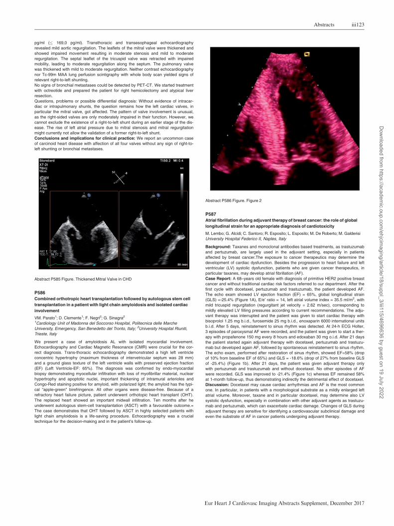
Atrial fibrillation during adjuvant therapy of breast cancer: the role of global
longitudinal strain for an appropriate diagnosis of cardiotoxicity
M. Lembo; G. Alcidi; C. Santoro; R. Esposito; L. Esposito; M. De Roberto; M. Galderisi
University Hospital Federico II, Naples, Italy
Background: Taxanes and monoclonal antibodies based treatments, as trastuzumab
and pertuzumab, are largely used in the adjuvant setting, especially in patients
affected by breast cancer.The exposure to cancer therapeutics may determine the
development of cardiac dysfunction. Besides the progression to heart failure and left
ventricular (LV) systolic dysfunction, patients who are given cancer therapeutics, in
particular taxanes, may develop atrial fibrillation (AF).
Case Report: A 68–years old female with diagnosis of primitive HER2 positive breast
cancer and without traditional cardiac risk factors referred to our department. After the
first cycle with docetaxel, pertuzumab and trastuzumab, the patient developed AF.
The echo exam showed LV ejection fraction (EF) = 65%, global longitudinal strain
(GLS) =-25.4% (Figure 1A), E/e’ ratio = 14, left atrial volume index = 35.5 ml/m2, with
mild tricuspid regurgitation (regurgitant jet velocity = 2.62 m/sec), corresponding to
mildly elevated LV filling pressures according to current recommendations. The adju-
vant therapy was interrupted and the patient was given to start cardiac therapy with
bisoprolol 1.25 mg b.i.d., furosemide 25 mg b.i.d., enoxaparin 6000 international units
b.i.d. After 5 days, reinstatement to sinus rhythm was detected. At 24-h ECG Holter,
3 episodes of paroxysmal AF were recorded, and the patient was given to start a ther-
apy with propafenone 150 mg every 8 hours and edoxaban 30 mg o.i.d. After 21 days
the patient started again adjuvant therapy with docetaxel, pertuzumab and trastuzu-
mab but developed again AF, followed by spontaneous reinstatement to sinus rhythm.
The echo exam, performed after restoration of sinus rhythm, showed EF=58% (drop
of 10% from baseline EF of 65%) and GLS = -18.6% (drop of 27% from baseline GLS
of -25.4%) (Figure 1b). After 21 days, the patient was given adjuvant therapy only
with pertuzumab and trastuzumab and without docetaxel. No other episodes of AF
were recorded. GLS was improved to -21.4% (Figure 1c) whereas EF remained 58%
at 1-month follow-up, thus demonstrating indirectly the detrimental effect of docetaxel.
Discussion: Docetaxel may cause cardiac arrhythmias and AF is the most common
one. In particular, in patients with a morphological substrate as a mildly enlarged left
atrial volume. Moreover, taxane and in particular docetaxel, may determine also LV
systolic dysfunction, especially in combination with other adjuvant agents as trastuzu-
mab and pertuzumab, which can exacerbate cardiac damage. Changes of GLS during
adjuvant therapy are sensitive for identifying a cardiovascular subclinical damage and
even the substrate of AF in cancer patients undergoing adjuvant therapy.
Abstracts iii123
Eur Heart J Cardiovasc Imaging Abstracts Supplement, December 2017
Dow
nloaded from https://academ
ic.oup.com/ehjcim
aging/article/18/suppl_3/iii115/4696636 by guest on 19 July 2022

Abstract P587 Figure.
P588
An adult with syncope who has a sinus venosus type atrial septal defect and
partial anomalous pulmonary venous return misdiagnosed as arrhythmogenic
right ventricular dysplasia
U. Yildiz; E. Renda; LD. Asarcikli; F. Can; H. Kasikcioglu
Dr. Siyami Ersek Thoracic and Cardiovascular Surgery Center, Cardiology, Istanbul,
Turkey
An adult with syncope who has a sinus venosus type atrial septal defect and partial
anomalous pulmonary venous return misdiagnosed as arrhythmogenic right ventricu-
lar dysplasia
Background: Syncope is one of the most important symptom in cardiology.
Cardiological causes of syncope are commonly related with arrhytmias however struc-
tural abnormalities of heart also should be considered. One of the structural abnor-
malities of heart which may lead to syncope is atrial septal defect (ASD). We describe
a patient, whom is suspected as arrhytmogenic right ventricular dysplasia (ARVD),
has primarily sinus venous type ASD with partial anomalous pulmonary venous return.
Both ARVD and ASD can cause syncope and result in dilatation of right ventricle in
transthoracic echocardiography(TTE).
Case report: The patient is a 24-year-old man who was examined about the origin
syncope at a center and cardiac magnetic resonance has been performed and there
has been a suspicion about ARVD but full criterion can not be provided. Therefore the
patient is referred to us as a tertiary center to make definitive diagnosis of ARVD and
if the diagnosis of ARVD is confirmed, the patient would be evaluated for internal car-
diac defibrilator (ICD) implantation. This patient was evaluated in our arrhythmia
department and decided to perform electrophysiological study (EPS) before ICD
implantatition. Arrhytmia department demands detailed TTE from our echocardiogra-
phy laboratory before EPS. We perform TTE and we found hyperreflective moderator
band in right ventricle apex, dilatation of right heart chambers (Figure 1) (Figure 2).
Interatrial septum (IAS) is intact but apical-4-chamber view with injection of agitated
saline into right antecubital vein demonstrated shunt from right atrium to left atrium at
first cycle. Therefore, we reported that transesophageal echocardiography(TEE) is
recommended for the exclusion of ASD and if ASD is excluded with TEE, cardiac MRI
result will be significant with the presence of hyperreflective moderator band in TTE
examination. We discussed the patient with arrhytmia department department and we
decide to perform TEE. There is an approximately 1,5 cm defect at IAS near to supe-
rior vena cava(SVC) in 2D TEE examination (Figure 3). Diameter of defect is 1.6 cm
x 1.7 cm and defect area is 2,3 cm2 in 3D examination (Figure 4 ). There is a wide
foramen ovale (PFO) at IAS. Right lower pulmonar vein and both left pulmonary veins
drain into left atrium however right upper pulmonary vein drain into right atrium in
neighbour of defect (Figure 5) (Figure 6). Our diagnosis is sinus venosus type ASD
and partial anomalous pulmonary venous return. We canceled EPS and further ICD
implantation, we prepare the patient for ASD operation. Cardiovascular surgery
department perform repair of sinus venosus type ASD with pericardial patch and pri-
mary closure of PFO. Patient has been discharged at the 4th day postoperatively with
no complaint.
Conclusion: Syncope is one of the most important symptom in patients whom admit
to outpatient clinic. ASD should be considered in scope of cardiological reasons of
syncope. This is also important that undiagnosed and progressed cases of ASD may
be confused with ARVD echocardiographically and even MRI imaging could support
that as in our case. Therefore when we detect right ventricular dilatation in a patient
with syncope even if IAS is intact, contrast TTE should be performed and if there is a
shunt, TEE should be performed for further investigation. ASD should take place in
differential diagnosis in scope of cardiological causes of syncope if right heart cham-
ber dilatation occurs particularly. This is important because treatment of these two dis-
eases mentioned in our cases are completely different.
Abstract P588 Figure.
P589
Managing a case of high-output heart failure with a tangled aetiology
RG. Badea1; L. Predescu1; A. Bucsa1; R. Nechifor2; BA. Popescu1; C. Ginghina1;
R. Enache1
1Institute of Cardiovascular Diseases Prof. C.C. Iliescu, Cardiology, Bucharest,
Romania; 2Monza Hospital, interventional radiology, Bucharest, Romania
Introduction: High-flow arteriovenous malformations (AVMs) have both local and
systemic consequences. When progression to heart failure occurs, cardiomegaly and
high output are trademarks. In this rare scenario curative procedures are appealing
and theoretically feasible, but practically difficult and often insufficient.
Case report: 39 year old hipertensive female presents for evaluation after being
recently diagnosed with pulmonary hypertension (PH) of unknown aetiology. She
describes dyspnea at moderate exercise. She had a C-section 8 years earlier, without
post-op complications. Problem and procedures. She has normal clinical exam but
elevated BNP. Echocardiography finds indirect signs of PH, dilated heart chambers,
normal biventricular function, no significant valvulopathies and no intracardiac shunts.
Catheterisation confirms PH (mPAP 30 mmHg) with low pulmonary arterial and high
cardiac output (11,6 L/min); inferior vena cava Sat 91%; femoral vein Sat 68%.
AngioCT reveals large pelvic AVMs. Arteriography shows multiple periuterine and
perirectal AVMs feeding from the internal iliac arteries. Bilateral embolisation of the
iliac arteries’ anterior divisions with Amplazer plugs was performed. It resulted in
immediate 80% reduction of shunt flow and symptomatic relief. Improvement in clini-
cal and hemodynamic parameters is expected at later follow-up (pending).
Questions and problems: Right from the echo findings, hypothesis of an extracar-
diac shunt was raised and further supported by invasive hemodynamic data. Imaging
modalities were diagnostic. The question of congenital vs. aquired AVMs remains
open, but has low clinical importance. Therapeutic options (interventional vs. surgical)
were weighed against the impressive dimensions and the topography of the AVMs.
Concerns regarding bilateral internal iliac artery surgical ligation were: 1) pelvic
ischaemia and thigh claudication 2) collateralisation and recanalisation 3) limitation of
future endovascular approach.
Answers and discussions: Percutaneous embolisation is supraselective, has less
complications and it prevents neovascular recruitment. When associated with venous
sclerotherapy, it has even better curative rates. It may also precede surgical ligation,
iii124 Abstracts
Eur Heart J Cardiovasc Imaging Abstracts Supplement, December 2017
Dow
nloaded from https://academ
ic.oup.com/ehjcim
aging/article/18/suppl_3/iii115/4696636 by guest on 19 July 2022

also with good outcomes. Both options are open for our patient if long-term evolution
is unsatisfactory.
Conclusions and implications: Arteriovenous malformations should always be con-
sidered in the differential diagnosis of heart failure and/or pulmonary hypertension
with no obvious aetiology. This rare pathology poses problems in both diagnosis
(requiring clinical suspicion and complementary imaging modalities) and treatment
(promoting combined arterial and venous percutaneous procedures and sometimes
requiring surgical ligation). Larger case series are needed to clarify the role of endo-
vascular management in high-flow extracardiac shunts.
Abstract P589 Figure. pelvic AVMs 3d CT reconstruction
P590
Early diagnosis of hypereosinophilic syndrome with loeffler endocarditis by
echocardiography
UD. Breskvar; J. Ambrozic; L. Dimitrovska; M. Kozelj; M. Bervar
University Medical Centre of Ljubljana, Department of cardiology, Ljubljana, Slovenia
The hypereosinophilic syndromes (HES) are a group of disorders marked by a sus-
tained overproduction of eosinophils. Cardiac involvement is one of the more serious
manifestations of HES. We report a case of previously undiagnosed Churg-Strauss
syndrome with Loeffler endocarditis detected by echocardiography.A 68-year old
male presented with symptoms of behavioural change. Development of Churg-
Strauss vasculitis was suspected but not yet confirmed a couple of months ago
because of persistent peripheral eosinophilia and an increase in pANCA with symp-
toms of pansinusitis and asthma. Cranial MRI revealed multiple ischemic lesions in
watershed border zone distribution. Echocardiography was recommended for evalua-
tion of possible cardiac source of emboli. Transthoracic echocardiography (TTE)
revealed a normal size left ventricle with normal systolic function without regional wall
motion abnormalities and no signs of diastolic disfunction. The valves showed no sig-
nificant pathology.A homogenous echodense mass obliterating the left ventricular
apex was detected, without apical wall motion abnormality.The right ventricle was not
affected. The formation was further investigated with administration of contrast
(Sonovue) and three-dimensional TTE and transesophageal echocardiography.
Loeffler endocarditis with apical thrombus was suspected with distant embolization to
the central nervous system. High-dose prednisolone therapy in combination with
pulsed cyclophosphamide regimen was started. Anticoagulation therapy with warfarin
was also instituted. The symptoms began to resolve and the patient reported no fur-
ther manifestations of the disease. Serial monthly follow-up TTE exams were per-
formed which demonstrated apical thrombus regression and no further cardiac
involvement so far over an 18–month period.HES is a rare multisystemic disease with
a grave prognosis. Cardiac involvement is present in 50 % of cases with high overall
mortality in untreated patients. Cytotoxic injury by eosinophils evolves through three
stages: an acute necrotic stage, thrombus formation and a fibrotic stage characterized
by heart failure due restrictive cardiomyopathy. The major complication of intracardiac
thrombus formation is the detachment and distal embolization of thrombotic material,
leading to embolic events. A high degree of suspicion for cardiac involvement among
patients with HES must be maintained, as it is the most serious complication in this
clinical situation. Quick and accurate diagnosis with prompt initiation of medical treat-
ment may improve symptoms and survival in patients with HES. However, the diagno-
sis is often difficult as it is a disease with multisystemic involvement. In our case echo-
cardiography provided a crucial clue to correct diagnosis. Our case also shows that
the endomyocardial lesion in the early stage of Loeffler endocarditis can be regressed
by optimal medical therapy.
Abstract P590 Figure. apical four chamber view
P591
A rare cause of post-operative cardiac tamponade
L. Curran; MB. Castro Verdes; A. Gambaro; N. Unni; S. Price
Royal Brompton Hospital, Intensive care unit, London, United Kingdom
A 69-year-old lady presented with a four-month history of increasing dyspnoea and
intermittent chest tightness. Coronary angiography revealed severe triple vessel dis-
ease. Transthoracic echo (TTE) was normal. She underwent elective coronary artery
bypass grafting which was uneventful. As is usual practice, she returned to the inten-
sive care unit with temporary atrial and ventricular pacing wires, and left pleural,
mediastinal and pericardial redivac drains in situ. Over the next 12 hours, she was
persistently hypotensive despite fluid resuscitation and vasopressor support. An
urgent bedside TTE revealed a small right ventricular (RV) cavity with severely
impaired longitudinal RV function, and apparent compression at the mid-RV cavity
level causing tamponade. No obvious pericardial collection was seen, however image
quality was suboptimal due to mediastinal air. The differential diagnoses was of com-
pression from a loculated effusion/haematoma, or from an external structure such as
a surgical drain. A transoesphageal echo (TOE) demonstrated that the left pleural
drain was lying across the body of the RV, and causing significant RV compression
and tamponade. TOE allowed for real-time visualisation of the drain’s removal by the
surgical team with alleviation of RV compression and immediate restoration of cardiac
function and corresponding haemodynamic improvement. TOE is a vital tool in the
prompt diagnosis of post-operative cardiac complications and is a useful guide in
directing the correct emergency management of haemodynamic compromise due to
pericardial tamponade (whatever the cause).
Abstract P591 Figure. Tamponade due to pleural drain
P592
Effusive constrictive pericarditis: is it real?
A. Fareed1; H. Awad2
1Suez Canal University, Department of Cardiology, Ismailia, Egypt; 2Suez Canal
University, Cardiothoracic Surgery, Ismailia, Egypt
A 64-year old female known to have diabetes, hypertension and end stage renal dis-
ease on regular hemodialysis since 2014 was presented with progressive dyspnea
Abstracts iii125
Eur Heart J Cardiovasc Imaging Abstracts Supplement, December 2017
Dow
nloaded from https://academ
ic.oup.com/ehjcim
aging/article/18/suppl_3/iii115/4696636 by guest on 19 July 2022

(NYHA class III) and signs of right sided congestive heart failure over the last 2
months. Physical examination: the patient was distressed with bilateral lower extrem-
ity edema, hepatomegaly, moderate ascites. Cardiovascular examination revealed
resting tachycardia with BP: 140/80 mmHg, jugular venous distension with clear lung
bases. The ECG showed sinus rhythm at a rate of 110 bpm. Echocardiography
showed normal biventricular size with normal systolic function, mild biatrial enlarge-
ment, moderate amount of pericardial effusion with no clear 2D chamber collapse.
There were signs of pericardial constriction in the form of abnormal interventricular
septal motion with inspiration (septal bounce), significant pulsed wave velocity varia-
bility over the mitral and tricuspid valves with respiration, IVC plethora with echogenic
pericardium. Computed tomography revealed pericardial thickening and calcification.
Surgical pericardiectomy was performed without complications and revealed markedly
thickened and calcified pericardium (both the parietal and visceral layers).
Echocardiographic assessment 4 days after surgery revealed persistence of constric-
tion signs (septal bounce and PW variability over the mitral and tricuspid valves)
though the patient symptoms were markedly improved. Follow up echocardiography
after 2 months of surgery revealed just mild diastolic dysfunction with disappearance
of echocardiographic parameters of constriction. Conclusion: Effusive constrictive
pericarditis is rare condition. Meticulous searching for signs of pericardial constriction
should be performed in patients with pericardial effusion with risk factors for pericar-
dial constriction and signs of heart failure. Late resolution of echocardiographic signs
of pericardial constriction could be seen after surgical pericardiectomy and follow up
both clinically and echocardiographically is mandatory for these patients.
Abstract P592 Figure. Inspiratory septal bounce with effusion
P593
A rare case of a cardiac arteriovenous fistula
J. Ferreira1; R. Ramalho1; S. Monteiro1; M. Pego1; B. Graca2; F. Silva2; A. Pissarra2
1University Hospitals of Coimbra, Cardiology, Coimbra, Portugal; 2University Hospitals
of Coimbra, Radiology, Coimbra, Portugal
Introduction and case report description: Arteriovenous fistulae (AVF), by its very
nature, describe an abnormal communication between arteries and veins. These com-
munications can be congenital or acquired, occuring at any site in the vascular sys-
tem, and differ in length, size, location and number. Because in AVFs blood flows
directly from an artery into a vein, sometimes relevant capillary beds are excluded
from circulation. AVFs are rare defects that can remain asymptomatic and undetected
until later in life, and can lead to serious complications. We report a case of an
unusual manifestation of a cardiac AVF.
Case report: A 78 years old male was referred to our valvular disease consultation
by his assistant general practitioner for long-standing fatigue symptoms and abnormal
echocardiographic findings. Of medical relevance, our patient had a background of
permanent atrial fibrillation and peripheral venous insufficiency. The echocardiogram
reported exuberant right atrial dimensions of 168 mL/m2 with dilated and dysfunc-
tional right ventricle. Despite these right heart findings, the exam reported far less
dilated left atrium with 70 mL/m2 and non-dilated, normal functioning left ventricle. It
was also reported severe tricuspid regurgitation, moderate mitral regurgitation and a
PASP of 38 mmHg. To reach an explanation for the disproportionate right heart find-
ings, our patient underwent several studies. The ventilation/perfusion lung scan
showed signs compatible with moderate extension thromboembolism, however, the
thoracic angiotomography ruled out not only acute and chronic pulmonary throm-
boembolism but also parenchymal disease which could be linked to possible pulmo-
nary hypertension. A left and right heart cathetherization were then requested, the first
showing normal coronary arteries and the second a mean pulmonary arterial pressure
of 12 mmHg, pulmonary capillary wedge pressure of 9 mmHg and cardiac index of
2.55 L/min/m2. To try to clarify the clinical scenario, a cardiac magnetic resonance
(CMR) was undertaken. The contrast-enhanced CMR revealed considerably dilated
right cavities, with mildy depressed right ventriclular sistolic function while the left ven-
tricle showed no abnormalities. When our radiology team reviewed the thoracic
angiotomography images (pulmonary arteries window) they found an abnormal retro-
grade filling vessel that continued into the right atrium, which seemed to correspond
to the coronary sinus. This vessel continued superiorly, across the left atrioventricular
sulcus, ending in the lateral coronary sinus. This aspect was very suggestive of AVF,
which explained all the findings above described. Our patient is now waiting for an
angiography scan in order to better clarify the AVF.
Conclusions: and implications for clinical practice: This case shows a rare cause of
right heart failure in an older patient and features the growing importance of a multi-
disciplinary approach when in front of an uncertain diagnosis. In the present case,
thoracic angiotomography was paramount for the differential diagnosis, and proved its
utility as a robust clinical tool.
Abstract P593 Figure. Abnormal retrograde filling vessel
P594
A membranous septal aneurysm causing right ventricular outflow tract
obstruction in an adult
F. Privitera1; A. Indelicato2; L. D’urso1; V. Losi1; C. Tamburino1; IP. Monte1
1University of Catania, Catania, Italy; 2CardioThoracoVascular Department AOU
Policlinic University, Catania, Italy
We describe a case of a 69-years-old woman referred for evaluation of exertional dysp-
noea, with a small membranous ventricular septal defect (VSD) and right ventricle out-
flow tract obstruction. Using Transthoracic Echo (TTE) was diagnosed Ventricular Septal
Defect with left to right shunting and Trans-Esophageal-Echo (TEE) was used to a better
anatomical characterization. TEE showed a supracristal VSD considered restrictive
because it developed an high velocity flow in Right Ventricle. Pulmonary valve appears
normal and right ventricular infundibular hypertrophy or double-chambered right ventricle
were excluded. Furthermore, TEE showed a malaligned ventricular septal defect and the
presence of perimembranous mobile tissue protruding in right ventricle. We hypothe-
sized that this tissue can be attributed to broken septum aneurysm and, protruding dur-
ing systole, it causes a dynamic RVOT obstruction.
Abstract P594 Figure. TEE: Four chamber at midesophageal
iii126 Abstracts
Eur Heart J Cardiovasc Imaging Abstracts Supplement, December 2017
Dow
nloaded from https://academ
ic.oup.com/ehjcim
aging/article/18/suppl_3/iii115/4696636 by guest on 19 July 2022

P595
Late closure of a ventricular septal defect with a fenestrated patch after
treatment with pulmonary vasodilators: the good, the bad and the ugly
DM. Dorobantu; RG. Badea; L. Predescu; P. Platon; BA. Popescu; C. Ginghina;
IM. Coman; R. Enache
Institute of Cardiovascular Diseases Prof. C.C. Iliescu, Bucharest, Romania
Introduction: Closure of ventricular septal defects (VSD) with increased pulmonary
resistances (PVR) is not recommended, with pulmonary vasodilators being the ther-
apy of choice in those situations. The possibility of closure with a fenestrated patch in
selected case has been explored, but its role is still unclear.
Case description: We present the case of a 22 year old male patient, diagnosed with
a VSD at 3 years, reevaluated after an episode of paroxysmal dyspnea. He had mild
exertional dyspnea and unremarkable physical examination beyond a loud precordial
murmur. The echocardiography showed a subaortic VSD with bidirectional shunting
(A), predimonantly left to right (Qp/Qs 1.5), PHT with a mean pulmonary pressure
(mPAP) of 69 mmHg, moderate tricuspid regurgitation, normal biventricular function.
The catheterization showed a mPAP of 69 mmHg, a PVR of 6.2 uW and a Qp/Qs od
1.9. As such, we did not recommend the closure of the VSD and the patient was
started on bosentan. At one month of treatment he already showed clinical improve-
ment. At 6 months, the patient opted for surgical correction with a fenestrated patch
abroad, complicated with a post-op PHT crisis and persistence of the PHT which
required the addition of sildenafil. He returned to our clinic for follow-up and he is cur-
rently at 14 months from surgery and 20 months from treatment initiation, asympto-
matic and with quality of life improvement. The progression of main prognostic param-
eters are shown in Table. Following surgery he developed severe tricuspid
regurgitation, most likely due to an intraoperatory valvular lesion (B), but the right ven-
tricular function and size remained stable. Catheterization was repeated at 1 year
after surgery and showed a decrease in the pulmonary pressures (mPAP 32 mmHg)
and PVR (4.5 uW). He continues to receive dual pulmonary vasodilator therapy and
diuretics. Discussions: In the modern era, where medical treatment of PHT has made
significant progress, late closure of VSDs is controversial, as even the European
Guidelines recognize the presence of a "grey" area where the treatment option is
unclear, and do not recommend a "treat and close" approach. This case highlights
these exact issues: the "good" aspect is that the patient is clinically better, the "bad"
is that the long term benefits for this approach are unknown, while the "ugly" is the
unwanted lesion to the tricuspid valve.
Conclusions: We report a case of successful closure of a VSD with a fenestrated
patch after 6 months of treatment with bosentan, in the presence of pulmonary arterial
disease, with a decrease in pulmonary pressures and resistances at one year. The
intraoperative lesion of the tricuspid valve is a rare complication which can lead to fur-
ther reinterventions in an already high risk patient.
Abstract P595 Figure. Figure A/B
P596
Intraventricular septal dissection: a rare complication after cardiac surgery
G. Aurrekoetxea Bajeneta; L. Ruiz Gomez; M. Codina Prat; I. Bravo Martinez;
P. Mendoza Cuartero; N. Garcia Ibarrondo; A. Manzanal Rey; C. Asla Ormaza;
G. Ramirez-Escudero
Hospital de Basurto, Bilbao, Spain
Interventricular septal (IVS) dissection is a rare entity that may appear as a result
from different causes such as an aneurysm of one of the sinuses of Valsalva, bacte-
rial endocarditis, cardiac surgery, acute myocardial infarction, and chest trauma, pro-
ducing a widely range of complications and worsening the overall prognosis of the
patient. We report the case of a 75 year-old-woman with a history of mitral commis-
surotomy at the age of 35 because of rheumatic mitral disease, who during follow up
developed a symptomatyc double mitral disease with severe stenosis, therefore sur-
gery was decided. After surgery, a routine transthoracic echocardiogram showed a
cystic cavity within the interventricular septum, which was interpreted as IVS dissec-
tion. Despite this complication, the patient remained in a situation of clinical and
hemodynamic stability, so conservative treatment with serial echocardiography was
decided. Serial echocardiograms revealed a progressive decrease of the cystic cavity
size, hence the patient was discharged two months later. Considering the lack of
other posible etiologies in our patient, mitral valve replacement and tricuspid annulo-
plasty were accepted as the triggering event, being only described in the literature as
a complication of aortic valve surgery and mitral valve surgery with coronary fistula.
IVS dissection is a rare entity which diagnose was often postmortem until some deca-
des ago, being a highly letal complication. Up to now it was believed that surgical
treatment was the best option for the management of this condition, even though at
present is controversial due to acceptable medium-long term survival obtained with-
conservative treatment in some asymptomatic patients.
Thanks to the imaging advances available nowadays, it can be diagnosed earlier,
implying different management possibilities in these patients.
Abstract P596 Figure. Cystic cavity within IVS
P597
Left upper lobe partial anomalous pulmonary venous connection associated
with patent foramen ovale as a rare cause of dyspnea: an imaging-based
diagnosis
AA. Ionescu1; M. Dobrovie1; M. Chivulescu1; RA. Ionescu2; A. Raica3; D. Deleanu1;
C. Ginghina1; BA. Popescu1; R. Jurcut1
1Institute of Cardiovascular Diseases Prof. C.C. Iliescu, Bucharest, Romania; 2’Carol
Davila’ Emergency Clinical Military Hospital, Bucharest, Romania; 3County
Emergency Hospital of Constanta, Constanta, Romania
Progression of clinical, biological and
07.2015
Bosentan
start 125
mg/day
08.2015
Bosentan
25 mg/day
11.2015 01.2016 03.2016 07.2016 10.2016 01.2017
6MWT 400 m 505 m 510 m Surgical
correction
and addition
of sildenafil
504 m 520 m 524 m 535
BNP 43 pg/ml 11 pg/ml 10 pg/ml 40 pg/ml 51 pg/ml 22 pg/ml 79 pg/ml
sPAP 103 mmHg 103 mmHg 69 mmHg 70 mmHg 67 mmHg
TAPSE 19 mm 20 mm 19 mm 18 mm 19 mm
S-VD 15 cm/s 13 cm/s 11 cm/s 13 cm/s 12 cm/s
Right
atrial
area
23 cm2 20 cm2 29 cm2 24,6 cm2 26,6 cm2
Abstracts iii127
Eur Heart J Cardiovasc Imaging Abstracts Supplement, December 2017
Dow
nloaded from https://academ
ic.oup.com/ehjcim
aging/article/18/suppl_3/iii115/4696636 by guest on 19 July 2022

Case report and description: We report the case of a 66-year-old woman who sought
medical attention on account of dyspnea and fatigability and short episodes of irregu-
lar palpitations. She was diagnosed with pulmonary hypertension (PHT) and persis-
tent episodes of atrial fibrillation (AF) 2 years earlier but was not investigated further.
Of note, on clinical exam we found a loud S2, a systolic tricuspid murmur and a dia-
stolic pulmonary valve murmur. Her ECG showed AF with signs of right ventricular
overload with complete RBBB with secondary repolarization abnormalities. Her labo-
ratory findings revealed only hypercholesterolemia, slight hypothyroidism and a BNP
of 80 pg/ml. Her walking test was stopped prematurely at 5 minutes on account of
dyspnea, reaching only 378m with a slight decrease in oxygen saturation (92%).
Transthoracic echocardiography (TTE) raised the suspicion of left to right shunt, with
a patent foramen ovale (PFO) but a Qp/Qs ratio above 1.5, moderate-severe tricuspid
insufficiency with an estimated systolic pulmonary artery pressure (PAP) of 50 mmHg,
dilated right heart chambers with normal systolic function of both ventricles, diastolic
dysfunction of the left ventricle and severe biatrial enlargement. All of these findings
where confirmed by transesophageal echocardiography (TEE), adding a flattening of
the IVS during diastole. In the absence of a clear etiology to explain the PHT and its
accompanying symptoms, right heart catheterization (RHC) was performed – this con-
firmed the PHT, with a mean PAP of 28 mmHg, but with a Qp/Qs only slightly higher
than 1. A venous abnormality was observed, with the left upper pulmonary lobe drain-
ing into the left brachiocephalic vein. A thoracic CT confirmed and detailed the venous
anomaly.
Question: This case illustrates the difficulties encountered in establishing the precise
etiology of PHT and its accompanying symptoms and the need for multimodality imag-
ing in such complex clinical scenarios. The differential diagnosis for PHT covers an
extremely wide spectrum of diseases. Concerning intrathoracic venous anomalies,
special care needs to be taken to exclude pulmonary varix, enlarged left superior
intercostal vein and persistent left superior vena cava.
Discussion: While a partial anomalous pulmonary venous return is infrequently
encountered, especially pertaining to the left side, they are frequently associated with
an atrial septal defect (most of them of sinus venous type). These anomalies, usually
incidentally discovered, frequently lead to a left to right shunt which goes unnoticed
until adulthood, but predispose the patient to right-sided volume overload with right
ventricular dysfunction and PHT. Taking into account the degree of the PHT and its
implications, surgical correction is rarely employed and a conservative strategy can
be applied as in our patient.
Conclusions: Multimodality imaging techniques are an essential approach to cor-
rectly diagnose the etiology behind PHT and to reach the most suitable therapeutic
strategy, either surgical or conservative.
P598
A strange extra aortic valve leaflet
A. Marques1; O. Simoes1; S. Alegria1; AC. Gomes1; R. Pereira1; D. Sebaiti1; I. Joao1;
J. Cruz2; N. Junqueira3; H. Pereira1
1Hospital Garcia de Orta, Cardiology, Almada, Portugal; 2Hospital Garcia de Orta,
Radiology, Almada, Portugal; 3University Hospital De Santa Maria, Cardiothoracic sur-
gery, Lisbon, Portugal
We present a case of a 22 years-old caucasian male, an active smoker, with Marfan
Syndrome and ascending aortic aneurysm (42 mm) both diagnosed at 17 years-old,
but with no follow-up since his 18 years-old and without usual medical therapy.
The patient was admitted to our hospital due to de novo anterior thoracalgia of 15
days duration, predominantly retrosternal with posterior irradiation, aggravating with
dorsal decubitus, deep inspiration and cough, with more frequent and intense epi-
sodes, being continuous and intense, reason for which it appealed to the emergency
service. He also reported dyspnoea and tiredness for small efforts.
Physical examination revealed tachycardia, polypnea and hypersudorese, blood pres-
sure of 98/49 mmHg and apyrexia. In cardiac auscultation a decreasing diastolic
grade 3 (Levine scale) murmur in the aortic focus was present; pulmonary ausculta-
tion revealed abolished vesicular murmur in the right lung base. Arterial pulses were
synchronous and symmetrical.
The blood samples showed elevation of inflammatory parameters and troponin T hs
516 ng/L (N<13), with normal values of haemoglobin and creatinine. In the gasom-
etry, hypoxemia with hypocapnia were present, with normal lactate values. The elec-
trocardiogram revealed sinus tachycardia with ST-depression in inferior leads and V5-
V6 (maximum amplitude of 1.5 mm). The chest X-ray showed cardiomegalia with
signs of small right pleural effusion. A transthoracic echocardiogram was performed
and revealed a dilated left ventricle (78 mm), with severely compromised systolic
function; ascending aortic aneurysm (aortic root: 79 mm) and a tricuspid aortic valve
with an eccentric major aortic regurgitation being visible a flap with diastolic protrusion
through the aortic valve suggestive of aortic root dissection, without pericardial
effusion.
An urgent non ECG-Gated angio-Computer tomography was performed but it wasn�t
clear the diagnosis of aortic dissection due to the presence of motion artifacts. The
patient refused to perform a transoesophageal echocardiogram.
Given the suggestive clinic associated with the changes present in the transthoracic
echocardiogram it was admitted ascending aortic aneurysm with aortic root dissection
(Stanford type A classification) and major aortic regurgitation, in a patient with dilated
and severely compromised left ventricle systolic function.
The patient was submitted to an emergent surgery and a Bentall procedure was per-
formed successfully. The patient was transferred to his hometown hospital after 8
days.
Aortic dissection is the commonest cause of death in the natural history of Marfan
syndrome. Prior to the introduction of prophylactic root replacement this was the
cause of death in two-thirds of all patients. Detection, diagnosis, monitoring and pro-
phylactic surgery have dramatically reduced deaths from dissection. This case aims
to alert to the importance of both clinical and physical examination, highlight the role
of the transthoracic echocardiogram in the diagnosis of aortic root dissection in this
case and alert to the importance of a right follow-up and prophylactic surgery in
patients with Marfan Syndrome.
Abstract P598 Figure. Aortic dissection
P599
A rare vascular structure
R. Laymouna1; E. Elsharkawy2; Y. Hassan1; A. Almaghraby2; A. Sammoka1
1International Cardiac Center Hospital, cardiology, Alexandria, Egypt; 2Alexandria
University, Cardiology, Alexandria, Egypt
Introduction: The aorta right atrial fistula lies in a group of abnormal vascular con-
nections of the aorta named aortocameral fistulas. Aortocameral fistulas of the heart
are rare extra-cardiac vascular channels, and their natural history, clinical presenta-
tions, and suitability of treatment options are unclear.
Case report: A 31-year-old male patient, non-smoker, with no medical history of
hypertension nor DM. He started complaining of bilateral lower limb edema, general-
ized fatigue and effort intolerance in the past six months. The patient denied any pre-
vious chest trauma or surgical interventions.
Upon examination, the patient had marked jugular venous distension, hepatomegaly,
bilateral pitting edema to the level of the upper thigh, and marked abdominal disten-
tion. On cardiac auscultation, a 3/6 continuous murmur was heard over the left sec-
ond and third intercostal spaces. Electrocardiogram showed sinus tachycardia (nor-
mal ST segment and no pathological Q waves). Routine laboratory investigations
were unremarkable.
Echocardiography revealed dilated right atrium (RA) and right ventricle (RV) with
reduced RV systolic function and evidence of pulmonary hypertension. An Abnormal
vascular structure was seen connected to the left coronary sinus of Valsalva with con-
tinuous systolic and diastolic flow. This structure was also observed to be connected
to the right atrium. Mildly dilated left ventricular (LV) dimensions with preserved sys-
tolic function (EF=65%) were also noted.
128-multidetector cardiac computed tomography (MDCT) showed a large vascular
structure (diameter 3 cm at the beginning and 2.8 cm at the end) was arising from the
left coronary sinus of Valsalva, passing through the left atrio-ventricular groove, drain-
ing into the right atrium (taking the course of coronary sinus but with separate draining
sites into the right atrium). The left anterior descending artery (LAD) was seen arising
from the proximal part of the fistula, then it continued with normal course and caliber
in the anterior inter-ventricular groove. The obtuse marginal branch (OM) was also
arising from this fistula. Right coronary artery (RCA) was arising normally from the
right sinus of Valsalva.
So, we have a case of large aorto-right atrial fistula with LAD and OM arising from
this fistula and complicated with right ventricular systolic failure. The patient was
referred for surgical closure.
iii128 Abstracts
Eur Heart J Cardiovasc Imaging Abstracts Supplement, December 2017
Dow
nloaded from https://academ
ic.oup.com/ehjcim
aging/article/18/suppl_3/iii115/4696636 by guest on 19 July 2022

Conclusion: Aortoatrial fistulas are rare extracardiac vascular channels. These fistu-
lous communications can have an origin from any of the three sinuses of Valsalva.
Although the ascending aorta is reported to be the most common site of origin for aor-
tocameral fistulas, reports have described fistulas emanating from the descending
thoracic aorta. Various treatment options are available according to the type of fistula,
its caliber, tortuosity, calcification, course, and relation of the coronary ostia to the
aortic orifice of the fistula.
Abstract P599 Figure. Aorto-right atrial fistula by MDCT
Abstracts iii129
Eur Heart J Cardiovasc Imaging Abstracts Supplement, December 2017
Dow
nloaded from https://academ
ic.oup.com/ehjcim
aging/article/18/suppl_3/iii115/4696636 by guest on 19 July 2022


![10 11 Session HR Session Seri Management[1]](https://static.fdokumen.com/doc/165x107/6314ba61fc260b71020fb0ee/10-11-session-hr-session-seri-management1.jpg)