2013 - ly Ju ANTIBACTERIAL EFFICIENCY AND DNA IMPAIRMENTUNVEILIN SOME BACTERIA STRAINS TREATED WITH...
11
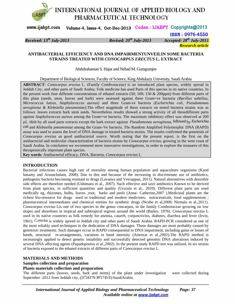
2013 - ly Ju th 28 Accepted: 2013 - ly Ju th 20 Revised: 2013 - ly Ju th 13 Received: Research article ANTIBACTERIAL EFFICIENCY AND DNA IMPAIRMENTUNVEILIN SOME BACTERIA STRAINS TREATED WITH CONOCARPUS ERECTUS L. EXTRACT Abdulrahaman S. Hajar and Nehad M. Gumgumjee Department of Biological Sciences, Faculty of Science, King Abdulaziz University, Saudi Arabia ABSTRACT: Conocarpus erectus L. (Family Combretaceae) is an introduced plant species, widely spread in Jeddah City, and other parts of Saudi Arabia. Folk medicine has used Parts of this species in its native countries. In the present work four different concentrations of ethanol extracts (50; 100; 150 & 200ppm) from different parts of this plant (seeds, stem, leaves and bark) were assessed against three Gram+ve bacteria (Bacillus subtilius, Micrococcus luteus, Staphylococcus aureus) and three Gram-ve bacteria (Escherichia coli, Pseudomonas aeruginosa & Klebsiella pneumoniae).The effect magnitude of these extracts on tested bacteria strains was as follows: leaves extracts bark stem seeds. Nevertheless results showed a strong activity of all thesedifferent parts against Staphylococcus aureus among the Gram+ve bacteria. The maximum inhibitory effect was observed at 200 μL /dish by all used parts extracts except the bark extract against Pseudomonas aeruginosa, followed by Escherichia coli and Klebsiella pneumoniae among the Gram–Ve bacteria. The Random Amplified Polymorphic DNA (RAPD) assay was used to assess the level of DNA damage in treated bacteria strains. The results confirmed the potentials of Conocarpus erectus as good antibacterial source. Worth noting that the present report, is the first on the antibacterial and molecular characterization of bacteria strains by Conocarpus erectus, growing in the west coast of Saudi Arabia. In conclusion we recommend more innovative investigations, in order to explore the treasures of this therapeutically important plant species. Key words: Antibacterial efficacy, DNA, Bacteria, Conocarpus erectus L INTRODUCTION Bacterial infections causes high rate of mortality among human population and aquaculture organisms (Kand hasamy and Arunachalam, 2008). Due to this and because of the increasing in discriminate use of antibiotics, pathogenic bacteria becoming resistant to drugs (Lavanya and Veerappan, 2011). Natural alternatives with desirable side effects are therefore needed (Ushimaru et al., 2007). Such effective and save antibiotics Known to be derived from plant species, in sufficient quantities and quality (Urszula et al., 2010). Different plant parts are used medically eg, rhizomes, bulbs, leaves ,root, barks and peels (Anne- Catherine,2007 ).Medicinal plants are the richest bio-resource for drugs used in traditional and modern medicines; nutraceuticals; food sapplementsm ; pharmaceutical intermediates and chemical entities for synthetic drugs (Ncube et al,2008; Nirmala et al.,2011). Conocarpus erectus Lis one of two species in the genus concarpus, in the family Combretaceae growing on low slopes and shorelines in tropical and subtropical regions around the world (Bailey, 1976). Conocarpus erectus L used in its native countries as folk remedy for anemia, catarrh, conjunctivitis, diabetes, diarrhea and fever (Irvin, 1961). C.erectus is widely spread in Jeddah city and other parts of Saudi Arabia. RAPD-PCR considered as one of the most reliably used techniques in the dedication of DNA damages. These damages are most probably caused by genotoxic treatments. Such damages occur in RAPD consequential to DNA impairment, including gains or losses of bands, structural re-arrangements, variation in band intensity (Atienzar et al.,2002).The RAPD method is increasingly applied to detect genetic instability and successfully detected genomic DNA alterations induced by several DNA affecting agents (Papadopoulos et al.,2002). In the present study RAPD test was utilized. In six strains of bacteria exposed to the ethanol extracts of different parts of Conocarpus erectus L. MATERIALS AND METHODS Samples collection and preparation Plants materials collection and preparation The different parts (leaves, seeds, bark and stems) of the plant under investigation were collected during September -2012 from Jeddah city, (21 o 24 ' 3"N , 39 o 17 ' 45 " E) SaudiArabia. International Journal of Applied Biology and Pharmaceutical Technology Page: 37 www.ijabpt.com Available online at
-
Upload
kingabdulaziz -
Category
Documents
-
view
2 -
download
0
Transcript of 2013 - ly Ju ANTIBACTERIAL EFFICIENCY AND DNA IMPAIRMENTUNVEILIN SOME BACTERIA STRAINS TREATED WITH...
2013-lyJu th28Accepted: 2013 -lyJu th20Revised: 2013 -lyJu th13Received:
Research article
ANTIBACTERIAL EFFICIENCY AND DNA IMPAIRMENTUNVEILIN SOME BACTERIA STRAINS TREATED WITH CONOCARPUS ERECTUS L. EXTRACT
Abdulrahaman S. Hajar and Nehad M. Gumgumjee
Department of Biological Sciences, Faculty of Science, King Abdulaziz University, Saudi Arabia
ABSTRACT: Conocarpus erectus L. (Family Combretaceae) is an introduced plant species, widely spread in Jeddah City, and other parts of Saudi Arabia. Folk medicine has used Parts of this species in its native countries. In the present work four different concentrations of ethanol extracts (50; 100; 150 & 200ppm) from different parts of this plant (seeds, stem, leaves and bark) were assessed against three Gram+ve bacteria (Bacillus subtilius, Micrococcus luteus, Staphylococcus aureus) and three Gram-ve bacteria (Escherichia coli, Pseudomonas aeruginosa & Klebsiella pneumoniae).The effect magnitude of these extracts on tested bacteria strains was as follows: leaves extracts bark stem seeds. Nevertheless results showed a strong activity of all thesedifferent parts against Staphylococcus aureus among the Gram+ve bacteria. The maximum inhibitory effect was observed at 200 µL /dish by all used parts extracts except the bark extract against Pseudomonas aeruginosa, followed by Escherichia coli and Klebsiella pneumoniae among the Gram–Ve bacteria. The Random Amplified Polymorphic DNA (RAPD) assay was used to assess the level of DNA damage in treated bacteria strains. The results confirmed the potentials of Conocarpus erectus as good antibacterial source. Worth noting that the present report, is the first on the antibacterial and molecular characterization of bacteria strains by Conocarpus erectus, growing in the west coast of Saudi Arabia. In conclusion we recommend more innovative investigations, in order to explore the treasures of this therapeutically important plant species. Key words: Antibacterial efficacy, DNA, Bacteria, Conocarpus erectus L INTRODUCTION Bacterial infections causes high rate of mortality among human population and aquaculture organisms (Kand hasamy and Arunachalam, 2008). Due to this and because of the increasing in discriminate use of antibiotics, pathogenic bacteria becoming resistant to drugs (Lavanya and Veerappan, 2011). Natural alternatives with desirable side effects are therefore needed (Ushimaru et al., 2007). Such effective and save antibiotics Known to be derived from plant species, in sufficient quantities and quality (Urszula et al., 2010). Different plant parts are used medically eg, rhizomes, bulbs, leaves ,root, barks and peels (Anne- Catherine,2007 ).Medicinal plants are the richest bio-resource for drugs used in traditional and modern medicines; nutraceuticals; food sapplementsm ; pharmaceutical intermediates and chemical entities for synthetic drugs (Ncube et al,2008; Nirmala et al.,2011). Conocarpus erectus Lis one of two species in the genus concarpus, in the family Combretaceae growing on low slopes and shorelines in tropical and subtropical regions around the world (Bailey, 1976). Conocarpus erectus L used in its native countries as folk remedy for anemia, catarrh, conjunctivitis, diabetes, diarrhea and fever (Irvin, 1961). C.erectus is widely spread in Jeddah city and other parts of Saudi Arabia. RAPD-PCR considered as one of the most reliably used techniques in the dedication of DNA damages. These damages are most probably caused by genotoxic treatments. Such damages occur in RAPD consequential to DNA impairment, including gains or losses of bands, structural re-arrangements, variation in band intensity (Atienzar et al.,2002).The RAPD method is increasingly applied to detect genetic instability and successfully detected genomic DNA alterations induced by several DNA affecting agents (Papadopoulos et al.,2002). In the present study RAPD test was utilized. In six strains of bacteria exposed to the ethanol extracts of different parts of Conocarpus erectus L. MATERIALS AND METHODS Samples collection and preparation Plants materials collection and preparation The different parts (leaves, seeds, bark and stems) of the plant under investigation were collected during September -2012 from Jeddah city, (21o24'3"N, 39o17'45"E) SaudiArabia.
International Journal of Applied Biology and Pharmaceutical Technology Page: 37 www.ijabpt.comAvailable online at

Hajar and Nehad Species statusof this plant was fervid at Faculty of Sciences Herbarium, KingAbdulaziz University, Jeddah. The sampels were brought to the laboratory, thoroughly washed in running tap water to remove debris and dust particles and then rinsed in distilled water for five minutes, then air-dried under room temperature until constant weight. Extracts preparation Ten grams of dried Conocarpus erectusparts (leaves, seeds, bark and stems) were catted into small pieces by blender 1-2 mm. Extraction was done by adding 100 ml of ethanol (1:10W/V) left under cold conditions for 48h. then the extracts were filtered through a filter paper. The extracts solutions were evaporated under reduced pressure at 40°C until dryness, subsequently, the extract was diluted by dimethyl sulf.oxide (DMSO) and stored under 20°C until analysis according to (Boeru and Derevici, 1978). Bacterial strains
Cultures were prepared for in vitro antibacterial assay of the six bacteriastrains,three Gram positive: Bacillus subtilis (ATCC11774); S. aureus (ATCC29213) and Micrococcus luteus (ATCC4698) and three Gram negative Escherichia coli (ATCC8739); Klebsiella pneumoniae (ATCC700603) and Pseudomonas aeruginosa. (ATCC27853) Those strains were provided by Microbiologics® USA. Tested organisms were sub cultured on nutrient agar (Oxoid laboratories, UK) slopes. These stock cultures were stored in the dark at 4°C until used. Antimicrobial activity
Antimicrobial activity was determined using the agar well diffusion assay method as described by Holder and Boyce (1994). DMSO was used as a negative control. The plates were done in triplicate. Bacterial cultures were incubated at 37°C for 24 h at. Antimicrobial activity was determined by measurement zone of inhibition (Agwa et al., 2000).
DNA extraction and RAPD – PCR amplification conditions. Genomic DNA was prepared from 18h cultures in exponential phase in Luria – Bertani medium (1000 mL deionized water ,10 g Bactotryptone, 5 g Bacto yeast, 5 g NaCl several drops, 5 M NaOH several drops and 1 M HCl ) . Aliquots of 10 ml of each bacterialculture were harvested by centrifugation at 12.000 rpm for 15 min at 4 °c and washed once in sterile distilled water. The pellets were resuspended in 400 µl of lysis buffer containing 2% glucose, 50 m M Tris – HCl(pH 8.0), 25 m M EDTA, 3 mg / ml lysozyme and 200mg / ml RNase. The cell suspension was incubated for 1h at 37ºC. Further DNA extraction was performed as described by Sambrook et al. (1989). PCR amplification was carried out in a DNA themocycler (Biomatra, Germany) for 30 cycles each. The PCR reaction was carried out in a final volume of 25 µL with 1X PCR buffer containing 10 mMTris – HCl, 25 mM MgCl2, 1 ml of Template DNA, 0.2 mM deoxy nucleoside triphosphate, 1 to 2 µM (each) primer and 0.5 u of Taq DNA polymerase (Promega).PCR conditions consisted of initial denaturation at 95ºc for 2min followed by 95ºc for 1min, annealing to primers at 37ºc for 1 min, and extension at 72ºc for 1 min with a final extension step at 72ºc for 5 min. PCR – amplified products were separated using agarose gel electrophoresis in 1% TBE buffer (Tris base 108 g, boric acid 55 g, 0.5M EDTA 20 ml and H2O2 to 1 L) dilute accordingly for 1X stock and stained with 0.2 µg /ml ethidium bromide according to Sambrook et al. (1989).Amplified fragments were detected and photographed under UV light. Two random primers were synthesized from University of British Colombia (UBC), Vancouver, Canda (UBC 16 , 28 and 89) and their sequences arepresented in Tabulation. The random primer names and sequences that used for RAPD analysis.Williams etal. (1990).
Primer Sequences Primer A7 Primer B5
G AAACGGG TG TGCGCCCTTC
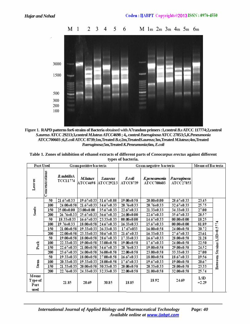
RESULTS AND DISCUSSION Antibacterial activity The antibacterial activity of ethanolic extracts of seeds ,stem, leaves and bark of C. erectus was assessed against three Gram +ve bacteria (B.subtilis, M.lutues and S.aureus) , three Gram – ve bacteria (E.coli, K.pneumonia and P. aeruginosa ) at different concentrations ranged from (50-200 µL), the leaves extract showed the highest activity against tested organisms followed by the bark > stem> seeds respectively (Table 1).Itcan be deduced that Conocarpus erectus leaf ethanol extract had the broadest spectrum of activity on the tested bacteria.
International Journal of Applied Biology and Pharmaceutical Technology Page: 38 comwww.ijabpt.Available online at

Hajar and Nehad The magnitude of its inhibition zones against the tested bacteria was as follows: S. aurueus (36.7mm ), B.subtilis (26.7mm ), M.lutues (25.7 mm),P.aeruginosa (35.7mm) , E.coli(24.0 mm) and K.peneumonia (22.7 mm ) were more sensitive at 200 µl /plates concentration. Conocarpus erectus bark extract exhibited remarkable antibacterial activities against the tested bacteria in the following sensitivity order S. aureus> P. aeruginosa with equal effect on K.peneumonia and B.subtilis but the bark extractsantibacterial action showed the least effect on E.coli (22mm) at concentration of 200µl/plates. The stem extract showed the highest activities against S. aureus and P.aeruginosa (32.3 and 32.0 ) then on K.peneumonia (21.00)at concentration of 200µl. Seeds extract have the least antibacterial activity compared to other parts.The present results coincided with Bukar, et al. (2010)whom reported that Moringaoliferaseeds chloroform extract have the least effect on the tested organisms. They also stated that Moringaolifera seeds MSC extract low antibacterial activity might be linked to their phytochemical contents, due to that alkaloids, tannins and flavonoids were not detected in MSC extract of those seeds. Presence of such compounds in plant species parts enhancestheir antimicrobialproperties singh and Bhat (2003) and Tscheche (1971). In previous report on Conocarpus erectus, the methanol extract of leaves, stem, fruits, and flowers showed antimicrobial properties (El-Sayed et al., 2012). However their reported inhibition zones were far less than those recorded in the present study, eg. records of leaves and stems extracts. This may indicate an ecotype differences in this plant species, hence the currently studied plant grows under higher temperature and drier climate. Such circumstances increases the concentrations of theactive substances in the plant parts, and thereby their effects on the tested organisms. Phenolic compounds, especially tannins are the major components of this plant species (El.Sayed et al., 2012 ). This would suggest that the antimicrobial effects observed in this study and with more pronouns effects in present study could be attributed to such compounds. Therefore it would be of great benefits to separate those compounds and use them on the same bacteria strains, in order to verify the usefulness of this important plant species. Moreover to produce more abundant and inexpensive natural antibiotics. Gumgumjee et al. (2012) stated that Tamarindus indica leaves ethanol extracthas produced strong antibacterial effects against S.aureus> MRSA>B.subtilis respectively. S.aureus showed also similar results when treated with Casuarina equisetifolia leaves ethanol extract (Gumgumjee and Hajar, 2012).The higher sensitivity of Gram +ve bacteria to natural plants extracts has beendocumented (Dulger and Gonuz,2004; Chaieb et al., 2011). The ethanol extracts of different parts of Conocarpus erectus were also tested on Gram – ve bacteria; E.coli, K. pneumoniae and P.aeruginosa . The results showed a strong effect against P. aeruginosa followed by E.coli and K. pneumoniae at 200 µl /dish by all tested parts of Conocarpus erectus bark extracts. The present results agreed to some extent with El-Sayed et al. (2012). The susceptibility of bacteria strains to plant species extracts may draw attention to plants potentials as a natural antibiotic producers, these can be used against the susceptible bacteria strains (Khosravi &Behzadi, 2006). Due to the nature of the bacteria species response, differences betweenthem are expected (Moyo et al ., 2011). Results obtained from present study provide more evidence on the antimicrobial efficacy of the different plant parts, eg. leaves,bark,stem and seedsalthough they differ in their magnitude. The mechanisms of actions of these compounds have been proven to be via cell membranes perturbations (Esimone et al., 2006). Molecular characterization of Bacteria by RAPD analysis Two random primers A7and B5 were used for RAPD analysis of the tested bacteria.
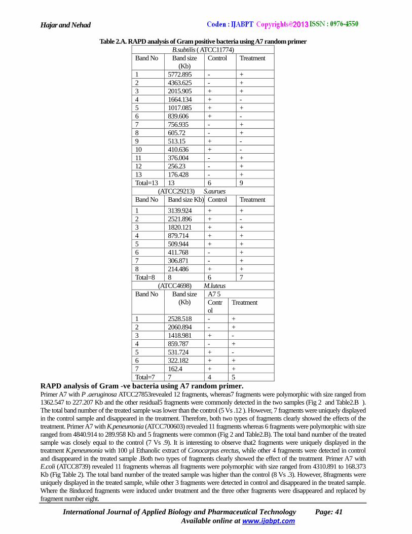
RAPD analysis of Gram +ve bacteria using A7 random primer Primer A7 with B.subtilis revealed 13 fragments were polymorphic with size ranged from 5772.895 to 176.428Kb and 2 fragments were common (Fig 1 and Table 2.A). The total band number of the treated bacteria was higher than the control (9 Vs .6). It is interesting to note that 9 fragments were uniquely displayed in the treated B.subtilis ATTC11774with 100 µl ethanol extract of Conocarpus erectus, while other 4 fragments were detected in the control and absent in the treated bacteria. Both of the two fragments types clearly showed the effect of the treatment. Primer A7 with S.aurues ATTC29213revealed 8 fragments were common (Fig 1and Table2.A).The total band number of the treated sample was closely equal to the control (7vs.6). It is interesting to note that 2 fragments were uniquely displayed in the treated S.aureus with 100 µl Ethanolic extract of Conocarpus erectus. However, 1 fragment was uniquely displayed in the control and disappeared in the treated sample. Therefore, both two types of fragments clearly showed the effects of the treatment. Primer A7 with M.luteus ATTC 4698revealed 7 fragments, whereas 5 fragments were polymorphic with sizes ranged from 2528.518 to 162.4 Kb and 2 fragments were common (Fig 1and Table 2.A ). The total band number of the treated sample was closely near to the control (5vs.4). However, 3 fragments were uniquely displayed in the treatment sample. While other two fragments were detected in the control and displayed in the treatment sample. Therefore, both fragments clearly showed the effects of the treatment, where the three fragments were induced under treatment and the two other fragments were disappeared and replaced by the other three.
International Journal of Applied Biology and Pharmaceutical Technology Page: 39 www.ijabpt.comAvailable online at
Hajar and Nehad
Figure 1. RAPD patterns for6 strains of Bacteria obtained with A7random primers :1,control B.s ATCC 117774;2,control
S.aureus ATCC 29213;3,control M.luteus ATCC4698 ; 4,, control P.aeruginosa ATCC 27853;5,K.Peneumonia ATCC700603 ;6,E.coli ATCC 8739;1m,Treated B.s;2m,TreatedS.aureus;3m,Treated M.luteus;4m,Treated
P.aeruginosa;5m,Treated K.Peneumonia;6m, E.coli
Table 1. Zones of inhibition of ethanol extracts of different parts of Conocarpus erectus against different types of bacteria.
International Journal of Applied Biology and Pharmaceutical Technology Page: 40 www.ijabpt.comAvailable online at
Hajar and Nehad
Table 2.A. RAPD analysis of Gram positive bacteria using A7 random primer B.subtilis ( ATCC11774)
Treatment Control Band size (Kb)
Band No
+ - 5772.895 1 + - 4363.625 2 + + 2015.905 3 -+1664.1344 + + 1017.085 5 - + 839.606 6 + - 756.935 7 + - 605.72 8 - + 513.15 9 - + 410.636 10 + - 376.004 11 + - 256.23 12 + - 176.428 13 9 6 13 Total=13
(ATCC29213) S.aurues Treatment Control Band size Kb)Band No + + 3139.924 1 - + 2521.896 2 + + 1820.121 3 + + 879.714 4 + + 509.944 5 + - 411.768 6 + - 306.871 7 + + 214.486 8 7 6 8 Total=8
(ATCC4698) M.luteus A7 5 Band size
(Kb) Band No
Treatment Control
+ - 2528.518 1 + - 2060.894 2 - + 1418.981 3 + - 859.787 4 - + 531.724 5 + + 322.182 6 + + 162.4 7 5 4 7 Total=7
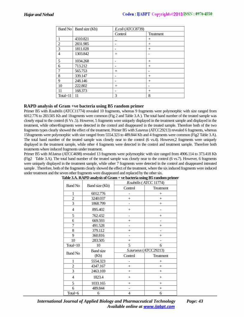
RAPD analysis of Gram -ve bacteria using A7 random primer. Primer A7 with P .aeruginosa ATCC27853revealed 12 fragments, whereas7 fragments were polymorphic with size ranged from 1362.547 to 227.207 Kb and the other residual5 fragments were commonly detected in the two samples (Fig 2 and Table2.B ). The total band number of the treated sample was lower than the control (5 Vs .12 ). However, 7 fragments were uniquely displayed in the control sample and disappeared in the treatment. Therefore, both two types of fragments clearly showed the effects of the treatment. Primer A7 with K.peneumonia (ATCC700603) revealed 11 fragments whereas 6 fragments were polymorphic with size ranged from 4840.914 to 289.958 Kb and 5 fragments were common (Fig 2 and Table2.B). The total band number of the treated sample was closely equal to the control (7 Vs .9). It is interesting to observe that2 fragments were uniquely displayed in the treatment K.peneumonia with 100 µl Ethanolic extract of Conocarpus erectus, while other 4 fragments were detected in control and disappeared in the treated sample .Both two types of fragments clearly showed the effect of the treatment. Primer A7 with E.coli (ATCC8739) revealed 11 fragments whereas all fragments were polymorphic with size ranged from 4310.891 to 168.373 Kb (Fig Table 2). The total band number of the treated sample was higher than the control (8 Vs .3). However, 8fragments were uniquely displayed in the treated sample, while other 3 fragments were detected in control and disappeared in the treated sample. Where the 8induced fragments were induced under treatment and the three other fragments were disappeared and replaced by fragment number eight.
International Journal of Applied Biology and Pharmaceutical Technology Page: 41 www.ijabpt.comAvailable online at
Hajar and Nehad
Figure 2. RAPD patterns for 6 strains of Bacteria obtained with B5random primers :1,control B.s ATCC 117774;2,
control S.aureus ATCC 29213;3,control M.luteus ATCC 4698 ; 4, , control P.aeruginosa ATCC 27853;5,K.Peneumonia ATCC700603 ;6,E.coli ATCC 8739;1m,Treated B.s;2m,Treated S.aureus;3m,Treated M.luteus;4m, Treated
P.aeruginosa;5m,Treated K.Peneumonia;6m, E.coli
Table 2.B. RAPD analysis of Gram negative bacteria using A7 random primer
International Journal of Applied Biology and Pharmaceutical Technology Page: 42 www.ijabpt.comAvailable online at
P.aeruginosa(ATCC27853) Treatment Control Band size
Kb)( Band No
++1362.547 1 + + 941.74 2 + + 2462.996 3 - + 913.578 4 - + 1262.957 5 - + 1577.902 6 - + 2317.892 7 - + 3077.198 8 - + 4659.654 9 - + 631.43 10 + + 550.797 11 + + 227.207 12 5 12 12 Total12
K.penumonia (ATCC700603) Band size Kb)(
Band No Treatment Control
+ + 4840.914 1 - + 8835.777 2 - + 6470.229 3 + + 2420.727 4 + + 1921.403 5 _ + 1172.101 6 + + 822.194 7 + - 480.458 8 - + 381.354 9 + + 171.271 10 + _ 289.958 11 7 9 11 Total=11
Hajar and Nehad
E.coli (ATCC8739) Band size (Kb) Band No Treatment Control + _ 4310.821 1 + - 2831.985 2 +-1811.028 3 - + 1303.842 4
+-1034.268 5 + - 713.212 6 - + 565.753 7 + - 339.147 8 + - 248.146 9 - + 222.802 10 + - 168.373 11 8 3 11 Total=11
RAPD analysis of Gram +ve bacteria using B5 random primer Primer B5 with B.subtilis (ATCC11774) revealed 10 fragments, whereas 9 fragments were polymorphic with size ranged from 6012.776 to 283.505 Kb and 1fragments were common (Fig 2 and Table 3.A ). The total band number of the treated sample was closely equal to the control (6 Vs .5). However, 5 fragments were uniquely displayed in the treatment sample and displayed in the treatment, while other4fragments were detected in the control and disappeared in the treated sample. Therefore both of the two fragments types clearly showed the effect of the treatment. Primer B5 with S.aureus (ATCC29213) revealed 6 fragments, whereas 15fragments were polymorphic with size ranged from 5554.323 to 489.844 Kb and 4 fragments were common (Fig2 Table 3.A). The total band number of the treated sample was closely near to the control (6 vs.4). However,2 fragments were uniquely displayed in the treatment sample, while other 4 fragments were detected in the control and treatment sample. Therefore both treatments where induced fragments under treatment. Primer B5 with M.luteus (ATCC4698) revealed 13 fragments were polymorphic with size ranged from 4906.114 to 373.418 Kb (Fig2 Table 3.A). The total band number of the treated sample was closely near to the control (6 vs.7). However, 6 fragments were uniquely displayed in the treatment sample, while other 7 fragments were detected in the control and disappeared intreated sample . Therefore, both of the fragments clearly showed the effect of the treatment, where the six induced fragments were induced under treatment and the seven other fragments were disappeared and replaced by the other six.
Table 3.A. RAPD analysis of Gram + ve bacteria using B5 random primer B.subtilis ( ATCC 11774)
Band size (Kb) Band No Treatment Control + - 6012.776 1 + + 3240.037 2 +-1868.7993 - + 895.402 4 +-762.4325 -+669.5936 + - 491.528 7 - + 379.112 8 + - 360.816 9 - + 283.505 10 6 5 10 Total=10
S.aurueus (ATCC29213) Band size (Kb) Band No Treatment Control
+-5554.3231 + + 4347.167 2 + + 2463.169 3 + + 1823.4 4 + + 1033.165 5 + - 489.844 6 6 4 6 Total=6
International Journal of Applied Biology and Pharmaceutical Technology Page: 43 www.ijabpt.comAvailable online at

Hajar and Nehad
M.luteus (ATCC4698) Band size (Kb) Band No Treatment Control
+ - 4906.114 1 - + 3842.811 2 + - 3400.985 3 - + 2621.787 4 + - 2433.938 5 - + 1947.359 6 + _ 1751.139 7 - + 1371.614 8 + - 1342.786 9 - + 997.368 10 - + 589.569 11 + - 437.908 12 - + 373.418 13 6713Total =13
Table 3.B. RAPD analysis of Gram - ve bacteria using B5 random primer
P.aeruginosa (ATCC27853) Band size (Kb) Band No Treatment Control + + 2584.406 1 + + 1851.405 2 -+1553.7773 + - 1493.489 4 - + 1274.839 5 + _ 1151.492 6 - + 1069.899 7 - + 934.15 8 - + 824.9 9 - + 740.887 10 - + 423.333 11 - + 330.105 12 4 10 12 Total
K.pneumonia (ATCC29213) Band size (Kb) Band No Treatment Control - + 7605.217 1 + _ 5163.37 2 - + 4809.945 3 + - 3319.464 4 - + 2639.868 5 + - 2379.998 6 - + 1821.839 7 + - 1763.184 8 + - 1327.776 9 -+983.66210 + - 916.332 11 - + 716.904 12 - + 484.076 13 + - 429.341 14 - + 328.651 15 7 8 15 Total
International Journal of Applied Biology and Pharmaceutical Technology Page: 44 www.ijabpt.comAvailable online at

Hajar and Nehad
E. coli (ATCC4698) Band size Kb Band No Treatment Control + - 6715.413 1 - + 5055.914 2 +-4538.9553 + - 3417.297 4 -+2817.4565 + - 2723.104 6 + + 2157.647 7 + - 1904.321 8 - + 1433.729 9 - + 1025.664 10 + - 991.316 11 + - 826.642 12 + - 452.875 13 - + 413.553 14 9 6 14 Total=14
RAPD analysis of Gram -ve bacteria using B5 random primer Primer B5 with P.aeroginosa (ATCC27853) revealed 12 fragments, whereas 8 fragments were polymorphic with size ranged from 2584.406 to 330.105 Kb and 2 fragments were common (Fig 2 and Table 3.B). The total band number of the treated sample was lower than the control (4vs .10). It is interesting to note that 2 fragments were uniquely displayed in the treatment P.aeroginosa (ATCC27853) with 100 µl ethanol extract of Conocarpus erectus, while other 8 fragments were detected in the control and disappeared in the treated sample. Both two types of fragments clearly showed the effect of the treatment. Primer B5 with K.pneumonia (ATCC700603) revealed 15 fragments were polymorphic with size ranged from 7605.217 to 328.651 Kb (Fig and Table). The total band number of the treated sample was closely near tothe control (7 vs .8). However, 7fragments were uniquely displayed in the treatment sample with 100 µl ethanolic extract of Conocarpus erectus,while other 8 fragments were detected in the control and disappeared in the treated sample. Where the seven induced under treatment and the eight other fragments were disappeared and replaced by the seven one. Primer B5 with E.coli (ATCC700603) revealed 14 fragments, whereas 13 fragments were polymorphic with size ranged from 6715.413 to 413.553 Kb and 1 fragments were common (Fig 2 and Table 3.B) . The total band number of the treated sample was higher than the control (9 vs .6 ).However, 8 fragments were uniquely displayed in the treatment sample, while other 5 fragments were detected in the treated samples, both two types of fragments clearly showed the effect of the treatment. Worth mentioning that as far as we know this present study is the first to report on the RAPD method utilization on Bacteria strains treated with plant species parts ethanol extracts, definitely it is the case for those of Conocarpus erectus with the bacteria strains studied presently. In the present study RAPD assays detected DNA damage, caused by the plant extracts at different doses. However,the interpretation of the molecular events responsible for differences in the RAPD patterns is not an easy task since different DNA alterations can induce similar type of changes.(Lalrotluanga et al.,2011).Exposure of an organism to these plant extracts may result in the formation of covalently bound adducts between the chemical or its metabolites and the DNA; faulty repair of these adducts often results in mutations and, sometimes, cytogenetic changes. In this contest, presentresults showed that the fragments were displayed in the control and disappeared in the treated bacteria strains. Therefore it can be stated that RAPD could be of great help in the explanation of the antibacterial mode of action of ethanol extract of Conocarpus erectus on DNA synthesis. The effects seen in the present study may reflect a chemical and physiological changes in tested bacteria strains, this change of gene expression clearly showed the effect of change in nucleic acid, and may also reveals anew genotypes (Miki et al., 2001 ).Therefore more rigors investigation is needed in this matter. Disappearing bands are likely to be due to changes in oligonucleotide priming sites, originated from rearrangements and less likely from point mutations and DNA damage in the primer binding sites (Nelson et al., 1996; Liu et al., 2005; Enan,2006; Liu et al., 2009). Total or partial changes in DNA sequence, due to mutation and/or large deletions create in new priming sites can be induced by effect-induced genotoxicity eg, see Enan (2006) and Maryam et al.(2010). Our finding support this claim that DNA polymorphisms detected by RAPD can be considered as a powerful biomarker assay for detection of the level of DNA damage in various treated bacterial strains to ethanol extracts of Conocarpus erectusat different concentrations.
International Journal of Applied Biology and Pharmaceutical Technology Page: 45 www.ijabpt.comAvailable online at
Hajar and Nehad
REFERENCES Agwa H, Aly MM, Bonaly R (2000). Isolation and characterization of two Streptomyces species produced non
polyenic antifungal agents. J. Union Arab Biol. 7:62-82. Anne-Catherine, F (2007). Medicinal Plants: A Botanic Garden for the Nation. Bot. Garden 121. Atienzar FA, Venier P, Jha AN & Depledge MH (2002). Evaluation of the random amplified polymorphic DNA
(RAPD) assay for the detection of DNA damage and mu-tations. Mutat Res, 521, 151-163. Bailey, L.H. (1976). Hortus Third: A Concise Dictionary of Plants Cultivated in the United States and Canada. New
York, MacMillan Collier MacMillan, p.306. Boeru V, Derevici A (1978). Some chemical and physical data on Romania propolis .Apimondia "propolis"
Bucharest pp. 19-26. Buker, A;Uba,A and Oyeyi, T.I. (2010). Antimicrobial profile of Moriga olifera LAM. Extracts against some food-
born microorganisms. Bayero Journal of Pure and Applied Sciences 3(1): 43048. Chaieb, K., Koudhi, B., Jarh, H., Mahdouani, K., Bakhrouf, A. (2011). Antibacterial activity of thymoquinone an
active principle of Nigella sativa and its potency to prevent bacterialbiofilm. BMC Complement. Altern. Med., 11, 29.
Dulger, B., Gonuz, F. (2004). Antimicrobial activity of certain plants used in Turkish traditional medicine. Asian J. Plant Sci., 3, 304-306.
El-Sayed S. Abdel-Hameed, Salih A. Bazaid,Mohamed M. Shohayeb, Mortada M. El-Sayed and Eman A. El-Waki (2012). Phytochemical Studies and Evaluation of Antioxidant, Anticancer and AntimicrobialProperties of Conocarpus erectus L. Growing in Taif, Saudi Arabia. European Journal of Medicinal Plants. 2(2): 93-112,
Enan M. R. (2006) Application of random amplified polymorphic DNA (RAPD) to detect the genotoxic effect of heavy metals. Biotechnology and Applied Biochemistry 43: 147-154.
Esimone CO, Iroha IR, Ibezim EC, Okeh CO, Okpana EM (2006). In vitro evaluation of the interaction between tea extracts and penicillin G against Staphylococcus aureus. Afr. J. Biotechnol. 5 (11): 1082-1086.
Gumgumjee, Nehad M. and Hajar A. S. (2012). Antimicrobial efficacy of Casuarina equisetifolia extracts against some pathogenic microorganisms. Journal of Medicinal Plants Research. Vol. 6(47), pp. 5819-5825.
Gumgumjee, Nehad M., Khedr Alaa and Hajar A. S.. (2012). Antimicrobial activities and chemical properties of Tamarindus indica L . leaves extract. African Journal of Microbiology Research Vol. 6(32), pp.6172-6181.
Holder IA, Boyce ST (1994). Agar well diffusion assay testing of bacterial susceptibility to various antimicrobials in concentrations non-toxic for human cells in culture. Burns 20:426-429
Irvin, F.R. (1961). Woody Plants of Ghana. London, Oxford University Press, p. 868. Jagessar, R.C., Cox, M. (2010). Phytochemical screening of the CHCl3 and CH3CH2OH extract of stems, twigs,
roots and barks of Conocarpus erectus L. Int. J. Acad. Res., 2,36-45. Kandhasamy M, Arunachalam KD (2008) . Evaluation of in vitro antibacterial property of seaweeds of southest
coast of India. Afr. J. Biotechnol. 7: 1958-1961. Khosravi AD, Behzadi A (2006). Evaluation of the antibacterial activity of the seed hull of Quercus brantii on some
gram negative bacteria. Pak J. Med. Sci. 22(4): 429-432. Lavanya R, Veerappan N (2011). Antibacterial Potential of Six Seaweeds Collected from Gulf of Mannar of the
Southeast Coast of India. Adv.Biol. Res. 5: 38-44. Lalrotluanga, N. Senthil Kumar and G. Gurusubramanian (2011). Evaluation of the random amplified polymorphic
DNA (RAPD) assay for the detection of DNA damage in mosquito larvae treated with plant extracts. Sci Vis 11 (3), 155-158.
Liu W., Li P., Qi X., Zhou Q., Zheng L., Sun T. H and Yang, Y. S. (2005) DNA changes in barely (Hordeum vulgare) seedlings induced by cadmium pollution using RAPD. Chemosphere 61: 158-167.
Liu W., Yang Y. S., Li P. J., Zhou Q. X., Xie L. J.and Han Y. P. (2009). Risk assessment of cadmiumcontaminated soil on plant DNA damage using RAPD and physiological indices. Journal of Hazardous Materials 161: 878-883.
Maryam S, Sasan M and Hasan M (2010). Cadmium-induced genotoxicity detected by the random amplification of polymorphism DNA in the maize seedling roots. J. Vet. Sci.7 (2), 181–187.
Miki Tamura, Kayo W, Yuzurn M, Katsukiyom Y, Kazuko N. (2001). Molecular characterization of new clinical isolates of Candida albicans and C.dubliniesis in Japan: Analysis Reveals a New Genotype of C. albicans with Group Iintron. J. Clin. Microbiol, 39(12): 4309-4315.
Moyo, Busani, Masika, Patrick Julius and Muchenje,Voster1 (2012). Antimicrobial activities of Moringa oleifera Lam leaf extracts. African Journal of Biotechnology Vol. 11(11), pp. 2797-2802, 7.
International Journal of Applied Biology and Pharmaceutical Technology Page: 46 www.ijabpt.comAvailable online at

Hajar and Nehad Ncube, N.S., Afolayan, A.J., Okoh, A.I. (2008). Assessment techniques of antimicrobial properties of natural
compounds of plant origin: current methods and future trends. Afri. J. Biotechnol, 7, 1797-1806. Nelson J. R., Lawrence C. W. and Hinkle D. C.(1996). Thymine-thymine dimmer bypass by yeast DNA-
polymerase-zeta. Science 272: 1646-1649. Nirmala, M.J., Samundeeswari, A., Sankar, P.D. (2011). Natural plant resources in anticancer therapy-A review.
Res. Plant Biol., 1, 1-14 Papadopoulos S, Benter T, Anastassiou G, Pape M, Gerhard S, Bornfeld N, Ludwig WD & Dorken B (2002).
Assessment of genomic instability in breast cancer and uveal melanoma by random amplified polymorphic DNA analysis. Int J Cancer, 99, 193-200.
Sambrook J, Fritsch EF, Maniatis T. 1989. Molecular cloning. A laboratory Manual (2nd Edn), Cold Spring Harbor Laboratory Press. NY. PP. 6.3–6.59.
Singh B. and Bhat T. K. (2003): Potential therapeutic applications of some anti-nutritional plant secondary metabolites. Journ. Agric. Food Chem. 51:5579-5597
Tscheche, R. (1971): Advances in the chemistry of antibiotic substances from higher plants: Pharmacognosy and phytochemistry. In proceeding of 1st International Congress, Munich, 1970: 274-289.
Ursuzula, K.O., Helena, D. Smolarz and Anna,M (2010). Antimicrobial activity and total content of polyphenoles of Rheum L.Species growing in Poland.Cent.Eur.J.Biol.10 : 65- 69.
Ushimaru PI, Mariama TN, Luiz C, Luciano BD, Ary FJ (2007). Antibacterial plant extract. Braz. J. Microbial. 38: 717-719.
Williams, J. G . K , Kubelik, A. R., Livak, K J , Rafalski, J A., Tingey, S. V (1990) DNA polymorphisms amplified by arbitrary primers are useful as genetic markers Nucl Acids Res. 18: 6531-6535.
International Journal of Applied Biology and Pharmaceutical Technology Page: 47 www.ijabpt.comAvailable online at


![[ADVERBIAL[Pr LY]] - Norbert Corver](https://static.fdokumen.com/doc/165x107/63275702051fac18490e32af/adverbialpr-ly-norbert-corver.jpg)