2012 Sayali etal BT&F
17
RESEARCH Open Access A time course analysis of the extracellular proteome of Aspergillus nidulans growing on sorghum stover Sayali Saykhedkar 1 , Anamika Ray 1 , Patricia Ayoubi-Canaan 1 , Steven D Hartson 1 , Rolf Prade 2 and Andrew J Mort 1* Abstract Background: Fungi are important players in the turnover of plant biomass because they produce a broad range of degradative enzymes. Aspergillus nidulans, a well-studied saprophyte and close homologue to industrially important species such as A. niger and A. oryzae, was selected for this study. Results: A. nidulans was grown on sorghum stover under solid-state culture conditions for 1, 2, 3, 5, 7 and 14 days. Based on analysis of chitin content, A. nidulans grew to be 4-5% of the total biomass in the culture after 2 days and then maintained a steady state of 4% of the total biomass for the next 12 days. A hyphal mat developed on the surface of the sorghum by day one and as seen by scanning electron microscopy the hyphae enmeshed the sorghum particles by day 5. After 14 days hyphae had penetrated the entire sorghum slurry. Analysis (1-D PAGE LC-MS/MS) of the secretome of A. nidulans, and analysis of the breakdown products from the sorghum stover showed a wide range of enzymes secreted. A total of 294 extracellular proteins were identified with hemicellulases, cellulases, polygalacturonases, chitinases, esterases and lipases predominating the secretome. Time course analysis revealed a total of 196, 166, 172 and 182 proteins on day 1, 3, 7 and 14 respectively. The fungus used 20% of the xylan and cellulose by day 7 and 30% by day 14. Cellobiose dehydrogenase, feruloyl esterases, and CAZy family 61 endoglucanases, all of which are thought to reduce the recalcitrance of biomass to hydrolysis, were found in high abundance. Conclusions: Our results show that A. nidulans secretes a wide array of enzymes to degrade the major polysaccharides and lipids (but probably not lignin) by 1 day of growth on sorghum. The data suggests simultaneous breakdown of hemicellulose, cellulose and pectin. Despite secretion of most of the enzymes on day 1, changes in the relative abundances of enzymes over the time course indicates that the set of enzymes secreted is tailored to the specific substrates available. Our findings reveal that A. nidulans is capable of degrading the major polysaccharides in sorghum without any chemical pre-treatment. Keywords: Cellulose, Biofuels, Lignocellulosic biocoversion, A. nidulans, Sorghum, Enzymatic hydrolysis, Proteome Introduction Lignocellulose, a major structural component of woody and non-woody plants, is abundant in nature and has a potential for bioconversion [1]. Lignocellulosic feed- stocks like sorghum stover can contribute to abundant fermentable sugars after enzymatic treatment because 60% of its dry weight is cellulose and hemicelluloses [2]. Further it is drought tolerant, noninvasive, grows ro- bustly, has low water requirement and a commercially viable annual crop producing up to 56 metric tons of dry biomass per hectare in the USA [3]. The major challenge imposed by plant cell walls to en- zymatic hydrolysis is recalcitrance due to the complexity of the network of lignin, hemicelluloses and cellulose and the crystalinity of cellulose. Different processes in- cluding pretreatment with dilute acids, hot water, am- monia fiber explosion and treatment with FeCl 3 have been tested to overcome this challenge [4-7]. The draw- backs of these pretreatments are accumulation of inhibi- tory compounds, use of harsh chemicals, hence the expense of the equipment, and lack of reaction specifi- city [8,9]. * Correspondence: [email protected] 1 Department of Biochemistry and Molecular Biology, Oklahoma State University, Stillwater, OK 74078, USA Full list of author information is available at the end of the article © 2012 Saykhedkar et al.; licensee BioMed Central Ltd. This is an Open Access article distributed under the terms of the Creative Commons Attribution License (http://creativecommons.org/licenses/by/2.0), which permits unrestricted use, distribution, and reproduction in any medium, provided the original work is properly cited. Saykhedkar et al. Biotechnology for Biofuels 2012, 5:52 http://www.biotechnologyforbiofuels.com/content/5/1/52
-
Upload
independent -
Category
Documents
-
view
4 -
download
0
Transcript of 2012 Sayali etal BT&F

Saykhedkar et al. Biotechnology for Biofuels 2012, 5:52http://www.biotechnologyforbiofuels.com/content/5/1/52
RESEARCH Open Access
A time course analysis of the extracellularproteome of Aspergillus nidulansgrowing on sorghum stoverSayali Saykhedkar1, Anamika Ray1, Patricia Ayoubi-Canaan1, Steven D Hartson1, Rolf Prade2 and Andrew J Mort1*
Abstract
Background: Fungi are important players in the turnover of plant biomass because they produce a broad range ofdegradative enzymes. Aspergillus nidulans, a well-studied saprophyte and close homologue to industrially importantspecies such as A. niger and A. oryzae, was selected for this study.
Results: A. nidulans was grown on sorghum stover under solid-state culture conditions for 1, 2, 3, 5, 7 and 14 days.Based on analysis of chitin content, A. nidulans grew to be 4-5% of the total biomass in the culture after 2 days andthen maintained a steady state of 4% of the total biomass for the next 12 days. A hyphal mat developed on the surfaceof the sorghum by day one and as seen by scanning electron microscopy the hyphae enmeshed the sorghumparticles by day 5. After 14 days hyphae had penetrated the entire sorghum slurry. Analysis (1-D PAGE LC-MS/MS) ofthe secretome of A. nidulans, and analysis of the breakdown products from the sorghum stover showed a wide rangeof enzymes secreted. A total of 294 extracellular proteins were identified with hemicellulases, cellulases,polygalacturonases, chitinases, esterases and lipases predominating the secretome. Time course analysis revealed a totalof 196, 166, 172 and 182 proteins on day 1, 3, 7 and 14 respectively. The fungus used 20% of the xylan and cellulose byday 7 and 30% by day 14. Cellobiose dehydrogenase, feruloyl esterases, and CAZy family 61 endoglucanases, all ofwhich are thought to reduce the recalcitrance of biomass to hydrolysis, were found in high abundance.
Conclusions: Our results show that A. nidulans secretes a wide array of enzymes to degrade the major polysaccharidesand lipids (but probably not lignin) by 1 day of growth on sorghum. The data suggests simultaneous breakdown ofhemicellulose, cellulose and pectin. Despite secretion of most of the enzymes on day 1, changes in the relativeabundances of enzymes over the time course indicates that the set of enzymes secreted is tailored to the specificsubstrates available. Our findings reveal that A. nidulans is capable of degrading the major polysaccharides in sorghumwithout any chemical pre-treatment.
Keywords: Cellulose, Biofuels, Lignocellulosic biocoversion, A. nidulans, Sorghum, Enzymatic hydrolysis, Proteome
IntroductionLignocellulose, a major structural component of woodyand non-woody plants, is abundant in nature and has apotential for bioconversion [1]. Lignocellulosic feed-stocks like sorghum stover can contribute to abundantfermentable sugars after enzymatic treatment because60% of its dry weight is cellulose and hemicelluloses [2].Further it is drought tolerant, noninvasive, grows ro-bustly, has low water requirement and a commercially
* Correspondence: [email protected] of Biochemistry and Molecular Biology, Oklahoma StateUniversity, Stillwater, OK 74078, USAFull list of author information is available at the end of the article
© 2012 Saykhedkar et al.; licensee BioMed CenCreative Commons Attribution License (http:/distribution, and reproduction in any medium
viable annual crop producing up to 56 metric tons ofdry biomass per hectare in the USA [3].The major challenge imposed by plant cell walls to en-
zymatic hydrolysis is recalcitrance due to the complexityof the network of lignin, hemicelluloses and celluloseand the crystalinity of cellulose. Different processes in-cluding pretreatment with dilute acids, hot water, am-monia fiber explosion and treatment with FeCl3 havebeen tested to overcome this challenge [4-7]. The draw-backs of these pretreatments are accumulation of inhibi-tory compounds, use of harsh chemicals, hence theexpense of the equipment, and lack of reaction specifi-city [8,9].
tral Ltd. This is an Open Access article distributed under the terms of the/creativecommons.org/licenses/by/2.0), which permits unrestricted use,, provided the original work is properly cited.

Saykhedkar et al. Biotechnology for Biofuels 2012, 5:52 Page 2 of 17http://www.biotechnologyforbiofuels.com/content/5/1/52
Fungi achieve lignocellulosic conversion under mildconditions, albeit rather slowly. Fungi are known to se-crete hydrolytic enzymes that are responsible for poly-saccharide degradation, but also must produceligninolytic systems and enzymes to decrease the crys-talinity of cellulose [1,10-12]. Here we present a descrip-tion of the enzymes secreted by A. nidulans to supportits growth on powdered sorghum stover. A. nidulans is amodel saprophytic fungus, with a sequenced and anno-tated genome [13-15]. It is also a well-known producerof plant cell wall degrading enzymes [16,17]. Severalproteomic studies on extracellular proteins from asper-gillus species growing on various carbon sources havebeen reported [11,12,18-20] . However, no studies havebeen reported on growth of A. nidulans on sorghum toelucidate the comprehensive strategy of A. nidulans fordegradation of plant cell walls.In this study we grew A. nidulans on sorghum stover
under solid state culture conditions to simulate the nat-ural environment of the fungus. We aimed to identify all
A
C
Figure 1 Growth of A. nidulans on sorghum stover. A. Scanning electroinoculation, B. Scanning electron micrograph of sorghum particles enmeshnidulans. C. Transmission electron micrograph of cross section of sorghumparticles showing presence of fungal particles inside sorghum cells.
secreted enzymes involved in degradation of the sor-ghum over a time course of 1, 2, 3, 5, 7 and 14 days andin 1% glucose grown cultures. Results from the study ofgrowth, enzyme activities, quantification of breakdownproducts from the enzymes, and the nature of theremaining undigested sorghum should enhance ourunderstanding of the plant cell wall degradation processand help us to devise ways to accelerate the process oflignocellulosic bioconversion using in vitro enzymemixtures.
ResultsTo visualize the growth of A. nidulans in solid state sor-ghum cultures, A. nidulans samples grown on sorghumstover were sampled on 0, 3 and 5 days after inoculationand analyzed by scanning electron microscopy (SEM)and transmission electron microscopy (TEM). The SEMimage in Figure 1A shows uninoculated sorghum parti-cles as a control. Figure 1B indicates dense growth of A.nidulans on sorghum stover on day 5 by SEM.
B
D
n micrograph (SEM) of sorghum particles without any fungaled with fungal mycelia on day 5 showing substantial growth of A.particles without any fungal inoculations. D. Cross section of sorghum

0
0.02
0.04
0.06
0 2 4 6 8 10 12 14 16
mg
of
chit
in/m
g o
f re
sid
ual
dry
mas
s
Days
Figure 2 Growth of fungus on sorghum measured quantitatively by estimation of chitin. Determination of fungal biomass was done byestablishing a conversion factor relating glucosamine to mycelial dry weight. Results are expressed as mg of cells/ mg of dry mass. Dry massrefers to total mass including fungal biomass and residual sorghum. Data represent mean± SE.
Saykhedkar et al. Biotechnology for Biofuels 2012, 5:52 Page 3 of 17http://www.biotechnologyforbiofuels.com/content/5/1/52
Figure 1C is a control depicting only sorghum cell wallsimaged by TEM. Figure 1D depicts fungal cells sur-rounding and within the sorghum cells on day 5 byTEM. By day 1 we saw a mat of fungus covering the sur-face of the sorghum slurry. The mat appeared to be get-ting thicker and penetrated throughout the sorghumslurry by day 14.To quantitate the growth of A. nidulans in the solid-
state cultures, the total chitin content of the cultureswas measured. We selected this approach because fungalbiomass is difficult to recover and separate from the sor-ghum particles as the fungal hyphae enmesh and bindtightly to the substrate [21]. Chitin is a long-chain poly-mer of N-acetyl glucosamine and a key constituent ofthe fungal cell walls. As no chitin-like materials occur insorghum stover, determination of chitin content is a
0
0.2
0.4
0.6
0.8
1
1.2
1.4
1.6
1.8
0 1 2 3
U/m
l
Days
Figure 3 Estimation of enzyme activities. Levels of xylanase, cellulase, psorghum for 1, 2, 3, 5, 7 and 14 days. Enzyme activities were measured byacid (DNS) method and expressed as U/ml. One unit of enzyme activity waminute. Data represent mean± SE.
good measure of fungal growth. Results from the chitinestimation revealed that A. nidulans grew rapidly on day1 reaching to 3.8% of the total dry biomass on the plate.After day 2 it reached a maximum (4.4% of the total drybiomass) but decreased on day 3 and then became rela-tively constant for rest of the days (Figure 2).Enzyme activities in the extracellular filtrate (ECF) var-
ied during the growth of A. nidulans on the sorghum(Figure 3). Xylanases showed high activity (0.98 U/ml)on day 1 and the activity increased slowly over the next6 days. Polygalacturonase activity was lower than xyla-nase activity on days 1 and 2 (0.23 and 0.59 U/ml re-spectively) with the polygalacturonase activity becomingconstant subsequently. Cellulase activity, as measuredusing carboxymethylcelluose as substrate, reached afairly constant level of around 0.27 U/ml after day 1.
5 7 14
xylanase
cellulase
polygalacturonase
mannanase
olygalacturonase and mannanase activities in A. nidulans grown onthe released reducing sugars as measured by the 3, 5-dinitrosalicylics defined as the amount of enzyme releasing 1 μmol of product per

Saykhedkar et al. Biotechnology for Biofuels 2012, 5:52 Page 4 of 17http://www.biotechnologyforbiofuels.com/content/5/1/52
Mannanase activity was not detectable by colorimetricmethods. Capillary zone electrophoresis (CZE) analysis,of fluorescently labeled substrates revealed a range ofhemicellulase, cellulase, pectinase and mannanase activ-ities in the A. nidulans cultures (Data not shown). Wedid not detect any of the above polysaccharide degradingenzyme activities in ECF collected from glucose control.The next logical step after determining the enzymatic
activities was to identify the individual extracellularenzymes involved in degradation of the sorghum. The A.nidulans secretome was analyzed using 1D PAGE LC-MS/MS. Spectrum counts were used to show the relativeabundance of the proteins in extracellular filtrates.
Table 1 Identified hemicellulose-degrading proteins and spec
Accession numbera GH familya Identified proteinsa
AN8401 GH3 Beta-1,4-xylosidase
AN2217 GH3 Beta 1,4-Xylosidase
AN2359 GH3 Beta-xylosidase
AN1818 GH10 Beta-1,4-endoxylanase
AN7401 GH10 Beta-1,4-endoxylanase
AN3613 GH11 Beta-1,4-endoxylanase A precursor
AN7152 GH27 Alpha-1,4-galactosidase
AN8138 GH36 Alpha-1,4-galactosidase
AN7117 GH39 Xylosidase
AN8007 GH43 Endoarabinase
AN2533 GH43 Alpha N-arabinofuranosidase
AN7781 GH43 Arabinosidase, putative
AN2534 GH43 Endoarabinase
AN10919 GH43 1,4-endoxylanase D precursor
AN7313 GH43 Alpha L-arabinofuranosidase C
AN7275 GH43 Putative xylosidase
AN8477 GH43 Xylosidase/arabinofuranosidase
AN5727 GH53 Beta-1,4-endogalactanase
AN1571 GH54 Alpha-arabinofuranosidase
AN2632 GH62 Arabinoxylan/arabinofuranohydrolas
AN7908 GH62 Arabinoxylan/arabinofuranohydrolas
AN9286 GH67 Alpha-glucuronidase
AN5061 GH74 Xyloglucanase
AN2060 GH93 Exo-arabinanase
AN6093 CE1 Acetyl xylan esterase
AN1320 Beta-1,4-endoxylanase B
AN6673 Alpha-fucosidase
AN9380 Bifunctional xylanase/ deacetylaseaAccession numbers along with protein information and glycosyl hydrolase (GH) fambHypothetical molecular weight of the proteins.cQuantifying changes in protein abundance between samples from different time panalysis.dSignalP was used to predict secretion signals [23,24].eSignalP as reported at [25].fNot found by SignalP (N-terminal may be incorrectly annotated , a novel signal pepby autolysis).
Spectrum counts are the total number of tandem spectraassigned to each protein and are commonly used to de-termine relative protein abundances because previousstudies have demonstrated a linear correlation betweenspectrum count and protein abundance in complex sam-ples [22]. The results of the analysis of the secretome arepresented in Tables 1, 2, 3, 4, 5, 6, 7, 8, 9 and 10 andadditional file 1: Table S1. A total of 7 proteins wereidentified in the ECF of 1% glucose grown cultures.These proteins were a hypothetical protein (AN4575),thioredoxin reductase (AN8218), N-acetyl-6-hydroxy-tryptophan oxidase (AN0231), heat shock protein(AN5129), alkaline protease (AN5558), nucleoside
trum counts on 1, 3, 7, and 14 days
Spectrum countc
MW (kDa)b Day 1 Day 3 Day 7 Day 14 SignalPd
82 60 98 107 132 Y
83 39 48 58 79 Y
87 53 115 57 0 Y
34 101 142 576 720 Y
38 0 4 11 31 Y
24 188 174 194 140 Y
69 67 138 124 121 Y
82 0 0 27 24 Y
50 0 9 13 12 Y
34 6 29 20 19 Y
36 0 13 10 7 Y
38 32 74 52 60 Y
41 0 13 12 7 Y
42 2 39 50 48 Y
52 0 5 0 0 Y
55 0 0 0 24 Ye
60 37 69 64 97 Nf
41 11 19 16 18 Y
53 45 98 80 96 Y
e 33 13 30 29 21 Y
e 36 27 106 90 113 Y
94 14 17 69 104 Y
88 0 0 0 7 Y
43 17 24 24 27 Y
34 0 9 6 4 Y
28 10 36 46 55 Y
92 - - 30 31 Y
26 10 6 9 14 Y
ily information was obtained from Pedant [23].
oints was done using the spectral count method, yielding a semiquantitative
tide may be present, or the protein is normally intracellular but was released

Table 2 Identified cellulose-degrading proteins and spectrum counts on 1, 3, 7, and 14 days
Spectrum countc
Accession numbera GH familya Identified proteinsa MW (kDa)b Day 1 Day 3 Day 7 Day 14 SignalPd
AN9183 GH1 Beta −1,4-glucosidase 66 11 14 22 14 Y
AN2227 GH3 Beta-1,4-glucosidase 92 9 0 0 0 Nf
AN2828 GH3 Beta-1,4-glucosidase 78 33 144 131 156 Y
AN4102 GH3 Beta glucosidase 92 78 222 204 215 Y
AN5976 GH3 Beta glucosidase 89 53 105 22 0 Y
AN7396 GH3 Beta glucosidase 84 0 116 107 59 Y
AN1804 GH3 Beta-1,4-glucosidase 68 4 4 49 31 Y
AN10482 GH3 Beta-1,4-glucosidase 94 0 9 21 10 Y
AN1285 GH5 Beta-1,4-endoglucanase 36 21 49 38 42 Y
AN8068 GH5 Putative endoglucanase 63 0 20 46 28 Y
AN9166 GH5 Cellulase family protein 45 0 9 0 5 Y
AN1273 GH6 Cellobiohydrolase 41 12 37 23 39 Y
AN5282 GH6 Cellobiohydrolase 47 0 15 49 54 Y
AN0494 GH7 Cellobiohydrolase 56 15 33 58 80 Y
AN5176 GH7 Cellobiohydrolase 48 63 142 195 234 Y
AN3418 GH7 Beta-1,4-endoglucanase 46 65 82 76 88 Y
AN2664 GH43 Beta-glucanase, putative 55 0 0 0 7 Y
AN3046 GH61 Endoglucanase, putative 32 44 0 0 0 Y
AN3860 GH61 Endoglucanase IV precursor 26 5 0 14 17 Y
AN10419 GH61 Beta-1,4-endoglucanase 29 0 10 10 16 Y
AN6428 GH61 Endoglucanase 4 24 2 0 5 7 YaAccession numbers along with protein information and glycosyl hydrolase (GH) family information was obtained from Pedant [23].bHypothetical molecular weight of the proteins.cQuantifying changes in protein abundance between samples from different time points was done using the spectral count method, yielding a semiquantitativeanalysis.dSignalP was used to predict secretion signals [23,24].eSignalP as reported at [25].fNot found by SignalP (N-terminal may be incorrectly annotated , a novel signal peptide may be present, or the protein is normally intracellular but was releasedby autolysis).
Saykhedkar et al. Biotechnology for Biofuels 2012, 5:52 Page 5 of 17http://www.biotechnologyforbiofuels.com/content/5/1/52
diphosphate kinase (AN8216) and catalase B precursor(AN9339). We identified a total of 196, 166, 172 and 182proteins on days 1, 3, 7 and 14 respectively during thegrowth of A. nidulans on the sorghum stover. In the sor-ghum grown samples, eighty-nine proteins were presenton all days. Eighty-proteins were exclusively secreted onday 1, whereas only 7, 5 and 19 proteins were unique todays 3, 7 and 14 respectively. Results of LC-MS/MSstudies on A. nidulans are described below for enzymesdirected against different polymers or processes. Wherepossible the CAZy family affiliation for each enzyme hasbeen noted.
Hemicellulose degradationHemicellulose is the second most abundant fraction (25-35%) of lignocellulosic biomass [20]. Hydrolysis of hemi-celluloses into simple sugars requires multiple enzymesincluding beta-1,4-endoxylanase, beta-xylosidase, alpha-glucuronidase, alpha-L-arabinofuranosidase and acetylxylan esterase [26,27]. Beta-endoxylanases cleaves thebackbone of xylan and beta-xylosidase releases the
xylose units from xylobiose and xylooligomers. Removalof xylan side chains is catalyzed by acetyl xylan esterases,ferulic acid esterases, alpha-L-arabinofuranosidases andalpha-D-glucuronidases [28,29]. Xyloglucanactive beta-1,4-endoglucanase and beta-1,4-glucosidase cleaves xylo-glucan backbone and beta-1,4-endomannanase and beta-1,4-mannosidase acts on (galacto-) mannan backbone[28,30]. Twenty-eight different hemicelluloses degradingenzymes were identified in our study (Table 1). A totalof twenty A. nidulans ORFs have been assigned to GH3family with three of them being beta-xylosidases [31].We found all three of these beta-xylosidases (AN8401AN2359, AN2217). Beta-xylosidases (AN8401 andAN2217) were present in abundance on day 1 andshowed a steady increase in spectrum count until day14. Another beta-xylosidase (AN2359) was present onday 1, 3 and 7 but not on day 14. AN7275, a putativeGH43 family xylosidase, only appeared on day 14. Wefound two of the potential three endoxylanses belongingto GH10 family (AN1818 and AN7401) and one out ofthe two belonging to GH11 family (AN3613). Beta-1,4-

Table 3 Identified pectin-degrading proteins and spectrum counts on 1, 3, 7, and 14 days
Spectrum countc
Accession numbera GH familya Identified proteinsa MW (kDa)b Day 1 Day 3 Day 7 Day 14 SignalPd
AN2463 GH2 Beta-galactosidase 115 0 0 50 96 Nf
AN2395 GH2 Beta-galactosidase/mannosidase 69 25 70 83 81 Y
AN8761 GH28 Exopolygalaturonase 48 49 38 18 0 Y
AN8891 GH28 Exopolygalaturonase 49 30 20 0 0 Y
AN10274 GH28 Exo-polygalacturonase, putative 46 0 4 0 0 Y
AN0980 GH35 Beta-galactosidase 109 2 14 8 25 Y
AN0756 GH35 Beta-galactosidase 109 0 5 2 8 Y
AN7151 GH78 Alpha-rhamnosidase 100 4 14 64 83 Nf
AN7828 GH88 Unsaturated rhamnogalacturonan hydrolase 44 11 0 0 0 Y
AN9383 GH105 Unsaturated rhamnogalacturonan hydrolase 43 92 54 60 39 Y
AN0741 PL1 Pectate lyase precursor 35 7 41 28 41 Y
AN2331 PL1 Pectin lyase A precursor 41 17 0 0 0 Y
AN2569 PL1 Pectin lyase A precursor 39 32 29 47 31 Y
AN7646 PL1 Pectate lyase A 35 4 3 19 18 Y
AN6106 PL3 Pectate lyase C 26 6 22 20 23 Y
AN8453 PL3 Pectate lyase C 28 10 0 5 3 Y
AN7135 PL4 Rhamnogalaturonan lyase 56 13 71 71 80 Y
AN4139 PL4 Rhamnogalaturonan lyase 117 6 15 3 5 Y
AN3390 CE8 Pectin methylesterase 35 0 19 11 16 Y
AN4860 CE8 Pectin methylesterase 42 27 3 0 0 Y
AN2528 CE12 Rhamnogalaturonan acetyl esterase 26 4 0 16 16 Y
AN2537 Exopolygalacturonate lyase 44 4 12 6 5 YaAccession numbers along with protein information and glycosyl hydrolase (GH) family information was obtained from Pedant[23].bHypothetical molecular weight of the proteins.cQuantifying changes in protein abundance between samples from different time points was done using the spectral count method, yielding a semiquantitativeanalysis.dSignalP was used to predict secretion signals [23,24].eSignalP as reported at [25].fNot found by SignalP (N-terminal may be incorrectly annotated , a novel signal peptide may be present, or the protein is normally intracellular but was releasedby autolysis).
Saykhedkar et al. Biotechnology for Biofuels 2012, 5:52 Page 6 of 17http://www.biotechnologyforbiofuels.com/content/5/1/52
endoxylanase (AN1818) showed a dramatic increase inspectrum count from 101 on day 1 to 720 on day 14.AN3613 started with a high spectrum count on day 1and then reached a plateau. AN7401 (which has a CBM)
Table 4 Identified starch degrading proteins and spectrum co
Accession numbera GH familya Identified proteinsa MW
AN3388 GH13 Alpha amylase
AN3402 GH13 Alpha amylase
AN7402 GH15 Glucoamylase
AN2017 GH31 Alpha-1,4-glucosidase
AN8953 GH31 Alpha-1,4-glucosidase B
AN0941 GH31 Alpha-1,4-glucosidaseaAccession numbers along with protein information and glycosyl hydrolase (GH) fambHypothetical molecular weight of the proteins.cQuantifying changes in protein abundance between samples from different time panalysis.dSignalP was used to predict secretion signals [23,24].eSignalP as reported at [25].
was present in lower abundance compared to the otherxylanases. An alpha-1,4-galactosidase belonging to fam-ily GH27 and another belonging to GH36 (AN7152 andAN8138) were identified. AN7152 was found in high
unts on 1, 3, 7, and 14 days
Spectrum countc
(kDa)b Day 1 Day 3 Day 7 Day 14 SignalPd
50 33 0 49 41 Y
69 11 0 0 0 Y
71 7 43 24 15 Ye
110 5 12 5 6 Y
108 85 117 95 121 Y
94 23 24 2 5 Y
ily information was obtained from Pedant [23].
oints was done using the spectral count method, yielding a semiquantitative

Table 5 Identified fungal cell wall degradation/remodeling proteins and spectrum counts on 1, 3, 7, and 14 days
Spectrum countc
Accession numbera GH familya Identified proteinsa MW (kDa)b Day 1 Day 3 Day 7 Day 14 SignalPd
AN0933 GH16 Extracellular cell wall glucanase 42 18 35 11 7 Y
AN0245 GH16 Beta-1,3(4)-endoglucanase, putative 37 0 33 15 29 Y
AN6620 GH16 Beta-1,3(4)-endoglucanase, putative 42 4 0 0 0 Y
AN6819 GH16 Endo-1,3 (4)-glucanase 32 9 7 8 7 Y
AN7950 GH17 Cell wall beta-1,3-endoglucanase 47 17 32 32 26 Y
AN4871 GH18 Protein similar to class V chitinase A 44 5 224 277 317 Nf
AN8241 GH18 Class III Chi A chitinase 97 0 5 2 0 Y
AN1502 GH20 Protein similar to N-acetylglucosaminidase 68 11 101 124 176 Y
AN0779 GH55 Putative beta-1,3-exoglucanase 84 0 19 19 15 Y
AN4825 GH55 Glucan 1,3-beta glucosidase precursor 97 0 102 108 135 Y
AN9042 GH71 Putative alpha 1,3- glucanase 69 0 51 55 60 Y
AN7657 GH72 1,3-beta-glucanosyltransferase 49 14 37 0 4 Y
AN0472 GH81 Putative beta-1,3-endoglucanase 98 0 102 99 146 Y
AN9339 Catalase B precursor 79 58 111 109 108 Y
AN4390 GPI-anchored cell wall organization protein Ecm33 41 4 7 - - Y
AN2385 GPI anchored beta-1,3(4)-endoglucanase, putative 65 3 - - - YaAccession numbers along with protein information and glycosyl hydrolase (GH) family information was obtained from Pedant [23].bHypothetical molecular weight of the proteins.cQuantifying changes in protein abundance between samples from different time points was done using the spectral count method, yielding a semiquantitativeanalysis.dSignalP was used to predict secretion signals [23,24].eSignalP as reported at [25].fNot found by SignalP (N-terminal may be incorrectly annotated , a novel signal peptide may be present, or the protein is normally intracellular but was releasedby autolysis).
Saykhedkar et al. Biotechnology for Biofuels 2012, 5:52 Page 7 of 17http://www.biotechnologyforbiofuels.com/content/5/1/52
abundance on all 4 days, whereas AN8138 was onlysecreted on day 7 and 14. Eight proteins (AN2533,AN7781, AN8007, AN2534, AN7275, AN8477,AN10919 and AN7313) belonging to family GH43 wereidentified. Out of these eight proteins, AN7275 andAN8477 do not have a signal peptide. AN7781 wassecreted on all days. Its spectrum count increased morethan twice from day 1 to day 3 and then it was constanton day 7 and 14. We identified an arabinosidase fromfamily GH53 (AN1571), and two other arabinosidases(AN7908 and AN2632) belonging to family GH62. Wealso found one enzyme belonging to family GH53
Table 6 Identified proteins involved in various plant cell wall14 days
Accession numbera GH familya Identified proteinsa M
AN1772 Feruloyl esterase type B
AN5267 CE1 Feruloyl esterase
AN5311 Putative tyrosinase
AN7230 Cellobiose dehydrogenaseaAccession numbers along with protein information and glycosyl hydrolase (GH) fambHypothetical molecular weight of the proteins.cQuantifying changes in protein abundance between samples from different time panalysis.dSignalP was used to predict secretion signals [23,24].
(endogalactanase), one from GH67 (alpha-glucuroni-dase), and one from GH93 (exo-arabinase). Most of thehemicellulases were secreted on all days except for ninehemicellulases, which were absent on day 1 but secretedon all other days. One xyloglucanase (AN5061) appearedon day 14 only whereas galactosidase (AN8138) wasfound on only day 7 and 14.
Cellulose degradationThe complete hydrolysis of cellulose requires randomcleavage of internal bonds by endoglucanases, removalof cellobiose by cellobiohydrolases (exoglucanases), and
modifications and spectrum counts on 1, 3, 7, and
Spectrum countc
W (kDa)b Day 1 Day 3 Day 7 Day 14 SignalPd
58 105 148 154 142 Y
28 21 12 56 65 Y
42 14 10 19 19 Y
83 0 17 39 77 Y
ily information was obtained from Pedant [23].
oints was done using the spectral count method, yielding a semiquantitative

Table 7 Identified mannan degrading proteins and spectrum counts on 1, 3, 7, and 14 days
Spectrum countc
Accession numbera GH familya Identified proteinsa MW (kDa)b Day 1 Day 3 Day 7 Day 14 SignalPd
AN5361 GH2 Beta-1,4-mannosidase 71 0 3 0 0 Y
AN9276 GH5 Beta-1,4-endomannanase 42 0 5 8 15 Y
AN6427 GH5 Beta-1,4-endomannanase 45 0 0 4 6 Y
AN2936 GH38 Alpha-mannosidase 124 0 4 38 72 Nf
AN0787 GH47 Similar to class I alpha-mannosidase 1B 56 10 48 53 58 Y
AN2325 GH92 Alpha-1,2-mannosidase 82 0 11 15 7 Y
AN1197 GH92 Alpha-1,2-mannosidase 88 0 3 7 6 YaAccession numbers along with protein information and glycosyl hydrolase (GH) family information was obtained from Pedant [23,24].bHypothetical molecular weight of the proteins.cQuantifying changes in protein abundance between samples from different time points was done using the spectral count method, yielding a semiquantitativeanalysis.dSignalP was used to predict secretion signals [23,24].eSignalP as reported at [25].fNot found by SignalP (N-terminal may be incorrectly annotated , a novel signal peptide may be present, or the protein is normally intracellular but was releasedby autolysis).
Saykhedkar et al. Biotechnology for Biofuels 2012, 5:52 Page 8 of 17http://www.biotechnologyforbiofuels.com/content/5/1/52
release of glucose from cellobiose by beta-glucosidase[32-35]. Twenty-one different enzymes likely to beinvolved in cellulose degradation were identified in ourcultures (Table 2). One beta-glucosidase (AN9183)belonging to family GH1 and seven beta-glucosidasesbelonging to family GH3 were detected. All of the familyGH3 enzymes except AN2227 had recognizable signalpeptide sequences. Beta-1,4-glucosidase (AN2828 andAN4102) showed high spectrum counts on all days,whereas AN7396 and AN5976 were absent on day 1 andday 14 respectively. Cellobiohydrolases are assigned tofamily GH6 and GH7. To date only family GH7 cellobio-hydrolases have been characterized in A. nidulans [36]out of four predicted cellobiohydrolases belonging tofamily GH6 and GH7 from the A. nidulans genome se-quence. We identified two cellobiohydrolases (AN1273and AN5282) from family GH6 and two cellobiohydro-lases (AN0494 and AN5176) belonging to family GH7.All showed a gradual increase in spectrum count fromday 1 to day 14. Notably cellobiohydrolase (AN5176)showed high spectrum count of 234 on day 14 as com-pared to other cellobiohydrolases, whereas AN5282 wasabsent on day 1. We found one beta-1,4-endoglucanase(AN3418), from family GH7 and three beta-endoglucanases belonging to family GH5. Endogluca-nases (AN1285 and AN8068) showed relatively low
Table 8 Identified bacterial wall-degrading proteins and spec
Accession numbera GH familya Identified proteinsa
AN6470 GH25 Putative N,O-diacetyl muramidase
AN8969 GH25 N,O-diacetylmuramidaseaAccession numbers along with protein information and glycosyl hydrolase (GH) fambHypothetical molecular weight of the proteins.cQuantifying changes in protein abundance between samples from different time panalysis.dSignalP was used to predict secretion signals [23,24].
spectrum counts compared to the beta-endoglucanasesfrom family GH7. A cellulase family protein (AN9166)was only present on day 3 and 14. We identified fourbeta-1,4-endoglucanases from family GH61 (AN10419,AN6428, AN3860 and AN3046) out of nine predictedORFs for GH61 family proteins in the genome. Out of atotal of twenty-one cellulases, seven enzymes were notsecreted on day 1 whereas one putative endoglucanaseAN2664 appeared only on day 14.
Pectin degradationDue to the complex structure of pectin an array ofenzymes are needed for its degradation [37,38]. Weidentified twenty-two enzymes involved in pectin deg-radation (Table 3). The GH2 family contains beta-galactosidase, beta-mannosidase and beta-glucuronidases.We found two GH2 family members (AN2935 andAN2463). Out of these two GH2 family members,AN2395 showed increased spectrum count across alldays. GH28 family consists of endo- and exo-polygalacturonases, rhamnogalacturonan hydrolases andxylogalacturonan hyrolases. Based on the A. nidulansgenome nine ORFs have been assigned to this family.We identified three exopolygalacturonases (AN8761,AN8891, and AN10274) in the secretome. Out of threeGH28 family exopolygalacturonases only AN8761 was
trum counts on 1, 3, 7, and 14 days
Spectrum countc
MW (kDa)b Day 1 Day 3 Day 7 Day 14 SignalPd
23 32 32 23 17 Y
23 0 12 0 0 Y
ily information was obtained from Pedant [23].
oints was done using the spectral count method, yielding a semiquantitative

Table 9 Identified proteases and spectrum counts on 1, 3, 7, and 14 days
Spectrum countc
Accession numbera GH familya Identified proteinsa MW (kDa)b Day 1 Day 3 Day 7 Day 14 SignalPd
AN7962 Penicillolysin/deuterolysin metalloprotease 37 170 232 302 318 Y
AN5558 Alkaline protease 42 18 194 65 67 Y
AN2903 Vacuolar aspartyl protease (protienase A) 43 0 41 51 58 Y
AN7159 Hypothetical tripeptidyl-peptidase 71 21 0 0 0 Y
AN10030 Hypothetical serine protease 50 0 12 18 31 Y
AN2366 Hypothetical serine protease 25 11 12 9 6 Y
AN8445 Putative aminopeptidases 54 40 72 74 69 Y
AN1426 Serine carboxypeptidase 62 10 52 51 31 Y
AN7231 Serine carboxypeptidase 57 8 16 9 15 Y
AN0224 Membrane dipeptidase 46 7 9 23 32 Ye
AN2572 Dipetidyl dipeptidase 79 9 0 43 91 YaAccession numbers along with protein information and glycosyl hydrolase (GH) family information was obtained from Pedant [23].bHypothetical molecular weight of the proteins.cQuantifying changes in protein abundance between samples from different time points was done using the spectral count method, yielding a semiquantitativeanalysis.dSignalP was used to predict secretion signals [23,24].eSignalP as reported at [25].
Saykhedkar et al. Biotechnology for Biofuels 2012, 5:52 Page 9 of 17http://www.biotechnologyforbiofuels.com/content/5/1/52
secreted with high abundance on all days. So far GH78family contains alpha-L-rhamnosidase exclusively. SevenA. nidulans ORFs have been assigned to this family. Wefound one alpha-rhamnoside from GH78 (AN7151) andit showed a gradual increased in spectrum count across allthe days. We identified two pectate and two pectin lyasesfrom PL1 family and two from PL3 family. Four A. nidulansORFs have been assigned to rhamnogalacturonan lyasesfrom family PL4. We identified two members of PL4. Twounsaturated rhamnogalacturonan hydrolases (AN7828 andAN9383), two pectin methylesterases (AN4860 and
Table 10 Identified esterases and lipases and spectrum count
Accession numbera GH familya Identified proteinsa
AN5309 CE5 Putative cutinase
AN7541 CE5 Cutinase, putative
AN2834 CE12 Esterase, putative
AN6422 CE16 Cellulose-binding GDSL Lipase/Acylhy
AN5321 Extracellular lipase putative
AN9287 GDSL lipase/acylhydrolase
AN7691 Phosphoesterase superfamily protein
AN1792 GDSL lipase/acylhydrolase
AN8046 Putative extracellular lipase
AN1433 Putative triacylglycerol lipase
AN1799 Putative lipase
AN7046 Similar to triacylglycerol lipase
AN9361 LipaseaAccession numbers along with protein information and glycosyl hydrolase (GH) fambHypothetical molecular weight of the proteins.cQuantifying changes in protein abundance between samples from different time panalysis.dSignalP was used to predict secretion signals [23,24].
AN3390), and one rhamnogalacturonan acetylesterase(AN2528) were also identified. With the exception of mem-bers from GH2, GH78, GH105 and PL4, the pectinaseswere expressed at higher levels only in the early growthstages.
Starch degradationTen A. nidulans ORFs have been assigned to familyGH31. We identified only three alpha-1,4-glucosidasesfrom family GH31, two alpha-amylases from familyGH13 and one amylase from family GH15 (Table 4).
s on 1, 3, 7, and 14 days
Spectrum countc
MW (kDa)b Day 1 Day 3 Day 7 Day 14 SignalPd
22 17 15 39 48 Y
26 23 0 0 0 Y
27 6 0 0 0 Y
drolase 33 0 4 10 9 Y
62 75 103 71 50 Y
47 23 52 49 50 Y
50 23 55 44 13 Y
38 16 54 35 37 Y
31 11 48 44 47 Y
60 24 16 6 0 Y
48 0 0 13 39 Y
25 3 0 0 0 Y
63 5 - - - Y
ily information was obtained from Pedant [23].
oints was done using the spectral count method, yielding a semiquantitative

Saykhedkar et al. Biotechnology for Biofuels 2012, 5:52 Page 10 of 17http://www.biotechnologyforbiofuels.com/content/5/1/52
Fungal cell wall remodeling enzymesSixteen enzymes, including chitinases and beta-1,3-endoglucanases involved in fungal cell wall modification/degradation were identified (Table 5). Though chitinasesand beta-1,3-endoglucanases were present on day 1, thespectrum count of most of them increased notably onday 3.
Enzymes involved in various plant cell wall modificationsTable 6 reports four enzymes, two feruloyl esterases, acellobiose dehydrogenase, and a tyrosinase potentiallyinvolved in modification of lignin and other phenolics.The cellobiose dehydrogenase may also be involved inmodification of cellulose.
Mannan and bacterial cell wall degrading enzymesTable 7 and Table 8 show seven mannan degradingenzymes and two bacterial cell wall degrading enzymesrespectively.
ProteasesTable 9 lists eleven fungal extracellular proteases, theproduction of which increased over time. They includedone metallopreotease (AN7962) one alkaline protease,one aspartyl protease and two hypothetical serine pro-teases, two serine carboxypeptidases (AN1226, AN2237,AN7121), two putative aminopeptidase, an alkaline pro-tease (AN5558), a putative dipeptidyl aminopeptidase(AN6438), and a membrane peptidase (AN7159).Table 10 show thirteen esterases and lipases secreted byA. nidulans. Out of these 12 esterases and lipases, sevenwere secreted with high abundance throughout the time
0
0.5
1
1.5
2
2.5
Ara RHA XYL GALU
ug
of
sug
ars/
mg
of
dry
mas
s
Sugars released in ext
Figure 4 Estimation of soluble sugars in the extracellular medium ofproducts in the extracellular media after 1, 2, 3, 5, 7 and 14 days. The amousorghum. Soluble sugars in the un-inoculated control samples are designat
course, whereas the rest of them were only present onone or two time points.
Miscellaneous proteinsTotal 164 proteins were identified in this category in-cluding several hypothetical proteins (Additional file 1:Table S1).The presence of high enzyme activities in the extracel-
lular fluid of A. nidulans led us to quantify the solubi-lized breakdown products from the sorghum in theextracellular filtrate (ECF). Soluble polysaccharides andoligosaccharides present in ECF were estimated by gaschromatography after depolymerization by methanolysisand conversion of trimethylsilyl methyl glycosides. Be-tween 0.01% and 0.23% of the total dry mass of the ini-tial sorghum was soluble as each sugar type (Figure 4).The galacturonic acid and glucose content fell dramatic-ally on day 1 and day 2 compared to the control suggest-ing that these already soluble sugars were readilyconverted into a form that could be taken up by the fun-gus. The amounts of arabinose and xylose containingsoluble material increased compared to the control,reflecting digestion of the xylan by the enzymes and per-haps a lower ability to take up the solubilized products.The amount of mannose increased fairly consistentlyfrom day 1 to day 14. Rhamnose and galactose levelsremained relatively constant. The amount of sugars atthe different time points was probably a result of the dy-namic balance between enzymatic breakdown of sor-ghum polysaccharides and uptake of sugars by fungus.To find out how effective the fungus was at digesting
the sorghum polysaccharides, we quantified the amount
MAN GAL GLC
racellular medium
UC
1D
2D
3D
4D
7D
14D
A. nidulans grown on sorghum. This figure depicts breakdownnts of each sugar are shown as μg of sugars / mg of dry mass ofed as UC. Data represent mean± SE.
Saykhedkar et al. Biotechnology for Biofuels 2012, 5:52 Page 11 of 17http://www.biotechnologyforbiofuels.com/content/5/1/52
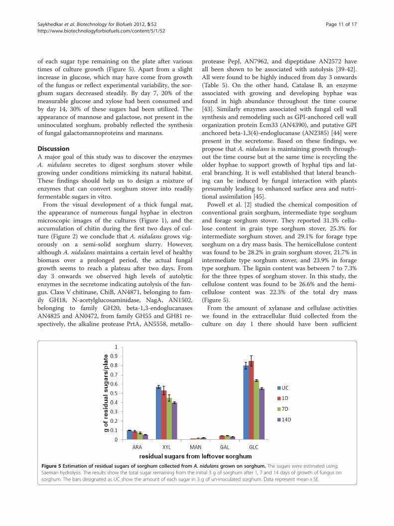
of each sugar type remaining on the plate after varioustimes of culture growth (Figure 5). Apart from a slightincrease in glucose, which may have come from growthof the fungus or reflect experimental variability, the sor-ghum sugars decreased steadily. By day 7, 20% of themeasurable glucose and xylose had been consumed andby day 14, 30% of these sugars had been utilized. Theappearance of mannose and galactose, not present in theuninoculated sorghum, probably reflected the synthesisof fungal galactomannoproteins and mannans.
DiscussionA major goal of this study was to discover the enzymesA. nidulans secretes to digest sorghum stover whilegrowing under conditions mimicking its natural habitat.These findings should help us to design a mixture ofenzymes that can convert sorghum stover into readilyfermentable sugars in vitro.From the visual development of a thick fungal mat,
the appearance of numerous fungal hyphae in electronmicroscopic images of the cultures (Figure 1), and theaccumulation of chitin during the first two days of cul-ture (Figure 2) we conclude that A. nidulans grows vig-orously on a semi-solid sorghum slurry. However,although A. nidulans maintains a certain level of healthybiomass over a prolonged period, the actual fungalgrowth seems to reach a plateau after two days. Fromday 3 onwards we observed high levels of autolyticenzymes in the secretome indicating autolysis of the fun-gus. Class V chitinase, ChiB, AN4871, belonging to fam-ily GH18, N-acetylglucosaminidase, NagA, AN1502,belonging to family GH20, beta-1,3-endoglucanasesAN4825 and AN0472, from family GH55 and GH81 re-spectively, the alkaline protease PrtA, AN5558, metallo-
Figure 5 Estimation of residual sugars of sorghum collected from A. nSaeman hydrolysis. The results show the total sugar remaining from the inisorghum. The bars designated as UC show the amount of each sugar in 3
protease PepJ, AN7962, and dipeptidase AN2572 haveall been shown to be associated with autolysis [39-42].All were found to be highly induced from day 3 onwards(Table 5). On the other hand, Catalase B, an enzymeassociated with growing and developing hyphae wasfound in high abundance throughout the time course[43]. Similarly enzymes associated with fungal cell wallsynthesis and remodeling such as GPI-anchored cell wallorganization protein Ecm33 (AN4390), and putative GPIanchored beta-1,3(4)-endoglucanase (AN2385) [44] werepresent in the secretome. Based on these findings, wepropose that A. nidulans is maintaining growth through-out the time course but at the same time is recycling theolder hyphae to support growth of hyphal tips and lat-eral branching. It is well established that lateral branch-ing can be induced by fungal interaction with plantspresumably leading to enhanced surface area and nutri-tional assimilation [45].Powell et al. [2] studied the chemical composition of
conventional grain sorghum, intermediate type sorghumand forage sorghum stover. They reported 31.3% cellu-lose content in grain type sorghum stover, 25.3% forintermediate sorghum stover, and 29.1% for forage typesorghum on a dry mass basis. The hemicellulose contentwas found to be 28.2% in grain sorghum stover, 21.7% inintermediate type sorghum stover, and 23.9% in foragetype sorghum. The lignin content was between 7 to 7.3%for the three types of sorghum stover. In this study, thecellulose content was found to be 26.6% and the hemi-cellulose content was 22.3% of the total dry mass(Figure 5).From the amount of xylanase and cellulase activities
we found in the extracellular fluid collected from theculture on day 1 there should have been sufficient
idulans grown on sorghum. The sugars were estimated usingtial 3 g of sorghum after 1, 7 and 14 days of growth of fungus ong of un-inoculated sorghum. Data represent mean± SE.

Saykhedkar et al. Biotechnology for Biofuels 2012, 5:52 Page 12 of 17http://www.biotechnologyforbiofuels.com/content/5/1/52
enzyme activity to break down all the xylan and cellulosein the sorghum in less than a day. However the concen-tration of soluble sugars (break-down products from theenzymatic hydrolysis), in the extracellular fluid was low(less than 1 mM), and less than 5% of the total xylanhad been digested and seemingly none of the cellulose.This emphasizes the well-known recalcitrance of plantbiomass to enzymatic degradation and suggests that thefungus is growing under carbon limited conditions.However, over time, the fungus did appear to be able todigest all classes of cell wall polysaccharides present andit did continue to maintain a certain level of healthybiomass.We were interested in determining if the fungus
attacked the most easily digestible polymers first andthen moved on to using the more recalcitrant ones. Ithas been suggested that fungi first degrade the pectin inplant cell walls to make the hemicelluloses and cellulosemore accessible [46]. Sugars characteristic of pectin, i.e.rhamnose and galacturonic acid, were not detected intotal hydrolysates of sorghum stover, although both ofthese sugars were found in the solubilized sugars rinsedfrom the cultures. This indicates the presence of a smallamount of pectin in sorghum. Polygalacturonase activitywas induced rapidly in the culture but xylanase and cel-lulase activity increased at the same time. Looking at theproteomics results one can deduce that almost all of themany enzymes involved in hemicellulose and cellulosedegradation are induced in parallel. However in pectindegradation there is a steady decrease over time in exo-polygalacturonases, one of the pectin methyl esterases,and one of the pectin lyases. In contrast there was asteep increase in other pectate lyases and rhamnogalac-turonan hydrolases.The recalcitrance of the sorghum or any other plant
biomass to digestion is thought to arise from physical in-accessibility of the polymers to the enzymes designed todigest them. Two major factors are the incrustation ofthe polysaccharides with lignin and the crystalinity ofthe cellulose. We identified three classes of proteinswhich might play a key role in overcoming the recalci-trance of the sorghum to digestion.There is strong evidence that some of the ferulic acids
esterified to xylans crosslink to make diferulates whichlink xylans together thus making them less accessible toenzyme digestion. The ferulates can also be incorporatedinto lignin thus attaching xylan directly to lignin [47].Hydrolysis of the ferulate ester bonds is expected to de-crease the recalcitrance of biomass to enzymatic hy-drolysis [48-50]. Two feruloyl esterases were secreted byA. nidulans in high abundance throughout the timecourse.Another enzyme in our cultures which might play a
role in overcoming recalcitrance of the sorghum to
digestion is cellobiose dehydrogenase (CDH). CDHsoxidize cellobiose, or other reducing sugars, and transferthe electrons to Fe+++, quiniones, radicals, or oxygen tomake hydrogen peroxide [51]. The combination of theresulting Fe++ and hydrogen peroxide can lead to thegeneration of hydroxyl radical which will attack polysac-charides and lignin. At least three putative sequences forCDHs are present in the A. nidulans genome. We onlydetected one form. Interestingly CDH was not presenton day 1 and then increased gradually showing aspectrum count of 17 on day 3 up to 77 on day 14. Noother studies have reported the secretion of cellobiosedehydrogenases by A. nidulans on solid state cultures.The presence of cellobiose dehydrogenase after day 3may indicate its important role in the degradation ofcrosslinks between lignin, hemicelluloses and cellulose.Whenever investigated, GH61 family endoglucanases
lack measurable endoglucanase activity, but recentlythey have been reported to accelerate the hydrolysis ofcellulose in cellulase preparations [52,53]. GH61 was ini-tially described as an endoglucanase, but more recentlyhas been shown to be members of the family are coppermono-oxygenases that catalyse cleavage of cellulose oxi-datively, releasing cellodextrins [53,54]. Their active sitecontains a type II copper site, which, after being reducedto Cu+ by a reductant such as ascorbate or gallate, isthought to activate oxygen which can then oxidize glyco-sidic linkages on the surface of crystalline cellulose. Thisgenerates new chain ends rendering the substrate farmore prone to attack by the classical endoglucanasesand cellobiohydrolases [52,53]. Cellobiose dehydrogen-ase appears to be able to mediate the reduction of thecopper site [54]. Recent studies have shown that cello-biose dehydrogenase may enhance cellulose degradationby coupling the oxidation of cellobiose to the reductiveactivation of copper-dependent polysaccharide monoox-ygenases [53,54]. Four GH61 enzymes were found in ourstudy.Studies similar to ours were conducted by Schneider
et al. [11] who grew A. nidulans and the mesophilic bac-teria Pectobacterium carotovorum on leaf litter for20 days, both individually and in co-culture, and identi-fied the proteins secreted into the medium by 1-DPAGE-LC-MS/MS. The aim of their study was to deter-mine the relative contribution of the fungus and the bac-terium to the decomposition. They reported a total of 90proteins secreted by A. nidulans on leaf litter during thecourse of 20 days in comparison with the 294 proteinsidentified in our sorghum cultures. Seventy- two pro-teins were found to be common in both sorghum andleaf litter cultures, 59 proteins were exclusively found inthe sorghum cultures and 13 were only identified in leaflitter cultures. The identities of the proteins in the twocultures are reported in Additional file 2: Table S2. More

Saykhedkar et al. Biotechnology for Biofuels 2012, 5:52 Page 13 of 17http://www.biotechnologyforbiofuels.com/content/5/1/52
than half of the cellulases and xylanases were commonto both cultures with two xylanases and four endogluca-nases unique to leaf litter cultures. Only one pectinaseout of 22 was unique to leaf litter. Interestingly enzymesinvolved in degradation of crosslinks between lignin, cel-lulose and hemicelluloses such as cellobiose dehydrogen-ase and feruloyl esterases were only identified insorghum cultures. Only half the proteases and less thanone third of cell wall remodeling enzymes were commonto both the cultures. In another recent study, Couturieret al. [55] identified total 66 proteins in the secretome ofA. nidulans grown on maize bran. Out of 19 GHs identi-fied in their study in A. nidulans secretome, seven hemi-cellulases belonging to family GH10, GH11, GH39,GH43, GH62 and GH93 and five beta-1,3-glucanasesfrom the GH17, GH55 and GH81 families were identi-fied. They could not detect any beta-1,4-endoglucanaseor cellobiohydrolase or any enzyme activity on CMC[55]. The difference in growth and enzyme activities ofA. nidulans on sorghum and on leaf litter may be attrib-uted to different substrates compositions and growthconditions [12].
ConclusionsWe have identified extracellular proteins secreted dur-ing the entire time course of cultivation of A. nidulanson sorghum stover. The aim was to learn the cocktailof enzymes we can devise to hydrolyze lignocellulosicbiomass efficiently under mild conditions. In this studywe identified a total of 294 proteins including cellulases,hemicellulases, pectinases, carbohydrate esterases, chiti-nases, and many proteins of unknown function. Theenzymes such as feruloyl esterases, cellobiose dehydro-genase, and the family GH61 endoglucanases can beimportant in accelerating biomass conversion by redu-cing the recalcitrance of cellulose. Some of the hypo-thetical proteins we found may work as non-hydrolyticaccessory proteins aiding hydrolytic enzymatic effi-ciency. Further work involving global/whole transcrip-tome studies of A. nidulans during its growth onsorghum may give insight into their function. Cloningand expression of the enzymes such as feruloylesterases, cellobiose dehydrogenase and family 61 endo-glucanases will let us test directly if they have beneficialeffects.
Materials and methodsGrowth conditionsAspergillus nidulans Strain A78 was obtained from thefungal genetic stock center (FGSC) [56]. 3 g of groundsorghum (variety mix of AtX2752/RtX2783 and AtX2752/RtX430) was ground in a Thomas WileyW Mini-Mill (Tho-mas Scientific, Swedesboro, NJ, USA) by passing througha 60-mesh screen. Fungal spores were counted using a
hemacytometer (Spectrum Scientifics, Philadelphia, PA,USA) and adjusted to a million spores per milliliter inwater. The ground sorghum was moistened with 6 mlwater in a petri plate and autoclaved for an hour at 1210C.To these petri plates 15 ml of minimal media (pH 6.5;95 ml of water, 5 ml 20X nitrate salts and 0.1 ml 1000Xtrace elements) and 1 ml of spore suspension containing106 spores was added. Then the petri plates were incu-bated at 370C and 70% of relative humidity for 1, 2, 3, 5, 7and 14 days. Uninoculated sorghum stover incubated insame conditions as inoculated stover was used as control.All the chemicals used in this study were purchased fromSigma-Aldrich (St. Louis, MO, USA), unless otherwise sta-ted. Fungus grown on borosilicate 3 mm solid glass beads(Aldrich, Milwaukee, WI) in 1% glucose for 34 hours wasused as a control for comparison of secretomes. A. nidu-lans cultured in 1% glucose for 34 hrs utilized only 1/4th
of the initial glucose and thus should not be undergoingstarvation.
Sample preparations for microscopyFor scanning electron microscopy (SEM), fungal samplesapproximately, 5 mm squares, were cut from the platesand placed in the first fixative solution (2.5% glutaralde-hyde in 0.2 M sodium phosphate, pH 7.2) and incubatedfor 2 h. The glutaraldehyde solution was removed andreplaced with 0.2 M sodium phosphate buffer, pH 7.2and samples were washed 3X for 20 min each. Post fix-ation was done in 1% osmium tetraoxide in dH2O for anhour. Dehydration was done with a series of ethanolsolutions (50, 70, 90, 95,100,100,100%), for 15 minuteseach. Samples were critical point dried and mounted onaluminum stubs using silver paint adhesive followed bycoating with gold/palladium. Samples were observedusing an FEI quanta 600 electron microscopy (FEI, Hills-boro, OR, USA).For transmission electron microscopy (TEM), the fix-
ation was done as for the SEM samples. Post fixationwas done in 1% osmium tetroxide /1.5% potassium ferri-cyanide in 0.1% sodium cacodylate buffer for 1 hour.Using a series of ethanol solutions (as described in SEMprotocol), samples were dehydrated for 15 minutes each.The dehydrated samples were washed 2X with 100%propylene oxide and then incubation overnight in pro-pylene oxide/polyresin/bed 812 overnight (PolysciencesInc., Warrington, PA, USA) followed by immersion inpolyresin/ bed 812 and polymerized at 600C. Sampleswere sectioned using a Leica EMCU 6 ultramicrotome(Midwest Lab Equipment, Iowa City, IA, USA). Sec-tioned samples were placed on nickel; carbon/formavarcoated grids and stained with uranyl acetate and lead cit-rate and observed using a JEOL 2100 Transmission elec-tron microscope (JEOL, Austin, TX, USA).

Saykhedkar et al. Biotechnology for Biofuels 2012, 5:52 Page 14 of 17http://www.biotechnologyforbiofuels.com/content/5/1/52
Estimation of fungal growth by chitin estimationThe entire plate contents of A. nidulans cultures afterday 1, 2, 3, 5, and 14 was freeze-dried and then mixedthoroughly. One mg samples were transferred to vialswith teflon lined caps and hydrolyzed in 3 ml of 6 NHCl, at 1000 C for 6 h. The HCl was evaporated over-night in a speed-vac [57]. Once the extracts were dried,10 μl of 1 M ammonium hydroxide was added toneutralize any residual HCl. Then 10 μl of 100 nano-moles methyl glucamine was added as an internal stand-ard, followed by 100 μl of 20 mg/ml potassiumborohydride in DMSO and incubated at 400C for 90 minto reduce hexosamines to hexosaminitols. After 90 minthe reaction was stopped by adding 10 μl glacial aceticacid, followed by addition of 20 μl methyl imidazole and200 μl of acetic anhydride and incubated for 10 min atroom temperature to acetylate the hexosaminitols[58,59]. The reaction was stopped by adding 500 μlwater and the alditol acetates were purified by adsorbingthem to a C18 sep-pak and desorbing them with methy-lene chloride to remove the polyphoenols. The dichloro-methane was then evaporated to dryness. 25 μl of ethylacetate was added to the dried sample, out of which,1 μl was injected into the gas chromatograph (Agilent,Santa Clara, CA, USA). A standard curve was preparedusing known amounts of glucosamine ranging from 10to 250 nmol. The glucosamine content of 1 mg aliquotsof dried A. nidulans grown in liquid media (49.50 μg)was used as a conversion factor to estimate milligramsof fungus per milligram of dry mass.
Estimation of enzyme activities by capillary zoneelectrophoresis (CZE) and dinitrosalicylic acid (DNS) assayExtracellular extracts were collected by washing thewhole petri plate contents with 5 ml of autoclaved waterand filtering the extract using Whatman no 1 filter paper.Enzyme activities were detected semi-quantitatively byCZE using 8-aminopyrene-1,3,6-trisulfonate (APTS)labeled substrates (xylohexose, cellopentose, arabinohep-taose, mannohexose) [60]. 1 μl aliquots of extracellularextract and 2 μl of respective APTS labeled substrateswere added to 22 μl of 50 mM ammonium acetate bufferof optimum pH for the respective enzymes to make atotal volume of 25 μl. Samples were incubated for 1 h at370C and the progress of enzymes was followed byCZE (Bio-Rad, Hercules, CA, USA).The DNS assay, developed by Sumner and Graham
[61] for determination of reducing sugar, was used forquantitative determination of enzyme activity. We useda DNS reagent composed of 0.75% di-nitrosalicylic acid,0.5% phenol, 0.5% sodium metabisulfite, 1.4% sodiumhydroxide, and 21% sodium potassium tartrate [62,63].To 10 μl of aliquot of extracellular extract, 20 μl of50 mM ammonium buffer of optimum pH and 20 μl of
appropriate substrate (1%W/V) prepared in water, wereadded. The reaction was carried out in a 96 well PCRplate (Corning, NY, USA) and allowed to incubate for 30minutes at 370C. The reaction was terminated byaddition of 40 μl of DNS mixture to each well and theplate was heated at 1000C for 5 min. We read the platein a 96 well plate reader at 550 nanomoles (Tecan, SanJose, CA, USA). After subtracting substrate blanks fromthe reading enzyme activities were calculate from stand-ard graphs.
Estimation of solublized sugars in extracellular filtrateTo identify and quantitate the breakdown products ofthe polysaccharides, oligosaccharides present in extracel-lular filtrate were estimated by gas chromatographic(GC) analysis of trimethylsilyl methyl glycosides. Metha-nolysis and derivatization were performed using theprotocol of Chaplin [64] modified by Komalavilas andMort [65]. A 25 μl aliquot of sample and 100 nano-moles of inositol as an internal standard were dried in 1dram glass vials in speed vac. 200 μl of methanol 1.5 Mof HCI was added to each vial followed by addition of100 μl of methyl acetate and kept in a heating block at800C for minimum 3 hours. The vials were cooled, fol-lowed by addition of few drops of t-butanol and thenevaporated under a stream of nitrogen at room tempera-tures. Butanol co-evaporates with the HCl, helping to re-move the HCl without degrading sugars. 25 μl of 1:1:5mixture of hexamethyldisilazane: trimethylchlrosilane:pyridine (TMS) was added to all samples. TMS in all thesamples was evaporated using nitrogen gas. Dried sam-ples were re-dissolved in 300 μl of isooctane. 1 μl ofsample was injected in the gas chromatograph (Agilent,Santa Clara, CA, USA). Standards were made by takingaliquot of 100 nanomoles of each sugar standards and100 nanomoles of inositol. From the integrator we calcu-lated relative peak areas and used these to calculate howmuch of each sugar in sample. Area of sugar peak insample/area of inositol peak in the sample/area of sugarpeak in the standards/area of inositol in standards X100 =number of nanomoles in the sample.
Liquid chromatography-tandem mass spectroscopyThe fungal cultures grown on sorghum for day 1, 3, 7and 14 and fungal culture grown in 1% glucose werewashed with 5 ml autoclaved water and the filtrates werecollected. Concentration of total protein in theseextracts was measured by Bradford assay. Small aliquotsof the extracellular filtrates containing 50 ug of totalprotein were run on SDS-PAGE. Gel bands were excised,reduced with Tris (2-carboxyethyl) phosphine), alkylatedwith 2-Iodoacetamide, and digested for 16 hrs with8 μg/ml trypsin using ammonium bicarbonate buffer.Peptides were extracted and analyzed by LC-MS/MS

Saykhedkar et al. Biotechnology for Biofuels 2012, 5:52 Page 15 of 17http://www.biotechnologyforbiofuels.com/content/5/1/52
using an LTQ-Orbitrap XL hybrid mass spectrometer(Thermo Fisher Scientific, San Jose, CA, USA). For thisanalysis, an Eksigent spilt less LC pump (Eksigent, Dub-lin, CA, USA) was used to separate peptide populationson analytical C18 nano columns (Bruker-Michrom Inc.,Auburn, CA, USA), with the column effluent beingsprayed directly into a new objective pico view ionsource. Using a “Big Three” MS/MS method, the Orbi-trap analyzer collected an ultra-accurate (R = 60,000)scan of intact peptides, whilst the LTQ ion trap simul-taneously performed MS/MS fragmentation analysis ofthe each of three most abundant peptides eluting in thatchromatographic fractions. The LC-MS/MS raw fileswere used for database searching via the software appli-cation Mascot (v2.2.2 from Matrix Science, Boston, MA,USA) against sequences from A. nidulans as well asagainst a sorghum database [23,66] . Searches were vali-dated using Scaffold (Proteome Software Inc., Portland,OR, USA) and the Peptide Prophet algorithm [67]. Cri-teria for accepting protein identification, was only pro-tein probability thresholds greater than 99% wereaccepted and at least two peptides needed to be identi-fied, each with 95% certainty. Protein candidates con-taining similar peptides were grouped to fulfill theprinciples of parsimony. Search results were alsochecked for false discoveries using reversed decoy se-quence databases, and no decoy sequences weredetected, thus bringing our false discovery rate to zero.Changes in protein expression between samples fromdifferent time points were examined using the spectralcount method [68]. The complete peptide reports fromthe Scaffold results for samples taken from days 1, 3, 7,and 14 are given in Additional file 3: Table S3, Add-itional file 4: Table S4, Additional file 5: Table S5, andAdditional file 6: Table S6 respectively. SignalP was usedto predict secretion signals in the identified proteins[69,70]. Additional information for the presence of a sig-nal peptide was obtained by accessing the following URL[25].
Estimation of residual sorghum hydrolysates by saemanhydrolysisSaeman hydrolysis was done on the total biomass on theplates after 1, 7, and 14 days of fungal growth to esti-mate total sugars [30]. Three hundred mg samples of thethoroughly mixed freeze dried biomass from each petriplate were suspended in 3 ml of 72% sulfuric acid. Thesamples were incubated at 300 C for 60 min with stirringevery 5–10 min followed by dilution of sulfuric acid to4% by adding 84 ml of water and autoclaved for an hourat 1210C. 2 ml aliquot of each sample was neutralizedwith calcium carbonate to pH 5–6. 25 μl of sample wasprocessed for methanolysis, trimethylsilylation, and gas
liquid chromatography (GLC) analysis as describedabove.
Additional Files
Additional file 1: Table s1. Identified miscellaneous proteins andspectrum counts on 1, 3, 7, and 14 days.
Additional file 2: Table S2. Comparison of identified proteins inA. nidulans cultures from different carbon sources.
Additional file 3: Table S3. Protein report of A. nidulans grown onsorghum as carbon source on day1.
Additional file 4: Table S4. Protein report of A. nidulans grown onsorghum as carbon source on day3.
Additional file 5: Table S5. Protein report of A. nidulans grown onsorghum as carbon source on day7.
Additional file 6: Table S6. Protein report of A. nidulans grown onsorghum as carbon source on day14.
Abbreviations1D-PAGE: One dimensional polyacrylamide electrophoresis; LC-MS/MS: Liquidchromatography-tandem mass spectroscopy; CAzY families: Carbohydrateactive enzyme families; SEM: Scanning electron microscopy;TEM: Transmission electron microscopy; CZE: Capillary zone electrophoresis;ORF: Open reading frame; GH: Glycosyl hydrolase; AN: Acession number;CBM: Carbohydrate binding module; PL: Pectin lyase; ECF: Extracellularfiltrate; CDH: Cellobiose dehydrogenase; CMC: Carboxymethyl cellulose;FGSC: Fungal genetic stock center; APTS: 8-aminopyrene-1,3,6-trisulfonate;DNS: Dinitrosalicylic acid; PCR: Polymer chain reaction; GC: Gaschromatography; TMS: Hexamethyldisilazane: trimethylchlrosilane: pyridine.
Competing interestThe authors declare that they have no competing interest.
AcknowledgmentsThis work was supported by USDA CSREES grant 2007-35504-18244, a grantfrom The Oklahoma Bioenergy Center, and the Oklahoma AgriculturalExperiment Station. Microscopy work was performed in the OSU MicroscopyLaboratory using electron microscopes purchased with grants from theNational Science Foundation. We appreciate the technical assistance offeredby the staff of the Laboratory. Mass spectrometry analyses were performedin the DNA/Protein Resource Facility at Oklahoma State University, usingresources supported by the NSF MRI and EPSCoR programs (MRI/0722494).We appreciate the help and technical assistance offered by Janet Rogers inthe DNA/Protein Resource Facility.
Author details1Department of Biochemistry and Molecular Biology, Oklahoma StateUniversity, Stillwater, OK 74078, USA. 2Department of Microbiology andMolecular Genetics, Oklahoma State University, Stillwater, OK 74078, USA.
Authors’ contributionSS, AR, PAC, SDH, RP, and AJM conceived and designed various aspects ofthe experiments. SS and AR performed the experiments. SS, AR, PAC, SDH,RP, and AJM al contributed to the analysis of the data, and the writing of themanuscript. All authors read and approved the final manuscript.
Received: 20 April 2012 Accepted: 26 July 2012Published: 26 July 2012
References1. Carmen S: Lignocellulosic residues: Biodegradation and bioconversion by
fungi. Biotechnol Adv 2009, 27(2):185–194.2. Powell J, Hons F, McBee G: Nutrient and carbohydrate partitioning in
sorghum stover. Agron J 1991, 83:933–937.3. Sierra R, Smith A, Cesar H, Mark T: Producing Fuels and Chemicals from
Lignocellulosic. Chem Eng Prog 2008, 104(8):S10–S18.

Saykhedkar et al. Biotechnology for Biofuels 2012, 5:52 Page 16 of 17http://www.biotechnologyforbiofuels.com/content/5/1/52
4. Lloyd TA, Wyman CE: Combined sugar yields for dilute sulfuric acidpretreatment of corn stover followed by enzymatic hydrolysis of theremaining solids. Bioresour Technol 2005, 96(18):1967–1977.
5. Teymouri F, Laureano-Pérez L, Alizadeh H, Dale B: Ammonia fiberexplosion treatment of corn stover. Appl Biochem Biotechnol 2004,115(1):951–963.
6. Kim S, Holtzapple MT: Effect of structural features on enzyme digestibilityof corn stover. Bioresour Technol 2006, 97(4):583–591.
7. Liu L, Sun J, Li M, Wang S, Pei H, Zhang J: Enhanced enzymatic hydrolysisand structural features of corn stover by FeCl3 pretreatment. BioresourTechnol 2009, 100(23):5853–5858.
8. Shi J, Chinn MS, Sharma-Shivappa RR: Microbial pretreatment of cottonstalks by solid state cultivation of Phanerochaete chrysosporium. BioresourTechnol 2008, 99(14):6556–6564.
9. Zheng R, Zhang H, Zhao J, Lei M, Huang H: Direct and simultaneousdetermination of representative byproducts in a lignocellulosichydrolysate of corn stover via gas chromatography–mass spectrometrywith a Deans switch. J Chromatogr A 2011, 5(31):5319–5327.
10. Szakacs G, Tengerdy RP: Production of cellulase and xylanase withselected filamentous fungi by solid substrate fermentation. In Enzymesfor Pulp and Paper Processing, Volume 655.: American Chemical Society;1996:175–182.
11. Schneider T, Gerrits B, Gassmann R, Schmid E, Gessner MO, Richter A, BattinT, Eberl L, Riedel K: Proteome analysis of fungal and bacterialinvolvement in leaf litter decomposition. Proteomics 2010,10(9):1819–1830.
12. Han M-J, Kim N-J, Lee S, Chang H: Extracellular proteome of Aspergillusterreus grown on different carbon sources. Curr Genet 2010,56(4):369–382.
13. Aspergillus nidulans Project Information. http://www.broadinstitute.org/annotation/fungi/aspergillus_nidulans_old/background.html.
14. Galagan JE, Calvo SE, Cuomo C, Ma L-J, Wortman JR, Batzoglou S, Lee S-I,Basturkmen M, Spevak CC, Clutterbuck J, et al: Sequencing of Aspergillusnidulans and comparative analysis with A. fumigatusand A. oryzae. Nature2005, 438(7071):1105–1115.
15. Wortman JR, Gilsenan JM, Joardar V, Deegan J, Clutterbuck J, Andersen MR,Archer D, Bencina M, Braus G, Coutinho P, et al: The 2008 update of theAspergillus nidulans genome annotation: A community effort. FungalGenetics and Biology 2009, 46(1, Supplement):S2–S13.
16. Bauer S, Vasu P, Persson S, Mort AJ, Somerville CR: Development andapplication of a suite of polysaccharide-degrading enzymes foranalyzing plant cell walls. Proc Natl Acad Sci 2006, 103(30):11417–11422.
17. Coutinho PM, Andersen MR, Kolenova K, VanKuyk PA, Benoit I, Gruben BS,Trejo-Aguilar B, Visser H, Solingen P, Pakula T, et al: Post-genomic insightsinto the plant polysaccharide degradation potential of Aspergillusnidulans and comparison to Aspergillus niger and Aspergillus oryzae.Fungal Genetics and Biology 2009, 46(Supplement 1):S161–S169.
18. Medina ML, Haynes PA, Breci L, Francisco WA: Analysis of secretedproteins from Aspergillus flavus. Proteomics 2005, 5(12):3153–3161.
19. Oda K, Kakizono D, Yamada O, Iefuji H, Akita O, Iwashita K: Proteomicanalysis of extracellular proteins from Aspergillus oryzae grown undersubmerged and solid-state culture conditions. Appl Environ Microbiol 2006,72(5):3448–3457.
20. Adav SS, Li AA, Manavalan A, Punt P, Sze SK: Quantitative iTRAQSecretome Analysis of Aspergillus niger Reveals Novel HydrolyticEnzymes. Journal of Proteome Research 2010, 9(8):3932–3940.
21. Suraini Abd-Aziz GSH, Mohd Ali H, Mohamed Ismail Abdul K, Noraini S:Indirect method for quantification of cell biomass during solid-statefermentation of palm kernel cake based on protein content. AsianJournal of Scientific Research 2008, 1(4385–393):385–393.
22. Zhou JY, Schepmoes AA, Zhang X, Moore RJ, Monroe ME, Lee JH, CampDG, Smith RD, Qian WJ: Improved LC-MS/MS spectral counting statisticsby recovering low-scoring spectra matched to confidently identifiedpeptide sequences. J Proteome Res 2010, 9(11):5698–5704.
23. Pedant Database. http://pedant.gsf.de.24. Walter MC, Rattei T, Arnold R, Guldener U, Munsterkotter M, Nenova K,
Kastenmuller G, Tischler P, Wolling A, Volz A, et al: PEDANT covers allcomplete RefSeq genomes. Nucleic Acids Res 2009, 37(Database issue):21.
25. Aspergillus genome database. http://www.aspergillusgenome.org.26. Biely P: Microbial xylanolytic systems. Trends Biotechnol 1985,
3(11):286–290.
27. Coughlan MP, Hazlewood GP: beta-1,4-D-xylan-degrading enzymesystems: biochemistry, molecular biology and applications. BiotechnolAppl Biochem 1993, 17(Pt 3):259–289.
28. Javier PFI, Óscar G, Sanz-Aparicio J, Díaz P: Xylanases: Molecular propertiesand applications industrial enzymes. In In. Edited by Polaina J, MacCabeAP.: Springer Netherlands; 2007:65–82.
29. de Vries RP, Visser J: Aspergillus enzymes involved in degradation of plantcell wall polysaccharides. Microbiol Mol Biol Rev 2001, 65(4):497–522.
30. Saeman JF, Bubl JL, Harris EE: Quantitative saccharification of wood andcellulose. Ind Eng Chem Anal Ed 1945, 17(1):35–37.
31. de Vries RP, van Grieken C, vanKuyk PA, Wösten HAB: The value ofgenome sequences in the rapid Identification of novel genes encodingspecific plant cell wall degrading enzymes. Current Genomics 2005,6(3):157–187.
32. Goyal A, Ghosh B, Eveleigh D: Characteristics of fungal cellulases. BioresourTechnol 1991, 36(1):37–50.
33. Rabinovich ML, Bolobova AV, Vasil’chenko LG: Fungal decomposition ofnatural aromatic structures and xenobiotics: A Review. Appl BiochemMicrobiol 2004, 40(1):1–17.
34. Rabinovich ML, Melnick MS, Bolobova AV: The structure and mechanism ofaction of cellulolytic enzymes. Biochem Mosc 2002, 67(8):850–871.
35. Lynd LR, Cushman JH, NICHOLS RJ, WYMAN CE: Fuel ethanol fromcellulosic biomass. Science 1991, 251(4999):1318–1323.
36. Bagga PS, Sandhu DK, Sharma S: Purification and characterization ofcellulolytic enzymes produced by Aspergillus nidulans. J Appl Microbiol1990, 68(1):61–68.
37. Martens-Uzunova ES, Schaap PJ: Assessment of the pectin degradingenzyme network of Aspergillus niger by functional genomics. FungalGenetics and Biology 2009, 46(1, Supplement):S170–S179.
38. van den Brink J, de Vries RP: Fungal enzyme sets for plant polysaccharidedegradation. Appl Microbiol Biotechnol 2011, 91(6):1477–1492.
39. Emri T, Molnár Z, Szilágyi M, Pócsi I: Regulation of autolysis in Aspergillusnidulans. Appl Biochem Biotechnol 2008, 151(2):211–220.
40. Jaques AK, Fukamizo T, Hall D, Barton RC, Escott GM, Parkinson T, HitchcockCA, Adams DJ: Disruption of the gene encoding the ChiB1 chitinase ofAspergillus fumigatus and characterization of a recombinant geneproduct. Microbiology 2003, 149(10):2931–2939.
41. Yamazaki H, Yamazaki D, Takaya N, Takagi M, Ohta A, Horiuchi H: Achitinase gene, chiB, involved in the autolytic process of Aspergillusnidulans. Curr Genet 2007, 51(2):89–98.
42. Shin KS, Kwon NJ, Kim YH, Park HS, Kwon GS, Yu JH: Differential roles ofthe ChiB chitinase in autolysis and cell death of Aspergillus nidulans.Eukaryot Cell 2009, 8(5):738–746.
43. Kawasaki L, Wysong D, Diamond R, Aguirre J: Two divergent catalasegenes are differentially regulated during Aspergillus nidulansdevelopment and oxidative stress. J Bacteriol 1997, 179(10):3284–3292.
44. de Groot PWJ, Brandt BW, Horiuchi H, Ram AFJ, de Koster CG, Klis FM:Comprehensive genomic analysis of cell wall genes in Aspergillusnidulans. Fungal Genetics and Biology 2009, 46(1, Supplement):S72–S81.
45. Harris SD: Branching of fungal hyphae: regulation, mechanisms andcomparison with other branching systems. Mycologia 2008,100(6):823–832.
46. Karr AL, Albersheim P: Polysaccharide-degrading enzymes are unable toattack plant cell walls without Prior Action by a “Wall-modifyingEnzyme”. Plant Physiol 1970, 46(1):69–80.
47. Hatfield RD, Ralph J, Grabber JH: Cell wall cross-linking by ferulates anddiferulates in grasses. J Sci Food Agric 1999, 79(3):403–407.
48. Koseki T, Hori A, Seki S, Murayama T, Shiono Y: Characterization of twodistinct feruloyl esterases, AoFaeB and AoFaeC, from Aspergillus oryzae.Appl Microbiol Biotechnol 2009, 83(4):689–696.
49. Koseki T, Fushinobu S, Ardiansyah A, Shirakawa H, Komai M: Occurrence,properties, and applications of feruloyl esterases. Appl Microbiol Biotechnol2009, 84(5):803–810.
50. Benoit I, Danchin E, Bleichrodt R-J, de Vries R: Biotechnological applicationsand potential of fungal feruloyl esterases based on prevalence,classification and biochemical diversity. Biotechnol Lett 2008,30(3):387–396.
51. Henriksson G, Johansson G, Pettersson G: A critical review of cellobiosedehydrogenases. J Biotechnol 2000, 78(2):93–113.
52. Harris PV, Welner D, McFarland KC, Re E, Navarro Poulsen J-C, Brown K,Salbo R, Ding H, Vlasenko E, Merino S, et al: Stimulation of lignocellulosic
Saykhedkar et al. Biotechnology for Biofuels 2012, 5:52 Page 17 of 17http://www.biotechnologyforbiofuels.com/content/5/1/52
biomass hydrolysis by proteins of glycoside hydrolase family 61:Structure and function of a large, enigmatic family. Biochemistry 2010,49(15):3305–3316.
53. Quinlan RJ, Sweeney MD, Lo Leggio L, Otten H, Poulsen J-CN, Johansen KS,Krogh KBRM, Jørgensen CI, Tovborg M, Anthonsen A, et al: Insights into theoxidative degradation of cellulose by a copper metalloenzyme thatexploits biomass components. Proc Natl Acad Sci 2011,108(37):15079–15084.
54. Langston JA, Shaghasi T, Abbate E, Xu F, Vlasenko E, Sweeney MD:Oxidoreductive cellulose depolymerization by the enzymes cellobiosedehydrogenase and glycoside hydrolase 61. Appl Environ Microbiol 2011,77(19):7007–7015.
55. Couturier M, Navarro D, Olive C, Chevret D, Haon M, Favel A, Lesage-Meessen L, Henrissat B, Coutinho P, Berrin J-G: Post-genomic analyses offungal lignocellulosic biomass degradation reveal the unexpectedpotential of the plant pathogen Ustilago maydis. BMC Genomics 2012,13(1):57.
56. Fungal Genetics Stock Center. http://www.fgsc.net/.57. Penman D, Britton G, Hardwick K, Collin HA, Isaac S: Chitin as a measure of
biomass of Crinipellis perniciosa, causal agent of witches’ broom diseaseof Theobroma cacao. Mycol Res 2000, 104(6):671–675.
58. Blakeney AB, Harris PJ, Henry RJ, Stone BA: A simple and rapid preparationof alditol acetates for monosaccharide analysis. Carbohydr Res 1983,113(2):291–299.
59. Mort AJ, Parker S, Kuo MS: Recovery of methylated saccharides frommethylation reaction mixtures using Sep-Pak C18 cartridges. AnalBiochem 1983, 133(2):380–384.
60. Naran R, Pierce ML, Mort AJ: Detection and identification ofrhamnogalacturonan lyase activity in intercellular spaces of expandingcotton cotyledons. The Plant Journal 2007, 50(1):95–107.
61. Sumner JB, Graham Wtao VA: Dinitrosalicylic acid: a reagent for theestimation of sugar in normal and diabetic urine. J Biol Chem 1921,47(1):5–9.
62. Miller GL: Use of dinitrosalicylic acid reagent for determination ofreducing sugar. Anal Chem 1959, 31(3):426–428.
63. Squina FM, Mort AJ, Decker SR, Prade RA: Xylan decomposition byAspergillus clavatus endo-xylanase. Protein Expr Purif 2009, 68(1):65–71.
64. Martin FC: A rapid and sensitive method for the analysis of carbohydratecomponents in glycoproteins using gas–liquid chromatography. AnalBiochem 1982, 123(2):336–341.
65. Komalavilas P, Mort AJ: The acetylation of O-3 of galacturonic acid in therhamnose-rich portion of pectins. Carbohydr Res 1989, 189:261–272.
66. Sorghum Genome Database. http://www.phytozome.net/sorghum.67. Keller A, Nesvizhskii AI, Kolker E, Aebersold R: Empirical statistical model to
estimate the accuracy of peptide identifications made by MS/MS anddatabase search. Anal Chem 2002, 74(20):5383–5392.
68. Old WM, Meyer-Arendt K, Aveline-Wolf L, Pierce KG, Mendoza A, Sevinsky JR,Resing KA, Ahn NG: Comparison of label-free methods for quantifyinghuman proteins by shotgun proteomics. Molecular & Cellular Proteomics2005, 4(10):1487–1502.
69. SignalP software. http://www.cbs.dtu.dk/services/SignalP/.70. Bendtsen JD, Nielsen H, von Heijne G, Brunak S: Improved prediction of
signal peptides: SignalP 3.0. J Mol Biol 2004, 340(4):783–795.
doi:10.1186/1754-6834-5-52Cite this article as: Saykhedkar et al.: A time course analysis of theextracellular proteome of Aspergillus nidulansgrowing on sorghum stover. Biotechnology for Biofuels 2012 5:52.
Submit your next manuscript to BioMed Centraland take full advantage of:
• Convenient online submission
• Thorough peer review
• No space constraints or color figure charges
• Immediate publication on acceptance
• Inclusion in PubMed, CAS, Scopus and Google Scholar
• Research which is freely available for redistribution
Submit your manuscript at www.biomedcentral.com/submit