2008 Alavandi DAO
9
DISEASES OF AQUATIC ORGANISMS Dis Aquat Org Vol. 81: 163–171, 2008 doi: 10.3354/dao01955 Published August 27 INTRODUCTION More than 20 viruses are known to infect farmed pe- naeid shrimp globally (Lightner 1996, Flegel 1997, Lightner & Redman 1998), and the number of viral pathogens from different geographical regions has been increasing steadily, with reports of new diseases and syndromes such as Syndrome 93 (Costa et al. 1998), spawner isolated mortality syndrome (Fraser & Owens 1996), infectious myonecrosis (Poulos et al. 2006), and monodon slow growth syndrome (Sritun- yalucksana et al. 2006). Loose shell syndrome (LSS) has been reported among farmed Penaeus monodon since 1998 in India, and since then, reports of its incidence have been increasing year after year. LSS is a slow progressive disease of farmed black tiger shrimp in India, which is characterized by a flaccid spongy abdomen due to muscular dystrophy. The exoskeleton forms a sort of loose covering over the abdominal musculature, with a space in between (Alavandi et al. 2007). The feed con- version efficiency of shrimp substantially declines, leading to poor meat quality, and affected ponds suffer low-level progressive mortalities. Until recently, LSS was prevalent on the east coast states of the Indian subcontinent, viz. Tamil Nadu, Andhra Pradesh, Orissa, and West Bengal. During 1998 to 1999, the incidence of LSS was reported in about 23 and 14% of shrimp farms around the Vellar estuary in Tamil Nadu during summer and winter © Inter-Research 2008 · www.int-res.com *Email: [email protected] Loose shell syndrome of farmed Penaeus monodon in India is caused by a filterable agent S. V. Alavandi 1, *, T. D. Babu 1 , K. S. Abhilash 1 , K. K. Vijayan 1, 2 , N. Kalaimani 1 , T. C. Santiago 1 1 Central Institute of Brackishwater Aquaculture (Indian Council of Agricultural Research), 75 Santhome High Road, Raja Annamalai Puram, Chennai 600 028, India 2 Present address: Marine Biotechnology Division, Central Marine Fisheries Research Institute, PO Box 1603, Ernakulam North PO, Kochi 682 018, India ABSTRACT: Loose shell syndrome (LSS) of farmed black tiger shrimp Penaeus monodon has been reported from Indian shrimp farms since 1998 and is recognized as a major disease problem causing significant economic loss to the shrimp aquaculture sector. Unlike the rapid mortalities associated with viral pathogens such as white spot syndrome virus and yellow head virus, progression of LSS is gradual, leading to low-level progressive mortalities. The signs of LSS include a flaccid spongy abdomen due to muscular dystrophy, space between the exoskeleton and muscle, and a shrunken hepatopancreas. The feed conversion efficiency is reduced, and shrimp have poor meat quality, caused by impairment of the hepatopancreatic functions such as digestion and absorption as evi- denced by the atrophy of the hepatopancreas. Histopathological investigations on LSS-affected shrimp showed shrinkage of extensor and flexor muscles with occasional hemocytic infiltration. The hepatopancreas showed inflammation of hepatopancreatic tubules with enlargement of intertubular spaces, hemocytic infiltration, and low levels of lipid reserves in the R cells. In advanced stages of LSS, many tubules were in highly necrotic condition with a sloughed epithelium, reflecting the dys- function of the digestive gland. LSS could be induced in healthy tiger shrimp by challenge studies using membrane-filtered LSS-affected shrimp tissues, suggesting involvement of a filterable infec- tious agent. KEY WORDS: Histopathology · Loose shell syndrome · LSS · Tiger shrimp · Penaeus monodon · Filterable agent Resale or republication not permitted without written consent of the publisher
-
Upload
chennaiuniv -
Category
Documents
-
view
0 -
download
0
Transcript of 2008 Alavandi DAO
DISEASES OF AQUATIC ORGANISMSDis Aquat Org
Vol. 81: 163–171, 2008doi: 10.3354/dao01955
Published August 27
INTRODUCTION
More than 20 viruses are known to infect farmed pe-naeid shrimp globally (Lightner 1996, Flegel 1997,Lightner & Redman 1998), and the number of viralpathogens from different geographical regions hasbeen increasing steadily, with reports of new diseasesand syndromes such as Syndrome 93 (Costa et al.1998), spawner isolated mortality syndrome (Fraser &Owens 1996), infectious myonecrosis (Poulos et al.2006), and monodon slow growth syndrome (Sritun-yalucksana et al. 2006).
Loose shell syndrome (LSS) has been reportedamong farmed Penaeus monodon since 1998 in India,and since then, reports of its incidence have been
increasing year after year. LSS is a slow progressivedisease of farmed black tiger shrimp in India, which ischaracterized by a flaccid spongy abdomen due tomuscular dystrophy. The exoskeleton forms a sort ofloose covering over the abdominal musculature, with aspace in between (Alavandi et al. 2007). The feed con-version efficiency of shrimp substantially declines,leading to poor meat quality, and affected ponds sufferlow-level progressive mortalities.
Until recently, LSS was prevalent on the east coaststates of the Indian subcontinent, viz. Tamil Nadu,Andhra Pradesh, Orissa, and West Bengal. During1998 to 1999, the incidence of LSS was reported inabout 23 and 14% of shrimp farms around the Vellarestuary in Tamil Nadu during summer and winter
© Inter-Research 2008 · www.int-res.com*Email: [email protected]
Loose shell syndrome of farmed Penaeus monodonin India is caused by a filterable agent
S. V. Alavandi1,*, T. D. Babu1, K. S. Abhilash1, K. K. Vijayan1, 2, N. Kalaimani1,T. C. Santiago1
1Central Institute of Brackishwater Aquaculture (Indian Council of Agricultural Research), 75 Santhome High Road,Raja Annamalai Puram, Chennai 600 028, India
2Present address: Marine Biotechnology Division, Central Marine Fisheries Research Institute, PO Box 1603,Ernakulam North PO, Kochi 682 018, India
ABSTRACT: Loose shell syndrome (LSS) of farmed black tiger shrimp Penaeus monodon has beenreported from Indian shrimp farms since 1998 and is recognized as a major disease problem causingsignificant economic loss to the shrimp aquaculture sector. Unlike the rapid mortalities associatedwith viral pathogens such as white spot syndrome virus and yellow head virus, progression of LSS isgradual, leading to low-level progressive mortalities. The signs of LSS include a flaccid spongyabdomen due to muscular dystrophy, space between the exoskeleton and muscle, and a shrunkenhepatopancreas. The feed conversion efficiency is reduced, and shrimp have poor meat quality,caused by impairment of the hepatopancreatic functions such as digestion and absorption as evi-denced by the atrophy of the hepatopancreas. Histopathological investigations on LSS-affectedshrimp showed shrinkage of extensor and flexor muscles with occasional hemocytic infiltration. Thehepatopancreas showed inflammation of hepatopancreatic tubules with enlargement of intertubularspaces, hemocytic infiltration, and low levels of lipid reserves in the R cells. In advanced stages ofLSS, many tubules were in highly necrotic condition with a sloughed epithelium, reflecting the dys-function of the digestive gland. LSS could be induced in healthy tiger shrimp by challenge studiesusing membrane-filtered LSS-affected shrimp tissues, suggesting involvement of a filterable infec-tious agent.
KEY WORDS: Histopathology · Loose shell syndrome · LSS · Tiger shrimp · Penaeus monodon ·Filterable agent
Resale or republication not permitted without written consent of the publisher

Dis Aquat Org 81: 163–171, 2008
crops, respectively (Mayavu et al. 2003). In AndhraPradesh, West Godavari district (improved traditionalshrimp farms with average stocking density of3 shrimp m–2) and Nellore (average stocking density ofabout 8 shrimp m–2), the incidence of LSS was reportedin about 27% and 5% of the farms, respectively(MPEDA/NACA 2003). A survey conducted by theSociety of Aquaculture Professionals (SAP) in 2002recorded the incidence of LSS in more than 50% of thefarms (>1100 ha surveyed) in coastal Andhra Pradesh(SAP 2004). LSS resulted in the reduction of averagedaily growth rate, average body weight, and poor sur-vival of shrimp, leading to a reduction in biomass pro-duction at the affected farms (Gopalakrishnan & Parida2005). Since 2005, LSS has also been reported fromGujarat and Kerala on the west coast of India. A con-servative estimate of losses due to LSS in the states ofAndhra Pradesh and Tamil Nadu during the 2 crops in2006 has been estimated to be about Rupees 365 mil-lion (~US $ 9.125 million) in about 73 000 ha area underculture in these 2 states alone (Central Institute ofBrackishwater Aquaculture [CIBA] unpubl.). Giventhe serious economic consequences of LSS, the shrimpfarming community in India recognizes LSS as themost devastating disease syndrome next to white spotsyndrome virus (WSSV).
The MPEDA/NACA (2003) study suggested that thecause of LSS could be chronic bacterial infections andtoxic pond-bottom conditions. The clinical signs ofnecrotizing hepatopancreatitis (NHP) such as discol-oration, atrophy, and necrosis of the hepatopancreas,reduced feed intake, soft shell, and flaccid musclereported in farmed Litopenaeus vannamei from theAmericas (Frelier et al. 1992, Lightner 1996) closelyresembles LSS. SAP (2004) suggested possible involve-ment of algal toxin(s), NHP, or a new pathogen, as thecausative agent of LSS.
There is growing evidence that, along with seriousviral diseases such as WSSV, LSS is becoming a signif-icant limiting factor in brackish-water pond farming oftiger shrimp in India. Owing to geographic localizationof the occurrence of LSS in India and slow mortalitypattern, only a few reports have been published on itspathology (Mayavu et al. 2003, Gopalakrishnan &Parida 2005), and thus far, no information exists on itsetiology. Here we describe the histopathology of LSSand the possible involvement of a filterable agent in itstransmission to Penaeus monodon through laboratorychallenge.
MATERIALS AND METHODS
Information on farming, occurrence, signs, and pro-gression of LSS in the shrimp farms was obtained from
shrimp farmers and farmers’ associations in variousmaritime states of India and by periodic visits to shrimpfarms located on the coasts of 2 states, Tamil Nadu andAndhra Pradesh.
Shrimp samples. Weak and anorexic black tigershrimp Penaeus monodon weighing 18 to 26 g, restingat the edges of the ponds and showing advanced signsof LSS, such as a spongy abdomen and loose exoskele-ton, were collected from shrimp farms affected withLSS near Marakkanam in Tamil Nadu, and Ongoleand Nellore in Andhra Pradesh. Shrimp were trans-ported to the CIBA laboratory live in pond water withaeration.
Histopathology. Tissue samples of hepatopancreas(HP), lymphoid organ (LO), and muscle (M) were dis-sected from shrimp showing signs of LSS at farm sites,fixed in Davidson’s fixative for 24 h, and then trans-ferred to 70% propanol for subsequent processing byconventional histological techniques (Bell & Lightner1988). Sections of 5 to 6 µm thickness were stainedwith hematoxylin and eosin. Photomicrographs weretaken using a Carl Zeiss Axiostar Plus microscope fit-ted with a ProgRess C-10 Plus CCD camera.
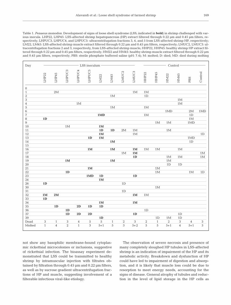
Bioassay. Bioassays were carried out using 9 types ofinocula prepared from HP and muscle of LSS-affectedshrimp and purified fractions obtained by sucrose gra-dient ultracentrifugation (see Table 1). Preparation ofthese inocula and details of the challenge experimentare given below.
Preparation of inocula for bioassay. Filtrates of HPand muscle tissues of LSS-affected shrimp were pre-pared by homogenizing these tissues in sterile PBSseparately with a mortar and pestle, while taking pre-cautions to maintain aseptic conditions. The homo-genized tissues were clarified by centrifugation at4000 × g at 4oC for 15 min. The resultant supernatantswere divided into 2 portions each, which were filteredthrough sterile 0.45 and 0.22 µm filters (Millipore) andstored in a deep freezer (–70°C) until used for chal-lenge experiments. Control inocula were also pre-pared in a similar way from tissues of healthy Penaeusmonodon collected from a farm at Mamallapuram nearChennai, where there was no report of an LSS episode.
Purification of the putative infectious agent. Theputative infectious agent was purified following theprotocols of Bonami et al. (1997) with some modifica-tions. Immediately upon arrival of the shrimp in thelaboratory, HP and M tissue of LSS-affected shrimpwere dissected out, pooled separately (~20 g of each ofthe tissues), and aseptically homogenized in sterile TNbuffer (10 mM Tris-HCl pH 7.5, 400 mM NaCl) in amortar and pestle. The suspension was clarified bylow-speed centrifugation at 4000 × g for 15 min at 4°C3 times using a Sorvall RC 5B Plus centrifuge. TheHP and M supernatants were centrifuged for 2 h at
164
Alavandi et al.: Loose shell syndrome of farmed shrimp
209 000 × g at 4°C using a SW41 swinging-bucket rotor(Beckman Coulter OptimaTM XL-100K), and the resul-tant pellet was resuspended in 1.0 ml sterile TN buffer.This suspension was layered onto a 10 to 50% (w/w)linear sucrose gradient and fractionated by ultra-cen-trifugation at 209 000 × g at 4°C for 3 h. Five fractionswere obtained (and were numbered starting from thetop), which were collected using sterile disposablePasteur pipettes with long narrow nozzles, washed,and resuspended in 1.0 ml sterile TN buffer. The opti-cal density (OD) of gradient fractions at OD254 wasrecorded using a SmartspecTM 3000 UV-VIS spec-trophotometer (BioRad). The suspected etiologic agentpurified by sucrose gradient ultracentrifugation(described above) with OD254 0.166, 0.145, and 0.094(third, fourth, and fifth fractions) of HP and second andthird fractions of muscle were used for the challengeexperiment.
Challenge experiment. Healthy Penaeus monodonwith an average body weight of 15.7 g and free fromLSS were obtained from culture ponds in Mamallapu-ram near Chennai, Tamil Nadu. These shrimp wereassured to be free from WSSV by nested PCR (using aWSSV kit; Genei) and MBV by malachite green wetmount preparations of fecal matter. Five shrimp werestocked per 100 l fiberglass-reinforced plastic tanks ina water recirculating system to maintain oligotrophicgrade water quality. The salinity was 32 ppt, pH was7.9, and the temperature was ~28°C during the chal-lenge experiments. The experimental tanks were con-tinuously aerated to provide 4 to 6 ppm of dissolvedoxygen. Shrimp were fed (10% of body weight) withCIBA-formulated pellet feed with 35% protein andacclimatized to laboratory conditions for 1 wk. Eachinoculum prepared as above was injected intramuscu-larly (50 µl) using tuberculin syringes between the sec-ond and third abdominal segment. Another control setof shrimp was maintained without any injection (seeTable 1). The experiment was conducted for 40 d, andexperimental shrimp were examined daily for thedevelopment of signs of LSS. Tissues of HP of 2 surviv-ing moribund shrimp, 1 from each group challengedwith 0.22 µm and 0.45 µm filtrate of LSS-affected HP,were also examined for histopathology as describedabove.
RESULTS
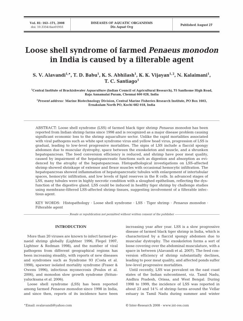
Incidences of LSS were first reported during 1998from the farms located on the east coast of India, whilethe disease has been reported from the west coast onlysince 2005 (Fig. 1). During the initial years of its occur-rence (1998 to 2004), shrimp weighing >20 g afterabout 80 d of culture (DOC) were affected, and since
2006, shrimp weighing 8 to 10 g after 40 to 50 DOChave also been reported to be affected. The diseasehas been recorded in ponds with shrimp stocking den-sities as low as 3 m–2 and also in densities of 10 to15 m–2 and in salinities ranging from 20 to 60 ppt. In the
165
Fig. 1. Occurrence of loose shell syndrome (LSS) in shrimpfarms in the maritime states of India
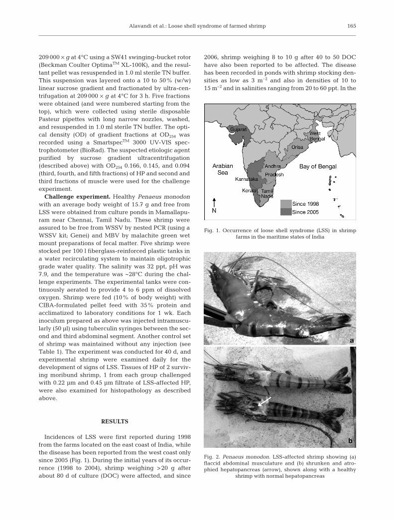
Fig. 2. Penaeus monodon. LSS-affected shrimp showing (a)flaccid abdominal musculature and (b) shrunken and atro-phied hepatopancreas (arrow), shown along with a healthy
shrimp with normal hepatopancreas
Dis Aquat Org 81: 163–171, 2008
present study affected shrimp were lethargic andanorexic and had spongy/flaccid abdomens due tomuscular dystrophy (Fig. 2a). These LSS shrimp had agap between the muscle and the shell, and occasion-ally showed pinkish discoloration of the branchioste-gite, pleopods, and lower part of the abdominal seg-ments. During the acute phase, the HP was shrunken(Fig. 2b). Epicommensal fouling with Zoothamnium,Vorticella, and Epistylis was also common in advancedstages. Whitening of the gut before the onset of signsof LSS was also occasionally observed. Shrimp growthwas poor due to impaired molting. Moribund, weakshrimp with spongy abdomens and loose exoskeletons
appeared along the periphery of ponds, and theaffected ponds suffered progressive low-level shrimpmortality, leading to reduced survival at harvest.
Histopathology
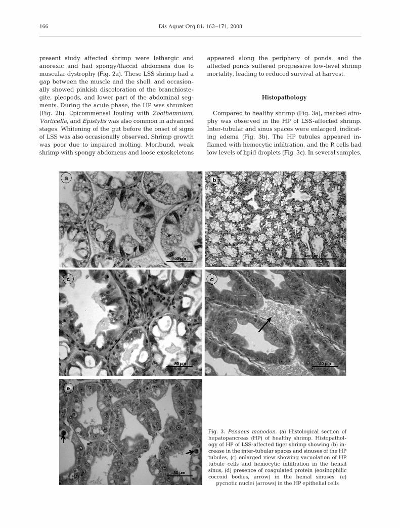
Compared to healthy shrimp (Fig. 3a), marked atro-phy was observed in the HP of LSS-affected shrimp.Inter-tubular and sinus spaces were enlarged, indicat-ing edema (Fig. 3b). The HP tubules appeared in-flamed with hemocytic infiltration, and the R cells hadlow levels of lipid droplets (Fig. 3c). In several samples,
166
Fig. 3. Penaeus monodon. (a) Histological section ofhepatopancreas (HP) of healthy shrimp. Histopathol-ogy of HP of LSS-affected tiger shrimp showing (b) in-crease in the inter-tubular spaces and sinuses of the HPtubules, (c) enlarged view showing vacuolation of HPtubule cells and hemocytic infiltration in the hemalsinus, (d) presence of coagulated protein (eosinophiliccoccoid bodies, arrow) in the hemal sinuses, (e)
pycnotic nuclei (arrows) in the HP epithelial cells
Alavandi et al.: Loose shell syndrome of farmed shrimp
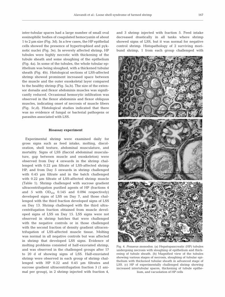
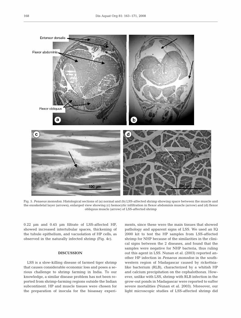
inter-tubular spaces had a large number of small ovaleosinophilic bodies of coagulated hemocyanin of about1 to 2 µm size (Fig. 3d). In a few cases, the HP epithelialcells showed the presence of hypertrophied and pyk-notic nuclei (Fig. 3e). In severely affected shrimp, HPtubules were highly necrotic with thickening of thetubule sheath and some sloughing of the epithelium(Fig. 4a). In some of the tubules, the whole tubular ep-ithelium was being sloughed, with a thickened tubularsheath (Fig. 4b). Histological sections of LSS-affectedshrimp showed prominent increased space betweenthe muscle and the outer exoskeletal layer comparedto the healthy shrimp (Fig. 5a,b). The size of the exten-sor dorsalis and flexor abdominis muscles was signifi-cantly reduced. Occasional hemocytic infiltration wasobserved in the flexor abdominis and flexor obliquusmuscles, indicating onset of necrosis of muscle fibers(Fig. 5c,d). Histological studies indicated that therewas no evidence of fungal or bacterial pathogens orparasites associated with LSS.
Bioassay experiment
Experimental shrimp were examined daily forgross signs such as feed intake, molting, discol-oration, shell texture, abdominal musculature, andmortality. Signs of LSS (flaccid abdominal muscula-ture, gap between muscle and exoskeleton) wereobserved from Day 4 onwards in the shrimp chal-lenged with 0.22 µm filtrate of LSS-affected shrimpHP, and from Day 5 onwards in shrimp challengedwith 0.45 µm filtrate and in the batch challengedwith 0.22 µm filtrate of LSS-affected shrimp muscle(Table 1). Shrimp challenged with sucrose gradientultracentrifugation purified agents of HP (fractions 4and 5 with OD254 0.145 and 0.094 respectively)developed signs of LSS on Day 7, and those chal-lenged with the third fraction developed signs of LSSon Day 13. Shrimp challenged with the third ultra-centrifugation fraction obtained from muscle devel-oped signs of LSS on Day 15. LSS signs were notobserved in shrimp batches that were challengedwith the negative controls or in those challengedwith the second fraction of density gradient ultracen-trifugation of LSS-affected muscle tissue. Moltingwas normal in all negative controls but was affectedin shrimp that developed LSS signs. Evidence ofmolting problems consisted of half-exuviated shrimp,and was observed in the challenged groups after 17to 20 d of showing signs of LSS. Half-exuviatedshrimp were observed in each group of shrimp chal-lenged with HP 0.22 and 0.45 µm filtrates andsucrose gradient ultracentrifugation fraction 3 (1 ani-mal per group), in 2 shrimp injected with fraction 4,
and 3 shrimp injected with fraction 5. Feed intakedecreased drastically in all tanks where shrimpshowed signs of LSS, but it was normal for negativecontrol shrimp. Histopathology of 2 surviving mori-bund shrimp, 1 from each group challenged with
167
Fig. 4. Penaeus monodon. (a) Hepatopancreatic (HP) tubulesundergoing necrosis with sloughing of epithelium and thick-ening of tubule sheath. (b) Magnified view of the tubulesshowing various stages of necrosis, sloughing of tubular epi-thelium with thickened tubular sheath in advanced stage ofLSS. (c) HP of experimentally challenged shrimp showingincreased intertubular spaces, thickening of tubule epithe-
lium, and vacuolation of HP cells
Dis Aquat Org 81: 163–171, 2008
0.22 µm and 0.45 µm filtrate of LSS-affected HP,showed increased intertubular spaces, thickening ofthe tubule epithelium, and vacuolation of HP cells, asobserved in the naturally infected shrimp (Fig. 4c).
DISCUSSION
LSS is a slow-killing disease of farmed tiger shrimpthat causes considerable economic loss and poses a se-rious challenge to shrimp farming in India. To ourknowledge, a similar disease problem has not been re-ported from shrimp-farming regions outside the Indiansubcontinent. HP and muscle tissues were chosen forthe preparation of inocula for the bioassay experi-
ments, since these were the main tissues that showedpathology and apparent signs of LSS. We used an IQ2000 kit to test the HP samples from LSS-affectedshrimp for NHP because of the similarities in the clini-cal signs between the 2 diseases, and found that thesamples were negative for NHP bacteria, thus rulingout this agent in LSS. Nunan et al. (2003) reported an-other HP infection in Penaeus monodon in the south-western region of Madagascar caused by rickettsia-like bacterium (RLB), characterized by a whitish HPand calcium precipitation on the cephalothorax. How-ever, unlike with LSS, shrimp with RLB infection in thegrow-out ponds in Madagascar were reported to suffersevere mortalities (Nunan et al. 2003). Moreover, ourlight microscopic studies of LSS-affected shrimp did
168
Fig. 5. Penaeus monodon. Histological sections of (a) normal and (b) LSS-affected shrimp showing space between the muscle andthe exoskeletal layer (arrows); enlarged view showing (c) hemocytic infiltration in flexor abdominis muscle (arrow) and (d) flexor
obliquus muscle (arrow) of LSS-affected shrimp
Alavandi et al.: Loose shell syndrome of farmed shrimp
not show any basophilic membrane-bound cytoplas-mic rickettsial microcolonies or inclusions, suggestiveof rickettsial infection. The bioassay experiment de-monstrated that LSS could be transmitted to healthyshrimp by intramuscular injection with filtrates ob-tained by filtration through 0.45 µm and 0.22 µm filters,as well as by sucrose gradient ultracentrifugation frac-tions of HP and muscle, suggesting involvement of afilterable infectious viral-like etiology.
The observation of severe necrosis and presence ofmany completely sloughed HP tubules in LSS-affectedshrimp is an indication of impairment of the HP and itsmetabolic activity. Breakdown and dysfunction of HPcould have led to impairment of digestion and absorp-tion, and it is likely that muscle loss could be due toresorption to meet energy needs, accounting for thesigns of disease. General atrophy of tubules and reduc-tion in the level of lipid storage in the HP cells as
169
Day LSS inoculum Control
01 2M 1M 1M2 1M 1D3 1M4 1M 1M5 1M 1M6 1MD 2M 1MD7 1MD 1M 1D8 1D 1M9 1M 1M 1MD10 1M 1M11 1D 1D 2M 1M 1M12 1M 1M 1D13 1D 1M 1MD14 1M 1D1516 1M 1M 1M 1M 1M 1M17 1M 1M 1M18 1D 1M 1M 1M19 1M 1M 1M20 1D 1D21 1M 1M22 1D 1M 1M 1D23 1MD 1D 1D24 1M26 1D 1D30 1M31 1D32 1M 2M 1M 1M33 1D34 1M 1M35 1D 2D 1D 1D36 1D 1D37 1D 2D 2D 1D 1D39 1D 1D 1M 1DDead 3 1 3 4 5 5 1 2 3 2 1 2 3 4 3Molted 1 4 2 1 3 5+1 5 3 5+2 5 5 5+1 4 5+1 3
Table 1. Penaeus monodon. Development of signs of loose shell syndrome (LSS; indicated in bold) in shrimp challenged with var-ious inocula. LHP22, LHP45: LSS-affected shrimp hepatopancreas (HP) extract filtered through 0.22 µm and 0.45 µm filters, re-spectively; LHPUC3, LHPUC4, and LHPUC5: ultracentrifugation fractions 3, 4, and 5 from LSS-affected shrimp HP, respectively;LM22, LM45: LSS-affected shrimp muscle extract filtered through 0.22 µm and 0.45 µm filters, respectively; LMUC2, LMUC3: ul-tracentrifugation fractions 2 and 3, respectively, from LSS-affected shrimp muscle; HHP22, HHP45: healthy shrimp HP extract fil-tered through 0.22 µm and 0.45 µm filters, respectively; HM22 and HM45: healthy shrimp muscle extract filtered through 0.22 µmand 0.45 µm filters, respectively; PBS: sterile phosphate buffered saline (pH: 7.4); M: molted; D: died; MD: died during molting
LH
P22
LH
P45
LH
PU
C3
LH
PU
C4
LH
PU
C5
LM
22
LM
45
LM
UC
2
LM
UC
3
HH
P22
HH
P45
HM
22
HM
45
PB
S
Con
trol

Dis Aquat Org 81: 163–171, 2008
noticed in the present study has also been reported inthe American lobster Homarus americanus subjectedto nutritional stress (Rosemark et al. 1980). Similarly, inshrimp affected by soft shell syndrome, the HP tubuleswere compressed, disarranged, and disrupted, and thelumen appeared to be larger because of the break-down of secretory cells (Baticados et al. 1987).Histopathological observations such as an increase inthe inter-tubular spaces and sinuses of the HP tubuleshave also been reported in infections due to spawnerisolated mortality virus and lymphoid organ vacuoliza-tion virus (LOVV; Fraser & Owens 1996, Lightner1996). The histopathological observations of the LSS-affected shrimp muscle were distinct from those de-scribed in Litopenaeus vannamei infected with infec-tious myonecrosis virus (IMNV) and the nodavirusPvNV, which causes muscle necrosis (Tang et al. 2005,2007) except for occasional hemocytic infiltration.
Soft shell syndrome, a disease condition similar toLSS, among farmed shrimp results from multiple fac-tors such as nutritional deficiency, pesticide pollution,and poor water quality, and is usually associated withcertain management practices (Baticados et al. 1986).Soft shell syndrome shrimp were reported to be weak,to have wrinkled, papery, and loose exoskeletons,often with lesions or blisters, and to be prone to canni-balism leading to low survival rates. It was alsoreported that soft shell syndrome could be reversed byfeeding diets containing mussel meat (Baticados et al.1986). However, in our study, LSS could not be re-versed by administration of diets containing clam meatand certain vitamins (data not included).
Six baculoviruses attacking the HP of decapod crus-taceans including penaeid shrimp, brachyuran crabs,and anamuran crabs have been described, of which4 are non-occluded (Johnson & Lightner 1988). Ourhistopathological studies on LSS did not suggestinvolvement of these or similar viral pathogens. In arecent report on multiple viral infections amongshrimp, Laem Singh virus (LSNV) was recorded inLSS-affected Penaeus monodon (Prakasha et al. 2007).In the present study, we have described thehistopathology of LSS and demonstrated that LSScould be transmitted to healthy shrimp through exper-imental challenge, suggesting involvement of a filter-able infectious agent as a causative factor for LSS offarmed black tiger shrimp in India.
Acknowledgements. We thank the Indian Council of Agricul-tural Research (ICAR), New Delhi, for the financial assistance(F.No.4 [56/2004-ASR-I]). We are indebted to the variousshrimp farms in Tamil Nadu and Andhra Pradesh for provid-ing samples and extending their cooperation. We also thankMr. D. Ramraj, Padmanabha Lab, Ongole, for help in screen-ing samples for NHP, and Dr. A.G. Ponniah, Director, CentralInstitute of Brackishwater Aquaculture, for critical evaluationof this work.
LITERATURE CITED
Alavandi SV, Babu TD, Abhilash KS, Kalaimani N,Chakravarthy N, Santiago TC, Vijayan KK (2007) Looseshell syndrome causes low-level mortality in India’s blacktiger shrimp. Glob Aquac Advocate 10:80–81
Baticados MCL, Coloso RM, Duremdez RC (1986) Studieson the chronic soft shell syndrome in the tiger prawn,Penaeus monodon Fabricus, from brackishwater ponds.Aquaculture 56:271–285
Baticados MCL, Coloso RM, Duremdez RC (1987) Histo-pathology of the chronic soft shell syndrome in the tigerprawn Penaeus monodon. Dis Aquat Org 3:13–28
Bell TA, Lightner DV (1988) A handbook of normal penaeidshrimp histology. World Aquaculture Society, BatonRouge, LA
Bonami JR, Hasson KW, Mari J, Poulos BT, Lightner DV(1997) Taura syndrome of marine penaeid shrimp: charac-terization of the viral agent. J Gen Virol 78:313–319
Costa R, Mermoud I, Mari J, Bonami JR, Hasson K, LightnerDV (1998) Investigations of Penaeus stylirostris diseaseSyndrome 93 in New Caledonia, exploring a viral hypoth-esis. Aquaculture 164:311–322
Flegel TW (1997) Special topic review: major viral diseases ofthe black tiger prawn (Penaeus monodon) in Thailand.World J Microbiol Biotechnol 13:433–442
Fraser CA, Owens L (1996) Spawner-isolated mortality virusfrom Australian Penaeus monodon. Dis Aquat Org 27:141–148
Frelier PF, Sis RF, Bell TA, Lewis DH (1992) Microscopic andultrastructural studies of necrotizing hepatopancreatitis inPacific white shrimp (Penaeus vannamei) cultured inTexas. Vet Pathol 29:269–277
Gopalakrishnan A, Parida A (2005) Incidence of loose shellsyndrome disease of the shrimp Penaeus monodon and itsimpact in the grow-out culture. Curr Sci 88:1148–1154
Johnson PT, Lightner DV (1988) Rod-shaped nuclear virusesof crustaceans: gut-infecting species. Dis Aquat Org 5:123–141
Lightner DV (1996) A handbook of pathology and diagnosticprocedures for diseases of penaeid shrimp. Department ofVeterinary Science, University of Arizona, Tucson, AZ
Lightner DV, Redman RM (1998) Shrimp diseases and currentdiagnostic methods. Aquaculture 164:201–220
Mayavu P, Purushothaman A, Kathiresan K (2003) Histologyof loose-shell affected Penaeus monodon. Curr Sci 85:1629–1634
MPEDA/NACA (Marine Products Export DevelopmentAuthority/Network of Aquaculture Centres in Asia-Pacific) (2003) Shrimp health management extensionmanual. Prepared by NACA and MPEDA, India, in coop-eration with the Aquatic Animal Health Research Insti-tute, Bangkok, Thailand; Siam Natural Resources,Bangkok, Thailand; and AusVet Animal Health Services,Australia. MPEDA, Cochin. Available at: http://library.enaca.org/Shrimp/manual/ShrimpHealthManual.pdf
Nunan LM, Poulos B, Redman R, Le Groumellec M, LightnerDV (2003) Molecular detection methods developed for asystemic rickettsia-like bacterium (RLB) in Penaeus mon-odon (Decapoda: Crustacea). Dis Aquat Org 53:15–23
Poulos BT, Tang KFJ, Pantoja CR, Bonami JR, Lightner DV(2006) Purification and characterization of infectious myo-necrosis virus of penaeid shrimp. J Gen Virol 87:987–996
Prakasha BK, Ramakrishna RP, Karunasagar I, Karunasagar I(2007) Detection of Laem-Singh virus (LSNV) in culturedPenaeus monodon from India. Dis Aquat Org 77:83–86
Rosemark R, Boswer PR, Baum N (1980) Histological obser-
170
Alavandi et al.: Loose shell syndrome of farmed shrimp
vations on the hepatopancreas in juvenile lobsters sub-jected to dietary stress. Proc World Maric Soc 11:471–478
SAP (Society of Aquaculture Professionals) (2004) Proceed-ings of the workshop on loose shell problem in farmedblack tiger shrimp, Chennai, 28 April 2004. Society ofAquaculture Professionals, Chennai
Sritunyalucksana K, Apisawetakan S, Boon-nat A, Withy-achumnarnkul B, Flegel TW (2006) A new RNA virusfound in black tiger shrimp Penaeus monodon from Thai-land. Virus Res 118:31–38
Tang KFJ, Pantoja CR, Poulos BT, Redman RM, Lightner DV(2005) In situ hybridization demonstrates that Litopenaeusvannamei, L. stylirostris and Penaeus monodon are sus-ceptible to experimental infection with infectious myo-necrosis virus (IMNV). Dis Aquat Org 63:261–265
Tang KFJ, Pantoja CR, Redman RM, Lightner DV (2007)Development of in situ hybridization and RT-PCR assayfor the detection of a nodavirus (PvNV) that causesmuscle necrosis in Penaeus vannamei. Dis Aquat Org75:183–190
171
Editorial responsibility: John Austin,Oldendorf/Luhe, Germany
Submitted: January 7, 2008; Accepted: June 25, 2008Proofs received from author(s): August 16, 2008