2006 BBRC Cp G
7
CpG oligodeoxynucleotides induce Ca 2+ -dependent phospholipase D activity leading to phagolysosome maturation and intracellular mycobacterial growth inhibition in monocytes Emanuela Greco a , Marco De Spirito b , Massimiliano Papi b , Marco Fossati a , Giovanni Auricchio a , Maurizio Fraziano a, * a Department of Biology, University of Rome ‘‘Tor Vergata’’, Rome, Italy b Institute of Physics, Catholic University of S. Hearth, Rome, Italy Received 16 June 2006 Available online 18 July 2006 Abstract Synthetic oligodeoxynucleotides containing CpG motifs (CpG ODN) have been reported to induce antimycobacterial activity both in vitro and in vivo. The present study analyzes the signals leading to CpG ODN-induced antimicrobial activity in monocytes. In this context, CpG, but not GpC, ODN induced cytosolic Ca 2+ influx of extracellular origin which, in turn, activated host phospholipase D (PLD). The production of CpG-induced PLD-dependent phosphatidic acid induced the maturation of phagolysosomes and intracellular mycobacterial growth inhibition. These results show the presence of an antimicrobial pathway in monocytes, mediated by Ca 2+ -dependent PLD which can be useful for the exploitation of novel anti-tuberculosis immunotherapy approaches. Ó 2006 Elsevier Inc. All rights reserved. Keywords: Mycobacterium; Monocytes/macrophages; Tuberculosis; Lipid mediators; Phagolysosome Many distinct signal transduction pathways contribute to the activation of phagocyte antimicrobial defenses. Stimulation-induced increases in cytosolic Ca 2+ concentra- tion are essential for activation of the phagocyte respirato- ry burst, production of nitric oxide, secretion of microbicidal granule constituents, and synthesis of proin- flammatory mediators, including TNF-a [1–5]. Moreover, a rapidly expanding body of evidence shows that phospho- lipases, such as phospholipase D (PLD), play an integral role in the induction of intracellular antimycobacterial activity by generating essential second messengers. PLD catalyzes hydrolysis of the terminal diester bond of phos- phatidylcholine as a preferential substrate, to generate phosphatidic acid (PA) and choline [6,7]. PA is involved in the induction of a number of macrophage antimicrobial activities, such as phagocytosis [8], ROI production [9], intracellular trafficking of endocytosed immunocomplexes to lysosomes [10], and phagolysosome maturation in the course of intracellular mycobacterial killing induced by ATP [11] or sphingosine 1-phosphate [12]. PLD activity can be regulated by protein kinase C, Rho family small G proteins, protein tyrosine kinases, and Ca 2+ [13]. In par- ticular, it has been reported that treatment with Ca 2+ che- lators may result in inhibition of the activation of PLD by several agonists and that Ca 2+ ionophores can activate PLD in many cell types [7]. Bacterial DNA, in comparison with mammalian DNA, contains a higher proportion of unmethylated CpG dinu- cleotide motifs. These motifs are believed to be the pri- mary source of its immunogenicity [14]. Synthetic oligodeoxynucleotides (ODN) containing unmethylated CpG dinucleotides (CpG ODN) are capable of mimicking the immunostimulatory effects of bacterial DNA on B cells, monocytes/macrophages, NK cells, and dendritic cells, and can induce them to secrete proinflammatory 0006-291X/$ - see front matter Ó 2006 Elsevier Inc. All rights reserved. doi:10.1016/j.bbrc.2006.06.186 * Corresponding author. Fax: 39 06 72594224. E-mail address: [email protected] (M. Fraziano). www.elsevier.com/locate/ybbrc Biochemical and Biophysical Research Communications 347 (2006) 963–969 BBRC
Transcript of 2006 BBRC Cp G

www.elsevier.com/locate/ybbrc
Biochemical and Biophysical Research Communications 347 (2006) 963–969
BBRC
CpG oligodeoxynucleotides induce Ca2+-dependent phospholipase Dactivity leading to phagolysosome maturation and intracellular
mycobacterial growth inhibition in monocytes
Emanuela Greco a, Marco De Spirito b, Massimiliano Papi b, Marco Fossati a,Giovanni Auricchio a, Maurizio Fraziano a,*
a Department of Biology, University of Rome ‘‘Tor Vergata’’, Rome, Italyb Institute of Physics, Catholic University of S. Hearth, Rome, Italy
Received 16 June 2006Available online 18 July 2006
Abstract
Synthetic oligodeoxynucleotides containing CpG motifs (CpG ODN) have been reported to induce antimycobacterial activity bothin vitro and in vivo. The present study analyzes the signals leading to CpG ODN-induced antimicrobial activity in monocytes. In thiscontext, CpG, but not GpC, ODN induced cytosolic Ca2+ influx of extracellular origin which, in turn, activated host phospholipaseD (PLD). The production of CpG-induced PLD-dependent phosphatidic acid induced the maturation of phagolysosomes andintracellular mycobacterial growth inhibition. These results show the presence of an antimicrobial pathway in monocytes, mediatedby Ca2+-dependent PLD which can be useful for the exploitation of novel anti-tuberculosis immunotherapy approaches.� 2006 Elsevier Inc. All rights reserved.
Keywords: Mycobacterium; Monocytes/macrophages; Tuberculosis; Lipid mediators; Phagolysosome
Many distinct signal transduction pathways contributeto the activation of phagocyte antimicrobial defenses.Stimulation-induced increases in cytosolic Ca2+ concentra-tion are essential for activation of the phagocyte respirato-ry burst, production of nitric oxide, secretion ofmicrobicidal granule constituents, and synthesis of proin-flammatory mediators, including TNF-a [1–5]. Moreover,a rapidly expanding body of evidence shows that phospho-lipases, such as phospholipase D (PLD), play an integralrole in the induction of intracellular antimycobacterialactivity by generating essential second messengers. PLDcatalyzes hydrolysis of the terminal diester bond of phos-phatidylcholine as a preferential substrate, to generatephosphatidic acid (PA) and choline [6,7]. PA is involvedin the induction of a number of macrophage antimicrobialactivities, such as phagocytosis [8], ROI production [9],
0006-291X/$ - see front matter � 2006 Elsevier Inc. All rights reserved.
doi:10.1016/j.bbrc.2006.06.186
* Corresponding author. Fax: 39 06 72594224.E-mail address: [email protected] (M. Fraziano).
intracellular trafficking of endocytosed immunocomplexesto lysosomes [10], and phagolysosome maturation in thecourse of intracellular mycobacterial killing induced byATP [11] or sphingosine 1-phosphate [12]. PLD activitycan be regulated by protein kinase C, Rho family smallG proteins, protein tyrosine kinases, and Ca2+ [13]. In par-ticular, it has been reported that treatment with Ca2+ che-lators may result in inhibition of the activation of PLD byseveral agonists and that Ca2+ ionophores can activatePLD in many cell types [7].
Bacterial DNA, in comparison with mammalian DNA,contains a higher proportion of unmethylated CpG dinu-cleotide motifs. These motifs are believed to be the pri-mary source of its immunogenicity [14]. Syntheticoligodeoxynucleotides (ODN) containing unmethylatedCpG dinucleotides (CpG ODN) are capable of mimickingthe immunostimulatory effects of bacterial DNA on Bcells, monocytes/macrophages, NK cells, and dendriticcells, and can induce them to secrete proinflammatory

964 E. Greco et al. / Biochemical and Biophysical Research Communications 347 (2006) 963–969
cytokines including interleukin-1b (IL-1b), IL-6, IL-12,IL-18, tumor necrosis factor-a (TNF-a), interferon-a(IFN-a), and interferon-c (IFN-c) [15]. Moreover, system-ic administration of CpG ODN promotes type-1 immuneresponses characterized by enhanced IL-12 and IFN-cproduction [16,17]. CpG ODN have also been successfullyused as adjuvant against a range of intracellular patho-gens and they have been described to increase antimicro-bial activity against Listeria monocytogenes [18],Leishmania maior [19], Mycobacterium avium [20], andMycobacterium tuberculosis [21]. In this context, CpGODN have been shown to induce a number of antimicro-bial effector mechanisms such as nitric oxide (NO) pro-duction [22] and reactive oxygen intermediates (ROI)generation [23]. Finally, we have recently reported thatCpG ODN-induced intracellular mycobacterial killing inhuman macrophages was nitric oxide independent and,at least in part, mediated by host PLD [24]. However,whether and how CpG ODN induce antimycobacterialactivity in human monocytes is a still uninvestigated issue.The results reported herein show that CpG ODN inducePLD activation which is dependent on the increase ofcytosolic Ca2+ influx. Moreover, PLD-dependent genera-tion of phosphatidic acid promotes phagolysosome matu-ration and intracellular mycobacterial killing.
Materials and methods
Cell culture and infection of monocytoid THP-1 cells. Human mon-ocytoid THP-1 leukemia cell line was used as a model of humanmonocytes. Cells were grown in complete medium consisting of RPMI1640 supplemented with 10% FBS, 2 mM L-glutamine, gentamicin 5 lg/ml, 1 mM non-essential aminoacids, and 1 mM sodium pyruvate.Infection was carried out by exposing cells for 3 h to MTB at the MOI(multiplicity of infection) of 1. After removal of non-phagocytosedbacilli, cells were stimulated for 0, 3, and 7 days with 1 lM of eitherphosphorothioate CpG or GpC oligodeoxynucleotides (ODN) (ATCGAC TCT CGA GCG TTC TC and ATG CAC TCT GCA GGC TTCTC, respectively; Primm, Italy), or 1 lM phosphatidic acid. In someexperiments, in order to evaluate the role of PLD activity in CpGODN-induced mycobacterial growth inhibition, CpG ODN was addedtogether with 0.3% ethanol, 1-butanol (used at the concentration of0.3%, 0.03% or 0.003%) or 0.3% 2-butanol. Any modification, in termsof macrophage viability, was not detected in all experimental conditionsused (data not shown).
Intracellular Ca2+measurement. Intracellular Ca2+ was measured byusing the fluorescent intracellular Ca2+ indicator Fluo-3/AM (MolecularProbes, NL). THP-1 cells were washed twice with complete medium andincubated with 3 lM Fluo-3/AM at 37 �C for 1 h in the dark. Cells werecollected, centrifuged, suspended in complete medium, and stimulatedwith either 1 lM CpG or GpC ODN. Fluo-3 fluorescence was followed bya Varian Cary Eclipse fluorometer, with excitation and emission wave-lengths of 505 and 530 nm, respectively, using 5 nm slits for both lightpaths. Minimum and maximum fluorescence were determined on completemedium only or on THP1 cells stimulated with 50 ng/ml phorbol myristateacetate (PMA). In some experiments, 2 mM EDTA or 3 mM EGTA(Calbiochem, San Diego, CA) was added 15 min before CpG ODNaddition.
Analysis of phospholipase D activity. Monocyte phospholipase D(PLD) activity was measured by the transphosphatidylation assay[12,24]. Briefly, 24-well plate-cultured THP1 cells were suspended incomplete medium supplemented with 20 mM Hepes (pH 7.4) and
labeled with 1 lCi/ml [3H]myristic acid (Amersham International, UK)for 120 min at 37 �C, followed by washing with RPMI 1640 to removeunincorporated radioactivity. Thereafter, cells were incubated for15 min at 37 �C in complete medium with 1% ethanol to allow detectionof [3H]PetOH, as a specific transphosphatidylation reaction product ofPLD. THP-1 cells were then stimulated with 1 lM CpG ODN for30 min. Finally, the incubation was stopped with 1 ml ice-cold methanoland lipids were extracted and separated by thin layer chromatography(TLC) in an ethyl acetate/isooctane/acetic acid/water (130:20:30) solventsystem, as previously described [12,24]. Areas containing [3H]PetOHwere collected from silica gel plate as compared by standard PetOHand quantified by liquid scintillation counter. The radioactivity presentin PetOH fraction was expressed as the percentage of the total radio-activity collected from the lane. In order to evaluate the role ofextracellular Ca2+ in PLD activity, 2 mM EDTA was added to the cellcultures 10 min before ethanol labeling and CpG ODN stimulation.Phospholipase D activity was also evaluated at the same experimentalconditions by ‘‘Amplex Red Phospholipase D assay kit’’ used accordingto the manufacturer’s instructions.
Confocal fluorescent microscopy. The degree of maturation of MTB-containing phagosome was assessed by analyzing the colocalization ofbacilli with lysosomes after staining mycobacteria with auramine andlysosomes with either the acidophilic dye Lysotracker Red (MolecularProbes, NL) or anti-LAMP-1 monoclonal antibody. THP1 cells, sus-pended in complete medium, were incubated with Lysotracker Red at a1:10,000 dilution for 2 h at 37 �C. Unincorporated dye was removedand cells were infected for 3 h with MTB. After removal of non-phagocytosed bacilli, Lysotracker Red was added again to each well for30 min. Cells were washed and further incubated for 16–18 h with 1 lMCpG ODN in presence or absence of 0.3% ethanol. Thereafter, cellswere washed with PBS, fixed by 10 min incubation with 4% parafor-maldehyde at 4 �C and permeabilized with ice-cold methanol–acetone(1:1) followed by further three washings with PBS. Detection of thelysosomal protein markers LAMP-1 was accomplished by incubatingcells with anti-lysosomal associated membrane protein (LAMP)-1monoclonal antibody (IgG2b, clone 25, Transduction Laboratories,Becton–Dickinson, MD) for 1 h, repeated washings, and incubationwith TRITC-conjugated secondary anti-IgG monoclonal antibody(SIGMA, MO) for further 1 h. Cells were then seeded on poly-L-lysine(Sigma, MO) pretreated slides. The localization of MTB was deter-mined by incubating infected monolayer with Auramine (Becton–Dickinson, MD) for 20 min at room temperature, followed by 3 minincubation in 0.5% acid alcohol and repeated washing with PBS.Finally, coverslips were mounted with Vectashield mounting mediumH-1000 (Vector laboratories, CA) and edges were sealed with nailpolish. Confocal fluorescence microscopy was performed by using aLeica Laser system linked to a Leica inverted microscope (TCS SP2).An argon–krypton laser (k = 488 nm) and a helium–neon laser(k = 543 nm) were used in combination with a 505–530 nm,580–600 nm, and 560–600 emission band pass filter for detection ofAuramine, Lysotracker Red, and TRITC fluorescence, respectively.
Bacteria and colony forming unit assay. Pathogenic M. tuberculosis
(MTB) H37Rv strain was grown in Middle brook 7H9 (Becton–Dickinson, MD) broth supplemented with albumin dextrose catalase.Mycobacteria were then harvested, suspended in sterile phosphate-buffered saline (PBS), pH 7.2, aliquoted, and stored at �80 �C untiluse. Before infection, aliquots were grown on 7H10 agar (Becton–Dickinson, MD) plates to titer the bacteria after thawing. Colonyforming unit assay was performed as previously described [12].Briefly, at the indicated time points after infection and stimulations,cells were lysed by using ice-cold sterile PBS containing 0.1% sapo-nin, serially diluted in PBS containing 0.01% Tween 80 and plated intriplicate on Middle brook 7H10 agar. MTB colonies were enumer-ated after incubation of the plates at 37 �C in humidified air for 21days.
Statistics. Statistical analysis was carried out by Graphpad Prism 3.0software package. Student’s t test was used to compare differences betweenmeans.

Fig. 2. CpG ODN induce Ca2+-dependent PLD activity. (A) THP1 cellswere labeled with 1 lCi/ml [3H]myristic acid for 3 h before the addition of1 lM CpG ODN for 30 min. Transphosphatidylation reaction wasallowed by the addition of 1% ethanol 15 min before CpG ODNstimulation. The role of extracellular Ca2+ in PLD activity was assessedby addition of 2 mM EDTA, 15 min before CpG ODN stimulation. Dataare reported as means ± SD of three different experiments. *p < 0.001 incomparison with control cells. (B) Phospholipase D activity wasmonitored by ‘‘Amplex Red Phospholipase D detection kit’’ on THP1cells stimulated or not with CpG ODN for 30 min. Fluorescence intensitywas measured at an excitation wavelength of 542 nm and an emissionwavelength of 590 nm. Values are shown as means ± SD of five indepen-dent experiments. *p < 0.001 as compared with unstimulated THP-1control cells.
E. Greco et al. / Biochemical and Biophysical Research Communications 347 (2006) 963–969 965
Results
CpG ODN induce Ca2+-dependent PLD activity in human
monocytes
Circulating undifferentiated monocytes represent bothone of the preferential targets of MTB infection and a poolof inflammatory cells with potential antimycobacterialactivity [25]. Different signal transduction pathways mayparticipate in the activation of monocyte CpG ODN-in-duced antimicrobial activity. As it has been previouslyreported that both Ca2+ mobilization can represent impor-tant upstream signalling leading to the activation of antimi-crobial effecter mechanisms [26], we have analyzedcytosolic Ca2+ influx following CpG ODN stimulation inmonocytoid cell line THP1. Results showed a significantincrease of cytosolic Ca2+ following stimulation withCpG ODN whereas no effect was observed after stimula-tion by GpC ODN (Fig. 1). Moreover, increase of cytosolicCa2+ was of extracellular origin as the simultaneous treat-ment with either EDTA or EGTA, which chelates extracel-lular Ca2+, and make it unavailable for intracellular stores,completely inhibited CpG ODN-induced increase of cyto-solic Ca2+ (Fig. 1). As Ca2+ may be a requirement forPLD activation, phospholipase activity has been analyzedfollowing stimulation with CpG ODN in THP1 cell line.Results showed an increase of PLD activity after CpGODN stimulation which resulted to be Ca2+-dependent asenzymatic activity was completely abrogated by EDTAtreatment (Fig. 2A and B). Altogether, these results showthat CpG ODN induce a signal transduction pathway,involving increase of cytosolic Ca2+ concentration of extra-cellular origin which is necessary for host PLD activation.
CpG ODN promote phagolysosome biogenesis through PLD
activity
As MTB is known to reside in non-acidified vacuolessequestered away from late endosomal compartments[27], the acidification of MTB containing phagosomes in
Fig. 1. CpG ODN induce Ca2+ mobilization in monocytes. THP-1 cellswere labeled with 3 lM Fluo-3/AM for 60 min, treated or not with 2 mMEDTA or 3 mM EGTA for 15 min, and further stimulated with 1 lM ofeither CpG or GpC ODN. Data are reported as means ± SD of fivedifferent experiments. *p < 0.001 in comparison with untreated THP1 cells.
CpG ODN-stimulated THP1 cell line has been investigatedby confocal fluorescent microscopy (Fig. 3A). In theabsence of CpG ODN, phagocytosed mycobacteriaappeared green, indicating their presence in non-acidic ves-icles separate from the red stained acidic vacuoles. In con-trast, CpG ODN treatment induced a significant increasein the acidification of MTB-containing phagosomes, whichappeared yellow, indicating the colocalization of greenbacilli in acidified phagosomes. As phosphatidic acid isan important second messenger involved in phagolysosomebiogenesis [11], the role of PLD in the CpG ODN-inducedacidification of MTB containing endosomes has been ana-lyzed in the presence of ethanol. The addition of primaryalcohol almost completely reverted the CpG ODN-inducedphagolysosome acidification. The percentage of MTBresiding in acidic phagosomes over total intracellularmycobacteria was expressed after serial observations(Fig. 3B). As acidification of phagosome does not necessar-ily indicate full phagolysosome maturation because partialacidification is also present in immature phagosomes, the

Fig. 3. Mycobacterium tuberculosis (MTB) colocalizes with acidic endosomes after CpG ODN stimulation. Representative picture from three separateexperiments shows the increase of MTB residing in acidic vacuoles after CpG ODN treatment and the reverse effect exerted by ethanol (A). Summary ofthe mean percentage ± SD of MTB-colocalized with acidic phagosomes, determined by counting over 60 bacilli from at least 40 macrophages percondition (B). Three different experiments were assessed. *p < 0.0001 in comparison with MTB-infected control cells.
966 E. Greco et al. / Biochemical and Biophysical Research Communications 347 (2006) 963–969
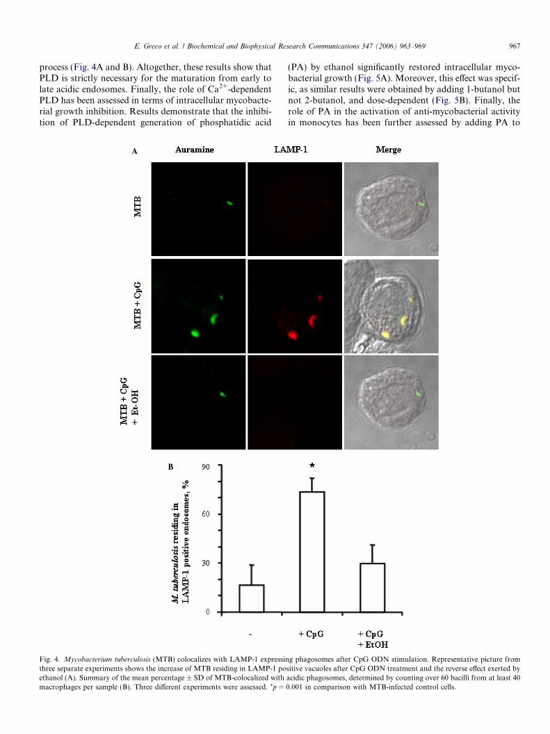
expression of lysosomal associated membrane protein(LAMP)-1 as a marker of mature phagolysosome was alsoanalyzed. Results show that CpG ODN stimulation
induces the colocalization of green mycobacteria inLAMP-1 expressing vacuoles and that the addition of eth-anol together with CpG almost completely reverted such
E. Greco et al. / Biochemical and Biophysical Research Communications 347 (2006) 963–969 967
process (Fig. 4A and B). Altogether, these results show thatPLD is strictly necessary for the maturation from early tolate acidic endosomes. Finally, the role of Ca2+-dependentPLD has been assessed in terms of intracellular mycobacte-rial growth inhibition. Results demonstrate that the inhibi-tion of PLD-dependent generation of phosphatidic acid
Fig. 4. Mycobacterium tuberculosis (MTB) colocalizes with LAMP-1 expressinthree separate experiments shows the increase of MTB residing in LAMP-1 poethanol (A). Summary of the mean percentage ± SD of MTB-colocalized withmacrophages per sample (B). Three different experiments were assessed. *p =
(PA) by ethanol significantly restored intracellular myco-bacterial growth (Fig. 5A). Moreover, this effect was specif-ic, as similar results were obtained by adding 1-butanol butnot 2-butanol, and dose-dependent (Fig. 5B). Finally, therole of PA in the activation of anti-mycobacterial activityin monocytes has been further assessed by adding PA to
g phagosomes after CpG ODN stimulation. Representative picture fromsitive vacuoles after CpG ODN treatment and the reverse effect exerted byacidic phagosomes, determined by counting over 60 bacilli from at least 400.001 in comparison with MTB-infected control cells.

Fig. 5. Monocyte PLD mediates CpG ODN-induced intracellular myco-bacterial growth inhibition. (A) THP1 cells were infected with MTB at theMOI of 1 and incubated for 0, 3, and 7 days alone or with CpG ODN, CpGODN plus 0.3% ethanol, GpC ODN. CFU are expressed as means ± SD oftriplicate values and are representative of two independent experiments. (B)THP1 cells were infected with MTB at the MOI of 1 and incubated for 0, 3,and 7 days alone or with CpG ODN, CpG ODN plus 1-butanol (used at theconcentration of 0.3%, 0.03% or 0.003%), CpG ODN plus 2-butanol (used atthe concentration of 0.3%), GpC ODN. Results coming from three differentexperiments have been normalized and expressed as means ± SD ofpercentage of mycobacterial viability. The percentage of MTB viabilitywas calculated as follows: (CFU coming from MTB-infected monocytesreceiving the different treatments/CFU coming from MTB-infected controlmonocytes) · 100. (C) THP1 cells were infected with MTB at the MOI of 1and incubated for 0, 3, and 7 days alone or with phosphatidic acid. CFU areexpressed as means ± SD of the triplicate values and are representative ofthree separate experiments.
968 E. Greco et al. / Biochemical and Biophysical Research Communications 347 (2006) 963–969
MTB-infected THP1 cells. Results show that PA stimula-tion induced a significant decrease of intracellularmycobacterial growth (Fig. 5C).
Discussion
Pathogen-associated molecular patterns (PAMPs) havebeen described to induce antimicrobial activity in innateimmune cells [28]. In particular, CpG ODN have beendescribed to enhance host defense during murine tubercu-losis [22] and induce in vitro anti-mycobacterial activity inhuman differentiated macrophages in the course ofM. tuberculosis infection through a PLD-dependent path-way [24]. As circulating undifferentiated monocytes repre-sent both one of the preferential targets of MTBinfection and a pool of inflammatory cells with potential
antimycobacterial activity [25], this study addresses the sig-nal transduction pathway underlying CpG ODN stimula-tion in monocytes, with reference to the involvement ofphospholipase D in CpG ODN-induced anti-mycobacterialactivity. The results reported herein show that CpG ODNstimulation in THP1 monocytoid cell line triggers a signaltransduction involving extracellular Ca2+ influx which inturn induces a downstream activation of phospholipaseD. This phospholipase was involved in phagolysosomematuration and intracellular mycobacterial killing inducedby CpG ODN.
Macrophages possess multiple and reciprocally indepen-dent microbicidal mechanisms to eliminate phagocytosedmicroorganisms and, consequently, represent a strategictarget for inactivation by potential pathogens [29]. In thiscontext, M. tuberculosis exploits several strategies to cir-cumvent host immune response and antimicrobial activityof mononuclear phagocytes [30], including inhibition ofCa2+ mobilization [26]. The activation of Ca2+-dependentPLD reported herein following stimulation with CpGODN suggests that CpG ODN may have the capabilityto restore this signal transduction pathway and that extra-cellular ion environment is important for the activation ofinnate PLD-dependent antimycobacterial activity.
The dynamic interactions between M. tuberculosis andhuman monocytes are central to each of the complex stagesof tuberculosis, from initial infection through the develop-ment of active disease. A crucial feature of pathogenesis isthe ability of tubercle bacilli to evade the microbicidalactivities of macrophages and to persist as intracellularparasites within membrane-enclosed vesicles [27]. Follow-ing primary infection, MTB containing phagosomes exhibitcharacteristics of early, recycling endosomes and fails toprogress along the physiological maturation pathway to amycobacteriocidal phagolysosome [31]. The previouslyreported requirement for increase in Ca2+ and PLD activ-ity for both phagolysosome maturation and intracellularmycobacterial killing in ATP [11] or Sphingosine 1-phos-phate [12] stimulated macrophages directly supports thehypothesis that phagosomal maturation contributes tomycobacteriocidal activity. Results reported herein showthat CpG ODN promotes phagolysosome maturationand require PLD activity to allow this process. In fact,the specific inhibition of PLD-mediated PA generation byethanol completely reverted CpG ODN-induced phagoly-sosome maturation. The mechanism through which PLDmay regulate membrane trafficking events can involve (i)direct mechanisms, by altering membrane shape and/orbinding proteins, (ii) indirect mechanisms, by stimulatingor inhibiting signal transduction pathways through phos-phatidic acid release, and (iii) a combination of both [32].In this context, phagolysosome biogenesis has beenhypothesized to be a ‘‘kiss-and-run’’ process through whichphagosomes undergo only a transient and partial fusionwith endocytic organelles which allows the transfer ofselected membranes and luminal contents between thephagosome and the endosome [33,34].

E. Greco et al. / Biochemical and Biophysical Research Communications 347 (2006) 963–969 969
In conclusion, the present study shows that CpG ODNinduce increase of cytosolic Ca2+ leading to PLD activa-tion which in turn promote phagolysosome maturationand intracellular mycobacterial growth inhibition. This sig-nal transduction pathway may be exploited for the poten-tiation of antimycobacterial innate immune response.
Acknowledgments
The present study was financially supported by theItalian Ministry of University (COFIN 2004) and by theItalian Ministry of Health (Grant No. 4AD/F9).
References
[1] B.C. Chen, C.F. Chou, W.W. Lin, Pyrimidinoceptor-mediatedpotentiation of inducible nitric-oxide synthase induction in J774macrophages. Role of intracellular calcium, J. Biol. Chem. 273 (1998)29754–29763.
[2] N. Watanabe, J. Suzuki, Y. Kobayashi, Role of calcium in tumornecrosis factor-alpha production by activated macrophages, J.Biochem. 120 (1996) 1190–1195.
[3] H. Tapper, The secretion of preformed granules by macrophages andneutrophils, J. Leukoc. Biol. 59 (1996) 613–622.
[4] L.C. Denlinger, P.L. Fisette, K.A. Garis, G. Kwon, A. Vazquez-Torres, A.D. Simon, B. Nguyen, R.A. Proctor, P.J. Bertics, J.A.Corbett, Regulation of inducible nitric oxide synthase expression bymacrophage purinoreceptors and calcium, J. Biol. Chem. 271 (1996)337–342.
[5] W.K. Kim-Park, M.A. Moore, Z.W. Hakki, M.J. Kowolik, Activa-tion of the neutrophil respiratory burst requires both intracellular andextracellular calcium, Ann. N. Y. Acad. Sci. 832 (1997) 394–404.
[6] M. Liscovitch, M. Czarny, G. Fiucci, X. Tang, Phospholipase D:molecular and cell biology of a novel gene family, Biochem. J. 3(2000) 401–415.
[7] J.H. Exton, Phospholipase D: enzymology, mechanisms of regulation,and function, Physiol. Rev. 77 (1997) 303–320.
[8] D.J. Kusner, C.F. Hall, S.L. Schlesinger, Activation of phospholipaseD is tightly coupled to the phagocytosis of Mycobacterium tubercu-
losis or opsonized zymosan by human macrophages, J. Exp. Med. 184(1996) 585–595.
[9] J. Giron-Calle, H.J. Forman, Phospholipase D and priming of therespiratory burst by H2O2 in NR8383 alveolar macrophages, Am. J.Respir. Cell Mol. Biol. 23 (2000) 748–754.
[10] A. Melendez, R.A. Floto, D.J. Gillooly, M.M. Harnett, J.M. Allen,FccRI coupling to phospholipase D initiates sphingosine kinase-mediated calcium and vesicular trafficking, J. Biol. Chem. 273 (1998)9393–9402.
[11] D.J. Kusner, J.A. Barton, ATP stimulates human macrophages to killintracellular virulent Mycobacterium tuberculosis via Ca2+ dependentphagosome–lysosome fusion, J. Immunol. 167 (2001) 3308–3315.
[12] S.K. Garg, E. Volpe, G. Palmieri, M. Mattei, D. Galati, A. Martino,E. Bonanno, P. De Vito, P.M. Baldini, L.G. Spagnoli, V. Colizzi, M.Fraziano, Sphingosine 1-phosphate induces antimicrobial activityboth in vitro and in vivo, J. Infect. Dis. 189 (2004) 2129–2138.
[13] J.H. Exton, Regulation of phospholipase D, FEBS Lett. 531 (2002)58–61.
[14] A.M. Krieg, A.K. Yi, S. Matson, T.J. Waldschmidt, G.A. Bishop, R.Teasdale, G.A. Koretzky, D.M. Klinman, CpG motifs in bacterialDNA trigger direct B-cell activation, Nature 374 (1995) 546–549.
[15] A.M. Krieg, CpG motifs in bacterial DNA and their immune effects,Annu. Rev. Immunol. 20 (2002) 709–760.
[16] D.M. Klinman, A.K. Yi, S.L. Beaucage, J. Conover, A.M. Krieg,CpG motifs present in bacteria DNA rapidly induce lymphocytes tosecrete interleukin 6, interleukin 12, and interferon-c, Proc. Natl.Acad. Sci. USA 93 (1996) 2879–2883.
[17] R.S. Chu, O.S. Targoni, A.M. Krieg, P.V. Lehmann, C.V. Harding,CpG oligodeoxynucleotides act as adjuvants that switch on T helper 1(Th1) immunity, J. Exp. Med. 186 (1997) 1623–1631.
[18] A.M. Krieg, L. Love-Homan, A.K. Yi, J.T. Harty, CpG DNAinduces sustained IL-12 expression in vivo and resistance to Listeria
monocytogenes challenge, J. Immunol. 161 (1998) 2428–2434.[19] P.S. Walker, T. Scharton-Kersten, A.M. Krieg, L. Love-Homan,
E.D. Rowton, M.C. Udey, J.C. Vogel, Immunostimulatory oligo-deoxynucleotides promote protective immunity and provide sys-temic therapy for leishmaniasis via IL-12- and IFN-gamma-dependent mechanisms, Proc. Natl. Acad. Sci. USA 96 (1999)6970–6975.
[20] T. Hayashi, S.P. Rao, K. Takabayashi, J.H. Van Uden, R.S.Kornbluth, S.M. Baird, M.W. Taylor, D.A. Carson, A. Catanzaro,E. Raz, Enhancement of innate immunity against Mycobacterium
avium infection by immunostimulatory DNA is mediated by indole-amine 2,3-dioxygenase, Infect. Immun. 69 (2001) 6156–6164.
[21] N.P. Juffermans, J.C. Leemans, S. Florquin, A. Verbon, A.H. Kolk,P. Speelman, S.J. van Deventer, T. van der Poll, CpG oligodeoxy-nucleotides enhance host defense during murine tuberculosis, Infect.Immun. 70 (2002) 147–152.
[22] D.K. Ghosh, M.A. Misukonis, C. Reich, D.S. Pisetsky, J.B. Wein-berg, Host response to infection: the role of CpG DNA in inductionof cyclooxygenase 2 and nitric oxide synthase 2 in murine macro-phages, Infect. Immun. 69 (2001) 7703–7710.
[23] H. Weighardt, C. Feterowski, M. Veit, M. Rump, H. Wagner, B.Holzmann, Increased resistance against acute polymicrobial sepsis inmice challenged with immunostimulatory CpG oligodeoxynucleotidesis related to an enhanced innate effector cell response, J. Immunol.165 (2000) 4537–4543.
[24] G. Auricchio, S.K. Garg, A. Martino, E. Volpe, A. Ciaramella, P. DeVito, P.M. Baldini, V. Colizzi, M. Fraziano, Role of macrophagephospholipase D in natural and CpG-induced antimycobacterialactivity, Cell. Microbiol. 5 (2003) 913–920.
[25] C. Manca, M.B. Reed, S. Freeman, B. Mathema, B. Kreiswirth, C.E.Barry III, G. Kaplan, Differential monocyte activation underliesstrain-specific Mycobacterium tuberculosis pathogenesis, Infect.Immun. 72 (2004) 5511–5514.
[26] Z.A. Malik, G.M. Denning, D.J. Kusner, Inhibition of Ca(2+)signaling by Mycobacterium tuberculosis is associated with reducedphagosome–lysosome fusion and increased survival within humanmacrophages, J. Exp. Med. 191 (2000) 287–302.
[27] I. Vergne, J. Chua, S.B. Singh, V. Deretic, Cell biology of Mycobac-
terium tuberculosis phagosome, Annu. Rev. Cell. Dev. Biol. 20 (2004)367–394.
[28] A. Iwasaki, R. Medzhitov, Toll-like receptor control of the adaptiveimmune responses, Nat. Immunol. 5 (2004) 987–995.
[29] P. Zhang, W.R. Summer, G.J. Bagby, S. Nelson, Innate immunityand pulmonary host defense, Immunol. Rev. 173 (2000) 39–51.
[30] J.L. Flynn, J. Chan, Immune evasion by Mycobacterium tuberculosis:living with the enemy, Curr. Opin. Immunol. 15 (2003) 450–455.
[31] J. Pieters, Evasion of host cell defense mechanisms by pathogenicbacteria, Curr. Opin. Immunol. 13 (2001) 37–44.
[32] W.J. Brown, K. Chambers, A. Doody, Phospholipase A2 (PLA2)enzymes in membrane trafficking: mediators of membrane shape andfunction, Traffic 4 (2003) 214–221.
[33] M. Desjardins, Biogenesis of phagolysosome : the ‘‘kiss and run’’hypothesis, Trends Cell. Biol. 5 (1995) 183–186.
[34] M. Desjardins, L.A. Huber, R.G. Parton, G. Griffiths, Biogenesis ofphagolysosomes proceeds through a sequential series of interactionswith the endocytic apparatus, J. S Biol. 124 (1994) 677–688.