2 3 Morindone
14
1 23 Applied Biochemistry and Biotechnology Part A: Enzyme Engineering and Biotechnology ISSN 0273-2289 Volume 167 Number 4 Appl Biochem Biotechnol (2012) 167:885-896 DOI 10.1007/s12010-012-9744-2 Morindone, an Anthraquinone, Intercalates DNA Sans Toxicity: a Spectroscopic and Molecular Modeling Perspective Dipita Bhakta & Ramamoorthy Siva
Transcript of 2 3 Morindone

1 23
Applied Biochemistry andBiotechnologyPart A: Enzyme Engineering andBiotechnology ISSN 0273-2289Volume 167Number 4 Appl Biochem Biotechnol (2012)167:885-896DOI 10.1007/s12010-012-9744-2
Morindone, an Anthraquinone,Intercalates DNA Sans Toxicity: aSpectroscopic and Molecular ModelingPerspective
Dipita Bhakta & Ramamoorthy Siva

1 23
Your article is protected by copyright and
all rights are held exclusively by Springer
Science+Business Media, LLC. This e-offprint
is for personal use only and shall not be self-
archived in electronic repositories. If you
wish to self-archive your work, please use the
accepted author’s version for posting to your
own website or your institution’s repository.
You may further deposit the accepted author’s
version on a funder’s repository at a funder’s
request, provided it is not made publicly
available until 12 months after publication.

Morindone, an Anthraquinone, Intercalates DNA SansToxicity: a Spectroscopic and Molecular ModelingPerspective
Dipita Bhakta & Ramamoorthy Siva
Received: 24 October 2011 /Accepted: 14 May 2012 /Published online: 26 May 2012# Springer Science+Business Media, LLC 2012
Abstract For the first time, interaction between non-toxic anthraquinone morindone withboth natural and synthetic DNA duplexes has been demonstrated in this paper. Detailedanalyses of the binding of morindone with DNA via UV–vis, FTIR, and circular dichroismspectroscopies were carried out. In addition, bioinformatics tools have been employed toscrutinize the binding of the dye with DNA in silico. Results represent morindone to be abetter binder with a score of −5.79 compared to EtBr (known mutagenic intercalator)recorded at −5.02. Further interaction is accentuated by the microscope-assisted evidenceof nuclear specific staining of tissues by morindone. The electrophoretic analysis reveals theefficacy endowed within morindone dye in rendering protection to DNA exposed to H2O2
damage and thereby conferring it safe to the nucleic acid. As DNA is often the target formajority of anticancer and antibiotic drugs, study on the interaction between molecules likemorindone and DNA has relevance and implications in several biological applicationsincluding cancer therapy. Thus, we propose that morindone can also be harnessed as adiagnostic probe for DNA structure in addition to DNA-directed therapeutics.
Keywords Morindone . Interaction . Intercalating agent . Therapeutic . Spectroscopy
Introduction
The interaction of small molecules with DNA plays an imperative role in manybiological processes [1]. As DNA is often the target for majority of anticancer andantibiotic drugs, study about the interaction of drug and DNA has a key role inpharmacology [2–4]. Moreover, understanding the interactions of small molecules withDNA is of prime significance in the rational design of more powerful and selective
Appl Biochem Biotechnol (2012) 167:885–896DOI 10.1007/s12010-012-9744-2
Electronic supplementary material The online version of this article (doi:10.1007/s12010-012-9744-2)contains supplementary material, which is available to authorized users.
D. Bhakta : R. Siva (*)School of Bio Sciences and Technology, VIT University, Vellore 632014 Tamil Nadu, Indiae-mail: [email protected]
Author's personal copy
anticancer agents [5]. The intercalative binding properties of such molecules can alsobe harnessed as diagnostic probes for DNA structure in addition to DNA-directedtherapeutics [6].
Anthraquinones are widely distributed compounds in nature that contain a pyrazole ring,represent a class of potential anticancer drugs, some of which have demonstrated clinicalefficacy for the treatment of breast cancer [7].
Morindone is a natural anthraquinone, derived from the roots of Morinda tinctoria(Roxb.), member of the family Rubiaceae [8]. Interestingly, its counterpart from the samechemical group like damnacanthal is a known anticancer agent [9]. However, the potentialsof morindone in context to therapeutic abilities still remain elusive [10]. To the best of ourknowledge, no studies have been reported on its interaction with DNA.
Using ultraviolet–visible (UV–vis) spectroscopy, Fourier transform infrared (FTIR)and circular dichroism (CD) methods, we report here for the first time that morindonedirectly binds with natural and synthetic DNA. In silico analysis was also employedto further authenticate the probable interaction of morindone with DNA. The dye wasalso checked for its capability to stain nucleus specifically. Moreover, the electropho-retic analysis reveals the efficacy endowed within morindone dye in rendering pro-tection to DNA exposed to H2O2 damage and thereby being ascertained as safe.
Based on the results obtained from the spectral data and molecular modelingcalculations, it can be conclusively suggested that morindone binds as an intercalator.In addition, morindone can be considered as a new nontoxic anthraquinone, interca-lator capable of interaction with nucleic acids which may help us to better understandthe observed anticancer potential and other pharmacological effects.
Materials and Methods
Preparation of DNA Stock Solution
Five grams of sodium-calf thymus DNA (CT-DNA) obtained from Sigma ChemicalCo. (St. Louis, MO) was deproteinated by the addition of CHCl3 and isoamyl alcoholin NaCl solution. The stock solution of DNA was prepared by dissolving sodium-DNA (1 mg ml−1) in TE buffer at 4 °C for 24 h with intermittent stirring to ensure ahomogenous solution. The concentration of the CT-DNA solution was determinedfrom the UV absorbance at 260 nm using molar extinction coefficient ε26006,600 M−1 cm−1
[11]. The absorbance at 260 and 280 nm was recorded in order to check the proteincontent of DNA solution. The A260/A280 ratio was found to be 1.85 depicting that theDNA was sufficiently free from protein. Different concentrations of the DNA wereused (1 , 2, 3, 4, and 5 mg ml−1) to obtain a diverse molar ratio of the DNA–dyeadduct in the course of the study.
Preparation of the Dye
Morindone dye was extracted from the roots of M. tinctoria (Roxb.) as per [12]. Thepurity and the composition of the dye were analyzed by FTIR and subsequent LC-MSreports were found to be in total congruence with the previously published data [12].The dye thus obtained was stored at 4 °C as a stock solution of 10 mg ml−1.Different concentrations were prepared subsequently based on the diverse assaysundertaken.
886 Appl Biochem Biotechnol (2012) 167:885–896
Author's personal copy
DNA Isolation
Genomic DNA was isolated from plants by the aid of regular standard procedures. Thepurity of the DNA thus extracted was checked. The absorbance ratio at 260 and 280 nmranged between 1.8 to 1.86 thereby depicting DNA arguably free from contamination ofother biomolecules such as RNA and proteins. The binding property of morindone withplant genomic DNA was also studied.
UVAnalysis
The various absorbances and wavelengths of the different peaks during each experimentalsetup were determined through a wave scan ranging from 200 to 800 nm. The UV–visiblespectra were recorded on a Shimadzu UV-2401 PC UV–Vis spectrophotometer. The absor-bance spectrum of morindone dye was examined. Two separate groups were studied whereconstant concentration of DNA (3 mg ml−1) was read for its absorption spectra with differentconcentrations of dye (5, 6, 7, 8, 9, and 10 mg ml−1). The other set comprised of a constantdye concentration (10 mg ml−1) to be monitored for the absorption spectral shift withvariable DNA concentrations (1, 2, 3, 4, and 5 mg ml−1, respectively).
FTIR Spectra
Infrared spectra were recorded on a Magna 750 FTIR spectrophotometer (DTGS detector,Ni− chrome source and KBr beam splitter) with a total of 100 scans and a resolution of 4 cm−1.Spectra were collected and treated using the OMNIC software supplied by the manufacturingcompany of the spectrophotometer. Solution spectra were recorded after 1 h of incubation,using AgBr windows. In the present investigation, ethidium bromide’s (10 mg/ml) bindingnature was allocated as the control. The binding propensity of morindone dye (10 mg/ml) wasexamined with DNA (3 mg/ml) through FTIR spectrum.
Circular Dichroism Study
CD spectra of DNA (3 mg/ml) with EtBr and morindone (10 mg/ml each) were recorded ona Jasco J-715 spectropolarimeter in a rectangular cuvette with 3 cm optical pathlength at 25±0.2 °C. Temperature was regulated by a Peltier type temperature control system. All spectrawere accumulated five times with a bandwidth of 1.0 nm and a resolution of 0.5 nm at a scanspeed of 100 nm min−1.
Molecular Docking
The present investigation was carried out using biological databases such as Protein DataBank; drug bank; and softwares such as Yasara, Argus Lab, Molecular Docking server.
For the docking studies, DNA structure was prepared by using DNA coordinatesextracted from Protein Data Bank. The protein databank was searched for DNA fragmentsbound with intercalators and the structure 1DSC (an octamer complexed with actinomycinD; Supplementary Fig. 1) was selected. The original ligand was removed and the DNAsequence (5'-D (*GP*AP*AP*GP*CP*TP*TP*C)-3') was used for the particular analysis.
The docking simulation of the chosen DNA with ethidium bromide and morindone dye(Supplementary Fig. 2a and b) was explored by Argus Lab and Molecular Docking Server.This server integrates autodock 4.0, Marvin and Mopac. Ethidium bromide and morindone
Appl Biochem Biotechnol (2012) 167:885–896 887
Author's personal copy

dye were investigated for their hypothesized interaction with DNA via docking and theirrelative stabilities were evaluated using molecular dynamics, free energy simulations, andtheir binding affinities. All the parameters used in Argus Lab were selected by default.
Histological Staining Procedures
Histological staining of onion tissue was done as per routine laboratory procedure. Finesections of Allium cepa inner peel were dipped in isotonic solution (NaCl) for 10 min. Thesections were stained with morindone (30 mg/ml) and left to stand for 5 min. The excessstain was washed away with double-distilled water and the slide was thereafter observedunder microscope adjusted at various magnifications.
DNA Binding by Agarose Gel Electrophoresis
The experiment was set up to check whether the intercalator (morindone) itself causes anydamage to the DNA it is interacting with. The protection of DNA from damage incurred dueto exposure to H2O2 and UV irradiation shall be a proof of the nontoxicity quotient as well asthe protective efficacy of the anthraquinone. The modified method of [13] was followed forconducting the experiment. The experiment set up with 1 μl of pBR322 DNA (200 μg/ml),was treated with 3 % H2O2 and irradiated with UV by placing the tubes directly on thesurface of UV transilluminator at 254 nm for 15 min to impose damage to the nucleic acid(negative control). Untreated pBR322 was used as the positive control. In case of the test orexperimental sample, 50 μl of 10 mg ml−1 morindone was added to pBR322. The mixturewas then exposed to H2O2 as well as UV irradiation. All the samples were run in a 1 %agarose gel and subsequently photographed in a Image Quant 300 Gel Imaging System.
Results and Discussion
Interaction of molecules with DNA persists to be an extremely vital parameter whileendeavoring to formulate therapeutics. In particular, owing to the hazards posed by syntheticderivatives, the search for natural products in interacting with nucleic acid remains a viableinterest in the health sector. The three most common mechanisms undertaken by moleculesin the quest of binding with DNA are: external binding with DNA-helix, intercalationbetween base pairs, and binding with minor or major grooves [11, 14, 15]. The bindingefficacy as a function of assorted spectroscopic studies remain a major focus of the presentinvestigation. The analyses of the various studies are discussed at length.
UV Spectral Analysis
The results of the UV–vis spectral analysis of binding phenomenon of dye with variable DNAconcentrations has been represented in Fig. 1. One of the usual peaks of morindone dye at 216 nmgets shifted to 260 nm on addition of DNA of variable concentrations ranging from 1 to 5 mgml−1. The occurrence is an indicator of the bathochromic or red shift. This is a primary testimonialto the intercalation of morindone within DNA [16] as it is a regular phenomenon particularly incase of small molecules when they bind rather intercalate with DNA [5]. The shifting to a longerwavelength is a consequence of an ordered stacking of the aforesaid dye between the aromaticheterocyclic base pairs. In addition, the intercalating surface is entrapped between the base pairsand stabilized electronically in the helix by p–p stacking and dipole–dipole interactions [17].
888 Appl Biochem Biotechnol (2012) 167:885–896
Author's personal copy

FTIR Analysis of the DNA–Dye Adducts
Comparable similarities were witnessed in the spectra of the DNA–morindone dye as well asDNA–EtBr adducts. The infrared in-plane vibration in the CT-DNA spectrum at 1,051 cm−1
depicting the P–O backbone stretch was shifted to 1,048 and 1,044 cm−1 in case of morindoneand EtBr, respectively (Fig. 2). The almost similar sharpening of peaks and equivalentintensities at 1,383 cm−1 in case of free DNA to 1,384 and 1,382 cm−1 in case of the dye andEtBr were an indicator of the non-involvement of the base-sugar moieties (exhibited usually inthe range of 1,375 to 1,395 cm−1 [18]) in the DNA–adduct interaction. Free DNA exhibits apeak at 1,633 cm−1 representing vibrations of C60O of guanine and C40O of thymine [19].Similarly, the peak gets shifted to 1,640 cm−1 in case of morindone dye subsequently implyingthe binding of the dye to the DNA. This feature can be attributed to the entrapment of hydrogendue to the dye–DNA binding or interaction. It indicates that the migration of the hydrogen atomis restricted. In case of morindone dye, which is a member of the anthraquinone group, majorpeaks are obtained at 3,305 (representing the presence of one or more OH groups), 2,957,1,648, 1,379, 1,251 cm−1, etc. (Supplementary Fig. 3b) in congruity with previous publisheddata of FTIR spectral analyses of anthraquinones. These peaks were observed to be shifted to2,925, 1,629, and 1,384 cm−1, respectively assuring a positive interaction between theDNA andthe dye under investigation. The broadening of the peaks in the Fig. 2a is a measure of thetautomerism, i.e., the labile head, usually hydrogen or proton interacting with other molecules.The analogous spectral data of the DNA–EtBr and DNA–morindone dye adduct substantiatesthe arguable efficacy of the interaction of the natural product similar to that of EtBr. The FTIRspectrum of EtBr is represented in Supplementary Fig. 3a.
Further spectral analysis was carried out by checking the interaction of the dye and EtBrwith the isolated plant genomic DNA. The almost similar spectral scans (Fig. 2) of theDNA–EtBr and DNA–dye adduct is a representation of the positive interaction of either withthe nucleic acid. This study definitely affirms the comparable efficiency of morindone dye ininteracting with diverse types of nucleic acids.
CD Spectroscopic Affirmation of the Interaction
This analysis endows with the measurement of the amount of rotation of ellipticallypolarized light as a function of wavelength. In the present study, wave scan in the rangeof 230 to 320 nmwas applied to check the probable binding of the dye under investigation with
Fig. 1 UV–vis spectrum of DNA–dye adduct at varied concentrations of dye interaction with a constantconcentration (3 mg ml−1) of CT-DNA
Appl Biochem Biotechnol (2012) 167:885–896 889
Author's personal copy

DNA (Fig. 3). The spectral display of congruent peak formations and near similar absorbancepattern in case of ethidium bromide and DNA adduct as well as morindone dye and DNAadduct specifically in the wavelength stretch of 240 to 280 nm is again a designator towards theprobable intercalator role being adorned by the natural compound morindone. Earlier anthra-quinones such as emodin [5] and anthracenediones have been known to be good intercalatorsspecifically binding with DNA helix in the grooves. These are usually minor groove bindersand therefore are being emphasized on their therapeutic efficacies [20].
Conformational Studies with Molecular Docking
To study the molecular basis of interaction and affinity of binding of the compounds, ethidiumbromide and morindone were docked into the active site of DNA, respectively (Fig. 4a, b). Thedocking results are given in the Table 1. The ranking of ligands was based on the scoresgenerated by the docking server. The energy minimization was carried out with Yasararepresenting a starting energy at −38,379.6 KJ mol−1 and end energy at −52,350.6 KJ mol−1.The scores read as −3.28 and −2.11 for the respective energies.
Both the ligands namely ethidium bromide and morindone, accepted poses with the receptor1DSC. The free energy binding score for the two ligands docked with DNA was −5.02 for
Fig. 2 FTIR spectrum of a CT-DNA, b CT-DNAwith morindone dye, c CT-DNAwith ethidium bromide, dplant genomic DNA and morindone dye, e plant genomic DNA and ethidium bromide
890 Appl Biochem Biotechnol (2012) 167:885–896
Author's personal copy

ethidium bromide and −5.79 with morindone (Table 1). The other related significant parametersare represented in Table 1. The docking calculation produced reasonable binding modes.
The docking results depicts that VDW in association with the hydrogen bond anddissolved energies predicted from the docking study for the compounds, ethidiumbromide and morindone are −6.10 and −4.76, respectively (Table 1). If the VDWenergies are positive (which causes the poses to be rejected by the default filters), it isan indication that the ligand is not fitting well into the active site. But the result ofthe study shows that VDW energies are substantially negative which in turn provesthat both ethidium bromide and morindone are fitting well in to the DNA molecule.The forces that are known to contribute to biomolecular recognition and also toDNA–drug binding are electrostatic interactions, van der Waals, complex hydration/dehydration contributions composed of hydrophobic component, etc. When the elec-trostatic contribution is salt dependent, the value of electrostatic energy will bepositive and will stop binding of the molecules [21]. On contrary, the result showsthat the values are negative and which confirms the binding of the molecules. Theelectrostatic contribution from the binding of ethidium bromide and morindone mol-ecules to DNA are −0.10 and −0.15, respectively (Table 1).
Nuclear Staining Towards the Assessment of DNA–Dye Interaction
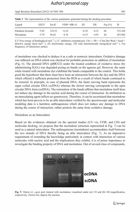
The nucleus of any cell being the harbinger of significant macromolecular interactions painta clear picture of the various mechanisms going within the same [22]. Staining of the nucleushas been employed as a measure to study the extent of interaction of several dyes such asethyl violet, Geimsa with DNA [23]. The ability of any dye to interact with DNA implicatesfor its preferential binding to the cell nuclei as seen in case of 4’6-diamidino-2-phenylindol.The staining exercise with tissue samples can be referred to as a direct visual evidencetowards the hypothesized interaction of morindone with dye. Figure 5 represents thespecificity of morindone in binding with the nucleus as seen under×10 and×40
Fig. 3 Circular dichroism spectral representation of CT-DNA, morindone dye, ethidium bromide, CT-DNA–morindone dye adduct, and CT-DNA–ethidium bromide adduct
Appl Biochem Biotechnol (2012) 167:885–896 891
Author's personal copy

magnifications, respectively. The anthraquinone owing to its interaction with the nucleicacid as evident from the study complies with the other assays carried out.
Electrophoretic Analysis Depicts Safety of the Dye
Literature reveals that several intercalators induce a toxic effect on DNA causing it damageduring the interaction [24–26]. Thus, it remains a prime requisite to address a compound assafe intercalator provided it itself does not damage DNA. Therefore the protective efficacy
Fig. 4 a The docked structure of ethidium bromide with 1DSC DNA. b The docked structure of morindonewith 1DSC DNA. The green, red, and white colors of morindone were carbon, oxygen, and hydrogen atoms,respectively
892 Appl Biochem Biotechnol (2012) 167:885–896
Author's personal copy
of morindone was checked to deduce it as a safe or nontoxic intercalator. Oxidative damagewas inflicted on DNAwhich was checked for probable protection on addition of morindone(Fig. 6). The plasmid DNA (pBR322) under the treated condition of oxidative stress (byadministering H2O2) was degraded posing no bands on the agarose gel. However, the samewhile treated with morindone dye exhibited the bands comparable to the control. This holdsgood the hypothesis that there must have been an interaction between the dye and the DNAwhich offered it sufficient protection from the ROS as a result of which bands continued tobe restored. In principle, in case of plasmid DNA, the faster moving band represents thesuper coiled circular DNA (scDNA) whereas the slower moving corresponds to the opencircular DNA form (ocDNA). The restoration of the bands affirms that morindone itself doesnot induce any damage to the nucleic acid during the course of interaction. Its attribution asan intercalating agent inflicts no genotoxicty. Therefore, it can be ascertained that morindonewhich has been proven to be an able intercalator verified by the spectroscopic and molecularmodeling data is a harmless anthraquinone which does not induce any damage to DNAduring the course of interaction, rather protects the same from oxidative damage.
Morindone as an Intercalator
Based on the evidences obtained via the spectral studies (UV–vis, FTIR, and CD) andmolecular docking, we propose that the morindone (structure represented in Fig. 7) can beused as a natural intercalator. The anthraquinone (morindone) accommodates itself betweenthe two strands of DNA thereby being an able intercalator (Fig. 7). As an imperativerequirement of extending the knowledge particularly in context with interaction of variousmolecules with nucleic acid and the implications they exhibit, it is of prime importance toinvestigate the binding property of DNA and morindone. Out of several class of compounds,
Fig. 5 Onion (A. cepa) peel stained with morindone visualized under (a)×10 and (b)×40 magnification,respectively. Dotted line depicts the nucleus
Table 1 The representation of the various parameters generated during the docking procedure
Ligand EST.F Est.IC VDW+HB+d EE TIE Frq (%) IS
Ethidium bromide −5.02 210.52 −6.10 −0.10 −6.21 60 553.444
Morindone −5.79 56.62 −4.76 −0.15 −4.91 20 453.982
E.F. Free energy of binding(kcal mol−1 ), I.C. inhibition constant(μm), Vdw + Hb + d Van Der Waal + bond +dissolve energy (kcal mol−1 ), EE electrostatic energy, TIE total intermolecular energy(kcal mol−1 ), Frqfrequency, IS interaction surface
Appl Biochem Biotechnol (2012) 167:885–896 893
Author's personal copy

anthraquinones are well-known to be endowed with anticancer potential [5] and known tointeract with DNA [1, 4, 5]. Therefore exploring the same shall definitely be a benefittowards biological perspectives. Furthermore, the intercalation property can be judiciouslyexploited in formulating therapeutics. As exhibited by the electrophoretic experiment, theability of morindone to render DNA damage free in the presence of reactive oxygen speciesis an affirmative approach towards using the natural anthraquinone as a molecule that canprotect DNA from injuries and thereby mold itself as a potent drug in health formulations.
Fig. 7 A hypothetical illustration of the intercalation mode of interaction of morindone with DNA. The redrectangular bars are graphic representation of the red, natural anthraquinone morindone
Fig. 6 Agarose gel representing pBR322 DNA treated with 3 % H2O2 and exposed to UV radiation (Ct),normal pBR322 DNA (without any treatment) (C), and pBR322 treated with morindone dye and thensubjected to 3 % H2O2 and UV irradiation (E). scDNA represents super coiled DNAwhereas ocDNA depictsopen circular DNA
894 Appl Biochem Biotechnol (2012) 167:885–896
Author's personal copy
Conclusions
Interaction with DNA has become an indispensible arena of interest owing to its impeccableimpact on pharmacy. The versatile modes of action followed by different compounds whileinteracting with DNA can be attributed to several parameters. The most significant onesbeing structural variations such as side chain flexibility, conformational diversity of thefunctional groups as well as unique charge densities [1]. Binding modes provide ampleinformation on the mechanism of potent anticancer drugs that can bind to DNA and therebybolster drug designing. Anthracenediones which have been earlier reported to be abletherapeutics owing to their structural benefactions, morindone (anthraquinone) holds prom-ise as per its structural congruity with the former. In addition, the spectroscopic analysessubstantiate sufficient interaction between the dye of interest and DNA majorly by the modeof intercalation. Therefore, morindone can be used as an agent which integrates itself withinthe strands of DNA and thereby confirms its aptness for therapeutic applications. Moreover,the ability of the same to stain nucleus specifically gives the dye a two pronged advantage ofbeing proficient in interacting with DNA as well as further being molded into a suitablenucleus specific stain. Also the significant feature of not inducing any damage to DNA itselfis yet another upper edge towards the probable therapeutic efficacy. The critical implicationsof the study shall definitely be an avenue towards devising therapeutics which can bedelivered to the targets and subsequently curb the progression of the disease in case DNAfunctionality is a prime contributor of the same, as seen in the manifestation of cancer. Inaddition, the property of binding (particularly intercalation) can be further exploited inreplacing mutagenic dyes such as ethidium bromide and subsequently proposing a safernatural alternative. As per the knowledge of the authors, this investigation is a first report onthe interaction of a natural anthraquinone dye with DNA.
Acknowledgments We express our sincere gratitude to the support extended by CSIR, New Delhi, Indiathrough the means of the sanctioned project [37(1451)/10/EMR-II]. The authors are grateful to VIT Universitymanagement for their constant support. We thank Professor G. Jayaraman for his critical suggestionsregarding the CD analysis. Thanks to Dr. A. Ramakrishna for chemical characterization of morindone. Theauthors thank Dr. Anand Prem Rajan for his immaculate help in the histological analyses.
Conflict of Interest The authors declare that there is no conflict of interest.
References
1. Tan, J. H., Lu, Y., Huang, Z. S., Gu, L. Q., & Wu, J. Y. (2007). Spectroscopic studies of DNA bindingmodes of cation-substituted anthrapyrazoles derived from emodin. European Journal of MedicinalChemistry, 42, 1169–1175.
2. Brana, M. F., Cacho, M., Gradillas, B., DePascual-Teresa, B., & Ramos, A. (2001). Intercalators asanticancer drugs. Current Pharmaceutical Design, 7, 1745–1780.
3. Neidle, S., Pearl, L. H., & Skelly, J. V. (1987). DNA structure and perturbation by drug binding.Biochemical Journal, 2143, 1–13.
4. Tan, W. B., Bhambhani, A., Duff, M. R., Rodger, A., & Kumar, C. V. (2006). Spectroscopic identificationof binding modes of anthracene probes and DNA sequence recognition. Photochemistry and Photobiol-ogy, 82, 20–30.
5. Bi, S., Zhang, H., Qiao, C., Sunc, Y., & Liu, C. (2008). Studies of interaction of emodin andDNA in the presence of ethidium bromide by spectroscopic method. Spectrochimica Acta A, 69,123–129.
Appl Biochem Biotechnol (2012) 167:885–896 895
Author's personal copy

6. Paul, P., Hossain, M., Yadav, R. C., & Kumar, G. S. (2010). Biophysical studies on the base specificityand energetics of the DNA interaction of photoactive dye thionine: spectroscopic and calorimetricapproach. Biophysical Chemistry, 148, 93–103.
7. Lown, J. W., & Hanstock, C. C. (1985). High filed 1H-NMR analysis of the 1:1 intercalation complex ofthe anti tumor agent mitoxantrone and the DNA duplex [d(CpGpCpG)]. Journal of BiomolecularStructure and Dynamics, 2, 1097–1106.
8. Siva, R. (2007). Status of natural dyes and dye yielding plants in India. Current Science, 92, 916–925.9. Anekpankul, T., Goto, M., Sasaki, M., Pavasant, P., & Shotipruk, A. (2007). Extraction of anti-cancer
damnacanthal from roots of Morinda citrifolia by subcritical water. Separation and Purification Tech-nology, 55, 343–349.
10. Alagesaboopathi, C. (2009). Ethnomedicinal plants and their utilization by villagers in Kumaragiri Hillsof Salem District of Tamil Nadu, India. African Journal of Traditional Complementary and AlternativeMedicine, 6, 222–227.
11. Vijayalakshmi, R., Kanthimanthi, M., & Subramanian, V. (2000). DNA cleavage by a Chromium(III)complex. Biochemical and Biophysical Research Communications, 271, 731–734.
12. Aobchey, P., Sriyam, S., Praharnripoorab, W., Lhieochaiphant, S., & Phutrakul, S. (2002). Production ofred pigment from the root ofMorinda angustifolia Roxb. var. scabridula Craib. by root cell culture. CMUJournal, 1, 66–78.
13. Russo, A., Izzo, A. A., Cardile, V., Borrelli, F., & Vanella, A. (2001). Indian medicinal plants asantiradicals and DNA cleavage protector. Phytomedicine, 8, 125–132.
14. Vaidyanathan, V. G., & Nair, B. U. (2005). Synthesis, characterization and DNA-binding studies of mixedligand complexes of ruthenium(II) with DNA. Journal of Chemical Society Dalton Transactions, 17,2842–2848.
15. Cate, J. H., & Doudna, J. A. (1996). Metal binding sites in the major groove of a large ribozyme domain.Structure, 4, 1221–1229.
16. Zhang, L. Z., & Tang, G. Q. (2004). The binding properties of photosensitizer methylene blue to herringsperm DNA: a spectroscopic study. Journal of Photochemistry and Photobiology B: Biology, 74, 119–125.
17. Long, E. C., & Barton, J. K. (1990). On demonstrating DNA intercalation. Accounts of ChemicalResearch, 23, 271–273.
18. Mao, Y., Daniel, L. N., Whittaker, N., & Saffiottil, U. (1994). DNA binding to crystalline silicacharacterized by Fourier-transform infrared spectroscopy. Environmental Health Perspectives, 102, 65–171.
19. Hackl, E. V., Kornilova, S. V., Kapinos, L. E., Andrushchenko, V. V., Galkin, V. L., Grigoriev, D. N., et al.(1997). Study of Ca+2, Mn+2 and Cu+2 binding to DNA in solution by means of IR spectroscopy. Journalof Molecular Structure, 408(409), 229–232.
20. Zunino, F., Animati, F., & Capranico, C. (1995). DNA minor-groove binding drugs. Current Pharma-ceutical Design, 1, 83–94.
21. Rajendra, T. K., Gonsalvez, G. B., Walker, M. P., Shpargel, K. B., Salz, H. K., & Matera, A. G. (2007). ADrosophila melanogaster model of spinal muscular atrophy reveals a function for SMN in striatedmuscle. The Journal of Cell Biology, 176, 831–841.
22. Cremazy, F. G. E., Manders, E. M. M., Bastiaens, P. I. H., Kramer, G., Hager, G. L., van Munster, E. B., etal. (2005). Imaging in situ protein–DNA interactions in the cell nucleus using FRET–FLIM. ExperimentalCell Research, 309, 390–396.
23. Dutt, M. K. (1982). Basic dyes for the staining of DNA in mammalian tissues and absorption spectra ofstained nuclei in the visible light. Microscopica Acta, 86, 59–68.
24. Lu, W., Vicic, D. A., & Barton, J. K. (2005). Reductive and oxidative DNA damage by photoactiveplatinum(II) intercalators. Inorganic Chemistry, 44, 7970–7980.
25. Faddeeva, M. D., & Beliaeva, T. N. (1991). DNA intercalators: their interaction with DNA and other cellcomponents and their use in biological research. Tsitologiia, 33, 3–31.
26. Ferguson, L. R., & Denny, W. A. (2007). Genotoxicity of non-covalent interactions: DNA Intercalators.Mutation Research, 623, 14–23.
896 Appl Biochem Biotechnol (2012) 167:885–896
Author's personal copy