Synthesis, SAR and antibacterial activity of hybrid chloro, dichloro-phenylthiazolyl- s-triazines
Upload
independentCategory
view
1download
0
2-(2,5-Dichlorobenzenesulfonamido)-3-methylbutanoic acid
Islam Ullah Khan,a‡ Peter John,a Haffsah Iqbal,a Shahzad
Sharifa and Edward R. T. Tiekinkb*
aMaterials Chemistry Laboratory, Department of Chemistry, Government College,
University, Lahore 54000, Pakistan, and bDepartment of Chemistry, University of
Malaya, 50603 Kuala Lumpur, Malaysia
Correspondence e-mail: [email protected]
Received 14 October 2010; accepted 15 October 2010
Key indicators: single-crystal X-ray study; T = 293 K; mean �(C–C) = 0.005 A;
R factor = 0.041; wR factor = 0.127; data-to-parameter ratio = 18.9.
The structure of the title compound, C11H13Cl2NO4S, shows
one sulfonamide-O atom to lie almost in the plane of the
benzene ring [C—C—S—O = �178.7 (2) �] and the other to
one side [C—C—S—O =�49.4 (3)�]. Lying to the other side is
the amine residue, which occupies a position almost
perpendicular to the plane [C—S—N—C = 70.2 (2)�]; the
carboxylic acid group is orientated to lie over the benzene
ring. In the crystal, the appearance of an 11-membered
{� � �OH� � �OCOH� � �OC2NH} synthon, which features the
hydroxy group forming both donor (to a carbonyl-O) and
acceptor (from the amine-H) interactions, leads to the
formation of a supramolecular chain along the a axis. Chains
are connected in the crystal structure by C—H� � �O contacts.
Related literature
For background to the pharmacological uses of sulfonamides,
see: Korolkovas (1988); Mandell & Sande (1992). For related
structures, see: Sharif et al. (2010); Khan et al. (2010).
Experimental
Crystal data
C11H13Cl2NO4SMr = 326.18Orthorhombic, P212121
a = 5.4584 (2) Ab = 14.0623 (6) Ac = 19.4545 (8) A
V = 1493.28 (10) A3
Z = 4Mo K� radiation� = 0.58 mm�1
T = 293 K0.19 � 0.13 � 0.07 mm
Data collection
Bruker APEXII CCDdiffractometer
Absorption correction: multi-scan(SADABS; Sheldrick, 1996)Tmin = 0.805, Tmax = 0.921
14542 measured reflections3405 independent reflections2876 reflections with I > 2�(I)Rint = 0.040
Refinement
R[F 2 > 2�(F 2)] = 0.041wR(F 2) = 0.127S = 1.003405 reflections180 parameters2 restraints
H atoms treated by a mixture ofindependent and constrainedrefinement
��max = 0.61 e A�3
��min = �0.51 e A�3
Absolute structure: Flack (1983),1415 Friedel pairs
Flack parameter: 0.09 (8)
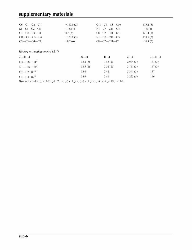
Table 1Hydrogen-bond geometry (A, �).
D—H� � �A D—H H� � �A D� � �A D—H� � �A
O3—H3o� � �O4i 0.82 (3) 1.86 (2) 2.674 (3) 171 (3)N1—H1n� � �O3ii 0.85 (2) 2.32 (2) 3.161 (3) 167 (3)C7—H7� � �O1iii 0.98 2.42 3.341 (3) 157C4—H4� � �O2iv 0.93 2.41 3.223 (5) 146
Symmetry codes: (i) xþ 12;�yþ 1
2;�z; (ii) x� 1; y; z; (iii) xþ 1; y; z; (iv)�x þ 2; yþ 1
2;�zþ 12.
Data collection: APEX2 (Bruker, 2007); cell refinement: SAINT
(Bruker, 2007); data reduction: SAINT; program(s) used to solve
structure: SHELXS97 (Sheldrick, 2008); program(s) used to refine
structure: SHELXL97 (Sheldrick, 2008); molecular graphics:
ORTEP-3 (Farrugia, 1997) and DIAMOND (Brandenburg, 2006);
software used to prepare material for publication: publCIF (Westrip,
2010).
The authors are grateful to the Higher Education
Commission of Pakistan for financial support to purchase the
diffractometer.
Supplementary data and figures for this paper are available from theIUCr electronic archives (Reference: HG2730).
References
Brandenburg, K. (2006). DIAMOND. Crystal Impact GbR, Bonn, Germany.Bruker (2007). APEX2 and SAINT. Bruker AXS Inc., Madison Wisconsin,
USA.Farrugia, L. J. (1997). J. Appl. Cryst. 30, 565.Flack, H. D. (1983). Acta Cryst. A39, 876–881.Khan, I. U., Sharif, S., Batool, S., Mumtaz, A. M. & Tiekink, E. R. T. (2010).
Acta Cryst. E66, o2641.Korolkovas, A. (1988). Essentials of Medicinal Chemistry, 2nd ed, pp. 699–716.
New York: Wiley.Mandell, G. L. & Sande, M. A. (1992). In Goodman and Gilman, The
Pharmacological Basis of Therapeutics 2, edited by A. Gilman, T. W. Rall,A. S. Nies & P. Taylor, 8th ed, pp. 1047–1057. Singapore: McGraw-Hill.
Sharif, S., Iqbal, H., Khan, I. U., John, P. & Tiekink, E. R. T. (2010). Acta Cryst.E66, o1288.
Sheldrick, G. M. (1996). SADABS. University of Gottingen, Germany.Sheldrick, G. M. (2008). Acta Cryst. A64, 112–122.Westrip, S. P. (2010). J. Appl. Cryst. 43, 920–925.
organic compounds
o2888 Khan et al. doi:10.1107/S1600536810041620 Acta Cryst. (2010). E66, o2888
Acta Crystallographica Section E
Structure ReportsOnline
ISSN 1600-5368
‡ Additional correspondence author, e-mail: [email protected].
supplementary materials

supplementary materials
sup-1
Acta Cryst. (2010). E66, o2888 [ doi:10.1107/S1600536810041620 ]
2-(2,5-Dichlorobenzenesulfonamido)-3-methylbutanoic acid
I. U. Khan, P. John, H. Iqbal, S. Sharif and E. R. T. Tiekink
Comment
The crystal structure of the title compound, (I), was determined in connection with on-going structural studies of sulfonam-ides (Sharif et al., 2010; Khan et al., 2010), of interest owing to their biological properties (Korolkovas, 1988; Mandell& Sande, 1992).
With reference to the benzene ring in (I), Fig. 1, the O2 atom lies in the plane [the O2—S1—C1—C2 torsion angle =-178.7 (2) °] but the O1 atom lies out of the plane [O1—S1—C1—C2 = -49.4 (3) °]. The amine group lies to the oppositeside of the plane to the O1 atom and occupies a position almost perpendicular to it [C1—S1—N1—C7 = 70.2 (2) °]. Withinthe amine residue, the carboxylic acid group is co-planar with the amine-N1 [N1—C7—C11—O4 = -1.6 (4) °], and is foldedto be orientated over the benzene ring with the carbonyl-O4 atom closest to it.
In the crystal packing, the hydroxyl-O3 group forms both donor and acceptor interactions, the former to a symmetry re-lated carbonyl-O4 and the latter with a symmetry related amine-N1—H atom, Table 1. These lead to a linear supramolecularchain, Fig. 2, aligned along the a axis and mediated by an 11-membered {···OH···OCOH···OC2NH} synthon; the chain is
further stabilized by a C7—H7···O1 contact, Table 1. Chains are held in the crystal structure by C—H···O contacts, Fig.3 and Table 1.
Experimental
To 2-amino-3-methylbutanoic acid (234 mg, 2 mmol) in distilled water (15 ml), was added 2,5-dichlorobenzenesulfonylchloride (491 mg, 2 mmol) while maintaining the pH of reaction mixture at 8 by using 3% sodium carbonate solution. Theconsumption of the reactants was confirmed by TLC. The pH of reaction mixture was adjusted to 3 using 3 N HCl. Theprecipitates were washed with water and crystallized from methanol
Refinement
The C-bound H atoms were geometrically placed (C–H = 0.93–0.98 Å) and refined as riding with Uiso(H) = 1.2–1.5Ueq(C).
The O– and N-bound H atoms were refined with the distance restraints O—H = 0.82±0.01 Å and N–H = 0.86±0.01 Å, andwith Uiso(H) = yUeq(parent atom) for y = 1.5 (parent atom = O) and y = 1.2 (N). In the final refinement four low angle
reflections evidently effected by the beam stop were omitted, i.e. (002), (012), (011) and (021).

supplementary materials
sup-2
Figures
Fig. 1. The molecular structure of (I) showing the atom-labelling scheme and displacement el-lipsoids at the 35% probability level.
Fig. 2. A view of the linear supramolecular chain along the a axis in (I). The O–H···O andN–H···O hydrogen bonds are shown as orange and blue dashed lines, respectively.
Fig. 3. View in projection down the a axis of the unit-cell contents for (I). The O–H···O,N–H···O and C—H···O contacts are shown as orange, blue and pink dashed lines, respect-ively.
2-(2,5-Dichlorobenzenesulfonamido)-3-methylbutanoic acid
Crystal data
C11H13Cl2NO4S F(000) = 672
Mr = 326.18 Dx = 1.451 Mg m−3
Orthorhombic, P212121 Mo Kα radiation, λ = 0.71073 ÅHall symbol: P 2ac 2ab Cell parameters from 4852 reflectionsa = 5.4584 (2) Å θ = 2.6–25.1°b = 14.0623 (6) Å µ = 0.58 mm−1
c = 19.4545 (8) Å T = 293 K
V = 1493.28 (10) Å3 Prism, colourlessZ = 4 0.19 × 0.13 × 0.07 mm
Data collection
Bruker APEXII CCDdiffractometer 3405 independent reflections
Radiation source: fine-focus sealed tube 2876 reflections with I > 2σ(I)graphite Rint = 0.040
φ and ω scans θmax = 27.5°, θmin = 3.5°Absorption correction: multi-scan(SADABS; Sheldrick, 1996) h = −5→7
Tmin = 0.805, Tmax = 0.921 k = −18→1814542 measured reflections l = −25→25

supplementary materials
sup-3
Refinement
Refinement on F2 Secondary atom site location: difference Fourier map
Least-squares matrix: full Hydrogen site location: inferred from neighbouringsites
R[F2 > 2σ(F2)] = 0.041H atoms treated by a mixture of independent andconstrained refinement
wR(F2) = 0.127w = 1/[σ2(Fo
2) + (0.0887P)2]where P = (Fo
2 + 2Fc2)/3
S = 1.00 (Δ/σ)max = 0.001
3405 reflections Δρmax = 0.61 e Å−3
180 parameters Δρmin = −0.51 e Å−3
2 restraints Absolute structure: Flack (1983), 1415 Friedel pairsPrimary atom site location: structure-invariant directmethods Flack parameter: 0.09 (8)
Special details
Geometry. All s.u.'s (except the s.u. in the dihedral angle between two l.s. planes) are estimated using the full covariance matrix. Thecell s.u.'s are taken into account individually in the estimation of s.u.'s in distances, angles and torsion angles; correlations betweens.u.'s in cell parameters are only used when they are defined by crystal symmetry. An approximate (isotropic) treatment of cell s.u.'s isused for estimating s.u.'s involving l.s. planes.
Refinement. Refinement of F2 against ALL reflections. The weighted R-factor wR and goodness of fit S are based on F2, conventional
R-factors R are based on F, with F set to zero for negative F2. The threshold expression of F2 > 2σ(F2) is used only for calculating R-
factors(gt) etc. and is not relevant to the choice of reflections for refinement. R-factors based on F2 are statistically about twice as largeas those based on F, and R- factors based on ALL data will be even larger.
Fractional atomic coordinates and isotropic or equivalent isotropic displacement parameters (Å2)
x y z Uiso*/Ueq
Cl1 0.36922 (17) 0.23427 (6) 0.14486 (5) 0.0617 (3)Cl2 1.2300 (2) 0.15728 (10) 0.34471 (6) 0.0952 (4)S1 0.59333 (11) 0.01761 (4) 0.15989 (3) 0.03515 (17)O1 0.3334 (3) 0.01414 (15) 0.16803 (12) 0.0491 (5)O2 0.7383 (4) −0.06093 (14) 0.18173 (11) 0.0485 (5)O3 1.1937 (3) 0.12919 (13) 0.00395 (12) 0.0465 (5)H3O 1.231 (8) 0.1812 (14) −0.0118 (19) 0.070*O4 0.8427 (4) 0.19737 (14) 0.03380 (13) 0.0554 (6)N1 0.6435 (4) 0.03271 (15) 0.07885 (11) 0.0359 (5)H1N 0.528 (4) 0.0668 (18) 0.0625 (15) 0.043*C1 0.7060 (5) 0.11804 (19) 0.20583 (13) 0.0375 (6)C2 0.6085 (6) 0.2087 (2) 0.19980 (15) 0.0475 (7)C3 0.6997 (8) 0.2826 (2) 0.23875 (19) 0.0671 (10)H3 0.6316 0.3430 0.2348 0.080*C4 0.8904 (8) 0.2675 (3) 0.28335 (19) 0.0740 (12)H4 0.9526 0.3172 0.3096 0.089*

supplementary materials
sup-4
C5 0.9879 (7) 0.1778 (3) 0.28870 (16) 0.0615 (10)C6 0.8996 (5) 0.1019 (2) 0.25120 (14) 0.0453 (6)H6 0.9673 0.0416 0.2560 0.054*C7 0.8902 (5) 0.02917 (16) 0.05012 (13) 0.0336 (5)H7 1.0025 0.0063 0.0858 0.040*C8 0.9042 (6) −0.0395 (2) −0.01197 (18) 0.0525 (8)H8 1.0741 −0.0392 −0.0283 0.063*C9 0.7472 (9) −0.0072 (3) −0.0703 (2) 0.0855 (13)H9A 0.7767 −0.0468 −0.1097 0.128*H9B 0.7860 0.0576 −0.0815 0.128*H9C 0.5778 −0.0117 −0.0574 0.128*C10 0.8466 (11) −0.1396 (2) 0.0105 (3) 0.103 (2)H10A 0.6806 −0.1426 0.0266 0.155*H10B 0.9559 −0.1578 0.0468 0.155*H10C 0.8666 −0.1821 −0.0278 0.155*C11 0.9694 (5) 0.12818 (16) 0.02879 (14) 0.0352 (6)
Atomic displacement parameters (Å2)
U11 U22 U33 U12 U13 U23
Cl1 0.0687 (5) 0.0524 (4) 0.0638 (5) 0.0192 (4) 0.0005 (4) 0.0038 (4)Cl2 0.0659 (6) 0.1600 (11) 0.0597 (6) −0.0443 (7) −0.0190 (5) 0.0070 (7)S1 0.0295 (3) 0.0337 (3) 0.0422 (3) −0.0026 (2) 0.0021 (3) 0.0031 (3)O1 0.0314 (9) 0.0567 (11) 0.0593 (13) −0.0080 (9) 0.0077 (9) −0.0015 (10)O2 0.0518 (11) 0.0383 (10) 0.0553 (12) 0.0021 (9) −0.0007 (10) 0.0110 (8)O3 0.0367 (10) 0.0367 (10) 0.0661 (13) −0.0051 (8) 0.0044 (10) 0.0114 (9)O4 0.0589 (13) 0.0357 (9) 0.0717 (15) 0.0117 (9) 0.0111 (11) 0.0116 (10)N1 0.0282 (11) 0.0421 (11) 0.0375 (12) 0.0024 (9) −0.0012 (9) 0.0013 (9)C1 0.0346 (13) 0.0432 (14) 0.0347 (13) −0.0069 (11) 0.0054 (11) −0.0010 (11)C2 0.0564 (19) 0.0437 (14) 0.0423 (16) −0.0038 (15) 0.0099 (14) −0.0035 (11)C3 0.097 (3) 0.0490 (17) 0.055 (2) −0.0171 (19) 0.011 (2) −0.0090 (15)C4 0.093 (3) 0.074 (2) 0.055 (2) −0.042 (2) 0.002 (2) −0.0117 (18)C5 0.0509 (19) 0.094 (3) 0.0393 (16) −0.0319 (18) 0.0020 (15) −0.0026 (17)C6 0.0367 (14) 0.0618 (16) 0.0375 (14) −0.0073 (14) 0.0050 (12) 0.0050 (12)C7 0.0283 (12) 0.0323 (11) 0.0402 (13) 0.0020 (10) −0.0001 (10) 0.0013 (10)C8 0.0497 (17) 0.0456 (15) 0.0623 (19) −0.0054 (14) 0.0187 (16) −0.0146 (13)C9 0.086 (3) 0.115 (3) 0.056 (2) −0.018 (3) −0.005 (2) −0.036 (2)C10 0.150 (5) 0.0403 (17) 0.119 (4) −0.019 (2) 0.050 (4) −0.026 (2)C11 0.0336 (13) 0.0321 (12) 0.0399 (13) 0.0004 (10) −0.0042 (11) 0.0024 (10)
Geometric parameters (Å, °)
Cl1—C2 1.725 (3) C4—C5 1.372 (6)Cl2—C5 1.737 (4) C4—H4 0.9300S1—O2 1.424 (2) C5—C6 1.380 (5)S1—O1 1.4286 (19) C6—H6 0.9300S1—N1 1.614 (2) C7—C11 1.516 (3)S1—C1 1.781 (3) C7—C8 1.549 (4)O3—C11 1.317 (3) C7—H7 0.9800

supplementary materials
sup-5
O3—H3o 0.82 (3) C8—C9 1.494 (6)O4—C11 1.197 (3) C8—C10 1.506 (5)N1—C7 1.459 (3) C8—H8 0.9800N1—H1n 0.853 (10) C9—H9A 0.9600C1—C2 1.387 (4) C9—H9B 0.9600C1—C6 1.395 (4) C9—H9C 0.9600C2—C3 1.379 (5) C10—H10A 0.9600C3—C4 1.372 (6) C10—H10B 0.9600C3—H3 0.9300 C10—H10C 0.9600
O2—S1—O1 119.51 (12) N1—C7—C11 109.66 (19)O2—S1—N1 107.41 (12) N1—C7—C8 111.5 (2)O1—S1—N1 106.34 (13) C11—C7—C8 110.2 (2)O2—S1—C1 105.87 (13) N1—C7—H7 108.5O1—S1—C1 108.33 (13) C11—C7—H7 108.5N1—S1—C1 109.10 (12) C8—C7—H7 108.5C11—O3—H3O 112 (3) C9—C8—C10 112.6 (4)C7—N1—S1 121.76 (17) C9—C8—C7 112.0 (3)C7—N1—H1N 124 (2) C10—C8—C7 110.3 (3)S1—N1—H1N 108 (2) C9—C8—H8 107.2C2—C1—C6 119.6 (3) C10—C8—H8 107.2C2—C1—S1 123.7 (2) C7—C8—H8 107.2C6—C1—S1 116.8 (2) C8—C9—H9A 109.5C3—C2—C1 120.5 (3) C8—C9—H9B 109.5C3—C2—Cl1 117.2 (3) H9A—C9—H9B 109.5C1—C2—Cl1 122.3 (2) C8—C9—H9C 109.5C4—C3—C2 120.3 (4) H9A—C9—H9C 109.5C4—C3—H3 119.8 H9B—C9—H9C 109.5C2—C3—H3 119.8 C8—C10—H10A 109.5C3—C4—C5 119.0 (3) C8—C10—H10B 109.5C3—C4—H4 120.5 H10A—C10—H10B 109.5C5—C4—H4 120.5 C8—C10—H10C 109.5C6—C5—C4 122.3 (3) H10A—C10—H10C 109.5C6—C5—Cl2 118.0 (3) H10B—C10—H10C 109.5C4—C5—Cl2 119.7 (3) O4—C11—O3 123.9 (2)C5—C6—C1 118.3 (3) O4—C11—C7 124.0 (2)C5—C6—H6 120.9 O3—C11—C7 112.0 (2)C1—C6—H6 120.9
O2—S1—N1—C7 −44.1 (2) C3—C4—C5—C6 −0.6 (6)O1—S1—N1—C7 −173.12 (18) C3—C4—C5—Cl2 179.8 (3)C1—S1—N1—C7 70.2 (2) C4—C5—C6—C1 0.8 (5)O2—S1—C1—C2 −178.7 (2) Cl2—C5—C6—C1 −179.6 (2)O1—S1—C1—C2 −49.4 (3) C2—C1—C6—C5 −0.1 (4)N1—S1—C1—C2 66.0 (3) S1—C1—C6—C5 −178.7 (2)O2—S1—C1—C6 −0.2 (2) S1—N1—C7—C11 −108.9 (2)O1—S1—C1—C6 129.1 (2) S1—N1—C7—C8 128.8 (2)N1—S1—C1—C6 −115.5 (2) N1—C7—C8—C9 63.4 (3)C6—C1—C2—C3 −0.7 (4) C11—C7—C8—C9 −58.6 (3)S1—C1—C2—C3 177.8 (3) N1—C7—C8—C10 −62.8 (4)
supplementary materials
sup-6
C6—C1—C2—Cl1 −180.0 (2) C11—C7—C8—C10 175.2 (3)S1—C1—C2—Cl1 −1.6 (4) N1—C7—C11—O4 −1.6 (4)C1—C2—C3—C4 0.8 (5) C8—C7—C11—O4 121.4 (3)Cl1—C2—C3—C4 −179.8 (3) N1—C7—C11—O3 178.5 (2)C2—C3—C4—C5 −0.2 (6) C8—C7—C11—O3 −58.4 (3)
Hydrogen-bond geometry (Å, °)
D—H···A D—H H···A D···A D—H···A
O3—H3o···O4i 0.82 (3) 1.86 (2) 2.674 (3) 171 (3)
N1—H1n···O3ii 0.85 (2) 2.32 (2) 3.161 (3) 167 (3)
C7—H7···O1iii 0.98 2.42 3.341 (3) 157
C4—H4···O2iv 0.93 2.41 3.223 (5) 146Symmetry codes: (i) x+1/2, −y+1/2, −z; (ii) x−1, y, z; (iii) x+1, y, z; (iv) −x+2, y+1/2, −z+1/2.

supplementary materials
sup-7
Fig. 1

supplementary materials
sup-8
Fig. 2

supplementary materials
sup-9
Fig. 3
Copyright © 2022 FDOKUMEN


![Human Serum Albumin Binding of 2-[(Carboxymethyl)sulfanyl]-4-oxo-4-(4-tert-butylphenyl)butanoic Acid and its Mono-Me Ester](https://static.fdokumen.com/doc/165x107/6334ae932532592417002ca9/human-serum-albumin-binding-of-2-carboxymethylsulfanyl-4-oxo-4-4-tert-butylphenylbutanoic.jpg)


![Synthesis, Spectroscopic and Electrochemical Studies of Isomeric Dichloro-bis-[ N (1)-Alkyl-2-(Arylazo)Imidazole]-Osmium(II). Single Crystal X-ray Structures of Blue-Violet Dichloro-Bis-[](https://static.fdokumen.com/doc/165x107/6333de5ece61be0ae50edc8a/synthesis-spectroscopic-and-electrochemical-studies-of-isomeric-dichloro-bis-.jpg)