
2,5-Dihydroxybenzaldehyde 4-methylthiosemicarbazone
7
2,5-Dihydroxybenzaldehyde 4-methyl- thiosemicarbazone Kong Wai Tan, a Chew Hee Ng, b Mohd Jamil Maah c * and Seik Weng Ng c a Department of Chemistry, University of Malaya, 50603 Kuala Lumpur, Malaysia, b Faculty of Engineering and Science, Universiti Tunku Abdul Rahman, 53300 Kuala Lumpur, Malaysia, and c Department of Chemistry, University of Malaya, 50603 Kuala Lumpur, Malaysia Correspondence e-mail: [email protected] Received 20 June 2008; accepted 21 June 2008 Key indicators: single-crystal X-ray study; T = 100 K; mean (C–C) = 0.003 A ˚ ; R factor = 0.055; wR factor = 0.124; data-to-parameter ratio = 14.8. The planar molecules of the title compound, C 9 H 11 N 3 O 2 S, are linked into a supramolecualr chain via O—HS hydrogen bonds. These chains are connected into a two-dimensional array via N—HO hydrogen bonds; an intramolecular O— HN hydrogen bond is also present. Related literature For the medicinal activity of 2,5-dihydroxybenzaldehyde thiosemicarbazone, see: Libermann et al. (1953); Taniyama & Tanaka (1965); Xue et al. (2007). For the structure of 2- hydroxybenzaldehyde 4-methylthiosemicarbazone, see: Vrdoljak et al. (2005). For the structure of 3,4-dihydroxy- benzaldehyde 4-ethylthiosemicarbazone, see: Kayed et al. (2008). Experimental Crystal data C 9 H 11 N 3 O 2 S M r = 225.27 Triclinic, P 1 a = 5.9932 (4) A ˚ b = 8.5207 (6) A ˚ c = 10.3272 (6) A ˚ = 78.552 (4) = 74.181 (4) = 81.743 (4) V = 495.06 (6) A ˚ 3 Z =2 Mo K radiation = 0.31 mm 1 T = 100 (2) K 0.24 0.16 0.02 mm Data collection Bruker SMART APEX diffractometer Absorption correction: multi-scan (SADABS; Sheldrick, 1996) T min = 0.929, T max = 0.994 4189 measured reflections 2258 independent reflections 1580 reflections with I >2(I) R int = 0.050 Refinement R[F 2 >2(F 2 )] = 0.054 wR(F 2 ) = 0.123 S = 1.03 2258 reflections 153 parameters 4 restraints H atoms treated by a mixture of independent and constrained refinement Á max = 0.32 e A ˚ 3 Á min = 0.47 e A ˚ 3 Table 1 Hydrogen-bond geometry (A ˚ , ). D—HA D—H HA DA D—HA O1—H1oN3 0.84 (3) 1.97 (2) 2.698 (3) 144 (3) O2—H2oS1 i 0.84 (3) 2.46 (2) 3.182 (2) 144 (3) N2—H2nO1 ii 0.84 (3) 2.47 (3) 3.111 (3) 134 (3) Symmetry codes: (i) x þ 1; y þ 1; z 1; (ii) x 1; y; z. Data collection: APEX2 (Bruker, 2007); cell refinement: SAINT (Bruker, 2007); data reduction: SAINT; program(s) used to solve structure: SHELXS97 (Sheldrick, 2008); program(s) used to refine structure: SHELXL97 (Sheldrick, 2008); molecular graphics: X- SEED (Barbour, 2001); software used to prepare material for publication: publCIF (Westrip, 2008). We thank the University of Malaya (P0265/2007 A) for supporting this study. KWT thanks the Ministry of Higher Education for a SLAI scholarship. Supplementary data and figures for this paper are available from the IUCr electronic archives (Reference: TK2278). References Barbour, L. J. (2001). J. Supramol. Chem. 1, 189–191. Bruker (2007). APEX2 and SAINT. Bruker AXS Inc., Madison, Wisconsin, USA. Kayed, S. F., Farina, Y., Baba, I. & Simpson, J. (2008). Acta Cryst. E64, o824– o825. Libermann, D., Moyeux, M., Rouaix, A., Maillard, J., Hengl, L. & Himbert, J. (1953). Bull. Soc. Chim. Fr. pp. 957–962. Sheldrick, G. M. (1996). SADABS. University of Go ¨ttingen, Germany. Sheldrick, G. M. (2008). Acta Cryst. A64, 112–122. Taniyama, H. & Tanaka, Y. (1965). Yakugaku Kenkyu, 36, 319–328. Vrdoljak, V., Cindric ´, M., Milic ´, D., Matkovic ´-C ˇ alogovic ´, D., Novak, P. & Kamenar, B. (2005). Polyhedron, 24, 1717–1726. Westrip, S. P. (2008). publCIF. In preparation. Xue, C.-B., Zhang, L., Luo, W.-C., Xie, X.-Y., Jiang, L. & Xiao, T. (2007). Bioorg. Med. Chem. 15, 2006–2015. organic compounds o1344 Tan et al. doi:10.1107/S1600536808018801 Acta Cryst. (2008). E64, o1344 Acta Crystallographica Section E Structure Reports Online ISSN 1600-5368
Transcript of 2,5-Dihydroxybenzaldehyde 4-methylthiosemicarbazone
2,5-Dihydroxybenzaldehyde 4-methyl-thiosemicarbazone
Kong Wai Tan,a Chew Hee Ng,b Mohd Jamil Maahc* and
Seik Weng Ngc
aDepartment of Chemistry, University of Malaya, 50603 Kuala Lumpur, Malaysia,bFaculty of Engineering and Science, Universiti Tunku Abdul Rahman, 53300 Kuala
Lumpur, Malaysia, and cDepartment of Chemistry, University of Malaya, 50603
Kuala Lumpur, Malaysia
Correspondence e-mail: [email protected]
Received 20 June 2008; accepted 21 June 2008
Key indicators: single-crystal X-ray study; T = 100 K; mean �(C–C) = 0.003 A;
R factor = 0.055; wR factor = 0.124; data-to-parameter ratio = 14.8.
The planar molecules of the title compound, C9H11N3O2S, are
linked into a supramolecualr chain via O—H� � �S hydrogen
bonds. These chains are connected into a two-dimensional
array via N—H� � �O hydrogen bonds; an intramolecular O—
H� � �N hydrogen bond is also present.
Related literature
For the medicinal activity of 2,5-dihydroxybenzaldehyde
thiosemicarbazone, see: Libermann et al. (1953); Taniyama &
Tanaka (1965); Xue et al. (2007). For the structure of 2-
hydroxybenzaldehyde 4-methylthiosemicarbazone, see:
Vrdoljak et al. (2005). For the structure of 3,4-dihydroxy-
benzaldehyde 4-ethylthiosemicarbazone, see: Kayed et al.
(2008).
Experimental
Crystal data
C9H11N3O2SMr = 225.27Triclinic, P1a = 5.9932 (4) Ab = 8.5207 (6) A
c = 10.3272 (6) A� = 78.552 (4)�
� = 74.181 (4)�
� = 81.743 (4)�
V = 495.06 (6) A3
Z = 2Mo K� radiation� = 0.31 mm�1
T = 100 (2) K0.24 � 0.16 � 0.02 mm
Data collection
Bruker SMART APEXdiffractometer
Absorption correction: multi-scan(SADABS; Sheldrick, 1996)Tmin = 0.929, Tmax = 0.994
4189 measured reflections2258 independent reflections1580 reflections with I > 2�(I)Rint = 0.050
Refinement
R[F 2 > 2�(F 2)] = 0.054wR(F 2) = 0.123S = 1.032258 reflections153 parameters4 restraints
H atoms treated by a mixture ofindependent and constrainedrefinement
��max = 0.32 e A�3
��min = �0.47 e A�3
Table 1Hydrogen-bond geometry (A, �).
D—H� � �A D—H H� � �A D� � �A D—H� � �A
O1—H1o� � �N3 0.84 (3) 1.97 (2) 2.698 (3) 144 (3)O2—H2o� � �S1i 0.84 (3) 2.46 (2) 3.182 (2) 144 (3)N2—H2n� � �O1ii 0.84 (3) 2.47 (3) 3.111 (3) 134 (3)
Symmetry codes: (i) x þ 1; yþ 1; z� 1; (ii) x � 1; y; z.
Data collection: APEX2 (Bruker, 2007); cell refinement: SAINT
(Bruker, 2007); data reduction: SAINT; program(s) used to solve
structure: SHELXS97 (Sheldrick, 2008); program(s) used to refine
structure: SHELXL97 (Sheldrick, 2008); molecular graphics: X-
SEED (Barbour, 2001); software used to prepare material for
publication: publCIF (Westrip, 2008).
We thank the University of Malaya (P0265/2007 A) for
supporting this study. KWT thanks the Ministry of Higher
Education for a SLAI scholarship.
Supplementary data and figures for this paper are available from theIUCr electronic archives (Reference: TK2278).
References
Barbour, L. J. (2001). J. Supramol. Chem. 1, 189–191.Bruker (2007). APEX2 and SAINT. Bruker AXS Inc., Madison, Wisconsin,
USA.Kayed, S. F., Farina, Y., Baba, I. & Simpson, J. (2008). Acta Cryst. E64, o824–
o825.Libermann, D., Moyeux, M., Rouaix, A., Maillard, J., Hengl, L. & Himbert, J.
(1953). Bull. Soc. Chim. Fr. pp. 957–962.Sheldrick, G. M. (1996). SADABS. University of Gottingen, Germany.Sheldrick, G. M. (2008). Acta Cryst. A64, 112–122.Taniyama, H. & Tanaka, Y. (1965). Yakugaku Kenkyu, 36, 319–328.Vrdoljak, V., Cindric, M., Milic, D., Matkovic-Calogovic, D., Novak, P. &
Kamenar, B. (2005). Polyhedron, 24, 1717–1726.Westrip, S. P. (2008). publCIF. In preparation.Xue, C.-B., Zhang, L., Luo, W.-C., Xie, X.-Y., Jiang, L. & Xiao, T. (2007).
Bioorg. Med. Chem. 15, 2006–2015.
organic compounds
o1344 Tan et al. doi:10.1107/S1600536808018801 Acta Cryst. (2008). E64, o1344
Acta Crystallographica Section E
Structure ReportsOnline
ISSN 1600-5368

supplementary materials
supplementary materials
sup-1
Acta Cryst. (2008). E64, o1344 [ doi:10.1107/S1600536808018801 ]
2,5-Dihydroxybenzaldehyde 4-methylthiosemicarbazone
K. W. Tan, C. H. Ng, M. J. Maah and S. W. Ng
Comment
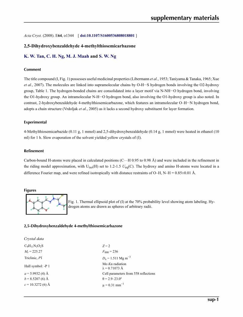
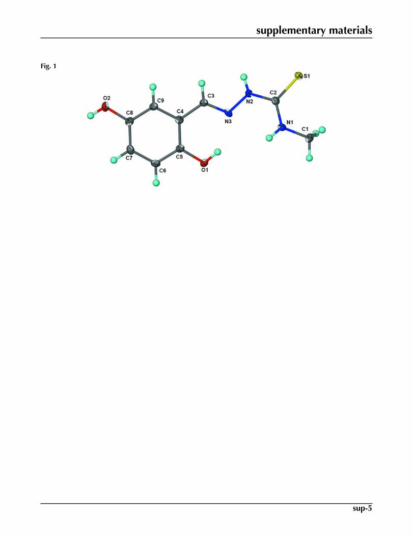
The title compound (I, Fig. 1) possesses useful medicinal properties (Libermann et al., 1953; Taniyama & Tanaka, 1965; Xueet al., 2007). The molecules are linked into supramolecular chains by O-H···S hydrogen bonds involving the O2-hydroxygroup, Table 1. The hydrogen-bonded chains are consolidated into a layer motif via N-NH···O hydrogen bond, involvingthe O1-hydroxy group. An intramolecular N-H···O hydrogen bond, also involving the O1-hydroxy group is also noted. Incontrast, 2-hydroxybenzaldehyde 4-methylthiosemicarbazone, which features an intramolecular O–H···N hydrogen bond,adopts a chain structure (Vrdoljak et al., 2005) as it lacks a second hydroxy substituent for layer formation.
Experimental
4-Methylthiosemicarbazide (0.11 g, 1 mmol) and 2,5-dihydroxybenzaldehyde (0.14 g, 1 mmol) were heated in ethanol (10ml) for 1 h. Slow evaporation of the solvent yielded yellow crystals of (I).
Refinement
Carbon-bound H-atoms were placed in calculated positions (C—H 0.95 to 0.98 Å) and were included in the refinement inthe riding model approximation, with Uiso(H) set to 1.2-1.5 Ueq(C). The hydroxy and amino H-atoms were located in a
difference Fourier map, and were refined isotropically with distance restraints of O–H, N–H = 0.85±0.01 Å.
Figures
Fig. 1. Thermal ellipsoid plot of (I) at the 70% probability level showing atom labeling. Hy-drogen atoms are drawn as spheres of arbitrary radii.
2,5-Dihydroxybenzaldehyde 4-methylthiosemicarbazone
Crystal data
C9H11N3O2S Z = 2Mr = 225.27 F000 = 236
Triclinic, P1 Dx = 1.511 Mg m−3
Hall symbol: -P 1 Mo Kα radiationλ = 0.71073 Å
a = 5.9932 (4) Å Cell parameters from 558 reflectionsb = 8.5207 (6) Å θ = 2.9–23.0ºc = 10.3272 (6) Å µ = 0.31 mm−1
supplementary materials
sup-2
α = 78.552 (4)º T = 100 (2) Kβ = 74.181 (4)º Plate, yellowγ = 81.743 (4)º 0.24 × 0.16 × 0.02 mm
V = 495.06 (6) Å3
Data collection
Bruker SMART APEXdiffractometer 2258 independent reflections
Radiation source: fine-focus sealed tube 1580 reflections with I > 2σ(I)Monochromator: graphite Rint = 0.050
T = 100(2) K θmax = 27.5º
ω scans θmin = 2.5ºAbsorption correction: Multi-scan(SADABS; Sheldrick, 1996) h = −7→7
Tmin = 0.929, Tmax = 0.994 k = −11→84189 measured reflections l = −13→13
Refinement
Refinement on F2 Secondary atom site location: difference Fourier map
Least-squares matrix: full Hydrogen site location: inferred from neighbouringsites
R[F2 > 2σ(F2)] = 0.054H atoms treated by a mixture ofindependent and constrained refinement
wR(F2) = 0.123 w = 1/[σ2(Fo
2) + (0.0471P)2]where P = (Fo
2 + 2Fc2)/3
S = 1.03 (Δ/σ)max = 0.001
2258 reflections Δρmax = 0.32 e Å−3
153 parameters Δρmin = −0.47 e Å−3
4 restraints Extinction correction: nonePrimary atom site location: structure-invariant directmethods
Fractional atomic coordinates and isotropic or equivalent isotropic displacement parameters (Å2)
x y z Uiso*/Ueq
S1 0.44298 (12) 0.47335 (9) 0.72966 (7) 0.0189 (2)O1 1.3132 (3) 0.8655 (2) 0.47568 (18) 0.0188 (4)O2 1.2446 (3) 1.1893 (2) −0.03406 (18) 0.0200 (5)N1 0.8637 (4) 0.5405 (3) 0.7312 (2) 0.0147 (5)N2 0.7415 (4) 0.6387 (3) 0.5359 (2) 0.0159 (5)N3 0.9289 (4) 0.7298 (3) 0.4833 (2) 0.0145 (5)C1 0.8391 (5) 0.4576 (3) 0.8709 (2) 0.0188 (6)H1A 0.9802 0.4633 0.8996 0.028*H1B 0.8160 0.3447 0.8763 0.028*H1C 0.7044 0.5088 0.9310 0.028*C2 0.7001 (4) 0.5552 (3) 0.6642 (2) 0.0134 (6)
supplementary materials
sup-3
C3 0.9368 (4) 0.8121 (3) 0.3637 (2) 0.0136 (6)H3 0.8213 0.8018 0.3194 0.016*C4 1.1147 (4) 0.9200 (3) 0.2937 (2) 0.0133 (6)C5 1.2915 (4) 0.9450 (3) 0.3503 (2) 0.0140 (6)C6 1.4505 (5) 1.0551 (3) 0.2796 (2) 0.0157 (6)H6 1.5673 1.0745 0.3193 0.019*C7 1.4389 (5) 1.1372 (3) 0.1504 (3) 0.0153 (6)H7 1.5496 1.2111 0.1016 0.018*C8 1.2669 (4) 1.1117 (3) 0.0929 (2) 0.0149 (6)C9 1.1055 (4) 1.0049 (3) 0.1638 (2) 0.0146 (6)H9 0.9866 0.9887 0.1243 0.018*H1o 1.206 (4) 0.805 (3) 0.511 (3) 0.048 (12)*H2o 1.352 (5) 1.245 (4) −0.082 (3) 0.059 (13)*H1n 0.988 (3) 0.581 (4) 0.689 (3) 0.032 (9)*H2n 0.640 (4) 0.656 (4) 0.491 (3) 0.042 (10)*
Atomic displacement parameters (Å2)
U11 U22 U33 U12 U13 U23
S1 0.0149 (4) 0.0241 (4) 0.0175 (3) −0.0083 (3) −0.0051 (3) 0.0033 (3)O1 0.0222 (11) 0.0221 (12) 0.0140 (9) −0.0081 (9) −0.0089 (8) 0.0025 (8)O2 0.0244 (11) 0.0202 (12) 0.0149 (9) −0.0088 (9) −0.0059 (8) 0.0047 (8)N1 0.0114 (12) 0.0181 (13) 0.0148 (11) −0.0035 (10) −0.0038 (9) −0.0007 (9)N2 0.0153 (12) 0.0168 (13) 0.0158 (11) −0.0060 (10) −0.0056 (9) 0.0024 (9)N3 0.0121 (11) 0.0136 (12) 0.0175 (11) −0.0050 (9) −0.0013 (9) −0.0023 (9)C1 0.0198 (15) 0.0231 (16) 0.0128 (12) −0.0048 (12) −0.0051 (11) 0.0015 (11)C2 0.0137 (13) 0.0106 (14) 0.0155 (12) −0.0011 (11) −0.0032 (10) −0.0022 (11)C3 0.0136 (13) 0.0138 (14) 0.0147 (12) −0.0019 (11) −0.0053 (10) −0.0025 (11)C4 0.0134 (13) 0.0118 (14) 0.0153 (12) −0.0008 (10) −0.0034 (10) −0.0038 (11)C5 0.0137 (13) 0.0154 (15) 0.0133 (12) 0.0001 (11) −0.0041 (10) −0.0034 (11)C6 0.0139 (13) 0.0162 (15) 0.0193 (13) −0.0029 (11) −0.0070 (10) −0.0039 (11)C7 0.0163 (14) 0.0102 (14) 0.0178 (13) −0.0021 (11) −0.0024 (10) −0.0006 (11)C8 0.0160 (14) 0.0144 (15) 0.0143 (12) 0.0006 (11) −0.0050 (10) −0.0023 (11)C9 0.0158 (14) 0.0156 (15) 0.0140 (12) −0.0021 (11) −0.0059 (10) −0.0031 (11)
Geometric parameters (Å, °)
S1—C2 1.695 (3) C1—H1B 0.9800O1—C5 1.367 (3) C1—H1C 0.9800O1—H1o 0.84 (3) C3—C4 1.450 (4)O2—C8 1.379 (3) C3—H3 0.9500O2—H2o 0.84 (3) C4—C5 1.401 (4)N1—C2 1.325 (3) C4—C9 1.403 (3)N1—C1 1.453 (3) C5—C6 1.388 (4)N1—H1n 0.84 (3) C6—C7 1.393 (3)N2—C2 1.349 (3) C6—H6 0.9500N2—N3 1.382 (3) C7—C8 1.383 (4)N2—H2n 0.84 (3) C7—H7 0.9500N3—C3 1.286 (3) C8—C9 1.379 (4)
supplementary materials
sup-4
C1—H1A 0.9800 C9—H9 0.9500
C5—O1—H1O 110 (2) C4—C3—H3 118.6C8—O2—H2O 117 (3) C5—C4—C9 118.9 (2)C2—N1—C1 123.8 (2) C5—C4—C3 123.2 (2)C2—N1—H1N 117 (2) C9—C4—C3 117.9 (2)C1—N1—H1N 119 (2) O1—C5—C6 117.7 (2)C2—N2—N3 121.3 (2) O1—C5—C4 122.3 (2)C2—N2—H2N 122 (2) C6—C5—C4 120.0 (2)N3—N2—H2N 116 (2) C5—C6—C7 120.1 (3)C3—N3—N2 114.5 (2) C5—C6—H6 120.0N1—C1—H1A 109.5 C7—C6—H6 120.0N1—C1—H1B 109.5 C8—C7—C6 120.3 (2)H1A—C1—H1B 109.5 C8—C7—H7 119.9N1—C1—H1C 109.5 C6—C7—H7 119.9H1A—C1—H1C 109.5 O2—C8—C9 116.7 (2)H1B—C1—H1C 109.5 O2—C8—C7 123.3 (2)N1—C2—N2 118.2 (2) C9—C8—C7 119.9 (2)N1—C2—S1 123.80 (19) C8—C9—C4 120.8 (2)N2—C2—S1 118.0 (2) C8—C9—H9 119.6N3—C3—C4 122.7 (2) C4—C9—H9 119.6N3—C3—H3 118.6
C2—N2—N3—C3 −175.1 (2) C3—C4—C5—C6 −177.4 (2)C1—N1—C2—N2 178.3 (2) O1—C5—C6—C7 178.8 (2)C1—N1—C2—S1 −2.5 (4) C4—C5—C6—C7 −2.0 (4)N3—N2—C2—N1 −11.0 (4) C5—C6—C7—C8 1.1 (4)N3—N2—C2—S1 169.69 (19) C6—C7—C8—O2 179.7 (2)N2—N3—C3—C4 177.5 (2) C6—C7—C8—C9 0.2 (4)N3—C3—C4—C5 −0.6 (4) O2—C8—C9—C4 179.8 (2)N3—C3—C4—C9 −179.5 (2) C7—C8—C9—C4 −0.7 (4)C9—C4—C5—O1 −179.3 (2) C5—C4—C9—C8 −0.2 (4)C3—C4—C5—O1 1.8 (4) C3—C4—C9—C8 178.8 (2)C9—C4—C5—C6 1.5 (4)
Hydrogen-bond geometry (Å, °)
D—H···A D—H H···A D···A D—H···AO1—H1o···N3 0.84 (3) 1.97 (2) 2.698 (3) 144 (3)
O2—H2o···S1i 0.84 (3) 2.46 (2) 3.182 (2) 144 (3)
N2—H2n···O1ii 0.84 (3) 2.47 (3) 3.111 (3) 134 (3)Symmetry codes: (i) x+1, y+1, z−1; (ii) x−1, y, z.
supplementary materials
sup-5
Fig. 1


![4* • Pharingeal grooves/cleft : 4 • [Pharyngeal membrane]](https://static.fdokumen.com/doc/165x107/6334ea00b9085e0bf5093ec7/4-pharingeal-groovescleft-4-pharyngeal-membrane.jpg)

