150 ECG Problems
316
Transcript of 150 ECG Problems


ouC
O
CD
oS
EC
ON
D E
DITIO
N
Administrator
Sherlock

Commissioning Editor: Laurence HunterProject Development Manager: Lynn Watt and HeliusProject Manager: Nancy ArnottDesigner: Erik Bigland and HeliusIllustrator: Helius and Chartwell IllustratorsIllustration Manager: Bruce Hogarth

150ECGPROBLEMSJohn R. HamptonEmeritus Professor of CardiologyUniversity of NottinghamNottinghamUK
DM MA DPhil FRCP FFPM FESC
CHURCHILLLIVINGSTONE
EDINBURGH LONDON NEW YORK OXFORD PHILADELPHIAST LOUIS SYDNEY TORONTO 2003

CHURCHILL LIVINGSTONE An imprint of Elsevier Science Limited
© Pearson Professional 1997©2003, Elsevier Science Limited. All rights reserved.
The right of Professor J. R. Hampton to be identified as author of this work has been asserted byhim in accordance with the Copyright, Designs and Patents Act 1988.
No part of this publication may be reproduced, stored in a retrieval system, or transmitted in anyform or by any means, electronic, mechanical, photocopying, recording or otherwise, without eitherthe prior permission of the publishers or a licence permitting restricted copying in the UnitedKingdom issued by the Copyright Licensing Agency, 90 Tottenham Court Road, London W1T 4LP.Permissions may be sought directly from Elsevier's Health Sciences Rights Department in Philadelphia,USA: phone: (+1) 215 238 7869, fax: (+1) 215 238 2239, e-mail: [email protected]).You may also complete your request on-line via the Elsevier Science homepage(http://www.elsevier.com), by selecting 'Customer Support' and then 'Obtaining Permissions'.
First edition 1997Second edition 2003
Reprinted 2003
Standard edition ISBN 0 443 072485International edition ISBN 0 443 072493
British Library Cataloguing in Publication DataA catalogue record for this book is available from the British Library
Library of Congress Cataloging in Publication DataA catalog record for this book is available from the Library of Congress
NoteMedical knowledge is constantly changing. Standard safety precautions must be followed, but asnew research and clinical experience broaden our knowledge, changes in treatment and drugtherapy may become necessary or appropriate. Readers are advised to check the most currentproduct information provided by the manufacturer of each drug to be administered to verify therecommended dose, the method and duration of administration, and contraindications. It is theresponsibility of the practitioner, relying on experience and knowledge of the patient, to determinedosages and the best treatment for each individual patient. Neither the Publisher nor the authorassumes any liability for any injury and/or damage to persons or property arising from thispublication.
your source for books,journals and multimediain the health sciences
www.elsevierhealth.com
ELSEVIERSCIENCE
Thepublisher's
policy is to usepaper manufactured
from sustainable forestsPrinted in ChinaP/02

Preface
Learning about ECG interpretation from books such as The ECGMade Easy or The ECG in Practice is fine so far as it goes, but itnever goes far enough. As with most of medicine, there is nosubstitute for experience, and to make the best use of the ECGthere is no substitute for reviewing large numbers of them. ECGsneed to be seen in the context of the patient from whom theywere recorded. You have to learn to appreciate the variations bothof normality and of the patterns associated with different diseases,and to think about how the ECG can help patient management.
Although no book can substitute for practical experience, 250ECG Problems goes a stage nearer the clinical world than books thatsimply aim to teach ECG interpretation. It presents 150 clinicalproblems in the shape of simple case histories, together with therelevant ECG. It invites the reader to interpret the ECG in the lightof the clinical evidence provided, and to decide on a course ofaction before looking at the answer. Having seen the answers, thereader may feel the need for more information, so each one is cross-referenced to The ECG Made Easy or The ECG in Practice.
The ECGs in 250 ECG Problems range from the simple to thecomplex. About one-third of the problems are of a standard that amedical student should be able to cope with, and will be answeredcorrectly by anyone who has read The ECG Made Easy. A houseofficer, specialist nurse or paramedic should get another third right,and will certainly be able to do so if they have read The ECG inPractice. The remainder should challenge the MRCP candidate.
As a very rough guide to the level of difficulty, each answer isgiven one, two or three stars (see the summary box of eachanswer): one star represents the easiest records, and three stars themost difficult.
The ECGs are arranged in random order, not in order ofdifficulty: this is to maintain interest and to challenge the reader toattempt an interpretation before looking at the star rating. This is,after all, the real-life situation: one never knows which patient willbe easy and which will be difficult to diagnose or treat.

150 ECG Problems is the successor to 100 ECG Problems, publishedin 1997. The popularity of the latter has encouraged me to includemore examples of common abnormalities and also some problemsfor which there was previously no space. I hope the reader will find250 ECG Problems an entertaining and an easy way to learn andrevise.
John R. HamptonNottingham
The symbols | ME I and | IP | denote cross-references to usefulinformation in the books The ECG Made Easy, 6th edn, and TheECG in Practice, 4th edn, respectively (written by ProfessorHampton and published by Elsevier Science).

ECG 1 This ECG was recorded from a 25-year-old pregnant woman who complained of an irregular heart beat. Auscultation revealed a soft systolic murmur but her heart was otherwise normal. What does the ECG show and what ^

ANSWER 1
The ECG shows:
• Sinus rhythm• Ventricular extrasystoles• Normal axis• Normal QRS complexes and T waves
Clinical interpretationThe extrasystoles are fairly frequent but the ECGis otherwise normal. Ventricular extrasystoles arevery common in pregnancy, and systolic murmursare almost universal. Her heart is almost certainlynormal.
What to doRemember that anaemia is a common cause of asystolic murmur. Doubts about the significance ofthe murmur can be resolved by echocardiography, Summarybut this need not be performed in every pregnant Sinus rhythm with ventricular extrasystoles.woman - it is best reserved for the investigation ofapparently important murmurs that persist afterdelivery. The patient should be reassured and theextrasystoles left untreated.

rnnECG 2 A 60-year-old man was seen as an out-patient, complaining of rather vague central chest pain on exertion. ®He had never had pain at rest. What does this ECG show and what would vou do next? I
m

ANSWER 2
The ECG shows:
• Sinus rhythm• Normal axis• Small Q waves in leads II, III, VF• Biphasic T waves in leads II, V6; inverted T
waves in leads III, VF• Markedly peaked T waves in leads V1 -V2
Clinical interpretationThe Q waves in the inferior leads, togetherwith inverted T waves, point to an old inferiormyocardial infarction. While symmetricallypeaked T waves in the anterior leads can bedue to hyperkalaemia, or to ischaemia, they arefrequently a normal variant.
What to doThe patient seems to have had a myocardialinfarction at some point in the past, and byimplication his vague chest pain may be dueto cardiac ischaemia. Attention must be paid torisk factors (smoking, blood pressure, plasma
cholesterol), and he probably needs long-termtreatment with aspirin and a statin. An exercisetest will be the best way of deciding whether hehas coronary disease that merits angiography.
SummaryOld inferior myocardial infarction.
E I Seep. 103
1 See p. 238
ANSWER 3
The ECG shows:
• Complete heart block• Ventricular rate 45/min
Clinical interpretationIn complete heart block there is no relationshipbetween the P waves (here with a rate of 70/min)and the QRS complexes. The ventricular 'escape'rhythm has wide QRS complexes and abnormal Twaves. No further interpretation of the ECG ispossible.
What to doIn the absence of a history suggesting a myocardialinfarction, this woman almost certainly has chronicheart block: the fall may or may not have beendue to a Stokes-Adams attack. She needs apermanent pacemaker, ideally immediately tosave the morbidity of first temporary, and thenpermanent, pacemaker insertion. If permanentpacing is not possible immediately, a temporarypacemaker will be needed preoperatively.
SummaryComplete (third degree) heart block.
IfJ See p. 33
lp~~] Seep. 213

EEH An 80-year-old woman, who had previously had a few attacks of dizziness, fell and broke her hip. Shfound to have a slow pulse, and this is her ECG. The surgeons want to operate as soon as possible but the anaestheis unhappy. What does the ECG show and what should be done?n

ANSWER 3
The ECG shows:
• Complete heart block• Ventricular rate 45/min
Clinical interpretationIn complete heart block there is no relationshipbetween the P waves (here with a rate of 70/min)and the QRS complexes. The ventricular 'escape'rhythm has wide QRS complexes and abnormal Twaves. No further interpretation of the ECG ispossible.
What to doIn the absence of a history suggesting a myocardialinfarction, this woman almost certainly has chronicheart block: the fall may or may not have beendue to a Stokes-Adams attack. She needs apermanent pacemaker, ideally immediately tosave the morbidity of first temporary, and thenpermanent, pacemaker insertion. If permanentpacing is not possible immediately, a temporarypacemaker will be needed preoperatively.
SummaryComplete (third degree) heart block.
IfJ See p. 33
lp~~] Seep. 213
• Ventricular rate 45/min
possible.
What to do

^3ZH A 50-year-old man is seen in the A & E department with severe central chest pain which has been present for18 h. What does this ECG show and what would you do?«JB

ANSWER 4
The ECG shows:
• Sinus rhythm• Normal axis• Q waves in leads V2-V4
• Raised ST segments in leads V2-V4
• Inverted T waves in leads I, VL, V2-V6
Clinical interpretationThis is a classic acute anterior myocardialinfarction.
What to doMore than 18 h have elapsed since the onset ofpain, so this patient is outside the conventionallimit for thrombolysis. Nevertheless, if he is still inpain and still looks unwell, thrombolytic treatmentshould be given unless there are good reasons notto do so. In any case he should be given pain reliefand aspirin, and must be admitted to hospital forobservation.
\
SummaryAcute anterior myocardial infarction.
IE | See p. 96
IP I Seep. 239

ECG 5 This ECG was recorded from a 60-year-old woman with rheumatic heart disease. She had been in heartfailure, but this had been treated and she was no longer breathless. What does the ECG show and what question mightyou ask her?

ANSWER 5
The ECG shows:
• Atrial fibrillation with a ventricular rate of60-65/min
• Normal axis• Normal QRS complexes• Prominent U wave in lead V2
• Downward-sloping ST segments, best seen inleads V5-V6
Clinical interpretationThe downward-sloping ST segments (the 'reversetick') indicate that digoxin has been given. Theventricular rate seems well-controlled. Theprominent U waves in lead V2 could indicatehypokalaemia.
What to doAsk the patient about her appetite: the earliestsymptom of digoxin toxicity is appetite loss,followed by nausea and vomiting. If the patientis being treated with diuretics, check the serumpotassium level - a low potassium level potentiates
the effects of digoxin. If in doubt, the serumdigoxin level is easily measured.
1
SummaryAtriat fibrillation with digoxin effect.
IE j See pp. 78 and 107
IP I See pp. 367 and 373

ECG 6 A 26-year-old woman, who has complained of palpitations in the past, is admitted via the A & E department^with palpitations. What does the ECG show and what should you do

ANSWER 6
The ECG shows:
• Narrow-complex tachycardia, rate about 200/min• No P waves visible• Normal axis• Regular QRS complexes• Normal QRS complexes, ST segments and T
waves
Clinical interpretationThis is a supraventricular tachycardia, and sinceno P waves are visible this is a junctional, oratrioventricular nodal, tachycardia.
What to doJunctional tachycardia is the commonest formof paroxysmal tachycardia in young people, andpresumably explains her previous episodes ofpalpitations. Attacks of junctional tachycardiamay be terminated by any of the manoeuvres thatlead to vagal stimulation - Valsalva's manoeuvre,carotid sinus pressure, or immersion of the face incold water. If these are unsuccessful, intravenous
adenosine should be given by bolus injection.Adenosine has a very short half-life, but can causeflushing and occasionally asthma. If adenosineproves unsuccessful, verapamil 5-10 mg given bybolus injection will usually restore sinus rhythm.Otherwise, DC cardioversion is indicated.
Summary *Junctional (atrioventricular nodal re-entry) tachycardia.
See p. 72
See p. 159

ECG 7 This ECG was recorded in the A & E department from a 55-year-old man who had had chest pain at rest for6 h. There were no abnormal physical findings. What does the trace show, and how would you manage him?
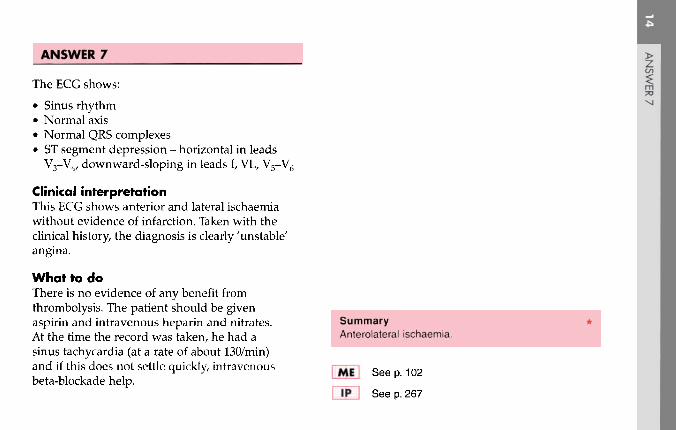
ANSWER 7
The ECG shows:
• Sinus rhythm• Normal axis• Normal QRS complexes• ST segment depression - horizontal in leads
V3-V4, downward-sloping in leads I, VL, V5-V6
Clinical interpretationThis ECG shows anterior and lateral ischaemiawithout evidence of infarction. Taken with theclinical history, the diagnosis is clearly 'unstable'angina.
What to doThere is no evidence of any benefit fromthrombolysis. The patient should be givenaspirin and intravenous heparin and nitrates. SummaryAt the time the record was taken, he had a Anterolateral ischaemia.sinus tachycardia (at a rate of about 130/min)and if this does not settle quickly, intravenous I "Mil See p 102beta-blockade help.
See p. 267

ECG 8 These three rhythm strips (all lead II) came from the ECGs of three different patients. They were all in theireighties, and all complained of breathlessness. What other symptoms might they have had, what diagnoses would youconsider, and what treatment is possible?

ANSWER 8
The ECGs show:
(a) No P waves can be seen but the baselineis irregular; the QRS complexes are broad,regular, and slow. This is atrial fibrillation withcomplete block.
(b) In the conducted beats the PR interval isconstant, so this is sinus rhythm with seconddegree (2:1) block. The second small deflectionafter the R wave is not a P wave, but is part ofthe QRS complex.
(c) There is no fixed relationship between theP waves and the QRS complexes, so this iscomplete (third degree) heart block.
Clinical interpretationSingle ECG leads can only be used to identify therhythm, and further interpretation is unreliable.
What to doAll the patients are probably suffering the effectsof their bradycardia; additional symptoms mightbe angina, dizziness, and collapse (Stokes-Adams
attacks). In each case the likely diagnosis isidiopathic fibrosis of the conducting system, butalmost all cardiac conditions can be associatedwith heart block - rheumatic disease, ischaemia,cardiomyopathy, trauma, metastases and so on.In the elderly, heart block is often associated witha calcified aortic valve. Whatever their age, suchpatients benefit from a permanent pacemaker.
Summary(a) Atrial fibrillation and complete block.(b) Second degree (2:1) block.(c) Complete (third degree) block.
«_] See p. 30
l|»~l See p. 199
Co

[j ^Q A 40-year-old woman is referred to the out-patient department because of increasing breathlessness. Whatdoes this ECG show, what physical signs might you expect, and what might be the underlying problem? What might youdo?

ANSWER 9
The ECG shows:
• Sinus rhythm• Peaked P waves, best seen in lead II• Right axis deviation• Dominant R waves in lead Vj• Deep S waves in lead V6
• Inverted T waves in leads II, III, VF, V1-V3
Clinical interpretationThis combination of right axis deviation,dominant R waves in lead Vl and inverted Twaves spreading from the right side of the heart,is classical of severe right ventricular hypertrophy.Right ventricular hypertrophy can result fromcongenital heart disease, or from pulmonaryhypertension secondary to mitral valve disease,lung disease, or pulmonary embolism. Thephysical signs of right hypertrophy are a leftparasternal heave and a displaced but diffuseapex beat. There may be a loud pulmonarysecond sound. The jugular venous pressure may
be elevated and a 'flicking A' wave in the jugularvenous pulse is characteristic.
What to doThe two main causes of pulmonary hypertensionof this degree in a 40-year-old woman arerecurrent pulmonary emboli, and primarypulmonary hypertension. Clinically, it is difficultto differentiate between the two, but a lung scanmay help. In either case anticoagulants areindicated. In fact, this patient had primarypulmonary hypertension and eventually neededheart and lung transplantation.
SummarySevere right ventricular hypertrophy.
See p. 91
See p. 336

ECG 10 This ECG was recorded from an 80-year-old man who complained of breathlessness and ankle swe
which had become slowly worse over the preceding few months. He had had no chest pain and was on no trn.ECG 10 This ECG was recorded from an 80-year-old man who complained of breathlessness and ankle swelling

ANSWER 10
The ECG shows:
• Atrial fibrillation with a ventricular rate of about40/min
• Left axis• Left bundle branch block
Clinical interpretationWhen an ECG shows left bundle branch block,no further interpretation is usually possible. Herethere is atrial fibrillation, and the ventricularresponse is very slow, suggesting that there isconduction delay in the His bundle as well as theleft bundle branch.
What to doIt is always important to establish the cause ofheart failure. In this patient the slow ventricularrate may be at least part of the problem. The mostimportant causes of left bundle branch block areischaemia, aortic stenosis and cardiomyopathy. Inthis patient an echocardiogram will show whetherhe has significant valve disease and how impaired
left ventricular function is. In the absence of pain,coronary angiography is probably not indicated.The heart failure needs to be treated with diureticsand an angiotensin-converting enzyme inhibitor,but digoxin must be avoided as it may slow theventricular response still further. He almostcertainly needs a permanent pacemaker.
SummaryAtrial fibrillation and left bundle branch block.
MM See pp. 36 and 78
See p. 209
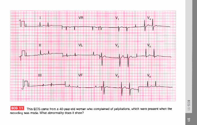
ECG 11 This ECG came from a 40-year-old woman who complained of palpitations, which were present when therecordingwas made. What abnormality does it show?—9

ANSWER 11
The ECG shows:
• Sinus rhythm• Atrial extrasystoles, identified by early beats
with broad and abnormal P waves (best seen inleads V2 and V3)
• Extrasystoles are followed by a 'compensatorypause'
• Normal axis• There is an RSR pattern in lead III, but the QRS
complex is narrow• The ST segments and T waves are normal
Clinical interpretationSince the patient had her symptoms at the timeof the recording, we can be confident that theECG findings explain her symptoms. Atrialextrasystoles, like junctional (atrioventricularnodal) extrasystoles, are not a manifestation ofcardiac disease.
What to doProvided there is nothing else in the history orexamination suggesting cardiac disease, the patientcan be assured that her heart is normal.
SummarySinus rhythm with atrial extrasystoles.
ME See p. 62
1F1 See p. 150

______^23^0 A 90-year-old woman is admitted to hospital after a fall resulting in a fractured hip. On questioning sheadmits to breathless and 'dizzy turns' for several months. This is her preoperative ECG. What does it show and whatwould you do?

ANSWER 12
The ECG shows:
• Second degree (2:1) heart block• Prolonged PR interval (440 ms) in the conducted
beats• Ventricular rate about 40/min• Normal QRS complexes and T waves
Clinical interpretationAlthough the slow ventricular response raises thepossibility of complete heart block, the fact thatthe PR interval is constant (albeit prolonged)shows that this is actually second degree block.The non-conducted P waves are not easy to see,but the clue lies in the abnormally shaped T wavesin the anterior leads. Second degree block explainswhy the QRS complexes are narrow and the Twaves are normal.
What to doSince this woman has been breathless and dizzyfor some time, and since there is nothing in thehistory or on the ECG to suggest an acute
infarction, it is unlikely that this conductiondisturbance is new. She therefore needs apermanent pacemaker: the only problem is todecide whether the urgent hip surgery should becovered with a temporary pacemaker - ideally shewould be saved that procedure and a permanentsystem implanted immediately.
>
?
SummarySecond degree (2:1) heart block.
IE | See p. 31
See p. 212

ECG 7 This ECG was recorded in the A & E department from a 55-year-old man who had had chest pain at rest for6 h. There were no abnormal physical findings. What does the trace show, and how would you manage him?

ANSWER 13
The ECG shows:
• Atrial flutter with 2:1 block (best seen in leads II,VR, VF)
• Normal axis• Normal QRS complexes and T waves
Clinical interpretationThe sudden onset of atrial flutter presumablyexplains the heart failure. There is nothing onthe ECG to suggest a cause for the arrhythmia.
What to doWhen an arrhythmia causes severe heart failure,immediate treatment is more important thanestablishing the underlying diagnosis. Carotidsinus pressure and adenosine may increasethe degree of block, but are unlikely to convertthe heart to sinus rhythm. It is worth tryingintravenous flecainide, but a patient with severelycompromised circulation is best promptly treatedwith DC cardioversion.
SummaryAtrial flutter with 2:1 block.
IE I See p. 68
1|> I Seep. 160

l jjcj j A 50-year-old man is admitted to hospital as an emergency, having had chest pain characteristic of amyocardial infarction for 4 h. Apart from the features associated with pain there are no abnormal physical findings. Whatdoes this ECG show and what would you do?

ANSWER 14
The ECG shows:
• Sinus rhythm• Normal axis• Small Q waves in lead III but not elsewhere• Elevated ST segments in leads II, III, VF, with
upright T waves• T wave inversion in lead VL• Suggestion of ST segment depression in leads
V2-V3
Clinical interpretationA classic ECG of an acute inferior myocardialinfarction, with lead VL indicating ischaemia. Therate of development of Q waves is very variable:compare this record with ECG 32, which camefrom a patient with a similar duration of symptoms.
What to doPain relief must take priority. In the absence ofcontraindications (i.e. risk of bleeding from anyimportant site), the patient should be given aspirinand then a thrombolytic agent.
SummaryAcute inferior myocardial infarction.
See p. 96
See p. 237

ECG 15 A 20-year-old student complains of palpitations. Attacks occur about once per year. They start suddenly,his heart feels very fast and regular, and he quickly feels breathless and faint. The attacks stop suddenly after a fewminutes. There are no abnormalities on examination, and this is his ECG. What would you do?

ANSWER 15
The ECG shows:
• Sinus rhythm• Right axis• Short PR interval (112ms)• QRS complexes a little wide (124 ms)• Slurred upstroke of QRS (delta wave)• Dominant R wave in lead Vj• Widespread T wave inversion
Clinical interpretationThis is a classical Wolff-Parkinson-Whitesyndrome. The resemblance to the ECG of rightventricular hypertrophy is because this is type A,with a left-sided accessory pathway. The ECGchanges of right axis, the dominant R wave inlead Vv and the T wave changes have no furthersignificance.
What to doThe patient gives a clear story of a paroxysmaltachycardia, and during attacks the circulation isclearly compromised because he feels dizzy. The
attacks are infrequent so there is little point inambulatory ECG recording. He needs immediatereferral to an electrophysiologist for ablation of theaberrant conducting pathway.
SummaryWolff-Parkinson-White syndrome type A.
If •) See p. 81
II* I See pp. 126 and 198

ECG 16 This ECG was recorded from a 75-year-old woman who complained of attacks of dizziness. It shows oneabnormality: what is its significance?

ANSWER 16
The ECG shows:
• Sinus rhythm• Prolonged PR interval of 280 ms (best seen in
leads V1, V2)• Normal axis• Normal QRS complexes• Normal ST segments and T waves
Clinical interpretationSinus rhythm with first degree block.
What to doFirst degree block does not cause anyhaemodynamic impairment, and by itself is oflittle significance. However, when a patient hassymptoms which might be due to a bradycardia(in this case dizziness), there may be episodesof second or third degree block, or possiblyStokes-Adams attacks, associated with a slowventricular rate. The appropriate action istherefore to request a 24 h ECG tape-recordinginvthe hope that the patient will have one of her
dizzy turns while wearing it. It would then bepossible to see whether or not the dizzinesswas associated with a change in heart rhythm.First degree block itself is not an indication forpermanent pacing or for any other intervention.
SummarySinus rhythm with first degree block.
See p. 30
See p. 137

ECG 17 This ECG was recorded in the A & E department from a 60-year-old man who had had severe central chestpain for 1 h. What does it show and what would you do?

ANSWER 17
The ECG shows:
• Sinus rhythm• One ventricular extrasystole• Normal axis• Q waves in leads V2-V3; small Q waves in leads
VL, V4
• Raised ST segments in leads I, VL, V3-V5
Clinical interpretationAcute anterolateral myocardial infarction isindicated. Although a Q wave is well developed inlead V3, the changes are entirely consistent withthe story of pain for 1 h.
What to doThis patient needs pain relief with diamorphine.The ECG shows raised ST segments of more than2 mm in several leads, so he needs immediatethrombolysis once any excess risk of bleedinghas been excluded. This treatment should not bedelayed by waiting for a chest X-ray or any otherinvestigations, and should be commenced in the
A & E department before transfer to the coronarycare unit. Ventricular extrasystoles do not needtreating.
SummaryAcute anterolateral myocardial infarction.
See p. 96
See p. 242
>CO
m70
XJ

ECG 18 A 70-year-old retired orthopaedic surgeon telephones to say that he always gets dizzy playing golf. You findthat he has a systolic heart murmur, and this is his ECG. What is the diagnosis and what do you do next?
tO

ANSWER 18
IThe ECG shows: m73
• Sinus rhythm, rate 48/min oo• Normal axis• QRS duration normal, but the R wave height in
lead V5 is 30 mm, and the S wave depth in leadV2 is 25 mm
• Inverted T waves in leads I, VL, V5-V6
Clinical interpretationThis is the classical ECG appearance of leftventricular hypertrophy.
What to doThe combination of dizziness on exercise, asystolic murmur, and evidence of left ventricularhypertrophy suggests significant aortic stenosis.The next step is an echocardiogram: in this patient Summary *it showed a gradient across the aortic valve of Left ventricular hypertrophy.140 mmHg, indicating severe stenosis. He neededan urgent aortic valve replacement. fMH See D 93
I IP | See p. 117

A 75-year-old woman complaining of central chest discomfort on climbing hills, together with dizziness; onone occasion she had 'fainted' while climbing stairs. What abnormality does this ECG show and what physical signswould you look for?
oO
ANSWER 19
The ECG shows:
• Sinus rhythm• Broad QRS complexes (140 ms)• 'M' pattern in lead V6
• Inverted T waves in leads I, VL, V6
Clinical interpretationThis is a characteristic pattern of left bundlebranch block. The ECG cannot be interpretedfurther.
What to doA patient who has chest pain that could be angina,and who has dizziness and syncope on exertion,probably has severe aortic stenosis and this wasthe case with this woman. Clinically she had aslow rising pulse, a blood pressure of 100/80, and aslightly enlarged heart. There was a loud ejectionsystolic murmur, best heard at the upper rightsternal edge and radiating to both carotids. Thediagnosis was confirmed by an echocardiogram,which showed a gradient across the aortic valve of
about 100 mmHg. A cardiac catheter was necessaryto exclude coronary disease and she then had anaortic valve replacement and made a completerecovery.
SummarySinus rhythm with left bundle branch block.
'ME I See p. 39
[ IP | Seep. 117

ECG 20 A 70-year-old man is seen in the clinic because of breathlessness, which began over a few days 3 monthsago. This is his ECG: what does it show and what treatment is needed?
nO

ANSWER 20
The ECG shows:
• Sinus rhythm• Second degree (2:1) heart block (most obvious in
lead V3)• Ventricular rate 30/min• Normal PR interval in the conducted beats• Normal axis• QRS duration prolonged (160 ms)• RSR pattern in leads V1-V3and a wide S wave
in lead V6
• Prominent U wave in leads V3-V6
Clinical interpretationThis patient has second degree block and rightbundle branch block, so he clearly has extensiveconduction tissue disease.
What to doThe slow heart rate is probably the cause ofhis heart failure, and he needs a permanentpacemaker. The story suggests that the onset ofheart failure was not associated with chest pain,
so the underlying disease is probably fibrosis ofthe conducting system rather than ischaemia. Heneeds an echocardiogram and treatment with anangiotensin-converting enzyme inhibitor if there isevidence of left ventricular dysfunction.
Summary **Second degree atrioventricular block and right bundlebranch block.
See pp. 31 and 37
See p. 140

ooK>
This ECG was recorded from a medical student during a practical class. What does it show?

ANSWER 21
The ECG shows:
• Sinus rhythm• Sinus arrhythmia• Normal axis• Normal QRS complexes• Normal ST segments and T waves
Clinical interpretationThis is a perfectly normal ECG. There is a beat-to-beat variation in the interval between QRScomplexes, with the heart rate speeding up andslowing down. Comparison of the rate recorded inleads V1, V2 and V3 with that recorded in leads V4,V5 and V6 may give a false impression of a changeof rhythm. This variation in heart rate relates torespiration and is called sinus arrhythmia, which isnormal in young people. Sinus arrhythmia can bedistinguished from atrial extrasystoles because insinus arrhythmia the morphology of the P wavesis unchanged.
What to doNothing! Z
C/)
SummaryNormal ECG with sinus arrhythmia.

This ECG was recorded from a 48-year-old man who had had severe central chest pain for 1 h. What doesit show and what would you do?

ANSWER 22
The ECG shows:
• Sinus rhythm• Normal axis• Normal QRS complexes• Biphasic T waves in leads V2, V3, V5
• Inverted T waves in lead V4
Clinical interpretationThis is a classic acute anterior non-Q waveinfarction.
What to doThis ECG does not meet the conventional criteriafor thrombolysis, which are raised ST segmentsor new left bundle branch block. The immediateoutlook is good but the patient should bemonitored and the ECG repeated after an hourto see if ST segment elevation is appearing.
Summary *Acute anterior non-Q wave myocardial infarction.
See p. 103
See p. 266

Tnis ECG was recorded from a 70-year-old man who had had angina for some time and was treated with abeta-blocker. He came to the A & E department complaining of pain similar to his angina, but much more severe andpersistent for 4 h. What does the ECG show and what treatment would be appropriate?
m
8K>CJ

ANSWER 23
The ECG shows (note: leads at half sensitivity):
• Sinus rhythm• Supraventricular (junctional) extrasystoles• Normal axis• Broad QRS complexes (140 ms)• 'M' pattern of QRS complex in leads V4-V6
• Inverted T waves in leads I, VL, V4-V6
Clinical interpretationThis ECG shows sinus rhythm withsupraventricular extrasystoles and left bundlebranch block (LBBB). No further interpretation ispossible.
What to doIf a patient has symptoms suggestive of amyocardial infarction of less than 6 h duration buthas LBBB on the ECG, thrombolysis should begiven only if the bundle branch block is known tobe new. Here the patient had a history of anginaso the first thing to do is to relieve his pain and thesecond is to find his old notes and see if the LBBB
had been noted previously. If no old ECGs areavailable, thrombolysis should not be given, andthe patient should be treated as an acute coronarysyndrome. The supraventricular extrasystoles arenot important.
SummaryLeft bundle branch block; supraventricularextrasystoles.
If7! See pp. 36 and 62
See p. 259
Iin
TONJCO

^nis ECG was recorded from a 60-year-old man being treated as an out-patient for severe congestivecardiac failure. What might be the diagnosis of the underlying heart condition and what would you do?

ANSWER 24
The ECG shows:
• Atrial fibrillation• Ventricular rate 75-200/min• Normal axis• Normal QRS complexes• Downward-sloping ST segment depression,
especially in leads V5, V6
Clinical interpretationThe ventricular rate is not adequately controlled,though the ST segment depression suggests thathe is taking digoxin. There are no changes tosuggest ischaemia.
What to doIn the absence of clinical or ECG evidence ofischaemia, possible diagnoses include rheumaticheart disease, thyrotoxicosis, alcoholic heartdisease, and other forms of cardiomyopathy.Echocardiography is necessary. The serum digoxinlevel must be checked and the digoxin doseincreased if appropriate. In addition to digoxin,
the patient will need an angiotensin-convertingenzyme inhibitor, a diuretic and, probably,anticoagulants. Beta-blockers must be consideredonce his cardiac failure is controlled.
Summary **Atrial fibrillation with an uncontrolled ventricular rate,and digoxin effect.
fif] See pp. 78 and 107
IP I See p. 315
1rn73
ro^

A 60-year-old man, who 3 years earlier had had a myocardial infarction followed by mild angina, wasadmitted to hospital with central chest pain that had been present for 1 h and had not responded to sublingual nitrates.What does his ECG show, and what would you do?

ANSWER 25
The ECG shows:
• Sinus rhythm• Normal axis• Q waves in leads II, III, VF• Normal QRS complexes in the anterior leads• Marked ST segment elevation in leads V1-V6
Clinical interpretationThe Q waves in leads III and VF suggest an oldinferior infarction, while the elevated ST segmentsin leads V1-V6 indicate an acute anterior infarction.
What to doThe patient should be given pain relief, and inthe absence of the usual contraindicationsshould immediately be treated with aspirin anda thrombolytic agent. If he was treated withstreptokinase for his previous infarction, heshould be given alteplase or reteplase on thisoccasion.
z ]3mTO IM icn I
Summary **Old inferior and acute anterior myocardial infarctions. :
See p. 96
See p. 254

A 15-year-old boy was referred to the out-patient department because of a heart murmur. He had nosymptoms. What does this ECG show and what physical signs would you look for?

ANSWER 26
The ECG shows:
• Sinus rhythm• Normal axis• Broad QRS complexes (140 ms)• RSR pattern in lead I• Wide and slurred S waves in lead V5
• Normal ST segments and T waves
Clinical interpretationRight bundle branch block.
What to doRight bundle branch block is seen in a smallproportion of people with perfectly normal hearts.In the presence of a heart murmur, however, thepossibility of an atrial septal defect should beconsidered. This is what this patient had. Thephysical signs were a widely-split pulmonarysecond sound which did not vary with inspiration(this is typical of right bundle branch block) andan ejection systolic murmur best heard at the leftsternal edge. On deep inspiration a soft diastolic
murmur could be heard at the lower left sternaledge. The systolic murmur is a pulmonary flowmurmur due to the extra flow through the rightside of the heart, and the diastolic murmurthat occurs on inspiration is a tricuspid flowmurmur. The diagnosis was confirmed byechocardiography, and the defect was closedwith a percutaneous 'umbrella' device. Followingoperation, the right bundle branch block persisted.
SummarySinus rhythm with right bundle branch block.
See p. 37
Jp~] See pp. 103 and 352
O

This ECG was recorded from a 40-year-old man who complained of breathlessness on climbing stairs. Hewas not aware of a fast heart rate and had had no chest pain. Apart from a rapid rate there were no cardiovascularabnormalities, but he looked a little jaundiced and had an enlarged spleen. What would you do?

ANSWER 27
The ECG shows:
• Atrial flutter• Ventricular rate 140/min• Left axis• Normal QRS complexes, except that there is an S
wave in lead V6
Clinical interpretationThis ECG shows atrial flutter with 2:1 block. Theleft axis may be due to left anterior hemiblock,although the QRS has a normal duration so thesignificance of the axis is uncertain. The persistentS wave in lead V6 suggests chronic lung disease.
What to doProvided the patient is not in heart failure it isalways a good idea to identify the cause of anarrhythmia before treating it. The combinationof an atrial arrhythmia, jaundice andsplenomegaly suggests alcoholism. The patientneeds anticoagulants, but his internationalnormalized ratio (INR) may already be high.
An echocardiogram is needed to assess leftventricular function, and digoxin could be givenin an attempt to control the ventricular rate. Afteranticoagulation, cardioversion, either electrical orwith flecainide, will be necessary.
SummaryAtrial flutter with 2:1 conduction.
See p. 68
Ij> [ See p. 160

ECG 28 This EGG was recorded from a 39-year-old woman who complained of a sudden onset of breathlessness.She had no previous history, and no chest pain. Examination reveals nothing other than a rapid heart rate. What is thediagnosis?

ANSWER 28
The ECG shows:
• Sinus rhythm, rate 140/min• Normal conduction• Normal axis• Normal QRS complexes• Slightly depressed ST segments in leads V1-V4
• Diphasic or inverted T waves in the inferior andall the chest leads
Clinical interpretationThe ECG shows a marked sinus tachycardia, withno change in the cardiac axis and normal QRScomplexes. The widespread ST/T changes areclearly very abnormal, but are not specific for anyparticular disease. However, the fact that leadsVj-Vs are affected suggests a right ventricularproblem.
What to doThis is a case where the ECG must be consideredin the light of the patient's history and physicalsigns (if any). Clearly something has happened;
the sudden onset of breathlessness without painsuggests a pulmonary embolus, and here the VQscan confirmed multiple small pulmonary infarcts.
Summary **Sinus tachycardia with widespread ST/T changessuggesting pulmonary embolism.
See p. 92
See p. 289
11soo

|££££i This ECG was recorded from a 50-year-old man who was admitted to hospital as an emergency, having hadchest pain characteristic of a myocardial infarction for 3 h. What does the ECG show and how should the patient betreated?
ANSWER 29
The ECG shows:
• Sinus rhythm• PR intervals markedly prolonged (480 ms)• Normal axis• Normal QRS complexes• T wave inversion in leads V1-V3
Clinical interpretationFirst degree block associated with a non-Q waveanterior myocardial infarction. Since the Twave inversion is in leads V1-V3 but not V4 thepossibility of a pulmonary embolus must beconsidered.
What to doThe changes on the ECG do not meet theconventional criteria for thrombolysis for Summary **myocardial infarction (raised ST segments or First de9ree block and anterior non-Q-wave infarction.new left bundle branch block). First degree blockis not an indication for temporary pacing, but r^| See pp 3Q and 1Q3
the patient must be monitored in case higherdegrees of block develop. I IP I See p. 266
'11sm•73K)O

pScyiii A 65-year-old man is seen in the out-patient department complaining of breathlessness and chest pain thathas the characteristics of angina. He is untreated. Does his ECG help with his diagnosis and management?

ANSWER 30
The ECG shows:
• Atrial fibrillation• Ventricular rate 50-70/min• Normal axis• Poor R wave progression (loss of R wave in lead
V3, with a normal R wave in lead V4)• Normal ST segments and T waves
Clinical interpretationNormal ventricular rate, despite untreated atrialfibrillation. The poor R wave progression betweenleads V3 and V4 could result from inaccuratepositioning of the chest leads, but may indicate anold anterior myocardial infarction.
What to doCauses of atrial fibrillation other than ischaemiamust be excluded. An exercise test will revealwhether or not his pain is angina, and will alsoshow whether the ventricular rate remainscontrolled or whether it increases inappropriately.
Summary **Atrial fibrillation and possible old anterior myocardialinfarction.
See pp. 78 and 103
See pp. 243 and 251

This EGG was recorded in a coronary care unit from a patient admitted 2 h previously with an acute anteriormyocardial infarction. The patient was cold and clammy, and confused, and his blood pressure was unrecordable. Whatdoes the ECG show and what would you do?
nO

ANSWER 31
The ECG shows:
• Broad-complex tachycardia, rate about 250/min• Regular QRS complexes• QRS duration 200 ms• Indeterminate axis and QRS configurations
Clinical interpretationIn the context of acute myocardial infarction,broad-complex tachycardias should be consideredto be ventricular in origin unless the patient isknown to have bundle branch block when in sinusrhythm. Here the regularity of the rhythm and thevery broad complexes of bizarre configurationleave no room for doubt that this is ventriculartachycardia.
What to do Summary ***In cases of severe circulatory failure, immediate Ventricular tachycardia.DC cardioversion is needed.
E | See p. 72
See p. 178
23rn3
CO

ECG 32 A 50-year-old man is admitted to hospital as an emergency, having had chest pain for 4 h. The pain ischaracteristic of a myocardial infarction. Apart from signs due to pain, the examination is normal. What does this ECGshow and what would you do?

ANSWER 32
The ECG shows:
• Sinus rhythm• Normal axis• Q waves in leads II, III, VF• Elevated ST segments in leads II, III, VF with
biphasic T waves• Downward-sloping ST segments in lead VL• Normal QRS complexes, ST segments and T
waves in the chest leads
Clinical interpretationThis is an acute inferior myocardial infarction.The rapidity of Q wave development is extremelyvariable, but the trace is certainly consistent with a4 h history.
What to do SummaryPain relief is the most important part of the Acute inferior myocardial infarction.treatment. In the absence of contraindications,the patient should be given aspirin immediately, |~SH See D 96and then thrombolysis as soon as possible.
' See p. 242
>O1
rn70GOIS)

I Jejcjcj This EGG was recorded from a 35-year-old man who had no symptoms, but who had been found at aroutine examination to have a blood pressure of 180/105. What does it show and what action would you suggest?
nO

ANSWER 33
The ECG shows (note: leads at half sensitivity(0.5 cm = 1 mV)):
• Sinus rhythm, rate 50/min• Very short PR interval• Normal axis• Slurred upstroke to QRS complexes - delta wave• QRS duration prolonged (200 ms)• Very tall QRS complexes in the lateral leads• Inverted T waves in leads I, VL, III, VF, V5-V6
Clinical interpretationThis is an example of the Wolff-Parkinson-Whitesyndrome type B. In a patient with high bloodpressure the tall QRS complexes and invertedT waves in the lateral leads would raise thepossibility of left ventricular hypertrophy, butthe changes here are too gross for that, and theyare compatible with this pre-excitation syndrome.
What to doIf the patient has no symptoms to suggest aparoxysmal tachycardia, no further action isnecessary - many patients with pre-excitation ontheir ECG never have an episode of tachycardia.
SummaryWolff-Parkinson-White syndrome type B.
See p. 81
See pp. 38 and 104

^2323 An 80-year-old man being observed in the recovery room following a femoral-popliteal bypass operationwas noted to have an abnormal ECG. What does it show and what would you do?

ANSWER 34
The ECG shows:
• Sinus rhythm• Normal axis• Normal QRS complexes• Marked (about 8 mm) horizontal ST segment
depression in leads V2-V4, and downward-sloping ST segment depression in the lateralleads
Clinical interpretationThe patient is elderly and has peripheral vasculardisease, so coronary disease is likely to be present.The appearance of the ECG is characteristic ofsevere cardiac ischaemia. The lack of a tachycardiais surprising.
What to do Summary **This is not an easy situation to deal with because Severe anterolateral ischaemia.the patient's postoperative condition dictatesmanagement. He needs anticoagulation with |~MH See D 102aspirin and heparin, and intravenous nitratesshould be given cautiously. \ W I See p. 267
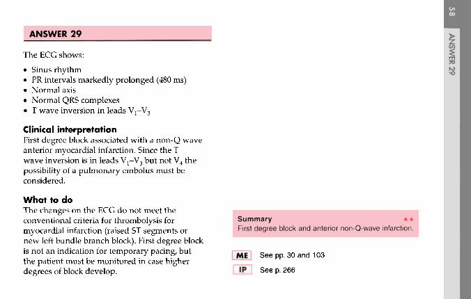
ECG 35 This ECG was recorded from a 75-year-old man who complained of breathlessness. He had not had anychest pain or dizziness. Apart from a slow pulse there were no abnormalities on examination. What three abnormalitiesare present in this record and how would you treat the patient?

ANSWER 35
The ECG shows:
• Sinus rhythm• Second degree (2:1) block• Left axis deviation• Poor R wave progression in the anterior leads• Normal T waves
Clinical interpretationThe second degree block is associated with aventricular rate of 45/min, which may well be thecause of his breathlessness. The left axis deviationindicates left anterior hemiblock. The poor R waveprogression (virtually no R wave in lead V3, asmall R wave in lead V4, and a normal R wavein lead V5) suggests an old anterior infarction.
What to doThis patient needs a permanent pacemaker.
Summary ***Second degree (2:1) block, left anterior hemiblock,and probable old anterior infarction.
IE | See pp. 31 and 46
See p. 140
:m73COOl
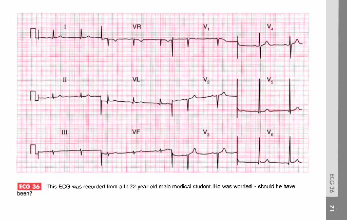
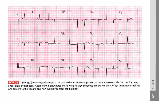
| JJ3 This ECG was recorded from a fit 22-year-old male medical student. He was worried - should he havebeen?

ANSWER 36
The ECG shows:
• Sinus rhythm• Normal axis• Tall R waves (28 mm in lead V6, 32 mm in lead V5)• Loss of R waves in lead V3
• Normal ST segments and T waves
Clinical interpretationThis record shows left ventricular hypertrophy byVoltage criteria' (R waves greater than 25 mm inlead V5 or V6, or the sum of the R wave in lead V5
or V6 plus the S wave in lead V1 or V2 is greaterthan 35 mm). There are, however, no T wavechanges. 'Voltage criteria' on their own areunreliable, and in a fit young man this may wellbe a normal variant. A loss of R waves in lead V3
could indicate an old anterior infarction, but this isextremely unlikely in a young man and it probablyresults from faulty positioning of lead V3.
What to doTell the student to buy a good book on ECGinterpretation, but if reassurance is not enough,echocardiography could be used to measure leftventricular thickness.
Summary **Left ventricular hypertrophy on 'voltage criteria', butprobably normal.
(EJ See p. 93
lp~| See p. 68
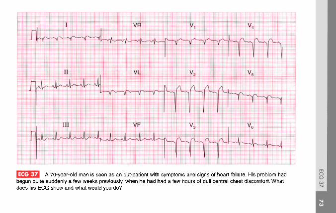
I JJJ A 70-year-old man is seen as an out-patient with symptoms and signs of heart failure. His problem hadbegun quite suddenly a few weeks previously, when he had had a few hours of dull central chest discomfort. Whatdoes his ECG show and what would you do?

ANSWER 37
The ECG shows:
• Sinus rhythm, rate 100/min• Normal axis• Q waves in leads I, VL, V2-V5
• Raised ST segments in leads I, VL, V2-V6
• T wave inversion in lead V6
Clinical interpretationThe raised ST segments suggest an acuteinfarction, but the deep Q waves suggest thatthe infarction occurred at least several hourspreviously. From the patient's story it seems clearthat he had an infarction several weeks before hewas seen, and there was nothing in the history tosuggest a more recent episode. These ECG changesare therefore probably all old; the anterior changesmight indicate a left ventricular aneurysm.
What to doAn ECG should always be interpreted in the lightof the patient's clinical state. Since the ECG iscompatible with an old infarction it should be
assumed that this is the case, and the patientshould be treated for heart failure in the usual waywith diuretics, angiotensin-converting enzymeinhibitors and beta-blockers. Since the heart failureis clearly due to ischaemia he also needs aspirinand a statin.
Summary *Anterolateral myocardial infarction of uncertain age.
See p. 103
See p. 243
f>
a

ECG 38 A 60-year-old man was referred to the out-patient department because of exercise-induced chest pain, andhis GP had recorded this ECG. What does it show and what physical signs would you look for?

ANSWER 38
The ECG shows:
• Sinus rhythm• Normal axis• Normal QRS complexes• Slight ST segment depression in leads I, II, VL• 2-3 mm flat or downward-sloping ST segment
depression in leads V4-V6
Clinical interpretationThe ST segment changes in leads I, II and VLare non-specific, but those in leads V4-V5 areundoubtedly due to ischaemia because thedepression is horizontal and more than 2 mm.The downward-sloping ST segment in lead V6 isalso probably due to ischaemia, but could be dueto digoxin.
What to doThere are no physical signs of angina, but theremay be signs of pain (pallor, sinus tachycardia),heart failure (including a gallop rhythm at thecardiac apex), or there may be evidence of
hypertension, hypercholesterolaemia, or smoking.There may be absent pulses or bruits over aperipheral artery, suggesting peripheral vasculardisease. An exercise test would probablyaccentuate the ischaemic changes, but is notnecessary for diagnostic purposes.
SummaryST segment depression due to ischaemia.
IE | See p. 102
tf» | See pp. 267-75

| ^23 A 65-year-old man, who had had a myocardial infarction 3 years previously, presents with 2 h of chest painthat sounds ischaemic. By the time he was seen his pain had settled. What does his ECG show, what do you think hashappened, and how should he be treated?

ANSWER 39
The ECG shows:
• Sinus rhythm• Second degree block (Mobitz type 2 - best seen
in leads I and II)• Ventricular rate 50/min• Normal PR interval in the conducted beats• Left axis deviation• Broad QRS complexes (160 ms)• No R waves in anterior chest leads• Deep S wave in lead V6
Clinical interpretationThe combination of Mobitz type 2 block andleft interior hemiblock (shown by the left axis)indicates severe conduction tissue disease. The lossof R waves in the chest leads may be due to an oldanterior infarction, but the deep S wave in lead V6
may indicate an intraventricular conduction delay.
What to doThe recent episode of chest pain may have beendue to a further myocardial infarction, or may
have been associated with bradycardia due tocomplete heart block. If repeat ECGs and bloodmarkers suggest there has no infarction, thena permanent pacemaker is needed; if thereis evidence of a new infarction it would bereasonable to monitor the patient closely andsee if the heart block improves.
Summary **Mobitz type 2 (second degree) block and left anteriorhemiblock; probable old anterior infarction.
tt] See pp. 31 and 46
lr*~] Seep. 140

EG3EJ A 30-year-old woman, who had a normal pregnancy and delivery 3 months ago, complains ofbreathlessness but has no other symptoms. She has a soft systolic murmur, and this is her ECG. What does it show andwhat would you do?
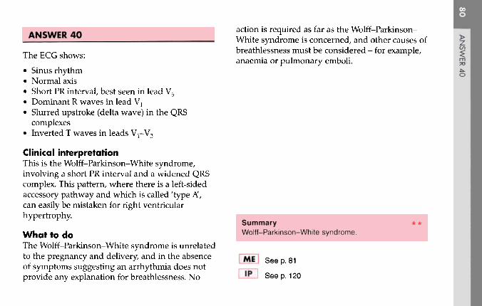
ANSWER 40
The ECG shows:
• Sinus rhythm• Normal axis• Short PR interval, best seen in lead V5
• Dominant R waves in lead V1
• Slurred upstroke (delta wave) in the QRScomplexes
• Inverted T waves in leads V1-V3
Clinical interpretationThis is the Wolff-Parkinson-White syndrome,involving a short PR interval and a widened QRScomplex. This pattern, where there is a left-sidedaccessory pathway and which is called 'type A',can easily be mistaken for right ventricularhypertrophy.
What to doThe Wolff-Parkinson-White syndrome is unrelatedto the pregnancy and delivery, and in the absenceof symptoms suggesting an arrhythmia does notprovide any explanation for breathlessness. No
action is required as far as the Wolff-Parkinson-White syndrome is concerned, and other causes ofbreathlessness must be considered - for example,anaemia or pulmonary emboli.
SummaryWolff-Parkinson-White syndrome.
fiT| See p. 81
Seep. 120

[32HI A 70-year-old woman, from whom this ECG was recorded, was admitted to hospital with increasingcongestive cardiac failure. What does the ECG show and what would you do?

ANSWER 41
The ECG shows:
• Atrial fibrillation• Normal axis• Normal QRS complexes• Downward-sloping ST segment depression in
lead V6
Clinical interpretationThe rhythm could be interpreted as atrial flutter,particularly in leads II and VT. However, theflutter-like activity is variable, and the QRScomplexes are completely irregular. The old-fashioned term for this was 'flutter fibrillation'.The ST segment depression suggests digoxineffect.
What to do'Flutter fibrillation' has the characteristics of atrialfibrillation, and it is better simply to use this latterterm. The ventricular rate in this case is fairlyrapid, suggesting that the patient may not havebeen given adequate digoxin. It would be prudent
to check her digoxin level before increasingthe dose. The ventricular rate may well slowdown after treatment for heart failure with anangiotensin-converting enzyme inhibitor anda diuretic. Some form of anticoagulation isnecessary. The thyroid function tests should bechecked.
SummaryAtrial fibrillation and digoxin effect.
See pp. 78 and 107
See p. 170

UEE3 A 50 year old man, who had had exertional chest pain for some months, was seen in the A & E departmentwith an hour of persistent central chest pain, and this is his ECG. What does the ECG show and what would you do?
m
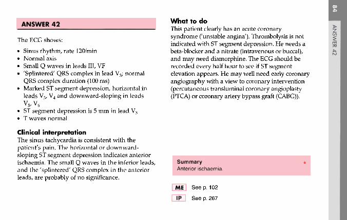
ANSWER 42
The ECG shows:
• Sinus rhythm, rate 120/min• Normal axis• Small Q waves in leads III, VF• 'Splintered' QRS complex in lead V3; normal
QRS complex duration (100 ms)• Marked ST segment depression, horizontal in
leads V3, V4 and downward-sloping in leadsV5,V6
• ST segment depression is 5 mm in lead V3
• T waves normal
Clinical interpretationThe sinus tachycardia is consistent with thepatient's pain. The horizontal or downward-sloping ST segment depression indicates anteriorischaemia. The small Q waves in the inferior leads,and the 'splintered' QRS complex in the anteriorleads, are probably of no significance.
What to doThis patient clearly has an acute coronarysyndrome ('unstable angina'). Thrombolysis is notindicated with ST segment depression. He needs abeta-blocker and a nitrate (intravenous or buccal),and may need diamorphine. The ECG should berecorded every half hour to see if ST segmentelevation appears. He may well need early coronaryangiography with a view to coronary intervention(percutaneous transluminal coronary angioplasty(PTCA) or coronary artery bypass graft (CABG)).
SummaryAnterior ischaemia.
I Seep. 102
See p. 267
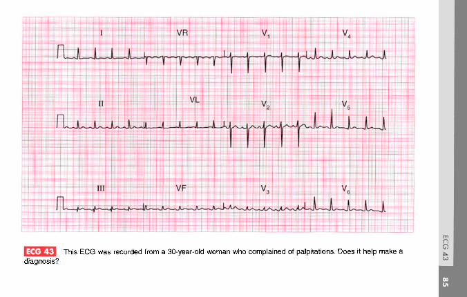
ECG 43 This ECG was recorded from a 30-year-old woman who complained of palpitations. Does it help make adiagnosis?

ANSWER 43
The ECG shows:
• Sinus rhythm, rate 110/min• Normal axis• Small Q waves in lead III• Otherwise, normal QRS complexes and T waves
Clinical interpretationSmall Q waves in lead III but not in lead VF arenormal. In establishing the cause of palpitationsthe history and examination are vital, and theECG is not often helpful unless it is recorded whenthe patient has symptoms. A persistent sinustachycardia, as shown here, may be due to anxiety,thyrotoxicosis, acute blood loss, anaemia, or heartfailure. This patient had thyrotoxicosis.
What to do Summary **Treat the underlying cause of the sinus tachycardia. Sinus tachycardia.
See p. 54
Seep. 152
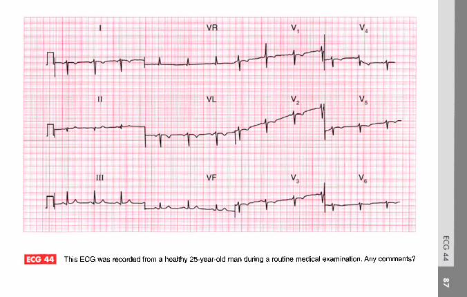
ECG 44 This ECG was recorded from a healthy 25-year-old man during a routine medical examination. Any comments?

ANSWER 44
The ECG shows:
• A very odd appearance• Sinus rhythm• Inverted P waves in lead I• Right axis deviation• Normal width QRS complexes• Dominant R waves in lead VR• No R wave development in the chest leads, with
lead V6 still showing a right ventricular pattern
Clinical interpretationThis is dextrocardia. A normal trace would beobtained with the limb leads reversed and thechest leads attached in the usual rib spaces but onthe right side of the chest.
What to doEnsure that the leads were properly attached - forexample inverted P waves in lead I will be seen ifthe right and left arm attachments are reversed. Ofcourse this would not affect the appearance of theECG in the chest leads.
SummaryDextrocardia. ***
See pp. 57 and 60

ECG 45 An 80-year-old woman, who has apparently been treated for heart failure for years, complains of nauseaand vomiting. No previous records are available. Does her ECG help her management?

ANSWER 45
The ECG shows:
• Atrial fibrillation, ventricular rate 80/min• Normal axis• Normal QRS complexes• Downward-sloping ST segment depression,
especially in leads V4-V6
• T waves probably upright
Clinical interpretationThe ECG shows atrial fibrillation with a controlledventricular rate. There is nothing on the ECG tosuggest a cause for the arrhythmia or the patient'sheart failure. The 'reversed tick' ST segmentdepression suggests that she is being treatedwith digoxin. The ECG does not suggest digoxintoxicity, but nevertheless this is the most likelycause for her nausea. Summary
Atrial fibrillation and the digoxin effect.
What to doDigoxin therapy should be temporarily gg See pp ?8 and 1 Qy
discontinued, and her plasma potassium anddigoxin levels should be checked. i IP I See p. 372

E^UJ A 60-year-old man, whose heart and preoperative EGG had been normal, developed a cough with pleuriticchest pain a few days after a cholecystectomy. This is his ECG: what does it show and what would you do?

ANSWER 46
The ECG shows:
• Atrial fibrillation• Normal axis• Right bundle branch block
Clinical interpretationIn this ECG the usual 'irregular baseline' of atrialfibrillation is not apparent, but the QRS complexesare so irregular that this must be the rhythm. Therhythm change, together with the development ofright bundle branch block, could be due to a chestinfection but is more likely to have been caused bya pulmonary embolus.
What to doIn a postoperative patient, anticoagulation canalways cause haemorrhage. Nevertheless, the riskof death from a pulmonary embolus is so high thatthe patient should immediately be given heparinwhile steps are taken (chest X-ray examination,white blood cell count, sputum culture, lung scan)
to differentiate between a chest infection and apulmonary embolus.
Summary ***Atrial fibrillation with right bundle branch block.
See pp. 36 and 78
See p. 289

I Je J This ECG was recorded in the A & E department from a 50-year-old man with severe central chest painthat radiated into his back. The pain had been present for 6 h. What does the ECG show and what would you do?

ANSWER 47history or physical examination suggests adissection, a thrombolytic should be given.
The ECG shows:
• Sinus rhythm• PR interval 320 ms - first degree block• Q waves in leads II, III, VF• Raised ST segments in leads II, III, VF• Inverted T waves in leads III, VF
Clinical interpretationThis ECG shows an acute inferior myocardialinfarction, which often causes first degreeblock. The Q waves and raised ST segments areconsistent with the story of 6 h of chest pain,and the first degree block is not important.
What to doChest pain radiating through to the back has toraise the possibility of aortic dissection, which canocclude the opening of the coronary arteries andso cause a myocardial infarction. However, this isrelatively rare whereas back pain associated withmyocardial infarction is common. If nothing in the
Summary *Acute myocardial infarction with first degree block.
IE i See pp. 30 and 100
See p. 242

| e J This EGG was recorded from a 23-year-old pregnant woman who had been found to have a heart murmur.What does it show and what might be the problem?

ANSWER 48
The ECG shows:
• Sinus rhythm• Supraventricular (atrial) extrasystoles• Normal PR interval• Normal axis• Wide QRS complex (160 ms)• RSR pattern in lead Vl
• Broad slurred S wave in lead V6
• Inverted T waves in leads V1-V3
Clinical interpretationThe broad QRS complex with an RSR pattern inlead V1 and a slurred S wave in lead V6, togetherwith the inverted T waves in leads V1-V3 indicateright bundle branch block. The extrasystoles areSupraventricular because they have the same(abnormal) QRS pattern as the sinus beats; theyare atrial in origin because each is preceded by aT wave of slightly different shape from the sinusbeats.
What to doThe palpitations of which the patient complainsmay well be due to the extrasystoles: it isimportant to ensure that they correspond to hersymptoms. Right bundle branch block in a youngperson may indicate an atrial septal defect, andshe should have an echocardiogram. The heartmurmur could be due to a septal defect, but couldwell be a 'flow murmur' due to the increasedcardiac output associated with pregnancy.
SummaryRight bundle branch block and atrial extrasystoles.
jrf] See pp. 36 and 62
See pp. 351-2
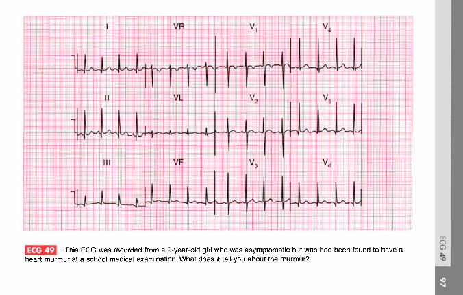
ECG 49 This ECG was recorded from a 9-year-old girl who was asymptomatic but who had been found to have aheart murmur at a school medical examination. What does it tell you about the murmur?

ANSWER 49
The ECG shows:
• Sinus rhythm, rate 100/min• Normal axis• Normal QRS complexes, but narrow, deep Q
waves in leads I, II, V4-V6
• Inverted T waves in lead Vl
Clinical interpretationA sinus tachycardia with normal QRS complexes,showing prominent 'septa!' Q waves, ischaracteristic of ECGs of children. The invertedT wave in lead V1 is normal at any age. A normalECG helps to exclude serious causes of heartmurmurs, but the record has not been very helpfulin this case.
What to doIf in doubt, an echocardiogram will show whether Summary **there is any important structural abnormality in Normal ECG in a 9-year-old child,the heart.
|-fl»l| Seep. 102
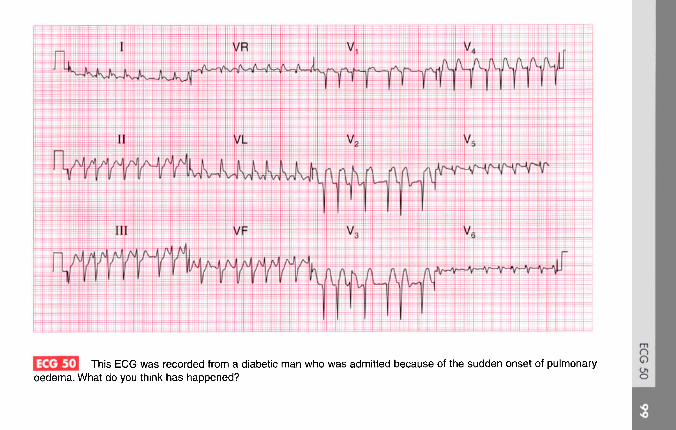
^^^^3 This ECG was recorded from a diabetic man who was admitted because of the sudden onset of pulmonaryoedema. What do you think has happened?
oO
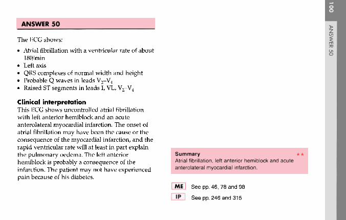
ANSWER 50
The ECG shows:
• Atrial fibrillation with a ventricular rate of about180/min
• Left axis• QRS complexes of normal width and height• Probable Q waves in leads V2-V4
• Raised ST segments in leads I, VL, V2-V4
Clinical interpretationThis ECG shows uncontrolled atrial fibrillationwith left anterior hemiblock and an acuteanterolateral myocardial infarction. The onset ofatrial fibrillation may have been the cause or theconsequence of the myocardial infarction, and therapid ventricular rate will at least in part explainthe pulmonary oedema. The left anteriorhemiblock is probably a consequence of theinfarction. The patient may not have experiencedpain because of his diabetes.
Summary **Atrial fibrillation, left anterior hemiblock and acuteanterolateral myocardial infarction.
MEJ See pp. 46, 78 and 98
IP~1 See pp. 246 and 315
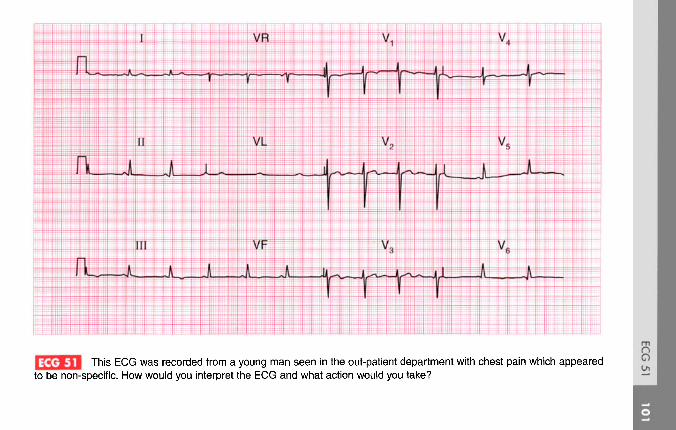
[ ^^Jj This ECG was recorded from a young man seen in the out-patient department with chest pain which appearedto be non-specific. How would you interpret the ECG and what action would you take?

ANSWER 51
The ECG shows:
• Sinus rhythm• Normal axis• Normal QRS complexes
Inverted T waves in leads III, VF; biphasic Twaves in lead V4 and flattened T waves in leadsv5-v6
Clinical interpretationThese T wave changes, particularly those in theinferior leads, could well be caused by ischaemia.The flattened T waves in the lateral leads can onlybe described as 'non-specific'.
What to doWhen confronted with an ECG showing this sortof 'non-specific' abnormality, action dependsprimarily on the clinical diagnosis. If the patient isasymptomatic it is fair to report the ECG asshowing 'non-specific changes'; if the patient hassymptoms at all - as in this case - it is probablyworth proceeding to an exercise test. In this
patient, the exercise test was perfectly normal, andhis symptoms cleared without any intervention. Arepeat ECG, recorded purely out of interest amonth later, showed similar changes.
Summary ***Non-specific ST segment and T wave changes.
See p. 83

ECG 52 This ECG was recorded from a 65-year-old woman admitted to hospital as an emergency because of severechest pain for 1 h. What does the ECG show? What other investigations would you order?

ANSWER 52
The ECG shows:
• Sinus rhythm• Normal axis• Probably normal QRS complexes• Gross elevation of ST segments in anterior and
lateral leads• Depressed ST segments in the inferior leads and
leads III, VF
Clinical interpretationAcute anterolateral myocardial infarction. Inthe lateral leads I, VL and V4-V6, it is difficult tosee where the QRS complexes end and the STsegments begin, but in lead II it is clear that theQRS complex is of normal width.
What to doIf the patient gives a history suggestive of amyocardial infarction and has this ECG, no furtherinvestigations are needed in the acute phase ofthe illness, and in particular there is no place fora chest X-ray. Routine treatment for a myocardial
infarction - pain relief, aspirin and thrombolysis -should be commenced immediately.
SummaryAcute anterolateral myocardial infarction.
IE | See p. 96
See p. 242

ECG 53 A 45-year-old woman had complained of occasional attacks of palpitations for 20 years, and eventually thisEGG was recorded during an attack. What are the palpitations due to, and what would you do?

ANSWER 53
The ECG shows:
• Narrow complex tachycardia at 200/min• No P waves visible• Normal axis• QRS complexes normal• Some ST segment depression
Clinical interpretationThis ECG shows supraventricular tachycardia,probably junctional. These rhythms are usuallydue to a re-entry pathway within, or near to, theatrioventricular node. The ST segment depressioncould indicate ischaemia, but the ST segments arenot horizontally depressed, nor is the depressiongreater than 2 mm, so it is probably of nosignificance.
What to doThe first action is carotid sinus pressure, whichmay terminate the attack. If this fails it willalmost certainly respond to adenosine. As withany tachycardia, electrical cardioversion must be
considered if there is haemodynamic compromise.Once sinus rhythm has been restored the patientmust be taught the various methods (e.g. theValsalva manoeuvre) with which she might tryto terminate an attack. Prophylactic medicationmay not be needed if attacks are infrequent, butmost patients with this problem should havean electrophysiological study to try to identifya re-entry pathway that can be ablated.
SummarySupraventricular (junctional) tachycardia.
(E | See p. 73
IP I See pp. 29 and 167

I JeJJJ This EGG was recorded from a 35-year-old woman who complained of breathlessness. She was anxious,but there were no abnormalities on examination. Does this ECG help with her diagnosis and management?
ANSWER 54
The ECG shows:
• Sinus rhythm, rate 120/min• Normal axis• Normal QRS complexes• Slight downward-sloping ST segment
depression, especially in lead V4
• Widespread T wave flattening• T wave inversion in lead III
Clinical interpretationA sinus tachycardia would be compatible withanxiety, though other causes of 'high output' (e.g.pregnancy, thyrotoxicosis, anaemia, volume loss,CO2 retention, beri-beri) have to be considered.The widespread ST segment and T wavechanges have to be described as 'non-specific';in an anxious patient they could be due tohyperventilation. They do not help with diagnosisand management.
What to doIf a full history and examination fail to suggestany underlying physical disease, furtherinvestigations are unlikely to be helpful.
Summary **Non-specific ST segment and T wave changes.
See p. 89

I JeJ-JJ This ECG was recorded from a 60-year-old man seen in the clinic because of severe breathlessness, whichhad developed over several years. His jugular venous pressure is raised. What do you think the problem is?

ANSWER 55
The ECG shows:
• Sinus rhythm, rate 140/min• One ventricular extrasystole• Peaked P waves (best seen in leads II, III, VF)• Normal PR interval• Right axis• Dominant R wave in lead Vl
• Deep S wave in lead V6
• Normal ST segments and T waves
Clinical interpretationThe sinus tachycardia suggests a major problem.The peaked P waves indicate right atrialhypertrophy. The right axis and dominant Rwave in lead Va suggest right ventricularhypertrophy. The deep S wave in lead V6, withno Teft ventricular' complexes in the chest leads,indicates 'clockwise rotation' of the heart, with theright ventricle occupying the precordium. Thesechanges suggest lung disease.
What to doSince the ECG is entirely 'right sided' one canassume that the problem is due to chronic lungdisease or recurrent pulmonary embolism.The story sounds more in keeping with a lungproblem. The raised jugular venous pressure ispresumably due to cor pulmonale. The sinustachycardia is worrying, and suggests respiratoryfailure.
Summary **Sinus tachycardia and one ventricular extrasystole,right atrial and right ventricular hypertrophy, andclockwise rotation suggest chronic lung disease.
fifl See pp. 64, 89 and 91
IP I Seep. 342

ECG 56 A 60-year-old man is seen in the out-patient department complaining of breathlessness which began quitesuddenly 2 months previously. He had had no chest pain. Examination revealed a raised jugular venous pressure, basalcrackles in the lungs and a third sound at the cardiac apex. This is his ECG. What does it show and how does it fit theclinical picture? What would you do?

ANSWER 56
The ECG shows:
• Sinus rhythm• Normal axis• Large Q waves in leads V1-V4 and small Q
waves in leads I and VL• Elevated ST segments and inverted T waves in
leads V2-V5
• Flattened, biphasic T waves in leads I, VL, V6
Clinical interpretationThis ECG would be compatible with an acuteanterior myocardial infarction, but this does notfit the clinical picture: it appears that an eventoccurred 2 months previously. This pattern of STsegment elevation in the anterior leads can persistfollowing a large infarction, and is seen in thepresence of a ventricular aneurysm.
What to doAn echocardiogram will show if left ventricularfunction is impaired - which it almost certainlyis - and if there is an aneurysm; an aneurysm was
present in this case. The patient should be treatedwith diuretics and an angiotensin-convertingenzyme inhibitor, and surgical resection of theaneurysm might be considered.
Summary ***Old anterior myocardial infarction with a ventricularaneurysm.
WE See p. 95
IP See pp. 243, 250 and 314

ECG 57 The senior house officer in the A & E department is puzzled by this ECG which was recorded from an 80-year-old admitted unconscious with a stroke. What has the house officer missed?
ANSWER 57
The ECG shows:
• Regular rhythm at 60/min• Occasional P waves not related to QRS
complexes (e.g. lead I)• Left axis• QRS complexes preceded by a sharp 'spike'• Broad QRS complexes (160 ms)• Deep S wave in lead V6
• Inverted T waves in leads I and VL
Clinical interpretationThe broad QRS complexes show that this is eithera supraventricular rhythm with bundle branchblock, or a ventricular rhythm. This rhythm isventricular. The sharp spikes preceding each QRScomplex are due to a pacemaker. The P wavesthat can occasionally be seen indicate that theunderlying rhythm, presumably the reason whythe pacemaker was inserted, is complete heartblock.
What to doThe SHO has missed the pacemaker, which isusually buried below the left clavicle. There is noparticular reason why the pacemaker should berelated to the stroke, except that patients withvascular disease in one territory usually have it inothers - this man probably has both coronary andcerebrovascular disease.
Summary *Permanent pacemaker and underlying compete block.
See p. 33
See p. 222
enXJ

ECG 58 A 70-year-old woman who complained of 'dizzy turns' was found to have an irregular pulse, and this ECGwas recorded. There are three abnormalities. What advice would you give her?

ANSWER 58 _
The ECG shows:
• Sinus rhythm• Normal and constant PR intervals in the
conducted beats• Occasional non-conducted P waves• Left axis deviation• Right bundle branch block
Clinical interpretationThis combination of second degree block (Mobitztype 2) plus left axis deviation (left anteriorhemiblock) with right bundle branch blockindicates disease throughout the conductionsystem. This combination of conductionabnormalities is sometimes called 'trifascicular'block. Summary **
Second degree block (Mobitz type 2) and bifascicularWhat to do blockThe 'dizzy turns' may represent intermittentcomplete block. Permanent pacing is essential.
See p. 212

A 50-year-old man who had come to the A & E department with chest pain, collapsed while his EGG wasbeing recorded. What happened and what would you do?

ANSWER 59
The ECG shows:
• Sinus rhythm with ventricular extrasystoles• The third extrasystole occurs on the peak of the
T of the preceding sinus beat• After three or four beats of ventricular
tachycardia ventricular fibrillation develops• In the sinus beats there is a Q wave in lead III,
raised ST segments in leads II and III, and STsegment depression and T wave inversion inlead I
Clinical interpretationAlthough only leads I, II and III are available itlooks as if the chest pain was due to an inferiormyocardial infarction. This was probably the causeof the ventricular extrasystoles and an 'R on T'extrasystole caused ventricular tachycardia, whichrapidly decayed the ventricular fibrillation. Itmight be argued that in lead III, and perhapsalso in lead I, 'torsade de pointes' ventriculartachycardia is present, but this is not apparent inlead II.
What to doPrecordial thump and immediate defibrillation, butif no defibrillator is at hand then cardiopulmonaryresuscitation should be performed, and the usualprocedure for the management of the cardiacarrest instituted.
Summary *Probable inferior myocardial infarction; R on Tventricular extrasystole causing ventricular fibrillation.
ME_\ See p. 80
lp~\ See pp. 195 and 215
c/3

JJj A 60-year-old man complained of severe central chest pain, and a few minutes later became extremelybreathless and collapsed. He was brought to the A & E department where his heart rate was found to be 150/min, hisblood pressure was unrecordable and he had signs of left ventricular failure. This is his ECG. What has happened andwhat would you do?

ANSWER 60
The ECG shows:
• Broad complex tachycardia at 150/min• No P waves visible• Normal axis• QRS duration about 200 ms• Concordance of QRS complexes (i.e. all point
upwards) in the chest leads
Clinical interpretationA broad complex tachycardia can be ventricularin origin, or can be due to a supraventriculartachycardia with aberrant conduction (i.e. bundlebranch block). Here the very broad complexesand the QRS concordance suggest a ventriculartachycardia. In a patient with a myocardialinfarction it is always safe to assume that such arhythm is ventricular. From the story, one wouldguess that this patient had a myocardial infarctionand then developed ventricular tachycardia, butit is possible that the chest pain was due to thearrhythmia.
What to doThis patient has haemodynamic compromise -low blood pressure and heart failure - and needsimmediate cardioversion. While preparationsare being made it would be reasonable to tryintravenous lignocaine or amiodarone.
SummaryVentricular tachycardia.
IE | See p. 75
IP I See p. 178
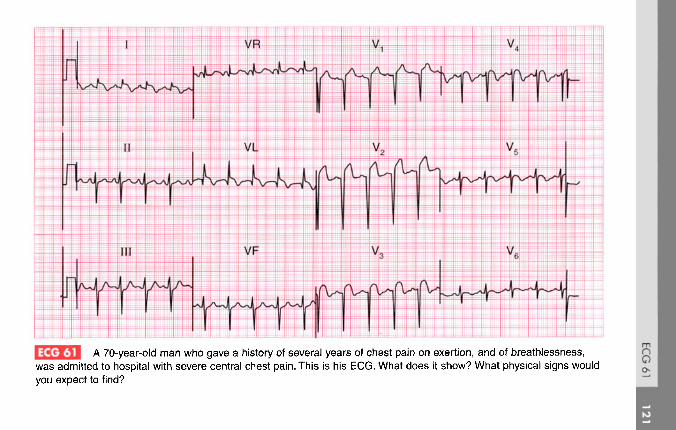
A 70-year-old man who gave a history of several years of chest pain on exertion, and of breathlessness,was admitted to hospital with severe central chest pain. This is his ECG. What does it show? What physical signs wouldyou expect to find?

ANSWER 61
The ECG shows:
• Sinus rhythm• Left axis deviation• Q wave in lead VL; QS pattern in leads V2-V3
• Persistent S waves in lead V5
• Raised ST segments and inverted T waves inleads V17 VL
• Raised ST segments in leads V2-V3 with biphasicT waves in leads V3-V4
Clinical interpretationLeft axis deviation is due to left anteriorhemiblock, probably associated with the infarctionshown in the anterior leads: it is difficult to be sureof the age of this infarction. The ECG appears toshow an acute lateral infarction. The persistent Swaves in lead V6 suggest chronic lung disease.
What to doThe patient has probably had quite severe leftventricular damage and may have the signs of left
ventricular failure; he may well also have signs ofchronic airway disease.
SummaryAcute lateral myocardial infarction, anterior infarctionof uncertain age, left axis deviation and possiblechronic lung disease.
See p. 98
IP I See pp. 242 and 342

ECG 62 This ECG was recorded from a 30-year-old woman who complained of an irregular heart beat, especiallyon lying down at night. She is worried about heart disease. What would you advise her to do?

ANSWER 62
The ECG shows:
• Sinus rhythm, rate 85/min• Frequent ventricular extrasystoles• Normal axis in sinus beats• Sinus beats show normal QRS complexes, ST
segments and T waves
Clinical interpretationThis ECG is normal apart from the ventricularextrasystoles. Provided there is nothing else in thehistory or physical examination to suggest heartdisease, the extrasystoles are not important.
What to doThe patient must be reassured that extrasystolesdo not of themselves indicate heart disease.She should be advised not to smoke, and to tryabstaining from alcohol, coffee and tea to see if theextrasystoles become less troublesome. Medicationis best avoided, but if she insists on treatment abeta-blocker will be safe.
SummaryNormal ECG with ventricular extrasystoles.
See p. 64
See p. 155

ECG 63 A 60-year-old man who was being treated in hospital complained of palpitations, and this ECG wasrecorded. What do you think the underlying disease was, and what were the palpitations due to?

ANSWER 63
The ECG shows:
• Atrial fibrillation• Ventricular extrasystoles with two distinct
morphologies (best seen in lead II)• A four-beat run of ventricular tachycardia• Right axis deviation• Small QRS complexes• No R wave development in the chest leads; lead
V6 shows a dominant S wave• T wave inversion in leads V5, V6
Clinical interpretationThis ECG suggests chronic lung disease - smallcomplexes, right axis deviation, and marked'clockwise rotation' with lead V6 still showinga ight ventricular type of complex. The atrialfibrillation is probably secondary to the lungdisease, though the usual other possibilities mustbe considered. The patient's lung condition isprobably being treated with a beta-agonist, suchas salbutamol, and this could be the cause of theextrasystoles and ventricular tachycardia.
What to doStop the beta-agonist but do not give a beta-blocker. Check the electrolyte levels; consider thepossibility of digoxin toxicity.
Summary ***Atrial fibrillation with ventricular extrasystoles andventricular tachycardia; changes suggesting chroniclung disease.
See pp. 64, 75 and 78
See pp. 170, 175 and 178

ECG 64 A 45-year-old man complained of palpitations, weight loss and anxiety. His blood pressure was 180/110,and his heart seemed normal. This is his ECG. His thyroid function tests, measured several times, were normal. Whatmight be going on?

ANSWER 64
The ECG shows:
• Narrow complex rhythm at 140/minute• Apparently one P wave per QRS complex• Normal PR interval• Normal axis• Normal QRS complexes• Inverted T wave in lead VL and a flat T wave in
leads I, V5-V6
Clinical interpretationThe immediate problem is to decide whether thisis a sinus or atrial tachycardia. Carotid sinuspressure caused transient slowing, so this isprobably sinus rhythm. A sinus tachycardia of140/min could be due to anxiety, but seemsvery fast for this; other possibilities are drugeffects (beta-agonists, amphetamine) and a Summary **phaeochromocytoma - which turned out to be Sinus tachycardia.the diagnosis.
See p. 54
See p. 152
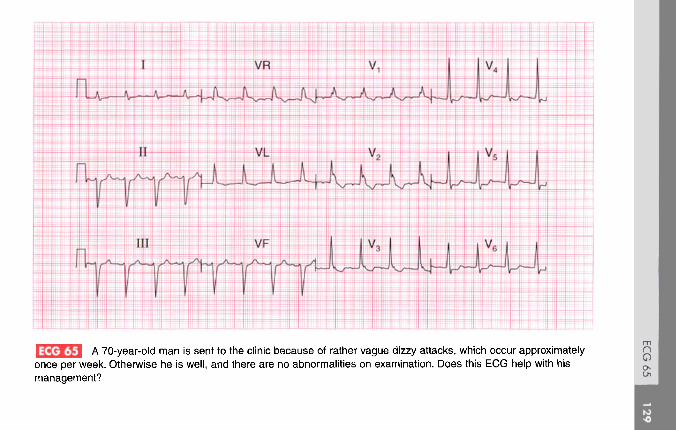
ECG 65 A 70-year-old man is sent to the clinic because of rather vague dizzy attacks, which occur approximatelyonce per week. Otherwise he is well, and there are no abnormalities on examination. Does this ECG help with hismanagement?

ANSWER 65
The ECG shows:
• Sinus rhythm, rate 100/min• PR interval at the upper limit of normal (200 ms)• Left axis• QRS duration prolonged (160 ms)• RSR pattern in lead VL, wide S wave in lead V6
• Inverted T waves in leads VL, V1-V4
Clinical interpretationThe left axis is accompanied by widening of theQRS complexes and an inverted T wave in leadVL, and this is characteristic of left anteriorhemiblock. There is also right bundle branch block,so two of the main conducting pathways areblocked, which is known as 'bifascicular block'.The fact that the PR interval is at the upper limit ofnormal raises the possibility of delayed conductionin the remaining pathway; if the PR interval weredefinitely prolonged the pattern would be called'trifascicular block'.
What to doBifascicular block is not an indication for pacingif the patient is asymptomatic. The problemhere is to decide if the dizzy attacks are due tointermittent complete heart block. Ideally an ECGwould be recorded during an attack; since theyonly occur every week or so, ambulatory ECGtape recording may not be helpful, but an eventrecorder would be worth trying. In the absenceof clear evidence the decision whether or notto insert a permanent pacemaker is a matter ofjudgement, but in a patient with this story andECG it would be a perfectly reasonable thing to do.
Summary **Left anterior hemiblock and right bundle branchblock - bifasciular block.
See p. 48
See pp. 141 and 147

you do?This ECG was recorded from a 25-year-old black football professional. What does it show, and what would

ANSWER 66
The ECG shows:
• Sinus rhythm• Normal axis• Normal QRS complexes• Widespread T wave inversion, particularly in
leads V2-V5
Clinical interpretationRepolarization (T wave) abnormalities are quitecommon in black people, but alternativeexplanations for this ECG appearance would bea non-Q wave infarction, or a cardiomyopathy.
What to doThis man is a professional football player, soit is important to exclude hypertrophiccardiomyopathy, and this can be done byechocardiography. Because his career dependedupon coronary disease being excluded, a coronaryangiogram was performed and was entirelynormal.
Summary ***Widespread T wave inversion, probably normal in ablack man.
See p. 112
See pp. 91 and 117

ECg 67 This ECG was recorded from an asymptomatic 20-year-old man at a pre-employment medical examination.The employer will require him to hold a vocational (HGV) driving licence. What advice would you give?

ANSWER 67
The ECG shows:
• Sinus rhythm• Very short PR interval• Normal axis• Widened QRS complexes with a slurred upstroke
(delta wave), best seen in leads I and V4
Clinical interpretationThis is the Wolff-Parkinson-White syndrome: theaccessory pathway is on the right side and this issometimes called 'type B'.
What to doIt would be prudent to obtain an echocardiogramto make sure there is no structural abnormality(such as a cardiomyopathy) as well as theWolff-Parkinson-White syndrome. Provided there Summary **is no history to suggest an arrhythmia, he can Wolff-Parkinson-White syndrome.hold a vocational driving licence.
E I See p. 81
See pp. 38 and 120

ECG 68 A 60-year-old woman is seen in the out-patient department complaining of breathlessness. There are noabnormal physical findings. What does this ECG show, what might be the underlying problem, and how would you treat her?

ANSWER 68
The ECG shows:
• Atrial flutter• 4:1 block• Normal axis• Normal QRS complexes• Sloping ST segment depression, best seen in
leads V5-V6
Clinical interpretationThis shows atrial flutter with what appears to be astable 4:1 block. The ST segment depressionsuggests digoxin effect.
What to doThe stable 4:1 block has caused a regular heartbeat, so the arrhythmia was not suspected at thetime of the clinical examination. There is nothingin this ECG to indicate the underlying disease,which could be ischaemic, rheumatic or acardiomyopathy; echocardiography is needed.Digoxin will tend to maintain a fairly high degreeof block but will not affect the underlying rhythm.
Intravenous flecainide may convert the heart tosinus rhythm, but DC cardioversion may benecessary.
SummaryAtrial flutter with 4:1 block.
IE | See p. 68
See p. 160

m«U£| This ECG was recorded from a 30-year-old man at a medical examination required by the Civil AviationAuthority. Is it normal?

ANSWER 69
The ECG shows:
• Sinus rhythm• Right axis deviation (dominant S waves in lead
I)• Dominant R wave in lead Vl
• Prominent U waves in leads V2-V5
Clinical interpretationRight axis deviation can be a normal variant(particularly in tall thin people) but also occurswith right ventricular hypertrophy. The smalldominant R wave in lead Vl suggests rightventricular hypertrophy, but this can be anormal variant. The U waves could indicatehypokalaemia, but when associated with normalT waves (as here) they are a normal variant.
SummaryWhat to do Probably a normal ECG.Exclude causes of right ventricular hypertrophy.A chest X-ray with a lateral view, and anechocardiogram will help determine whetherthe right ventricle really is enlarged. I IP I See pp. 69 and 74
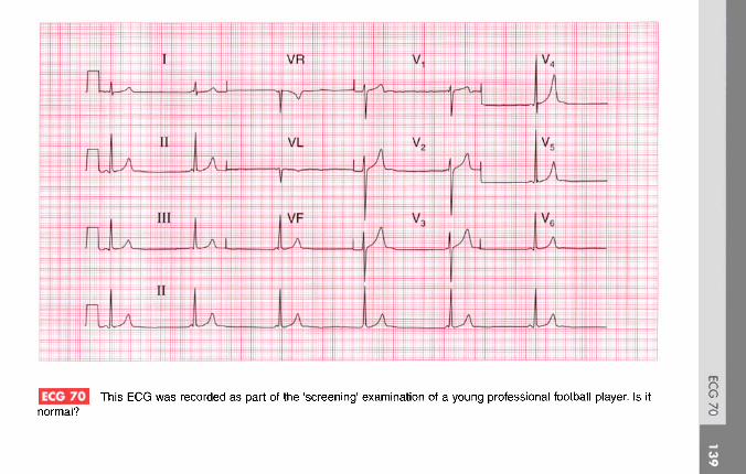
[•J This ECG was recorded as part of the 'screening' examination of a young professional football player. Is itnormal?

ANSWER 70
The ECG shows:
• Regular narrow complex rhythm at 35/min• P waves sometimes, but not always, visible just
before the QRS complexes• PR interval, when measurable, is always short
but varies• Height of R wave in lead V4 plus depth of S
wave in lead V2 = 45 mm• Normal QRS complexes and ST segments.• Peaked T waves, especially in lead V4
Clinical interpretationThe short PR interval raises the possibility ofpre-excitation, but the interval varies, and in thefirst complex of leadsVj-V3 no P wave can beseen. The slow, narrow, complex rhythm suggestsatrioventricular nodal escape. Here there is apronounced slowing of the sinoatrial node,presumably due to athletic training, and anaccelerated idionodal rhythm has taken over.This pattern used to be called a 'wandering atrial
pacemaker'. The tall R waves are perfectly normalin young fit people, and so are the peaked P waves.
What to doThis is a normal variant in athletes, and no actionis required.
SummaryAccelerated idionodal rhythm.
IE I Seep. 60
See p. 98

ECG 71 A 45-year-old patient is admitted to the A & E department having had severe central chest pain for 1 h.There are no signs of heart failure, and this is his ECG. What does the ECG show and what would you do?

ANSWER 71effectively, perhaps especially in young patientswith anterior infarcts.
The ECG shows:
• Sinus rhythm• Normal axis• Q waves in leads V2-V4
• Raised ST segments in leads I, VL, V2-V5
• Flat ST segment depression in leads III, VF
Clinical interpretationThis ECG shows an acute anterior myocardialinfarction with inferior ischaemia.
What to doUnless there are any potential risks of bleeding(previous stroke, peptic ulcer, diabetic retinopathy,etc.) this patient should be given aspirin, 300 mg tobe chewed, and then a thrombolytic agent. Thechoice of agent is still open to debate: since he hasnot had a thrombolytic agent before, the cheaperagent streptokinase would be perfectly acceptable,but there is some evidence that the moreexpensive alteplase (rt-PA) reduces mortality more
SummaryAcute anterior myocardial infarction and inferiorischaemia.
WJ See p. 95
IP~1 See p. 242

ECG 72 A 30-year-old man, who had complained of palpitations for many years without anything abnormal beingfound, came to the A & E department during an attack, and this ECG was recorded. Apart from signs of marked anxietythere was nothing to find except a heart rate of 140/min. What does the ECG show?
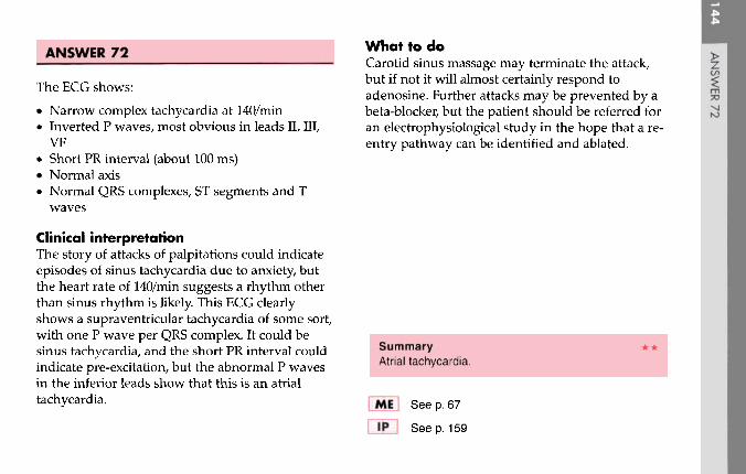
ANSWER 72
The ECG shows:
• Narrow complex tachycardia at 140/min• Inverted P waves, most obvious in leads II, III,
VF• Short PR interval (about 100 ms)• Normal axis• Normal QRS complexes, ST segments and T
waves
Clinical interpretationThe story of attacks of palpitations could indicateepisodes of sinus tachycardia due to anxiety, butthe heart rate of 140/min suggests a rhythm otherthan sinus rhythm is likely. This ECG clearlyshows a supraventricular tachycardia of some sort,with one P wave per QRS complex. It could besinus tachycardia, and the short PR interval couldindicate pre-excitation, but the abnormal P wavesin the inferior leads show that this is an atrialtachycardia.
What to doCarotid sinus massage may terminate the attack,but if not it will almost certainly respond toadenosine. Further attacks may be prevented by abeta-blocker, but the patient should be referred foran electrophysiological study in the hope that a re-entry pathway can be identified and ablated.
SummaryAtrial tachycardia.
Ifl See p. 67
See p. 159

ECG 73 A confused 80-year-old woman was sent in from a nursing home because of a collapse. No other historywas available, except that she was said to be having treatment for her heart. There were no obvious physical signs, butthis is her ECG. What is going on?

ANSWER 73
The ECG shows:
• Atrial flutter with a ventricular rate of 50/min• Ventricular extrasystoles• Left axis• Normal QRS complexes• Inverted T waves in the anterior leads• Prolonged QT interval (about 650 ms)
Clinical interpretationThe atrial flutter with a slow ventricular rate raisesthe possibility that a bradycardia caused hercollapse; the left anterior hemiblock indicates thatshe has conduction tissue disease. The anterior Twave inversion may be due to ischaemia. There isno ST segment depression, suggesting that sheis taking digoxin, but the prolonged QT intervalsuggests either an electrolyte abnormality or thatshe is being treated with one of the many drugsthat have this effect. A collapse in a patient witha long QT interval suggests torsade de pointesventricular tachycardia. Since she has atrial flutter
you need to think about amiodarone - which iswhat she turned out to be taking.
Summary ***Atrial flutter with a slow ventricular rate, ventricularextrasystoles, left anterior hemiblock and a prolongedQT interval.
See pp. 375 and 376

ECG 74 This ECG was recorded as part of a routine health check on a 50-year-old woman who said she wasasymptomatic. The only other abnormality detected in the usual screening tests was a serum cholesterol level of 7.2mmol/l. What would you do?

ANSWER 74
The ECG shows:
• Sinus rhythm• Normal axis• Normal QRS complexes• Widespread T wave flattening and inversion• Prominent U waves especially V3-V5
Clinical interpretationFlattened T waves with prominent U wavesusually result from hypokalaemia. The serumpotassium level is usually checked during healthscreening, but the same ECG changes can resultfrom hypocalcaemia or hypomagnasaemia. A highcholesterol level can be a marker for coronarydisease, but elevated cholesterol levels can also besecondary to thyroid or renal disease.
What to doCheck the thyroid function. This woman hadmyxoedema, and her ECG became normal when itwas treated.
Summary ***Widespread T wave flattening with prominent Uwaves - classically due to hypokalaemia, but in thiscase due to myxoedema.
IfJ See p. 108
IP~] See pp. 362 and 368

ECG 75 This ECG was recorded from an asymptomatic 45-year-old man at a 'health screening' examination. Is itnormal, and what advice would you give him?

ANSWER 75
The ECG shows:
• Sinus rhythm, rate 64/min• Prolonged PR interval (360 ms)• Normal QRS complexes, ST segments, and
T waves
Clinical interpretationThis ECG shows first degree atrioventricular blockbut is otherwise entirely normal.
What to doAlthough the upper limit of the PR interval isusually taken to be 200 ms, longer durations(technically first degree block) are frequently seenin healthy people. Provided you can be sure thatthis patient has no symptoms, and provided thephysical examination is normal, no further actionis required. Some individuals in occupations thatrequire a totally normal ECG may have to have anambulatory ECG recording to demonstrate thatthere are no episodes of higher-degree block.
SummaryFirst degree atrioventricular block.
(E | See p. 30
IP | See pp. 62 and 137

ECG 76 An 80-year-old woman was admitted to hospital because of a sudden onset of palpitations associated withbreathlessness. She was in congestive cardiac failure and had a heart murmur suggestive of aortic stenosis. What doesthis ECG show and how would you treat her?

ANSWER 76
The ECG shows:
• Broad-complex tachycardia• Irregular rhythm, rate 130-200/min• No clear P waves but irregular baseline, best
seen in lead VL• Left bundle branch block pattern
Clinical interpretationThe marked irregularity of rhythm, coupled withthe irregular baseline glimpsed in one beat in leadVL, shows that this is atrial fibrillation with leftbundle branch block.
What to doAortic stenosis is commonly associated with leftbundle branch block. In the presence of aorticstenosis, vasodilators must be used with cautionand she should be treated with digoxin anddiuretics. As soon as possible the aortic valvegradient should be assessed by echocardiography,and even at the age of 80 years, aortic valvereplacement might be considered.
SummaryAtrial fibrillation with left bundle branch block.
IE | See pp. 36 and 78
Seep. 117

ECG 77 An 80-year-old man is found at routine examination to have a slow heart rate and a harsh systolic murmur.This is his ECG. What does it show, and what is the likely diagnosis? Is treatment necessary?

ANSWER 77
The ECG shows:
• Sinus rhythm, P wave rate 75/min• Second degree (2:1) block• Left axis deviation• Right bundle branch block
Clinical interpretationThis is second degree block and not complete(third degree) block because the PR interval in theconducted beats is normal: at times it appears tovary but in fact this variation is due to leadchanges. Left axis deviation (left anteriorhemiblock) and right bundle branch blockconstitute bifascicular block, but the 2:1 blockshows that there is also disease either in the Hisbundle or in the remaining posterior fascicle. Thiscombination is sometimes called 'trifascicular'block.
What to doThe combination of a heart murmur and heartblock suggests aortic stenosis. The severity of this
can be assessed with echocardiography, thoughthe slow rate (and thus high stroke volume) willaccentuate the recorded valve gradient. Aorticvalve replacement may or may not be needed,but the patient certainly needs a permanentpacemaker to prolong his survival.
Summary **Second degree (2:1) block and bifascicular block.
IE I See pp. 32 and 48
IP I See pp. 141 and 146

3 A 70-year-old woman, who had been breathless for several months, was admitted with chest pain, and thisis her ECG. What does it show and what would you do?

ANSWER 78
The ECG shows:
• Atrial fibrillation with one ventricularextrasystole
• Ventricular rate about 110/min• Normal axis• Normal QRS complexes• Horizontal ST segment depression of up to
7 mm in leads V2-V5
• Downward-sloping ST segment depression inlead V6
• Inverted T waves in leads I, VL, V6, andindeterminate T waves elsewhere
Clinical interpretationThe anterior horizontal ST depression indicatessevere ischaemia, which is presumably the causeof the chest pain. The downward-sloping STsegment in lead V6 could be due to digoxintherapy. The ventricular rate is not too fast, andalthough the heart rate may be contributing to theishchaemia it seems unlikely that it is the mainproblem.
What to doThe patient should be treated for an acutecoronary syndrome with heparin, a beta-blockerand nitrates. If the pain does not settle, earlyangiography with a view to revascularizationby a coronary artery bypass graft (CABG) orpercutaneous transluminal angioplasty (PTCA)will have to be considered.
SummaryAtrial fibrillation and anterior ischaemia.
See pp. 78 and 102
See pp. 273 and 274

ECG 79 An elderly woman is admitted to hospital unconscious, evidently having had a stroke. No cardiacabnormalities are noted, but this is her ECG. What has been missed?

ANSWER 79
The ECG shows:
• No P waves; irregular baseline suggesting atrialfibrillation
• Regular QRS complexes• Left axis deviation• Wide QRS complexes of an indeterminate
pattern• Each QRS complex is preceded by a deep and
narrow spike
Clinical interpretationThe narrow spike is due to a pacemaker, andsomeone has not noticed the permanent pacingbattery which is probably below the left clavicle.The pacing wire will be stimulating the rightventricle, giving rise to broad QRS complexesresembling a bundle branch block pattern. Theunderlying rhythm here is atrial fibrillation:the patient may have had atrial fibrillation withcomplete block, or there may simply have been aslow ventricular response to the atrial fibrillation.
What to doThe stroke may have been due to an embolusarising in the left heart as a result of atrialfibrillation. There may have been temporarypacemaker failure, but probably the stroke wasnot related to the pacemaker.
>
Summary **Ventricular-paced rhythm and atrial fibrillation.
IP' I See p. 222

l ejjjj A 30-year-old man is seen in the A & E department with left-sided chest pain that appears to be pleuritic innature. What does his ECG show?
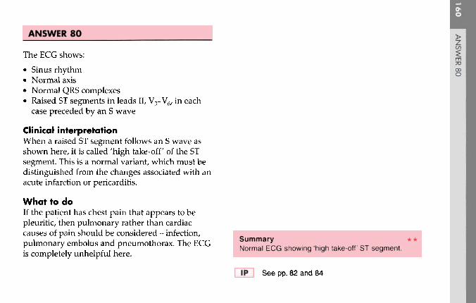
ANSWER 80
The ECG shows:
• Sinus rhythm• Normal axis• Normal QRS complexes• Raised ST segments in leads II, V3-V6, in each
case preceded by an S wave
Clinical interpretationWhen a raised ST segment follows an S wave asshown here, it is called 'high take-off of the STsegment. This is a normal variant, which must bedistinguished from the changes associated with anacute infarction or pericarditis.
What to doIf the patient has chest pain that appears to bepleuritic, then pulmonary rather than cardiaccauses of pain should be considered - infection, Summary **pulmonary embolus and pneumothorax. The ECG Norma, ECG shOwing 'high take-off' ST segment.is completely unhelpful here.
I Ip | See pp. 82 and 84

ECG 81 A 50-year-old man returned from holiday in Spain saying that while there he had had some bad indigestion,but was now perfectly well. This is his ECG: what does it show and what would you do?

ANSWER 81
The ECG shows:
• Sinus rhythm• Normal conduction• Normal axis• Q waves in leads V2-V4
• Slightly elevated ST segments in leads V2-V3
• Inverted T waves in leads I, VL, V2-V6
Clinical interpretationThis ECG shows an old anterior myocardialinfarction with lateral ischaemia. The slightelevation of ST segments might suggest an acuteprocess if the pain were recent, but with this storythe changes are almost certainly old.
What to doThe assumption has to be that the 'indigestion'was actually a myocardial infarction. Since he isnow well, the important thing is to ensure that hetakes the appropriate steps to prevent a furtherattack - he must stop smoking and reduce weightif necessary, and he should be treated with aspirin,
a beta-blocker, an angiotensin-converting enzymeinhibitor and a statin. In view of his age it mightbe worth doing an exercise test to ensure thatthere is no evidence of ischaemia at a lowworkload.
SummaryOld anterior myocardial infarction.
See p. 103
See pp. 243 and 250

This ECG was recorded from an asymptomatic 30-year-old man at a routine examination. Is it normal?

ANSWER 82
The ECG shows:
• Sinus rhythm• Right axis deviation (S waves bigger than R
waves in lead I, large R waves in lead VR, verysmall R waves and deep S waves in lead VL)
• Notched QRS complexes in lead III• Otherwise entirely normal QRS complexes and
T waves
Clinical interpretationRight axis deviation can be a feature of rightventricular hypertrophy, but in tall thin people it isa normal variant. The notched QRS complexes inlead III are normal, though if present in all leadsthey could be the ']' waves of hypothermia.
What to doExamine the patient and exclude right ventricular Summaryhypertrophy (you should have done this before Normal ECGrecording the ECG!).
I l|* | See p. 62

ECG 83 This ECG was recorded from a 70-year-old woman who had complained of attacks of dizziness for about ayear. Note that only leads I, II and III are shown, but there are three abnormalities. How should this woman be treated?

ANSWER 83with dizziness. Whatever these findings, however,the patient needs a permanent pacemaker.
The ECG shows:
• Variable rhythm• Sinus rhythm at times• One ventricular extrasystole• Intermittent second degree (both Mobitz type 2
and 2:1) heart block• Left axis deviation
Clinical interpretationThere are two varieties of second degree blockhere - Mobitz type 2 at the beginning of the record(single non-conducted beats), with 2:1 block forfour sinus beats later. The ventricular extrasystoleis not significant, but the left axis deviationindicates left anterior hemiblock.
What to doThe full ECG may indicate the underlyingdisease. It may show, for example, an old anteriormyocardial infarction. A 24 h ambulatory ECGtape-recording may reveal the rhythm associated
SummarySecond degree block with left axis deviation.
See pp. 16 and 31
It* | Seep. 140

ECG 84 This ECG was recorded in the A & E department from a 60-year-old man who had had intermittent centralchest pain for 24 h. What does it show and how should he be managed?

ANSWER 84
The ECG shows:
• Sinus rhythm• Normal conduction intervals• Normal axis• Normal QRS complexes• Normal ST segments• T wave inversion in leads I, VL, V2-V4
Clinical interpretationThis ECG shows an anterior non-Q waveinfarction of uncertain age.
What to doThis patient clearly has an acute coronarysyndrome. He must be admitted and treated withlow-molecular-weight heparin, a nitrate and abeta-blocker. If the pain does not settle quickly, Summarythe use of a glycoprotein Ilb/IIIa inhibitor such as Anterior non-Q wave infarction.tirofiban or abciximab should be considered as aprelude to early angiography and angioplasty. fJivtfl See D 103
See p. 266

ECG 85 A 25-year-old man, known to have an atrial septal defect, was admitted to hospital as an emergencybecause of palpitations. His heart rate was 170/min, his blood pressure was 140/80 and there were no signs of heartfailure. What is the cardiac rhythm and what would you do?

ANSWER 85
The ECG shows:
• Broad-complex tachycardia, rate 170/min• No clear P waves but possibly some P waves
visible in lead VR• Normal axis• Right bundle branch block pattern• Horizontal ST segment depression in V4, V5
Clinical interpretationThe QRS complex duration is 120 ms, the axis isnormal, and the QRS complexes show the classicright bundle branch block pattern. It is likely thatthis is a supraventricular tachycardia with rightbundle branch block, and this would be certainif we were sure of the existence of P waves inlead VR. This is either an atrial or a junctionaltachycardia. The ST segment depression suggestsischaemia.
What to doIf the patient is known to have an atrial septaldefect he is likely to have right bundle branch
block, and this could be confirmed in pre-existinghospital records. The initial treatment is carotidsinus massage, and if this proves ineffective,intravenous adenosine.
Summary ***Supraventricular tachycardia (possibly atrial) withright bundle branch block.
[|] See pp. 36 and 74
See p. 170

ECG 86 A 30-year-old woman, who had a baby 3 months previously, complains of breathlessness, and this is herECG. What is the problem?

ANSWER 86
The ECG shows:
• Sinus rhythm• Short PR interval at 100 ms• Normal axis• Normal QRS duration• Slurred upstroke to QRS (delta wave)• Dominant R wave in lead V1
• Normal ST segments and T waves
Clinical interpretationThis ECG shows a Wolff-Parkinson-Whitesyndrome type A, which is characterized by adominant R wave in lead Vj.
What to doThe catch here is that the dominant R wave in leadV may be mistakenly thought to be due to rightventricular hypertrophy. In a young woman whocomplains of breathlessness after a pregnancy,pulmonary embolism is obviously a possibility,and this might well cause ECG evidence of rightventricular hypertrophy, but in the presence of a
Wolff-Parkinson-White syndrome this would bevery difficult to diagnose from the ECG. The onlything that might help would be the appearanceof right axis, which is not part of the Wolff-Parkinson-White syndrome, and is not presenthere.
SummaryWolff-Parkinson-White syndrome type A.
IE | See p. 81
See pp. 117 and 120

ECG 87 A 30-year-old man, who had had brief episodes of palpitations for at least 10 years, was seen during anattack in the A & E department and this is his ECG. What is the rhythm, and what would you do immediately, and in thelong term?

ANSWER 87
The ECG shows:
• Broad complex tachycardia at about 250/min• No P waves visible• Right axis• QRS duration of about 180 ms• QRS complexes point upwards in lead V1 and
downwards in lead V6 - no concordance• Right bundle branch block QRS configuration; the
first R wave peak is higher than the second peak
Clinical interpretationThere are essentially three causes of a broad complextachycardia: ventricular tachycardia, supraventriculartachycardia with bundle branch block, and theWolff-Parkinson-White syndrome. The key to thediagnosis lies in the ECG when the patient is in sinusrhythm, but this is not always available. Patients witha broad complex tachycardia in the context of anacute myocardial infarction must be assumed to havea ventricular tachycardia, but that does not applyhere. In this record the QRS is not very broad, theaxis is to the right, and there is no concordance of theQRS complexes - all pointing to a supraventricular
origin. In favour of a ventricular tachycardia is thefact that the height of the primary R wave is greaterthan that of the secondary R wave. Taking thesefeatures together with the clinical picture, therhythm is probably supraventricular.
What to doCarotid sinus pressure is the first move. If there issevere haemodynamic compromise the patient mayneed urgent electrical cardioversion, but intravenousflecainide would be a reasonable first choice. In factthe arrhythmia terminated spontaneously, revealinga short PR interval and a QRS complex with a deltawave. So this patient has a Wolff-Parkinson-Whitesyndrome, and needs an electrophysiological studywith a view to ablation of the accessory tract.
SummaryBroad complex tachycardia due to aWolff-Parkinson-White syndrome.
Mn See p. 81
1F1 Seep. 127
***

ECG 88 A 65-year-old woman with rheumatic heart disease, who had had severe heart failure for years, wasadmitted with increasing breathlessness and ankle swelling. Despite having been treated with angiotensin-convertingenzyme inhibitors and diuretics, there was evidence of severe heart failure. Having seen the ECG what more do youwant to know?

ANSWER 88
The ECG shows:
• Uncertain rhythm - no P waves, irregular QRSbut no 'fibrillation' activity
• Right axis• Normal QRS complexes except for a deep S
wave in V6
• Symmetrically peaked T waves• Inverted T waves in leads III and VF
Clinical interpretationThe absence of atrial activity and the peaked Twaves are consistent with hyperkalaemia. Theright axis and deep S waves in lead V6 couldindicate right ventricular hypertrophy and couldresult from chronic lung disease. The inverted Twaves in leads III and VF suggest ischaemia.
What to doFind out what the patient's medication has beenand check the electrolyte levels. This woman hadbeen treated with a combination of captopril25 mg, three times daily (which tends to raise the
serum potassium level) and three co-amilofrusetablets per 24 h (frusemide 40 mg plus amiloride5 mg in each tablet). The combination of captopriland amiloride causes marked potassium retention,and in this case the serum potassium level was7.4 mmol/1.
When the hyperkalaemia was corrected, sinusrhythm with clear P waves was restored and thepeaked T waves became normal. The right axis,clockwise rotation and inverted T waves in theinferior leads persisted.
SummaryHyperkalaemia.
ftj See p. 108
See p. 366
***

This ECG was recorded from a 55-year-old man who was admitted to hospital as an emergency with severecentral chest pain that had been present for about an hour. He was pale, cold and clammy; his blood pressure was100/80, but there were no signs of heart failure. What does this ECG show? Does anything about it surprise you?

ANSWER 89
The ECG shows:
• Sinus rhythm, rate 55/min• First degree block (PR interval 350 ms)• Normal axis• Small Q waves in leads II, III, VF• Raised ST segments in leads II, III, VF• Depressed ST segments and inverted T waves in
leads I, VL• Slight ST segment depression in the chest leads
Clinical interpretationAcute inferior myocardial infarction withanterolateral ischaemia, and first degree block.Patients who are in pain with an acute myocardialinfarction usually have a sinus tachycardia, buthere vagal overactivity is causing a bradycardia.
What to doFirst degree block is not important in itself, but withevidence of vagal overactivity, atropine should begiven. Otherwise this patient can be treated in theusual way with pain relief, aspirin, and thrombolytics.
Summary it-Acute inferior myocardial infarction with first degreeblock.
See p. 96
See p. 238

ECG 90 This ECG was recorded from a 17-year-old girl who was breathless, had marked ankle swelling with signsof right heart failure, and who had been known to have a heart murmur since birth. She was acyanotic. What ECGabnormalities can you identify and can you suggest a diagnosis?

ANSWER 90
The ECG shows:
• Sinus rhythm• Markedly peaked P waves (best seen in leads II,
Vi)• Normal axis• Dominant R wave in lead Vl
Clinical interpretationThe ECG shows right atrial and right ventricularhypertrophy.
What to doRight atrial hypertrophy is seen with pulmonaryhypertension of any cause, tricuspid stenosis, andEbstein's anomaly. Right ventricular hypertrophyis seen with pulmonary stenosis and pulmonaryhypertension. These conditions can all bediagnosed by echocardiography. This patient hadEbstein's anomaly and an atrial septal defect.
SummaryRight atrial and right ventricular hypertrophy.
See p. 91
See p. 344

ECG 91 A 35-year-old woman, who had had attacks of what sound like a paroxysmal tachycardia for many years,was seen in the A & E department, and this ECG was recorded. What is the diagnosis?

ANSWER 91
The ECG shows:
• Narrow complex tachycardia at about 170/min• No P waves visible• Normal axis• QRS duration 112ms• Slurred upstroke to QRS complexes, best seen in
leads V3-V4
• Depressed ST segments in leads V3-V6
• Inverted T waves in the lateral leads
Clinical interpretationThis is a narrow complex tachycardia, so it issupraventricular. The slurred upstroke to theQRS complex suggests a Wolff-Parkinson-Whitesyndrome, so this is a re-entry tachycardia withdepolarization spreading down the accessorypathway. This diagnosis is consistent with thepatient's history.
What to doCarotid sinus pressure is always the first thing totry in patients with a supraventricular tachycardia.
In most such patients adenosine is the first drug touse, but in Wolff-Parkinson-White syndromes itmust be used with caution. It can block theatrioventricular node and increase conductionthrough the accessory pathway, and if atrialfibrillation is present this can lead to ventricularfibrillation. Digoxin, verapamil and lignocainecan have the same effect. The safe drugs in thissituation are the beta-blockers, flecainide andamiodarone.
Summary **Supraventricular tachycardia and a Wolff-Parkinson-White syndrome.
IfJ See pp. 74 and 81
lp~| See pp. 122 and 198

ECG 92 A 60-year-old woman, who has been treated for some time for heart failure, complains of increasingpalpitations and weight loss. Having seen this ECG, what diagnoses would you consider, and what would you do?

ANSWER 92
The ECG shows:
• Atrial fibrillation with one ventricularextrasystole
• Ventricular rate 75-150/min• Normal axis• Normal QRS complexes• Horizontal ST segment depression in lead V4;
downward-sloping ST segment depression inlead V6
Clinical interpretationThe ventricular rate is inadequately controlled(probably explaining the palpitations), althoughthe ST segment in lead V6 suggests that the patientis being treated with digoxin. The horizontal STsegment depression in lead V4 suggests ischaemia.
What to doWhen a patient has atrial fibrillation but theventricular rate does not respond to digoxin, thinkabout thyrotoxicosis. This would explain theweight loss, but there are other conditions in
which atrial fibrillation and weight loss coexist,including valve disease with infective endocarditis,malignancy with pericardial or cardiac metastases,and alcoholism. Echocardiography, a chest X-rayexamination, and measurements of thyroid andliver function tests are needed.
Summary **Atrial fibrillation, with a rapid ventricular rate andevidence of both ischaemia and digoxin effect.
IE I See pp. 78 and 100
See pp. 267 and 373

ECG 93 A 60-year-old man, who had been well apart from mild breathlessness on exertion, was admitted to hospitalwith the sudden onset of pulmonary oedema, and this is his ECG. He had no pain. What is the rhythm, and how will youtreat him?

ANSWER 93
The ECG shows:
• Broad complex rhythm at 130/min• No P waves• Left axis• QRS duration 200 ms• QRS complexes show right bundle branch block
configuration• QRS complexes in the anterior leads are not
concordant
Clinical interpretationThe very broad QRS complexes and left axis suggestthat this is a ventricular tachycardia. However, thelack of concordance (i.e. the QRS complexes in leadsV1-V4 are upright, while those in leads V5-V6 arepredominantly downward) and the right bundlebranch block pattern with the secondary R wavepeak being higher than the primary peak suggestthat this could be a supraventricular rhythm withbundle branch block. Comparison with the patient'sECG when in sinus rhythm is the only way of beingcertain what the rhythm is.
What to doIf the patient has pulmonary oedema, preparationsfor DC cardioversion should be made immediately,and while waiting for this he should be treatedwith diamorphine, intravenous diuretics andlignocaine or intravenous amiodarone. The ECGfollowing cardioversion is shown in the nextexample.
Summary **Broad complex tachycardia of uncertain aetiology.
\U See p. 75
\f~~l Seep. 171

ECG 94 This is another ECG from the 60-year-old man who was admitted with a broad complex tachycardia (seethe previous example). This ECG was recorded after cardioversion, when he was well. His troponin level remainednormal following admission so he had not had a myocardial infarction. How would you report on this ECG, and whatdo you think the underlying disease is?

ANSWER 94
The ECG shows:
• Sinus rhythm• First degree block (PR interval 220 ms)• Normal axis• Broad QRS complexes (200 ms)• Left bundle branch block
Clinical interpretationComparison with this patient's previous ECG(see p. 185) shows that when he had thetachycardia there was a change in axis and in QRSconfiguration. The broad complex tachycardia wastherefore almost certainly ventricular in origin.He now has evidence of conduction tissue disease,with first degree block and left bundle branchblock. Since chest pain has not been a featureof his illness it seems likely that he has a dilatedcardiomyopathy.
What to doIf after treatment with an angiotensin-convertingenzyme inhibitor and amiodarone the patient has
another episode of ventricular tachycardia/ animplanted defibrillator may be needed.
Summary **First degree block and left bundle branch block.
U5J See pp. 30 and 39
JPl Seep. 114

ECG 95 This ECG was recorded from a 50-year-old man admitted to hospital following 2 h of central chest pain thatwas characteristic of a myocardial infarction. Six months previously, his ECG had been normal. What does this recordshow and what would you do?

ANSWER 95
The ECG shows:
• Sinus rhythm• Normal axis• Ventricular extrasystoles• Left bundle branch block in the sinus beats
Clinical interpretationThe ventricular extrasystoles can be identifiedbecause they have a different morphology fromthe left bundle branch block pattern, and becausethey have no preceding P waves. Left bundlebranch block masks any changes there might beas the result of a myocardial infarction.
What to doThe left bundle branch block has evidentlydeveloped in the last 6 months, and the historysuggests a myocardial infarction. Provided thereare no contraindications, a thrombolytic agentshould be given. The ventricular extrasystolesshould not be treated.
SummarySinus rhythm with left bundle branch block andventricular extrasystoles.
See pp. 39 and 64
IP ] See p. 259
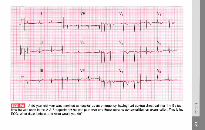
ECG 96 A 50-year-old man was admitted to hospital as an emergency, having had central chest pain for 1 h. By thetime he was seen in the A & E department he was pain-free and there were no abnormalities on examination. This is hisECG. What does it show, and what would you do?
ANSWER 96
The ECG shows:
• Sinus rhythm with one supraventricularextrasystole (there appears to be an abnormal Pwave in lead V1, so this is atrial in origin)
• Normal axis• Normal QRS complexes• Inverted T waves in leads VL, V1-V6
Clinical interpretationThere are many causes of inverted T waves, and ECGsshould always be interpreted as part of the overallclinical picture. In this case the history suggests amyocardial infarction, and the ECG is characteristicof an acute anterior non-Q wave infarction.
What to doThe immediate risk is low and there is no evidencefor benefit from thrombolysis. Although thepatient is now asymptomatic he should remain inhospital for observation. The risk of reinfarction inthe next 3 months is relatively high compared with
the risk following a Q wave infarction, and furtherinvestigation is needed.
SummaryAnterolateral non-Q wave myocardial infarction.
See p. 103
See p. 266

[2J3 This ECG was recorded as part of the routine preoperative assessment of a 65-year-old man who had nocardiovascular symptoms, and whose heart was clinically normal. What does it show? Is any action necessary?

ANSWER 97
The ECG shows:
• Sinus rhythm• First degree block (PR interval 280 ms)• Normal axis• Right bundle branch block
Clinical interpretationThe first degree block indicates conduction delayin the His bundle or left bundle branch, inaddition to complete block of the right bundlebranch.
What to doThe danger is that complete heart block willdevelop, though the present situation couldremain stable for a prolonged period. There iscertainly no urgency regarding treatment, butmany cardiologists would recommend theinsertion of a permanent pacemaker. No othertreatment would be helpful. Consider thepossibility that he has an anterior septal defect.
Summary *Sinus rhythm, first degree block and right bundlebranch block.
See pp. 30 and 37
See p. 137

fcEl This ECG was recorded from a 50-year-old man who had had severe chest pain for 1 h. What does it showand what would you do?

ANSWER 98
The ECG shows:
• Sinus rhythm with ventricular extrasystoles• Normal axis• Q waves in leads V3-V5
• Raised ST segments in leads I, VL, V3-V6
• Depressed ST segments in leads III, VF
Clinical interpretationVentricular extrasystoles associated with an acuteanterolateral myocardial infarction.
What to doThe patient should be given diamorphine andaspirin immediately, and thrombolysis as soon aspossible. The extrasystoles should not be treated.
SummaryAcute anterolateral myocardial infarction withventricular extrasystoles.
iE I See p. 98
See p. 242

ECG 99 This ECG was recorded from a 30-year-old man who complained of chest pain: the pain did not appear tobe cardiac in origin, and physical examination was normal. Can this man be allowed to hold a commercial drivinglicence?

ANSWER 99
The ECG shows:
• Sinus rhythm• Normal axis• Small Q waves, especially prominent in leads I,
II,V4-V6
• Otherwise normal QRS complexes• T wave inversion in lead III but not VF
Clinical interpretationThese Q waves are quite deep but only 40 ms induration, and they are most prominent in thelateral leads. They represent septal depolarization,not an old lateral infarction. An inverted T wave inlead III but not in lead VF is a normal variant.
What to doThe ECG is normal, and if the man has noother evidence of heart disease he can hold acommercial driving licence. If in doubt an exercisetest should be performed.
SummaryNormal ECG.
See p. 89

This ECG was recorded from an 80-year-old woman who had been found unconscious with physical signssuggesting a stroke. Any comments?

ANSWER 100
The ECG shows:
• Atrial fibrillation with a ventricular rate of50/min
• QRS duration prolonged at 160 ms• Prominent']' waves, best seen in leads V3-V6
• Widespread but non-specific ST/T changes
Clinical interpretationThe atrial fibrillation may or may not be relatedto her stroke - she may have had a cerebralembolus, or she may have both coronary andcerebrovascular disease. The slow ventricularrate and the ']' waves indicate hypothermia,and her core temperature was 25°C. ECGs fromhypothermic patients seldom show ']' waves asclearly as this because there are too many artefactsdue to shivering - but this patient was too cold toshiver. She did not survive. Summary
Atrial fibrillation and hypothermia.
See p. 358

A 32-year-old woman, who had a normal pregnancy and delivery 3 months previously, was seen in theout-patient department complaining of breathlessness and dizziness on exertion. She had had no chest pain. Does herECG help with her diagnosis and treatment?

ANSWER 101
The ECG shows:
• Sinus rhythm• Right axis deviation• Normal QRS complexes except for an RSR
pattern in lead V2 and deep S waves in lead V6
• Inverted T waves in leads V1-V4
Clinical interpretationThe right axis deviation, the deep S waves in leadV6 ('clockwise rotation') and the inverted T wavesin the chest leads are all characteristic of markedright ventricular hypertrophy: the only missingfeature is the absence of dominant R waves in leadV1. Note how the T wave inversion is at amaximum in lead V1 and becomes progressivelyless marked from lead V2 to V4.
What to doIn the context of a delivery 3 months previously,this ECG pattern of right ventricular hypertrophyalmost certainly indicates multiple small pulmonaryemboli causing pulmonary hypertension. A lung scan
would confirm this diagnosis and anticoagulants areneeded urgently.
SummaryRight ventricular hypertrophy.
IE I See pp. 91 and 117
See p. 289

This ECG was recorded from a fit 60-year-old man at a routine medical examination. What does it showand what would you recommend?

ANSWER 102
The ECG shows:
• Sinus rhythm• Normal PR interval• Left axis deviation• Right bundle branch block
Clinical interpretationThe combination of left axis deviation (also calledleft anterior hemiblock, because it is due to blockof the anterior fascicle of the left bundle branch)and right bundle branch block is called bifascicularblock. Atrioventricular conduction depends on theposterior fascicle of the left bundle branch.
What to doProgression to complete block can occur but isrelatively rare. In the absence of symptoms,standard UK practice would be not to insert apermanent pacemaker.
SummaryLeft axis deviation and right bundle branchblock - bifascicular block.
See p. 48
See pp. 140 and 146

This ECG was recorded as part of a routine examination of a healthy 25-year-old professional athlete.There were no abnormal physical findings. What does it show and what would you do?

ANSWER 103
The ECG shows:
• Sinus rhythm• Normal axis• Normal QRS complexes apart from narrow Q
waves in lead VL• Marked T wave inversion in leads I, VL, V2-V6
Clinical interpretationIf this ECG had been recorded from a middle-aged man presenting with acute chest pain, thediagnosis would be an anterior non-Q waveinfarction, or possibly an early lateral Q waveinfarction. The ECGs of athletes can show STsegment and T wave changes due to leftventricular hypertrophy, but anteroseptal T waveinversion of this degree in a healthy young manalmost certainly represents hypertrophiccardiomyopathy.
What to doEchocardiography will confirm the diagnosis.Ambulatory ECG tape-recording will show
whether the patient is having ventriculararrhythmias. He must not play competitive sportsand his close relatives should be screened.
SummaryProbable hypertrophic cardiomyopathy.
'ifr:;) Seep. 117 and 120
***

j This ECG was recorded from a 45-year-old man, who had been admitted to a coronary care unit with amyocardial infarction and who was recovering well. What is the rhythm, and what would you do?

ANSWER 104
The ECG shows:
• Broad complex rhythm at 90/min• No P waves• Marked left axis• QRS duration 160 ms• All chest leads show a downward QRS complex
(concordance)
Clinical interpretationIf the heart rate were fast there would be littledifficulty in recognizing this as ventriculartachycardia, and this rhythm used to be called'slow VT. It is, however, an acceleratedidioventricular rhythm.
What to doThis rhythm is quite commonly seen in patients withan acute myocardial infarction, and indeed is notuncommon in ambulatory ECG records from normalpeople. It never causes problems, and it is importantnot to attempt to treat it: suppressing any 'escape'rhythm may lead to a dangerous bradycardia.
SummaryAccelerated idioventricular rhythm.
IE I See p. 63
See pp. 35 and 36
***

This ECG was recorded from a 50-year-old man who had hypertension but was otherwise well. Despiteshowing four possible 'abnormalities', is the trace actually normal?

ANSWER 105
The ECG shows:
• Sinus rhythm, rate 34/min• Normal axis• Bifid P waves, best seen in the lateral leads• Small narrow Q waves in the lateral leads• Normal QRS complexes• Peaked T waves
Clinical interpretationSinus bradycardia can be due to physicalfitness, vagal overactivity or myxoedema. In ahypertensive patient, beta-blocker treatment isthe likely explanation. The bifid P waves mayindicate left atrial hypertrophy ('P mitrale'), butcan be normal. The lateral Q waves are normal,representing septal depolarization. The peakedT waves could be due to hyperkalaemia but aremore often a normal variant.
What to doCheck the serum potassium level.
SummaryNormal ECG.
See pp. 80 and 96
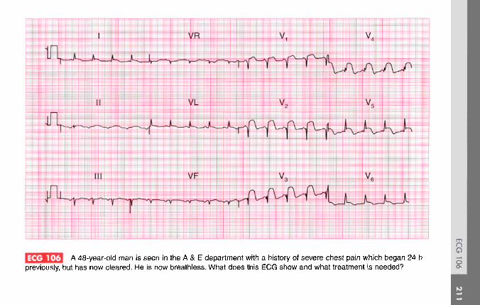
A 48-year-old man is seen in the A & E department with a history of severe chest pain which began 24 hpreviously, but has now cleared. He is now breathless. What does this ECG show and what treatment is needed?

ANSWER 106
The ECG shows:
• Sinus rhythm• Normal axis• Normal QRS complexes• Raised ST segments in leads I, VL, V2-V6
Clinical interpretationThe ECG has the classic appearance of an acuteanterolateral myocardial infarction.
What to doSince this patient's chest pain began more than24 h ago, thrombolysis is not indicated. Thebreathlessness suggests that he may havedeveloped left ventricular failure, and he must beadmitted to hospital and treated with diureticsand if necessary intravenous nitrates to inducevasodilation. The patient will need long-termtreatment with an angiotensin-converting enzymeinhibitor: the best time to begin treatment is amatter for debate but it should be within 2 or 3
days. He will also need long-term treatment withaspirin as a prophylactic against further infarction.
SummaryAcute anterolateral myocardial infarction.
fij See p. 98
See pp. 242 and 244

A 50-year-old woman came to the A & E department because of the sudden onset of palpitations andsevere breathlessness. What abnormality does this ECG show, and what conditions might be responsible?

ANSWER 107
The ECG shows:
• Atrial fibrillation• Irregular QRS complexes with a ventricular rate
of up to 200/min• Normal axis• Height of R waves in lead V6 plus depth of S
waves in lead V2 is greater than 35 mm,suggesting left ventricular hypertrophy
• Otherwise normal QRS complexes, apart froman RSR pattern in lead VF
• ST segments depressed in leads V4-V6,suggesting ischaemia
• Normal T waves
Clinical interpretationAtrial fibrillation with an uncontrolled ventricularrate. The ischaemic changes in leads V4 and V5 areprobably related to the heart rate.
What to doIschaemia may have been the cause of the atrialfibrillation, or the rapid ventricular rate itself may
be responsible for the ischaemic changes.Ischaemia is not a likely primary diagnosis in a50-year-old woman, and the things to think aboutare rheumatic heart disease (particularly withmitral stenosis), thyrotoxicosis, alcoholism, andother forms of cardiomyopathy. Immediatetreatment of heart failure with diuretics andintravenous nitrates may be necessary, but theventricular rate is best controlled by digoxin,which can be given intravenously if necessary. DCcardioversion may be necessary if the patient is insevere heart failure, but it is best to establish theunderlying cause of the atrial fibrillation first ifpossible. Remember that a patient with atrialfibrillation probably needs long-termanticoagulants.
SummaryAtrial fibrillation with a rapid ventricular rate andischaemic changes.
See pp. 78 and 102
See p. 315

ECG 108 A 70-year-old woman, admitted to hospital because of increasing heart failure of uncertain cause, collapsedand was found to have a very rapid pulse and a low blood pressure. This is her ECG. She recovered spontaneously. Whatis this rhythm, and what would you do?
ANSWER 108
The ECG shows:
• Broad complex tachycardia at about 180/min• No P waves visible• Left axis• QRS duration about 140 ms• The fourth and fifth QRS complexes are narrow• QRS complexes are probably concordant (in the
chest leads all point upwards) though it isdifficult to be certain
Clinical interpretationBroad complex tachycardias may be ventricular,supraventricular with bundle branch block, or maybe due to a Wolff-Parkinson-White syndrome. Wehave no ECG from this patient recorded when shewas in sinus rhythm, which is always the mosthelpful thing in deciding between these possibilities.The complexes are not very wide, which wouldbe consistent with a supraventricular origin withaberrant conduction, but the left axis and (probable)concordance point to ventricular tachycardia. Thekey is the two narrow complexes near the beginning
of the record: these are slightly early and areprobably capture beats. They indicate that with anearly supraventricular beat the conducting systemcan function normally; by implication, the broadcomplexes must be due to ventricular tachycardia.
What to doAn elderly patient with heart failure is more likelyto have ischaemic disease than anything else, butall ther possible causes of heart failure must beconsidered. The sudden onset of an arrhythmiacould be due to a myocardial infarction. Pulmonaryemboli can cause sudden arrhythmias, though theseare more often supraventricular than ventricular. Itis important to consider whether this rhythmchange is related to treatment, either because of anelectrolyte imbalance or the pro-arrhythmic effect ofa drug the patient is taking.
SummaryVentricular tachycardia.
IE I See p. 72
See p. 192
***

This is a continuous rhythm strip (lead II) from a 25-year-old man who complained of episodes of fast,regular, palpitations associated with breathlessness and dizziness. There were no abnormalities on examination otherthan a slow and irregular pulse, and apart from the rhythm shown here, the ECG was normal. What is the diagnosis andhow can his problem be treated?

ANSWER 109
The ECG shows:
• Variable QRS rate (16-60/min)• No P waves• No irregular baseline to suggest atrial fibrillation• As far as can be ascertained from the rhythm
strip, normal QRS complexes and T waves
Clinical interpretationThis is the 'sick sinus syndrome' or 'sinoatrialdisease'. The record shows a 'silent atrium'with a variable junctional escape rhythm. Thepalpitations described by the patient are probablydue to a paroxysmal supraventricular tachycardia,so he probably has the 'bradycardia-tachycardia'variant of sinoatrial disease.
What to doAmbulatory ECG tape-recording will confirm thecause of the patient's palpitations. Even thoughhis bradycardia is asymptomatic, he will need apermanent pacemaker because anti-arrhythmic
agents given for the tachycardia may make hisbradycardia worse.
Summary ***Sinoatrial disease with a 'silent atrium' and junctionalescape.
Ip- | Seep. 199

This ECG was recorded from an 80-year-old man during a routine preoperative assessment. What does itshow? What are the implications for surgery?

ANSWER 110
The ECG shows:
• Sinus rhythm with ventricular extrasystoles• Ventricular extrasystoles are of two types, seen
best in lead V3
• Normal axis• Normal QRS complexes• ST segments and T waves are normal
Clinical interpretationSinus rhythm, with multifocal ventricularextrasystoles but otherwise normal.
What to doIn large groups of patients, ventricularextrasystoles are correlated with heart diseaseof all types. In individuals, however,extrasystoles may well occur in a perfectlynormal heart - indeed virtually everyone hasextrasystoles at times. Ventricular extrasystolesbecome more common with increasing age,and this patient is 80 years old. In the absenceof symptoms or clinical signs suggesting
cardiovascular disease, the extrasystoles donot have any great significance and should notmaterially affect the patient's fitness for surgery.They should not be treated.
SummarySinus rhythm with multifocal ventricular extrasystoles.
See p. 64
Seep. 104

A 50-year-old man was admitted to hospital as an emergency with chest pain; he was not aware of a rapidheart rate. He had had several episodes of pain that appeared to be due to ischaemia, but these had no clearrelationship with exertion. Shortly after this ECG was recorded, his heart rate suddenly slowed and his ECG was thennormal. What does this record show, and what would you do?
nO

ANSWER 111
The ECG shows:
• Narrow complex tachycardia, rate about 200/min• No P waves• Normal axis• Normal QRS complexes• Horizontal ST segment depression, most marked
in lead V4
Clinical interpretationNarrow complex tachycardia without P waves -atrioventricular re-entry (junctional) tachycardia.Ischaemic ST segment depression, accounting forhis pain.
What to doNot all patients with a paroxysmal tachycardiacomplain of palpitations; this patient's recurrentchest pain may well have been due to thisarrhythmia. He should be taught the methodsof inducing vagal activity, but prophylactic drugtherapy will be needed: a beta-blocker or verapamilshould be tried first. Electrophysiological
investigation, with a view to ablating an abnormalpathway, may be needed.
SummaryJunctional tachycardia with ischaemia.
Ill See pp. 72 and 102
IP ] See p. 44

A 40-year-old man is seen in the out-patient department with a history that suggests a myocardialinfarction 3 weeks previously. There are no abnormalities on examination, and this is his ECG. There are two possibleexplanations for the abnormality it shows, though only one of these explains his history. What is the likely diagnosis?
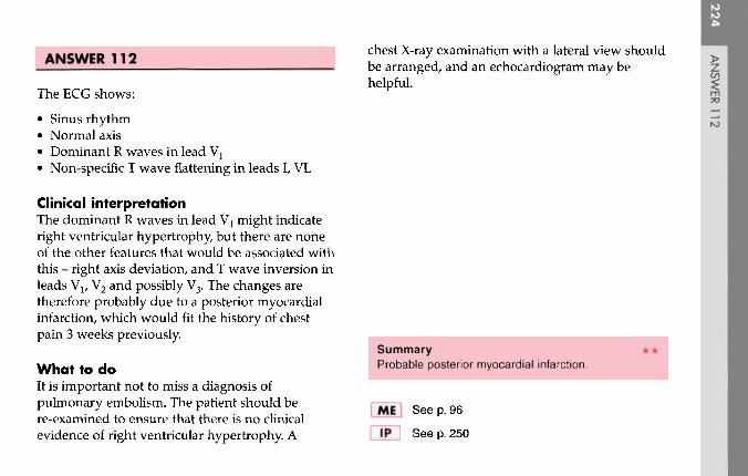
The ECG shows:
• Sinus rhythm• Normal axis• Dominant R waves in lead Va
• Non-specific T wave flattening in leads I, VL
Clinical interpretationThe dominant R waves in lead V1 might indicateright ventricular hypertrophy, but there are noneof the other features that would be associated withthis - right axis deviation, and T wave inversion inleads Vl, V2 and possibly V3. The changes aretherefore probably due to a posterior myocardialinfarction, which would fit the history of chestpain 3 weeks previously.
What to doIt is important not to miss a diagnosis ofpulmonary embolism. The patient should bere-examined to ensure that there is no clinicalevidence of right ventricular hypertrophy. A
chest X-ray examination with a lateral view shouldbe arranged, and an echocardiogram may behelpful.
SummaryProbable posterior myocardial infarction.
See p. 96
See p. 250
ANSWER 112

This ECG was recorded from a 60-year-old man with no symptoms, who wanted a private pilot's licence.Is it normal? How could you convince the licensing authority?

ANSWER 113
The ECG shows:
• Sinus rhythm• Normal conduction• Normal axis• Q wave in lead III but not in lead VF• Inverted T wave in lead III but not in
lead VF
Clinical interpretationA Q wave and an inverted T wave inlead III, but not in lead VF, is a normalvariant. If lead III is recorded with thepatient taking a deep breath in, thechanges will usually normalize asshown on the right.
SummaryNormal record.
See p. 80

This ECG was recorded from a 55-year-old black woman who had been complaining for several years ofchest pain, and who was admitted to hospital with persistent pain that was not characteristic of ischaemia. How wouldyou have managed her?

ANSWER 114
The ECG shows:
• Sinus rhythm• Normal axis• Normal QRS complexes• T wave inversion in leads I, VL, V2-V6
Clinical interpretationWith this history a non-Q wave anterolateralinfarction has to be the first diagnosis, but T wave'abnormalities' are common in black people, andthis ECG could be normal.
What to doIn this patient an infarction was excluded whenthe cardiac enzyme levels were found to benormal. An exercise test was performed, but waslimited by breathlessness without further ECGchange. A coronary angiogram was completelynormal.
Summary ***Widespread T wave 'abnormalities,' normal in a blackwoman.
IE Seep. 112
See p. 89

A 50-year-old man, who had complained of attacks of dizziness and palpitations for several years,collapsed at work and was brought to the A & E department. He was cold and clammy. His heart rate was rapid and hisblood pressure was unrecordable. There were signs of left ventricular failure. This is his ECG. What does it show andwhat would you do?

ANSWER 115
The ECG shows:
• Narrow-complex tachycardia, rate 300/min• No definite P waves• Normal QRS complexes• Horizontal ST segment depression in lead V5
Clinical interpretationA regular narrow-complex tachycardia at 300/minprobably represents atrial flutter with 1:1conduction (i.e. each atrial activation causesventricular activation).
What to doThe cardiovascular collapse results from the rapidheart rate, with a loss of diastolic filling. Carotidsinus pressure may temporarily increase thedegree of block and establish the diagnosis, but itis unlikely to convert atrial flutter to sinus rhythm.Intravenous adenosine is likely to have the sameeffect. A patient who is haemodynamicallycompromised by a tachycardia should be treatedwith immediate DC cardioversion.
SummaryProbable atrial flutter with 1:1 conduction.
IfJ See p. 68
IP~l See p. 160

A 70-year-old man, who had had angina for several years, began to complain of dizzy attacks. This is hisECG. What does it show and what would you do?

ANSWER 116
The ECG shows:
• Sinus rhythm with frequent multifocalventricular extrasystoles
• Normal PR interval• Normal axis• Q waves in leads II, III, VF• T waves flattened or inverted in leads II, III,
v5-v6
Clinical interpretationThe ECG shows a probable old inferiormyocardial infarction, which accounts for hisangina. Ventricular extrasystoles are ofthemselves usually not important, but in apatient complaining of dizzy attacks, ventricularextrasystoles that are frequent and multifocalmay be causing haemodynamic impairment.
What to doIt would be worth recording an ambulatory ECGto see if the patient is having runs of ventriculartachycardia, but the extrasystoles probably do
need suppressing. Sotalol would be the first drugto try, and then amiodarone.
SummaryOld inferior myocardial infarction and frequentmultifocal ventricular extrasystoles.
lEj See p. 96
IP~1 See p. 154

This ECG was recorded from a 50-year-old woman who complained of breathlessness and palpitations.What physical signs would you look for, and what is the next stage in her management?

ANSWER 117
The ECG shows:
• Sinus rhythm with ventricular extrasystoles• Broad notched P waves, best seen in leads II, III,
VF• Normal axis• Normal QRS complexes• Diphasic T waves in leads V2, V3; inverted T
waves in lead VL
Clinical interpretationThe broad P waves suggest left atrial hypertrophy.There is nothing to suggest left ventricularhypertrophy, so mitral stenosis must beconsidered. Ventricular extrasystoles may explainthe palpitations, but if the patient has mitralstenosis she may be having episodes of atrialfibrillation. The cause of the T wave changes isnot clear and may be ischaemia.
What to doLook for the tapping apex beat, the loud firstsound, the opening snap and the mid-diastolic
murmur that are characteristic of mitral stenosis.Echocardiography will probably be helpful, andambulatory ECG tape-recording may be necessaryto identify the cause of the palpitations.Treatments to be considered if she has mitralvalve disease and atrial fibrillation are digoxin,anticoagulants, and mitral valve surgery.
If the breathlessness turns out to be due topoor left ventricular function, an angiotensin-converting enzyme inhibitor is needed.
Summary **Sinus rhythm with ventricular extrasystoles, left atrialhypertrophy and possible ischaemia.
IfJ See p. 89
IP~~] See p. 318

A 60-year-old man who complains of ankle swelling is found to have a regular pulse, a blood pressure of115/70, an enlarged heart, and signs of congestive cardiac failure. This is his ECG. What does it show? He isuntreated - how would you manage him?

ANSWER 118
The ECG shows:
• Atrial flutter with 4:1 block• Normal axis• Slight QRS widening• T waves are difficult to identify but at least in
lead VL are inverted
Clinical interpretationAtrial flutter with stable 4:1 conduction explainswhy the heart is clinically regular. The slight QRSwidening is non-specific. There is no evidence of aprevious infarction or of ventricular hypertrophy;the T wave inversion could be ischaemic, but maywell be due to a dilated cardiomyopathy.
What to doAn echocardiogram will show if he has a dilatedleft ventricle, if there is general left ventriculardysfunction or if there are parts of the leftventricle that contract well and others that donot, which would suggest ischaemia.
Atrial flutter is a difficult rhythm to treat: ifthe heart is enlarged it is unlikely that sinusrhythm can be restored either by drugs or by DCcardioversion, and if the block is stable (as appearshere) it may be best left alone. Digoxin must beused cautiously in case it induces a higher degreeof block and bradycardia. The heart failure shouldbe treated with an angiotensin-converting enzymeinhibitor and a diuretic.
This patient had an alcoholic dilated cardio-myopathy.
SummaryAtrial flutter with 4:1 block.
If I Seep. 68
See pp. 160 and 230

Q A 70-year-old man is sent to the out-patient department because of attacks of dizziness. Whatabnormalities does his ECG show, and what treatment is needed?

ANSWER 119
The ECG shows:
• Sinus rhythm• Second degree (2:1) heart block• Left axis deviation• Right bundle branch block
Clinical interpretationLeft axis deviation is due to left anteriorfascicular block (left anterior hemiblock).Since right bundle branch block is present,intra-ventricular conduction depends on theposterior fascicle. The presence of second degreeblock suggests that conduction in this fascicle isalready impaired or that there may be impairedconduction in the bundle of His.
What to doThis patient's attacks of dizziness may be due tothe slow heart rate demonstrated in this ECG, ormay represent intermittent periods of completeblock with an even slower rate and Stokes-Adamsattacks. Permanent pacing is urgently needed and
the patient should be admitted to the hospitalfrom the out-patient department.
Summary ***Second degree (2:1) block, left anterior hemiblock,and right bundle branch block (trifascicular block).
KM See pp. 31 and 48
IP I See p. 21

This ECG was recorded from a 30-year-old man admitted with severe central chest pain. He was a heavysmoker and had a bad family history of ischaemic heart disease. The physical examination was reported to be normal.What do you think is going on?

ANSWER 120
The ECG shows:
• Sinus rhythm, rate 100/min• Normal conduction• Normal axis• Normal QRS complexes• Raised ST segments in leads I, II, VL, V3-V6
• 'High take off ST segment in leads V3-V5
• T wave inversion in lead III
Clinical interpretationIn a patient with chest pain and risk factorsfor a myocardial infarction, an infarction mustobviously be the first diagnosis. The raised STsegments on this ECG could support thisdiagnosis. However, the patient is very youngto have an infarction. The 'high take-off STsegments in leads V3-V5 (raised ST segmentfollowing an S wave) are a normal variant. Theother raised ST segments could well be due topericarditis.
What to doThe patient should be examined lying flat, becausethis gives the best chance of hearing a pericardialfriction rub - and this is what was found here. Thepericarditis could, of course, be due to an infarction,but repeated ECGs showed no development of aninfarction pattern, and the raised ST segmentspersisted for several days. A diagnosis of viralpericarditis was eventually made.
Summary **ST segment elevation, partly 'high take-off' and partlydue to pericarditis.
See p. 100
See pp. 82 and 298

An 18-year-old student complains of occasional attacks of palpitations. These start suddenly withoutprovocation; the heart beat seems regular and is 'too fast to count'. During attacks she does not feel dizzy or breathless,and the palpitations stop suddenly after a few seconds. Physical examination is normal, and this is her ECG. What is thediagnosis and what advice would you give?

ANSWER 121
The ECG shows:
• Sinus rhythm• Very short PR interval• Normal axis• Normal QRS complexes and T waves
Clinical interpretationThis is the Lown-Ganong-Levine syndrome.Unlike the Wolff-Parkinson-White syndrome, inwhich there is an accessory pathway separatefrom the atrioventricular node and His bundle,in the Lown-Ganong-Levine syndrome thereis a bypass close to the atrioventricular nodeconnecting the left atrium and the His bundle.In the Wolff-Parkinson-White syndrome theQRS complex shows an early delta wave, but inthe Lown-Ganong-Levine syndrome the QRScomplex is normal.
What to doAmbulatory ECG tape-recording may confirm thediagnosis if attacks are frequent enough.
Infrequent and short-lived attacks such as thispatient describes are not dangerous, but sheshould be taught vagal-stimulating proceduressuch as Valsalva's manoeuvre and carotid sinuspressure. An electrophysiological study andablation of the abnormal tract may be necessary.
SummaryLown-Ganong-Levine syndrome.
See pp. 40 and 127
***
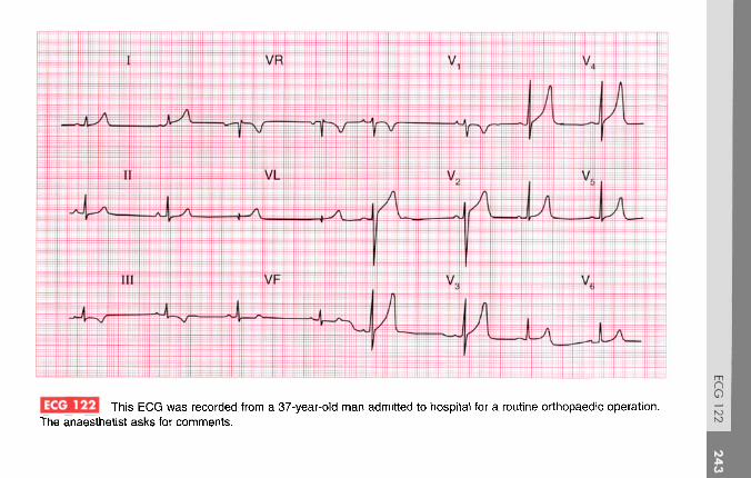
This ECG was recorded from a 37-year-old man admitted to hospital for a routine orthopaedic operation.The anaesthetist asks for comments.

ANSWER 122
The ECG shows:
• Sinus rhythm, 45/min• Normal axis• Normal QRS complexes• Inverted T waves in lead III and ST segment
depression in lead VF• Peaked T waves in the anterior leads
Clinical interpretationProvided the patient is not taking a beta-blocker,the slow heart rate is probably a reflection ofphysical fitness. Inverted T waves in lead III, andthe downward-sloping ST segments in lead VF, areprobably normal. Peaked T waves are characteristicof hyperkalaemia, and are sometimes describedas 'hyperacute' in ischaemia. However, when aslarge as this - and particularly when the patient isasymptomatic - peaked T waves are nearly alwaysperfectly normal.
What to doEnsure that the patient has no cardiac symptoms,and check his electrolyte levels pre-operatively.
SummaryNormal ECG.
See p. 91
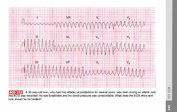
A 30-year-old man, who had had attacks of palpitations for several years, was seen during an attack, andthis ECG was recorded. He was breathless and his blood pressure was unrecordable. What does the EG show andhow should he be treated?
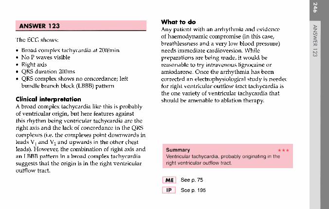
ANSWER 123
The ECG shows:
• Broad complex tachycardia at 200/min• No P waves visible• Right axis• QRS duration 200ms• QRS complex shows no concordance; left
bundle branch block (LBBB) pattern
Clinical interpretationA broad complex tachycardia like this is probablyof ventricular origin, but here features againstthis rhythm being ventricular tachycardia are theright axis and the lack of concordance in the QRScomplexes (i.e. the complexes point downwards inleads V1 and V2 and upwards in the other chestleads). However, the combination of right axis andan LBBB pattern in a broad complex tachycardiasuggests that the origin is in the right ventricularoutflow tract.
What to doAny patient with an arrhythmia and evidenceof haemodynamic compromise (in this case,breathlessness and a very low blood pressure)needs immediate cardioversion. Whilepreparations are being made, it would bereasonable to try intravenous lignocaine oramiodarone. Once the arrhythmia has beencorrected an electrophysiological study is needed,for right ventricular outflow tract tachycardia isthe one variety of ventricular tachycardia thatshould be amenable to ablation therapy.
Summary ***Ventricular tachycardia, probably originating in theright ventricular outflow tract.
IE | See p. 75
See p. 195
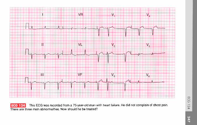
This ECG was recorded from a 75-year-old man with heart failure. He did not complain of chest pain.There are three main abnormalities. How should he be treated?

ANSWER 124
The ECG shows:
• Sinus rhythm with one ventricular extrasystole• Left axis deviation• Slight widening of the QRS complexes• Q waves in leads V2-V5
• Raised ST segments in the anterior leads• Inverted T wave in lead VL; flattened T waves
in leads I, V6
Clinical interpretationA 'silent' anterior infarction of uncertain age hascaused left anterior hemiblock, which explains theleft axis deviation and widening of the QRScomplexes. The lateral T wave changes arepresumably due to ischaemia.
What to doVentricular extrasystoles should not be treated,and left anterior hemiblock is not an indication forpacing. In the absence of pain, the anterior infarctioncannot be assumed to be new, so thrombolysis
should not be given. He needs an angiotensin-
Summary **Left anterior hemiblock and anterior infarction ofuncertain age; one ventricular extrasystole.
See pp. 46 and 95
IP I See p. 18

A 35-year-old woman, who had had palpitations for many years without any diagnosis being made, waseventually seen in the A & E department during an attack. She looked well and was not in heart failure, and her bloodpressure was 120/70. This is her ECG. What is the rhythm, and what would you do?
ANSWER 125
The ECG shows:
• Broad-complex tachycardia (QRS complex200 ms), rate nearly 200/min
• No P waves visible• Right axis deviation• Right bundle branch block pattern• In lead Vl, the R1 peak is higher than the R peak
Clinical interpretationThe problem here is to distinguish asupraventricular tachycardia with bundle branchblock from ventricular tachycardia. The clinicalhistory is not helpful, nor is the fact that the patientis haemodynamically stable. The combination ofright axis deviation, right bundle branch block andthe R1 peak being higher than the R peak make itlikely that this is a supraventricular tachycardiawith right bundle branch block rather thanventricular tachycardia. However, the very broadQRS complex (>140 ms) would favour a ventricularorigin for the arrhythmia.
What to doCarotid sinus massage. If this has no effect tryintravenous adenosine, and if this is ineffective tryintravenous lignocaine.
Summary ***Broad-complex tachycardia with right bundle branchblock pattern, probably supraventricular in origin.
UsJ See p. 77
IPl See p. 171

This ECG was recorded as part of the routine investigation of a 40-year-old man who was admittedfollowing a first seizure. He was unconscious, had a stiff neck, and bilateral extensor plantar responses. His heart wasclinically normal. What do you think has happened?

ANSWER 126
This ECG shows:
• Sinus rhythm, rate 80/min• Normal PR interval and QRS duration• Normal axis• Normal QRS complexes• ST depression in lead II• T wave inversion in leads III, VF, V4-V6
Clinical interpretationThe appearances here are suggestive of an anteriornon-Q wave myocardial infarction, but this doesnot correspond with the clinical picture.
What to doIt is possible that this patient had a myocardialinfarction which caused a cerebrovascular accidentbecause of an arrhythmia, or because of a cerebralembolus, and that the cerebrovascular accidentcaused the seizure. The unconsciousness and thebilateral extensor plantar responses could simplybe post-ictal. However, such a sequence would notexplain the stiff neck, which would seem to point
to either a subarachnoid haemorrhage ormeningitis. Changes like those on this ECG arecommon in subarachnoid haemorrhage, probablybecause of intense coronary vasospasm resultingfrom catecholamine release. Measurements ofthe blood troponin level are unlikely to helpdifferentiate between a primarily cardiac and aprimarily neurological event. This patient didindeed have a subarachnoid haemorrhage, andthe ECG eventually returned to normal.
SummaryAnterior and inferior T wave inversion due tosubarachnoid haemorrhage.
See p. 382
***

A 70-year-old man with lung cancer is admitted to hospital with abdominal pain and ankle swelling. He hasa raised jugular venous pressure, a tender and distended liver, and marked peripheral oedema. Does this ECG help withthe diagnosis and what might you need to do?

ANSWER 127
The ECG shows:
• Sinus rhythm• Normal axis• Normal width but generally small QRS
complexes• T wave inversion in leads I, II, III, VF, V5-V6
Clinical interpretationSmall QRS complexes are seen with a pericardialeffusion, and sometimes in patients with chroniclung disease. The widespread T wave changeswould be consistent with pericardial disease. Thereis nothing in this record to suggest pulmonarydisease.
What to doThe physical findings and the ECG would fit witha pericardial effusion associated with malignancy.You should look carefully at the jugular venouspressure to see if it rises with inspiration, indicatingpericardial tamponade. Echocardiography isessential, and if there is evidence of right
ventricular collapse in diastole, a pericardial drainshould be inserted.
This patient had a malignant pericardial effusion.
Summary ***Small QRS complexes and widespread T wavechanges consistent with a pericardial effusion.
If] See p. 11
See p. 363

This ECG was recorded from a 65-year-old woman who had had a mitral valve replacement to treatrheumatic valve disease, and who was admitted to hospital with generalised lethargy, nausea and vomiting. What doesthe ECG show and what would you do? Unfortunately the chemical pathology laboratory burned down last night!

ANSWER 128
The ECG shows (note: chest leads at half sensitivity):
• Atrial fibrillation• Right axis deviation• Normal QRS complexes, except for a tall R wave
in lead V1
• Generally flattened T waves• U waves best seen in leads V4-V 5
• Downward-sloping ST segments/ best seen inleads II, III, VF
Clinical interpretationThe atrial fibrillation, and the right axis deviationand tall R waves in lead V1 (indicating rightventricular hypertrophy) probably pre-date thevalve replacement. The flat T waves with obviousU waves suggest hypokalaemia. The downward-sloping ST segments suggest digoxin effect.
What to doThe clinical picture fits hypokalaemia and digoxintoxicity. Since the electrolyte and digoxin levelscannot be measured, stop the patient taking
digoxin and any potassium-losing diuretics. Giveher potassium orally. Monitoring the T and Uwaves is a crude but effective way of judging theserum potassium level.
Summary ***Atrial fibrillation, hypokalaemia and digoxin effect.
IE] Seep. 108
See pp. 336 and 366

3 A 20-year-old man is seen in the A & E department with a head injury; there is a vague story of a collapse.What does the ECG show?

ANSWER 129
The ECG shows:
• Sinus rhythm• Short PR interval• Left axis deviation• Broad QRS complexes with a slurred upstroke
(delta wave) seen best in leads V2-V4
• Inverted T waves in leads I, VL, V6
Clinical interpretationThe short PR intervals and the delta waves arecharacteristic of the Wolff-Parkinson-Whitesyndrome. Superficially, leads I, VL and V6 mightmistakenly be interpreted as suggesting left bundlebranch block, but it is important to look at all leadsbecause the diagnosis here is best seen in lead V2.
What to doThe Wolff-Parkinson-White syndrome isassociated with paroxysmal tachyarrhythmia,which may cause collapse. AsymptomaticWolff-Parkinson-White syndrome should beleft untreated, but it is important in this case to
establish - perhaps by ambulatory ECG recordingand exercise testing - whether the patient ishaving a paroxysmal tachycardia or not.
If there is reason to suppose that an arrhythmiacaused a collapse and head injury he needselectrophysiological ablation of the abnormalconducting pathway.
SummaryWolff-Parkinson-White syndrome.
See p. 81
See pp. 37 and 198
***

An 85-year-old man is seen in the out-patient department complaining of typical angina and of occasionaldizziness when walking up hills. This is his EGG. What is the diagnosis, and what would you do?

ANSWER C30
The ECG shows:
• Sinus rhythm• Normal axis• Tall R waves and deep S waves in the chest leads• Inverted T waves in leads I, II, VL, V3-V6
Clinical interpretationThis is marked left ventricular hypertrophy. Itcan be difficult to distinguish T wave inversiondue to ischaemia from the T wave inversion ofleft ventricular hypertrophy, and when the Twave is inverted in the septal leads (V3-V4),ischaemia has to be considered. However, herethe change is most marked in the lateral leads,and is associated with Voltage criteria' for leftventricular hypertrophy. Angina, dizziness andleft ventricular hypertrophy in an 85-year-old arealmost certainly due to tight aortic stenosis,though hypertension is a possibility.
What to doLook for the signs of aortic stenosis ('plateau'pulse, narrow pulse pressure, displaced apex beat,aortic ejection systolic murmur) and confirm thevalve gradient with echocardiography. In thispatient the aortic valve gradient was 95 mmHgindicating severe stenosis of the valve. Anginaand dizziness indicate severe disease and a poorprognosis: even at the age of 85 years, aortic valvereplacement can be a very successful procedure.
SummaryLeft ventricular hypertrophy.
IE I See pp. 93 and 117
U» | See p. 117

This ECG was recorded from a 15-year-old boy who collapsed while playing football, but was well by thetime he was seen. What are the possible diagnoses?

ANSWER 131
The ECG shows:
• Sinus rhythm• Normal PR and QRS duration• Normal axis• Normal QRS complexes• Inverted T waves in leads V1-V3
• Long QT interval (520 ms)
Clinical interpretationA collapse during exercise raises the possibilityof aortic stenosis, hypertrophic cardiomyopathy,or an exercise-induced arrhythmia. This ECGdoes not show the pattern of left ventricularhypertrophy, so aortic stenosis is unlikely. AnteriorT wave inversion is characteristic of hypertrophiccardiomyopathy, but this does not typically causea prolonged QT interval. Exercise-inducedarrhythmias are typical of the familial long-QTsyndrome, and this boy's sister had died suddenly.
What to doInitial treatment is with a beta-blocker, but an ICD(implanted defibrillator) must be considered.
SummaryCongenital long-QT syndrome.
See p. 128

A 70-year-old man, who has had angina for 10 years, is admitted to hospital with severe central chest painthat has been present for 4 h. This is his ECG. What does it show and what would you do?

ANSWER 132
The ECG shows:
• Sinus rhythm• Normal axis• Q waves in leads III, VF• Normal QRS complexes elsewhere• Raised ST segments in leads II (following small S
waves), III, VF• Diphasic T waves in leads V2-V3
• Inverted T waves in leads V4-V5
Clinical interpretationThe inferior Q waves suggest an old infarction.The raised ST segments in leads III and VF wouldbe compatible with an acute infarction, though theraised ST segment in lead II is a 'high take-offbecause it follows an S wave, and this raises thepossibility that the change in leads III and VF maynot be significant. The anterior changes suggest anon-Q wave infarction.
What to doThere is enough evidence here to justifythrombolysis which should, of course, becombined with pain relief and aspirin.
Summary *Possible old and/or possible new inferior myocardialinfarction; non-Q wave anterior myocardial infarction.
IE I See pp. 94 and 103
IP | See p. 254

This ECG was recorded from a 30-year-old man as part of a private 'health screening' examination. Hewas asymptomatic, but is the ECG normal?

ANSWER 133
The ECG shows:
• Sinus rhythm• Normal axis• 'Voltage criteria' for left ventricular hypertrophy
(height of R wave in lead V6 plus depth of Swave in lead V2 exceeds 35 mm)
• Inverted T waves in lead III• Prominent U waves in leads V2-V4
Clinical interpretation'Voltage criteria' for left ventricular hypertrophyare unreliable, and are often exceeded in youngmen. There is nothing else here to suggestventricular hypertrophy. The inverted T wavesin lead III are an acceptable variation of normal.When U waves are due to electrolyte abnormalities(usually hypokalaemia), they are associated withflattened T waves. When they follow tall T waves,as here, they are normal.
What to doProvided the patient's blood pressure is normaland the examination reveals no other cause for leftventricular hypertrophy, such as aortic valvedisease, no further action is required.
SummaryNormal ECG. ***
See pp. 89 and 96

This ECG was recorded from a 60-year-old man admitted to hospital with severe heart failure. What does itshow and what would you do?

ANSWER 134
The ECG shows:
• Atrial fibrillation• Left anterior hemiblock• Left ventricular hypertrophy by Voltage criteria'
(height of R waves in lead V6 plus depth of Swaves in lead V1 = 40 mm)
• Lateral T wave inversion
Clinical interpretationDespite atrial fibrillation, the ventricular rate iswell controlled. Left anterior hemiblock indicatesdisease of the conduction system. The lateral Twave inversion may be due to intraventricularconduction delay, to left ventricular hypertrophy,or may be due to ischaemia. The T wave changesdo not have the characteristics of digoxin effect.
What to doIt is essential to know what treatment the patientis already receiving. If he is not taking digoxin thismust be used cautiously, because the ventricularrate is slower than one might expect in atrial
fibrillation. It is important to seek a cause for leftventricular hypertrophy, remembering that thepatient's present blood pressure may not berepresentative of its usual level.
Summary ***Atrial fibrillation, left anterior hemiblock and probableleft ventricular hypertrophy.
See pp. 46, 78 and 91
See pp. 20 and 320

A 60-year-old man had complained of occasional episodes of palpitations for several years. Betweenattacks he was well, there were no physical abnormalities, and his ECG was normal. Eventually this ECG was recordedduring one of his attacks. What is the arrhythmia and what would you do?

ANSWER 135
The ECG shows:
• Regular broad-complex tachycardia• QRS complex duration 160 ms• Left axis deviation• Indeterminate QRS complex configuration, but
the complexes point downwards in all the chestleads, with a QS pattern in lead V6
Clinical interpretationThe combination of broad-complex, regulartachycardia; 'concordant' QRS complexes pointingdownwards in all leads; and the QS pattern in leadV6 is characteristic of ventricular tachycardia.
What to doPatients who only have occasional episodes ofan arrhythmia, and who are otherwise well, arealways difficult to manage. This patient shouldcertainly have an echocardiogram to exclude acardiomyopathy, and an exercise test to excludeischaemia and exercise-induced arrhythmias. Atthe age of 60 years, coronary angiography is
probably indicated. Treatment with amiodarone isas effective as the selection of an anti-arrhythmicagent on the basis of repeated electrophysiologicalstudies.
If the episodes were causing syncope animplanted defibrillator could be considered.
SummaryVentricular tachycardia.
IE] See p. 75
l|> I See p. 170

A 25-year-old woman, who had had episodes of what sound like a paroxysmal tachycardia for 10 years,produced this ECG when seen during an attack. What is the rhythm, and what is the underlying problem?

ANSWER 136
The ECG shows:
• Irregular tachycardia at about 200/min• No consistent P waves visible• Left axis• QRS duration varies between about 120 and 160 ms• QRS shows a dominant R wave in lead V1 and a
prominent S wave in lead V6
• After the longer pauses, the upstroke of the QRScomplexes appear slurred
Clinical interpretationThe marked irregularity of this rhythm must beexplained by atrial fibrillation. The broad QRScomplexes might be due to right bundle branchblock, but the dominant R wave in lead Vl,
together with the slurred upstroke of the QRScomplex in at least some leads, indicate aWolff-Parkinson-White syndrome (type A).
What to doThe combination of a Wolff-Parkinson-Whitesyndrome and atrial fibrillation is very dangerous,
for it can degenerate to ventricular fibrillation.The arrhythmia needs treating as an emergencywhatever the clinical state of the patient. It isimportant not to use drugs that may block theatrioventricular node and increase conductionthrough the accessory pathway, for this willincrease the risk of ventricular fibrillation.Therefore, adenosine, digoxin, verapamil andlignocaine are contraindicated. The drugs thatslow conduction in the accessory pathway, and aretherefore safe, are the beta-blockers, flecainide andamiodarone. Thereafter, an electrophysiologicalstudy to identify and ablate the accessory pathwayis essential.
Summary ***Atrial fibrillation and a Wolff-Parkinson-Whitesyndrome.
See pp. 78 and 81
See p. 198

ECG 137 A 35-year-old white man is seen in the out-patient department complaining of chest pain on exertion,sometimes with exertion-induced dizziness, and this is his ECG. What is the likely diagnosis? What physical signs wouldyou look for?
ANSWER 137
The ECG shows:
• Sinus rhythm• Normal axis• Normal QRS complexes• Marked T wave inversion in leads I, II, VL,
v4-v6
Clinical interpretationAnterolateral T wave inversion as gross as this maybe due to a non-Q wave infarction, or even to leftventricular hypertrophy. However, there are noother features of left ventricular hypertrophy onthis trace, which is fairly characteristic ofhypertrophic cardiomyopathy.
What to doPhysical signs of hypertrophic cardiomyopathyinclude a 'jerky pulse'; an aortic flow murmurwhich is characteristically louder after thepause that follows an extrasystole; and mitralregurgitation. Hypertrophic cardiomyopathy isbest diagnosed by echocardiography, which will
show asymmetric septal hypertrophy, systolicanterior movement of the mitral valve apparatus,and sometimes early closure of the aortic valve.
This patient's echocardiogram showed all thesefeatures confirming the diagnosis of hypertrophiccardiomyopathy.
Summary ***Gross T wave inversion in the anterolateral leads,suggesting hypertrophic cardiomyopathy.
See p. 328

This ECG was recorded from a 30-year-old woman admitted with diabetic ketoacidosis. Any comments?

ANSWER 138
The ECG shows:
• Sinus rhythm• Normal PR interval• Normal axis• QRS duration upper limit of normal of 120 ms,
but QRS complexes otherwise normal• ST segment not easy to identify, but appears
raised in leads Va and V2, and depressed(upward-sloping) in leads V4-V6
• T waves inverted in the inferior leads, andpeaked in all leads
Clinical interpretationThese changes are characteristic of hyperkalaemia,which of course is likely to be present in diabeticketoacidosis.
Summary ***What to do Hyperkalaemia.This ECG should alert you to check the serumpotassium level immediately: in this patient it was fSii~| SPP o 10Rfound to be 7.1 mmol/1. It settled rapidly withtreatment of the diabetes. I ir I See p. 366

3 This ECG was recorded as part of the health screening of an asymptomatic 40-year-old man. How wouldyou proceed?

ANSWER 139
The ECG shows:
• Sinus rhythm• Normal axis• Loss of R waves in lead V3
• 'High take-off ST segments in lead V4
• Inverted T waves in leads III, VF
Clinical interpretationA loss of R waves in lead V3, with the suddenappearance of the R wave in lead V4, couldindicate an old anterior infarct. The raised STsegments in lead V4, following S waves, arecertainly a normal variant. The T wave inversionin the inferior leads is probably 'non-specific', butcould be due to ischaemia.
What to doWith a totally asymptomatic patient it is verydifficult to know how to interpret this ECG. Thelikelihood is that the changes are all non-specific,but ischaemia cannot be excluded on this record. Itwould be worth repeating the ECG to see if the
loss of R waves in lead V3 results from faultypositioning of the chest electrodes when this ECGwas recorded. It would also be worth asking thepatient to take a deep breath when the inferiorleads are recorded, to see if the T waves normalize.If the ECG continues to show the changes seenhere, an exercise test is the only way forward.
Summary ***Probably normal with 'high take-off ST segments, butpossible inferior ischaemia and possible old anteriorinfarction.
See p. 51

A 60-year-old woman, with long-standing heart failure of uncertain cause, complains of anorexia, weightloss, and general weakness and lethargy. Does this ECG help with her diagnosis and management?

ANSWER 140
The ECG shows:
• Atrial fibrillation• Coupled ventricular extrasystoles• Q waves in lead VL (in the supraventricular
beats)• Flattened T waves and prominent U waves (best
seen in lead V3)• Sloping ST segment depression in lead V6
Clinical interpretationA patient with heart failure who is in atrialfibrillation will probably be receiving digoxinand diuretics. The history of anorexia and weightloss suggests digoxin toxicity and the weaknesscould be due to hypokalaemia. The ECG supportsthis. Lead V6 shows digoxin effect, and coupledventricular extrasystoles are a feature of digoxintoxicity. The flat T waves and prominent U wavessuggest hypokalaemia.
What to doRemember that hypokalaemia potentiates theeffect of digoxin. Therefore stop the digoxin,check the electrolytes, and give oral potassiumsupplements. Do not give antiarrhythmicagents. Treat the heart failure with vasodilators.
This woman improved dramatically when herdigoxin dose was reduced, she was given oralpotassium, and then she was started on anangiotensin-converting enzyme inhibitor and areduced dose of diuretics.
Summary ***Atrial fibrillation with ventricular extrasystoles;probable digoxin toxicity and hypokalaemia.
IjjJ See pp. 78 and 107
lp~1 See pp. 368 and 372

A 50-year-old man complains of typical angina. His blood pressure is 150/90, and he has an aortic ejectionsystolic murmur. This is his ECG. What is the probable cause of his angina, and what would you do?

ANSWER 141
The ECG shows:
• Sinus rhythm• Normal axis• Normal QRS complexes• Raised ST segments following S waves in leads
v4-v5• Inverted T waves in leads I, VL, V5-V6
Clinical interpretationThe raised ST segments in leads V4-V5 are due to'high take-off and are not important. The lateralT wave inversion could indicate left ventricularhypertrophy or ischaemia, and this patient couldhave aortic stenosis or coronary disease. In theabsence of tall R waves, lateral ischaemia seemsmore likely than left ventricular hypertrophy, butit is often difficult to distinguish between these onthe ECG.
What to doEchocardiography will show whether the patienthas significant aortic valve disease. Remember that
anaemia can cause systolic murmurs and angina,though probably not this degree of T waveinversion.
This patient had coronary disease.
SummaryProbable lateral ischaemia, but possible leftventricular hypertrophy.
IfJ See pp. 93 and 103
IP~1 See pp. 302 and 324
***

This ECG was recorded by paramedics from a 50-year-old woman who had had episodes of chest painfor several years, but who called an ambulance because of a severe attack. By the time she reached the A & Edepartment her pain had gone, and her ECG was found to be totally normal. What has happened?
ANSWER 142
The ECG shows:
• Artefacts in leads Vl and V3
• Sinus rhythm• Normal PR interval• Normal axis• Broad QRS complexes (about 160 ms)• Raised ST segments in leads II, III, VF, V4-V6
• T waves probably normal
Clinical interpretationThese appearances seem to indicate an acuteinferolateral myocardial infarction. An alternativeexplanation, given the widespread changes, wouldbe pericarditis. However, since the ECG revertedto normal when the pain cleared it seems likelythat they represent Prinzmetal's variant angina.
What to doPrinzmetal's variant angina was first described in1959. It occurs at rest, and the characteristic raisedST segments seen in the ECG are not reproducedby exertion. It has been shown by angiography
during pain to be due to spasm of one or morecoronary arteries. However, relatively few patientswith this type of angina have totally normalarteries, and spasm may occur at the site ofatheromatous plaques. Coronary angiography isindicated.
SummaryPrinzmetal's variant angina.
See p. 274

A 45-year-old man was admitted to hospital with a history of 2 h of ischaemic chest pain. His bloodpressure was 150/80, and there were no signs of heart failure. What does his ECG show, and how would you treat him?
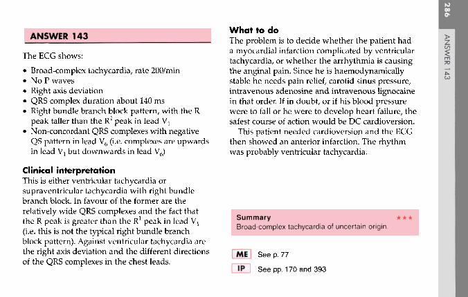
ANSWER 143
The ECG shows:
• Broad-complex tachycardia, rate 200/min• No P waves• Right axis deviation• QRS complex duration about 140 ms• Right bundle branch block pattern, with the R
peak taller than the R1 peak in lead V1
• Non-concordant QRS complexes with negativeQS pattern in lead V6 (i.e. complexes are upwardsin lead Vl but downwards in lead V6)
Clinical interpretationThis is either ventricular tachycardia orsupraventricular tachycardia with right bundlebranch block. In favour of the former are therelatively wide QRS complexes and the fact thatthe R peak is greater than the R1 peak in lead VT
(i.e. this is not the typical right bundle branchblock pattern). Against ventricular tachycardia arethe right axis deviation and the different directionsof the QRS complexes in the chest leads.
What to doThe problem is to decide whether the patient hada myocardial infarction complicated by ventriculartachycardia, or whether the arrhythmia is causingthe anginal pain. Since he is haemodynamicallystable he needs pain relief, carotid sinus pressure,intravenous adenosine and intravenous lignocainein that order. If in doubt, or if his blood pressurewere to fall or he were to develop heart failure, thesafest course of action would be DC cardioversion.
This patient needed cardioversion and the ECGthen showed an anterior infarction. The rhythmwas probably ventricular tachycardia.
Summary ***Broad-complex tachycardia of uncertain origin.
tf] See p. 77
IP~1 See pp. 170 and 393

This ECG was recorded from a 65-year-old man who complained of breathlessness and who showed thephysical signs of moderate heart failure. What does this ECG show? Does it have implications for treatment?

ANSWER 144
The ECG shows:
• Sinus rhythm• Multifocal ventricular extrasystoles• Right bundle branch block• Q waves in the sinus beats in leads III, VF
Clinical interpretationThe presence of Q waves in the inferior leadssuggests an old infarction. Ischaemic disease istherefore probably the cause of the extrasystolesand the right bundle branch block.
What to doControl of the heart failure may well cause theextrasystoles to disappear; the extrasystoles should Summarynot be treated with antiarrhythmic drugs. The Multifocal ventricular extrasystoles, right bundlepresence of multifocal extrasystoles should alert branch block, and probable old inferior myocardialyou to consider electrolyte abnormalities and infarction.digoxin toxicity.
\E | See pp. 36, 64 and 103
See p. 259

This ECG was recorded in the A & E department from a 25-year-old man with severe chest pain. Nophysical abnormalities had been detected, but having seen the ECG what would you look for and what would you do?

ANSWER 145
The ECG shows:
• Sinus rhythm• Normal axis• Normal QRS complexes• Raised ST segments in leads I, II, III, VF, V4-V6
Clinical interpretationThe raised ST segments in leads I and V4 follow Swaves and are therefore 'high take-off and are ofno significance. The ST elevation elsewhere couldindicate an acute infarction, but since the change isso widespread, pericarditis seems more probable.
What to doIn a 25-year-old, pericarditis is a much more likelydiagnosis than infarction and thrombolysis mustbe avoided. The diagnosis is made by lying thepatient flat, when a pericardial rub will becomemuch easier to hear. Echocardiography will showa pericardial effusion if one is present.
Summary ***Widespread ST segment elevation, suggestingpericarditis.
See p. 100
See p. 298

This ECG was recorded from a 70-year-old man who complained of breathlessness. What abnormalitiesdoes it show and what is the most likely diagnosis?

ANSWER 146
The ECG shows:
• Sinus rhythm• Peaked P waves, best seen in leads V1-V2
• Right axis deviation (deep S waves in lead I)• RSR pattern with normal QRS duration in lead
Vx (partial right bundle branch block)• Deep S waves in lead V6, with no left ventricular
pattern
Clinical interpretationPeaked P waves suggest right atrial hypertrophy.The partial right bundle branch block pattern isnot significant. Right axis deviation may be seenin tall thin people with normal hearts, but withthe deep S waves in lead V6 it suggests rightventricular hypertrophy. A lack of development ofa left ventricular pattern in the V leads (i.e. deep Swaves persisting into lead V6) results from theright ventricle occupying most of the pericardium.This is sometimes called 'clockwise rotation'(looking at the heart from below) and ischaracteristic of chronic lung disease.
What to doA chest X-ray examination, including a lateral view(which shows the right ventricle better), and lungfunction tests will be more helpful thanechocardiography.
Summary **Right atrial hypertrophy and probable chronic lungdisease.
IE I See pp. 89 and 91
See p. 342

This ECG was recorded from a 15-year-old boy who collapsed while playing football. His brother had diedsuddenly. What does the ECG show and what clinical possibilities should be considered?

ANSWER 147
The ECG shows:
• Sinus rhythm• Normal PR intervals except for the third
complex in lead VL, where there is a suggestionof pre-excitation
• Normal axis• Normal QRS complexes• Prolonged QT intervals (about 640 ms)• Prominent U waves, best seen in the chest leads
Clinical interpretationThe most important abnormalities here are theprolonged QT intervals and the presence of Uwaves. This is a pattern often associated withventricular tachycardia of the 'torsade' type andwith sudden death.
What to doThe family history suggests that this may well bean example of one of the congenital forms ofprolonged QT interval - the Jervell-Lange-Nielsonor the Romano-Ward syndrome. These are
characterized by episodes of loss of consciousness inchildren, often at times of increased sympatheticnervous system activity, and beta-blockers are theimmediate form of treatment. The insertion of apermanent defibrillator may be necessary. ProlongedQT interval syndrome is also associated withantiarrhythmic drugs (quinidine, procainamide,disopyramide, amiodarone and sotalol) and withother drugs such as ketanserin, prenylamine, thetricyclic antidepressants, erythromycin, thioridazineand lidoflazine. Electrolyte abnormalities (lowpotassium, magnesium or calcium levels) alsoprolong the QT interval.
SummaryMarked prolongation of the QT interval withpathological U waves - the 'long QT' syndrome.
See p. 128
***

ECG 148 The houseofficer from the health care of the elderly ward is puzzled by this ECG and asks for your help.What questions would you ask him?

ANSWER 148
The ECG shows:
• Sinus rhythm• Slow rhythmic waves, the baseline in some ways
resembling atrial flutter, but slower and coarser• Short PR intervals• Slurred upstroke of the QRS complexes,
particularly in lead I• T wave inversion in the anterior leads
significant problem that the Wolff-Parkinson-Whitesyndrome might cause in an elderly patient.
Clinical interpretationThe slow rhythmic variation is due to muscletremor, and is not cardiac in origin. The short PRintervals, slurred upstroke of the QRS complexesand inverted T waves are due to theWolff-Parkinson-White syndrome.
What to doAsk if the patient has Parkinson's disease: aParkinsonian tremor would explain the baselinevariation. Does the patient give a history ofpalpitations or syncope? This would be the only
Summary ***Muscle artefact, possibly Parkinson's disease;Wolff-Parkinson-White syndrome.
See pp. 104 and 357

A 30-year-old woman, who had been treated for depression for several years, was admitted to hospital asan emergency following deliberate self-harm involving a small number of aspirin tablets. There were no abnormalities onexamination but this was her ECG. Does it worry you?

ANSWER 149
The ECG shows:
• Sinus rhythm• Normal axis• Normal QRS complexes• T wave inversion in leads I, VL, V4-V6
Clinical interpretationAnterolateral T wave inversion is most commonlydue to ischaemia, but this seems unlikely in ayoung woman with no evidence of heart disease.A cardiomyopathy would be another possibility,but repolarization (T wave) abnormalities can becaused by lithium therapy.
What to doAs always when a diagnosis is not clear, find outwhat drugs the patient is taking. This patientwas taking lithium, and exercise testing and Summaryechocardiography showed no evidence of heart Anterolateral T wave inversion due to lithium therapy.disease.
I !(» | See p. 372

ECG 150 This ECG was recorded from a 40-year-old man who was admitted after collapsing in a supermarket. Bythe time he was seen he was well, and there were no abnormal physical signs. Would you pass this ECG as normal?

ANSWER 150
The ECG shows:
• Sinus rhythm, rate 70/min• Normal PR and QRS duration• Normal axis• QRS complexes in leads V1-v2
snow an
pattern• ST segment elevated, and downward-sloping, in
V1-V2
• Normal T waves
Clinical interpretationThis is not a normal ECG. The appearances inleads V1 and V2 are characteristic of the Brugadasyndrome.
What to doThe Brugada syndrome involves a geneticabnormality that alters sodium transport in themyocardium, and predisposes to ventriculartachycardia and fibrillation. This patient's collapsemay well have been due to an arrhythmia. Thesyndrome is often familial. The ECG changes are
not constant, and on the day after admission thispatient's ECG was perfectly normal. The ECGchanges can be induced, and ventriculartachycardia caused, by antiarrhythmic drugs. Theonly treatment is an implanted defibrillator.
SummaryBrugada syndrome.
See p. 134
***

This page intentionally left blank

This page intentionally left blank
Index
Note: Numbers refer to page numbers not question numbers; numbers initalics refer to pages showing ECG traces
Abciximab 168Accelerated idionodal rhythm 140Accelerated idioventricular rhythm
208Adenosine 12, 26, 106, 144, 230, 250,
272Alcoholic heart disease 48, 54, 184,
236Alteplase 50,142Amiloride 176Amiodarone 120, 146,182,186,188,
232, 246, 270, 272, 294Amphetamine 128Anaemia 2
sinus tachycardia 86, 108systolic murmur 282
Aneurysm, ventricular 112Angina 45, 46, 59, 60, 231, 259, 260,
263, 264, 281, 282unstable 14, 84
Angiotensin-converting enzymeinhibitor 20, 74, 162,188, 212
ankle swelling 19Anorexia 279, 280Anticoagulants
atrial fibrillation 214left atrial hypertrophy 234
Anxietybreathlessness 107palpitations 127sinus tachycardia 86
Aortic stenosis 20, 38, 151,152, 262angina 260left ventricular hypertrophy 260systolic murmur 282
Aspirin 4, 74, 142, 297, 298Athlete 139, 140, 162, 205, 206Atrial extrasystole 22, 96Atrial fibrillation 10, 48, 60, 82, 90,
156, 182, 256with complete heart block 16and hypothermia 200with left anterior hemiblock 100,
268with left bundle branch block 20,
152with rapid ventricular rate 184,
214with right bundle branch block 92with ventricular extrasystole 126,
280ventricular-paced rhythm 158Wolff-Parkinson-White
syndrome and 272

INDEX
Atrial flutter 136with 1:1 conduction 230with 2:1 block 26, 54with 4:1 block 136, 236elderly patient 146, 296with slow ventricular rate 146
Atrial tachycardia 144Atrial septal defect 52, 269, 170, 180Atrioventricular block
first degree 150second degree 40
Atrioventricular nodal escape 140Atrioventricular re-entry 222
Beri-beri 108Beta-agonists 128Beta-blockers 43, 74, 84, 124,144,
156, 162,168, 182, 210, 272Bifascicular block 116, 130,154, 204Black people, T wave abnormalities
131,132,228Blood loss, acute 86Bradycardia 16, 78, 208
sinus 210Bradycardia-tachycardia variant of
sinoatrial disease 218Brugada syndrome 300
Captopril 176Cardiomyopathy 20, 48, 132
atrial fibrillation 214dilated 188, 236hypertrophic 132, 206, 262, 274
Carotid sinus massage 144Carotid sinus pressure 26, 106,128,
230, 242, 250Cerebral embolus 200, 252Chest pain 33, 73, 141,155,156,283
angina 59, 60, 263, 264at rest 13breathlessness 211exercise-induced 3, 4, 75, 83
ischaemic 77, 284myocardial infarction 27, 28, 49,
50, 57, 58, 64nonspecific 101pleuritic 91, 159, 160severe central 7, 43, 93, 103,119,
121,141,165,177,191,195,239, 291,292
Cholesterol, raised 147, 148Clockwise rotation 126, 176, 292
right ventricular hypertrophy 202CO2 retention, sinus tachycardia 108Co-amilofruse 176Collapse during sport 293, 294Conducting pathway,
electrophysiological ablation258
Conducting systemdisease 268idiopathic fibrosis 16
Congestive cardiac failure 47, 48,81, 235, 236
Cor pulmonale 110Coronary bypass graft (CABG) 84,
156
DC cardioversion 26, 62, 230, 286atrial fibrillation 214atrial flutter 136
Defibrillator, implanted 270, 294Delta wave 80, 134, 242, 258Dextrocardia 88Diabetes 99,100, 275, 276Diamorphine 84, 186Digitalis effect 10, 48, 82, 256
anorexia/weight loss 279, 280atrial fibrillation 184, 256, 280atrial flutter 136
Digoxinatrial fibrillation 20, 90,156,182,
256, 280, 272atrial flutter 54, 136, 144

INDEX
dilated cardiomyopathy 236dosage 48flutter fibrillation 82left atrial hypertrophy 234left bundle branch block 20ST segment changes 76toxicity 10, 256
Disopyramide 294Dizziness 23, 129,130, 231, 232, 237,
238aortic stenosis 260exertion-induced 35, 36, 273first degree block 32intermittent complete block 116irregular pulse 225second degree block 265, 166
Driving licence 233, 297, 198
Ebstein's anomaly 180Endocarditis, infective 118, 184Exercise test 4, 60
Flecainide 26, 54, 136, 182, 272Flutter fibrillation 82Frusemide 176
Heart failure 29, 247, 268, 279, 280Heart murmur 35, 52, 52
adolescent 179, 180child 97flow 96pregnancy 95, 96
Hemiblock, left anterior 78,146,238, 248
bifascicular block 130, 204conduction system disease 268left axis deviation 54, 70, 116, 122,
166Heparin 156,168His bundle conduction delay 194Hyperkalaemia 210, 244, 276Hypertension 65, 209, 210Hypertrophic cardiomyopathy 132,
206, 262, 274Hypocalcaemia 148Hypokalaemia 10,148, 266Hyperkalaemia 4,176
atrial fibrillation 256, 280Hypomagnesaemia 148Hypotension 120, 246Hypothermia, atrial fibrillation and
200
Glycoprotein Ilb/IIIa inhibitor 168
Head injury 257, 258Heart block
complete 6,16intermittent 116
first degree 32, 58, 94,178,188,194
atrioventricular 150second degree
2:1 type 16, 24, 26, 70,154, 166,238
atrioventricular 40Mobitz type 2, 78, 116,166
third degree 6, 16
ICD 262Infective endocarditis 118,184International normalized ratio
(INK) 54Ischaemia 4, 20, 144, 214, 222, 239
anterolateral 14, 68, 84,156hyperacute T waves 244inferior 142lateral 282ST segment depression 76, 184
J waves of hypothermia 164, 200Jaundice 53, 54Jervell-Lange-Nielson syndrome
294

INDEX
Jugular venous pressure 109, 110,222,253,254
Junctional escape rhythm 218Junctional tachycardia 12, 106, 222
Ketanserin 294
Left atrial hypertrophy 234Left axis deviation 122, 158,166,
258, 270bifascicular block 130, 154, 204left anterior hemiblock 70,100,
116, 122, 166, 238silent infarction 248
Left bundle branchblock 20, 38, 46, 152,188,190,
246conduction delay 194
Left ventricular failure 25,118, 229Left ventricular hypertrophy 36, 50,
72, 206, 214, 260, 268, 282T wave inversion 66
anterolateral 274lateral 282
voltage criteria 260, 266, 268Lignocaine, intravenous 120, 182,
186, 246, 250, 272Lithium therapy 298Long QT syndrome 262, 294Lown-Ganong-Levine syndrome
242Lung cancer 253, 254Lung disease, chronic 54,110,122,
126, 292, 292
Malignancy 184Meningitis 252Mitral stenosis 234Mitral valve replacement 255, 256Muscle artefact 296Muscle tremor 296
Myocardial infarction 46,120, 223,224, 207
acute anterior 8, 50, 62, 62, 64,112,142
acute anterolateral 34,100,104,196, 212
acute inferior 28, 64with first degree block 94,178
acute lateral 122anterior 122
non-Q-wave 58,168, 206, 264uncertain age 122, 248, 74
inferior 4,118, 264, 288left anterior 248non-Q-wave 44,192, 264, 274
anterior 30, 58, 206anterolateral 192, 228
old 60, 77,162anterior 60, 70, 112,112inferior 50, 232, 264
posterior 224Myxoedema 148
sinus bradycardia 210
Nitrates 84, 156,168Normal ECG 42, 72,136,198, 210,
226, 228, 244, 266child 98commercial driving licence 297,198high take-off ST segment 160, 278hypertension 209, 210peaked T waves 244prominent U waves 268right axis deviation 164sinoatrial disease 218sinus arrhythmia 42ventricular extrasystoles 124voltage criteria for left ventricular
hypertrophy 268
P mitrale 118, 210P wave 24

INDEX
absent 176bifid 118, 210inverted 88notched 234peaked 180, 292
Pacemakercomplete heart block 114first degree heart block with right
bundle branch block 194second degree block 24, 238
with left axis deviation 166second degree and bifascicular
heart block 78,130,154sinoatrial disease 218ventricular-paced rhythm 158
Palpitations 11,12, 25, 85, 104,127,142, 143,173,249, 250
atrial extrasystole 22atrial fibrillation 126with breathlessness 223, 217, 233,
234sinus tachycardia 86,144ventricular tachycardia 270
Parkinson's disease 296Percutaneous transluminal
coronary angioplasty (PTCA)84,156
Pericardial effusion 254malignant 254
Pericarditis 240, 290Pericardial friction rub 240Phaeochromocytoma 128Physical fitness, sinus bradycardia
210Pilot's licence 225PR interval
constant 24short 80, 134, 140, 242, 258, 296
Pre-excitation 66, 294Pregnancy 1, 2
breathlessness 79, 80, 201heart murmur 95, 96sinus tachycardia 108
Prenylamine 294Prinzmetal's variant angina 284Procainamide 294Pulmonary emboli 56, 92,172, 202,
216, 224recurrent 18,110
Pulmonary hypertension 202primary 18
Pulmonary oedema 99,100,185,186Pulse, jerky 274
Q wave 4,196, 248, 288acute anterolateral myocardial
infarction 34anterolateral myocardial
infarction of uncertain age 74development 64myocardial infarction 28small 198
QRS complexbroad 96, 114,158, 250, 258slurred upstroke 80,134, 296small 126, 254tall 66
QT intervalcongenital prolonged 294
prolonged 146, 262Quinidine 294
R wavedominant 18, 80, 224loss 278poor progression 60, 70,126tall 72
Reteplase 50Reverse tick 10Rheumatic heart disease 9, 48,175,
255, 256atrial fibrillation 214
Right atrial hypertrophy 110,180,292
Right axis 176

INDEX
Right axis deviation 250, 256broad complex tachycardia 286chronic lung disease 126normal ECG 138,164, 292right ventricular hypertrophy 18,
138,164, 202, 292Right bundle branch block
atrial extrasystoles 96atrial septal defect 52,180atrioventricular block, second
degree 40bifascicular block 130,154, 204broad-complex tachycardia 250first degree block 194ischaemic disease 288partial 292pulmonary embolus 92supraventricular tachycardia 170trifascicular block 130, 238
Right heart failure 179Right ventricular hypertrophy 110,
138,172, 202, 224right axis deviation 164severe 16,18
Romano-Ward syndrome 294
S wavedeep 176, 202persistent in lead V6, 96, 122
Salbutamol 126Septal depolarization 198, 210Sick sinus syndrome 218Silent atrium 218Sinoatrial disease 218Sinus arrhythmia 42Sinus bradycardia 210Sinus rhythm
with atrial extrasystole 22with first degree block 32,194with left bundle branch block 38,
190with right bundle branch block
52,194
with ventricular extrasystoles 2,118, 220, 234
Sinus tachycardia 14, 56, 84, 86,108,110,128, 144
Slow VT 208Sotalol 232, 294Splenomegaly 53, 54Sport 131,132, 139,140,162, 205,
206, 262, 262, 293, 294ST segment
elevation 84,104,112,122,142,160, 178, 196, 240, 264, 282,290
high take-off 160, 240, 264, 278,282, 290
nonspecific changes 76,102, 108'reversed-tick' 90
ST segment depression 104,106,178
downsloping 10, 64, 68, 82, 108,256
flat 76,142horizontal 68,184, 222, 230ischaemia 76sloping 280
statin 4, 74,162Stokes-Adams attacks 6,16, 32, 238Streptokinase 50, 142Stroke 113,157,158,199, 200Subarachnoid haemorrhage 252Supraventricular extrasystole 46,192Supraventricular tachycardia 12,
106, 120,170, 182with bundle branch block 174,
250Syncope 270Systolic murmur 1, 2, 52, 153, 154
aortic ejection 281
T wavebiphasic 234, 264flattened 102, 148, 256, 280

inversion 66,176,198, 244, 252dilated cardiomyopathy 236hypertrophic cardiomyopathy
180, 206, 262ischaemia 176, 278lithium therapy 298myocardial infarction 192non-Q-wave infarction 274pericardial disease 254widespread 132, 254
nonspecific changes 102,108peaked 176, 210, 244
Tachycardiaatrial 144broad complex, 62,120,152,174,
186,187,188, 246, 250, 270,286
junctional 12,106, 222narrow complex, 222, 230paroxysmal 66, 280, 271sinus 14, 56, 84, 86,108,110,128,
144supraventricular 12,106, 120, 170,
174,182, 250ventricular 62,126,174, 216, 246,
250, 270torsade type 146, 294
Thrombolysis 8,14, 58left bundle branch block 190myocardial infarction 28, 64,104,
142,178,192,196, 212, 264Thyrotoxicosis 48,184
atrial fibrillation 214
sinus tachycardia 86,108Tirofiban 168Tricyclic antidepressants 294Trifascicular block 130, 238Troponin 252
U wave 10,138,148, 256, 266, 280,294
Valsalva manoeuvre 206, 242vagal stimulation 12
Ventricular aneurysm 112Ventricular extrasystole 2,124,126,
146,190,196, 234, 248, 280multifocal 220, 232, 288
Ventricular tachycardia 62,126,174,216, 246, 250, 270
torsade type 118,146, 294Ventricular-paced rhythm 158,114Verapamil 12,182, 222, 272Voltage criteria, left ventricular
hypertrophy 72Volume loss, sinus tachycardia 108
Wandering atrial pacemaker 140Weight loss 127, 279, 280Wolff-Parkinson-White syndrome
174,182, 216, 242, 258 296atrial fibrillation and 272type A 30, 80,172type B 66, 272