15.- Retroperitoneo.pdf - Facultad de Medicina
40
RADIOLOGÍA DEL RETROPERITONEO Cátedra de Diagnóstico por Imágenes Facultad de Medicina -UNNE Dr. Guillermo Pepe Dr. Rubén González
-
Upload
khangminh22 -
Category
Documents
-
view
2 -
download
0
Transcript of 15.- Retroperitoneo.pdf - Facultad de Medicina

RADIOLOGÍA DEL RETROPERITONEORETROPERITONEO
Cátedra de Diagnóstico por ImágenesFacultad de Medicina -UNNE
Dr. Guillermo PepeDr. Rubén González

RADIOLOGIA DEL RETROPERITONEO
1 Perirenal space2 Anterior pararenal space3 Posterior pararenal space4 Abdominal cavity5 Properitoneal fat6 Anterior renal fascia7 Posterior renal fascia8 Subperitoneal fascia8 Subperitoneal fascia9 Peritoneum10 Kidney11 Aorta12 Inferior vena cava13 Colon14 Duodenum15 Pancreas16 Psoas muscle17 Liver18 Pelvis

RADIOLOGIA DEL RETROPERITONEORADIOLOGIA DEL RETROPERITONEO
ÓRGANOS RETROPERITONEALESÓRGANOS RETROPERITONEALES
•• PÁNCREASPÁNCREAS•• PÁNCREASPÁNCREAS•• RIÑONESRIÑONES•• MARCO DUODENAL MARCO DUODENAL •• COLON ASCENDENTE Y DESCEND.COLON ASCENDENTE Y DESCEND.•• GRANDES VASOSGRANDES VASOS•• GANGLIOSGANGLIOS

RADIOLOGIA DEL RETROPERITONEO
1 Tail of pancreas2 Body of pancreas3 Head of pancreas (uncinate process)4 Abdominal aorta5 Inferior vena cava6 Celiac trunk7 Hepatic artery8 Splenic artery9 Splenic vein10 Portal vein10 Portal vein11 Sup. mesenteric artery12 Sup. mesenteric vein13 Bile duct14 Pancreatic duct15 Duodenum16 Stomach17 Spleen18 Liver19 Small and large intestine20 Gallbladder21 Cystic duct22 Kidney23 Renal artery24 Renal vein

PÁNCREAS(ANTES DE TAC Y ECO : “ORGANO OCULTO”)

PÁNCREASCUERPO
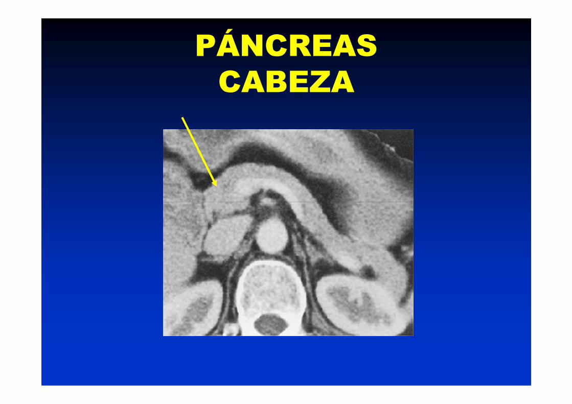
PÁNCREASCABEZA
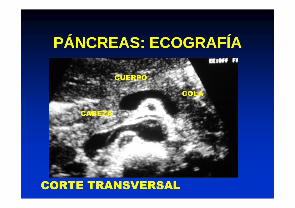
PÁNCREAS: ECOGRAFÍAPÁNCREAS: ECOGRAFÍA
CUERPO
COLA
CABEZA
CORTE TRANSVERSALCORTE TRANSVERSAL

PANCREATITIS AGUDAPANCREATITIS AGUDA
MODERADA GRAVE
Área de necrosis

PANCREATITIS AGUDAPANCREATITIS AGUDA
GRAVE ABSCESOGRAVE ABSCESO

PANCREATITIS CRÓNICAPANCREATITIS CRÓNICA

PÁNCREAS:ECOGRAFÍAPÁNCREAS:ECOGRAFÍA
COMPLICACIONES DE LA PANCREATITIS:COMPLICACIONES DE LA PANCREATITIS:
PSEUDOQUISTE DE PÁNCREASPSEUDOQUISTE DE PÁNCREAS

COMPLICACIONES DE LA PANCREATITISCOMPLICACIONES DE LA PANCREATITISPSEUDOQUISTE DE PÁNCREASPSEUDOQUISTE DE PÁNCREAS
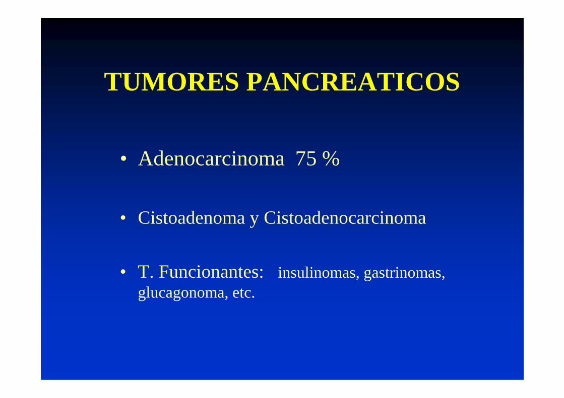
TUMORES PANCREATICOS
• Adenocarcinoma 75 %
• Cistoadenoma y Cistoadenocarcinoma
• T. Funcionantes: insulinomas, gastrinomas, glucagonoma, etc.

TUMORES PANCREATICOS
Dilatación de vías biliares
Agrandamiento de la cabeza
Alteración de los contornos

Cistoadenocarcinoma
TUMORES PANCREATICOS

TUMORES RETROPERITONEALES
•En niños los mas frecuentes son:WilmsNeuroblastomaTeratoma
La mayoría se presentan como Teratoma
Linfoma
•En adultos:LinfomaLiposarcomaOtros sarcomasMts
presentan como grandes masas abdominales
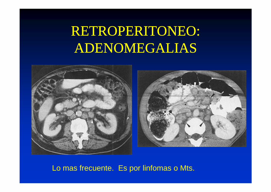
RETROPERITONEO:RETROPERITONEO:ADENOMEGALIASADENOMEGALIAS
Lo mas frecuente. Es por linfomas o Mts.

RETROPERITONEO:RETROPERITONEO:ADENOMEGALIASADENOMEGALIAS
AORTA
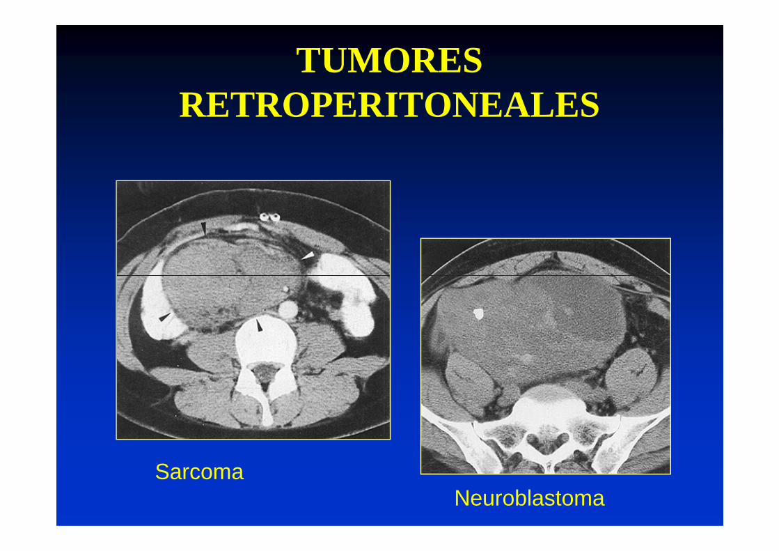
TUMORES RETROPERITONEALES
Sarcoma Neuroblastoma
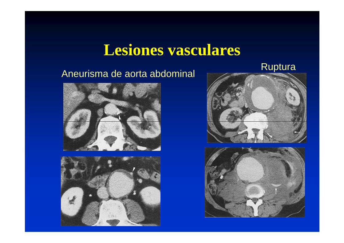
Lesiones vascularesAneurisma de aorta abdominal
Ruptura

Compartimiento del PsoasAbscesos

RETROPERITONEO:RETROPERITONEO:ESPACIOSESPACIOS
1. Pararrenal anterior
2. Perirrenal
3. Pararrenal posterior
4. Espacio del Psoas

Pararrenal anterior PancreatitisPerforación duodenalPerforación extraperitonealHematoma retroperitonealHemorragia art. viscerales
Perirrenal Absceso
RETROPERITONEORETROPERITONEOESPACIOSESPACIOS
Colecciones liquidas o solidasColecciones liquidas o solidas
Perirrenal Absceso(+ frec.C. renales) Urinoma
Hematoma **Invasión neoplásicaLesiones suprarrenales
Pararrenal posterior Hemorragias (espontáneas o traumat.)Infección (OM, Postquirurg.,perfor. rectosig.) Extravasación linfática
Espacio del Psoas InflamatoriasHemorragias

El espacio perirrenal se encuentra El espacio perirrenal se encuentra el RIÑON

APARATO URINARIOAPARATO URINARIO
UBICACIÓN ANATÓMICAUBICACIÓN ANATÓMICA

APARATO URINARIOAPARATO URINARIO
UBICACIÓN ANATÓMICAUBICACIÓN ANATÓMICA

APARATO URINARIO
• UROGRAMA EXCRETOR
• ECOGRAFÍA• ECOGRAFÍA
• TOMOGRAFÍA COMPUTADA
• RESONANCIA MAGNÉTICA
APARATO URINARIO
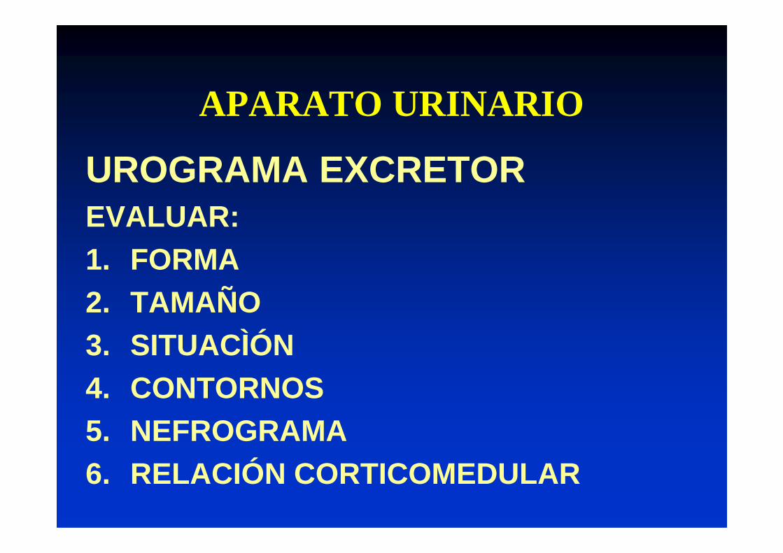
UROGRAMA EXCRETOREVALUAR:1. FORMA2. TAMAÑO3. SITUACÌÓN4. CONTORNOS5. NEFROGRAMA6. RELACIÓN CORTICOMEDULAR

APARATO URINARIO
RADIOGRAFRADIOGRAFÍA DIRECTAÍA DIRECTA POSTCONTRASTE : 5 MIN.POSTCONTRASTE : 5 MIN.
APARATO URINARIO
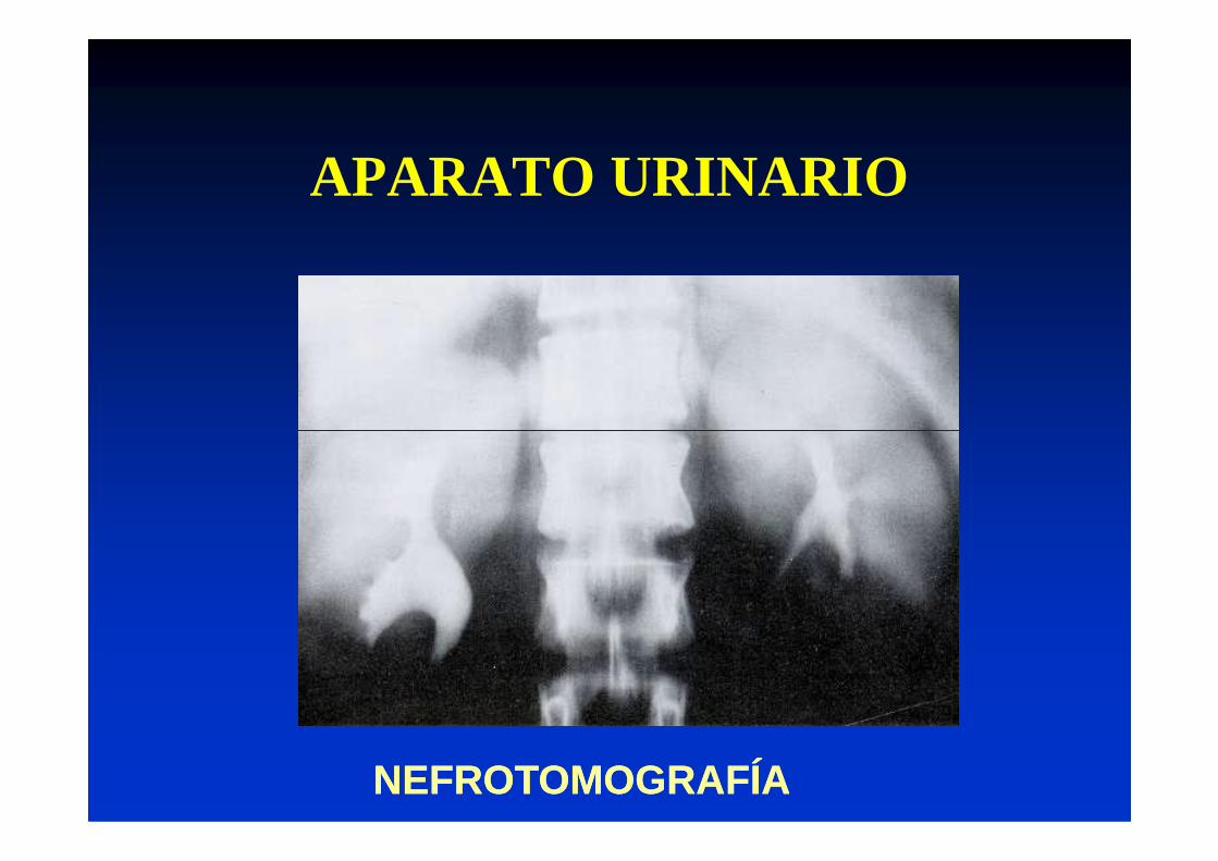
NEFROTOMOGRAFÍANEFROTOMOGRAFÍA

APARATO URINARIOAPARATO URINARIO
RIÑRIÑÓN NORMALÓN NORMAL

APARATO URINARIO
POSTCONTRASTE: 15 MIN.POSTCONTRASTE: 15 MIN. POSTCONTRASTE: 30 MIN.POSTCONTRASTE: 30 MIN.
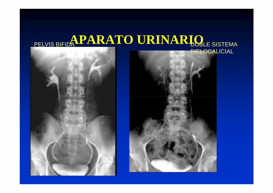
APARATO URINARIOPELVIS BIFIDA DOBLE SISTEMA PIELOCALICIAL

APARATO URINARIO
LITIASIS RENALLITIASIS RENALDENSAS U OPACASDENSAS U OPACAS

APARATO URINARIO
LITIASIS CORALIFORMES
APARATO URINARIO
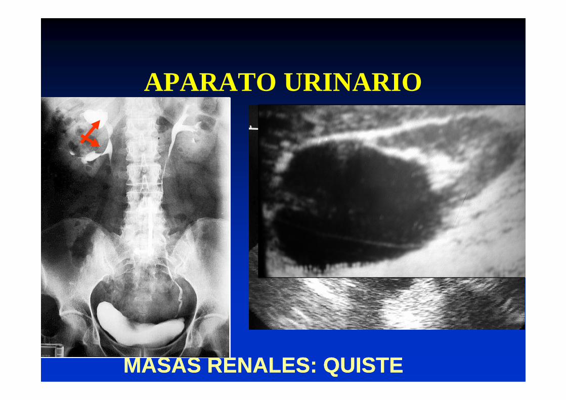
MASAS RENALES: QUISTEMASAS RENALES: QUISTE

APARATO URINARIO
TUMORES RENALES

APARATO URINARIOTUMORES RENALESTUMORES RENALES
CARCINOMACARCINOMA T. DE WILMST. DE WILMS

LA RADIOLOGIA DEL LA RADIOLOGIA DEL APARATO URINARIOAPARATO URINARIO
• Además de las litiasis y los tumores deben tener en cuenta las malformaciones e tener en cuenta las malformaciones e infecciones, así como las patologías del tracto urinario inferior. Tema de otra clase.