1475-2875-8-73.pdf - Malaria Journal
10
BioMed Central Page 1 of 10 (page number not for citation purposes) Malaria Journal Open Access Research Morphological features and differential counts of Plasmodium knowlesi parasites in naturally acquired human infections Kim-Sung Lee, Janet Cox-Singh and Balbir Singh* Address: Malaria Research Centre, Faculty of Medicine and Health Sciences, Universiti Malaysia Sarawak, 93150 Kuching, Sarawak, Malaysia Email: Kim-Sung Lee - [email protected]; Janet Cox-Singh - [email protected]; Balbir Singh* - [email protected] * Corresponding author Abstract Background: Human infections with Plasmodium knowlesi, a simian malaria parasite, are more common than previously thought. They have been detected by molecular detection methods in various countries in Southeast Asia, where they were initially diagnosed by microscopy mainly as Plasmodium malariae and at times, as Plasmodium falciparum. There is a paucity of information on the morphology of P. knowlesi parasites and proportion of each erythrocytic stage in naturally acquired human infections. Therefore, detailed descriptions of the morphological characteristics and differential counts of the erythrocytic stages of P. knowlesi parasites in human infections were made, photographs were taken, and morphological features were compared with those of P. malariae and P. falciparum. Methods: Thick and thin blood films were made prior to administration of anti-malarial treatment in patients who were subsequently confirmed as having single species knowlesi infections by PCR assays. Giemsa-stained blood films, prepared from 10 randomly selected patients with a parasitaemia ranging from 610 to 236,000 parasites per μl blood, were examined. Results: The P. knowlesi infection was highly synchronous in only one patient, where 97% of the parasites were at the late trophozoite stage. Early, late and mature trophozoites and schizonts were observed in films from all patients except three; where schizonts and early trophozoites were absent in two and one patient, respectively. Gametocytes were observed in four patients, comprising only between 1.2 to 2.8% of infected erythrocytes. The early trophozoites of P. knowlesi morphologically resemble those of P. falciparum. The late and mature trophozoites, schizonts and gametocytes appear very similar to those of P. malariae. Careful examinations revealed that some minor morphological differences existed between P. knowlesi and P. malariae. These include trophozoites of knowlesi with double chromatin dots and at times with two or three parasites per erythrocyte and mature schizonts of P. knowlesi having 16 merozoites, compared with 12 for P. malariae. Conclusion: Plasmodium knowlesi infections in humans are not highly synchronous. The morphological resemblance of early trophozoites of P. knowlesi to P. falciparum and later erythrocytic stages to P. malariae makes it extremely difficult to identify P. knowlesi infections by microscopy alone. Published: 21 April 2009 Malaria Journal 2009, 8:73 doi:10.1186/1475-2875-8-73 Received: 2 February 2009 Accepted: 21 April 2009 This article is available from: http://www.malariajournal.com/content/8/1/73 © 2009 Lee et al; licensee BioMed Central Ltd. This is an Open Access article distributed under the terms of the Creative Commons Attribution License (http://creativecommons.org/licenses/by/2.0 ), which permits unrestricted use, distribution, and reproduction in any medium, provided the original work is properly cited.
-
Upload
khangminh22 -
Category
Documents
-
view
0 -
download
0
Transcript of 1475-2875-8-73.pdf - Malaria Journal

BioMed CentralMalaria Journal
ss
Open AcceResearchMorphological features and differential counts of Plasmodium knowlesi parasites in naturally acquired human infectionsKim-Sung Lee, Janet Cox-Singh and Balbir Singh*Address: Malaria Research Centre, Faculty of Medicine and Health Sciences, Universiti Malaysia Sarawak, 93150 Kuching, Sarawak, Malaysia
Email: Kim-Sung Lee - [email protected]; Janet Cox-Singh - [email protected]; Balbir Singh* - [email protected]
* Corresponding author
AbstractBackground: Human infections with Plasmodium knowlesi, a simian malaria parasite, are morecommon than previously thought. They have been detected by molecular detection methods invarious countries in Southeast Asia, where they were initially diagnosed by microscopy mainly asPlasmodium malariae and at times, as Plasmodium falciparum. There is a paucity of information on themorphology of P. knowlesi parasites and proportion of each erythrocytic stage in naturally acquiredhuman infections. Therefore, detailed descriptions of the morphological characteristics anddifferential counts of the erythrocytic stages of P. knowlesi parasites in human infections were made,photographs were taken, and morphological features were compared with those of P. malariae andP. falciparum.
Methods: Thick and thin blood films were made prior to administration of anti-malarial treatmentin patients who were subsequently confirmed as having single species knowlesi infections by PCRassays. Giemsa-stained blood films, prepared from 10 randomly selected patients with aparasitaemia ranging from 610 to 236,000 parasites per μl blood, were examined.
Results: The P. knowlesi infection was highly synchronous in only one patient, where 97% of theparasites were at the late trophozoite stage. Early, late and mature trophozoites and schizontswere observed in films from all patients except three; where schizonts and early trophozoites wereabsent in two and one patient, respectively. Gametocytes were observed in four patients,comprising only between 1.2 to 2.8% of infected erythrocytes. The early trophozoites of P. knowlesimorphologically resemble those of P. falciparum. The late and mature trophozoites, schizonts andgametocytes appear very similar to those of P. malariae. Careful examinations revealed that someminor morphological differences existed between P. knowlesi and P. malariae. These includetrophozoites of knowlesi with double chromatin dots and at times with two or three parasites pererythrocyte and mature schizonts of P. knowlesi having 16 merozoites, compared with 12 for P.malariae.
Conclusion: Plasmodium knowlesi infections in humans are not highly synchronous. Themorphological resemblance of early trophozoites of P. knowlesi to P. falciparum and latererythrocytic stages to P. malariae makes it extremely difficult to identify P. knowlesi infections bymicroscopy alone.
Published: 21 April 2009
Malaria Journal 2009, 8:73 doi:10.1186/1475-2875-8-73
Received: 2 February 2009Accepted: 21 April 2009
This article is available from: http://www.malariajournal.com/content/8/1/73
© 2009 Lee et al; licensee BioMed Central Ltd. This is an Open Access article distributed under the terms of the Creative Commons Attribution License (http://creativecommons.org/licenses/by/2.0), which permits unrestricted use, distribution, and reproduction in any medium, provided the original work is properly cited.
Page 1 of 10(page number not for citation purposes)
Malaria Journal 2009, 8:73 http://www.malariajournal.com/content/8/1/73
BackgroundPlasmodium knowlesi, a malaria parasite species commonlyfound in long-tailed and pig-tailed macaques (Macaca fas-cicularis and Macaca nemestrina, respectively) is the onlymalaria parasite of primates with a 24-hour erythrocyticcycle [1,2]. Humans were shown to be susceptible to P.knowlesi by blood passage soon after the parasite was firstisolated in 1932 [2] and until recently, naturally acquiredhuman infections with P. knowlesi were thought to beextremely rare. However, following a number of reports ofhuman knowlesi malaria infections detected by molecularmethods in various countries in Southeast Asia [3-9], P.knowlesi is now recognized as the fifth species of Plasmo-dium infecting humans [10].
Malaria diagnosis relies heavily on examination of stainedblood films by microscopy, leading to parasite detection,enumeration and identification of Plasmodium species forrational decisions on appropriate patient treatment andmanagement. The parasite species is identified by mor-phological characteristics and for the diagnosis of humanmalaria, detailed morphological descriptions for Plasmo-dium falciparum, Plasmodium vivax, Plasmodium malariaeand Plasmodium ovale are given in medical text books andmalaria diagnostic reference literature [11,12]. Morpho-logical descriptions of P. knowlesi in the parasitology liter-ature are largely based on experimental infections ofrhesus macaques, since P. knowlesi causes a high parasitae-mia in these hosts, as opposed to low parasitaemia in itsnatural macaque hosts [1,13,14]. There are no detaileddescriptions of the morphology of P. knowlesi in naturally-acquired human infections other than the description bySingh et al [3] in which it was reported that the early tro-phozoites resemble P. falciparum while the other stages aresimilar to those of P. malariae. These morphological simi-larities between P. knowlesi, P. malariae and P. falciparumhave been noted previously by other workers but withoutaccompanying detailed accounts with coloured photo-graphs [1,15,16]. It remains unclear whether P. knowlesiparasite stages in human erythrocytes are truly indistin-guishable from P. falciparum and P. malariae infections bymicroscopy. Therefore, the morphology of the differentblood stages of P. knowlesi parasites observed in humanerythrocytes from knowlesi malaria patients is described,together with quantitative estimates of the different eryth-rocytic stages, and compared with those of the othermalaria parasites causing human disease.
MethodsPatient details and collection of blood filmsThick and thin blood films from ten patients admitted toKapit hospital and subsequently confirmed by nested PCRassays [3] as having single P. knowlesi infections were ran-domly selected from films that had been prepared from83 knowlesi malaria patients admitted to Kapit Hospital
[4]. These patients were aged between 14 to 65 years, and6 were males (patients numbered KH364, KH370,KH399, KH412, KH421, KH433) and 4 were females(KH328, KH369, KH422, KH431). Blood films were pre-pared at the time of admission prior to commencement ofanti-malarial treatment. Informed verbal consent wasobtained from each patient before blood samples weretaken.
Staining of thick and thin blood filmsThin blood films were fixed with absolute methanol(BDH, England) for 10 seconds and were allowed to dryat room temperature before staining. Thick and thinblood films were stained with 3% and 10% Giemsa(BDH, England) respectively, in Gurr® buffered water, pH7.2 (BDH, England) for 30 minutes, as described previ-ously [11]. All thick and thin blood films were examinedunder the microscope at a magnification of × 1,000 withimmersion oil.
Parasite counts and determination of differential parasitaemiaThe parasitaemia of each patient in the study was calcu-lated from thick blood films by counting the number ofparasitized red blood cells per white blood cells in over100 fields and calculated using the actual white blood cellcount of each patient. The percentage of each of the para-site developmental stages was determined based on thenumber of early trophozoites, late and mature tropho-zoites, schizonts, and gametocytes in thin blood films. Atotal of 500 parasitized erythrocytes were counted for eachof the 10 patients except for patients KH328 and KH422where 50 parasitized erythrocytes were counted due to thelow parasitaemia. The morphology of each of P. knowlesiparasite developmental stages was scored according to thedescriptions by Garnham [1] and Coatney et al [16] as fol-lows: early trophozoite – parasite appears as ring of cyto-plasm with chromatin dot and no malaria pigment; lateand mature trophozoites – parasites with denser cyto-plasm, undivided nuclear chromatin, with or withoutmalaria pigment; schizont – parasite with multiple massesof nuclear chromatin and clumps of dark brown pigment;gametocytes – parasites appear to fill most of the hosterythrocyte with single large chromatin mass and scat-tered prominent pigment. In addition, other characteris-tics such as multiple infections of single erythrocytes withearly and mature trophozoites, the number of chromatindots in ring-form stages and descriptions on the abun-dance and location of malaria pigment were recorded. Thesizes of the parasites were measured using a measuringeyepiece graticule at 1,000× magnification. The sizes ofinfected erythrocytes were measured relative to surround-ing uninfected erythrocytes.
Page 2 of 10(page number not for citation purposes)

Malaria Journal 2009, 8:73 http://www.malariajournal.com/content/8/1/73
ResultsDifferential counts and morphological characteristics of P. knowlesi parasitesThick and thin blood films were examined from tenpatients with P. knowlesi, with a parasitaemia between 610to 236,000 parasites per μl blood (Table 1). All of thedevelopmental stages of the malaria parasite life cycleexpected in the peripheral blood were observed inGiemsa-stained thin blood films and are presented (Fig-ures 1, 2, 3, 4 and 5). The infections in all 10 patients wereasynchronous, except for one patient (KH433) who had97% of parasites in the late trophozoite stage withoutmalaria pigment at the time of admission (Table 1; Figure1g, i, q; Figure 5).
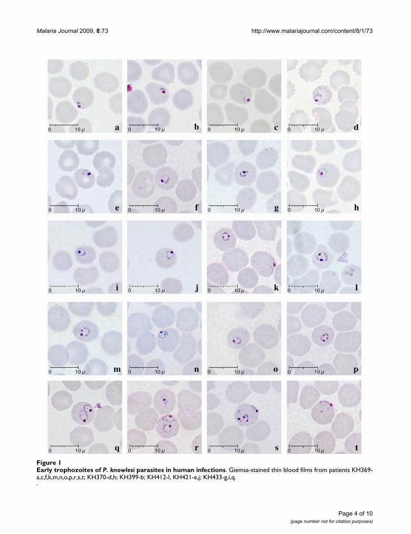
Early trophozoites of P. knowlesi were seen in all patientsexamined except one (KH422). They were characterizedby the appearance of a ring-like cytoplasm enclosing avacuole with a dot of round nuclear chromatin projectingfrom the cytoplasm (Figure 1c–g). Some early tropho-zoites with small and non-vacuolated cytoplasm appearedas a mass of bluish stained bodies measuring between 1.5to 2 μm (Figure 1, a–b). The ring form trophozoite of P.knowlesi measured approximately one third to half thediameter (2.5 to 4 μm) of the infected erythrocyte (Figure1, c–s). The cytoplasm of the ring form trophozoite wasslightly thicker especially on the side opposite the nuclearchromatin dot (Figure 1, c–g). A single prominent nuclearchromatin dot (Figure 1, a–j, ) was common (62.5 to100% of ring forms in all nine patients with early tropho-zoites; Table 2) though double chromatin dots (Figure 1,k–o) were also frequently observed (7.8 to 37.5% of ringforms in 8 of 9 patients with early trophozoites; Table 2).Double chromatin dots were mostly situated at opposingpoles of the ring form (Figure 1, k–l) but occasionally seento be closely together (Figure 1m). In addition, a single oraccessory chromatin dot lying within the vacuole wasoccasionally seen (Figure 1,h, i, n, o). Early trophozoites
with triple chromatin dots were rare and only seen in oneof the patients (Table 2; Figure 1, p). Multiply-infectederythrocytes were observed in blood films from three ofthe 10 patients, of which only one (KH364) had a parasi-taemia above 100,000 parasites/μl blood. Single erythro-cytes containing more than three parasites were observedin two patients (Table 2; Figure 1, s), and doubly infectederythrocytes (Figure 1, q, r) was the more common formof multiple infections in three patients (Table 2). Appli-qué forms, that resembled those of P. falciparum, wereinfrequently seen, being observed in only three patients(Figure 1, k, s, t; Table 2). In all erythrocytes containingearly trophozoites of P. knowlesi, enlargement of erythro-cytes and stippling were not observed.
Late and mature trophozoites were seen in all 10 patientsexamined and were characterized by denser cytoplasmcompared to the ring-like cytoplasm in early trophozoites(Figure 2, a–t). The cytoplasm of the late trophozoites ofP. knowlesi appeared to be slightly amoeboid and irregularin shape, while vacuoles were maintained (Figure 2, a–h).Late trophozoites measured approximately 3 to 5 μm. Thecytoplasm of the parasites that extended across the eryth-rocyte forming "band-like" trophozoites was seen in six ofthe 10 patients (Figure 2, g, h, k, l, r; Table 2). Nuclearchromatin dots of late trophozoites seemed to be slightlylarger in size compared to those in the early trophozoites(Figure 2). There was very little or no obvious malaria pig-ment in the majority of the late trophozoites stages.
Mature trophozoites of P. knowlesi appeared slightly largercompared to the late trophozoite with more solid anddense cytoplasm measuring approximately 5 to 6 μm andthe presence of malaria pigment. The nuclear chromatinmass was more conspicuous but remained undivided(Figure 2, l–t) and vacuoles were small or completelyabsent (Figure 2, m–t). Malaria pigment was observedeither in the form of scattered, fine dark brown grains
Table 1: Differential parasitaemia in 10 patients with single infections of P. knowlesi.
Percentages (%)
Patient code Parasitaemia (parasites per μl blood) Early trophozoites Late/Mature trophozoites Schizonts Gametocytes
KH433 7,708 2.0 97.0 1.0 0KH421 10,290 77.0 17.0 6.0 0KH370 1,908 72.1 26.1 0 1.8KH328 610 66.7 33.3 0 0KH369 9,751 34.4 20.9 43.2 1.5KH399 9,700 31.0 65.0 4.0 0KH412 2,752 29.0 67.0 4.0 0KH431 1,155 25.6 61.7 11.5 1.2KH364 *236,000 4.4 82.1 10.7 2.8KH422 920 0 76.0 24.0 0
*based on examination of thin blood film
Page 3 of 10(page number not for citation purposes)
Malaria Journal 2009, 8:73 http://www.malariajournal.com/content/8/1/73
Page 4 of 10(page number not for citation purposes)
Early trophozoites of P. knowlesi parasites in human infectionsFigure 1Early trophozoites of P. knowlesi parasites in human infections. Giemsa-stained thin blood films from patients KH369-a,c,f,k,m,n,o,p,r,s,t; KH370-d,h; KH399-b; KH412-l, KH421-e,j; KH433-g,i,q.
a b c d
e f g h
i j k l
m n o p
q r s t

Malaria Journal 2009, 8:73 http://www.malariajournal.com/content/8/1/73
Page 5 of 10(page number not for citation purposes)
Late (a-l) and mature (m-t) trophozoites of P. knowlesi parasites in human infectionsFigure 2Late (a-l) and mature (m-t) trophozoites of P. knowlesi parasites in human infections. Giemsa-stained thin blood films from patients KH364-n; KH369-a,g,l,s; KH370-b,j; KH399-h,p; KH412-c,m,t; KH421-f,i; KH422-o,q,r; KH433-d,e; KH431-k.
a b c d
e f g h
i j k l
m n o p
q r s t

Malaria Journal 2009, 8:73 http://www.malariajournal.com/content/8/1/73
(18.5 to 100% of infected erythrocytes; Table 2, Figure 2,g, h,l, m, o, p, s), or clumps of dense golden brown gran-ules (5.6 to 100% of infected erythrocytes; Table 2, Fig 2,n, q, r, t) in mature trophozoites. Infected erythrocyteswere generally not enlarged and stippling such as morerecent entomological Schüffner's dots was not observed.However, irregular and sparse dots that were unevenly dis-
tributed were noticed in some infected erythrocytes withmature trophozoites (Figure 2, l, n, r, t).
Young and mature schizonts were seen in eight of the 10patients examined, though they were found to be lesscommon in three of these patients (<5% of total infectederythrocytes, Table 1). Young schizonts, characterized by
Schizonts of P. knowlesi parasites in human infectionsFigure 3Schizonts of P. knowlesi parasites in human infections. Giemsa-stained thin blood films from patients KH364-b; KH369-c,d,f,g,h,n,o,p; KH412-a; KH421-j,m; KH422-e,i; KH431-k,l.
a b c d
e f g h
i j k l
m n o p
Page 6 of 10(page number not for citation purposes)

Malaria Journal 2009, 8:73 http://www.malariajournal.com/content/8/1/73
the appearance of between 2 to 5 divided nuclear chroma-tin masses and abundant pigment granules, were com-mon, occurring in eight of the 10 patients (Figure 3, a–i).They occupied at least two thirds (5 to 6 μm) of theinfected erythrocyte while mature schizonts appeared tooccupy nearly the whole infected erythrocyte. A maximumnumber of 16 merozoites in mature schizonts wasobserved in the blood film from patient KH369 (Figure 3,p). The divided chromatin masses and merozoites inyoung and mature schizonts were either irregularly scat-tered (Figure 3, l, m, n) or in the form of a grape-like clus-ter (Figure 3, o, p). Malaria pigment in both young andmature schizonts appeared to be either aggregated intomany small granules (Figure 3, i, j, k, l, n, p ) or aggregatedinto dense clumps of brownish black round mass (Figure3, b, d, e, f, m, o). The position of pigment aggregates wasvariable. Infected erythrocytes with mature schizonts werenot enlarged compared to uninfected erythrocytes. Dis-
torted and fimbriated infected erythrocytes were observedrarely in the blood film from only one patient, KH369(Figure 3, f, g, h). Fine irregular stippling was evident inmost of the erythrocytes containing schizonts (Figure 3, a,e–j, o, p), where the parasites occupied approximately twothirds of the erythrocyte.
Gametocytes, comprising only between 1.2 to 2.8% ofinfected erythrocytes, were observed in four of the 10patients examined (Figure 4; Table 2). Mature gameto-cytes were characterized by their spherical shape occupy-ing most of the infected erythrocyte, chromatin wascompact and the dark brown pigment was irregularly scat-tered (Figure 4, a–h). Young gametocytes occupied abouttwo thirds of the infected erythrocytes and were difficultto differentiate from mature trophozoites (Figure 4 i–l).The cytoplasm of macrogametocytes appeared bluish withthe dense pinkish chromatin situated at the periphery of
Gametocytes of P. knowlesi parasites in human infectionsFigure 4Gametocytes of P. knowlesi parasites in human infections. Giemsa-stained thin blood films from patients KH370-a,h; KH369-b,c,j,k; KH364-d,e,f,g; KH431-i,l.
a b c d
e f g h
i j k l
Page 7 of 10(page number not for citation purposes)

Malaria Journal 2009, 8:73 http://www.malariajournal.com/content/8/1/73
the parasite (Figure 4 a–d). In contrast, the cytoplasm ofmicrogametocytes stained a pinkish-purple hue with avariably positioned darker large chromatin mass (Figure 4e–h). Pigment grains in both macro and microgameto-cytes were seen to be irregularly distributed (Figure 4 a, b,e–l), however, macrogametocytes with clumps of densedark-brownish malaria pigment were also observed (Fig-ure 4 c–d). The infected erythrocytes were generally notenlarged, though some gametocytes were seen to beslightly smaller than the uninfected erythrocytes (Figure 4d, f, i, l).
Morphological comparison between P. knowlesi and P. malariaeThe morphology of P. knowlesi parasites and the character-istic of the infected erythrocyte in human infections in thepresent study were compared with those of P. malariae asdescribed in the literature [1,12,16] (Table 3).
DiscussionPlasmodium knowlesi was first isolated in 1931 in Indiafrom a long-tailed macaque imported from Singapore [2].It was studied in a number of different primate speciesand was also shown to be infectious to humans via bloodpassage in 1932 [1,2]. The detailed morphology of P.knowlesi was described by Sinton and Mulligan [14], asobserved in infections in rhesus monkeys. Here, the mor-phological descriptions of P. knowlesi malaria parasitesfrom naturally acquired human infections, that had allbeen misdiagnosed by microscopy as P. malariae, aredescribed.
Detailed observations of Giemsa-stained thin blood filmsfrom 10 patients with varying degree of parasite load,showed that human infections were generally asynchro-nous at the time of admission with all stages of the eryth-rocytic cycle of P. knowlesi parasites present in seven of the10 patients. Only one patient (KH433) had a highly syn-chronous infection with 97% of infected erythrocytes con-taining late trophozoites.
Early trophozoites of P. knowlesi appeared as ring formswithin the infected erythrocyte and possessed characteris-tics that resembled the early trophozoites of P. falciparumsuch as double chromatin dots, multiply-infected erythro-cytes and appliqué forms. The resemblance of the earlytrophozoites of P. knowlesi to those of P. falciparum hasalso been noted previously in human P. knowlesi infec-tions [3,15] and in experimental infections in rhesusmonkeys [2,14]. Human infections with P. knowlesi can beeasily mistaken for falciparum malaria when the infectingparasites are predominantly at the early trophozoite orring form developmental stage. If the blood film had beenprepared earlier from the patient with the highly synchro-nous infection, most of the trophozoites would have
Giemsa-stained thick blood film from patient KH433 showing late trophozoites of P. knowlesiFigure 5Giemsa-stained thick blood film from patient KH433 showing late trophozoites of P. knowlesi.
Table 2: Characteristics of P. knowlesi parasites in naturally acquired human infections.
Malaria pigment (%) *Chromatin dots (%) **Number of parasites in single erythrocyte (%) Band form trophozoite
Appliqué form
Patient code Clumped Scattered Single Double Triple 1 2 3
KH369 81.5 18.5 71.6 25.8 2.6 92.4 6.8 0.8 Yes YesKH364 75.0 25.0 92.2 7.8 0 74.9 20.6 4.5 Yes YesKH370 n n 88.9 11.1 0 100.0 - - Yes -KH412 100.0 0 62.5 37.5 0 100.0 - - Yes YesKH433 n n 85.7 14.3 0 100.0 - - - -KH431 5.6 94.4 89.3 10.7 0 98.2 1.8 - Yes -KH421 16.7 83.3 81.3 18.7 0 100.0 - - - -KH422 0 100 0 0 0 100.0 - - Yes -KH328 0 100 85.7 14.3 0 100.0 - - - -KH399 33.3 66.7 100.0 0 0 100.0 - - - -
*based on early trophozoites or ring form stages, and these were absent in patient KH422.**based on early and late trophozoite stages.n = no pigment because no mature trophozoites were observed.
Page 8 of 10(page number not for citation purposes)

Malaria Journal 2009, 8:73 http://www.malariajournal.com/content/8/1/73
appeared as ring forms and the infection would have beendiagnosed by microscopy as P. falciparum.
Late and mature trophozoites, schizonts and gametocytesof P. knowlesi in human infections were generally indistin-guishable from those of P. malariae. There were no specificfeatures of the cytoplasm, nucleus and pigment of the par-asites or the infected erythrocytes that could easily distin-guish P. knowlesi from P. malariae, other than the presenceof very amoeboid cytoplasm. However, this was observedin only some late trophozoites of P. knowlesi. Moreover,'band form' trophozoites, which are a characteristic fea-ture for P. malariae parasites [1,16,17] were observed inmore than half of the blood films examined.
In agreement with previous descriptions of the number ofmerozoites in mature P. knowlesi schizonts in infections ofrhesus macaques [1,14,16], a maximum of 16 merozoitesper mature schizont were observed in one of the humaninfections, which is higher than the 12 reported for amature schizont of P. malariae [1,16,17]. In addition, nomature schizonts of P. knowlesi were observed with sym-metrical arranged merozoites surrounding clumps ofmalaria pigments or "rosette pattern," which was oftendescribed as a characteristic of schizonts of P. malariae[1,16,17].
The infected erythrocytes for all stages were generally notenlarged, a feature that is shared with erythrocytic stagesof P. falciparum and P. malariae. Distorted infected eryth-rocytes, which are a common feature in P. knowlesi infec-
tions of rhesus macaques [13,14], were rarely observed inhuman P. knowlesi infections in the present study. No stip-pling was apparent that resembled Schüffner's dots, whichare observed in P. vivax infected erythrocytes. However,faint stippling that appeared as light irregular dots wereevident in only some of the infected erythrocytes withmature trophozoite and schizont stages. This type of stip-pling in erythrocytes infected with P. knowlesi is referred toas 'Sinton and Mulligan's stippling' and was noted previ-ously in infections in rhesus monkeys [1,14,16] andhumans [18]. According to Sinton and Mulligan [14],who first described this, the stippling was only consist-ently observed in some infected erythrocytes of macaques,when the panoptic method of Green [19] was used forstaining. Similarly in P. malariae infections, the infectederythrocytes are not enlarged or distorted and may displaysome form of irregular fine stippling in blood films withintense staining [1].
Morphologically, and depending on the predominant cir-culating parasite stage, it was not possible to identifyrobust morphological differences to distinguish betweenP. knowlesi and P. malariae or P. falciparum using routinemicroscopy methods for malaria diagnosis. This lack ofdistinguishing features accounts for P. knowlesi infectionsbeing overlooked, despite major differences in the patho-genesis of each of these different lineage parasites [4,20].Laboratory misidentification of P. knowlesi as P. malariaeby microscopy is largely unavoidable, although it is diffi-cult to rationalize how a parasitaemia greater than100,000 per μl blood, together with signs of severe
Table 3: Comparison between P. knowlesi and P. malariae parasites in human infections
P. malariae* P. knowlesi
HOST ERYTHROCYTESize Not enlarged Not enlargedShape Rounded, not distorted Rounded, generally not distortedStippling Ziemen's stippling under certain staining conditions Irregular dots or stippling in some erythrocytes with
mature trophozoites, schizonts and gametocytes
PARASITEEarly trophozoite (ring form) Ring form with dense cytoplasm and single chromatin;
sometimes with accessory dotRing forms are compact with dense cytoplasm; single or double chromatin and rarely triple chromatin; appliqué forms; multiple parasites in single erythrocyte
Late trophozoite Regular and compact cytoplasm; appearance of pigment Dense and thick cytoplasm; Cytoplasm slightly amoeboid and irregular; band forms; varying pigmentation
Mature trophozoite Compact, rounded, heavily pigmented; band forms; not amoeboid
Compact and dense cytoplasm, rounded shape with dark brown pigment; band forms; not amoeboid
Schizont Occupies whole erythrocyte, contains 6–12 merozoites, usually 8; merozoites clustered around dark-brown malaria pigment forming rosette pattern
Occupies whole erythrocyte; contains up to 16 merozoites; merozoites irregularly scattered or grape-like cluster; malaria pigment scattered or collects into a single mass
Gametocyte Round, compact, occupies whole erythrocyte, scattered malaria pigment; early forms are very similar to mature trophozoites
Round, compact, fills whole erythrocyte, scattered or clumped malaria pigment; early forms are very similar to mature trophozoites
*Based on descriptions by Garnham [1], Coatney et al [16] and Warrell & Gilles [12].
Page 9 of 10(page number not for citation purposes)

Malaria Journal 2009, 8:73 http://www.malariajournal.com/content/8/1/73
malaria, could clinically be mistaken for a P. malariaeinfection, except that until recently, such infections wouldhave been relatively rare and on a background of P. falci-parum transmission. Until the application of DNA basedtechnology, the definitive identification test for P. knowlesiparasites was to inject infected blood into rhesus monkeyand observe for a fatal outcome [15]. Clearly this was, andis beyond the scope of all but a few international centreswith this type of facility. Readily available moleculartools, most notably the P. knowlesi-specific PCR assay [3],provide a relatively available means to properly identify P.knowlesi [3,6-9]. Molecular methods are expensive, notavailable in most rural diagnostic laboratories and assuch, are not currently used for routine malaria diagnosis[21]. Areas of Southeast Asia, where there is a suspicionthat P. knowlesi infects humans, would benefit from a PCRscreening exercise to estimate the relative proportion of P.knowlesi to P. malariae infections. This information wouldbe valuable to prime local healthcare professionals to rec-ognize severe knowlesi malaria in ill patients with a labo-ratory report of P. malariae.
Despite morphological similarities with P. malariae, thereare other clinical and parasitological parameters such aspresentation with one or more of the WHO clinical crite-ria for severe malaria and a parasitaemia greater than5,000/μl blood, that, together with a history of time spentin the forest and forest fringe areas of Southeast Asia,should strongly implicate a P. knowlesi rather than a P.malariae infection [4,20]. Given the consequence of mis-diagnosis and delayed treatment for this parasite with arelatively short erythrocytic cycle, every effort should bemade to accurately diagnose knowlesi malaria in popula-tions at risk.
ConclusionThe morphology of P. knowlesi parasites in human infec-tions closely resembled those of P. falciparum in the earlytrophozoite stage and P. malariae in the later stages of theerythrocytic cycle. Although there were some minor mor-phological differences between the blood stages of P.knowlesi and P. malariae, it would be difficult to positivelyidentify knowlesi malaria based on morphology alone.However, even in the absence of PCR facilities, severesymptoms, a parasitaemia >5,000/μl blood, with P. malar-iae parasite morphology and a recent history of time spentin the forest fringe areas of Southeast Asia should beenough to make a diagnosis of knowlesi malaria. In viewof what is now known of the distribution and severe man-ifestation of knowlesi malaria, continued misdiagnosis ofthis important pathogen is no longer acceptable.
Competing interestsThe authors declare that they have no competing interests.
Authors' contributionsKSL, BS and JCS wrote the paper, BS and JCS conceived thestudy, KSL stained and examined the slides by micros-copy.
AcknowledgementsThis study was supported by grants from The Wellcome Trust and Univer-siti Malaysia Sarawak. The funding bodies had no role in the study design, in the collection, analysis and interpretation of data, in the writing of the man-uscript and in the decision to submit the manuscript for publication.
References1. Garnham PCC: Malaria parasites and other haemosporidia Oxford:
Blackwell Scientific Publications; 1966. 2. Knowles R, Das Gupta BM: A study of monkey-malaria and its
experimental transmission to man. Indian Medical Gazette 1932,67:301-320.
3. Singh B, Sung LK, Matusop A, Radhakrishnan A, Shamsul SS, Cox-Singh J, Thomas A, Conway D: A large focus of naturallyacquired Plasmodium knowlesi infections in human beings.Lancet 2004, 363:1017-1024.
4. Cox-Singh J, Davis TM, Lee KS, Shamsul SS, Matusop A, Ratnam S,Rahman HA, Conway DJ, Singh B: Plasmodium knowlesi malaria inhumans is widely distributed and potentially life threatening.Clin Infect Dis 2008, 46:165-171.
5. Jongwutiwes S, Putaporntip C, Iwasaki T, Sata T, Kanbara H: Natu-rally acquired Plasmodium knowlesi malaria in human, Thai-land. Emerg Infect Dis 2004, 10:2211-2213.
6. Luchavez J, Espino FE, Curameng P, Espina R, Bell D, Chiodini P,Nolder D, Sutherland C, Lee KS, Singh B: Human infections withPlasmodium knowlesi, the Philippines. Emerg Infect Dis 2008,14:811-813.
7. Ng OT, Ooi EE, Lee CC, Jarrod LP, Ng LC, Wong PS, Tu TM, Loh JP,Leo YS: Naturally acquired human Plasmodium knowlesi infec-tion, Singapore. Emerg Infect Dis 2008, 14:814-816.
8. Vythilingam I, NoorAzian YM, Huat TC, Jiram AI, Yusri YM, AzahariAH, NorParina I, NoorRain A, Hakim L: Plasmodium knowlesi inhumans, macaques and mosquitoes in peninsular Malaysia.Parasit Vectors 2008, 1:26.
9. Zhu HM, Li J, Zheng H: Human natural infection of Plasmodiumknowlesi. Zhongguo Ji Sheng Chong Xue Yu Ji Sheng Chong Bing Za Zhi.2006, 24(1):70-71.
10. White NJ: Plasmodium knowlesi: the fifth human malaria para-site. Clin Infect Dis 2008, 46:172-173.
11. Basic malaria microscopy Part 1. Learner's Guide: World Health Organ-ization – Geneva; 1991.
12. Warrell DA, Gilles HM: Essential Malariology 4th edition. Arnold; Lon-don, UK; 2002.
13. Singh J, Ray AP, Nair CP: Further observations on transmissionexperiments with Plasmodium knowlesi. Ind J Malariol 1950,4:317-335.
14. Sinton JA, Mulligan HW: A critical review of the literature relat-ing to the identification of the malarial parasites recordedfrom monkeys of the families Cercopithecidae and Colobi-dae. Records of the Malaria Survey of India 1933, 3:381-443.
15. Chin W, Contacos PG, Coatney GR, Kimball HR: A naturallyacquired quotidian-type malaria in man transferable to mon-keys. Science 1965, 149:865.
16. Coatney RG, Collins WE, Warren M, Contacos PG: The PrimateMalarias Washington: U.S. Government Printing Office; 1971.
17. Sandosham AA: Malariology with special reference to Malaya Singapore:University of Malaya Press; 1959.
18. Fong YL, Cadigan FC, Coatney GR: A presumptive case of natu-rally occurring Plasmodium knowlesi malaria in man in Malay-sia. Trans R Soc Trop Med Hyg 1971, 65:839-840.
19. Green RTB: A malarial parasite of Malayan monkeys and itsdevelopment in anopheline mosquitoes. Trans R Soc Trop MedHyg 1932, 24:455-477.
20. Cox-Singh J, Singh B: Knowlesi malaria: newly emergent and ofpublic health importance? Trends Parasitol 2008, 24:406-410.
21. Singh B: Molecular methods for diagnosis and epidemiologicalstudies of parasitic infections. Int J Parasitol 1997, 27:1135-1145.
Page 10 of 10(page number not for citation purposes)
http://www.ncbi.nlm.nih.gov/entrez/query.fcgi?cmd=Retrieve&db=PubMed&dopt=Abstract&list_uids=5003320
http://www.ncbi.nlm.nih.gov/entrez/query.fcgi?cmd=Retrieve&db=PubMed&dopt=Abstract&list_uids=5003320