0764267_00000_0000.pdf - accedaCRIS
164
María Eugenia Manera Las Palmas de Gran Canaria, 2019
-
Upload
khangminh22 -
Category
Documents
-
view
1 -
download
0
Transcript of 0764267_00000_0000.pdf - accedaCRIS

María Eugenia Manera
Las Palmas de Gran Canaria, 2019


a.D DE LAS PALMAS DE GRAN CANARIA Escuela de Doctorado
Programa de Doctorado Investigación en Biomedicina
JOSÉ ALBERTO MONTOY A ALONSO, Catedrático de Medicina Animal y COORDINADOR DEL PROGRAMA DE DOCTORADO DE INVESTIGACIÓN EN BIOMEDICINA DE LA UNIVERSIDAD DE LAS PALMAS DE GRAN CANARIA
INFORMA:
Que la Comisión Académica del Programa de Doctorado de Investigación en Biomedicina,
en su sesión de fecha nueve de enero de 2019 tomó el acuerdo de dar el consentimiento para su
tramitación, a la tesis doctoral titulada "Uso de las plataformas cinéticas en la detección
de cojeras por osteoartritis en pe"os" presentada por la doctoranda Dª. María Eugenia
Manera y dirigida por el Dr. José Manuel Vilar Guereño.
Que la citada tesis doctoral reúne todos los requisitos exigidos por la normativa de
este programa de doctorado y de esta universidad, para ser tramitada como tesis doctoral.
Y para que así conste, y a efectos de lo previsto en el Artº 11 del Reglamento de
Estudios de Doctorado (BOULPGC 7/10/2016) de la Universidad de Las Palmas de Gran
Canaria, firmo el presente informe en Las Palmas de Gran Canaria, a 14 de enero de dos mil
diecinueve.
Fdo.Alberto Montoya Alonso

Universidad de las Palmas de Gran Canarias
PROGRAMA DE DOCTORADO INVESTIGACIÓN EN BIOMEDICINA
ESCUELA DE DOCTORADO
USO DE LAS PLATAFORMAS CINÉTICAS EN LA DETECCIÓN DE COJERAS POR OSTEOARTRITIS EN
PERROS
Presentada por Doña MARÍA EUGENIA MANERA Dirigida por el Doctor Don JOSE MANUEL VILAR GUEREÑO
El director La doctoranda
Las Palmas de Gran Canaria, 2019.


A mis padres, que gracias a ellos soy lo que soy hoy.
A mis mascotas, que son la inspiración en mi profesión.


AGRADECIMIENTOS


Después de tantos años fuera, lejos de mi tierra al otro lado del océano, con
diferentes costumbres, me siento muy feliz de agradecer a todos aquellos que me
ayudaron a superar tantos obstáculos en esta parte de mi vida.
A mi tutor de tesis, José Manuel Vilar, por siempre estar presente y ser exigente
en la manera justa. También tener presente las dificultades que uno tiene en la vida
cotidiana y a pesar de eso, ayudar en todo lo que este a su alcance.
Gracias a mis amigas y hermanas que me dio la vida, Jorgelina, Rosa y Vanina
porque ellas son el apoyo incondicional, con esas conversaciones de la vida siempre
acompañadas de un buen mate de por medio y un rico queque con sobrinos.
A mis amigas, que a pesar de estar lejos siempre están cerca preguntando que tal
la vida por el viejo continente.
Diego, Sole y Mariano, mis hermanos, por ayudarme a tomar decisiones
importantes y por el solo hecho de siempre estar presentes; a Laura, por ayudarme con
los dibujos y diseños de este trabajo, y en especial por darme esos dos sobrinos que
tanto amo.
Pero todo mi amor y gratitud es para mis padres, Gustavo y Cristina, que con su
amor, compañía y apoyo, mi vida aquí y este trabajo no seria posible.


ÍNDICE


ÍNDICE
1. INTRODUCCIÓN………………………….…………………….…………..………………………..……. 1
2. OBJETIVOS………………………………………………………..……..…………………………….….…. 7
3. REVISIÓN BIBLIOGRÁFICA …………………….……………………..……………………..……… 11
Osteoartritis
3.1. Definición…………..…………….……………………………….…………....................................... 13
3.2. Articulaciones más frecuentes afectadas por OA y patologías más
común…...…………………………………………………...……………..…………………………….…….. 14
3.2.1. ¿Qué es una articulación?............................................................................................. 14
3.2.2. Tipos de articulaciones……..……………………………..………………………………. 15
3.2.2.1. Sinartrosis…………………………………………..………………………………….. 15
3.2.2.2. Diartrosis…………………………………………...…………………………………… 15
3.2.2.2.1. Articulaciones sinoviales……………………….……...………………… 15
3.2.2.2.2. Componentes de una articulación sinovial…………….……. 16
3.2.3. Articulación coxofemoral …...………………………………..…………………… 17
3.2.3.1. Anatomía ………...………………………………………………………………… 17
3.2.3.2. Displasia de cadera …………………………………….……………………... 18
3.2.4. Articulación del codo …………...…………………….…………………………….. 20
3.2.4.1. Anatomía …………...……………………….………………………………………… 20
3.2.4.2. Displasia de codo …………………………….……………………………….. 21
3.2.5. Articulación de la rodilla …………………………..………………………………… 24
3.3. Diagnóstico de la OA ……………………………………...……………………………………… 28
3.3.1. Técnicas de imagen ……..………………………..…….………………………………… 28
3.3.1.1. Radiología ………………………………………………………………………….… 28
3.3.1.2. Artroscopia ..…………………………....…………………………………………… 28

3.3.2. Técnicas de evaluación funcional .……………………...……………..………….. 29
3.3.2.1. Método de análisis subjetivo ...…………………..………….………… 29
3.3.2.2. Análisis objetivo (biomecánico) ……………………………………… 33
3.3.2.2.1. Análisis cinemático ……………………...……………………… 36
a. Electrogoniometría ………......................……...…………………..…….. 36
b. Cinematografía de alta velocidad y videografía ………...….…… 38
c. Sensores inerciales o unidades de medición inercial (IMU)…42
3.3.2.2.2. Análisis cinético ………………………….……….………….…… 44
a. Plataforma de Fuerza …………………….………………………………. 45
b. Plataforma de Presión ……...………………………………………...…. 48
c. Relación postura-‐cojera …………..………………..………………….. 51
3.4. Tratamiento de la OA ……………………………..…………………………………….…….. 60
3.4.1. Tratamientos convencionales ……………………….………………….………… 60
3.4.2. Terapias regenerativas ………………….……………...……………………… 60
4. ARTÍCULOS …………………………………………………………………………………………….... 65
-‐ #1: Static Posturography: A New Perspective in the Assessment of Lameness
in a Canine Model …………………………………………………………………………………….….. 67
-‐ #2: Effect of leukocyte-‐reduced platelet-‐rich plasma on osteoarthritis
caused by cranial cruciate ligament rupture: A canine gait analysis model ….…... 83
-‐ #3: Posturography and dynamic pedobarography in lame dogs with
elbow displasia and cranial cruciate ligament rupture..…………..……………..….…… 99
5. CONCLUSIONES ………………………………………………………………………………………... 113
6. RESUMEN …………………………………………………………….………………………………… 117
7.SUMMARY ………..……………………………………………………..……………………………….. 123
8. REFERENCIAS BIBLIOGRÁFICAS …………………………………….………………………… 129


1. INTRODUCCIÓN

1. INTRODUCCION
2

1. INTRODUCCION
3
La osteoartritis (OA) es un desorden común en medicina veterinaria, y en
concreto en los perros, debido sobre todo al incremento de la esperanza de vida en esta
especie.
Los animales con OA suelen cojear de forma intermitente, progresiva o
persistente, y su evaluación visual suele basarse en parámetros subjetivos.
Existen, además, métodos de examen complementario clásicos como el
diagnóstico por imagen y en concreto la radiología, aunque las técnicas tomográficas
más actuales como el TC o la resonancia magnética proporcionan más información,
aunque ha quedado ampliamente demostrada la falta de correlación entre los hallazgos
por imagen y el nivel de cojera.
Los cinco principios del tratamiento medico de la OA se basa en el control del
peso corporal, suplementos nutricionales, limitación del ejercicio, terapia física y
fármacos antiinflamatorios de diversa naturaleza o incluso el plasma rico en plaquetas
(PRP) o alguno de sus derivados.
Si una determinada estrategia terapéutica es establecida, adquiere especial
relevancia conocer de forma objetiva el estado inicial de la cojera del animal para, de
manera igualmente objetiva, determinar cuál es el nivel de eficacia del tratamiento, así
como su duración.
En este sentido, tal y como ya se ha apuntado anteriormente el diagnóstico de la
cojera y su evolución tras la instauración de un tratamiento se ha basado clásicamente
en una serie de cuestionarios o escalas numéricas que tenían en cuenta parámetros
subjetivos diversos, como la dificultad al levantarse o dolor o crepitación a la
manipulación.
1. INTRODUCCION
4
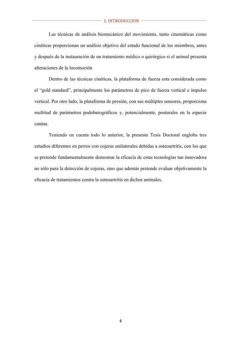
Las técnicas de análisis biomecánico del movimiento, tanto cinemáticas como
cinéticas proporcionan un análisis objetivo del estado funcional de los miembros, antes
y después de la instauración de un tratamiento médico o quirúrgico si el animal presenta
alteraciones de la locomoción
Dentro de las técnicas cinéticas, la plataforma de fuerza esta considerada como
el “gold standard”, principalmente los parámetros de pico de fuerza vertical e impulso
vertical. Por otro lado, la plataforma de presión, con sus múltiples sensores, proporciona
multitud de parámetros podobarográficos y, potencialmente, posturales en la especie
canina.
Teniendo en cuenta todo lo anterior, la presente Tesis Doctoral engloba tres
estudios diferentes en perros con cojeras unilaterales debidas a osteoartritis, con los que
se pretende fundamentalmente demostrar la eficacia de estas tecnologías tan innovadora
no sólo para la detección de cojeras, sino que además pretende evaluar objetivamente la
eficacia de tratamientos contra la osteoartritis en dichos animales.

1. INTRODUCCION
5

1. INTRODUCCION
6

2. OBJETIVOS

2. OBJETIVOS
8

2. OBJETIVOS
9
- Demostrar que la posturografía es una técnica fiable y objetiva para la detección de
cojeras en perros con OA de codo.
- Demostrar de forma objetiva la utilidad de la plataforma de fuerza y la
electrogoniometría para determinar la duración y nivel de eficacia del plasma rico en
plaquetas (PRP) en perros con OA de rodilla, de modo que pueda utilizarse para el
establecimiento de un régimen efectivo de administración de éste u otros tratamientos.
- Demostrar que la podobarografía dinámica es una herramienta de detección de cojeras
objetiva y fiable en animales afectados de OA de codo y rodilla. Validar dichos
resultados con los obtenidos con una plataforma de fuerza, considerado “el Gold
standard” del análisis del paso.

2. OBJETIVOS
10

3. REVISIÓN BIBLIOGRÁFICA

3. REVISIÓN BIBLIOGRÁFICA
12

3. REVISIÓN BIBLIOGRÁFICA
13
OSTEOARTRITIS
3.1. Definición.
La osteoartritis (OA) es un desorden común en medicina veterinaria, y se ha
incrementado la casuística en la clínica veterinaria (Rychel, 2010, Malek y cols., 2012)
debido, tal y como ocurre en la especie humana, por el aumento de la expectativa de
vida. La enfermedad se presenta em última instancia de forma crónica y degenerativa,
afectando al cartílago y el hueso subcondral que causa dolor e inhibe el movimiento
(Ramírez-flores, 2017).
El cartílago continúa deteriorándose por completo y esto causa fricción entre los
huesos, lo cual genera una inflamación, engrosamiento de los tejidos blandos y perdida
de movilidad de la articulación (Bland, 2015).
Los síntomas más habituales de la OA son el dolor, disminución del rango de
movilidad, e incluso inflamación evidente en la zona lo que va a conllevar en ultimo
termino una pérdida de la funcionalidad (Cuervo y cols., 2015).
Por otro lado, y como consecuencia de las alteraciones dinámicas el cuadro
puede evolucionar en última término hacia una excesiva laxitud o hacia la rigidez,
agravando el cuadro clínico (Saito y cols., 2002).
El inicio del proceso puede ser multifactorial ya que puede originarse por un
traumatismo, herida, sobrecarga, desgaste, o bien asociado a un proceso de
envejecimiento. Todos estos procesos cambiaran la composición, estructura y

3. REVISIÓN BIBLIOGRÁFICA
14
propiedades de los tejidos que forman dicha articulación, limitando el apropiado
funcionamiento del cartílago articular (Cuervo, 2015).
En el caso concreto de la especie canina, es de vital importancia el
reconocimiento temprano de la patología, toda vez que los signos clínicos (cojera)
pueden ser enmascarados por compensación por el propio animal, lo que puede
desembocar en que el diagnóstico certero de la enfermedad ocurre cuando el estadio es
muy avanzado. En este sentido, ocupa un lugar importante la revisión periódica de los
animales tratando de valorar sobre todo el estado de la musculatura intendente detectar
asimetrías por atrofia que pudieran indicar desuso del miembro afectado (Rychel, 2010).
3.2. Articulaciones más frecuentemente afectadas de OA y
patología más común
En el ámbito de la clínica de pequeños animales, sobre todo en perros, las
articulaciones más afectadas por OA son el codo, la cadera y la rodilla (Malek y cols.,
2012).
3.2.1. ¿Qué es una articulación?
Anatómicamente, una articulación se define como la unión o medio de contacto
de dos o más huesos entre sí además de sus elementos de fijación. La función más
importante de las articulaciones es la de constituir los puntos de unión del esqueleto y
producir movimientos mecánicos, proporcionando elasticidad y plasticidad al cuerpo,
además de ser lugares de crecimiento (Piermattei y cols., 2006).

3. REVISIÓN BIBLIOGRÁFICA
15
3.2.2. Tipos de articulaciones
3.2.2.1. Sinartrosis
No existe espacio articular. Se subdividen, dependiendo del tejido que se
encuentra entre los huesos en:
Articulaciones fibrosas: en general son articulaciones con movilidad muy limitada,
como las denominadas “suturas” del cráneo (könig y Liebich, 2008):
Articulaciones cartilaginosas: apenas conservan pequeños movimientos de
compresión/expansión, como por ejemplo la sínfisis pelviana (könig y Liebich, 2008):
3.2.2.2. Diartrosis
En este casi si podemos encontrar espacio articular, lo cual permite movimiento
entre los componentes óseos complementarios. Existen varios tipos (Todhunter y
Johnston, 2006):
3.2.2.2.1. Articulaciones sinoviales
En general estas articulaciones gozan de gran rango de movilidad. El espacio
articular está relleno de una sustancia viscosa lubrificante que se denomina liquido
sinovial o sinovia. En dichas articulaciones se encuentra también el cartílago articular,
formando superficies complementarias. De forma fisiológica en este tipo de
articulaciones se dan diferentes movimientos como la flexión y extensión, abducción y
aducción e incluso de rotación (Cuervo, B., 2015).

3. REVISIÓN BIBLIOGRÁFICA
16
Este gran rango articular que caracteriza a las articulaciones sinoviales viene
limitado de forma fisiológica por otros componentes como los músculos, ligamentos,
cápsula articular y por la forma que adoptan los huesos (Piermattei y cols., 2006).
3.2.2.2.2. Componentes de una articulación sinovial
- Cápsula articular y Membrana sinovial
La cápsula articular está formada por una gruesa porción fibrosa por debajo de la
cual encontramos una delgada capa subsinovial o lámina propia que se continúa con la
membrana sinovial o sinovia. Esta membrana sinovial es la que está en contacto directo
con el líquido sinovial, el cual en condiciones normales ocupa el espacio articular
(Todhunter y Johnston, 2006).
Este líquido, por su naturaleza viscosa reduce la fricción de las superficies
articulares y además proporciona nutrición al cartílago articular (könig y Liebich, 2008,
Todhunter y Johnston, 2006, Cuervo B, 2015)
- Ligamentos
Los ligamentos están compuestos por fibras de tejido conectivo anclados en
diferentes zonas de las superficies complementarias que conforman la articulación de
modo que limitan su rango de movimiento, por un lado, además de estabilizarla. Los
ligamentos, dependiendo de que estén dentro o fuera de la capsula articular se
denominan intracapsulares o extracapsulares, respectivamente (Bruyn y cols., 2012).

3. REVISIÓN BIBLIOGRÁFICA
17
- Formaciones anejas
Los meniscos son componentes fibrocartilaginosos interpuestos entre las
superficies articulares que sirven, gracias a sus propiedades elásticas, para amortiguar
los impactos (Todhunter y Johnston, 2006).
Con esta misma finalidad se disponen los discos intervertebrales (könig y
Liebich, 2008).
- Cartílago articular
Estructura de cartílago hialino que permite el deslizamiento de las articulaciones
y se une a la superficie de la epífisis calcificada del hueso subyacente (Bhosale y
Richardson, 2008).
- Hueso subcondral
Es un tejido subarticular que sirve de soporte físico, así como de nutrición al
cartílago hialino (Burr, 2004).
3.2.3. Articulación coxofemoral
3.2.3.1. Anatomía
Se trata de una articulación esferoide formada por la cabeza del fémur encajada
en el acetábulo del hueso coxal. La articulación se estabiliza mediante un fuerte
ligamento de la cabeza del fémur que lo fija en su posición (Sandoval 2000)

3. REVISIÓN BIBLIOGRÁFICA
18
Hill´s Atlas of Veterinary Clinical Anatomy (2006).
3.2.3.2. Displasia cadera
La displasia de cadera (DC) es a día de hoy la enfermedad ortopédica más
común en perros de razas grandes y gigantes. Aunque la mayoría de los perros que
padecen esta enfermedad no manifiestan signos clínicos o los muestran a edades
avanzadas, la enfermedad degenerativa articular a la que conlleva, con las consecuentes
molestias para el animal, y la transmisi6n del problema a la descendencia hacen que la
preocupaci6n por parte de propietarios, criadores y veterinarios sea importante
(Rodríguez, 2003).
Desde el punto de vista etimológico, la palabra displasia es griega, significando
"dys" anormal y "plassein" formación. Eso significa que DC hace referencia al
desarrollo anormal de la cadera.

3. REVISIÓN BIBLIOGRÁFICA
19
La causa o causas precisas de la aparición de la DC aún no se conoce a día de
hoy, pero parece ser que todos aquellos procesos que favorezcan la inestabilidad
articular son los que contribuyen de manera decisiva a la aparición del proceso
displásico, Por el contrario, aquellos que contribuyan a la distribución anormal de las
fuerzas en la articulación y que por lo tanto la van a convertir en inestable, favorecerán
la aparición del proceso osteoartrósico (Cardinet, 1997).
Este desarrollo articular normal conllevara a falta de congruencia entre las
superficies articulares complementarias, lo que provocara su remodelación, así como un
aumento de líquido sinovial. Si el proceso avanza en estas circunstancias, la articulación
cada vez será más laxa y más incongruente (Smith, 1997; Madsen, 1995).
Las lesiones iniciales van a estar caracterizadas por una osteoartritis con
sinovitis agudas, así como lesiones cartilaginosas. El primer cambio es una osteoartritis
aguda, que se caracteriza por sinovitis y lesiones degenerativas en la superficie del
cartílago articular (Lippincott, 1992). Según avanza el proceso, la articulación se
remodela, el acetábulo será cada vez más plano favoreciendo incluso la subluxación
(Riser, 1987). En última instancia, una combinación de alteraciones hemodinámicas,
inflamatorias y degenerativas va a producir un proceso cíclico perpetuante, con lo que
las caderas sufrirán un proceso osteoartrósico que es la patología que las caracteriza
mayormente (Madsen, 1994, Rodríguez, 2003).

3. REVISIÓN BIBLIOGRÁFICA
20
Radiografías correspondientes a un caso de displasia de grado D (a) y grado E
(b). Resultan más que evidentes la falta de profundidad acetabular, pérdida de la forma
esférica de la cabeza del fémur, esclerosis en el borde acetabular, así como la presencia
de osteofitos.
3.2.4. Articulación del codo
3.2.4.1. Anatomía
La articulación del codo, o más propiamente, la articulación humerorradiocubital
está formada por las superficies articulares condilares del humero, así como los
extremos proximales del radio y el cubito (Adams, 2004). En el desarrollo de esta
articulación, participan seis centros de osificación: epicóndilo medial del húmero,
porción medial y lateral de la tróclea del húmero, porción proximal del radio, proceso
ancóneo y olecranon (Morgan, 1999; Adams, 2004). Esto hace que la coordinación
entre el crecimiento y el desarrollo de la articulación sea crítica, aún más si el
crecimiento longitudinal entre radio y cúbito es asimétrico (Durante, 1998; Kirberger,
2006).

3. REVISIÓN BIBLIOGRÁFICA
21
Hill´s Atlas of Veterinary Clinical Anatomy (2006).
3.2.4.2. Displasia de codo
La displasia de codo (DC) es un síndrome que engloba diferentes patologías en
dicha articulación, que en general supone una incongruencia entre las superficies
articulares que la forman, lo cual conlleva una inestabilidad que en ultimo termino
provocara la osteoartritis-enfermedad articular degenerativa, sobre todo si no se aplica
un tratamiento efectivo. Las patologías más conocidas que provocan displasia de codo
son la fragmentación del proceso coronoides, la osteocondritis dissecans y por último el
proceso ancóneo no unido (Tobias y Johnston, 2011).
3. REVISIÓN BIBLIOGRÁFICA
22
La DC es de carácter hereditario, y multifactorial. Lo cierto es que puede
desencadenarse por anomalías en el desarrollo de los centros de osificación, anomalías
en alguno de los huesos largos que forman el codo o una combinación de estos
(Durante, 1998; Kirberger y Barr, 1998).
El diagnóstico de la DC está basado en la anamnesis, la presencia de signos
clínicos, así como los hallazgos radiológicos (Quiroga y cols., 2014). Respecto a la
presencia de signos, a la palpación es típica la presencia de dolor, crepitación,
resistencia a la movilización, inflamación evidente e incluso atrofia muscular (Kirberger
y Barr, 1998).
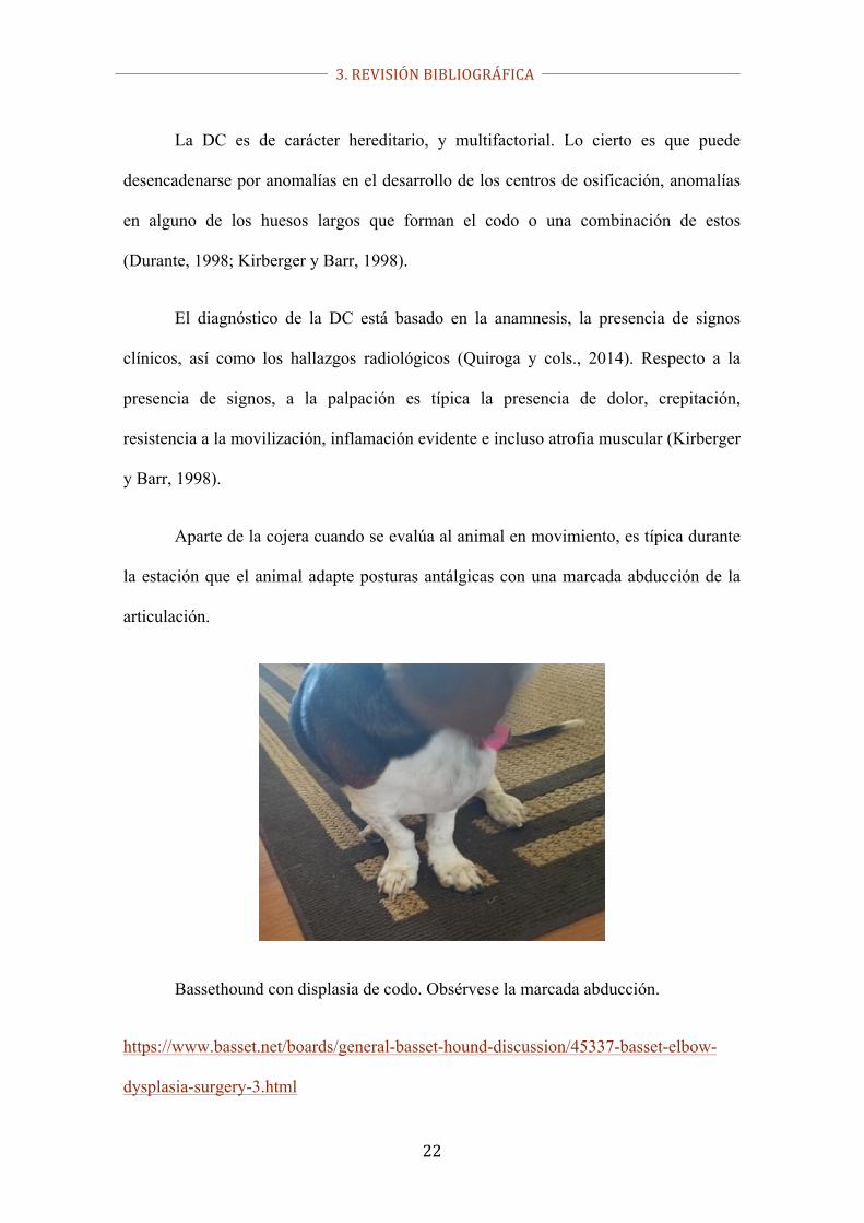
Aparte de la cojera cuando se evalúa al animal en movimiento, es típica durante
la estación que el animal adapte posturas antálgicas con una marcada abducción de la
articulación.
Bassethound con displasia de codo. Obsérvese la marcada abducción.
https://www.basset.net/boards/general-basset-hound-discussion/45337-basset-elbow-
dysplasia-surgery-3.html

3. REVISIÓN BIBLIOGRÁFICA
23
Las razas caninas grandes y gigantes suelen ser las más afectadas; de ellas
podemos destacar las razas bernese, rottweiler, labrador y golden retrievers, pastor
alemán, bullmastiff, collie, san bernardo, chow-chow, mastín de los pirineos, terranova,
etc. La patología afecta más a los machos es una proporción de 3:1 respecto a las
hembras (Durante y Brusa, 1998; Kirberger, 2003; Komsta y cols., 2008).
Proceso ancóneo no unido en un perro de 18 meses. Se observa como dicho
núcleo de osificación permanece independiente al cúbito (fleche amarilla).
https://www.acvs.org/small-animal/canine-elbow-dysplasia

3. REVISIÓN BIBLIOGRÁFICA
24
Osteoartritis avanzada con neoformación ósea (flechas) así como esclerosis bajo las
superficies articulares (cruz). https://www.acvs.org/small-animal/canine-elbow-
dysplasia
3.2.5. Articulación de la rodilla
Anatomía
La articulación de la rodilla esta subdividida en otras dos articulaciones:
1) La articulación femororrotuliana, entre la rótula y la tróclea femoral.
2) La articulación fémorotibial entre los cóndilos femorales y la meseta tibial (Barone,
1980). Ambas articulaciones forman una única entidad funcional pues, en virtud de las
inserciones proximales y, sobre todo, distales mediante el ligamento rotuliano, hace que
cualquier movimiento entre el fémur y la tibia, provoca otro entre la rótula y la tróclea
femoral (Castañón, 2015).
Existe una gran incongruencia entre las superficies de la articulación
femorotibial, por lo que se interponen dos meniscos de naturaleza fibrocartilaginosa que
3. REVISIÓN BIBLIOGRÁFICA
25
aparte de adaptar ambas superficies estabilizando la articulación, absorben los impactos
durante el apoyo (Evans y col., 2000).
Varios ligamentos proporcionan estabilidad adicional a la articulación entre los
que destacan los ligamentos colaterales y los ligamentos cruzados craneal y caudal,
ambos en situación intraarticular y con gran significación clínica en el ámbito humano y
sobre todo en la medicina y cirugía veterinaria de pequeños animales, en concreto del
perro.
El ligamento cruzado craneal (CCL por sus siglas en inglés) se origina
caudomedialmente, en el cóndilo femoral lateral y fosa intercondilar desarrollándose en
dirección craneomedial para insertarse en el área intercondilar de la tibia. El ligamento
cruzado caudal (CaCL, por sus siglas en inglés) se origina en la cara lateral del cóndilo
femoral medial y se dirige caudodistalmente para insertarse en la fosa poplítea de la
tibia (Heffron and Campbell 1978, Evans 1993). Ambos ligamentos, por lo tanto, evitan
el deslizamiento del fémur respecto a la tibia en el sentido caudal y craneal,
respectivamente.

3. REVISIÓN BIBLIOGRÁFICA
26
Hill´s Atlas of Veterinary Clinical Anatomy (2006).
Esta afirmación viene dada porque la rotura del CCL es la primera causa de
cojera de miembro pelviano, y una de las más comunes de las que afectan a la rodilla en
perros (Whitehair y cols., 1993, Johnson y cols., 1994, Duval y cols., 1999). Como se
ha explicado anteriormente, este ligamento contribuye a la estabilidad articular por lo
que su rotura la compromete seriamente; dicha inestabilidad en última instancia
conducirá progresivamente al desarrollo de una osteoartritis (OA) (Elkins y cols. 1991,
Innes and Barr 1998a).
El diagnóstico clínico se basa en evidenciar la falta de estabilidad femorotibial,
siendo estos animales positivos al test de compresión y de cajón anterior. En las

3. REVISIÓN BIBLIOGRÁFICA
27
radiografías realizadas bajo sedación y con la articulación en compresión, se confirma el
desplazamiento craneal de la tibia respecto al fémur (Sánchez-Bustinduy y cols., 2010).
La intensidad de cojera es muy variable en esos casos, pudiendo presentar alteraciones
del paso muy leves hasta cojeras sin apoyo del miembro afectado. Algunos autores
describen como característica el apoyo con las falanges únicamente, permaneciendo la
almohadilla metatarsiana sin apoyar (Harasen, 2002).
Postura antálgica típica de un perro con CCLR. No existe apoyo de la
almohadilla metatarsiana.

3. REVISIÓN BIBLIOGRÁFICA
28
3.3. Diagnóstico de la OA
3.3.1. Técnicas de imagen
3.3.1.1. Radiología
En las patologías citadas anteriormente, la radiología no solo va a proporcionar
un diagnóstico de la patología, sino que además podrá decir en el estadío en el que se
encuentra, en virtud de la identificación de los signos osteoartritis (Bland 2015, Vilar
2013), por lo que, aunque ha sido demostrada la falta de correlación entre signos
clínicos y radiológicos, el estudio radiológico en este tipo de patologías debe hacerse de
forma rutinaria.
Las técnicas de imagen más modernas como el TAC y la resonancia magnética
tienen la ventaja de poder visualizar mejor las estructuras articulares sin superposición,
evaluando los cambios óseos o incongruencias articulares (Fossum, 1999).
3.3.1.2. Artroscopia
La artroscopia se ha convertido en uno de los métodos de diagnóstico en los
últimos años, sobre todo en las displasias de codo. La artroscopia es menos invasiva que
la artrotomía y proporciona una visualización excepcional de todas las estructuras
intraarticulares (Kim y Joo, 2018). Adrian y cols., (2017), recomiendan, sin embargo,
que en lesiones femorotibiales el estudio del uso combinado de la ecografía y
artroscopia para acceder visualmente a todas las zonas articulares de interés clínico.

3. REVISIÓN BIBLIOGRÁFICA
29
3.3.2. Técnicas de evaluación funcional
Como se deduce de lo explicado anteriormente, la presencia de OA produce
dolor en mayor o menor grado. La consecuencia funcional de este dolor determina la
aparición de una cojera variable. Como hemos mencionado anteriormente, existe una
pobre correlación entre signos clínicos y radiológicos, por lo que la evaluación de la
funcionalidad del miembro o miembros afectados resulta importante (Gordon y cols.
2003).
Teniendo en cuenta lo anteriormente expuesto, la metodología para evaluar el
estado funcional del animal se puede dividir en:
3.3.2.1. Métodos de análisis funcional subjetivos
Clásicamente, los veterinarios clínicos basaban la cuantificación de la severidad
del dolor en sus pacientes en datos subjetivos como la vocalización, o los niveles de
actividad, la reacción del animal a la manipulación, así como la visualización subjetiva
de las alteraciones del movimiento. Basados en el examen subjetivo se han publicado
escalas numéricas para tratar de cuantificar el dolor de origen en el aparato locomotor
El cuestionario denominado Bristol Osteoarthritis in Dogs (BrOAD) (Innes and
Barr 1998b) o el Texas A&M Client (Hudson y cols. 2004) son escalas análogas
validadas que se basan en las apreciaciones del propietario del animal. Más difundido es
el Canine Brief Pain Inventory (CBPI) donde se añaden más datos subjetivos (Cimino
Brown y cols., 2007). similar a esta encontramos el Helsinki Chronic Pain Index
(HCPI), el cual contiene aspectos cuantificables (subjetivamente) como el
comportamiento del animal, así como su desempeño locomotor, teniendo en cuenta

3. REVISIÓN BIBLIOGRÁFICA
30
también el aspecto emocional del dolor (Hielm-Björkman y cols., 2003; Hielm-
Björkman y cols., 2009; Walton y cols., 2013). El HCPI ha sido utilizado de forma
rutinaria para la evaluación del dolor crónico en perros con osteoartritis del codo, la
rodilla e incluso la cadera (Hielm-Björkman y cols., 2003; Hielm-Björkman y cols.,
2009; Wernham y cols., 2011; Hielm-Björkman y cols., 2012; Heikkilä y cols., 2014).
En nuestro país, quisiéramos destacar la escala denominada Bioarth®, que
combina aspectos clínicos, radiológicos, y comportamentales los cuales deben ser
puntuados; finalmente se obtiene una puntuación que puede ser utilizada
comparativamente cuando, por ejemplo, se ha aplicado un tratamiento contra el dolor
que está provocando la cojera. Ésta escala puede ser utilizada en cadera, codo o rodilla
(Vilar y cols., 2016).

3. REVISIÓN BIBLIOGRÁFICA
31

3. REVISIÓN BIBLIOGRÁFICA
32
Escala Bioarth® adaptada para la evaluación de la articulación de la cadera.
Como puede observarse, este sistema está basado en una escala numérica que engloba
aspectos funcionales inherentes al animal, así como al clínico.

3. REVISIÓN BIBLIOGRÁFICA
33
Como su propio nombre indica, todos estos métodos son subjetivos por lo que el
factor observador (intraobservador e interobservador) pueden ser fuente de una gran
variabilidad en el resultado final (Horstam y cols., 2004).
3.3.2.2. Análisis objetivo (Biomecánica)
Bajo las premisas anteriores, la evaluación objetiva sobre el estado de la cojera
de un animal, así como la evaluación de su evolución en el caso de que dicho animal
este siendo tratado es un aspecto muy discutido científicamente (Mölsä y cols.,2010).
Los métodos de avaluación biomecánica pretenden ofrecer un método de
evaluación de los mecanismos que participan en el movimiento, especialmente centrado
en el aparato locomotor. Se dividen en métodos de evaluación cinemáticos y cinéticos.
Sin embargo, antes de empezar con las diferentes tecnologías, deberíamos citar
el uso del tapiz rodante o treadmill.
Este instrumento se utiliza como complemento a muchas de las técnicas que
vamos a describir a continuación. El hecho de que el animal se desplace sobre la cinta y
no sobre el terreno ofrece múltiples ventajas ya que permite extraer datos no solo
biomecánicos, sino también bioquímicos o fisiológicos, pero bajo condiciones
controladas de temperatura, humedad y nivel de trabajo, sin necesidad de disponer de un
espacio amplio de trabajo (Khumsap y cols., 2004; Cruz y cols., 2017), Además,
permite el registro de pasos consecutivos (Drevemo y cols., 1980).
Esta última ventaja, ha supuesto un gran avance para avanzar en los mecanismos
compensatorios que se desarrollan cuando los animales presentan cojera; en este
sentido, queremos destacar un reciente trabajo (Gómez Álvarez y cols., 2017)

3. REVISIÓN BIBLIOGRÁFICA
34
Donde se recopilan datos cinéticos y cinemáticos para evaluar la simetría de los
movimientos de la cabeza y la cadera en perros con cojera inducida. También conviene
destacar el uso concreto de sensores inerciales, de los que se hablara más adelante, para
obtener dichos índices de simetría en animales cojos (Rhodin y cols., 2017). En
cualquier caso, es oportuno mencionar que la toma de datos debe realizarse una vez que
los animales se han habituado a este (y otros) dispositivo (Gustås y cols., 2013).
En un estudio más reciente (Vilar y cols., 2016) se ha utilizado el treadmill para
el establecimiento de las diferentes características dinámicas, tanto cinéticas como
cinemáticas, de cuatro razas de perros con conformaciones muy diferentes; para ello, se
realizó la grabación de los animales con una cámara de alta velocidad con los animales
al paso, para obtener los datos de tipo cinemático. Para los de tipo cinético, se recurrió a
la inserción de una plataforma de fuerza sobre la cinta rodante.
Uso del treadmill en perros. En este caso, la línea verde demarca el lugar donde esta
insertada una placa de fuerza, y al mismo tiempo están señalados en verde reflectante
diferentes centros de rotación articular para cálculos de tipo cinemático.

3. REVISIÓN BIBLIOGRÁFICA
35
El treadmill también se utiliza como complemento en rehabilitación, como en este caso
hacienda trabajar al animal con los miembros en inmersión.
http://trends.medicalexpo.com/project-45657.html
Sin embargo, donde este instrumento adquiere un papel preponderante es en la
especie equina, desde el punto de vista deportivo, pero también desde el punto de vista
clínico. Así, este instrumento ha servido para comparar patrones de recuperación
funcional en casos de osteoartritis del carpo con el animal trabajando en tierra o con los
miembros sumergidos (King y cols., 2013); en otros casos, para estudiar el cambio de
dimensiones de estructuras tendinosas sometidas a rutinas de ejercicio en el treadmill
(Birch y cols., 1999a), así como, de forma análoga a los perros, para el estudio de la
3. REVISIÓN BIBLIOGRÁFICA
36
redistribución de las cargas en animales cojos (Buchner y cols., 1996a; Uhlir y cols.,
1997; Kelmer y cols., 2005).
No obstante, esta técnica no está exenta de inconvenientes y limitaciones; sobre
todo, respecto a la probable alteración del movimiento natural que el animal
desarrollaría ni estuviera sobre suelo normal (Buchner y cols., 1994; Barrey y cols.,
1993).
3.3.2.2.1. Análisis cinemáticos
Los métodos de evaluación cinemática proporcionan parámetros relacionados
con el movimiento sin tener en cuenta las fuerzas que lo producen. En concreto, se
pueden obtener parámetros lineares como la amplitud del paso, altura del miembro en la
fase de vuelo, etc.; parámetros angulares, como los ángulos de flexión, extensión, rango
angular de movimiento, velocidades y aceleraciones angulares, etc. Por último, también
se pueden obtener parámetros temporales para el cálculo de la duración de las diferentes
fases del paso cono la de apoyo, vuelo, etc.
a. Electrogoniometría.
Los electrogoniómetros son unos instrumentos que, aplicados sobre las
articulaciones, van obteniendo parámetros angulares en tiempo real con el sujeto en
movimiento. Esta técnica ha sido utilizada desde hace mucho tiempo en la especie
humana (Karpovich y cols., 1960; Cole y Abbs, 1986), aunque más recientemente ha
servido también como instrumento de medición en estudios con la especie canina
(Adrian y cols., 1966; Malouin y Bedard, 1983) así como en équidos (Taylor y cols.,

3. REVISIÓN BIBLIOGRÁFICA
37
1966). A grosso modo, el aparato consiste en un potenciómetro sujeto por dos barras, y
dichas barras son las que se fijan a la piel. Este potenciómetro, el cual se hace coincidir
con el centro de rotación de la articulación en estudio, generara una corriente eléctrica
proporcional a los valores angulares (ángulo, velocidad angular, aceleración angular…)
que desarrolle el sujeto de estudio.
Aunque este instrumento goza de gran precisión y fiabilidad en las mediciones,
existen dos factores limitantes que condicionan enormemente su uso. El primero, es el
hecho de que el dinamómetro debe coincidir exactamente con el centro de rotación de la
articulación. El segundo, es que en el campo concreto de la veterinaria, existen
articulaciones no accesibles a esta metodología (Ratzlaff, 1989), o también el hecho de
que muchas articulaciones no solo se limitan a movimientos de flexión / extensión.
Colocación de un electrogoniómetro sobre la articulación del codo

3. REVISIÓN BIBLIOGRÁFICA
38
Colocación del electrogoniómetro sobre la articulación de la rodilla
b. Cinematografía de alta velocidad y videografía.
La cinematografía, o con su terminología más actualizada, la videografía de alta
velocidad es probablemente la técnica más ampliamente utilizada para la investigación
biomecánica cinemática en, al menos, los équidos, y son numerosos los estudios que se
han basado en esta técnica para determinaciones en pequeños y grandes animales
(Keegan y cols.,2000; Kramer y cols, 2004; Vilar y cols., 2010).
Por cuestiones técnicas, ya que se deben grabar a los animales desde ciertas
perspectivas concretas (fundamentalmente desde una vista lateral para las mediciones
angulares y sobre todo lineares) se suele preferir medir a estos animales sobre un
treadmill (Vilar y cols., 2016), aunque otros, sobre todo aquellos en los que se prefiere
que el animal no esté sometido a condiciones de trabajo tan controladas, se suelen
grabar en pistas de competición, por ejemplo (Vilar y cols., 2010a; Vilar y cols.,
2010b).
Los principales datos cinemáticos pueden ser obtenidos con esta metodología
(ángulos, tiempos de las distintas fases del paso, ángulos articulares en 2D y 3D, etc.).

3. REVISIÓN BIBLIOGRÁFICA
39
Para ello, la forma más habitual es fijar sobre la piel en los centros de rotación de las
articulaciones unos pequeños marcadores reflectantes que faciliten su identificación. La
obtención de los datos puede ser manual, o incluso completamente automática (Langlois
y cols., 1978; Leach y Cymbaluk, 1986; Leach y Dyson, 1988; Kramer y Keegan, 2007,
mio dinámica movimiento). El inconveniente de los sistemas automatizados es que en
ocasiones la vibración de los marcadores durante el impacto de los miembros sobre la
superficie de apoyo crea artefactos que impiden la correcta medición (van Weeren y
cols., 1990).
(a)
3. REVISIÓN BIBLIOGRÁFICA
40
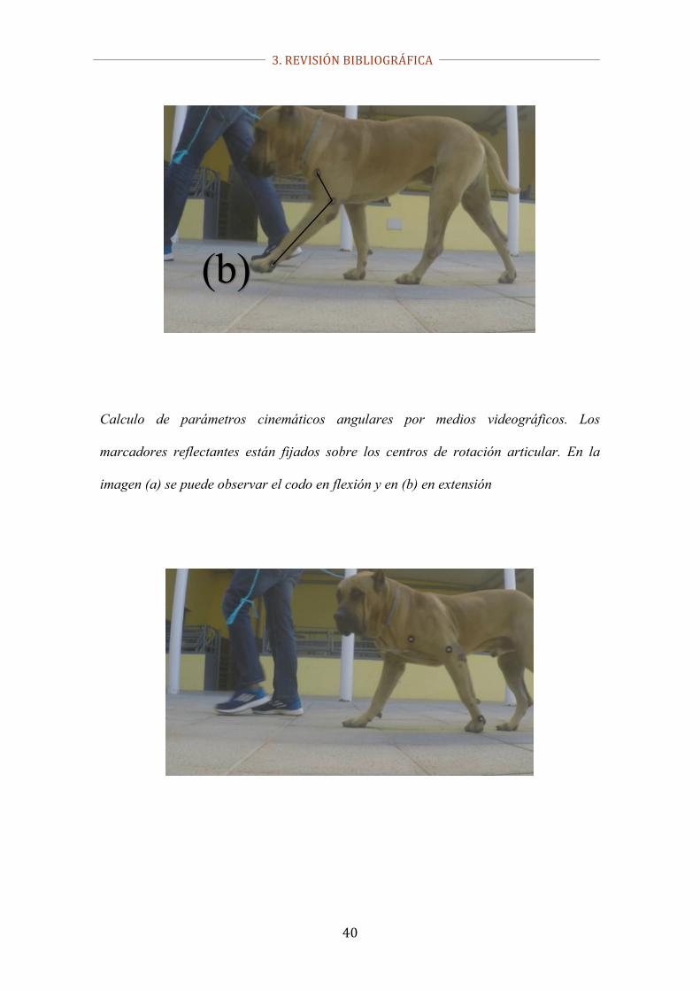
Calculo de parámetros cinemáticos angulares por medios videográficos. Los
marcadores reflectantes están fijados sobre los centros de rotación articular. En la
imagen (a) se puede observar el codo en flexión y en (b) en extensión
(b)

3. REVISIÓN BIBLIOGRÁFICA
41
Calculo de parámetros cinemáticos temporales por medios videográficos. En este caso
para el cálculo de la duración de la fase de vuelo. En la imagen (a) se muestra el
ultimo fotograma (frame) en el que el miembro está pisando, y la imagen (b) el inicio
de la fase de apoyo. Sabiendo el número de frames que han transcurrido y la velocidad
de grabación (frames grabados/segundo), se puede calcular el tiempo de vuelo.
3. REVISIÓN BIBLIOGRÁFICA
42
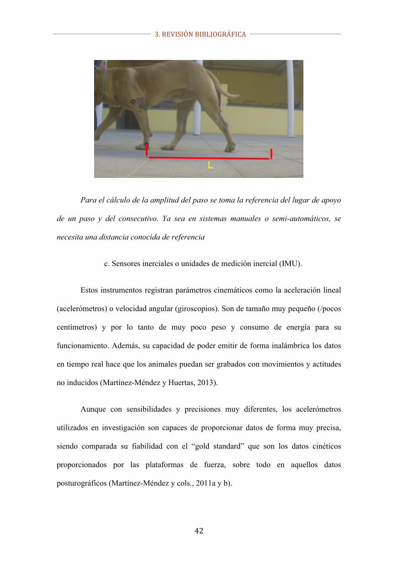
Para el cálculo de la amplitud del paso se toma la referencia del lugar de apoyo
de un paso y del consecutivo. Ya sea en sistemas manuales o semi-automáticos, se
necesita una distancia conocida de referencia
c. Sensores inerciales o unidades de medición inercial (IMU).
Estos instrumentos registran parámetros cinemáticos como la aceleración lineal
(acelerómetros) o velocidad angular (giroscopios). Son de tamaño muy pequeño (/pocos
centímetros) y por lo tanto de muy poco peso y consumo de energía para su
funcionamiento. Además, su capacidad de poder emitir de forma inalámbrica los datos
en tiempo real hace que los animales puedan ser grabados con movimientos y actitudes
no inducidos (Martínez-Méndez y Huertas, 2013).
Aunque con sensibilidades y precisiones muy diferentes, los acelerómetros
utilizados en investigación son capaces de proporcionar datos de forma muy precisa,
siendo comparada su fiabilidad con el “gold standard” que son los datos cinéticos
proporcionados por las plataformas de fuerza, sobre todo en aquellos datos
posturográficos (Martínez-Méndez y cols., 2011a y b).

3. REVISIÓN BIBLIOGRÁFICA
43
Sensor inercial de la marca Xsens®. La gran ventaja de este tipo de sensores es
que son totalmente autónomos, disponiendo de sistema de emisión de datos instantánea
y una batería que permite la toma de datos de muchos pasos consecutivos, pudiendo
discriminar sólo aquellos que se consideran más representativos.
https://www.xsens.com/products/
En équidos, son bastantes las publicaciones que pueden encontrarse respecto a
su utilización en la detección de anomalías de la locomoción en esta especie
demostrando su fiabilidad y precisión, así como su reproducibilidad, por lo que se
puede definir como un examen cuantitativo válido. En esta misma línea, se ha
demostrado que su utilidad clínica es directa (Keegan y cols., 2002; Watanabe y cols.,
2011).
Los sensores inerciales se pueden fijar en la cabeza, grupa y/o miembros; gracias
a esto, probablemente sea la técnica que ha ayudado enormemente a entender de forma
profunda como en esta especie, cuando está presente la cojera, se redistribuyen las
fuerzas a otro/s miembro/s como mecanismo de alivio ante el dolor (Maliye y cols.,
2013).

3. REVISIÓN BIBLIOGRÁFICA
44
En la especie canina conviene destacar los estudios de Rhodin y cols. (2017),
con aplicaciones en el diagnóstico de cojeras teniendo en cuenta las asimetrías que se
presentan en la zona craneal y pelviana. Cabe resaltar las conclusiones, donde con esta
tecnología no solo sirve para detectar las cojeras, sino también para cuantificarlas, al
menos al trote.
Ejemplo de zonas anatómicas donde pueden colocarse los sensores inerciales
3.3.2.2.2. Análisis cinético
La cinética es aquella rama de la biomecánica que estudia el movimiento de les
seres vivos estudiando, detectando y midiendo las fuerzas que lo generan. Dichas
fuerzas están generadas por los propios organismos en movimiento. Del cálculo de las
fuerzas, las fuerzas de reacción del suelo (GRF por sus siglas en ingles), son las más
difundidas, ya que por sí solas proporcionan valiosa información, además de que
también nos van a permitir calcular el centro de presiones (COP); por todo ello,
consideramos importante describir estos parámetros cinéticos, así como la relación que
existe entre ellos:

3. REVISIÓN BIBLIOGRÁFICA
45
- las GRF más son el pico de fuerza vertical (PVF, por sus siglas en inglés) que
se define como el valor más alto de fuerza (normalmente medida en Newton) que se
aplica contra el suelo cuando el miembro está apoyando durante la marcha; el impulso
vertical (VI, por sus siglas en inglés) se define como el producto de la fuerza por el
tiempo de apoyo. Este parámetro se visualiza gráficamente como el área bajo la curva
de apoyo, aunque su objetividad en el diagnóstico de cojeras aún está en discusión
(Vilar y cols., 2013).
-el centro de masas (COM) o, por simplificación del concepto, centro de
gravedad (CG) se define como la suma de las trayectorias de todos los segmentos que
componen el cuerpo en ambos planos (antero/posterior o cráneo/caudal en cuadrúpedos;
y latero/medial) (Winter y cols., 1991). Por tanto, la señal del COM expresa un
movimiento real, ya que como se explicará más adelante en este apartado, el equilibrio
postural, aunque sea fisiológico, no es un proceso estático (Baratto y cols., 2002;
-el COP, el cual podríamos definir como el punto de localización del vector de
las fuerzas verticales de reacción del suelo (GRF). Representa el promedio de todas las
presiones que están en contacto con el suelo y es independiente del COM, aunque por
simplificación se suele decir que el COP es la proyección vertical del COM en el plano
de apoyo (suelo) (Winter y cols., 1996); así, podemos decir que la señal del COP no
queda definida como un simple movimiento si no que muestra la fuerza aplicada en un
área determinada (Baratto y cols., 2002).
a. Plataforma de fuerza
Las plataformas de fuerza son unos medidores de fuerza (dinamométricos o
piezoeléctricos) insertados (normalmente 4) en las esquinas de un armazón

3. REVISIÓN BIBLIOGRÁFICA
46
cuadrangular. La fuerza ejercida con la pisada en la fase de apoyo es registrada por
estos medidores y producen proporcionalmente una corriente eléctrica que es recogida
en una computadora.
Plataforma dinamométrica de fuerza con 4 sensores de la marca Pasco®
En comparación con las escalas numéricas subjetivas el análisis del paso
mediante la plataforma de fuerza proporciona mediciones objetivas desde el punto de
vista cuantitativo del apoyo dinámico y de este modo ser capaz de detectar cojeras
moderadas o leves e incluso anomalías del paso que podrían no ser evidentes en la
evaluación visual (Evans y cols. 2005). Como se ha dicho anteriormente, el PVF y el VI
son los parámetros más usados en el análisis cinético con este dispositivo,
considerándose hoy en día el “gold standard” (Walton y cols., 2014) en el análisis del
paso.
Las GRF se pueden medir indistintamente al paso o al trote, pero hay que tener
en cuenta que los valores se van a ver significativamente modificados por la velocidad a

3. REVISIÓN BIBLIOGRÁFICA
47
la que el sujeto es evaluado; dicho esto, es fácil entender que un análisis adecuado del
movimiento con fines comparativos debe ser efectuado en un rango muy estrecho de
velocidad (Riggs y cols. 1993).
Asimismo, los cambios de velocidad (aceleración y deceleración) durante los
registros deben ser también suprimidos (Budsberg y cols. 1999).
El peso corporal, necesariamente, va a determinar también el valor de las GRF,
así que cuando se comparar individuos diferentes las comparaciones son muy difíciles
de efectuar de forma objetiva. Con este propósito, se ha propuesto que ,
independientemente de que se utilicen animales en lose estudios con pesos y/o
conformaciones similares, los datos se expresen en relación al peso corporal, es decir,
en porcentaje (Vilar y cols., 2013).
De todos modos, es importante indicar que, por lo general, se ha observado
mayores valores de PVF en los miembros anteriores que en los posteriores (Schnabl-
Feichter y cols., 2017), debido a que el CG en cuadrúpedos está desplazado
cranealmente.
La plataforma de fuerza se ha utilizado también para evaluar los resultados
terapéuticos en animales con osteoartritis derivadas de CCLR o displasia de cadera
(Nelson y cols., 2013; Skinner y cols., 2013; Wurcherer y cols., 2013; Vilar y cols.,
2013).
A pesar de sus múltiples indicaciones, esta técnica no está exenta de
limitaciones; en este sentido, la limitación más importante es que solo se puede medir la
fuerza ejercida por un solo miembro a la vez, lo que hace que los exámenes sean largos
y tediosos para el animal. Por otro lado, al solo medir la fuerza en la fase de apoyo, se

3. REVISIÓN BIBLIOGRÁFICA
48
descarta del análisis la que se ejerce durante la fase de vuelo. Por último, este
dispositivo, por sus dimensiones, tampoco es capaz de registrar pasos sucesivos.
Estudio cinético con plataforma de fuerza en un perro al paso. La plataforma se oculta
y nivela con el suelo para asegurar que la detección de la pisada es lo más natural
posible
b. Plataforma de presión
Las plataformas de presión se han ido incorporando a la clínica y la
investigación como una alternativa a la plataforma de fuerza, sobre todo por contener
una gran cantidad de sensores, llegando a ser miles, así como a la misma capacidad para
regular la frecuencia de adquisición de datos (Oosterlinck y cols., 2010a). A esto
debemos añadir que su costo no es superior y, como ventaja más sobresaliente, su
diseño permite obtener varios pasos consecutivos. Este instrumento de análisis cinético
es multiparamétrico, es decir, permite no solo la obtención de los datos clásicos
derivados directamente de las GRF como son el PVF y el IV, sino que además
proporciona datos como la distribución del peso en el cuerpo, así como entre los

3. REVISIÓN BIBLIOGRÁFICA
49
diferentes miembros cuando estos están en la fase de apoyo. Otros parámetros
adicionales con el cálculo de la presión media y máxima de apoyo, que en conjunto
conforman lo que se denomina el análisis podobarométrico (Oosterlinck y cols., 2011a;
Carr y cols., 2015; Bockstahler y cols., 2016; Schnabl-Feichter y cols., 2017), y cuya
representación gráfica para su posterior estudio se denomina genéricamente
podobarografía. Como más adelante se describirá, este instrumento sirve además para
detectar los cambios posturales y estabilométricos (Gomes-Costa y cols., 2015), que en
última instancia puede servir incluso para la detección de cojeras.
Las plataformas de presión de gran longitud (hasta 2 metros) permiten el
registro de varios pasos consecutivos. Plataforma tekscan.
https://twitter.com/vetbiomechanics/status/666602613715173376
En el campo de la medicina humana estos dispositivos sirven como fuente
importante de obtención de información para patologías tan complejas como la
esclerosis múltiple, en concreto de qué manera y a qué nivel esta patología afecta a la
preservación del equilibrio en posición estática (Abrantes y Santos, 2012)Se ha
utilizado también como herramienta diagnostica en personas con lepra (Cordeiro y
cols., 2014) así como en pacientes con diabetes (Anjos y cols., 2010), para ver en qué

3. REVISIÓN BIBLIOGRÁFICA
50
modo concreto las presiones plantares (es decir, el apoyo) se modifica con esta
patología, y sus repercusiones sobre el resto de las unidades del aparato locomotor.
En el campo de la veterinaria, es procedente resaltar los estudios que se han
realizado en el campo equino con plataformas de fuerza para conocer los elementos
cinéticos íntimos que se desarrollan durante la fase de apoyo del casco (Van Heel y
cols., 2005), los efectos del recorte del casco en la distribución de presiones en la suela
(Moleman y cols., 2006), así como la simetría del casco en caballos y ponies sanos
Oosterlinck et al., 2010ª;201b). Más recientemente, se está empezando a utilizar para la
detección de cojeras basándose en los cambios posturales. La gran ventaja de esta
metodología es que se puede realizar el diagnostico de cojeras con el animal en la
estación, es decir, quieto. Esto significa que se puede realizar en espacios relativamente
pequeños, (Gomes-Costa, 2015), pero este concepto se desarrollara con mucha más
precisión más adelante.
En pequeños animales la incorporación de esta tecnología ha sido si cabe, aún
más reciente, destacando en primer lugar la investigación encaminada a aportar datos
meramente descriptivos en perros sanos (Marghitu y cols., 2003; Souza y cols., 2013),
para posteriormente comenzar a describir alteraciones en animales con rotura de
ligamento cruzado craneal (Souza y cols., 2014), osteoartritis de cadera (Upchurch y
cols., 2016) o incluso a animales con patologías de cadera a los que se les había
reemplazado dicha articulación con una prótesis (Tomas y cols., 2014). El hecho de que
con este dispositivo se puedan aportar datos de los miembros de forma independiente y
simultánea, ha permitido que se publiquen datos derivados de diferentes índices de
simetría para poder detectar la cojera, especialmente cuando esta es unilateral (Carr y
cols., 2015).

3. REVISIÓN BIBLIOGRÁFICA
51
En la actualidad, y tratando de satisfacer las necesidades que en el campo
veterinario se pueden presentar, se están diseñando dispositivos con tamaños, pero sobre
todo con resoluciones, adaptadas a las diferentes especies, pudiéndose en la actualidad
obtener diferencias significativas en el área de apoyo de las regiones palmares/plantares
de gatos, en función de la presión ejercida (Schnabl-Feichter y cols., 2017)
Existen en este dispositivo una serie de limitaciones de índole técnica que
conviene citar, como es una respuesta más lenta de los sensores que en el caso de la
plataforma de fuerza; además, en un estudio realizado en caballos al trote, se comparó
los datos de PVF obtenidos mediante plataforma de presión y de fuerza
simultáneamente, que de forma consistente los datos obtenidos con la plataforma de
presión eran más bajos (Oosterlinck y cols., 2010b).
De todos modos, la gran ventaja de este dispositivo sobre la plataforma de fuerza
es que, debido a su diseño, ya sea modular o directamente rectangular y de grandes
dimensiones, permite obtener los datos de varias pisadas consecutivas. Sin embargo, tal
y como ocurre en las plataformas de fuerza cuando estas se acoplan, no existe todavía
investigación concerniente al estudio de la postura, es decir análisis estáticos. De este
modo, al igual que ocurre en la especie humana, se podrían estudiar las características
del COP y las consecuencias derivadas de la modificación del equilibrio (y la postura)
cuando se presente una patología que afecte al aparato locomotor, como las cojeras.
c. Relación postura-cojera
Anteriormente se ha citado el uso de la posturografía y la podobarografía
estática pare el diagnóstico de cojeras, sobreentendiendo que nos referimos a aquéllas
producidas por dolor, lo cual puede en principio no ser congruente. Por ello, hemos

3. REVISIÓN BIBLIOGRÁFICA
52
creído conveniente describir la relación que existe entre la postura (anómala) y las
cojeras.
La adopción del equilibrio postural correcto y su preservación continua es un
proceso complejo y combinado entre el sistema nervioso central, la vista, y los sistemas
neuromusculares (Ruhe y cols., 2010). En posición estática, al contrario de lo que
parece, el control de la postura y mantenimiento del equilibrio se entiende como una
acción constante de desestabilización / estabilización que se desarrolla, de forma
gráfica, como un péndulo invertido (Winter y col., 1996; Maurer y cols., 2005), que se
va a corresponder por el intento constante de mantener el COM simétricamente a la
BOS (Blaszcyk y cols., 1994). Debido a que el equilibrio va a estar constantemente
perturbado por factores internos y externos como los movimientos diafragmáticos
durante la respiración, los latidos cardiacos, la superficie de apoyo, etc., la recuperación
del equilibrio se realiza con movimientos compensatorios constantes en forma de vaivén
que se denominan balanceo postural, o postural sway, en inglés. De este modo, el
estudio del equilibrio mediante la posturografía estática se convierte en una metodología
de evaluación objetiva del equilibrio, que ha venido estudiando en los últimos años en
medicina humana para, mediante el estudio de las características del COP y sus
modificaciones en el campo de la medicina (Blasczcyk, 2016), rehabilitación
(Tamburella y cols., 2013) o incluso en el campo del deporte para corregir errores que
se traducen en mermas en el rendimiento deportivo (Whiteside y cols, 2015).
Desde el punto de vista práctico, y sobre todo en el campo clínico, está
ampliamente difundida la asunción de que la posición del COP coincide de forma casi
exacta con la proyección vertical del COM sobre la superficie de apoyo, aunque, como
se ha explicado anteriormente, estos son dos parámetros basados en conceptos muy

3. REVISIÓN BIBLIOGRÁFICA
53
diferentes (Baratto y cols., 2002). Esta deambulación (sway) del COP se puede
descomponer en el sentido latero-lateral (X) y craneocaudal (Y) respecto a la posición
del cuerpo, lo que se denominaría estabilograma. Si, por el contrario, el COP el registro
directo del COP sway queda registrado en un diagrama, dicho registro se denomina
estatoquinesiograma.
Pero, ¿qué relación tiene una cojera provocada por dolor con estos parámetros?
¿Cómo y en qué grado los afecta? Cuando el sujeto de estudio presenta dolor en el
aparato locomotor, especialmente los miembros, se manifiesta por una cojera. Dicho
dolor va a provocar también, de forma teórica, una pérdida del equilibrio postural
cuando el sujeto no está en movimiento, ya que transferirá la presión hacia el lado sano
(o el que sienta menos dolor) tratando de minimizar el dolor, aunque constantemente su
sistema nervioso lo corregirá, generándose un ciclo continuo de perturbaciones-
correcciones (Buchner y cols., 2001). Dicho de otro modo, el dolor va a provocar
alteraciones posturales que van a poder ser detectadas si las características del COP se
analizan. De forma adicional, estos valores podrían ser reintegrados a la normalidad si,
por ejemplo, un tratamiento efectivo contra la patología pudiera ser aplicado; esto
significaría que, de forma directa, desde el punto de vista clínico los cambios en el COP
servirían también para evaluar de forma objetiva la eficacia de diferentes tratamientos
para las patologías que cursen con cojera (Scivoletto y cols., 2008), y que, por lo tanto,
sería interesante estudiarlo (Tamburella y cols., 2013).
Por otro lado, siempre desde el punto de vista teórico, tenemos que considerar
que las almohadillas digitales son estructuras elásticas, lo que hace que sean sensibles a
los cambios de presión que se aplican sobre ellas, deformándose, y, por lo tanto,
aumentando su superficie. De este modo se ha concluido en estudios previos como este

3. REVISIÓN BIBLIOGRÁFICA
54
proceso, dentro de unos límites, va determinar cambios en el área de apoyo (Basher,
1994; Swaim, 1985). Por último, aunque difícilmente cuantificable, los patrones en los
que la presión se redistribuye en la superficie palmar/plantar cuando se varia de forma
patológica el COP también pueden server como datos complementarios que reafirmen la
utilidad de la posturografía y podobarografía estática para general datos fiables y útiles
como por ejemplo la migración del punto de máxima presión, etc.
Examen posturográfico en el perro. Dada la necesidad de obtener los datos con el
animal inmóvil durante bastantes segundos, resulta indispensable para obtener datos
fiables que tengan un carácter tranquilo.
A modo de resumen, los parámetros que de forma teórica podrían ser útiles para
la determinación de cojeras serían los siguientes:
3. REVISIÓN BIBLIOGRÁFICA
55
• Distribución de la presión de apoyo entre los miembros. Se suele medir en
kilopascales (Kpa) y se suele expresar en porcentaje. Este parámetro se convierte en un
medidor relativo del estado postural, ya que, en el caso de las cojeras, se va a producir
cambios posturales que van a modificar la distribución de la presión (teóricamente
simétrica) debido a la instabilidad (Anker y cols., 2008).
• Cambios en el área de apoyo de los miembros, que se mide en cm2 y se puede
expresar en términos absolutos o, cuando se comparan con otros miembros o
individuos, en porcentaje.
• Presión media y máxima de cada miembro, medida en Kpa.
• Gráficos de distribución de los rangos de presión en la zona palmar/plantar
mostrados en 2d e incluso 3d. estos gráficos muestran en una escala de colores los
diferentes rangos de presión desde el azul (rango inferior) hasta el rojo (rango máximo).
A este punto conviene aclarar que para obtener una escala que contemple el máximo
número de rangos necesita de una calibración manual, que dependerá por lo tanto de la
presión que se está aplicando sobre los sensores. Si esto no ocurre, podrían perderse de
la calibración los rangos inferiores o, en el externo contrario, saturar completamente
todos los sensores apareciendo toda la superficie de apoyo de color rojo.

3. REVISIÓN BIBLIOGRÁFICA
56
Gráfico de presiones obtenido en estática. Se muestra la distribución de presión,
presión media y máxima, así como los rangos de presión por zonas de apoyo
Rangos de presión 3D. Cuanto mayor es el rango de presión (rojo), más alta se muestra
la zona
• Estatoquinesiograma, que como se ha descrito anteriormente va a representar la
migración espacial en el plano bidimensional del COP, y que normalmente se mide en
mm2. El conjunto de los puntos por donde se ha ido midiendo el COP van a formas una
“nube” de puntos. El estatoquinesiograma recoge el 90% de dichos puntos, y

3. REVISIÓN BIBLIOGRÁFICA
57
normalmente adopta una forma esférica o, más frecuentemente, elíptica. Esto es debido
a que la estabilidad nunca es igual en el sentido craneocaudal que laterolateral. Por
ejemplo, los humanos se desestabilizan más en el sentido craneocaudal, mientras que
los cuadrúpedos se desestabilizan mayormente en el eje laterolateral, pues la BOS es
más larga que ancha.
Estatoquinesiograma de un perro cojo de la derecha. Aparte de la gran superficie (61,92
mm2), se ve la elipse desplazada hacia el lado izquierdo
Además, las articulaciones de los animales domésticos cuadrúpedos están
diseñadas para fluctuar en el plano sagital, contando con una musculatura flexora-
extensora predominante, facilitando de esta forma el control del movimiento CC, pero
con menor capacidad para generar fuerzas de abducción-aducción lo que lleva a que el
3. REVISIÓN BIBLIOGRÁFICA
58
movimiento ML sea más difícil de controlar, pudiendo obtener amplitudes del COP más
grandes en esta dirección.
En cualquier caso, se acepta que a mayor BOS se cuenta con una mejor
estabilidad, hecho que ha sido ampliamente estudiado en la especie equina (Clayton y
cols., 2013; Clayton y Nauwelaerts, 2014). En este sentido, se han descrito aumentos en
la amplitud ML, velocidad y área del COP, cuando los caballos adquirían una base de
soporte más estrecha en sus miembros anteriores (King y cols., 2013). De este modo,
cuando el equilibrio se ve comprometido, los caballos adquieren una postura abarcando
mayor amplitud en la base, como sucede cuando aprender a caminar en un treadmill
(Buchner y cols., 1994a).
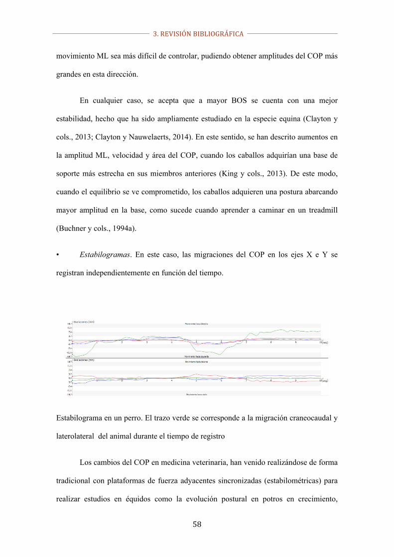
• Estabilogramas. En este caso, las migraciones del COP en los ejes X e Y se
registran independientemente en función del tiempo.
Estabilograma en un perro. El trazo verde se corresponde a la migración craneocaudal y
laterolateral del animal durante el tiempo de registro
Los cambios del COP en medicina veterinaria, han venido realizándose de forma
tradicional con plataformas de fuerza adyacentes sincronizadas (estabilométricas) para
realizar estudios en équidos como la evolución postural en potros en crecimiento,

3. REVISIÓN BIBLIOGRÁFICA
59
(Nauwelaerts y cols., 2013), las alteraciones posturales tras la administración de
sedantes alfa-2 adrenérgico como la detomidina® (Bialski y cols., 2004), o incluso para
cuantificar la mejora en caballos con osteoartritis de carpo a los que se les sometió a
rehabilitación (King y cols., 2013). También se ha evaluado la participación de la vista
en el equilibrio (Clayton y Nauwelaerts, 2014). Existen autores que afirman que esta
tecnología debería de ser capaz de discriminar entre las cojeras, ataxias o déficits
neurológicos, lo cual supone, en la actualidad, un gran elemento de discusión si se
realiza el diagnóstico convencional (Bialski y cols., 2004; Ishihara y cols., 2009;
Clayton y cols., 2013).
La evaluación estabilométrica (postural) tiene como gran ventaja respecto a la
evaluación dinámica que los exámenes se ejecutan, por propia definición, con el animal
quieto. Este hecho supone que no sea necesario un espacio amplio para realizar dichos
exámenes (Clayton y Nauwelaerts, 2012).
En las evaluaciones dinámicas suelen utilizarse una media de tres repeticiones
por individuo, utilizando el valor medio de dichas mediciones para la elaboración
estadística de la mayoría de los estudios científicos relacionados con el estudio de la
cojera (Vilar y cols., 2013); en las evaluaciones posturales, el número de repeticiones
adecuado para conseguir datos fiables oscila de 2-5, siendo la media de 3 (Golriz y
cols., 2012.

3. REVISIÓN BIBLIOGRÁFICA
60
3.4. Tratamiento de la OA
3.4.1. Tratamientos convencionales
Dentro de lo que podríamos denominar tratamientos higiénico-dietéticos, así
como alternativos se deben destacar la reducción de peso, el ejercicio de bajo impacto,
así como la fisioterapia, laser de baja intensidad, acupuntura y ultrasonidos entre otros
(Cakir y cols., 2013, Fang y cols., 2013, Gudbergsen y cols., 2012).
Respecto a los tratamientos farmacológicos, se han utilizado ampliamente los
NSAIDs, analgésicos, corticoides e incluso nutracéuticos como por ejemplo los
glucosaminoglicanos (Reid y cols., 2012, Godley, 2013, Merashly y Uthman, 2012).
Entre los NSAIDs, queremos destacar el Mavacoxib (Trocoxil™, Pfizer, MY,
USA), ya que es un inhibidor COX-2 relacionado estructuralmente con el celecoxib
(Penning y cols., 1997). La modificación bioquímica ha permitido obtener una molécula
más estable desde el punto de vista metabólico (Paulson y cols, 2000). Esta
modificación ha conseguido obtener un compuesto con un bajo índice de aclaramiento y
una vida media muy alta (Cox y cols., 2010). Esto hace que en última instancia la dosis
que se debe administrar para obtener un efecto terapéutico sea muy baja respecto a otros
NSAIDs clásicos (Paulson y cols., 2001).
3.4.2. Terapias regenerativas
En los últimos años, se ha ido incorporando al elenco de estrategias terapéuticas
contra la OA las denominadas terapias regenerativas, donde debemos destacar sobre
todo el plasma rico en plaquetas (PRP) y las células mesenquimales (CM). Este tipo de

3. REVISIÓN BIBLIOGRÁFICA
61
tratamientos suponen un nuevo enfoque terapéutico ya que no solo pretenden aliviar los
síntomas de la enfermedad de forma directa sino que pretende estimular y hacer
prevalecer los mecanismos reparativos y regenerativos del propio organismo que los
degenerativos, sin que aparentemente tengan efectos nocivos detectables,
convirtiéndose en una alternativa válida a otros tratamientos más agresivos (Singh,
2012; Wu y cols., 2007).
En este sentido, la irrupción de las CM ha revolucionado este campo
creando una gran expectativa en el ámbito de las terapias celulares (Diekman y Guilak,
2013, Fortier y Travis, 2011).
El origen de estas células madre puede ser embrionario (CME), lo que les
confiere una alta tasa de multiplicación, así como la posibilidad de diferenciarse en
múltiples líneas celulares, o también pueden ser obtenidas de tejidos adultos. Las
células mesenquimales adultas (CMA) presentas mayores limitaciones que las
anteriores para la multiplicación y diferenciación, ya que solo pueden derivarse por el
momento a los diferentes tejidos conectivos como el adiposo, el óseo o incluso el
cartilaginoso; sin embargo, estos tejidos, fundamentalmente el óseo y cartilaginoso
presentan gran interés al formar parte de los componentes articulares. Además, las
CMA son compatibles desde el punto de vista inmunitario (mayoritariamente se pueden
realizar injertos autólogos) y no tienen limitaciones de uso desde el punto de vista ético,
como las CME. Estas últimas, aunque tienen menor capacidad de multiplicación y
diferenciación, son inmunocompatibles, y su uso no está restringido por los problemas
éticos asociados a las células embrionarias. Por último, en algunas investigaciones, se
ha achacado a las CME complicaciones derivadas del crecimiento incontrolado
(Oldershaw, 2012).

3. REVISIÓN BIBLIOGRÁFICA
62
Las células mesenquimales adultas (CMA), son fáciles de recolectar,
fundamentalmente del tejido adiposo y tejido adiposo entre otros (Black y cols., 2008,
Yarak y Okamoto, 2010), aunque también se pueden obtener de medula ósea y/o hueso.
Otro componente terapéutico en la estrategia reparativa/regenerativa para la OA
es el plasma rico en plaquetas (PRP), sobre todo por la eficacia demostrada para el
control del dolor de origen articular, muscular, etc. Existen multitud de derivados en el
mercado en virtud de los diferentes modos de procesamiento (tiempos y velocidades de
centrifugación, activadores, etc.) así como la adición o supresión de elementos celulares
o incluso factores concretos que les van a conferir a estos productos diferentes
propiedades como duración, intensidad analgésica, antiinflamatoria, etc. En este
sentido, queremos destacar el plasma rico en factores de crecimiento-endoret® ,,,que es
de origen autólogo y carente de leucocitos y citoquinas proinflamatorias, y con un
nivel moderado de plaquetas y factores de c crecimiento (Anitua y cols., 2004),
demostrando una mejora significativa de la sintomatología de la OA tanto en la especie
humana como canina (Fahie y cols., 2013; Anitua y cols., 2014; Cuervo y cols., 2013).
Estudios recientes han demostrado que además es una terapia aparentemente
inocua, sin efectos a nivel local (ergogénico) ni a nivel general, descartando la supuesta
actividad carcinogénica del IGF-1 (Vilar y cols., 2017, Damiá y cols., 2018)
Teniendo en cuenta lo anteriormente expuesto, donde queda en evidencia el
prometedor futuro que tienen las terapias regenerativas en el tratamiento de la
osteoartritis, nuestra hipótesis de trabajo es que la posturografía y podobarografía
pueden resultar útiles para detectar de forma objetiva las cojeras en los perros de origen

3. REVISIÓN BIBLIOGRÁFICA
63
osteoartrítico. Por otro lado, si un tratamiento efectivo es instaurado, su nivel de
eficacia, e incluso duración, podría ser establecidas con esta metodología.

3. REVISIÓN BIBLIOGRÁFICA
64

4. ARTÍCULOS

4. ARTICULOS
66

4. ARTICULOS
67
ARTÍCULO #1
Manera ME, Carrillo JM, Batista M, Rubio M, Sopena J, Santana A,
Vilar JM. Static Posturography: A New Perspective in the
Assessment of Lameness in a Canine Model. PLoS One. 2017 Jan
23;12(1):e0170692. doi: 10.1371/journal.pone.0170692.
JCR Impact factor (2017): 2.766
Cuartil: Q1
Grupo: Multidisciplinary sciences
Posición: 15/64

4. ARTICULOS
68

4. ARTICULOS
69
RESEARCH ARTICLE
Static Posturography: A New Perspective inthe Assessment of Lameness in a CanineModelMaria E. Manera1, Jose M. Carrillo2, Miguel Batista1, Monica Rubio2, Joaquin Sopena2,Angelo Santana3, Jose M. Vilar1*
1 Departamento de Patologıa Animal, Instituto Universitario de Investigaciones Biomedicas y Sanitarias,Universidad de las Palmas de Gran Canaria, Arucas, Las Palmas, Spain, 2 Departamento Medicina y CirugıaAnimal, Catedra Garcıa Cugat, Universidad CEU Cardenal Herrera, Valencia, Spain, 3 Departamento deMatematicas, Universidad de las Palmas de Gran Canaria, Las Palmas, Spain
Abstract
The aim of this study was to assess the static posturography in dogs as a useful tool for diag-
nosis of lameness by means of the use of a pressure platform. For this purpose, a series of
different parameters (pressure distribution, area of support, mean pressure, maximum pres-
sure and statokinesiograms) were obtained from five lame dogs with unilateral elbow osteo-
arthritis treated with plasma rich in growth factors. Data were obtained before and 3 months
after treatment, and results were compared with a control group of sound dogs of similar
conformation. Significant differences were found in the above mentioned parameters
between sound and lame limbs. Improvement after 3 months of treatment was also
detected, demonstrating that this multi-parametric technique is an effective and reliable
method for the assessment of lameness in dogs.
Introduction
Peak vertical force (PVF) and vertical impulse (VI) are two of the most common kineticparameters used for lameness detection in dogs, horses and other domestic animal species [1];these parameters are usually obtained using force [2] or pressure platforms [3–5].
Pressure platforms, with their multiple sensors, have the potential to provide more informa-tion than force platforms; however, references describing the use of pressure platforms remainscarce, and the majority of these studies are descriptive. Previous studies describe distributionof force in the pads during the support phase in healthy dogs [6,7] or in dogs with pathologiessuch as cranial cruciate ligament rupture [8] or hip fractures [9]; the dogs walk or trot across asimple or multiple pressure walkway that provides standard parameters as PVF and VI, usuallymeasured by force platforms. The advantage of this method is that consecutive steps can berecorded; however, research has still not been published on static analysis in lame dogs withpostural changes, such as spatial modifications in body center of pressure (COP) and the
PLOS ONE | DOI:10.1371/journal.pone.0170692 January 23, 2017 1 / 13
a1111111111a1111111111a1111111111a1111111111a1111111111
OPENACCESS
Citation: Manera ME, Carrillo JM, Batista M, RubioM, Sopena J, Santana A, et al. (2017) StaticPosturography: A New Perspective in theAssessment of Lameness in a Canine Model. PLoSONE 12(1): e0170692. doi:10.1371/journal.pone.0170692
Editor: Steven Allen Gard, Northwestern University,UNITED STATES
Received: June 11, 2016
Accepted: January 9, 2017
Published: January 23, 2017
Copyright: © 2017 Manera et al. This is an openaccess article distributed under the terms of theCreative Commons Attribution License, whichpermits unrestricted use, distribution, andreproduction in any medium, provided the originalauthor and source are credited.
Data Availability Statement: All relevant data arewithin the paper and its Supporting Informationfiles.
Funding: The authors received no specific fundingfor this work. The Catedra Garcia Cugat providedthe pressure platform for this study.
Competing Interests: The authors have declaredthat no competing interests exist.

4. ARTICULOS
70
derived consequences of changes in paw area, and mean or maximum pressure values, amongother parameters.
The correct balance and its continuous preservation is a combined process connected withthe central nervous system, sight, and muscular system [10]. In quiet stance position, the con-trol of body posture is assumed as a constant action of stabilization of a multilink inverted pen-dulum [11,12], which corresponds with the attempt of to keep the center of mass (COM)symmetrically to the support base [13]. As posture is being constantly perturbed by internaland external mechanisms, the balance recovery is performed by constant compensatory move-ments, known as postural sway. In this way, static posturography becomes an objective evalua-tion method of the balance system, and it is widely used in human medicine [14], rehabilitation[15] or sport fields [16].
Many clinical practitioners assume that the COP position coincides with the projection ofthe COM on the support surface, although these two parameters are based on different con-cepts [17]. This sway is registered in both the latero-lateral (X) and craniocaudal (Y) axes ofthe body. Based on these principles, we could obtain two different graphical recordings: thestatokinesiogram, which depicts the movement of COP in an X-Y coordinate system, and thestabilogram, representing the location of the COP as a time function, where movements in theX and Y axes are considered separately.
When lameness is present, the associated pain causes loss of balance in the static positionand is provoked by the patient transferring weight from the painful limb to the healthy (orless lame) contralateral in an effort to alleviate the pain [18]. In other words, pain can alsocause postural (COP) modifications. Clinically, impaired COP balance returns to a morenormal value in some cases when an effective treatment is applied; this suggests thatchanges in COP balance could also be a predictive tool for gait recovery [19], thus meritingevaluation [20]. In addition, theoretically, changes in COP balance in lame dogs shoulddetermine changes in paw area, as the pads are elastic structures that spread when groundcontact pressure grows [21,22]. The patterns of pressure distribution in the paws might alsobe assessed to generate useful, objective, and complementary data as location of maximalpressure point and/or limb COP location within the paw to evaluate locomotor systemstatus.
The term medial coronoid disease (MCD) encompasses all pathologic changes of articularcartilage and subchondral bone involving the medial coronoid process of the elbow joint[23,24]. MCD is the most common cause of thoracic limb lameness in large and giant breeddogs [25], and MCD lesions have traditionally been evaluated radiographically [26–28]. A pre-sumptive diagnosis of MCD is frequently based on detection of the resultant secondary osteo-arthritis (OA), rather than on detection of the primary lesion [26,29].
Different strategies have been proposed for the treatment of OA, and among them, PlateletRich Plasma (PRP)-based products such as plasma rich in growth factor (PRGF) therapy, hasbeen widely used as a single or co-adjuvant therapy in the treatment of OA in dogs [30]. Basedon this, we hypothesized that assessment of COP variations, together with other secondarystatic parameters, could serve as an objective and quantifiable tool to detect lameness and itsvariations.
The aim of this study was to test static posturography as a potentially reliable, objectivemethod to evaluate lameness in five lame dogs affected by OA in elbow joints. The verificationof this objective builds a foundation for the feasibility of using static posturography in the clin-ical assessment of postural stability in lame dogs.
Static Posturography in a Lame Canine Model
PLOS ONE | DOI:10.1371/journal.pone.0170692 January 23, 2017 2 / 13

4. ARTICULOS
71
Materials and Methods
Animals
A total of 10 client-owned, adult dogs of similar conformation were used in this study. Thebody weight of enrolled dogs ranged from 30 to 41.8 kg, and ages were 3 to 9 years.
The control (sound) group was formed by five dogs (two Labradors and three Rottweilers).As they were sound dogs, a certain asymmetry should be assumed; for that reason, for compar-ison purposes, limbs with lesser and higher values were specifically considered. The studygroup contained five lame dogs (three Labradors and two Rottweilers). The lameness was uni-lateral and clinically classified as “severe” although with presence of weight bearing in all dogsand attributable to OA secondary to MCD of the elbow joints. Dogs did not receive medicationof any kind over the 4 weeks before the analysis.
To confirm or rule out OA, three standard radiographic views of both elbow joints (a lateralextension, lateral flexion, and a 15˚ oblique craniomedial caudolateral) [24] were taken undersedation with dexmedetomidine 10–20 μg/kg (Dexdomitor, zoetis, Spain)from dogs belongingto both study and control groups. Additional standard radiographs of knee and hip joints weretaken in order to ensure that elbow OA was the unique reason for the observed clinical signs.A complete clinical evaluation (physical examination, including vital signs and neurologic andorthopedic exams) assured that general health was otherwise normal.
The procedure was revised and authorized by the Ethical Committee of Animal Welfare(CEBA) of the University CEU Cardenal Herrera of Valencia. The owners of each animal gavepermission and signed a written consent form.
Obtention-inoculation of PRP
PRP was obtained using similar procedure to the PRGF1 (BTI, Vitoria, Spain), but with differ-ent materials. This product produces a moderated amount of platelets (double respecting toperipheral blood) and less than 0.3 leukocytes/ μL. The procedure is as follows: whole blood(10 mL) was aseptically extracted from the cephalic vein and collected in two 4.5-mL centri-fuge tubes (BD Vacutainer1, Plymouth, UK), each containing 0.5 mL citrate solution, thencentrifuged for 8 minutes at 460 × g. Only the inferior third of the obtained plasma (adjacentto the buffy coat) was used to be activated with 5% of its volume with 10% calcium chloride.The resultant ~2 mL solution was injected aseptically into the elbow joint through the conven-tional arthrocentesis site previous sedation with dexmedetomidine iv. The appearance of jointfluid confirmed proper needle placement. A total of four doses were administered on D0, D7,D14, and D21. After every inoculation, exercise was restricted to a walk of maximum of 30minutes at leash during the following 2 days.
Static posturography
For the recording of data, a pressure platform (EPS/R1, Loran Engineering, Bologne, Italy)was used. The device contains a total of 2096 pressure sensors of 1 cm2 distributed in an areaof 48 × 48 cm. The range of pressure was 30–400 kPa, and acquisition frequency 100 Hz. Ani-mals were placed in quiet stance with their thoracic limbs over the pressure platform, perpen-dicular to the ground, and each dog’s owner remained in front of the animal to attract thedog’s attention at a close distance. Three recordings of 10 seconds were obtained from eachanimal. Only recordings in which the animal was completely immobile in symmetric positionwere considered valid.
Data acquisition was performed using Biomech software (Loran Engineering). The parame-ters for static posturography were as follows:
Static Posturography in a Lame Canine Model
PLOS ONE | DOI:10.1371/journal.pone.0170692 January 23, 2017 3 / 13

4. ARTICULOS
72
• Distribution of pressure between both limbs measured in kilopascal (Kpa) and expressed inpercentage.
• Changes in paw area measured in cm2 and expressed in percentage.
• Mean and maximum pressure of each thoracic limb measured in Kpa.
• Graphic distribution of pressure ranges in paws of lame and sound limbs shown in a2-dimensional and 3-dimensional (3-D) color scale from blue (low pressure), to red (highpressure). In order to obtain a correct contrast among the colors in the different pressureranges, calibration was set manually to 212 Kpa to avoid saturation of the sensors.
• Statokinesiogram represents the amplitude of the spatial migration of the COP in a 2-Dspace, estimated by computing ellipse area (measured in mm2), which contains 90% of thedata points of the COP trajectory.
• Stabilogram recorded independent X and Y oscillations (measured in mm) as a function oftime.
Statistical analysis
Parameters were estimated by using the free R statistical software (https://www.r-project.org/).Analysis of variance with repeated measures and a Tukey test were used to determine signifi-cant differences. Normality and homoscedasticity of residuals was confirmed using Shapiroand Levene tests, respectively.
Results
The animals had a mean body weight of 36.88 ± 4.25 kg and a mean age of 5.8 ± 2.04 years.The mean values± SD of all obtained parameters are summarized in Table 1
Pressure distribution between both limbs
Differences between lame and sound limbs in the study group diminished significantly frombefore treatment (day 0) to day 90 post treatment (p< 0.001). Compared with the controlgroup, differences in pressure distribution at day 90 were also significant (p = 0.0044). Datawere normal (p = 0.24) and homoscedastic (p = 0.64)
Changes of paw area
Differences in area between lame and sound limbs in the study group diminished signifi-cantly from day 0 to day 90 (p < 0.001). Compared with the control group, differences inpaw area at day 90 were not significant (p = 0.072). Data were normal (p = 0.10) and homo-scedastic (p = 0.68).
Mean pressure
Differences in mean pressure between lame and sound limbs in the study group diminishedsignificantly from day 0 to day 90 (p< 0.001). Compared with the control group, differencesat day 90 were also significant (p< 0.001). Data were normal (p = 0.76) and homoscedastic(p = 0.72)
Static Posturography in a Lame Canine Model
PLOS ONE | DOI:10.1371/journal.pone.0170692 January 23, 2017 4 / 13

4. ARTICULOS
73
Table 1. Posturographic Parameters in Dogs, Expressed as Mean ± SD, and 95% Confidence Intervals.
Day LL SL % Difference
Pressure Distribution
0
39.72 ± 2.50% 60.28 ± 2.50% 20.56 ± 5.00%
(38.34, 41.10) (58.90, 61.66) (17.79, 23.33)
90
45.75 ± 2.63% 54.25 ± 2.63% 8.51 ± 5.25%
(44.29, 47.20) (52.80, 55.71) (5.60, 11.42)
Controls
48.19 ± 1.24% 51.81 ± 1.24% 3.63 ± 2.48%
(47.50, 48.87) (51.13, 52.50) (2.25, 5.00)
Paw Area (cm2)
0
38.20 ± 2.68 49.93 ± 2.63 26.70 ± 5.63%
(36.72, 39.68) (48.48, 51.39) (23.58, 29.82)
90
45.93 ± 2.28 49.53 ± 1.73 7.60 ± 3.27%
(44.67, 47.20) (48.58, 50.49) (5.79, 9.41)
Controls
42.93 ± 2.74 44.20 ± 2.65 2.93 ± 1.88%
(41.42, 44.45) (42.73, 45.67) (1.89, 3.97)
Mean Pressure
0
89.61 ± 13.79 112.11 ± 13.03 22.78 ± 5.82%
(81.97, 97.25) (104.89, 119.32) (19.56, 26.00)
90
91.93 ± 7.62 101.22 ± 7.43 9.69 ± 4.16%
(87.71, 96.15) (97.10, 105.34) (7.38, 11.99)
Controls
93.39 ± 9.61 96.42 ± 9.02 3.27 ± 1.81%
(88.07, 98.71) (91.43, 101.41) (2.27, 4.28)
Maximum Pressure
0
242.44 ± 26.84 278.76 ± 20.25 36.86 ± 20.62%
(227.57, 257.31) (267.55, 289.97) (25.44, 48.28)
90
296.23 ± 14.88 324.41 ± 10.86 29.25 ± 15.47%
(287.99, 304.47) (318.40, 330.43) (20.69, 37.82)
Controls
298.18 ± 34.36 359.89 ± 25.65 66.06 ± 34.13%
(279.15, 317.21) (345.69, 374.10) (47.16, 84.96)
Statokinesiogram (mm2)
0
44.71 ± 26.82
(29.86, 59.57)
90
13.91 ± 4.12
(11.63, 16.20)
(Continued )
Static Posturography in a Lame Canine Model
PLOS ONE | DOI:10.1371/journal.pone.0170692 January 23, 2017 5 / 13

4. ARTICULOS
74
Maximum pressure
Maximum pressure in sound limbs in the study group increased significantly from day 0 today 90 (p< 0.001). Compared with the control group, differences at day 90 were also signifi-cant (p< 0.001). Data were normal (p = 0.20) and homoscedastic (p = 0.10). In lame limbs,maximum pressure also increased significantly from day 0 to day 90 (p< 0.001). However,compared with the control group, differences at day 90 were not significant (p = 0.99). Datawere normal (p = 0.63) and homoscedastic (p = 0.42).
Statokinesiogram
Oscillations of the COP were greater in lame dogs during the 10 seconds of recording, asobserved in graphics, which sway measure area is greater in lame dogs, and assymetric (Fig 1).The sway area in the study group diminished significantly from day 0 to day 90 (p< 0.001).Compared with the control group, differences at day 90 were also significant (p = 0.005). Datawere not normal (p = 0.005) but were homoscedastic (p = 0.14). For that reason, the results arereliable due to the robustness of analysis of variance when data are homoscedastic.
Stabilogram
A symmetric latero-lateral oscillation could be seen in sound dogs, while craniocaudal oscilla-tion was almost insignificant. In contrast, lame dogs evidenced greater or/and asymmetricoscillations.
Graphical representation of COP sway in X and Y axes showed asymmetry in the studygroup in all cases during the 10-second recording period (Fig 2).
Graphic pressure distribution in the paws
2-D graphics showed as, when limbs were compared in sound dogs, a similar color (pressure)distribution pattern was evident: In addition, limb COP was symmetrically located within thepaw, and the maximal pressure point was located over the 2nd digital pad. In lame group, thepressure distribution pattern when limbs were compared was markedly different, with maxi-mal pressure point deviated laterally in the sound limb and cranially in the lame limb. LimbCOP in the sound limb of lame dogs was located in the center, but in the lame limb hasmigrated craniomedially. The maximal pressure point was found in the sound limb laterallyover the 5th digital pad, while in the lame limb, was located cranially, over the 4th digital pad.Body COP deviation towards the sound limb could also be seen (Fig 3).
In 3-D color scale graphics, the pressure patterns in sound limbs were similar, while inlame limbs they are asymmetric (Fig 4).
Table 1. (Continued)
Day LL SL % Difference
Controls
2.17 ± 1.10
(1.56, 2.79)
Day 0: Before treatment in study group; Day 90: After first application of treatment in study group; SL: Sound limb in study group or limb with higher value in
control group; LL: Lame limb in study group or limb with lesser value in control group.
doi:10.1371/journal.pone.0170692.t001
Static Posturography in a Lame Canine Model
PLOS ONE | DOI:10.1371/journal.pone.0170692 January 23, 2017 6 / 13

4. ARTICULOS
75
Fig 1. Statokinesiograms of Sound (A) and Lame (B) Dogs, Showing the Ellipse that Contains 90% ofthe Body COP Migration. In addition to show a greater area, in this case, a left deviation of the ellipse isevident in lame dog.
doi:10.1371/journal.pone.0170692.g001
Static Posturography in a Lame Canine Model
PLOS ONE | DOI:10.1371/journal.pone.0170692 January 23, 2017 7 / 13

4. ARTICULOS
76
Discussion
With static posturography, we were able to determine differences in a set of parametersbetween lame and sound dogs. Additionally, these differences diminished following treatment,indicating a positive response and thus, effective therapy, as previously published in humans[31].
While the redaction of this paper was conducted, we were unable to find specific studiesregarding the use of static posturography and pressure measurements of dog paws other thanPVF and VI using a pressure platform. Moreover, this methodology is widely used in humansin different fields [14–16] with excellent results; this should encourage the increase of pressureplatform technology use in dogs.
Statokinesiograms and stabilograms require the necessary time for recording the spatialvariations of the body COP; in humans, it is advisable to calculate parameters as the average ofthose obtained in three successive recordings of 10–60-second duration between studies
Fig 2. Stabilograms of Sound (A) and Lame (B) Dogs, Showing Oscillation of Body COP (Green Lines). Graphic shows that this lame dog had agreater oscillation to the right side.
doi:10.1371/journal.pone.0170692.g002
Static Posturography in a Lame Canine Model
PLOS ONE | DOI:10.1371/journal.pone.0170692 January 23, 2017 8 / 13

4. ARTICULOS
77
[32,33]. In agreement with these recommendations, we used three successive recordings of 10seconds on each animal. This was enough to detect asymmetries in the dislocation of the COP.
Regarding the sampling frequency, recent observations in humans [34] suggest to consideran effective sampling rate of about 50 Hz. Considering that COP sway is a pendular move-ment, the pendulus length is shorter in dogs than in humans; thus, the frequency of oscillationshould be, theoretically, higher. For that reason, in order to gain accuracy, we decided toincrease the sampling rate to 100 Hz.
In this study, the method suitability was assessed by using dogs with forelimb lamenessbecause the evaluation of hind limb lameness has been demonstrated harder to perform [35].Some authors try to explain this fact arguing that the proximal joints of the hind limb are moreable to decrease the supported load during the stance phase when comparing with the fore-limb, as occurs in horses [36]. In addition, the lameness should be less noticeable as a lowerproportion of body weight is supported by the hind limb [37].
Fig 3. 2-D Color Scale Graphics of Sound (A) and Lame (B) Dogs. In (A) symmetry is found in all measured parameters as pressure distribution, limbCOP (black circle) and maximal pressure point (white circle). In (B), the pressure distribution pattern is manifestly different between lame (right) and soundlimb (left), and assymetry in the other parameters is evident: limb COP (black and red in sound limb or blue circle in lame limb) maximal pressure point(white circle), body COP (black and grey circle).
doi:10.1371/journal.pone.0170692.g003
Static Posturography in a Lame Canine Model
PLOS ONE | DOI:10.1371/journal.pone.0170692 January 23, 2017 9 / 13

4. ARTICULOS
78
Static posturography in dogs not only can contribute to the scientific knowledge, but alsocan help clinicians to graphically show lameness status to pet owners; however, some limita-tions were found during the design and development of this study. First, to obtain substantialdifferences in the parameters derived from COP migration, the use of large breed dogs wasnecessary. Second, it was absolutely necessary to enroll calm and obedient dogs to stand inquiet stance for the minimum time required to obtain reliable statokinesiograms and stabilo-grams. In addition, each trial required an adaptation phase, and during this phase, some dogsshowed fatigue or lack of attention; for that reason, acquisition time was established at 10 sec-onds. More than this time was almost impossible to achieve in most cases.
Although 6 months is considered the minimum standard for testing the evolution of a med-ical or surgical treatment, we tested the animals after 3 months; we did this because after 6months, and based on some clinical evidences, the effect of the treatment could be diminished,possibly making variation in posturographic parameters less evident or undetectable.
Fig 4. 3-D Color Scale Graphic of a Sound (A) and Lame (B) Dog. In this case, lame dog shows a lateral deviation of the pressure in the sound limb(left) and a craniomedial deviation of pressure in the lame limb (right).
doi:10.1371/journal.pone.0170692.g004
Static Posturography in a Lame Canine Model
PLOS ONE | DOI:10.1371/journal.pone.0170692 January 23, 2017 10 / 13

4. ARTICULOS
79
This study used a relative low number of animals. Although the study used dogs of similarconformation, the low sample size could result in a potential lack of statistical power. Never-theless, we believe that this study clearly demonstrates that static posturography is a powerfultool to assess lameness in dogs and advance the objective quantification of pressure distribu-tion anomalies in limbs due to COP sway variations. For example, in the future, the maps ofpressure redistribution could help to identify and even characterize specific pathologies likethose detectable using other kinematic [38] or kinetic studies [39].
Great disparity of criteria has been published about the regimen of PRP administration forOA, oscillating from one to a series of six injections [40–43]. Based on these, we decided aseries of four injections with a 1-week interval between them.
Conclusion
Based on our results, static posturography was possible to perform in dogs and provided fairlyaccurate measurements of various parameters not assessed before in the evaluation of forelimblameness. These parameters, in addition to complement the classic kinetic PVF and VI, mayfurnish to veterinary practitioners a useful tool to assess asymmetries simultaneously due toCOP sway in lame dogs.
Supporting Information
S1 File. ARRIVE guidelines.(PDF)
Acknowledgments
The authors thank Thomas Michael Oxlee and Misty Bailey for translation and editing, respec-tively. Also thanks to the dogs’ owners for their collaboration. Thanks also to the Catedra Gar-cia Cugat for its technical support.
Author Contributions
Conceptualization: JMV JMC MB.
Data curation: AS.
Formal analysis: AS.
Investigation: JS MEM MR.
Methodology: JMV.
Resources: JMC MC JS.
Supervision: JMV.
Writing – original draft: MEM.
Writing – review & editing: JMV JMC MB.
References1. Fanchon L, Grandjean D. Accuracy of asymmetry indices of ground reaction forces for diagnosis of hind
limb lameness in dogs. Am J Vet Res. 2007; 68: 1089–1094. doi: 10.2460/ajvr.68.10.1089 PMID:17916016
Static Posturography in a Lame Canine Model
PLOS ONE | DOI:10.1371/journal.pone.0170692 January 23, 2017 11 / 13

4. ARTICULOS
80
2. Vilar JM, Batista M, Morales M, Santana A, Cuervo B, Rubio M, et al. Assessment of the effect of intra-articular injection of autologous adipose-derived mesenchymal stem cells in osteoarthritic dogs using adouble blinded force platform analysis. BMC Vet Res. 2014; 10: 143. doi: 10.1186/1746-6148-10-143PMID: 24984756
3. Gibert S, Lequang T, Maitre P, Cachon T, Carozzo C, Fau D, et al. Sensitivity and specificity to deter-mine lameness in dogs with a pressure walkway system. Vet Comp Orthop Traumatol. 2012; A21.
4. Besancon MF, Conzemius MG, Derrick TR, Ritter MJ. Comparison of vertical forces in normal grey-hounds between force platform and pressure walkway measurement systems. Vet Comp Orthop Trau-matol. 2003; 16: 153–157.
5. Oosterlinck M, Pille F, Back W, Dewulf J, Gasthuys F. A pressure plate study on fore and hindlimb load-ing and the association with hoof contact area in sound ponies at the walk and trot. Vet J. 2011; 190:71–76. doi: 10.1016/j.tvjl.2010.08.016 PMID: 20875762
6. Marghitu DB, Swaim SF, Rumph PF, Cojonaru D, Gillette RL, Scardino MS. Dynamics Analysis ofGround Contact Pressure of English Pointer Dogs. Nonlinear Dyn. 2003; 33: 253–265,
7. Souza AN, Pinto AC, Marvulle V, Matera JM. Evaluation of vertical forces in the pads of German Shep-herd dogs. Vet Comp Orthop Traumatol. 2013; 26: 6–11. doi: 10.3415/VCOT-11-07-0100 PMID:23111688
8. Souza AN, Tatarunas AC, Matera JM. Evaluation of vertical forces in the pads of Pitbulls with cranialcruciate ligament rupture. BMC Vet Res. 2014; 10: 51. doi: 10.1186/1746-6148-10-51 PMID:24581287
9. Vassalo FG, Rahal SC, Agostinho FS, Mamprim MJ, Melchert A, Kano WT, et al. Gait analysis in dogswith pelvic fractures treated conservatively using a pressure-sensing walkway. Acta Vet Scand. 2015;57: 68. doi: 10.1186/s13028-015-0158-3 PMID: 26438541
10. Ruhe A, Fejer R, Walker B. The test-retest reliability of centre of pressure measures in bipedal statictask conditions–a systematic review of the literature. Gait Posture. 2010, 32: 436–445. doi: 10.1016/j.gaitpost.2010.09.012 PMID: 20947353
11. Winter DA, Prince F, Frank JS, Powell C, Zabjek KF. Unified theory regarding A/P and M/L balance inquiet stance. J Neurophysiol. 1996; 75: 2334–2343. PMID: 8793746
12. Maurer C, Peterka RJ. A new interpretation of spontaneous sway measures based on a simple modelof human postural control. J Neurophysiol. 2005; 93: 189–200. doi: 10.1152/jn.00221.2004 PMID:15331614
13. B¯aszczyk JW, Lowe DL, Hansen PD. Ranges of postural stability and their changes in the elderly. GaitPosture. 1994; 2: 11–17.
14. Blaszczyk JW. The use of force-plate posturography in the assessment of postural instability. Gait Pos-ture. 2016; 44: 1–6. doi: 10.1016/j.gaitpost.2015.10.014 PMID: 27004624
15. Tamburella F, Scivoletto G, Molinari M. Balance training improves static stability and gait in chronicincomplete spinal cord injury subjects: a pilot study. Eur J Phys Rehabil Med. 2013; 49: 353–364.PMID: 23486301
16. Whiteside D, Elliott BC, Lay B, Reid M. Coordination and variability in the elite female tennis serve. JSports Sci. 2015; 33: 675–686 doi: 10.1080/02640414.2014.962569 PMID: 25358037
17. Baratto L, Morasso PG, Re C, Spada G. A new look at posturographic analysis in the clinical context:sway-density versus other parameterization techniques. Motor control. 2002; 6: 246–270. PMID:12122219
18. Buchner HH, Obermuller S, Scheidl M. Body centre of mass movement in the lame horse. Equine Vet JSuppl. 2001; 33: 122–127.
19. Scivoletto G, Romanelli A, Mariotti A, Marinucci D, Tamburella F, Mammone A, et al. Clinical factorsthat affect walking level and performance in chronic spinal cord lesion patients. Spine. 2008; 33: 259–264. doi: 10.1097/BRS.0b013e3181626ab0 PMID: 18303457
20. Tamburella F, Scivoletto G, Molinari M. Balance training improves static stability and gait in chronicincomplete spinal cord injury subjects: a pilot study. Eur J Phys Rehabil Med. 2013; 49: 53–364.
21. Basher, A. ‘Foot injuries in dogs and cats’, Compendium on Continuing Education Practicing Veterinar-ian. 1994; 1159–1176.
22. Swaim S F. Management and bandaging of soft tissue injuries of dog and cat feet, Journal of the Ameri-can Animal Hospital Association. 1985; 21: 329–340.
23. Moores AP, Benigni L, Lamb CR. Computed tomography versus arthroscopy for detection of canineelbow dysplasia lesions. Vet Surg. 2008; 37: 390–398. doi: 10.1111/j.1532-950X.2008.00393.x PMID:18564264
Static Posturography in a Lame Canine Model
PLOS ONE | DOI:10.1371/journal.pone.0170692 January 23, 2017 12 / 13
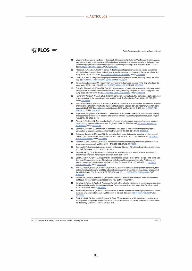
4. ARTICULOS
81
24. Villamonte-Chevalier A, van Bree H, Broeckx B, Dingemanse W, Soler M, Van Ryssen B, et al. Assess-ment of medial coronoid disease in 180 canine lame elbow joints: a sensitivity and specificity compari-son of radiographic, computed tomographic and arthroscopic findings. BMC Vet Res. 2015; 11: 243.doi: 10.1186/s12917-015-0556-9 PMID: 26407863
25. Fitzpatrick N, Yeadon R, Smith T, Schulz K. Techniques of application and initial clinical experiencewith sliding humeral osteotomy for treatment of medial compartment disease of the canine elbow. VetSurg. 2009; 38: 261–278. doi: 10.1111/j.1532-950X.2008.00493.x PMID: 19236684
26. Cook CR, Cook JL. Diagnostic imaging of canine elbow dysplasia: a review. Vet Surg. 2009; 38: 144–153. doi: 10.1111/j.1532-950X.2008.00481.x PMID: 19236671
27. Temwichitr J, Leegwater PA, Hazewinkel HA. Fragmented coronoid process in the dog: a heritable dis-ease. Vet J. 2010; 185: 123–129. doi: 10.1016/j.tvjl.2009.06.022 PMID: 19640749
28. Smith TJ, Fitzpatrick N, Evans RB, Pead MJ. Measurement of ulnar subtrochlear sclerosis using a per-centage scale in labrador retrievers with minimal radiographic signs of periarticular osteophytosis. VetSurg. 2009; 38: 199–208. doi: 10.1111/j.1532-950X.2008.00488.x PMID: 19236678
29. Hornof WJ, Wind AP, Wallack ST, Schulz KS. Canine elbow dysplasia. The early radiographic detectionof fragmentation of the coronoid process. Vet Clin North Am Small Anim Pract. 2000; 30: 257–266.PMID: 10768233
30. Vilar JM, Morales M, Santana A, Spinella G, Rubio M, Cuervo B, et al. Controlled, blinded force platformanalysis of the effect of intraarticular injection of autologous adipose-derived mesenchymal stem cellsassociated to PRGF-Endoret in osteoarthritic dogs. BMC Vet Res. 2013; 9: 131. doi: 10.1186/1746-6148-9-131 PMID: 23819757
31. Hirjakova Z, Singliarova H, Bzduskova D, Kimijanova J, Bučkova K, Valkovič P, et al. Postural stabilityand responses to vibrations in patients after anterior cruciate ligament surgical reconstruction. PhysiolRes. 2016, 24: S409–S416.
32. Pinsault N, Vuillerme N. Test-retest reliability of centre of foot pressure measures to assess posturalcontrol during unperturbed stance. Med Eng Phys. 2009; 31: 276–286. doi: 10.1016/j.medengphy.2008.08.003 PMID: 18835738
33. Schmid M, Conforto S, Camomilla V, Cappozzo A, D’Alessio T. The sensitivity of posturographicparameters to acquisition settings. Med Eng Phys. 2002; 24: 623–631. PMID: 12376049
34. Bottaro A, Casadio M, Morasso PG, Sanguineti V. Body sway during quiet standing: is it the residualchattering of an intermittent stabilization process? Hum Mov Sci. 2005; 24: 588–615. doi: 10.1016/j.humov.2005.07.006 PMID: 16143414
35. Peham C, Licka T, Girtler D, Scheidl M. Hindlimb lameness: clinical judgement versus computerisedsymmetry measurement. Vet Rec. 2001; 148: 750–752. PMID: 11442235
36. Buchner HHF. Gait adaptation in lameness. In: Back W, Clayton HM, editors. Equine Locomotion. Lon-don: WB Saunders: London, 2013. p. 251–279.
37. Gillette R, Graig T. Canine locomotion analysis. In: Millis D, Levine D, editors. Canine Rehabilitationand Physical Therapy. Amsterdam: Elsevier, 2014. p 201–210.
38. Caron A, Caley A, Farrell M, Fitzpatrick N. Kinematic gait analysis of the canine thoracic limb using a sixdegrees of freedom marker set. Study in normal Labrador Retrievers and Labrador Retrievers withmedial coronoid process disease. Vet Comp Orthop Traumatol. 2014; 27: 461–469. doi: 10.3415/VCOT-14-03-0051 PMID: 25345466
39. Kim SE, Pozzi A, Banks SA, Conrad BP, Lewis DD. Effect of cranial cruciate ligament deficiency, tibialplateau leveling osteotomy, and tibial tuberosity advancement on contact mechanics and alignment ofthe stifle in flexion. Vet Surg. 2010; 39: 363–370. doi: 10.1111/j.1532-950X.2010.00655.x PMID:20522216
40. Moraes VY, Lenza M, Tamaoki MJ, Faloppa F, Belloti JC. Platelet-rich therapies for musculoskeletalsoft tissue injuries. Cochrane Database Syst Rev. 2014: 4: CD010071.
41. Sanchez M, Anitua E, Azofra J, Aguirre JJ, Andia I. Intra- articular injection of an autologous preparationrich in growth factors for the treatment of knee OA: a retrospective cohort study. Clin Exp Rheumatol.2008; 26: 910–913 PMID: 19032827
42. Franklin SP, Garner BC, Cook JL. Characteristics of canine platelet-rich plasma prepared with five com-mercially available systems. Am J Vet Res. 2015; 76: 822–827. doi: 10.2460/ajvr.76.9.822 PMID:26309111
43. Cook JL, Smith PA, Bozynski CC, Kuroki K, Cook CR, Stoker AM, et al. Multiple injections of leukore-duced platelet rich plasma reduce pain and functional impairment in a canine model of ACL and menis-cal deficiency. Orthop Res. 2016; 34: 607–615.
Static Posturography in a Lame Canine Model
PLOS ONE | DOI:10.1371/journal.pone.0170692 January 23, 2017 13 / 13

4. ARTICULOS
82

4. ARTICULOS
83
ARTÍCULO #2
Vilar JM, Manera ME, Santana A, Spinella G, Rodriguez O, Rubio
M, Carrillo JM, Sopena J, Batista M. Effect of leukocyte-reduced
platelet-rich plasma on osteoarthritis caused by cranial cruciate
ligament rupture: A canine gait analysis. PLoS One. 2018 Mar
19;13(3):e0194752. doi: 10.1371/journal.pone.0194752.
JCR Impact factor (2017): 2.766
Cuartil: Q1
Grupo: Multidisciplinary sciences
Posición: 15/64

4. ARTICULOS
84

4. ARTICULOS
85
RESEARCH ARTICLE
Effect of leukocyte-reduced platelet-richplasma on osteoarthritis caused by cranialcruciate ligament rupture: A canine gaitanalysis modelJose M. Vilar1,2*, Maria E. Manera1, Angelo Santana1, Giuseppe Spinella3,Oliver Rodriguez1, Monica Rubio4, Jose M. Carrillo4, Joaquın Sopena4, Miguel Batista1,2
1 Departamento de Patologıa Animal, Facultad de Veterinaria de la Universidad de Las Palmas de GranCanaria, Trasmontaña S/N, Arucas, Las Palmas, Spain, 2 Instituto Universitario de InvestigacionesBiomedicas y Sanitarias, Universidad de Las Palmas de Gran Canaria, Juan de Quesada 30, Las Palmas deGran Canaria, Las Palmas, Spain, 3 Dipartimento di Science Mediche Veterinarie, Facoltà di Veterinaria dela Università di Bologna, Via Tolara di sopra 50, Ozzano, Bologna, Italy, 4 Departamento de Medicina yCirugıa Animal, Universidad CEU Cardenal Herrera, C/ Tirant lo Blanc, 7, Alfara del Patriarca, Valencia,Spain
Abstract
The goal of this study was to objectively assess the effect of a platelet-rich plasma (PRP)
derivate in English bulldogs with stifle degenerative joint disease secondary to cranial cruci-
ate ligament rupture (CCLR). We used a force platform and affixed electrogoniometers to
measure peak vertical force (PVF), vertical impulse (VI), stance time (ST), and angular
range of motion (AROM), from 12 lame client-owned English bulldogs with post-CCLR stifle
joint abnormalities. The 12 affected subjects were treated with 4 intra-articular injections of
PRP, at 30-day intervals. Ten untreated, sound English bulldogs were used as a reference
group. Clinical outcomes were evaluated using a linear mixed effects model. Mean values
of PVF, VI, ST, and AROM were improved within the first 3 months post-treatment in the
CCLR group, with mean measured changes increasing to maximum 4.56% body weight
gain, 1.5% body weight/second, 0.07 seconds, and 6.18 degrees, respectively. The effects
declined progressively after the treatment interval, ending at nearly initial levels after 6
months. This study demonstrates that dogs with CCLR treated with intra-articular PRP had
improved PVF, VI, ST, and AROM over time; the duration of effect was waning by the end of
the post-treatment period.
Introduction
Cranial cruciate ligament rupture (CCLR) is one of the most significant stifle injuries in domesticdogs, and is a common cause of chronic lameness [1]. A CCLR results in joint instability leadingto osteoarthritis (OA) and degenerative joint disease [2–6]. Progressive joint degeneration pro-duces changes in catabolic and anabolic activity of chondrocytes [7], impairing functional capabil-ity, producing pain, limiting flexibility and motion and lowering quality of life [8].
PLOS ONE | https://doi.org/10.1371/journal.pone.0194752 March 19, 2018 1 / 12
a1111111111a1111111111a1111111111a1111111111a1111111111
OPENACCESS
Citation: Vilar JM, Manera ME, Santana A, SpinellaG, Rodriguez O, Rubio M, et al. (2018) Effect ofleukocyte-reduced platelet-rich plasma onosteoarthritis caused by cranial cruciate ligamentrupture: A canine gait analysis model. PLoS ONE13(3): e0194752. https://doi.org/10.1371/journal.pone.0194752
Editor: Dennis F. Lawler, Illinois State Museum,UNITED STATES
Received: February 7, 2018
Accepted: March 8, 2018
Published: March 19, 2018
Copyright: © 2018 Vilar et al. This is an openaccess article distributed under the terms of theCreative Commons Attribution License, whichpermits unrestricted use, distribution, andreproduction in any medium, provided the originalauthor and source are credited.
Data Availability Statement: All relevant data arewithin the paper and its Supporting Informationfiles.
Funding: The authors received no specific fundingfor this work.
Competing interests: The authors have declaredthat no competing interests exist.

4. ARTICULOS
86
Various surgical techniques have been advocated to treat CCLR, including extracapsularstabilization, tibial plateau-leveling osteotomy, and tibial tuberosity advancement [9]. Conser-vative management includes weight loss [10], omega-3 fatty acid supplementation [11] anduse of NSAID drugs [12].
Recently, innovative treatments for modulating joint pain have been introduced, includingchemical precursors, mesenchymal stem cell therapy, and growth factors obtained from plate-let-rich plasma (PRP). These treatments can be used alone or in combinations [6,13]. Plateletshave high concentration of growth factors, making PRP an appealing therapeutic alternative.The platelet growth factors could favorably influence need for analgesia, soft tissue healing, reg-ulation of anti-inflammatory signals [14], and vascularizing and innervating of autografts [15].
Based on experimental results, PRP alone or as a co-therapy with surgical repair, can reduceprogression of OA, without undesirable side effects [16,17]. It can be difficult to compare ther-apeutic studies directly because of differing assessment criteria, clinical measures (ie, OA clas-sification systems), inclusion and exclusion criteria, varying aetiologies or the many possibleintrinsic patient factors. Thus, objective measures of level and duration of efficacy representsignificant advancements in experimental methods [18].
Force platform gait analysis is an objective, quantitative, non-invasive, and reliable methodto characterize ground reaction forces during locomotion [19,20]. This kinetic methodologyhas been used to characterize gait in dogs after CCLR [1], but not for objective assessment ofthe effect of PRP in dogs with stifle OA that is secondary to naturally-occurring CCLR. On theother hand, electrogoniometers have been used to measure dog joint metrics such as range ofangular movement (AROM) [21].
We hypothesize that PRP therapy has beneficial effects in dogs with CCLR, but that thiseffect may be limited in terms of time. To test this hypothesis, we used force platform and elec-trogoniometric gait analysis to determine efficacy and duration of effect of PRP therapy inEnglish bulldogs with unilateral CCLR. An additional goal was to estimate an initial dosingregimen for further research.
Materials and methods
Dogs
All procedures were approved by the Ethics Committee of the Research Institute in Biomedicaland Health Sciences of the University of Las Palmas de Gran Canaria (Spain), in compliancewith national (Royal Decree 1201/2005) and European Union regulations (European Directive86/609/CE) for projects using animals for research. A total of 22 adult, client-owned Englishbulldogs were included in the study. Ten of them served as clinically normal, unaffected,untreated control group. General inclusion criteria consisted of body weight between 18–27 kgand age between 1.5–6 years.
CCLR group. To qualify for our study design, post-CCLR dogs (n = 12) would be overtlylame on the ipsilateral injury side and would lack evidence of concurrent systemic or otherorthopaedic disease. Medical screening included hematology, urinalysis, serum biochemicalprofiles, and no medication given over the preceding 4 weeks. All dogs were evaluated forarticular effusion and for tibial compression and cranial drawer tests to assess the integrity ofthe cranial cruciate ligament.
Bilateral stifle radiographs were made under tibial compression with each dog in ipsilateralrecumbency. CCLR was confirmed by observing cranial displacement of the proximal end ofthe tibia with respect to the femoral condyles, and any radiographic signs that were consistentwith osteoarthritis. The radiological and physical evaluations also were needed to confirm thatthe stifle joint disease was unilateral.
Gait analysis of the effect of PRP in dogs with CCLR
PLOS ONE | https://doi.org/10.1371/journal.pone.0194752 March 19, 2018 2 / 12

4. ARTICULOS
87
Control group. The dogs selected for the Control group (n = 10) would not have currentor previous history of orthopaedic or neurologic disease, nor persistent injury or systemic dis-ease, confirmed by general physical examination, orthopedic evaluation, and radiology. Noneof the control dogs were forced to perform physical activities. Dog owners signed an informedconsent form that allowed their dogs to participate in the study.
Preparation and administration of PRP
Prior to platelet collection and activation, blood platelet and leukocyte counts were obtained.Whole blood (10 mL) was extracted aseptically from a cephalic vein and collected in two 5-mLcentrifuge tubes, each containing 1.0 mL sodium citrate (2.5% solution) as anticoagulant. Thetubes were centrifuged (Centronic JP selecta, model 7000015, Barcelona, Spain) for 10 min at210 x g. Only the lower one-third of the yielded plasma (adjacent to the buffy coat) was acti-vated with 5% of its volume, using 10% calcium chloride. The resultant ~2 mL solution wasinjected aseptically into the affected stifle joint through the conventional arthrocentesis site[22]. The appearance of joint fluid confirmed proper needle placement. A total of four doseswere administered on days 0, 7, 14 and 21. After each inoculation, exercise was restricted for 2days by confining to a walk at a maximum of 30 minutes/day on a leash. For the remainingdays, dogs could walk on a leash a maximum of three times daily for 30 minutes, until the endof the experiment. No dogs were engaged in strenuous activity during the study.
Gait analysis
Kinetic analysis was performed using a single platform mounted in the centre of, and levelwith, a 7-m runway covered by a rubber mat. The mat weight was eliminated by setting it to “0force” with the tare button, after the platform was covered. Dogs were leash-guided at a walkover the force platform, always by the same handler. Walk velocity was measured using amotion sensor (Pasco, Roseville, California, USA) positioned 1-m from the platform.
Five valid trials were obtained for each dog, at a sampling frequency of 250 Hz. A trial wasconsidered valid when the limb fully contacted the force platform and the dog walked next tothe handler without pulling on the leash. The trial was discarded if the dog was distracted dur-ing the measurement, if the limb struck the edge of the force plate, or if any portion of the con-tralateral paw hit the force plate. A member of the research team (OR) evaluated the trial toconfirm which limb touched the centre of the force platform. The platform was interfacedwith a dedicated computer using DataStudio (Pasco, Roseville, California, USA), a softwarespecially designed for acquisition, numerical conversion, and storage of data. A team member(JMV) recorded data from affected limbs at Day D0, D30, D90 and D180 after the first admin-istration of PRP. Data from sound dogs were obtained at the same intervals; the Peak VerticalForces (PVF) and Vertical Impulse (VI) values were normalized to body weight (as %) to char-acterize possible improvement of lameness during treatment with PRP. The Stance Time (ST)value was acquired directly from DataStudio.
Immediately after the force platform examination, electrogoniometry was performed usingan electrogoniometer (Pasco) placed over the lateral collateral ligament of the stifle and affixedwith adhesive plasters by an experienced researcher (OR). After the dogs were observed to becomfortable with the device, five consecutive steps were recorded. Electrogoniograms wereobtained with the same software used to obtain the AROM.
Statistical analysis
The purpose of the experiment was to determine if systematic differences exist in PVF, VI, STand AROM in dogs with CCLR under PRP treatment given at four fixed moments in time: D0,
Gait analysis of the effect of PRP in dogs with CCLR
PLOS ONE | https://doi.org/10.1371/journal.pone.0194752 March 19, 2018 3 / 12
4. ARTICULOS
88
D30, D90, D180. For this reason, a linear mixed effects model was used with the experimentalfactor (time) as a fixed effect and the blocking factor (dog) as a random effect. The model is asfollows:
yijk à mj á bi á bij á mijk; i à 1; . . . ; 5; j à 1; . . . ; 4; k à 1; . . . ; nij
bi ⇡ NÖ0; sbÜ; bij ⇡ NÖ0; slÜ; mijk ⇡ NÖ0; sÜ
where yijk is the k-th measure of PVF, VI, ST and AROM, for the dog (i = 1 . . . 5) on day j = 1(0), 2 (30), 3 (90), and 4 (180); μj is the (fixed) effect of time j. This parameter represents PVF,VI, ST, and AROM in the CCLR group, at the j-th observation. For bi, the (random) effect ofdog, values are anticipated to be normally distributed, with mean 0 and standard deviation σb.Thus, σb is the variability in the response due to the dogs; is the interaction effect between dogand time of observation (j). This term allows for the possibility that some dogs could improveat the same time period as others worsen. μijk is the residual in the measure ijk. This variablealso is assumed to be normally distributed with mean 0 and standard deviation σ. ηij is thenumber of replicates of the PVF, VI, ST and AROM, measures on the dog at each timeinterval.
Metrics in this model were estimated using the nlme package in R statistical software(https://www.r-project.org/). For comparing fixed effects, the multiple comparison test ofTukey was applied. For assessing the validity of the model, a Shapiro-Wilk test was applied fortesting normality of the residuals, and a Levene’s test was done for testing homoscedasticity.Ninety-five percent confidence intervals for differences between CCLR and Control groups atserial intervals also were computed. Statistical power analysis was calculated with Glimmpse©(http://glimmpse.samplesizeshop.org/#/).
Results
The CCLR group contained 4 female and 8 male dogs, and the Control group consisted of 4female and 6 male dogs. The mean (± SD) body weight of enrolled dogs was 22.83 ± 2.62 kg inthe CCLR group and 23 ± 3.01 kg in the Control group (P = 0.30). Mean age was 3.6 ± 1.14 inthe CCLR group and 4 ± 1.22 in the Control group (P = 0.54).
The mean value for walking velocity of both sound (Control) and CCLR group dogs was0.7 ± 0.2 m/s (P = 0.12). Mean values (± SD) for PVF, VI, ST and AROM, in both CCLRand Control groups are summarized in Table 1. Data were distributed normally and werehomoscedastic.
The PRP obtained under this protocol revealed mean platelet count of 1013 ± 431 x 103cells/μL (⇡ 336% baseline count) and mean leukocyte count of 0.1 ± 0.1 x 10³ cells/μL(Table 2).
Analysis of PVF
Comparing with D0, mean PVF in the CCLR group increased by 4.56% at D30 (P< 0.0001);3.34% increase was found at D90 (P < 0.0001); and 0.97% increase at D180 (P = 0.0077).
Compared with the Control group, lower PVF was found at D0 (4.96%) (P < 0.0001) andD180 (3.57%) (P< 0.0001) (Fig 1).
Analysis of VI
VI was increased 1.5% at D30 (P< 0.0001), 0.9% at D90 (P< 0.0001), and 0.4% at D180, all ascompared with day 0 (P = 0.003).
Gait analysis of the effect of PRP in dogs with CCLR
PLOS ONE | https://doi.org/10.1371/journal.pone.0194752 March 19, 2018 4 / 12

4. ARTICULOS
89
Comparison of the CCLR and Control groups revealed significant differences at D0 (1.13%less) (P< 0.0001), and at D180 (0.63% less) (Fig 2) (P < 0.0001).
Analysis of ST
In the CCLR group, with respect to D0, ST increased in 0.07s, 0.04s, and 0.03s at D30 (P = 0),D90 (P = 0), and D180 (P = 0.004224), respectively.
Compared with Controls, affected dogs’ ST was 0.05s less at D0 (P = 9.1–9). At D30, the dif-ference was not significant. At D90 and D180, ST again was lower (0.044s and 0.019s, respec-tively) (P = 0.039) (Fig 3).
Analysis of AROM
Analysis of variance revealed that at D30, AROM increased by 5.68 degrees, compared withD0 (P < 0.0001). AROM increased to 6.18 degrees at D90 (P< 0.0001).
Compared with the Control group, CCLR dogs had AROM 6.48 degrees less at D0(P< 0.0001); at D30 and D90, no differences were found. However, at D180, AROM decreasedby 6.17 degrees (P< 0.0001) (Fig 4).
Our sample size of 12 dogs provided a statistical power of 83% for the detection of differ-ences of 0.5% of each parameter among individuals.
Discussion
We assessed PVF, VI, ST, and AROM changes in lame English bulldogs with unilateral CCLRtreated with PRP. We used a force platform and electrogoniometry. The data provided supportour hypothesis that PRP therapy is effective for OA secondary to CCLR, although the effect istemporary.
To our knowledge, this is the first study that provides objective data regarding PRP treat-ment outcomes in dogs with sequelae to naturally occurring CCLR.
Table 1. Mean and standard deviation (mean ± SD) of force plate analysis data (n = 22) for the lame leg in CCLR group and Control group. Data are shown in per-cent of dog weight (PVF, VI), in seconds (ST), and degrees (AROM).
Parameter Day after treatment
D0 D30 D90 D180 SWT LT
PVF 0.09 0.7
CCLR 35.13 ± 1.08# 39.69 ± 1.10⇤ 38.47 ± 1.31⇤ 36.10 ± 0.78#⇤
Control 40.09 ± 0.84 39.34 ± 0.66 39.32 ± 0.76 39.37 ± 0.51
VI 0.20 0.40
CCLR 10.79 ± 0.61# 13.28 ± 0.66⇤ 11.69 ± 0.55⇤ 11.19 ± 0.62#⇤
Control 11.90 ± 0.60 11.89 ± 0.65 11.95 ± 0.58 11.82 ± 0.46
ST 0.37 0.60
CCLR 0.38 ± 0.03# 0.45 ± 0.05⇤ 0.43 ± 0.03#⇤ 0.40 ± 0.03#⇤
Control 0.43 ± 0.03 0.44 ± 0.04 0.45 ± 0.03 0.43 ± 0.03
AROM 0.82 0.45
CCLR 25.10 ± 3.15# 30.78 ± 1.96⇤ 31.25 ± 1.56⇤ 25.49 ± 3.67#
Control 31.58 ± 0.61 31.51 ± 0.62 31.62 ± 0.69 31.67 ± 0.58
PVF = peak vertical force, CCLR = cranial cruciate ligament rupture group, VI = vertical impulse, ST = stance time, AROM = angular range of motion
# significant difference (p<0.05) between study and Control group.⇤ significant difference (p<0.05) in the study group between checking periods and D0, SWT Shapiro-Wilk test value, LT Levene test value.
https://doi.org/10.1371/journal.pone.0194752.t001
Gait analysis of the effect of PRP in dogs with CCLR
PLOS ONE | https://doi.org/10.1371/journal.pone.0194752 March 19, 2018 5 / 12
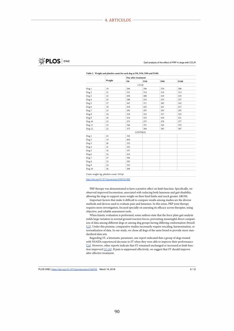
4. ARTICULOS
90
PRP therapy was demonstrated to have a positive effect on limb function. Specifically, weobserved improved locomotion, associated with reducing both lameness and gait disability,allowing the dogs to support more weight on their hind limbs and reach greater AROM.
Important factors that make it difficult to compare results among studies are the diversemethods and devices used to evaluate pain and lameness. In this sense, PRP joint therapyrequires more investigation, focused specially on assessing its efficacy across therapies, usingobjective, and reliable assessment tools.
When kinetic evaluation is performed, some authors state that the force plate gait analysisyields large variation in normal ground reaction forces, preventing meaningful direct compari-son of data among different dogs or among dog groups having differing conformation (breed)[23]. Under this premise, comparative studies necessarily require rescaling, harmonization, ornormalization of data. In our study, we chose all dogs of the same breed to provide more stan-dardized data sets.
Regarding ST, a kinematic parameter, one report indicated that a group of dogs treatedwith NSAIDs experienced decrease in ST when they were able to improve their performance[24]. However, other reports indicate that ST remained unchanged or increased as limb func-tion improved [25,26]. If pain is suppressed effectively, we suggest that ST should improveafter effective treatment.
Table 2. Weight and platelet count for each dog at D0, D30, D90 and D180.
WeightDay after treatment
D0 D30 D90 D180
CCLR
Dog 1 19 284 296 270 280
Dog 2 21 331 314 314 313
Dog 3 23 458 488 410 435
Dog 4 25 248 232 253 237
Dog 5 27 367 371 385 342
Dog 6 18 418 423 421 417
Dog 7 23 295 297 293 297
Dog 8 24 318 323 317 323
Dog 9 26 416 423 418 421
Dog 10 23 273 275 278 277
Dog 11 23 346 351 342 353
Dog 12 22 375 384 383 387
CONTROL
Dog 1 23 345
Dog 2 19 464
Dog 3 26 332
Dog 4 21 222
Dog 5 18 337
Dog 6 24 416
Dog 7 27 384
Dog 8 22 295
Dog 9 24 323
Dog 10 26 269
Units; weight: kg, platelets count: 103/μl
https://doi.org/10.1371/journal.pone.0194752.t002
Gait analysis of the effect of PRP in dogs with CCLR
PLOS ONE | https://doi.org/10.1371/journal.pone.0194752 March 19, 2018 6 / 12

4. ARTICULOS
91
Fig 1. PVF values in CCLR group at the 6-month follow-up period after treatment with platelet-rich plasma. Datafrom Control group also are shown.
https://doi.org/10.1371/journal.pone.0194752.g001
Fig 2. VI values in CCLR group at the 6-month follow-up period after treatment with platelet-rich plasma. Datafrom Control group also are shown.
https://doi.org/10.1371/journal.pone.0194752.g002
Gait analysis of the effect of PRP in dogs with CCLR
PLOS ONE | https://doi.org/10.1371/journal.pone.0194752 March 19, 2018 7 / 12

4. ARTICULOS
92
Fig 3. ST values in CCLR group at the 6-month follow-up period after treatment with platelet-rich plasma. Data from Control group also areshown.
https://doi.org/10.1371/journal.pone.0194752.g003
Fig 4. AROM values in CCLR group at the 6-month follow-up period after treatment with platelet-rich plasma. Data from Control group alsoare shown.
https://doi.org/10.1371/journal.pone.0194752.g004
Gait analysis of the effect of PRP in dogs with CCLR
PLOS ONE | https://doi.org/10.1371/journal.pone.0194752 March 19, 2018 8 / 12

4. ARTICULOS
93
Recent reports have shown that the presence of leukocytes in PRP preparations may be det-rimental to healing, due to their involvement in the inflammatory response [27] causing loss ofpain relief efficacy [28]. We avoided this complication, because our PRP was prepared withnearly complete absence of leukocytes.
Concerning actual administration, there is no consensus at this time about a standard regi-men. Thus, PRP treatment for OA can vary from one to a series of nine injections [29–33].Under this range of treatment protocols, we decide to apply a mid-level of four injections at7-day interval.
A follow-up evaluation of 6 months could be considered as a minimum standard for testingthe outcome of a medical or surgical treatment. In our study, this time was sufficient to recog-nize significant improvement during the first months after treatment, and also to establish thatlame dogs eventually returned nearly to their initial status. The duration of the effect that wenoted is consistent with previous reports [34,35]. Based on our results and taking into accountthat improvement by surgical means can extend relief for more than 2 years [29], PRP therapymay become an alternative resource for those instances wherein affected dogs cannot undergosurgery because of anaesthetic risk or when dog owners cannot afford surgical costs. Othercommercially available PRP derivates (PRGF- Endoret1, Orthokine1) have been associatedwith better effect and/or longer duration, but objectively-generated data remain to be col-lected, as is the case also with injections of hyaluronic acid or NSAIDs.
Our study has some limitations. First, the number of available dogs was relatively small.Small sample sizes can associate with low statistical power, and previous reports have estab-lished that dog conformation and size can affect kinetic measures [23]. Therefore, we decidedto prioritize data homogeneity (diminishing the inter-subject variability) by using dogs fromthe same breed within defined weights and ages. We thus noted that variation of only 0.5% inPVF could be detected with a statistical power of 83%.
Second, although both hind limbs were recorded, we provide only data from the affectedlimbs for comparison with control dogs. Based on previous reports, force redistribution tocontralateral limbs and even to forelimbs (in hind limb lameness) could call this choice intoquestion [36].
Third, our study design included sound dogs as controls when, typically, a Control groupwould have included untreated affected dogs. However, painful untreated or placebo-treateddogs in a positive Control group could worsen, rendering these dogs unable to provide fixedreference data. Additionally, ethical concerns arise when affected, untreated controls are partof biomedical experimental designs. In accordance with other authors using similar studydesigns [23], data from sound dogs were provided as an “ideal status” disease-free Controlgroup.
Conclusion
Force platform and electrogoniometric gait analysis established quantitatively that PRP ther-apy could be useful for treatment of chronic lameness in post-CCLR dogs. Our data indicateefficacy over a period of 3–6 months, without side effects. Maintaining efficacy for durationslonger than 3–6 months requires further research.
Supporting information
S1 File. Datasets.(CSV)
Gait analysis of the effect of PRP in dogs with CCLR
PLOS ONE | https://doi.org/10.1371/journal.pone.0194752 March 19, 2018 9 / 12

4. ARTICULOS
94
Acknowledgments
This work was supported technically by the Fundacion Garcıa-Cugat and the Veterinary ClinicHospital of the Universidad de Las Palmas de Gran Canaria. Technical editing was providedby Amanda K Hand, and Tizol S.
Author Contributions
Conceptualization: Jose M. Vilar, Giuseppe Spinella, Monica Rubio, Jose M. Carrillo, JoaquınSopena, Miguel Batista.
Data curation: Angelo Santana, Oliver Rodriguez.
Investigation: Jose M. Vilar, Maria E. Manera, Giuseppe Spinella, Oliver Rodriguez, MonicaRubio, Joaquın Sopena.
Resources: Monica Rubio, Jose M. Carrillo, Miguel Batista.
Software: Angelo Santana.
Validation: Angelo Santana, Jose M. Carrillo.
Visualization: Joaquın Sopena.
Writing – original draft: Jose M. Vilar, Maria E. Manera.
Writing – review & editing: Miguel Batista.
References1. Souza AN, Tatarunas AC, Matera JM. Evaluation of vertical forces in the pads of Pitbulls with cranial
cruciate ligament rupture. BMC Vet Res. 2014; 10:51. https://doi.org/10.1186/1746-6148-10-51 PMID:24581287
2. Comerford EJ, Smith K, Hayashi K. Update on the aetiopathogenesis of canine cranial cruciate ligamentdisease. Vet Comp Orthop Traumatol. 2011; 24:91–98. https://doi.org/10.3415/VCOT-10-04-0055PMID: 21243176
3. Johnson KA. Special issue on canine cruciate ligament disease. Vet Comp Orthop Traumatol. 2011;24:3–4.
4. Beraud R, Moreau M, Lussier B. Effect of exercise on kinetic gait analysis of dogs afflicted by osteoar-thritis. Vet Comp Orthop Traumatol. 2010: 23:87–92. https://doi.org/10.3415/VCOT-09-06-0068 PMID:20151081
5. Innes JF, Bacon D, Lynch C, Pollard A. Long-term outcome of surgery for dogs with cranial cruciate liga-ment deficiency. Vet Rec. 2000; 147:325–328. PMID: 11058021
6. Moreau M, Rialland P, Pelletier JP, Martel-Pelletier J, Lajeunesse D, Boileau C, Caron J, Frank D, Lus-sier B, del Castillo JR, Beauchamp G, Gauvin D, Bertaim T, Thibaud D, Troncy E. Tiludronate treatmentimproves structural changes and symptoms of osteoarthritis in the canine anterior cruciate ligamentmodel. Arthritis Res Ther. 2011; 13:98.
7. Rayegani SM, Raeissadat SA, Taheri MS, Babaee M, Bahrami MH, Eliaspour D, Ghorbani E. Doesintra articular platelet rich plasma injection improve function, pain and quality of life in patients with oste-oarthritis of the knee? A randomized clinical trial. Orthop Rev. 2014; 6:5405.
8. Zhu Y, Yuan M, Meng HY, Wang AY, Guo QY, Wang Y, Peng J. Basic science and clinical applicationof platelet rich plasma for cartilage defects and osteoarthritis: a review. Osteoarthritis Cartilage 2013;21:1627–1637. https://doi.org/10.1016/j.joca.2013.07.017 PMID: 23933379
9. David FH, Grierson J, Lamb CR. Reducing susceptibility artefacts in magnetic resonance images of thecanine stifle following surgery for cranial cruciate ligament deficiency. Vet Comp Orthop Traumatol.2012; 25:488–497. https://doi.org/10.3415/VCOT-12-03-0031 PMID: 22836300
10. Grierson J, Asher L, Grainger K. An investigation into risk factors for bilateral canine cruciate ligamentrupture. Vet Comp Orthop Traumatol. 2011; 24:192–196. https://doi.org/10.3415/VCOT-10-03-0030PMID: 21327290
Gait analysis of the effect of PRP in dogs with CCLR
PLOS ONE | https://doi.org/10.1371/journal.pone.0194752 March 19, 2018 10 / 12

4. ARTICULOS
95
11. Hansen RA, Harris MA, Pluhar GE, Motta T, Brevard S, Ogilvie GK, Fettman MJ, Allen KG. Fish oildecreases matrix metalloproteinases in knee synovia of dogs with inflammatory joint disease. J NutrBiochem. 2008; 19:101–108. https://doi.org/10.1016/j.jnutbio.2007.01.008 PMID: 17531456
12. Vilar JM, Morales M, Santana A, Batista M, Miro F, Spinella G. Long-term valuation of oral mavacoxib inosteoarthrosic dogs using force platform analysis. Pak Vet J. 2013; 33: 229–233.
13. Vilar JM, Morales M, Santana A, Spinella G, Rubio M, Cuervo B, Cugat R, Carrillo JM. Controlled,blinded force platform analysis of the effect of intraarticular injection of autologous adipose-derivedmesenchymal stem cells associated to PRGF-Endoret in osteoarthritic dogs. BMC Vet Res. 2013;9:131. https://doi.org/10.1186/1746-6148-9-131 PMID: 23819757
14. Raeissadat SA, Rayegani SM, Hassanabadi H, Rahimi R, Sedighipour L, Rostami K. Is platelet-richplasma superior to whole blood in the management of chronic tennis elbow: one year randomized clini-cal trial. BMC Sports Sci Med. Rehabil. 2014; 6:12. https://doi.org/10.1186/2052-1847-6-12 PMID:24635909
15. Xiaoxing X, Song Z, Hua W, Guoming X, Xiaoqiao H, Yaohua H, Jinzhong Z. Platelet-rich plasmaenhances autograft revascularization and reinnervation in a dog model of anterior cruciate ligamentreconstruction. J Surg Res. 2013; 183:214–222. https://doi.org/10.1016/j.jss.2013.01.020 PMID:23472861
16. Silva RF, Carmona JU, Rezende CM. Intra-articular injections of autologous platelet concentrates indogs with surgical reparation of cranial cruciate ligament rupture: a pilot study. Vet Comp Orthop Trau-matol. 2013; 26:285–290. https://doi.org/10.3415/VCOT-12-06-0075 PMID: 23612687
17. Fahie MA, Ortolano GA, Guercio V, Schaffer JA, Johnston G, Au J, Hettlich BA, Phillips T, Allen MJ,Bertone AL. A randomized controlled trial of the efficacy of autologous platelet therapy for the treatmentof osteoarthritis in dogs. J Am Vet Med Assoc. 2013; 243:1291–1297 https://doi.org/10.2460/javma.243.9.1291 PMID: 24134578
18. Rodriguez-Merchan C. Intraarticular injections of platelet-rich plasma (PRP) in the management ofknee osteoarthritis. Arch Bone Joint Surg. 2013; 1:5–8. PMID: 25207275
19. Anderson MA, Mann FA. Force plate analysis: A noninvasive tool for gait evaluation. Compend ContinEduc Pract Vet. 1994; 16:857–867.
20. Ragetly CA, Griffon DJ, Klump LM, Hsiao-Wecksler ET. Pelvic limb kinetic and kinematic analysis inLabrador retrievers predisposed or at a low risk for cranial cruciate ligament disease. Vet Surg. 2012;41:973–982. https://doi.org/10.1111/j.1532-950X.2012.01042.x PMID: 23198924
21. Colin A, Maitre P, Lequang T, Thiebaut J, Viguier E. Dog limbs electrogoniometric study at walking gait.Comput Methods Biomech Biomed Engin. 2007;Suppl 1:107–108.
22. Piermattei D L, Flo GL, DeCamp CE. In Piermattei D L, Flo GL, DeCamp CE, Editors. The Stifle Joint,handbook of Small Animal Orthopedics and Fracture Repair, St. Louis: Elsevier Inc; 2006, pp. 562–632.
23. Molsa SH, Hyytiainen HK, Hielm-Bjorkman AK, Laitinen-Vapaavuori OM. Long-term functional outcomeafter surgical repair of cranial cruciate ligament disease in dogs. BMC Vet Res. 2014; 10:266. https://doi.org/10.1186/s12917-014-0266-8 PMID: 25407015
24. Moreau M, Lussier B, Doucet M, Vincent G, Martel-Pelletier J, Pelletier JP: Efficacy of licofelone in dogswith clinical osteoarthritis. Vet Rec. 2007; 160:584–588. PMID: 17468321
25. Bennett RL, DeCamp CE, Flo GL, Hauptman JG, Stajich M. Kinematic gait analysis in dogs with hipdysplasia. Am J Vet Res. 1996; 57:966–971. PMID: 8807004
26. DeCamp CE, Riggs CM, Olivier NB, Hauptman JG, Hottinger HA, Soutas-Little RW. Kinematic evalua-tion of gait in dogs with cranial cruciate ligament rupture. Am J Vet Res. 1996; 57:120–126. PMID:8720250
27. Gonzalez JC, Lopez C, Alvarez ME, Perez JE, Carmona JU. Autologous leukocyte-reduced platelet-rich plasma therapy for Achilles tendinopathy induced by collagenase in a rabbit model. Sci Rep. 2016Jan 19; 6:19623. https://doi.org/10.1038/srep19623 PMID: 26781753
28. Yoshida M, Funasaki H, Marumo K Efficacy of autologous leukocyte-reduced platelet-rich plasma ther-apy for patellar tendinopathy in a rat treadmill model. Muscles Ligaments Tendons J. 2016; 6:205–215.https://doi.org/10.11138/mltj/2016.6.2.205 PMID: 27900294
29. Moraes VY, Lenza M, Tamaoki MJ, Faloppa F, Belloti JC. Platelet-rich therapies for musculoskeletalsoft tissue injuries. Cochrane Database Syst Rev. 2014: 4;CD010071.
30. Sanchez M, Anitua E, Azofra J, Aguirre JJ, Andia I. Intra- articular injection of an autologous preparationrich in growth factors for the treatment of knee OA: a retrospective cohort study. Clin Exp Rheumatol.2008; 26:910–913 PMID: 19032827
31. Franklin SP, Garner BC, Cook JL. Characteristics of canine platelet-rich plasma prepared with five com-mercially available systems. Am J Vet Res. 2015; 76:822–827. https://doi.org/10.2460/ajvr.76.9.822PMID: 26309111
Gait analysis of the effect of PRP in dogs with CCLR
PLOS ONE | https://doi.org/10.1371/journal.pone.0194752 March 19, 2018 11 / 12

4. ARTICULOS
96
32. Cook JL, Smith PA, Bozynski CC, Kuroki K, Cook CR, Stoker AM, Pfeiffer FM. Multiple injections of leu-koreduced platelet rich plasma reduce pain and functional impairment in a canine model of ACL andmeniscal deficiency. Orthop Res. 2016; 34:607–615.
33. Hart R, Safi A, Komzak M, Jajtner P, Puskeiler M, Hartova P. Platelet-rich plasma in patients with tibio-femoral cartilage degeneration. Arch Orthop Trauma Surg. 2013; 133:1295–301. https://doi.org/10.1007/s00402-013-1782-x PMID: 23736793
34. Kon E, Buda R, Filardo G, Di Martino A, Timoncini A, Cenacchi A, Fornasari PM, Giannini S, MarcacciM. Platelet-rich plasma: intraarticular knee injections produced favorable results on degenerative carti-lage lesions. Knee Surg Sports Traumatol Arthrosc. 2010; 18:472–479. https://doi.org/10.1007/s00167-009-0940-8 PMID: 19838676
35. Filardo G, Kon E, Buda R, Timoncini A, Di Martino A, Cenacchi A, Fornasari PM, Giannini S, MarcacciM. Platelet-rich plasma intra-articular knee injections for the treatment of degenerative cartilage lesionsand osteoarthritis. Knee Surg Sports Traumatol Arthrosc. 2011; 19:528–535. https://doi.org/10.1007/s00167-010-1238-6 PMID: 20740273
36. Rumph PF, Kincaid SA, Visco DM, Baird DK, Kammermann JR, West MS: Redistribution of verticalground reaction force in dogs with experimentally induced chronic hindlimb lameness. Vet Surg. 1995,24:384–389. PMID: 8585145
Gait analysis of the effect of PRP in dogs with CCLR
PLOS ONE | https://doi.org/10.1371/journal.pone.0194752 March 19, 2018 12 / 12

4. ARTICULOS
97

4. ARTICULOS
98

4. ARTICULOS
99
ARTÍCULO #3
Carrillo JM, Manera ME, Rubio M, Sopena J, Santana A, Vilar JM.
Posturography and dynamic pedobarography in lame dogs with
elbow dysplasia and cranial cruciate ligament rupture. BMC Vet
Res. 2018 Mar 24;14(1):108. doi: 10.1186/s12917-018-1435-y.
JCR Impact factor (2017): 1.958
Cuartil: Q1
Grupo: Veterinary sciences
Posición: 20/140

4. ARTICULOS
100

4. ARTICULOS
101
RESEARCH ARTICLE Open Access
Posturography and dynamicpedobarography in lame dogs with elbowdysplasia and cranial cruciate ligamentruptureJosé M. Carrillo1, Maria E. Manera2, Mónica Rubio1, Joaquin Sopena1, Angelo Santana3 and José M. Vilar2*
Abstract
Background: The usefulness of studying posture and its modifications due to locomotor deficiencies of multipleorigins has been widely proven in humans. To assess its suitability in the canine species, static posturography anddynamic pedobarography were performed on lame dogs affected with unilateral elbow dysplasia and cranial cruciateligament rupture by using a pressure platform. With this objective, statokinesiograms and stabilograms, the percentageof pressure distribution between limbs, paw area, mean pressure, and peak pressure, were obtained from lame andsound dogs. These data were compared with Peak Vertical Force values originated from a force platform in the samerecording sessions.
Results: Significant differences were found in the parameters mentioned above between sound and lame dogs and limbs.
Conclusions: Posturography and pedobarography are useful and reliable for the monitoring of fore and hindlimblameness in dogs, providing a new set of parameters for lameness detection.
Keywords: Pressure platform, Posturography, Pedobarography, Lameness, Dog, COP
BackgroundOsteoarthritis (OA) could affect up to 20% of the caninepopulation [1]. OA represents 47% of musculoskeletaldiseases, affecting 42.5% of hips, 18.5% of stifles, and 12.8% of elbow joints [2]. Although it could have a multi-factorial etiology, OA appears mainly secondary toarticular instabilities, which occur in elbow dysplasia(ED) and cranial cruciate ligament rupture (CCLR),respectively. ED is an inherited, complex syndrome thatcomprises medial compartment disease, osteochondritisdissecans, ununited anconeal process, and articularsurface incongruity [3, 4]. The classic diagnosis is basedon radiological signs and/or arthroscopy [5]. On theother hand, CCLR is one of the most frequent stiflelesions, often causing lameness in dogs [6, 7], and OAdevelops over time from these lesions [8]. Moreover,
animals with CCLR are positive for tibial compressionand cranial drawer tests. Radiographies under tibialcompression with the animal in recumbency confirm thepresence of CCLR by means of visualization of cranialdisplacement of the proximal end of the tibia withrespect to the femoral condyles among other signs [9].Lameness intensity in a walking animal could vary fromobvious lameness to not supporting. Some authorsdescribe it as a “toe-touching” gait [10].The presence of poor agreement between clinical,
radiological, or even arthroscopic signs [11] in lame dogsis producing a rapid development of kinematic andkinetic-based gait analysis techniques [12, 13], whichcould be a complementary and objective method ofdefining lameness in dogs [14, 15]. Posturographyassesses the integrity of the balance system, and it iswidely used in human medicine for the detection ofmusculoskeletal disorders [16]. The pathologic changesin posture are detected by means of recording the body’scenter of pressure (COP) sway via statokinesiograms
* Correspondence: [email protected] de Patología Animal, Instituto Universitario deInvestigaciones Biomédicas y Sanitarias, Universidad de las Palmas de GranCanaria, Arucas, Las Palmas, SpainFull list of author information is available at the end of the article
© The Author(s). 2018 Open Access This article is distributed under the terms of the Creative Commons Attribution 4.0International License (http://creativecommons.org/licenses/by/4.0/), which permits unrestricted use, distribution, andreproduction in any medium, provided you give appropriate credit to the original author(s) and the source, provide a link tothe Creative Commons license, and indicate if changes were made. The Creative Commons Public Domain Dedication waiver(http://creativecommons.org/publicdomain/zero/1.0/) applies to the data made available in this article, unless otherwise stated.
Carrillo et al. BMC Veterinary Research (2018) 14:108 https://doi.org/10.1186/s12917-018-1435-y

4. ARTICULOS
102
and/or stabilograms; statokinesiograms graphically rep-resent the area in mm2 of an ellipse that includes 90% ofthe points registered during the COP sway in a 2-Dspace. On the other hand, stabilograms show specificCOP migration in the X and Y axes. In this way, thebetter the stability, the smaller the value [17]. Thesechanges determine an abnormal distribution of pressurewithin the paws during the support phase, which can beevaluated through pedobarography studies not onlywhile standing still (static pedobarography) but alsowhile walking (dynamic pedobarography) [18]. Inaddition, due to the elastic nature of the dog’s pads, pawarea increases as pressure increases [19, 20].Studies of pressure platforms are increasing in veterin-
ary medicine, although the majority describe force-related data in healthy [20, 21] and lame dogs withCCLR [6], hip osteoarthritis [22], or dogs with total hipreplacement [23]. More recently, pressure-dependentcharacteristics have started to be assessed as a valuablefactor to assess lameness via symmetry index or staticpedobarography [19, 24]. Regarding postural changes inlame dogs, only statokinesiograms have been included ina single report [19], and no studies involving dynamicpedobarography could be found.In terms of limb function, Peak Vertical Force (PVF) is
currently considered the gold standard test [25]; for thatreason, the present study aims to obtain a series of pos-tural and dynamic pedobarographic parameters thatcould objectively help detect lameness in OA dogssuffering from ED and CCLR. Validation of data wasperformed, comparing these results with PVF valuessimultaneously originated from a force platform. Wehypothesize that lame, OA dogs have postural and pedo-barographic changes when compared with sound dogs,as occurs when PVF has routinely been used as param-eter for lameness assessment.
MethodsAnimalsThis study utilized 34 client-owned, adult dogs withsimilar conformation. The body weights of the enrolleddogs ranged from 30 to 44.6 kg, and the ages were 3 to9 years. The control (sound, n = 10) group was formedby healthy dogs without previous clinical history oflameness. Two study groups were formed with dogswith unilateral ED (n = 12) and CCLR (n = 12). Genderand sexual status of all dogs, as well as each body condi-tion score under WASAVA criteria [26] are showed inTable 1.Inclusion criteria constituted the absence of any
concurrent systemic or orthopedic disease, including adetermination of hematologic, blood, and urinebiochemical profiles, and the dog could not have re-ceived treatment of any kind since the previous month.
A complete clinical evaluation (physical examination,including vital signs, neurologic, and orthopedic exams)assured that their specific joint OA was the only reasonfor the lameness.
Table 1 Breed and gender distribution of 34 dogs included inthis study. Body condition score and bioarth scale score are alsoprovided for each dog
Breed Gender-status BCS BSS
Control
1 Mixed MN 7 2
2 Mixed M 5 3
3 Labrador FS 6 2
4 Pit bull M 5 4
5 Rottweiler F 6 2
6 Bull terrier MN 6 2
7 Rottweiler F 4 3
8 Mixed FS 5 4
9 Husky M 6 3
10 Pit bull M 5 3
ED
1 Chow-chow F 4 26
2 Schnauzer F 6 23
3 Mixed MN 7 27
4 Labrador M 3 18
5 Mixed M 5 19
6 Weimaraner FS 7 22
7 Mixed MN 5 23
8 Alaskan m FS 5 26
9 Labrador F 4 30
10 Weimaraner M 5 24
11 Rottweiler M 17
12 Schnauzer MN 6 16
CCLR
1 Mixed M 6 21
2 Pit-bull F 5 16
3 Bull terrier FS 5 29
4 Siberian H M 4 32
5 Mixed MN 7 25
6 Pitt-bull M 5 14
7 Weimaraner F 7 15
8 Labrador M 5 23
9 Bull terrier M 4 25
10 Mixed F 5 20
11 Labrador FS 5 16
12 Mixed MN 6 18
M male, MN male, neutered, F female, FS female, spayed, BCS body conditionscore, BSS bioarth scale score
Carrillo et al. BMC Veterinary Research (2018) 14:108 Page 2 of 12

4. ARTICULOS
103
Table 2 Posturographic,Dynamic Pedobarographic Parameters and PVF in ED Group, Expressed as Mean ± SD, and 95% ConfidenceIntervals, as well as P-values of t-test, Shapiro - wilk test and Levene test
Difference SWT LT
Statokinesiogram (mm2)
Study
46.57 ± 22.47
38.20, 54.95
Controls
2.29 ± 1.38 44.28 ± 3.76 0.21 0.98
1.66, 2.93 36.45,52.11
Stabilogram (mm)
Study
X
10.26 ± 4.14
8.71, 11.82
Controls
3.14 ± 0.68 7.12 ± 0.72 0.16 0.98
2.75, 3.53 5.62,8.62
Study
Y
1.70 ± 0.63
1.46, 1.94
Controls
1.35 ± 0.5 0.35 ± 0.14 0.32 0.99
1.13, 1.57 0.05,0.65
LL CL % Difference
Pressure distribution p = 0.26 p = 0.96
Study 38.51 ± 3.70% 61.49 ± 3.70% 22.98 ± 7.40%
37.26, 39.76 59.77, 63.21 20.48, 25.49
Controls 47.93 ± 1.16% 52.07 ± 1.16% 4.15 ± 2.33%
47.49, 48.36 50.30, 53.85 3.28, 5.02
Paw Area (cm2) p = 0.009 p = 0.56
Study 40.94 ± 3.70 51.97 ± 2.76 23.94 ± 9.97%
39.69, 42.19 50.81, 53.14 20.57, 27.31
Controls 44.00 ± 2.82 47.20 ± 3.59 8.90 ± 6.64%
43.45, 46.35 47.04, 49.16 6.41, 11.38
MP p = 0.87 p = 0.72
Study 97.90 ± 12.99 149.69 ± 14.32 41.85 ± 18.94%
93.50, 102.29 143.88, 155.05 35.44, 48.26
Controls 118.06 ± 9.19 130.15 ± 7.09 9.88 ± 8.70%
114.62, 121.49 127.51, 132.80 6.63, 13.12
PP p = 0.75 p = 0.25
Study 388.67 ± 33.41 461 ± 40.63 58.95 ± 40.78%
377.36, 399.97 448.21, 475.70 45.16, 72.75
Controls 402.19 ± 42.03 435.51 ± 40.40 26.83 ± 30.43%
386.50, 417.88 420.42, 450.60 15.47, 38.20
PVF P = 0.05 P = 0.85
Study 62.29 ± 4.40% 74.33 ± 4.90% 12.04 ± 1.80%
59.95, 64.64 71.83, 76.83 (8.30, 15.77)
Controls 68.85 ± 4.89% 69.56 ± 4.75% 0.71 ± 1.15) %
65.72, 71.98 66.39, 72.72 −3.02,1.61
Carrillo et al. BMC Veterinary Research (2018) 14:108 Page 3 of 12

4. ARTICULOS
104
Table 3 Posturographic and Dynamic Pedobarographic Parameters in CCLR Group, Expressed as Mean ± SD, and 95% ConfidenceIntervals
Difference SWT LT
Statokinesiogram (mm2)
Study
11.91 ± 2.51
10.95, 12.86
Controls
2.23 ± 0.71 9.67 ± 0.46 0.18 0.93
1.87, 2.60 (8.72,10.63)
Stabilogram (mm)
Study
X
7.60 ± 2.82
6.54, 8.65
Controls
2.62 ± 0.60 4.97 ± 0.48 0.65 0.99
2.37, 2.87 3.96,5.98
Study
Y
1.51 ± 0.56
1.31, 1.72
Controls
1.00 ± 0.42 0.52 ± 0.12 0.61 0.97
0.82, 1.17 0.26,0.77
LL CL % Difference
Pressure distribution p = 0.85 p = 0.57
Study 21.21 ± 3.77% 78.79 ± 3.77% 57.59 ± 7.54%
(19.93, 22.48) 77.52, 80.07 55.04, 60.14
Controls 47.77 ± 0.81% 52.23 ± 0.81% 4.45 ± 1.61%
47.77, 48.07 51.93, 52.53 3.85, 5.06
Paw Area (cm2) p = 0.93 p = 0.99
Study 27.50 ± 3.17 36.08 ± 3.46 27.10 ± 14.19%
26.43, 28.57 34.91, 37.25 22.30, 31.90
Controls 31.63 ± 3.42 32.50 ± 2.43 2.98 ± 2.40%
30.36, 32.91 31.59, 33.41 1.65, 7.61
MP p = 0.16 p = 0.43
Study 137.84 ± 16.23 165.03 ± 13.51 18.19 ± 15.99%
132.35, 143.33 160.46, 169.60 12.78, 23.60
Controls 155.40 ± 11.92 164.08 ± 14.48 5.30 ± 11.46%
150.95, 159.85 158.67, 169.48 1.02, 9.58
PP p = 0.59 p = 0.73
Study 367.00 ± 49.78 440.21 ± 42.12 48.06 ± 43.26%
350.16, 383.84 425.96, 454.46 33.42, 62.69
Controls 401.13 ± 39.98 411.97 ± 55.35 7.50 ± 40.82%
386.20, 416.05 391.30, 432.63 7.74, 22.74
PVF p = 0.66 p = 0.93
Study 38.19 ± 4.52% 54.05 ± 5.34% 15.85 ± 1.89%
(35.64, 40.75) (51.27, 56.83) (11.93, 19.78)
Controls 43.87 ± 3.62% 44.58 ± 3.88% 0.71 ± 0.53
(40.99, 46.75) (41.71, 47.46) (0,34, 1.77)
Carrillo et al. BMC Veterinary Research (2018) 14:108 Page 4 of 12

4. ARTICULOS
105
a) Group ED: To confirm or discard OA, standardradiographic views of both elbow joints weretaken.
b) Group CCLR: At physical examination, all dogsshowed articular effusion of some degree andwere positive for tibial compression and cranialdrawer tests, which were done to assess the lackof stifle joint stability. Radiographs under tibialcompression in recumbency confirmed thepresence of unilateral CCLR.
Radiographs in all groups (included control group)were taken under sedation with dexmedetomidine IV 10± 20 μg/kg (Dexdomitor, Zoetis, Spain).Additionally, the Bioarth score [13], a numeric rating
scale based in radiological findings and joint functional-ity, was also reported Additional file 1.
Pressure platform analysisA pressure platform (Loran Engineering, Bologna, Italy)was placed, leveled, and aligned in the center of a 7 mrunway. The device contained 2096 pressure sensors,consisting of 1cm2 distribution in an area of 48 × 48 cm.The range of pressure was 30-400 kPa with an acquisi-tion frequency 100 Hz.
Posturographic examDogs were placed in a square standing stance (with theirlimbs in a rectangular position and the head held directlyin front), while the dog’s owner remained in front of theanimal to attract the dog’s attention at a close distance.When the dogs seemed relaxed, data collection began andcontinued for 20 s at a sampling frequency of 100 Hz. Inthis way, statokinesiograms and stabilograms were ob-tained. Pressure distribution (%) between contralateral
Fig. 1 Individual Statokinesiogram Values Corresponding with ED (a) and CCLR (b) Dogs, Compared with Control Dogs. The ellipse area is alwaysbigger in the study animals when compared with their respective controls
Fig. 2 Individual Stabilogram Values Corresponding with ED (a) and CCLR (b) Dogs in both X-Y Axes, Compared with Control Dogs. All studydogs revealed a higher X displacement than the control dogs
Carrillo et al. BMC Veterinary Research (2018) 14:108 Page 5 of 12

4. ARTICULOS
106
limbs, as in a static recording, was also obtained in thisphase. Three valid trials were obtained from fore orhindlimbs depending on the study group and from each ofthe four limbs of the control dogs.
Dynamic PedobarographyDogs were leash guided by their owners when walkingover the pressure platform. Walk velocity was measuredthrough a motion sensor (Pasco, California, USA)positioned 1 m from the platform. Moreover, only thosetrials in which the animals walked in a narrow intervalof velocity (1.2 ± 0.2 m/s) and acceleration (± 0.2 m/s2)were considered. Three valid trials for each dog wererecorded at a sampling frequency of 100 Hz. A trial wasconsidered valid when the studied limb fully supportedover the pressure platform and when the dog walkednext to the owner without pulling on the leash and with-out head turns. The pressure platform was interfacedwith a dedicated computer using Biomech® (LoranEngineering, Bologna, Italy) software designed for theacquisition, storage, and graphic conversion of data. Toavoid interference in measurements, this softwareallowed data to be discarded from those sensors thatrecorded different limbs within the same gait cycle ofthose studied.Measured parameters with this technique were:
1. Paw area (cm2); The difference between lame andsound limbs was calculated using the followingformula: % difference = 200 (ACL - ALL)/(ALL +
ACL), where ACL is the area of the sound limb inthe study group or the limb with a higher value inthe control group, and ALL is the area of the lamelimb in the study group or the limb with a lesservalue in the control group.
2. Mean pressure (MP) (Kpa); The difference betweenlame and sound limbs were calculated in the samemanner, that is, % difference = 200 (MPCL - MPLL)/(MPLL + MPCL).
3. Peak pressure (PP) (Kpa); similarly, the differencebetween the lame and sound limbs were calculated:% difference = 200 (PPCL - PPLL)/(PPLL + PPCL).
Force platform analysisA force platform (Pasco, California, USA) was placedadjacent to the pressure platform in such a way thatrecordings from animals were performed in the samesession. DataStudio software (Pasco, California, USA)was used to obtain PVF (N) values from three validtrials. Mean values were normalized to body weight(%BW).
Statistical analysisFor analyzing data, a linear mixed model was consid-ered, being that the status “Study-Control” was a fixedeffect and the dogs were random effects. The dogs wererandomly selected from the population of sound andlame dogs, and the interest was to check the differencesattributable to status. Normality was tested by theShapiro-Wilk test and homoscedasticity by the Levenetest. Significance level (alpha) has been established at 0.05, as usual. For statistical analysis, the R statisticalenvironment version 3.4.0 was used (https://www.r-pro-ject.org/).
ResultsThe mean (± SD) body weight of enrolled dogs was 38.3± 2.74 kg in the ED group, 36 ± 3.84 in the CCLR group,and 36.8 ± 3.44 kg in the control group. No statisticaldifferences were found between groups (P ≥ 0.24). Meanage was 6 ± 2.23 in the control group, 5.6 ± 1.51 in the
Fig. 3 Pressure Distribution between Contralateral Limbs of All Dogs of ED (a) and CCLR (b) Dogs. LLs of each dog are under the dotted line, andCLs are above it. All dogs showed a higher asymmetry compared with the controls
Carrillo et al. BMC Veterinary Research (2018) 14:108 Page 6 of 12

4. ARTICULOS
107
ED group, and 6 ± 1.87 in the CCLR group. No signifi-cant difference was found between groups (P ≥ 0.72).In the following tables, the mean values ± SD, 95%
confidence intervals, and p-values for the Shapiro-wilk(SWT) and levene (LT) tests of all obtained parametersare shown for ED (Table 2) and CCLR groups (Table 3),as well as reference values from Control group. In allcases, differences between LL and CL from ED andCCLR group were significant, and when compared withControl group, differences were also significant (p ≤ 0.02and p ≤ 0.03, respectively). Differences between contralat-eral limbs in sound dogs were not significant in all cases(p ≥ 0.18). Data were all normal and homoscedastic.In the posturographic exam, data from statokinesio-
grams (Fig. 1a and b) and stabilograms (Fig. 2 a and b)showed significantly higher values in lame dogs of bothstudy groups when compared with the control group,demonstrating a higher COP sway, or instability, in lame
dogs. Pressure distribution values between LL and CLshowed a clear asymmetry in both study groups, whichis not the case of control dogs (Fig. 3 a and b). Inaddition to these results, the visualization of pressureranges at standing revealed a medial migration of pres-sure in lame limbs in both the ED (Fig. 4) and CCLR(Fig. 5) groups.Regarding paw area, values were higher in sound limbs
than lame limbs in both study groups, even comparedwith the control group (Fig. 6 a and b). In the samemanner, MP and PP values were higher in sound limbsfrom the ED and CCLR groups, even when the controlgroup is included in the comparison (Figs. 7 and 8, aand b). This fact is discussed below.PVF values showed a parallelism with those obtained
with the pressure platform, with significant differencesbetween LL and CL in the study groups and when com-pared with the control group (Figs. 9 and 10, a and b).
Fig. 5 Pressure distribution color scale graphic of Sound (CL) and Lame (LL) Hindpaws of Dog #8 from CCLR Group. Antalgic posture can be seenwith the lame limb caudally displaced. In the sound limb, highest pressure ranges (red, yellow colors) are found caudomedially and are almostinexistent in the lame limb. Body COP (black-grey circle) is displaced towards the sound limb
Fig. 4 Pressure distribution color scale graphic of Sound (CL) and Lame (LL) Forepaws of Dog #2 from ED Group. In the sound limb, pressuredistribution is symmetric, while, in the lame limb, highest pressure ranges (red, yellow colors) are only present craniomedially. Body COP (blackcircle) is displaced towards the sound limb
Carrillo et al. BMC Veterinary Research (2018) 14:108 Page 7 of 12
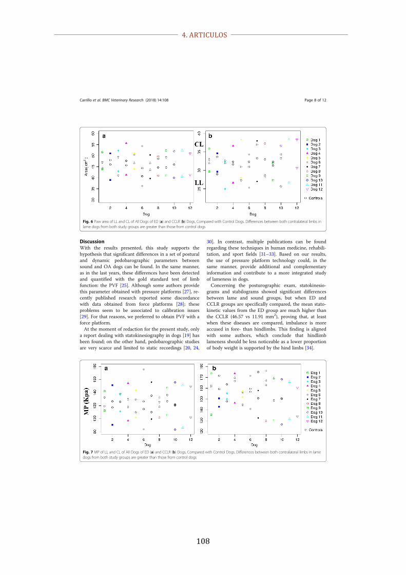
4. ARTICULOS
108
DiscussionWith the results presented, this study supports thehypothesis that significant differences in a set of posturaland dynamic pedobarographic parameters betweensound and OA dogs can be found. In the same manner,as in the last years, these differences have been detectedand quantified with the gold standard test of limbfunction: the PVF [25]. Although some authors providethis parameter obtained with pressure platforms [27], re-cently published research reported some discordancewith data obtained from force platforms [28]; theseproblems seem to be associated to calibration issues[29]. For that reasons, we preferred to obtain PVF with aforce platform.At the moment of redaction for the present study, only
a report dealing with statokinesiography in dogs [19] hasbeen found; on the other hand, pedobarographic studiesare very scarce and limited to static recordings [20, 24,
30]. In contrast, multiple publications can be foundregarding these techniques in human medicine, rehabili-tation, and sport fields [31–33]. Based on our results,the use of pressure platform technology could, in thesame manner, provide additional and complementaryinformation and contribute to a more integrated studyof lameness in dogs.Concerning the posturographic exam, statokinesio-
grams and stabilograms showed significant differencesbetween lame and sound groups, but when ED andCCLR groups are specifically compared, the mean stato-kinetic values from the ED group are much higher thanthe CCLR (46.57 vs 11.91 mm2), proving that, at leastwhen these diseases are compared, imbalance is moreaccused in fore- than hindlimbs. This finding is alignedwith some authors, which conclude that hindlimblameness should be less noticeable as a lower proportionof body weight is supported by the hind limbs [34].
Fig. 7 MP of LL and CL of All Dogs of ED (a) and CCLR (b) Dogs, Compared with Control Dogs. Differences between both contralateral limbs in lamedogs from both study groups are greater than those from control dogs
Fig. 6 Paw area of LL and CL of All Dogs of ED (a) and CCLR (b) Dogs, Compared with Control Dogs. Differences between both contralateral limbs inlame dogs from both study groups are greater than those from control dogs
Carrillo et al. BMC Veterinary Research (2018) 14:108 Page 8 of 12

4. ARTICULOS
109
pressure increases, indicating that the metacarpal pad isless responsive to pressure changes [20]. The presentstudy only shows modifications in the global paw area inresponse to pressure; however, the study of differentdeformation (or even restitution) rates could potentiallyhave clinical implications, which could be investigated inthe future.Greater disparity was found when MP differences
between lame and sound limbs of the ED and CCLRgroups were compared at a walk (41% vs 18%). The roleof the head (and forelimb musculature) displacement as
a counterweight when lame or sound horses and dogsmove could explain this event [39]; this role can bebreed-independent, according with previous studies [27].PP differences between sound and lame groups werealso significant; however, the high SD shown in theresults prevent any conclusive interpretation. The causeof this fact should be elucidated in the future.Although posturography and pedobarography have
provided a set of useful parameters to detect both fore-and hidlimb lameness in dogs, this study has some limi-tations. First, middle to large dog breeds were used to
Fig. 10 Comparison of Mean Values of all pedobarographic parameters of ED (a) and CCLR (b) Dogs with PVF. As can be seen, study groups (EDand CCLR) always show greater disparity (asymmetry) between contralateral limbs than the control group
Carrillo et al. BMC Veterinary Research (2018) 14:108 Page 10 of 12
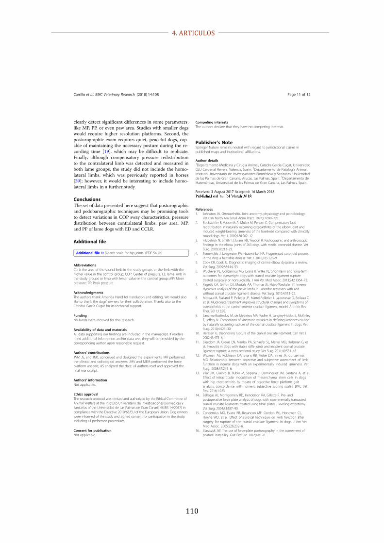
4. ARTICULOS
110
clearly detect significant differences in some parameters,like MP, PP, or even paw area. Studies with smaller dogswould require higher resolution platforms. Second, theposturographic exam requires quiet, peaceful dogs, cap-able of maintaining the necessary posture during the re-cording time [19], which may be difficult to replicate.Finally, although compensatory pressure redistributionto the contralateral limb was detected and measured inboth lame groups, the study did not include the homo-lateral limbs, which was previously reported in horses[39]; however, it would be interesting to include homo-lateral limbs in a further study.
ConclusionsThe set of data presented here suggest that posturographicand pedobarographic techniques may be promising toolsto detect variations in COP sway characteristics, pressuredistribution between contralateral limbs, paw area, MP,and PP of lame dogs with ED and CCLR.
Additional file
Additional file 1: Bioarth scale for hip joints. (PDF 54 kb)
AbbreviationsCL: is the area of the sound limb in the study groups or the limb with thehigher value in the control group; COP: Center of pressure; LL: lame limb inthe study groups or limb with lesser value in the control group; MP: Meanpressure; PP: Peak pressure
AcknowledgmentsThe authors thank Amanda Hand for translation and editing. We would alsolike to thank the dogs’ owners for their collaboration. Thanks also to theCátedra García Cugat for its technical support.
FundingNo funds were received for this research.
Availability of data and materialsAll data supporting our findings are included in the manuscript. If readersneed additional information and/or data sets, they will be provided by thecorresponding author upon reasonable request.
Authors’ contributionsJMV, JS, and JMC conceived and designed the experiments; MR performedthe clinical and radiological analyses; JMV and MEM performed the forceplatform analysis; AS analyzed the data; all authors read and approved thefinal manuscript.
Authors’ informationNot applicable.
Ethics approvalThe research protocol was revised and authorized by the Ethical Committee ofAnimal Welfare at the Instituto Universitario de Investigaciones Biomédicas ySanitarias of the Universidad de Las Palmas de Gran Canaria (IUIBS 14/2017) incompliance with the Directive 2010/63/EU of the European Union. Dog ownerswere informed of the study and signed consent for participation in the study,including all performed procedures.
Consent for publicationNot applicable.
Competing interestsThe authors declare that they have no competing interests.
Publisher’s NoteSpringer Nature remains neutral with regard to jurisdictional claims inpublished maps and institutional affiliations.
Author details1Departamento Medicina y Cirugía Animal, Cátedra García Cugat, UniversidadCEU Cardenal Herrera, Valencia, Spain. 2Departamento de Patología Animal,Instituto Universitario de Investigaciones Biomédicas y Sanitarias, Universidadde las Palmas de Gran Canaria, Arucas, Las Palmas, Spain. 3Departamento deMatemáticas, Universidad de las Palmas de Gran Canaria, Las Palmas, Spain.
Received: 3 August 2017 Accepted: 16 March 2018
References1. Johnston JA. Osteoarthritis. Joint anatomy, physiology and pathobiology.
Vet Clin North Am Small Anim Pract. 1997;27:699–723.2. Bockstahler B, Vobornik A, Muller M, Peham C. Compensatory load
redistribution in naturally occurring osteoarthritis of the elbow joint andinduced weight-bearing lameness of the forelimbs compared with clinicallysound dogs. Vet J. 2009;180:202–12.
3. Fitzpatrick N, Smith TJ, Evans RB, Yeadon R. Radiographic and arthroscopicfindings in the elbow joints of 263 dogs with medial coronoid disease. VetSurg. 2009;38:213–23.
4. Temwichitr J, Leegwater PA, Hazewinkel HA. Fragmented coronoid processin the dog: a heritable disease. Vet J. 2010;185:123–9.
5. Cook CR, Cook JL. Diagnostic imaging of canine elbow dysplasia: a review.Vet Surg. 2009;38:144–53.
6. Wucherer KL, Conzemius MG, Evans R, Wilke VL. Short-term and long-termoutcomes for overweight dogs with cranial cruciate ligament rupturetreated surgically or nonsurgically. J Am Vet Med Assoc. 2013;242:1364–72.
7. Ragetly CA, Griffon DJ, Mostafa AA, Thomas JE, Hsiao-Wecksler ET. Inversedynamics analysis of the pelvic limbs in Labrador retrievers with andwithout cranial cruciate ligament disease. Vet Surg. 2010;4:513–22.
8. Moreau M, Rialland P, Pelletier JP, Martel-Pelletier J, Lajeunesse D, Boileau C,et al. Tiludronate treatment improves structural changes and symptoms ofosteoarthritis in the canine anterior cruciate ligament model. Arthritis ResTher. 2011;13:98.
9. Sanchez-Bustinduy M, de Medeiros MA, Radke H, Langley-Hobbs S, McKinleyT, Jeffery N. Comparison of kinematic variables in defining lameness causedby naturally occurring rupture of the cranial cruciate ligament in dogs. VetSurg. 2010;4:523–30.
10. Harasen G. Diagnosing rupture of the cranial cruciate ligament. Can Vet J.2002;43:475–6.
11. Bleedorn JA, Greuel EN, Manley PA, Schaefer SL, Markel MD, Holzman G, etal. Synovitis in dogs with stable stifle joints and incipient cranial cruciateligament rupture: a cross-sectional study. Vet Surg. 2011;40:531–43.
12. Waxman AS, Robinson DA, Evans RB, Hulse DA, Innes JF, ConzemiusMG. Relationship between objective and subjective assessment of limbfunction in normal dogs with an experimentally induced lameness. VetSurg. 2008;37:241–6.
13. Vilar JM, Cuervo B, Rubio M, Sopena J, Domínguez JM, Santana A, et al.Effect of intraarticular inoculation of mesenchymal stem cells in dogswith hip osteoarthritis by means of objective force platform gaitanalysis: concordance with numeric subjective scoring scales. BMC VetRes. 2016;1:223.
14. Ballagas AJ, Montgomery RD, Henderson RA, Gillette R. Pre- andpostoperative force plate analysis of dogs with experimentally transactedcranial cruciate ligaments treated using tibial plateau leveling osteotomy.Vet Surg. 2004;33:187–90.
15. Conzemius MG, Evans RB, Besancon MF, Gordon WJ, Horstman CL,Hoefle WD, et al. Effect of surgical technique on limb function aftersurgery for rupture of the cranial cruciate ligament in dogs. J Am VetMed Assoc. 2005;226:232–6.
16. Blaszczyk JW. The use of force-plate posturography in the assessment ofpostural instability. Gait Posture. 2016;44:1–6.
Carrillo et al. BMC Veterinary Research (2018) 14:108 Page 11 of 12
4. ARTICULOS
111
17. Baratto L, Morasso PG, Re C, Spada G. A new look at posturographic analysisin the clinical context: sway-density versus other parameterizationtechniques. Mot Control. 2002;6:246–70.
18. Skopljak A, Muftic M, Sukalo A, Masic I, Zunic L. Pedobarography indiagnosis and clinical application. Acta Inform Med. 2014;6:374–8.
19. Manera ME, Carrillo JM, Batista M, Rubio M, Sopena J, Santana A, et al. Staticposturography: a new perspective in the assessment of lameness in acanine model. PLoS One. 2017;12(1):e0170692. https://doi.org/10.1371/journal.pone.0170692.
20. Marghitu DB, Swaim SF, Rumph PF, Cojonaru D, Gillette RL, Scardino MS.Dynamics analysis of ground contact pressure of English pointer dogs.Nonlinear Dyn. 2003;33:253–65.
21. Souza AN, Pinto AC, Marvulle V, Matera JM. Evaluation of vertical forcesin the pads of German shepherd dogs. Vet Comp Orthop Traumatol.2013;26:6–11.
22. Upchurch DA, Renberg WC, Roush JK, Milliken GA, Weiss ML. Effects ofadministration of adipose-derived stromal vascular fraction and platelet-rich plasma to dogs with osteoarthritis of the hip joints. Am J Vet Res.2016;77:940–5.
23. Tomas A, Marcellin-Little DJ, Roe SC, Motsinger-Reif A, Lascelles BD.Relationship between mechanical thresholds and limb use in dogs withcoxofemoral joint oa-associated pain and the modulating effects of painalleviation from total hip replacement on mechanical thresholds. Vet Surg.2014;43:542–8.
24. Carr BJ, Canapp SO Jr, Zink MC. Quantitative comparison of the walk andtrot of border collies and Labrador retrievers, breeds with differentperformance requirements. PLoS One. 2015;10(12):e0145396. https://doi.org/10.1371/journal.pone.0145396.
25. Walton MB, Cowderoy EC, Wustefeld-Janssens B, Lascelles BD, Innes JF.Mavacoxib and meloxicam for canine osteoarthritis: a randomised clinicalcomparator trial. Vet Rec. 2014;175:280.
26. The World Small Animal Veterinary Association Global Nutrition CommitteeBody Condition Score Chart. http://www.wsava.org/WSAVA/media/Documents/Guidelines/WSAVA-Global-Nutritional-Assessment-Guidelines-2011-final.pdf. Accessed 1 June 2017.
27. Souza AN, Tatarunas AC, Matera JM. Evaluation of vertical forces in the padsof Pitbulls with cranial cruciate ligament rupture. BMC Vet Res. 2014;10:51.
28. Schnabl-Feichter E, Tichy A, Bockstahler B. Coefficients of variation ofground reaction force measurement in cats. PLoS One. 2017;12(3):e0171946.
29. Gillette RL, Angle TC. Recent developments in canine locomotor analysis: areview. Vet J. 2008;178:165–76.
30. Bockstahler B, Tichy A, Aigner P. Compensatory load redistribution inLabrador retrievers when carrying different weights: a non-randomizedprospective trial. BMC Vet Res. 2016;12:92.
31. Scivoletto G, Romanelli A, Mariotti A, Marinucci D, Tamburella F, MammoneA, et al. Clinical factors that affect walking level and performance in chronicspinal cord lesion patients. Spine. 2008;33:259–64.
32. Tamburella F, Scivoletto G, Molinari M. Balance training improves staticstability and gait in chronic incomplete spinal cord injury subjects: a pilotstudy. Eur J Phys Rehabil Med. 2013;49:353–64.
33. Whiteside D, Elliott BC, Lay B, Reid M. Coordination and variability in theelite female tennis serve. J Sports Sci. 2015;7:675–86.
34. Gillette R, Graig T. Canine locomotion analysis. In: Millis D, Levine D, editors.Canine rehabilitation and physical therapy. Amsterdam: Elsevier; 2014. p. 201–10.
35. Bergamaschi M, Ferrari G, Gallamini M, Scoppa F. Laser acupuncture andauriculotherapy in postural instability-a preliminary report. J AcupunctMeridian Stud. 2011;4:69–74.
36. Arkov VV, Abramova TF, Nikitina TM, Ivanov VV, Suprun DV, Shkurnikov MU,et al. Comparative study of stabilometric parameters in sportsmen ofvarious disciplines. Bull Exp Biol Med. 2009;147:233–5.
37. Oosterlinck M, Pille F, Back W, Dewulf J, Gasthuys F. Use of a stand-alonepressure plate for the objective evaluation of forelimb symmetry in soundponies at walk and trot. Vet J. 2010;183:305–9.
38. Mölsä SH, Hyytiäinen HK, Hielm-Björkman AK, Laitinen-Vapaavuori OM.Long-term functional outcome after surgical repair of cranial cruciateligament disease in dogs. BMC Vet Res. 2014;10:266.
39. Maliye S, Voute LC, Marshall JF. Naturally-occurring forelimb lameness in thehorse results in significant compensatory load redistribution during trotting.Vet J. 2015;2:208–13.
• We accept pre-submission inquiries • Our selector tool helps you to find the most relevant journal• We provide round the clock customer support • Convenient online submission• Thorough peer review• Inclusion in PubMed and all major indexing services • Maximum visibility for your research
Submit your manuscript atwww.biomedcentral.com/submit
Submit your next manuscript to BioMed Central and we will help you at every step:
Carrillo et al. BMC Veterinary Research (2018) 14:108 Page 12 of 12

4. ARTICULOS
112

5. CONCLUSIONES

5. CONCLUSIONES
114

5. CONCLUSIONES
115
1. La posturografía es factible de realizar en perros y proveer datos precisos y
fiables de varios parámetros para la evaluación de cojeras de miembro torácico no
proporcionados en ninguna bibliografía previa consultada. Por lo tanto, estas cojeras
serían detectadas observando las asimetrías en las características del centro de presiones
(COP).
2. El análisis del paso mediante la plataforma de fuerza y la electrogoniometría
han permitido verificar la eficacia del plasma rico en plaquetas (PRP) para tratar perros
con rotura de ligamento cruzado, aunque la duración dicho efecto se ha prolongado por
un periodo inferior a los 6 meses.
3. La podobarografía dinámica proporciona un set de parámetros derivados de la
presión de apoyo que son válidos para la detección de cojeras como la redistribución de
presión entre los miembros, área de apoyo, presión media y máxima en animales
afectados de displasia de codo y rotura de ligamento cruzado craneal.

5. CONCLUSIONES
116

6. RESUMEN

6. RESUMEN
118

6. RESUMEN
119
Los métodos de evaluación biomecánica proporcionan datos fiables y objetivos
sobre el movimiento en los animales, cuya proyección clínica está enfocada
principalmente al diagnóstico de cojeras.
Dentro de los métodos cinéticos, que son aquellos que miden las fuerzas que
generan el movimiento, el análisis del paso con plataformas de fuerza está considerado
el “gold standard”. Por otro lado, la posturografía y podobarografía son técnicas
utilizadas en humana que, a día de hoy, no tienen un uso clínico en la clínica de
pequeños animales.
En este sentido, el objetivo común de esta tesis doctoral es el de validar estas
técnicas no solo para el diagnóstico de cojeras derivadas de patologías que cursan con
osteoartritis, sino además para el control objetivo de la evolución en animales que están
siendo tratados con terapias regenerativas como es el plasma rico en plaquetas y sus
derivados.
Con este objetivo, se han diseñado tres procedimientos experimentales
diferentes:
1) Se utilizaron 5 perros con OA de codo, a los cuales se trató con plasma rico en
factores de crecimiento. Dichos animales se evaluaron posturográficamente antes y tres
meses después de la instauración del tratamiento. La conclusión fue que dicha técnica
no sólo fue capaz de detectar la cojera en los animales afectados de OA, sino que
además fue capaz de detectar la mejoría tras el tratamiento.

6. RESUMEN
120
2) Se utilizaron 12 perros de raza bulldog inglés con rotura de ligamento cruzado
craneal. A dichos animales se les trató con un plasma derivado en plaquetas “genérico”
y se les evaluó periódicamente mediante plataforma de fuerza y electrogoniometría. Se
concluye que objetivamente la duración del tratamiento es inferior a los 6 meses,
necesitando los animales por lo tanto volver a tratarse antes de dicho periodo.
3) por último, se utilizaron animales con OA de codo (n=10) y de rodilla (n=12); de
dichos animales se obtuvieron datos podobarográficos con el fin de verificar si podían
ser validados con el “gold standard”, o sea, la plataforma de fuerza. Se corrobora que
dicha técnica es también efectiva para la detección de cojeras tanto de miembro torácico
como pelviano

6. RESUMEN
121

6. RESUMEN
122

7. SUMMARY

7. SUMMARY
124

7. SUMMARY
125
Biomechanic assessment methodology provides reliable and objective data
regarding animal locomotion. In the clinical field, its main application is focused on the
lameness detection.
Among kinetic methodology, which concerns those measuring the forces that
generate the movement, gait analysis with force platforms is considered the “gold
standard”. On the other hand, posturography and podobarography are being widely used
in humans for many years but till these days, no publications can be found regarding
clinical applications in veterinary medicine.
In this sense, the main objective of this Doctoral Thesis consists in the validation
these techniques, not only for the detection of lameness as consequence of OA, but also
for the objective assessment of the follow-up when animals are treated with
regenerative therapies as platelet rich plasma and its derivates.
With this aim, three different experimental procedures have been designed:
1) Five dogs with elbow OA treated with plasma rich in growth factors were used.
Those animals were evaluated posturographically before and three months after
treatment. The study concluded that this technique was able not only to detect the
lameness in the OA dogs, but also to detect the improvement after treatment.
2) Twelve English bulldogs with cranial cruciate ligament rupture were used in this
study. Those animals were treated with a “generic” platelet rich plasma and were
periodically evaluated using a force platform and electrogoniometers. The conclusion is

7. SUMMARY
126
that objectively the treatment duration was under the six months, then treatment should
be re-applied before that period.
3) Finally, animals with elbow OA (n=10) and stifle (n=12) were enrolled for this study;
podobarographic data were obtained in order to be validated with the “gold standard”,
the force platform data. This technique proved that is effective and reliable for lameness
detection of both fore and hindlimbs.

7. SUMMARY
127

7. SUMMARY
128

8. REFERENCIAS BIBLIOGRÁFICAS

8. REFERENCIAS BIBLIOGRÁFICAS
130

8. REFERENCIAS BIBLIOGRÁFICAS
131
Abrantes, J. & Santos, L., 2012. Plantar pressure assessment: a new tool for postural
instability diagnosis in multiple sclerosis. En: Jorge, R., Tavares, J., Barbosa, M., Slade,
A. (eds). Technologies for medical sciences. Springer, The Netherlands. 179-206.
ISBN: 978-94-007-4067-9.
Adams, D., 2004. Canine anatomy a systematic study. 4a. ed. Wiley-Blackwell, Iowa.
41-8; 71-81.
Adrian, A.M., Barrett, M.F., Werpy, N.M., Kawcak, C.E., Chapman, P.L., Goodrich,
L.R., 2017. A comparison of arthroscopy to ultrasonography for identification of
pathology of the equine stifle. Equine Vet J. 49(3), 314-321.
Adrian, M.J., Rog, W.E. & Karpovich, D.V. Normal gait of the dog: An
electrogoniometric study. Am J. Vet. Res. 27 (116), 90-95.
Anitua, E., Sánchez, M., Nurden, A.T., Zalduendo, M.M., de la Fuente, M., Azofra, J.
& Andia, I., 2007a. Platelet--�released growth factors enhance the secretion of
hyaluronic acid and induce hepatocyte growth factor production by synovial fibroblasts
from arthritic patients. Rheumatology. 46 (12), 1769--1772.
Anjos, D.M., Gomes, L.P., Sampaio, L.M., Correa, J.C. & Oliveira C.S., 2010.
Assessment of plantar pressure and balance in patients with diabetes. Arch Mmed Sci. 6
(1), 43-48.
Anker, L.C., Weerdesteyn, V., van Nes, I.J.W., Nienhuis, B., Straatman, H. & Geurts,
A.C.H., 2008. The relation between postural stability and weight distribution in healthy
subjects. Gait Posture. 27 (3), 471-477.
Baratto, L., Morasso, P.G., Re, C. & Spada, G., 2002. A new look at posturographic
analysis in the clinical context: sway-density versus other parameterization techniques.
Motor control. 6 (3), 246-270.
Barone, R., 1980. Anatomie Comparée des mammifères domestiques. 2da ed. Vigot,
Paris. Tomo 2.

8. REFERENCIAS BIBLIOGRÁFICAS
132
Barrey, E., Galloux, P., Valette J.P., Auvinet, B. & Wolter, R., 1993. Stride
characteristics of overground versus treadmill locomotion in the saddle horse. Acta
Anat (Basel). 146 (2-3), 90-94.
Bhosale, A. M. & Richardson, J. B., 2008. Articular cartilage: structure, injuries and
review of management. Br Med Bull. 87(1), 77-�95.
Bialski, D., Lanovaz, J.L., Bohart, G.V., Mullineaux, D.R. & Clayton, H.M., 2003.
Effect of detomidine on postural sway in horses. Comp Exerc Physiol. 1 (1), 45–50.
Birch, H.L., McLaughlin, L., Smith, R.K. & Goodship, A.E. 1999a. Treadmill exercise-
induced tendon hypertrophy: assessment of tendons with different mechanical
functions. Equine Vet J. 31 (S30), 222-226.
Black, L.L., Gaynor, J., Adams, C., Dhupa, S., Sams, A.E., Talor, R., Harman, S.,
Ginggerich, D.A. & Harman, R., 2008. Effect of intraarticular injection of autologous
adipose--�derived mesenchymal stem and regenerative cells on clinical signs of
chronic osteoarthritis of the elbow joint in dogs. Vet Ther. 9 (3), 192-�200.
Bland, S.D., 2015. Canine osteoarthritis and treatments: a review. Vet Sci Dev. 5 (1),
84-89.
Bockstahler, B., Tichy, A. & Aigner, P., 2016. Compensatory load redistribution in
Labrador retrievers when carrying different weights-a non-randomized prospective trial.
BMC Vet Res. 12 (1), 92. doi: 10.1186/s12917-016-0715-7.
Buchner, H.H., Savelberg, H.H., Schamhardt, H.C., & Merkens, H.W., 1994.
Kinematics of treadmill vs overground locomotion in horses. Vet Q. 16 (2), S87- S90.
Buchner, H.H., Savelberg, H.H., Schamhardt, H.C. & Barneveld, A., 1996. Head and
trunk movement adaptations in horses with experimentally induced fore-or hindlimb
lameness. Equine Vet J. 28 (1), 71-76.
Budsberg, S.C., Rytz, U. & Johnston, S.A., 1999. Effects of acceleration on ground
reaction forces collected in healthy dogs at a trot. Vet Comp Orthop Traumatol. 12 (1),
15-19.

8. REFERENCIAS BIBLIOGRÁFICAS
133
Burr, D. B., 2004. Anatomy and physiology of the mineralized tissues: role in the
pathogenesis of osteoarthrosis. Osteoarthritis Cartilage. 12, 20�30.
Cakir, S., Hepguler, S., Ozturk, C., Korkmarz, M., Isleten, B. & Atamaz, F.C., 2014.
Efficacy of Therapeutic Ultrasound for the Management of Knee Osteoarthritis: A
Randomized, Controlled, and Double--�Blind Study. Am J Phys Med Rehabil. 93 (5),
405- 412.
Cardinet, G. H., Kass, P. H., Wallace, L.J., Guffy, M. M., 1997. Association between
pelvic muscle mass and canine hip dysplasia. JAVMA. 210 (10), 1466-1473.
Carr, B.J., Canapp, S.O. & Zink, M.C., 2015. Quantitative Comparison of the Walk and
Trot of Border Collies and Labrador Retrievers, Breeds with Different Performance
Requirements. PLoS ONE. 10 (12), e0145396. doi:10.1371/journal.pone.0145396
Castañon, F., 2015. Estudios comparativo de las técnicas quirúrgicas, TTA clásicas
Securos ®, TTA Porous ® y TTA Porous ® con PRP, para el tratamiento de la rotura
del ligamento cruzado anterior en el perro. Tesis Doctoral, León.
Cimino Brown, D., Boston, R.C., Coyne. J.C. & Farrar, J. T., 2007. Development and
psychometric testing of an instrument designed to measure chronic pain in dogs with
osteoarthritis. Am J Vet Res. 68 (6), 631-637.
Clayton, H.M., Buchholz, R. & Nauwelaerts, S., 2013. Relationship between
morphological and stabilographic variables in standing horses. Vet J. 198(1), e65–e69.
Clayton, H.M. & Nauwelaerts, S., 2014. Effect of blindfolding on Centre of pressure
variables in healthy horses during quiet standing. Vet J. 199 (3), 365–369.
Cole, K. & Abbs, J.H., 1986. Coordination of three-joint digit movements for rapid
fingerthumb grasp. J. Neurophysiol. 55 (6), 1407-1423.
Cordeiro, T.L., Frade, M.A., Barros, A.R. & Foss, N.T. 2014. Postural balance control
of the leprosy patient with plantar sensibility impairment. Occup Med Health Aff. 2, 3.

8. REFERENCIAS BIBLIOGRÁFICAS
134
Cuervo, B., Rubio, M., Sopena, J., Domínguez, J.M., Vilar, J., Morales, M., Cugat, R.,
Carrillo, J.M., 2014. Hip osteoarthritis in dogs: a randomized study using mesenchymal
stem cells from adipose tissue and plasma rich in growth factors. Int J Mol Sci. 15 (8),
13437-13460.
Cuervo, B. 2015. “Estudio Clínico Multicéntrico del Efecto de la Aplicación de Células
Mesenquimales de Grasa (CMG), Plasma Rico en Factores de Crecimiento (PRGF) y la
Combinación de Ambos en el Tratamiento de la Enfermedad Degenerativa Articular en
Perro”, Tesis Doctoral, Valencia.
Cruz, A.M., Maninchedda, U.E., Burger, D., Wanda, S., Vidondo, B., 2017.
Repeatability of gait pattern variables measured by use of extremity-mounted inertial
measurement units in nonlame horses during trotting. Am J Vet Res. 78 (9), 1011-1018.
Doi: 10.2460/ajvr. 78.9.1011.
Damiá, E., Chicharro, D., Rubio, M., Carrillo, J.M., Sopena, J., Cuervo, B., López, S.,
Vilar, J.M., 2018. Can Plasma Rich in Growth Factors Be Safe for Parental Use? A
Safety Study in the Canine Model. Int J Mol Sci. 19 (9), 2701.
Diekman, B.O. & Guilak, F., 2013. Stem cell--�based therapies for osteoarthritis:
challenges and opportunities. Curr Opin Rheumatol. 25 (1), 119--126.
Drevemo, S., Dalin, G., Fredricson, I., Hjertén, G. & Hjertén, G., 1980. Equine
locomotion: 1. The analysis of linear and temporal stride characteristics of trotting
standardbreds. Equine Vet J. 12 (2), 60-65.
Durante, E.J., Brusa, M.C., 1998. Algunos aspectos de la displasia del codo de los
caninos. Analecta Veterinaria. 18 (1-2), 59-70.
Duval, J.M., Budsberg, S.C., Flo, G.L. & Sammarco, J.L., 1999. Breed, sex, and body
weight as risk factors for rupture of the cranial cruciate ligament in young dogs. J Am
Vet Med Assoc. 215(6), 811-814.

8. REFERENCIAS BIBLIOGRÁFICAS
135
Elkins, A.D., Pechman, R., Kearney, M. & Herron, N., 1991. A retrospective study
evaluating the degree of degenerative joint disease in the stifle e joint of dogs following
surgical repair of anterior cruciate ligament rupture. J Am Anim Hosp Assoc. 27(5),
533-540.
Evans, H.E., 1993. Arthrology. En: Evans, H.E., (Eds.), Miller’s anatomy of the dog, ed
3, WB Sunders, Philadelphia.
Evans, H.E., DelaHunta, A., 2000. Disección del perro. 5ta ed. McGraw-Hill-
Interamericana, México. 384.
Evans, R., Horstman, C. & Conzemius, M., 2005. Accuracy and optimization of force
platform gait analysis in Labradors with cranial cruciate disease evaluated at a walking
gait. Vet Surg. 34 (5), 445-449.
Fahie, M.A., Ortolano, G.A., Guercio, V., Schaffer, J.A., Johnston, G., AU, J., Hettlich,
B.A., Phillips, T., Allen, M.J. & Bertone, A.L., 2013. A randomized controlled trial of
the efficacy of autologous platelet therapy for the treatment of osteoarthritis in dogs. J
Am Vet Med Assoc. 243 (9), 1291--1297.
Fang, J., Zheng, N., Wang, Y., Cao, H., Sun, S., Dai, J., Li, Q. & Zhang, Y. 2013.
Understanding Acupuncture Based on ZHENG Classification from System Perspective.
Evid Based Complement Alternat Med. 2013, 956967- 10.
Fortier, L.A., Barker, J.U., Strauss, E.J., McCarrel, T.M. & Cole, B.J., 2011. The role of
growth factors in cartilage repair. Clin Orthop Relat Res. 469 (10), 2706--2715.
Fossum, T. W., 2009. Cirugía en pequeños animales. 3ra Ed. Elsevier España,
Barcelona. 1632 pp.
Godley, D.R., 2013. Assessment, diagnosis, and treatment of developmental dysplasia
of the hip. JAAPA. 26 (3), 54--58.
Gomes-Costa, M., Roupa, I., Pequito, M., Prazeres, J., Gaivão, M., Abrantes, J. &
Clayton, H.M., 2015. The use of pressure plates for static Center of Pressure Analysis in
horses. J Equine Vet Sci. 35 (4), 315–320.

8. REFERENCIAS BIBLIOGRÁFICAS
136
Gómez Álvares C.B., Gustås P., Bergh A. & Rhodin M., 2017. Vertical head and pelvic
movement symmetry at the trot in dogs with induced supporting limb lameness. Vet J.
229, 13-18.
Gordon, W.J., Conzemius, M.G., Riedesel, E., Besancon, M. F., Evans, R., Wilke, V. &
Ritter, 2003. The relationship between limb function and radiographic osteoarthrosis in
dogs with stifle osteoarthrosis. Vet Surg. 32 (5), 451-454.
Gudbergsen, H., Boesen, M., Lohmander, L.S., Christensen, R., Henriksen, M., Bartels,
E.M., Christensen, P., Rindel, L., Aaboe, J., Danneskiold-�Samsoe, B., Riecke, B.F. &
Bliddal, H., 2012. Weight loss is effective for symptomatic relief in obese subjects with
knee osteoarthritis independently of joint damage severity assessed by high--field MRI
and radiography. Osteoarthritis Cartilage. 20 (6), 495-�502.
Gustås, P., Pettersson, K., Honkavaara, S., Lagerstedt, A., Byström, A & Sveriges, I.,
2003. Kinematic and temporospatial assessment of habituation of Labrador retrievers to
treadmill trotting. Vet J. 198(1), 114-119. Doi.org/10.1016/j.tvjl.2013.09.044
Harasen, G., 2002. Diagnosing rupture of the cranial cruciate ligament. Can Vet J. 43
(6), 475–476.
Heffron, L.E., Campbell, J.R., 1978. Morphology, histology and functional anatomy of
the canine cranial cruciate ligament. Vet Rec. 102, 280-283.
Heikkilä, H.M., Hielm-Björkman, A.K., Morelius, M., Larsen, S., Honkavaara, J.,
Innes, J. F. & Laitinen- Vapoavouri, O.M., 2014. Intra-articular botulinum toxin A for
the treatment of osteoarthritic joint pain in dogs: A randomized, double-blinded,
placebocontrolled clinical trial. Vet Journal. 200 (1), 162-169.
Hielm-Björkman, A.K., Kuusela, E., Liman, A., Markkola, A., Saarto, E., Huttunen, P.,
Leppäluoto, J., Tulamo, R. & Reakallio, M., 2003. Evaluation of methods for
assessment of pain associated with chronic osteoarthritis in dogs. J Am Vet Med Assoc.
222 (11), 1552-1558.

8. REFERENCIAS BIBLIOGRÁFICAS
137
Hielm-Björkman, A.K., Kapatkin, A.S. & Rita, H.J., 2011. Reliability and validity of a
visual analogue scale used by owners to measure chronic pain attributable to
osteoarthritis in their dogs. Am J Vet Res. 72 (5), 601-607.
Hielm-Björkman, A.K., Roine, J., Elo, K., Lappalainen, A., Junnila, J. & Laitinen-
Vapaavuari, O., 2012. An un-commissioned randomized, placebo-controlled double-
blind study to test the effect of deep sea fish oil as a pain reliever for dogs suffering
from canine OA. BMC Vet Res. 8 (1), 157.
Horstam, C.L., Conzemius, M.G., Evans, R. & Gordon, W. J., 2004. Assessing the
efficacy of perioperative oral carprofen after cranial cruciate surgery using noninvasive,
objective pressure platform gait analysis. Vet Surg. 33(3), 286-292.
Hudson, J.T., Slater, M.R., Taylor, L., Scott, H.M. & Kerwin, S.C., 2004. Assessing
repeatability and validity of a visual analoque scale questionnaire for use in assessing
pain and lameness in dogs. Am J Vet Res. 65 (12), 1634-1643.
Innes, J.F., Barr, A.R.S., 1998a Clinical natural history of the postsurgical cruciate
deficient canine stifle joint: year 1. J Small Anim Pract. 39 (7), 325-332.
Innes, J.F., Barr, A.R.S., 1998b. Can owners assess outcome following treatment of
canine cruciate ligament deficinency? J Small Anim Pract. 39 (8), 373-378.
Ishihara, A., Reed, S.M., Rajala-Schultz, P.J., Robertson, J.T. & Bertone, A.L., 2009.
Use of kinetic gait analysis for detection, quantification, and differentiation of hindlimb
lameness and spinal ataxia in horses. J Am Vet Med Ass. 234 (5), 644–651.
Johnson, J.A., Austin, C., Breur, G.J., 1994. Incidence of canine appendicular
musculoskeletal disorders in 16 veterinary teaching hospitals from 1980 to 1989. Vet
Comp Orthop Traumatol. 7, 56-59.
Karpovich, P.V., Herden, E.L. & Asa, M.M., 1960. Electrogoniometric study of joints.
U.S. Armed Forces Med. J. 11, 425-450.
Keegan, K.G., Wilson, D.A., Smith, B.K. & Wilson, D.J., 2000. Changes in kinematic
variables observed during pressure-induced forelimb lameness in adult horses trotting
on a treadmill. Am J Vet Res. 61(6), 612-619.

8. REFERENCIAS BIBLIOGRÁFICAS
138
Keegan, K.G., Yonezawa, Y., Pai, P.F. & Wilson, D.A., 2002. Accelerometer-based
system for the detection of lameness in horses. Biomed Sci Instrum. 38, 107-112.
Kelmer, G., Keegan, K.G., Kramer, J., Wilson, D.A., Pai, F.P. & Singh, P., 2005.
Computer-assisted kinematic evaluation of induced compensatory movements
resembling lameness in horses trotting on a treadmill. Am J Vet Res. 66 (4), 646-655.
Kirberger, G., 2003. Elbow dysplasia in the dog- What is it? IEWG. 15, 15-19.
Kiberger, R., Barr, F., 2006. Manual of canine and musculosketetal imaging. BSAVA,
London. 220.
Kim, Y.M., Joo, Y.B., 2018. Anteromedial Meniscofemoral Ligament of the Anterior
Horn of the Medial Meniscus: Clinical, Magnetic Resonance Imaging, and Arthroscopic
Features. Arthroscopy. 34(5), 1590-1600.
King, M.R., Haussler, K.K., Kawcak, C.E., Mcllwraith, C.W. & Reiser R.F., 2013.
Effect of underwater treadmill exercise on postural sway in horses with experimentally
induced carpal joint osteoarthritis. Am J Vet Res. 74 (7), 971- 982.
Khumsap, S., Lanovaz, J.L., Clayton, H.M. 2004 Verification of skin-based markers for
3-dimentional kinematic analysis of the equine tarsal joint. Equine Vet J. 36 (8), 655-
658.
Komsta, R., Debiak, P., Twardowski, P. 2008. Radiographic evaluation of joints in dogs
with elbow dyplasia- clinical observations. Bull Vet Inst Pulawy. 52, 179- 183.
König, H. & Liebich, H., 2008. Introducción. En: König, H. & Liebich, H. (eds.)
Anatomía de los animales domésticos. Aparato locomotor. 2ª ed. Montevideo, Uruguay:
Editorial médica panamericana.
Kramer, J., Keegan, K.G., Kelmer, G. & Wilson D.A., 2004. Objetive determination of
pelvic movement during hind limb lameness by use of a signal decomposition method
and pelvic height differences. Am J Vet Res. 65(6), 741-747.
Kramer, J. & Keegan, K., 2007. Capítulo 13: Cinemática de la claudicación. En:
medicina y cirugía en los equinos de deporte. Kkenneth W. Hinchcliff, Andris J.
8. REFERENCIAS BIBLIOGRÁFICAS
139
Kaneps, Raymond J. Geor. (eds). Editorial Inter-Médica S.A.I.C.I. 266-282. ISBN:
978-950-555-327-3.
Langlois B., Froideveaux J., Lamarche L., Legault, C., Legault, P., Tassencourt, L. &
Théret, M., 1978. Analyse des liaisons entre la morphologie et l’aptitude au gallop, au
trot et au saut d’obstacles chez le cheval. Annales de Génétique et de Sélection
Animale, 10 (3), 443-474.
Leach, D.H. & Cymbaluk, N.F., 1986. Relationship between stride length, stride
frequency, velocity and morphometrics of foals. Am J Vet Res. 47 (9), 2090-2097.
Leach, D.H. & Dyson, S., 1988. Instant centres of rotation of equine limb joints and
their relationship to standard skin marker locations. Equine Vet J. 20 (6), 113-119.
Lippincott, C.L. Femoral head and neck excision in the management of canine hip
dysplasia. Vet Clin North Am- Small Anim Pract. 1992; vol. 22 (3):. 721- 737.
Luzio Quiroga, A., Campos Oyarce, P., Troncoso Toro, I., Fischer Wiethunter, C., Gili
Graf, R., 2014. Evaluación clínica y radiológica de la articulación húmero- radio-ulnar
en perros de trabajo policial en la ciudad de Concepción, Chile. Rev Med Vet. 27, 121-
131.
Madsen, J. S., Svalastoga, E. Inclination and anteversion of collum femoris in hip
dysplasia and coxarthritis. Acta Vet Scand. 1994; vol. 35 (2):. 115-119.
Madsen, J. S., Svalastoga, E. Early diagnosis of hip dyspasia- A stress radiographic
study. Vet Comp Orthop Traumatol. 1995; vol 8:. 114-117.
Malek, S., Sample, S. J., Schwartz, Z., Nemke, B., Jaconson, P. B., Cozzi, E. M.,
Schaefer, S. L., Bleedorm, J. A., Holzman, G. & Muir, P., 2012. Effect of analgesic
therapy on clinical outcome measures in a randomized controlled trial using client--�
owned dogs with hip osteoarthritis. BMC Vet Resch. 8, 185.
Maliye, S., Voute, L., Lund, D. & Marshall, J.F., 2013. An inertial sensor-based system
can objectively assess diagnostic anesthesia of the equine foot. Equine Vet J. 45 (45)
26-30. Doi: 10.1111/evj.12158.

8. REFERENCIAS BIBLIOGRÁFICAS
140
Malouin, F. & Bedard, P., 1983. Evaluation of head motility and posture in cats with
horizontal torticollis. Exp. Neurol. 81(3) 559-570.
Marghitu, D.B., Swaim, S.F., Rumph, P.F., Cojonaru, D., Gillette, R.L. & Scardino,
M.S., 2003. Dynamics analysis of ground contact pressure of English pointer dogs.
Nonlinear Dyn. 33 (3), 253–265.
Martinez- Méndez R., Sekine, M. & Tamura, T., 2011a. Postural Sway parameters
using a triaxial accelerometer: comparing elderly and young healthy adults. Comput
Methods Biomech Biomed Engin. 15 (9), 899- 910.
Martinez- Méndez, R., Sekine, M. & Tamura, T., 2011b. Detection of anticipatory
postural adjustments prior to gait initiation using inertial wearable sensors. J Neuroeng
Rehabil. 8 (1), 17.
Martinez-Méndez, R. & Huertas, M.R., 2013. Uso de sensores inerciales en la medición
y evaluación de movimiento humano para aplicaciones en la salud. Universidad
Autónoma del Estado de México, Facultad de Ingeniería.
https://www.researchgate.net/publication/263198667
Merashly, M. & Uthman, I., 2012. Management of knee osteoarthritis: an evidence--�
based review of treatment options. J Med Liban. 60 (4), 237--242.
Moleman, M., van Heel, M.C., van Weeren, P.R. & Back, W., 2006. Hoof growth
between two shoeing sessions leads to a substantial increase of the moment about the
distal, but not the proximal, interphalangeal joint. Equine Vet J. 38 (2), 170–174.
Morgan, J., Wind, A., Davidson, A., 1999. Bone dysplasias in the labrador retriever: A
radiographic study. J Am Anim Hosp Assoc. 35 (4), 332- 340.
Mölsä, S.H., Hielm-Bjorkman, A. K & Laitinen-Vapaavuori, O.M., 2010. Force
platform in clinically healthy rottweilers: comparison with labrador. Vet Surg. 39 (6),
701-107.
Nauwelaerts, S., Malone, S.R. & Clayton, H.M., 2013. Development of postural balance
in foals. Vet J. 198(1), e70–e74.

8. REFERENCIAS BIBLIOGRÁFICAS
141
Nelson, S.A., Krotscheck, U., Rawlinson, J., Todhunter, R.J., Zhang, Z. & Mohammed,
H., 2013. Long-term functional outcome of tibial plateau leveling osteotomy versus
extracapsular repair in a heterogeneous population of dogs. Vet Surg. 42 (1), 38-50.
Oldershaw, R.A., 2012. Cell sources for the regeneration of articular cartilage: the past,
the horizon and the future. Int J Exp Pathol. 93 (6), 389--400.
Oosterlinck, M., Pille, F., Back, W., Dewulf, J. & Gasthuys, F., 2010a. Use of a stand-
alone pressure plate for the objective evaluation of forelimb symmetry in sound ponies
at walk and trot. Vet J. 183 (3), 305-309.
Oosterlinck, M., Pille, F., Huppes, T., Gasthuys, F. & Back, W., 2010b. Comparison of
pressure plate and force plate gait kinetics in sound warmbloods at walk and trot. Vet J.
186 (3), 347–351.
Oosterlinck, M., Pille, F., Back, W., Dewulf, J. & Gasthuys, F., 2011. A pressure plate
study on fore and hindlimb loading and the association with hoof contact area in sound
ponies at the walk and trot. Vet J. 190 (1), 71-76.
Paulson, S.K., Zhang, J.Y., Jessen, S.M., Lawal, Y., Liu, N.W., Dudkowski, C.M.,
Wang, Y.F., Chang, M., Yang, D., Findlay, J.W., Berge, M.A., Markos, C.S., Breau,
A.P., Hribar, J.D. & Yuan, J., 2000. Comparison of celecoxib metabolism and excretion
in mouse, rabbit, dog, cynomolgus monkey and rhesus monkey. Xenobiotica. 30 (7),
731-744.
Paulson, S.K., Vaughn, M.B., Jessen, S.M., Lawal, Y., Gresk, C.J., Yan, B., Maziasz,
T.J., Cook, C.S. & Karim, A., 2001. Pharmacokinetics of celecoxib after oral
administration in dogs and humans: effect of food and site of absorption. J Pharmacol
Exp Ther. 297 (2), 638-645.
Penning, T.D., Talley, J.J., Bertenshaw, S.R., Carter, J.S., Collins, P.W., Docter, S.,
Graneto, M.J., Lee, L.F., Malecha, J.W., Miyashiro, J.M., Rogers, R.S., Rogier, D.J.,
Yu, S.S., Anderson, G.D., Burton, E.G., Cogburn, J.N., Gregory, S.A., Koboldt, C.M.,
Perkins, W.E., Seibert, K., Veenhuizen, A.W., Zhang, Y.Y. & Isakson, P.C., 1997.
Synthesis and biological evaluation of the 1, 5-diarylpyrazole class of cyclooxygenase 2

8. REFERENCIAS BIBLIOGRÁFICAS
142
inhibitors: identification of 4-[5-(4-methylphenyl)-3-trifluoromethyl)- 1H-pyrazol-1-
y1]benze nesulfonamide (SC-58635, celecoxib). J Med Chem. 40 (9), 1347- 1365.
Piermattei, D. L., Flo, G.L. & DeCamp, C.E., 2006. The Stifle Joint. En: Brinker,
Permattei & Flo`s (eds.) Handbook of Small Animal Orthopedics and Fracture Repair,
St. Louis: Elsevier Inc, 562-632.
Ramírez-Flores, G.I., Del Angel-Caraza, J., Quijano-Hernández, I.A., Hulse, D.A.,
Beale, B.S., Victoria-Mora, J.M., 2017. Correlation between osteoarthritic changes in
the stifle joint in dogs and the results of orthopedic, radiographic, ultrasonographic and
arthroscopic examinations. Vet Res Commun. 41(2), 129-137.
Ratzlaff, M., 1989. Quantitative Methods for the Analysis of Equine Locomotion and
Their Applications to Other Species. American Zoologist. 29(1), 267-285.
Reid, M.C., Shengelia, R. & Parker, S.J., 2012. Pharmacologic Management of
Osteoarthritis-- Related Pain in Older Adults: A Review Shows that Many Drug
Therapies Provide Small--to-�Modest Pain Relief. HSS J. 8 (2), 159--164.
Riser, W. H. A half century of canine hip dysplasia. Semin Vet Med Surg (Small
Anim). 1987; vol. 2 (2):. 87-91.
Rhodin, M., Bergh, A., Gustås, P. & Gómez-Álvarez, C.B., 2017. Inertial sensor-based
system for lameness detection in trotting dogs with induced lameness. Vet J. 222, 54-
59.
Riggs, C.M., DeCamp, C.E., Soutas-Little, R.W., Braden, T.D. & Richter, M.A., 1993.
Effects of subject velocity on force plate-measured ground reaction forces in healthy
Greyhounds at the trot. Am J Vet Res. 54 (9), 1523-1526.
Rodriguez , J. 2003. La displasia de cadera en el dogo canario: Determinación del
índice de distracción. Tesis Doctoral, Las Palmas de Gran Canaria.
Ruhe, A., Fejer, R. & Walker, B., 2010. The test-retest reliability of centre of pressure
measures in bipedal static task conditions- a systematic review of the literature. Gait
Posture. 32 (4), 436-445.

8. REFERENCIAS BIBLIOGRÁFICAS
143
Rychel, J. K., 2010. Diagnosis and treatment of osteoarthritis. Top Companion Anim
Med. 25 (1), 20�25.
Saito, I., Koshino, T., Nakashima, K., Uesugi, M. & Saito, T., 2002. Increased cellular
infiltrate in inflammatory synovia of osteoarthritic knees. Osteoarthritis Cartilage. 10
(2), 156--162.
Sanchez-Bustinduy, M., de Medeiros, M.A., Radke, H., Langley-Hobbs, S., McKinley,
T., Jeffery, N., 2010. Comparison of kinematic variables in defining lameness caused by
naturally occurring rupture of the cranial cruciate ligament in dogs. Vet Surg. 39 (4),
523–530.
Sandoval J. 2000. Tratado de anatomía Veterinaria Tomo II. Aparato Locomotor.
Imprenta Sorles, León.
Schnabl-Feichter, E., Tichy, A. & Bockstahler, B., 2017. Coefficients of variation of
ground reaction force measurement in cats. PLoS One. 12 (3), e0171946.
Smith, G.K. Advances in diagnosing canine hip dysplasia. JAVMA. 1997; vol. 210
(10):. 1451-1457.
Singh, J.A., 2012. Stem cells and other innovative intra--�articular therapies for
osteoarthritis: what does the future hold? BMC Med. 10 (1), 44.
Skinner, O.T., Kim, S.E., Lewis, D.D. & Pozzi, A., 2013. In vivo femorotibial
subluxation during weight-bearing and clinical outcome following tibial tuberosity
advancement for cranial cruciate ligament insufficiency in dogs. Vet J. 196 (1), 86-91.
Souza, A.N., Tatarunas, A.C. & Matera, J.M., 2014. Evaluation of vertical forces in the
pads of Pitbulls with cranial cruciate ligament rupture. BMC Vet Res. 10 (1), 51.
Taylor, B. M., Tipton, C.M., Adrian, M. & Karpovich P.V., 1966. Action of certain
joints in the legs of the horse recorded electrogoniometrically. Am. J. Vet. Res. 27
(116), 85-89.
Tobias, Karen M., Johnston, Spencer A., 2011. Veterinary Surgery: Small Animal. 1st
ed. Saunders. 2332.

8. REFERENCIAS BIBLIOGRÁFICAS
144
Todhunter, R. & Johnston, S., 2006. Osteoartritis. En: Slatter, D. (ed.) Tratado de
Cirugía en Pequeños Animales. 3ª ed. Buenos Aires, República Argentina: Inter-
-Médica.
Tomas, A., Marcellin-Little, D.J., Roe, S.C., Motsinger-Reif, A. & Lascelles, B.D.,
2014. Relationship between mechanical thresholds and limb use in dogs with
coxofemoral joint oa-associated pain and the modulating effects of pain alleviation from
total hip replacement on mechanical thresholds. Vet Surg. 43 (5), 542-548.
Uhlir, C., Licka, T., KÜbber, P., Peham, C., Scheidl M. & Girtler, D., 1997.
Compensatory movements of horses with a stance phase lameness. Equine Vet J.
Supplement. 29 (S23), 102-105.
Upchurch, D.A., Renberg, W.C., Roush, J.K., Milliken, G.A. & Weiss, M.L., 2016.
Effects of administration of adipose-derived stromal vascular fraction and platelet-rich
plasma to dogs with osteoarthritis of the hip joints. Am J Vet Res. 77 (9), 940-945.
Van Heel, M.C., Moleman, M., Barneveld, A., Van Weeren, P.R. & Back, W., 2005.
Changes in location of Centre of pressure and hoof-unrollment pattern in relation to an
8-week shoeing interval in the horse. Equine Vet J. 37 (6), 536–540.
van Weeren, P.R., van den Bogert, A.J. & Barneveld, A., 1990. A quantitative analysis
of skin displacement in the trotting horse. Equine Vet J. 22 (9), 101-109.
Vilar, J.M., Miró, F., Santana, A. & Spinella, G., 2010a. Biokinematics Under
Competitive Racing Conditions in Young Standardbred Trotter Horses: a Preliminary
Report. 30(8), 432-435. doi: 10.1016/j.jevs.2010.07.002.
Vilar, J.M., Ramirez, G., Spinella, G. & Martinez, A., 2010b. Kinematic characteristics
of myositis ossificans of the semimembranosus muscle in a dog. Can Vet J. 51 (3), 289-
292.
Vilar, J.M., Morales, M., Santana, A., Batista, M., Miró, F. & Spinella, G., 2013a.
Long-term valuation of oral mavacoxib in osteoarthrosic dogs using force platform
analysis. Pakistan Vet J. 33 (2), 229-233.

8. REFERENCIAS BIBLIOGRÁFICAS
145
Vilar, J.M.; Morales, M.; Santana, A.; Spinella, G.; Rubio, M.; Cuervo, B.; Cugat, R.;
Carrillo, J.M., 2013b. Controlled, blinded force platform analysis of the effect of
intraarticular injection of autologous adipose-derived mesenchymal stem cells
associated to prgf-endoret in osteoarthritic dogs. BMC Vet. Res. 9 (1), 131.
Vilar, J. M., Rubio, M., Carrillo, J.M., Domínguez, A.M., Mitat, A. & Batista, M.,
2016a. Biomechanic characteristics of gait of four breeds of dogs with different
conformations at walk on a treadmill. J Appl Anim Res. 44 (1), 252-257.
Vilar, J.M., Cuervo, B., Rubio, M., Sopena, J., Domínguez, J.M., Santana, A., Carrillo,
J.M., 2016b. Effect of intraarticular inoculation of mesenchymal stem cells in dogs
with hip osteoarthritis by means of objective force platform gait analysis: concordance
with numeric subjective scoring scales. BMC Vet Res. 12(1), 223.
Vilar, J.M., Damiá, E., Rubio, M., Santana, A., Sopena, J., Ceron, J., Tvarijonaviciute,
A., Cugat, R. & Carrillo, J.M., 2017. Therapeutic doses of plasma rich in growth factors
cannot provoke cancer by means of the IGF-1 pathway or inflammation in dogs. J Appl
Anim Res. 45 (1), 490-493.
Walton, M.B., Cowderoy, E., Lascelles, D. & Innes, J. F., 2013. Evaluation of construct
and criterion validity for the “Liverpool osteoarthritis in dogs” (LOAD) clinical
metrology instrument and comparison to two other instruments. PLoS ONE. 8(3),
e58125. doi:10.1371/journal.pone.0058125
Walton, M.B., Cowderoy, E.C., Wustefeld-Janssens, B., Lascelles, B.D. & Innes, J.F.,
2014. Mavacoxib and meloxicam for canine osteoarthritis: a randomised clinical
comparator trial. Vet Rec. 175 (11), 280.
Watanabe, T., Saito, H., Koike, E. & Nitta, K., 2011. A preliminary test of measurement
of joint angles and stride length with wireless inertial sensors for wearable gait
evaluation system. Comp Intel Neurosc. 975193, 12.
Wernham, B.G.J., Trumpatori, B., Hash. J., Lipsett, J., Davidson, G., Wackerow, P.,
Thomson, A. & Lascelles, B., 2011. Dose reduction of meloxicam in dogs with
osteoarthritis-associated pain and impaired mobility. J Vet Intern Med. 25 (6), 1298-
1305.

8. REFERENCIAS BIBLIOGRÁFICAS
146
Whitehair, J.G., Vasseur, P.B., Willits, N.H., 1993. Epidemiology of cranial cruciate
ligament rupture in dogs. J Am Vet Med Assoc. 203, 1016-1019.
Winter, D.A., 1991. Kinetics. En: The biomechanics and motor control of human gait:
normal, elderly and pathological. 2nd ed. Waterloo, University of Waterloo Press. 35-
53.
Winter, D.A., Prince, F., Frank, J.S., Powell, C. & Zabjek K.F., 1996. Unified theory
regarding A/P and M/L balance in quiet stance. J Neurophysiol. 75 (6), 2334-2343.
Wu, W., Chen, F., Liu, Y., Ma, Q. & Mao, T., 2007. Autologous injectable tissue--�
engineered cartilage by using platelet--�rich plasma: experimental study in a rabbit
model. J Oral Maxillofac Surg. 65 (10), 1951--1957.
Wucherer, K.L., Conzemius, M.G., Evans, R. & Wilke, V.L., 2013. Short-term and
long-term outcomes for overweight dogs with cranial cruciate ligament rupture treated
surgically or nonsurgically. J Am Vet Med Assoc. 242 (10), 1364-1372.
Yarak, S. & Okamoto, O.K., 2010. Human adipose--�derived stem cells: current
challenges and clinical perspectives. An Bras Dermatol. 85 (5), 647--656.