PARA210 Summary - StudentVIP · 2018-02-23 · PARA210 SUMMARY Page Topic 01-03 Diabetes Mellitus...
6
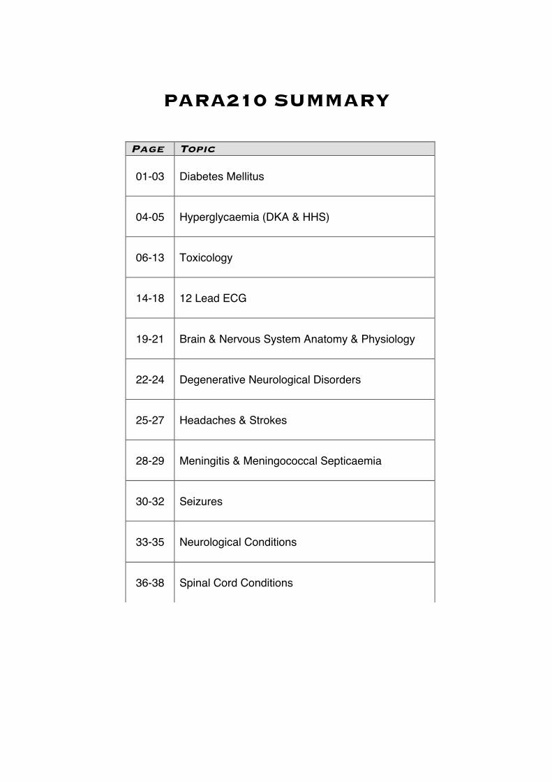
PARA210 SUMMARY Page Topic 01-03 Diabetes Mellitus 04-05 Hyperglycaemia (DKA & HHS) 06-13 Toxicology 14-18 12 Lead ECG 19-21 Brain & Nervous System Anatomy & Physiology 22-24 Degenerative Neurological Disorders 25-27 Headaches & Strokes 28-29 Meningitis & Meningococcal Septicaemia 30-32 Seizures 33-35 Neurological Conditions 36-38 Spinal Cord Conditions
Transcript of PARA210 Summary - StudentVIP · 2018-02-23 · PARA210 SUMMARY Page Topic 01-03 Diabetes Mellitus...
PARA210 SUMMARY
Page Topic
01-03
Diabetes Mellitus
04-05
Hyperglycaemia (DKA & HHS)
06-13
Toxicology
14-18
12 Lead ECG
19-21
Brain & Nervous System Anatomy & Physiology
22-24
Degenerative Neurological Disorders
25-27
Headaches & Strokes
28-29
Meningitis & Meningococcal Septicaemia
30-32
Seizures
33-35
Neurological Conditions
36-38
Spinal Cord Conditions
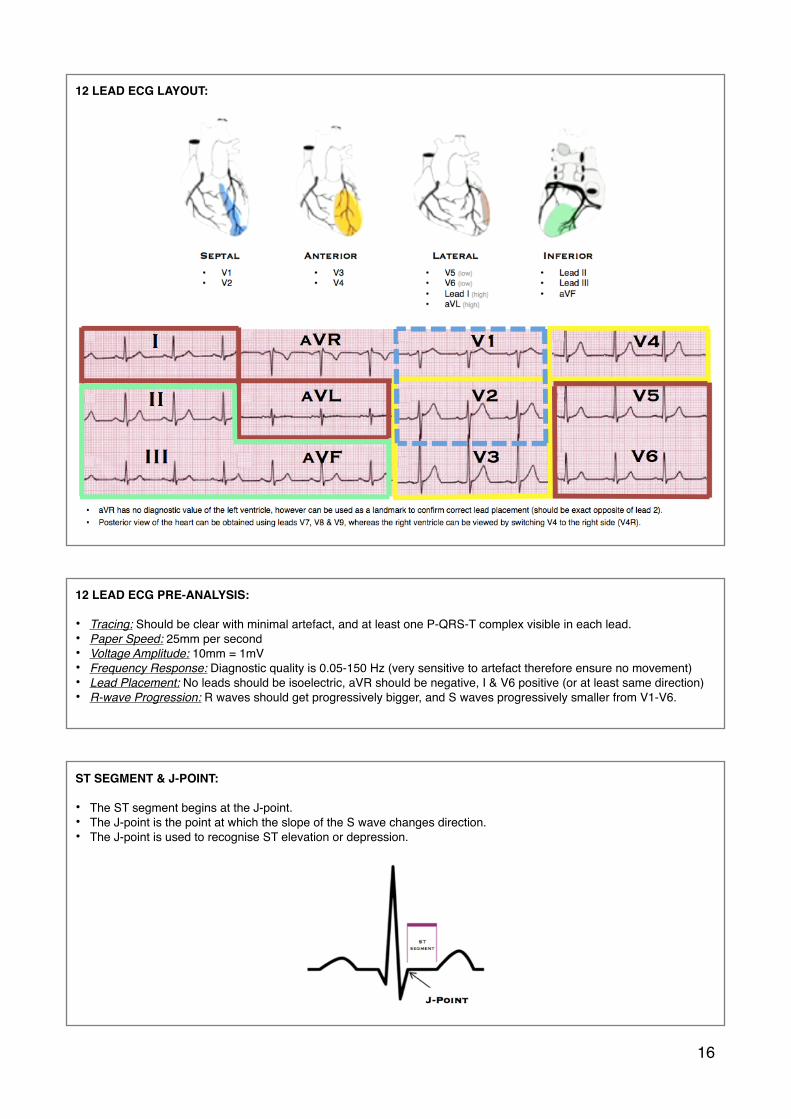
12 LEAD ECG LAYOUT:
12 LEAD ECG PRE-ANALYSIS:
• Tracing: Should be clear with minimal artefact, and at least one P-QRS-T complex visible in each lead.• Paper Speed: 25mm per second• Voltage Amplitude: 10mm = 1mV• Frequency Response: Diagnostic quality is 0.05-150 Hz (very sensitive to artefact therefore ensure no movement)• Lead Placement: No leads should be isoelectric, aVR should be negative, I & V6 positive (or at least same direction)• R-wave Progression: R waves should get progressively bigger, and S waves progressively smaller from V1-V6.
ST SEGMENT & J-POINT:
• The ST segment begins at the J-point.• The J-point is the point at which the slope of the S wave changes direction.• The J-point is used to recognise ST elevation or depression.
�16

CORONARY ARTERIES: The arteries that supply the myocardium with blood and oxygen.
STEACS vs NON-STEACS: STEACS (70%) and NON-STEACS (30%) are commonly known as heart attacks.
Pathophysiology: STEACS occurs when a major coronary artery is completely occluded, resulting in full thickness damage to heart muscle. NON-STEACS occurs when there is a complete occlusion of a minor coronary artery, or a partial occlusion of a major coronary artery, resulting in partial thickness damage to heart muscle. STEACS is indicated by ST elevation on the ECG, whereas NON-STEACS is indicated by ST depression or T-wave inversion.
ANTERIOR STEACS:
• Results from LAD occlusion. Anterior MI carries worst prognosis of all infarct locations, due to larger infarct sizes. • The more areas involved, the more proximal the obstruction is in the LCA.• Septal Infarct: V1 & V2• Anterior Infarct: V3 & V4• Anteroseptal Infarct: V1-V4• Anterolateral Infarct: V2-V6, I + aVL• Extensive Anterior/Anterolateral Infarct: V1-V6, I & aVL (‘Widowmaker MI’)
INFERIOR STEACS:
• Results from RCA occlusion. Accounts for 40-50% of all MIs, with more favourable outcomes than anterior MI.• Inferior STEACS may also be associated with RV MI (40%), 2nd or 3rd degree AV block (20%), or posterior MI.• Inferior Infarct: Leads II, III, & aVF (progressive development of Q waves in II, III, aVF, & reciprocal depression in aVL)
�18
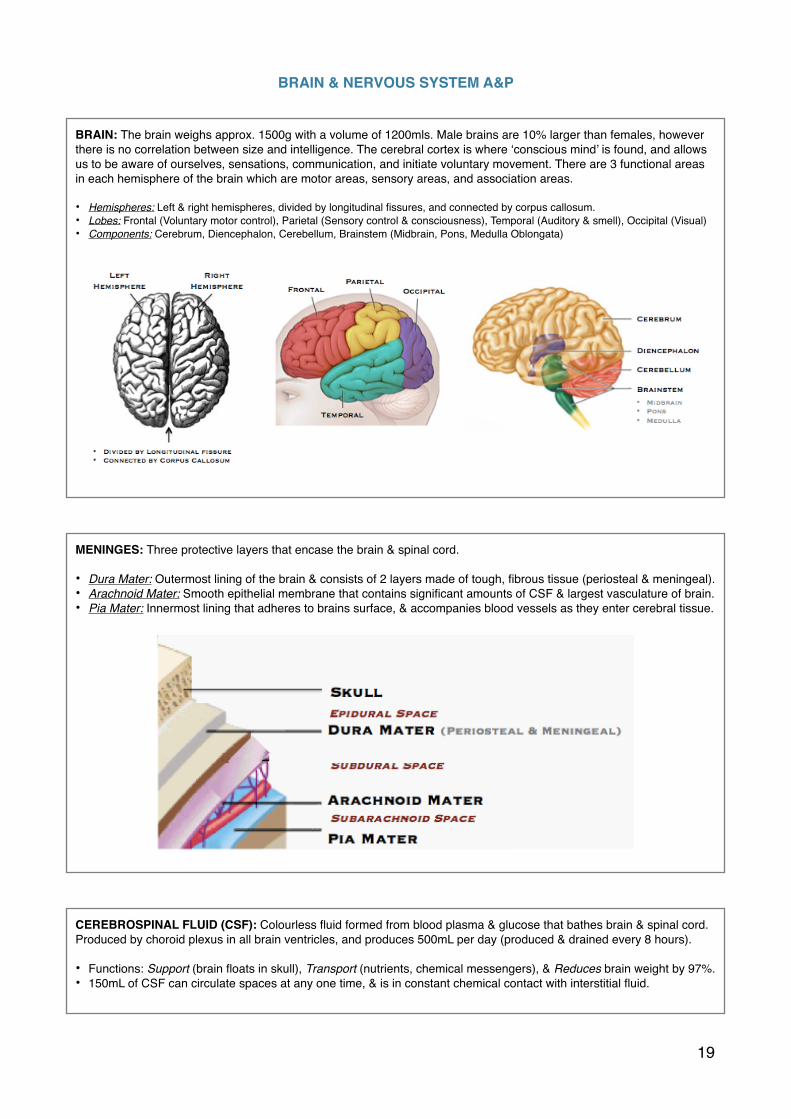
BRAIN & NERVOUS SYSTEM A&P
BRAIN: The brain weighs approx. 1500g with a volume of 1200mls. Male brains are 10% larger than females, however there is no correlation between size and intelligence. The cerebral cortex is where ‘conscious mind’ is found, and allows us to be aware of ourselves, sensations, communication, and initiate voluntary movement. There are 3 functional areas in each hemisphere of the brain which are motor areas, sensory areas, and association areas.
• Hemispheres: Left & right hemispheres, divided by longitudinal fissures, and connected by corpus callosum. • Lobes: Frontal (Voluntary motor control), Parietal (Sensory control & consciousness), Temporal (Auditory & smell), Occipital (Visual)• Components: Cerebrum, Diencephalon, Cerebellum, Brainstem (Midbrain, Pons, Medulla Oblongata)
MENINGES: Three protective layers that encase the brain & spinal cord.
• Dura Mater: Outermost lining of the brain & consists of 2 layers made of tough, fibrous tissue (periosteal & meningeal).• Arachnoid Mater: Smooth epithelial membrane that contains significant amounts of CSF & largest vasculature of brain.• Pia Mater: Innermost lining that adheres to brains surface, & accompanies blood vessels as they enter cerebral tissue.
CEREBROSPINAL FLUID (CSF): Colourless fluid formed from blood plasma & glucose that bathes brain & spinal cord. Produced by choroid plexus in all brain ventricles, and produces 500mL per day (produced & drained every 8 hours).
• Functions: Support (brain floats in skull), Transport (nutrients, chemical messengers), & Reduces brain weight by 97%.• 150mL of CSF can circulate spaces at any one time, & is in constant chemical contact with interstitial fluid.
�19

HEADACHES AND STROKES
HEADACHES: Common neurological disorders which usually are benign. There are 5 primary headache syndromes:
CEREBRAL HAEMORRHAGES: Cerebral haemorrhages can cause herniation of brain tissue (compresses brain).
‘
�25
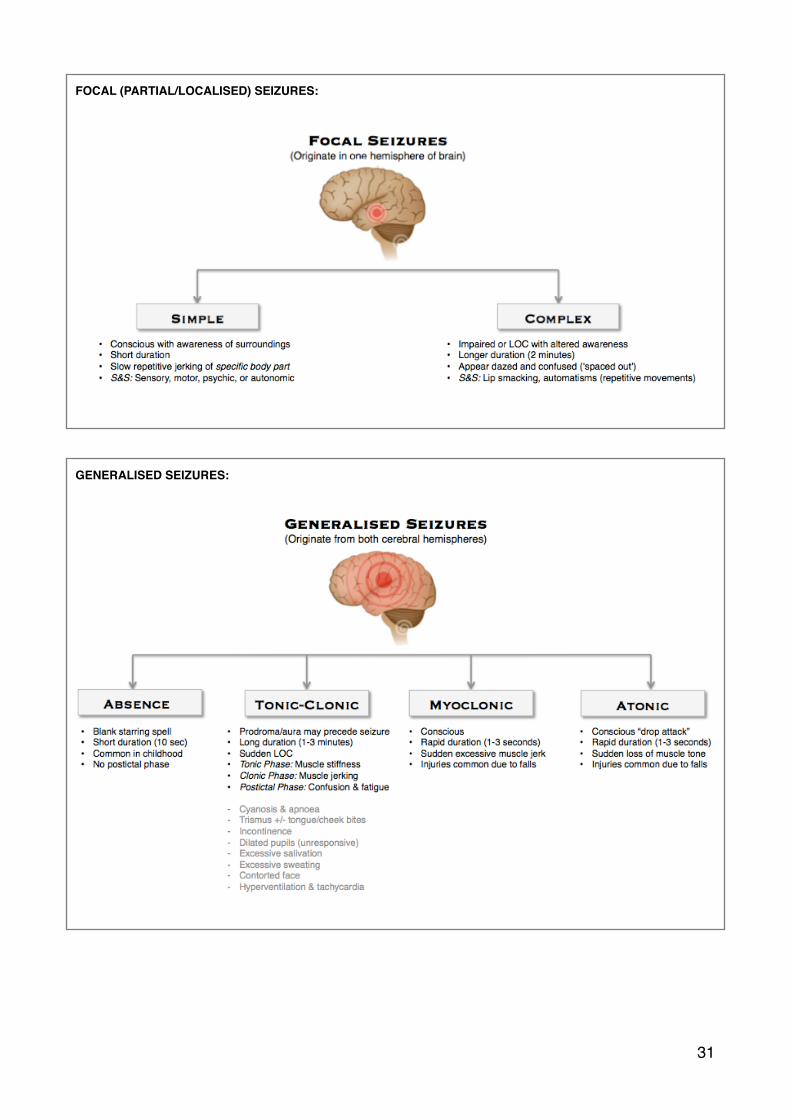
FOCAL (PARTIAL/LOCALISED) SEIZURES:
GENERALISED SEIZURES:
�31