


Bahasa
Halaman
Hukum

REGULAR ARTICLE
Micro-analytical, physiological and molecular aspectsof Fe acquisition in leaves of Fe-deficient tomato plantsre-supplied with natural Fe-complexes in nutrient solution
Nicola Tomasi & Cecilia Rizzardo & Rossella Monte & Stefano Gottardi &Nahida Jelali & Roberto Terzano & Bart Vekemans & Maria De Nobili &Zeno Varanini & Roberto Pinton & Stefano Cesco
Received: 17 February 2009 /Accepted: 8 June 2009# Springer Science + Business Media B.V. 2009
Abstract It is well known that in the rhizospheresoluble Fe sources available for plants are mainly amixture of complexes between the micronutrient andorganic ligands such as organic acids and phytosider-ophores (PS) released by roots, microbial sidero-phores as well as fractions of humified organic matter.In the present work, mechanisms of Fe acquisition
operating at the leaf level of plants fed with differentFe-complexes were investigated at the micro-analytical, physiological and molecular levels. Fe-deficient tomato plants (Solanum Lycopersicum L.,cv. ‘Marmande’) were fed for 24 h with a solution(pH 7.5) containing 1 µM Fe as Fe-PS, Fe-citrate orFe-WEHS. Thereafter, leaf tissue was used for thevisualization of Fe distribution, measurements of Fecontent, reduction and uptake, and evaluation ofexpression of Fe-chelate reductase (LeFRO1), Fe-transporter (LeIRT1) and Ferritin (Ferritin2) genes.Leaf discs isolated from Fe-deficient plants treated for24 h with Fe-WEHS developed higher rates oftranslocation, Fe-chelate reduction and 59Fe uptake ascompared to plants supplied with Fe-citrate or Fe-PS.Leaves of plants treated with Fe-WEHS also showedhigher transcript levels of LeFRO1, LeIRT1 andFerritin2 genes with respect to plants fed with theother Fe-sources. Data obtained support the idea thatthe efficient use of Fe complexed to WEHS-like humicfractions involves, at least in part, also the activation ofFe-acquisition mechanisms operating at the leaf level.
Keywords Iron chlorosis . Natural Fe-sources .
Solanum lycopersicum L. . Humic substances . 59Fe .
Synchrotron µ-XRF analyses
AbbreviationsPS PhytosiderophoresWEHS Water-extractable humic fractionµ-XRF Micro x-ray fluorescence
Plant SoilDOI 10.1007/s11104-009-0069-z
Responsible Editor: Jian Feng Ma.
N. Tomasi : C. Rizzardo :R. Monte : S. Gottardi :N. Jelali :M. De Nobili : R. Pinton : S. Cesco (*)Dipartimento di Scienze Agrarie e Ambientali,University of Udine,Via delle Scienze 208,33100 Udine, Italye-mail: [email protected]
N. JelaliCBT de Borj-Cedria,2050 Tunis, Tunisia
R. TerzanoDipartimento di Biologia e Chimica Agro-forestalee Ambientale, University of Bari,70126 Bari, Italy
B. VekemansDepartment of Analytical Chemistry, Ghent University,9000 Ghent, Belgium
Z. VaraniniDipartimento di Scienze, Tecnologie e Mercati della Vitee del Vino, University of Verona,37029 San Floriano, Italy
Introduction
Iron deficiency is a yield-limiting factor and aworldwide problem in crop production of manyagricultural regions, particularly in calcareous soils(Mengel et al. 2001). Theoretically, total soil-Fecontent would be sufficient to meet Fe needs ofplants; however, most of the Fe in the soil is presentas inorganic forms, predominantly goethite, hematiteand ferrihydrite, all poorly available for root uptakeunder aerobic conditions (Lindsay 1974). Thus, thelevel of plant-available Fe in the soil solution isdetermined by a variety of natural ligands (organicacids, siderophores of microbial or plant origin, andcomponents of humified organic matter of the soil)that can mobilize Fe from oxides/hydroxides or fromFe-humates (Lindsay and Schwab 1982). It is wellaccepted that especially in the rhizosphere a mixtureof Fe-complexes is present, and various authors haveproved that dicotyledonous plants are able to usethem, at least in some cases, as a source of thismicronutrient via a reduction-based mechanism(Römheld and Marschner 1986a; Hoerdt et al. 2000;Cesco et al. 2002). Despite these clear evidence, theseworks were performed using extremely differentexperimental conditions which render very difficultto comprehend what is the contribution of each Fesource to the Fe acquisition by plants. In theframework of a previous study aimed at evaluatingthe relative contribution of different natural chelatesto Fe-acquisition by plants, it has been demonstratedthat Fe complexed to a water extractable humicsubstances fraction (WEHS) could be accumulatedin tomato plants at levels 4–5 times higher than whenFe was supplied as Fe-citrate or Fe-PS (Tomasi et al.2007). Furthermore, a higher up-regulation of Fe-deficiency related genes (LeFRO1, LeIRT1, LeIRT2)was observed at the root level of plants fed with Fe-WEHS as compared with those supplied with other Fesources.
At the leaf level, a common consequence of Feshortage is a low chlorophyll content associated witha limited CO2 fixation activity (Marschner 1995)which is accompanied by an organic-acid export fromthe roots to the leaves via xylem (López-Millán et al.2001). Moreover, it has been demonstrated that plantproductivity is highly dependent upon the photosyn-thetic activity which take place in chloroplasts whereN and S assimilation also occurs. These metabolic
processes, which require Fe-containing enzymes,leads ultimately to the synthesis of a wide variety oforganic compounds (like sugars, amino acids, vita-mins), therefore impacting the nutritional quality ofedible parts of plants (Briat et al. 2007). For thesereasons, the amount of Fe allocated at the leaf levelcould play an important role to achieve crops of high-nutritional quality.
In the present work, using 34-d-old Fe-deficienttomato plants (Solanum lycopersicum L., cv‘Marmande’) the contribution to Fe-acquisition ofdifferent natural chelates (59Fe complexed to barley-born phytosiderophores, citrate or a water-solublehumic fraction, applied at a final Fe concentration of1 µM for 24 h) was studied, evaluating the micronu-trient fraction allocated at the leaf level. Mechanismsof Fe acquisition operating in the leaves of plantssupplied with different Fe-complexes at the end of thetreatments were also investigated at the physiologicaland molecular levels.
Materials and methods
Isolation and purification of Water-Extractable HumicSubstances (WEHS)
Water extractable humic substances (WEHS) wereobtained as reported by Pinton et al. (1998). Briefly,WEHS were extracted from finely ground sphagnumpeat (2.5 g) by adding 50 mL of distilled water andshaking for 15 h at room temperature. Thereafter, thesuspension was centrifuged at 8000 RPM for 30 minand the supernatant filtered through a Whatman WCN0.2 µm membrane filter. The resulting solution wasacidified to pH 2 with H2SO4 and, in order to purifyand concentrate the humified fraction, loaded onto aAmberlite XAD-8 column (Ø 20 mm, height 200 mm;Aiken et al. 1979). Adsorbed humic substances werewashed with 100 mL of distilled water according toAiken et al. (1979) and eluted from the column with0.1 N NaOH. In order to remove the exchangeablemetals, the solution was treated with Amberlite IR-120 (H+ form) up to pH 1–2, and then adjusted toneutrality with 0.1 N NaOH. The humified organicfraction was then freeze-dried before storage anddissolved in distilled water before use at a concentra-tion of 167 mmol organic C L−1. Molecular sizedistribution analysis has shown that the fraction
Plant Soil
contained mostly humic substances of molecularweight lower than 1 kDa (Pinton et al. 1998).
Collection of root exudates and quantitative analysisof epi-hydroxymugineic acid (epi-HMA)
Barley seedlings (Hordeum vulgare L., cv ‘Europa’provided by V. Römheld, Hohenheim University,Stuttgart, D), germinated for 4 days on filter papermoistened with 1 mM CaSO4, were transferred for afurther 14 days to an aerated, Fe-free nutrient solutionas described by Zhang et al. (1991). From the 8th dayof hydroponic culture, root exudates released by Fe-deficient barley plants were collected by transferringplants to 100 ml of aerated distilled water (pH 6.0) for4 h during the morning (a period of high PS release).Root exudates containing phytosiderophores (mainlyepi-HMA; Walter et al. 1995) were filtered through aWhatman WCN 0.2 µm membrane filter and thenstored at −80°C.
In order to evaluate the epi-HMA content, rootexudates containing phytosiderophores (pH 6.0) werepassed through a cation-exchange resin column filledwith Amberlite IR-120B resin (H+ -form; Sigma-Aldrich; Ma et al. 1999). After washing with distilledwater, the PS retained by the cation-exchange resinwere eluted with 2 M NH4OH and then the eluate wasconcentrated to dryness in a rotary evaporator (at 40°C;Ma et al. 1999).The residue was re-dissolved in 1 mlwater and an aliquot of 100 µl was air-dried,derivatized with phenylisothiocyanate (PITC) andanalysed (Howe et al. 1999) by HPLC (LC-1000,Jasco, Tokyo, Japan). The HPLC system wasequipped with a C18 column (XTerra RP 18;150 mm long, 4.6 mm i.d., 3 mm particle size;Waters, Milano, Italy), a Borwin-PDATM 1.50 ver-sion (JMBS, Grenoble, France) controller and a PU-1580 pump. The UV absorption spectra of eluatecomponents were obtained using a Jasco model UV-VIS MD-1510 photodiode array detector. Purifiedepi-HMA was used as a standard.
Preparation of natural 59Fe-complexes
Iron-(59Fe)-WEHS complex was prepared as de-scribed by Cesco et al. (2000) by mixing WEHSfraction with 59FeCl3 in 5 mM Mes-NaOH at pH 6.0;59Fe-PS and 59Fe-citrate was prepared accordingly tovon Wirén et al. (1994) by mixing an aliquot of Fe-
free (epi-HMA)-containing root exudates collectedfrom Fe-deficient barley plants or citrate (10% excess)with FeCl3. The specific activity of 59Fe in the threeFe-sources was 144 KBq µmol−1Fe.
Plant material, growth conditions and planttissue analysis
Tomato seedlings (Solanum Lycopersicum L., cv.‘Marmande superprecoce’ from DOTTO SpA, Italy)were germinated for 6 days on filter paper moistenedwith 1 mM CaSO4 and then grown for 21 days in acontinuously aerated nutrient solution (pH adjusted at6.0 with 1 M KOH) as reported by Pinton et al.(1999) being exposed to 5 µM Fe (Fe-EDTA);thereafter, the plants were transferred for a furtherweek to a Fe-free nutrient solution (Fe-deficient). Thenutrient solution were renewed every three days;before the change, the pH of the old nutrient solutionswas recorded using a pH meter. The controlledclimatic conditions were the following: day/nightphotoperiod, 16/8; light intensity, 220 µE m−2 s−1;temperature (day/night) 25/20°C; RH 70 to 80%.
SPAD index values of fully expanded young leaveswas determined using a portable SPAD-502 meter(Minolta, Osaka, Japan). Fe concentration in leaf androot tissues of tomato plants was determined by ICP-AES, after digestion with concentrated HNO3; rootapoplastic Fe pool was removed before the digestionby 1.2 g L−1 sodium dithionite and 1.5 mM 2,2′-bipyridyl in 1 mM Ca(NO3)2 under N2 bubblingaccording to the method described by Bienfait et al.(1985). Reduction of Fe(III)-EDTA by the roots ofintact plants was measured as described by Pintonet al. (1999) using the bathophenanthrolinedisul-fonate (BPDS) reagent (Chaney et al. 1972). Rootswere incubated for 30 min in an aerated solutioncontaining 0.5 mM CaSO4, 250 µM Fe-EDTA,300 µM BPDS, 10 mM Mes-NaOH (pH 5.5) in thedark at 25°C.
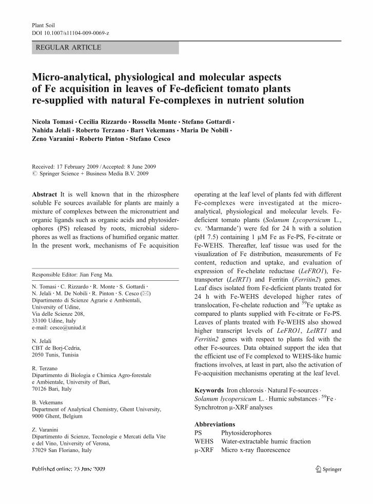
At the end of the growing period (34 days), Fe-deficient tomato plants clearly showed visible symp-toms of Fe deficiency (at the leaf level: yellowing ofthe full expanded apical leaves; at the root level:proliferation of lateral roots, increase in the diameterof the sub-apical zone and amplified root hairformation; Photo in Table 1). The deficiency causedalso a marked decrease in root and shoot dry matteraccumulation; concomitantly, roots were able to lower
Plant Soil
the pH of the nutrient solution and developed anenhanced Fe(III)-EDTA reductase activity (Table 1).These observations are consistent with the inductionand operation of a response mechanism to Fe shortagetypically ascribable to the Strategy I plants
Iron-(59Fe) uptake from natural Fe-sources by rootsof intact plants
As reported by Cesco et al. (2002), roots of twointact Fe-deficient tomato plants (34-d-old) werewashed with micronutrient-free nutrient solution for30 min and then transferred to beakers containing250 mL of a freshly prepared micronutrient-freenutrient solution; 59Fe-PS, 59Fe-citrate or 59FeWEHSwas added in order to give a final Fe concentrationof 1 µM. The addition to the nutrient solution of1 µM Fe as Fe-WEHS brought 5 mg org. C L−1 ofWEHS. In order to limit photo-chemical reductionphenomena of the micronutrient in the nutrientsolution (Zancan et al. 2006) added by the Fe-sources, during the entire experiment, beakers hasbeen covered.
The uptake solution was buffered at pH 7.5 with10 mM Hepes-NaOH and the uptake period was 24 h.
Thereafter, plants were transferred to a freshlyprepared 59Fe-free nutrient solution for 10 min inorder to remove the excess of 59Fe at the root surfaceand then harvested. Root apoplastic 59Fe pools wereremoved by 1.2 g L−1 sodium dithionite and 1.5 mM2,2′-bipyridyl in 1 mM Ca(NO3)2 under N2 bubblingaccording to the method described by Bienfait et al.(1985) (treatment repeated 3 times). Root and shoottissues were oven-dried at 80°C, weighed, ashed at550°C, and suspended in 1% (w/v) HCl for 59Fedetermination by liquid scintillation counting. The59Fe uptake rate, measured as nmol 59Fe, is referred tothe whole plant (root+shoot) and is presented per gdry weight of roots per 1 or 24 h. The 59Fetranslocation rate is presented as nmol 59Fe measuredin shoot per g of root dry weight per 24 h. Theequivalence in ppm of 59Fe taken up by the plants anddetermined by liquid scintillation after the treatmentswith the natural 59Fe-sources, was also calculated intomato roots and shoot.
In order to calculate the contribute of re-supplytreatment with the different 59Fe-sources, in roots andshoot of tomato plants before and after the 24 htreatment with unlabelled natural Fe-sources, theconcentrations (ppm) of total Fe, determined by
Fe sufficient plants Fe deficient plants
Root Shoot Root Shoot
Dry matter (mg) 51 ± 5 467 ± 39 39 ± 5 243 ± 43
Fe content (ppm) 777 ± 98 105 ± 13 124 ± 8 51 ± 2
SPAD index value ------ 29.9 ± 0.8 ------ 17.9 ± 0.8
Root FeIII-chelate reductase (µmol g-1
root FW h-1)
0.37 ± 0.12 ------ 2.89 ± 0.89 ------
pH of nutrient solution 7.4 ± 0.2 ------ 6.7 ± 0.3 ------
Data are means ± SD of three independent experiments
Table 1 Dry matter (mg), iron concentration (ppm) and SPADindex values of 34-d-old Fe-sufficient and Fe-deficient tomatoplants. Root Fe(III)-chelate reducing activity, pH of nutrient
solutions and photos of root and shoot apparatus of Fe-sufficientand Fe-deficient plants, are also reported
Plant Soil
ICP-AES after digestion of the tissues with concen-trated HNO3, were also determined.
Xylem sap collection
Collection of xylem sap was obtained as reported byLópez-Millán et al. (2009). Briefly, plants weredetopped with a razorblade approximately 5 cm abovethe roots. Stumps were allowed to bleed for 1 min,then exuded fluid was carefully wiped out with papertissue and the stem was fitted with plastic tubing.Xylem sap was then allowed to bleed into the plastictubes for 15 min. After this period, samples wereimmediately collected, filtered through a WhatmanWCN 0.2 µm membrane filter and frozen untilanalysis by ICP-AES (previous digestion with con-centrated HNO3).
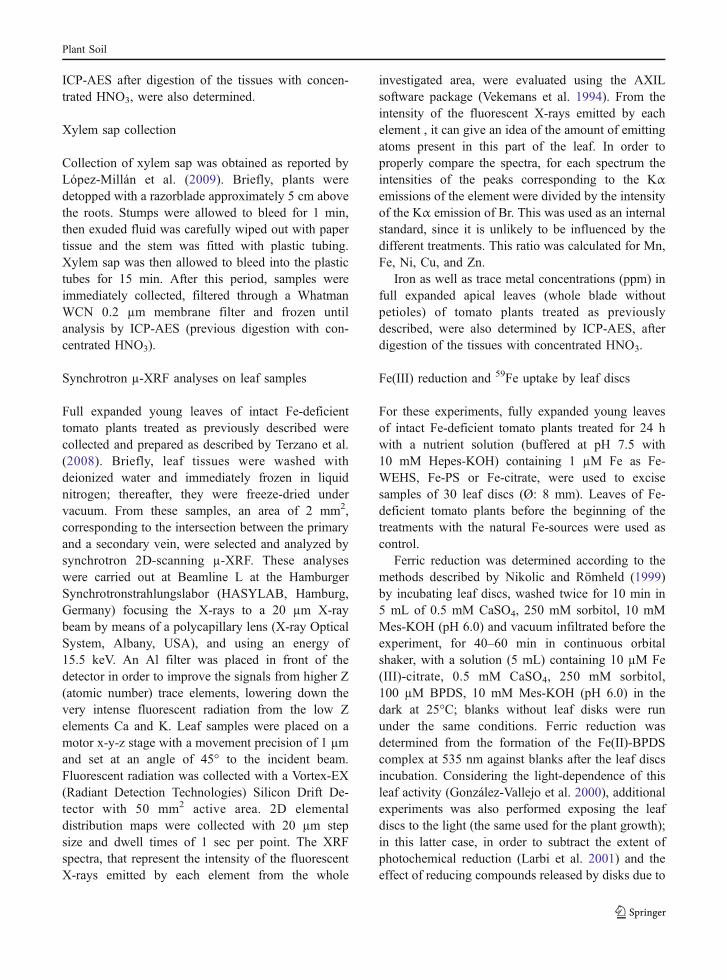
Synchrotron µ-XRF analyses on leaf samples
Full expanded young leaves of intact Fe-deficienttomato plants treated as previously described werecollected and prepared as described by Terzano et al.(2008). Briefly, leaf tissues were washed withdeionized water and immediately frozen in liquidnitrogen; thereafter, they were freeze-dried undervacuum. From these samples, an area of 2 mm2,corresponding to the intersection between the primaryand a secondary vein, were selected and analyzed bysynchrotron 2D-scanning µ-XRF. These analyseswere carried out at Beamline L at the HamburgerSynchrotronstrahlungslabor (HASYLAB, Hamburg,Germany) focusing the X-rays to a 20 µm X-raybeam by means of a polycapillary lens (X-ray OpticalSystem, Albany, USA), and using an energy of15.5 keV. An Al filter was placed in front of thedetector in order to improve the signals from higher Z(atomic number) trace elements, lowering down thevery intense fluorescent radiation from the low Zelements Ca and K. Leaf samples were placed on amotor x-y-z stage with a movement precision of 1 µmand set at an angle of 45° to the incident beam.Fluorescent radiation was collected with a Vortex-EX(Radiant Detection Technologies) Silicon Drift De-tector with 50 mm2 active area. 2D elementaldistribution maps were collected with 20 µm stepsize and dwell times of 1 sec per point. The XRFspectra, that represent the intensity of the fluorescentX-rays emitted by each element from the whole
investigated area, were evaluated using the AXILsoftware package (Vekemans et al. 1994). From theintensity of the fluorescent X-rays emitted by eachelement , it can give an idea of the amount of emittingatoms present in this part of the leaf. In order toproperly compare the spectra, for each spectrum theintensities of the peaks corresponding to the Kαemissions of the element were divided by the intensityof the Kα emission of Br. This was used as an internalstandard, since it is unlikely to be influenced by thedifferent treatments. This ratio was calculated for Mn,Fe, Ni, Cu, and Zn.
Iron as well as trace metal concentrations (ppm) infull expanded apical leaves (whole blade withoutpetioles) of tomato plants treated as previouslydescribed, were also determined by ICP-AES, afterdigestion of the tissues with concentrated HNO3.
Fe(III) reduction and 59Fe uptake by leaf discs
For these experiments, fully expanded young leavesof intact Fe-deficient tomato plants treated for 24 hwith a nutrient solution (buffered at pH 7.5 with10 mM Hepes-KOH) containing 1 µM Fe as Fe-WEHS, Fe-PS or Fe-citrate, were used to excisesamples of 30 leaf discs (Ø: 8 mm). Leaves of Fe-deficient tomato plants before the beginning of thetreatments with the natural Fe-sources were used ascontrol.
Ferric reduction was determined according to themethods described by Nikolic and Römheld (1999)by incubating leaf discs, washed twice for 10 min in5 mL of 0.5 mM CaSO4, 250 mM sorbitol, 10 mMMes-KOH (pH 6.0) and vacuum infiltrated before theexperiment, for 40–60 min in continuous orbitalshaker, with a solution (5 mL) containing 10 µM Fe(III)-citrate, 0.5 mM CaSO4, 250 mM sorbitol,100 µM BPDS, 10 mM Mes-KOH (pH 6.0) in thedark at 25°C; blanks without leaf disks were rununder the same conditions. Ferric reduction wasdetermined from the formation of the Fe(II)-BPDScomplex at 535 nm against blanks after the leaf discsincubation. Considering the light-dependence of thisleaf activity (González-Vallejo et al. 2000), additionalexperiments was also performed exposing the leafdiscs to the light (the same used for the plant growth);in this latter case, in order to subtract the extent ofphotochemical reduction (Larbi et al. 2001) and theeffect of reducing compounds released by disks due to
Plant Soil
their edge, blanks were performed using leaf disksexposed to the light and maintained in ice temperatureduring the entire experiment.
Fe(III) uptake was evaluated after incubating leafdiscs in 5 mL of a solution (0.5 mM CaSO4, 250 mMsorbitol, 10 mM Mes-KOH at pH 6.0) containing10 µM Fe as 59Fe-labeled Fe(III)-citrate in the darkfor 1 h. Iron(59Fe) radioactivity was measured afterremoval of apoplastic 59Fe with bipyridyl and sodiumdithionite for 15 min (Bienfait et al. 1985). Then, theleaf discs were oven dried, ashed at 550°C, and theresidues were dissolved in 1 M HCl to measure 59Feradioactivity by liquid scintillation counting. The 59Feuptake, measured as nmol 59Fe, is referred to the totalamount of 59Fe in leaf discs per gram fresh weightbasis of the tissues.
Uptake of Fe(II) was assayed after incubating leafdiscs in 5 mL of a solution (0.5 mM CaSO4, 250 mMsorbitol, 10 mM Mes-KOH at pH 6.0) containing10 µM Fe as 59Fe(II)SO4 prepared according toZaharieva and Römheld (2000) by mixing theradiochemical tracer (59FeCl3 in 10 mM ascorbate)with 10 mM FeSO4 (in 0.04 M HCl). In order tomaintain the micronutrient in the ferrous status duringthe entire experiment, the uptake solution, with thesame composition previously described, was addedwith ascorbate at a final concentration of 1 mM. Theexperiment was started by adding 59FeSO4 (specificactivity of 59Fe was 180 KBq µmol−1Fe) into theuptake solution and lasted 30 min. Radioactivity of59Fe was measured as previously described.
RNA extraction and cDNA synthesis
Fully expanded young leaves of intact Fe-deficienttomato plants treated as previously described werecollected, immediately frozen in liquid nitrogen andconserved until further processing at −80°C.
RNA extractions were performed using TRIzol®reagent (Invitrogen, Carlsbad, USA) following manu-facturer’s instructions, and contaminant genomic DNAwere removed using 10 U of DNase I (GE Healthcare,Munich, Germany). The total-RNA samples werecleaned up using the standard phenol:chloroformprotocol (Maniatis et al. 1989). One µg of total RNA(checked for quality and quantity using a spectropho-tometer, followed by a migration in an agarose gel) ofeach sample was retrotranscribed using 1 pmol ofOligo d(T)23VN (New England Biolabs, Beverly,
USA) and 10 U M-MulV RNase H− for 1 h at 42°C(Finnzymes, Helsinki, Finland) following the applica-tion protocol of the manufacturers.
Gene expression analyses
After RNA digestion with 1 U RNase A (USB,Cleveland, USA) for 1 h at 37°C, gene expressionanalyses were performed by adding 0.1 µl of thecDNA to FluoCycleTM sybr green (20 µl finalvolume; Euroclone, Pero, Italy) in a DNA EngineOpticon Real-Time PCR Detection (Biorad, Hercules,USA). Primers used (Tm=58°C) were the following:as housekeeping gene: EF1 (X14449) tggatatgctccagtgcttg and ttccttacctgaacgcctgt; IRT1 (AF136579)tcactaggtgcgtcaagcaa and gtaggatgcaaccaccaagg;FRO1 (AY224079) atccaataaaggcggtgttg and tgcatcagtcccactctgtc, and Ferrit in2 (BE431630)gttgctctcaagggacttgc and ccaccacgcttgttctgata. EachReal-Time RT-PCR was performed 3 times on 2independent experiments; analyses of real-time resultwere performed using Opticon Monitor 2 software(Biorad, Hercules, USA) and R (version 2.7.0; http://www.r-project.org/) with the qPCR package (version1.1–4; http://www.dr-spiess.de/qpcR.html). Efficien-cies of amplification were calculated following theauthors’ indications (Ritz and Spiess 2008): PCRefficiencies were 80.25%, 76.25%, 77.70% and82.35% for EF1, IRT1, FRO1 and Ferritin2 genes,respectively.
Statistical analysis
Computation of the graphical representation andstatistical validation (Student’s t-test; p<0.05) wereperformed using SigmaPlot 11.0 (Systat software,Point Richmond, USA). Gene expression data wereillustrated considering the differences in the PCRefficiency of amplification and using the geneexpression levels in leaves of untreated Fe-deficientplants (control) as reference.
Results
In order to study the capability of tomato plants toutilize natural Fe-sources, 59Fe-uptake experimentswere performed incubating roots of intact Fe-deficientplants in a nutrient solution (pH 7.5) for 24 h in the
Plant Soil
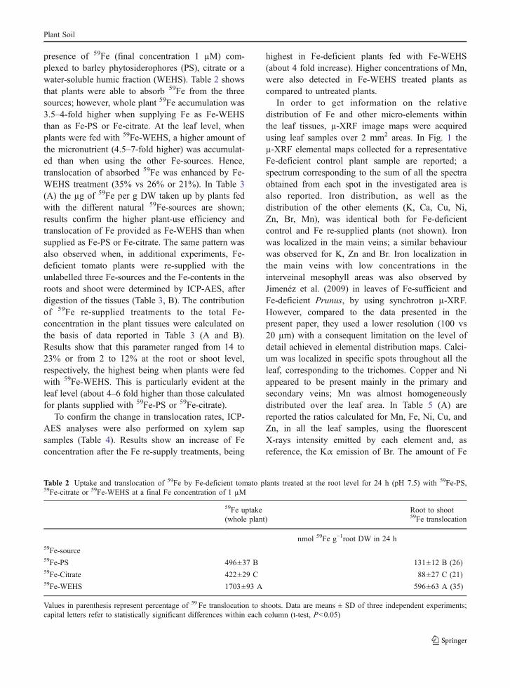
presence of 59Fe (final concentration 1 µM) com-plexed to barley phytosiderophores (PS), citrate or awater-soluble humic fraction (WEHS). Table 2 showsthat plants were able to absorb 59Fe from the threesources; however, whole plant 59Fe accumulation was3.5–4-fold higher when supplying Fe as Fe-WEHSthan as Fe-PS or Fe-citrate. At the leaf level, whenplants were fed with 59Fe-WEHS, a higher amount ofthe micronutrient (4.5–7-fold higher) was accumulat-ed than when using the other Fe-sources. Hence,translocation of absorbed 59Fe was enhanced by Fe-WEHS treatment (35% vs 26% or 21%). In Table 3(A) the µg of 59Fe per g DW taken up by plants fedwith the different natural 59Fe-sources are shown;results confirm the higher plant-use efficiency andtranslocation of Fe provided as Fe-WEHS than whensupplied as Fe-PS or Fe-citrate. The same pattern wasalso observed when, in additional experiments, Fe-deficient tomato plants were re-supplied with theunlabelled three Fe-sources and the Fe-contents in theroots and shoot were determined by ICP-AES, afterdigestion of the tissues (Table 3, B). The contributionof 59Fe re-supplied treatments to the total Fe-concentration in the plant tissues were calculated onthe basis of data reported in Table 3 (A and B).Results show that this parameter ranged from 14 to23% or from 2 to 12% at the root or shoot level,respectively, the highest being when plants were fedwith 59Fe-WEHS. This is particularly evident at theleaf level (about 4–6 fold higher than those calculatedfor plants supplied with 59Fe-PS or 59Fe-citrate).
To confirm the change in translocation rates, ICP-AES analyses were also performed on xylem sapsamples (Table 4). Results show an increase of Feconcentration after the Fe re-supply treatments, being
highest in Fe-deficient plants fed with Fe-WEHS(about 4 fold increase). Higher concentrations of Mn,were also detected in Fe-WEHS treated plants ascompared to untreated plants.
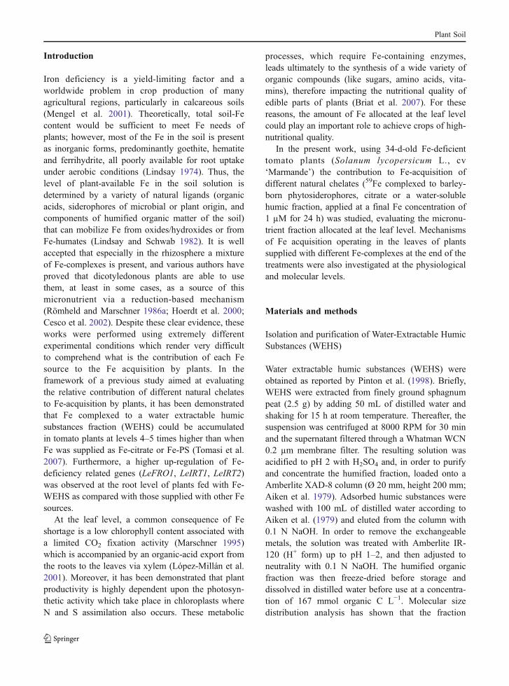
In order to get information on the relativedistribution of Fe and other micro-elements withinthe leaf tissues, µ-XRF image maps were acquiredusing leaf samples over 2 mm2 areas. In Fig. 1 theµ-XRF elemental maps collected for a representativeFe-deficient control plant sample are reported; aspectrum corresponding to the sum of all the spectraobtained from each spot in the investigated area isalso reported. Iron distribution, as well as thedistribution of the other elements (K, Ca, Cu, Ni,Zn, Br, Mn), was identical both for Fe-deficientcontrol and Fe re-supplied plants (not shown). Ironwas localized in the main veins; a similar behaviourwas observed for K, Zn and Br. Iron localization inthe main veins with low concentrations in theinterveinal mesophyll areas was also observed byJimenéz et al. (2009) in leaves of Fe-sufficient andFe-deficient Prunus, by using synchrotron µ-XRF.However, compared to the data presented in thepresent paper, they used a lower resolution (100 vs20 µm) with a consequent limitation on the level ofdetail achieved in elemental distribution maps. Calci-um was localized in specific spots throughout all theleaf, corresponding to the trichomes. Copper and Niappeared to be present mainly in the primary andsecondary veins; Mn was almost homogeneouslydistributed over the leaf area. In Table 5 (A) arereported the ratios calculated for Mn, Fe, Ni, Cu, andZn, in all the leaf samples, using the fluorescentX-rays intensity emitted by each element and, asreference, the Kα emission of Br. The amount of Fe
Table 2 Uptake and translocation of 59Fe by Fe-deficient tomato plants treated at the root level for 24 h (pH 7.5) with 59Fe-PS,59Fe-citrate or 59Fe-WEHS at a final Fe concentration of 1 µM
59Fe uptake Root to shoot59Fe translocation(whole plant)
nmol 59Fe g−1root DW in 24 h59Fe-source59Fe-PS 496±37 B 131±12 B (26)59Fe-Citrate 422±29 C 88±27 C (21)59Fe-WEHS 1703±93 A 596±63 A (35)
Values in parenthesis represent percentage of 59 Fe translocation to shoots. Data are means ± SD of three independent experiments;capital letters refer to statistically significant differences within each column (t-test, P<0.05)
Plant Soil

in leaves of Fe-WEHS fed plants was estimated to beabout 5 times higher than that of Fe-deficient controlplants; only a slight increase in Fe amount wasrecorded in leaves of plants treated with Fe-PS orFe-citrate. Higher amounts of Mn, Ni, Cu and Znwere also detected in Fe-WEHS treated plants ascompared to the other Fe treatments. In Table 5 (B) thetrace-element concentrations (µg g−1DW) determinedby ICP-AES in full expanded apical leaves (wholeblade without petioles) are reported. Although the twoanalytical approaches analyzed diverse areas of the leafblade, results obtained by ICP-AES also showed thatthe concentration levels of trace metal in leaves ofplants fed with Fe-WEHS were significantly higherthan those recorded in Fe-deficient plants treated withFe-PS or Fe-citrate, confirming what was observed bysynchrotron analyses. The Fe re-supply treatmentscaused also a recovery of the SPAD index values of
the apical leaves, the highest being when Fe-deficientplants were supplied with Fe-WEHS (Table 5, B).
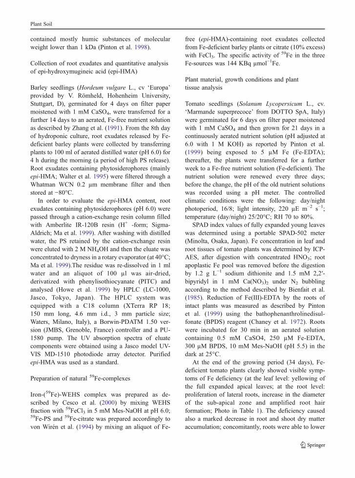
In order to evaluate the functionality of Fe-acquisition mechanisms working at the leaf level ofplants supplied with different Fe-complexes, Fe(III)-citrate reduction, 59Fe(III)-citrate uptake and 59FeSO4
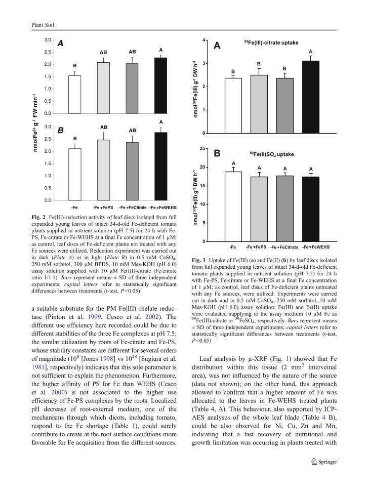
uptake were measured using leaf discs excised fromfully expanded young leaves of intact Fe-deficienttomato plants treated in the nutrient solution for 24 hwith 1 µM Fe as Fe-WEHS, Fe-PS or Fe-citrate andadopting assay conditions reported by Nikolic andRömheld (1999). Figure 2a shows that supply of Fecaused a higher capacity of leaf tissues to reduceexogenous Fe(III)-citrate; however, reduction activitywas significantly higher than that measured in controlFe-deficient plants only in leaves of plants fed withFe-WEHS. For the dependence on the light of thisleaf activity (González-Vallejo et al. 2000), Fe(III)-
Table 3 Iron-(59Fe) acquired from 59Fe-sources (A) (deter-mined by liquid scintillation and expressed in ppm) and totalFe concentration (B) (determined by ICP-AES) in roots and
shoot of Fe-deficient tomato plants treated for 24 h (pH 7.5)with Fe-PS, Fe-citrate or Fe-WEHS at a final Fe concentrationof 1 µM
A B
59Fe acquired (μg g−1 DW) from 59Fe-sources Total Fe concentration (μg g−1 DW)
determined by liquid scintillation determined by ICP-AES
Fe-source root shoot root shoot
+Fe-PS 21.3±2.6 B [14.8%] 1.6±0.1 B [3.0%] 143.9±8.6 B 53.2±1.9 B
+Fe-Citrate 19.7±1.9 B [14.1%] 0.9±0.2 C [1.7%] 139.2±11.3 BC 52.7±2.3 B
+Fe-WEHS 40.4±8.8 A [23.4%] 7.2±1.2 A [12.2%] 172.3±13.4 A 59.1±2.9 A
Values in square brackets indicate contribute of resupply treatment of each Fe-sources. Data are means ± SD of three independentexperiments; capital letters refer to statistically significant differences within each column (t-test, P<0.05)
Table 4 Concentrations of cationic nutrients (determined by ICP-AES) in xylem sap samples of Fe-deficient tomato plants treated for24 h (pH 7.5) with Fe-PS, Fe-citrate or Fe-WEHS at a final Fe concentration of 1 µM
Control Fe-PS Fe-Citrate Fe-WEHSFe-deficientNutrient
µg mL−1
Mn 0.031±0.008 B (100) 0.029±0.005 B (93) 0.037±0.014 AB (119) 0.057±0.010 A (184)
Fe 0.449±0.088 C (100) 0.655±0.024 B (146) 0.578±0.190 BC (129) 1.999±0.362 A (445)
Zn 0.591±0.174 AB (100) 0.419±0.120 B (71) 0.624±0.272 AB (106) 0.647±0.113 A (109)
K 1045±60 A (100) 760±106 B (73) 879±84 AB (84) 979±49 A (94)
Mg 217±19 A (100) 171±13 C (79) 191±5 B (88) 205±11 AB (94)
Values in parenthesis represent percentage of–Fe control leaf. Data are means ± SD of two independent experiments; capital lettersrefer to statistically significant differences within each line (t-test, P<0.05)
Plant Soil
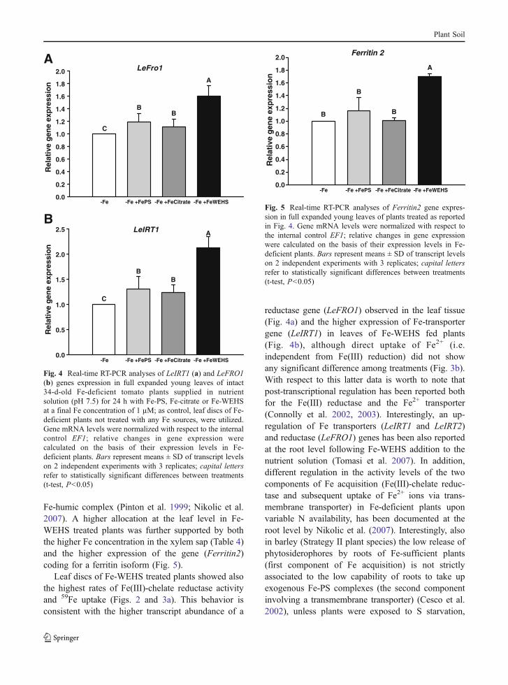
citrate reduction was also evaluated exposing the leafdiscs to light. Results reported in Fig. 2b confirmthose obtained in the dark experiments. In Fig. 3 thelevels of Fe uptake evaluated incubating in darknessleaf discs in a solution containing 10 µM 59Fe(III)-citrate or 59FeSO4, are reported. Results show thatonly the treatment of tomato plants with Fe-WEHSdetermined a significant increase in Fe uptake from59Fe(III)-citrate (Fig. 3a); on the other hand, no effectdue to the treatment with the different natural Fesources was recorded in the uptake rates of Fe(II)from 59FeSO4 (Fig. 3b).
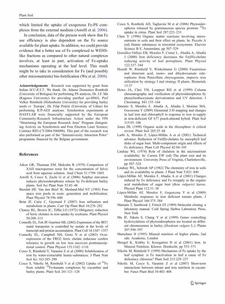
To evaluate the involvement of a transcriptionalregulation of Fe-uptake mechanisms at the leaf level,mRNA abundance of LeFRO1 (coding for an isoformof the PM Fe(III)-chelate reductase) and LeIRT1(coding for Fe2+ transporter) were analyzed in leavesof intact Fe-deficient tomato plants treated for 24 hwith Fe-WEHS, Fe-PS or Fe-citrate in nutrientsolution. Results reported in Fig. 4 show that therelative expression levels of the two genes, evaluatedby real-time RT-PCR, were influenced by the treat-ments. In fact, in leaf cells of Fe-deficient plants fedwith Fe-WEHS a significant increase of LeIRT1 and
1 m
m
2 mm
Fe K Ca
ZnNiCu
Br
Mn
Fig. 1 Fe, K, Ca, Cu, Ni, Zn, Br, and Mn distributions on a2 mm2 area of a leaf imaged by 2-D scanning µ-XRF. The µ-XRF elemental maps is referred to a representative Fe-deficientcontrol plant sample. Darker pixels correspond to areas with a
relatively higher element concentration. The sum-spectrumcorresponding to the same area is also reported. The Kαemission peak for each element is indicated
Plant Soil

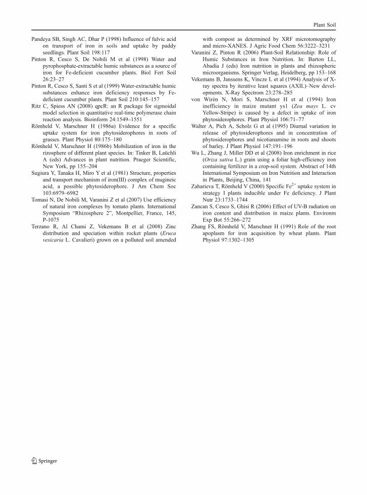
LeFRO1 gene expression levels occurred. A slight butsignificant increase in transcript abundance of the twogenes was also recorded in plants fed with Fe-PS andFe-citrate. Evaluation of expression level of Ferritin2gene was also performed showing that higher Feaccumulation in leaves of Fe-WEHS treated plantswas accompanied by an up-regulation of the gene(Fig. 5).
Discussion
The ability of plants to take up Fe from the soildepends on their capacity to utilize natural solublesources and/or to mobilize sparingly soluble Fe forms(Römheld and Marschner 1986b). This task isaccomplished by releasing Fe-chelating substances(i.e. PS, citrate) able to form Fe(III)-complexes whichin turn act as substrates for root-uptake mechanisms(Römheld and Marschner 1986a). Furthermore, frac-tions of humified organic matter may be present in thesoil solution, providing an additional soluble Fesource directly utilizable by plants (Chen 1996;Pandeya et al. 1998; Varanini and Pinton 2006).
While mechanisms adopted by plants to take up Fefrom complexes like Fe-PS and Fe-WEHS have beenwell described (Römheld and Marschner 1986a;Cesco et al. 2002, 2006), poor evidence is availableon the use efficiency of these different natural Fesources in a context more similar to what is occurringin the rhizosphere.
In the present work we compared three natural Fecomplexes, namely Fe-PS, Fe-citrate and Fe-WEHS,with respect to their capacity to provide Fe to Fe-deficient tomato plants; particularly, accumulation atthe leaf level was evaluated after supplying for 24 heach Fe source to the nutrient solution. Furthermore,the functioning of Fe-uptake mechanisms (Fe(III)reduction and Fe(II) uptake) was assayed using leafdiscs isolated from control and treated plants andcorrelated to gene expression analyses. Results showthat Fe-deficient tomato plants are able to utilize thethree Fe sources; however, when plants were put incontact with Fe-WEHS, a higher Fe accumulation inthe whole plant was recorded, with root-shoottranslocation showing a more than proportionalincrease (Tables 2, 3 and 4). These data confirmprevious observation that Fe(III)-WEHS could act as
Table 5 A: Trace element relative peak intensity (Br used asreference peak) calculated for samples (area of 2 mm2) isolatedfrom full expanded apical leaves. B: SPAD index values and
trace element concentration (ppm) of full expanded apicalleaves (whole blade without petioles) determined by ICP-AESafter its digestion
Control Fe-PS Fe-Citrate Fe-WEHSFe-deficientTrace element
A
I/I[Br-Kα]
Mn 0.4±0.1 B (100) 0.5±0.2 B (125) 0.4±0.2 B (100) 1.6±0.4 A (400)
Fe 3.5±0.4 C (100) 3.9±0.4 BC (111) 4.4±0.3 B (126) 16±3 A (457)
Ni 0.5±0.3 B (100) 0.6±0.2 B (120) 0.4±0.2 B (80) 1.9±0.6 A (380)
Cu 2.7±0.4 C (100) 2.6±0.5 C (96) 4.0±0.5 B (148) 14±3 A (518)
Zn 15±3 C (100) 17±4 B (113) 13±3 C (87) 66±5 A (440)
B
µg g−1 DW
Mn 16.3±0.4 B (100) 17.6±0.3 AB (108) 17.1±0.5 B (105) 18.8±1.0 A (115)
Fe 59.1±0.9 C (100) 63.5±2.1 B (107) 65.1±4.1 B (110) 79.2±3.8 A (134)
Ni 2.9±0.4 B (100) 3.2±0.3 B (110) 3.0±0.3 B (103) 4.7±0.4 A (162)
Cu 18.2±0.2 B (100) 17.7±0.7 B (97) 21.7±2.0 A (120) 25.1±1.8 A (138)
Zn 44.9±6.1 B (100) 49.7±7.3 AB (111) 37.1±13.2 B (83) 65.5±11.3 A (146)
SPAD index 17.9±0.8 C 19.8±0.3 AB 19.2±0.5 BC 21.0±1.2 A
Values in parenthesis represent percentage of–Fe control leaf. Data are means ± SD of three independent analyses; capital letters referto statistically significant differences within each line (t-test, P<0.05)
Plant Soil
a suitable substrate for the PM Fe(III)-chelate reduc-tase (Pinton et al. 1999, Cesco et al. 2002). Thedifferent use efficiency here recorded could be due todifferent stabilities of the three Fe complexes at pH 7.5;the similar utilization by roots of Fe-citrate and Fe-PS,whose stability constants are different for several ordersof magnitude (108 [Jones 1998] vs 1018 [Sugiura et al.1981], respectively) indicates that this sole parameter isnot sufficient to explain the phenomenon. Furthermore,the higher affinity of PS for Fe than WEHS (Cescoet al. 2000) is not associated to the higher useefficiency of Fe-PS complexes by the roots. LocalizedpH decrease of root-external medium, one of themechanisms through which dicots, including tomato,respond to the Fe shortage (Table 1), could surelycontribute to create at the root surface conditions morefavorable for Fe acquisition from the different sources.
Leaf analysis by µ-XRF (Fig. 1) showed that Fedistribution within this tissue (2 mm2 interveinalarea), was not influenced by the nature of the source(data not shown); on the other hand, this approachallowed to confirm that a higher amount of Fe wasallocated to the leaves in Fe-WEHS treated plants(Table 4, A). This behaviour, also supported by ICP-AES analyses of the whole leaf blade (Table 4 B),could be also observed for Ni, Cu, Zn and Mn,indicating that a fast recovery of nutritional andgrowth limitation was occurring in plants treated with
0.0
0.5
1.0
1.5
2.0
2.5
3.0
0.0
0.5
1.0
1.5
2.0
2.5
3.0
-Fe -Fe +FePS -Fe +FeCitrate -Fe +FeWEHS
nm
olF
e2+
g-1
FW
min
-1
A
B
B
AB AB A
B
ABAB
A
Fig. 2 Fe(III)-reduction activity of leaf discs isolated from fullexpanded young leaves of intact 34-d-old Fe-deficient tomatoplants supplied in nutrient solution (pH 7.5) for 24 h with Fe-PS, Fe-citrate or Fe-WEHS at a final Fe concentration of 1 µM;as control, leaf discs of Fe-deficient plants not treated with anyFe sources were utilized. Reduction experiment was carried outin dark (Plate A) or in light (Plate B) in 0.5 mM CaSO4,250 mM sorbitol, 300 µM BPDS, 10 mM Mes-KOH (pH 6.0)assay solution supplied with 10 µM Fe(III)-citrate (Fe/citrateratio 1/1.1). Bars represent means ± SD of three independentexperiments; capital letters refer to statistically significantdifferences between treatments (t-test, P<0.05)
0
1
2
3
4
A
B
BB
0
5
10
15
20
25
AA
A A
59Fe(II)SO4 uptake
59Fe(III)-citrate uptakeA
B
-Fe -Fe +FePS -Fe +FeCitrate -Fe +FeWEHS
nm
ol 5
9F
e(I
II)
g-1
DW
h-1
nm
ol 5
9F
e(I
I) g
-1D
W h
-1
Fig. 3 Uptake of Fe(III) (a) and Fe(II) (b) by leaf discs isolatedfrom full expanded young leaves of intact 34-d-old Fe-deficienttomato plants supplied in nutrient solution (pH 7.5) for 24 hwith Fe-PS, Fe-citrate or Fe-WEHS at a final Fe concentrationof 1 µM; as control, leaf discs of Fe-deficient plants untreatedwith any Fe sources, were utilized. Experiments were carriedout in dark and in 0.5 mM CaSO4, 250 mM sorbitol, 10 mMMes-KOH (pH 6.0) assay solution; Fe(III) and Fe(II) uptakewere evaluated supplying to the assay medium 10 µM Fe as59Fe(III)-citrate or 59FeSO4, respectively. Bars represent means± SD of three independent experiments; capital letters refer tostatistically significant differences between treatments (t-test,P<0.05)
Plant Soil
Fe-humic complex (Pinton et al. 1999; Nikolic et al.2007). A higher allocation at the leaf level in Fe-WEHS treated plants was further supported by boththe higher Fe concentration in the xylem sap (Table 4)and the higher expression of the gene (Ferritin2)coding for a ferritin isoform (Fig. 5).
Leaf discs of Fe-WEHS treated plants showed alsothe highest rates of Fe(III)-chelate reductase activityand 59Fe uptake (Figs. 2 and 3a). This behavior isconsistent with the higher transcript abundance of a
reductase gene (LeFRO1) observed in the leaf tissue(Fig. 4a) and the higher expression of Fe-transportergene (LeIRT1) in leaves of Fe-WEHS fed plants(Fig. 4b), although direct uptake of Fe2+ (i.e.independent from Fe(III) reduction) did not showany significant difference among treatments (Fig. 3b).With respect to this latter data is worth to note thatpost-transcriptional regulation has been reported bothfor the Fe(III) reductase and the Fe2+ transporter(Connolly et al. 2002, 2003). Interestingly, an up-regulation of Fe transporters (LeIRT1 and LeIRT2)and reductase (LeFRO1) genes has been also reportedat the root level following Fe-WEHS addition to thenutrient solution (Tomasi et al. 2007). In addition,different regulation in the activity levels of the twocomponents of Fe acquisition (Fe(III)-chelate reduc-tase and subsequent uptake of Fe2+ ions via trans-membrane transporter) in Fe-deficient plants uponvariable N availability, has been documented at theroot level by Nikolic et al. (2007). Interestingly, alsoin barley (Strategy II plant species) the low release ofphytosiderophores by roots of Fe-sufficient plants(first component of Fe acquisition) is not strictlyassociated to the low capability of roots to take upexogenous Fe-PS complexes (the second componentinvolving a transmembrane transporter) (Cesco et al.2002), unless plants were exposed to S starvation,
Rel
ativ
e g
ene
exp
ress
ion
0.0
0.5
1.0
1.5
2.0
2.5 A
BB
C
-Fe -Fe +FePS -Fe +FeCitrate -Fe +FeWEHS
-Fe -Fe +FePS -Fe +FeCitrate -Fe +FeWEHS
Rel
ativ
e g
ene
exp
ress
ion
0.0
0.2
0.4
0.6
0.8
1.0
1.2
1.4
1.6
1.8
2.0A
BB
C
LeFro1A
BLeIRT1
Fig. 4 Real-time RT-PCR analyses of LeIRT1 (a) and LeFRO1(b) genes expression in full expanded young leaves of intact34-d-old Fe-deficient tomato plants supplied in nutrientsolution (pH 7.5) for 24 h with Fe-PS, Fe-citrate or Fe-WEHSat a final Fe concentration of 1 µM; as control, leaf discs of Fe-deficient plants not treated with any Fe sources, were utilized.Gene mRNA levels were normalized with respect to the internalcontrol EF1; relative changes in gene expression werecalculated on the basis of their expression levels in Fe-deficient plants. Bars represent means ± SD of transcript levelson 2 independent experiments with 3 replicates; capital lettersrefer to statistically significant differences between treatments(t-test, P<0.05)
Ferritin 2
-Fe -Fe +FePS -Fe +FeCitrate -Fe +FeWEHS
Rel
ativ
e g
ene
exp
ress
ion
0.0
0.2
0.4
0.6
0.8
1.0
1.2
1.4
1.6
1.8
2.0
A
B
B
B
Fig. 5 Real-time RT-PCR analyses of Ferritin2 gene expres-sion in full expanded young leaves of plants treated as reportedin Fig. 4. Gene mRNA levels were normalized with respect tothe internal control EF1; relative changes in gene expressionwere calculated on the basis of their expression levels in Fe-deficient plants. Bars represent means ± SD of transcript levelson 2 independent experiments with 3 replicates; capital lettersrefer to statistically significant differences between treatments(t-test, P<0.05)
Plant Soil
which limited the uptake of exogenous Fe-PS com-plexes from the external medium (Astolfi et al. 2006).
In conclusion, data of the present work show that Feuse efficiency is also dependent on the Fe sourceavailable for plant uptake. In addition, we could provideevidence that a better use of Fe complexed to WEHS-like fractions as compared to other natural complexesinvolves, at least in part, activation of Fe-uptakemechanisms operating at the leaf level. This resultmight be to take in consideration for Fe (and possiblyother micronutrients) bio-fortification (Wu et al. 2008).
Acknowledgements Research was supported by grant fromItalian M.U.R.S.T.. We thank: Dr. Adamo Domenico Rombolà(University of Bologna) for performing PS analysis, Dr. J.F. Ma(Kagawa University) for providing purified epi-HMA, ProfVolker Römheld (Hohenheim University) for providing barleyseeds cv ‘Europa’, Dr. Filip Pošćić (University of Udine) forperforming ICP-AES analyses. Synchrotron experiments atHASYLAB were financially supported by the EuropeanCommunity-Research Infrastructure Action under the FP6“Structuring the European Research Area” Program (Integrat-ing Activity on Synchrotron and Free Electron Laser Science;Contract RII3-CT-2004-506008). This part of the research wasalso performed as part of the “Interuniversity Attraction Poles”programme financed by the Belgian government.
References
Aiken GR, Thurman EM, Malcolm R (1979) Comparison ofXAD macroporous resin for the concentration of fulvicacid from aqueous solution. Anal Chem 51:1799–1803
Astolfi S, Cesco S, Zuchi G et al (2006) Sulphur starvationreduces phytosiderophores release by Fe-deficient barleyplants. Soil Sci Plant Nutr 52:43–48
Bienfait HF, Van den Briel W, Mesland-Mul NT (1985) Freespace iron pools in roots: generation and mobilization.Plant Physiol 78:596–600
Briat JF, Curie C, Gaymard F (2007) Iron utilization andmetabolism in plants. Curr Op Plant Biol 10:276–282
Chaney RL, Brown JC, Tiffin LO (1972) Obligatory reductionof ferric chelates in iron uptake by soybeans. Plant Physiol50:208–213
Connolly EL, Fett JP, Guerinot ML (2002) Expression of the IRT1metal transporter is controlled by metals at the levels oftranscript and protein accumulation. Plant Cell 14:1347–1357
Connolly EL, Campbell NH, Grotz N et al (2003) Over-expression of the FRO2 ferric chelate reductase conferstolerance to growth on low iron uncovers posttranscrip-tional control. Plant Physiol 133:1102–1110
Cesco S, Römheld V, Varanini Z et al (2000) Solubilization ofiron by water-extractable humic-substances. J Plant NutrSoil Sci 163:285–290
Cesco S, Nikolic M, Römheld V et al (2002) Uptake of 59Fefrom soluble 59Fe-humate complexes by cucumber andbarley plants. Plant Soil 241:121–128
Cesco S, Rombolà AD, Tagliavini M et al (2006) Phytosider-ophores released by graminaceous species promote 59Feuptake in citrus. Plant Soil 287:223–233
Chen Y (1996) Organic matter reactions involving micro-nutrients in soils and their effect on plants. In: Piccolo A(ed) Humic substances in terrestrial ecosystems. ElsevierScience B.V, Amsterdam, pp 507–529
González-Vallejo EB, Morales F, Cistué L, Abadía A, AbadíaJ (2000) Iron deficiency decreases the Fe(III)-chelatereducing activity of leaf protoplasts. Plant Physiol122:337–344
Hoerdt W, Römheld V, Winkelmann G (2000) Fusarininesand dimerum acid, mono- and dihydroxamate side-rophores from Penicillum chrysogenum, improve ironutilization by strategy I and strategy II plants. Biometals13:37
Howe JA, Choi YH, Loeppert RH et al (1999) Columnchromatography and verification of phytosiderophores byphenylisothiocyanate derivatization and UV detection. JChromatog. 841:155–164
Jimenéz S, Morales F, Abadia A, Abadia J, Moreno MA,Gocorcena Y (2009) Elemental 2-D mapping and changesin leaf iron and chlorophyll in response to iron re-supplyin iron-deficient GF 677 peach-almond hybrid. Plant Soil315:93–106
Jones DL (1998) Organic acids in the rhizosphere–A criticalreview. Plant Soil 205:25–44
Larbi A, Morales F, López-Millán A et al (2001) Technicaladvance: Reduction of Fe(III)-chelates by mesophyll leafdisks of sugar beet. Multi-component origin and effects ofFe deficiency. Plant Cell Physiol 42:94–105
Lindsay WL (1974) Role of chelation in the micronutrientavailability. In: Carson EW (ed) The plant root and itsenvironment. University Press of Virginia, Charlottesville,pp 507–524
Lindsay WL, Schwab AP (1982) The chemistry of iron in soilsand its availability to plants. J Plant Nutr 5:821–840
López-Millán AF, Morales F, Abadia A et al (2001) Changesinduced by Fe deficiency and Fe resupply in the organicacid metabolism of sugar beet (Beta vulgaris) leaves.Physiol Plant 112:31–38
López-Millán AF, Morales F, Gogorcena Y et al (2009)Metabolic responses in iron deficient tomato plants. JPlant Physiol 166:375–384
Maniatis T, Sambrook J, Fritsch EF (1989) Molecular cloning: alaboratory manual. Cold Spring Harbor Laboratory Press,New York
Ma JF, Taketa S, Chang Y et al (1999) Genes controllinghydroxylations of phytosiderophores are located on differ-ent chromosomes in barley (Hordeum vulgare L.). Planta207:590–597
Marschner H (1995) Mineral nutrition of higher plants, 2ndedn. Academic, London
Mengel K, Kirkby E, Kosegarten H et al (2001) Iron. InMineral Nutrition. Kluwer, Dordrecht, pp 553–571
Nikolic M, Römheld V (1999) Mechanism of Fe uptake by theleaf symplast: is Fe inactivation in leaf a cause of Fedeficiency chlorosis? Plant Soil 215:229–237
Nikolic M, Cesco S, Varanini Z et al (2007) Short-terminteractions between nitrate and iron nutrition in cucum-ber. Funct Plant Biol 34:402–408
Plant Soil
Pandeya SB, Singh AC, Dhar P (1998) Influence of fulvic acidon transport of iron in soils and uptake by paddyseedlings. Plant Soil 198:117
Pinton R, Cesco S, De Nobili M et al (1998) Water andpyrophosphate-extractable humic substances as a source ofiron for Fe-deficient cucumber plants. Biol Fert Soil26:23–27
Pinton R, Cesco S, Santi S et al (1999) Water-extractable humicsubstances enhance iron deficiency responses by Fe-deficient cucumber plants. Plant Soil 210:145–157
Ritz C, Spiess AN (2008) qpcR: an R package for sigmoidalmodel selection in quantitative real-time polymerase chainreaction analysis. Bioinform 24:1549–1551
Römheld V, Marschner H (1986a) Evidence for a specificuptake system for iron phytosiderophores in roots ofgrasses. Plant Physiol 80:175–180
Römheld V, Marschner H (1986b) Mobilization of iron in therizosphere of different plant species. In: Tinker B, LaüchliA (eds) Advances in plant nutrition. Praeger Scientific,New York, pp 155–204
Sugiura Y, Tanaka H, Miro Y et al (1981) Structure, propertiesand transport mechanism of iron(III) complex of mugineicacid, a possible phytosiderophore. J Am Chem Soc103:6979–6982
Tomasi N, De Nobili M, Varanini Z et al (2007) Use efficiencyof natural iron complexes by tomato plants. InternationalSymposium “Rhizosphere 2”, Montpellier, France, 145,P-1075
Terzano R, Al Chami Z, Vekemans B et al (2008) Zincdistribution and speciation within rocket plants (Erucavesicaria L. Cavalieri) grown on a polluted soil amended
with compost as determined by XRF microtomographyand micro-XANES. J Agric Food Chem 56:3222–3231
Varanini Z, Pinton R (2006) Plant-Soil Relationship: Role ofHumic Substances in Iron Nutrition. In: Barton LL,Abadía J (eds) Iron nutrition in plants and rhizosphericmicroorganisms. Springer Verlag, Heidelberg, pp 153–168
Vekemans B, Janssens K, Vincze L et al (1994) Analysis of X-ray spectra by iterative least squares (AXIL)–New devel-opments. X-Ray Spectrom 23:278–285
von Wirén N, Mori S, Marschner H et al (1994) Ironinefficiency in maize mutant ys1 (Zea mays L. cvYellow-Stripe) is caused by a defect in uptake of ironphytosiderophores. Plant Physiol 106:71–77
Walter A, Pich A, Scholz G et al (1995) Diurnal variation inrelease of phytosiderophores and in concentration ofphytosiderophores and nicotianamine in roots and shootsof barley. J Plant Physiol 147:191–196
Wu L, Zhang J, Miller DD et al (2008) Iron enrichment in rice(Oriza sativa L.) grain using a foliar high-efficiency ironcontaining fertilizer in a crop-soil system. Abstract of 14thInternational Symposium on Iron Nutrition and Interactionin Plants, Beijing, China, 141
Zaharieva T, Römheld V (2000) Specific Fe2+ uptake system instrategy I plants inducible under Fe deficiency. J PlantNutr 23:1733–1744
Zancan S, Cesco S, Ghisi R (2006) Effect of UV-B radiation oniron content and distribution in maize plants. EnvironmExp Bot 55:266–272
Zhang FS, Römheld V, Marschner H (1991) Role of the rootapoplasm for iron acquisition by wheat plants. PlantPhysiol 97:1302–1305
Plant Soil
Copyright © 2022 FDOKUMEN

