


Bahasa
Halaman
Hukum

Jou
rnal
of P
hysi
olog
y
Hearts may be protected from reperfusion injury by
subjecting them to one or more brief (3–5 min) ischaemic
periods with intervening recovery periods prior to
prolonged ischaemia. Protection is maintained for 1–2 h
(first window) and then reappears about 24 h later (second
window) (Schwarz et al. 1997). The mechanisms
responsible for such ischaemic preconditioning (IPC) are
debated, but activation of protein kinase C (PKC) by
factors released during the brief ischaemic periods (e.g.
adenosine, bradykinin, noradrenaline and endorphins
acting via their receptors) or intervening reperfusion
(reactive oxygen species) have been strongly implicated
(Vanden Hoek et al. 1998; Baines et al. 1999; Cohen et al.2001). It has also been proposed that sulphonylurea-
sensitive ATP-sensitive potassium (KATP) channels may be
involved since KATP channel openers and blockers can
mimic and block IPC, respectively (Gross & Fryer, 1999;
Szewczyk & Marban, 1999; D’Hahan et al. 1999; Ghosh etal. 2000). More recent pharmacological evidence has led to
the proposal that it is KATP channels in the mitochondrial
inner membrane (mitoKATP channels) rather than those in
the plasma membrane that are important for this effect. In
particular, diazoxide and 5-hydroxydecanoate (5HD),
allegedly a specific opener and blocker of mitoKATP
channels, mimic and antagonize IPC, respectively
(Szewczyk & Marban, 1999; Ghosh et al. 2000; Grover &
Garlid, 2000; Sato et al. 2000).
In support of this view, Marban and colleagues have
demonstrated that diazoxide can oxidize mitochondrial
flavoproteins in cardiac myocytes and argue that this
reflects opening of mitoKATP channels with resultant
mitochondrial depolarization and stimulation of the
respiratory chain. The effect was blocked by 5HD (Sato etal. 2000; Sato & Marban, 2000). However, serious
reservations have been raised over the theoretical basis of
this technique (Grover & Garlid, 2000), and other workers
have failed to detect such flavoprotein oxidation
(Lawrence et al. 2001; Hanley et al. 2002). Furthermore,
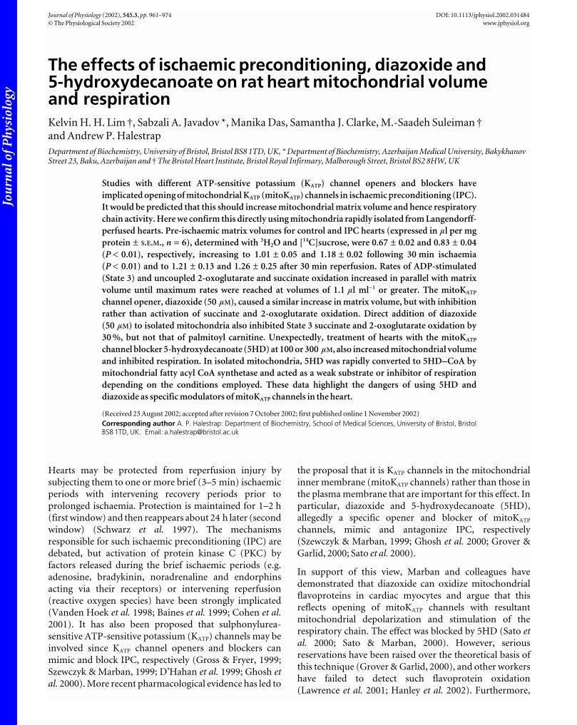
The effects of ischaemic preconditioning, diazoxide and5-hydroxydecanoate on rat heart mitochondrial volumeand respirationKelvin H. H. Lim †, Sabzali A. Javadov *, Manika Das, Samantha J. Clarke, M.-Saadeh Suleiman †and Andrew P. Halestrap
Department of Biochemistry, University of Bristol, Bristol BS8 1TD, UK, * Department of Biochemistry, Azerbaijan Medical University, BakykhanovStreet 23, Baku, Azerbaijan and † The Bristol Heart Institute, Bristol Royal Infirmary, Malborough Street, Bristol BS2 8HW, UK
Studies with different ATP-sensitive potassium (KATP) channel openers and blockers have
implicated opening of mitochondrial KATP (mitoKATP) channels in ischaemic preconditioning (IPC).
It would be predicted that this should increase mitochondrial matrix volume and hence respiratory
chain activity. Here we confirm this directly using mitochondria rapidly isolated from Langendorff-
perfused hearts. Pre-ischaemic matrix volumes for control and IPC hearts (expressed in ml per mg
protein ± S.E.M., n = 6), determined with 3H2O and [14C]sucrose, were 0.67 ± 0.02 and 0.83 ± 0.04
(P < 0.01), respectively, increasing to 1.01 ± 0.05 and 1.18 ± 0.02 following 30 min ischaemia
(P < 0.01) and to 1.21 ± 0.13 and 1.26 ± 0.25 after 30 min reperfusion. Rates of ADP-stimulated
(State 3) and uncoupled 2-oxoglutarate and succinate oxidation increased in parallel with matrix
volume until maximum rates were reached at volumes of 1.1 ml ml_1 or greater. The mitoKATP
channel opener, diazoxide (50 mM), caused a similar increase in matrix volume, but with inhibition
rather than activation of succinate and 2-oxoglutarate oxidation. Direct addition of diazoxide
(50 mM) to isolated mitochondria also inhibited State 3 succinate and 2-oxoglutarate oxidation by
30 %, but not that of palmitoyl carnitine. Unexpectedly, treatment of hearts with the mitoKATP
channel blocker 5-hydroxydecanoate (5HD) at 100 or 300 mM, also increased mitochondrial volume
and inhibited respiration. In isolated mitochondria, 5HD was rapidly converted to 5HD–CoA by
mitochondrial fatty acyl CoA synthetase and acted as a weak substrate or inhibitor of respiration
depending on the conditions employed. These data highlight the dangers of using 5HD and
diazoxide as specific modulators of mitoKATP channels in the heart.
(Received 23 August 2002; accepted after revision 7 October 2002; first published online 1 November 2002)
Corresponding author A. P. Halestrap: Department of Biochemistry, School of Medical Sciences, University of Bristol, BristolBS8 1TD, UK. Email: [email protected]
Journal of Physiology (2002), 545.3, pp. 961–974 DOI: 10.1113/jphysiol.2002.031484
© The Physiological Society 2002 www.jphysiol.org
Jou
rnal
of P
hysi
olog
y
the specificity of diazoxide for mitoKATP channels is in
doubt. Thus it was demonstrated more than 30 years ago
that diazoxide inhibits succinate dehydrogenase activity
with the result that the citric acid cycle is blocked and
mitochondrial flavoproteins become oxidized (Schäfer etal. 1971). These data have been confirmed more recently
(Grimmsman & Rustenbeck, 1998; Kowaltowski et al.2001; Hanley et al. 2002). In addition, diazoxide has been
shown to open the plasma membrane KATP channel at low
concentrations when ADP is present (as it will be in the
cell) (D’Hahan et al. 1999). Indeed, in mice lacking the
plasma membrane K+ channel, Kir6.2, IPC was ineffective
in reducing infarct size (Suzuki et al. 2002). Finally,
diazoxide has been reported to increase the mitochondrial
production of reactive oxygen species (ROS) by an
unknown mechanism (Pain et al. 2000; Liu & O’Rourke,
2001; Forbes et al. 2001), and ROS have been implicated in
IPC (Baines et al. 1997; Vanden Hoek et al. 1998). The use
of 5HD as a specific mitochondrial KATP channel inhibitor
is also open to question since it is a racemic mix of D- and
L-isomers of a substituted fatty acid. As such it has the
potential to be activated to its coenzyme A derivative, as
has recently been demonstrated (Hanley et al. 2002),
which itself might exhibit a range of metabolic and other
effects on the cell.
In order to confirm a role for mitoKATP channel opening in
IPC, more direct evidence is desirable and in this paper we
address this by measuring the matrix volume of
mitochondria rapidly isolated from the heart. It is
generally accepted that opening of mitoKATP channels will
cause an increase in matrix volume and that this in turn
will activate the respiratory chain, providing more ATP to
support the recovering heart (Halestrap, 1989; Grover &
Garlid, 2000; O’Rourke, 2000). We confirm that both
effects are observed in mitochondria rapidly isolated from
hearts subjected to IPC. However, in agreement with
others, diazoxide and 5HD were found to have additional
effects on mitochondrial function that may undermine
their use as specific mitoKATP channel openers and
blockers in vivo.
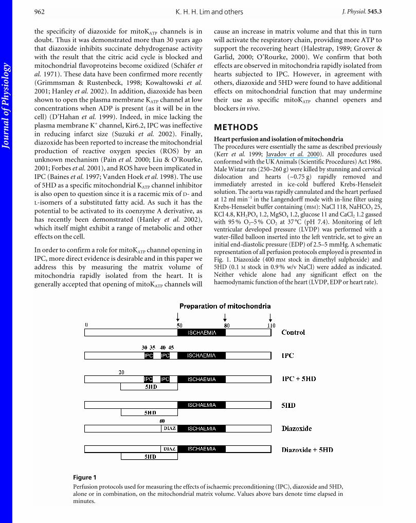
METHODS Heart perfusion and isolation of mitochondriaThe procedures were essentially the same as described previously(Kerr et al. 1999; Javadov et al. 2000). All procedures usedconformed with the UK Animals (Scientific Procedures) Act 1986.Male Wistar rats (250–260 g) were killed by stunning and cervicaldislocation and hearts (~0.75 g) rapidly removed andimmediately arrested in ice-cold buffered Krebs-Henseleitsolution. The aorta was rapidly cannulated and the heart perfusedat 12 ml min_1 in the Langendorff mode with in-line filter usingKrebs-Henseleit buffer containing (mM): NaCl 118, NaHCO3 25,KCl 4.8, KH2PO4 1.2, MgSO4 1.2, glucose 11 and CaCl2 1.2 gassedwith 95 % O2–5 % CO2 at 37 °C (pH 7.4). Monitoring of leftventricular developed pressure (LVDP) was performed with awater-filled balloon inserted into the left ventricle, set to give aninitial end-diastolic pressure (EDP) of 2.5–5 mmHg. A schematicrepresentation of all perfusion protocols employed is presented inFig. 1. Diazoxide (400 mM stock in dimethyl sulphoxide) and5HD (0.1 M stock in 0.9 % w/v NaCl) were added as indicated.Neither vehicle alone had any significant effect on thehaemodynamic function of the heart (LVDP, EDP or heart rate).
K. H. H. Lim and others962 J. Physiol. 545.3
Figure 1Perfusion protocols used for measuring the effects of ischaemic preconditioning (IPC), diazoxide and 5HD,alone or in combination, on the mitochondrial matrix volume. Values above bars denote time elapsed inminutes.
Jou
rnal
of P
hysi
olog
y
Measurement of mitochondrial matrix volumeAt defined stages during the perfusion protocol (see Fig. 1)ventricles were rapidly cut away, weighed, and homogenized witha Polytron homogenizer at setting 3 for 5 s in 5 ml ice-cold sucrosebuffer (mM): sucrose 300, Tris–HCl 10, EGTA 2; pH 7.4).Following homogenization, sucrose buffer containing 5 mg ml_1
bovine serum albumin (BSA) was added to give a final volume of40 ml and the homogenate centrifuged for 2 min at 2000 g and4 °C to sediment cell debris. The supernatant was then centrifugedat 10 000 g for 5 min (4 °C) to sediment a crude mitochondrialpellet which was resuspended in 4.5 ml of ice-cold sucrose buffercontaining 4.5 mCi 3H2O and 0.45 mCi [14C]-sucrose and dividedequally between four microcentrifuge tubes. Followingcentrifugation (14 000 r.p.m.) for 1 min at 4 °C, supernatants werecarefully transferred to tubes containing 100 ml 20 % (w/v)perchloric acid (PCA), protein sedimented by centrifugation anda 100 ml sample assayed for 3H/14C by scintillation counting usinga Packard 1600TR counter. One mitochondrial pellet wasresuspended in 100 ml sucrose buffer and used for measurementof respiration rates, protein and citrate synthase activity asoutlined below. The other three pellets were resuspended in 500 mlof 0.1 M KH2PO4 (pH 7.0) containing 1 % (w/v) Triton X-100 andsamples (100 ml) of each removed for the assay of proteinconcentration (Bradford reagent) and citrate synthase activity(spectrophotometrically) as decribed previously (Griffiths &Halestrap, 1995). The remaining mitochondrial extracts weredeproteinated with 20 % (w/v) PCA and the supernatants assayedfor 3H/14C. In order to resolve small differences (< 10 %) in matrixvolumes, all samples were subjected to three cycles of countingand great care was taken to avoid evaporation of 3H2O. Theprocedure used for calculating matrix volume from the 3H and 14Cd.p.m. was that described previously (Halestrap & McGivan,1979; Halestrap, 1989). Volumes were expressed as microlitresper milligram mitochondrial protein, the latter being determinedfrom the citrate synthase content of the pellet assuming 2.36 unitsper milligram pure mitochondrial protein (see Table 1).
Measurement of mitochondrial respiration was performed at30 °C in a Clark-type oxygen electrode as described previously(Javadov et al. 2000). The buffer (at 30 °C, pH 7.2) contained(mM): KCl 125, MOPS 20, Tris 10, EGTA 0.5, KH2PO4 2.5, MgCl2
2.5, and was supplemented with the required substrate (5 mM
2-oxoglutarate + 1 mM L-malate, 50 mM palmitoyl carnitine +1 mM L-malate or 5 mM succinate + 1 mM rotenone). Unlessotherwise stated, rates of respiration were routinely measured inthe absence (State 2) and presence of 1 mM ADP (State 3) or0.1 mM carbonylcyanide-p-trifluoromethoxy-phenylhydrazone(FCCP), an uncoupler. When required, 1 mM antimycin A wasadded to terminate substrate oxidation and then respirationrestarted by addition of 10 mM ascorbate + 0.3 mM N,N,N‚N ‚-tetramethyl-p-phenylendiamine (TMPD).
Measurement of fatty acyl CoA synthetase activityPercoll purified mitochondria (Halestrap, 1987) were added at0.3 mg protein ml_1 to 7 ml oxygen electrode medium supple-mented with 0.2 mM rotenone, 1 mM antimycin, 1 mM oligomycin,0.2 mM KCN, 1 mM ATP, 1 mM phosphoenolpyruvate, 0.1 mM
NADH and 1 unit ml_1 each of pyruvate kinase, adenylate kinaseand lactate dehydrogenase. Samples (3.5 ml) were placed in thesample and reference cuvettes of a split beam spectrophotometer,maintained at 30 °C, and A340 monitored to follow NADHoxidation. Additions of 0.1 mM coenzyme A and 5HD or otherfatty acid were made to the sample cuvette as required.
Statistical analysisData are expressed as mean ± S.E.M. Differences between groupsof hearts subject to different treatments were calculated byANOVA with multiple comparisons using Fisher’s PLSD post hoctest. Where the effects of agents were studied on isolatedmitochondria, significance was determined by Student’s paired ttest. Differences were considered to be significant when P < 0.05.
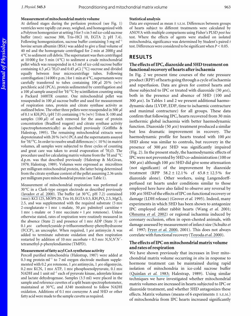
RESULTSThe effects of IPC, diazoxide and 5HD treatment onfunctional recovery of hearts after ischaemiaIn Fig. 2 we present time courses of the rate pressure
product (RPP) of hearts going through a cycle of ischaemia
and reperfusion. Data are given for control hearts and
those subjected to IPC or treated with diazoxide (50 mM),
both in the presence and absence of 5HD (100 and
300 mM). In Tables 1 and 2 we present additional haemo-
dynamic data (LVDP, EDP, time to ischaemic contracture
and maximal contracture) for all groups. These data
confirm that following IPC, hearts recovered from 30 min
isothermic global ischaemia with better haemodynamic
function. Diazoxide (50 mM) treatment produced a similar
but less dramatic improvement in recovery. The
haemodynamic profile for hearts treated with 100 mM
5HD alone was similar to controls, but recovery in the
presence of 300 mM 5HD was significantly impaired
(Fig. 2). In the present study, the haemodynamic effects of
IPC were not prevented by 5HD co-administration (100 or
300 mM) although 100 mM 5HD did give some attenuation
(not significant) of the improvement by diazoxide
treatment (RPP 58.2 ± 12.1 % cf 65.8 ± 12.5 % for
diazoxide alone). Other workers, using Langendorff-
perfused rat hearts under conditions similar to those
employed here have also failed to observe any reversal by
100 mM 5HD of the effects of IPC on functional recovery or
damage (LDH release) (Grover et al. 1995). Indeed, many
experiments in which 5HD has been shown to antagonize
IPC employed either rabbit hearts (Wang et al. 2001;
Ohnuma et al. 2002) or regional ischaemia induced by
coronary occlusion, often in open-chested animals, with
damage assessed by measurement of infarct size (Schultz etal. 1997; Fryer et al. 2000, 2001). This does not always
correlate with functional recovery (Toyoda et al. 2000).
The effects of IPC on mitochondrial matrix volumeand rates of respirationWe have shown previously that increases in liver mito-
chondrial matrix volume occurring in situ in response to
hormone treatment can be maintained during rapid
isolation of mitochondria in ice-cold sucrose buffer
(Quinlan et al. 1983; Halestrap, 1989). Using similar
techniques we have investigated whether mitochondrial
matrix volumes are increased in hearts subjected to IPC or
diazoxide treatment, and whether 5HD antagonizes these
effects. Matrix volumes (means of 6 experiments ± S.E.M.)
of mitochondria from IPC hearts increased significantly
Preconditioning and mitochondrial matrix volumeJ. Physiol. 545.3 963
Jou
rnal
of P
hysi
olog
y
from 0.67 ± 0.02 to 0.83 ± 0.04 ml (mg protein)_1 (P < 0.01)
prior to the ischaemic period and from 1.01 ± 0.05 to
1.18 ± 0.02 ml (mg protein)_1 (P < 0.02) at the end of the
ischaemic period. Following reperfusion matrix volumes
increased further to 1.21 ± 0.13 and 1.26 ± 0.25 ml (mg
protein)_1, respectively. The larger errors on the latter
values may reflect a higher and variable proportion of
damaged mitochondria following reperfusion as indicated
by the decrease in specific activity of citrate synthase
(Table 1). It should be noted that the mitochondrial
protein concentrations used to calculate matrix volumes
were determined from the citrate synthase activity of the
mitochondrial pellet. This procedure eliminates errors
caused by the presence of broken mitochondria in the
crude mitochondrial pellet that would be permeable to
[14C]sucrose. We conclude from these results that IPC
does cause an increase in matrix volume that is maintained
throughout the prolonged ischaemic phase, but not
during reperfusion. However, it appears that a 30 min
ischaemic period can induce a significant increase in
matrix volume in its own right.
Our previous work (Halestrap, 1987) would suggest that
such increases in matrix volume should be associated with
an increase of both State 3 (ADP-stimulated) and
uncoupler-stimulated oxidation of any substrate donating
electrons to the respiratory chain prior to Complex 3
including succinate and NADH-producing substrates
such as 2-oxoglutarate plus malate. In Fig. 3 we present
data that confirm this to be the case for State 3 respiration
and identical results were obtained when respiration was
stimulated by addition of 0.1 mM FCCP, an uncoupler
(data not shown). Rates of respiration are expressed as a
ratio relative to the rate of ascorbate + TMPD oxidation in
the same incubation. The rationale behind this is that
ascorbate oxidation is insensitive to matrix volume
(Halestrap, 1987) and hence using this ratio provides a
correction for any changes in respiratory chain activity
(such as cytochrome c loss) that are independent of matrix
K. H. H. Lim and others964 J. Physiol. 545.3
Jou
rnal
of P
hysi
olog
yPreconditioning and mitochondrial matrix volumeJ. Physiol. 545.3 965
Figure 2. The effects of ischaemic preconditioning, diazoxide and 5HD, alone or incombination, on the rate pressure product of hearts subject to 30 min global ischaemiafollowed by reperfusionData are presented as means ± S.E.M. (error bars) of 6–8 separate hearts. Full functional data for pre-ischaemic, end-ischaemic and reperfused hearts are given in Table 2. Horizontal bars indicate thecomposition of the perfusion medium: KHS, Krebs-Henseleit buffer alone; 5HD, KHS containing 5HD at100 or 300 µM (5HD100 or 5HD300); Diaz, KHS containing 50 µM diazoxide.
Jou
rnal
of P
hysi
olog
y
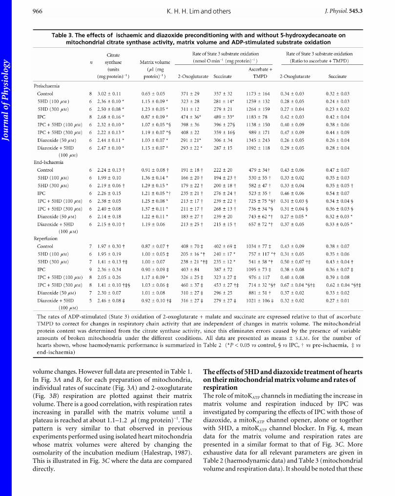
volume changes. However full data are presented in Table 1.
In Fig. 3A and B, for each preparation of mitochondria,
individual rates of succinate (Fig. 3A) and 2-oxoglutarate
(Fig. 3B) respiration are plotted against their matrix
volume. There is a good correlation, with respiration rates
increasing in parallel with the matrix volume until a
plateau is reached at about 1.1–1.2 ml (mg protein)_1. The
pattern is very similar to that observed in previous
experiments performed using isolated heart mitochondria
whose matrix volumes were altered by changing the
osmolarity of the incubation medium (Halestrap, 1987).
This is illustrated in Fig. 3C where the data are compared
directly.
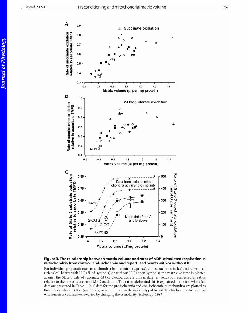
The effects of 5HD and diazoxide treatment of heartson their mitochondrial matrix volume and rates ofrespirationThe role of mitoKATP channels in mediating the increase in
matrix volume and respiration induced by IPC was
investigated by comparing the effects of IPC with those of
diazoxide, a mitoKATP channel opener, alone or together
with 5HD, a mitoKATP channel blocker. In Fig. 4, mean
data for the matrix volume and respiration rates are
presented in a similar format to that of Fig. 3C. More
exhaustive data for all relevant parameters are given in
Table 2 (haemodynamic data) and Table 3 (mitochondrial
volume and respiration data). It should be noted that these
K. H. H. Lim and others966 J. Physiol. 545.3
Jou
rnal
of P
hysi
olog
yPreconditioning and mitochondrial matrix volumeJ. Physiol. 545.3 967
Figure 3. The relationship between matrix volume and rates of ADP-stimulated respiration inmitochondria from control, end-ischaemia and reperfused hearts with or without IPCFor individual preparations of mitochondria from control (squares), end ischaemia (circles) and reperfused(triangles) hearts with IPC (filled symbols) or without IPC (open symbols) the matrix volume is plottedagainst the State 3 rate of succinate (A) or 2-oxoglutarate plus malate (B) oxidation expressed as ratiosrelative to the rate of ascorbate TMPD oxidation. The rationale behind this is explained in the text whilst fulldata are presented in Table 1. In C data for the pre-ischaemia and end-ischaemia mitochondria are plotted astheir mean values ± S.E.M. (error bars) in conjunction with previously published data for heart mitochondriawhose matrix volumes were varied by changing the osmolarity (Halestrap, 1987).
Jou
rnal
of P
hysi
olog
y
experiments were performed 12 months after the
experiments reported in Fig. 3 and thus an additional set of
control and IPC hearts were employed, although the two
sets of data were essentially the same. As would be
predicted for a mitoKATP channel opener, diazoxide caused
the matrix volume to increase to a similar extent to IPC in
both pre-ischaemic and end ischaemic hearts as indicated
by the dotted arrows in Fig. 4. However, in contrast to IPC,
diazoxide decreased rather than increased the rate of
succinate and 2-oxoglutarate oxidation. This probably
reflects a direct effect of diazoxide on mitochondrial
respiration as outlined below. Also, contrary to
expectation, the mitoKATP channel blocker 5HD induced a
significant increase in matrix volume, whether added
alone or in conjunction with IPC or diazoxide treatment.
Similar results were obtained for mitochondria isolated
from hearts before ischaemia or at the end of ischaemia.
This increase in matrix volume was usually accompanied
by a decrease in respiration rate, similar to that observed
with diazoxide. Our data suggest that in the heart, 5HD is
having additional effects on mitochondrial function,
independent of any effects on the mitoKATP channel. A
similar conclusion has been reached by others (Hanley etal. 2002). This deleterious action of 5HD was also reflected
in a significant reduction in the rate of ascorbate + TMPD
K. H. H. Lim and others968 J. Physiol. 545.3
Figure 4. The relationship between matrix volume and rates of ADP-stimulated respirationby mitochondria isolated from control, IPC, diazoxide- and 5HD–treated heartsData are presented as in Fig. 3 and as means ± S.E.M. (error bars) of 6–8 hearts for each treatment as indicated.The arrows indicate the distinctive effects of IPC and diazoxide. Full data are presented in Table 3 withparallel haemodynamic data in Table 2.
Jou
rnal
of P
hysi
olog
y
oxidation by mitochondria isolated from 5HD-treated
hearts following reperfusion, from a control value of
1034 ± 77 nmol O (mg protein)_1 min_1 to 757 ± 117
(P < 0.05) and 541 ± 58 (P < 0.01) for 100 mM and 300 mM
5HD-treated hearts respectively. In the IPC hearts the
deleterious effects of 5HD were still present, but less
dramatic (1095 ± 73, 976 ± 117 and 714 ± 32 (P < 0.05)
nmol O (mg protein)_1 min_1, respectively). The inhibitory
effect of 5HD treatment on ascorbate oxidation is most
likely to reflect breakage of the outer mitochondrial
membrane and release of cytochrome c, either in situ or
during isolation.
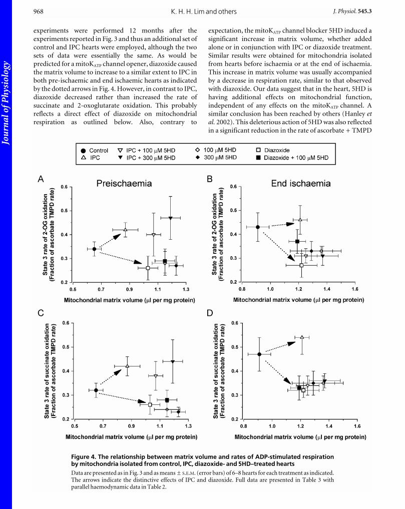
Direct effects of 5HD and diazoxide onmitochondrial respirationSince diazoxide can inhibit succinate dehydrogenase
activity (Schäfer et al. 1971; Grimmsman & Rustenbeck,
1998; Ovide Bordeaux et al. 2000; Kowaltowski et al. 2001;
Hanley et al. 2002), we investigated the effects on substrate
oxidation of adding of either diazoxide or 5HD directly to
isolated heart mitochondria. Data are presented in Fig. 5A.
At 50 mM diazoxide inhibited State 3 rates of succinate and
2-oxoglutarate + malate oxidation by 32.1 ± 3.7 and
28.2 ± 3.9 %, respectively (P < 0.01) whilst having little if
any effect on the oxidation of glutamate + malate (8.0 ±
3.9 %) or palmitoyl carnitine + malate (4.5 ± 2.8 %).
These results are consistent with inhibition of succinate
dehydrogenase since oxidation of 2-oxoglutarate by heart
mitochondria is likely to proceed via 2-oxoglutarate
dehydrogenase to succinate and then succinate dehydrog-
enase. In contrast, succinate dehydrogenase is not
involved to any great extent in the oxidation of either
palmitoyl carnitine + malate or glutamate + malate. In
the latter case, oxaloacetate formed from oxidation of
malate is transaminated to aspartate that leaves the mito-
chondria in exchange for glutamate.
At 100 and 300 mM, 5HD had little effect on the oxidation
of any substrate, but higher concentrations inhibited
oxidation of all substrates as illustrated for palmitoyl
carnitine in Fig. 5A. It has recently been demonstrated that
5HD, which is a substituted fatty acid can be activated to
5HD–CoA by purified acyl-CoA synthetase (Hanley et al.2002), which then has the potential to be either a substrate
or an inhibitor of the b-oxidation pathway. After one
round of b-oxidation 5HD–CoA would form 3-hydroxy-
octanoyl-CoA whose L-isomer is a substrate forb-oxidation (Eaton et al. 1996). Commercial 5HD is a
racemic mix and thus might be expected to contain both
the D- and L-isomers, with the potential for metabolites of
the D-isomer acting as inhibitors of b-oxidation. In the
presence of 0.2 mM L-malate, addition of 100 mM 5HD and
decanoate to liver mitochondria had no effect on
respiration in the absence of ADP (data not shown) but
stimulated State 3 respiration by 53 ± 10 % (P < 0.01) and
67 ± 24 % (P < 0.05) respectively, as illustrated in Fig. 5B.
Preconditioning and mitochondrial matrix volumeJ. Physiol. 545.3 969
Figure 5. The effects of diazoxide and 5HD on respirationby isolated heart mitochondriaIn A rates of ADP-stimulated respiration in the presence of 5 mM
succinate + 1 mM rotenone, 5 mM 2-oxoglutarate + 1 mM
L-malate, 5 mM L-glutamate + 1 mM L-malate or 50 mM palmitoylcarnitine + 1 mM L-malate were measured in the presence andabsence of diazoxide or 5HD at the concentrations shown. Data arepresented as the mean percentage inhibition of respiration cause bythe reagent ± S.E.M. (error bars) of 6 separate mitochondrialpreparations. In B rates of oxygen uptake by isolated heart and livermitochondria were measured in the presence of 0.2 mM L-malatesupplemented with 200 mM 5HD or decanoate as indicated. In thethree left bars, respiration was stimulated by addition of 0.4 mM
ADP whilst in the three right bars 1 mM ATP, 1 mM oligomycin,0.1 mM coenzyme A and 1 mM L-carnitine were added to enableextramitochondrial activation of the fatty acid (see Fig. 6) andoxidation was initiated by addition of uncoupler (0.2 mM FCCP).Data are presented as means ± S.E.M. (error bars) of 3–6 separatemitochondrial preparations. Significant increases or decreases inrespiration are indicated (*P < 0.05; **P < 0.01).
Jou
rnal
of P
hysi
olog
y
In contrast, with heart mitochondria the stimulation by
5HD was not significant (18 ± 8 %) whilst that by
decanoate was greater than for liver mitochondria
(218 ± 79 %; P < 0.05). These data suggest that in liver
mitochondria both 5HD and decanoate can enter the
mitochondria and be oxidized, albeit slowly, whereas in
heart mitochondria, only decanoate acts in this way.
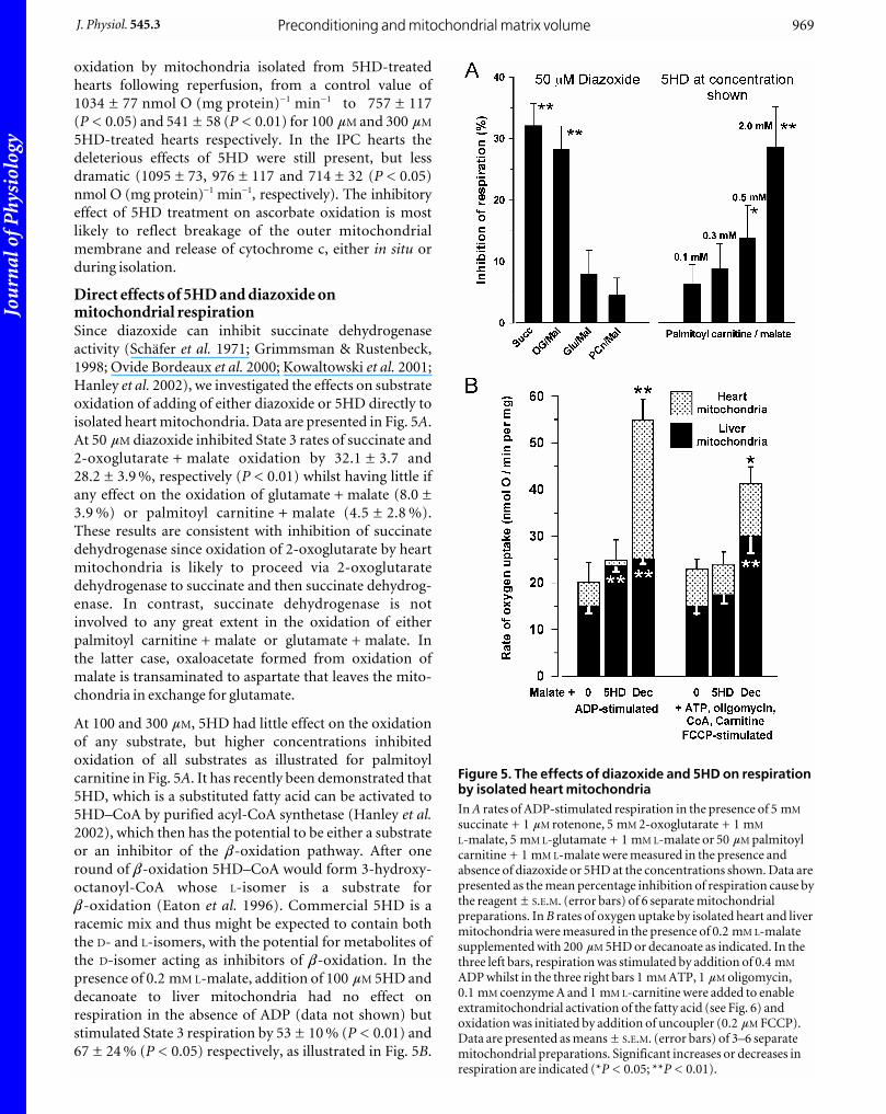
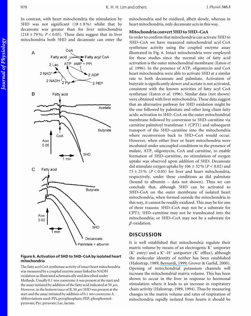
Mitochondria convert 5HD to 5HD–CoAIn order to confirm that mitochondria can activate 5HD to
5HD–CoA we have measured mitochondrial acyl-CoA
synthetase activity using the coupled enzyme assay
illustrated in Fig. 6. Intact mitochondria were employed
for these studies since the normal site of fatty acid
activation is the outer mitochondrial membrane (Eaton etal. 1996). In the presence of ATP, oligomycin and CoA
heart mitochondria were able to activate 5HD at a similar
rate to both decanoate and palmitate. Activation of
butyrate is significantly slower and acetate is not activated,
consistent with the known activities of fatty acyl CoA
synthetase (Eaton et al. 1996). Similar data (not shown)
were obtained with liver mitochondria. These data suggest
that an alternative pathway for 5HD oxidation might be
the one followed by palmitate and other long chain fatty
acids: activation to 5HD–CoA on the outer mitochondrial
membrane followed by conversion to 5HD–carnitine via
carnitine palmitoyl transferase 1 (CPT1) and subsequent
transport of the 5HD–carnitine into the mitochondria
where reconversion back to 5HD–CoA would occur.
However, when either liver or heart mitochondria were
incubated under uncoupled conditions in the presence of
malate, ATP, oligomycin, CoA and carnitine, to enable
formation of 5HD–carnitine, no stimulation of oxygen
uptake was observed upon addition of 5HD. Decanoate
did stimulate oxygen uptake by 106 ± 32 % (P < 0.02) and
75 ± 25 % (P < 0.05) for liver and heart mitochondria,
respectively, under these conditions as did palmitate
(bound to albumin – data not shown). Thus we can
conclude that, although 5HD can be activated to
5HD–CoA on the outer membrane of isolated heart
mitochondria, when formed outside the mitochondria in
this way, it cannot be readily oxidized. This may be for one
of three reasons: 5HD–CoA may not be a substrate for
CPT1; 5HD–carnitine may not be translocated into the
mitochondria; or 5HD–CoA may not be a substrate forb-oxidation.
DISCUSSIONIt is well established that mitochondria regulate their
matrix volume by means of an electrogenic K+ uniporter
(K+ entry) and a K+–H+ antiporter (K+ efflux), although
the molecular identity of neither has been established
(Halestrap, 1989; Bernardi, 1999; Grover & Garlid, 2000).
Opening of mitochondrial potassium channels will
increase the mitochondrial matrix volume. This has been
shown to occur in the liver in response to hormonal
stimulation where it leads to an increase in respiratory
chain activity (Halestrap, 1989, 1994). Thus by measuring
changes in the matrix volume and rates of respiration of
mitochondria rapidly isolated from hearts it should be
K. H. H. Lim and others970 J. Physiol. 545.3
Figure 6. Activation of 5HD to 5HD–CoA by isolated heartmitochondria
The fatty acyl CoA synthetase activity of intact heart mitochondria
was measured by a coupled enzyme assay linked to NADH
oxidation as illustrated schematically and described under
Methods. Usually 0.1 mM coenzyme A was present at the start and
the assay initiated by addition of the fatty acid indicated at 50 mM.
However, in the bottom trace of B, 50 mM 5HD was present at the
start and the assay initiated by addition of 0.1 mM coenzyme A.
Abbreviations used: PPi, pyrophosphate; PEP, phosphoenol-
pyruvate; Pyr, pyruvate; Lac, lactate.
Jou
rnal
of P
hysi
olog
y
possible to confirm whether or not mitochondrial
potassium channels have been opened in response to IPC.
The data we present in this paper are consistent with such
an opening of these channels (Fig. 3). Changes in cytosolic
osmolarity are unlikely to account for the observed
volume changes; nor are changes in anion fluxes since
phosphate anions can follow K+ ions without rate
limitation (Halestrap, 1989; Bernardi, 1999). However, on
their own, these data do not identify the potassium
channels that are opened as mitoKATP channels. For this
purpose we chose to use diazoxide and 5HD, the agents
commonly employed by others to specifically open and
block mitoKATP channels respectively. However, the data
we obtained provide further evidence that these agents
have other effects on the heart unrelated to their effects on
the mitoKATP channel.
Limitations of the technique used for measuringchanges in mitochondrial volumeIt would be preferable to measure the matrix volume of the
mitochondria in situ, without cell disruption, but
unfortunately methods are not available to do this. The
possibility of using electron microscopy of sections from
perfused hearts or confocal microscopy of isolated heart
cells was considered. However, the cubic relationship
between mitochondrial diameter and volume (a crude
approximation that assumes mitochondria to be a perfect
sphere) means that a 25 % increase in matrix volume
would only be reflected in a 3 % increase in diameter. This
is well below the limits of detection (Egner et al. 2002).
Furthermore, morphological changes in mitochondria
that may affect their diameter, do not necessarily reflect
changes in matrix volume, but rather may be the result of
changes in the intermembrane space or arrangement of the
cristae (Frey et al. 2002). Our methodology assumes that
the rapid isolation of mitochondria leads to little or no
change in their matrix volume, and that what change there
may be still allows differences between control and
experimental groups to be maintained. Although we
cannot prove this to be the case experimentally, there is
considerable evidence that implies it to be a valid
assumption. First, in liver cells, where we have devised
methods of measuring mitochondrial volume in situ,
changes measured by these techniques correlate with those
measured by rapid mitochondrial isolation (Quinlan et al.1983). Second, we have measured the mitochondrial
volume immediately upon isolation and after 5 min
incubation in KCl medium under energized conditions, to
mimic the situation in vivo. In three separate experiments
such incubation gave no significant increase in the matrix
volume (mean increase ± S.E.M. of 6.5 ± 5.2 %). This
implies that isolation of heart mitochondria in sucrose
buffer causes little or no loss of potassium ions that could
lead to a decrease in the matrix volume (Halestrap, 1989;
Grover & Garlid, 2000; O’ Rourke, 2000).
Diazoxide and 5HD treatments have effects onmitochondrial matrix volume and respirationindependent of their action on mitoKATP channelsIn contrast to IPC, diazoxide treatment of hearts led to a
decrease in respiratory chain activity despite an increase in
matrix volume (Fig. 4). The most probable explanation for
this is an inhibition of succinate dehydrogenase by
diazoxide that has been known for many years (Schäfer etal. 1971; Grimmsman & Rustenbeck, 1998; Kowaltowski etal. 2001; Hanley et al. 2002) and is confirmed by the data of
Fig. 5A. Contrary to expectation for a mitoKATP channel
blocker, 5HD treatment of hearts, alone or in conjunction
with IPC or diazoxide, also led to an increase in
mitochondrial matrix volume. This was accompanied by
an inhibition of respiration, suggesting that 5HD
treatment, like diazoxide, was having additional effects on
mitochondrial function. The data we present in Fig. 6
demonstrate that 5HD added to heart mitochondria is
converted to extramitochondrial D- and L-5HD–CoA.
However, the oxygen electrode studies show that this
cannot enter the mitochondria to be oxidized, although a
small amount of 5HD may be activated to 5HD–CoA
within the matrix and support slow rates of respiration,
especially in liver mitochondria (Fig. 5B). An
accumulation of 5HD–CoA within the cytosol is likely to
have a range of effects on the cell that may account for the
observed changes in mitochondrial matrix volume and
respiration we observe. Thus fatty acyl CoA derivatives are
known to bind to the adenine nucleotide translocase,
inhibiting its activity and hence oxidative
phosphorylation. In addition, 5HD–CoA, like other fatty
acyl CoAs, may bind tightly to cardiac acetyl–CoA
carboxylase, inhibiting its activity. This will cause
concentrations of malonyl–CoA to decrease and hence
CPT1 activity to increase leading to a stimulation of
endogenous fatty acid oxidation (Eaton, 2002). This is
known to be detrimental for post-ischaemic recovery
(Lopaschuk, 1997). Indeed, our data (Fig. 2 and Table 2)
suggest that higher concentrations of 5HD (300 mM) may
have a detrimental effect on the recovery of control hearts
from ischaemia. This is reflected in the properties of the
mitochondria isolated from such hearts which
demonstrate an inhibition of ascorbate oxidation that is
suggestive of mitochondrial outer membrane rupture and
cytochrome c loss (Table 3). Unfortunately, many of the
published results on the effects of 5HD on IPC and
diazoxide preconditioning do not include data for the
effects of 5HD alone on control hearts. Nevertheless, there
is some published evidence that these concentrations of
5HD can exacerbate postischaemic damage in a
subpopulation of hearts (Munch-Ellingsen et al. 2000).
Another indication that 5HD may have additional effects
on the heart, perhaps via its metabolism, is that when
exposed to 300 mM 5HD, a slight but significant (P < 0.05)
rise in LVDP was consistently observed during the pre-
ischaemic intervention phase (6.6 ± 2.0 % rise when
Preconditioning and mitochondrial matrix volumeJ. Physiol. 545.3 971
Jou
rnal
of P
hysi
olog
y
measured 10 min after its introduction) and this was
maintained until ischaemia, even in preconditioned
hearts. No similar effect was observed in the absence of
5HD (0.9 ±1.3 % fall) or in hearts treated with 100 mM
5HD (0.2 ± 1.4 % rise).
ConclusionsThree important conclusions can be drawn from the
discussion above. First, if opening of the mitoKATP channel
is important for the mechanism of IPC, matrix volume-
mediated activation of the respiratory chain and oxidative
phosphorylation is unlikely to be the major mechanism by
which this induces protection, since protection by
diazoxide is associated with inhibition of respiration.
Second, our data provide a strong warning against the
uncritical use of diazoxide and 5HD as indicators of the
involvement of mitoKATP channels in preconditioning.
These agents could be exerting their effects to mimic and
antagonize IPC through alternative pathways involving
5HD metabolism and diazoxide inhibition of succinate
dehydrogenase. The ability of 3-nitropropionic acid, a well
characterized succinate dehydrogenase inhibitor, to
induce preconditioning (Ockaili et al. 2001) would support
this view and more recently pinacidil, another mitoKATP
channel opener that confers preconditioning, has been
shown to inhibit NADH oxidation by submitochondrial
particles (Hanley et al. 2002). Diazoxide, like IPC, is
known to increase reactive oxygen species and these
appear to be essential for mediating the protective effects
(Baines et al. 1997; Vanden Hoek et al. 1998; Pain et al.2000; Forbes et al. 2001). The production of such free
radicals has been proposed to be downstream of mitoKATP
channel opening (Gross & Fryer, 2000; Pain et al. 2000; Liu
& O’Rourke, 2001; Patel & Gross, 2001). However, it is
known that the mitochondrial respiratory chain is a major
source of ROS, and since ischaemia and diazoxide both
inhibit respiration, it may be that it is this locus of action
rather than opening of the mitoKATP channel that is
important. Third, mechanisms other than opening of the
mitoKATP channel may cause changes in matrix volume in
the heart as discussed further below.
There is an extensive literature to support the presence of a
regulated mechanism for K+ entry into the mitochondria
that is not mediated by a sulphonylurea-sensitive mitoKATP
channel but is inhibited by matrix ATP (Halestrap, 1989;
Bernardi, 1999). We have previously provided strong
evidence that this K+ influx is mediated by the adenine
nucleotide translocase (ANT) (Halestrap, 1989). Dis-
placement of ADP or ATP from their binding sites on the
ANT by pyrophosphate (PPi) or phosphate makes the
ANT leaky to K+ ions which are driven into the matrix by
the membrane potential (Davidson & Halestrap, 1987).
This channel will be activated when matrix adenine
nucleotides are depleted and when intracellular Pi and
[Ca2+] are elevated, exactly the conditions that occur in
ischaemia (Halestrap, 1989). As such it could account for
both the increase in matrix volume that occurs during
ischaemic preconditioning and during the prolonged
ischaemic episode. Indeed, any intervention that inhibits
mitochondrial respiration might be expected to exert a
similar effect and so might provide an alternative
explanation for the effects of diazoxide. The unexpected
increase in matrix volume mediated by 5HD treatment of
hearts may also be explained through an action of
5HD–CoA on the ANT. Fatty acyl CoA derivatives are
known to displace adenine nucleotides from the ANT
(Devaux et al. 1975) and thus might also stimulate entry of
K+ into the matrix and so increase matrix volume.
REFERENCESBAINES, C. P., COHEN, M. V. & DOWNEY, J. M. (1999). Signal
transduction in ischemic preconditioning: The role of kinases and
mitochondrial K-ATP channels. Journal of CardiovascularElectrophysiology 10, 741–754.
BAINES, C. P., GOTO, M. & DOWNEY, J. M. (1997). Oxygen radicals
released during ischemic preconditioning contribute to
cardioprotection in the rabbit myocardium. Journal of Molecularand Cellular Cardiology 29, 207–216.
BERNARDI, P. (1999). Mitochondrial transport of cations: Channels,
exchangers, and permeability transition. Physiological Reviews 79,
1127–1155.
COHEN, M. V., YANG, X. M., LIU, G. S., HEUSCH, G. & DOWNEY, J. M.
(2001). Acetylcholine, bradykinin, opioids, and phenylephrine,
but not adenosine, trigger preconditioning by generating free
radicals and opening mitochondrial KATP channels. CirculationResearch 89, 273–278.
DAVIDSON, A. M. & HALESTRAP, A. P. (1987). Liver mitochondrial
pyrophosphate concentration is increased by Ca2+ and regulates
the intramitochondrial volume and adenine nucleotide content.
Biochemical Journal 246, 715–723.
DEVAUX, P. F., BIENVENUE, A., LAUQUIN, G., BRISSON, A. D., VIGNAIS,
P. M. & VIGNAIS, P. V. (1975). Interaction between spin-labeled
acyl-coenzyme A and the mitochondrial adenosine diphosphate
carrier. Biochemistry 14, 1272–1280.
D’HAHAN, N., MOREAU, C., PROST, A. L., JACQUET, H., ALEKSEEV, A. E.,
TERZIC, A. & VIVAUDOU, M. (1999). Pharmacological plasticity of
cardiac ATP-sensitive potassium channels toward diazoxide
revealed by ADP. Proceedings of the National Academy of Sciences ofthe USA 96, 12162–12167.
EATON, S. (2002). Control of mitochondrial beta-oxidation flux.
Progress in Lipid Research 41, 197–239.
EATON, S., BARTLETT, K. & POURFARZAM, M. (1996). Mammalian
mitochondrial beta-oxidation. Biochemical Journal 320, 345–357.
EGNER, A., JAKOBS, S. & HELL, S. W. (2002). Fast 100-nm resolution
three-dimensional microscope reveals structural plasticity of
mitochondria in live yeast. Proceedings of the National Academy ofSciences of the USA 99, 3370–3375.
FORBES, R. A., STEENBERGEN, C. & MURPHY, E. (2001). Diazoxide-
induced cardioprotection requires signaling through a redox-
sensitive mechanism. Circulation Research 88, 802–809.
FREY, T., RENKEN, C. & PERKINS, G. (2002). Insight into
mitochondrial structure and function from electron tomography.
Biochimica et Biophysica Acta 1555, 196–203.
K. H. H. Lim and others972 J. Physiol. 545.3
Jou
rnal
of P
hysi
olog
y
FRYER, R. M., EELLS, J. T., HSU, A. K., HENRY, M. M. & GROSS, G. J.
(2000). Ischemic preconditioning in rats: role of mitochondrial
K(ATP) channel in preservation of mitochondrial function.
American Journal of Physiology – Heart and Circulatory Physiology278, H305–312.
FRYER, R. M., HSU, A. K. & GROSS, G. J. (2001). Mitochondrial KATP
channel opening is important during index ischemia and
following myocardial reperfusion in ischemic preconditioned rat
hearts. Journal of Molecular and Cellular Cardiology 33, 831–834.
GHOSH, S., STANDEN, N. B. & GALINANES, M. (2000). Evidence for
mitochondrial K-ATP channels as effectors of human myocardial
preconditioning. Cardiovascular Research 45, 934–940.
GRIFFITHS, E. J. & HALESTRAP, A. P. (1995). Mitochondrial non-
specific pores remain closed during cardiac ischaemia, but open
upon reperfusion. Biochemical Journal 307, 93–98.
GRIMMSMAN, T. & RUSTENBECK, I. (1998). Direct effects of diazoxide
on mitochondria in pancreatic B-cells and on isolated liver
mitochondria. British Journal of Pharmacology 123, 781–788.
GROSS, G. J. & FRYER, R. M. (1999). Sarcolemmal versusmitochondrial ATP-sensitive K+ channels and myocardial
preconditioning. Circulation Research 84, 973–979.
GROSS, G. J. & FRYER, R. M. (2000). Mitochondrial K-ATP channels –
Triggers or distal effecters of ischemic or pharmacological
preconditioning? Circulation Research 87, 431–433.
GROVER, G. J. & GARLID, K. D. (2000). ATP-sensitive potassium
channels: A review of their cardioprotective pharmacology.
Journal of Molecular and Cellular Cardiology 32, 677–695.
GROVER, G. J., MURRAY, H. N., BAIRD, A. J. & DZWONCZYK, S. (1995).
The KATP blocker sodium 5-hydroxydecanoate does not abolish
preconditioning in isolated rat hearts. European Journal ofPharmacology 277, 271–274.
HALESTRAP, A. P. (1987). The regulation of the oxidation of fatty
acids and other substrates in rat heart mitochondria by changes in
matrix volume induced by osmotic strength, valinomycin and
Ca2+. Biochemical Journal 244, 159–164.
HALESTRAP, A. P. (1989). The regulation of the matrix volume of
mammalian mitochondria in vivo and in vitro, and its role in the
control of mitochondrial metabolism. Biochimica et BiophysicaActa 973, 355–382.
HALESTRAP, A. P. (1994). Regulation of mitochondrial metabolism
through changes in matrix volume. Biochemical SocietyTransactions 22, 522–529.
HALESTRAP, A. P. & MCGIVAN, J. D. (1979). Measurement of
membrane transport phenomena. In Techniques in MetabolicResearch, ed. KORNBERG, H. L., METCALFE, J. C., NORTHCOTE, D. H.,
POGSON, C. I. & TIPTON, K. F., vol. B206, pp. 1–23. Elsevier/North
Holland, Amsterdam.
HANLEY, P. J., MICKEL, M., LÄFFLER, M., BRANDT, U. & DAUT, J.
(2002). KATP channel-independent targets of diazoxide and
5-hydroxydecanoate in the heart. Journal of Physiology 542,
735–741.
JAVADOV, S. A., LIM, K. H. H., KERR, P. M., SULEIMAN, M. S., ANGELINI,
G. D. & HALESTRAP, A. P. (2000). Protection of hearts from
reperfusion injury by propofol is associated with inhibition of the
mitochondrial permeability transition. Cardiovascular Research45, 360–369.
KERR, P. M., SULEIMAN, M. S. & HALESTRAP, A. P. (1999). Reversal of
permeability transition during recovery of hearts from ischemia
and its enhancement by pyruvate. American Journal of Physiology276, H496–502.
KOWALTOWSKI, A. J., SEETHARAMAN, S., PAUCEK, P. & GARLID, K. D.
(2001). Bioenergetic consequences of opening the ATP-sensitive
K+ channel of heart mitochondria. American Journal of Physiology– Heart and Circulatory Physiology 280, H649–657.
LAWRENCE, C. L., BILLUPS, B., RODRIGO, G. C. & STANDEN, N. B.
(2001). The K-ATP channel opener diazoxide protects cardiac
myocytes during metabolic inhibition without causing
mitochondrial depolarization or flavoprotein oxidation. BritishJournal of Pharmacology 134, 535–542.
LIU, Y. G. & O’ROURKE, B. (2001). Opening of mitochondrial K-ATP
channels triggers cardioprotection are reactive oxygen species
involved? Circulation Research 88, 750–752.
LOPASCHUK, G. D. (1997). Alterations in fatty acid oxidation during
reperfusion of the heart after myocardial ischemia. AmericanJournal of Cardiology 80, 11A–16A.
MUNCH-ELLINGSEN, J., LOKEBO, J. E., BUGGE, E., JONASSEN, A. K.,
RAVINGEROVA, T. & YTREHUS, K. (2000). 5-HD abolishes ischemic
preconditioning independently of monophasic action potential
duration in the heart. Basic Research in Cardiology 95, 228–234.
OCKAILI, R. A., BHARGAVA, P. & KUKREJA, R. C. (2001). Chemical
preconditioning with 3-nitropropionic acid in hearts: role of
mitochondrial K-ATP channel. American Journal of Physiology –Heart and Circulatory Physiology 280, H2406–2411.
OHNUMA, Y., MIURA, T., MIKI, T., TANNO, M., KUNO, A., TSUCHIDA, A.
& SHIMAMOTO, K. (2002). Opening of mitochondrial K-ATP
channel occurs downstream of PKC-epsilon activation in the
mechanism of preconditioning. American Journal of Physiology –Heart and Circulatory Physiology 283, H440–447.
O’ROURKE, B. (2000). Myocardial K-ATP channels in
preconditioning. Circulation Research 87, 845–855.
OVIDE BORDEAUX, S., VENTURA CLAPIER, R. & VEKSLER, V. (2000). Do
modulators of the mitochondrial K-ATP channel change the
mitochondria in situ? Journal of Biological Chemistry 275,
37291–37295.
PAIN, T., YANG, X. M., CRITZ, S. D., YUE, Y., NAKANO, A., LIU, G. S.,
HEUSCH, G., COHEN, M. V. & DOWNEY, J. M. (2000). Opening of
mitochondrial K-ATP channels triggers the preconditioned state
by generating free radicals. Circulation Research 87, 460–466.
PATEL, H. & GROSS, G. J. (2001). Diazoxide induced cardioprotection:
what comes first, KATP channels or reactive oxygen species?
Cardiovascular Research 51, 633–636.
QUINLAN, P. T., THOMAS, A. P., ARMSTON, A. E. & HALESTRAP, A. P.
(1983). Measurement of the intramitochondrial volume in
hepatocytes without cell disruption and its elevation by hormones
and valinomycin. Biochemical Journal 214, 395–404.
SATO, T. & MARBAN, E. (2000). The role of mitochondrial K-ATP
channels in cardioprotection. Basic Research in Cardiology 95,
285–289.
SATO, T., SASAKI, N., SEHARASEYON, J., O’ROURKE, B. & MARBAN, E.
(2000). Selective pharmacological agents implicate mitochondrial
but not sarcolemmal K-ATP channels in ischemic
cardioprotection. Circulation 101, 2418–2423.
SCHÄFER, G., PORTENHAUSER, R. & TROLP, R. (1971). Inhibition of
mitochondrial metabolism by the diabetogenic thiadiazine
diazoxide. I. Action on succinate dehydrogenase and TCA-cycle
oxidations. Biochemical Pharmacology 20, 1271–1280.
SCHULTZ, J. E., QIAN, Y. Z., GROSS, G. J. & KUKREJA, R. C. (1997). The
ischemia-selective KATP channel antagonist, 5-hydroxydecanoate,
blocks ischemic preconditioning in the rat heart. Journal ofMolecular and Cellular Cardiology 29, 1055–1060.
SCHWARZ, E. R., WHYTE, W. S. & KLONER, R. A. (1997). Ischemic
preconditioning. Current Opinion in Cardiology 12, 475–481.
Preconditioning and mitochondrial matrix volumeJ. Physiol. 545.3 973
Jou
rnal
of P
hysi
olog
y
SUZUKI, M., SASAKI, N., MIKI, T., SAKAMOTO, N., OHMOTOSEKINE, Y.,
TAMAGAWA, M., SEINO, S., MARBAN, E. & NAKAYA, H. (2002). Role
of sarcolemmal K-ATP channels in cardioprotection against
ischemia/reperfusion injury in mice. Journal of ClinicalInvestigation 109, 509–516.
SZEWCZYK, A. & MARBAN, E. (1999). Mitochondria: a new target for
K+ channel openers? Trends in Pharmacological Sciences 20,
157–161.
TOYODA, Y., FRIEHS, I., PARKER, R. A., LEVITSKY, S. & MCCULLY, J. D.
(2000). Differential role of sarcolemmal and mitochondrial KATP
channels in adenosine-enhanced ischemic preconditioning.
American Journal of Physiology 279, H2694–2703.
VANDEN HOEK, T. L., BECKER, L. B., SHAO, Z. H., LI, C. Q. &
SCHUMACKER, P. T. (1998). Reactive oxygen species released from
mitochondria during brief hypoxia induce preconditioning in
cardiomyocytes. Journal of Biological Chemistry 273, 18092–18098.
WANG, S., CONE, J. & LIU, Y. G. (2001). Dual roles of mitochondrial
K-ATP channels in diazoxide-mediated protection in isolated
rabbit hearts. American Journal of Physiology – Heart andCirculatory Physiology 280, H246–255.
Acknowledgements This work was supported by project grants from the British HeartFoundation, a Royal Society International Exchange Fellowship(S.J.) and a British Heart Foundation Junior Research Fellowship(K.L.). We thank Professor Gianni Angelini for his support andencouragement throughout the course of this work.
K. H. H. Lim and others974 J. Physiol. 545.3
Top Related
Copyright © 2022 FDOKUMEN

