


Bahasa
Halaman
Hukum

Original Article
The Effects of Age and Ketogenic Diet on LocalCerebral Metabolic Rates of Glucose after Controlled
Cortical Impact Injury in Rats
Mayumi L. Prins1 and David A. Hovda1,2
Abstract
Previous studies from our laboratory have shown the neuroprotective potential of ketones after TBI in thejuvenile brain. It is our premise that acutely after TBI, glucose may not be the optimum fuel and decreasingmetabolism of glucose in the presence of an alternative substrate will improve cellular metabolism and recovery.The current study addresses whether TBI will induce age-related differences in the cerebral metabolic rates forglucose (CMRglc) after cortical controlled impact (CCI) and whether ketone metabolism will further decreaseCMRglc after injury. Postnatal day 35 (PND35; n¼ 48) and PND70 (n¼ 42) rats were given either sham or CCIinjury and placed on either a standard or a ketogenic (KG) diet. CMRglc studies using 14C-2deoxy-d-glucoseautoradiography were conducted on days 1, 3, or 7 post-injury. PND35 and PND70 standard-fed CCI-injuredrats exhibited no significant neocortical differences in CMRglc magnitude or time course compared to controls.Measurement of contusion volume also indicated no age differences in response to TBI. However, PND35subcortical structures showed earlier metabolic recovery compared to controls than PND70. Ketosis induced bythe KG diet was shown to affect CMRglc in an age-dependent manner after TBI. The presence of ketones afterinjury further reduced CMRglc in PND35 and normalized CMRglc in PND70 rats at 7 days bilaterally afterinjury. The changes in CMRglc seen in PND35 TBI rats on the KG diet were associated with decreased contusionvolume. These results suggest that conditions of reduced glucose utilization and increased alternative substratemetabolism may be preferable acutely after TBI in the younger rat.
Key words: age; metabolism; pediatric brain injury; traumatic brain injury
Introduction
Traumatic brain injury (TBI) is often accompanied byearly increases in serum glucose, which has been associ-
ated with poor outcome in adult and pediatric head injury(Rovlias and Kotsou, 2000; Zauner et al., 1997; Cochran et al.,2003). While there has been heavy focus on the relationshipbetween plasma glucose and outcome, few studies have ad-dressed the consequent cerebral glucose changes. Micro-dialysis studies in TBI patients and animal models haveshown that cerebral extracellular glucose (ECglc) concentra-tions decrease and extracellular lactate (EClac) increase afterbrain injury (Goodman et al., 1999; Alessandri et al., 2000),suggesting active metabolism of glucose early after injury.
Changes in cerebral glucose metabolism after TBI havebeen reported following adult fluid percussion injury (FPI)(Andersen and Marmarou, 1989, 1992; Yoshino et al., 1991,
1992; Kawamata et al., 1992; Richards et al., 2001; Chen et al.,2004) and human TBI (Bergsneider et al., 1997; O’Connellet al., 2005). In an experimental FPI study, the adult rat brainshowed a transient elevation in cerebral metabolic rate forglucose (CMRglc; mmol=100 g=min) followed by a prolongedperiod of glucose metabolic depression returning to controllevels within 10 days. While postnatal day (PND) 17 rats, whosustained a similar injury, showed an immediate increase inCMRglc as the adult, the suckling brain showed a faster re-covery from the injury-induced glucose metabolic depression,returning to control levels within 3 days (Thomas et al., 2000).
In addition to age-related differences in the duration ofCMRglc depression after FPI, there are injury type differencesin cerebral glucose metabolism following injury to adult rats.The depressed CMRglc observed after FPI differs greatly fromthat observed after controlled cortical impact (CCI) injury inthe adult rat (Sutton et al., 1994) in both regional distribution
1Department of Neurosurgery, UCLA Brain Injury Research Center, Los Angeles, California.2Department of Molecular and Medical Pharmacology, David Geffen School of Medicine at UCLA, Los Angeles, California.
JOURNAL OF NEUROTRAUMA 26:1083–1093 (July 2009)ª Mary Ann Liebert, Inc.DOI: 10.1089=neu.2008.0769
1083
and duration. Following CCI injury, long-term depressedglucose metabolism is observed in the ipsilateral cortex,hippocampus, and thalamic nuclei beginning at day 1 andremains depressed at 10 days post-injury. Currently, age-related differences in CMRglc after CCI injury have not beenaddressed.
During this period of depressed glucose metabolism, thereis an increased flux of glucose through the pentose phosphatepathway (Bartnik et al., 2005), free radical production, andactivation of PARP via DNA damage (Hall, 1993). PARP-mediated DNA repair process can deplete cytosolic NADþ
pool and can inhibit GAPDH (a key enzyme in the glycolyticpathway). Under these conditions of impaired glycolyticmetabolism, glucose may not be the most favorable energysubstrate. Administration of ketones (Prins, 2008) during thisperiod of glucose metabolic depression has resulted in age-related neuroprotection (Prins et al., 2005). PND35 andPND45 rats placed on a KG diet immediately after CCI injuryshowed a 58% and a 39% reduction, respectively, in corticalcontusion volume at 7 days after injury (Prins et al., 2005). Thegreater presence of monocarboxylate transporters duringdevelopment and the age-related induction of mono-carboxylate transporter 2 (Prins and Giza, 2006) after CCIinjury may explain why younger animals were able to utilizethis alternative substrate more rapidly after the injury.
It is our premise that, acutely after TBI, the metabolic fate ofglucose is altered and that providing an alternative substratein the face of this injury-induced depression of glucose me-tabolism will improve cellular metabolism and recovery offunction. The current study was designed to address twohypotheses: (1) that TBI will induce age-related differences inCMRglc after CCI injury and (2) that putting injured animalson an ketogenic diet will enhance the cerebral metabolism ofketones, which will further decrease the need for cerebralglycolysis, thereby reducing CMRglc acutely after injury in anage-dependent manner.
Methods
Subjects
Male PND35 (131� 3.2 g) and PND70 (306� 3.1 g)Sprague-Dawley rats were given sham surgery or CCI injuryand immediately placed on either a standard (Teklad no.7013) or ketogenic (KG) diet (Bioserv no. F3666). Cerebralmetabolic rates of glucose were determined using [14C]2-deoxy-D-glucose autoradiographic (2DG) method at 1, 3, or 7days post-injury.
Animals were randomly assigned to sham or injury groupsand diet groups as follows: PND35 sham (n¼ 6), PND35 shamKG (n¼ 6), PND35 CCI standard diet (n¼ 6 per 1-day, 3-day,7-day timepoint), PND35 CCI KG diet (n¼ 6 per 1-day, 3-day,7-day timepoint); PND70 sham (n¼ 6), PND70 CCI standarddiet (n¼ 6 per 1-day, 3-day, 7-day timepoint), and PND70CCIKG diet (n¼ 6 per 1-day, 3-day, 7-day timepoint).
2DG autoradiography was performed in the sham animalsat 24 h after surgery. Previous studies with 2DG autoradiog-raphy have shown that CMRglc are at normal levels 24 h aftersham surgery for both neonatal and adult rats (Thomas et al.,2000; Yoshino et al., 1991). The effects of the hyperketonemiaon CMRglc has been addressed in the literature in the normaladult brain; there has been shown to be no effect on CMRglc(Ruderman et al., 1974; Corddry et al., 1982; Cranet et al.,
1985; Mans et al., 1987; Cherel et al., 1988; Al-Mudallal et al.,1995). In our experience as well, PND70 sham animals on KGdiet for 24 h do not show any changes in CMRglc. Therefore,adult CCI KG-fed animals were compared to adult shamstandard-fed animals to minimize the number of animals inthe study. However, it is unclear whether PND35 sham ratson the KG diet would show changes in CMRglc; therefore, thisgroup was included in the study.
CCI injury
As characterized previously (Prins et al., 2004), the devel-opmental CCI injury model in the rat was used to generate afocal TBI to the left cortical hemisphere. Under isoflurane (1.5–2.0%=100%O2) applied by a gas mask, a midline incision wasmade and the skull was exposed. A 6-mm-diameter craniot-omy was drilled over the left hemisphere under a microscope,centered at �4 mm anterior-posterior, 5 mm lateral relative tobregma. The bone flap was removed and the dura left intact inall animals. An electronically controlled pneumatic pistoncylinder (Hydraulics Control, Inc., Emeryville, CA) wasmounted onto a stereotaxic micromanipulator (Kopf Instru-ments, Tujunga, CA) to allow for precise localization of theimpact center. The piston cylinder was angled 228 away fromvertical to allow the flat (5-mm-diameter) impactor surface tomake contact perpendicular to the brain’s surface. The im-pactor tip compressed the brain 2 mm below the pial surfaceat 1.9 m=s. Following the injury, a small piece of gelfoam wasplaced over the craniotomy site to reduce bleeding and thewound sutured closed.
Diets
Animals were given free access to water and were main-tained on either a standard rodent chow (Teklad no. 7013) orthe KG diet (no. F3666; Bioserv, Frenchtown, NJ). Putting theanimals on the KG diet results in an increase in blood con-centrations of the ketone bodies ß-hydroxybutyrate (ßOHB)(Prins et al., 2005) and acetoacetate (Rho et al., 1999). The KGdiet consists of 8.4% protein, 78.8% fat, 0.8% carbohydrates,and 5% fiber. The standard rodent chow contains 18.6%protein, 6.2% fat, 59.8% carbohydrates, and 4.5% fiber.
2DG autoradiography
Local cerebral metabolic rates for glucose (ICMRglc;m mol=100 g=min) were measured by the 2DG method origi-nally described by Sokoloff et al., 1977. Under isoflurane an-esthesia (2 L=min), the femoral artery and vein werecannulated with a polyethylene tube (PE-50) on the day ofstudy; each rat was restrained on a cardboard for at least 2 h toallow effects of anesthesia to diminish. Baseline blood-gasmeasurements were then taken (pH, pO2, pCO2; 1304 pH,Blood Gas Analyzer; Instrumentation Laboratory, Stockholm,Sweden) and 14C-2DG (150 m Ci=kg) was administered (i.v.)over 30 sec. Twelve timed arterial blood samples were col-lected in polyethylene-heparin lithium fluoride coated mi-crocentrifuge tubes (Beckman Instruments, Fullerton, CA)during the 45-min experiment. At 45 min after 2DG injection,the rats were sacrificed with a lethal dose of sodium pento-barbital (100 mg=kg, i.v.), and the brains were removed andimmediately frozen. Coronal sections (20 mm) were exposed toKodak Biomax film with 14C-methylmethacrylate standards
1084 PRINS AND HOVDA
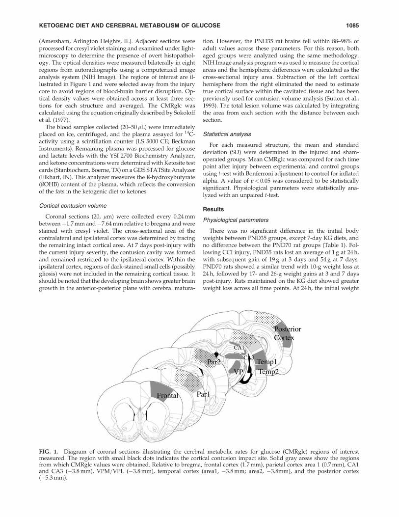
(Amersham, Arlington Heights, IL). Adjacent sections wereprocessed for cresyl violet staining and examined under light-microscopy to determine the presence of overt histopathol-ogy. The optical densities were measured bilaterally in eightregions from autoradiographs using a computerized imageanalysis system (NIH Image). The regions of interest are il-lustrated in Figure 1 and were selected away from the injurycore to avoid regions of blood-brain barrier disruption. Op-tical density values were obtained across at least three sec-tions for each structure and averaged. The CMRglc wascalculated using the equation originally described by Sokoloffet al. (1977).
The blood samples collected (20–50 mL) were immediatelyplaced on ice, centrifuged, and the plasma assayed for 14C-activity using a scintillation counter (LS 5000 CE; BeckmanInstruments). Remaining plasma was processed for glucoseand lactate levels with the YSI 2700 Biochemistry Analyzer,and ketone concentrations were determined with Ketosite testcards (Stanbiochem, Boerne, TX) on a GDS STATSite Analyzer(Elkhart, IN). This analyzer measures the ß-hydroxybutyrate(ßOHB) content of the plasma, which reflects the conversionof the fats in the ketogenic diet to ketones.
Cortical contusion volume
Coronal sections (20, mm) were collected every 0.24 mmbetweenþ1.7 mm and�7.64 mm relative to bregma and werestained with cresyl violet. The cross-sectional area of thecontralateral and ipsilateral cortex was determined by tracingthe remaining intact cortical area. At 7 days post-injury withthe current injury severity, the contusion cavity was formedand remained restricted to the ipsilateral cortex. Within theipsilateral cortex, regions of dark-stained small cells (possiblygliosis) were not included in the remaining cortical tissue. Itshould be noted that the developing brain shows greater braingrowth in the anterior-posterior plane with cerebral matura-
tion. However, the PND35 rat brains fell within 88–98% ofadult values across these parameters. For this reason, bothaged groups were analyzed using the same methodology.NIH Image analysis program was used to measure the corticalareas and the hemispheric differences were calculated as thecross-sectional injury area. Subtraction of the left corticalhemisphere from the right eliminated the need to estimatetrue cortical surface within the cavitated tissue and has beenpreviously used for contusion volume analysis (Sutton et al.,1993). The total lesion volume was calculated by integratingthe area from each section with the distance between eachsection.
Statistical analysis
For each measured structure, the mean and standarddeviation (SD) were determined in the injured and sham-operated groups. Mean CMRglc was compared for each timepoint after injury between experimental and control groupsusing t-test with Bonferroni adjustment to control for inflatedalpha. A value of p< 0.05 was considered to be statisticallysignificant. Physiological parameters were statistically ana-lyzed with an unpaired t-test.
Results
Physiological parameters
There was no significant difference in the initial bodyweights between PND35 groups, except 7-day KG diets, andno difference between the PND70 rat groups (Table 1). Fol-lowing CCI injury, PND35 rats lost an average of 1 g at 24 h,with subsequent gain of 19 g at 3 days and 54 g at 7 days.PND70 rats showed a similar trend with 10-g weight loss at24 h, followed by 17- and 26-g weight gains at 3 and 7 dayspost-injury. Rats maintained on the KG diet showed greaterweight loss across all time points. At 24 h, the initial weight
FIG. 1. Diagram of coronal sections illustrating the cerebral metabolic rates for glucose (CMRglc) regions of interestmeasured. The region with small black dots indicates the cortical contusion impact site. Solid gray areas show the regionsfrom which CMRglc values were obtained. Relative to bregma, frontal cortex (1.7 mm), parietal cortex area 1 (0.7 mm), CA1and CA3 (�3.8 mm), VPM=VPL (�3.8 mm), temporal cortex (area1, �3.8 mm; area2, �3.8mm), and the posterior cortex(�5.3 mm).
KETOGENIC DIET AND CEREBRAL METABOLISM OF GLUCOSE 1085

loss in animals maintained on the KG diet was 5 g in PND35and 18 g in PND70 rats. By 3 days, there was less weight lossin both age groups. The largest weight differences were seenafter 7 days on the KG diet with�18 and�20 g, in PND35 andPND70 rats, respectively. Both the TBI-induced and KG-diet-induced weight losses are within the expected ranges givenprevious reports (Prins et al., 2005).
Blood pH, pCO2, and pO2 values were not significantlydifferent among standard-fed animals (Table 1). The onlyexception was the PND70 sham animals, which showedslightly higher pCO2 values. PCO2 was decreased in thePND70 rats at 3 and 7 days after injury on the KG diet, as wasthe pH in the injured PND35 1-day KG group.
Plasma levels of glucose, lactate, and ß-hydroxybutyrate(ßOHB) were measured at 1, 3, and 7 days after TBI on boththe standard and KG diet (Table 1). Both PND35 and PND70animals maintained on the standard diet showed no signifi-cant change in plasma glucose after injury. PND35 ratsshowed a significant ( p< 0.05) decrease in plasma lactate at1 day, while PND70 rats showed decreased plasma lactate atall days measured after injury. Plasma ketone levels were notsignificantly different after injury or between age groupsamong the standard-fed animals. PND35 and PND70 animalson the KG diet showed a significant ( p< 0.05) increase inplasma ßOHB and a significant ( p< 0.05) decrease in plasmaglucose and lactate. The increase in plasma ketones wasgreater among PND35 animals at all time points after injury,including sham controls.
CMRglc in PND35 and PND70 shamstandard-fed animals
The average cortical rates of glucose metabolism obtainedfrom PND35 and PND70 sham animals were within the pre-viously reported ranges (Yoshino et al., 1991; Thomas et al,2000; Nehlig et al., 1988). There was no significant differencein the CMRglc of the cortex, thalamus, or hippocampus be-tween PND35 and PND70 rats, which is consistent with pre-
viously published developmental profile of glucosemetabolism across these age groups (Nehlig et al., 1988).
CMRglc in PND35 injured standard-fed
Following CCI injury in PND35 rats, ipsilateral cortical,hippocampal, and thalamic structures showed a significantdecrease in CMRglc at day 1, reaching its lowest levels at day3 after injury (Table 2, Fig. 2). While ipsilateral corticalstructures remained significantly depressed at 7 days, hip-pocampal and thalamic structures exhibited sham levels ofCMRglc by 7 days after injury. The contralateral structuresshowed no significant change in CMRglc, except the temporaland posterior cortex at day 3 after injury.
CMRglc in PND70 injured standard-fed
Following CCI injury in PND70 rats, ipsilateral cortical,hippocampal, and thalamic structures showed a significantdecrease in CMRglc on days 1 and 3 (Table 2). The magnitudeof the CMRglc depression was similar between PND35 (1–11%) and PND70 (2–14%) rats after CCI injury on the stan-dard diet. In contrast to the PND35 CCI rats, where thalamicand hippocampal structures showed an earlier return ofmetabolic rates, only PND70 hippocampal structures attainedsham levels of glucose metabolic rates by 7 days (Table 2). ThePND70 ipsilateral cortex and thalamus remained depressed atday 7 after injury. The contralateral structures showed nosignificant change in CMRglc after injury relative to sham,with the exception of the CA1 region of the dorsal hippo-campus, which remained depressed at 3 days post-injury.
CMRglc in PND35 injured KG-fed
PND35 sham animals maintained on the KG diet didnot show a significant difference in CMRglc compared tostandard-fed shams in any structure (Table 3). However, in-jured PND35 rats maintained on the KG diet showed signif-icant decreases in all ipsilateral structures at 1, 3, and 7 days
Table 1. Physiological Parameters and Changes in Plasma Substrates (mM) with Age, Injury, and Diet
Group Initial weight (g) Blood pH PCO2 PO2 Glucose (mM) Lactate (mM) ßOHB (mM)
PND35 standardSham 129� 1.9 7.43� 0.01 39� 1.7 94� 6.1 10.2� 0.44 1.1� 0.1 0.14� 0.051 day 148� 12.2 7.42� 0.017 37� 0.8 95� 1.5 9.34� 0.4 0.86� 0.06* 0.32� 0.053 days 123� 5.2 7.43� 0.01 38� 0.2 91� 3.2 10.5� 0.45 0.99� 0.08 0.45� 0.097 days 132� 3.6 7.41� 0.01 39� 1.6 88� 3.4 10.4� 0.39 0.91� 0.05 0.17� 0.04
PND35 KG dietSham 142� 14.2 — — — 7.6� 0.6* 0.8� 0.04* 1.8� 0.1*1 day 127� 6.0 7.38� 0.01* 37� 1.0 90� 1.1 7.2� 0.42* 0.8� 0.03* 1.68� 0.23*3 days 139� 7.6 7.41� 0.02 36� 1.1 89� 3.3 8.6� 0.56* 0.92� 0.11 1.67� 0.1*7 days 113� 6.9 7.41� 0.00 34� 1.4 97� 1.8 8.45� 1.0 1.04� 0.14 1.88� 0.09*
PND70 standardSham 305� 6.6 7.41� 0.01 42� 0.7* 91� 3.4 10.36� 1.0 0.90� 0.2 0.29� 0.161 day 305� 7.4 7.42� 0.01 38� 0.8 91� 2.8 9.24� 0.6 0.69� 0.1* 0.36� 0.073 days 298� 10.2 7.41� 0.01 41� 1.4 88� 3.8 10.69� 0.6 0.66� 0.04* 0.22� 0.037 days 321� 8.8 7.41� 0.01 40� 1.4 91� 2.3 9.19� 0.4 0.66� 0.06* 0.31� 0.09
PND70 KG dietSham1 day 298� 5.7 7.42� 0.007 39� 1.5 85� 5.4 6.62� 0.29* 0.68� 0.05* 1.55� 0.07*3 days 297� 10.1 7.41� 0.01 35� 1.1* 89� 3.7 9.38� 0.61 0.75� 0.05* 1.30� 0.13*7 days 312� 6.7 7.42� 0.01 36� 0.8* 86� 2.1 9.19� 1.1 1.04� 0.29 1.16� 0.26*
*p< 0.05, relative to age-matched standard diet sham.
1086 PRINS AND HOVDA

post-injury relative to KG-fed sham, and the magnitude of theCMRglc depression was on average 13% greater compared tostandard-fed CCI-injured animals (Table 3, Fig. 2). The con-tralateral cortical, hippocampal, and thalamic structures inPND35 rats also showed a pronounced decrease in CMRglc atpost-injury days 1 and 7. The magnitude of this depressionwas on average 8.9% greater than that of the ipsilateralregions.
CMRglc in PND70 injured KG-fed
Previous studies have shown that sham adult rats (PND65-90) maintained on the KG diet do not show changes inCMRglc relative to standard-fed rats (Cherel et al., 1985;Corddry et al., 1982; Al-Mudallal et al., 1995). For this reason,
PND70 injured KG-fed animals were compared to PND70sham standard-fed. PND70 injured rats maintained on the KGdiet showed a significant decrease in CMRglc within the ce-rebral cortical, hippocampal, and thalamus at 1 and 3 daysafter injury relative to sham. The CMRglc for all ipsilateralstructures returned to sham levels at 7 days (Table 3, Fig. 3).PND70 injured KG-fed rats showed no significant changes incontralateral cortical, hippocampal, or thalamic CMRglc atany time point relative to injured standard-fed animals.
Contusion volume
The CCI injury produced an evolving contusion, restrictedto the cerebral cortex, which averaged 30.4� 3.9mm3 inPND35 and 29.2� 4.9 mm3 in PND70 rats on the standard diet
Table 2. Rates of Glucose Metabolism following CCI Injury among Standard Fed PND35 and PND70 Rats
PND35 PND70
Area Sham 1 day 3 days 7 days Sham 1 day 3 days 7 days
FrontalIpsi 87.4� 6.3 50.7� 9.6** 47.5� 3.4** 65.8� 7.52* 91.2� 6.1 50.7� 8.9** 45.4� 2.5** 59.1� 6.3**Contra 90.0� 6.0 91.8� 6.9 82.9� 5.8 92.4� 5.9 92.2� 5.5 88.9� 6.1 86.7� 3.1 83.9� 3.2
Parietal 1Ipsi 87.5� 6.9 43.1� 9.8** 45.8� 3.6** 64.0� 5.6* 92.4� 5.6 34.0� 8.8** 50.3� 5.9** 56.9� 6.3**Contra 88.9� 5.6 91.2� 6.4 81.3� 6.6 90.1� 6.3 95.1� 5.8 90.3� 6.2 85.6� 3.4 80.7� 2.7
Post cortexIpsi 96.9� 8.2 53.7� 6.2** 46.1� 4.3** 63.0� 7.3* 95.0� 9.2 49.3� 8.2** 56.5� 7.1** 62.3� 7.5**Contra 106.1� 6.8 98.0� 9.6 85.9� 5.7* 102.5� 7.7 102.7� 6.9 102.0� 5.0 101.6� 7.1 95.6� 5.9
Temporal 1Ipsi 92.8� 6.2 42.0� 5.9** 51.2� 3.7** 69.2� 4.8** 99.1� 7.0 42.3� 6.2** 50.7� 6.3** 69.8� 7.5*Contra 90.6� 6.9 90.2� 10.2 84.4� 4.7* 95.3� 7.9 95.6� 7.1 97.2� 4.3 84.0� 3.9 85.5� 2.3
Temporal 2Ipsi 89.1� 5.2 55.6� 6.0** 47.0� 4.9** 58.3� 5.1** 98.9� 6.9 54.6� 6.4** 52.1� 7.9** 67.9� 1.8*Contra 96.5� 2.2 89.4� 8.9 86.1� 3.2* 91.6� 8.9 101.0� 8.6 97.0� 8.0 93.0� 6.1 85.1� 5.6
CA1Ipsi 58.3� 4.7 45.7� 4.1 39.3� 2.7* 54.8� 7.8 54.6� 3.0 39.9� 4.7* 33.3� 4.9* 50.0� 4.5Contra 56.3� 5.7 52.7� 6.3 43.2� 4.2 54.2� 7.3 54.9� 4.7 53.0� 5.0 41.3� 2.4* 47.0� 4.6
CA3Ipsi 62.5� 5.0 45.5� 3.9* 44.9� 1.9* 55.8� 8.2 62.1� 3.7 36.8� 5.1** 35.0� 2.3** 50.9� 3.5*Contra 59.3� 5.2 57.0� 4.7 50.6� 4.1 58.4� 7.0 59.6� 4.2 58.7� 4.6 50.6� 2.1 53.3� 3.7
VPM=VPLIpsi 73.8� 5.0 53.8� 5.7* 48.1� 3.3** 69.8� 9.7 72.8� 4.0 47.7� 5.0** 42.2� 1.7** 61.4� 3.1*Contra 77.9� 4.2 88.6� 7.4 71.6� 4.4 81.5� 5.7 80.2� 3.8 85.9� 5.7 72.6� 4.3 76.1� 4.5
*denotes value of p< 0.05 relative to age matched shams; **denotes values of p< 0.01 relative to age matched shams.
FIG. 2. Representative colorized autoradiographs showing changes in cerebral metabolic rates for glucose (CMRglc) aftercontrolled cortical impact (CCI) injury among standard-fed and ketogenic-fed postnatal day 35 (PND35) animals 1 day afterinjury. Arrow indicates ipsilateral hemisphere.
KETOGENIC DIET AND CEREBRAL METABOLISM OF GLUCOSE 1087

Sh 1d 3d 7d Sh 1d 3d 7d 0
20
40
60
80
100
120
140P35 Std
P35 KG
0
20
40
60
80
100
120
140P70 Std
P70 KG
†
*
†
† † † †
†† †
†*
††
A B
Sh 1d 3d 7d Sh 1d 3d 7d Ipsilateral Contralateral
Sh 1d 3d 7d Sh 1d 3d 7d
Sh 1d 3d 7d Sh 1d 3d 7d Ipsilateral Contralateral
0
20
40
60
80
100
120
140P35 Std
P35 KG
0
20
40
60
80
100
120
140P70 Std
P70 KG
* * * ** † * † *
C D
FIG. 3. Changes in cerebral metabolic rates for glucose (CMRglc) within the posterior cortex (A,B) and CA3 (C,D) with timeafter controlled cortical impact (CCI) among postnatal day 35 (PND35) standard-fed and ketogenic-fed animals, and PND70standard-fed and ketogenic-fed rats. {p< 0.01 and *p< 0.05 relative to age-matched sham.
Table 3. Rates of Glucose Metabolism following CCI Injury Among Ketogenic Fed PND35 and PND70 Rats
PND35 PND70
Area Sham KG 1 day 3 days 7 days 1 day 3 days 7 days
Frontal cortexIpsi 93.5� 9.8 38.9� 4.7** 52.6� 3.8** 38.3� 6.1** 41.4� 5.4** 43.7� 5.2** 78.9� 8.2Contra 92.8� 9.7 68.7� 4.0* 81.4� 5.4 48.8� 6.7 82.3� 9.8 82.4� 8.7 103.1� 11.7
Parietal 1Ipsi 94.1� 9.1 33.9� 2.4** 55.3� 6.3* 40.0� 5.6* 28.4� 3.3** 43.3� 4.5** 79.9� 8.7Contra 93.6� 10.1 68.4� 3.4* 80.0� 5.8 49.4� 6.2* 83.3� 10.3 81.4� 9.0 102.0� 9.4
Post cortexIpsi 102� 9.9 35.0� 2.1** 49.7� 3.3** 38.4� 5.8** 36.9� 2.9** 44.0� 5.6** 81.4� 7.4Contra 101� 9.1 71.3� 2.0* 90.1� 7.8 49.7� 7.7** 93.6� 9.7 89.7� 9.0 116.0� 12.1
Temporal 1Ipsi 88.1� 7.9 28.5� 2.3** 52.2� 3.7** 41.0� 5.4** 35.3� 3.6** 47.8� 8.4** 84.0� 9.4Contra 87.9� 8.5 65.9� 3.4* 77.4� 5.8 49.6� 6.6** 84.0� 9.0 83.2� 8.4 109.0� 14.4
Temporal 2Ipsi 105� 10.5 37.5� 2.5** 49.1� 4.3** 38.3� 5.4** 45.1� 5.8** 42.5� 5.1** 78.2� 7.4Contra 106� 9.0 65.3� 4.6** 81.6� 7.7 48.9� 6.3** 88.3� 10.1 82.1� 8.2 108.6� 11.2
CA1Ipsi 61.1� 5.7 38.9� 1.0* 46.5� 5.6 36.4� 5.6* 37.2� 5.2* 36.3� 7.4 63.7� 8.7Contra 59.2� 5.8 41.7� 2.4 50.2� 5.9 28.9� 5.5* 45.4� 7.0 47.3� 7.7 58.5� 4.7
CA3Ipsi 75.8� 7.5 38.7� 1.4* 52.6� 6.5 34.8� 5.0** 34.5� 4.8** 34.3� 8.0* 69.3� 12.6Contra 73.8� 6.4 47.9� 2.1 59.1� 6.9 32.3� 5.7* 51.1� 7.5 53.1� 8.2 65.8� 6.9
VPM=VPLIpsi 89.4� 7.7 36.8� 3.8** 51.6� 3.7* 42.0� 6.0** 40.9� 4.1** 45.2� 5.5** 86.7� 14.5Contra 89.4� 8.1 61.8� 3.5* 73.2� 7.3 43.8� 7.0** 79.5� 9.8 73.0� 7.2 99.5� 15.8
Gray blocks indicate significant ( p< 0.05) difference relative to standard fed, same age, region, and timepoint (Table 2).
1088 PRINS AND HOVDA

(Fig. 4). There was no statistical difference in the contusionvolume between the injured standard-fed age groups. The KGdiet did show a significant 31% reduction in contusion vol-ume among PND35 rats and a non-significant increase involume in PND70 rats. The contralateral hemisphere at 7 daysshowed no morphological evidence of contrecoup injury.
Discussion
Changes in substrate availability after TBI
The question of whether head-injured patients showchanges in the blood chemistry as a result of TBI is difficult todetermine given that blood glucose is aggressively managedwithin a specific range in human patients to prevent hyper-glycemia (Vespa et al., 2006). Results from the current studyshow that PND35 rats maintained on standard chow had littlechange in blood concentrations of glucose or lactate. The non-significant trend of elevated plasma ßOHB may be mild ke-tosis from loss of appetite acutely after injury. PND70 rats onstandard chow showed no changes in glucose or ßOHB, butdid show significant decrease in plasma lactate. The decreasein plasma lactate may reflect cerebral and=or systemic use oflactate as an alternative fuel in this age group (Chen et al.,2000a,b). While both age groups show a significant increase inplasma ketones when maintained on the KG diet, PND35 ratsshow sustained decreases in blood glucose compared toPND70 rats. This difference may be related to the magnitudeof ketosis achieved by each age group.
Age differences in CMRglc after TBI
Both PND35 and PND70 rats maintained on standard dietshowed acute decreases in CMRglc of similar magnitude,which was sustained within ipsilateral cortical areas. WhilePND35 rats showed return of sham-level metabolic rates ofglucose by post-injury day 7 within the hippocampus andthalamus, PND70 rats exhibited metabolic recovery in onlythe CA3 region of the hippocampus. These finding suggestthat the CCI injury induced at the severity reported in thispaper, produced cortically comparable metabolic responses inboth age groups within the injured hemisphere. In contrast to
our lateral FP injury which results in primarily ipsilateralcortical changes in CMRglc (Yoshino et al., 1991; Thomas et al.,2000), the current CCI injury produced significant contralat-eral changes. PND35 showed a decrease in CMRglc at 3 daysin the temporal cortex and posterior cortex. This pattern re-emphasizes the fact that the contralateral hemisphere cannotserve as a ‘‘non-injured’’ control in this injury model.
The reason for the prolonged decrease in CMRglc after TBIremains as yet unknown. There are numerous possiblemechanisms that may explain this depression including dis-ruption of mitochondrial metabolism by calcium accumula-tion (Fineman et al., 1993; Osteen et al., 2001), ionic fluxdisruptions (Katayama et al., 1990), reduced cerebral bloodflow (Cherian et al., 2004), or lactic acid accumulation (Ka-wamata et al., 1995). Another consideration is that followingthe initial surge in neurotransmitter release and ionic fluctu-ations the brain activity may become quiescent and thereforerequire less substrate. Several studies have shown decreasedability of the brain to show stimulation-evoked increases inCMRglc as early as 4hrs and as long as 2 months post-injury(Dietrich et al., 1994; Passineau et al., 2000). This quiescentstate may be a mechanism to reduce secondary injury fromactivation, which has been observed after direct corticalstimulation (Ip et al., 2003).
Finally, glucose metabolic pathways may be altered afterinjury, directly impacting cerebral glucose uptake. 2DG au-toradiography is a measure of glucose uptake, but it does notreveal how glucose is processed past hexokinase. 13C-glucosestudies have shown that, during this period of depressedCMRglc, there is an increase in unmetabolized glucose, adecrease in 13C-labeled lactate and increase glucose metabo-lism through the pentose phosphate pathway (Bartnik et al.,2005, 2007). The decrease in oxidation of glucose and increaseshunting of glucose towards NADPH synthesis suggests al-tered fates for glucose after TBI.
Age differences in CMRglc after TBI with ketones
How does the presence of ketones alter cerebral metabo-lism of glucose in the normal brain? In the normal adult ro-dent, ketosis induced by fasting, infusion, or diet has no affect
FIG. 4. Changes in cortical contusion volumes (mm3) between postnatal day 35 (PND35) and PND70 injured rats onstandard or ketogenic (KG) diet at 7 days post-injury (A). Representative histological sections showing the extent of corticalpreservation among PND35 injured ketogenic-fed animals (B). *p< 0.05 relative to age-matched standard-fed animals.
KETOGENIC DIET AND CEREBRAL METABOLISM OF GLUCOSE 1089
on glucose uptake. A 30% reduction in CMRglc was reportedonly in a PND20 rat infused with ßOHB (Miller et al., 1986).Thus, in the rodent, cerebral fuel interaction of glucose andketones appears to be age-dependent. In contrast, pigs andhumans all show decreased CMRglc in response to ketosis(Kammula, 1976; Redies et al., 1989; Hasselbalch et al., 1994,1996). While the decrease in CMRglc=glucose uptake duringketosis can reflect the shifting cerebral metabolism towardsketones, the lack of a decrease in glucose uptake does notnecessary mean the opposite. Cerebral metabolism of ketonesmay not affect glucose uptake at some age groups, species orconditions, but may shift glucose from its normal oxidativefate towards another biochemical fate (pentose phosphatepathway, methylglyoxyl pathway). This is what is believed tohappen in the adult rodent brain during ketosis. In the currentstudy, PND35 sham rats on the KG diet showed no change inCMRglc, which is consistent with the adult rodent responsereported in the literature and may indicate decreased oxida-tion of glucose, but no change in glucose uptake.
Injury-induced decreases in CMRglc were exacerbated bi-laterally in PND35 CCI KG-fed animals on days 1 and 7.Several mechanisms may contribute to this further decrease inCMRglc, including decreased plasma glucose, cell death, areduction in the need for glucose metabolism, and=or in-creased ketone metabolism. Insulin-induced hypoglycemiahas been shown to alter the lumped constant and decreaseCMRglc (Nedergaard et al., 1988). While the plasma glucosedecreased approximately 20% in the current study, it did notreach hypoglycemic levels, and therefore, this reduction is
unlikely to explain the additional 14–37% decrease in CMRglcobserved. While it is certainly possible that certain areas of thebrain become dennervated or exhibit retrograde degenera-tion, which could cause a reduction in glucose metabolism,cell death is unlikely to explain the contralateral cortical de-crease. In this case, it is more likely that the PND35 brainincreased its metabolism of ketones bilaterally. It has beenshown that accelerated rates of ketone metabolism can pro-duce an acetyl-CoA pool that competes with pyruvate de-rived acetyl-CoA and leads to pyruvate oxidation inhibition(Randle et al., 1966; Ruderman et al., 1974). This may explainthe mechanism by which cerebral ketone utilization results infurther decreases in CMRglc after TBI and may require acertain minimum threshold of ketone metabolism to generatesufficient acetyl-CoA pool to cause inhibition (Fig. 5) and mayexplain the neuroprotective effect on cortical lesion volume.
While the PND35 sham animals on the KG diet did notshow changes in cortical CMRglc, the contralateral cortex ofPND35 CCI animals on the KG diet exhibited a decrease inCMRglc. This again emphasizes the fact that the contralateralhemisphere is affected by the injury and should not be used asa ‘‘control.’’ More importantly, the metabolic changes occur-ring post-injury on the contralateral side may reflect re-organizational changes that have been shown to take placeduring this time period. At 7 days post-cortical ischemia,stimulation of right vibrissae triggered increased 14C-2DGuptake in the contralateral barrel cortex, suggesting com-pensatory plasticity ( Jablonka nd Kossut, 2006). fMRI studieshave also shown that unilateral lesion of the sensorimotor
Sham PND35 and PND70
BBB
glycolysis
glucose
pyruvate
ACoA KG
• Further decrease in uptake
Decrease pyruvate oxidation
• Decrease in uptake
• Shunt to PPP
CCI PND35 and PND70
BBB
glycolysis
glucose
pyruvate
ACoA
• Shunt to PPP?
CCI+KG Diet PND35
BBB
glycolysis
glucose
pyruvate
ACoA KG
• Recovery in uptake
Decrease pyruvate oxidation
• Shunt to PPP
CCI+KG Diet PND70
BBB
glycolysis
glucose
pyruvate
ACoA
• MG pathway
Lactate
A B C D
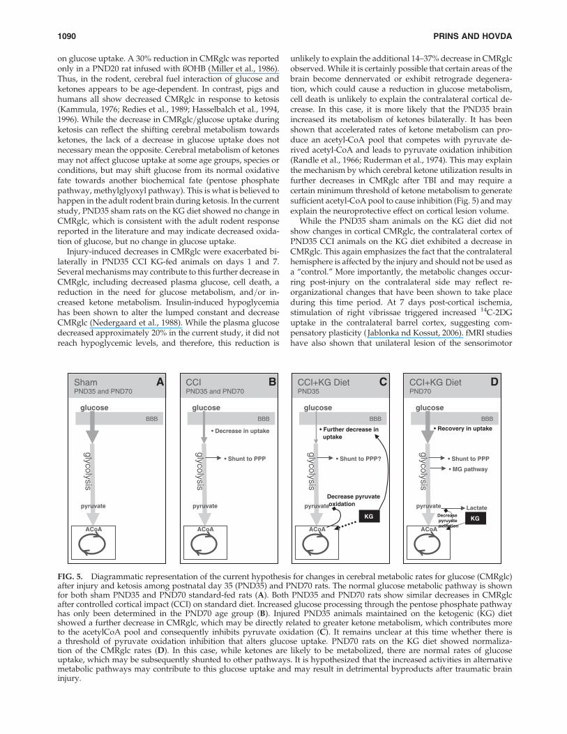
FIG. 5. Diagrammatic representation of the current hypothesis for changes in cerebral metabolic rates for glucose (CMRglc)after injury and ketosis among postnatal day 35 (PND35) and PND70 rats. The normal glucose metabolic pathway is shownfor both sham PND35 and PND70 standard-fed rats (A). Both PND35 and PND70 rats show similar decreases in CMRglcafter controlled cortical impact (CCI) on standard diet. Increased glucose processing through the pentose phosphate pathwayhas only been determined in the PND70 age group (B). Injured PND35 animals maintained on the ketogenic (KG) dietshowed a further decrease in CMRglc, which may be directly related to greater ketone metabolism, which contributes moreto the acetylCoA pool and consequently inhibits pyruvate oxidation (C). It remains unclear at this time whether there isa threshold of pyruvate oxidation inhibition that alters glucose uptake. PND70 rats on the KG diet showed normaliza-tion of the CMRglc rates (D). In this case, while ketones are likely to be metabolized, there are normal rates of glucoseuptake, which may be subsequently shunted to other pathways. It is hypothesized that the increased activities in alternativemetabolic pathways may contribute to this glucose uptake and may result in detrimental byproducts after traumatic braininjury.
1090 PRINS AND HOVDA

cortex resulted in increased activation of the contralateralcortex and regions lateral to the injury upon hindlimb stim-ulation (Abo et al., 2001). Also, at 1–4 days after lateral TBI,cerebral blood flow increases in the contralateral cortex(Scremin et al., 2007). These types of contralateral compen-satory processes have also been observed following humanTBI and suggest altered synaptic connectivity in the contra-lateral cortex in response to injury-related dennervation. Thedecrease in CMRglc observed in the contralateral cortexamong PND35 injured KG-fed rats may reflect an increase inketone metabolism during reorganization sufficient to sup-press glucose uptake.
Another explanation for the bilateral CMRglc decrease ofthe PND35 rats on the KG diet after TBI may be related todifferential response to injury. At 3 days post-injury, PND35rats show contralateral decreases in the temporal and poste-rior cortical regions, which were not observed among PND70rats. This may indicate that the CCI injury has contralateralconsequences of a different timecourse in the younger rat, andketone-induced decreases in CMRglc may be observed in in-jured regions. The depression of CMRglc at 7 days amongPND35 rats on the KG diet can be associated with the degreeof ketosis that is maintained in this age group. PND70 rats onthe KG diet do not maintain high levels of ketones, andplasma glucose is also no longer depressed at 7 days.
In contrast to the PND35 rats, PND70 injured standard-fedand KG-fed animals showed no difference from each other intheir CMRglc changes, except at post-injury day 7, whereCMRglc rates were greater among KG-fed animals. It appearsat this time point that animals in this age group show ‘‘re-covery’’ of their glucose metabolic rates when maintained onthe KG diet for 7 days after injury. Given the tremendousbody of literature reporting adult rodent brain capacity tometabolize ketones after prolonged ketosis, it is unlikely thatthe adult brain is not utilizing the substrate (Prins et al., 2004).There is also no evidence to contradict the inhibitory effects ofketone metabolism on pyruvate oxidation. There is, however,evidence that the adult brain bilaterally increases pentosepathway activity by 9–12% after CCI injury (Bartnik et al.,2005), and there is accumulation of propylene glycol (Hirtet al., 2007). Together these findings suggest that increasedactivity of alternative metabolic pathways may stimulate re-turn of sham levels of glucose uptake (Fig. 5), which may havenegative consequences on cortical contusion volume in themature brain.
Changes in the lumped constant (LC) have been shown tocause over- or under-estimation of the cerebral metabolic ratesof glucose. While the LC has been shown to decrease with age,between PND14 and PND90 there is little change (Takei et al.,1986), and it is not expected to contribute to the age groups inthe current study. The LC does not change with mild hypo-glycemia, but plasma glucose levels less than 5 mM cause andincrease in the LC (Schuier et al., 1990). While animalsmaintained on the KG diet in the current study did showdecreases in plasma glucose, they did not approach 5 mM.Finally, in regards to TBI, the global LC has been estimated todecrease from 0.65 to 0.43 in human patients (Wu et al., 2004)and may contribute to an underestimation of the CMRglcrates. The changes in the LC are not likely to explain themarked changes in CMRglc in the current study.
The results from the current study show that ketosis affectscerebral glucose metabolism in an age-dependent manner
after TBI. While both age groups were similar in terms ofglucose metabolism and contusion volume after TBI while onthe standard diet, the presence of ketones further reducedCMRglc in PND35 and normalized CMRglc in PND70 ratsafter injury. The changes in CMRglc among PND35 TBI ratson the KG diet were associated with decreased cortical con-tusion volume, which was consistent with previous findings(Prins et al., 2005). These results suggest that conditions ofreduced glucose utilization and increased alternative sub-strate metabolism may be preferable acutely after TBI in thejuvenile brain. Clinically, conditions of low plasma glucoseand elevated ketones were shown to improve nitrogen bal-ance (Ritter et al., 1996). Future studies addressing optimalpost-injury substrate conditions will help establish age-appropriate substrate management guidelines for TBIpatients.
Acknowledgments
This work was supported by NS052406, NS27544, andNS37363.
Author Disclosure Statement
No competing financial interests exist.
References
Abo, M., Chen, Z., Lai, L.J., Reese, T., and Bjelke, B. (2001).Functional recovery after brain lesion-contralateral neurmo-dulation: an fMRI study. Neuroreport 12, 1543–1547.
Alessandri, B., Reinert, M., Young, H.F., and Bullock, R. (2000).Low extracellular (ECF) glucose affects the neurochemicalprofile in severe head-injured patients. Acta Neurochir. Suppl.76, 425–430.
Al-Mudallal, A.S., Levin, B.E., Lust, W.D., and Harik, S.I. (1995).Effects of unbalanced diets on cerebral glucose metabolism inthe adult rat. Neurology 45, 2261–2265.
Andersen, B.J., and Marmarou, A. (1989). Isolated stimulation ofglycolysis following traumatic brain injury, in: IntracranialPressure VII. J.T. Hoff and A.L. Betz (eds), Springer-Verlag:Berlin, pps. 575–580.
Andersen, B.J., and Marmarou, A. (1992). Post-traumatic selec-tive stimulation of glycoslysis. Brain Res. 585, 184–189.
Bartnik, B.L., Sutton, R.L., Fukushima, M., Harris, N.G., Hovda,D.A., and Lee, S.M. (2005). Upregulation of pentose phosphatepathway and preservation of tricarboxylic acid cycle flux afterexperimental brain injury. J. Neurotrauma 22, 1052–1065.
Bartnik, B.L., Lee, S.M., Hovda, D.A., and Sutton, R.L. (2007).The fate of glucose during the period of decreased metabolismafter fluid percussion injury: a 13C NMR study. J. Neuro-trauma 24, 1079–1092.
Bergsneider, M., Hovda, D.A., Shalmon, E., Kelly, D.F., Vespa,P.M., Martin, N.A., Phelps, M.E., McArthur, D.L., Caron, M.J.,Kraus, J.F., and Becker, D.P. (1997). Cerebral hyperglycolysisfollowing severe traumatic brain injury in humans: a positronemission tomography study. J. Neurosurg. 86, 241–251.
Chen, S.F., Richards, H.K., Smielewski, P., Johnstrom, P., Sal-vador, R., Pickard, J.D., and Harris, N.G. (2004). Relationshipbetween flow-metabolism uncoupling and evolving axonalinjury after experimental traumatic brain injury. J. Cereb.Blood Flow Metab. 24, 1025–1036.
Chen, T., Qian, Y.Z., Di, X., Rice, A., Zhu, J.P., and Bullock, R.(2000a). Latate=glucose dynamics after rat fluid percussionbrain injury. J. Neurotrauma 17, 135–142.
KETOGENIC DIET AND CEREBRAL METABOLISM OF GLUCOSE 1091

Chen, T., Qian, Y.Z., Di, X., Rice, A., Zhu, J.P., and Bullock, R.(2000b). Brain lactate uptake increases at the site of impactafter traumatic brain injury. Brain Res. 861, 281–287.
Cherel, Y., Burnol, A.F., Leturque, A., and LeMaho, Y. (1988). Invivo glucose utilization in rat tissues during the three phasesof starvation. Metabolism 37, 1033–1039.
Cherian, L., Hlatky, R., and Robertson, C.S. (2004). Comparisonof tetrahydrobiopterin and l-arginine on cerebral blood flowafter controlled cortical impact injury in rats. J. Neurotrauma21, 1196–1203.
Cherian, L., Robertson, C.S., and Goodman, J.C. (1996). Sec-ondary insults increase injury after controlled cortical impactin rats. J. Neurotrauma 13, 371–383.
Corddry, D.H., Rapoport, S.I., and London, E.D. (1982). No ef-fect of hyperketonemia on local cerebral glucose utilization inconscious rats. J. Neurochem. 38, 1637–1641.
Cochran, A., Schaife, E.R., Hansen, K.W., and Downey, E.C.(2003). Hyperglycemia and outcomes from pediatric traumaticbrain injury. J. Trauma 55, 1035–1038.
Dietrich, W., Alonso, O., Busto, R., and Ginsberg, M.D. (1994).Widespread metabolic depression and reduced somatosen-sory circuit activation following traumatic brain injury in rats.J. Neurotrauma 11, 629–640.
Feise, G., Kogure, K., Busto, R., Scheinberg, P., and Reinmuth,O.M. (1976). Effect of insulin hypoglycemia upon cerebral en-ergy metabolism and EEG activity in the rat. Brain Res. 126,263–280.
Fineman, I., Hovda, D.A., Smith, M., Yoshino, A., and Becker,D.P. (1993). Concussive brain injury is associated with a pro-longed accumulation of calcium: a 45Ca autoradiographicstudy. Brain Res. 624, 94–102.
Goodman, J.C., Valadka, A.B., Gopinath, S.P., Uzura, M., andRobertson, C.S. (1999). Extracellular lactate and glucose al-terations in the brain after head injury measured by micro-dialysis. Crit. Care Med. 27, 2063–2064.
Hall, E.D., Andrus, P.K., and Yonkers, P.A. (1993). Brain hdroxylradical generation in acute experimental head injury. J. Neu-rochem. 60, 588–594.
Hirt, D., Kadri, M., Martin, N.A., Que Hee, S., Eliseo, M., Berg, J.,Vespa, P.M., and Glenn, T.C. (2007). Propylene glycol afterhuman traumatic brain injury: novel biomarker of alteredglucose metabolism. Brain B051.
Ip, E.Y., Zanier, E.R., Moore, A.H., Lee, S.M., and Hovda, D.A.(2003). Metabolic, neurochemical, and histological responsesto vibrissa motor cortex stimulation after traumatic brain in-jury. J. Cereb. Blood Flow Metab. 23, 900–910.
Jablonka, J., and Kossut, M. (2006). Focal stroke in the barrelcortex of rats enhances ipsilateral response to vibrissal input.Acta Neurobiol. Exp. 66, 261–266.
Katayama, Y., Becker, D.P., Tamura, T., and Hovda, D.A. (1990).Massive increases in extracellular potassium and the indis-criminate release of glutamate following concussive brain in-jury. J. Neurosurg. 73, 889–900.
Kawamata, T., Katayama, Y., Hovda, D.A., Yohino, A., andBecker, D.P. (1992). Administration of excitatory amino acidantagonists via microdialysis attenuates the increasein glucoseutilization seen following concussive brain injury. J. Cereb.Blood Flow Metab. 12, 12–24.
Miller, A.L. (1986). Regional glucose and beta-hydroxybutyrateuse by developing rat brain. Metab. Brain Dis. 1, 53–61.
Nedergaard, M., Jakobsen, J., and Diemer, N.H. (1988). Auto-radiographic determination of cerebral glucose content, bloodflow, and glucose utilization in focal ischemia of the rat brain:
influence of the plasma glucose concentration. J. Cereb. BloodFlow Metab. 8, 100–108.
Nehlig, A., Pereira de Vasconcelos, A., and Boyet, S. (1988).Quantitative autoradiographic measurement of local cerebralglucose utilization in freely moving rats during postnatal de-velopment. J. Neurosci. 8, 2321–2333.
O’Connell, M.T., Seal, A., Nortje, J., Al-Rawi, P.G., Coles, J.P.,Fryer, T.D., Menon, D.K., Pickard, J.D., and Hutchinson, P.J.(2005). Glucose metabolism in traumatic brain injury: a com-bined microdialysis and [18F]-2-fluoro-2-deoxy-d-glucose-positron emission tomography (FDG-PET) study. Acta Neu-rochir. Suppl. 95, 165–168.
Osteen, C.L., Moore, A.H., Prins, M.L., Hovda, and D.A. (2001).Age-dependency of 45calcium accumulation following lateralfluid percussion: acute and delayed patterns. J. Neurotrauma18, 141–162.
Passineau, M.J., Zhao, W., Buto, R., Dietrich, W., Alonso, O.,Loor, J.Y., Bramlett, H.M., and Ginsberg, M.D. (2000). Chronicmetabolic sequelae of traumatic brain injury: prolonged sup-pression of somatosensory activation. Am. J. Physiol. HeartCirc Physiol. 279, H924–H931.
Prins, M.L., Lee, S.M., Fujima, L., and Hovda, D.A. (2004). In-creased cerebral uptake and oxidation of exogenous betaHBimproves ATP following traumatic brain injury in adult rats.J. Neurochem. 90, 666–672.
Prins, M.L., Fujima, L.S., and Hovda, D.A. (2005). Age-dependent reduction of cortical contusion volume by ketonesafter traumatic brain injury. J. Neurosci. Res. 82, 413–420.
Prins, M.L., and Giza, C.C. (2006). Induction of monocarboxylatetransporter-2 expression and ketone transport followingtraumatic brain injury in juvenile and adult rats. Dev. Neu-rosci. 28, 447–456.
Randle, P.J., Garland, P.B., Hales, C.N., Newsholme, E.A.,Denton, R.M., and Pogson, C.I. (1966). Interactions of metab-olism and the physiological role of insulin. Recent Prog.Horm. Res. 22, 1–48.
Richards, H.K., Simac, S., Piechnik, S., and Pickard, J.D. (2001).Uncoupling of cerebral blood flow and metabolism after cere-bral contusion in the rat. J. Cereb. Blood Flow Metab. 21, 779–781.
Ritter, A.M., Robertson, C.S., Goodman, J.C., Contant, C.F., andGrossman, R.G. (1996). Evaluation of carbohydrate free dietfor patients with severe head injury. J. Neurotrauma 13, 473–485.
Rovlias, A., and Kotsou, S. (2000). The influence of hyperglyce-mia on neurological outcome in patients with severe headinjury. Neurosurgery 46, 335–342.
Ruderman, N.B., Ross, P.S., Berger, M., and Goodman, M.N.(1974). Regulation of glucose and ketone-body metabolism inbrain of anesthetized rats. Biochem. J. 138, 1–10.
Schuier, F., Orzi, F., Suda, S., Lucignani, G., Kennedy, C., andSokoloff, L. (1990). Influcence of plasma glucose concentrationon lumped constant of the deoxyglucose method: effects ofhyperglycemia in the rat. J. Cereb. Blood Flow Metab. 10, 765–773.
Scremin, O.U., Li, M.G., and Scremin, A.M.E. (2007). Corticalcontusion induces trans-hemispheric reorganization of bloodflow maps. Brain Res. 1141, 235–241.
Sokoloff, L., Reivich, M., and Kennedy, C. (1977). The [14C]deo-xyglucose method for the measurement of local cerebral glu-cose utilization: theory, procedure, and normal values in theconscious and anesthetized albino rat. J. Neurochem. 28, 897–916.
1092 PRINS AND HOVDA
Sutton, R.L., Hovda, D.A., Adelson, P.D., Benzel, E.C., andBecker, D.P. (1994). Metabolic changes following corticalcontusion: relationship to edema and morphological changes.Acta Neurochir. Suppl. 60, 446–448.
Takei, H., Fredericks, W.R., and Rapoport, S.I. (1986). Thelumped constant in the deoxyglucose procedure declines withage in Fisher-344 rats. J. Neurochem. 46, 931–938.
Thomas, S., Prins, M.L., Samii, M., and Hovda, D.A. (2000). Thecerebral metabolic response to traumatic brain injury sus-tained early in development: a 2-deoxy-d-glucose autoradi-ography study. J. Neurotrauma 17, 649–665.
Vespa, P., Boonyaputthikul, R., McArthur, D.L., Miller, C.,Etchepare, M., Bergsneider, M., Glenn, T., Martin, N., andHovda, D. (2006). Intensive insulin therapy reduces micro-dialysis glucose values without altering glucose utilization orimproving the lactate=pyruvate ratio after traumatic braininjury. Crit. Care Med. 34, 400–411.
Wu, H.M., Huang, S.C., Hattori, N., Glenn, T.C., Vespa, P.M., Yu,C.L., Hovda, D.A., Phelps, M.E., and Bergsneider, M. (2004).Selective metabolic reduction in gray matter acutely followinghuman traumatic brain injury. J. Neurotrauma 21, 149–161.
Yoshino, A., Hovda, D.A., Kawamata, T., Katayama, Y., andBecker, D.P. (1991). Dynamic changes in local cerebral glucose
utilization following cerebral concussion in rats: evidence of ahyper- and subsequent hypometabolic state. Brain Res. 561,106–119.
Yoshino, A., Hovda, D.A., Katayama, Y., Kawamata, T., andBecker, D.P. (1992). Hippocampal CA3 lesion prevents post-concussive metabolic dysfunction in CA1. J. Cereb. BloodFlow Metab. 12, 996–1006.
Zauner, A., Doppenberg, E., Woodward, J.J., Allen, C., Jebraili,S., Young, H.F., and Bullock, R. (1997). Multiparametric con-tinuous monitoring of brain metabolism and substrate deliv-ery in neurosurgical patients. Neurol. Res. 19, 265–273.
Address correspondence to:Mayumi L. Prins, Ph.D.
Department of NeurosurgeryDavid Geffen School of Medicine at UCLA
Semel Room 18-228Box 957039
Los Angeles, CA 90095-7039
E-mail: [email protected]
KETOGENIC DIET AND CEREBRAL METABOLISM OF GLUCOSE 1093

Top Related
Copyright © 2022 FDOKUMEN

