


Bahasa
Halaman
Hukum

1 FEMALE NECK STUDY
The Creation Of A Standardized Resistance Training Protocol For
Training The Muscles Of the Head and Neck In Female Athletes
Ralph Cornwell, Jr.
Virginia Polytechnic Institute
School of Education
2 FEMALE NECK STUDY
RUNNING HEAD: FEMALE NECK STUDY
Abstract
The Centers of Disease Control and Prevention along with the National Institute of Health have declared concussions a nationalepidemic. There is very little research being conducted about concussion reduction via proactive head and neck strengthening. If neck musculature reduces the concussive impact, then less force will be transmitted to the brain, thus decreasing the risk of concussion.
There is desperate need for a standardized head and neck resistance training protocol that should be adopted nationwide. The proposed protocol is designed to enhance the capabilities of the soft tissue that surrounds the cylindrical surface area of the human head and neck through sequenced resistance movements totrain the muscles of the head and neck.
The research participants were healthy female college students, age range 18-24. There were 12 participants. Of the 12 subjects used for this study, 6 participants were randomly assigned to theexperimental group and 6 participants in the control group. The participants followed a protocol consisting of 13 movements designed to sequentially train the musculature of the head, neck
3 FEMALE NECK STUDY
and upper back. The duration of the study was 8 weeks. The results of this study demonstrate that females can increase upperbody strength safely and without significant muscular size gains.The female neck during this study showed a very minimal increase in circumference while strength level increases were substantial.
Introduction
Since the Supreme Court ruling in 1972 and the inception of
title IX, females have acquired the opportunity to participate in
competitive sports at the collegiate level (Valentin, 1997).
With this ruling requiring gender equality in access to
collegiate sports, female sports teams comparable to those of
their male counterparts are commonly found in colleges. This
gender equity has resulted in female athletes also manifesting
the same injuries as the male athletes (Dugan, 2005). However in
"gender-comparable" sports, girls had a 70 percent higher
concussion rate than boys. It is well known in the sports
medicine world that females tear their anterior cruciate ligament
(ACL) six times more often than male athletes do (Dugan, 2005).
The Dugan (2005) research indicates that ACL tears in females can
be reduced with sports medicine preventive programs aimed at
4 FEMALE NECK STUDY
strengthening the muscles at the knee joint. Even with these
prevention programs, females will still tear their ACL ligament
in spite of the best efforts of strength coaches and athletic
trainers (Dugan, 2005).
Females participate in several sports that require contact
with possible collisions. Females concuss three to six times more
often than males (Tierney et al., 2008).
A concussion is a traumatic brain injury that alters brain
function. Effects are usually temporary, but can include problems
with headache, concentration, memory, judgment, balance, and
coordination. Although concussions are usually caused by a blow
to the head, they can also occur when the head and upper body are
violently shaken (Reid & Reid, 1981).
Concussions in females do not receive the publicity that
concussions in males do. This could possibly be related to the
great amount of attention that injured American football athletes
are receiving from several different organizations. For example,
brains of former football players are being collected by the
Sports Legacy Institute (SLI) in cooperation with the Boston
University School of Medicine (http://www.sportslegacy.org/cte-
5 FEMALE NECK STUDY
concussions/cte-cases-sli-legacy-donors). While there is no
mention of any female SLI Legacy (brain) donors on the
promotional webpage, Boston University School of Medicine has
just begun to study female brains. They have registered a number
of women for post-mortem brain donation and the program has
collected its first female brain. Analysis is in progress at the
time of this writing (C. Baugh, personal communication, December
15, 2011).
The human neck is a vital and complex anatomical and
morphological region of the body. While knee ligaments can be
repaired and if necessary the entire knee joint can be replaced,
there are no replacements for the neck, no prosthetics to take
the place of the cervical spine, and certainly no organ
transplant available for the human brain. Much like the ACL
tear, females are at a higher risk of sustaining both neck and
brain injuries. Hence, prevention of injury to these vital
structures should be of paramount concern.
Research has shown that a stronger, better conditioned neck will
help reduce concussions (Cantu, 1996). However, there are no
preventive medicine protocols to prepare the neck for the rigors
6 FEMALE NECK STUDY
of competition. Athletic trainers and strength coaches measure
the baseline strength levels of the quadriceps and hamstrings, so
that they will know if they are approaching pre-injury strength
levels in a rehabilitating athlete. This allows them to better
estimate when it is safe for the athlete to return to competitive
play. Neck injuries and brain concussions are treated very
differently. There is no established “return to play” protocol
that includes documentation of previous neck strength levels
prior to injury. Furthermore, there is no strength training
protocols established to rehabilitate the injured player (E.
Storsved, personal communication, March 2011). Any athlete
involved in a sport in which head and neck injuries are likely,
should strictly adhere to a year round neck-strengthening
program. Many coaches and athletes ignore neck strengthening,
use inefficient and dangerous training methods, or only exercise
the neck during the off-season (Riley, 1981).
There are no standardized protocols for resistance training the
muscles of the head and neck. If neck musculature reduces the
concussive impact, less force will be transmitted to the brain,
therefore decreasing the risk of concussion (Johnston et al.,
7 FEMALE NECK STUDY
2001). Research shows that if the muscles of the head and neck
are trained in a specific manner, and as individually as
possible, you will get an adaptation response. (Conley, Stone,
Nimmons & Dudley, 1997) Other research shows that
bigger/stronger muscle correlates with more energy absorption.
(Abbot, Aubert, & Hill, 1951) Also, the head of an athlete does
not react to a blow as if it were a free body. Studies with
cadaveric and anthropomorphic heads show that supporting the neck
reduces the incidence of head injury. (Reid & Reid, 1978) The
head is held firmly to the neck principally by neck musculature.
(Goel, Clark, Gallaes & Liu, 1988) .These research studies (Reid,
and Goel, 1981) reaffirm the necessity for a resistance protocol
that addresses the musculature of the head, neck and upper back.
Such a protocol reinforces the athlete’s body to impact, hence
becoming a better dissipater of kinetic energy. Theoretically,
low magnitude subconcussive forces have the possibility of being
the most dangerous of all. Why is there so much concern for low
magnitude repetitive blows to the athlete’s head? The problems
with sub-concussive forces are these: the ability to elude and
escape detectability from the professionals on the sideline
8 FEMALE NECK STUDY
because the injured athletes are unlikely to exhibit clinical
signs of head injury (such as headache or dizziness), or show
impairment on a sideline assessment for concussion. Self-
reporting by the athlete is impossible with no concussion like
symptoms they don’t realize they are hurt. The implication is
that long-term brain damage may emerge years later after the
athlete discontinues participation in competitive sports (Baugh
et al., 2012) Repetitive strikes to head are believed to
predispose the athlete to chronic traumatic
encephalopathy .Chronic traumatic encephalopathy is a progressive
tauopathy that occurs as a consequence of repetitive mild
traumatic brain injury (Mckee.et al,.2013). If we believe the
hypothesis about the undetectable, asymptomatic forces that are
believed to cause brain damage that appears later in the
athletes’ life, we must proactively approach this problem. Logic
would dictate we must assume all athletes are absorbing these
imperceptible forces. Furthermore, a logical remedy must also be
prescribed to protect the athlete from the dangers of a problem
with such surreptitiousness. You must prepare your athlete as if
he or she will be struck. You must have a continuity plan to
9 FEMALE NECK STUDY
combat the unseen “brain bruise.” A standardized resistance
program for training the muscles of the head and neck designed to
prepare athletes for the rigors of their sport would combat the
repetitive hits to the head thus reducing the very forces thought
to cause irreversible damage to the brain.
According to McGill, Jones, Bennett and Bishop, 1994, along with
the additional research by Cross and Serenelli, 2003, Peterson,
Taylor, Murray, Gandevia and Butler, 2011, Marino, 2011, Rousseau
and Hoshizaki, 2009, Berg, Gunnell and Tesch, 1994,Reid and Reid
1981, Scheip, Naglor, Ursa, Mentzer, Wilke, Lehman-Horn and
Kingler, 2006, 1. T. NAGASAKA, H. BRINNEL*, J. R. S. HALES t, T.
OGAWA1998, Kramer, 2002, the application of a proper head and
neck resistance training program will result in:
1. Increased passive stiffness of the head/neck.
Regular resistance training has exhibited increases and
alterations of the mechanical properties in passive muscle
tissue.
2. Increased resistance to deformation forces.
10 FEMALE NECK STUDY
As a stronger neck becomes less compliant to outside forces
reducing deformation of the neck therefore head reducing
displacement.
3. Lowering of concussive and sub-concussive forces.
Neck strength provides neck stabilization and bracing
against impact. A stronger head and neck segment aid in
skull placement rigidity, thus reducing concussion
occurrence.
4. Enhanced ability to move the head quickly.
A conditioned neck moves more fluidly with added strength. A
stronger neck can exude movement that one would call
increased atheism
5. Increased maximum oxygen uptake by strengthening the
musculature that elevates the rib cage. The muscles used in
heavy exertion breathing can be found between the ribs and
between the neck and the upper ribs. The diaphragm, muscles
between the ribs and one of the muscles in the neck, called
the scalene muscle, are involved in almost every breath we
take. If additional help is needed expanding the lungs,
other muscles in the neck are recruited. The scalene muscles
11 FEMALE NECK STUDY
are lateral vertebral muscles that begin at the first and
second ribs and pass up into the sides of the neck. There
are three of these muscles. The scalenus anterior muscle,
which, when the neck is fixed, elevates the first rib to aid
in breathing.
6. Increased blood flow to and from the brain to become more
effective at cooling.
The efficiency of selective brain cooling increased by
evaporation of sweat on the head and by ventilation through
the nose. The increases in intravenous pressure gradient
across the skull increases emissary flows and hence enhances
the efficiency of brain cooling. Exercising the neck is
known to increase blood flow to the brain. A properly
conditioned neck can cool the brain more effectively.
7. Reduction of headaches due to weakened head muscles.
Several studies have shown a well-trained stronger head and
neck reduce headaches. One reason is simply a stronger neck
does not fatigue during everyday activities while holding
the head upwards.
12 FEMALE NECK STUDY
8. Increased balance and athleticism by training the hot bed of
proprioception.
The proprioceptive inputs from the cervical musculature play
an important role in head-eye coordination and postural
processes. Muscle spindle density is extremely high in the
deep muscles of the human neck.
9. Have an ongoing strength measurement so it can be
determined when an athlete can safely return to play after
head and/or neck trauma.
Create a data base consisting of strength levels and
anthropometric measurements of the neck of every athlete.
This would include baseline and last strength and
measurement. This will aid in the determination of return to
play decisions of injured athletes.
Training the musculature of the cervical spine will induce
physiological changes that will decrease the likelihood of
concussion or other injuries to this region (Cantu, 1996).
Although these injuries can never be fully prevented, while the
athlete continues participation in sports, strength coaches and
13 FEMALE NECK STUDY
athletic trainers must implement a sound cervical/cranial
progressive resistance training protocol into their university
programs. Team member athletes may be strong, fast, graceful and
brilliant strategists, but if they are concussed and can’t play
their desired sport, they are of no benefit to themselves or the
team. There is an urgent need for a standardized resistance
training protocol for both male and female athletes. This
research will focus on the female athlete. Protecting the athlete
and enhancing athletic performance can be accomplished by
training the whole body and not forsaking the fragile yet
critical components of the head and neck.
Methods
The purpose of this study was to train the muscles of the
head, neck and upper back using resistance training with
progressive over load to determine and record organic
14 FEMALE NECK STUDY
morphological and physiological changes in the active participant
groups.
No methodical approach to producing a specific protocol to
strengthen the head and neck muscles exists and no systematic
study of an increase in neck musculature attributed to such a
protocol is documented. This study will attempt to do that
laying the foundation for further research in this area.
The two main functions of the cervical spine are to flex and
extend the head and flex and extend the cervical spine. With this
in mind, functionality will guide the purpose and development of
the actual protocol. We hypothesize that less head and neck
movement should translate in lower concussive force. In males the
larger surface area increase experienced through protocol
adherence will dissipate forces over a larger structure; a larger
internal cross-section muscle will better repel external forces
experienced during impact. The increase in muscle strength will
increase muscle stiffness which will also benefit females.
Therefore the following research questions are posed. (1) Can a
protocol be produced for the strength training of head and neck
15 FEMALE NECK STUDY
muscles? (2) Will this strength training protocol increase the
neck circumference and neck strength of athletes and therefore
ultimately increase neck mass and muscle stiffness?
The proposed protocol is designed to enhance the
capabilities of the soft tissue that surrounds the cylindrical
surface area of the human head and neck through sequenced
resistance movements to train the muscles of the head and neck.
Anticipated results from protocol adherence will produce the
following benefits:
(a) The increase in surface area due to neck cylinder size gain
(hypertrophy) lowers concussive and sub-concussive forces.
(b) Strength increases effectively alter (increase) muscle
stiffness, thus lowering deformation of head and neck cylinder
segment during impact.
(c) The anatomical and morphological changes produced in the test
subjects result in more effective kinetic energy dissipation.
(d) A protocol can be produced for the safe and effective
strength training of head and neck muscles.
16 FEMALE NECK STUDY
Setting and Participants
The research study was conducted in a University setting
located in the southeastern region of the United Sates. The
subjects were 12 female college students ranging in age 18-24.
The age range was selected because of the high activity level,
and this age range is involved in competitive sports. The
exclusion criteria include students with disorders/diseases
affecting the musculoskeletal system, and students with
preexisting cervical spine injuries or genetic abnormalities.
Students were randomly split into two groups; the study group
consisted of 6 students, the control group consisted of 6
students. The study group would follow the protocol designed to
obtain desired results. The control group was instructed not to
perform any exercises that involved direct stimulation to the
neck musculature.
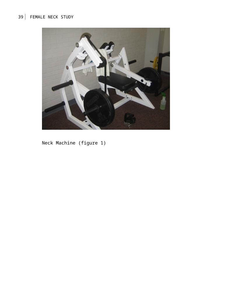
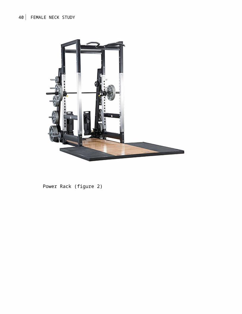
Materials
The equipment used for this study were prototypes. The
prototypical machines allowed for the participant to safely train
the musculature of the head and the neck. Neck circumference
17 FEMALE NECK STUDY
baseline measurements were taken using a medical grade tape
measure. The United States Army Protocol for measuring the neck
was used as a guide (Gordan & Brandtmiller, 1992). A
professional grade power rack was used to perform the shoulder
girdle elevation in conjunction with a standard 7 foot Olympic
bar. Olympic weight plates were used as resistance devices.
(Insert figures 1-3 about here)
Procedures
Obtain signed informed consent documents. Allow research
subjects time to familarize themselves with the equipment that
will be used in the research and the protocol. Take baseline
measurements of the neck cicumference using The United States
Military Standardized Protocol (USMSP). The USMSP requires 1
measurement for females. Female circumference measurements are
taken at the center measurement of the neck (Gordan &
Brandtmiller, 1992). A set schedule for individual training
sessions is composed to allow for one-on-one training sessions
with each active participant. The sessions will consist of 20
18 FEMALE NECK STUDY
minutes of training protocol three times in a seven day span for
an 8 week period.
Exercise Protocol
All exercise protocols were performed in a sports performance
laboratory. The research was conducted in a university setting
located in the southeastern region of the United States. A
starting weight was determined by the amount of weight a
participant could safely use while performing the Protocol for 12
repetitions in good form. There will be a 15 second rest period
between sets.
The target repetition range is 12 or until a repetition cannot
be performed with good form. Neck circumference measurements are
taken at the beginning of each training session. Data was
collected on training cards then uploades into a password
protected data base.
The test subjects performed 6 head and neck movements using
the Head and Neck Machine. They are: front flexion, extension,
lateral flexion both right and left; the Nod, 10 degrees of front
flexion (the movement resembles a person nodding "yes"), and the
19 FEMALE NECK STUDY
tilt (this movement is 25 degrees of flexion, the jaw is jutted
outward and head is gently tilted back). The 35 degrees range of
motion represents the movement of the head that does not directly
activate the cervicle neck musculature with the exception of the
atlas and axis vertbrea. By isolating the muscles of the head,
this allows for the hypertrophy of the capital muscles of the
head.
This was followed by a seated bilateral shrug, also
performed on the protypical head and neck machine to innervate
the lower trapezius muscles. A unilateral shrug was then
performed on the same machine to innervate the upper trapezius.
Next the Levator Scapula Shoulder Elevaton Shrug (LSSES) is a
movement to innervate the upper trapezius and the muscles
surrounding and involved in scapular retraction. The LSSES was
performed by placing a 7 foot standard olympic bar on the
posterior of the neck, at the nape or appoximately at cervical
vertebrae 7. The subject then performs scapular retraction. The
retraction of the scapula allows the bar to rise vertically at
that point the trapezius shrugs vertically. This allow subject to
20 FEMALE NECK STUDY
train upper trapezius and other muscles without the limiting the
factor of grip strength.
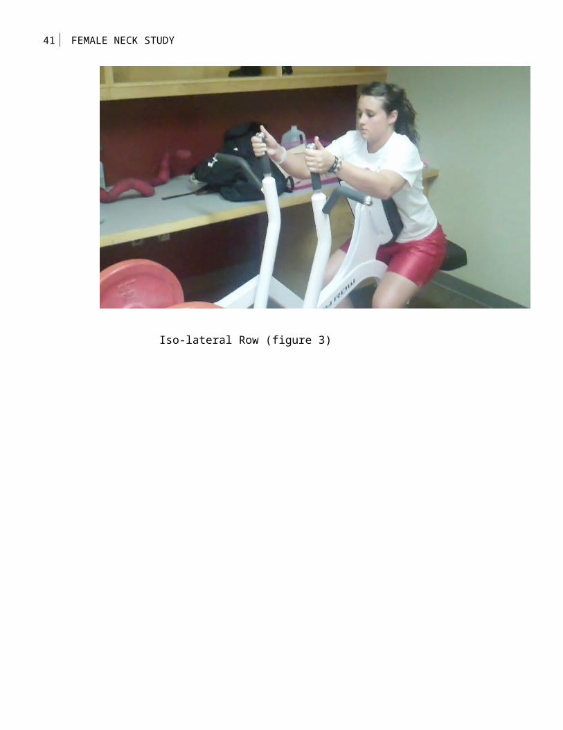
One set of seated rows was performed on the 3-way row using
a parallel grip, this movement allows for the innervation of the
large muscles of the back (i.e. latissimus dorsi, rhomboids major
and minor with contribution of the posterior deltoid). A scapular
shrug was performed on the 3-way Row to involve the muscles of
the upper back, posterior deltoid and the rhomboids that are
involved in scapular retraction. The scapular shrug movement
requires the particpant to keep the arms straight as they use a
parallel grip, then the scapula is retracted. It is the
retraction of the scapula and contraction of the upper back
muscles that successfully moves the weight loaded onto the row.
The Retraction and Pull is accomplished by using a supinated grip
on the other horizontial handles. With straight arms and
retraction of the scapula, participant then flexed elbows at 90
degrees appoximately 8 to 12 inches allowing for maximum
innervation of the middle trapezius and fibers to the lowest
fibers terminateing at thoracic vertebre 12 musculature.
21 FEMALE NECK STUDY
(Insert figures 4-15 about here)
Results
The female participant’s experienced significant
strength gains. All of the females gained upper body strength.
The head and neck muscles were the most impressive result of this
study. We had a participant increase her neck strength in
extension, flexion, and lateral flexion (right and left) by 40
pounds. The strength of the capital muscles was equally
significant. One participant increased their capital movements by
40 pounds. Each of the strength gains represented the amount of
weight the participant could lift in good form for 12
repetitions. Although statistically impossible to quantify; a
phenomenon was observed by the researcher. During weeks 4-6,
we made two key observations: an improvement in protocol form and
reduction of speed of movement. Together these two observations
suggest an increase in the participants’ true strength and muscle
control both concentrically and eccentrically. The muscles were
forced to work harder due to the reduction of speed of movement,
resulting in the virtual elimination of momentum in the protocol.
22 FEMALE NECK STUDY
For all neck and rowing exercises performed the Wilcoxon Signed-
Rank Test utilized to determine if the exercises had resulted in
a significant increase in neck and upper body strength. The
increase in neck and upper body strength is compute by
FinalWeight−BaselineWeight. I chose the nonparametric Wilcoxon
Signed-Rank Test due to the small sample size (6 subjects).
However, I also ran the paired T-test and noticed that the
results from the parametric test agree with the results from the
nonparametric test. There was no visible hypertrophy. Final
circumference measurements revealed only one active participant
exhibiting a minimal increase in neck circumference. The increase
totaled 1/32 of an inch. Conversely, there were no neck
circumference changes in the control group.
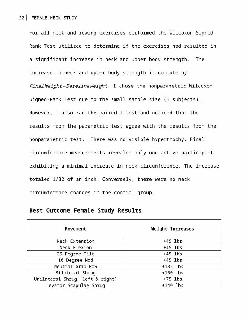
Best Outcome Female Study Results
Movement Weight Increases
Neck Extension +45 lbsNeck Flexion +45 lbs
25 Degree Tilt +45 lbs10 Degree Nod +45 lbs
Neutral Grip Row +185 lbsBilateral Shrug +150 lbs
Unilateral Shrug (left & right) +75 lbsLevator Scapulae Shrug +140 lbs
23 FEMALE NECK STUDY
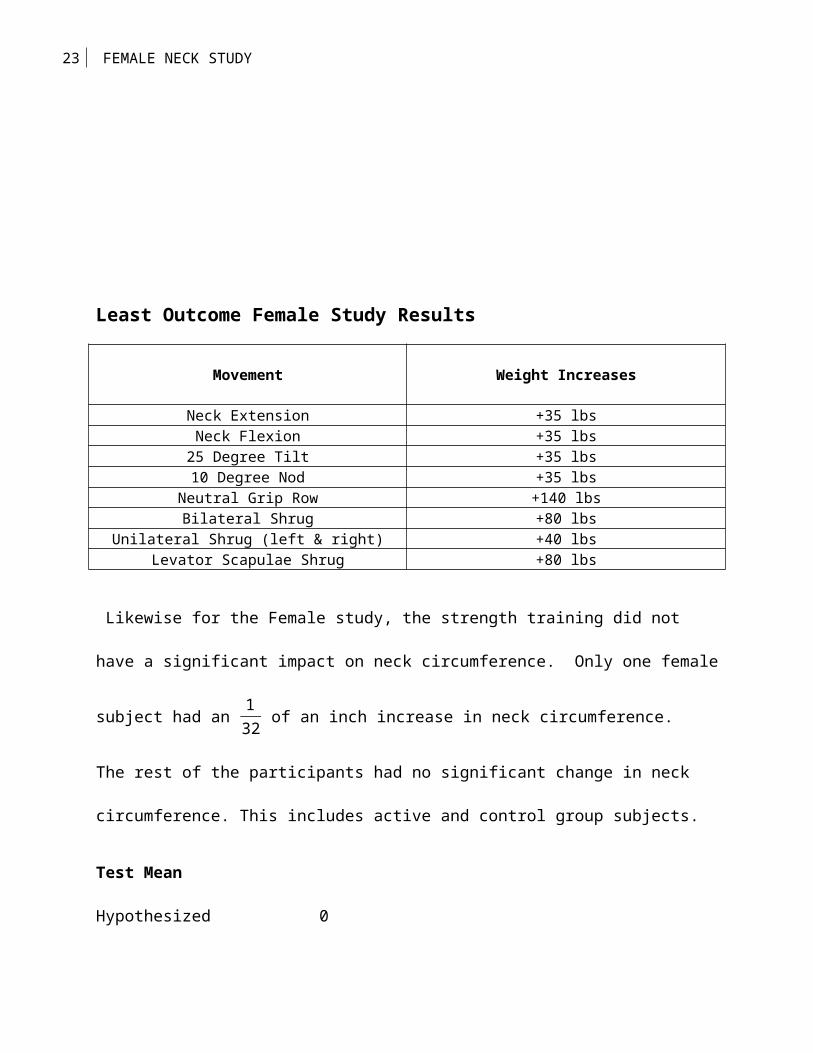
Least Outcome Female Study Results
Movement Weight Increases
Neck Extension +35 lbsNeck Flexion +35 lbs
25 Degree Tilt +35 lbs10 Degree Nod +35 lbs
Neutral Grip Row +140 lbsBilateral Shrug +80 lbs
Unilateral Shrug (left & right) +40 lbsLevator Scapulae Shrug +80 lbs
Likewise for the Female study, the strength training did not
have a significant impact on neck circumference. Only one female
subject had an 132 of an inch increase in neck circumference.
The rest of the participants had no significant change in neck
circumference. This includes active and control group subjects.
Test Mean
Hypothesized 0
24 FEMALE NECK STUDY
Value
Actual Estimate 0.00522
DF 5
Std Dev 0.01278
t Test Signed-Rank
Test
Statistic
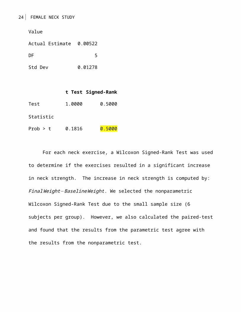
1.0000 0.5000
Prob > t 0.1816 0.5000
For each neck exercise, a Wilcoxon Signed-Rank Test was used
to determine if the exercises resulted in a significant increase
in neck strength. The increase in neck strength is computed by:
FinalWeight−BaselineWeight. We selected the nonparametric
Wilcoxon Signed-Rank Test due to the small sample size (6
subjects per group). However, we also calculated the paired-test
and found that the results from the parametric test agree with
the results from the nonparametric test.
25 FEMALE NECK STUDY
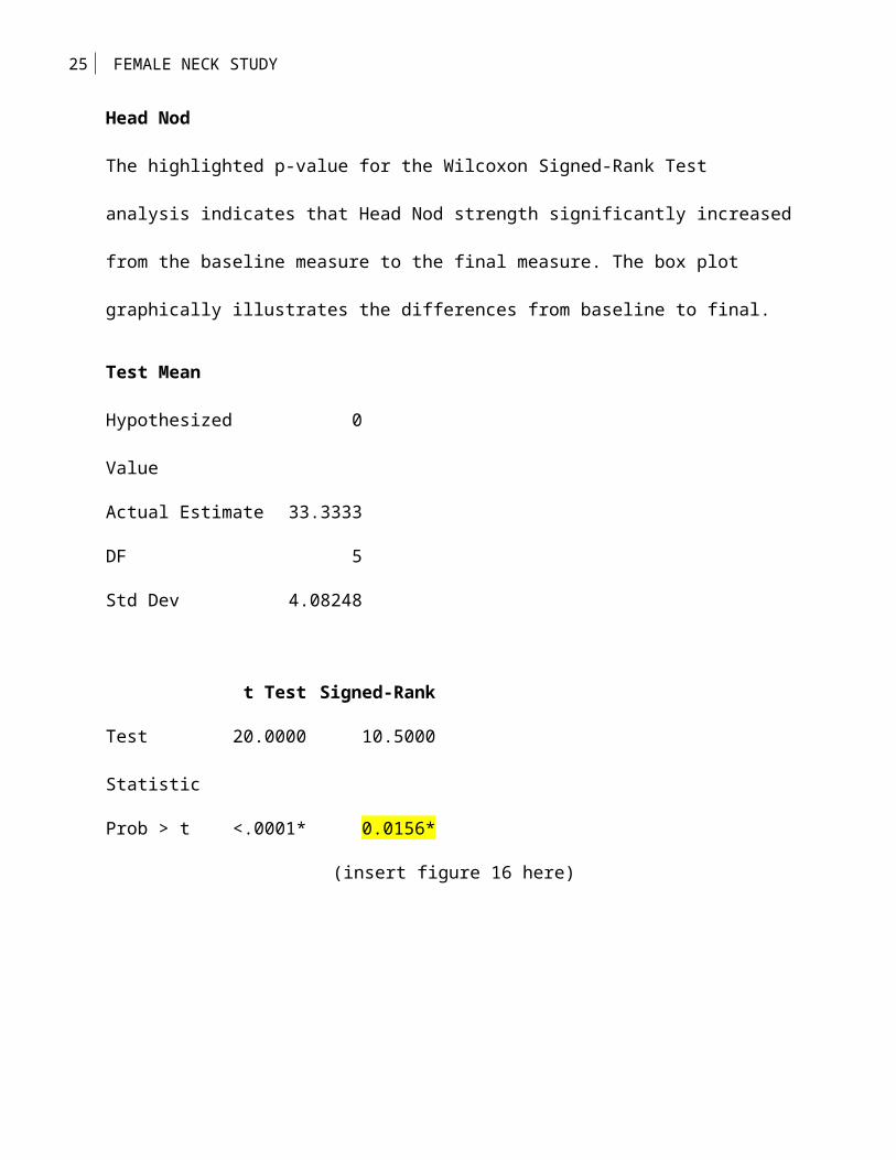
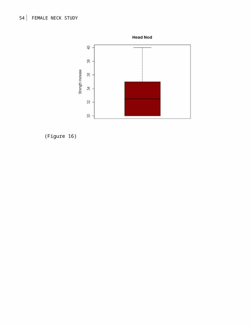
Head Nod
The highlighted p-value for the Wilcoxon Signed-Rank Test
analysis indicates that Head Nod strength significantly increased
from the baseline measure to the final measure. The box plot
graphically illustrates the differences from baseline to final.
Test Mean
Hypothesized
Value
0
Actual Estimate 33.3333
DF 5
Std Dev 4.08248
t Test Signed-Rank
Test
Statistic
20.0000 10.5000
Prob > t <.0001* 0.0156*
(insert figure 16 here)
26 FEMALE NECK STUDY
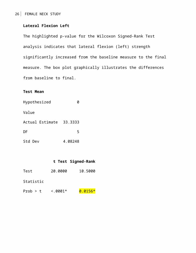
Lateral Flexion Left
The highlighted p-value for the Wilcoxon Signed-Rank Test
analysis indicates that lateral flexion (left) strength
significantly increased from the baseline measure to the final
measure. The box plot graphically illustrates the differences
from baseline to final.
Test Mean
Hypothesized
Value
0
Actual Estimate 33.3333
DF 5
Std Dev 4.08248
t Test Signed-Rank
Test
Statistic
20.0000 10.5000
Prob > t <.0001* 0.0156*
27 FEMALE NECK STUDY
(Insert figure 17 here)
Lateral Flexion Right
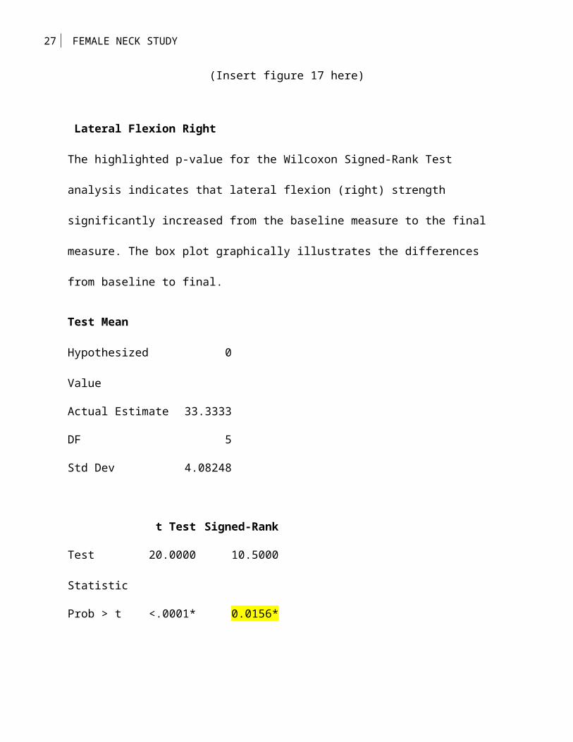
The highlighted p-value for the Wilcoxon Signed-Rank Test
analysis indicates that lateral flexion (right) strength
significantly increased from the baseline measure to the final
measure. The box plot graphically illustrates the differences
from baseline to final.
Test Mean
Hypothesized
Value
0
Actual Estimate 33.3333
DF 5
Std Dev 4.08248
t Test Signed-Rank
Test
Statistic
20.0000 10.5000
Prob > t <.0001* 0.0156*
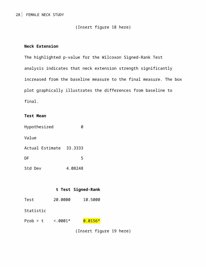
28 FEMALE NECK STUDY
(Insert figure 18 here)
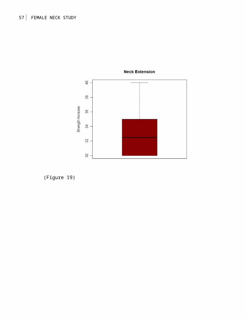
Neck Extension
The highlighted p-value for the Wilcoxon Signed-Rank Test
analysis indicates that neck extension strength significantly
increased from the baseline measure to the final measure. The box
plot graphically illustrates the differences from baseline to
final.
Test Mean
Hypothesized
Value
0
Actual Estimate 33.3333
DF 5
Std Dev 4.08248
t Test Signed-Rank
Test
Statistic
20.0000 10.5000
Prob > t <.0001* 0.0156*
(Insert figure 19 here)
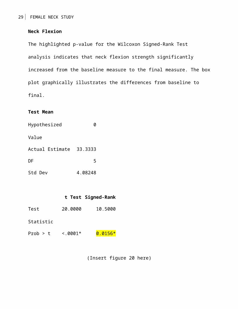
29 FEMALE NECK STUDY
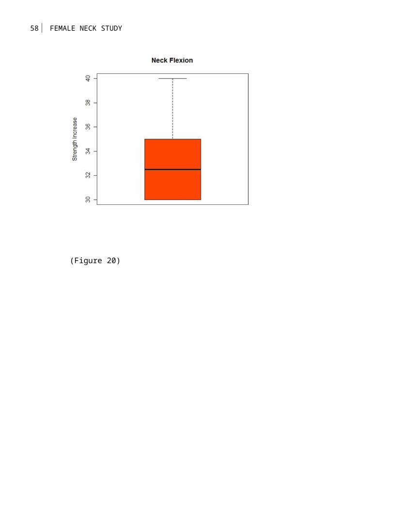
Neck Flexion
The highlighted p-value for the Wilcoxon Signed-Rank Test
analysis indicates that neck flexion strength significantly
increased from the baseline measure to the final measure. The box
plot graphically illustrates the differences from baseline to
final.
Test Mean
Hypothesized
Value
0
Actual Estimate 33.3333
DF 5
Std Dev 4.08248
t Test Signed-Rank
Test
Statistic
20.0000 10.5000
Prob > t <.0001* 0.0156*
(Insert figure 20 here)
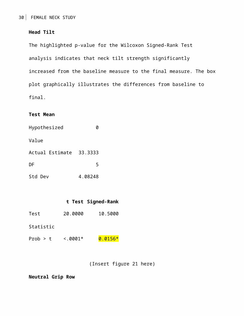
30 FEMALE NECK STUDY
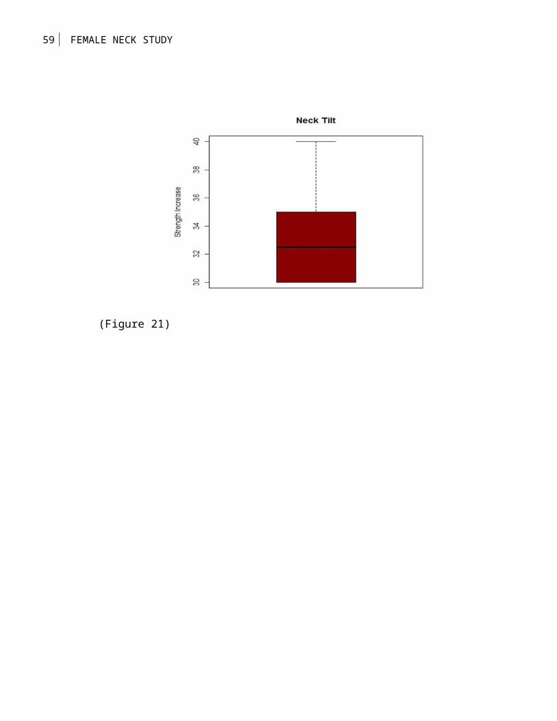
Head Tilt
The highlighted p-value for the Wilcoxon Signed-Rank Test
analysis indicates that neck tilt strength significantly
increased from the baseline measure to the final measure. The box
plot graphically illustrates the differences from baseline to
final.
Test Mean
Hypothesized
Value
0
Actual Estimate 33.3333
DF 5
Std Dev 4.08248
t Test Signed-Rank
Test
Statistic
20.0000 10.5000
Prob > t <.0001* 0.0156*
(Insert figure 21 here)
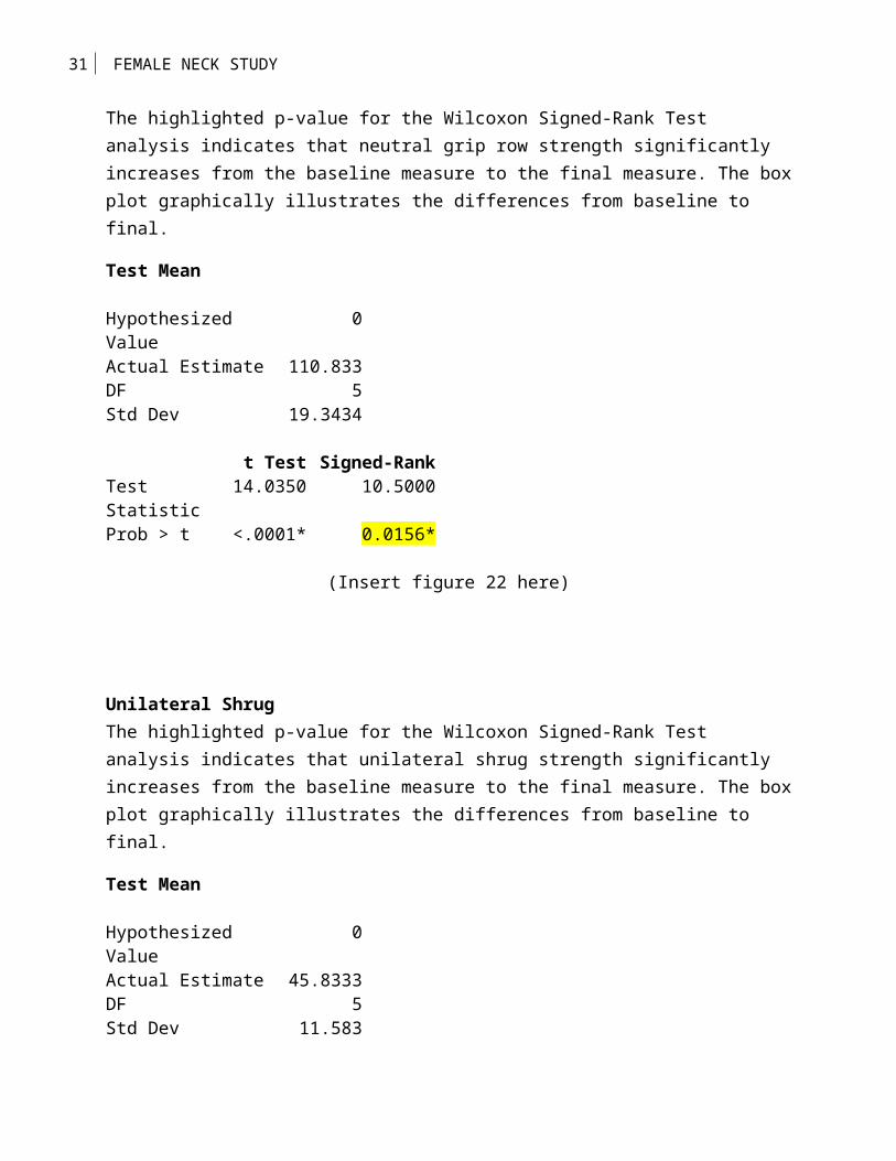
Neutral Grip Row
31 FEMALE NECK STUDY
The highlighted p-value for the Wilcoxon Signed-Rank Test analysis indicates that neutral grip row strength significantly increases from the baseline measure to the final measure. The boxplot graphically illustrates the differences from baseline to final.
Test Mean
Hypothesized Value
0
Actual Estimate 110.833DF 5Std Dev 19.3434
t Test Signed-RankTest Statistic
14.0350 10.5000
Prob > t <.0001* 0.0156*
(Insert figure 22 here)
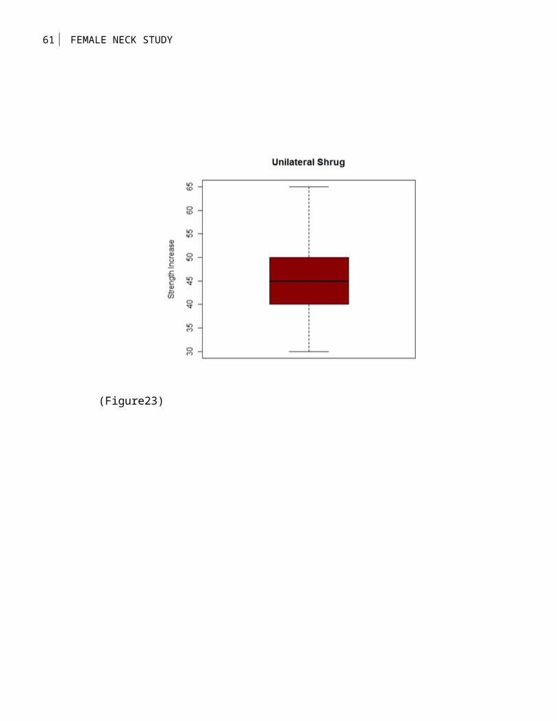
Unilateral ShrugThe highlighted p-value for the Wilcoxon Signed-Rank Test analysis indicates that unilateral shrug strength significantly increases from the baseline measure to the final measure. The boxplot graphically illustrates the differences from baseline to final.
Test Mean
Hypothesized Value
0
Actual Estimate 45.8333DF 5Std Dev 11.583

32 FEMALE NECK STUDY
t Test Signed-RankTest Statistic
9.6925 10.5000
Prob > t <.0001* 0.0156*
(Insert figure 23 here)
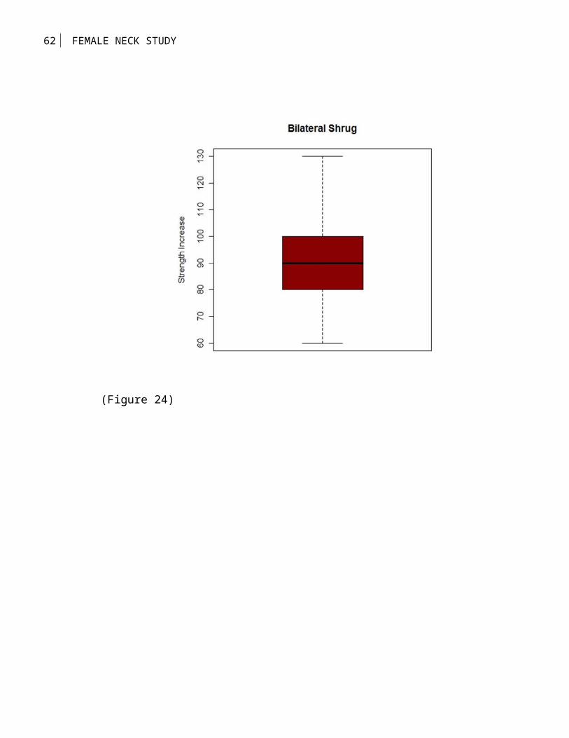
Bilateral ShrugThe highlighted p-value for the Wilcoxon Signed-Rank Test analysis indicates that bilateral shrug strength significantly increases from the baseline measure to the final measure. The boxplot graphically illustrates the differences from baseline to final.
Test Mean
Hypothesized Value
0
Actual Estimate 91.6667DF 5Std Dev 23.1661
t Test Signed-RankTest Statistic
9.6925 10.5000
Prob > t <.0001* 0.0156*
(Insert figure 24 here)
33 FEMALE NECK STUDY
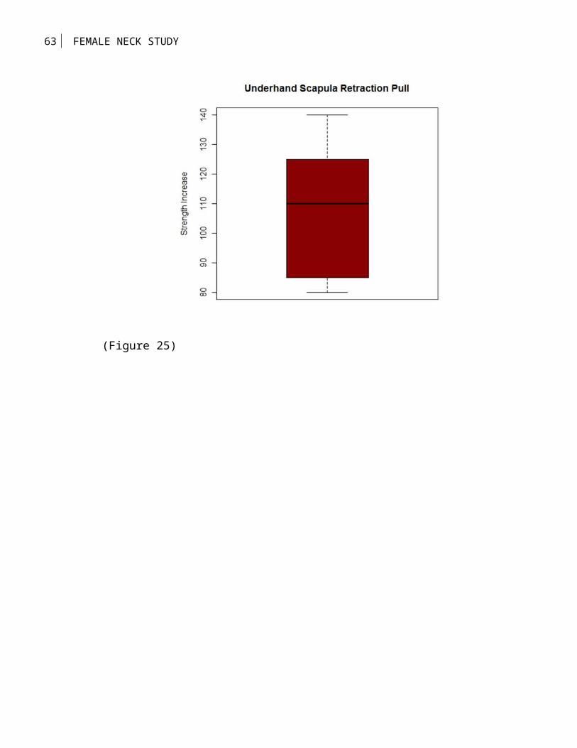
Underhand Scapula Retraction PullThe highlighted p-value for the Wilcoxon Signed-Rank Test analysis indicates that underhand scapula retraction pull strength significantly increases from the baseline measure to thefinal measure. The box plot graphically illustrates the differences from baseline to final.
Test Mean
Hypothesized Value
0
Actual Estimate 108.333DF 5Std Dev 23.8048
t Test Signed-RankTest Statistic
11.1474 10.5000
Prob > t <.0001* 0.0156*
(Insert figure 25 here)
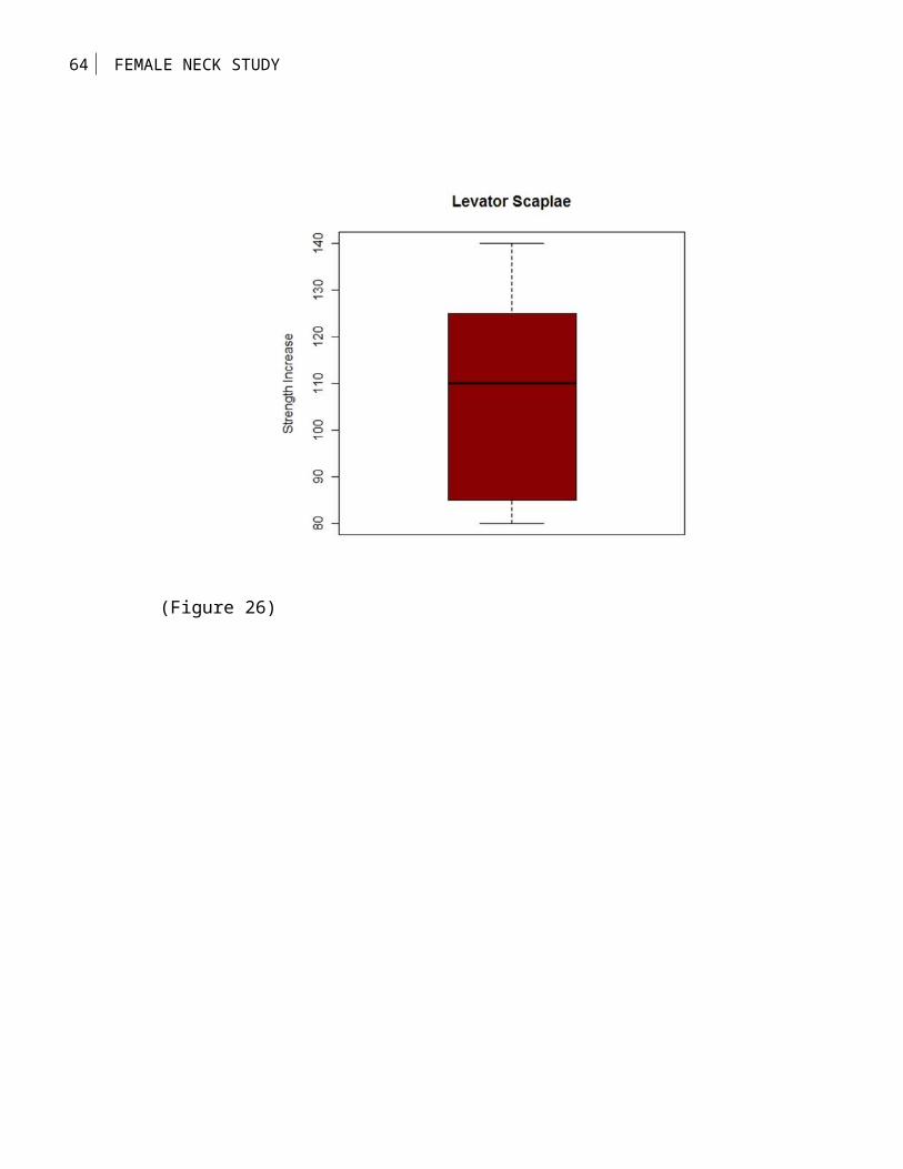
Levator Scaplae ShrugThe highlighted p-value for the Wilcoxon Signed-Rank Test analysis indicates that levator scapulae strength significantly increases from the baseline measure to the final measure. The boxplot graphically illustrates the differences from baseline to final.
Test Mean
Hypothesized Value
0
Actual Estimate 108.333
34 FEMALE NECK STUDY
DF 5Std Dev 23.8048
t Test Signed-RankTest Statistic
11.1474 10.5000
Prob > t <.0001* 0.0156*
(Insert figure 26 here)
Scapula RetractionThe highlighted p-value for the Wilcoxon Signed-Rank Test analysis indicates that scapula retraction strength significantlyincreases from the baseline measure to the final measure. The boxplot graphically illustrates the differences from baseline to final.
Test Mean
Hypothesized Value
0
Actual Estimate 110.833DF 5Std Dev 19.3434
t Test Signed-RankTest Statistic
14.0350 10.5000
Prob > t <.0001* 0.0156*
(Insert figure 27 here)
35 FEMALE NECK STUDY
Discussion
The results of this study demonstrate that females can
increase upper body strength safely and without significant
muscular size gains. The female neck during this study showed a
very minimal increase in circumference while strength level
increases were substantial. The females did not exhibit the
hypertrophy of their male counterparts, in comparison with a
previous study by this author (the male study). The strength
gains obtained by the female participants will add stiffness to
the head and neck musculature. To the researchers knowledge the
capital muscles had never been isolated and aggressively trained
in the allotted 35 degrees of movement at cervical levels 1 axis
and 2 atlas. This researcher hypothesizes that the
strength/stiffness increase will lower both concussive and sub-
concussive forces. Year around adherence to the proposed
protocol will result in reduction of head displacement due to
capital (head) and neck strength increases. It is intuitive that
a stronger athlete will be a better protected athlete and less
susceptible to injury. A properly trained and conditioned head
and neck segment will increase performances as well as
36 FEMALE NECK STUDY
protection. Kinetic energy is more effectively dissipated by the
properly trained and prepared muscles of the head, neck, and
upper back including the shoulder girdle. During weeks 4-6 we
observed strength increases coupled with performing the movements
in better protocol form, and reduced speed of movement both
concentrically and eccentrically, thus indicating greater
strength and muscle control. With reduced deflection you educe
lower deformation of the affected area. If the body is to be
prepared for competition, strengthening and protection of the
head neck should certainly be of the highest priority. An
interesting side note; recruiting females for the research study
was extremely difficult because of the unwarranted fear of a
developing an enormous neck. At week 5 of the study the peers of
the participants noticed no increase in neck size but fitter
female were emerging due the participant’s efforts. At week 6 we
had a waiting list of 15 that wanted to be involved in the study.
I see this as a true paradigm shift and step in right direction
removing untruths about females and developing large muscles.
Conclusion
37 FEMALE NECK STUDY
There is desperate need for a standardized head and neck
resistance training protocol that should be adopted nationwide.
As concussion rates continue to increase their mounts a
preponderance of evidence showing stronger, larger head and neck
muscles lowers the susceptibility of becoming concussed. The
scale of subconcussive damage will not be known until years after
an athlete leaves competitive sports. Once that tipping point has
been reached it will be too late for those athletes in a
preventionary sense because the damage has been done. Instead of
managing concussions better we should prepare our athletes
better. Not having a concussion would be much better for the
athlete than managing one. Proactivity is the key to combating
this debilitating epidemic. Educating coaching staffs, athletic
trainers, strength coaches and team physicians to not only be
aware of concussions but aluminate the proper methods of safe,
effective and prudent strength training principles. At the
completion of the study our collected data revealed tremendous
strength increases that is believed will translate in a much more
resilient athlete that can tolerate the forces both concussive
and subconcussive of the particular sport.

38 FEMALE NECK STUDY
39 FEMALE NECK STUDY
Neck Machine (figure 1)
40 FEMALE NECK STUDY
Power Rack (figure 2)
41 FEMALE NECK STUDY
Iso-lateral Row (figure 3)
42 FEMALE NECK STUDY
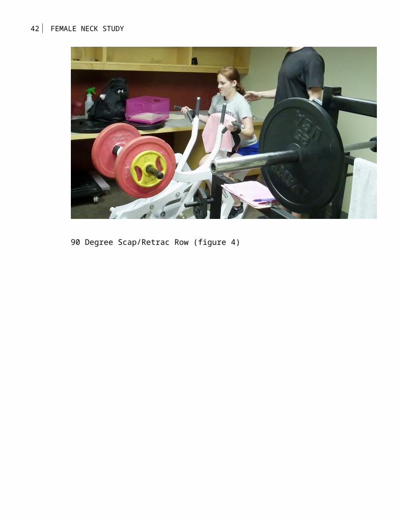
90 Degree Scap/Retrac Row (figure 4)
43 FEMALE NECK STUDY
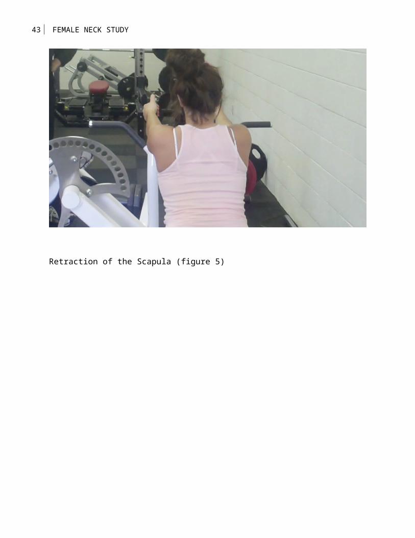
Retraction of the Scapula (figure 5)
44 FEMALE NECK STUDY
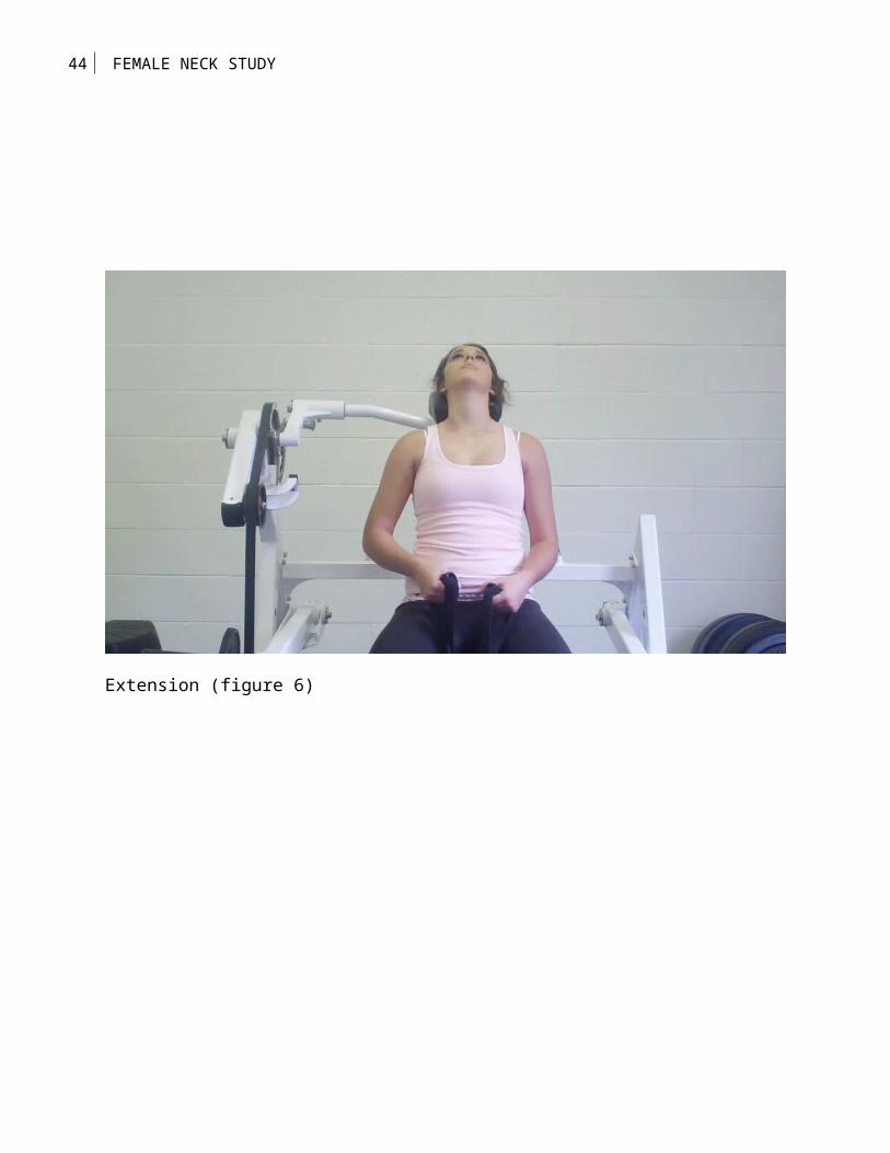
Extension (figure 6)
45 FEMALE NECK STUDY
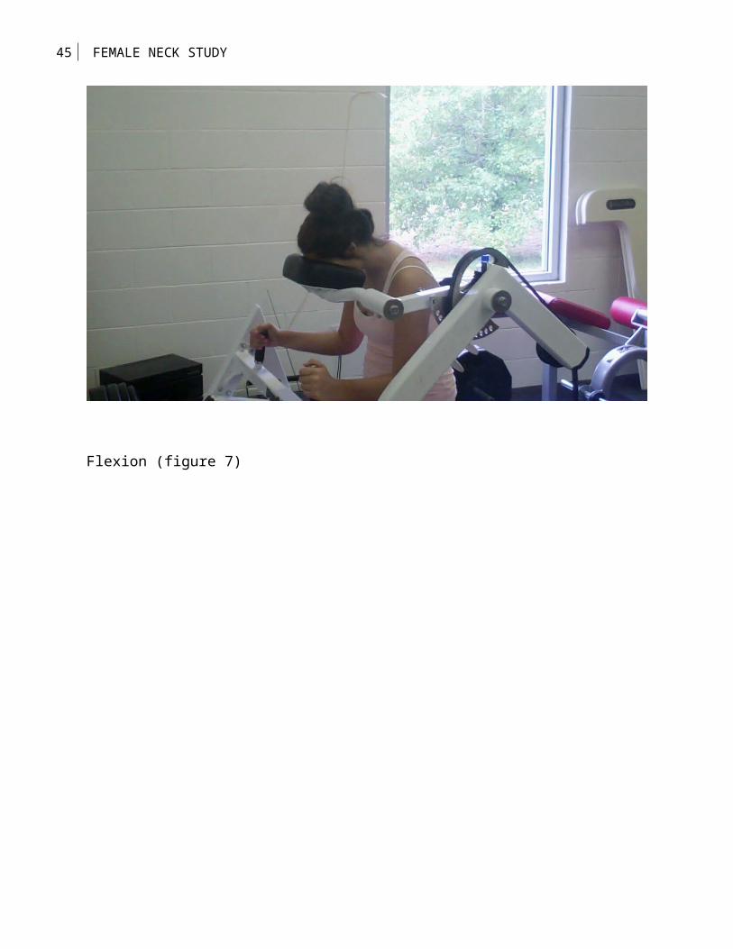
Flexion (figure 7)
46 FEMALE NECK STUDY
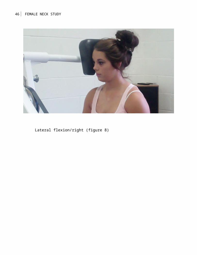
Lateral flexion/right (figure 8)
47 FEMALE NECK STUDY
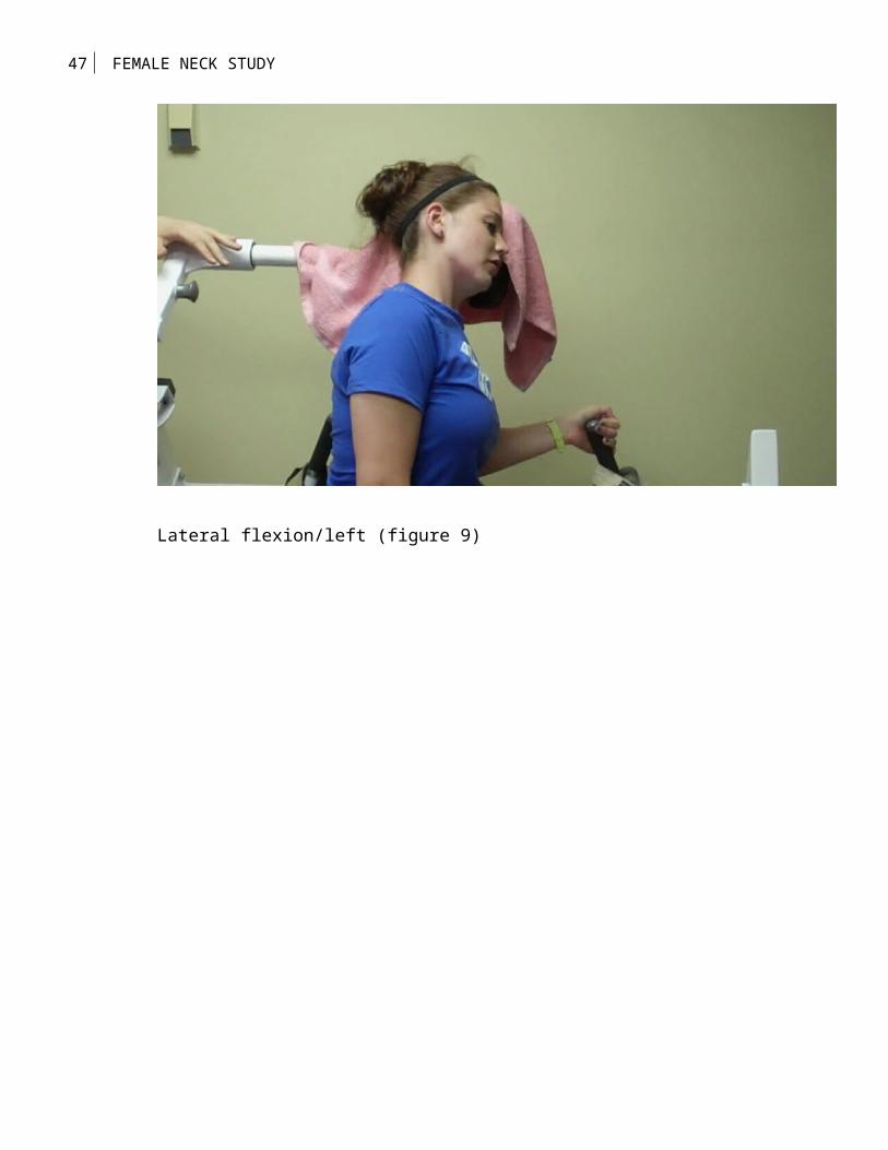
Lateral flexion/left (figure 9)
48 FEMALE NECK STUDY
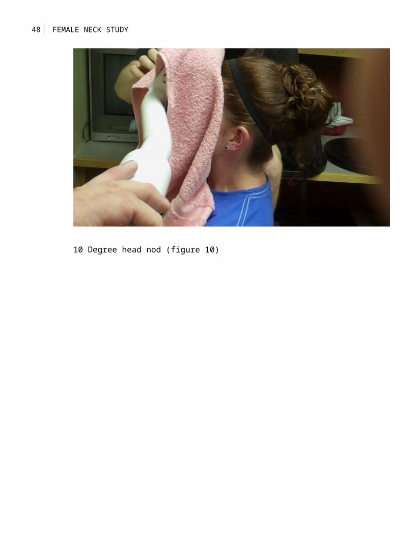
10 Degree head nod (figure 10)
49 FEMALE NECK STUDY
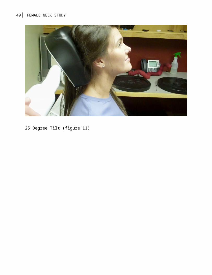
25 Degree Tilt (figure 11)
50 FEMALE NECK STUDY
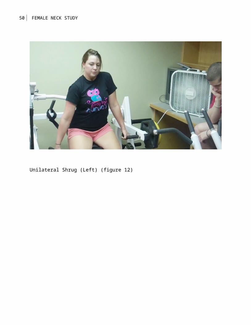
Unilateral Shrug (Left) (figure 12)
51 FEMALE NECK STUDY
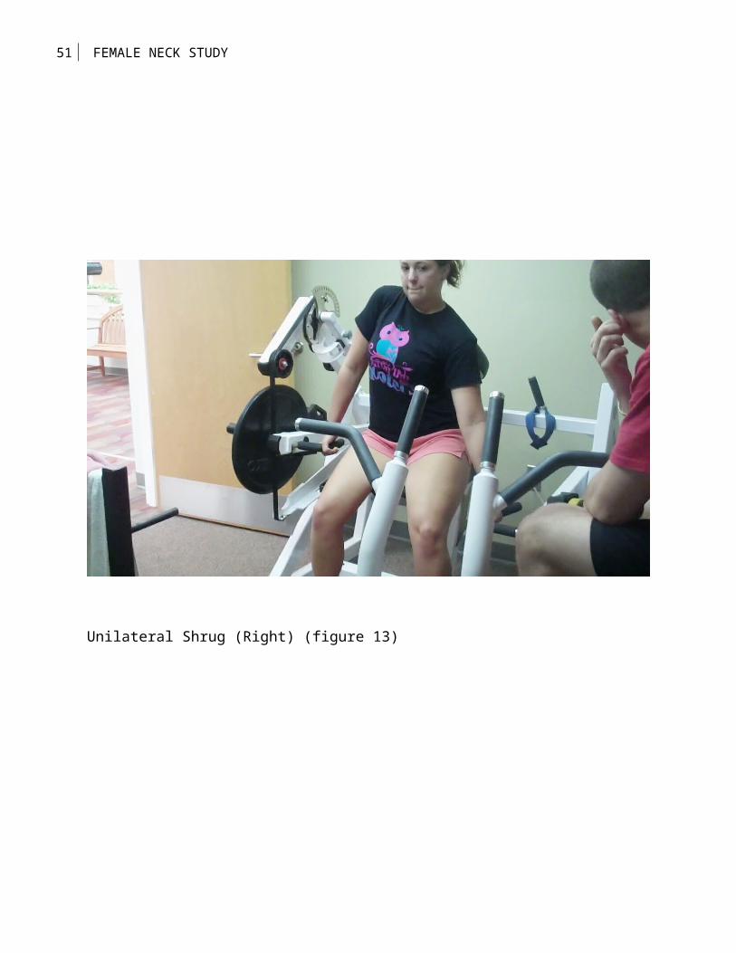
Unilateral Shrug (Right) (figure 13)
52 FEMALE NECK STUDY
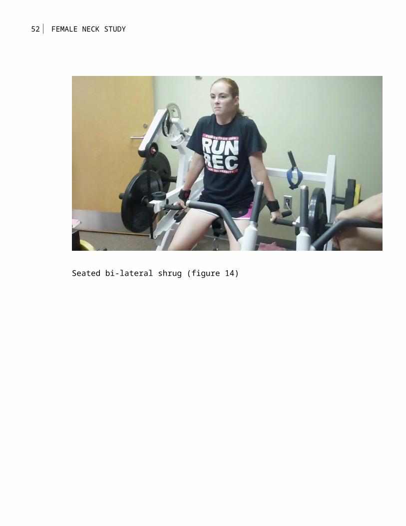
Seated bi-lateral shrug (figure 14)
53 FEMALE NECK STUDY
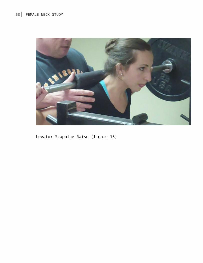
Levator Scapulae Raise (figure 15)
54 FEMALE NECK STUDY
(Figure 16)

55 FEMALE NECK STUDY
(Figure 17)

56 FEMALE NECK STUDY
(Figure 18)
57 FEMALE NECK STUDY
(Figure 19)
58 FEMALE NECK STUDY
(Figure 20)
59 FEMALE NECK STUDY
(Figure 21)

60 FEMALE NECK STUDY
(Figure 22)
61 FEMALE NECK STUDY
(Figure23)
62 FEMALE NECK STUDY
(Figure 24)
63 FEMALE NECK STUDY
(Figure 25)
64 FEMALE NECK STUDY
(Figure 26)

65 FEMALE NECK STUDY
(Figure 27)
66 FEMALE NECK STUDY
References
Berg, H.E., Gunnel, M.S. and Tesch, P.A. (1994). Dynamic neck
strength training effect on pain. Physical Medicine and
Rehabilitation, 75, 661-665.
Cantu, R. C. (1996). Head injuries in sport. British Journal of
Sports Medicine, 30, 289-296.
Conley, Stone, Nimmons & Dudley, (1997). Specificity of
resistance training responses in neck muscle size and
strength. European Journal of Applied Physiology and
Occupational Physiology, 75 (5), 443-8
Cross, K. and Serenelli, C. (2003). Training and Equipment to
prevent athletic head and neck injuries. Clinical Sport
Medicine, 22, 639-667.
67 FEMALE NECK STUDY
Dugan, S. (2005). Sports related knee injuries in female
athletes. American Journal of Physical Medicine and
Rehabilitation, 84 (2), 122-128.
Gordan, C. C. and Brandtmiller, B. (1992). Interobserver error in
a large scales anthropometric survey. American Journal of
Human Biology, 4, 253-263.
http://www.sportslegacy.org/cte-concussions/cte-cases-sli-legacy-
donors/
(Johnston et al., 2001) Johnston, K.M., McCrory, P., Mohtadi,
N.G & Meeuwisse, W. (2001). Evidence based review of sport
related concussion: Clinical science. Clinical Journal of
Sports Medicine, 11, 150-159.
Mallika, Marar, McIlivain, Natalie, Fields, Sarah, Comstock,
Dawn, (2012). Epidemiology of Concussions Among United
States High School Athletes in 20 Sports. The American
College Sports Medicine, 40 747-755
68 FEMALE NECK STUDY
Marino, F.E. (2011). The critical limiting temperature and
selective brain cooling: neuroprotection during exercise.
International Journal of Hyperthermia, 27 (6), 582-590.
McGill, S. M., Jones, K., Bennett, G., Bishop P. J. (1994).
Passive stiffness of the human neck in flexion, extension,
and lateral bending. Clinical Biomechanics, 9 (3), 193-198.
Peterson, N.C., Taylor, J.L., Murray, P.S., Gandevia, S.C.,
Butler, J. (2011). Differential effects of low-intensity
motor cortical stimulation on the inspiratory activity in
scalene muscles during voluntary and involuntary breathing.
Respiratory Physiological Neurobiology, 175, 265-271.
Reid, S, E.,Reviv, Gil, Reid S. E. Jr. (1981). Neck Muscle
Response to Head Impact. Aviation, Space and Environmental
Medicine, 52 (2), 78-84
Reid S. E., & Reid S. E. Jr. (1981). Advances in sports medicine:
Prevention of head and neck injuries in football. Surgeons
Annual, 13, 251-270.
69 FEMALE NECK STUDY
Riley, D. (1981). Strength training for the neck. The Physician
and Sports Medicine, 9, 165.
Rousseau, P. and Hoshizaki, T.B. (2009). The influence of
deflection and neck compliance on impact dynamics of a
hybrid III hear form. Journal of Sports Engineering and
Technology, v223 issue 3, 89-97.
Scheip, R., Naglor, K., Ursa, D., Mentzer, W., Wilke, H.J.,
Lehman-Horn, F. and Klingler, W. (2006). Passive muscle
stiffness may be influenced by active controllability of
intramuscular connective tissue. Medical Hypotheses (1), 66.
Tierney, R. T., Higgins, M., Caswell, S. V., Brady, J., McHardy,
K., Driban, J. B., & Darvish, K. (2008). Sex differences in
head acceleration during heading while wearing soccer
headgear. Journal of Athletic Training, 43(6), 578–584.
Valentin, I. (1997). Title IX: A brief history. WEEA Digest, 8,
1-12.
Top Related
Copyright © 2022 FDOKUMEN

