and retractor penis muscles of mammals, with special ... - NCBI
20
[ 61 OBSERVATIONS ON THE RETRACTOR CLITORIDIS AND RETRACTOR PENIS MUSCLES OF MAMMALS, WITH SPECIAL REFERENCE TO THE EWE BY E. G. BASSETT* Ruakura Animal Research Station, Department of Agriculture, Hamilton, New Zealand INTRODUCTION Smooth muscle, although generally described as one of the components of a parti- cular organ, occasionally resembles striated muscle in being grouped longitudinally into a separate entity. Striking examples of this peculiarity are to be found in the bilateral retractor penis muscle and its female homologue, which are comparable in form with the adjacent perineal skeletal muscles. In the male, the retractor penis is usually described as being attached to the ventral surface of the first or second coccygeal vertebra, and continuing in a caudo- ventral direction between the levator ani muscle and the rectum, to be inserted into the penis towards the end of its free extremity.t The female homologue is less frequently recorded. It is commnonly known as the retractor clitoridis, but also has been called by such names as, in the mare, the anogenital ligament (Ellenberger & Baum, 1921; M'Fadyean, 1922), the muscular ligament of the vulva (Fleming, 1873), the labial ligament (Habel, 1953); in the cat, the caudovaginalis muscle (Davison, 1923); and the retractor cloacae muscle (Nishi, 1938) in animals with a common urorectal opening. Some authors have stated that the muscle terminates in the clitoris, but others are less definite, saying merely that it goes to the genitals, or that it terminates in the vulva, or that it disappears among the fibres of the constrictor vulvae muscle. Although the most commonly described origin of the retractor penis is from the first or second coccygeal vertebra, a sacral attachment is occasionally mentioned. In some species, and perhaps also breeds, there is no rectococcygeal part, the muscle arising proximally in these instances in the lateral rectal wall. Such a rectal origin is recorded for the dog (Paulet, 1877; Langley & Anderson, 1895; Bradley, 1948; although a coccygeal origin has also been described-Miller, 1952); according to Paulet (1877), it is the same in wolves, and certain species of deer, although in one species he found a sacral attachment. Mivart (1881) gives a rectal origin for the retractor penis of the cat. In the horse, in which a coccygeal (or sacral) origin is constant, the muscles from each side unite beneath the rectum to form a sling, before * Present address: Endocrinology Department, Medical Research Council of New Zealand, Medical School, Dunedin, New Zealand. t Some early anatomists (e.g. Owen, 1868) describe levatores as well as retractores penis muscles in several species, arising from the pubis and serving mainly as compressors of the vena dorsalis penis; levatores clitoridis muscles have been recorded in the female elephant, though the origin was not stated (Paterson & Dun, 1898). Most later writers do not mention two such distinct muscles, however-only retractores.
-
Upload
khangminh22 -
Category
Documents
-
view
0 -
download
0
Transcript of and retractor penis muscles of mammals, with special ... - NCBI

[ 61
OBSERVATIONS ON THE RETRACTOR CLITORIDISAND RETRACTOR PENIS MUSCLES OF MAMMALS,
WITH SPECIAL REFERENCE TO THE EWE
BY E. G. BASSETT*Ruakura Animal Research Station, Department of Agriculture,
Hamilton, New Zealand
INTRODUCTION
Smooth muscle, although generally described as one of the components of a parti-cular organ, occasionally resembles striated muscle in being grouped longitudinallyinto a separate entity. Striking examples of this peculiarity are to be found in thebilateral retractor penis muscle and its female homologue, which are comparable inform with the adjacent perineal skeletal muscles.
In the male, the retractor penis is usually described as being attached to theventral surface of the first or second coccygeal vertebra, and continuing in a caudo-ventral direction between the levator ani muscle and the rectum, to be inserted intothe penis towards the end of its free extremity.tThe female homologue is less frequently recorded. It is commnonly known as the
retractor clitoridis, but also has been called by such names as, in the mare, theanogenital ligament (Ellenberger & Baum, 1921; M'Fadyean, 1922), the muscularligament of the vulva (Fleming, 1873), the labial ligament (Habel, 1953); in the cat,the caudovaginalis muscle (Davison, 1923); and the retractor cloacae muscle (Nishi,1938) in animals with a common urorectal opening. Some authors have stated thatthe muscle terminates in the clitoris, but others are less definite, saying merely thatit goes to the genitals, or that it terminates in the vulva, or that it disappears amongthe fibres of the constrictor vulvae muscle.Although the most commonly described origin of the retractor penis is from the
first or second coccygeal vertebra, a sacral attachment is occasionally mentioned.In some species, and perhaps also breeds, there is no rectococcygeal part, the musclearising proximally in these instances in the lateral rectal wall. Such a rectal originis recorded for the dog (Paulet, 1877; Langley & Anderson, 1895; Bradley, 1948;although a coccygeal origin has also been described-Miller, 1952); according toPaulet (1877), it is the same in wolves, and certain species of deer, although in onespecies he found a sacral attachment. Mivart (1881) gives a rectal origin for theretractor penis of the cat. In the horse, in which a coccygeal (or sacral) origin isconstant, the muscles from each side unite beneath the rectum to form a sling, before
* Present address: Endocrinology Department, Medical Research Council of New Zealand,Medical School, Dunedin, New Zealand.
t Some early anatomists (e.g. Owen, 1868) describe levatores as well as retractores penismuscles in several species, arising from the pubis and serving mainly as compressors of the venadorsalis penis; levatores clitoridis muscles have been recorded in the female elephant, though theorigin was not stated (Paterson & Dun, 1898). Most later writers do not mention two such distinctmuscles, however-only retractores.

62 E. G. Bassettcontinuing to the penis or clitoris; as a result, the proximal (rectococcygeal) part ofthemuscle in the horse has frequently been termed the suspensory ligament of the anus,although its continuation as the rectopenile part has been appreciated. May (1955)described a similar suspensory ligament in the sheep; in the ram, it is continuouswith the retractor penis, but in the ewe there is no mention of a retractor clitoridis.The muscle is always described as long, cylindrical, pale, enclosed in a connective
tissue sheath, and composed of smooth muscle. In some species, dissections haveshown the main innervation of the retractores muscles to be from a branch of thepudendal nerve (bull-Larson, 1953; sheep-May, 1955). Although the chief inner-vation may be from the pudendal nerve, and this has been shown experimentallyto contain orthosympathetic fibres in certain mammals (cat and dog-Langley &Anderson, 1895; cat-Oppenheimer, 1938; bull-Larson, 1953), the pelvic plexusmay also contribute (Langley & Anderson, 1895). There is some indication thatsacral para-sympathetic fibres are also involved (Langley & Anderson, 1895;Oppenheimer, 1938), but the precise route taken by such fibres is unknown. Noreference can be found concerning its blood supply.The muscle is present in the horse (males-Owen, 1868; both sexes-Fleming,
1873; Ellenberger & Baum, 1921; M'Fadyean, 1922; Bradley, 1922; Sisson, 1953;Habel, 1953); cattle (males-Owen, 1868; Sisson, 1953; Larson, 1953; females-Geiger, 1956), sheep (males-May, 1955; Geiger, 1956) goat (Geiger, 1956), dog(males-Owen, 1868; Paulet, 1877; Bradley, 1948; Miller, 1952; both sexes-Langley & Anderson, 1895); cat (males-Mivart, 1881; Oppenheimer, 1938; bothsexes-Langley & Anderson, 1895; Davison, 1923). The retractor penis has beenrecorded also, for males only, in the tiger, wolf, three species of deer (Paulet, 1877).pig (Owen, 1868; Sisson, 1953), kangaroo (Owen, 1868; Paterson, 1907), platypus(Owen, 1868), and guinea-pig (Kochakian, Tillotson, Austin, Dougherty, Haag &Coalson, 1956). The retractor penis or clitoridis muscle is apparently absent in therabbit (Langley & Anderson, 1895; Bensley, 1948), the rat (Greene, 1955) and in man(Nishi, 1938). Nishi states that it is best developed in marsupials, carnivores andanimals with well-formed tails, and also commented that there is no homologousstructure in reptiles. Paulet (1877), who found no retractor penis in two species ofmonkey, considered that it occurs only in animals with a penis fixed in the abdominalwall; in primates, which have a free penis, it would be unnecessary from a functionalpoint of view. Paulet examined only males, and did not consider the probablefunction of the homologous muscle in the female.
Physiological experiments on the dog and cat (Langley & Anderson, 1895;Oppenheimer, 1938) and on the bull (Larson, 1953), suggest that in these species atleast, the retractores muscles function as their names imply. Sisson (1953) suggestedfurther that the proximal part of the muscle may act as an accessory anal sphincterin the horse. Habel (1953) considered that in the mare the 'labial ligament' maytransmit the pull of the ventral part of the levator ani muscle to the labia, and thuscause gaping of the vulva.No reference can be found to the embryology of this muscle in any species.The investigations described in this paper comprise a special study of the retractor
clitoridis muscle in the ewe-including its development, microscopical structure andalterations in pregnancy.

Retractor penis and retractor clitoridis musclesThe morphology and morphogenesis of the muscle were investigated as part of a
comprehensive study of the gross anatomy of the pelvic and perineal region of theewe (Bassett, 1956) and further observations have been made subsequently onanimals killed for other experiments.The chemical changes during pregnancy in the retractor clitoridis muscle have
been the subject of a separate experiment (unpublished) by another investigator,R. J. Newbold. A summary of his findings, communicated privately, is included.
Observations have been made also on the retractor clitoridis and retractor penismuscles in certain other species.
MATERIAL AND METHODS
In all, eleven different genera were investigated.Sheep
For gross anatomy, 126 ewes were dissected, of three different breeds and onecross-breed, ranging in age from 70 days prenatal to 8 years postnatal; two rams werealso included. Some of the specimens were fixed with 10% formalin, but the majoritywere dissected fresh.The early development of the retractor clitoridis muscle in relation to other
perineal structures was studied microscopically in a sectioned series of sevenembryos and foetuses of unknown breed with crown-rump lengths between 9-5 and46 mm. Relating crown-rump length to foetal age, it can be estimated that thelargest of these was approximately 42 days old (Malan & Curson, 1936; Winters &Feuffel, 1936; Harris, 1937); the smallest embryo was known to be 28 days old. Thegonads of the three youngest (9.5-15 mm.) were undifferentiated, and the other fourwere females. A graphical reconstruction was made from traced projected sections ofthe oldest (46 mm.) foetus.The microscopical structure was examined in one complete muscle (approximately
2J in. long) and on segments from the clitorial termination of the muscles of eweskilled to determine the effects of pregnancy. These tissues were fixed in 10% neutralformalin, sectioned at 4-6 ,u and stained with Weigert's haematoxylin, followed byvan Gieson and orcein, haematoxylin-azur II-eosin (Maximow, 1924), toluidineblue, and Long's (1948) silver impregnation method.The changes in pregnancy were studied in forty-eight ewes at several different
stages of gestation including a group of non-pregnant controls (Table 1). Themuscles from each side were dissected out and weighed together. A small portionwas taken for histology and the remainder used for chemical analysis.
Other speciesMorphological observations were made on the retractor clitoridis or retractor
penis muscles of six cattle, four goats, eleven pigs and on small numbers of cats,rabbits, guinea-pigs, rats, mice, hedgehogs and phalangers (Australian 'opossums').
63

64 E. G. Bassett
OBSERVATIONS
(1) The retractor clitoridis muscle in the mature ewe(a) Gross anatomy
Since the proximal attachment of the retractor clitoridis is medial to the insertionof the ischiococcygeus muscle, this muscle, and the fat and fascia covering it, must,therefore, be removed before this part of the retractor clitoridis is seen (Text-fig. 1).It appears as a whitish band attached to the fascia under the tail, in the area of the
l1.c.
D-ext.s ph.Tah
Text-fig 1 Laterocaudal view of the external pelvic and perineal muscles of amature ewe, with fascia and fat removed.
third-fourth (or occasionally between second-third) coccygeal vertebrae; here it isclosely associated with the unpaired rectococcygeus muscle which is attached, at themidline, to the ventral tail surface slightly caudal to the origin of the retractorclitoridis (Text-fig. 2).The retractor clitoridis muscle runs caudoventrally from its coccygeal attach-
ment to the lateral wall of the caudal rectum, to which it adheres beneath ischio-coccygeus. It then continues between the deep external sphincter muscle and the

Retractor penis and retractor clitoridis musclesrectal and vulval walls to terminate ventral to the vulva at the base of the clitoriswhere it meets its fellow from the opposite side (Text-fig. 3). The body of the musclehas a well-defined connective tissue sheath, and can be readily separated fromsurrounding structures. It is somewhat flattened in its rectococcygeal part, but therectoclitoral part is characteristically cylindrical.Although this was the general arrangement of the muscle in all ewes, there were,
nevertheless, some minor variations. For example, in a foetus of 127 days, therectococcygeal part was split into two at its proximal attachment. In one ewe, therectoclitoral part divided as it passed ventrally from the wall of the vulva, and inanother a small bundle of fibres left the main muscle at the junction of rectal andvulval walls, to pass dorsal to the anus and mingle with the fibres of the externalsphincter.
Text-fig. 2. Laterocaudal view of the dissected perineal region of an immature ewe, showing therelation of the retractor clitoridis muscle to the rectococcygeus and perineal sphincter muscles.
Ret. clit.
Text-fig. 3. The relation of the retractor clitoridis muscle to the perineal and constrictor urethraemuscles at the ventral surface of the urogenital tract.
The clitoris itself is very insignificant. It is embedded in the tissue of the ventralwall of the vulva, the two crura being attached on either side of the ischial symphysisto a tough tendon which passes across the medial part of the ischial arch betweenthe margins of the two bones (equivalent to the subpubic ligament in man).
Grossly, the muscle is innervated by a branch of the pudendal nerve, which arises5 Anat. 95
65

66 E. G. Bassettwell within the pelvic cavity, about the level of the cranial margin of the ischiococ-cygeus muscle, and enters the retractor muscle at its attachment to the rectal wall.(This, however, does not preclude a possible dual innervation by the pelvic splanchnicnerves.)The blood supply is from a branch of the internal pudendal artery, arising at a
level with the caudal margin of the lesser sciatic notch.
(b) Microscopical structureThe muscle consists predominantly of bundles of smooth muscle fibres (PI. 1,
fig. 1), although small areas of striated fibres were scattered throughout the length ofthe serially sectioned whole muscle, and were also a feature of the clitoral termina-tions of muscles from several other animals (PI. 1, fig. 4). The muscle fibre bundlesare separated by aggregations chiefly of collagenous connective tissue, althoughmany elastic fibres are also present (PI. 1, fig. 2). There appears to be more con-nective tissue and a greater number of small blood vessels and nerves than is thecase in the deep external sphincter (PI. 1, fig. 3), a striated muscle of similar size inthe same region (Text-figs. 1-3).
(2) Development of the retractor clitoridis muscle in sheep(a) Early prenatalThe retractor clitoridis muscle is first discernible at 21 mm. as a diffuse thickening
in the mesenchyme at the side of the rectum (PI. 3, fig. 9) extending into the proximalpart of the clitoris. At this stage, the rectum and urogenital sinus are completelyseparated, smooth muscle has formed in the wall of the rectum, but not in theurogenital sinus, the rudimentary skeletal muscles are quite distinct, but theexternal sphincter muscle complex is represented only by a slight thickening dorsalto the anus.The retractor clitoridis becomes progressively more clearly defined; at 36 and
46 mm. it is the most distinct of all the perineal muscles, although the rectococcygealpart has not yet appeared.By 46 mm. (Text-fig. 5 and PI. 2, figs. 5-8) the anlage of the external sphincter
has spread lateroventrally from the dorsum of the anus to embrace the lateral wallsof the rectum and urogenital sinus, but does not yet continue round the anus. Therectococcygeus muscle is also incomplete, as it still has no coccygeal attachment.The ischiocavernosus muscle, on the other hand, is very well developed in theseearly stages, although it cannot be distinguished at 21 mm. it is completely formedby 26 mm. extending from the lateral ischial margin to the base of the clitoris.The chief innervation of the retractor clitoridis, as seen in the 36mm and 46 mm.
foetuses, is by a branch of the pudendal nerve, which arises at a level with thecranial margin of the ischiococcygeus muscle, then sends a twig to the skin near thetail (PI. 2, fig. 5) before dividing to enter the retractor clitoridis near its origin at therectal wall, and the external sphincter muscle (Text-fig. 5 and PI. 3, fig. 10). Theblood supply is by a small branch of the internal pudendal artery.

Retractor penis and retractor clitoridis muscles 67(b) Late prenatal and postnatal developmentNearer term (from 70 days onwards), although the clitoris is now relatively much
smaller than in the early stages, the cylindrical rectoclitoral part of the retractormuscle is nevertheless strongly developed. The rectococcygeal part is now attachedto the ventral surface of the tail but is yet very poorly developed. This distinction,
Text-fig. 4. Traced outline of a 36 mm. foetus. The region from which serial sections of all foetuseswere examined is shown between lines A and B. x 1-4.
_,- 1
~~2
~~3\~ 4
3
4
Text-fig. 5. Lateral graphical reconstruction of a 46 mm. foetus, showing the retractor clitoridismuscle descending from the rectal wall into the large clitoris, the innervation of the muscle bya branch of the pudendal nerve (black), and its relation to the external sphincter and theischiococeygeus muscles (only cranial margin and ischial origin shown). Lines 1-4 indicatepositions of sections illustrated in PI. 2. x 18.
which is still present in young unbred animals (including the ram), is not evidenthowever, in the mature ewe; in one case, in fact, in a ewe of 8 years which was atterm, the rounded muscles from each side were nearly 1 cm. in diameter for theirfull extent from the coccygeal to the clitoral attachment.Although the retractor clitoridis muscle remains a distinctive structure in the
5-2

68 E. G. Bassettmature ewe, in spite of the regression of the clitoris, the ischiocavernosus musclediminishes as development proceeds. Usually, it is absent postnatally; if it existsit is only as a very insignificant muscle.
(3) The effects ofpregnancy in the ewe(a) Weight changesThe standard deviations show that there is considerable range in the variability
of weight of the retractor clitoridis between ewes at the various gestational and in-volutionary stages, i.e. in some the muscle appears to undergo much greater changethan in others. This conclusion is supported by comparison of the variance (Table1 b) within control subgroups (0.035) with that of all other groups combined (0.438).
Table 1. Gestational changes in weight of retractor clitoridis muscle(a) Means and variability
Reproductive Group No. of Mean S.D. Mean Date ofcondition ewes muscle carcass slaughter
weight weightg g
Control, (1) 3 1-80 0*11 28,910 8. vii. 57-19. vii. 57
Dioestrous (2) 3 2-50 0-24 33,720 23. ix. 57-24. ix. 57Total (1+2) 6 2*13 0-42 31,320
5 weeks 6 2-23 0-40 24,250 3. v. 57-10. v. 5710 weeks 6 2-15 0-51 24,210 28. v. 57-13. vi. 5715 weeks 6 3-25 0-73 27,290 24. vi. 57-3. vii. 5720 weeks 6 3871 0-87 27,990 24. vii. 57-6. viii. 57
Post. ( within 12 hr. 6 4-09 0-66 27,510 7. viii. 57-22. ix. 57parm-1 week 6 3-45 0-63 28,220 12. viii. 57-19. viii. 57partum 4 weeks 6 3-30 0-72 23,940 31. viii. 57-9. ix. 57
(b) Analysis of variance
Source of variation df Ms
Between groups 7 3-522**Within groups
Control j (1) v. (2) 1 0.743**6errorl... 4 0-035
Other groups 35 0*438Total 40 0 405
** P < 0*01.
The analysis of variance shows highly significant overall differences between themean muscle weights at the several stages. The trend through gestation shown inTable 1 a indicates that the increase in muscle weight first occurs between 10 and15 weeks and reaches its maximum immediately after parturition. The mean weightthen decreases during the month after lambing, although it is still higher than that ofthe controls.The finding of a significant difference between the mean muscle weights of the
two control subgroups (slaughtered at two different times approximately 2 monthsapart) implies a temporal change in the weight of the muscle, independent of repro-ductive condition. Caution is necessary, therefore, in ascribing the observed trendsentirely to the stage of gestation. Although it seemed possible at first that these

Retractor penis and retractor clitoridis muscles 69differences in muscle weight between the two non-pregnant groups might be relatedto differences in size of the whole animal, analysis of covariance of retractor clitoridisweight with carcass weight yielded no evidence of significant association betweenthem. The difference between means of the control subgroups remained significantwhen allowance was made for differences in carcass weight. A subjective assessmentof the condition of the two lots of animals at time of slaughter had suggested thatewes in the second group were fatter than those in the first. Chemical determinationson these same muscles showed a significantly higher content of both fat and waterin muscles from group 2. It thus seems that although the weight of the muscle isnot related to body size, it may be affected by the nutritional condition as well as bythe reproductive state. In New Zealand there is considerably more grass availablefor grazing animals in September than there is in July.
(b) Chemical changesChemical analysis shows that the increase in wet weight of the muscles between
10 and 15 weeks is associated with a significant increase in weight of non-connectivetissue protein and solids-not-fat. There is, however, no significant increase inabsolute weight of connective tissue (collagen + elastin).The weight of water increases significantly between 10 and 15 weeks; although
the mean values show a progressive increase to a maximum at parturition the trendis not significant statistically. The percentage of water increases between 15 and20 weeks, and moisture on a fat-free basis is higher at 20 weeks than at other times.
(c) Microscopical changesThe most marked difference in the microscopical structure of muscles from non-
pregnant ewes as compared with those 20 weeks pregnant (a week before estimatedparturition date) is in the fibroblast cells of the connective tissue. In non-pregnantewes, the nuclei of these cells are small and dense, with no stainable cytoplasm(PI. 3, fig. 11), whereas in the pregnant ewes nuclei of the fibroblasts are large, clearand ovoid in shape, and the cytoplasm, which is strikingly evident, stains heavilywith haematoxylin and is strongly basophilic with azur II and toluidine blue (PI. 3,fig. 12). A second remarkable difference between the two groups is that in musclesfrom the pregnant ewes numerous eosinophil leucocytes are present among thecollagenous fibres (PI. 3, fig. 12), whereas none can be found in the non-pregnantewes. As with the weights, there was some variation between animals at each stagein the intensity of these changes in fibroblasts and leucocytes.There was no marked difference between the two groups of animals in the other
connective tissue components, although the collagenous fibres appeared to be some-what less densely arranged in pregnancy. No mitotic figures were found in thenuclei of fibroblasts or smooth muscle cells.
(4) The retractor penis and clitoridis muscles in some other species
(a) CattleThe structure, arrangement and attachments of the retractor clitoridis muscle is
the same as in the ewe; the two parts-rectococcygeal and rectoclitoral-are like-wise continuous. The proximal attachment is to the fascia beneath the second and

third coccygeal vertebrae and the muscle, composed of smooth muscle fibres, passesbetween the rectal part of the ischiococcygeus muscle and the rectum to terminateat the base of the clitoris.
In the bull calf, the muscle is attached proximally to the ventral tail surfacebetween the third and fourth coccygeal vertebrae. The rectococcygeal part is con-tinuous with, although somewhat smaller in diameter than, the rectopenile part.The muscles from each side join at a level with the ischial symphysis, to continuebetween the legs along the ventral surface of the penis (i.e. ventral with respect toits position fixed within the abdominal wall).
(b) GoatThe arrangement is again similar to that in sheep and cattle, the only variation
being in the exact position of the coccygeal attachment, which in the female (bothfoetal and mature) is to the fascia of the ventral tail surface, between the secondand third coccygeal vertebrae, while in the male it is between the third and fourthcoccygeal vertebrae. In the female foetuses and in the male the rectococcygeal partof the muscle is less well developed than the rectoclitoral or rectopenile parts, thejunction being discernible on the lateral rectal wall, as in the immature ewe. In themature female both parts are well developed. Smooth muscle fibres are present.
(C) PigThe retractor clitoridis and penis muscles are rather less strongly developed than
in the sheep, cattle and goats. The position of the proximal attachment differs inthe male and the female. In the male, the retractor penis from each side attachesto the fascia of the ventral sacral surface, immediately cranial to the joint betweenthe third and fourth sacral vertebrae, and since the ischiococcygeus muscle has norectal attachment it passes caudally alongside the rectum until it reaches the levelof the external anal sphincter, then passes ventrally to join the penis at the marginof the ischial symphysis. In the female the rectococcygeal part is absent; the recto-clitoral part passes from the lateroventral rectal wall to the base of the clitoris, whichis a very insignificant structure.
In both sexes smooth muscle fibres are present and the muscle is innervated bythe pudendal nerve.
(d) CatThe retractor clitoridis is a white rounded band of smooth muscle attaching to
the ventral surface of the tail at the level of the first coccygeal vertebra. It adheresby its fascia to the side of the rectum and then descends to the ventral surface ofthe vulva, at the base of the clitoris.
(e) RabbitIn the rabbit there appears to be no muscle comparable with the retractor penis or
clitoridis of the other species. There is, however, a band of striated fibres passingfrom the rectal wall to the base of the penis, and in two animals a second band ofstriated muscle was observed, covering the other and extending from the fascia onthe ventral surface of the fifth coccygeal vertebra to the base of the penis.
70 E. G. Bassett

Retractor peni8 and retractor clitoridi8 muscle 71
(f) Guina-pigIn both male and female a pale band of smooth muscle passes from the latero-
ventral rectal wall to the penis or clitoris. There is no rectococcygeal continuation ofthe muscle.
(g) RatThe perineal muscles in the rat are not clearly distinguishable. A pale band of
striated muscle could be seen passing from the ventral surface of the third coccygealvertebra to the flexure of the penis.
(h) MouseHere too, the perineal muscles are seen only with difficulty, even with a dis-
secting microscope. In both males, two bands of smooth muscle were observed,extending from the ventrolateral rectal wall to the ventral surface of the penis.Traction with forceps on the cut rectal endings of these muscles was effective inretracting the extended penis.
(i) HedgehogA retractor penis or clitoridis is clearly present, but the rectococcygeal part is
absent. The muscle arises proximally from the lateroventral rectal wall at a levelwith the margin of the transverse process of the third coccygeal vertebra. In themale the muscles from each side insert into the last quarter of the penis, and in thefemale they at least reach the base of the clitoris.
(j) Phalanger (Australian opossum)In these animals the retractor penis, composed of smooth muscle, is very well
developed. It is attached to the ventral surface of the first coccygeal vertebra. Inthe phalanger the sacrum has only two vertebrae, and the first coccygeal vertebrais at a level with the cranial margin of the acetabulum. The origin of the retractorpenis is thus much further craniad than in the other species described; therefore,it passes horizontally along the rectal wall for some distance, before being deflectedventrally to join the penis.
DISCUSSION
Although the form and position of the retractor clitoridis muscle in the ewe issimilar to striated muscles such as the closely associated perineal sphincter, it hasbeen shown to have several characteristic differences in microscopical structure.Not only are the fibre bundles largely composed of smooth muscle, but also they areinterspersed with more connective tissue and there are numerous small nerve fibresand blood vessels within its body in comparison with the normal distribution withinstriated muscle, in the same region at least.The findings concerning this muscle suggest that in the female it may possess a
function other than that indicated by its name, for, in spite of the regression, withmaturity, of the clitoris and the ischiocavernosus muscle, the retractor clitoridisremains characteristically well formed with an abundant nerve and blood supply.

In some species the retractor penis or clitoridis muscle has been shown to be undercontrol of the autonomic nervous system and hence to possess the involuntaryactivity typical of smooth muscle elsewhere. Thus, whatever additional function theretractor clitoridis may have, it is likely to be also of an involuntary nature. Therelation of the muscle to the vagina suggests that it could constrict the orifice of thisorgan during copulation, or at least enhance the action of the sphincter-like structure,also of smooth muscle, which is present within the vaginal walls immediatelycranial to the urethral orifice. This hypothesis, however, requires experimentalinvestigation.The striated muscle fibres which are found among the smooth muscle bundles are
of some interest, as it is unusual to find a mixture of the two types of muscle fibre insuch close association. Langley & Anderson (1895) described striated muscle withinthe retractor penis of the dog and cat, and considered that these fibres may bederived from the external anal sphincter and the bulbocavernosus muscles. There isno evidence in the present investigations for such a developmental possibility. Inthe rat and rabbit, in the position occupied by the retractor penis in other species abilateral structure is found which is composed, predominantly at least, of striatedmuscle, but as investigations on the innervation and function of these muscles werenot carried out, no comment can be made concerning their homology. In all theother species investigated here the retractor penis, or clitoridis, was composed ofsmooth muscle.Study of the development of the retractor clitoridis muscle has provided some new
information concerning it. In the ewe it is primarily associated with the clitoris,for it is strongly developed in the early embryo when this organ is relatively verylarge. The principal origin is from the lateral wall of the terminal rectum, theattachment extending to the coccygeal vertebrae only later in development, some-where between the 46th and 70th day. The distinction between the two parts is alsoquite obvious in young unbred ewes, in which the rectoclitoral portion is stillnoticeably larger than the rectococcygeal part. The facts that the muscle developsin two such distinct parts in the sheep and that in several other species there is nococcygeal attachment even in the mature animal, suggests that the rectococcygealpart is both phylogenetically and ontogenetically a secondary development.Observations on the pig indicate that in some species there may also be a sex dif-ference in the extent of development of the muscle.The various pregnancy changes in the ewe are not confined to the retractor
clitoridis muscle. A gestational weight increase has been recorded also for theischiococcygeus and deep perineal sphincter muscles; in the former this was shownto be associated, in part at least, with the increase in muscle fibre diameter (Bassett,1956 and unpublished work).Newbold's finding of an increase in non-connective tissue protein (largely smooth
muscle) of the retractor clitoridis muscle is in line with the report of Needham &Cawkwell (1956) concerning the sarcoplasm fraction of the pregnant rat's uterus.In pregnancy there is hyperplasia and hypertrophy of uterine smooth muscle fibres.Experimental evidence shows that both hormones and physical distension stimuluscan cause hyperplasia, though the relative effects of these two factors on hypertrophyseem less well known (Reynolds, 1949, 1951).
72 E. 0. Bassett

Retractor penis and retractor clitoridis musclesAlthough no references can be found dealing with the effects of pregnancy on
whole muscle weight, castration and androgen treatment in some rodents have beenshown respectively to decrease and increase the size of the levator ani (= ischio-coccygeus) and other perineal muscles, including the retractor penis (Wainman &Shipounoff, 1941, who also related these effects to change in fibre width; Eisenberg& Gordon, 1950; Kochakian et al. 1956; Kochakian & Tillotson, 1957; Kochakian,Tillotson & Austin, 1957).
It can be inferred that increase in weight of smooth and striated perineal musclesduring pregnancy, whether by hypertrophy or hyperplasia, is effected largely byendogenous hormones. In the ewe, this hypothesis is strengthened by the fact thatwhole weight and non-connective tissue protein changes in the retractor clitoridis,and weight increase in other perineal muscles, become apparent at about 15 weeksof gestation, which is the stage when oestrogenic and androgenic hormones can firstbe detected in the urine (Bassett, Sewell & White, 1955).The relative decrease in connective tissue weight of the retractor clitoridis muscle
during pregnancy is in agreement with findings of other investigators of gestationalconnective tissue changes in the reproductive tract (Needham & Cawkwell, 1956;Harkness, 1955-56, 1957). Harkness has suggested qualitative differences eitherin the ground substance or in the collagenous fibres themselves to account for'relaxation' of pelvic connective tissue structures during pregnancy. The micro-scopical changes in the connective tissue in the retractor clitoridis muscle and inother pelvic tissues of the ewe (Bassett, 1956, 1958) support this hypothesis. Themarked basophilia of the fibroblast cells denotes intense activity-probablythat of breaking down mature collagenous fibres and forming new ones and con-tributing to the increase in matrix (Bassett, 1959). Storey (1957) has describedsimilar modifications in the symphyseal ligament of the pregnant mouse.The accumulation of eosinophil cells which was seen in the retractor clitoridis of
the pregnant ewe has been found also in all the pelvic tissues described above,except the sacroiliac joint (Bassett, 1956); it also occurs in the uterine wall of ewesat term (unpublished work). No other references could be found concerning thisphenomenon, although leucocytes other than eosinophils have been studied inseveral species during pregnancy; for instance, Hofbauer (1926) mentions theappearance of monocytes, clasmatocytes and 'pseudo-eosinophils' in the broad(uterine) ligament, and Fluhmann (1928) found an increase in macrophages in theuterus. It is known, however, that there is a decrease in number of eosinophils inthe circulating blood at the end of pregnancy (Paterson, 1957; Marcus, Cibley,Brandt, Millman & Barlas, 1958; Moore, 1958), and also that hormone administra-tion (chiefly adrenal hormones) has an effect on eosinophils and other leucocytesin the blood (see Dougherty, 1952; Dougherty & Dougherty, 1953; and Gordon, 1954,for reviews). So it is not improbable that the concentration of eosinophils in certaintissues in pregnancy is also affected by endogenous hormones. It seems likely thatthis gestational increase in eosinophils in the tissues takes place by removal of thesecells from the blood, but as the specific function of eosinophils is uncertain (Code &Mitchell, 1957; Speirs, 1958), the purpose of such a transfer is a matter for conjecture.The observations recorded suggest that the retractor clitoridis muscle may be of
importance to the animal in two different ways. In view of its homology with the
73

74 E. G. Baasettmale muscle, its strong development and type of innervation, and its position withrelation to the vagina, it could have an involuntary action, probably of quite shortduration, with respect to the copulation process. Adequate functioning in sucha manner may be necessary for conception. Since it undergoes pregnancy changessimilar to those in other perineal structures, it must share with them also a morepassive role in enlargement of the pelvic outlet at parturition.
SUMMARY
The mammalian retractor penis muscle and its female homologue, the retractorclitoridis, both composed largely of smooth muscle, are found only in species havingthe penis fixed in the abdominal wall.A detailed study has been made of the retractor clitoridis muscle in a large
number of ewes (Ovis), including its morphogenesis, morphology, microscopicalstructure and alterations in pregnancy. The morphology of the retractor penis andclitoridis muscles has been observed also in various other species, including cattle(Bos), goat (Capra), pig (Sus), cat (Felis), rabbit (Oryctolagus), guinea-pig (Cavia),rat (Rattus), mouse (Mus), hedgehog (Erinaceus), and phalanger (Australianopossum: Trichosurus).The muscle takes origin primarily from the lateral rectal walls, the coccygeal
attachment present in many species, including the sheep, being both phylogeneticallyand ontogenetically a secondary one; it is absent in the sow (though not the boar),hedgehog, guinea-pig and possibly the mouse.
In the ewe, the muscle contains largely smooth muscle but also some striatedmuscle fibres, much connective tissue including collagenous and elastic fibres, andmany small nerves and blood vessels. The gestational effects include an increase inwhole muscle weight, great activity of fibroblast cells, and an accumulation ofeosinophil leucocytes.
Since the muscle is so well developed in spite of the small size of the clitoris, itmay have some additional involuntary (? sphincteric) function other than thatsuggested by its name.
The observations recorded in this paper, and which are part only of a muchlarger study, were made in two different laboratories and written up in yet a third.Detailed acknowledgement of all the people who have rendered assistance wouldthus be out of proportion to the length of this text.The early part of the investigation was made during a period of study leave from
the Ruakura Animal Research Station, Animal Research Division of the NewZealand Department of Agriculture. My thanks are due to the appropriate authoritiesfor providing this opportunity.
I am especially indebted to Prof. J. D. Boyd, of the Anatomy School, Universityof Cambridge, for his most valuable advice and for the provision of laboratoryfacilities
I am also very grateful to Prof. W. E. Adams, of the Anatomy Department,University of Otago Medical School, for his assistance in the preparation of themanuscript.

Retractor penis and retractor clitoridis muscles 75Dr A. H. Carter, of the Ruakura Animal Research Station, is to be thanked for
the statistical analysis.Mr B. H. Millar, of Ruakura, is responsible for the excellent line drawings.
Many of the photographs were taken by M. T. Crane, of the Anatomy School,University of Cambridge, and Mr D. H. B. Macqueen of Ruakura. The MissesJ. Hawke and N. Hall of Ruakura rendered technical assistance.
Material was kindly contributed by several people, including: Prof. J. D. Boyd,Prof. A. St G. Huggett, Dr I. de Burgh Daly and Dr K. S. Comline.
REFERENCESBASSETT, E. G. (1956). Observations on the Anatomy of the Ewe; with special reference to pregnancy
and parturition. Thesis presented to the University of Cambridge for degree of Ph.D. pp. 214.BASSETT, E. G. (1958). Gestational changes in connective tissue in the ewe. Nature, Lond., 181,
196-197.BASSETT, E. G. (1959). Fibroblast cells in pregnancy. Proc. Univ. Otago med. Sch. 37, 15-16.BASSETT, E. G. & PHILLIPS, D. S. M. (1954). Pelvic relaxation in sheep. Nature, Lond., 174, 1020.BASSETT, E. G., SEWELL, 0. K. & WHITE, E. P. (1955). Sex hormone studies on sheep. N.Z. J.
Sci. Tech. Sect. A, 36, 437-449.BENSLEY, B. A. (1948). Practical Anatomy of the Rabbit. An elementary laboratory text-book in
mammalian anatomy, 8th ed., ed. by E. Home Craigie. Philadelphia: The Blakiston Co.BRADLEY, 0. C. (1922). The Topographical Anatomy of the Thorax and Abdomen of the Horse.
Edinburgh: W. Green and Son Ltd.BRADLEY, 0. C. (1948). Topographical Anatomy of the Dog, 5th ed. Revised by T. Grahame.
London: Oliver and Boyd.CODE, C. F. & MITCHELL, R. G. (1957). Histamine, eosinophils and basophils in the blood. J.
Physiol. 136, 449-468.DAvISON, A. (1923). Mammalian Anatomy, with Special Reference to the Cat, 4th ed. Philadelphia:
P. Blakiston's Son and Co.DOUGHERTY, T. F. (1952). Effect of hormones on lymphatic tissue. Physiol. Rev. 32, 379-401.DOUGHERTY, T. F. & DOUGHERTY, J. H. (1953). Blood: formed elements. Ann. Rev. Physiol. 15,
195-212.EISENBERG, E. & GORDON, G. S. (1950). The levator ani muscle of the rat as an index of myto-
trophic activity of steroidal hormones. J. Pharmacol. 99, 38-44.ELLENBERGER, W. & BAUM, H. (1921). Handbuch der vergleichenden Anatomie der Haustiere.
Berlin: Hirschwald.FLEMING, G. (1873). Chauveau's Comparative Anatomy of the Domesticated Animals, 1st ed.
London: J. and A. Churchill.FLUHMANN, C. F. (1928). The reticulo-endothelial cells of the uterus; experimental study. Amer.
J. Obstet. Gynec. 15, 783-796.GEIGER, G. (1956). Die anatomische Struktur des Beckenausganges der kleinen Wiederkauer.
Anat. Anz. 103, 321-339.GORDON, A. S. (1954). Endocrine influence upon the formed elements of blood and blood-forning
organs. Recent. Progr. Hormone Res. 10, 339-394.GREENE, E. C. (1955). Anatomy of the Rat. Trans. of Amer. Phil. Soc. New series, 27. New York:
Hafner.HABEL, R. E. (1953). The perineum of the mare. Cornell Vet. 43, 249-278.HARKNESS, R. D. (1955-56). Metabolism of collagen. Lectures on the Scientific Basis of Medicine,
5, 183-216.HARKNESS, R. D. (1957). Ch. in Recent Advances in Gelatine and Glue Research. Collagen and
mechanical properties of tissues, pp. 58-61. London: Pergamon Press.HARRIS, H. A. (1957). The foetal growth of the sheep. J. Anat., Lond., 71, 516-527.HOFBAUER, J. (1926). The defensive mechanism of the parametrium during pregnancy and labor.
John Hopk. Hosp. Bull. 38, 255-272.KOCHAKIAN, C. D. & TILLOTSON, C. (1957). Influence of several C 19 steroids on the growth of
individual muscles of the guinea pig. Endocrinology, 60, 607-618.

76 E. G. BassettKOCHAKIAN, C. D., TILLOTSON, C. & AUSTIN, J. (1957). A comparison of the effect of inanition,
castration and testosterone of the muscles of the male guinea pig. Endocrinology, 60, 144-152.KOCHAKIAN, C. D., TILLOTSON, C., AUSTIN, J., DOUGHERTY, E., HAAG, V. & COALSoN, R. (1956).
The effect of castration on the weight and composition of the muscles of the guinea pig.Endocrinology, 58, 315-326.
LANGLEY, J. N. & ANDERSON, H. K. (1895). The innervation of the pelvic and adjoining viscera.Part III. The external generative organs. J. Physiol. 19, 85-121.
LARSON, L. L. (1953). The internal pudendal (pudic) nerve block for anesthesia of the penis andrelaxation of the retractor penis muscle. J. Amer. vet. med. Ass. 123, 18-27.
LONG, M. E. (1948). Differentiation of myofibrillae, reticular and collagenous fibrils in vertebrates.Stain Tech. 23, 69-75.
M'FADYEAN, J. (1922). The Anatomy of the Horse, 3rd ed. Edinburgh: W. and A. K. Johnston Ltd.MALAN, A. P. & CURSON, H. H. (1936). Studies in sex physiology. No. 15. Further observations
on the body weight and crown-rump length of Merino foetuses. Ondersteeport. J. vet. Sci. 7,239-249.
MARCUS, M. B., CIBLEY, L. J., BRANDT, M. L., MILLMAN, L. & BARLAS, D. (1958). Circulatingeosinophils in labor and puerperium. Amer. J. Obstet. Gynec. 75, 11-15.
MAxIMOW, A. A. (1924). Tuberculosis of mammalian tissues in vitro. Amer. J. infect Dis. 34,549-584.
MAY, N. D. S. (1955). The Anatomy of the Sheep. Brisbane: University of Queensland Press.MILLER, M. E. (1952). Guide to the Dissection of the Dog. Michigan: Edwards Bros. Inc. (Litho-
printed).MIVART, ST G. (1881). The Cat, an Introduction to the Study of Background Animals, Especially
Mammals. London: John Murray.MOORE, R. (1958). The white blood cell count during pregnancy and the puerperium in African
women at Mwanza, Tanganyika. J. tropical. Med. (Hyg.), 61, 144-145.NEEDHAM, D. M. & CAWKWELL, J. M. (1956). The protein composition and adenosine triphosphatase
activity of the uterus in normal ovariectomized and pregnant animals. J. Endocrinol. 13,xxiii-xxiv.
NISHI, S. (1938). Handbuch der vergleichenden Anatomie der Wirbeltiere, ed. by L. Bolk, E. G6ppert,E. Kallius, W. Lubosch, Bd v, Kap. Iv, p. 351. Berlin und Wien: Urban und Schwarzenberg.
OPPENHEIMER, M. J. (1938). Autonomic control of retractor penis muscle in the cat. Amer. J.Physiol. 122, 745-752.
OWEN, R. (1868). On the Anatomy of Vertebrates. Vol. III. Mammals, Ch. 37. London: Longmans,Green and Co.
PATERSON, A. M. (1907). The mechanical supports of the pelvic viscera. J. Anat., Lond., 14,93-108.PATERSON, A. M. & DUN, R. C. (1898). The genito-urinary organs of the female Indian elephant.
J. Anat., Lond., 32, 582-604.PATERSON, J. Y. F. (1957). 17 hydroxycorticosteroids and leucocytes in the blood of dairy cattle.
J. comp. Path. 67, 165-179.PAULET, V. (1877). Recherches sur l'anatomie comparee du perinee. J. Anat., Paris, 13, 144-180.REYNOLDS, S. R. M. (1949). Physiology of the Uterus, 2nd ed. New York: Paul E. Hoeber, Inc.REYNOLDS, S. R. M. (1951). Determinants of uterine growth and activity. Physiol. Rev. 31,
244-273.SISSON, S. (1953). The Anatomy of the Domestic Animals, 4th ed. Philadelphia and London
W. Saunders Co.SPEIRS, R. S. (1958). A theory of antibody formation involving eosinophils and reticuloendothelial
cells. Nature, Lond., 181, 681-682.STOREY, E. (1957). Relaxation in the pubic symphysis of the mouse during pregnancy and after
relaxin administration, with special reference to the behaviour of collagen. J. Path. Bact. 74,147-162.
WAINMAN, P. & SHIPOUNOFF, G. C. (1941). The effects of castration and testosterone propionate onthe striated perineal musculature of the rat. Endocrinology, 29, 975-978.
WINTERS, L. M. & FEUFFEL, G. (1936). Studies on the physiology of reproduction in the sheep, IV.Fetal development. Tech. Bull. Minn. agric. Exp. Sta. No. 118, pp. 20.

Journal of Anatomy, Vol. 95, Part 1
* S ..P 80 ._ _ Iti 90A
Plate 1
'i
4.X.~~~~~~~~~~~~~~~~.f. Vr''Of ee ia
; E r ^;> 1
#.pi
p:
I.51'-.m
^.A3,
3
BASSETT RETRACTOR PENIS AND RETRACTOR CLITORIDIS MUSCLES
(Facing p. 76)
*, . 71,Jf:,.. f:~

Journal of Anatomy, Vol. 95, Part 1
Isc. caYS- .........,
.:.__
Clit.
.._ a
Ca.
7 *
i.
UJg.S. Ext.sph.
Ret.ci
a ,- z.. 9 4
Ur.gr.
_ Ret.clit. /
0 . t.
I.
SABASSETT RETRACTOR PENIS AND RETRACTOR CLITORIDIS MUSCLES
Plate 2
An mm , ..!A I
7w .,.:
11,,i
.

Journal of Anatomy, Vol. 95, Part 1
tt
.f000000 0 s ....... .. , sxWStt t040. . {- ........ . ' '. r .fit; t00 Se,'
-F't Fs
;, w
'0 2-"u :. s =
"sS*>£ Xe EtV '3ED:
rx ;
' W..:|;kry't0n'M
.'d 1: W. W; iALaN; .§s s
1 W:k"*-.'']8t ., S]
.+g., | .Ft: a
.? k s-
,: -
-".' X? 10 ''ilSi: ::
w:: !: R M a
:.0 -X: --:00- .'.'3V 0;- r.X.
.:.
*|:.@,,'..
* ,:
@ i's:.
__ ii: S4v
-09
,7:,
BASSETT RETRACTOR PENIS AND RETRACTOR CLITORIDIS MUSCLES
-t
%.*....'W.,sx,Jv
Plate 3
sll
Fh 14
'i
:. i.
v
;t..Al"Wilj .. 'k
fn4f:,P.
J '. " .;:l
:. -% 41--.i.; 141
i..:. .0
.06
lw
Retractor penis and retractor clitoridis muscles 77
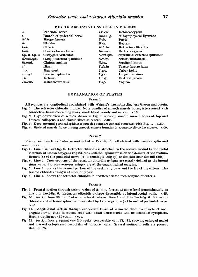
KEY TO ABBREVIATIONS USED IN FIGURESA Pudendal nerve Isc.coc. Ischiococcygeusa Branch of pudendal nerve Mid.s.lg. Midsymphyseal ligamentBi.fe. Biceps femoris Pub. PubisBl. Bladder Rect. RectumClit. Clitoris Ret.clit. Retractor clitoridisC.ur. Constrictor urethrae Rec.coc. RectococcygeusCy. 2, Cy. 3 Coceygeal vertebrae S.ext.sph. Superficial external sphincter(D)ext.sph. (Deep) external sphincter S.mem. SemimembranosusGl.med. Gluteus medius S.ten. Semitendinosus11. Ilium T.fa.la. Tensor fasciae lataeI.cr. Iliac crest T.isc. Tuber ischiiInt.sph. Internal sphincter Ug.s. Urogenital sinusIsc. Ischium Ur.gr. Urethral grooveIsc.cav. Ischiocavernosus Vag. Vagina.
EXPLANATION OF PLATESPLATE 1
All sections are longitudinal and stained with Weigert's haematoxylin, van Gieson and orcein.Fig. 1. The retractor clitoridis muscle. Note bundles of smooth muscle fibres, interspersed with
connective tissue containing many small blood vessels and nerves. x 150.Fig. 2. High-power view of section shown in Fig. 1, showing smooth muscle fibres at top and
bottom, collagenous and elastic fibres at centre. x 405.Fig. 3. Deep external perineal sphincter muscle; compare general structure with Fig. 1. x 150.Fig. 4. Striated muscle fibres among smooth muscle bundles in retractor clitoridis muscle. x 90.
PLATE 2
Frontal sections from foetus reconstructed in Text-fig. 6. All stained with haematoxylin andeosin. x 22.Fig. 5. Line 1 in Text-fig. 6. Retractor clitoridis is attached to the rectum medial to the rectal
insertion of ischiococcygeus (right). The external sphincter is on the dorsum of the rectum.Branch (a) of the pudendal nerve (A) is sending a twig (p) to the skin near the tail (left).
Fig. 6. Line 2. Cross-sections of the retractor clitoridis anlagen are clearly defined at the lateralsinus walls. Ischiocavernosus anlagen are at the caudal ischial margins.
Fig. 7. Line 3. Shows the cranial portion of the urethral groove and the tip of the clitoris. Re-tractor clitoridis anlagen at sides of groove.
Fig. 8. Line 4. Shows the retractor clitoridis in undifferentiated mesenchyme of clitoris.
PLATE 3
Fig. 9. Frontal section through pelvic region of 21 mm. foetus, at same level approximately asline 1 in Text-fig. 6. Retractor clitoridis anlagen discernible at lateral rectal walls. x 45.
Fig. 10. Section from 36 mm. foetus, at a level between lines 1 and 2 in Text-fig. 6. Retractorclitoridis and external sphincter innervated by two twigs (a, a') of branch of pudendal nerve.x45.
Fig. 11. Longitudinal section through connective tissue of retractor clitoridis muscle of non-pregnant ewe. Note fibroblast cells with small dense nuclei and no stainable cytoplasm.Haematoxylin-azur II-eosin. x 675.
Fig. 12. Section from pregnant ewe (20 weeks) comparable with Fig. 11, showing enlarged nucleiand marked cytoplasmic basophilia of fibroblast cells. Several eosinophil cells are presentalso. x 675.