


Bahasa
Halaman
Hukum

CERAMICSINTERNATIONAL
Available online at www.sciencedirect.com
0272-8842/$ - sehttp://dx.doi.org/
nCorrespondinE-mail addre
Ceramics International 40 (2014) 7151–7160www.elsevier.com/locate/ceramint
Superhydrophobic surface decorated with vertical ZnO nanorods modifiedby stearic acid
Annaso B. Gurava, Sanjay S. Lattheb, Rajiv S. Vhatkara, Jong-Gun Leec, Do-Yeon Kimc,Jung-Jae Parkc, Sam S. Yoonc,n
aDepartment of Physics, Shivaji University, Kolhapur 416404, Maharashtra, IndiabPhotocatalysis International Research Center, Tokyo University of Science, Japan
cSchool of Mechanical Engineering, Korea University, Seoul, South Korea
Received 11 November 2013; received in revised form 12 December 2013; accepted 12 December 2013Available online 27 December 2013
Abstract
Static and dynamic wettability of the ZnO nanorod surface prepared by a facile and inexpensive route is reported. The wettability of the ZnOsurface was controlled and tuned by post hydrophobization using different stearic acid concentrations. The surface of the ZnO nanorods modifiedwith 8 mM stearic acid showed a static water contact angle of 1521 and sliding angle of 91, which indicates superhydrophobicity. This suggeststhat the combination of the rough structures achieved by the ZnO nanorods and low surface energy provided by stearic acid modification resultsin superhydrophobicity and a very low sliding angle. The crystal structure, surface chemical elements, surface morphology, surface roughness,and static and dynamic water contact angles of the ZnO coatings were studied in detail. Further, the surface properties were assessed bycalculating the surface free energies and work of adhesion for unmodified and stearic-acid-modified ZnO nanostructure surfaces. These coatingscan find potential industrial applications in the electronic industry.& 2013 Elsevier Ltd and Techna Group S.r.l. All rights reserved.
Keywords: Sol–gel; Dip coating; Superhydrophobic; Self-cleaning; Surface free energy
1. Introduction
Interest in solid surfaces with integrated superhydrophobic(water contact angle41501) and self-cleaning (very lowsliding angleo51) properties has provoked intensive researchin an attempt to duplicate the amazing superhydrophobic andself-cleaning phenomena observed in biological systems [1].The best known natural superhydrophobic and self-cleaningphenomenon occurs on lotus leaves. When water dropletscontact the leaf surface, they form nearly spherical drops andimmediately roll off carrying away any dust particles withthem; as a result, the leaves remain clean and dry even in dirtywater, which reveals their efficient self-cleaning ability. Thecombination of micro/nano-scale patterned structures and lowsurface energy because of the presence of a wax-like coating
e front matter & 2013 Elsevier Ltd and Techna Group S.r.l. All ri10.1016/j.ceramint.2013.12.052
g author.ss: [email protected] (S.S. Yoon).
may be the source of the very high water contact angle andvery low sliding angle of lotus leaves. This discovery, calledthe “lotus effect” by Neinhuis and Barthlott [2] generatedenormous technological interest and greatly stimulated funda-mental research on non-wettable surfaces. To create a solidsurface with both superhydrophobic and self-cleaning proper-ties, the surface roughness and surface energy are the two mainparameters for consideration: essentially, a rough structuremust be produced on a hydrophobic surface or modified with aspecial low-surface-energy material [3,4]. Solid surfaces withboth superhydrophobicity and self-cleaning functionality havebecome necessary in recent years because of their variousvaluable and promising practical applications such as in micro/nano-fluidic devices, automobile windshields, dust-free andself-cleaning surfaces for solar cells, building walls and roofglass and self-cleaning windows [5–7].Zinc oxide (ZnO) based micro/nano-structured surfaces have
acquired a special place among the various metal oxide
ghts reserved.
A.B. Gurav et al. / Ceramics International 40 (2014) 7151–71607152
materials and are important semiconducting materials becauseof their attractive electronic properties including a wide bandgap (3.37 eV) and large exciton binding energy (60 meV) atroom temperature [8]. ZnO nanostructures are extremelyvaried and include structures such as nanorods, nanopillars,nanowires, nanodonuts, nanodrums, nanopropellers, nanonailsand nanobridges, which have been widely reported in literature[9]. The ZnO surface is intrinsically hydrophilic because of thestrong adhesion of hydroxyl groups to its surface [10,11].Although, the hydrophobic behaviors of ZnO coatings withand without chemical modifications have been reported else-where [12–16], still there remains great curiosity about thewetting properties of ZnO coatings with various micro/nanostructures [17,18]. Such ZnO structures have inherent surfaceroughness, which should cause air to be trapped under waterdroplets resulting in the majority of the droplet floating on alayer of air. The surface energy can be lowered throughchemical modification, which affects the surface wettabilityand may be useful for producing superhydrophobic ZnOsurfaces. There are many applications of ZnO structures forwhich superhydrophobic and self-cleaning properties aredesirable to enhance their conventional functionality, includingin outdoor optoelectronics, display devices, photovoltaic solarcells, and gas sensors [19,20].
There are numerous reports from various laboratories on thewettability of micro/nano-structured ZnO coatings. Fujishima0sgroup obtained hydrophobic ZnO films with water contactangles of 1091 using spray pyrolysis without any furtherchemical modification and investigated the wettability conver-sion of ZnO thin films before and after UV illumination [21].Li et al. [22] fabricated 2D ZnO ordered pore arrays using asolution-dipping colloidal monolayer method and demon-strated photo-induced reversible wettability between hydro-phobicity and superhydrophilicity. The as-prepared 2D ZnOordered pore arrays samples exhibit a Wenzel-type surface andtransition to a Cassie-type surface i.e. superhydrophobic state,after modification with fluoroalkylsilane. Zhang et al. [23]synthesized hydrophobic ZnO textured surfaces with well-controlled periodical micro-bowl patterns with the assistanceof a polystyrene monolayer template and suggested theirapplication in self-cleaning and micro-fluidic devices. Houet.al. [24] fabricated superhydrophobic ZnO sub-microrodfilms on zinc sheets through a H2O2-assisted surface-etchingprocess with subsequent surface modification with a mono-layer of 1H,1H,2H,2H-perfluorodecyltriethoxysilane (FDS).They observed that the surface of the ZnO film on a zincsubstrate was initially hydrophobic and became superhydro-phobic after modification with FDS. A systematic comparativestudy was made on the effect of surface micro-/nanostructureon the wettability and apparent surface free energy of sputter-deposited ZnO, thermally oxidized Zn–ZnO, and vacuum-annealed ZnO coatings [25]. In addition, various attempts toobtain superhydrophobic ZnO surfaces are evident in literature[17,26–28].
The synthesis methods as well as experimental conditionsare very important parameters in controlling the ZnO micro/nano-structure and its wetting properties. The present study
involves simple sol–gel dip-coated ZnO seed-assisted epitaxialZnO nanorods growth onto a glass substrate at a mildtemperature (�90 1C) using a solution immersion method;superhydrophobic and self-cleaning properties were obtainedby post synthesis chemical modification. We adopted a sol–geldip-coating method for the preparation of the ZnO seed layerowing to its inherent advantages including simplicity, cost-effectiveness and low processing temperature. This approachprovides an alternative method of controlling the surfacewettability without tailoring the geometric parameters bymicro-fabrication techniques.
2. Experimental
2.1. Precursor
Zinc acetate dihydrate [(CH3COO)2Zn∙2H2O, analyticalreagent grade, minimum assay 99.5%], diethanolamine[(CH2(OH)CH2)2NH, analytical reagent grade, minimum assay99.5%], isopropyl alcohol [(CH3)2CHOH, analytical reagentgrade, minimum assay 99.7%], and stearic acid [CH3(CH2)16COOH, laboratory reagent grade, minimum assay 97.0%] werepurchased from Thomas Baker Chemicals (India). Hexamethy-lenetetramine [(CH7)6N4, minimum assay 99.5%] was pur-chased from Molychem (India), and ethanol [C2H5OH,analytical reagent grade, minimum assay 99.5%] was pur-chased from Changshu Yangyun Chemicals (China). Double-distilled water was used in all experiments. No furtherpurification was made in any of the chemicals and used asreceived from the manufacturers. The micro glass slides (BlueStars) were purchased from Polar Industrial Corporation(India).
2.2. Sol–gel dip-coating
To obtain uniform seeding, the glass substrates were cleanedthoroughly prior to deposition using the following procedure:the glass substrates were initially soaked overnight in chromicacid, then washed with detergent and Labolene and finallyultrasonically cleaned with water for 15 min followed bysequential rinsing with water and acetone to remove surfacecontaminants. All glass substrates were dried at 50 1C for 2 hbefore coating. ZnO seeds were applied on the glass substrateby the sol–gel dip-coating method. The seed-layer coating solcomprised of the zinc acetate dihydrate (Zn-Ac; 0.05 M),isopropyl alcohol (i-PrOH) and diethanolamine (DEA;0.05 M) as the solute, solvent and complexing agent, respec-tively. The Zn-Ac was first dissolved in i-PrOH and then DEAwas added slowly under constant stirring at room temperature(�27 1C). The resultant solution was stirred under constantstirring at 60 1C for 1 h to get a homogeneous solution. Thissolution was left to age for at least 24 h to complete thehydrolysis and condensation processes. The ZnO seed layerwas deposited under ambient conditions according to thefollowing procedure: The pre-cleaned glass substrate wasimmersed in the prepared ZnO coating sol for 1 min andwithdrawn at a speed of approximately 5 mm/s. After
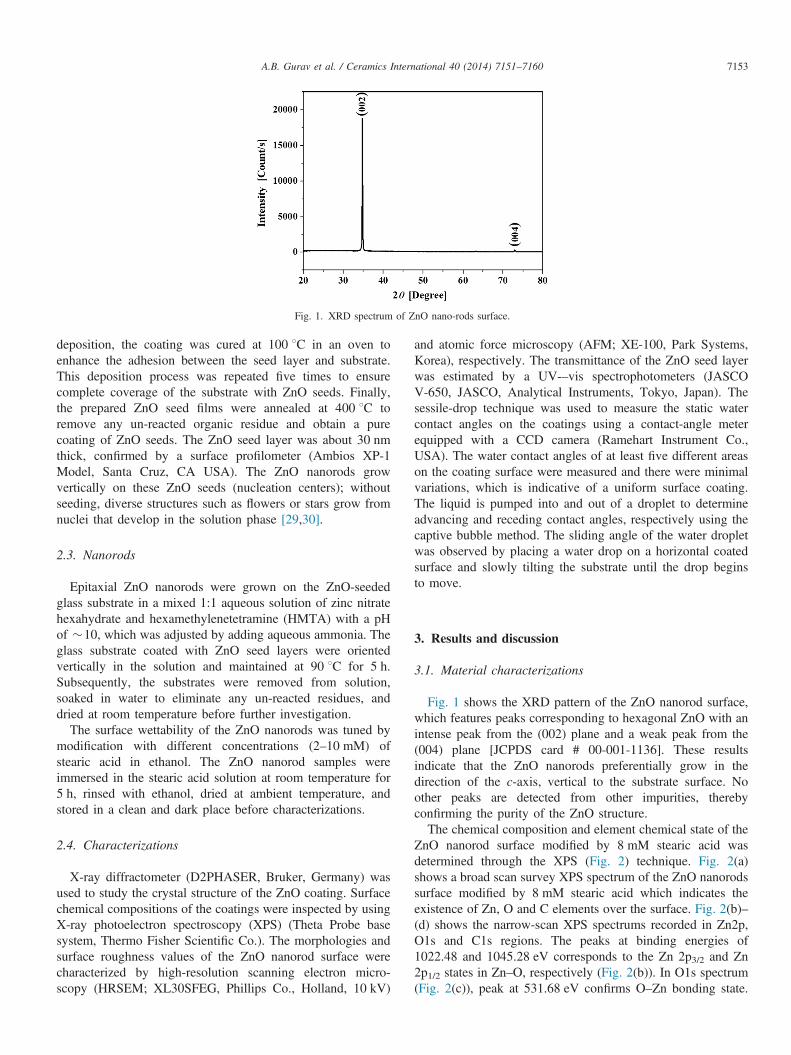
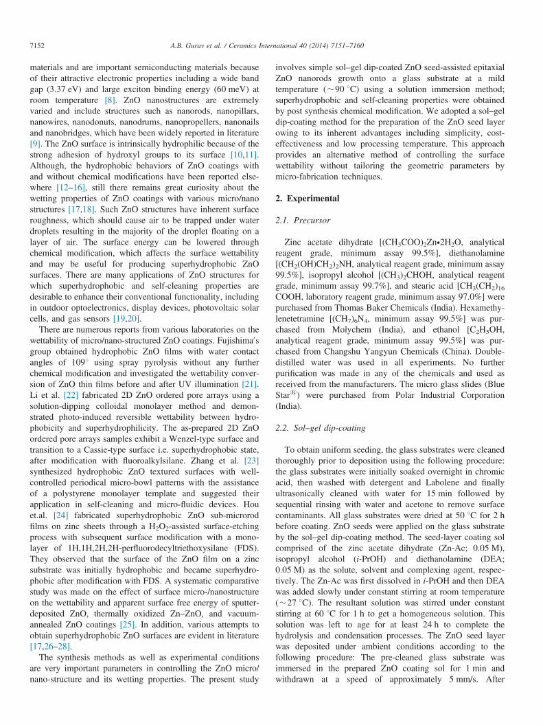
Fig. 1. XRD spectrum of ZnO nano-rods surface.
A.B. Gurav et al. / Ceramics International 40 (2014) 7151–7160 7153
deposition, the coating was cured at 100 1C in an oven toenhance the adhesion between the seed layer and substrate.This deposition process was repeated five times to ensurecomplete coverage of the substrate with ZnO seeds. Finally,the prepared ZnO seed films were annealed at 400 1C toremove any un-reacted organic residue and obtain a purecoating of ZnO seeds. The ZnO seed layer was about 30 nmthick, confirmed by a surface profilometer (Ambios XP-1Model, Santa Cruz, CA USA). The ZnO nanorods growvertically on these ZnO seeds (nucleation centers); withoutseeding, diverse structures such as flowers or stars grow fromnuclei that develop in the solution phase [29,30].
2.3. Nanorods
Epitaxial ZnO nanorods were grown on the ZnO-seededglass substrate in a mixed 1:1 aqueous solution of zinc nitratehexahydrate and hexamethylenetetramine (HMTA) with a pHof �10, which was adjusted by adding aqueous ammonia. Theglass substrate coated with ZnO seed layers were orientedvertically in the solution and maintained at 90 1C for 5 h.Subsequently, the substrates were removed from solution,soaked in water to eliminate any un-reacted residues, anddried at room temperature before further investigation.
The surface wettability of the ZnO nanorods was tuned bymodification with different concentrations (2–10 mM) ofstearic acid in ethanol. The ZnO nanorod samples wereimmersed in the stearic acid solution at room temperature for5 h, rinsed with ethanol, dried at ambient temperature, andstored in a clean and dark place before characterizations.
2.4. Characterizations
X-ray diffractometer (D2PHASER, Bruker, Germany) wasused to study the crystal structure of the ZnO coating. Surfacechemical compositions of the coatings were inspected by usingX-ray photoelectron spectroscopy (XPS) (Theta Probe basesystem, Thermo Fisher Scientific Co.). The morphologies andsurface roughness values of the ZnO nanorod surface werecharacterized by high-resolution scanning electron micro-scopy (HRSEM; XL30SFEG, Phillips Co., Holland, 10 kV)
and atomic force microscopy (AFM; XE-100, Park Systems,Korea), respectively. The transmittance of the ZnO seed layerwas estimated by a UV-–vis spectrophotometers (JASCOV-650, JASCO, Analytical Instruments, Tokyo, Japan). Thesessile-drop technique was used to measure the static watercontact angles on the coatings using a contact-angle meterequipped with a CCD camera (Ramehart Instrument Co.,USA). The water contact angles of at least five different areason the coating surface were measured and there were minimalvariations, which is indicative of a uniform surface coating.The liquid is pumped into and out of a droplet to determineadvancing and receding contact angles, respectively using thecaptive bubble method. The sliding angle of the water dropletwas observed by placing a water drop on a horizontal coatedsurface and slowly tilting the substrate until the drop beginsto move.
3. Results and discussion
3.1. Material characterizations
Fig. 1 shows the XRD pattern of the ZnO nanorod surface,which features peaks corresponding to hexagonal ZnO with anintense peak from the (002) plane and a weak peak from the(004) plane [JCPDS card # 00-001-1136]. These resultsindicate that the ZnO nanorods preferentially grow in thedirection of the c-axis, vertical to the substrate surface. Noother peaks are detected from other impurities, therebyconfirming the purity of the ZnO structure.The chemical composition and element chemical state of the
ZnO nanorod surface modified by 8 mM stearic acid wasdetermined through the XPS (Fig. 2) technique. Fig. 2(a)shows a broad scan survey XPS spectrum of the ZnO nanorodssurface modified by 8 mM stearic acid which indicates theexistence of Zn, O and C elements over the surface. Fig. 2(b)–(d) shows the narrow-scan XPS spectrums recorded in Zn2p,O1s and C1s regions. The peaks at binding energies of1022.48 and 1045.28 eV corresponds to the Zn 2p3/2 and Zn2p1/2 states in Zn–O, respectively (Fig. 2(b)). In O1s spectrum(Fig. 2(c)), peak at 531.68 eV confirms O–Zn bonding state.
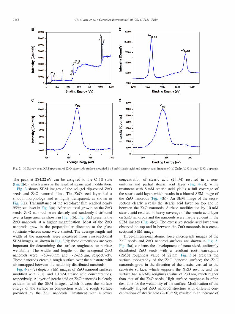
Fig. 2. (a) Survey scan XPS spectrum of ZnO nano-rods surface modified by 8 mM stearic acid and narrow scan images of (b) Zn2p (c) O1s and (d) C1s spectra.
A.B. Gurav et al. / Ceramics International 40 (2014) 7151–71607154
The peak at 284.22 eV can be assigned to the C 1S state(Fig. 2(d)), which arises as the result of stearic acid modification.
Fig. 3 shows SEM images of the sol–gel dip-coated ZnOseeds and ZnO nanorod films. The ZnO seed layer had asmooth morphology and is highly transparent, as shown inFig. 3(a). Transmittance of the seed-layer film reached nearly95%; see inset in Fig. 3(a). After epitaxial growth on the ZnOseeds, ZnO nanorods were densely and randomly distributedover a large area, as shown in Fig. 3(b). Fig. 3(c) presents theZnO nanorods at a higher magnification. Most of the ZnOnanorods grew in the perpendicular direction to the glasssubstrate whereas some were slanted. The average length andwidth of the nanorods were measured from cross-sectionalSEM images, as shown in Fig. 3(d); these dimensions are veryimportant for determining the surface roughness for surfacewettability. The widths and lengths of the hexagonal ZnOnanorods were �50–70 nm and �2–2.5 mm, respectively.These nanorods create a rough surface over the substrate withair entrapped between the randomly distributed nanorods.
Fig. 4(a)–(c) depicts SEM images of ZnO nanorod surfacesmodified with 2, 8, and 10 mM stearic acid concentrations,respectively. A layer of stearic acid on ZnO nanorods is clearlyevident in all the SEM images, which lowers the surfaceenergy of the surface in conjunction with the rough surfaceprovided by the ZnO nanorods. Treatment with a lower
concentration of stearic acid (2 mM) resulted in a non-uniform and partial stearic acid layer (Fig. 4(a)), whiletreatment with 8 mM stearic acid yields a full coverage ofthe stearic acid layer, which results in a blurred SEM image ofthe ZnO nanorods (Fig. 4(b)). An SEM image of the cross-section clearly reveals the stearic acid layer on top and inbetween the ZnO nanorods. Surface modification by 10 mMstearic acid resulted in heavy coverage of the stearic acid layeron ZnO nanorods and the nanorods were hardly evident in theSEM images (Fig. 4(c)). The excessive stearic acid layer wasobserved on top and in between the ZnO nanorods in a cross-sectional SEM image.Three-dimensional atomic force micrograph images of the
ZnO seeds and ZnO nanorod surfaces are shown in Fig. 5.Fig. 5(a) confirms the development of nano-sized, uniformlydistributed ZnO seeds with a resultant root-mean-square(RMS) roughness value of 22 nm. Fig. 5(b) presents thesurface topography of the ZnO nanorod surface; the ZnOnanorods grew in the direction of the c-axis, vertical to thesubstrate surface, which supports the XRD results, and thesurface had a RMS roughness value of 230 nm, much higherthan that of the ZnO seeds. High surface roughness is oftendesirable for the wettability of the surface. Modification of thevertically aligned ZnO nanorod structure with different con-centrations of stearic acid (2–10 mM) resulted in an increase of
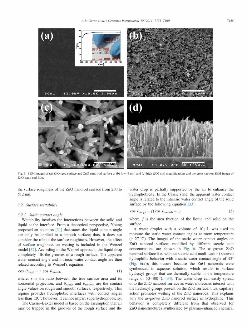
Fig. 3. SEM images of (a) ZnO seed surface and ZnO nano-rod surface at (b) low (3 um) and (c) high (500 nm) magnifications and the cross-section SEM image ofZnO nano-rod film.
A.B. Gurav et al. / Ceramics International 40 (2014) 7151–7160 7155
the surface roughness of the ZnO nanorod surface from 230 to512 nm.
3.2. Surface wettability
3.2.1. Static contact angleWettability involves the interactions between the solid and
liquid at the interface. From a theoretical perspective, Youngproposed an equation [31] that states the liquid contact anglecan only be applied to a smooth surface; thus, it does notconsider the role of the surface roughness. However, the effectof surface roughness on wetting is included in the Wenzelmodel [32]. According to the Wenzel approach, the liquid dropcompletely fills the grooves of a rough surface. The apparentwater contact angle and intrinsic water contact angle are thenrelated according to Wenzel0s equation
cos θrough ¼ r cos θsmooth ð1Þwhere, r is the ratio between the true surface area and itshorizontal projection, and θrough and θsmooth are the contactangle values on rough and smooth surfaces, respectively. Thisregime provides hydrophobic interfaces with contact anglesless than 1201; however, it cannot impart superhydrophobicity.
The Cassie–Baxter model is based on the assumption that airmay be trapped in the grooves of the rough surface and the
water drop is partially supported by the air to enhance thehydrophobicity. In the Cassie state, the apparent water contactangle is related to the intrinsic water contact angle of the solidsurface by the following equation [33]:
cos θrough ¼ f ð cos θsmoothþ1Þ ð2Þwhere, f is the area fraction of the liquid and solid on thesurface.A water droplet with a volume of 10 mL was used to
measure the static water contact angles at room temperature(�27 1C). The images of the static water contact angles onZnO nanorod surfaces modified by different stearic acidconcentrations are shown in Fig. 6. The as-grown ZnOnanorod surface (i.e. without stearic-acid modification) showedhydrophilic behavior with a static water contact angle of 431(Fig. 6(a)); this occurs because the ZnO nanorods weresynthesized in aqueous solution, which results in surfacehydroxyl groups that are thermally stable in the temperaturerange of 50–400 1C [34]. The water drop can easily spreadonto the ZnO nanorod surface as water molecules interact withthe hydroxyl groups present on the ZnO surface; thus, capillaryaction promotes wetting of the ZnO nanorods. This explainswhy the as-grown ZnO nanorod surface is hydrophilic. Thisbehavior is completely different from that observed forZnO nanostructures synthesized by plasma-enhanced chemical
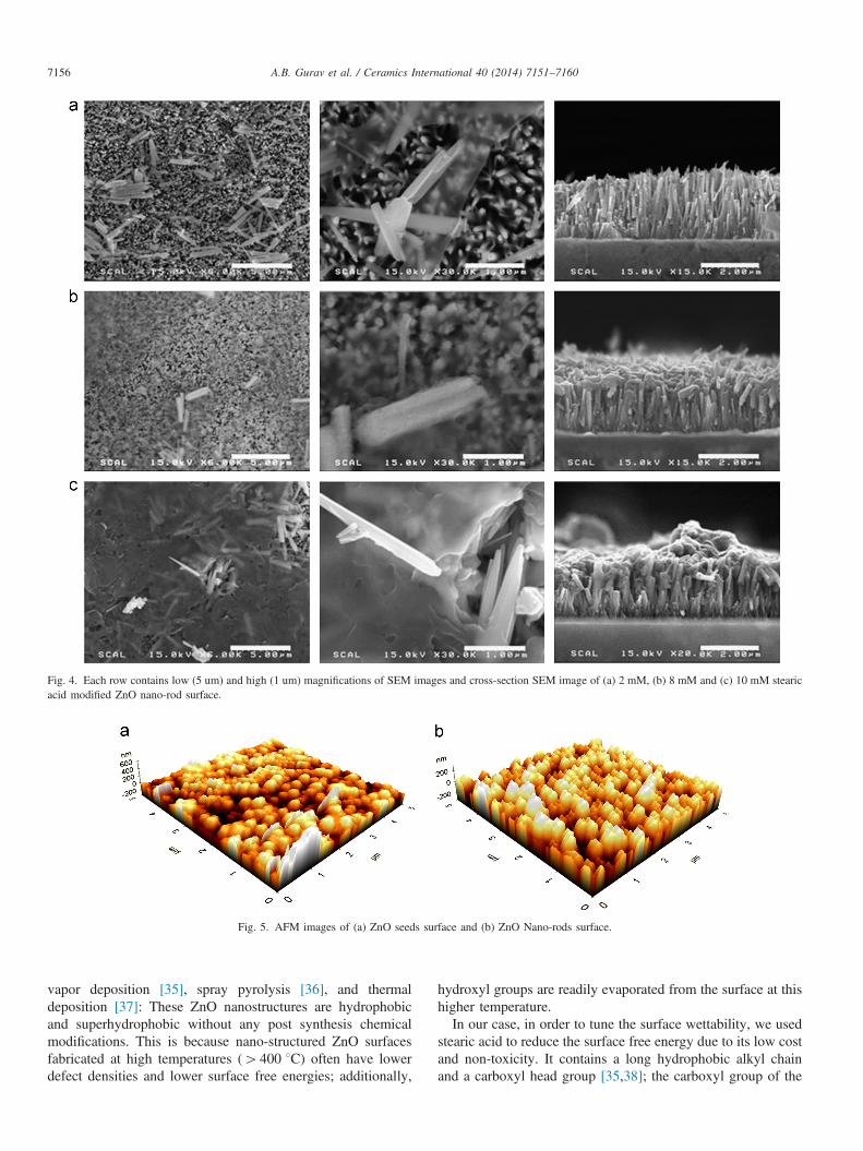
Fig. 4. Each row contains low (5 um) and high (1 um) magnifications of SEM images and cross-section SEM image of (a) 2 mM, (b) 8 mM and (c) 10 mM stearicacid modified ZnO nano-rod surface.
Fig. 5. AFM images of (a) ZnO seeds surface and (b) ZnO Nano-rods surface.
A.B. Gurav et al. / Ceramics International 40 (2014) 7151–71607156
vapor deposition [35], spray pyrolysis [36], and thermaldeposition [37]: These ZnO nanostructures are hydrophobicand superhydrophobic without any post synthesis chemicalmodifications. This is because nano-structured ZnO surfacesfabricated at high temperatures (4400 1C) often have lowerdefect densities and lower surface free energies; additionally,
hydroxyl groups are readily evaporated from the surface at thishigher temperature.In our case, in order to tune the surface wettability, we used
stearic acid to reduce the surface free energy due to its low costand non-toxicity. It contains a long hydrophobic alkyl chainand a carboxyl head group [35,38]; the carboxyl group of the
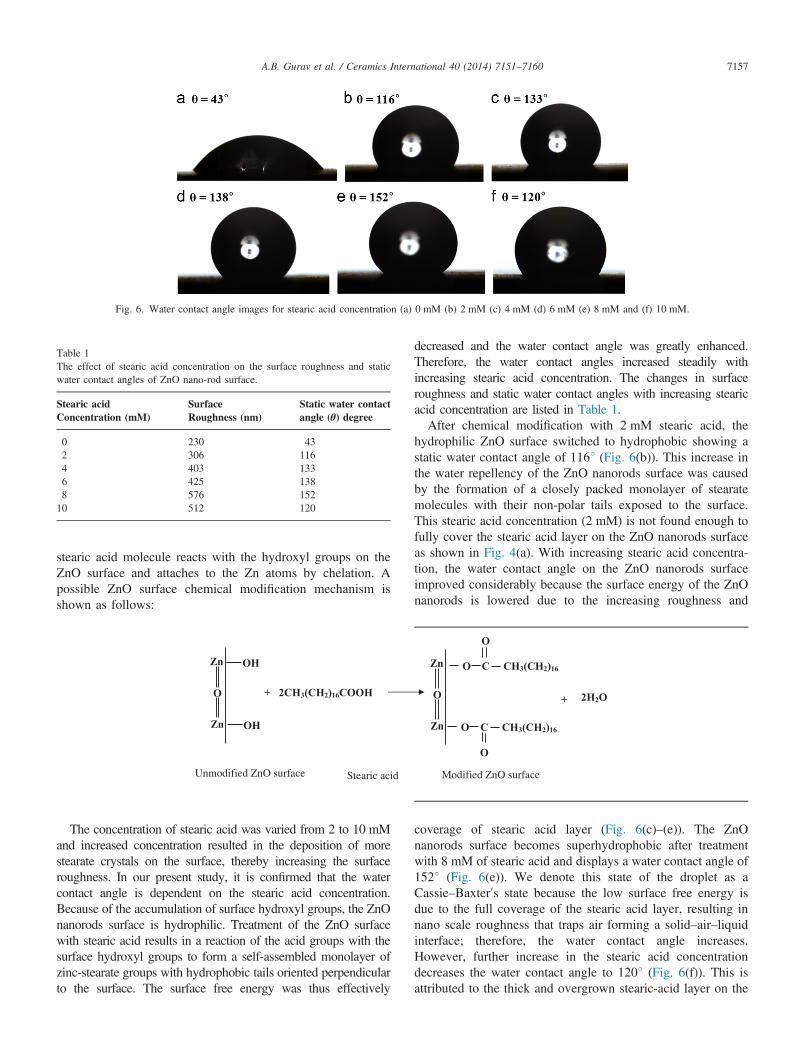
Fig. 6. Water contact angle images for stearic acid concentration (a) 0 mM (b) 2 mM (c) 4 mM (d) 6 mM (e) 8 mM and (f) 10 mM.
Table 1The effect of stearic acid concentration on the surface roughness and staticwater contact angles of ZnO nano-rod surface.
Stearic acidConcentration (mM)
SurfaceRoughness (nm)
Static water contactangle (θ) degree
0 230 432 306 1164 403 1336 425 1388 576 15210 512 120
A.B. Gurav et al. / Ceramics International 40 (2014) 7151–7160 7157
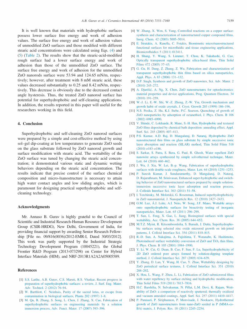
stearic acid molecule reacts with the hydroxyl groups on theZnO surface and attaches to the Zn atoms by chelation. Apossible ZnO surface chemical modification mechanism isshown as follows:
+
Zn
Zn
O
OH
OH
2CH3(CH2)16COOH
Zn
Zn
O
O C CH3(CH2)16
O
O
O C CH3(CH2)16
+ 2H2O
Unmodified ZnO surface Stearic acid Modified ZnO surface
The concentration of stearic acid was varied from 2 to 10 mMand increased concentration resulted in the deposition of morestearate crystals on the surface, thereby increasing the surfaceroughness. In our present study, it is confirmed that the watercontact angle is dependent on the stearic acid concentration.Because of the accumulation of surface hydroxyl groups, the ZnOnanorods surface is hydrophilic. Treatment of the ZnO surfacewith stearic acid results in a reaction of the acid groups with thesurface hydroxyl groups to form a self-assembled monolayer ofzinc-stearate groups with hydrophobic tails oriented perpendicularto the surface. The surface free energy was thus effectively
decreased and the water contact angle was greatly enhanced.Therefore, the water contact angles increased steadily withincreasing stearic acid concentration. The changes in surfaceroughness and static water contact angles with increasing stearicacid concentration are listed in Table 1.After chemical modification with 2 mM stearic acid, the
hydrophilic ZnO surface switched to hydrophobic showing astatic water contact angle of 1161 (Fig. 6(b)). This increase inthe water repellency of the ZnO nanorods surface was causedby the formation of a closely packed monolayer of stearatemolecules with their non-polar tails exposed to the surface.This stearic acid concentration (2 mM) is not found enough tofully cover the stearic acid layer on the ZnO nanorods surfaceas shown in Fig. 4(a). With increasing stearic acid concentra-tion, the water contact angle on the ZnO nanorods surfaceimproved considerably because the surface energy of the ZnOnanorods is lowered due to the increasing roughness and
coverage of stearic acid layer (Fig. 6(c)–(e)). The ZnOnanorods surface becomes superhydrophobic after treatmentwith 8 mM of stearic acid and displays a water contact angle of1521 (Fig. 6(e)). We denote this state of the droplet as aCassie–Baxter0s state because the low surface free energy isdue to the full coverage of the stearic acid layer, resulting innano scale roughness that traps air forming a solid–air–liquidinterface; therefore, the water contact angle increases.However, further increase in the stearic acid concentrationdecreases the water contact angle to 1201 (Fig. 6(f)). This isattributed to the thick and overgrown stearic-acid layer on the
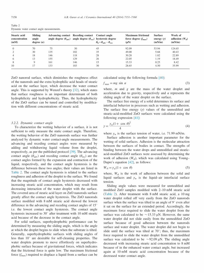
Table 2Dynamic water contact angle measurements.
Stearic acidconcentration(mM)
Slidingangledegree (α)
Advancing contactangle degree (θadv)
Receding contactangle degree (θrce)
Contact anglehysteresis degreeθh¼θadv�θrce
Maximum frictionalforce degree (fmax)(µN)
Surfaceenergy (γ)(mN/m)
Work ofadhesion (Wsl)(mN/m)
0 70 75 30 45 92.09 53.94 124.652 30 135 102 35 49.00 5.68 40.434 18 146 118 28 30.28 1.82 22.896 13 155 129 26 22.05 1.19 18.498 9 161 146 15 15.33 0.25 8.4210 17 133 103 30 28.65 4.50 35.99
A.B. Gurav et al. / Ceramics International 40 (2014) 7151–71607158
ZnO nanorod surface, which diminishes the roughness effectof the nanorods and the extra hydrophilic acid heads of stearicacid on the surface layer, which decrease the water contactangle. This is supported by Wenzel0s theory [32], which statesthat surface roughness is an important determinant of bothhydrophilicity and hydrophobicity. Thus, the hydrophobicityof the ZnO surface can be tuned and controlled by modifica-tion with different concentrations of stearic acid.
3.2.2. Dynamic contact angleTo characterize the wetting behavior of a surface, it is not
sufficient to only measure the static contact angle. Therefore,the wetting behavior of the ZnO nanorods surface was furtheranalyzed by dynamic water contact-angle measurements. Theadvancing and receding contact angles were measured byadding and withdrawing liquid volume from the droplet,respectively, as per the published protocol [39]. The advancingcontact angle (θadv) and receding contact angle (θrec) are thecontact angles formed by the expansion and contraction of theliquid, respectively, and the contact angle hysteresis is thedifference between these two angles; their values are listed inTable 2. The contact angle hysteresis is related to the surfaceroughness and adhesion of the droplet to the surface. We foundthat the magnitude of contact angle hysteresis decreased withincreasing stearic acid concentration, which may result fromdecreasing interaction of the water droplet with the surface.The covered area of stearic acid layer on ZnO nanorods surfacegreatly affect the contact angle hysteresis. The ZnO nanorodssurface modified with 8 mM stearic acid showed the lowestdifference in the advancing and receding contact angles of 151i.e. the lowest contact angle hysteresis. The contact anglehysteresis increased to 301 after treatment with 10 mM stearicacid because of the decrease in the contact angle.
For solid surfaces, superhydrophobic performance can bedetermined by measuring the sliding angle, which is the angleat which the droplet begins to slide when the substrate is tilted.Generally, superhydrophobic surfaces with sliding angles ofless than 101 are desirable for self-cleaning properties. Thewater droplets promote to move effortlessly on superhydro-phobic surface because of gravitational forces, which indicatesthat the frictional force is quite low. The maximum frictionalforce (fmax) required to displace a liquid from a surface can be
calculated using the following formula [40]:
fmax ¼mg sin α ð3Þwhere, m and g are the mass of the water droplet andacceleration due to gravity, respectively and α represents thesliding angle of the water droplet on the surface.The surface free energy of a solid determines its surface and
interfacial behavior in processes such as wetting and adhesion.The surface free energy (γ) values of the unmodified andstearic-acid-modified ZnO surfaces were calculated using thefollowing expression [41]:
γ ¼ γwð1þ cos θÞ24
ð4Þ
where γw is the surface tension of water, i.e. 71.99 mN/m.Surface adhesion is another important parameter for the
wetting of solid surfaces. Adhesion is the molecular attractionbetween the surfaces of bodies in contact. The strengths ofbinding between the water drops and unmodified and stearic-acid-modified ZnO surfaces were assessed by determining thework of adhesion (Wsl), which was calculated using Young–Dupre0s equation [42], as follows:
W sl ¼ γwð1þ cos θÞ ð5Þwhere, Wsl is the work of adhesion between the solid andliquid surfaces and γw is the liquid–air interfacial surfacetension.Sliding angle values were measured for unmodified and
modified ZnO samples modified with 2–10 mM stearic acid(Table 2). After treatment with 8 mM stearic acid, a 10 mgwater droplet rolled off very easily from the ZnO nanorodssurface when the surface was tilted to an angle of 91 even afterit sat on the surface for an extended period. Accordingly, themaximum force required to slide the water droplet from thesurface was calculated to be �15.33 mN. However, the samewater droplet did not slide easily from the unmodified ZnOsurface because of good adhesion between the nanorodssurface and water droplet. The water droplet did not begin toslide until the surface was tilted at 701; thus, the maximumforce required to slide the water droplet from the unmodifiedsurface was calculated to be �92.09 mN. The sliding angledecreased with increasing stearic acid concentration to 8 mMbecause of in the enhanced water contact angle, but increasedagain at 10 mM stearic acid concentration because of thedecreased water contact angle.
A.B. Gurav et al. / Ceramics International 40 (2014) 7151–7160 7159
It is well known that materials with hydrophobic surfacespossess lower surface free energy and work of adhesionvalues. The surface free energy and work of adhesion valuesof unmodified ZnO surfaces and those modified with differentstearic acid concentrations were calculated using Eqs. (4) and(5) (Table 2). The results show that the stearic-acid-modifiedrough surface had a lower surface energy and work ofadhesion than those of the unmodified ZnO surface. Thesurface free energy and work of adhesion for the unmodifiedZnO nanorods surface were 53.94 and 124.65 mN/m, respec-tively; however, after treatment with 8 mM stearic acid, thesevalues decreased substantially to 0.25 and 8.42 mN/m, respec-tively. This decrease is obviously due to the decreased contactangle hysteresis. Thus, the treated ZnO nanorod surface haspotential for superhydrophobic and self-cleaning applications.In addition, the results reported in this paper will useful for theresearchers working in this field.
4. Conclusion
Superhydrophobic and self-cleaning ZnO nanorod surfaceswere prepared by a simple and cost-effective method by usingsol–gel dip-coating at low temperatures to generate ZnO seedson the glass substrate followed by ZnO nanorod growth andsurface modification with stearic acid. The wettability of theZnO surface was tuned by changing the stearic acid concen-tration; it demonstrated various static and dynamic wettingbehaviors depending on the stearic acid concentration. Theresults indicate that precise control of the surface chemicalcomposition and micro-/nanostructure is necessary to attainhigh water contact angles and low sliding angles, which isparamount for designing practical superhydrophobic and self-cleaning technology.
Acknowledgments
Mr. Annaso B. Gurav is highly grateful to the Council ofScientific and Industrial Research-Human Resource DevelopmentGroup (CSIR-HRDG), New Delhi, Government of India, forproviding financial support by awarding Senior Research Fellow-ship [File no. 09/816(0036)/2012-EMR-I, Dated 30/03/2012].This work was partly supported by the Industrial StrategicTechnology Development Program (10045221), the GlobalFrontier R&D Program (2013-073298) on Center for HybridInterface Materials (HIM), and NRF-2013R1A2A2A05005589.
References
[1] S.S. Latthe, A.B. Gurav, C.S. Maruti, R.S. Vhatkar, Recent progress inpreparation of superhydrophobic surfaces: a review, J. Surf. Eng. Mater.Adv. Technol. 2 (2012) 76–94.
[2] W. Barthlott, C. Neinhuis, Purity of the sacred lotus, or escape fromcontamination in biological surfaces, Planta 202 (1997) 1–8.
[3] M. Qu, B. Zhang, S. Song, L. Chen, J. Zhang, X. Cao, Fabrication ofsuperhydrophobic surfaces on engineering materials by a solutionimmersion process, Adv. Funct. Mater. 17 (2007) 593–596.
[4] W. Zhang, X. Wen, S. Yang, Controlled reactions on a copper surface:synthesis and characterization of nanostructured copper compound films,Inorg. Chem. 42 (2003) 5005–5014.
[5] E. Stratakis, A. Ranella, C. Fotakis, Biomimetic micro/nanostructuredfunctional surfaces for microfluidic and tissue engineering applications,Biomicrofluidics 5 (2011) 013411.
[6] H. Shang, Y. Wang, S. Limmer, T. Chou, K. Takahashi, G. Cao,Optically transparent superhydrophobic silica-based films, Thin SolidFilms 472 (2005) 37–43.
[7] G. Gu, H. Dang, Z. Zhang, Z. Wu, Fabrication and characterization oftransparent superhydrophobic thin films based on silica nanoparticles,Appl. Phys. A 83 (2006) 131–132.
[8] D.P. Singh, Synthesis and growth of ZnO nanowires, Sci. Adv. Mater. 2(2010) 245–272.
[9] A. Djurišić, A. Ng, X. Chen, ZnO nanostructures for optoelectronics:material properties and device applications, Prog. Quantum Electron. 34(2010) 191–259.
[10] W.-J. Li, E.-W. Shi, W.-Z. Zhong, Z.-W. Yin, Growth mechanism andgrowth habit of oxide crystals, J. Cryst. Growth 203 (1999) 186–196.
[11] N.S. Pesika, Z. Hu, K.J. Stebe, P.C. Searson, Quenching of growth ofZnO nanoparticles by adsorption of octanethiol, J. Phys. Chem. B 106(2002) 6985–6990.
[12] V. Shinde, C. Lokhande, R. Mane, S.-H. Han, Hydrophobic and texturedZnO films deposited by chemical bath deposition: annealing effect, Appl.Surf. Sci. 245 (2005) 407–413.
[13] P.S. Kumar, A.D. Raj, D. Mangalaraj, D. Nataraj, Hydrophobic ZnOnanostructured thin films on glass substrate by simple successive ioniclayer absorption and reaction (SILAR) method, Thin Solid Films 518(2010) e183–e186.
[14] S. Sarkar, S. Patra, S. Bera, G. Paul, R. Ghosh, Water repellent ZnOnanowire arrays synthesized by simple solvothermal technique, Mater.Lett. 64 (2010) 460–462.
[15] J. Wu, J. Xia, W. Lei, B.-p. Wang, Fabrication of superhydrophobicsurfaces with double-scale roughness, Mater. Lett. 64 (2010) 1251–1253.
[16] P. Suresh Kumar, J. Sundaramurthy, D. Mangalaraj, D. Nataraj,D. Rajarathnam, M. Srinivasan, Enhanced super-hydrophobic and switch-ing behavior of ZnO nanostructured surfaces prepared by simple solution-immersion successive ionic layer adsorption and reaction process,J. Colloids Interface Sci. 363 (2011) 51–58.
[17] I. Torchinsky, M. Molotskii, G. Rosenman, Induced superhydrophobicityin ZnO nanomaterial, J. Nanoparticle Res. 12 (2010) 2427–2433.
[18] G.M. Luz, Á.J. Leite, A.I. Neto, W. Song, J.F. Mano, Wettable arraysonto superhydrophobic surfaces for bioactivity testing of inorganicnanoparticles, Mater. Lett. 65 (2011) 296–299.
[19] T. Sun, L. Feng, X. Gao, L. Jiang, Bioinspired surfaces with specialwettability, Acc. Chem. Res. 38 (2005) 644–652.
[20] M.T.Z. Myint, R. Kitsomboonloha, S. Baruah, J. Dutta, Superhydropho-bic surfaces using selected zinc oxide microrod growth on ink-jettedpatterns, J. Colloid Interface Sci. 354 (2011) 810–815.
[21] R.-D. Sun, A. Nakajima, A. Fujishima, T. Watanabe, K. Hashimoto,Photoinduced surface wettability conversion of ZnO and TiO2 thin films,J. Phys. Chem. B 105 (2001) 1984–1990.
[22] Y. Li, W. Cai, G. Duan, B. Cao, F. Sun, F. Lu, Superhydrophobicity of2D ZnO ordered pore arrays formed by solution-dipping templatemethod, J. Colloid Interface Sci. 287 (2005) 634–639.
[23] Y. Zhang, D. Lan, Y. Wang, H. Cao, Y. Zhao, Wettability designing byZnO periodical surface textures, J. Colloid Interface Sci. 351 (2010)288–292.
[24] X. Hou, L. Wang, F. Zhou, L. Li, Fabrication of ZnO submicrorod filmswith water repellency by surface etching and hydrophobic modification,Thin Solid Films 519 (2011) 7813–7816.
[25] H.C. Barshilia, N. Selvakumar, N. Pillai, L.M. Devi, K. Rajam, Wett-ability of ZnO: a comparison of reactively sputtered; thermally oxidizedand vacuum annealed coatings, Appl. Surf. Sci. 257 (2011) 4410–4417.
[26] P. Pannasri, P. Siriphannon, P. Monvisade, J. Nookaew, Hydrothermalgrowth of ZnO nanostructures from nano-ZnO seeded in P (MMA-co-BA) matrix, J. Polym. Res. 18 (2011) 2245–2254.
A.B. Gurav et al. / Ceramics International 40 (2014) 7151–71607160
[27] J. Wu, J. Xia, W. Lei, B. Wang, Facile synthesis of three-dimensionalZnO nanostructure: realization of a multifunctional stable superhydro-phobic surface, PloS One 6 (2011) e29047.
[28] U.P. Shaik, S. Kshirsagar, M.G. Krishna, S.P. Tewari, D. Dhar Pur-kayastha, V. Madhurima, Growth of superhydrophobic zinc oxidenanowire thin films, Mater. Lett. 75 (2012) 51–53.
[29] S.-H. Yi, S.-K. Choi, J.-M. Jang, J.-A. Kim, W.-G. Jung, Low-temperature growth of ZnO nanorods by chemical bath deposition,J. Colloid Interface Sci. 313 (2007) 705–710.
[30] S. Ma, R. Li, C. Lv, W. Xu, X. Gou, Facile synthesis of ZnO nanorodarrays and hierarchical nanostructures for photocatalysis and gas sensorapplications, J. Hazard. Mater. 192 (2011) 730–740.
[31] T. Young, Phil. Trans. R. Soc. London 95 (1805) 65.[32] R.N. Wenzel, Resistance of solid surfaces to wetting by water, Ind. Eng.
Chem. 28 (1936) 988–994.[33] A. Cassie, S. Baxter, Wettability of porous surfaces, Trans. Faraday Soc.
40 (1944) 546–551.[34] R. Xie, T. Sekiguchi, T. Ishigaki, N. Ohashi, D. Li, D. Yang, B. Liu,
Y. Bando, Enhancement and patterning of ultraviolet emission in ZnOwith an electron beam, Appl. Phys. Lett. 88 (2006) (134103-134103-134103).
[35] V. Rico, C. López, A. Borrás, J. Espinós, A. González-Elipe, Effect ofvisible light on the water contact angles on illuminated oxide
semiconductors other than TiO2, Sol. Energy Mater. Sol. Cells 90 (2006)2944–2949.
[36] N. Tarwal, P. Patil, Superhydrophobic and transparent ZnO thin filmssynthesized by spray pyrolysis technique, Appl. Surf. Sci. 256 (2010)7451–7456.
[37] L. Huang, S. Lau, H. Yang, E. Leong, S. Yu, S. Prawer, Stablesuperhydrophobic surface via carbon nanotubes coated with a ZnO thinfilm, J. Phys. Chem. B 109 (2005) 7746–7748.
[38] S.H. Ahn, S.H. Kim, S.G. Lee, Surface modified silica nanoparticle-reinforced poly(ethylene 2,6 naphthalate), J. Appl. Polym. Sci. 94 (2004)812–818.
[39] J. Drelich, J.D. Miller, R.J. Good, The effect of drop (bubble) size onadvancing and receding contact angles for heterogeneous and rough solidsurfaces as observed with sessile-drop and captive-bubble techniques,J. Colloid Interface Sci. 179 (1996) 37–50.
[40] Z.-G. Guo, F. Zhou, J.-C. Hao, Y.-M. Liang, W.-M. Liu, W.T. Huck,“Stick and slide” ferrofluidic droplets on superhydrophobic surfaces,Appl. Phys. Lett. 89 (2006) (081911-081911-081913).
[41] L. Yeping, F. Yue-E, F. Rong, X. Jinyun, Study of plasma-polymerization deposition of C2H2/CO2/H2 onto ethylene-co-propylenerubber membranes, Radiat. Phys. Chem. 60 (2001) 637–642.
[42] A. Dupré, Theorie Mecanique de la Chaleur, chapter IX, ActionsMoleculaires (Suite), Gauthier-Villars, Paris, 1869.
Top Related
Copyright © 2022 FDOKUMEN

