


Bahasa
Halaman
Hukum

Structural and functional analysis of theMutS C-terminal tetramerization domainLaura Manelyte, Claus Urbanke1, Luis Giron-Monzon and Peter Friedhoff*
Institut fur Biochemie, Justus-Liebig-Universitat, Heinrich-Buff-Ring 58, D-35392 Giessen, Germany and1Medizinische Hochschule, Strukturanalyse, Carl Neuberg Strasse 1, D-30625 Hannover, Germany
Received May 22, 2006; Revised and Accepted June 27, 2006
ABSTRACT
The Escherichia coli DNA mismatch repair (MMR)protein MutS is essential for the correction ofDNA replication errors. In vitro, MutS exists in adimer/tetramer equilibrium that is converted into amonomer/dimer equilibrium upon deletion of theC-terminal 53 amino acids. In vivo and in vitrodata have shown that this C-terminal domain (CTD,residues 801–853) is critical for tetramerization andthe function of MutS in MMR and anti-recombination.We report the expression, purification and analysisof the E.coli MutS-CTD. Secondary structure predic-tion and circular dichroism suggest that the CTDis folded, with an a-helical content of 30%. Basedon sedimentation equilibrium and velocity analyses,MutS-CTD forms a tetramer of asymmetric shape. Asingle point mutation (D835R) abolishes tetramer-ization but not dimerization of both MutS-CTD andfull-length MutS. Interestingly, the in vivo and invitro MMR activity of MutSCF/D835R is diminished to asimilar extent as a truncated MutS variant (MutS800,residues 1–800), which lacks the CTD. Moreover, thedimer-forming MutSCF/D835R has comparable DNAbinding affinity with the tetramer-forming MutS, butis impaired in mismatch-dependent activation ofMutH. Our data support the hypothesis that tetra-merization of MutS is important but not essential forMutS function in MMR.
INTRODUCTION
DNA mismatch repair (MMR) is critical for avoidingmutations, anti-recombination and cell checkpoint activationleading to apoptotic responses (1–3). In Escherichia coli,MutS recognizes mismatches in DNA and subsequentlyrecruits MutL in an ATP-dependent manner. This ternarycomplex activates downstream effector proteins such as thestrand-discrimination endonuclease MutH and UvrD helicase.
MutH nicks a hemimethylated GATC site in the unmethyl-ated, erroneous strand followed by unwinding of this strandby UvrD helicase and exonucleolytic digestion by one of sev-eral exonucleases. Repair is completed by DNA polymeraseIII holoenzyme and DNA Ligase I [reviewed in Ref. (2)].
The mismatch-recognizing MutS belongs to a family ofproteins whose members are found in organisms rangingfrom bacteria to eukaryotes. In eukaryotes, several MutShomologs exist that form heterodimers (e.g. MSH2-MSH6and MSH2-MSH3), with no indication of higher oligomers(2). Likewise, several heterodimeric MutL homologs havebeen studied from both human (MLH1-PMS2) and yeast(MLH1-PMS1) (3). In humans, mutations in the MMR geneshMSH2, hMSH6 or hMLH1 cause predisposition to a com-mon form of cancer called hereditary non-polyposis colorec-tal cancer (HNPCC)/Lynch-syndrome (4).
The co-crystal structures of both E.coli and Thermusaquaticus MutS bound to heteroduplex DNA have beensolved, using C-terminal truncated protein variants ofMutS (MutS800, residues 1–800 E.coli numbering) (5,6).MutS800 crystallizes as a dimer, wherein only one monomerhas specific contacts to the mismatch and bound ADP. Usinganalytical ultracentrifugation it was shown that in solutionMutS800 exists in monomer/dimer equilibrium while thefull-length MutS exist in a dimer/tetramer equilibrium (7,8).The physiological effect of truncating MutS is controversiallyreported in the literature, with data obtained using a multi-copy plasmid resulting in either no effect or impaired MMRfunction (6,9,10). Recently, E.coli with a truncated chromo-somal mutS gene (mutSD800) was shown to exhibit a MutSnull phenotype for mutation avoidance, anti-recombinationand sensitivity to cytotoxic agents in a dam mutant (10,11).However, the function of the tetrameric form still remainsunclear (12).
In vitro, MutS800 is impaired in DNA binding andmismatch-provoked MutH activation (7). Deletion of a b slid-ing clamp binding motif (812QMSLL816) of MutS abolishesthe interaction between MutS and b sliding clamp proteinsin vitro, although tetramerization and in vivo MMR activityof MutS are unaffected (13). Since MutS800 lacks residuesinvolved in dimerization, tetramerization and binding to theb sliding clamp, the primary cause of the observed defects
*To whom correspondence should be addressed: Tel: +49 641 99 35407; Fax: +49 641 99 35409; Email: [email protected]
� 2006 The Author(s).This is an Open Access article distributed under the terms of the Creative Commons Attribution Non-Commercial License (http://creativecommons.org/licenses/by-nc/2.0/uk/) which permits unrestricted non-commercial use, distribution, and reproduction in any medium, provided the original work is properly cited.
Nucleic Acids Research, 2006, Vol. 00, No. 00 1–10doi:10.1093/nar/gkl489
Nucleic Acids Research Advance Access published September 29, 2006 by guest on January 14, 2015
http://nar.oxfordjournals.org/D
ownloaded from
of MutS800 in MMR and anti-recombination remain to beidentified. Without structural data, the determinants of thedimerization and tetramerization within the MutS-CTD areunknown.
Here we describe the expression, purification and biophys-ical characterization of the MutS-CTD (residues 801–853).We demonstrate that a single point mutation (D835R)in MutS-CTD has a strong effect on the tetramer/dimerequilibrium, mimicking results observed for MutS800. Inaddition, we analyzed the effect of this point mutation incontext of the full-length MutS and assayed MutSCF/D835R
for MMR, DNA binding and mismatch-provoked activationof MutH. The implications of our results concerning thefunction of the MutS tetramer are discussed.
MATERIALS AND METHODS
Sequence analysis
To search for MutS homologs sequences five rounds ofPSI-BLAST were performed on the NCBI non-redundantdatabase (November 2005) using E.coli MutS as querysequence (gi: 16130640). Sequences were then clusteredwith CLANS (14), multiple alignments of the differentclusters performed with MUSCLE (15) and visualized withBioEdit (16). Secondary structure prediction was performedwith the genesilico metaserver (17). Mutations in the humanMutS homologs were taken from the InSiGHT database(http://www.insight-group.org/) (18) and the Human GeneMutation Database (19).
Strains, plasmids, enzymes and reagents
E.coli K12 strains CC106 (P90C [araD[lac-proXIII] [F’laciZproB+]) (20), TX2929 (CC106 mutS201::Tn5; Kmr) andthe pET-15b (Novagen) derived plasmids pTX412 andpTX418 containing the mutS and mutL genes, respectively,under control of the T7 promoter were kindly providedby Dr M. Winkler (21). Plasmid pMQ402 (His6-MutH), apBAD18 derivative, was a kind gift of Dr M. Marinus (22).For MutS and MutL protein expression E.coli strainHMS174 (lDE3) (Novagen) and for MutH the E.coli strainXL1 blue MRF0 were used (Stratagene).
Site-directed mutagenesis
pTX412/Cys-free containing the gene for a cysteine-freeMutS variant (referred to as MutSCF) was generated by repla-cing all six codons of the native cysteine residues. The muta-tions are, in order from the first cysteine in the sequence tothe last, C93A, C235S, C239A, C297S, C569S and C711Vwith the numbering referring to the E.coli MutS sequence(L. Manelyte and P. Friedhoff, manuscript in preparation).Variants of MutS were generated with modifications of theQuikchange protocol (Stratagene) as described previously(23) using pTX412/Cys-free as a template and the respectivemutagenesis oligodeoxynucleotides: CTD 50-TTG CGT AGCGGC GGC GTT CGG ATG GCT GCC GCG CG GCA CCAG-30; MutS800 50-TAG CGG CCG CGT TCT ACG AAATGC TTT-30; MutSD835R 50-GGT GAG TGA TCT CGGGTC CAG ATT TTC CA-30.
E.coli XL1-blue MRF0 were transformed with the full-length PCR product. Marker positive clones were inoculated
in LB medium containing ampicillin for overnight growth.Plasmid DNA was isolated using the QIAprep Spin Miniprep(Qiagen) and the whole mutS gene sequenced.
Complementation mutator assay
Cells lacking a functional chromosomal mutS gene show amutator phenotype, which is analyzed by the frequency ofrifampicin-resistant clones arising from unrepaired poly-merase errors in the rpoB gene (22). Single colonies ofmutS-deficient TX2929 cells transformed with vector controlor plasmids carrying the indicated gene were grown overnightat 37�C in 3 ml LB cultures containing 100 mg/ml ampicillin.Aliquots of 50 ml from the undiluted or 10�6 diluted culturewere plated on LB agar containing 25 mg/ml ampicillin withor without 100 mg/ml rifampicin. Colonies were counted afterovernight incubation at 37�C.
Purification of proteins
Recombinant His6-tagged proteins were expressed and puri-fied by Ni-NTA chromatography essentially as describedelsewhere (21,23,24). MutH was stored in 10 mM HEPES–KOH (pH 7.9), 500 mM KCl, 1 mM EDTA, 1 mM DTTand 50% glycerol at �20�C while MutL, MutS and MutS-CTD proteins in 10 mM HEPES–KOH (pH 7.9), 200 mMKCl and 1 mM EDTA (for MutS 10% glycerol was added)were snap-frozen in liquid nitrogen and stored at �70�C.Protein concentrations were determined using the theoreticalextinction coefficients calculated from amino acid composi-tion (25). In order to remove N-terminal His6-tag 10 mU/mldiaminopeptidase DAPase (Qiagen) was incubated withMutS-CTD in phosphate-buffered saline for 16 h at roomtemperature. DAPase was removed following manufactureprotocol using Ni-NTA (Qiagen).
MutH endonuclease assay
MutH endonuclease was assayed on heteroduplex DNA sub-strate (484 bp) containing a G/T mismatch at position 385 anda single unmethylated GATC site at position 210 (26). DNA(10 nM) was incubated with 200 nM MutH, 1 mM MutLand 0, 50, 100, 200, 400 and 1 mM (monomer equivalents)MutS in 10 mM Tris–HCl (pH 7.9), 5 mM MgCl2, 1 mMATP, 50 mg/ml BSA and 125 mM KCl at 37�C. MutHendonuclease activity was scored by the appearance ofcleaved products (analyzed by 6% polyacrylamide gelelectrophoresis).
Size-exclusion chromatography
MutS variants were analyzed by size-exclusion chromato-graphy using a Sephadex 200 10/300 column. The columnwas calibrated using the following standard proteins (Sigma):thyroglobulin (MW ¼ 669 kDa, RS ¼ 8.5 nm), apoferritin(443 kDa, 6.1 nm), b-amylase (200 kDa, 5.4 nm), alcoholdehydrogenase (150 kDa, 4.5 nm), albumin (66 kDa,3.55 nm), carbonic anhydrase (29 kDa, 2.35 nm), myoglobin(16.9 kDa, 2.0 nm) and cytochrome c (12.5 kDa, 1.77 nm).Samples (100 ml) containing different concentrations ofMutS full-length or CTD were injected into the columnequilibrated with 10 mM HEPES–KOH (pH 8.0), 500 mMKCl, 1 mM EDTA, at flow rates of 0.5 ml/min. Elutionprofiles were monitored at 280 nm.
2 Nucleic Acids Research, 2006, Vol. 00, No. 00
by guest on January 14, 2015http://nar.oxfordjournals.org/
Dow
nloaded from
Circular dichroism
Circular dichroism (CD) spectra were recorded in a JascoJ-710 dichrograph between 185 and 280 nm at 20�C in acylindrical cuvette of 0.05 cm path length. A baseline wasrecorded and subtracted after each spectrum. Protein was20 mM in 10 mM HEPES–KOH (pH 8.0), 0.1 mM EDTA,50 mM KCl. CDNN Deconvolution software (version 2;Bioinformatik.biochemtech.uni-halle.de/cdnn) was employedfor estimation of secondary structure content.
DNA binding
DNA binding of MutS was determined by electrophoreticmobility shift assay (EMSA). The following oligodesoxynuc-leotides were used: G-strand 50-TAT TAA TTT CGC GGGCTC GAG AGC TTC ATC CTC TAC GCC GGA, T-strand50-TCC GGC GTA GAG GAT GAA GCT TTC GAG CCCGCG AAA TTA ATA and C-strand 50-TCC GGC GTAGAG GAT GAA GCT CTC GAG CCC GCG AAA TTAATA. G-strand was labeled at 30 end using terminal desoxy-nucleotidyl transferase (Fermentas) following the manufac-turers instructions. G/T heteroduplex or G/C homoduplexof 42 bp were prepared by annealing the G- with the T orC-strand, respectively. MutS was incubated with 1 nM of[32P]-labeled 42 bp G/T heteroduplex in binding buffer[20 mM HEPES–KOH (pH 7.5), 125 mM KCl, 5 mM MgCl2,50 mg/ml BSA and 0.5 mM ADP] in a total volume of 20 ml.After 10 min at 37�C 4 ml of 50% glycerol and 20 mM EDTAsolution was added, samples were placed on ice and loadedunder voltage onto 4% native polyacrylamide gels (29:1acrylamide:bisacrylamide) in 40 mM Tris (pH 7.5), 20 mMsodium acetate, 1 mM EDTA. After electrophoresis at 4�Cunder 11.4 V/cm for 70 min, gels were analyzed using anInstant-Imager (Packard). SigmaPlot was used to fit the datato the following sigmoidal equation:
Y ¼ Y0 þ Y max
Sn
Sn þ Kn ‚
where Y is the fraction of bound DNA, Y0 is the backgroundsignal, Ymax is the maximum fraction of bound DNA, S isthe concentration of MutS in monomers, K is the apparentdissociation constant and n is the Hill-coefficient.
Sedimentation equilibrium analysis
For determination of molecular masses in solution, sedimen-tation equilibrium experiments in the analytical ultracentri-fuge (Beckman-Coulter XL-A, UV absorption optics) wereexploited. Samples containing MutS-CTD at concentrationbetween 14 and 57 mM in 10 mM HEPES–KOH (pH 7.9),250 mM KCl were run in two-channel centerpieces filledwith 150 ml sample and underlaid with 50 ml FC43 (ABCR,Karlsruhe, Germany) as artificial bottom. Equilibrium runswere performed at both 16 000 and 23 000 r.p.m. at 20�Cfor at least 32 h. Concentration profiles were measuredevery hour and equilibrium attainment was assumed whenno change in these concentration profiles could be observedover at least 12 h. Buffer absorption was determined aftersedimenting the protein for 7 h at 44 000 r.p.m. Apparentmolecular masses were calculated from traces averagedover these last 12 h as described elsewhere (27).
Sedimentation velocity analysis
Determination of sedimentation coefficient s20,w were per-formed at 20�C in 10 mM HEPES–KOH (pH 7.9), 0.1 mMEDTA and 50 or 250 mM KCl at concentration between30 and 50 mM of protein. Sedimentation profiles were mon-itored at 280 nm. Sedimentation rate constants were obtainedby analyzing the movement of the sedimenting boundary orby fitting a numerical solution of Lamm’s differential equa-tion to the concentration profiles using the BPCFIT softwarepackage (28).
RESULTS
Since deletion of the C-terminal 53 amino acid residues(CTD) of MutS results in a truncated protein (MutS800) thatno longer forms tetramers in the concentration range studied(7,8), we started to investigate the properties of the CTD inmore detail.
Secondary structure predictions
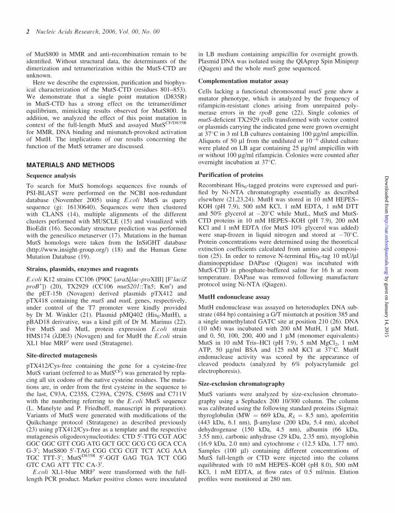
Our secondary structure prediction analysis of the CTDregion based on a multiple sequence alignment revealed abipartite organization of the CTD (Figure 1A). It consistsof a less conserved N-terminal region (residues 801–822)with no predicted secondary structure which contains the bsliding clamp binding motif (13) and a conserved C-terminalregion (residues 823–853) with a predicted helix–loop–helixmotif. Comparison of the MutS-CTD secondary structure pre-diction from bacteria and eukarya revealed that all homologscontain similar helical content (Figure 1B). Interestingly, sev-eral cancer causing mutations are localized in the C-terminusof the human MutS homologs, however, the biochemicalconsequences of these mutations are still unknown (29).
Purification and secondary structure analysis ofthe MutS-CTD
In order to analyze the structure and function of this domain,we created a His6-tagged protein (MutS-CTD) correspondingto residues 801–853 of the E.coli MutS. MutS-CTD wasexpressed and purified to homogeneity by Ni-NTA affinityand size-exclusion chromatography (Figure 2A). Analysisby electrospray ionization mass spectrometry indicated thatthe N-terminal methione was missing resulting in a 71 residueprotein with a calculated mass of 7.7 kDa (Table 1). Upontreatment with the diaminopeptidase DAPase, the N-terminalHis6-tag was removed to give MutS-CTDDHis6 with a calcu-lated mass of 6.4 kDa (Figure 2A).
Full-length MutS has been shown to form aggregates atelevated concentrations especially in the absence of nucleo-tide (30). In part the CTD was considered to be responsiblefor this phenomenon since deletion of this domain allowedcrystallization of the protein (5,6). However, MutS-CTDwas soluble and homogeneous at concentrations up to500 mM with no apparent formation of aggregates as judgedby size-exclusion chromatography and analytical ultracentri-fugation (see below). The CD spectra of MutS-CTD andMutS-CTDDHis6 revealed that the protein adopts a foldedstructure with �30% a-helical content (Figure 2B) consistentwith the secondary structure prediction (Figure 1A).
Nucleic Acids Research, 2006, Vol. 00, No. 00 3
by guest on January 14, 2015http://nar.oxfordjournals.org/
Dow
nloaded from
Quaternary structure analysis
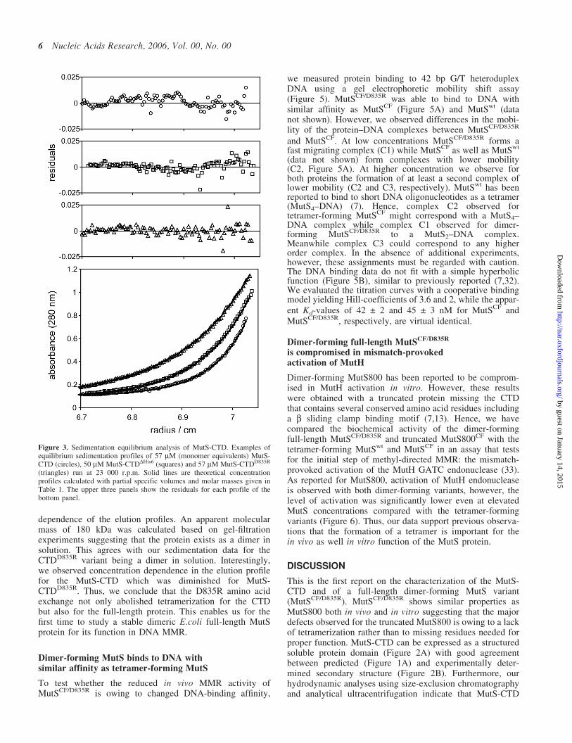
Since the CTD had been shown to be essential for tetra-merization of the full-length protein we asked whether thedomain on its own is sufficient to form tetramers. Sedimen-tation equilibrium analysis indicates that MutS-CTD and
MutS-CTDDHis6 form tetramers (Figure 3 and Table 1).Thus, tetramerization is a characteristic feature of this domainand is not influenced by the N-terminal His6-tag. In theconcentration range tested (14–60 mM) we did not observeany significant formation of either lower or higher molecular
Figure 1. Sequence alignment and secondary structure prediction of MutS. (A) C-terminal part of MutS-1 protein sequences from nine different species(accession numbers for these proteins are as follows: E.coli K12 NCBI accession no. 16130640; Haloarcula marismortui, NCBI accession no. 55232253;T.aquaticus, NCBI accession no. 2497995; Neisseria meningitides, NCBI accession no. 15677973; Listeria monocytogenes, NCBI accession no. 47095354;Methanosarcina barkeri, NCBI accession no. 68133332; Bacillus subtilis, NCBI accession no. 1002520; Brucella melitensis, NCBI accessionno.17983835; Anaeromyxobacter dehalogenans, NCBI accession no. 66857729) aligned using MUSCLE. Invariant residues are shaded in black whereassimilar residues are shaded in gray. Residue numbering and secondary structure predictions are for the E.coli sequence. (B) Secondary structure prediction of thehuman MSH2, MSH3, and MSH6 C-terminal domains. Cancer causing mutations are annotated with the amino acid exchange, insertion (IN) or deletion (X).
4 Nucleic Acids Research, 2006, Vol. 00, No. 00
by guest on January 14, 2015http://nar.oxfordjournals.org/
Dow
nloaded from
weight species. Moreover, sedimentation velocity analysisrevealed sedimentation coefficients s20,w of 2.1 and 1.4 S forMutS-CTD and MutS-CTDDHis6, respectively. Based on themolecular mass and sedimentation coefficient obtained bythe sedimentation analysis, MutS-CTD is a tetramer withhighly asymmetric shape (Table 1).
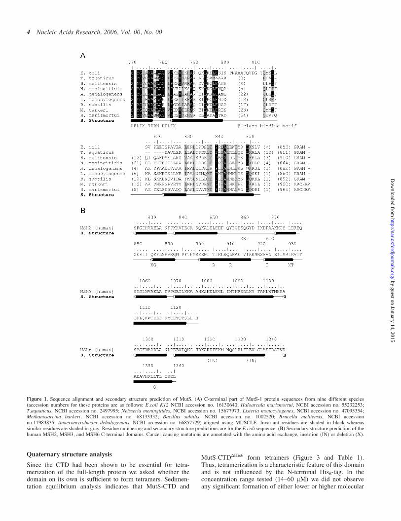
Disruption of the tetramer by D835R mutation
In order to investigate the MutS-CTD in more detail wegenerated several variants which we analyzed for their abilityto form tetramers (L. Manelyte, D. Goldeck and P. Friedhoff,unpublished data). A single point mutant, MutS-CTDCF/D835R
abolished tetramerization while still preserving dimerization(Figures 3 and 4 and Table 1). CD analysis of MutS-CTDD835R indicates that the protein is very similar to MutS-CTD with respect to secondary structure content. Therefore,
the amino acid exchange did not affect the overall structureof the protein (Figure 2B). Sedimentation equilibrium andvelocity experiments revealed that the protein exists in solu-tion as a dimer with asymmetric shape indicated by the fric-tional ratio of 1.55. Our data imply that the CTD stabilizesdimerization of the protein and allows tetramerization ofdimers. Since Asp-835 is located in the acidic loop of thehelix–loop–helix motif it is tempting to speculate that tetra-merization is mediated via this loop.
Tetramerization of full-length MutS is important butnot essential for in vivo function in mismatch repair
As mentioned above the role of the MutS tetramer in vitroand in vivo is controversially discussed in the literature.This is in part owing to comparing the function of thetetramer-forming full-length protein with a dimer-formingtruncated protein missing residues involved not only in tetra-merization but also dimerization and probably interactionwith other proteins, e.g. the b sliding clamp (13). Con-sequently, we asked whether the D835R mutation affectsthe function of the full-length MutS protein. Starting froman active cysteine-free MutS variant (MutSCF), we generateda truncated MutS variant (MutS800CF) and the D835R variant(MutSCF/D835R) which we assayed for MMR activity in vivo(Table 2). MutSwt as well as MutSCF encoded on a multicopyplasmid were able to complement the mutS-mutator pheno-type resulting in low mutation frequencies on rifampicin/ampicillin plates compared with the vector control. However,both variant MutS800CF and MutSCF/D835R have impairedMMR activity as indicated by elevated mutation frequencies,�5- to 10-fold higher compared with MutSwt and MutSCF,respectively (Table 2).
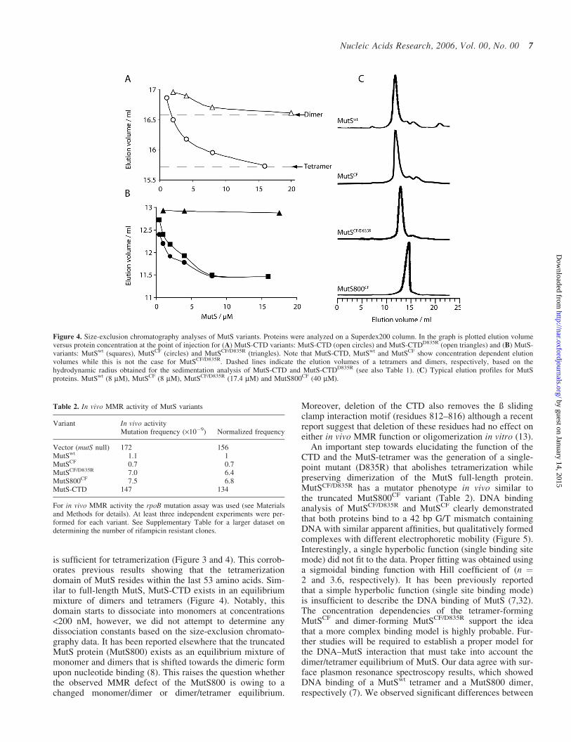
Further, we purified the proteins and characterizedtheir biophysical and biochemical properties in vitro. Size-exclusion chromatography experiments showed a concen-tration dependence of elution profiles for both MutSwt andMutSCF in agreement with the observed dimer/tetramerequilibrium for MutSwt (8,31) (Figure 4, Table 3). In con-trast to this, MutSCF/D835R did not show any concentration
Table 1. Hydrodynamic properties of MutS-CTD variants
CTD CTDDHis6 CTDD835R
Partial specific volume (ml/g)a 0.727 0.738 0.728Sedimentation
coefficient s20,w (Svedberg)2.1 ± 0.04 1.4 ± 0.13 1.5 ± 0.04
Stoke radius Rs (nm)Calculated from s20,w
b 3.65 4.3 2.55Estimated by size-exclusion
chromatography3.5 n.d. 2.3
Molecular weight determinedBy sedimentation
equilibrium29 800 ± 410c 25 300 ± 380d 16 500 ± 370e
From sequence (dimer)a 15405 12835 15487From sequence (tetramer)a 30792 25651 30956
Oligomeric status Tetramer Tetramer DimerPerrin factor f/fmin 1.74 2.19 1.55
aUsing sequence without N-terminal methionine as determined by ESI massspectrometry.bUsing SEDNTERP version 1.08 using the vbar method.cDetermined at 29 mM protein and 23 000 r.p.m.dDetermined at 50 mM protein and 23 000 r.p.m.eDetermined at 29 mM protein and 16 000 r.p.m.
Figure 2. Analysis of purified MutS-CTD variants. (A) Coomassie blue-stained Tris-Tricine 15% polyacrylamide gel is shown. Lane 1, proteinmarkers (M) with sizes indicated on the left; lane 2, MutS-CTD; lane 3,MutS-CTDDHis6 (His6-tag removed by DAPase; for details see Material andMethods); lane 4, MutS-CTDD835R. (B) CD spectra of MutS-CTD variants.All spectra were recorded with 20 mM proteins in HEPES–KOH 10 mM(pH 8.0), EDTA 0.1 mM and KCl 50 mM at 20�C. Residual molar ellipticity[Q] of MutS-CTD variants was measured from 190 to 280 nm as describedunder Materials and Methods. The a-helical content was estimated to be 30%using secondary structure deconvolution program CDNN.
Nucleic Acids Research, 2006, Vol. 00, No. 00 5
by guest on January 14, 2015http://nar.oxfordjournals.org/
Dow
nloaded from
dependence of the elution profiles. An apparent molecularmass of 180 kDa was calculated based on gel-filtrationexperiments suggesting that the protein exists as a dimer insolution. This agrees with our sedimentation data for theCTDD835R variant being a dimer in solution. Interestingly,we observed concentration dependence in the elution profilefor the MutS-CTD which was diminished for MutS-CTDD835R. Thus, we conclude that the D835R amino acidexchange not only abolished tetramerization for the CTDbut also for the full-length protein. This enables us for thefirst time to study a stable dimeric E.coli full-length MutSprotein for its function in DNA MMR.
Dimer-forming MutS binds to DNA withsimilar affinity as tetramer-forming MutS
To test whether the reduced in vivo MMR activity ofMutSCF/D835R is owing to changed DNA-binding affinity,
we measured protein binding to 42 bp G/T heteroduplexDNA using a gel electrophoretic mobility shift assay(Figure 5). MutSCF/D835R was able to bind to DNA withsimilar affinity as MutSCF (Figure 5A) and MutSwt (datanot shown). However, we observed differences in the mobi-lity of the protein–DNA complexes between MutSCF/D835R
and MutSCF. At low concentrations MutSCF/D835R forms afast migrating complex (C1) while MutSCF as well as MutSwt
(data not shown) form complexes with lower mobility(C2, Figure 5A). At higher concentration we observe forboth proteins the formation of at least a second complex oflower mobility (C2 and C3, respectively). MutSwt has beenreported to bind to short DNA oligonucleotides as a tetramer(MutS4–DNA) (7). Hence, complex C2 observed fortetramer-forming MutSCF might correspond with a MutS4–DNA complex while complex C1 observed for dimer-forming MutSCF/D835R to a MutS2–DNA complex.Meanwhile complex C3 could correspond to any higherorder complex. In the absence of additional experiments,however, these assignments must be regarded with caution.The DNA binding data do not fit with a simple hyperbolicfunction (Figure 5B), similar to previously reported (7,32).We evaluated the titration curves with a cooperative bindingmodel yielding Hill-coefficients of 3.6 and 2, while the appar-ent Kd-values of 42 ± 2 and 45 ± 3 nM for MutSCF andMutSCF/D835R, respectively, are virtual identical.
Dimer-forming full-length MutSCF/D835R
is compromised in mismatch-provokedactivation of MutH
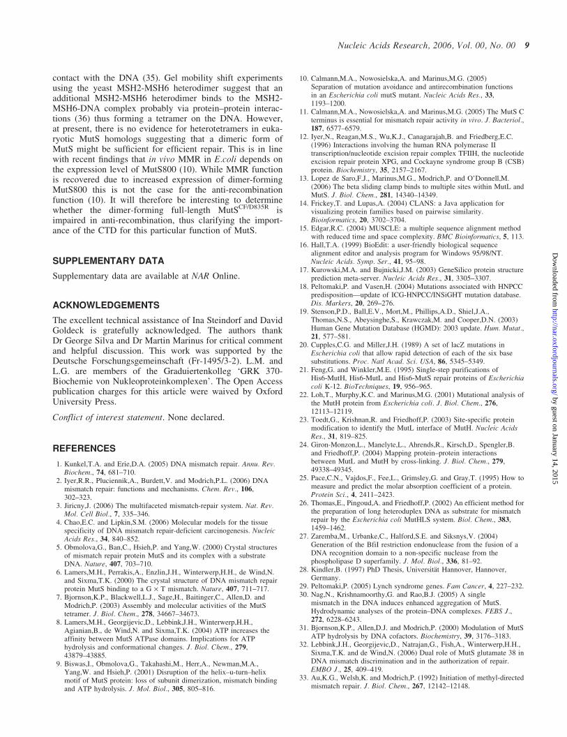
Dimer-forming MutS800 has been reported to be comprom-ised in MutH activation in vitro. However, these resultswere obtained with a truncated protein missing the CTDthat contains several conserved amino acid residues includinga b sliding clamp binding motif (7,13). Hence, we havecompared the biochemical activity of the dimer-formingfull-length MutSCF/D835R and truncated MutS800CF with thetetramer-forming MutSwt and MutSCF in an assay that testsfor the initial step of methyl-directed MMR: the mismatch-provoked activation of the MutH GATC endonuclease (33).As reported for MutS800, activation of MutH endonucleaseis observed with both dimer-forming variants, however, thelevel of activation was significantly lower even at elevatedMutS concentrations compared with the tetramer-formingvariants (Figure 6). Thus, our data support previous observa-tions that the formation of a tetramer is important for thein vivo as well in vitro function of the MutS protein.
DISCUSSION
This is the first report on the characterization of the MutS-CTD and of a full-length dimer-forming MutS variant(MutSCF/D835R). MutSCF/D835R shows similar properties asMutS800 both in vivo and in vitro suggesting that the majordefects observed for the truncated MutS800 is owing to a lackof tetramerization rather than to missing residues needed forproper function. MutS-CTD can be expressed as a structuredsoluble protein domain (Figure 2A) with good agreementbetween predicted (Figure 1A) and experimentally deter-mined secondary structure (Figure 2B). Furthermore, ourhydrodynamic analyses using size-exclusion chromatographyand analytical ultracentrifugation indicate that MutS-CTD
Figure 3. Sedimentation equilibrium analysis of MutS-CTD. Examples ofequilibrium sedimentation profiles of 57 mM (monomer equivalents) MutS-CTD (circles), 50 mM MutS-CTDDHis6 (squares) and 57 mM MutS-CTDD835R
(triangles) run at 23 000 r.p.m. Solid lines are theoretical concentrationprofiles calculated with partial specific volumes and molar masses given inTable 1. The upper three panels show the residuals for each profile of thebottom panel.
6 Nucleic Acids Research, 2006, Vol. 00, No. 00
by guest on January 14, 2015http://nar.oxfordjournals.org/
Dow
nloaded from
is sufficient for tetramerization (Figure 3 and 4). This corrob-orates previous results showing that the tetramerizationdomain of MutS resides within the last 53 amino acids. Sim-ilar to full-length MutS, MutS-CTD exists in an equilibriummixture of dimers and tetramers (Figure 4). Notably, thisdomain starts to dissociate into monomers at concentrations<200 nM, however, we did not attempt to determine anydissociation constants based on the size-exclusion chromato-graphy data. It has been reported elsewhere that the truncatedMutS protein (MutS800) exists as an equilibrium mixture ofmonomer and dimers that is shifted towards the dimeric formupon nucleotide binding (8). This raises the question whetherthe observed MMR defect of the MutS800 is owing to achanged monomer/dimer or dimer/tetramer equilibrium.
Moreover, deletion of the CTD also removes the ß slidingclamp interaction motif (residues 812–816) although a recentreport suggest that deletion of these residues had no effect oneither in vivo MMR function or oligomerization in vitro (13).
An important step towards elucidating the function of theCTD and the MutS-tetramer was the generation of a single-point mutant (D835R) that abolishes tetramerization whilepreserving dimerization of the MutS full-length protein.MutSCF/D835R has a mutator phenotype in vivo similar tothe truncated MutS800CF variant (Table 2). DNA bindinganalysis of MutSCF/D835R and MutSCF clearly demonstratedthat both proteins bind to a 42 bp G/T mismatch containingDNA with similar apparent affinities, but qualitatively formedcomplexes with different electrophoretic mobility (Figure 5).Interestingly, a single hyperbolic function (single binding sitemode) did not fit to the data. Proper fitting was obtained usinga sigmoidal binding function with Hill coefficient of (n ¼2 and 3.6, respectively). It has been previously reportedthat a simple hyperbolic function (single site binding mode)is insufficient to describe the DNA binding of MutS (7,32).The concentration dependencies of the tetramer-formingMutSCF and dimer-forming MutSCF/D835R support the ideathat a more complex binding model is highly probable. Fur-ther studies will be required to establish a proper model forthe DNA–MutS interaction that must take into account thedimer/tetramer equilibrium of MutS. Our data agree with sur-face plasmon resonance spectroscopy results, which showedDNA binding of a MutSwt tetramer and a MutS800 dimer,respectively (7). We observed significant differences between
Figure 4. Size-exclusion chromatography analyses of MutS variants. Proteins were analyzed on a Superdex200 column. In the graph is plotted elution volumeversus protein concentration at the point of injection for (A) MutS-CTD variants: MutS-CTD (open circles) and MutS-CTDD835R (open triangles) and (B) MutS-variants: MutSwt (squares), MutSCF (circles) and MutSCF/D835R (triangles). Note that MutS-CTD, MutSwt and MutSCF show concentration dependent elutionvolumes while this is not the case for MutSCF/D835R. Dashed lines indicate the elution volumes of a tetramers and dimers, respectively, based on thehydrodynamic radius obtained for the sedimentation analysis of MutS-CTD and MutS-CTDD835R (see also Table 1). (C) Typical elution profiles for MutSproteins. MutSwt (8 mM), MutSCF (8 mM), MutSCF/D835R (17.4 mM) and MutS800CF (40 mM).
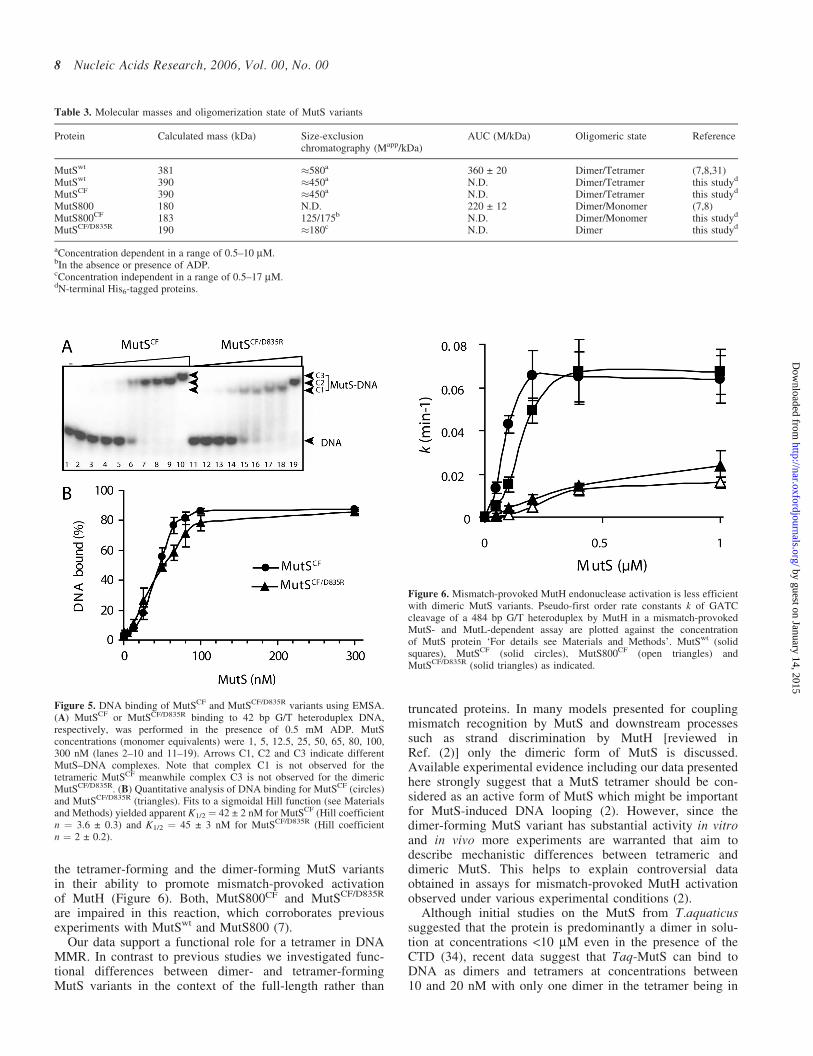
Table 2. In vivo MMR activity of MutS variants
Variant In vivo activityMutation frequency (·10�9) Normalized frequency
Vector (mutS null) 172 156MutSwt 1.1 1MutSCF 0.7 0.7MutSCF/D835R 7.0 6.4MutS800CF 7.5 6.8MutS-CTD 147 134
For in vivo MMR activity the rpoB mutation assay was used (see Materialsand Methods for details). At least three independent experiments were per-formed for each variant. See Supplementary Table for a larger dataset ondetermining the number of rifampicin resistant clones.
Nucleic Acids Research, 2006, Vol. 00, No. 00 7
by guest on January 14, 2015http://nar.oxfordjournals.org/
Dow
nloaded from
the tetramer-forming and the dimer-forming MutS variantsin their ability to promote mismatch-provoked activationof MutH (Figure 6). Both, MutS800CF and MutSCF/D835R
are impaired in this reaction, which corroborates previousexperiments with MutSwt and MutS800 (7).
Our data support a functional role for a tetramer in DNAMMR. In contrast to previous studies we investigated func-tional differences between dimer- and tetramer-formingMutS variants in the context of the full-length rather than
truncated proteins. In many models presented for couplingmismatch recognition by MutS and downstream processessuch as strand discrimination by MutH [reviewed inRef. (2)] only the dimeric form of MutS is discussed.Available experimental evidence including our data presentedhere strongly suggest that a MutS tetramer should be con-sidered as an active form of MutS which might be importantfor MutS-induced DNA looping (2). However, since thedimer-forming MutS variant has substantial activity in vitroand in vivo more experiments are warranted that aim todescribe mechanistic differences between tetrameric anddimeric MutS. This helps to explain controversial dataobtained in assays for mismatch-provoked MutH activationobserved under various experimental conditions (2).
Although initial studies on the MutS from T.aquaticussuggested that the protein is predominantly a dimer in solu-tion at concentrations <10 mM even in the presence of theCTD (34), recent data suggest that Taq-MutS can bind toDNA as dimers and tetramers at concentrations between10 and 20 nM with only one dimer in the tetramer being in
Figure 5. DNA binding of MutSCF and MutSCF/D835R variants using EMSA.(A) MutSCF or MutSCF/D835R binding to 42 bp G/T heteroduplex DNA,respectively, was performed in the presence of 0.5 mM ADP. MutSconcentrations (monomer equivalents) were 1, 5, 12.5, 25, 50, 65, 80, 100,300 nM (lanes 2–10 and 11–19). Arrows C1, C2 and C3 indicate differentMutS–DNA complexes. Note that complex C1 is not observed for thetetrameric MutSCF meanwhile complex C3 is not observed for the dimericMutSCF/D835R. (B) Quantitative analysis of DNA binding for MutSCF (circles)and MutSCF/D835R (triangles). Fits to a sigmoidal Hill function (see Materialsand Methods) yielded apparent K1/2 ¼ 42 ± 2 nM for MutSCF (Hill coefficientn ¼ 3.6 ± 0.3) and K1/2 ¼ 45 ± 3 nM for MutSCF/D835R (Hill coefficientn ¼ 2 ± 0.2).
Table 3. Molecular masses and oligomerization state of MutS variants
Protein Calculated mass (kDa) Size-exclusionchromatography (Mapp/kDa)
AUC (M/kDa) Oligomeric state Reference
MutSwt 381 �580a 360 ± 20 Dimer/Tetramer (7,8,31)MutSwt 390 �450a N.D. Dimer/Tetramer this studyd
MutSCF 390 �450a N.D. Dimer/Tetramer this studyd
MutS800 180 N.D. 220 ± 12 Dimer/Monomer (7,8)MutS800CF 183 125/175b N.D. Dimer/Monomer this studyd
MutSCF/D835R 190 �180c N.D. Dimer this studyd
aConcentration dependent in a range of 0.5–10 mM.bIn the absence or presence of ADP.cConcentration independent in a range of 0.5–17 mM.dN-terminal His6-tagged proteins.
Figure 6. Mismatch-provoked MutH endonuclease activation is less efficientwith dimeric MutS variants. Pseudo-first order rate constants k of GATCcleavage of a 484 bp G/T heteroduplex by MutH in a mismatch-provokedMutS- and MutL-dependent assay are plotted against the concentrationof MutS protein ‘For details see Materials and Methods’. MutSwt (solidsquares), MutSCF (solid circles), MutS800CF (open triangles) andMutSCF/D835R (solid triangles) as indicated.
8 Nucleic Acids Research, 2006, Vol. 00, No. 00
by guest on January 14, 2015http://nar.oxfordjournals.org/
Dow
nloaded from
contact with the DNA (35). Gel mobility shift experimentsusing the yeast MSH2-MSH6 heterodimer suggest that anadditional MSH2-MSH6 heterodimer binds to the MSH2-MSH6-DNA complex probably via protein–protein interac-tions (36) thus forming a tetramer on the DNA. However,at present, there is no evidence for heterotetramers in euka-ryotic MutS homologs suggesting that a dimeric form ofMutS might be sufficient for efficient repair. This is in linewith recent findings that in vivo MMR in E.coli depends onthe expression level of MutS800 (10). While MMR functionis recovered due to increased expression of dimer-formingMutS800 this is not the case for the anti-recombinationfunction (10). It will therefore be interesting to determinewhether the dimer-forming full-length MutSCF/D835R isimpaired in anti-recombination, thus clarifying the import-ance of the CTD for this particular function of MutS.
SUPPLEMENTARY DATA
Supplementary data are available at NAR Online.
ACKNOWLEDGEMENTS
The excellent technical assistance of Ina Steindorf and DavidGoldeck is gratefully acknowledged. The authors thankDr George Silva and Dr Martin Marinus for critical commentand helpful discussion. This work was supported by theDeutsche Forschungsgemeinschaft (Fr-1495/3-2). L.M. andL.G. are members of the Graduiertenkolleg ‘GRK 370-Biochemie von Nukleoproteinkomplexen’. The Open Accesspublication charges for this article were waived by OxfordUniversity Press.
Conflict of interest statement. None declared.
REFERENCES
1. Kunkel,T.A. and Erie,D.A. (2005) DNA mismatch repair. Annu. Rev.Biochem., 74, 681–710.
2. Iyer,R.R., Pluciennik,A., Burdett,V. and Modrich,P.L. (2006) DNAmismatch repair: functions and mechanisms. Chem. Rev., 106,302–323.
3. Jiricny,J. (2006) The multifaceted mismatch-repair system. Nat. Rev.Mol. Cell Biol., 7, 335–346.
4. Chao,E.C. and Lipkin,S.M. (2006) Molecular models for the tissuespecificity of DNA mismatch repair-deficient carcinogenesis. NucleicAcids Res., 34, 840–852.
5. Obmolova,G., Ban,C., Hsieh,P. and Yang,W. (2000) Crystal structuresof mismatch repair protein MutS and its complex with a substrateDNA. Nature, 407, 703–710.
6. Lamers,M.H., Perrakis,A., Enzlin,J.H., Winterwerp,H.H., de Wind,N.and Sixma,T.K. (2000) The crystal structure of DNA mismatch repairprotein MutS binding to a G · T mismatch. Nature, 407, 711–717.
7. Bjornson,K.P., Blackwell,L.J., Sage,H., Baitinger,C., Allen,D. andModrich,P. (2003) Assembly and molecular activities of the MutStetramer. J. Biol. Chem., 278, 34667–34673.
8. Lamers,M.H., Georgijevic,D., Lebbink,J.H., Winterwerp,H.H.,Agianian,B., de Wind,N. and Sixma,T.K. (2004) ATP increases theaffinity between MutS ATPase domains. Implications for ATPhydrolysis and conformational changes. J. Biol. Chem., 279,43879–43885.
9. Biswas,I., Obmolova,G., Takahashi,M., Herr,A., Newman,M.A.,Yang,W. and Hsieh,P. (2001) Disruption of the helix–u-turn–helixmotif of MutS protein: loss of subunit dimerization, mismatch bindingand ATP hydrolysis. J. Mol. Biol., 305, 805–816.
10. Calmann,M.A., Nowosielska,A. and Marinus,M.G. (2005)Separation of mutation avoidance and antirecombination functionsin an Escherichia coli mutS mutant. Nucleic Acids Res., 33,1193–1200.
11. Calmann,M.A., Nowosielska,A. and Marinus,M.G. (2005) The MutS Cterminus is essential for mismatch repair activity in vivo. J. Bacteriol.,187, 6577–6579.
12. Iyer,N., Reagan,M.S., Wu,K.J., Canagarajah,B. and Friedberg,E.C.(1996) Interactions involving the human RNA polymerase IItranscription/nucleotide excision repair complex TFIIH, the nucleotideexcision repair protein XPG, and Cockayne syndrome group B (CSB)protein. Biochemistry, 35, 2157–2167.
13. Lopez de Saro,F.J., Marinus,M.G., Modrich,P. and O’Donnell,M.(2006) The beta sliding clamp binds to multiple sites within MutL andMutS. J. Biol. Chem., 281, 14340–14349.
14. Frickey,T. and Lupas,A. (2004) CLANS: a Java application forvisualizing protein families based on pairwise similarity.Bioinformatics, 20, 3702–3704.
15. Edgar,R.C. (2004) MUSCLE: a multiple sequence alignment methodwith reduced time and space complexity. BMC Bioinformatics, 5, 113.
16. Hall,T.A. (1999) BioEdit: a user-friendly biological sequencealignment editor and analysis program for Windows 95/98/NT.Nucleic Acids. Symp. Ser., 41, 95–98.
17. Kurowski,M.A. and Bujnicki,J.M. (2003) GeneSilico protein structureprediction meta-server. Nucleic Acids Res., 31, 3305–3307.
18. Peltomaki,P. and Vasen,H. (2004) Mutations associated with HNPCCpredisposition—update of ICG-HNPCC/INSiGHT mutation database.Dis. Markers, 20, 269–276.
19. Stenson,P.D., Ball,E.V., Mort,M., Phillips,A.D., Shiel,J.A.,Thomas,N.S., Abeysinghe,S., Krawczak,M. and Cooper,D.N. (2003)Human Gene Mutation Database (HGMD): 2003 update. Hum. Mutat.,21, 577–581.
20. Cupples,C.G. and Miller,J.H. (1989) A set of lacZ mutations inEscherichia coli that allow rapid detection of each of the six basesubstitutions. Proc. Natl Acad. Sci. USA, 86, 5345–5349.
21. Feng,G. and Winkler,M.E. (1995) Single-step purifications ofHis6-MutH, His6-MutL and His6-MutS repair proteins of Escherichiacoli K-12. BioTechniques, 19, 956–965.
22. Loh,T., Murphy,K.C. and Marinus,M.G. (2001) Mutational analysis ofthe MutH protein from Escherichia coli. J. Biol. Chem., 276,12113–12119.
23. Toedt,G., Krishnan,R. and Friedhoff,P. (2003) Site-specific proteinmodification to identify the MutL interface of MutH. Nucleic AcidsRes., 31, 819–825.
24. Giron-Monzon,L., Manelyte,L., Ahrends,R., Kirsch,D., Spengler,B.and Friedhoff,P. (2004) Mapping protein–protein interactionsbetween MutL and MutH by cross-linking. J. Biol. Chem., 279,49338–49345.
25. Pace,C.N., Vajdos,F., Fee,L., Grimsley,G. and Gray,T. (1995) How tomeasure and predict the molar absorption coefficient of a protein.Protein Sci., 4, 2411–2423.
26. Thomas,E., Pingoud,A. and Friedhoff,P. (2002) An efficient method forthe preparation of long heteroduplex DNA as substrate for mismatchrepair by the Escherichia coli MutHLS system. Biol. Chem., 383,1459–1462.
27. Zaremba,M., Urbanke,C., Halford,S.E. and Siksnys,V. (2004)Generation of the BfiI restriction endonuclease from the fusion of aDNA recognition domain to a non-specific nuclease from thephospholipase D superfamily. J. Mol. Biol., 336, 81–92.
28. Kindler,B. (1997) PhD Thesis, Universitat Hannover, Hannover,Germany.
29. Peltomaki,P. (2005) Lynch syndrome genes. Fam Cancer, 4, 227–232.30. Nag,N., Krishnamoorthy,G. and Rao,B.J. (2005) A single
mismatch in the DNA induces enhanced aggregation of MutS.Hydrodynamic analyses of the protein–DNA complexes. FEBS J.,272, 6228–6243.
31. Bjornson,K.P., Allen,D.J. and Modrich,P. (2000) Modulation of MutSATP hydrolysis by DNA cofactors. Biochemistry, 39, 3176–3183.
32. Lebbink,J.H., Georgijevic,D., Natrajan,G., Fish,A., Winterwerp,H.H.,Sixma,T.K. and de Wind,N. (2006) Dual role of MutS glutamate 38 inDNA mismatch discrimination and in the authorization of repair.EMBO J., 25, 409–419.
33. Au,K.G., Welsh,K. and Modrich,P. (1992) Initiation of methyl-directedmismatch repair. J. Biol. Chem., 267, 12142–12148.
Nucleic Acids Research, 2006, Vol. 00, No. 00 9
by guest on January 14, 2015http://nar.oxfordjournals.org/
Dow
nloaded from
34. Biswas,I., Ban,C., Fleming,K.G., Qin,J., Lary,J.W., Yphantis,D.A.,Yang,W. and Hsieh,P. (1999) Oligomerization of a MutS mismatchrepair protein from Thermus aquaticus. J. Biol. Chem., 274,23673–23678.
35. Wang,H., Yang,Y., Schofield,M.J., Du,C., Fridman,Y., Lee,S.D.,Larson,E.D., Drummond,J.T., Alani,E., Hsieh,P. et al. (2003) DNA
bending and unbending by MutS govern mismatch recognition andspecificity. Proc. Natl Acad. Sci. USA, 100, 14822–14827.
36. Surtees,J.A. and Alani,E. (2006) Mismatch repair factor MSH2-MSH3binds and alters the conformation of branched DNA structurespredicted to form during genetic recombination. J. Mol. Biol., 360,523–536.
10 Nucleic Acids Research, 2006, Vol. 00, No. 00
by guest on January 14, 2015http://nar.oxfordjournals.org/
Dow
nloaded from
Top Related
Copyright © 2022 FDOKUMEN

