


Bahasa
Halaman
Hukum


Sequential Assignment Strategiesin Proteins
NMR assignments
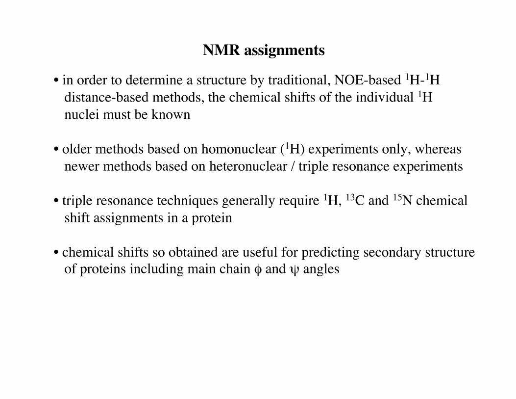
• in order to determine a structure by traditional, NOE-based 1H-1H distance-based methods, the chemical shifts of the individual 1H nuclei must be known
• older methods based on homonuclear (1H) experiments only, whereas newer methods based on heteronuclear / triple resonance experiments
• triple resonance techniques generally require 1H, 13C and 15N chemical shift assignments in a protein
• chemical shifts so obtained are useful for predicting secondary structure of proteins including main chain φ and ψ angles
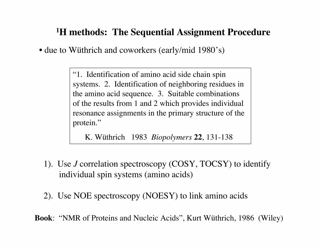
1H methods: The Sequential Assignment Procedure
• due to Wüthrich and coworkers (early/mid 1980’s)
“1. Identification of amino acid side chain spinsystems. 2. Identification of neighboring residues inthe amino acid sequence. 3. Suitable combinationsof the results from 1 and 2 which provides individualresonance assignments in the primary structure of theprotein.”
K. Wüthrich 1983 Biopolymers 22, 131-138
1). Use J correlation spectroscopy (COSY, TOCSY) to identify individual spin systems (amino acids)
2). Use NOE spectroscopy (NOESY) to link amino acids
Book: “NMR of Proteins and Nucleic Acids”, Kurt Wüthrich, 1986 (Wiley)
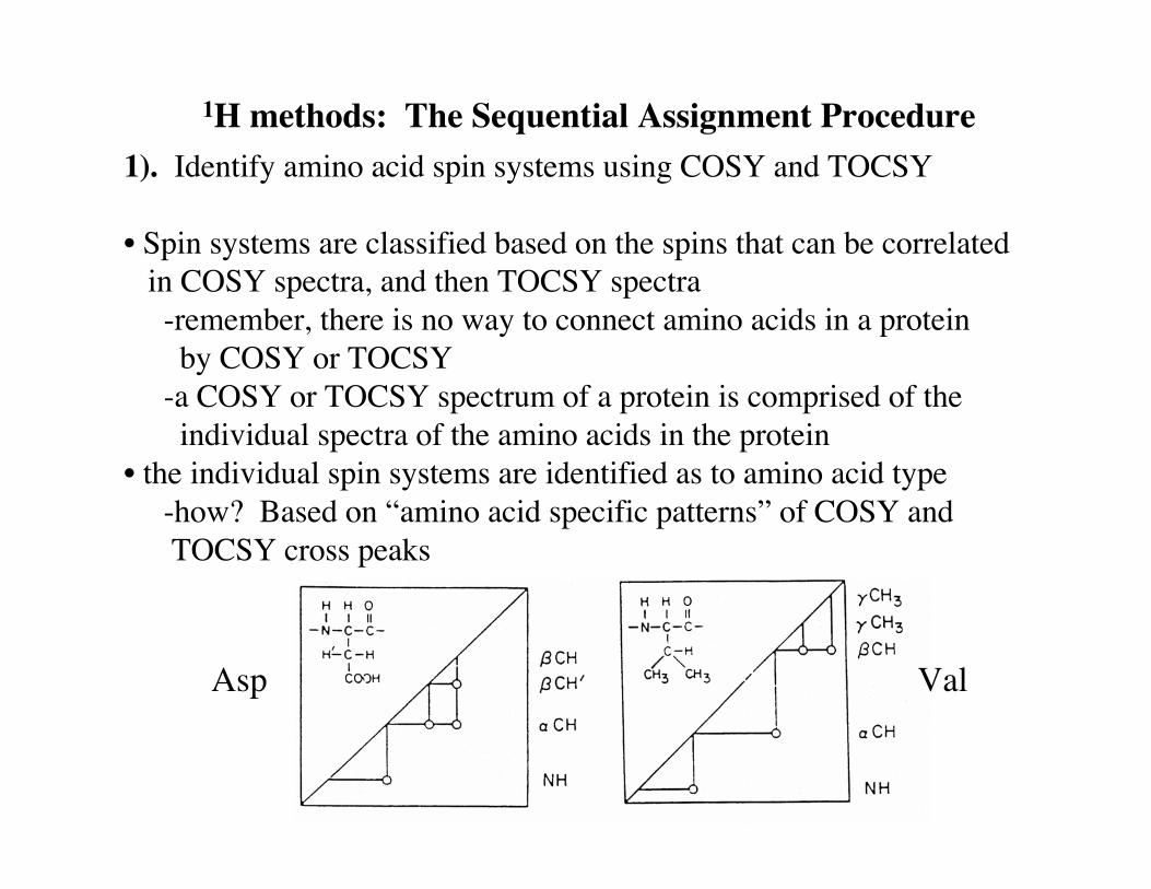
1H methods: The Sequential Assignment Procedure1). Identify amino acid spin systems using COSY and TOCSY
• Spin systems are classified based on the spins that can be correlated in COSY spectra, and then TOCSY spectra -remember, there is no way to connect amino acids in a protein by COSY or TOCSY -a COSY or TOCSY spectrum of a protein is comprised of the individual spectra of the amino acids in the protein• the individual spin systems are identified as to amino acid type -how? Based on “amino acid specific patterns” of COSY and TOCSY cross peaks
Asp Val

More examples of COSY patterns

1H chemical shift information
• 1H chemical shift information is also very useful in assisting to identify spin systems
-shown are distributions of 1H chemical shifts for amino acids in proteins
Wishart et al. (1991) J. Mol. Biol. 222, 311-333

1H methods: The Sequential Assignment Procedure
2). Link identified amino acids using NOESY data
• critical observation: it is virtually impossible to align the main chain atoms of two adjacent amino acid residues in a protein so that there is not at least one pair of interresidue distances between main chain hydrogens that is significantly less than the NOE detection limit (~5Å)
dashed lines: nonlabile protons correlated by J correlation experiments
arrows: sequential interresidue NOEs

Sequential interresidue NOEs
• most short interproton (1HN, 1Hα, 1Hβ) distances in proteins are between directly adjacent amino acid residues -intense NOEs indicate adjacent amino acids• less intense i, i+2 and i, i+3 NOEs (“nonsequential”) also observed and useful, particularly in well-defined secondary structures
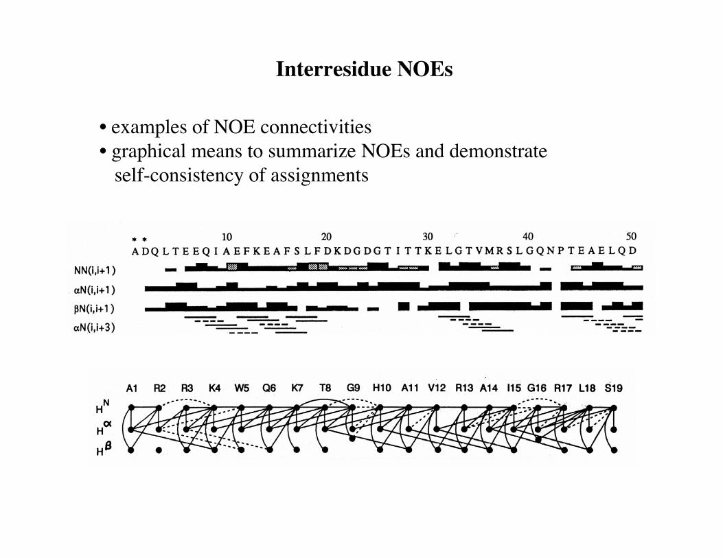
Interresidue NOEs
• examples of NOE connectivities• graphical means to summarize NOEs and demonstrate self-consistency of assignments
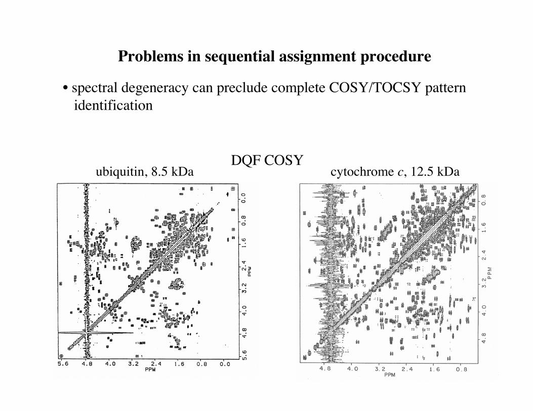
Problems in sequential assignment procedure
• spectral degeneracy can preclude complete COSY/TOCSY pattern identification
cytochrome c, 12.5 kDaDQF COSY
ubiquitin, 8.5 kDa
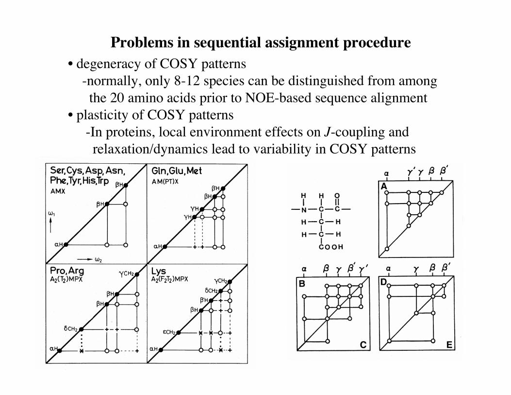
Problems in sequential assignment procedure• degeneracy of COSY patterns -normally, only 8-12 species can be distinguished from among the 20 amino acids prior to NOE-based sequence alignment• plasticity of COSY patterns -In proteins, local environment effects on J-coupling and relaxation/dynamics lead to variability in COSY patterns

Main Chain Directed (MCD) approach
• due to Wand/Englander and coworkers (mid/late 1980’s)
1). use J correlated and spectra to first identify 1HN-1Hα-1Hβ
(“NAB”) units -the 1HN region of COSY spectra is usually less crowded -individual amino acid type identification not attempted at this point
2). next align the NAB units sequentially using NOESY spectra -pattern recognition routines employed to search for well-established patterns of NOEs -amino acid type identification is then substantially restricted
• no initial reliance on identity of amino acid to establish connectivity• amenable to automation (pattern matching algorithms)

Triple resonance approach
• the triple resonance approach has largely replaced 1H-only methods in cases where the protein can be isotopically labeled -is the only method currently available for large proteins
• experiments provide selective chemical shift correlation of main chain (plus 1Hβ and 13Cβ) nuclei in adjacent amino acids -these correlations permit links between individual amino acids to be established, and thus assignment of the main chain -the chemical shifts of side chain nuclei are then correlated with assigned main chain nuclei to complete side chain assignments
• in the ideal case, no other information is required -in the ideal case, and in theory, amino acid type need not be established initially -in practice, many other types of information, including chemical shift/amino acid type information, NOE distance information, etc., play important roles
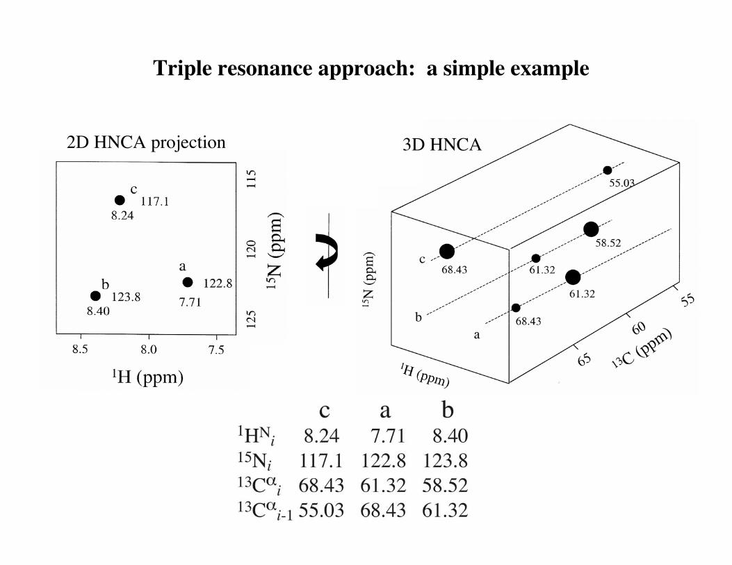
Triple resonance approach: a simple example
2D HNCA projection 3D HNCA
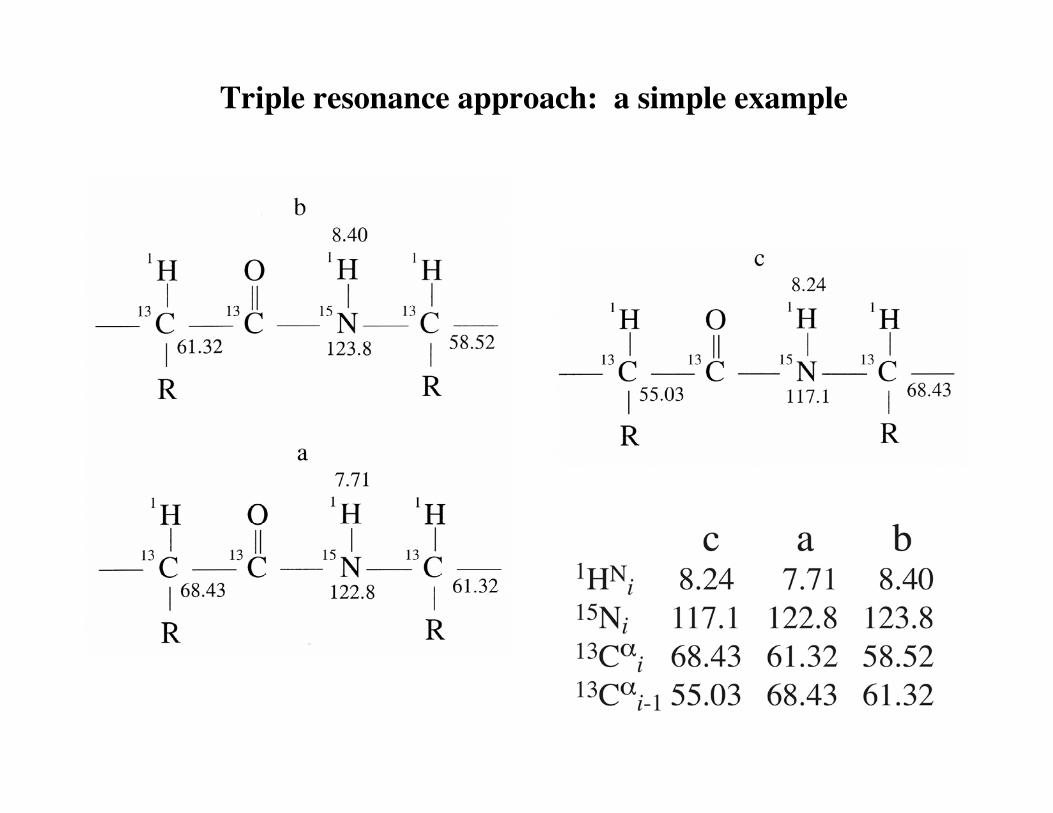
Triple resonance approach: a simple example
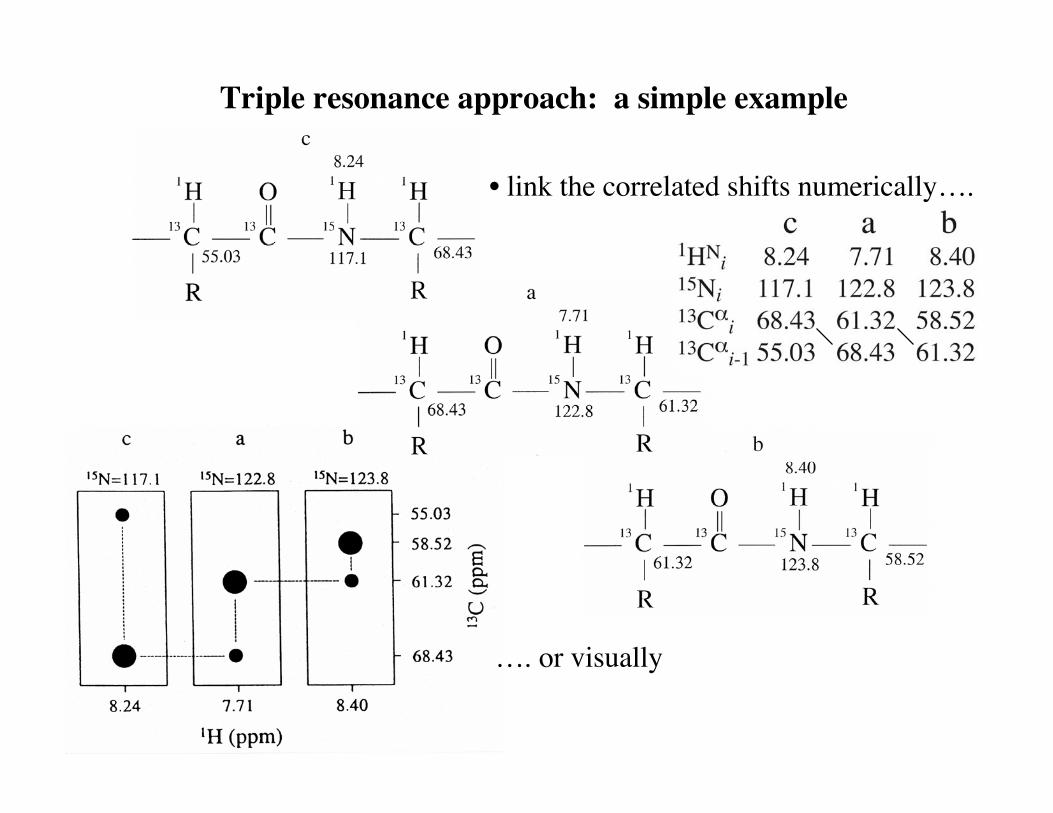
Triple resonance approach: a simple example
• link the correlated shifts numerically….
…. or visually

Triple resonance approach: HNCA/HN(CO)CA example
• problems:13Cα chemical shift degeneracy in proteins13Cα linewidths/resolution
-these preclude complete linkage via 13Cα alone -the same is true for 13Cβ, 13C′
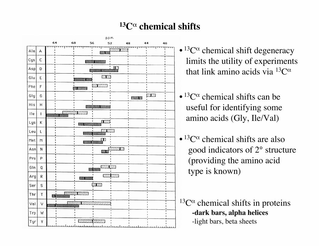
13Cα chemical shifts
• 13Cα chemical shift degeneracy limits the utility of experiments that link amino acids via 13Cα
• 13Cα chemical shifts can be useful for identifying some amino acids (Gly, Ile/Val)
• 13Cα chemical shifts are also good indicators of 2° structure (providing the amino acid type is known)
13Cα chemical shifts in proteins -dark bars, alpha helices -light bars, beta sheets

13Cα
13Cβ
13Cβ Thr
HNCACB / CBCA(CO)NH, connectivity via 13Cβ
• 13Cβ correlations permit resolution of ambiguities in connectivities due to 13Cα
degeneracy
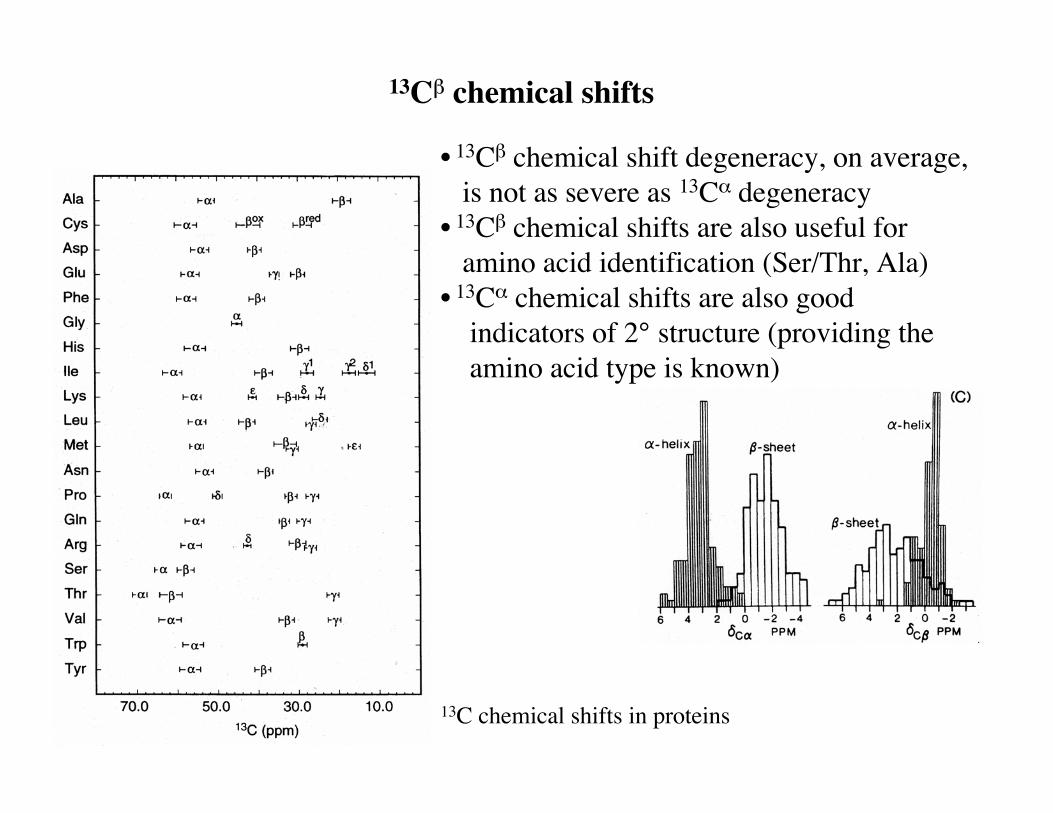
• 13Cβ chemical shift degeneracy, on average, is not as severe as 13Cα degeneracy• 13Cβ chemical shifts are also useful for amino acid identification (Ser/Thr, Ala)• 13Cα chemical shifts are also good indicators of 2° structure (providing the amino acid type is known)
13C chemical shifts in proteins
13Cβ chemical shifts
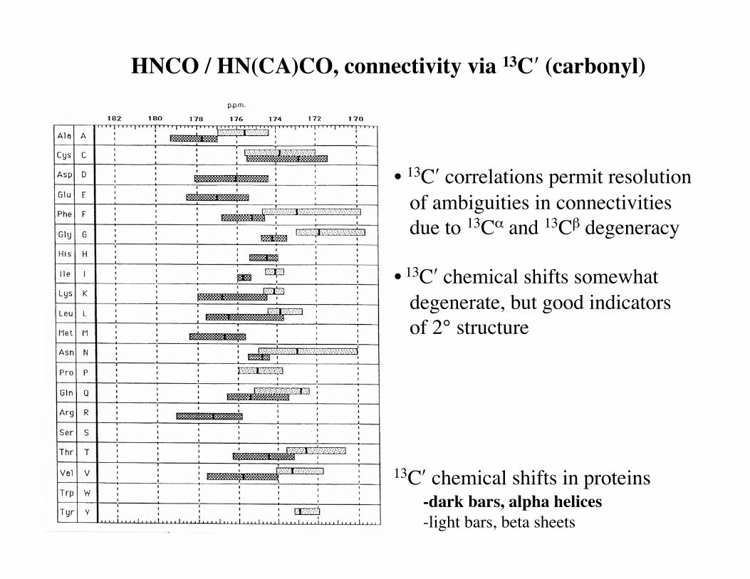
13C′ chemical shifts in proteins -dark bars, alpha helices -light bars, beta sheets
HNCO / HN(CA)CO, connectivity via 13C′ (carbonyl)
• 13C′ correlations permit resolution of ambiguities in connectivities due to 13Cα and 13Cβ degeneracy
• 13C′ chemical shifts somewhat degenerate, but good indicators of 2° structure
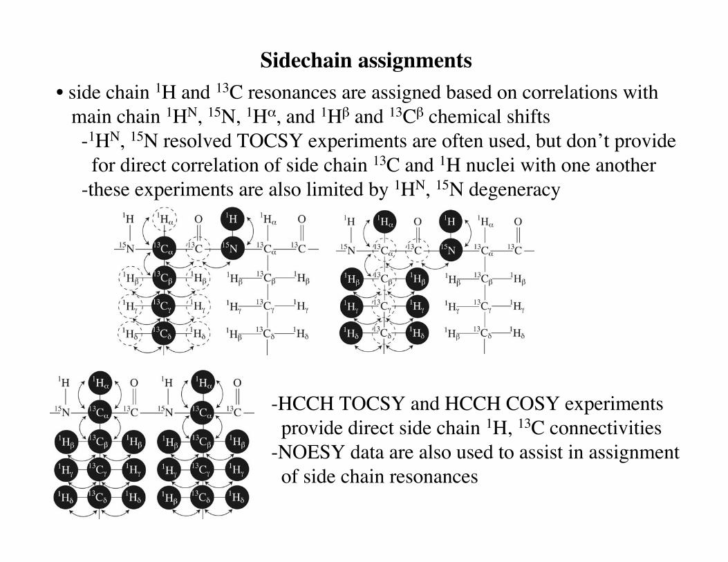
Sidechain assignments• side chain 1H and 13C resonances are assigned based on correlations with main chain 1HN, 15N, 1Hα, and 1Hβ and 13Cβ chemical shifts -1HN, 15N resolved TOCSY experiments are often used, but don’t provide for direct correlation of side chain 13C and 1H nuclei with one another -these experiments are also limited by 1HN, 15N degeneracy
-HCCH TOCSY and HCCH COSY experiments provide direct side chain 1H, 13C connectivities-NOESY data are also used to assist in assignment of side chain resonances

Other (main chain) assignment procedures
• automated/semiautomated assignment procedures -“Autoassign” (1), “PACES” (2), “GARANT” (3), “Assign” (Accelrys), etc. -performance (usually) highly dependent on data quality, completeness, and peak picking -usually require, as input, correlated spins from many triple resonance experiments (1HN
i, 15Ni,
13Cαi,
13Cαi-1,
13Cβi,
13Cβi-1,
13C′i, 13C′i-1)
-other types of useful information permitted (amino acid type if known, etc.) -usually require, or work best with, some interactive user intervention• “no assignment” procedures -residual dipolar coupling method for simultaneous structure determination and resonance assignment (4, Prestegard group) -measured dipolar couplings and chemical shifts limit connectivity ambiguity via 13Cα permitting
1. Zimmerman et al. (1997) J. Mol. Biol. 269, 592-610; Moseley and Montelione (1999) Curr. Opin. Struct. Biol. 9, 635-642 http://www-nmr.cabm.rutgers.edu/NMRsoftware/nmr_software.html)2. Coggins and Zhou (2003) J. Biomol. NMR 26, 93-111 (http://zhoulab.biochem.duke.edu/paces/)3. Bartels et al. (1996) J. Biomol. NMR 7, 207-213; Malmodin et al. (2003) J. Biomol. NMR 27, 69-79. (http://guentert.gsc.riken.go.jp/Software/Garant.html)4. Tian et al. (2001) J. Am. Chem. Soc. 123, 11791-11796.

Some uses of chemical shifts
• predicting secondary structure -because 1Hα, 13Cα, 13Cβ, and 13C′ chemical shifts depend on secondary structure, these shifts can be used to predict secondary structure -Chemical Shift Index (“CSI” (1))
• predicting main chain dihedral angles (φ and ψ) -“TALOS” (2): uses chemical shift and sequence information / database matching to predict reliable values for φ and ψ
1. Wishart et al. (1992) Biochemistry 31, 1647-1651; Wishart and Sykes (1994) J. Biomol. NMR 4, 171-180 also http://www.pence.ca/software/csi/latest/csi.html2. Cornilescu et al. (1999) J. Biomol. NMR 13, 289-302. also http://spin.niddk.nih.gov/bax/software/TALOS
Copyright © 2022 FDOKUMEN

