


Bahasa
Halaman
Hukum

Chapter 4
*Corresponding Author: Jesús Salgado—Instituto de Ciencia Molecular, University of Valencia Pol. La Coma, 46980 Paterna (Valencia) Spain. Email: [email protected]
Proteins: Membrane Binding and Pore Formation, edited by Gregor Anderluh and Jeremy Lakey ©2010 Landes Bioscience and Springer Science+Business Media.
Role of Membrane Lipids for the Activity of Pore Forming Peptides and ProteinsGustavo Fuertes, Diana Giménez, Santi Esteban-Martín, Ana J. García-Sáez, Orlando Sánchez and Jesús Salgado*
Abstract
Bilayer lipids, far from being passive elements, have multiple roles in polypeptide-dependent pore formation. Lipids participate at all stages of the formation of pores by providing the binding site for proteins and peptides, conditioning their active structure and modulating
the molecular reorganization of the membrane complex. Such general functions of lipids super-impose to other particular roles, from electrostatic and curvature effects to more specific actions in cases like cholesterol, sphingolipids or cardiolipin.
Pores are natural phenomena in lipid membranes. Driven by membrane fluctuations and pack-ing defects, transient water pores are related to spontaneous lipid flip-flop and non-assisted ion permeation. In the absence of proteins or peptides, these are rare short living events, with properties dependent on the lipid composition of the membrane. Their frequency increases under conditions of internal membrane disturbance of the lipid packing, like in the presence of membrane-bound proteins or peptides. These latter molecules, in fact, form dynamic supramolecular assemblies to-gether with the lipids and transmembrane pores are one of the possible structures of the complex. Active peptides and proteins can thus be considered inducers or enhancers of pores which increase their probability and lifetime by modifying the thermodynamic membrane balance. This includes destabilizing the membrane lamellar structure, lowering the activation energy for pore formation and stabilizing the open pore structure.
IntroductionBiomembranes can be regarded as supramolecular complexes where the structure, dynamics
and mechanical properties are dominated by the background physical chemistry of lipids. The lipids impose liquid-crystal order within the membrane complex, including embedded proteins or peptides1 and may affect their structure, orientation, dynamics and aggregation state.2-4 The bound polypeptides, in turn, change the composition and the physicochemical context of the membrane where they are hosted and can end up affecting its molecular organization.5,6 Such bilayer perturba-tions or deformations, which can also be related to the membrane material properties, are important to define the stability and functional structure of the polypeptide-bilayer complex.7,8 Thus, many dynamic processes occurring in biological membranes result from the mutual adaptation between lipids and polypeptides. Pore formation is an example of such processes.

32 Proteins: Membrane Binding and Pore Formation
Although with a very low probability, or associated with stress conditions, pores can exist in lipid membranes in the absence of peptides or proteins.9-12 It is thus natural to relate polypeptide induced pores and tension-induced lipidic pores as closely connected phenomena.13 Even for the cases where polypeptides are clear protagonists, lipids are more than just a passive barrier traversed by the pore.5,14-16 However, in analogy with intrinsic membrane-protein channels and transporters, the mechanisms and structures associated to pore formation have most often been studied using pro-teocentric views. In this chapter we discuss the possible roles of lipids for the activity of pore-forming peptides and proteins, here named generically pore-forming polypeptides, or PFPP(s). With the exception of some specific cases, which will be clearly identified, we will use a general, integrated view for these two types of molecules, supported among other things by the fact that the essentials of the membrane activity of pore forming proteins can be reproduced by individual peptide frag-ments.17-19 Additionally, the large number and diversity of polypeptide molecules exhibiting similar pore activities over multiple types of membranes shows that this is a weakly specific phenomenon, loosely codified by the polypeptide sequence. In fact, structure-function relationships in these cases appear to follow special rules, based on interfacial activity and modulated by physicochemical bal-ances of properties like hydrophobicity and amphipathicity.20 Thus, we will favor a generic discussion of the role of lipids, instead of detailed descriptions for particular systems and well known cases of specific roles, like those of sphingolipids (SL), cholesterol (Cho) or cardiolipin (CL), will be just briefly summarized for some examples. The integrated view extends up to the model of the pore formation (see below). For this, we will extract the main consensus ideas of previous models by Matsuzaki, Huang and Shai,21 complemented with recent interpretations from molecular dynamics (MD) simulations,22,23 kinetics and single-vesicle studies.24-27
With this in mind, the role of lipids in pore formation can be envisioned at three different levels. First, lipids can be regarded as receptors or dynamic docking-surfaces for the binding of PFPPs to membranes from the external water milieu. Second, the lipids may condition the structure adopted by PFPPs upon membrane binding. And third, lipids can participate in the molecular reorganiza-tion of the polypeptide-membrane complex, to end up with the formation of a pore. Depending on the particular mechanism and the type of pore structure which is finally formed, the lipids can be more or less directly involved. For example, participation is clear when lipids act as specific receptors, like in the cases of Cho for Cho-dependent cytolysins (CDC)28 and sphingomyelin (SM) for actinoporins.29 Lipids play also a very direct role for the so called toroidal pores,30,31 where they form part of the pore wall, a model which appears appropriate for most antimicrobial peptides21,30-32 and many �-pore forming proteins.14,18,33-36 Once pores are formed, the lipids may exert a further active role cooperating with polypeptides in the stabilization of the pore.13 We should bear in mind that the first two levels of lipid participation (docking and refolding) apply in general to any spontaneous membrane protein binding, insertion and folding. The third level applies to any case where pores are formed at some stage, even if they are not stable, functional or final structures. For example, it is known that fusion peptides may induce vesicle perturbations defined as pore-like structures37-39; as it is also known that cargo peptides work through transient transmembrane pores.40,41
Membrane Interfaces Are Ideal Binding Sites for Pore-Forming Peptides and Proteins
Most natural and synthetic pore-forming peptides as well as membrane active parts of pore-forming proteins are composed of hydrophobic, hydrophilic and cationic residues which arrange into amphipathic structures.21,42,43 The lipid bilayer interface provides an optimal region where physicochemical properties complement the amphipathicity of PFPPs for an effective bind-ing (Fig. 1A-D).44-48 The charged and polar residues will prefer to reside in the hydrated headgroup region, where they may participate in a variety of stabilizing electrostatic forces.45,49 With most PFPPs being cationic, the positively charged groups (of Lys and Arg residues) interact closely with the phosphate groups of phospholipids.5,50-53 This binding mode allows simultaneous immersion of the hydrophobic side-chains into the membrane hydrophobic core, facilitated by the fact that
33Role of Membrane Lipids for the Activity of Pore Forming Peptides and Proteins
the charges in Lys and Arg are at the end of long and flexible aliphatic chains and can thus snorkel toward the interface from relatively deep positions.50,51 For amphipathic �-helices (Fig. 1E), the binding depth is expected to depend on the helix polar angle, which determines the size of the hydrophobic sector of the helix relative to the polar sector.42,54,55 Such an adaptation of amphipathic polypeptides for binding at membrane interfaces has been termed partition-folding coupling44
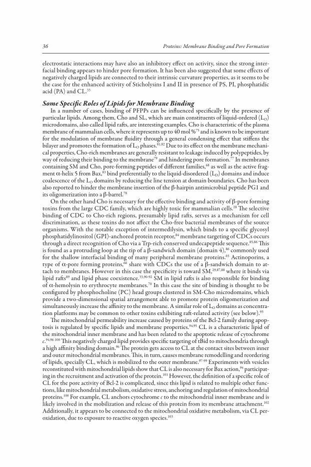
Figure 1. The membrane as a docking surface. Structure of a phospholipid bilayer with emphasis on the physicochemical complexity of the interfaces and its complementarity with amphipathic polypeptides. (A) and (B) show representations of the polarity gradient and structure, respectively of a fluid liquid-crystalline dioleoylphosphatidylcholine (DOPC) bilayer from X-ray and neutron diffraction data.44,46,205 The structure in (B) is represented as the time-averaged Gaussian distribu-tions of the main chemical groups of the lipid projected onto the bilayer normal. The interfaces are the regions defined by the distribution of headgroup’s water of hydration and the hydrocarbon core is the center slab where the presence of water drops to zero. From these distributions of quasimolecular groups, average charge densities and a polarity profile have been derived.44 The profile is represented in (A) by a heavy line and the corresponding polarity gradient is schematized by a gray scale, which in the interface goes from more polar (pale gray) to more hydrophobic (dark gray) and in the hydrocarbon core is a constant black slab. For comparison, a circle repre-senting the cross section of an amphipathic �-helix is drawn to scale and placed at the steepest point of the polarity gradient, corresponding to the position determined by X-ray diffraction.206 The experimental membrane structure can be appropriately modelled at atomic detail by MD simulations, as is shown in (C) for a simulated self-assembled dimiristoylphosphatidylcholine (DMPC) bilayer taken from work reported in reference 1. Acyl-chains are drawn with light-gray lines, phosphate groups as light-grey balls, nitrogens of choline as dark-grey balls and water oxygens as black dots (the oxygens of lipids are not represented). A DMPC lipid highlighted in (C) with thick lines is shown enlarged with more detail in (D) (same colors as in (C) and glycerol oxygens as black lines). In (E) we represent lateral and top views (left and right, respectively) of the amphipathic �-helix structure of magainin 2, solved in detergent micelles207 (pdb ID: 2mag). Only residues 4 to 20 are represented; positively charged residues (Lys and His) are colored black, the negatively charged Glu is colored dark gray and hydrophobic residues are pale gray. Panels (A) and (B) are reprinted with permission from the Annual Review of Biophysics, Volume 28 (ref. 46), © 1999 by Annual Reviews, www.annualreviews.org
34 Proteins: Membrane Binding and Pore Formation
and is explained with more detail in the next section (see Fig. 2A). It implies that the stability of the membrane-polypeptide complex increases as the secondary structure is formed, as it is indeed observed for a number of different systems.44,45,56-62 Thus, phospholipid membrane interfaces can be envisioned as ideal binding sites for docking amphipathic, PFPPs (see Fig. 1).45,46 Supporting a direct targeting role of phospholipid membranes, with no intervention of receptor proteins, are the facts that PFPPs are active against pure lipid vesicles and at least in the case of peptides, independent on chirality (all D-aminoacid peptides are as active as natural L-aminoacid versions).63
One consequence of the direct lipid-based membrane targeting is a relatively low specificity. For example, scrambled sequences of pore forming peptides tend to have similar activity20,64 and in general hundreds of different peptides and proteins, differing in size, secondary, tertiary and quaternary structure, share a similar mode of binding.21,32,65,66 Moreover, the similarity extends outside the family of PFPPs to cell penetrating peptides67,68 fusion peptides37 and with striking relationships to membrane active proteins of different types and across disparate organisms.65 Nevertheless, lipid-based targeting can be also the source of complex binding schemes, including high affinity, cooperativity and lipid-dependent protein assembly.69,70 The general non specific interfacial binding can in some cases superimpose to additional interactions with a different degree of specificity, from strong electrostatic effects, like in the case of negatively charged membranes, to sophisticated and efficient control mechanisms through specific interactions with receptor lipids or lipid-anchored proteins. Some examples of these, more specific, roles are summarized below.
General Effects of Negatively Charged LipidsBecause most PFPPs are cationic, a way to increase their binding from solution is by presence
of negatively charged lipids. In neutral membranes, binding of PFPPs depends mainly on their hydrophobicity, which accentuates the importance of structural parameters like hydrophobic moment and helicity.42 Partitioning cationic peptides into zwitterionic lipids is generally weak, corresponding to dissociation constants of up to 1 mM. However, the presence of negatively charged lipids, like those with phosphatidylglycerol (PG), phosphatidylserine (PS) and phos-phatidylinositol (PI) head groups, pose an electrostatic attraction over the peptides which increases the strength of their binding up to dissociation constants in the �M range. A careful kinetic analysis shows that the stronger binding of cecropin and magainin to acidic lipids is due mainly to a reduced desorption rate.24,71 It is also seen that the main contribution of electrostatic interactions is increasing the concentration of the interfacially adsorbed peptide. Thus, discount-ing this effect on the basis of Gouy-Chapman theory72 (i.e., replacing bulk concentrations by surface concentrations) yields similar binding constants and pore activities regardless of the membrane surface-charge density.27,56,73,74
The electrostatic contribution is the main basis for the selective binding of peptide anti-microbials to bacteria,21 since the outer membrane of these microorganisms is abundant in negatively charged lipids, in contrast to the plasma membranes of eukaryotic cells, abundant in neutral lipids.75 However, this alone cannot explain the selective killing of peptide antibiotics against bacteria, compared to host cells. Such a selectivity can be understood considering the characteristic strong, membrane-mediated cooperativity of these systems, observed as a nonlinear concentration dependence with a rapid rise of activity passed a threshold concentration value.69 Thus, the different affinity for neutral compared to acidic membranes places normal extracel-lular peptide concentrations well above the threshold for bacteria, but below the threshold for eukaryotic cells. The cooperativity originates from the effect of peptide binding on the bilayer material properties69,76 and is discussed below with more detail in connexion to the mechanism of pore formation. An additional factor explaining cell-type selectivity of antimicrobial peptides is the presence of Cho in eukaryotic cells, which in general reduces peptide and protein binding to the membrane and affects as well their oligomeric assembly, membrane insertion pattern and pore activity77-79 (see below).
A preferential binding to negatively charged membranes is also observed for some pore forming colicins80 and for active fragments of the Bcl-2 family.18 However, in these cases the

35Role of Membrane Lipids for the Activity of Pore Forming Peptides and Proteins
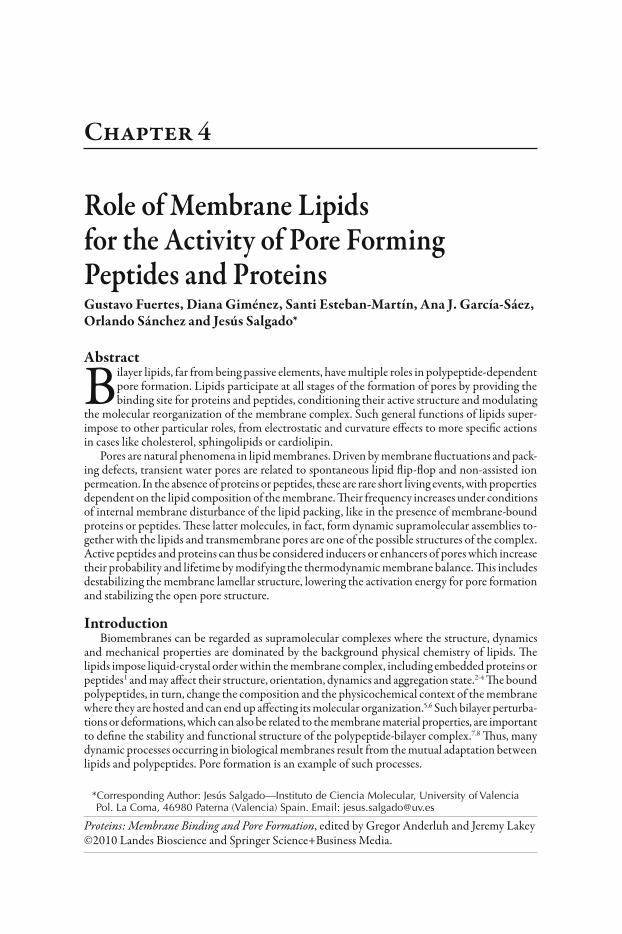
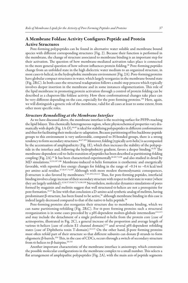
Figure 2. Chaperon-like foldase activity of membranes. Lipid membranes control refolding of polypeptides partitioning into them. A) Partitioning-folding coupling of amphipathic pep-tides at the bilayer interface. The membrane is represented by three slabs colored light-grey (interfaces) and dark-gray (hydrocarbon core). Curled lines are coil structure and cylinders represent �-helices. The peptides are mostly unstructured in water. Immediately after binding to the membrane surface, the interface promotes the appearance of secondary structure, most often through a coil-to-helix transition, which accentuates amphipathicity and strengthens binding following the interface polarity gradient (see Fig. 1). In some cases the helices might interact with each other forming oligomers. B,C) Partitioning-refolding of globular water-soluble pore-forming proteins as they bind to the membrane. The structure change can be dramatic (B) upon adsorbing into the interface (may also include a molten-globule intermediate113), like in the case of �-helix bundle proteins (for example channel domains of colicins or Bcl-2 proteins). These form a dynamic two-dimensional array of helices, which may evolve further through intermediates to insert the most hydrophobic helices across the membrane. In (C), sticholysin II (initial structure from ref. 87) suffers a partial refolding upon specific interfacial binding to SM through a cluster or Tyr residues (highlighted with thick black lines). The change of structure affects mainly an N-terminal amphipathic helix, which detaches from an almost unchanged �-sandwich core and lays flat in the membrane. In all cases successive molecular reorganizations give rise to transmembrane pores (not shown), which depending on the case may also involve protein oligomerization.
36 Proteins: Membrane Binding and Pore Formation
electrostatic interactions may have also an inhibitory effect on activity, since the strong inter-facial binding appears to hinder pore formation. It has been also suggested that some effects of negatively charged lipids are connected to their intrinsic curvature properties, as it seems to be the case for the enhanced activity of Sticholysins I and II in presence of PS, PI, phosphatidic acid (PA) and CL.33
Some Specific Roles of Lipids for Membrane BindingIn a number of cases, binding of PFPPs can be influenced specifically by the presence of
particular lipids. Among them, Cho and SL, which are main constituents of liquid-ordered (LO) microdomains, also called lipid rafts, are interesting examples. Cho is characteristic of the plasma membrane of mammalian cells, where it represents up to 40 mol %75 and is known to be important for the modulation of membrane fluidity through a general condensing effect that stiffens the bilayer and promotes the formation of LO phases.81,82 Due to its effect on the membrane mechani-cal properties, Cho-rich membranes are generally resistant to leakage induced by polypeptides, by way of reducing their binding to the membrane79 and hindering pore formation.77 In membranes containing SM and Cho, pore-forming peptides of different families,68 as well as the active frag-ment �-helix 5 from Bax,83 bind preferentially to the liquid-disordered (LD) domains and induce coalescence of the LO domains by reducing the line tension at domain boundaries. Cho has been also reported to hinder the membrane insertion of the �-hairpin antimicrobial peptide PG1 and its oligomerization into a �-barrel.78
On the other hand Cho is necessary for the effective binding and activity of �-pore forming toxins from the large CDC family, which are highly toxic for mammalian cells.28 The selective binding of CDC to Cho-rich regions, presumably lipid rafts, serves as a mechanism for cell discrimination, as these toxins do not affect the Cho-free bacterial membranes of the source organisms. With the notable exception of intermedilysin, which binds to a specific glycosyl phosphatidylinositol (GPI)-anchored protein receptor,84 membrane targeting of CDCs occurs through a direct recognition of Cho via a Trp-rich conserved undecapeptide sequence.85,86 This is found as a protruding loop at the tip of a �-sandwich domain (domain 4),86 commonly used for the shallow interfacial binding of many peripheral membrane proteins.65 Actinoporins, a type of �-pore forming proteins,66 share with CDCs the use of a �-sandwich domain to at-tach to membranes. However in this case the specificity is toward SM,29,87,88 where it binds via lipid rafts89 and lipid phase coexistence.33,90-92 SM in lipid rafts is also responsible for binding of �-hemolysin to erythrocyte membranes.70 In this case the site of binding is thought to be configured by phosphocholine (PC) head groups clustered in SM-Cho microdomains, which provide a two-dimensional spatial arrangement able to promote protein oligomerization and simultaneously increase the affinity to the membrane. A similar role of LO domains as concentra-tion platforms may be common to other toxins exhibiting raft-related activity (see below).93
The mitochondrial permeability increase caused by proteins of the Bcl-2 family during apop-tosis is regulated by specific lipids and membrane properties.94,95 CL is a characteristic lipid of the mitochondrial inner membrane and has been related to the apoptotic release of cytochrome c.94,96-100 This negatively charged lipid provides specific targeting of tBid to mitochondria through a high affinity binding domain.96 The protein gets access to CL at the contact sites between inner and outer mitochondrial membranes. This, in turn, causes membrane remodelling and reordering of lipids, specially CL, which is mobilized to the outer membrane.97-99 Experiments with vesicles reconstituted with mitochondrial lipids show that CL is also necessary for Bax action,94 participat-ing in the recruitment and activation of the protein.101 However, the definition of a specific role of CL for the pore activity of Bcl-2 is complicated, since this lipid is related to multiple other func-tions, like mitochondrial metabolism, oxidative stress, anchoring and regulation of mitochondrial proteins.100 For example, CL anchors cytochrome c to the mitochondrial inner membrane and is likely involved in the mobilization and release of this protein from its membrane attachment.102 Additionally, it appears to be connected to the mitochondrial oxidative metabolism, via CL per-oxidation, due to exposure to reactive oxygen species.103
37Role of Membrane Lipids for the Activity of Pore Forming Peptides and Proteins
A Membrane Foldase Activity Configures Peptide and Protein Active Structures
Pore-forming polypeptides can be found in alternative water soluble and membrane bound species with different corresponding structures (Fig. 2). Because their function is performed in the membrane, the change of structure associated to membrane binding is an important step for their activation. The question of how membrane-mediated activation takes place is connected to the more general question of how solvent influences protein folding.46 Pore-forming peptides change from an unfolded state in the high dielectric water medium to an organized structure, in most cases �-helical, in the hydrophobic membrane environment (Fig. 2A). Pore-forming proteins have globular compact structures in water, which largely reorganize in the membrane-bound state (Fig. 2B,C). In both cases the structural readaptation follows a multi-step process which typically involves deeper insertion in the membrane and in some instances oligomerization. This role of the lipid membrane in promoting protein activation through a control of protein folding can be described as a chaperon-like foldase activity. How these conformational changes take place can be very different depending on the case, especially for the pore forming proteins.104 Here, again, we will distinguish a generic role of the membrane, valid for all cases at least to some extent, from other more specific roles.
Structure Remodelling at the Membrane InterfaceAs we have discussed above, the membrane interface is the receiving surface for PFPPs reaching
the lipid bilayer. This chemically heterogeneous region, where physicochemical properties vary dra-matically with depth (Fig. 1A-D),44,45 is ideal for stabilizing polypeptides in different conformations and thus for facilitating their molecular re-adaptation. Because partitioning of free backbone-peptide groups to this environment is very unfavorable, compared to H-bonded groups, there is a strong tendency to form secondary structures.46,47,105 Moreover, folding (typically as �-helix) is accompanied by the accentuation of amphipathicity (Fig. 1E), which then increases the stability of the polypep-tide in the interface and, following the hydrophobicity gradient, favors a deeper binding.45,56 The membrane-dependent coil-to-helix transition of peptides has been described as partitioning-folding coupling (Fig. 2A).44 It has been characterized experimentally56-60,73,106 and also studied in detail by MD simulations.52,53,107,108 Membrane-induced �-helix formation is exothermic and energetically favorable, with reported free energy changes for folding in the range of –0.14 to –0.4 kcal/mol per amino acid residue.47,56,57,74,109 Although with more modest thermodynamic consequences, �-structure is also favored by membranes.47,61,105,110,111 Thus, for pore-forming peptides, interfacial binding involves a large increase of their secondary structure with respect to their state in water (where they are largely unfolded).42,56,57,59,60,73,74,106,109 Nevertheless, molecular dynamics simulations of pores formed by magainin and melittin suggest that well structured �-helices are not a prerequisite for pore formation.22,23 In line with that conclusion a D-amino acid synthetic analog of melittin, having predominant �-structure, has been found to be active,56 although membrane binding in this case is indeed largely decreased compared to that of the native �-helix peptide.56
Pore-forming proteins also reorganize their structure due to membrane binding, which we can name partitioning-refolding (Fig. 2B,C). For �-pore forming proteins such a structural reorganization is in some cases preceded by a pH-dependent molten-globule intermediate112,113 and may include the detachment of a single preformed �-helix from the protein core (case of actinoporins, illustrated in Fig. 2C), a general increase of the proportion and average length of various �-helices (case of colicin E1 channel domain)114 and several pH-dependent refolding states (case of Diphtheria toxin T-domain).62,115 On the other hand, �-pore forming proteins must often refold part of their structure so that different subunits can donate � strands to form oligomeric �-barrels.104 This, in the case of CDCs, occurs through a switch of secondary structure from �-helices to �-hairpins.28,116
Another important characteristic of the membrane interface is anisotropy, which constrains the possible molecular configurations in the membrane complex to a small number. This selects a flat arrangement of amphipathic polypeptides (Fig. 2A), with the main axis of peptide segments
38 Proteins: Membrane Binding and Pore Formation
running near parallel to the membrane plane. Such is the configuration most often found experi-mentally for pore-forming peptides.117 In these cases, changes into a perpendicular alignment have been seen accompanying pore formation over a threshold peptide concentration118 (see below and Fig. 6) or associated to a change of the phase of the lipids.119,120 For helix-bundle �-pore forming proteins, this corresponds to extended two-dimensional arrays of helices (Fig. 2B), which have been characterized as membrane-dependent refolding intermediates for some colicins114,121,122 and members of the Bcl-2 family.123-125 Because the hydrophobic length of �-helices in these proteins is relatively short (with the exception of a C-terminal helix in some Bcl-2 members), binding across the membrane should be disfavored, at least in monomeric prepore states. However, a characteristic central hairpin of helices is often found in a transmembrane fashion.122,126-130
The Lipid Membrane Controls Inter-Protein InteractionsAs another way to reshape peptides and proteins, membrane interfaces can promote inter-mo-
lecular association of these molecules. Oligomerization is in many cases a characteristic step for the activation of PFPPs. However, with a few exceptions,131 the water soluble states are monomeric and oligomers form as prepore structures which are strictly dependent on membrane binding.104 In general the membrane controls oligomerization at the level of protein (or peptide) folding, by reconfiguring the structure to shape the binding sites and/or by making such binding sites accessible. In the case of pore forming peptides the presence and possible role of oligomers is not always clear.76 In molecular dynamics simulations of pore formation by magainin and melittin the appearance of interfacially adsorbed aggregates is a prerequisite for pore induction.22,23 A charac-teristic endothermic step in the calorimetric titration of melittin has been assigned as a reversible peptide aggregation (coupled to pore formation), occurring after membrane binding and �-helix formation.56 In that study peptide aggregation is described with a phase diagram depending on the total peptide and lipid concentrations, with three phases corresponding to monomers, aggregates and coexistence of monomers and aggregates and phase boundaries defined by threshold values of the peptide-to-lipid molar fractions (P/L).56 Such boundaries correspond to the threshold peptide-to-lipid mole fraction (P/L*) in the two-state model of Huang and colleagues,118 which has been recently reformulated also as a two-phase model.69
In some CDCs the release of the oligomerization site is performed through proteolytic cleavage of a propeptide by a membrane-restricted protease.28 Another powerful mechanism for promoting inter-protein oligomerization is by two-dimensional clustering.93 As mentioned above, some toxins bind selectively to lipid rafts, which may function as protein concentration platforms and enhance oligomeric assembly. Reduction of dimensionality, as corresponding to binding in the membrane two dimensional surface, can lead to an effective increase of concentration of about 1.5 � 103.132 Additionally, recruitment of proteins in membrane microdomains can largely increment the concentration factor. This has been described for aerolysin, whose GPI-anchored receptor associ-ates transiently with lipid rafts93 and can also be the case for other toxins which bind to SM-Cho microdomains, like CDCs,28 actinoporins,29 �-hemolysin,70 Cry1A toxin133 and lysenin.134
The Complex Membrane-Dependent Regulation of Bcl-2 ProteinsThe lipid membrane exerts also a principal role for the intricate mechanism of action of
pore inducers and inhibitors of the Bcl-2 family. This uses a complicated allosteric mechanism, explained by the “embedded together” model135 where the capacity of the pore receptor (tBid), executor (Bax) and inhibitor (Bcl-xL) proteins to interact with each other changes after binding to the mitochondrial outer membrane, as this causes conformational changes that alter and/or expose new binding surfaces.136,137 Docking of tBid to mitochondrial membranes converts this protein into a high affinity receptor for Bax. A subsequent membrane dependent interaction of Bax to tBid is responsible of further activation involving the formation of a Bax oligomer, which can finally form a pore.136,137 The inhibition of this process by Bcl-xL can occur at different levels, but it is in any case also membrane dependent. This may consist, on the one hand, on the block-age of membrane-bound tBid, which can no longer receive Bax and on the other hand on a direct binding to Bax, which abolish the formation of the Bax oligomeric pore.136,137
39Role of Membrane Lipids for the Activity of Pore Forming Peptides and Proteins
Role of Lipids in the Formation and Stabilization of PoresThe Latent Membrane Pores: Relatives of Pores Induced by Polypeptides?
Although rare, spontaneous pores are inherent to lipid bilayer membranes. They occur indepen-dently of the presence of peptides or proteins, although in the absence of tension their frequency is very low. In pure lipid membranes pore formation is kinetically hindered by a large energy barrier, which cannot be easily overcome by thermal energy (Fig. 3).10-12,138-143 However, the fluctuation of bilayer lipids gives a chance for stochastic disruptions of the equilibrium bilayer structure, explain-ing, among other things, the spontaneous formation of pores.10,12,138 For example, the transbilayer movement of lipids, known as flip-flop, which in cell membranes is accelerated by a number of specialized catalytic proteins,144 can occur in pure lipid vesicles in time scales from hours to days, depending on the type of lipids and experimental conditions.145-148 Such unassisted flip-flop has been proposed to be mediated by lipid-packing defects.149,147 MD simulations have shown recently that this process may occur via transient water-pores (Fig. 4A) which allow passage of the hydrated charged groups of the lipids across the membrane hydrophobic slab.12 The pores are structurally similar to the ones simulated under mechanical and electrical stress (Fig. 4B),11,139 a type of bilayer disruption which is well known experimentally.9,10,150,151 These flip-flop coupled pores might also be responsible for the passive ion permeation through membranes,12,139,152,153 although they represent a negligible contribution to water permeation.12
The background spontaneous lipid flip-flop and pore formation can be largely affected by the phase state and composition of the membrane. For example, the passive permeability of lipid bilayers exhibits a maximum at conditions of coexistence of gel domains and fluid domains.147,154,155 On the other hand the presence of Cho increases the free energy barrier for
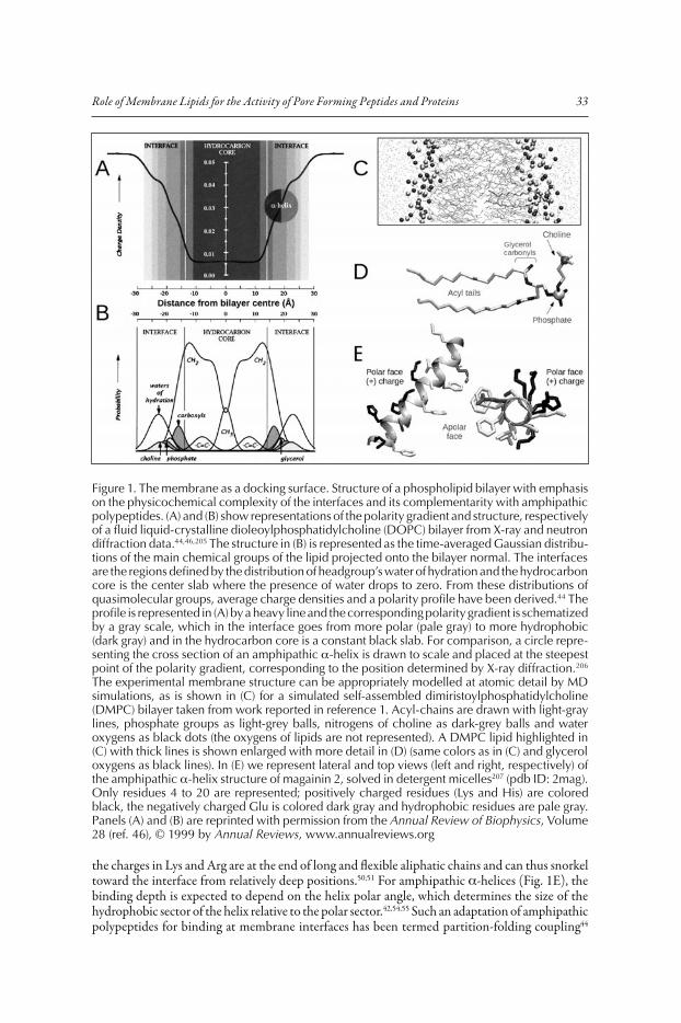
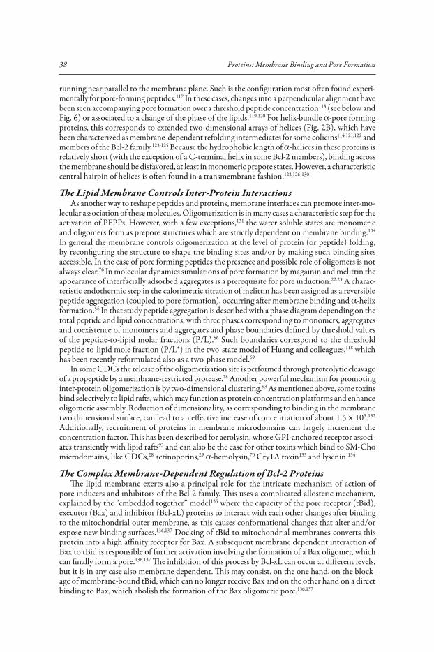
Figure 3. Free energy of pore formation along the reaction coordinate. Relative stability of intact and porated states of a lipid bilayer in the absence and presence of PFPPs. Pore forma-tion in pure lipid bilayers with no tension (black, dotted line) is unfavorable and kinetically hindered by a large activation energy barrier. Membrane-bound PFPPs destabilize the bilayer, increasing its energy via elastic deformations (left, arrow pointing upwards) and pore forma-tion may then become favorable (gray, continuous line). The likelihood of the pore state increases if the PFPP stabilize the pore once it is open (right, arrow pointing downwards). The rate-limiting step is the rearrangement of the lipids and the PFPP through a high energy transition state (middle). Such pore activation energy is, in a first instance, reduced by the changes of the relative stability of intact and porated bilayers, caused by the PFPP and could be decreased even further if the PFPP stabilizes the transition state. Note that the intact and porated bilayer states may correspond to the S-state and I-state in Huang’s model,69,118 as well as to the Bex-state and P-state in Tamba and Yamazaki’s model.26,27 However, there are fundamental differences between these two models in the definition and properties of the states (see the text). Thus, we think that this open and more general definition of the states would in either case be valid.
40 Proteins: Membrane Binding and Pore Formation
water pore formation82,156 and on the contrary, the presence of ceramide facilitates flip-flop and the formation of large and stable lipidic channels.157-161 Including proteins in the mem-brane composition has also been observed to affect lipid flip-flop, like in presence of �-helical proteins, in principle not related to pore formation, from the plasma membrane of bacteria.162 This latter effect appears weakly specific, since it is also found for other polypeptides, like glycophorin163 and synthetic model transmembrane peptides.164 It has been speculated that this type of protein-facilitated flip-flop is due to a much lower barrier for defect (water-pore) formation12 as a consequence of the protein-membrane interaction. Induction of lipid flip-flop
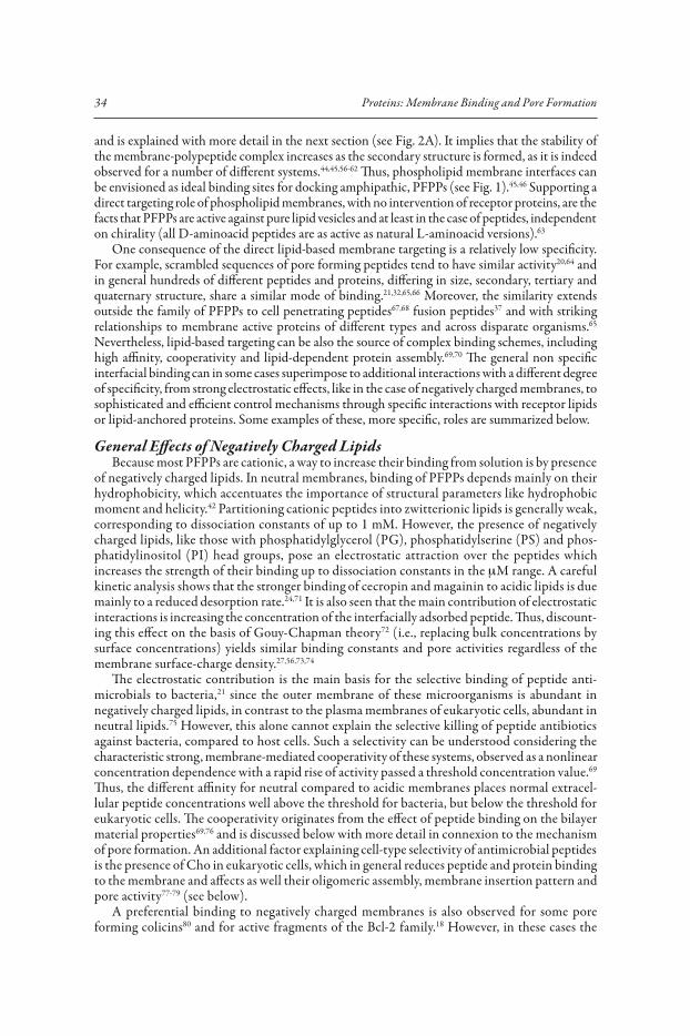
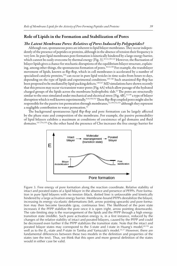
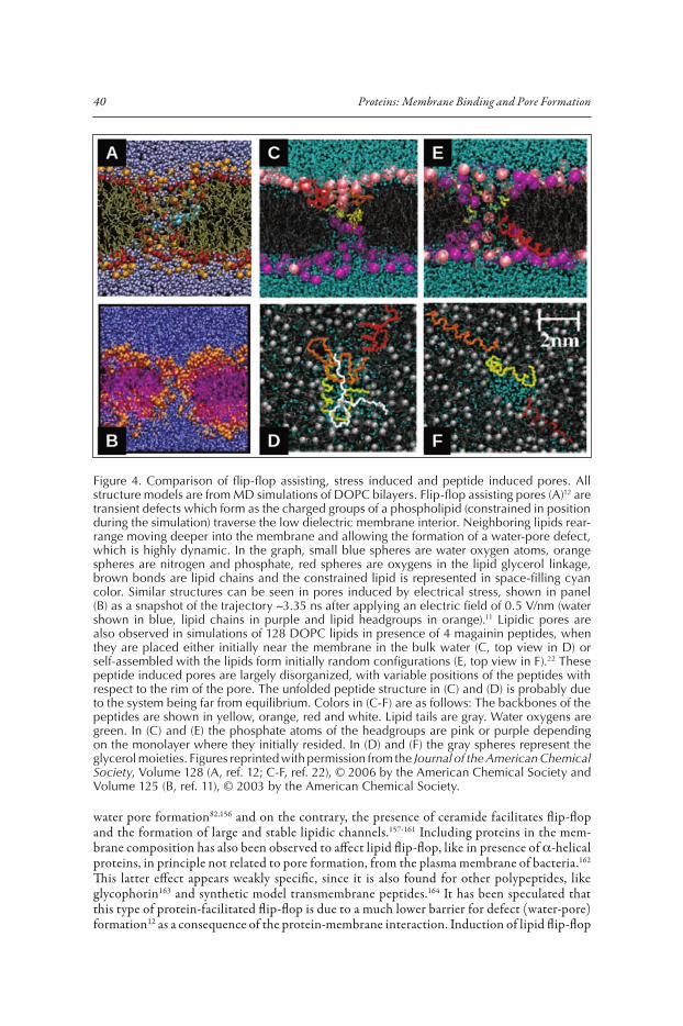
Figure 4. Comparison of flip-flop assisting, stress induced and peptide induced pores. All structure models are from MD simulations of DOPC bilayers. Flip-flop assisting pores (A)12 are transient defects which form as the charged groups of a phospholipid (constrained in position during the simulation) traverse the low dielectric membrane interior. Neighboring lipids rear-range moving deeper into the membrane and allowing the formation of a water-pore defect, which is highly dynamic. In the graph, small blue spheres are water oxygen atoms, orange spheres are nitrogen and phosphate, red spheres are oxygens in the lipid glycerol linkage, brown bonds are lipid chains and the constrained lipid is represented in space-filling cyan color. Similar structures can be seen in pores induced by electrical stress, shown in panel (B) as a snapshot of the trajectory �3.35 ns after applying an electric field of 0.5 V/nm (water shown in blue, lipid chains in purple and lipid headgroups in orange).11 Lipidic pores are also observed in simulations of 128 DOPC lipids in presence of 4 magainin peptides, when they are placed either initially near the membrane in the bulk water (C, top view in D) or self-assembled with the lipids form initially random configurations (E, top view in F).22 These peptide induced pores are largely disorganized, with variable positions of the peptides with respect to the rim of the pore. The unfolded peptide structure in (C) and (D) is probably due to the system being far from equilibrium. Colors in (C-F) are as follows: The backbones of the peptides are shown in yellow, orange, red and white. Lipid tails are gray. Water oxygens are green. In (C) and (E) the phosphate atoms of the headgroups are pink or purple depending on the monolayer where they initially resided. In (D) and (F) the gray spheres represent the glycerol moieties. Figures reprinted with permission from the Journal of the American Chemical Society, Volume 128 (A, ref. 12; C-F, ref. 22), © 2006 by the American Chemical Society and Volume 125 (B, ref. 11), © 2003 by the American Chemical Society.

41Role of Membrane Lipids for the Activity of Pore Forming Peptides and Proteins
is also a common phenomenon associated to the activity of many PFPPs.30,165-168 The large increase of transbilayer movement observed in these cases is often explained as due to lateral diffusion of lipids at points of monolayer fusion existing in the edge of the pore and it is one of the preferred tests to distinguish different types of pores166-170 (see below). Additionally, similar to intrinsic lipid flip-flop, pore formation by peptides and proteins is in many cases described as an stochastic process related to membrane disruption and nucleation of defects.22,24-26,91
The basic action of specialized pore-forming peptides and proteins may then overlap with the intrinsic pore-formation capacity of membranes. A number of specific examples support this idea: osmotic tension and class L amphipathic peptides act synergistically as they induce pores in vesicles.171 The general attenuation of membrane permeability exerted by Cho affects also the activity of pore forming peptides.77,79 For more sophisticated protein pores, like those formed by the Cho dependent Vibrio cholerae cytolysin, the pore inducer ceramide enhances the activity of the toxin.172 On the other hand, the lipidic pores induced by ceramide can be disassembled by Bcl-2,173 the physiological inhibitor of the �-pore forming protein Bax. And in some other cases, pore formation is favored by defect-rich domain boundaries91 and at the phase transition tem-perature.174 The lipopeptide syringomycin E, which forms a characteristic lipidic pore, provides an interesting example linking intrinsic membrane pores and polypeptide induced pores.175 The charge and dipolar moment of host membrane lipids modifies the effective gating charge of the syringomycin E ionic channel. Additionally the channel is inhibited in the presence of nonlamellar lipids with negative spontaneous curvature. Similarly, effects of lipid charge and intrinsic curvature have been observed for channels formed by peptides18 or proteins.168
Can we then establish mechanistic connections between intrinsic membrane pores and pores induced by peptides and proteins? In an attempt to do that, we make now an overview of different proposed models and extract from them a minimum general consensus from the point of view of the role of lipids.
A Consensus View of Pore Formation Stressing the Role of LipidsThere have been a number of different classical (general) models of pore formation by membrane
active polypeptides. Previous work has often stressed the differences between particular models, amplified by detailed (not always justified) drawings. Instead, we want to underline here their common points, as many of their apparent contradictions can be regarded as either superficial or arising from the use of different experimental conditions. Although mostly developed for mem-branolytic peptides, many of these ideas can be extended to pore-forming proteins14,29,43,66; they essentially leave a prominent role for lipids around the postulate of more or less stable and more or less organized, lipid-based pores.
Matsuzaki proposed a supramolecular peptide-lipid dynamic complex in order to explain the simultaneous transbilayer diffusion of magainin and membrane lipids, coupled to leakage of vesicles.30 In this model both, lipids and peptides form the pore wall, where the presence of acidic phospholipids may counteract repulsion between the positively charged peptides and explain the cation selectivity of the channel.176 This is basically the same as the toroidal wormhole model, proposed almost simultaneously by Huang’s group on the basis of neutron in-plane scattering and oriented circular dichroism (OCD) data.31 Huang’s view is sustained on the membrane thinning that accompanies peptide embedding in the head-group region (S-state). Above a certain threshold P/L* this triggers a molecular reorganization which involves the re-orientation of some peptide molecules (I-state) and formation of a pore.177,178 Noteworthy, from a similar S-state the model postulates different I-states for alamethicin-type peptides: barrel stave of interacting transmembrane peptides forming a relatively small pore, than for magainin-type peptides: larger pore where the two monolayers fuse like in a torus and the curvature strain is alleviated by peptides bound across the membrane, in the interface of lipids making the pore wall.118 From the membrane side, these two alternative pore states correspond to the two possible lipid structures at the edge of a pore, which have been experimentally observed by reconstruct-ing the lipid electron density profiles from X-ray diffraction179,180 (Fig. 5). This thermodynamic
42 Proteins: Membrane Binding and Pore Formation
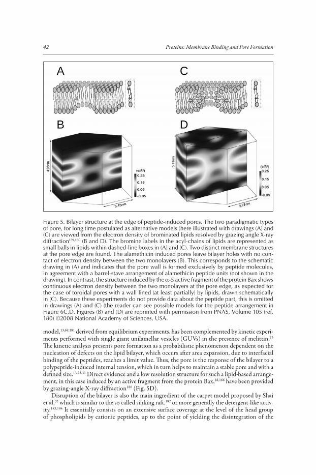
model,13,69,181 derived from equilibrium experiments, has been complemented by kinetic experi-ments performed with single giant unilamellar vesicles (GUVs) in the presence of melittin.25 The kinetic analysis presents pore formation as a probabilistic phenomenon dependent on the nucleation of defects on the lipid bilayer, which occurs after area expansion, due to interfacial binding of the peptides, reaches a limit value. Thus, the pore is the response of the bilayer to a polypeptide-induced internal tension, which in turn helps to maintain a stable pore and with a defined size.13,25,31 Direct evidence and a low resolution structure for such a lipid-based arrange-ment, in this case induced by an active fragment from the protein Bax,18,166 have been provided by grazing-angle X-ray diffraction180 (Fig. 5D).
Disruption of the bilayer is also the main ingredient of the carpet model proposed by Shai et al,32 which is similar to the so called sinking raft,182 or more generally the detergent-like activ-ity.183,184 It essentially consists on an extensive surface coverage at the level of the head group of phospholipids by cationic peptides, up to the point of yielding the disintegration of the
Figure 5. Bilayer structure at the edge of peptide-induced pores. The two paradigmatic types of pore, for long time postulated as alternative models (here illustrated with drawings (A) and (C) are viewed from the electron density of brominated lipids resolved by grazing angle X-ray diffraction179,180 (B and D). The bromine labels in the acyl-chains of lipids are represented as small balls in lipids within dashed-line boxes in (A) and (C). Two distinct membrane structures at the pore edge are found. The alamethicin induced pores leave bilayer holes with no con-tact of electron density between the two monolayers (B). This corresponds to the schematic drawing in (A) and indicates that the pore wall is formed exclusively by peptide molecules, in agreement with a barrel-stave arrangement of alamethicin peptide units (not shown in the drawing). In contrast, the structure induced by the �-5 active fragment of the protein Bax shows continuous electron density between the two monolayers at the pore edge, as expected for the case of toroidal pores with a wall lined (at least partially) by lipids, drawn schematically in (C). Because these experiments do not provide data about the peptide part, this is omitted in drawings (A) and (C) (the reader can see possible models for the peptide arrangement in Figure 6C,D. Figures (B) and (D) are reprinted with permission from PNAS, Volume 105 (ref. 180) ©2008 National Academy of Sciences, USA.
43Role of Membrane Lipids for the Activity of Pore Forming Peptides and Proteins
membrane.32 In this case the toroidal pore is adopted as a disordered early transient stage, before membrane rupture occurs through micellisation. Leaving this latter complete disruption aside, as it is observed only at very high peptide concentrations,183,185 both Matsuzaki and Shai coincide on proposing pore formation as due to asymmetric membrane disturbance or mass imbalance over the external (accessible) monolayer, where the peptide primarily binds. It follows that pores are necessarily transient, because they will close at equilibrium as soon as mass imbalance dissipates through the pore. This is supported by recent kinetic interpretations of content release experi-ments from large unilamellar vesicle (LUV) suspensions by Almeida’s group,24,71 as well as kinetic studies with single GUVs by Tamba and Yamazaki,26,27 which also introduce the idea of stochastic pores (or pores opening at random after a threshold stress). The latter authors propose a two state Bex P transition model26 where the Bex state corresponds to the peptide bound only to the external monolayer, the rate-limiting step is the insertion of the peptide across the membrane and the P state is a metastable transient pore.26,27 Although these ideas have some resemblance with Huang’s two state model,25,69,180 there are two main contrasting points: (i) In Huang’s S-state the peptides are assumed to reequilibrate fast across the membrane76 through small transient pores occurring even at low concentration,186 meaning that the stress responsible for pore induction is exerted symmetrically in the two monolayers. (ii) The pores in Huang’s I-state correspond to minimum energy and are thus stable once they are formed.31,69,180,181
The toroidal “pores in action” reported by MD simulations also form stochastically and after the asymmetric attack to the bilayer.22,23 In this case the interesting feature is the low level of molecular organization within the pore, both for lipids and peptides (Fig. 4C-F),23 which is put in contrast to Huang’s view. However, the disagreement may be illusory. Thus, on the lipid side the regular torus reported by Huang is an averaged structure which cannot be directly compared with single pores in non-equilibrium, relatively short MD trajectories.180 On the peptide side, there is no precise information about the number of peptides involved per pore, or their position and orientation with respect to the pore. For example, in the case of melittin, the estimates of the peptide aggregation number accompanying pore formation vary from 5 to 8 in an analysis of membrane thinning69 and up to 20 in analysis of calorimetry data.56 It should also be noted that Huang’s measurements of peptide reorientation upon pore formation, performed by OCD, do not give accurate orientation values, but rather inform of a change of tilt.177,178,187,188 Thus, the OCD results, normally interpreted as a transition between two extreme states, parallel and perpendicular to the membrane, might as well be compatible with other linear combinations of extreme tilts, or even a distribution of peptide orientations at or near the pore rim,188 perhaps similar to that seen in simulations (Fig. 4E,F).22,23 Membrane insertion of pore forming peptides at tilts other than perpendicular to the membrane has been described in the case of PGLa119,188 and for an active fragment of Bax.18
Thus, the three main classical models of pore formation have important elements in com-mon, recognized by Zasloff, who termed them collectively as the Shai-Matsuzaki-Huang (SMH) mechanism.21 Considering the new ingredients from MD simulations22,23 and kinetic and single vesicle studies,24-27,71 the emerging main consensus ideas are as follows: membrane disruption due to interfacial binding is the basic mechanism of polypeptide-induced pore formation. It proceeds through a stochastic cooperative transition, assisted by bilayer defects, in a two-state process modulated by the membrane elastic properties (see below). This, depending on the type of protein or peptide, may form barrel-like pores (stable cylindrical peptide or protein aggregates) or disordered mixed lipidic-proteic (or peptidic) pores. These ideas are valid for most (if not all) membranolytic peptides. Among the pore-forming proteins, the �-type form preferentially barrels of interacting �-strands66,104 while the �-type seem to prefer toroidal pores, so far described for colicins,168 actinoporins33,34 and Bcl-2 apoptotic regulators35,167,189 as well as their active fragments.18,83,166,180 In any case, the importance of membrane disturbance and nucleation of defects to facilitate protein insertion and pore formation, modulated by the membrane elastic properties, should be of general validity.
44 Proteins: Membrane Binding and Pore Formation
Physical Properties of Polypeptide-Induced Pores Related to the Role of LipidsSurface Tension, Line Tension and the Stability of Membrane Pores
As we have discussed above, the lytic pores induced by polypeptides have many ingredients of general lipidic pores, like those formed under tension (mechanical or electrical tension or osmotic swelling). According to proposed mechanisms,11,190 pores form after a build up of a critical surface tension (�10 pN/nm),191 which increases the probability of appearance of nucleation sites of packing defects. Theoretical models describe these pores as meta-stable arrangements, with free energy of formation being a function of the pore radius (r) as: Er
0 � 2�r��– �r2�. The first term is the energy needed to expand the rim of the pore and � is the line tension, opposing the pore. The second term, proportional to the pore area, represents the work done by the membrane to open the pore, � being the membrane tension, which favours pore opening and expansion.150,192 This model predicts an intrinsically unstable pore which tends to close for r � �/� and expands indefinitely for r � �/�. Thus, while external tension effects increase the pore lifetime and can lead to vesicle rupture, different lipids as well as non lipid inclusions, like detergents, may affect the pore stability by changes in the line tension.150
In a generalization of this model to polypeptide-induced toroidal pores, it has been proposed that pore forming peptides (we may extend it to PFPPs) act by affecting both, the line tension and membrane tension terms and making the open-pore state energetically favourable (see Fig. 3).13 This can occur through an effect of the PFPPs on the membrane elastic properties (see also below). The membrane thinning (area expansion) accompanying the interfacial binding of PFPPs is a source of (internal) surface tension which above a threshold value (�8 pN/nm in DOPC,181 similar to the rupture tension of vesicles of the same lipid191) overcomes the energy barrier to open the pore. Additionally, the binding of PFPPs at or near the pore rim may act by reducing the line tension.35,68,83 Evidence of this latter effect has been obtained as shape changes and coalescence of LO domains in phase-separated lipid bilayers, observed by fluorescence microscopy of GUVs and in situ atomic force microscopy (AFM) in presence of the Bax-�5 fragment and the reduction of line tension was quantified using AFM-film rupture experiments.83 A extensive AFM study of membrane remodelling for a variety of pore-forming and cell penetrating peptides suggests that line-tension activity may be a common ingredient of the mechanism of these systems.68
Lipid-Driven Cooperativity: A Many-Body Effect Triggering Pore FormationKinetic,24,25,27,71 structural31,76 and thermodynamic56 studies agree on describing pore formation by
active peptides as a cooperative process. It is manifested as a nonlinear concentration dependence of activity and a rapid rise to saturation as the concentration exceeds a threshold value and it consists on a steep transition between two main structural states of the peptide-membrane complex.76,177,178,193 This cooperativity phenomenon is not due to direct peptide-peptide binding, but rather to a membrane mediated peptide-peptide interaction which originates on the elastic properties of the membrane, in the form of area expansion25,194 and membrane thinning (Fig. 6).25,195,196 The interfacial binding of the peptide in the S-state (below a threshold P/L*) expands the membrane area and causes a local thinning. This corresponds to a positive energy of elastic deformation, proportional to the ratio of peptides bound per lipid. The consequence is an increase of the free energy of the S-state (Fig. 3) which reaching a threshold value (corresponding to a limit tension, see above) triggers a molecular reorganization (I-state) with the opening of lipidic pores and a change of orientation of a few peptides per pore.181,193 This has been recently interpreted as similar to a micellisation process (and the two states have been renamed phases).69 The new analysis allows an estimation of the number of peptides per pore (a minimum of 4) involved in the transition.69
Interestingly, the membrane-mediated cooperativity of pore-forming peptides suggests a model for the activity of �-helical pore-forming proteins, like proapoptotic Bax, pore-forming colicins and diphtheria toxin.69 The prepore state of these proteins is thought to consist on an extended two-di-mensional array of helices, which has been characterized for some colicins114,121,122 and members
45Role of Membrane Lipids for the Activity of Pore Forming Peptides and Proteins
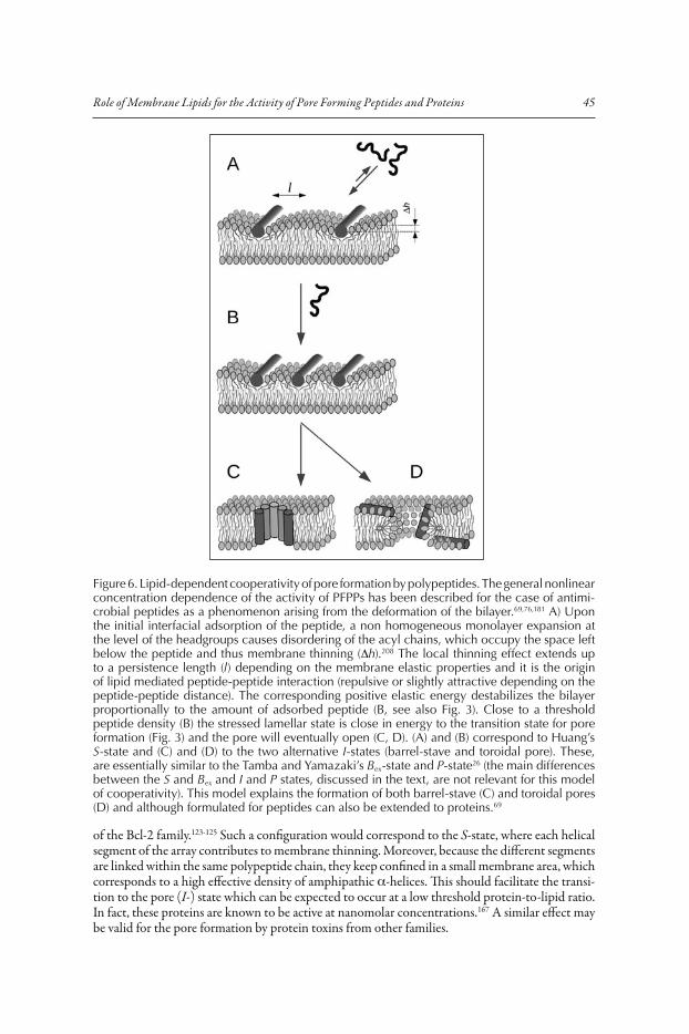
of the Bcl-2 family.123-125 Such a configuration would correspond to the S-state, where each helical segment of the array contributes to membrane thinning. Moreover, because the different segments are linked within the same polypeptide chain, they keep confined in a small membrane area, which corresponds to a high effective density of amphipathic �-helices. This should facilitate the transi-tion to the pore (I-) state which can be expected to occur at a low threshold protein-to-lipid ratio. In fact, these proteins are known to be active at nanomolar concentrations.167 A similar effect may be valid for the pore formation by protein toxins from other families.
Figure 6. Lipid-dependent cooperativity of pore formation by polypeptides. The general nonlinear concentration dependence of the activity of PFPPs has been described for the case of antimi-crobial peptides as a phenomenon arising from the deformation of the bilayer.69,76,181 A) Upon the initial interfacial adsorption of the peptide, a non homogeneous monolayer expansion at the level of the headgroups causes disordering of the acyl chains, which occupy the space left below the peptide and thus membrane thinning (h).208 The local thinning effect extends up to a persistence length (l) depending on the membrane elastic properties and it is the origin of lipid mediated peptide-peptide interaction (repulsive or slightly attractive depending on the peptide-peptide distance). The corresponding positive elastic energy destabilizes the bilayer proportionally to the amount of adsorbed peptide (B, see also Fig. 3). Close to a threshold peptide density (B) the stressed lamellar state is close in energy to the transition state for pore formation (Fig. 3) and the pore will eventually open (C, D). (A) and (B) correspond to Huang’s S-state and (C) and (D) to the two alternative I-states (barrel-stave and toroidal pore). These, are essentially similar to the Tamba and Yamazaki’s Bex-state and P-state26 (the main differences between the S and Bex and I and P states, discussed in the text, are not relevant for this model of cooperativity). This model explains the formation of both barrel-stave (C) and toroidal pores (D) and although formulated for peptides can also be extended to proteins.69
46 Proteins: Membrane Binding and Pore Formation
The Elusive Role of Spontaneous Curvature: Classical and Nonclassical EffectsBecause the pores imply bilayer deformations, they are also related to bilayer elastic (mechanical)
properties, namely the isothermal area compressibility modulus (Ka), the bending modulus (kc) and the monolayer spontaneous radius of curvature (R0).77,197,198 These three parameters depend on the bilayer composition.198 The energy of bilayer deformation contains terms accounting for area changes, proportional to Ka and curvature-elastic effects, proportional to kc/R0 and both have been observed to correlate linearly with the melittin-induced leakage.77 It is proposed that the interfacial adsorption of the peptide induces a bending moment in the bilayer causing deforma-tions which act as a nucleation site for pore opening. The opening probability, stability and size of the pores is then determined by the deformation energy of the lipids and it thus depends on the intrinsic lipid curvature.77 This observations are in line with the characteristic high curvature strain of lipidic and proteo-lipidic toroidal pores143,150,199,200 (Fig. 7).
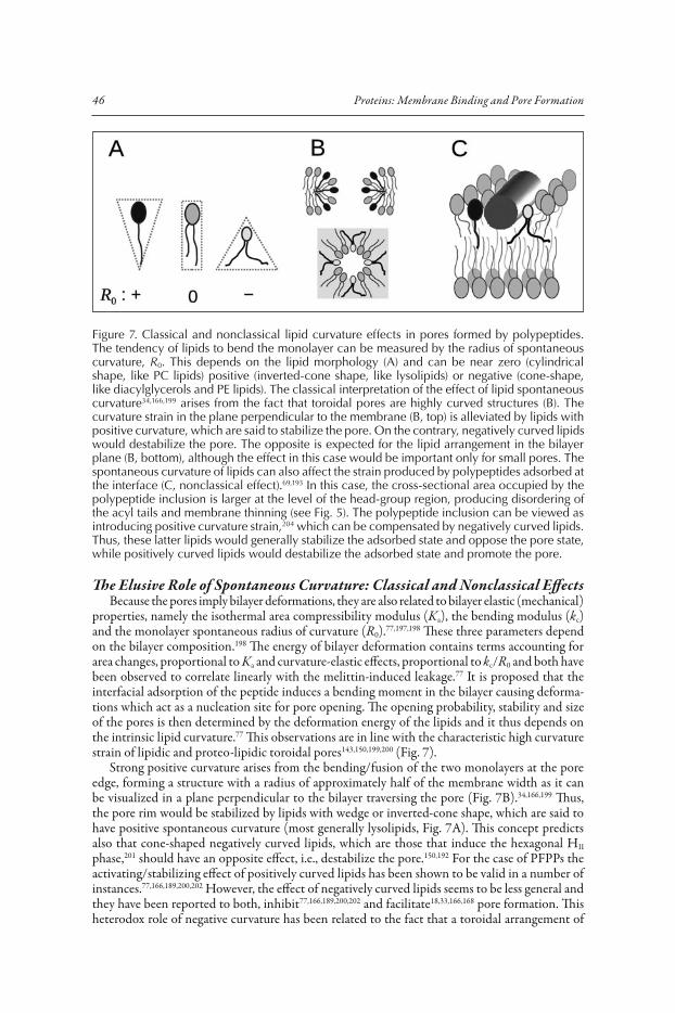
Strong positive curvature arises from the bending/fusion of the two monolayers at the pore edge, forming a structure with a radius of approximately half of the membrane width as it can be visualized in a plane perpendicular to the bilayer traversing the pore (Fig. 7B).34,166,199 Thus, the pore rim would be stabilized by lipids with wedge or inverted-cone shape, which are said to have positive spontaneous curvature (most generally lysolipids, Fig. 7A). This concept predicts also that cone-shaped negatively curved lipids, which are those that induce the hexagonal HII
phase,201 should have an opposite effect, i.e., destabilize the pore.150,192 For the case of PFPPs the activating/stabilizing effect of positively curved lipids has been shown to be valid in a number of instances.77,166,189,200,202 However, the effect of negatively curved lipids seems to be less general and they have been reported to both, inhibit77,166,189,200,202 and facilitate18,33,166,168 pore formation. This heterodox role of negative curvature has been related to the fact that a toroidal arrangement of
Figure 7. Classical and nonclassical lipid curvature effects in pores formed by polypeptides. The tendency of lipids to bend the monolayer can be measured by the radius of spontaneous curvature, R0. This depends on the lipid morphology (A) and can be near zero (cylindrical shape, like PC lipids) positive (inverted-cone shape, like lysolipids) or negative (cone-shape, like diacylglycerols and PE lipids). The classical interpretation of the effect of lipid spontaneous curvature34,166,199 arises from the fact that toroidal pores are highly curved structures (B). The curvature strain in the plane perpendicular to the membrane (B, top) is alleviated by lipids with positive curvature, which are said to stabilize the pore. On the contrary, negatively curved lipids would destabilize the pore. The opposite is expected for the lipid arrangement in the bilayer plane (B, bottom), although the effect in this case would be important only for small pores. The spontaneous curvature of lipids can also affect the strain produced by polypeptides adsorbed at the interface (C, nonclassical effect).69,193 In this case, the cross-sectional area occupied by the polypeptide inclusion is larger at the level of the head-group region, producing disordering of the acyl tails and membrane thinning (see Fig. 5). The polypeptide inclusion can be viewed as introducing positive curvature strain,204 which can be compensated by negatively curved lipids. Thus, these latter lipids would generally stabilize the adsorbed state and oppose the pore state, while positively curved lipids would destabilize the adsorbed state and promote the pore.
47Role of Membrane Lipids for the Activity of Pore Forming Peptides and Proteins
lipids in the pore edge posses also regions of negative curvature strain in a plain parallel to the membrane (Fig. 7B).34,166,199 Such an effect is predicted to be strictly dependent on the pore size, being comparable to the positive curvature effect for a pore radius of �2 nm (approximately half of membrane thickness) and vanishing small as the pores increase in size.199 Thus, it has been argued that the activating effect of negatively curved phosphatidylethanolamine (PE) lipids in pores formed by a Bax �5 fragment,18,166 compared to the classical inhibitory effect observed for complete Bax,167,189 may be due to the formation of smaller pores in the former. However, the pores induced by Bax �6 (another active Bax fragment), despite being apparently of a size comparable (or even smaller) to that of Bax �5 pores, are inhibited by the presence of PE.166 Other conflicting examples have been explained by additional properties of lipids complementing or compensating the curvature effects. Such are the cases of the stimulated opening of large conductance 600 pS ion channels of colicin E1 at increased lipid negative curvature, compared to the case of small conductance 60 pS channels168 and the increased activity of pores formed by sticholysins I and II in the presence of negatively charged PA, PS PG, PI and CL as well as zwitterionic PE. In the case of the colicin E1 600 pS channels, the increase of opening probability has been related to a simultaneous stimulating effect due to hydrophobic mismatch.168 With respect to the pores of sticholysins, the observed behavior is attributed to intrinsic negative curvature of the assayed lipids,33 which due to their negative charge, is enhanced in the presence of positively charged residues of the membrane-bound protein.201,203 Additionally, the existence of distorted toroidal pores23 might also explain the heterogeneity of negative curvature effects, since they are related to the local pore radius which will be variable in a complex manner for noncylindrical pores.
Based on structural measurements (membrane thinning), Huang et al have described a different effect of lipid spontaneous curvature, which we term nonclassical curvature effect (Fig. 7C).193 In agreement with leakage experiments, negatively and positively curved lipids increase and reduce, respectively, the threshold P/L value for the S-state to I-state transition. However, the observed correlations are similar for barrel-stave pores (alamethicin, Fig. 6C) and toroidal pores (melittin, Fig. 6D) and in either case the relative stabilities of the S and I states appear weakly dependent on lipid curvature.76,193 So, it appears that the bending stress of the toroidal pore is efficiently released by the peptides,204 in agreement with a reduction of the line tension (which implies that the pep-tides stabilize the lipidic pore, see Fig. 3).68,83 Instead, the major effect of varying the spontaneous curvature of lipid is a change of the degree of membrane thinning that accompanies the interfacial adsorption of the peptide (Fig. 6).193 This can be understood by considering that cone-shaped negatively curved lipids, with a smaller head-group area, compared to the area occupied by the lipid tails, accommodate better the interfacially adsorbed peptides (attenuate membrane thinning and increase the threshold for the S I transition) and the opposite occurs for inverted cone-shaped (positively curved) lipids (Fig. 7C).
ConclusionOn the functional scene, pores formed by peptides and proteins have a lot in common, as they
all are systems for defense and attack, or in a more general sense related to cell death. In contrast, reports about these functions often put the accent on diversity, helped by an ever increasing number and variety of structures and mechanisms. Membrane lipids and the lipid bilayer proper-ties are a unifying ingredient in this Universe of pore forming peptides and proteins and we can find a minimum of convergent ideas concerning the role of lipids. The compromise of the bilayer integrity is an intrinsic capability of lipid membranes, which can be modulated at a basic level by the lipid composition and phase changes. Polypeptides employed for pore formation use in part this intrinsic capability, while adding different levels of complexity which allow specificity and regulation. These ideas have benefited from recent improvements of structural techniques and the incorporation of new powerful methods, like single vesicle approaches and multiscale molecular simulations. Thus, the lipid part can now be studied as a protagonist of pore-formation, rather than a mere passive medium, which will surely introduce a new focus to help understanding this complex function.

48 Proteins: Membrane Binding and Pore Formation
AcknowledgmentsThis work is supported by the Spanish Ministerio de Ciencia e Innovación through the grant
BFU2007-67097/BMC, financed in part by the European Regional Development Fund (ERDF). GF DJ and OS are granted by the University of Valencia. We thank Huey W. Huang and Paulo Almeida for stimulating discussions about models and mechanisms of pore formation.
References 1. Esteban-Martín S, Salgado J. Self-assembling of peptide/membrane complexes by atomistic molecular
dynamics simulations. Biophys J 2007; 92:903-912. 2. Nyholm TKM, Özdirekcan S, Killian JA. How protein transmembrane segments sense the lipid environ-
ment. Biochemistry 2007; 46:1457-1465. 3. Kandasamy SK, Larson RG. Molecular dynamics simulations of model trans-membrane peptides in lipid
bilayers: a systematic investigation of hydrophobic mismatch. Biophys J 2006; 90:2326-2343. 4. Esteban-Martín S, Salgado J. The dynamic orientation of membrane-bound peptides: bridging simulations
and experiments. Biophys J 2007; 93:4278-4288. 5. Khandelia H, Ipsen JH, Mouritsen OG. The impact of peptides on lipid membranes. Biochim Biophys
Acta 2008; 1778:1528-1536. 6. Hickel A, Danner-Pongratz S, Amenitsch H et al. Influence of antimicrobial peptides on the formation
of nonlamellar lipid mesophases. Biochim Biophys Acta 2008; 1778:2325-2333. 7. Andersen OS, Koeppe RE. Bilayer thickness and membrane protein function: an energetic perspective.
Annu Rev Biophys Biomol Struct 2007; 36:107-130. 8. Tribet C, Vial F. Flexible macromolecules attached to lipid bilayers: impact on fluidity, curvature,
permeability and stability of the membranes. Soft Matter 2008; 4:68-81. 9. Sandre O, Moreaux L, Brochard-Wyart F. Dynamics of transient pores in stretched vesicles. Proc Natl
Acad Sci USA 1999; 96:10591-10596. 10. Evans E, Heinrich V, Ludwig F et al. Dynamic tension spectroscopy and strength of biomembranes.
Biophys J 2003; 85:2342-2350. 11. Tieleman DP, Leontiadou H, Mark AE et al. Simulation of pore formation in lipid bilayers by mechani-
cal stress and electric fields. J Am Chem Soc 2003; 125:6382-6383. 12. Tieleman DP, Marrink S. Lipids out of equilibrium: energetics of desorption and pore mediated flip-flop.
J Am Chem Soc 2006; 128:12462-12467. 13. Huang HW, Chen F, Lee M. Molecular mechanism of peptide-induced pores in membranes. Phys Rev
Lett 2004; 92:198304. 14. Zakharov SD, Kotova EA, Antonenko YN et al. On the role of lipid in colicin pore formation. Biochim
Biophys Acta 2004; 1666:239-249. 15. Matsuzaki K. Why and how are peptide-lipid interactions utilized for self-defense? Magainins and
tachyplesins as archetypes. Biochim Biophys Acta 1999; 1462:1-10. 16. Tillman TS, Cascio M. Effects of membrane lipids on ion channel structure and function. Cell Biochem
Biophys 2003; 38:161-190. 17. Lins L, El Kirat K, Charloteaux B et al. Lipid-destabilizing properties of the hydrophobic helices H8
and H9 from colicin E1. Mol Membr Biol 2007; 24:419-430. 18. García-Sáez AJ, Coraiola M, Dalla Serra M et al. Peptides derived from apoptotic Bax and Bid reproduce
the poration activity of the parent full-length proteins. Biophys J 2005; 88:3976-3990. 19. Gerber D, Shai Y. Insertion and organization within membranes of the delta-endotoxin pore-forming
domain, helix 4-loop-helix 5 and inhibition of its activity by a mutant helix 4 peptide. J Biol Chem 2000; 275:23602-23607.
20. Rathinakumar R, Wimley WC. Biomolecular engineering by combinatorial design and high-throughput screening : small, soluble peptides that permeabilize membranes. J Am Chem Soc 2008; 130:9849-9858.
21. Zasloff M. Antimicrobial peptides of multicellular organisms. Nature 2002; 415:389-95. 22. Leontiadou H, Mark AE, Marrink SJ. Antimicrobial peptides in action. J Am Chem Soc 2006;
128:12156-12161. 23. Sengupta D, Leontiadou H, Mark AE et al. Toroidal pores formed by antimicrobial peptides show
significant disorder. Biochim Biophys Acta 2008; 1778:2308-2317. 24. Gregory SM, Pokorny A, Almeida PFF. Magainin 2 Revisited: A Test of the Quantitative Model for
the All-or-None Permeabilization of Phospholipid Vesicles. Biophys J 2009; 96:116-131. 25. Lee M, Hung W, Chen F et al. Mechanism and kinetics of pore formation in membranes by water-soluble
amphipathic peptides. Proc Natl Acad Sci USA 2008; 105:5087-5092. 26. Tamba Y, Yamazaki M. Single giant unilamellar vesicle method reveals effect of antimicrobial peptide
magainin 2 on membrane permeability. Biochemistry 2005; 44:15823-15833.

49Role of Membrane Lipids for the Activity of Pore Forming Peptides and Proteins
27. Tamba Y, Yamazaki M. Magainin 2-Induced Pore formation in the lipid membranes depends on its concentration in the membrane interface. J Phys Chem B 2009; 113:4846-4852.
28. Tweten RK. Cholesterol-dependent cytolysins, a family of versatile pore-forming toxins. Infect Immun 2005; 73:6199-6209.
29. Alvarez C, Mancheño JM, Martínez D et al. Sticholysins, two pore-forming toxins produced by the caribbean sea anemone Stichodactyla helianthus: their interaction with membranes. Toxicon 2009; 54:1135-1147.
30. Matsuzaki K, Murase O, Fujii N et al. An antimicrobial peptide, magainin 2, induced rapid flip-flop of phospholipids coupled with pore formation and peptide translocation. Biochemistry 1996; 35:11361-11368.
31. Ludtke SJ, He K, Heller WT et al. Membrane pores induced by magainin. Biochemistry 1996; 35:13723-13728.
32. Shai Y. Mechanism of the binding, insertion and destabilization of phospholipid bilayer membranes by alpha-helical antimicrobial and cell nonselective membrane-lytic peptides. Biochim Biophys Acta 1999; 1462:55-70.
33. Valcarcel CA, Serra MD, Potrich C et al. Effects of lipid composition on membrane permeabilization by sticholysin I and II, two cytolysins of the sea anemone stichodactyla helianthus. Biophys J 2001; 80:2761-2774.
34. Anderluh G, Dalla Serra M, Viero G et al. Pore formation by equinatoxin II, a eukaryotic protein toxin, occurs by induction of nonlamellar lipid structures. J Biol Chem 2003; 278:45216-45223.
35. Basañez G, Nechushtan A, Drozhinin O et al. Bax, but not Bcl-xL, decreases the lifetime of planar phospholipid bilayer membranes at subnanomolar concentrations. Proc Natl Acad Sci USA 1999; 96:5492-5497.
36. Tilley SJ, Orlova EV, Gilbert RJC et al. Structural basis of pore formation by the bacterial toxin pneu-molysin. Cell 2005; 121:247-256.
37. Nir S, Nieva J. Interactions of peptides with liposomes: pore formation and fusion. Prog Lipid Res 2000; 39:181-206.
38. Nieva J, Agirre A. Are fusion peptides a good model to study viral cell fusion? Biochim Biophys Acta 2003; 1614:104-115.
39. Longo ML, Waring AJ, Hammer DA. Interaction of the influenza hemagglutinin fusion peptide with lipid bilayers: area expansion and permeation. Biophys J 1997; 73:1430-1439.
40. Deshayes S, Plénat T, Charnet P et al. Formation of transmembrane ionic channels of primary amphip-athic cell-penetrating peptides. Consequences on the mechanism of cell penetration. Biochim Biophys Acta 2006; 1758:1846-1851.
41. Yandek LE, Pokorny A, Florén A et al. Mechanism of the cell-penetrating peptide transportan 10 permeation of lipid bilayers. Biophys J 2007; 92:2434-2444.
42. Dathe M, Wieprecht T. Structural features of helical antimicrobial peptides: their potential to modulate activity on model membranes and biological cells. Biochim Biophys Acta 1999; 1462:71-87.
43. Menestrina G, Serra MD, Lazarovici P, ed(s). Pore-forming Peptides and Protein Toxins. Series: Cellular and Molecular Mechanisms of Toxin Action, Vol. 5. London: CRC Press (Taylor and Francis Group), 2003.
44. White SH, Wimley WC. Hydrophobic interactions of peptides with membrane interfaces. Biochim Biophys Acta 1998; 1376:339-352.
45. Fernández-Vidal M, Jayasinghe S, Ladokhin AS et al. Folding amphipathic helices into membranes: amphiphilicity trumps hydrophobicity. J Mol Biol 2007; 370:459-470.
46. White SH, Wimley WC. Membrane protein folding and stability: Physical principles. Annu Rev Biophys Biomol Struct 1999; 28:319-365.
47. Seelig J. Thermodynamics of lipid-peptide interactions. Biochim Biophys Acta 2004; 1666:40-50. 48. Hristova K, Dempsey CE, White SH. Structure, location and lipid perturbations of melittin at the
membrane interface. Biophys J 2001; 80:801-811. 49. Aliste MP, MacCallum JL, Tieleman DP. Molecular dynamics simulations of pentapeptides at interfaces:
salt bridge and cation-pi interactions. Biochemistry 2003; 42:8976-8987. 50. Killian JA, von Heijne G. How proteins adapt to a membrane-water interface. Trends Biochem Sci
2000; 25:429-434. 51. Planque MRD, Kruijtzer JA, Liskamp RM et al. Different membrane anchoring positions of tryptophan
and lysine in synthetic transmembrane alpha-helical peptides. J Biol Chem 1999; 274:20839-20846. 52. Kandasamy SK, Larson RG. Binding and insertion of alpha-helical anti-microbial peptides in POPC
bilayers studied by molecular dynamics simulations. Chem Phys Lipids 2004; 132:113-132. 53. Johnston JM, Cook GA, Tomich JM et al. Conformation and environment of channel-forming peptides:
a simulation study. Biophys J 2006; 90:1855-1864.
50 Proteins: Membrane Binding and Pore Formation
54. Pérez-Méndez O, Vanloo B, Decout A et al. Contribution of the hydrophobicity gradient of an amphipathic peptide to its mode of association with lipids. Eur J Biochem 1998; 256:570-579.
55. Uematsu N, Matsuzaki K. Polar angle as a determinant of amphipathic alpha-helix-lipid interactions: a model peptide study. Biophys J 2000; 79:2075-2083.
56. Klocek G, Schulthess T, Shai Y et al. Thermodynamics of melittin binding to lipid bilayers. Aggregation and pore formation. Biochemistry 2009; 48:2586-2596.
57. Wieprecht T, Apostolov O, Beyermann M et al. Thermodynamics of the alpha-helix-coil transition of amphipathic peptides in a membrane environment: implications for the peptide-membrane binding equilibrium. J Mol Biol 1999; 294:785-794.
58. Li Y, Han X, Tamm LK. Thermodynamics of fusion peptide-membrane interactions. Biochemistry 2003; 42:7245-7251.
59. Tucker MJ, Tang J, Gai F. Probing the kinetics of membrane-mediated helix folding. J Phys Chem B 2006; 110:8105-8109.
60. Tang J, Signarvic RS, DeGrado WF et al. Role of helix nucleation in the kinetics of binding of mas-toparan X to phospholipid bilayers. Biochemistry 2007; 46:13856-13863.
61. Meier M, Seelig J. Thermodynamics of the coil ���� beta-sheet transition in a membrane environment. J Mol Biol 2007; 369:277-289.
62. Ladokhin AS, Legmann R, Collier RJ et al. Reversible refolding of the diphtheria toxin T-domain on lipid membranes. Biochemistry 2004; 43:7451-7458.
63. Wade D, Boman A, Wåhlin B et al. All-D amino acid-containing channel-forming antibiotic peptides. Proc Natl Acad Sci USA 1990; 87:4761-4765.
64. Hilpert K, Elliott MR, Volkmer-Engert R et al. Sequence requirements and an optimization strategy for short antimicrobial peptides. Chem Biol 2006; 13:1101-1107.
65. Anderluh G, Lakey JH. Disparate proteins use similar architectures to damage membranes. Trends Biochem Sci 2008; 33:482-490.
66. Parker MW, Feil SC. Pore-forming protein toxins: from structure to function. Prog Biophys Mol Biol 2005; 88:91-142.
67. Magzoub M, Eriksson LEG, Gräslund A. Comparison of the interaction, positioning, structure induction and membrane perturbation of cell-penetrating peptides and nontranslocating variants with phospholipid vesicles. Biophys Chem 2003; 103:271-288.
68. Shaw J, Epand R, Hsu J et al. Cationic peptide-induced remodelling of model membranes: Direct visualization by in situ atomic force microscopy. J Struct Biol 2008; 162:121-138.
69. Huang HW. Free Energies of molecular bound states in lipid bilayers: lethal concentrations of antimi-crobial peptides. Biophys J 2009; 96:3263-3272.
70. Valeva A, Hellmann N, Walev I et al. Evidence that clustered phosphocholine head groups serve as sites for binding and assembly of an oligomeric protein pore. J Biol Chem 2006; 281:26014-26021.
71. Gregory SM, Cavenaugh A, Journigan V et al. A quantitative model for the all-or-none permeabilization of phospholipid vesicles by the antimicrobial peptide cecropin A. Biophys J 2008; 94:1667-1680.
72. McLaughlin S. The electrostatic properties of membranes. Annu Rev Biophys Biophys Chem 1989; 18:113-136.
73. Wieprecht T, Apostolov O, Beyermann M et al. Membrane binding and pore formation of the antibacte-rial peptide PGLa: thermodynamic and mechanistic aspects. Biochemistry 2000; 39:442-452.
74. Wieprecht, Apostolov, Seelig. Binding of the antibacterial peptide magainin 2 amide to small and large unilamellar vesicles. Biophys Chem 2000; 85:187-198.
75. van Meer G, Voelker DR, Feigenson GW. Membrane lipids: where they are and how they behave. Nat Rev Mol Cell Biol 2008; 9:112-124.
76. Huang HW. Molecular mechanism of antimicrobial peptides: the origin of cooperativity. Biochim Biophys Acta 2006; 1758:1292-1302.
77. Allende D, Simon SA, McIntosh TJ. Melittin-induced bilayer leakage depends on lipid material properties: evidence for toroidal pores. Biophys J 2005; 88:1828-1837.
78. Mani R, Cady SD, Tang M et al. Membrane-dependent oligomeric structure and pore formation of a beta-hairpin antimicrobial peptide in lipid bilayers from solid-state NMR. Proc Natl Acad Sci USA 2006; 103:16242-16247.
79. Wessman P, Strömstedt AA, Malmsten M et al. Melittin-lipid bilayer interactions and the role of cho-lesterol. Biophys J 2008; 95:4324-4336.
80. Zakharov SD, Heymann JB, Zhang YL et al. Membrane binding of the colicin E1 channel: activity requires an electrostatic interaction of intermediate magnitude. Biophys J 1996; 70:2774-2783.
81. McMullen T, Lewis R, McElhaney R. Cholesterol-phospholipid interactions, the liquid-ordered phase and lipid rafts in model and biological membranes. Curr Opin Colloid Interface Sci 2004; 8:459-468.
82. Bennett WFD, MacCallum JL, Tieleman DP. Thermodynamic analysis of the effect of cholesterol on dipalmitoylphosphatidylcholine lipid membranes. J Am Chem Soc 2009; 131:1972-1978.
51Role of Membrane Lipids for the Activity of Pore Forming Peptides and Proteins
83. García-Sáez AJ, Chiantia S, Salgado J et al. Pore formation by a Bax-derived peptide: effect on the line tension of the membrane probed by AFM. Biophys J 2007; 93:103-112.
84. Giddings KS, Zhao J, Sims PJ et al. Human CD59 is a receptor for the cholesterol-dependent cytolysin intermedilysin. Nat Struct Mol Biol 2004; 11:1173-1178.
85. Soltani CE, Hotze EM, Johnson AE et al. Structural elements of the cholesterol-dependent cytolysins that are responsible for their cholesterol-sensitive membrane interactions. Proc Natl Acad Sci USA 2007; 104:20226-20231.
86. Rossjohn J, Feil SC, McKinstry WJ et al. Structure of a cholesterol-binding, thiol-activated cytolysin and a model of its membrane form. Cell 1997; 89:685-692.
87. Mancheño JM, Martín-Benito J, Martínez-Ripoll M et al. Crystal and electron microscopy structures of sticholysin II actinoporin reveal insights into the mechanism of membrane pore formation. Structure 2003; 11:1319-1328.
88. Bakrač B, Gutiérrez-Aguirre I, Podlesek Z et al. Molecular determinants of sphingomyelin specificity of a eukaryotic pore-forming toxin. J Biol Chem 2008; 283:18665-18677.
89. Alegre-Cebollada J, Rodríguez-Crespo I, Gavilanes JG et al. Detergent-resistant membranes are platforms for actinoporin pore-forming activity on intact cells. FEBS J 2006; 273:863-871.
90. Martínez D, Otero A, Alvarez C et al. Effect of sphingomyelin and cholesterol on the interaction of St II with lipidic interfaces. Toxicon 2007; 49:68-81.
91. Schön P, García-Sáez AJ, Malovrh P et al. Equinatoxin II permeabilizing activity depends on the pres-ence of sphingomyelin and lipid phase coexistence. Biophys J 2008; 95:691-698.
92. Barlič A, Gutiérrez-Aguirre I, Caaveiro JMM et al. Lipid phase coexistence favors membrane insertion of equinatoxin-II, a pore-forming toxin from Actinia equina. J Biol Chem 2004; 279:34209-34216.
93. Abrami L, van Der Goot FG. Plasma membrane microdomains act as concentration platforms to facilitate intoxication by aerolysin. J Cell Biol 1999; 147:175-184.
94. Kuwana T, Mackey MR, Perkins G et al. Bid, Bax and lipids cooperate to form supramolecular openings in the outer mitochondrial membrane. Cell 2002; 111:331-342.
95. Lucken-Ardjomande S, Montessuit S, Martinou J. Contributions to Bax insertion and oligomerization of lipids of the mitochondrial outer membrane. Cell Death Differ 2008; 15:929-937.
96. Lutter M, Fang M, Luo X et al. Cardiolipin provides specificity for targeting of tBid to mitochondria. Nat Cell Biol 2000; 2:754-761.
97. Scorrano L, Ashiya M, Buttle K et al. A distinct pathway remodels mitochondrial cristae and mobilizes cytochrome c during apoptosis. Dev Cell 2002; 2:55-67.
98. Gonzalvez F, Pariselli F, Dupaigne P et al. tBid interaction with cardiolipin primarily orchestrates mito-chondrial dysfunctions and subsequently activates Bax and Bak. Cell Death Differ 2005; 12:614-626.
99. Tyurin VA, Tyurina YY, Osipov AN et al. Interactions of cardiolipin and lyso-cardiolipins with cyto-chrome c and tBid: conflict or assistance in apoptosis. Cell Death Differ 2006; 14:872-875.
100. Ott M, Zhivotovsky B, Orrenius S. Role of cardiolipin in cytochrome c release from mitochondria. Cell Death Differ 2007; 14:1243-1247.
101. Lucken-Ardjomande S, Montessuit S, Martinou J. Contributions to Bax insertion and oligomerization of lipids of the mitochondrial outer membrane. Cell Death Differ 2008; 15:929-937.
102. Ott M, Robertson JD, Gogvadze V et al. Cytochrome c release from mitochondria proceeds by a two-step process. Proc Natl Acad Sci USA 2002; 99:1259-1263.
103. Orrenius S, Gogvadze V, Zhivotovsky B. Mitochondrial oxidative stress: implications for cell death. Annu Rev Pharmacol Toxicol 2007; 47:143-183.
104. Tilley SJ, Saibil HR. The mechanism of pore formation by bacterial toxins. Curr Opin Struct Bio 2006; 16:230-236.
105. Wimley WC, White SH. Reversible unfolding of beta-sheets in membranes: a calorimetric study. J Mol Biol 2004; 342:703-711.
106. Silvestro L, Axelsen P. Membrane-induced folding of cecropin A. Biophys J 2000; 79:1465-1477. 107. Im W, Brooks III CL. Interfacial folding and membrane insertion of designed peptides studied by
molecular dynamics simulations. Proc Natl Acad Sci USA 2005; 102:6771-6776. 108. Marrink SJ, de Vries AH, Tieleman DP. Lipids on the move: simulations of membrane pores, domains,
stalks and curves. Biochim Biophys Acta 2009; 1788:149-168. 109. Ladokhin AS, White SH. Folding of amphipathic alpha-helices on membranes: energetics of helix
formation by melittin. J Mol Biol 1999; 285:1363-1369. 110. Wimley WC, Hristova K, Ladokhin AS et al. Folding of beta-sheet membrane proteins: a hydrophobic
hexapeptide model. J Mol Biol 1998; 277:1091-1110. 111. Meier M, Seelig J. Length dependence of the coil �—� beta-sheet transition in a membrane environ-
ment. J Am Chem Soc 2008; 130:1017-1024. 112. Perier A, Chassaing A, Raffestin S et al. Concerted Protonation of Key Histidines Triggers Membrane
Interaction of the Diphtheria Toxin T Domain. J Biol Chem 2007; 282:24239-24245.
52 Proteins: Membrane Binding and Pore Formation
113. van der Goot FG, González-Mañas JM, Lakey JH et al. A ‘molten-globule’ membrane-insertion inter-mediate of the pore-forming domain of colicin A. Nature 1991; 354:408-410.
114. Zakharov SD, Lindeberg M, Griko Y et al. Membrane-bound state of the colicin E1 channel domain as an extended two-dimensional helical array. Proc Natl Acad Sci USA 1998; 95:4282-4287.
115. Oh KJ, Zhan H, Cui C et al. Organization of diphtheria toxin T domain in bilayers: a site-directed spin labeling study. Science 1996; 273:810-812.
116. Rossjohn J, Polekhina G, Feil SC et al. Structures of perfringolysin o suggest a pathway for activation of cholesterol-dependent cytolysins. J Mol Biol 2007; 367:1227-1236.
117. Bechinger B. The structure, dynamics and orientation of antimicrobial peptides in membranes by mul-tidimensional solid-state NMR spectroscopy. Biochim Biophys Acta 1999; 1462:157-183.
118. Huang HW. Action of Antimicrobial Peptides: Two-State Model. Biochemistry 2000; 39:8347-8352. 119. Afonin S, Grage SL, Ieronimo M et al. Temperature-dependent transmembrane insertion of the am-
phiphilic peptide PGLa in lipid bilayers observed by solid state (19)F NMR spectroscopy. J Am Chem Soc 2008; 130:16512-16514.
120. Afonin S, Durr U, Wadhwani P et al. Solid state NMR structure analysis of the antimicrobial peptide gramicidin S in lipid membranes: concentration-dependent re-alignment and self-assembly as a beta-barrel. In: Peters T, ed. Bioactive Conformation II. Berlin: Springer International, 2008: 139-154.
121. Luo W, Yao X, Hong M. Large structure rearrangement of colicin ia channel domain after membrane binding from 2D 13C spin diffusion NMR. J Am Chem Soc 2005; 127:6402-6408.
122. Lindeberg M, Zakharov SD, Cramer WA. Unfolding pathway of the colicin E1 channel protein on a membrane surface. J Mol Biol 2000; 295:679-692.
123. Aisenbrey C, Sudheendra US, Ridley H et al. Helix orientations in membrane-associated Bcl-X(L) determined by 15N-solid-state NMR spectroscopy. Eur Biophys J 2007; 37:71-80.
124. Gong X, Choi J, Franzin C et al. Conformation of membrane-associated proapoptotic tBid. J Biol Chem 2004; 279:28954-28960.
125. Oh KJ, Barbuto S, Meyer N et al. Conformational changes in BID, a pro-apoptotic BCL-2 family mem-ber, upon membrane binding. A site-directed spin labeling study. J Biol Chem 2005; 280:753-767.
126. Annis MG, Soucie EL, Dlugosz PJ et al. Bax forms multispanning monomers that oligomerize to per-meabilize membranes during apoptosis. EMBO J 2005; 24:2096-2103.
127. García-Sáez AJ, Mingarro I, Pérez-Payá E et al. Membrane-insertion fragments of Bcl-xL, Bax and Bid. Biochemistry 2004; 43:10930-10943.
128. Franzin CM, Choi J, Zhai D et al. Structural studies of apoptosis and ion transport regulatory proteins in membranes. Magn Reson Chem 2004; 42:172-179.
129. Rosconi MP, Zhao G, London E. Analyzing topography of membrane-inserted diphtheria toxin T domain using BODIPY-streptavidin: at low pH, helices 8 and 9 form a transmembrane hairpin but helices 5-7 form stable nonclassical inserted segments on the cis side of the bilayer. Biochemistry 2004; 43:9127-9139.
130. Zhao G, London E. Behavior of diphtheria toxin T domain containing substitutions that block normal membrane insertion at Pro345 and Leu307: control of deep membrane insertion and coupling between deep insertion of hydrophobic subdomains. Biochemistry 2005; 44:4488-4498.
131. Chattopadhyay K, Banerjee KK. Unfolding of Vibrio cholerae hemolysin induces oligomerization of the toxin monomer. J Biol Chem 2003; 278:38470-38475.
132. McLaughlin S, Aderem A. The myristoyl-electrostatic switch: a modulator of reversible protein-membrane interactions. Trends Biochem Sci 1995; 20:272-276.
133. Zhuang M, Oltean DI, Gómez I et al. Heliothis virescens and Manduca sexta lipid rafts are involved in Cry1A toxin binding to the midgut epithelium and subsequent pore formation. J Biol Chem 2002; 277:13863-13872.
134. Yamaji-Hasegawa A, Makino A, Baba T et al. Oligomerization and pore formation of a sphingomyelin-specific toxin, lysenin. J Biol Chem 2003; 278:22762-22770.
135. Leber B, Lin J, Andrews DW. Embedded together: the life and death consequences of interaction of the Bcl-2 family with membranes. Apoptosis 2007; 12:897-911.
136. Billen LP, Kokoski CL, Lovell JF et al. Bcl-XL inhibits membrane permeabilization by competing with Bax. PLoS Biology 2008; 6:e147.
137. Lovell JF, Billen LP, Bindner S et al. Membrane binding by tBid initiates an ordered series of events culminating in membrane permeabilization by Bax. Cell 2008; 135:1074-1084.
138. Farago O, Santangelo C. Pore formation in fluctuating membranes. J Chem Phys 2005; 122:044901. 139. Gurtovenko AA, Vattulainen I. Pore formation coupled to ion transport through lipid membranes as
induced by transmembrane ionic charge imbalance: atomistic molecular dynamics study. J Am Chem Soc 2005; 127:17570-17571.
140. de Vries AH, Mark AE, Marrink SJ. Molecular dynamics simulation of the spontaneous formation of a small DPPC vesicle in water in atomistic detail. J Am Chem Soc 2004; 126:4488-4489.

53Role of Membrane Lipids for the Activity of Pore Forming Peptides and Proteins
141. Wang Z, Frenkel D. Pore nucleation in mechanically stretched bilayer membranes. J Chem Phys 2005; 123:154701.
142. Tolpekina TV, den Otter WK, Briels WJ. Nucleation free energy of pore formation in an amphiphilic bilayer studied by molecular dynamics simulations. J Chem Phys 2004; 121:12060-12066.
143. Marrink SJ, Lindahl E, Edholm O et al. Simulation of the spontaneous aggregation of phospholipids into bilayers. J Am Chem Soc 2001; 123:8638-8639.
144. Daleke D. Phospholipid flippases. J Biol Chem 2007; 282:821-825. 145. Kornberg RD, McConnell HM. Inside-outside transitions of phospholipids in vesicle membranes.
Biochemistry 1971; 10:1111-1120. 146. Wimley WC, Thompson TE. Exchange and flip-flop of dimyristoylphosphatidylcholine in
liquid-crystalline, gel and two-component, two-phase large unilamellar vesicles. Biochemistry 1990; 29:1296-1303.
147. De Kruijff B, Van Zoelen EJ. Effect of the phase transition on the transbilayer movement of dimyristoyl phosphatidylcholine in unilamellar vesicles. Biochim Biophys Acta 1978; 511:105-115.
148. Nakano M, Fukuda M, Kudo T et al. Flip-flop of phospholipids in vesicles: kinetic analysis with time-resolved small-angle neutron scattering. J Phys Chem B 2009; 113:6745-6748.
149. Wimley WC, Thompson TE. Transbilayer and interbilayer phospholipid exchange in dimyristoylphos-phatidylcholine/dimyristoylphosphatidylethanolamine large unilamellar vesicles. Biochemistry 1991; 30:1702-1709.
150. Karatekin E, Sandre O, Guitouni H et al. Cascades of transient pores in giant vesicles: line tension and transport. Biophys J 2003; 84:1734-1749.
151. Rodriguez N, Cribier S, Pincet F. Transition from long- to short-lived transient pores in giant vesicles in an aqueous medium. Phys Rev E 2006; 74.
152. Toyoshima Y, Thompson TE. Chloride flux in bilayer membranes: chloride permeability in aqueous dispersions of single-walled, bilayer vesicles. Biochemistry 1975; 14:1525-1531.
153. Gurtovenko AA, Vattulainen I. Ion leakage through transient water pores in protein-free lipid membranes driven by transmembrane ionic charge imbalance. Biophys J 2007; 92:1878-1890.
154. Bramhall J, Hofmann J, DeGuzman R et al. Temperature dependence of membrane ion conductance analyzed by using the amphiphilic anion 5/6-carboxyfluorescein. Biochemistry 1987; 26:6330-6340.
155. Clerc SG, Thompson TE. Permeability of dimyristoyl phosphatidylcholine/dipalmitoyl phosphati-dylcholine bilayer membranes with coexisting gel and liquid-crystalline phases. Biophys J 1995; 68:2333-2341.
156. Simons K, Vaz WLC. Model systems, lipid rafts and cell membranes. Annu Rev Biophys Biomol Struct 2004; 33:269-295.
157. Ruiz-Argüello M, Basáñez G, Goñi F et al. Different effects of enzyme-generated ceramides and diacyl-glycerols in phospholipid membrane fusion and leakage. J Biol Chem 1996; 271:26616-26621.
158. Siskind LJ, Colombini M. The lipids C2- and C16-ceramide form large stable channels. Implications for apoptosis. J Biol Chem 2000; 275:38640-38644.
159. Contreras F, Basanez G, Alonso A et al. Asymmetric addition of ceramides but not dihydroceramides promotes transbilayer (flip-flop) lipid motion in membranes. Biophys J 2005; 88:348-359.
160. Goñi FM, Alonso A. Biophysics of sphingolipids I. Membrane properties of sphingosine, ceramides and other simple sphingolipids. Biochim Biophys Acta 2006; 1758:1902-1921.
161. Anishkin A, Sukharev S, Colombini M. Searching for the molecular arrangement of transmembrane ceramide channels. Biophys J 2006; 90:2414-2426.
162. Kol MA, van Dalen A, de Kroon AIPM et al. Translocation of phospholipids is facilitated by a subset of membrane-spanning proteins of the bacterial cytoplasmic membrane. J Biol Chem 2003; 278:24586-24593.
163. de Kruijff B, van Zoelen EJ, van Deenen LL. Glycophorin facilitates the transbilayer movement of phosphatidylcholine in vesicles. Biochim Biophys Acta 1978; 509:537-542.
164. Kol MA, de Kroon AI, Rijkers DT et al. Membrane-spanning peptides induce phospholipid flop: a model for phospholipid translocation across the inner membrane of E. coli. Biochemistry 2001; 40:10500-10506.
165. Fattal E, Nir S, Parente RA et al. Pore-forming peptides induce rapid phospholipid flip-flop in membranes. Biochemistry 1994; 33:6721-6731.
166. García-Sáez AJ, Coraiola M, Dalla Serra M et al. Peptides corresponding to helices 5 and 6 of Bax can independently form large lipid pores. FEBS J 2006; 273:971-981.
167. Terrones O, Antonsson B, Yamaguchi H et al. Lipidic pore formation by the concerted action of proapoptotic BAX and tBID. J Biol Chem 2004; 279:30081-30091.
168. Sobko AA, Kotova EA, Antonenko YN et al. Lipid dependence of the channel properties of a colicin E1-lipid toroidal pore. J Biol Chem 2006; 281:14408-14416.
54 Proteins: Membrane Binding and Pore Formation
169. Müller P, Schiller S, Wieprecht T et al. Continuous measurement of rapid transbilayer movement of a pyrene-labeled phospholipid analogue. Chem Phys Lipids 2000; 106:89-99.
170. Epand R, Martinou J, Montessuit S et al. Transbilayer lipid diffusion promoted by Bax: Implications for apoptosis. Biochemistry 2003; 42:14576-14582.
171. Polozov IV, Anantharamaiah GM, Segrest JP et al. Osmotically induced membrane tension modulates membrane permeabilization by class L amphipathic helical peptides: nucleation model of defect forma-tion. Biophys J 2001; 81:949-959.
172. Zitzer A, Bittman R, Verbicky CA et al. Coupling of cholesterol and cone-shaped lipids in bilayers aug-ments membrane permeabilization by the cholesterol-specific toxins streptolysin O and Vibrio cholerae cytolysin. J Biol Chem 2001; 276:14628-14633.
173. Siskind LJ, Feinstein L, Yu T et al. Anti-apoptotic Bcl-2 Family Proteins Disassemble Ceramide Chan-nels. J Biol Chem 2008; 283:6622-6630.
174. Weinstein J, Klausner R, Innerarity T et al. Phase-transition release, a new approach to the interaction of proteins with lipid vesicles. Application to lipoproteins. Biochim Biophys Acta 1981; 647:270-284.
175. Malev VV, Schagina LV, Gurnev PA et al. Syringomycin E channel: a lipidic pore stabilized by lipopep-tide? Biophys J 2002; 82:1985-1994.
176. Cruciani RA, Barker JL, Durell SR et al. Magainin 2, a natural antibiotic from frog skin, forms ion channels in lipid bilayer membranes. Eur J Pharmacol 1992; 226:287-296.
177. Ludtke SJ, He K, Wu Y et al. Cooperative membrane insertion of magainin correlated with its cytolytic activity. Biochim Biophys Acta 1994; 1190:181-184.
178. Chen F, Lee M, Huang HW. Sigmoidal concentration dependence of antimicrobial peptide activities: a case study on alamethicin. Biophys J 2002; 82:908-914.
179. Qian S, Wang W, Yang L et al. Structure of the alamethicin pore reconstructed by x-ray diffraction analysis. Biophys J 2008; 94:3512-3522.
180. Qian S, Wang W, Yang L et al. Structure of transmembrane pore induced by Bax-derived peptide: evidence for lipidic pores. Proc Natl Acad Sci USA 2008; 105:17379-17383.
181. Lee M, Chen F, Huang HW. Energetics of pore formation induced by membrane active peptides. Bio-chemistry 2004; 43:3590-3599.
182. Pokorny A, Birkbeck TH, Almeida PFF. Mechanism and Kinetics of �-Lysin Interaction with Phospho-lipid Vesicles. Biochemistry 2002; 41:11044-11056.
183. Dufourc EJ, Bonmatin JM, Dufourcq J. Membrane structure and dynamics by 2H- and 31P-NMR. Effects of amphipatic peptidic toxins on phospholipid and biological membranes. Biochimie 1989; 71:117-123.
184. Bechinger B, Lohner K. Detergent-like actions of linear amphipathic cationic antimicrobial peptides. Biochim Biophy Acta 2006; 1758:1529-1539.
185. Pott T, Dufourc E. Action of melittin on the DPPC-cholesterol liquid-ordered phase: a solid state 2H- and 31P-NMR study. Biophys J 1995; 68:965-977.
186. Latorre R, Alvarez O. Voltage-dependent channels in planar lipid bilayer membranes. Physiol Rev 1981; 61:77-150.
187. Wu Y, Huang HW, Olah GA. Method of oriented circular dichroism. Biophys J 1990; 57:797-806. 188. Bürck J, Roth S, Wadhwani P et al. Conformation and membrane orientation of amphiphilic helical
peptides by oriented circular dichroism. Biophys J 2008; 95:3872-3881. 189. Basañez G, Sharpe JC, Galanis J et al. Bax-type apoptotic proteins porate pure lipid bilayers through a
mechanism sensitive to intrinsic monolayer curvature. J Biol Chem 2002; 277:49360-49365. 190. Glaser RW, Leikin SL, Chernomordik LV et al. Reversible electrical breakdown of lipid bilayers: forma-
tion and evolution of pores. Biochim Biophys Acta 1988; 940:275-287. 191. Olbrich K, Rawicz W, Needham D et al. Water permeability and mechanical strength of polyunsaturated
lipid bilayers. Biophys J 2000; 79:321-327. 192. Puech P, Borghi N, Karatekin E et al. Line Thermodynamics: Adsorption at a Membrane Edge. Phys
Rev Lett 2003; 90:128304. 193. Lee M, Hung W, Chen F et al. Many-body effect of antimicrobial peptides: on the correlation between
lipid’s spontaneous curvature and pore formation. Biophys J 2005; 89:4006-4016. 194. Longo M, Waring A, Gordon L et al. Area expansion and permeation of phospholipid membrane bilayers
by influenza fusion peptides and melittin. Langmuir 1998; 14:2385-2395. 195. Chen F, Lee M, Huang HW. Evidence for membrane thinning effect as the mechanism for peptide-induced
pore formation. Biophys J 2003; 84:3751-3758. 196. Li C, Salditt T. Structure of magainin and alamethicin in model membranes studied by x-ray reflectivity.
Biophys J 2006; 91:3285-3300. 197. Huang HW. Deformation free energy of bilayer membrane and its effect on gramicidin channel lifetime.
Biophys J 1986; 50:1061-1070.
55Role of Membrane Lipids for the Activity of Pore Forming Peptides and Proteins
198. Nielsen C, Goulian M, Andersen OS. Energetics of inclusion-induced bilayer deformations. Biophys J 1998; 74:1966-1983.
199. Fosnaric M, Kralj-Iglic V, Bohinc K et al. Stabilization of pores in lipid bilayers by anisotropic inclu-sions. J Phys Chem B 2003; 107:12519-12526.
200. Matsuzaki K, Sugishita K, Ishibe N et al. Relationship of membrane curvature to the formation of pores by magainin 2. Biochemistry 1998; 37:11856-11863.
201. Cullis PR, Hope MJ, Tilcock CP. Lipid polymorphism and the roles of lipids in membranes. Chem Phys Lipids 1986; 40:127-144.
202. Sobko AA, Kotova EA, Antonenko YN et al. Effect of lipids with different spontaneous curvature on the channel activity of colicin E1: evidence in favor of a toroidal pore. FEBS Lett 2004; 576:205-210.
203. de Kruijff B, Cullis PR. The influence of poly(L-lysine) on phospholipid polymorphism. Evidence that electrostatic polypeptide-phospholipid interactions can modulate bilayer/nonbilayer transitions. Biochim Biophys Acta 1980; 601:235-240.
204. Zemel A, Ben-Shaul A, May S. Modulation of the Spontaneous Curvature and Bending Rigidity of Lipid Membranes by Interfacially Adsorbed Amphipathic Peptides. J Phys Chem B 2008; 112:6988-6996.
205. Wiener MC, White SH. Structure of a fluid dioleoylphosphatidylcholine bilayer determined by joint re-finement of x-ray and neutron diffraction data. III. Complete structure. Biophys J 1992; 61:434-447.
206. Hristova K, Wimley WC, Mishra VK et al. An amphipathic alpha-helix at a membrane interface: a structural study using a novel X-ray diffraction method. J Mol Biol 1999; 290:99-117.
207. Gesell J, Zasloff M, Opella SJ. Two-dimensional 1H NMR experiments show that the 23-residue ma-gainin antibiotic peptide is an alpha-helix in dodecylphosphocholine micelles, sodium dodecylsulfate micelles and trifluoroethanol/water solution. J Biomol NMR 1997; 9:127-135.
208. Ludtke S, He K, Huang H. Membrane thinning caused by magainin 2. Biochemistry 1995; 34:16764-16769.
Top Related
Copyright © 2022 FDOKUMEN

