


Bahasa
Halaman
Hukum

10.1128/JB.187.12.4173-4186.2005.
2005, 187(12):4173. DOI:J. Bacteriol. K. TyagiNarayanan, C. N. Paramasivan, V. D. Ramanathan and Anil Amit Singh, Radhika Gupta, R. A. Vishwakarma, P. R. in the Spleens of Guinea Pigs
Mycobacterium tuberculosisPersistence of Appropriate Cell Wall Ultrastructure and
Operon formymARequirement of the
http://jb.asm.org/content/187/12/4173Updated information and services can be found at:
These include:
REFERENCEShttp://jb.asm.org/content/187/12/4173#ref-list-1at:
This article cites 64 articles, 37 of which can be accessed free
CONTENT ALERTS more»articles cite this article),
Receive: RSS Feeds, eTOCs, free email alerts (when new
http://journals.asm.org/site/misc/reprints.xhtmlInformation about commercial reprint orders: http://journals.asm.org/site/subscriptions/To subscribe to to another ASM Journal go to:
on Septem
ber 26, 2013 by guesthttp://jb.asm
.org/D
ownloaded from
on S
eptember 26, 2013 by guest
http://jb.asm.org/
Dow
nloaded from
on Septem
ber 26, 2013 by guesthttp://jb.asm
.org/D
ownloaded from
on S
eptember 26, 2013 by guest
http://jb.asm.org/
Dow
nloaded from
on Septem
ber 26, 2013 by guesthttp://jb.asm
.org/D
ownloaded from
on S
eptember 26, 2013 by guest
http://jb.asm.org/
Dow
nloaded from
on Septem
ber 26, 2013 by guesthttp://jb.asm
.org/D
ownloaded from
on S
eptember 26, 2013 by guest
http://jb.asm.org/
Dow
nloaded from
on Septem
ber 26, 2013 by guesthttp://jb.asm
.org/D
ownloaded from
on S
eptember 26, 2013 by guest
http://jb.asm.org/
Dow
nloaded from
on Septem
ber 26, 2013 by guesthttp://jb.asm
.org/D
ownloaded from
on S
eptember 26, 2013 by guest
http://jb.asm.org/
Dow
nloaded from
on Septem
ber 26, 2013 by guesthttp://jb.asm
.org/D
ownloaded from
on S
eptember 26, 2013 by guest
http://jb.asm.org/
Dow
nloaded from
on Septem
ber 26, 2013 by guesthttp://jb.asm
.org/D
ownloaded from
JOURNAL OF BACTERIOLOGY, June 2005, p. 4173–4186 Vol. 187, No. 120021-9193/05/$08.00�0 doi:10.1128/JB.187.12.4173–4186.2005Copyright © 2005, American Society for Microbiology. All Rights Reserved.
Requirement of the mymA Operon for Appropriate Cell Wall Ultrastructureand Persistence of Mycobacterium tuberculosis in the
Spleens of Guinea PigsAmit Singh,1† Radhika Gupta,1 R. A. Vishwakarma,2 P. R. Narayanan,3 C. N. Paramasivan,3
V. D. Ramanathan,3 and Anil K. Tyagi1*Department of Biochemistry, University of Delhi South Campus, Benito Juarez Road, New Delhi 110021, India1;
National Institute of Immunology, Aruna Asaf Ali Marg, New Delhi 110067, India2; and TuberculosisResearch Centre, Mayor V. R. Ramanathan Road, Chetput, Chennai 600031, India3
Received 17 November 2004/Accepted 3 March 2005
We had recently reported that the mymA operon (Rv3083 to Rv3089) of Mycobacterium tuberculosis is regu-lated by AraC/XylS transcriptional regulator VirS (Rv3082c) and is important for the cell envelope of M. tu-berculosis. In this study, we further show that a virS mutant (Mtb�virS) and a mymA mutant (Mtbmym::hyg)of M. tuberculosis exhibit reduced contents and altered composition of mycolic acids along with the accumu-lation of saturated C24 and C26 fatty acids compared to the parental strain. These mutants were markedly moresusceptible to major antitubercular drugs at acidic pH and also showed increased sensitivity to detergent(sodium dodecyl sulfate) and to acidic stress than the parental strain. We show that disruption of virS andmymA genes impairs the ability of M. tuberculosis to survive in activated macrophages, but not in restingmacrophages, suggesting the importance of the mymA operon in protecting the bacterium against harsherconditions. Infection of guinea pigs with Mtb�virS, Mtbmym::hyg, and the parental strain resulted in an�800-fold-reduced bacillary load of the mutant strains compared with the parental strain in spleens, but notin the lungs, of animals at 20 weeks postinfection. Phenotypic traits were fully complemented upon reintro-duction of the virS gene into Mtb�virS. These observations show the important role of the mymA operon in thepathogenesis of M. tuberculosis at later stages of the disease.
Mycobacterium tuberculosis, the etiological agent of ex-tremely serious human infections, is a highly successful intra-cellular pathogen because it can adapt itself to various hostileenvironments. The cell envelope of mycobacteria is known toplay a major role in their virulence and resistance to hostileenvironments. Besides, interaction of the mycobacterial cellenvelope with host cell receptors facilitates uptake of the bac-terium and modulation of host immune responses (12). Myco-bacterium tuberculosis has approximately 250 genes involved inits lipid metabolism, and the lipid contents of the pathogencontribute to 60% of the cell dry weight (10, 12). A number ofgenes involved in the synthesis of essential components of thecell envelope for maintaining appropriate cell wall architectureof M. tuberculosis have been identified, and their requirementfor the virulence of M. tuberculosis has been established, sug-gesting their importance as targets for the development of newantitubercular drugs (4, 8, 19, 22, 57). Genes responsible forthe biosynthesis of oxygenated mycolic acids and cyclopro-panated mycolic acids were shown to be important for the invivo growth and persistence of M. tuberculosis (19, 22, 57). Thegene cluster involved in the biosynthesis of major complexlipids phthiocerol and phenolphthiocerol dimycocerosates ofM. tuberculosis have been shown to play an important role in itsvirulence (11). Induction of various genes involved in fatty acid
metabolism in caseous granuloma (9) or human macrophages(20) or after exposure to sodium dodecyl sulfate (SDS) (37, 57)or acidic stress (21) suggests the importance of fatty acids inmaintaining appropriate cell envelope structure for the sur-vival of M. tuberculosis upon its exposure to various stressfulconditions encountered in the host. Various reports have sug-gested that the macrophage environment damages the lipid-rich cell surface of M. tuberculosis (37, 52). However, the sur-vival of M. tuberculosis inside the phagosome and upregulationof a number of genes involved in the modification of the cellenvelope clearly suggest the importance of remodeling the cellenvelope in the intracellular adaptation of the pathogen (57).The down regulation of the FAS II operon of M. tuberculosis atacidic pH (21), alterations in the mycolic acid composition ofMycobacterium smegmatis upon exposure to environmentalstresses like temperature (33), and the requirement of cyclo-propanated mycolic acids to counter oxidative stress (67) fur-ther suggest the importance of remodeling the cell envelopefor adaptation of the pathogen under stressful conditions.
We had recently identified mymA operon (named after thefirst gene of the operon, Rv3083, which is a homologue ofseveral monooxygenases present in the M. tuberculosis genomeand thus designated mymA, i.e., mycobacterial monooxygen-ase) of M. tuberculosis, which is organized divergently to virS,which acts as a transcriptional regulator of this operon. We hadshown that the promoter of mymA operon is induced in mac-rophages and upon exposure to acidic pH (58). Also, inductionof the mymA operon by VirS is important for maintainingappropriate cell envelope structure of M. tuberculosis (58). Inthis study, we show that the mymA operon is required for
* Corresponding author. Mailing address: Department of Biochem-istry, University of Delhi South Campus, Benito Juarez Road, NewDelhi 110021, India. Phone: 91-11-26881970. Fax: 91-11-26885270. E-mail: [email protected].
† Present address: Department of Microbiology, University of Ala-bama at Birmingham, Birmingham, AL 35294.
4173
maintaining the appropriate mycolic acid composition and per-meability of the M. tuberculosis envelope on its exposure toacidic pH. The functional loss of the mymA operon of M. tu-berculosis resulted in increased drug sensitivity and killing ofthe pathogen by activated macrophages and its reduced abilityto persist specifically in the spleen of infected guinea pigs.Complementation of the virS mutant of M. tuberculosis with afunctional copy of the virS gene resulted in restoration ofwild-type phenotype.
MATERIALS AND METHODS
Bacterial strains and culture conditions. M. tuberculosis Erdman, the parentalstrain, mutant strains (Mtb�virS and Mtbmym::hyg) and a virS-complementedstrain (Mtb�virS_virS) were grown in Middlebrook (MB) 7H9 broth (DifcoLaboratories) supplemented with 0.5% glycerol, 0.2% Tween 80, and 1� ADC(albumin-dextrose complex; Difco Laboratories) or MB 7H10 medium (DifcoLaboratories) supplemented with 1� OADC (oleic acid-albumin-dextrose com-plex; Difco Laboratories). Escherichia coli DH5 � and HB101 strains were grownin Luria-Bertani (LB) broth. Bacteria were cultured at 37°C with shaking at 200rpm, and, whenever appropriate, antibiotics were added at the following con-centrations: ampicillin, 50 �g/ml for E. coli; kanamycin, 25 �g/ml for E. coli andM. tuberculosis; hygromycin B, 150 �g/ml for E. coli and 50 �g/ml for M. tuber-culosis.
Recombinant DNA techniques. Mycobacterial genomic DNA was extracted asdescribed previously (32). Molecular cloning, digestion with restriction endo-nucleases, and Southern blot hybridization were carried out by standard tech-niques (54). Restriction endonucleases and other DNA-modifying enzymes (NewEngland Biolabs Inc.) were used according to manufacturer’s recommendations.
Disruption of the mymA gene of M. tuberculosis. The parent plasmid pSG10(24), which carries the entire open reading frame of mymA along with upstreamand downstream flanking sequences, was employed for the construction of non-replicative vector p10mym:hk. Plasmid pSG10 was digested with BglII, whichcleaves the mymA open reading frame at a unique site at its center. This BglIIsite was used to clone the hygromycin resistance gene in the mymA open readingframe. The entire hyg resistance cassette for this purpose was excised as a 1.9-kbBamHI fragment from the plasmid p10�virShyg (58) and ligated with BglII-digested pSG10 vector to generate p10mym:hyg. This construct was furthermodified by cloning the kanamycin resistance gene in the vector backbone. Thekanamycin resistance gene was excised out from pSD5 (15) as an NheI-BstEIIfragment, end repaired, and cloned into DraI-digested p10mym:hyg, resulting inp10mym:hk.
This plasmid was pretreated with UV radiation (26, 47) and electroporatedinto M. tuberculosis. The hygromycin-resistant and kanamycin-sensitive transfor-mants, resulting from double crossover, were selected on MB 7H10 agar plates.Loss of mymA (Rv3083) was confirmed by Southern blot and immunoblot anal-ysis using genomic DNA and cell extracts prepared from the wild-type and mymAmutant of M. tuberculosis (Mtbmym::hyg).
Construction of the virS complementation strain. For complementation stud-ies, the complete virS gene along with its upstream sequences containing virSpromoter region was obtained from pSG10 (24) by digesting it with EcoRV-SmaI. This fragment was cloned into the EcoRV site of mycobacteria-E. colishuttle vector pSD5 (15). The resulting plasmid, pSD5virS, was electropo-rated into the virS mutant strain of M. tuberculosis (Mtb�virS) to generateMtb�virS_virS. Expression of virS in the Mtb�virS_virS strain was confirmed byimmunoblot analysis using polyclonal antibodies raised against VirS in rabbit.
Electron microscopy. Transmission electron microscopy was performed asdescribed previously (58).
Isolation, derivatization, and analysis of fatty acids. The total fatty acids wereextracted from various strains of M. tuberculosis grown to an A600 of 1.5 andderivatized to UV-absorbing p-bromophenacyl esters as described previously (7).The separation of fatty acid derivatives was accomplished on a C18 octadecylsilylreversed-phase cartridge column (250 by 10 mm, with 5-�m-diameter sphericalparticles). A solvent gradient of increasing concentrations of p-dioxane in ace-tonitrile, comprising five linear segments (0 to 10 min, 0% p-dioxane; 10 to 20min, 0 to 5% p-dioxane; 20 to 40 min, 5 to 50% p-dioxane; 40 to 90 min, 50 to70% p-dioxane; 90 to 100 min, 70 to 50% p-dioxane and constantly at 50%p-dioxane) was run with a flow rate of 1 ml/min. Detection was carried out bymonitoring absorbance at 260 nm. The peak produced by the high-molecular-weight standard (Ribi ImmunoChem Research) was used as a reference peak toadjust and calculate the relative retention time (RRT). Percent peak height foreach mycolic acid peak was determined as described previously (51). Individual
mycolic acid peaks were labeled according to their RRT as determined frommultiple runs of mycolic acid samples.
Mycolic acid and fatty acid profiles of M. tuberculosis strains grown at acidicpH. The total fatty acids of various strains of M. tuberculosis growing at eitherneutral (7.0) or acidic (5.0) pH were radiolabeled using 1 �Ci/ml of 1,2-14Cacetate (50 to 62 mCi/mmol [1.85 to 2.29 GBq/mmol]; Amersham). The radio-labeled fatty acids and mycolic acids were extracted, methylated, and analyzed bythin-layer chromatography (TLC) using silica gel plates (Silica gel 60F254;Merck) as described previously (31). The intensities of radioactive fatty acidmethyl esters (FAMEs) and mycolic acid methyl esters (MAMEs) were detectedand compared using the phosphorimager (Fuji BAS5000). Radioactive FAMEsand MAMEs were also recovered by chloroform from the TLC plate as describedpreviously (30) and subjected to a � scintillation counter for determining theradioactive counts per minute.
Miscellaneous analytical techniques. For matrix-assisted desorption ioniza-tion–time of flight (MALDI-TOF) analysis, MAMEs were separated fromFAMEs on preparative silica gel-coated TLC plates (20 cm by 20 cm) by devel-oping in hexane ethyl acetate (95:5) followed by visualization using iodine vapors.MALDI-TOF spectra (in the positive mode) of purified MAMEs were acquiredon a Bruker ultraflex MALDI-TOF equipped with a pulsed nitrogen laser emit-ting at 337 nm in the Reflectron mode as described previously (18).
For gas chromatography, equivalent amounts of FAMEs or MAMEs derivedfrom various strains of M. tuberculosis were injected into a Varian 3800 apparatusequipped with an HP-5 column (30 m by 0.25 mm by 0.25 �m) using nitrogen ascarrier gas (flow rate, 1 ml/min). The temperature separation program involvedan increase from 150°C to 310°C at the rate of 5°C/min, followed by 20 min at310°C. The injector temperature for analysis was kept at 250°C for FAMEs and350°C for MAMEs (for thermal cleavage of MAMEs; pyrolytic gas chromatog-raphy [GC]). The fatty acid peaks were identified by comparing retention timeswith authentic fatty acid methyl ester standards (Sigma). The relative percent-ages of fatty acid peaks were determined by measuring total peak height, and thepercentage of each peak was calculated relative to total peak height.
Drug, detergent, and pH susceptibility/sensitivity assays. The MIC99s of var-ious antitubercular drugs were determined in triplicate by the serial dilutionplate method as described previously (68). Similarly, the antibiotic sensitivities ofvarious strains at acidic pH were determined by using modified MB 7H10medium adjusted to pH 5.0 as described previously (25). The concentration ofantitubercular drugs that resulted in three or fewer colonies was taken as MIC99
and was determined by conducting three independent experiments.Sensitivity of various M. tuberculosis strains to SDS was determined as de-
scribed by Camacho et al. (8).To study the effect of acidic pH, cultures were exposed to pH 4.0 either directly
(without preadaptation) or after adaptation at pH 5.0 for 16 to 18 h and percentsurvival of bacilli was calculated after exposure to pH 4.0 for various time points.
Preparation of M. tuberculosis cells for virulence studies. Various strains ofM. tuberculosis were processed for virulence studies as described previously (59).
In vitro infection of mouse macrophages by M. tuberculosis. Survival of myco-bacterial strains in J774A.1 mouse macrophage cells resting or activated withrecombinant gamma interferon (50 U/ml, 16 h) was studied as described earlier(59).
Studies of virulence in guinea pigs. Virulence of various strains of M. tuber-culosis was evaluated in guinea pigs as described earlier (44, 59).
Statistical methods. The Student’s unpaired t test was applied for statisticalevaluation of the data assuming normal distribution. A P value of �0.05 wasconsidered as a statistically significant observation for a parameter.
RESULTS
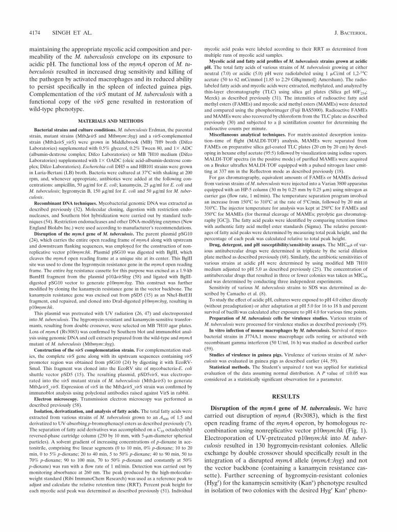
Disruption of the mymA gene of M. tuberculosis. We havecarried out disruption of mymA (Rv3083), which is the firstopen reading frame of the mymA operon, by homologous re-combination using nonreplicative vector p10mym:hk (Fig. 1).Electroporation of UV-pretreated p10mym:hk into M. tuber-culosis resulted in 130 hygromycin-resistant colonies. Allelicexchange by double crossover should specifically result in theintegration of a disrupted mymA allele (mymA::hyg) and notthe vector backbone (containing a kanamycin resistance cas-sette). Further screening of hygromycin-resistant colonies(Hygr) for the kanamycin sensitivity (Kans) phenotype resultedin isolation of two colonies with the desired Hygr Kans pheno-
4174 SINGH ET AL. J. BACTERIOL.
type. The disruption of mymA was confirmed in one Hygr Kans
colony by Southern blot analysis using the genomic DNA fromM. tuberculosis and its mymA mutant strain (Mtbmym::hyg)(data not shown) and by immunoblot analysis using polyclonalantibodies raised in rabbit against MymA. The wild-type strainshowed a specific protein band of 55 kDa corresponding toMymA (Fig. 1B, lane 1), which was absent in the mutant strain(Fig. 1B, lane 2). mymA represents the first gene of the mymAoperon (Rv3083 to Rv3089). The disruption of the mymA geneby insertion of the hygromycin resistance (Hygr) cassette re-sulted in the termination of transcription at the end of thehygromycin resistance gene (due to the presence of the tran-criptional terminator). Hence, the downstream genes of themymA operon Rv3084 to Rv3089 were not transcribed. Thiswas confirmed by reverse transcription (RT) analysis. No am-plified products were obtained by RT-PCR with primers span-ning the intergenic region of mymA and lipR (Rv3084) or
fadD13 (Rv3089) when the RNA isolated from Mtbmym::hygwas used, whereas both the primer pairs yielded an RT-PCRproduct of the predicted size when RNA from M. tuberculosiswas employed (Fig. 1C). These results confirmed that the dis-ruption of mymA resulted in functional inactivation of themymA operon.
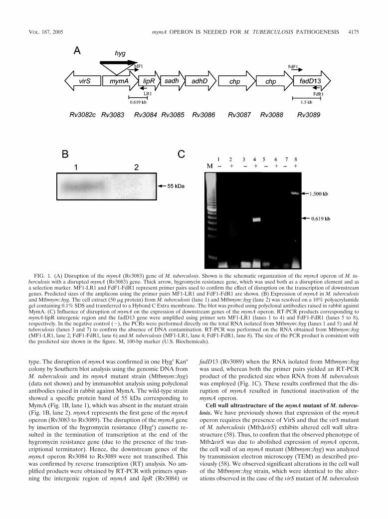
Cell wall ultrastructure of the mymA mutant of M. tubercu-losis. We have previously shown that expression of the mymAoperon requires the presence of VirS and that the virS mutantof M. tuberculosis (Mtb�virS) exhibits altered cell wall ultra-structure (58). Thus, to confirm that the observed phenotype ofMtb�virS was due to abolished expression of mymA operon,the cell wall of an mymA mutant (Mtbmym::hyg) was analyzedby transmission electron microscopy (TEM) as described pre-viously (58). We observed significant alterations in the cell wallof the Mtbmym::hyg strain, which were identical to the alter-ations observed in the case of the virS mutant of M. tuberculosis
FIG. 1. (A) Disruption of the mymA (Rv3083) gene of M. tuberculosis. Shown is the schematic organization of the mymA operon of M. tu-berculosis with a disrupted mymA (Rv3083) gene. Thick arrow, hygromycin resistance gene, which was used both as a disruption element and asa selection marker. MF1-LR1 and FdF1-FdR1 represent primer pairs used to confirm the effect of disruption on the transcription of downstreamgenes. Predicted sizes of the amplicons using the primer pairs MF1-LR1 and FdF1-FdR1 are shown. (B) Expression of mymA in M. tuberculosisand Mtbmym::hyg. The cell extract (50 �g protein) from M. tuberculosis (lane 1) and Mtbmym::hyg (lane 2) was resolved on a 10% polyacrylamidegel containing 0.1% SDS and transferred to a Hybond C Extra membrane. The blot was probed using polyclonal antibodies raised in rabbit againstMymA. (C) Influence of disruption of mymA on the expression of downstream genes of the mymA operon. RT-PCR products corresponding tomymA-lipR intergenic region and the fadD13 gene were amplified using primer sets MF1-LR1 (lanes 1 to 4) and FdF1-FdR1 (lanes 5 to 8),respectively. In the negative control (�), the PCRs were performed directly on the total RNA isolated from Mtbmym::hyg (lanes 1 and 5) and M.tuberculosis (lanes 3 and 7) to confirm the absence of DNA contamination. RT-PCR was performed on the RNA obtained from Mtbmym::hyg(MF1-LR1, lane 2; FdF1-FdR1, lane 6) and M. tuberculosis (MF1-LR1, lane 4; FdF1-FdR1, lane 8). The size of the PCR product is consistent withthe predicted size shown in the figure. M, 100-bp marker (U.S. Biochemicals).
VOL. 187, 2005 mymA OPERON IS NEEDED FOR M. TUBERCULOSIS PATHOGENESIS 4175
(Mtb�virS) (Fig. 2) (58). This confirmed that the disruption ofmymA or virS results in similar phenotypes. A dense staining ofthe electron-transparent zone (ETZ) observed in the case ofMtb�virS and Mtbmym::hyg cells due to compromised perme-ability of the cell wall is suggestive of alterations in the mycolicacids present in the lipid bilayer (65).
Influence of virS and mymA disruption on the mycolic acidprofile of M. tuberculosis. To investigate if the observed changesin the staining pattern of the ETZ were associated with alter-ations in the mycolic acids, we compared the profiles of mycolicacids extracted from the Mtb�virS, Mtbmym::hyg, virS-comple-mented (Mtb�virS_virS), and parental strains of M. tuberculo-
FIG. 2. Effect of mymA disruption on the cell wall ultrastructure of M. tuberculosis. The cell wall of M. tuberculosis and Mtbmym::hyg wasexamined by transmission electron microscopy. Cells were cultured in MB 7H9 medium to an A600 of 1.5, harvested, washed with phosphate buffer,and fixed in paraformaldehyde solution for 4 to 8 h. Cells were prepared for electron microscopy as described earlier (58). Shown is electronmicroscopic analysis (�44,000) of (A) M. tuberculosis and (B) Mtbmym::hyg. The photomicrographs shown are typical of the population as a whole,as judged by viewing many independent fields.
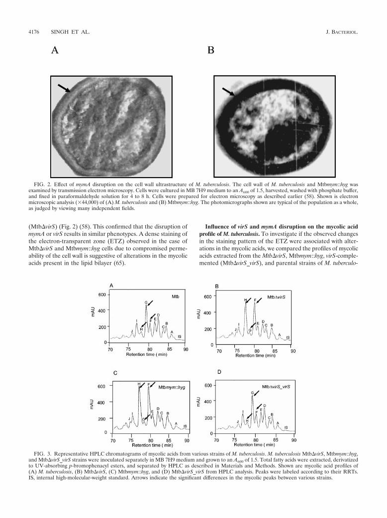
FIG. 3. Representative HPLC chromatograms of mycolic acids from various strains of M. tuberculosis. M. tuberculosis Mtb�virS, Mtbmym::hyg,and Mtb�virS_virS strains were inoculated separately in MB 7H9 medium and grown to an A600 of 1.5. Total fatty acids were extracted, derivatizedto UV-absorbing p-bromophenacyl esters, and separated by HPLC as described in Materials and Methods. Shown are mycolic acid profiles of(A) M. tuberculosis, (B) Mtb�virS, (C) Mtbmym::hyg, and (D) Mtb�virS_virS from HPLC analysis. Peaks were labeled according to their RRTs.IS, internal high-molecular-weight standard. Arrows indicate the significant differences in the mycolic peaks between various strains.
4176 SINGH ET AL. J. BACTERIOL.
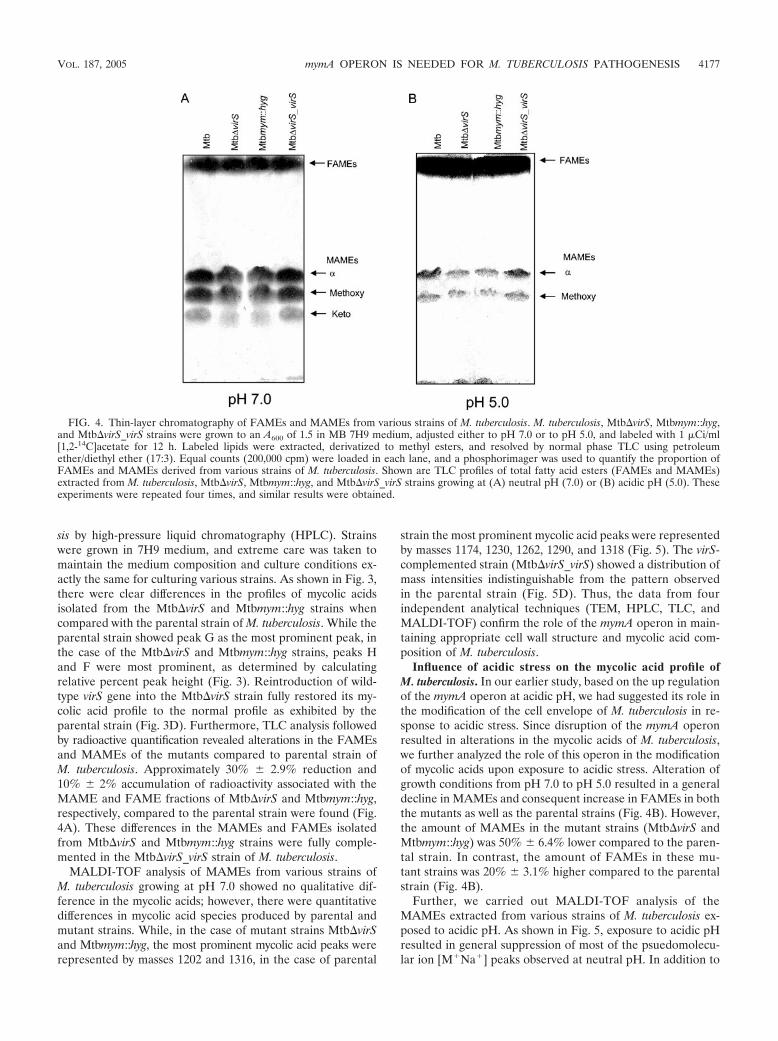
sis by high-pressure liquid chromatography (HPLC). Strainswere grown in 7H9 medium, and extreme care was taken tomaintain the medium composition and culture conditions ex-actly the same for culturing various strains. As shown in Fig. 3,there were clear differences in the profiles of mycolic acidsisolated from the Mtb�virS and Mtbmym::hyg strains whencompared with the parental strain of M. tuberculosis. While theparental strain showed peak G as the most prominent peak, inthe case of the Mtb�virS and Mtbmym::hyg strains, peaks Hand F were most prominent, as determined by calculatingrelative percent peak height (Fig. 3). Reintroduction of wild-type virS gene into the Mtb�virS strain fully restored its my-colic acid profile to the normal profile as exhibited by theparental strain (Fig. 3D). Furthermore, TLC analysis followedby radioactive quantification revealed alterations in the FAMEsand MAMEs of the mutants compared to parental strain ofM. tuberculosis. Approximately 30% 2.9% reduction and10% 2% accumulation of radioactivity associated with theMAME and FAME fractions of Mtb�virS and Mtbmym::hyg,respectively, compared to the parental strain were found (Fig.4A). These differences in the MAMEs and FAMEs isolatedfrom Mtb�virS and Mtbmym::hyg strains were fully comple-mented in the Mtb�virS_virS strain of M. tuberculosis.
MALDI-TOF analysis of MAMEs from various strains ofM. tuberculosis growing at pH 7.0 showed no qualitative dif-ference in the mycolic acids; however, there were quantitativedifferences in mycolic acid species produced by parental andmutant strains. While, in the case of mutant strains Mtb�virSand Mtbmym::hyg, the most prominent mycolic acid peaks wererepresented by masses 1202 and 1316, in the case of parental
strain the most prominent mycolic acid peaks were representedby masses 1174, 1230, 1262, 1290, and 1318 (Fig. 5). The virS-complemented strain (Mtb�virS_virS) showed a distribution ofmass intensities indistinguishable from the pattern observedin the parental strain (Fig. 5D). Thus, the data from fourindependent analytical techniques (TEM, HPLC, TLC, andMALDI-TOF) confirm the role of the mymA operon in main-taining appropriate cell wall structure and mycolic acid com-position of M. tuberculosis.
Influence of acidic stress on the mycolic acid profile ofM. tuberculosis. In our earlier study, based on the up regulationof the mymA operon at acidic pH, we had suggested its role inthe modification of the cell envelope of M. tuberculosis in re-sponse to acidic stress. Since disruption of the mymA operonresulted in alterations in the mycolic acids of M. tuberculosis,we further analyzed the role of this operon in the modificationof mycolic acids upon exposure to acidic stress. Alteration ofgrowth conditions from pH 7.0 to pH 5.0 resulted in a generaldecline in MAMEs and consequent increase in FAMEs in boththe mutants as well as the parental strains (Fig. 4B). However,the amount of MAMEs in the mutant strains (Mtb�virS andMtbmym::hyg) was 50% 6.4% lower compared to the paren-tal strain. In contrast, the amount of FAMEs in these mu-tant strains was 20% 3.1% higher compared to the parentalstrain (Fig. 4B).
Further, we carried out MALDI-TOF analysis of theMAMEs extracted from various strains of M. tuberculosis ex-posed to acidic pH. As shown in Fig. 5, exposure to acidic pHresulted in general suppression of most of the psuedomolecu-lar ion [M�Na�] peaks observed at neutral pH. In addition to
FIG. 4. Thin-layer chromatography of FAMEs and MAMEs from various strains of M. tuberculosis. M. tuberculosis, Mtb�virS, Mtbmym::hyg,and Mtb�virS_virS strains were grown to an A600 of 1.5 in MB 7H9 medium, adjusted either to pH 7.0 or to pH 5.0, and labeled with 1 �Ci/ml[1,2-14C]acetate for 12 h. Labeled lipids were extracted, derivatized to methyl esters, and resolved by normal phase TLC using petroleumether/diethyl ether (17:3). Equal counts (200,000 cpm) were loaded in each lane, and a phosphorimager was used to quantify the proportion ofFAMEs and MAMEs derived from various strains of M. tuberculosis. Shown are TLC profiles of total fatty acid esters (FAMEs and MAMEs)extracted from M. tuberculosis, Mtb�virS, Mtbmym::hyg, and Mtb�virS_virS strains growing at (A) neutral pH (7.0) or (B) acidic pH (5.0). Theseexperiments were repeated four times, and similar results were obtained.
VOL. 187, 2005 mymA OPERON IS NEEDED FOR M. TUBERCULOSIS PATHOGENESIS 4177
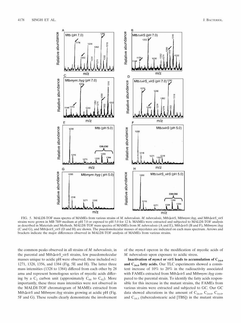
the common peaks observed in all strains of M. tuberculosis, inthe parental and Mtb�virS_virS strains, few psuedomolecularmasses unique to acidic pH were observed; these included m/z1271, 1328, 1356, and 1384 (Fig. 5E and H). The latter threemass intensities (1328 to 1384) differed from each other by 28amu and represent homologous series of mycolic acids differ-ing by a C2 carbon unit (approximately C88 to C92). Moreimportantly, these three mass intensities were not observed inthe MALDI-TOF chromatogram of MAMEs extracted fromMtb�virS and Mtbmym::hyg strains growing at acidic pH (Fig.5F and G). These results clearly demonstrate the involvement
of the mymA operon in the modification of mycolic acids ofM. tuberculosis upon exposure to acidic stress.
Inactivation of mymA or virS leads to accumulation of C24:0
and C26:0 fatty acids. Our TLC experiments showed a consis-tent increase of 10% to 20% in the radioactivity associatedwith FAMEs extracted from Mtb�virS and Mtbmym::hyg com-pared to the parental strain. To identify the fatty acids respon-sible for this increase in the mutant strains, the FAMEs fromvarious strains were extracted and subjected to GC. Our GCdata showed alterations in the amount of C26:0, C24:0, C16:0,and C18:3 (tuberculostearic acid [TBS]) in the mutant strains
FIG. 5. MALDI-TOF mass spectra of MAMEs from various strains of M. tuberculosis. M. tuberculosis, Mtb�virS, Mtbmym::hyg, and Mtb�virS_virSstrains were grown in MB 7H9 medium at pH 7.0 or exposed to pH 5.0 for 12 h. MAMEs were extracted and subjected to MALDI-TOF analysisas described in Materials and Methods. MALDI-TOF mass spectra of MAMEs from M. tuberculosis (A and E), Mtb�virS (B and F), Mtbmym::hyg(C and G), and Mtb�virS_virS (D and H) are shown. The psuedomolecular masses of mycolates are indicated on each mass spectrum. Arrows andbrackets indicate the major differences observed in MALDI-TOF analysis of MAMEs from various strains.
4178 SINGH ET AL. J. BACTERIOL.
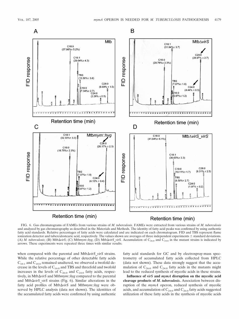
when compared with the parental and Mtb�virS_virS strains.While the relative percentage of other detectable fatty acidsC18:1 and C18:0 remained unaltered, we observed a twofold de-crease in the levels of C16:0 and TBS and threefold and twofoldincreases in the levels of C26:0 and C24:0 fatty acids, respec-tively, in Mtb�virS and Mtbmym::hyg compared to the parentaland Mtb�virS_virS strains (Fig. 6). Similar alterations in thefatty acid profiles of Mtb�virS and Mtbmym::hyg were ob-served by HPLC analysis (data not shown). The identities ofthe accumulated fatty acids were confirmed by using authentic
fatty acid standards for GC and by electrospray-mass spec-trometry of accumulated fatty acids collected from HPLC(data not shown). These data strongly suggest that the accu-mulation of C26:0 and C24:0 fatty acids in the mutants mightlead to the reduced synthesis of mycolic acids in these strains.
Influence of virS and mymA disruption on the mycolic acidcleavage products of M. tuberculosis. Association between dis-ruption of the mymA operon, reduced synthesis of mycolicacids, and accumulation of C24:0 and C26:0 fatty acids suggestedutilization of these fatty acids in the synthesis of mycolic acids
FIG. 6. Gas chromatograms of FAMEs from various strains of M. tuberculosis. FAMEs were extracted from various strains of M. tuberculosisand analyzed by gas chromatography as described in the Materials and Methods. The identity of fatty acid peaks was confirmed by using authenticfatty acid standards. Relative percentages of fatty acids were calculated and are indicated on each chromatogram. FID and TBS represent flameionization detector and tuberculostearic acid, respectively. The values shown are averages of three independent experiments standard deviations.(A) M. tuberculosis; (B) Mtb�virS; (C) Mtbmym::hyg, (D) Mtb�virS_virS. Accumulation of C26:0 and C24:0 in the mutant strains is indicated byarrows. These experiments were repeated three times with similar results.
VOL. 187, 2005 mymA OPERON IS NEEDED FOR M. TUBERCULOSIS PATHOGENESIS 4179
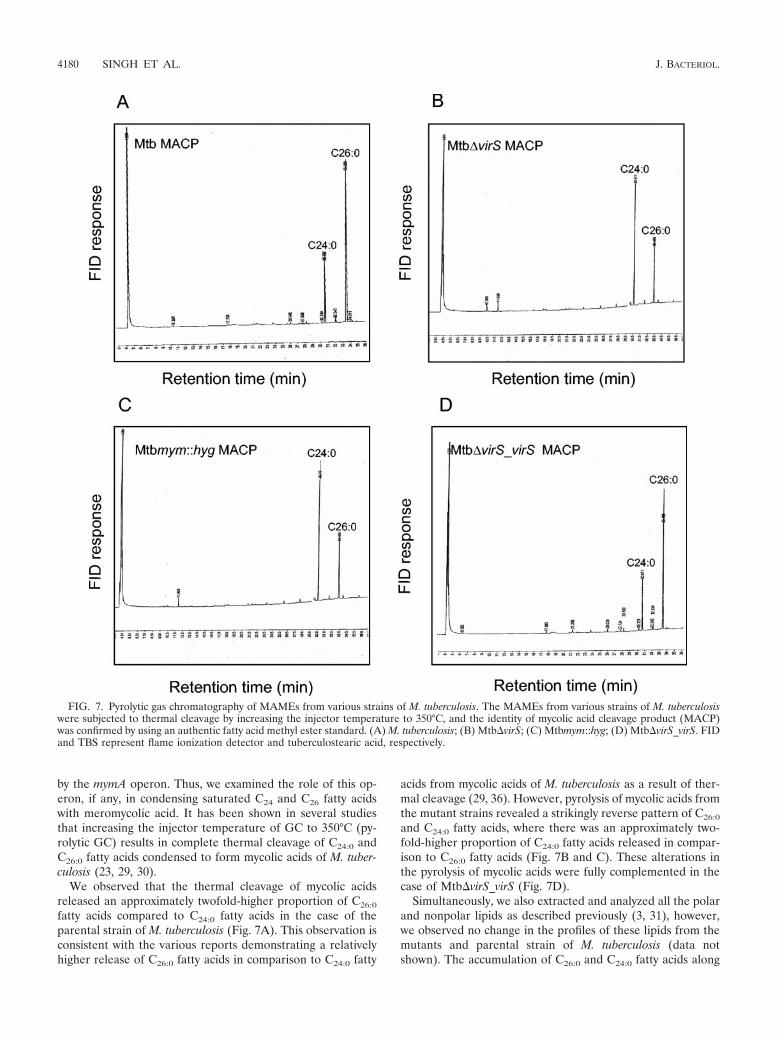
by the mymA operon. Thus, we examined the role of this op-eron, if any, in condensing saturated C24 and C26 fatty acidswith meromycolic acid. It has been shown in several studiesthat increasing the injector temperature of GC to 350°C (py-rolytic GC) results in complete thermal cleavage of C24:0 andC26:0 fatty acids condensed to form mycolic acids of M. tuber-culosis (23, 29, 30).
We observed that the thermal cleavage of mycolic acidsreleased an approximately twofold-higher proportion of C26:0
fatty acids compared to C24:0 fatty acids in the case of theparental strain of M. tuberculosis (Fig. 7A). This observation isconsistent with the various reports demonstrating a relativelyhigher release of C26:0 fatty acids in comparison to C24:0 fatty
acids from mycolic acids of M. tuberculosis as a result of ther-mal cleavage (29, 36). However, pyrolysis of mycolic acids fromthe mutant strains revealed a strikingly reverse pattern of C26:0
and C24:0 fatty acids, where there was an approximately two-fold-higher proportion of C24:0 fatty acids released in compar-ison to C26:0 fatty acids (Fig. 7B and C). These alterations inthe pyrolysis of mycolic acids were fully complemented in thecase of Mtb�virS_virS (Fig. 7D).
Simultaneously, we also extracted and analyzed all the polarand nonpolar lipids as described previously (3, 31), however,we observed no change in the profiles of these lipids from themutants and parental strain of M. tuberculosis (data notshown). The accumulation of C26:0 and C24:0 fatty acids along
FIG. 7. Pyrolytic gas chromatography of MAMEs from various strains of M. tuberculosis. The MAMEs from various strains of M. tuberculosiswere subjected to thermal cleavage by increasing the injector temperature to 350°C, and the identity of mycolic acid cleavage product (MACP)was confirmed by using an authentic fatty acid methyl ester standard. (A) M. tuberculosis; (B) Mtb�virS; (C) Mtbmym::hyg; (D) Mtb�virS_virS. FIDand TBS represent flame ionization detector and tuberculostearic acid, respectively.
4180 SINGH ET AL. J. BACTERIOL.
with the altered mycolic acid profile in the mutants indicatesthe role of mymA operon in the synthesis of mycolic acids byutilizing saturated C26 and C24 fatty acids as potential precur-sors.
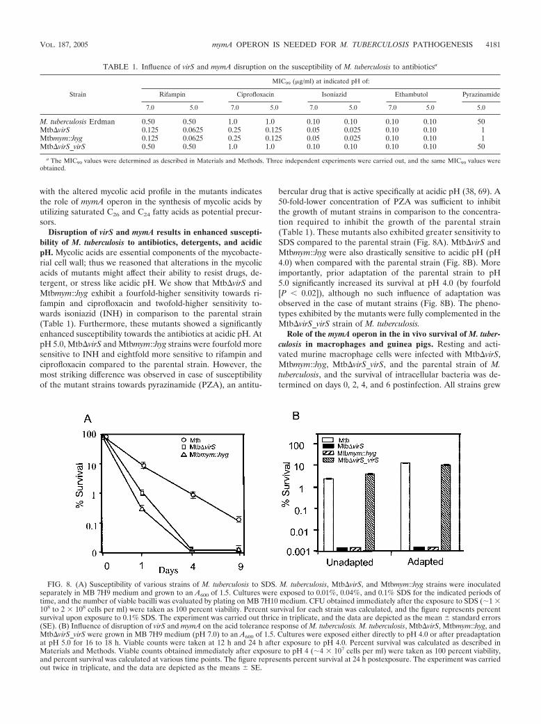
Disruption of virS and mymA results in enhanced suscepti-bility of M. tuberculosis to antibiotics, detergents, and acidicpH. Mycolic acids are essential components of the mycobacte-rial cell wall; thus we reasoned that alterations in the mycolicacids of mutants might affect their ability to resist drugs, de-tergent, or stress like acidic pH. We show that Mtb�virS andMtbmym::hyg exhibit a fourfold-higher sensitivity towards ri-fampin and ciprofloxacin and twofold-higher sensitivity to-wards isoniazid (INH) in comparison to the parental strain(Table 1). Furthermore, these mutants showed a significantlyenhanced susceptibility towards the antibiotics at acidic pH. AtpH 5.0, Mtb�virS and Mtbmym::hyg strains were fourfold moresensitive to INH and eightfold more sensitive to rifampin andciprofloxacin compared to the parental strain. However, themost striking difference was observed in case of susceptibilityof the mutant strains towards pyrazinamide (PZA), an antitu-
bercular drug that is active specifically at acidic pH (38, 69). A50-fold-lower concentration of PZA was sufficient to inhibitthe growth of mutant strains in comparison to the concentra-tion required to inhibit the growth of the parental strain(Table 1). These mutants also exhibited greater sensitivity toSDS compared to the parental strain (Fig. 8A). Mtb�virS andMtbmym::hyg were also drastically sensitive to acidic pH (pH4.0) when compared with the parental strain (Fig. 8B). Moreimportantly, prior adaptation of the parental strain to pH5.0 significantly increased its survival at pH 4.0 (by fourfold[P 0.02]), although no such influence of adaptation wasobserved in the case of mutant strains (Fig. 8B). The pheno-types exhibited by the mutants were fully complemented in theMtb�virS_virS strain of M. tuberculosis.
Role of the mymA operon in the in vivo survival of M. tuber-culosis in macrophages and guinea pigs. Resting and acti-vated murine macrophage cells were infected with Mtb�virS,Mtbmym::hyg, Mtb�virS_virS, and the parental strain of M.tuberculosis, and the survival of intracellular bacteria was de-termined on days 0, 2, 4, and 6 postinfection. All strains grew
FIG. 8. (A) Susceptibility of various strains of M. tuberculosis to SDS. M. tuberculosis, Mtb�virS, and Mtbmym::hyg strains were inoculatedseparately in MB 7H9 medium and grown to an A600 of 1.5. Cultures were exposed to 0.01%, 0.04%, and 0.1% SDS for the indicated periods oftime, and the number of viable bacilli was evaluated by plating on MB 7H10 medium. CFU obtained immediately after the exposure to SDS (�1 �108 to 2 � 108 cells per ml) were taken as 100 percent viability. Percent survival for each strain was calculated, and the figure represents percentsurvival upon exposure to 0.1% SDS. The experiment was carried out thrice in triplicate, and the data are depicted as the mean standard errors(SE). (B) Influence of disruption of virS and mymA on the acid tolerance response of M. tuberculosis. M. tuberculosis, Mtb�virS, Mtbmym::hyg, andMtb�virS_virS were grown in MB 7H9 medium (pH 7.0) to an A600 of 1.5. Cultures were exposed either directly to pH 4.0 or after preadaptationat pH 5.0 for 16 to 18 h. Viable counts were taken at 12 h and 24 h after exposure to pH 4.0. Percent survival was calculated as described inMaterials and Methods. Viable counts obtained immediately after exposure to pH 4 (�4 � 107 cells per ml) were taken as 100 percent viability,and percent survival was calculated at various time points. The figure represents percent survival at 24 h postexposure. The experiment was carriedout twice in triplicate, and the data are depicted as the means SE.
TABLE 1. Influence of virS and mymA disruption on the susceptibility of M. tuberculosis to antibioticsa
Strain
MIC99 (�g/ml) at indicated pH of:
Rifampin Ciprofloxacin Isoniazid Ethambutol Pyrazinamide
7.0 5.0 7.0 5.0 7.0 5.0 7.0 5.0 5.0
M. tuberculosis Erdman 0.50 0.50 1.0 1.0 0.10 0.10 0.10 0.10 50Mtb�virS 0.125 0.0625 0.25 0.125 0.05 0.025 0.10 0.10 1Mtbmym::hyg 0.125 0.0625 0.25 0.125 0.05 0.025 0.10 0.10 1Mtb�virS_virS 0.50 0.50 1.0 1.0 0.10 0.10 0.10 0.10 50
a The MIC99 values were determined as described in Materials and Methods. Three independent experiments were carried out, and the same MIC99 values wereobtained.
VOL. 187, 2005 mymA OPERON IS NEEDED FOR M. TUBERCULOSIS PATHOGENESIS 4181
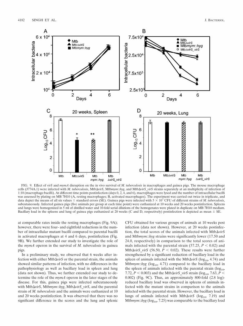
at comparable rates inside the resting macrophages (Fig. 9A);however, there were four- and eightfold reductions in the num-ber of intracellular mutant bacilli compared to parental bacilliin activated macrophages at 4 and 6 days, postinfection (Fig.9B). We further extended our study to investigate the role ofthe mymA operon in the survival of M. tuberculosis in guineapigs.
In a preliminary study, we observed that 6 weeks after in-fection with either Mtb�virS or the parental strain, the animalsshowed similar patterns of infection, with no differences in thepathophysiology as well as bacillary load in spleen and lung(data not shown). Thus, we further extended our study to de-termine the role of the mymA operon in the later stages of thedisease. For this, guinea pigs were infected subcutaneouslywith Mtb�virS, Mtbmym::hyg, Mtb�virS_virS, and the parentalstrain of M. tuberculosis and the animals were euthanized at 10and 20 weeks postinfection. It was observed that there was nosignificant difference in the scores and the lung and splenic
CFU obtained for various groups of animals at 10 weeks postinfection (data not shown). However, at 20 weeks postinfec-tion, the total scores of the animals infected with Mtb�virSand Mtbmym::hyg strains were significantly lower (17.50 and24.0, respectively) in comparison to the total scores of ani-mals infected with the parental strain (57.25, P 0.02) andMtb�virS_virS (56.50, P 0.02). These results were furtherstrengthened by a significant reduction of bacillary load in thespleen of animals infected with the Mtb�virS (log10, 4.74) andMtbmym::hyg (log10, 4.71) compared to the bacillary load inthe spleen of animals infected with the parental strain (log10,7.72, P 0.003) and the Mtb�virS_virS strain (log10, 7.63, P 0.002) (Fig. 9C). Thus, an approximately 800-fold (2.8 log)-reduced bacillary load was observed in spleens of animals in-fected with the mutant strains in comparison to the animalsinfected with the parental strain. However, the bacillary load inlungs of animals infected with Mtb�virS (log10, 7.19) andMtbmym::hyg (log10, 7.25) was comparable to the bacillary load
FIG. 9. Effect of virS and mymA disruption on the in vivo survival of M. tuberculosis in macrophages and guinea pigs. The mouse macrophagecells (J774A.1) were infected with M. tuberculosis, Mtb�virS, Mtbmym::hyg, and Mtb�virS_virS strains separately at an multiplicity of infection of1:10 (macrophage:bacilli). At different time points postinfection (days 0, 2, 4, and 6), macrophages were lysed and the number of intracellular bacilliwas assessed by plating on MB 7H10 (A, resting macrophages; B, activated macrophages). The experiment was carried out twice in triplicate, anddata depict the means of all six values standard errors (SE). Guinea pigs were infected with 5 � 107 CFU of different strains of M. tuberculosis,subcutaneously. Infected guinea pigs (five animals per group at each time point) were euthanized at 10 weeks and 20 weeks postinfection. Spleensand lungs were homogenized in 5 ml of distilled water and 10-fold serial dilutions of the homogenates were plated in duplicate on MB 7H10 medium.Bacillary load in the spleens and lung of guinea pigs euthanized at 20 weeks (C and D, respectively) postinfection is depicted as mean SE.
4182 SINGH ET AL. J. BACTERIOL.
in the lungs of animals infected with the parental strain (log10,7.25) and Mtb�virS_virS strain (log10, 7.21) (Fig. 9D). Theseobservations were further substantiated by significant differ-ences in the percent spleen weight per total body weight in caseof animals infected with Mtb�virS (0.16%) and Mtbmym::hyg(0.18%) in comparison to the animals infected with the paren-tal strain (0.31%, P 0.05) and Mtb�virS_virS strain (0.36%,P 0.05); however, we observed no difference in the percentlung weight per total body weight of animals infected with ei-ther the mutants or parental strain. These observations strong-ly suggest the role of the mymA operon in the survival of M.tuberculosis against potentially harsher conditions faced by thepathogen in activated macrophages and also at later stages ofprogression of disease in guinea pigs.
DISCUSSION
In an earlier study, we had reported that the mymA operonof M. tuberculosis is induced at acidic pH in macrophages andis transcriptionally regulated by VirS (58). We had also shownthat Mtb�virS has an altered cell wall structure and proposedthat these alterations were due to abolished expression of themymA operon. More direct proof of this observation emergedupon disruption of mymA (Rv3083), the first gene in the mymAoperon, which resulted in the functional inactivation of themymA operon and similar alterations in the cell wall ultrastruc-ture phenotype as were observed earlier for Mtb�virS (58).The Mtbmym::hyg strain exhibited a much denser and darkerstaining of cell surface, indicating an alteration in the ETZ,which is thought to be composed primarily of mycolic acidsarranged perpendicular to the plane of the cell surface (5, 33,34, 35, 48). Such dense staining of the cell wall has also beenobserved after treatment of Mycobacterium avium with isonia-zid resulting from the inhibition of mycolic acid synthesis bythe drug (42). The alterations in the cell surface of Mtb�virSand Mtbmym::hyg strains were further substantiated by theHPLC profiles of mycolic acids from the mutants and theparental strains. Furthermore, both mutants produced less my-colic acids in comparison to the parental strain as analyzed byTLC. These findings suggest that the observed alterations inthe cell wall ultrastructure result from the altered mycolic acidcomposition although the effect of latter on the arrangement ofother cell surface lipids and proteins and their consequentcontribution on the observed phenotype cannot be completelyruled out. On exposure to acidic pH, the reduction in mycolicacid synthesis was markedly more prominent in the Mtb�virSand Mtbmym::hyg strains in comparison to the parental strain.The accumulation of fatty acids (C24:0/C26:0) at acidic pH wasalso observed to be higher in the mutants compared to theparental strain. Although, a general reduction in the synthesisof mycolic acids at acidic pH can be expected to stem from therepression of the Fas II operon (21), a much sharper decline inmycolic acid synthesis in case of both the mutant strains im-plicates the mymA operon in the synthesis of mycolic acids onexposure of the pathogen to acidic pH. The emergence of newmass peaks corresponding to the C88 to C92 chain length ofmycolic acids (1328, 1356, and 1384) in the parental strain, butnot in the mutants, clearly suggested the role of the mymAoperon in the synthesis of these mycolic acids at acidic pH.Such modifications in the chain length of mycolic acids in
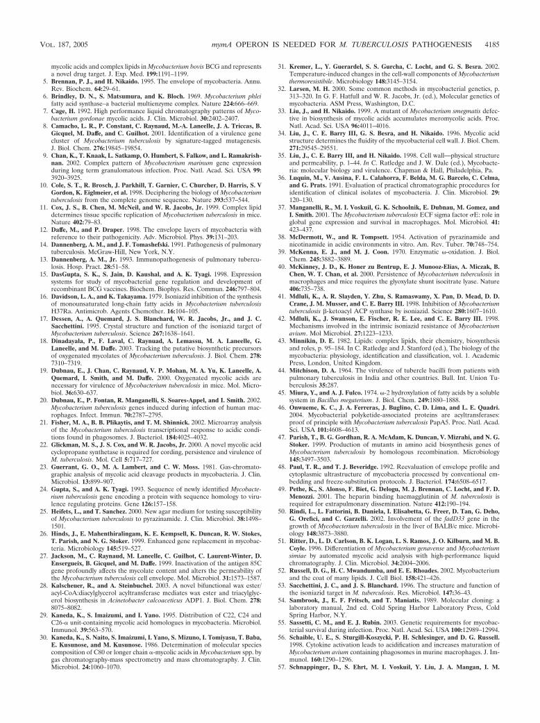
mycobacteria on exposure to environmental stresses have beenreported in several studies (2, 31, 33, 63, 64), emphasizing therequirement of the appropriate repertoire of mycolic acids torespond to environmental stresses. Further, the enhanced ac-cumulation of C24:0/C26:0 fatty acids in the mutant strains sub-stantiates their role in the synthesis of mycolic acids by themymA operon. Conventionally mycolic acids are believed to besynthesized by elongating long chain fatty acids (C16 to C26) tomeromycolic acids by the Fas II operon of M. tuberculosis, andthe final Claisen type condensation of the C24:0/C26:0 fatty acidwith meromycolates results in the production of full-lengthmycolic acids (6, 16, 33, 35, 41, 43). However, Asselineau et al.(1) have thoroughly reexamined the pathways of mycolic acidsynthesis and suggested an alternate approach of mycolic acidsynthesis by “head-to-tail” condensation of long chain fattyacids. The synthesis of mycolic acids by this approach involvesthe condensation of three common fatty acids. First, two ofthese are subjected to omega oxidation followed by condensa-tion to produce meromycolic acids, which in turn condensewith C24:0/C26:0 fatty acids to produce mycolic acids. This ap-proach of mycolic acid synthesis requires enzymes that cancarry out omega oxidation of fatty acids and their subsequentcondensation (1). Interestingly, analysis of gene products ofthe mymA operon revealed that Rv3083 (mymA) is a homo-logue of flavin-containing monooxygenases (58), which cancarry out omega hydroxylation of fatty acids (39, 45), the firststep in omega oxidation of fatty acids, while Rv3085 andRv3086 show homologies with dehydrogenases and could pos-sibly carry out subsequent steps to convert terminal methylgroups of fatty acids to carboxylic groups for condensation asdescribed previously (1). Release of acyl carrier protein ester-ified to the fatty acids by thioesetrase LipR (Rv3084) leadsto generation of diacids for the condensation. Rv3087 andRv3088 contain an HHXXXDG motif required for the thio-esterification or Claisen type condensation of fatty acids (28,46); the last gene, Rv3089, is an acyl-CoA synthase and canactivate the fatty acids. Thus, Rv3087 and Rv3088 can carry out“head-to-tail” condensation of fatty acids which were previ-ously omega oxidized by gene products of Rv3083 to Rv3086and further activation of the condensed fatty acids by Rv3089can yield long chain fatty acids (keto acids). These keto acidscan then be subjected to functional group modification likemethylation, decarboxylation, or cyclopropanation to generatemeromycolic acids as suggested previously (1). The condensa-tion process described above can produce long chain fatty acidsthat are indistinguishable from mycolic acids (1). Thus, thegenes present in the mymA operon can assemble meromycolicacids beginning from the omega oxidation of fatty acids fol-lowed by their condensation with fatty acids (C24:0/C26:0) toproduce mycolic acids as shown in Fig. 10.
Both the mutants showed increased sensitivity to major an-titubercular drugs along with enhanced susceptibility to SDSand acidic pH. The enhanced susceptibility of M. tuberculosisto antibiotics, detergents, and environmental stresses has beenshown to be associated with alterations in the mycolic acidcontents and composition (27, 67, 68). A mutant of Mycobac-terium smegmatis devoid of mycolic acids has been shown to behypersusceptible to antibiotics and was unable to grow at 37°C(33), while inactivation of the arylamine N-acetyltransferasegene of Mycobacterium bovis BCG perturbed biosynthesis of
VOL. 187, 2005 mymA OPERON IS NEEDED FOR M. TUBERCULOSIS PATHOGENESIS 4183
mycolic acids, with the consequent increase in the sensitivity toantibiotics (4). Interestingly, these mutants were susceptible toINH, which is known to target mycolic acid biosynthesis (53,60, 61, 62, 66). The precise target of INH in M. tuberculosis isstill debatable, and various pieces of evidence suggest thatthere might be several targets of INH (60). However, INH-treated M. tuberculosis accumulates C26:0 fatty acid (41), sug-gesting that INH targets enzymes which utilize C26:0 for gen-eration of mycolic acids. These observations, together with thefact that the mymA operon might utilize C26:0 fatty acids asprecursors for mycolic acid biosynthesis, suggest the possibilitythat the proteins present in this operon are potential targets ofINH. This possibility is further strengthened by the presence ofa NADH binding site in MymA (58), which has been shown as
the target of the active form of INH (iso-NAD) in the case ofInhA (17).
The induction of the mymA operon at acidic pH and asignificantly reduced ability of Mtb�virS and Mtbmym::hyg tosurvive in the activated macrophages compared to the parentalstrain supports the hypothesis that the mymA operon may playan important role in the survival of M. tuberculosis upon expo-sure to severely acidic conditions in activated macrophages(56) or caseating granuloma in the later stages of infection (13,14). This was substantiated by a drastic reduction (�2.8 log)observed in the ability of the mutant strains to specificallysurvive in spleen compared to the parental strain at 20 weekspostinfection. The involvement of the mymA operon in persis-tence during the chronic stage of disease and also the tropismshown by the pathogen are not surprising. Several genes in-volved in fatty acid metabolism such as icl, encoding isocitratelyase (catabolizes fatty acids via the glyoxylate cycle) and pcaA,encoding cyclopropane synthase (required for cyclopropana-tion of mycolic acids), and mutations affecting the lipid-richcell envelope are all known to reduce the survival of M. tuber-culosis in mice, only at the later stages of disease progression(22, 40, 55). Besides, several gene disruption studies have ledto the exhibition of tropism by the mutant bacilli; for exam-ple, FadD28, FadD33, Pps, MmpL7, and HBHA mutants allshowed tissue-specific growth rates (11, 49, 50). This tropismcould be due to different physiological conditions or immuneresponses which the pathogen encounters in different organs.The genes present in the mymA operon apparently may beinvolved in maintaining the cell wall integrity required for thepersistence of M. tuberculosis in spleen.
The involvement of the mymA operon in the persistence ofM. tuberculosis together with its role in maintaining appropri-ate mycolic acid composition to resist antitubercular drugs atacidic pH indicate that precise targeting of the mymA operongene products may increase the effectiveness of combinationchemotherapy and impede the mechanisms involved in thepersistence of M. tuberculosis.
ACKNOWLEDGMENTS
This work was supported by financial grants received from the In-dian Council of Medical Research and Department of Biotechnology,Government of India. A.S. and R.G. are grateful to the Council ofScientific and Industrial Research, India, for research fellowships.
Jaya Gopinath, S. Nambirajan, Lucas, K. Chandran, M. Asokan(TRC, Chennai, India) and Archana (NII, New Delhi, India) areacknowledged for assistance in animal experiments and fatty acid anal-ysis, respectively. We acknowledge T. K. Das, Incharge EM facility,Department of Anatomy, AIIMS, New Delhi, for transmission elec-tron microscopy and D. Chatterji and P. D. Deepalakshmi, InchargeProteomics facility, IISC, Bangalore, for MALDI-TOF analysis. Wethank Bindu Nair and Bahadur Singh for their technical assistance.Rajiv Chawla is thankfully acknowledged for excellent secretarial help.
REFERENCES
1. Asselineau, C., J. Asselineau, G. Laneelle, and M.-A. Laneelle. 2002. Thebiosynthesis of mycolic acids by Mycobacteria: current and alternative hy-pothesis. Prog. Lipid Res. 41:501–523.
2. Baba, T., K. Kaneda, E. Kusunose, M. Kusunose, and I. Yano. 1989. Ther-mally adaptive changes of mycolic acids in Mycobacterium smegmatis. J. Bio-chem. (Tokyo) 106:81–86.
3. Besra, G. S. 1998. Preparation of cell wall fractions from mycobacteria.Methods Mol. Biol. 101:91–107.
4. Bhakta, S., G. S. Besra, A. M. Upton, T. Parish, C. Sholto-Douglas-Vernon,K. J. C. Gibson, S. Knutton, S. Gordon, R. P. daSilva, M. C. Anderton, andE. Sim. 2004. Arylamine N-acetyltransferase is required for synthesis of
FIG. 10. Hypothetical scheme showing the involvement of themymA operon in the biosynthesis of mycolic acids via “head-to-tail”condensation. Biosynthesis of mycolic acids via “head-to-tail” conden-sation initiates with the omega hydroxylation of the terminal methylgroup of two fatty acid molecules (C24:0/C26:0) by MymA (Rv3083),which are converted into a carboxyl group by the dehydrogenasesSadH (Rv3085) and AdhD (Rv3085). Release of acyl carrier protein(ACP) esterified to the fatty acid by thioesterase LipR (Rv3084) leadsto generation of diacids for the condensation. Head-to-tail condensa-tion by Rv3087 and/or Rv3088 gene products between three fatty acids,two of which are omega oxidized and activation by FadD13 (Rv3089)produce keto acid. The keto acid can then be subjected to functionalmodification like methylation, cyclopropanation, etc., to produce mero-mycolic acids which can be condensed to C24:0/C26:0 to produce full-length mycolic acids.
4184 SINGH ET AL. J. BACTERIOL.
mycolic acids and complex lipids in Mycobacterium bovis BCG and representsa novel drug target. J. Exp. Med. 199:1191–1199.
5. Brennan, P. J., and H. Nikaido. 1995. The envelope of mycobacteria. Annu.Rev. Biochem. 64:29–61.
6. Brindley, D. N., S. Matsumura, and K. Bloch. 1969. Mycobacterium phleifatty acid synthase–a bacterial multienzyme complex. Nature 224:666–669.
7. Cage, H. 1992. High performance liquid chromatography patterns of Myco-bacterium gordonae mycolic acids. J. Clin. Microbiol. 30:2402–2407.
8. Camacho, L. R., P. Constant, C. Raynaud, M.-A. Laneelle, J. A. Triccas, B.Gicquel, M. Daffe, and C. Guilhot. 2001. Identification of a virulence genecluster of Mycobacterium tuberculosis by signature-tagged mutagenesis.J. Biol. Chem. 276:19845–19854.
9. Chan, K., T. Knaak, L. Satkamp, O. Humbert, S. Falkow, and L. Ramakrish-nan. 2002. Complex pattern of Mycobacterium marinum gene expressionduring long term granulomatous infection. Proc. Natl. Acad. Sci. USA 99:3920–3925.
10. Cole, S. T., R. Brosch, J. Parkhill, T. Garnier, C. Churcher, D. Harris, S. VGordon, K. Eiglmeier, et al. 1998. Deciphering the biology of Mycobacteriumtuberculosis from the complete genome sequence. Nature 393:537–544.
11. Cox, J. S., B. Chen, M. McNeil, and W. R. Jacobs, Jr. 1999. Complex lipiddetermines tissue specific replication of Mycobacterium tuberculosis in mice.Nature 402:79–83.
12. Daffe, M., and P. Draper. 1998. The envelope layers of mycobacteria withreference to their pathogenicity. Adv. Microbiol. Phys. 39:131–203.
14. Dannenberg, A. M., and J. F. Tomashefski. 1991. Pathogenesis of pulmonarytuberculosis. McGraw-Hill, New York, N.Y.
13. Dannenberg, A. M., Jr. 1993. Immunopathogenesis of pulmonary tubercu-losis. Hosp. Pract. 28:51–58.
15. DasGupta, S. K., S. Jain, D. Kaushal, and A. K. Tyagi. 1998. Expressionsystems for study of mycobacterial gene regulation and development ofrecombinant BCG vaccines. Biochem. Biophys. Res. Commun. 246:797–804.
16. Davidson, L. A., and K. Takayama. 1979. Isoniazid inhibition of the synthesisof monounsaturated long-chain fatty acids in Mycobacterium tuberculosisH37Ra. Antimicrob. Agents Chemother. 16:104–105.
17. Dessen, A., A. Quemard, J. S. Blanchard, W. R. Jacobs, Jr., and J. C.Sacchettini. 1995. Crystal structure and function of the isoniazid target ofMycobacterium tuberculosis. Science 267:1638–1641.
18. Dinadayala, P., F. Laval, C. Raynuad, A. Lemassu, M. A. Laneelle, G.Laneelle, and M. Daffe. 2003. Tracking the putative biosynthetic precursorsof oxygenated mycolates of Mycobacterium tuberculosis. J. Biol. Chem. 278:7310–7319.
19. Dubnau, E., J. Chan, C. Raynaud, V. P. Mohan, M. A. Yu, K. Laneelle, A.Quemard, I. Smith, and M. Daffe. 2000. Oxygenated mycolic acids arenecessary for virulence of Mycobacterium tuberculosis in mice. Mol. Micro-biol. 36:630–637.
20. Dubnau, E., P. Fontan, R. Manganelli, S. Soares-Appel, and I. Smith. 2002.Mycobacterium tuberculosis genes induced during infection of human mac-rophages. Infect. Immun. 70:2787–2795.
21. Fisher, M. A., B. B. Plikaytis, and T. M. Shinnick. 2002. Microarray analysisof the Mycobacterium tuberculosis transcriptional response to acidic condi-tions found in phagosomes. J. Bacteriol. 184:4025–4032.
22. Glickman, M. S., J. S. Cox, and W. R. Jacobs, Jr. 2000. A novel mycolic acidcyclopropane synthetase is required for cording, persistence and virulence ofM. tuberculosis. Mol. Cell 5:717–727.
23. Guerrant, G. O., M. A. Lambert, and C. W. Moss. 1981. Gas-chromato-graphic analysis of mycolic acid cleavage products in mycobacteria. J. Clin.Microbiol. 13:899–907.
24. Gupta, S., and A. K. Tyagi. 1993. Sequence of newly identified Mycobacte-rium tuberculosis gene encoding a protein with sequence homology to viru-lence regulating proteins. Gene 126:157–158.
25. Heifets, L., and T. Sanchez. 2000. New agar medium for testing susceptibilityof Mycobacterium tuberculosis to pyrazinamide. J. Clin. Microbiol. 38:1498–1501.
26. Hinds, J., E. Mahenthiralingam, K. E. Kempsell, K. Duncan, R. W. Stokes,T. Parish, and N. G. Stoker. 1999. Enhanced gene replacement in mycobac-teria. Microbiology 145:519–527.
27. Jackson, M., C. Raynaud, M. Laneelle, C. Guilhot, C. Laurent-Winter, D.Ensergueix, B. Gicquel, and M. Daffe. 1999. Inactivation of the antigen 85Cgene profoundly affects the mycolate content and alters the permeability ofthe Mycobacterium tuberculosis cell envelope. Mol. Microbiol. 31:1573–1587.
28. Kalscheuer, R., and A. Steinbuchel. 2003. A novel bifunctional wax ester/acyl-CoA:diacylglycerol acyltransferase mediates wax ester and triacylglyc-erol biosynthesis in Acinetobacter calcoaceticus ADP1. J. Biol. Chem. 278:8075–8082.
29. Kaneda, K., S. Imaizumi, and I. Yano. 1995. Distribution of C22, C24 andC26-� unit-containing mycolic acid homologues in mycobacteria. Microbiol.Immunol. 39:563–570.
30. Kaneda, K., S. Naito, S. Imaizumi, I. Yano, S. Mizuno, I. Tomiyasu, T. Baba,E. Kusunose, and M. Kusunose. 1986. Determination of molecular speciescomposition of C80 or longer chain �-mycolic acids in Mycobacterium spp. bygas chromatography-mass spectrometry and mass chromatography. J. Clin.Microbiol. 24:1060–1070.
31. Kremer, L., Y. Guerardel, S. S. Gurcha, C. Locht, and G. S. Besra. 2002.Temperature-induced changes in the cell-wall components of Mycobacteriumthermoresistibile. Microbiology 148:3145–3154.
32. Larsen, M. H. 2000. Some common methods in mycobacterial genetics, p.313–320. In G. F. Hatfull and W. R. Jacobs, Jr. (ed.), Molecular genetics ofmycobacteria. ASM Press, Washington, D.C.
33. Liu, J., and H. Nikaido. 1999. A mutant of Mycobacterium smegmatis defec-tive in biosynthesis of mycolic acids accumulates meromycolic acids. Proc.Natl. Acad. Sci. USA 96:4011–4016.
34. Liu, J., C. E. Barry III, G. S. Besra, and H. Nikaido. 1996. Mycolic acidstructure determines the fluidity of the mycobacterial cell wall. J. Biol. Chem.271:29545–29551.
35. Liu, J., C. E. Barry III, and H. Nikaido. 1998. Cell wall—physical structureand permeability, p. 1–44. In C. Ratledge and J. W. Dale (ed.), Mycobacte-ria: molecular biology and virulence. Chapman & Hall, Philadelphia, Pa.
36. Luquin, M., V. Ausina, F. L. Calahorra, F. Belda, M. G. Barcelo, C. Celma,and G. Prats. 1991. Evaluation of practical chromatographic procedures foridentification of clinical isolates of mycobacteria. J. Clin. Microbiol. 29:120–130.
37. Manganelli, R., M. I. Voskuil, G. K. Schoolnik, E. Dubnau, M. Gomez, andI. Smith. 2001. The Mycobacterium tuberculosis ECF sigma factor �E: role inglobal gene expression and survival in macrophages. Mol. Microbiol. 41:423–437.
38. McDermott, W., and R. Tompsett. 1954. Activation of pyrazinamide andnicotinamide in acidic environments in vitro. Am. Rev. Tuber. 70:748–754.
39. McKenna, E. J., and M. J. Coon. 1970. Enzymatic �-oxidation. J. Biol.Chem. 245:3882–3889.
40. McKinney, J. D., K. Honer zu Bentrup, E. J. Munooz-Elias, A. Miczak, B.Chen, W. T. Chan, et al. 2000. Persistence of Mycobacterium tuberculosis inmacrophages and mice requires the glyoxylate shunt isocitrate lyase. Nature406:735–738.
41. Mdluli, K., A. R. Slayden, Y. Zhu, S. Ramaswamy, X. Pan, D. Mead, D. D.Crane, J. M. Musser, and C. E. Barry III. 1998. Inhibition of Mycobacteriumtuberculosis �-ketoacyl ACP synthase by isoniazid. Science 280:1607–1610.
42. Mdluli, K., J. Swanson, E. Fischer, R. E. Lee, and C. E. Barry III. 1998.Mechanisms involved in the intrinsic isoniazid resistance of Mycobacteriumavium. Mol Microbiol. 27:1223–1233.
43. Minnikin, D. E. 1982. Lipids: complex lipids, their chemistry, biosynthesisand roles, p. 95–184. In C. Ratledge and J. Stanford (ed.), The biology of themycobacteria: physiology, identification and classification, vol. 1. AcademicPress, London, United Kingdom.
44. Mitchison, D. A. 1964. The virulence of tubercle bacilli from patients withpulmonary tuberculosis in India and other countries. Bull. Int. Union Tu-berculosis 35:287.
45. Miura, Y., and A. J. Fulco. 1974. �-2 hydroxylation of fatty acids by a solublesystem in Bacillus megaterium. J. Biol. Chem. 249:1880–1888.
46. Onwueme, K. C., J. A. Ferreras, J. Buglino, C. D. Lima, and L. E. Quadri.2004. Mycobacterial polyketide-associated proteins are acyltransferases:proof of principle with Mycobacterium tuberculosis PapA5. Proc. Natl. Acad.Sci. USA 101:4608–4613.
47. Parish, T., B. G. Gordhan, R. A. McAdam, K. Duncan, V. Mizrahi, and N. G.Stoker. 1999. Production of mutants in amino acid biosynthesis genes ofMycobacterium tuberculosis by homologous recombination. Microbiology145:3497–3503.
48. Paul, T. R., and T. J. Beveridge. 1992. Reevaluation of envelope profile andcytoplasmic ultrastructure of mycobacteria processed by conventional em-bedding and freeze-substitution protocols. J. Bacteriol. 174:6508–6517.
49. Pethe, K., S. Alonso, F. Biet, G. Delogu, M. J. Brennan, C. Locht, and F. D.Menozzi. 2001. The heparin binding haemagglutinin of M. tuberculosis isrequired for extrapulmonary dissemination. Nature 412:190–194.
50. Rindi, L., L. Fattorini, B. Daniela, I. Elisabetta, G. Freer, D. Tan, G. Deho,G. Orefici, and C. Garzelli. 2002. Involvement of the fadD33 gene in thegrowth of Mycobacterium tuberculosis in the liver of BALB/c mice. Microbi-ology 148:3873–3880.
51. Ritter, D., L. D. Carlson, B. K. Logan, L. S. Ramos, J. O. Kilburn, and M. B.Coyle. 1996. Differentiation of Mycobacterium genavense and Mycobacteriumsimiae by automated mycolic acid analysis with high-performance liquidchromatography. J. Clin. Microbiol. 34:2004–2006.
52. Russell, D. G., H. C. Mwandumba, and E. E. Rhoades. 2002. Mycobacteriumand the coat of many lipids. J. Cell Biol. 158:421–426.
53. Sacchettini, J. C., and J. S. Blanchard. 1996. The structure and function ofthe isoniazid target in M. tuberculosis. Res. Microbiol. 147:36–43.
54. Sambrook, J., E. F. Fritsch, and T. Maniatis. 1989. Molecular cloning: alaboratory manual, 2nd ed. Cold Spring Harbor Laboratory Press, ColdSpring Harbor, N.Y.
55. Sassetti, C. M., and E. J. Rubin. 2003. Genetic requirements for mycobac-terial survival during infection. Proc. Natl. Acad. Sci. USA 100:12989–12994.
56. Schaible, U. E., S. Sturgill-Koszycki, P. H. Schlesinger, and D. G. Russell.1998. Cytokine activation leads to acidification and increases maturation ofMycobacterium avium containing phagosomes in murine macrophages. J. Im-munol. 160:1290–1296.
57. Schnappinger, D., S. Ehrt, M. I. Voskuil, Y. Liu, J. A. Mangan, I. M.
VOL. 187, 2005 mymA OPERON IS NEEDED FOR M. TUBERCULOSIS PATHOGENESIS 4185
Monahan, G. Dolganov, B. Efron, P. D. Butcher, C. Nathan, and G. K.Schoolnik. 2003. Transcriptional adaptation of Mycobacterium tuberculosiswithin macrophages: insight into the phagosomal environment. J. Exp. Med.198:693–704.
58. Singh, A., S. Jain, S. Gupta, T. Das, and A. K. Tyagi. 2003. mymA operon ofMycobacterium tuberculosis: its regulation and importance in the cell enve-lope FEMS. Microbiol. Lett. 227:53–63.
59. Singh, R., V. Rao, H. Shakila, R. Gupta, A. Khera, N. Dhar, A. Singh, A.Koul, Y. Singh, M. Naseema, P. R. Narayanan, C. N. Paramasivan, V. D.Ramanathan, and A. K. Tyagi. 2003. Disruption of mptpB impairs the abilityof Mycobacterium tuberculosis to survive in guinea pigs. Mol. Microbiol. 50:751–762.
60. Slayden, R. A., and C. E. Barry III. 2000. The genetics and biochemistry ofisoniazid resistance in Mycobacterium tuberculosis. Microbes Infect. 2:659–669.
61. Slayden, R. A., and C. E. Barry III. 2002. The role of KasA and KasB in thebiosynthesis of meromycolic acids and isoniazid resistance in Mycobacteriumtuberculosis. Tuberculosis 82:149–160.
62. Takayama, K., H. K. Schones, E. L. Armstrong, and R. W. Boyle. 1975. Siteof inhibitory action of isoniazid in the synthesis of mycolic acids in Myco-bacterium tuberculosis. J. Lipid Res. 16:308–317.
63. Toriyama, S., I. Yano, M. Masui, E. Kusunose, M. Kusunose, and N. Aki-
mori. 1980. Regulation of cell wall mycolic acid biosynthesis in acid-fastbacteria. I. Temperature-induced changes in mycolic acid molecular speciesand related compounds in Mycobacterium phlei. J. Biochem. 88:211–221.
64. Toriyama, S., I. Yano, M. Masui, M. Kusunose, and E. Kusunose. 1978.Separation of C50–60 and C70–80 mycolic acid molecular species and theirchanges by growth temperatures in Mycobacterium phlei. FEBS Lett. 95:111–115.
65. Wang, L., R. A. Slayden, C. E. Barry III, and J. Liu. 2000. Cell wall structureof mutant Mycobacterium smegmatis defective in the biosynthesis of mycolicacids. J. Biol. Chem. 275:7224–7229.
66. Winder, F. G., and P. B. Collins. 1970. Inhibition by isoniazid of synthesis ofmycolic acids in Mycobacterium tuberculosis. J. Gen. Microbiol. 63:41–48.
67. Yuan, Y., R. E. Lee, G. S. Besra, J. T. Belisle, and C. E. Barry III. 1995.Identification of a gene involved in the biosynthesis of cyclopropanatedmycolic acids in Mycobacterium tuberculosis. Proc. Natl. Acad. Sci. USA 92:6630–6634.
68. Yuan, Y., Y. Zhu, D. D. Crane, and C. E. Barry III. 1998. The effect ofoxygenated mycolic acid composition on cell wall function and macrophagegrowth in Mycobacterium tuberculosis. Mol. Microbiol. 29:1449–1458.
69. Zhang, Y., A. Scorpio, H. Nikaido, and Z. Sun. 1999. Role of acid pH anddeficient efflux of pyrazinoic acid in unique susceptibility of Mycobacteriumtuberculosis to pyrazinamide. J. Bacteriol. 181:2044–2049.
4186 SINGH ET AL. J. BACTERIOL.
Top Related
Copyright © 2022 FDOKUMEN

