


Bahasa
Halaman
Hukum

Development of novel therapeutic ap-proaches to promote the healing of chronic wounds
Zauri kronikoen orbaintzea sustatzeko balia-bide terapeutiko berrien garapena
Itxaso García Orue
Vitoria-Gasteiz 2018
Laboratory of Pharmaceutics,
NanoBioCel Group
School of Pharmacy
University of the Basque Country (UPV/EHU)
(cc)2018 ITXASO GARCIA ORUE (cc by-nc-sa 4.0)

What if I fall?
Oh, but my darling, what if you fly?
Erin Hanson

ACKNOWLEDGEMENT FOR THE FINANCIAL SUPPORT
Itxaso García gratefully acknowledges the support provided by the Basque Government for the fellowship grant. This project has been funded by the Spanish Ministry of Econ-omy and Competitiveness (INNPACTO, IPT-2012-0602-300000, 2012). In addition, it has been partially supported by the Basque Government (Consolidated Groups, IT-428-10 and IT-528-10 and ELKARTEK 2015, Nanoplatform, KK-2015/0000036).
ACKNOWLEDGMENT TO THE EDITORIALS
Authors would like to thank the editorials for granting permission to reuse their pre-viously published articles in this thesis. The links to the final published versions are the following:
Garcia-Orue et al. J. Drug Deliv. Sci.Technol. 42, 2-17 (2017) https://www.sciencedirect.com/science/article/pii/S177322471630627X
Garcia-Orue et al. Eur. J. Pharm. Biopharm. 108, 310-316 (2016) https://www.sciencedirect.com/science/article/pii/S0939641116301308
Garcia-Orue et al. Int. J. Pharm. 523,556-566 (2017) https://www.sciencedirect.com/science/article/pii/S0378517316310584
Garcia-Orue et al. Application of Nanobiomaterials. Nanomaterial in soft tissue engi-neering, Vol. 5, Ch 2, 31-55 (2016) https://www.sciencedirect.com/science/article/pii/B9780323428651000027
The last two experimental works have been sent to the following journals:
Composite nanofibrous membranes of PLGA/Aloe vera containing lipid nanoparticles for wound dressing applications (Chapter 3) has been sent to the International Journal of Pharmaceutics.
Development of a gelatin/chitosan bilayer hydrofilm for wound healing (Chapter 4) has been sent to the European Journal of Pharmaceutics and Biopharmaceutics.
ACKNOWLEDGMENTS TO THE RESEARCH GROUPS
The thesis has been carried out in the NanoBioCel group of the School of Pharmacy (EHU/UPV). In addition, we would like to thank to the groups that have collaborate in this project:
• Department of Chemical Engineering and Pharmaceutical Technology, School of Pharmacy, Institute of Biomedical Technologies (ITB), Center for Biomedical Re-search of the Canary Islands (CIBICAN), University of La Laguna, Tenerife, Spain
• BIOMAT Research Group, Chemical and Environmental Engineering Department, Engineering College of Gipuzkoa, University of the Basque Country (UPV/EHU), Plaza de Europa 1, 20018 Donostia-San Sebastián, Spain
• Plastic & Reconstructive Surgery Research, Division of Musculoskeletal & Derma-tological Sciences, School of Biological Sciences, University of Manchester, Man-chester, UK.

GLOSSARY / GLOSARIOA
ADSC: adipose derived stem cells / gantzetik eratorritako zelula amak
AV: Aloe vera extract / Aloe veraren extraktua
b-FGF: basic fibroblast growth factor / fibroblastoen hazkuntza faktore basikoa
CCK-8: cell counting kit 8 / zelulak kontatzeko kit 8
CFU: colony forming unit / kolonia formatzaile unitatea
CPH: (1,6bis(pcarboxyphenoxy) hexane / (1,6-bis(p-karboxifenoxi)hexanoa
CPTEG: 1,6-bis(p-carboxyphenoxy)-3,6-dioxaoctane / 1,6-bis(p-karboxifenoxi)-3,6-dioxaoktanoa
DAB: 3,3'-Diaminobenzidine / 3.3'-diaminobenzidina
DDS: drug delivery system / farmakoak askatzeko sistema
DLS: dynamic light scattering / argi dispertsio dinamikoa
DMEM: Dulbeccos’s modified Eagle’s medium / Dulbeccok eraldatutako Eagleren in-gurunea
DMSO: dimethyl sulfoxide / dimetil sulfoxidoa
DSC: differential scanning calorimetry / ekorketazko kalorimetria diferentziala
ECM: extracellular matrix / matrize extrazelularra
EDTA: ethylenediaminetetraacetic acid / azido etilendiaminotetraazetikoa
EE: encapsulation efficacy / kapsularatze eraginkortasuna
EGF: epidermal growth factor / hazkuntza faktore epidermikoa
ELISA: enzyme-Linked Immunosorbent assay / entzimei lotutako immunoxurgapen sai-akuntza
FCS: foetal calf serum / zekor fetuaren seruma

FBS: foetal bovine serum / behi fetuen seruma
FDA: food and drug administration / farmako eta elikagaien administrazioa
FGF-1: fibrobast growth factor -1 /fibroblastoen hazkuntza faktore -1
FTIR: Fourier-transform infrared spectroscopy / Fourieren transformatu bidezko espek-troskopio infragorria
GAG: glycosaminoglycan /glukosaminoglikanoa
GF: growth factor / hazkuntza faktorea
GRAS: generally recognized as safe / orokorrean seguru bezala onartua
H&E: hematoxylin eosine / hematoxilina eosina
IGF-I: insulin like growth factor -I / intsulinaren antzeko hazkuntza faktorea -I
LDH: lactate deshydrogenase / laktato deshidrogenasa
LPS: lipopolysaccharide / lipopolisakaridoa
MMP: matrix metalloproteinase / matrizearen proteinasa metalikoak
MP: microparticle / mikropartikula
NLC: nanostructured lipid carriers / nanoegituratutako eramaile lipidikoak
NP: nanoparticle /nanopartikula
P3HT: poly(3-hexylthiphene / poli(3hexiltifenea)
PBS: phosphate buffered saline / gatz fosfato tanpoia
PCL: poly(ε-caprolactone) /poli(ε-kaprolaktona)
PCNA: proliferating cell nuclear antigen / ugaritzen daduen zelulen nukleoko antigenoa
PDGF: platelet derived growth factor / plaketetatik erratorritako hazkuntza faktorea
PDI: polydispersity index / polidispertsioaren indizea
PEG: polyethylene glicol / polietilenglikola

PELA: poly(ethylene glycol-co-lactic acid) / poli(etilenglikol-ko- azido laktikoa)
PEtU/PDM: poly(ether)urethane-polydimethyl-siloxane / polieter-uretano-polidimetilsi-loxanoa
PGA: poly(glycolic acid) / azido poliglikolikoa
PGIcNAc: poly-N-acetyl glucosamine / poli-N-azetil glukosamina
pHEMA: poly-2-hydroxyethylmethacrylate / poli-2-hidroxietilmetakrilatoa
pHPMA: poly-2-hydroxypropylmethacrylate / poli-2hidroxipropilmetakrilatoa
PLA: poly(lactic acid) / azido polilaktikoa
PLGA: poly(lactic-co-glycolic) acid / azido poli(laktiko-ko-glikolikoa)
PLLCL: poly(L-lactic acid-co-ε-caprolactone) / azido poli(L-laktikoa-ko-ε-kaprolak-tona)
PPADT: poly-(1,4-phenyleneacetone dimethylene thioketal) / poli-(1,4-fenileneazetona dimetilene tioketala
PRP: platelet rich plasma / plaketetan aberatsa den plasma
PU: polyurethane / poliuretanoa
PVA: polyvinyl alcohol/ alkohol polibinilikoa
rhEGF: recombinant human epidermal growth factor / giza hazkuntza faktore epidermal errekonbinantea
ROS: reactive oxygen species / oxigeno espezie erreaktiboak
SD: standard deviation / desbideraketa estandarra
SDF-1α: stromal-cell derived factor-1α / zelula estromaletatik erratorritako faktorea -1α
SEM: scanning electron microscope / ekorketazko mikroskopio elektronikoa
SLN: solid lipid nanoparticles / nanopartikula solido lipidikoak
STZ: streptozotocin / estreptozotozina

TEM: transmission electron microscope / transmisioko mikroskopio elektronikoa
Tg: glass transition temperature / beira-trantsiziozko tenperatura
TGFβ1: transforming growth factor β1 / hazkuntza faktore eraldatzaile β1
TNF-α: tumor necrosis factor α / tumoreen nekrosi faktorea α
UV: ultra violet / ultra morea
VEGF: vascular endothelial growth factor / hazkuntza faktore endotelial baskularra
VIP: vasoactive intestinal peptide / hesteko peptido basoaktiboa
WVPR: water vapour permeability rate / ur-lurrunaren iragazkortasun abiadura
WVTR: water vapour transmission rate / ur-lurrunaren transmisio abiadura
α-SMA: α smooth muscle actin / muskulu leunaren α aktina

INDEX
ENGLISH VERSION .......................................................................................................................... 1
Introduction .................................................................................................................................... 3
Nanotechnology-based delivery systems to release growth factors and other endogenous molecules for chronic wound healing .......................................................................................... 5
Objectives ...................................................................................................................................... 55
Experimental work ....................................................................................................................... 59
LL37 loaded nanostructured lipid carriers (NLC): a new strategy for the topical treatment of chronic wounds .......................................................................................................................... 61
Novel nanofibrous dressings containing rhEGF and Aloe vera for wound healing applications 83
Composite nanofibrous membranes of PLGA/Aloe vera containing lipid nanoparticles for wound dressing applications .................................................................................................... 117
Development of a gelatin/chitosan bilayer hydrofilm for wound healing ................................ 147
Discussion .................................................................................................................................... 179
Conclusions.................................................................................................................................. 209
EUSKARAZKO BERTSIOA .................................................................................................... 213
Sarrera ......................................................................................................................................... 215
Orbaintzean erabiltzeko eta nanoteknologian oinarritutako askatze sistemak, zeintzuk hazkuntza faktoreak edo bestelako molekula endogenoak kapsularatuta dituzten .................................... 217
Helburuak.................................................................................................................................... 257
Lan experimentala ...................................................................................................................... 261
LL37a duten nanoegituratutako eramaile lipidikoak (NLC): zauri kronikoen tratamendu topikorako estrategia berri bat. ................................................................................................. 263
rhEGF eta Aloe vera dituzten nanozuntzezko apositu berriak zaurien orbaintzean erabiltzeko. ................................................................................................................................................. 281
Nanopartikula lipidikoak barneratuta dituen PLGA/Aloe veraz osatutako nanozuntz mintz konpositea, zaurien apositu moduan erabiltzeko. ..................................................................... 309
Orbaintzea sustatzeko gelatina/kitosano bigeruzadun hidrofilm baten garapena ..................... 311
Eztabaida ..................................................................................................................................... 313
Ondorioak.................................................................................................................................... 337
APPENDIX/ IRUZKINA ................................................................................................................ 341
Nanotechnological approaches for skin wound regeneration using drug delivery systems ...... 343


ENGLISH VERSION


Introduction


Nanotechnology-based delivery systems to release growth factors and other endogenous molecules for chronic wound healing
I. Garcia-Oruea,b, J.L. Pedraza,b, R.M. Hernandeza,b, M. Igartuaa,b,*
a NanoBioCel Group, Laboratory of Pharmaceutics, School of Pharmacy, University of the Basque Country (UPV/EHU).
b Biomedical Research Networking Center in Bioengineering, Biomaterials and Nanomedicine (CIBER-BBN).
*Corresponding author: M. Igartua
ABSTRACT
The topical administration of growth factors (GFs) and other endogenous molecules (insulin, insulin like GF-1, stromal-cell derived factor, LL37, vasoactive intestinal pep-tide, heparin, melatonin, lipocalin, serpin-A1 and β-stradiol) have proven to enhance chronic wound healing. However, their low stability in vivo makes necessary to improve their administration in terms of dose, delivery system and security. In that regard, novel drug delivery systems (DDSs) have been used to address this problem, since they are able to release drugs in a localized and controlled manner, protecting them from the proteases present in wound bed. Among them, DDSs based in nanotechnology can be highlighted such as, polymeric or lipid micro- and nanoparticles, lipid nanoparticles and nanofibrous membranes. The aim of this review is to provide an overview of nano-techonology based DDSs for the controlled release of endogenous molecules. In addi-tion, an insight about the role of GFs in wound healing and the most common biomateri-als used in DDSs are also given. Those formulations present numerous advantages, such as protection of the drug, good biocompatibility, controlled or sustained release, high drug-loading and good mechanical properties. Overall, the controlled release of GFs in-corporated into nanotechnology based DDSs has demonstrated a great potential for chronic wound healing.
Published in: Journal of Drug Delivery Science and Technology 4(2017) 2-17, doi: https://doi.org/10.1016/j.jddst.2017.03.002


Introduction
7
Chronic wounds are becoming a cha-
llenging clinical problem despite of being
a very common pathology. In fact, in 2012
in USA approximately 6.5 million people
suffered from chronic wounds, and $25 bi-
llion were spent on wound-related compli-
cations. In Europe, wound management
cost an average of 6000-10000 € per pati-
ent and year, associated to nursing time,
hospitalisation, dressing changes and
wound infections [1,2]. Moreover, it is be-
lieved that 1-2% of the population will ex-
perience chronic wounds during their life-
time, because their incidence is growing
due to the rise of high-risk population,
such as, diabetic, obese, smokers or el-
derly people [3-5].
Current therapies cannot guarantee an
effective healing, and thereby, healing
time extends for large periods and recu-
rrence is frequent. Therefore, developing a
treatment able to heal the injuries effecti-
vely and in a short period of time has be-
come a mayor need. In that regard, signi-
ficant efforts have been made in the search
of new treatments and in the improvement
of the current ones, such as, the adminis-
tration of growth factors (GFs) and other
active endogenous compounds. A promi-
sing strategy to improve GF treatment is to
develop new drug delivery systems
(DDSs) to release GF in a local and con-
trolled way [6].
The aim of this review is to provide a
general overview of novel DDSs based in
nanotechnology for the controlled release
of GFs and other endogenous molecules
for wound healing, specifically of polyme-
ric micro- and nanoparticles (MP/NPs), li-
pid nanoparticles and nanofibrous structu-
res. In addition, the review also gives an
insight about the most common biomateri-
als used in DDSs.
1. The role of growth factors and other
endogenous molecules in wound healing
Physiologically, wounds heal in an or-
derly and efficient manner characterised
by 4 distinct but overlapping phases, com-
prising haemostasis, inflammation, proli-
feration and remodelling, as depicted in
Fig. 1 [7]. This complex process needs to
be tightly regulated to create a balanced
molecular environment that enables hea-
ling. The regulation relies on several GFs

Novel therapeutic approaches for wound healing
8
Fig. 1. Wound healing process. (A) Haemostasis. Right after skin injury, a short vasoconstric-
tion occurs to prevent bleeding, and thereafter platelets are activated leading to coagulation and
fibrin clot formation. (B) Inflammation. In this phase neutrophils, macrophages and lymphocytes
infiltrate into the wound, with the aim of avoiding wound infection and removing damage tissue.
(C) Proliferative phase. In response to chemotactic signals produced in the previous phases, fibro-
blasts and keratinocytes migrate to the wound bed and profusely proliferate. Moreover, fibroblasts
release proteins to form a new extracellular matrix (ECM) that replaces the fibrin clot; and neo-
angioenesis occurs to provide the necessary nutrients and oxygen to the wound. (D) Remodelling
phase. This phase consists in the development of the new epithelium or scar tissue. For that pur-
pose, the composition and the organisation of the provisional ECM changes to resemble that of
the normal skin [1-5].

Introduction
9
and cytokines that compose a complex sig-
naling network that alters the growth, di-
fferentiation and metabolism of targeted
cells [8,9]. The effect of GFs is executed
by their binding to specific receptors
which lead to the activation of a cascade
of molecular events [10,11].
Nevertheless, in some cases wounds
fail to progress through normal stages of
healing, delaying the restoration of skin
integrity and frequently relapsing, such is
the case of vascular, diabetic and pressure
ulcers [3,12,13]. The pathophysiology of
chronic wounds exhibits some differences
in comparison to normal wound healing.
Their most notable characteristic the con-
tinuous inflammatory state of chronic
wounds, due to an uncontrolled inflamma-
tory positive feedback loop. Therefore,
neutrophils are present during all the hea-
ling process, releasing large amounts of
degradative matrix metallic proteinases
(MMPs) which favor wound matrix degra-
dation. Moreover, the proteolytic microen-
vironment induces the degradation of GFs,
and thus their function is inhibited, alte-
ring the regulation of the process. In addi-
tion, fibroblasts present an impaired mi-
gration and a reduced ability to answer to
GF stimuli, which contribute to delay hea-
ling. Finally, chronic wounds are more
susceptible to infection due to the long he-
aling periods [5,6].
A promising strategy for the treatment
of chronic wounds is the exogenous admi-
nistration of GFs, since their levels are de-
creased due to the proteolytic environment
[8]. In fact, some GFs have been commer-
cialised for wound healing applications,
such as, PDGF (Regranex®), EGF (Hebe-
prot-P, Regen-D™ and Easyef®) and bFGF
(FIBLAST®) [6]. In addition, in order to
mimic the physiological combination of
GFs and chemokines, platelets rich plasma
(PRP) has been studied for wound healing.
PRP is an autologous concentration of hu-
man platelets in a small volume of plasma
that contains several GFs and other biolo-
gically active proteins important for
wound healing [14].
Unfortunately, due to their protein na-
ture, GFs have a very short stability in vivo
that leads to a frequent administration. To
overcome that limitation, GFs have been
incorporated in DDSs that protect them

Novel therapeutic approaches for wound healing
10
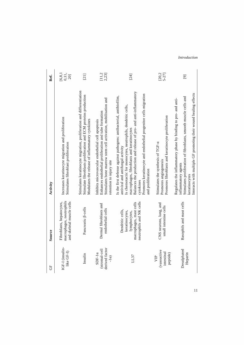
Table 1. Summary of the main activities and sources of GFs and other endogenous molecules involved in wound healing.
Ref
.
[15-
17]
[10,
15,1
6]
[6,1
0,14
,18]
[6,1
0,16
,19]
[8,1
0,11
]
Act
ivity
Indu
ces
the
rele
ase
of c
olla
gena
se a
nd p
lasm
inog
en a
ctiv
ator
St
imul
ates
fibr
obla
st p
rolif
erat
ion
and
diff
eren
tiatio
n Pr
omot
es e
ndot
helia
l cel
l and
ker
atin
ocyt
e m
igra
tion
and
prol
ifer
atio
n St
imul
ates
mac
roph
age
mig
ratio
n by
the
indu
ctio
n of
cyt
okin
e pr
oduc
tion
Reg
ulat
es th
e sy
nthe
sis
and
depo
sitio
n of
var
ious
ext
race
llula
r m
atrix
(EC
M) c
ompo
nent
s Pr
omot
es fi
brob
last
s an
d en
doth
elia
l cel
l mig
ratio
n an
d pr
olif
erat
ion
Incr
ease
s ke
ratin
ocyt
e m
igra
tion
durin
g re
epith
elis
atio
n
Prom
otes
ker
atin
ocyt
e an
d fib
robl
ast m
igra
tion
and
prol
ifer
atio
n St
imul
ates
the
prod
uctio
n of
pro
tein
s su
ch a
s fib
rone
ctin
In
crea
ses
the
expr
essi
on o
f ker
atin
s K
6 an
d K
16, i
nvol
ved
in th
e pr
olif
erat
ive
sign
alin
g ph
atw
ay
Prom
otes
end
othe
lial c
ell m
igra
tion,
pro
lifer
atio
n an
d di
ffer
entia
tion
(pot
ent a
ngio
geni
c fa
ctor
) In
duce
s va
scul
ar p
erm
eabi
lity
Indu
ces
lym
phan
giog
enes
is
Is c
hem
otac
tic fo
r neu
troph
ils, m
acro
phag
es, f
ibro
blas
ts a
nd
smoo
th m
uscl
e ce
lls
Stim
ulat
es m
acro
phag
es to
pro
duce
and
rele
ase
GFs
Is
invo
lved
in re
crui
ting
peric
ytes
to c
apill
arie
s, in
crea
sing
thei
r st
ruct
ural
inte
grity
En
hanc
es fi
brob
last
s pr
olife
ratio
n an
d in
duce
s th
e m
yofib
robl
ast
phen
otyp
e in
them
St
imul
ates
fibr
obla
sts
to p
rodu
ce E
CM
pro
tein
s an
d to
con
trac
t co
llage
n m
atrix
Sour
ce
Endo
thel
ial c
ells
and
m
acro
pagh
es
Cho
ndro
cyte
s,
fibro
blas
ts, e
ndot
helia
l ce
lls, k
erat
inoc
ytes
, m
acro
phag
es, m
ast c
ells
an
d sm
ooth
mus
cle
cells
Fibr
obla
sts,
mac
roph
ages
an
d pl
atel
ets
Endo
thel
ial c
ells
, fib
robl
asts
, ker
atin
ocyt
es,
mac
roph
ages
, ne
utro
phils
, pla
tele
ts a
nd
smoo
th m
uscl
e ce
lls
Endo
thel
ial c
ells
, fib
robl
asts
ker
atin
ocyt
es,
mac
roph
ages
and
pl
atel
ets
GF FG
F-1
(fib
robl
ast
GF-
1)
b-FG
F (b
asic
fib
robl
ast
GF)
EGF
(epi
derm
al
GF)
VEG
F (v
ascu
lar
endo
thel
ial
GF)
PDG
F (p
late
let
deriv
ed
GF)
Introduction
11
Ref
.
[6,8
,10,
11,
20]
[21]
[11,
22,
23]
[24]
[20,
25-
27]
[9]
Act
ivity
Incr
ease
s ke
ratin
ocyt
e m
igra
tion
and
prol
ifer
atio
n St
imul
ates
fibr
obla
st p
rolif
erat
ion
Stim
ulat
es k
erat
inoc
yte
mig
ratio
n, p
rolif
erat
ion
and
diff
eren
tiatio
n St
imul
ate
fibro
blas
ts p
rolif
erat
ion
and
ECM
pro
tein
s pr
oduc
tion
Mod
ulat
es th
e re
leas
e of
infla
mm
ator
y cy
toki
nes
Inhi
bits
mic
rova
scul
ar e
ndot
helia
l cel
l apo
ptos
is
Enha
nces
end
othe
lial p
rolif
erat
ion
and
tube
form
atio
n St
imul
ates
bon
e m
arro
w s
tem
cel
l act
ivat
ion,
mob
ilisa
tion
and
rete
ntio
n to
inju
ry s
ite
Is th
e fir
st d
efen
se a
gain
st p
atho
gens
: ant
ibac
teria
l, an
tibio
film
, an
tivira
l and
ant
ifung
al a
ctiv
ity
Is c
hem
otac
tic fo
r mon
ocyt
es, n
eutro
phils
, den
driti
c ce
lls,
mac
roph
ages
, fib
robl
asts
and
ker
atin
ocyt
es
Bal
ance
s th
e pr
oduc
tion
and
rele
ase
of p
ro- a
nd a
nti-i
nfla
mm
ator
y cy
toki
nes
Prom
otes
ker
atin
ocyt
e an
d en
doth
elia
l pro
geni
tor c
ells
mig
ratio
n an
d pr
olife
ratio
n
Stim
ulat
es th
e sy
nthe
sis
of T
GF-
α Pr
omot
es a
ngio
gene
sis
Prom
otes
fibr
obla
sts
and
kera
tinoc
yte
prol
ifer
atio
n
Reg
ulat
es th
e in
flam
mat
ory
phas
e by
bin
ding
to p
ro- a
nd a
nti-
infl
amm
ator
y ag
ents
St
imul
ates
pro
lifer
atio
n of
fibr
obla
sts,
sm
ooth
mus
cle
cells
and
ke
ratin
ocyt
es
Inte
ract
s w
ith m
ultip
le G
F pr
omot
ing
thei
r wou
nd h
ealin
g ef
fect
s
Sour
ce
Fibr
obla
sts,
hep
atoc
ytes
, m
acro
phag
es, n
eutro
phils
an
d sk
elet
al m
uscl
e ce
lls
Panc
reat
ic β
-cel
ls
Der
mal
fibr
obla
sts
and
endo
thel
ial c
ells
Den
driti
c ce
lls,
kera
tinoc
ytes
, ly
mph
ocyt
es,
mac
roph
ages
, mas
t cel
ls
neut
roph
ils a
nd N
K c
ells
CN
S ne
uron
s, lu
ng, a
nd
smal
l int
estin
e ce
lls
Bas
ophi
ls a
nd m
ast c
ells
GF
IGF-
I (in
sulin
-lik
e G
F-I)
Insu
lin
SDF-
1α
(stro
mal
-cel
l de
rived
fact
or
-1α)
LL37
VIP
(v
asoa
ctiv
e in
test
inal
pe
ptid
e)
Des
ulph
ated
H
epar
in

Novel therapeutic approaches for wound healing
12
Ref
.
[28]
[29,
30]
[31,
32]
[33,
34]
Act
ivity
Rel
ease
s in
flam
mat
ory
med
iato
rs
Is in
volv
ed in
ang
ioge
nesi
s an
d ce
ll pr
olif
erat
ion
and
mig
ratio
n St
imul
ates
the
accu
mul
atio
n of
col
lage
n an
d gl
ycos
amin
ogly
cans
(G
AG
s) a
t the
wou
nd s
ite
Prom
otes
ker
atin
ocyt
e m
igra
tion
Inhi
bits
ela
stas
e ac
tivity
Pr
esen
ts p
oten
t ant
i-inf
lam
mat
ory
activ
ity
Lim
its th
e lo
cal a
ccum
ulat
ion
of g
ranu
locy
tes
and
mac
roph
ages
fo
llow
ing
vasc
ular
inju
ry
Endo
rses
re-e
pith
elis
atio
n St
imul
ates
the
secr
etio
n of
TG
F-β1
by
fibro
blas
ts
Incr
ease
s co
llage
n de
posi
tion
Sour
ce Pi
neal
gla
nd
Glia
l cel
ls
Prim
arily
in th
e liv
er b
ut a
lso
in in
test
inal
epi
thel
ial c
ells
, m
acro
phag
es, m
onoc
ytes
and
ne
utro
phils
Ova
ry
GF Mel
aton
in
Lipo
calin
-2
Serp
in-A
1
β-st
radi
ol

Introduction
13
from proteases presents in the wound bed,
and the same has been done with some
other endogenous molecules that regulate
wound healing. In the Table 1, there are
summarised the GFs and the other active
molecules used in DDSs, their sources and
their main wound healing activities.
2. Biomaterials for the development of
nanotechnology based DDSs for wound
healing
A wide variety of biomaterials have
been used for developing novel DDSs, and
all of them are expected to meet a series of
characteristics, such as, good stability, bi-
ocompatibility and biodegradability, high
drug-loading, good mechanical properties,
controlled or sustained release and drug
protection [6,35].
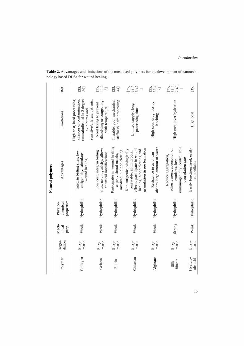
Polymers, either natural or synthetic,
have been widely used for the develop-
ment of nanofibers, micro- and nanoparti-
cles (MP/NP) and the advantages and limi-
tations of the most used ones are listed in
Table 2. Natural polymers include algi-
nate, gelatin, fibrin, chitosan, collagen,
hyaluronic acid, etc. They have been
broadly used because they have a great bi-
ocompatibility due to their similarity to
macromolecules recognised by the human
body. Moreover, some of them are in-
volved in the repair of damaged tissue and
have cell-binding sites and biomolecular
signatures, which give an added value to
their use in DDSs for wound healing [36].
Nevertheless, they show some sub-optimal
characteristics, including, batch to batch
differentiation, susceptibility to cross-con-
tamination, immunogenicity, presence of
immunogenic/pathogenic sections and
high price. In addition, regarding the pos-
sibility of developing nanofibrous mem-
branes, their poor mechanical properties
hinder the electrospinning process and the
handling of the obtained nanofibers [35].
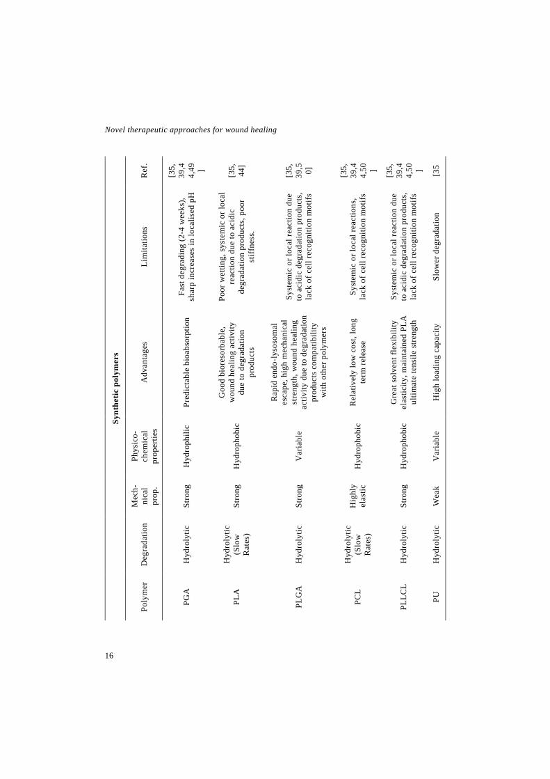
For that purpose, synthetic polymers
exhibit better mechanical properties, being
more easily electrospinnable. The rest of
the advantages of synthetic polymers over
naturals are reliability, easily controlled
physicochemical properties, lower price,
well-defined structures and degradation
kinetics [37]. However, they lack good
cell-recognition sites and have poor affin-
ity for cell attachment. Among the most

Novel therapeutic approaches for wound healing
14
used synthetic polymers there are polylac-
tic acid (PLA), poly(ε-caprolactone)
(PCL), polyglicolic acid (PGA) and their
combinations (PLGA and PLLCL) [35,38].
An approach to improve polymers´
characteristics is to develop DDSs that in-
clude both synthetic and natural polymers,
in order to obtain a formulation that can
take advantage of the strengths, bioactivi-
ties and degradation rates of all the com-
ponents [39].
Lipids used to develop lipid nanoparti-
cles need to exhibit similar characteristics
as polymers, namely, biocompatibility and
biodegradability, controlled release, tar-
geted drug delivery, high drug loading and
drug protection. In addition, they partially
fluidize skin lipids and increase drug par-
titioning, thereby facilitating drug
transport [40]. Depending on the DDS
type, different lipids are used in their de-
velopment. For instance, liposomes are
typically composed of phospholipids, cho-
lesterol and an aqueous medium. Phospho-
lipids are the major components of lipo-
somes and they are fluid at skin tempera-
ture. Among them, natural phosphatidyl-
cholines are the most used ones due to tox-
icological and price concerns. Cholesterol
is added to impart rigidity to the lipid bi-
layer, however it can reduce the encapsu-
lation efficiency of hydrophilic drugs and
can impact negatively in the permeation
through the skin [40,41]. On the other
hand, solid lipid nanoparticles (SLNs) are
prepared using various solid lipids, such
as, mono-, di- and triglycerides, fatty ac-
ids, waxes, and steroids. Moreover, nu-
merous surfactants are used to enhance
sterical stabilisation, including phospho-
lipids, poloxamers and polysorbates [42].
Finally, nanostructured lipid carriers
(NLCs), besides of a solid lipid, contain a
lipid liquid at room temperature, usually
up to 30% of the content [43].
3. Nanotechnology-based delivery sys-
tems for wound healing
3.1 Polymeric micro and nanoparticles
The encapsulation of peptides and pro-
teins in polymeric colloidal systems has
been widely used to overcome some of the
Introduction
15
Table 2. Advantages and limitations of the most used polymers for the development of nanotech-nology based DDSs for wound healing.
Nat
ural
pol
ymer
s
Ref
.
[35,
39]
[35,
44,4 5]
[35,
44]
[35,
39,4
6,47 ] [35,
39,4 7]
[35,
39,4
7,48 ]
[35]
Lim
itatio
ns
Hig
h co
st, h
ard
proc
essi
ng,
chan
ces
of c
onta
min
atio
n,
cont
rain
dica
ted
in 3
deg
ree
skin
bur
ns a
nd
sens
itive
/alle
rgic
pat
ient
s.
Nee
d fix
ing
to p
reve
nt
diss
olvi
ng o
r con
geal
ing
with
tem
pera
ture
Inst
able
, poo
r mec
hani
cal
stiff
ness
, har
d pr
oces
sing
Lim
ited
supp
ly, l
ong
proc
essi
ng ti
me
Hig
h co
st, d
rug
loss
by
leac
hing
Hig
h co
st, o
ver h
ydra
tion
Hig
h co
st
Adv
anta
ges
Inte
grin
bid
ing
site
s, lo
w
antig
enic
ity, s
timul
ates
w
ound
hea
ling
Low
cos
t, in
tegr
in b
idin
g si
tes,
no
antig
enic
ity, a
llow
s ch
emic
al m
odifi
catio
ns
Parti
cipa
tes
in w
ound
hea
ling
as p
rovi
sion
al m
atrix
, in
volv
ed in
blo
od c
lotti
ng
Non
ant
igen
ic, b
iolo
gica
lly
rene
wab
le, a
ntim
icro
bial
ef
fect
s, p
artic
ipat
e in
wou
nd
heal
ing:
blo
od c
lotti
ng a
nd
gran
ulat
ion
tissu
e fo
rmat
ion
Res
ista
nce
to a
cid,
can
ab
sorb
larg
e am
ount
of w
ater
Red
uce
aggr
egat
ion,
ad
hesi
vene
ss, a
bsor
ptio
n of
ex
udat
es, l
ow
imm
unog
enic
ity, c
ontro
llabl
e de
grad
atio
n ra
te
Easi
ly fu
nctio
nalis
ed, e
asily
co
ntro
llabl
e
Phys
ico-
chem
ical
pr
oper
ties
Hyd
roph
ilic
Hyd
roph
ilic
Hyd
roph
ilic
Hyd
roph
ilic
Hyd
roph
ilic
Hyd
roph
ilic
Hyd
roph
ilic
Mec
h-
nica
l pr
op.
Wea
k
Wea
k
Wea
k
Wea
k
Wea
k
Stro
ng
Wea
k
Deg
ra-
datio
n
Enzy
-m
atic
Enzy
-m
atic
Enzy
-m
atic
Enzy
-m
atic
Enzy
-m
atic
Enzy
-m
atic
Enzy
-m
atic
Poly
mer
Col
lage
n
Gel
atin
Fibr
in
Chi
tosa
n
Alg
inat
e
Silk
fib
roin
Hya
luro
-ni
c ac
id
Novel therapeutic approaches for wound healing
16
Synt
hetic
pol
ymer
s
Ref
.
[35,
39,4
4,49 ] [35,
44]
[35,
39,5 0]
[35,
39,4
4,50 ] [35,
39,4
4,50 ] [35
Lim
itatio
ns
Fast
deg
radi
ng (2
-4 w
eeks
), sh
arp
incr
ease
s in
loca
lised
pH
Poor
wet
ting,
sys
tem
ic o
r loc
al
reac
tion
due
to a
cidi
c de
grad
atio
n pr
oduc
ts, p
oor
stiff
ness
.
Syst
emic
or l
ocal
reac
tion
due
to a
cidi
c de
grad
atio
n pr
oduc
ts,
lack
of c
ell r
ecog
nitio
n m
otif
s
Syst
emic
or l
ocal
reac
tions
, la
ck o
f cel
l rec
ogni
tion
mot
ifs
Syst
emic
or l
ocal
reac
tion
due
to a
cidi
c de
grad
atio
n pr
oduc
ts,
lack
of c
ell r
ecog
nitio
n m
otif
s
Slow
er d
egra
datio
n
Adv
anta
ges
Pred
icta
ble
bioa
bsor
ptio
n
Goo
d bi
ores
orba
ble,
w
ound
hea
ling
activ
ity
due
to d
egra
datio
n pr
oduc
ts
Rap
id e
ndo-
lyso
som
al
esca
pe, h
igh
mec
hani
cal
stre
ngth
, wou
nd h
ealin
g ac
tivity
due
to d
egra
datio
n pr
oduc
ts c
ompa
tibili
ty
with
oth
er p
olym
ers
Rel
ativ
ely
low
cos
t, lo
ng
term
rele
ase
Gre
at s
olve
nt fl
exib
ility
el
astic
ity, m
aint
aine
d PL
A
ultim
ate
tens
ile s
treng
th
Hig
h lo
adin
g ca
paci
ty
Phys
ico-
chem
ical
pr
oper
ties
Hyd
roph
ilic
Hyd
roph
obic
Var
iabl
e
Hyd
roph
obic
Hyd
roph
obic
Var
iabl
e
Mec
h-
nica
l pr
op.
Stro
ng
Stro
ng
Stro
ng
Hig
hly
elas
tic
Stro
ng
Wea
k
Deg
rada
tion
Hyd
roly
tic
Hyd
roly
tic
(Slo
w
Rat
es)
Hyd
roly
tic
Hyd
roly
tic
(Slo
w
Rat
es)
Hyd
roly
tic
Hyd
roly
tic
Poly
mer
PGA
PLA
PLG
A
PCL
PLLC
L
PU

Introduction
17
issues related to the administration of pro-
teins for wound healing. For instance, in
vivo half-life is highly improved after en-
capsulation, due to the protective effect of
the MPs and NPs against the proteases pre-
sent in the wound bed. In addition, they
provide a controlled release of the encap-
sulated compound, avoiding the need of
frequent administration, and in some
cases, allowing a reduction of the dose.
That, jointly with the ability to enable a lo-
cal administration, improves treatment ef-
ficacy, avoiding the secondary effects
caused by high doses or systemic exposure
[6,51,52]. At the end of this section, Table
3 provides a summary of the polymeric
MP/NPs used for wound healing applica-
tions.
bFGF has been extensively incorpo-
rated into MPs for wound healing. For in-
stance, in a study conducted by Liu et al.
alginate microspheres loaded with bFGF
were incorporated into a carboxymethyl
chitosan-polyvinil alcohol (PVA) compo-
site hydrogel. The resulting formulation
achieved a faster wound recovery rate with
higher reepithelisation and regeneration of
the dermis than the hydrogel and the
hydrogel loaded with free bFGF [53]. In
addition, Place et al. developed a formula-
tion to deliver bFGF based in polyelectro-
lyte complex NPs containing cationic pol-
ysaccharides (chitosan and N,N,N-trime-
thyl chitosan) and anionic GAGs (heparin
and chondroitin sulfate). Those NPs were
designed to mimic aggrecan, a proteogly-
can that serves as reservoir for GFs physi-
ologically. Therefore, the bFGF, besides
of being stabilised against proteases, was
presented in a biomimetic context that fa-
vored it interaction with the surrounding
tissue. In vitro experiments demonstrated
the capability of the formulations to im-
prove bFGF administration, since they en-
hanced proliferation and metabolic activ-
ity of marrow stromal cells in comparison
to bFGF alone and bFGF bound to aggre-
can [54].
Li and colleagues encapsulated bFGF
into gelatin MPs because the electrostatic
binding between them can enhance the
protective effect. The particles were
loaded into a porous collagen/cellulose
nanocrystals scaffold, which proved to en-
hance angiogenesis in vitro and in vivo
[55]. Wound dressings containing bFGF

Novel therapeutic approaches for wound healing
18
loaded gelatin microspheres were further
studied by Park et al., who incorporated
them into a porous chitosan scaffold. The
efficacy of the dressing was proved in vivo
in pressure ulcers inflicted to aged mice,
and the results showed a higher wound
closure rate. Moreover, those animals pre-
sented lower protease levels due to chi-
tosan, and thus higher exogenous or en-
dogenous bFGF levels [56]. In another
study, Kawai and colleagues incorporated
bFGF containing gelatin MPs into a bi-
layer commercial artificial dermis
(Pelnac™) composed of an inner collagen
sponge and an outer silicon layer. The for-
mulation efficacy was assessed in two an-
imal models, a full thickness excisionals
wound in guinea pigs and a pressure-in-
duced decubitus ulcer in genetically dia-
betic mice. In both cases, the formulation
accelerated fibroblast proliferation and ca-
pillary formation. Moreover, diabetic mice
receiving bFGF containing gelatin MP
showed an enhanced resistance against in-
fection [57,58].
Huang and collaborators developed
another bilayer wound dressing, enclosing
bFGF containing gelatin MPs into a bi-
layer scaffold composed of a gelatin
sponge inner layer, which porous structure
serves as a host for proliferation cells, and
an elastomeric polyurethane (PU) mem-
brane external layer, which improves the
mechanical properties of the scaffold. The
application of the dressing into full thick-
ness wounds in York pigs led to a faster
wound closure and better skin remodeling
[59]. Ulubayram et al. replaced the bFGF
with EGF in the gelatin MPs of the bilayer
dressing and they evaluated in vivo in an
excisional wound model in rabbits. In the
higher dose tested, a greater area reduction
was observed in wounds treated with the
dressing than in wounds treated with EGF
solution. Moreover, the histological find-
ings showed that the newly formed tissue
had almost the structure of native skin
[60]. Later, they analysed the efficacy of
the gelatin sponge loaded with EGF con-
taining MPs in excisional wounds in nor-
mal and streptozotocin (STZ)-induced di-
abetic rats. Wound closure was specially
enhanced in non diabetic rats although a
slight improvement was observed in dia-
betic rats. In regard to histological anal-

Introduction
19
yses, the effect of the dressing was signif-
icantly different between normal and dia-
betic rats, but overall it improved granula-
tion tissue formation and reepithelisation
[61]. In addition, Zhou and colleagues en-
capsulated EGF in other natural polymer,
concretely in chitosan. The chitosan NPs
containing EGF were incorporated into a
fibrin gel that exhibited a sustained release
of the factor, maintaining its ability to
stimulate fibroblast proliferation for at
least 7 days [62]. EGF has also been en-
capsulated in synthetic MPs such as
PLGA, achieving an improved growth rate
of fibroblasts in vitro [63]. In another
study, EGF loaded into PLGA NPs exhib-
ited an accelerated wound closure in full
thickness wounds inflicted to diabetic rats,
because the EGF released from the parti-
cles promoted the highest level of fibro-
blast proliferation [64]. In a posterior
study conducted by our research group,
EGF was loaded into PLGA-alginate MPs.
Alginate was added with the aim of im-
proving the encapsulation efficiency by in-
creasing the viscosity of the internal aque-
ous phase and thus limiting the diffusion
of the GF from the MPs. In vitro, the MPs
demonstrated that the EGF remained ac-
tive after the encapsulation process. More-
over in vivo, they induced a faster and
more effective wound healing, in terms of
wound closure, reepithelisation and reso-
lution of the inflammatory process, after a
single intralesional injection into full
thickness wounds inflicted to STZ-in-
duced diabetic rats [65].
Additionally, PLGA has been used to
encapsulate other active compounds, such
as FGF-1, LL37 and insulin. FGF-1 loaded
MPs were embedded into a fibrin gel and
they improved fibroblast proliferation in
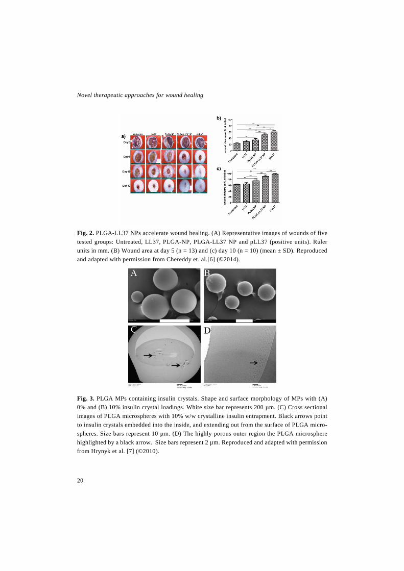
vitro [66]. On the other hand, LL37 loaded
MPs were applied in full thickness
wounds, where they produced an improve-
ment in granulation tissue formation, reep-
ithelisation, collagen deposition and neo-
angiogenesis as shown in Fig. 2 [67]. Re-
garding to the encapsulation of insulin in
PLGA (Fig. 3), it showed an enhancement
in wound closure in a keratinocyte migra-
tion assay [68]. Later, the insulin loaded
MPs were incorporated into alginate or al-
ginate-polyethylene glycol (PEG) sponges,
which did not alter their bioactivity [69].
Novel therapeutic approaches for wound healing
20
Fig. 2. PLGA-LL37 NPs accelerate wound healing. (A) Representative images of wounds of five tested groups: Untreated, LL37, PLGA-NP, PLGA-LL37 NP and pLL37 (positive units). Ruler units in mm. (B) Wound area at day 5 (n = 13) and (c) day 10 (n = 10) (mean ± SD). Reproduced and adapted with permission from Chereddy et. al.[6] (©2014).
Fig. 3. PLGA MPs containing insulin crystals. Shape and surface morphology of MPs with (A) 0% and (B) 10% insulin crystal loadings. White size bar represents 200 µm. (C) Cross sectional images of PLGA microspheres with 10% w/w crystalline insulin entrapment. Black arrows point to insulin crystals embedded into the inside, and extending out from the surface of PLGA micro-spheres. Size bars represent 10 µm. (D) The highly porous outer region the PLGA microsphere highlighted by a black arrow. Size bars represent 2 µm. Reproduced and adapted with permission from Hrynyk et al. [7] (©2010).
Introduction
21
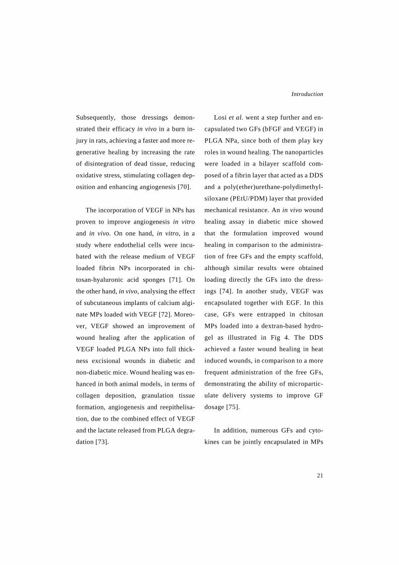
Subsequently, those dressings demon-
strated their efficacy in vivo in a burn in-
jury in rats, achieving a faster and more re-
generative healing by increasing the rate
of disintegration of dead tissue, reducing
oxidative stress, stimulating collagen dep-
osition and enhancing angiogenesis [70].
The incorporation of VEGF in NPs has
proven to improve angiogenesis in vitro
and in vivo. On one hand, in vitro, in a
study where endothelial cells were incu-
bated with the release medium of VEGF
loaded fibrin NPs incorporated in chi-
tosan-hyaluronic acid sponges [71]. On
the other hand, in vivo, analysing the effect
of subcutaneous implants of calcium algi-
nate MPs loaded with VEGF [72]. Moreo-
ver, VEGF showed an improvement of
wound healing after the application of
VEGF loaded PLGA NPs into full thick-
ness excisional wounds in diabetic and
non-diabetic mice. Wound healing was en-
hanced in both animal models, in terms of
collagen deposition, granulation tissue
formation, angiogenesis and reepithelisa-
tion, due to the combined effect of VEGF
and the lactate released from PLGA degra-
dation [73].
Losi et al. went a step further and en-
capsulated two GFs (bFGF and VEGF) in
PLGA NPa, since both of them play key
roles in wound healing. The nanoparticles
were loaded in a bilayer scaffold com-
posed of a fibrin layer that acted as a DDS
and a poly(ether)urethane-polydimethyl-
siloxane (PEtU/PDM) layer that provided
mechanical resistance. An in vivo wound
healing assay in diabetic mice showed
that the formulation improved wound
healing in comparison to the administra-
tion of free GFs and the empty scaffold,
although similar results were obtained
loading directly the GFs into the dress-
ings [74]. In another study, VEGF was
encapsulated together with EGF. In this
case, GFs were entrapped in chitosan
MPs loaded into a dextran-based hydro-
gel as illustrated in Fig 4. The DDS
achieved a faster wound healing in heat
induced wounds, in comparison to a more
frequent administration of the free GFs,
demonstrating the ability of micropartic-
ulate delivery systems to improve GF
dosage [75].
In addition, numerous GFs and cyto-
kines can be jointly encapsulated in MPs

Novel therapeutic approaches for wound healing
22
and NPs using platelets lysate or platelet
rich plasma (PRP). Fontana et al. loaded
platelet lysate into porous silicon MPs,
achieving promising results in a wound
model ex vivo: a greater proliferative ef-
fect than the positive control (platelet ly-
sate as in particles) and an effective, but
short lasting acidophilia of the collagen fi-
bres, sign of ongoing regeneration [76]. A
strategy to achieve a more sustained re-
lease of the GFs from PRP or plasma ly-
sate is to produce NPs containing heparin,
since it binds to them acting as a GF reser-
voir. In that regard, fragmin-protamine
MP/NPs containing PRP were developed,
since protamine forms water insoluble
complexes with the low molecular weight
heparin (fragmin). Those particles pro-
vided a faster wound closure and an en-
hanced angiogenesis in split thickness skin
graft donor site wounds in rats [77]. Those
results were in agreement with the ones
obtained by La and colleagues, who also
observed a faster wound closure and an
improved angiogenesis in large wounds
inflicted to athymic mice; after the admin-
istration of PRP into PLGA NPs conju-
gated with heparin and incorporated into a
fibrin gel [78].
Heparin, besides of stabilizing GFs has
anti-inflammatory activity and it can be
useful in burn wounds. For that purpose,
Lakshmi and collaborators developed a
DDS consisting of chitosan MPs loaded
with desulphated heparin embedded into a
collagen matrix. The DDSs were effective
to regulate inflammatory events in burn
wounds in rats, thereby achieving a faster
wound closure and granulation tissue for-
mation [79].
Fig. 4. Chitosan MPs containing EGF and VEGF, loaded into a dextran-based hydrogel. (A) SEM images of the chitosan MPs 400x. (B) Images of dextran hydrogel with microparti-cles incorporated. Reproduced and adapted with permission from Ribeiro et al. [8] (©2013).
Introduction
23
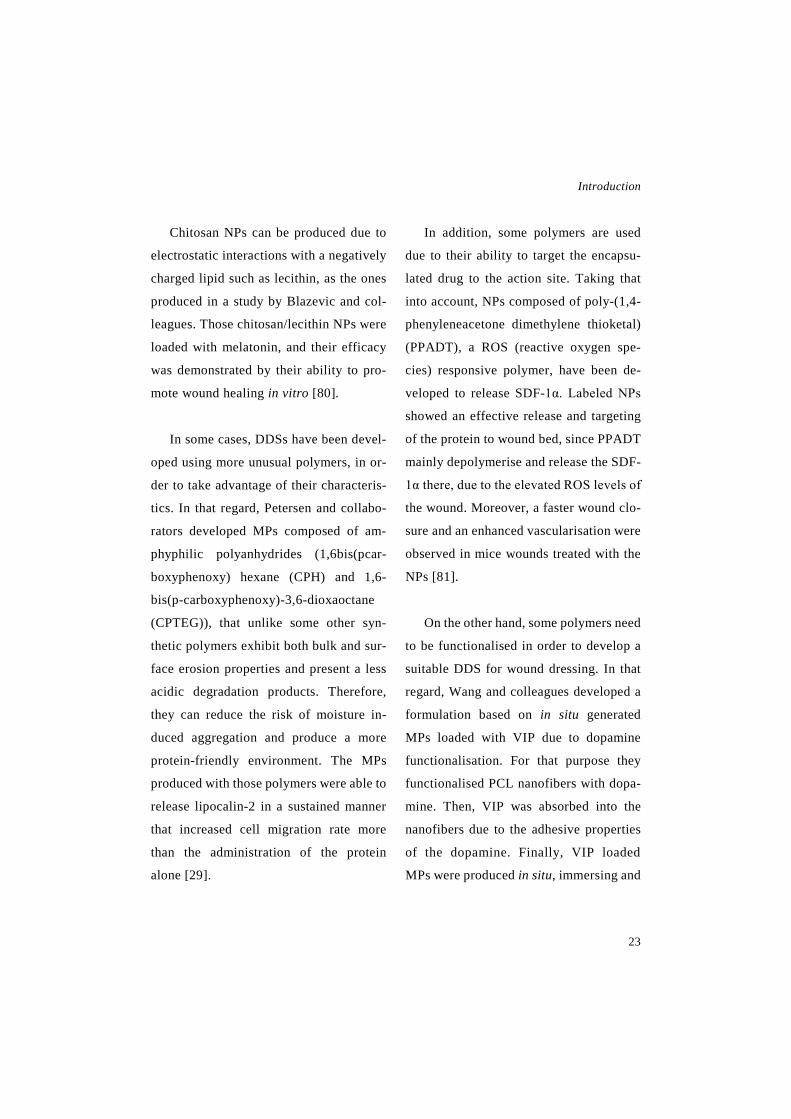
Chitosan NPs can be produced due to
electrostatic interactions with a negatively
charged lipid such as lecithin, as the ones
produced in a study by Blazevic and col-
leagues. Those chitosan/lecithin NPs were
loaded with melatonin, and their efficacy
was demonstrated by their ability to pro-
mote wound healing in vitro [80].
In some cases, DDSs have been devel-
oped using more unusual polymers, in or-
der to take advantage of their characteris-
tics. In that regard, Petersen and collabo-
rators developed MPs composed of am-
phyphilic polyanhydrides (1,6bis(pcar-
boxyphenoxy) hexane (CPH) and 1,6-
bis(p-carboxyphenoxy)-3,6-dioxaoctane
(CPTEG)), that unlike some other syn-
thetic polymers exhibit both bulk and sur-
face erosion properties and present a less
acidic degradation products. Therefore,
they can reduce the risk of moisture in-
duced aggregation and produce a more
protein-friendly environment. The MPs
produced with those polymers were able to
release lipocalin-2 in a sustained manner
that increased cell migration rate more
than the administration of the protein
alone [29].
In addition, some polymers are used
due to their ability to target the encapsu-
lated drug to the action site. Taking that
into account, NPs composed of poly-(1,4-
phenyleneacetone dimethylene thioketal)
(PPADT), a ROS (reactive oxygen spe-
cies) responsive polymer, have been de-
veloped to release SDF-1α. Labeled NPs
showed an effective release and targeting
of the protein to wound bed, since PPADT
mainly depolymerise and release the SDF-
1α there, due to the elevated ROS levels of
the wound. Moreover, a faster wound clo-
sure and an enhanced vascularisation were
observed in mice wounds treated with the
NPs [81].
On the other hand, some polymers need
to be functionalised in order to develop a
suitable DDS for wound dressing. In that
regard, Wang and colleagues developed a
formulation based on in situ generated
MPs loaded with VIP due to dopamine
functionalisation. For that purpose they
functionalised PCL nanofibers with dopa-
mine. Then, VIP was absorbed into the
nanofibers due to the adhesive properties
of the dopamine. Finally, VIP loaded
MPs were produced in situ, immersing and

Novel therapeutic approaches for wound healing
24
Table 3. Summary of different MP/NPs developed to release GFs and other active compounds and their main outcomes
Ref
.
[53]
[54]
[55]
[56]
[57,
58]
[59]
[60]
[61]
Res
ults
The
form
ulat
ion
achi
eved
a fa
ster
wou
nd re
cove
ry ra
te w
ith a
hig
her
reep
ithel
isat
ion
and
rege
nera
tion
of th
e de
rmis
in a
full
thic
knes
s bu
rn w
ound
in ra
ts.
The
form
ulat
ion
mai
ntai
ned
bFG
F ac
tivity
in v
itro,
in m
etab
olic
and
pr
olif
erat
ion
activ
ity a
ssay
s.
An
incr
emen
t of c
ell p
rolif
erat
ion
in e
ndot
helia
l cel
ls w
as a
chie
ved
with
the
DD
S. A
nd a
fter b
eing
sub
cuta
neou
sly
impl
ante
d in
to ra
ts it
en
hanc
ed a
ngio
gene
sis
in v
ivo.
Unt
il da
y 7
the
scaf
fold
and
the
scaf
fold
with
bFG
F ac
cele
rate
d w
ound
clo
sure
in p
ress
ure
ulce
r in
aged
mic
e m
odel
s. A
nd w
ith th
e bF
GF
scaf
fold
the
leve
ls o
f pro
teas
es d
ecre
ased
by
day
10.
A p
rolo
nged
in v
ivo
rete
ntio
n of
the
bFG
F in
to th
e ar
tific
ial d
erm
is
was
ach
ieve
d w
ith th
e M
Ps. I
n bo
th, a
full
thic
knes
s ex
cisi
onal
w
ound
mod
el in
gui
nea
pigs
and
a p
ress
ure
indu
ced
decu
bitu
s ul
cer
mod
el in
db/
db m
ice,
the
treat
ed g
roup
pre
sent
ed a
hig
her f
ibro
blas
t pr
olif
erat
ion
and
capi
llary
form
atio
n in
a d
ose-
depe
nden
t way
.
The
scaf
fold
s le
d to
a fa
ster
wou
nd c
losu
re a
nd b
ette
r wou
nd
rem
odel
ing
in a
full
thic
knes
s sk
in w
ound
in Y
ork
pigs
.
With
the
high
er d
ose
test
ed th
e fo
rmul
atio
n ac
hiev
ed a
gre
ater
are
a re
duct
ion
in a
full
thic
knes
s ex
cisi
onal
wou
nd in
rabb
its. T
he
hist
olog
ical
find
ings
sho
wed
that
the
new
ly fo
rmed
tiss
ue w
as
alm
ost a
s no
rmal
ski
n.
The
form
ulat
ion
was
ana
lyse
d in
a fu
ll th
ickn
ess
wou
nd m
odel
in
diab
etic
and
non
-dia
betic
rats
. The
wou
nd c
losu
re w
as e
spec
ially
im
prov
ed in
non
dia
betic
rats
trea
ted
with
the
form
ulat
ion.
In b
oth
diab
etic
and
non
-dia
betic
rats
, the
form
ulat
ion
impr
oved
the
hist
olog
ical
sco
res
(ree
pith
elis
atio
n, g
ranu
latio
n tis
sue
and
neov
ascu
laris
atio
n).
GF
bFG
F
bFG
F
bFG
F
bFG
F
bFG
F
bFG
F
EGF
EGF
DD
Ss
Alg
inat
e M
Ps in
corp
orat
ed to
a
carb
oxym
ethy
l chi
tosa
n-PV
A h
ydro
gel
Agg
reca
n m
imet
ic
poly
elec
troly
te c
ompl
ex N
Ps
Gel
atin
MPs
into
a p
orou
s co
llage
n/ce
llulo
se
nano
crys
tals
sca
ffol
d
Gel
atin
MP
in a
por
ous
chito
san
scaf
fold
Gel
atin
MPs
into
an
artif
icia
l de
rmis
(Pel
nac®
)
Gel
atin
MPs
load
ed in
to a
bi
laye
r sca
ffol
d (g
elat
in
spon
ge a
nd P
U m
embr
ane)
Gel
atin
MPs
in a
bila
yer
dres
sing
(gel
atin
spo
nge
+ PU
la
yer)
Gel
atin
MPs
in g
elat
in
spon
ges
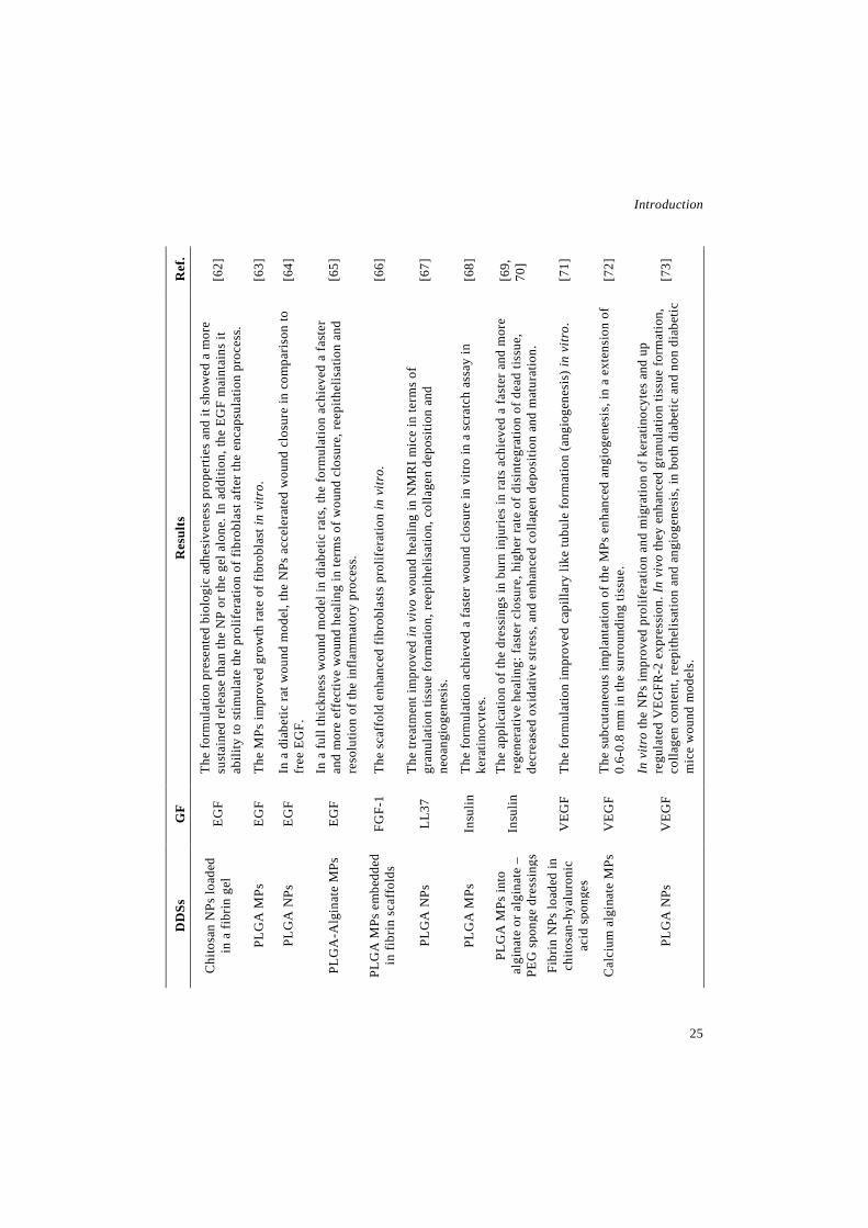
Introduction
25
Ref
.
[62]
[63]
[64]
[65]
[66]
[67]
[68]
[69,
70]
[71]
[72]
[73]
Res
ults
The
form
ulat
ion
pres
ente
d bi
olog
ic a
dhes
iven
ess
prop
ertie
s an
d it
show
ed a
mor
e su
stai
ned
rele
ase
than
the
NP
or th
e ge
l alo
ne. I
n ad
ditio
n, th
e EG
F m
aint
ains
it
abili
ty to
stim
ulat
e th
e pr
olif
erat
ion
of fi
brob
last
aft
er th
e en
caps
ulat
ion
proc
ess.
The
MPs
impr
oved
gro
wth
rate
of f
ibro
blas
t in
vitr
o.
In a
dia
betic
rat w
ound
mod
el, t
he N
Ps a
ccel
erat
ed w
ound
clo
sure
in c
ompa
rison
to
free
EG
F.
In a
full
thic
knes
s w
ound
mod
el in
dia
betic
rats
, the
form
ulat
ion
achi
eved
a fa
ster
an
d m
ore
effe
ctiv
e w
ound
hea
ling
in te
rms
of w
ound
clo
sure
, ree
pith
elis
atio
n an
d re
solu
tion
of th
e in
flam
mat
ory
proc
ess.
The
scaf
fold
enh
ance
d fib
robl
asts
pro
lifer
atio
n in
vitr
o.
The
treat
men
t im
prov
ed in
viv
o w
ound
hea
ling
in N
MR
I mic
e in
term
s of
gr
anul
atio
n tis
sue
form
atio
n, re
epith
elis
atio
n, c
olla
gen
depo
sitio
n an
d ne
oang
ioge
nesi
s.
The
form
ulat
ion
achi
eved
a fa
ster
wou
nd c
losu
re in
vitr
o in
a s
crat
ch a
ssay
in
kera
tinoc
ytes
. Th
e ap
plic
atio
n of
the
dres
sing
s in
bur
n in
juri
es in
rats
ach
ieve
d a
fast
er a
nd m
ore
rege
nera
tive
heal
ing:
fast
er c
losu
re, h
ighe
r rat
e of
dis
inte
grat
ion
of d
ead
tissu
e,
decr
ease
d ox
idat
ive
stre
ss, a
nd e
nhan
ced
colla
gen
depo
sitio
n an
d m
atur
atio
n.
The
form
ulat
ion
impr
oved
cap
illar
y lik
e tu
bule
form
atio
n (a
ngio
gene
sis)
in v
itro.
The
subc
utan
eous
impl
anta
tion
of th
e M
Ps e
nhan
ced
angi
ogen
esis
, in
a ex
tens
ion
of
0.6-
0.8
mm
in th
e su
rrou
ndin
g tis
sue.
In v
itro
the
NPs
impr
oved
pro
lifer
atio
n an
d m
igra
tion
of k
erat
inoc
ytes
and
up
regu
late
d V
EGFR
-2 e
xpre
ssio
n. In
viv
o th
ey e
nhan
ced
gran
ulat
ion
tissu
e fo
rmat
ion,
co
llage
n co
nten
t, re
epith
elis
atio
n an
d an
giog
enes
is, i
n bo
th d
iabe
tic a
nd n
on d
iabe
tic
mic
e w
ound
mod
els.
GF
EGF
EGF
EGF
EGF
FGF-
1
LL37
Insu
lin
Insu
lin
VEG
F
VEG
F
VEG
F
DD
Ss
Chi
tosa
n N
Ps lo
aded
in
a fi
brin
gel
PLG
A M
Ps
PLG
A N
Ps
PLG
A-A
lgin
ate
MPs
PLG
A M
Ps e
mbe
dded
in
fibr
in s
caff
olds
PLG
A N
Ps
PLG
A M
Ps
PLG
A M
Ps in
to
algi
nate
or a
lgin
ate
–PE
G s
pong
e dr
essi
ngs
Fibr
in N
Ps lo
aded
in
chito
san-
hyal
uron
ic
acid
spo
nges
Cal
cium
alg
inat
e M
Ps
PLG
A N
Ps

Novel therapeutic approaches for wound healing
26
Ref
.
[74]
[75]
[76]
[77]
[78]
[79]
[80]
[29]
[81]
[25]
Res
ults
The
adm
inis
tratio
n of
the
DD
S im
prov
ed w
ound
hea
ling
in a
full
thic
knes
s w
ound
mod
el in
dia
betic
mic
e, in
com
paris
on to
the
adm
inis
tratio
n of
the
free
GFs
, but
not
in c
ompa
rison
to th
e ad
min
istra
tion
of th
e G
Fs d
irec
tly in
to th
e sc
affo
lds.
A
fter
the
appl
icat
ion
of th
e hy
drog
el to
hea
t ind
uced
wou
nds
in ra
ts,
they
ach
ieve
d a
fast
er w
ound
hea
ling
in c
ompa
rison
to a
mor
e fr
eque
nt a
dmin
istr
atio
n of
the
free
GFs
. Th
e fo
rmul
atio
n im
prov
ed in
vitr
o w
ound
clo
sure
and
ex
vivo
it
enha
nced
wou
nd re
gene
ratio
n, a
s pr
oved
by
the
prol
ifera
tive
effe
ct
and
acid
ophi
lia o
bser
ved.
Th
e tre
atm
ent p
rom
otes
reep
ithel
isat
ion
and
angi
ogen
esis
in a
spl
it th
ickn
ess
skin
gra
ft do
nor s
ite w
ound
mod
el c
ondu
cted
in ra
ts.
The
wou
nds
trea
ted
with
the
form
ulat
ion
achi
eved
a fa
ster
wou
nd
clos
ure
and
an e
nhan
ced
angi
ogen
esis
in a
thym
ic m
ice.
The
treat
men
t app
lied
in b
urn
wou
nd in
juri
es w
as a
ble
to a
ccel
erat
e w
ound
clo
sure
, by
an e
arlie
r red
uctio
n of
the
infl
amm
atio
n an
d an
ac
cele
ratio
n of
the
gran
ulat
ion
tissu
e fo
rmat
ion.
In a
n in
vitr
o sc
ratc
h as
say
in H
aCaT
cel
ls th
e fo
rmul
atio
n im
prov
ed
cell
mig
ratio
n.
The
form
ulat
ion
incr
ease
d ce
ll m
igra
tion
mor
e th
an th
e ad
min
istra
tion
of th
e pr
otei
n al
one,
due
to s
usta
ined
rele
ase.
The
NPs
sho
wed
and
eff
ectiv
e re
leas
e an
d ta
rget
ing
to th
e w
ound
s,
due
to th
e hi
gh R
OS
cont
ent i
n th
e w
ound
bed
. In
addi
tion,
the
wou
nds
trea
ted
with
the
NPs
sho
wed
a fa
ster
wou
nd c
losu
re a
nd a
n en
hanc
ed v
ascu
lari
satio
n in
a m
ice
wou
nd m
odel
. Th
e de
velo
ped
DD
S si
gnifi
cant
ly p
rom
oted
wou
nd h
ealin
g fa
vour
ing
gran
ulat
ion
tissu
e fo
rmat
ion
and
angi
ogen
esis
in a
mic
e ex
cisi
onal
w
ound
mod
el.
GF
VEG
F an
d bF
GF
EGF
and
VEG
F
Plat
elet
ly
sate
PRP
PRP
Des
ulph
ated
he
parin
Mel
aton
in
Lipo
calin
-2
SDF-
1α
VIP
DD
Ss
PLG
A N
Ps lo
aded
in
PETu
/PD
M)/f
ibrin
bas
ed
scaf
fold
s
Chi
tosa
n M
Ps lo
aded
in a
de
xtra
n-ba
sed
hydr
ogel
Poro
us s
ilico
n (P
si) M
Ps
Frag
min
-pro
tam
ine
MP/
NPs
Hep
arin
con
juga
ted
PLG
A N
Ps in
a fi
brin
gel
Chi
tosa
n M
Ps e
mbe
dded
in
to a
col
lage
n m
atrix
Leci
thin
/chi
tosa
n N
Ps
Am
phyp
hilic
po
lyan
hidr
ide
MPs
bas
ed
on C
PH a
nd C
PTEG
PPA
DT
NPs
(a R
OS
reac
tive
nano
mat
eria
l)
In s
itu g
ener
ated
PC
L M
Ps in
to P
CL
nano
shee
ts
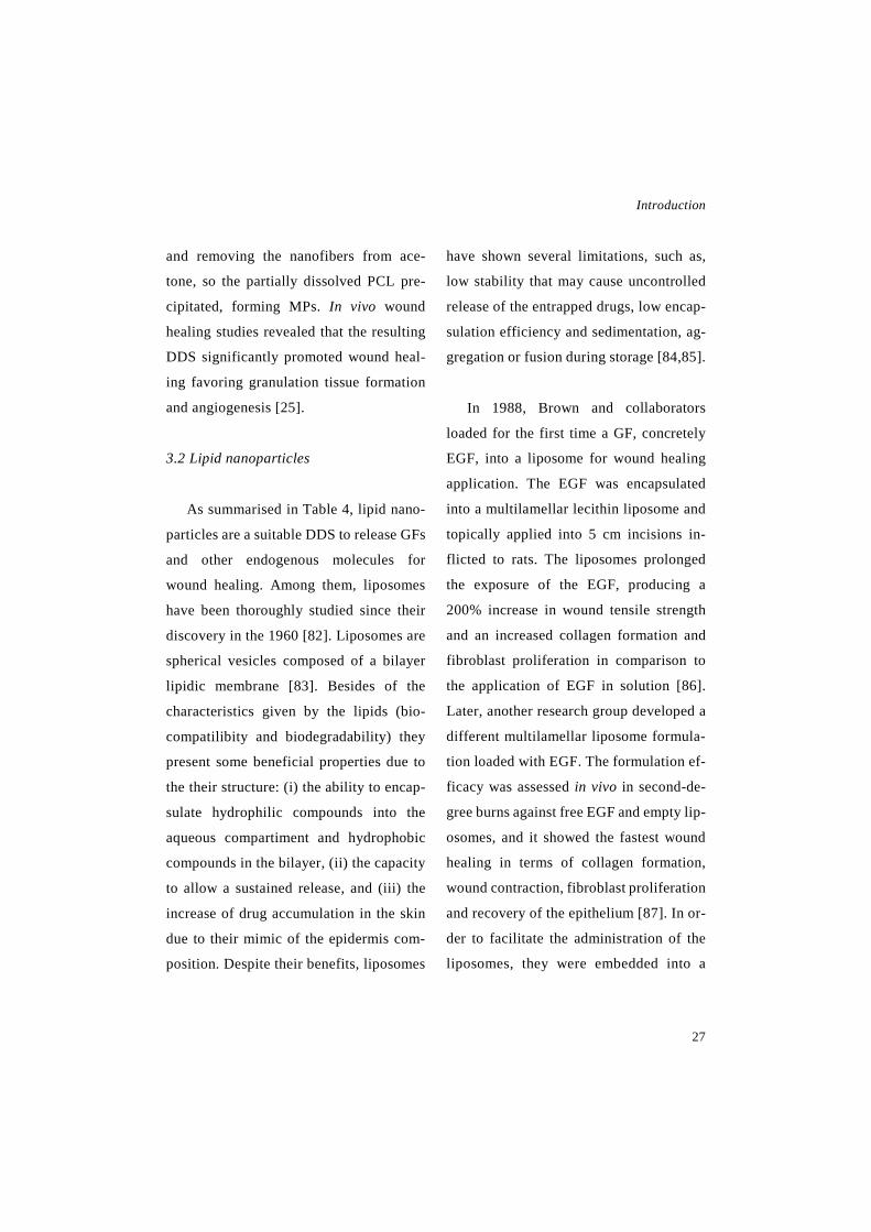
Introduction
27
and removing the nanofibers from ace-
tone, so the partially dissolved PCL pre-
cipitated, forming MPs. In vivo wound
healing studies revealed that the resulting
DDS significantly promoted wound heal-
ing favoring granulation tissue formation
and angiogenesis [25].
3.2 Lipid nanoparticles
As summarised in Table 4, lipid nano-
particles are a suitable DDS to release GFs
and other endogenous molecules for
wound healing. Among them, liposomes
have been thoroughly studied since their
discovery in the 1960 [82]. Liposomes are
spherical vesicles composed of a bilayer
lipidic membrane [83]. Besides of the
characteristics given by the lipids (bio-
compatilibity and biodegradability) they
present some beneficial properties due to
the their structure: (i) the ability to encap-
sulate hydrophilic compounds into the
aqueous compartiment and hydrophobic
compounds in the bilayer, (ii) the capacity
to allow a sustained release, and (iii) the
increase of drug accumulation in the skin
due to their mimic of the epidermis com-
position. Despite their benefits, liposomes
have shown several limitations, such as,
low stability that may cause uncontrolled
release of the entrapped drugs, low encap-
sulation efficiency and sedimentation, ag-
gregation or fusion during storage [84,85].
In 1988, Brown and collaborators
loaded for the first time a GF, concretely
EGF, into a liposome for wound healing
application. The EGF was encapsulated
into a multilamellar lecithin liposome and
topically applied into 5 cm incisions in-
flicted to rats. The liposomes prolonged
the exposure of the EGF, producing a
200% increase in wound tensile strength
and an increased collagen formation and
fibroblast proliferation in comparison to
the application of EGF in solution [86].
Later, another research group developed a
different multilamellar liposome formula-
tion loaded with EGF. The formulation ef-
ficacy was assessed in vivo in second-de-
gree burns against free EGF and empty lip-
osomes, and it showed the fastest wound
healing in terms of collagen formation,
wound contraction, fibroblast proliferation
and recovery of the epithelium [87]. In or-
der to facilitate the administration of the
liposomes, they were embedded into a
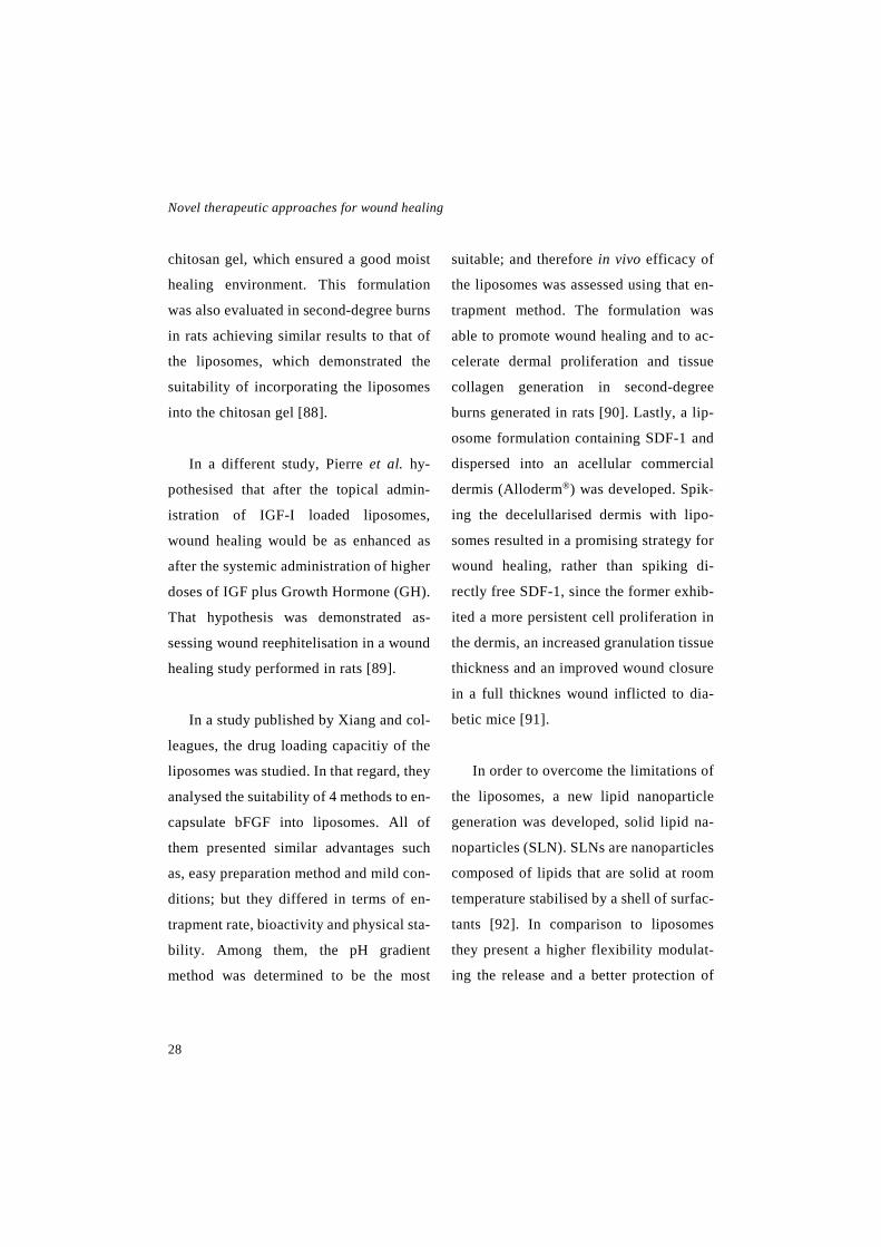
Novel therapeutic approaches for wound healing
28
chitosan gel, which ensured a good moist
healing environment. This formulation
was also evaluated in second-degree burns
in rats achieving similar results to that of
the liposomes, which demonstrated the
suitability of incorporating the liposomes
into the chitosan gel [88].
In a different study, Pierre et al. hy-
pothesised that after the topical admin-
istration of IGF-I loaded liposomes,
wound healing would be as enhanced as
after the systemic administration of higher
doses of IGF plus Growth Hormone (GH).
That hypothesis was demonstrated as-
sessing wound reephitelisation in a wound
healing study performed in rats [89].
In a study published by Xiang and col-
leagues, the drug loading capacitiy of the
liposomes was studied. In that regard, they
analysed the suitability of 4 methods to en-
capsulate bFGF into liposomes. All of
them presented similar advantages such
as, easy preparation method and mild con-
ditions; but they differed in terms of en-
trapment rate, bioactivity and physical sta-
bility. Among them, the pH gradient
method was determined to be the most
suitable; and therefore in vivo efficacy of
the liposomes was assessed using that en-
trapment method. The formulation was
able to promote wound healing and to ac-
celerate dermal proliferation and tissue
collagen generation in second-degree
burns generated in rats [90]. Lastly, a lip-
osome formulation containing SDF-1 and
dispersed into an acellular commercial
dermis (Alloderm®) was developed. Spik-
ing the decelullarised dermis with lipo-
somes resulted in a promising strategy for
wound healing, rather than spiking di-
rectly free SDF-1, since the former exhib-
ited a more persistent cell proliferation in
the dermis, an increased granulation tissue
thickness and an improved wound closure
in a full thicknes wound inflicted to dia-
betic mice [91].
In order to overcome the limitations of
the liposomes, a new lipid nanoparticle
generation was developed, solid lipid na-
noparticles (SLN). SLNs are nanoparticles
composed of lipids that are solid at room
temperature stabilised by a shell of surfac-
tants [92]. In comparison to liposomes
they present a higher flexibility modulat-
ing the release and a better protection of

Introduction
29
the incorporated drugs, because their solid
state reduced the exchange of the hydro-
philic drugs with the external water phase
[93,94]. However, SLNs have a limited
stability; since the number of imperfec-
tions in the crystal lattice is reduced during
storage, leading to drug expulsion [40]. To
address that problem, lipid nanoparticles
with an amorphous matrix were developed
using blends of solid and liquid lipids
[95,96]. Furthermore, those nanostruc-
tured lipid carriers (NLC) presented a
higher drug loading capacity and a lower
water content in the final dispersion than
SLNs [94]. Despite the limitations of
SLNs, both of them exhibit numerous ad-
vantages for wound healing, some of them
shared with liposomes: (i) they allow a
controlled release, (ii) they increase skin
hydration effect, due to their adhesiveness
and occlusivity, (iii) their small size en-
sures a close contact with the skin, and (iv)
they enable a topical administration, re-
ducing systemic exposure [95].
Taking that into account, our research
group encapsulated EGF into NLCs and
SLNs. The resulting formulations main-
tained the EGF bioactivity after the manu-
facturing process and even after the
gamma sterilisation process. Then, the ef-
ficacy of the formulation was assessed in
a full thickness excisional wound model in
diabetic mice (db/db mice). Both formula-
tions presented very similar improvement
in wound healing in comparison to a
higher dose of free EGF, in terms of
wound closure, reepithelisation and resto-
ration of the inflammatory process. How-
ever, the advantages of NLCs, such as the
higher encapsulating efficiency and the
lack of organic solvent in their prepara-
tion, made them a better option for wound
healing [97]. Therefore, the subsequent in
vivo wound healing study in a porcine
model was carried out using only EGF
loaded NLCs (Fig. 5). Although pigs are
more complicated to handle due to their
large size, they mimic better the wound
healing process of humans and they allow
creating bigger wounds, as in this case,
where the wound area was of 30 cm2. The
results obtained were in agreement with
the previous ones, since the NLCs also im-
proved wound closure and healing quality
in comparison to a higher dose of free EGF
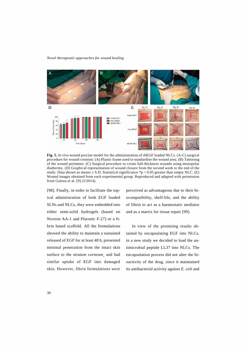
Novel therapeutic approaches for wound healing
30
Fig. 5. In vivo wound porcine model for the administration of rhEGF loaded NLCs. (A-C) surgical procedure for wound creation: (A) Plastic frame used to standardise the wound area. (B) Tattooing of the wound perimeter. (C) Surgical procedure to create full-thickness wounds using monopolar diathermy. (D) Graphical representation of wound closure from the second week to the end of the study. Data shown as means ± S.D. Statistical significance *p < 0.05 greater than empty NLC. (E) Wound images obtained from each experimental group. Reproduced and adapted with permission from Gainza et al. [9] (©2014).
[98]. Finally, in order to facilitate the top-
ical administration of both EGF loaded
SLNs and NLCs, they were embedded into
either semi-solid hydrogels (based on
Noveon AA-1 and Pluronic F-27) or a fi-
brin based scaffold. All the formulations
showed the ability to maintain a sustained
released of EGF for at least 48 h, presented
minimal penetration from the intact skin
surface to the stratum corneum¸ and had
similar uptake of EGF into damaged
skin. However, fibrin formulations were
perceived as advantageous due to their bi-
ocompatibility, shelf-life, and the ability
of fibrin to act as a haemostatic mediator
and as a matrix for tissue repair [99].
In view of the promising results ob-
tained by encapsulating EGF into NLCs,
in a new study we decided to load the an-
timicrobial peptide LL37 into NLCs. The
encapsulation process did not alter the bi-
oactivity of the drug, since it maintained
its antibacterial activity against E. coli and

Introduction
31
its anti-inflammatory activity by reversing
the macrophage activation caused by lipo-
polysaccharide (LPS). A full thickness ex-
cisional wound model in db/db diabetic
mice was utilised to evaluate the efficacy
of the formulation and an improved heal-
ing was observed in wounds treated with
the developed DDS in terms of wound clo-
sure, reepithelisation and restoration of the
inflammatory process [100]. LL37 has
also been loaded into SLNs. For instance,
in a study by Fumakia et al., LL37 and ser-
pin-1 were loaded into SLNs, and the re-
sulting formulation had antibacterial ac-
tivity against E. coli and S. aureus; pro-
moted in vitro wound closure in fibroblast
and keratinocytes and reversed inflamma-
tory activity of macrophages against LPS
[31].
3.3 Nanofibrous structures
The unique architectural structure of
the nanofibrous meshes makes them a
promising approach as wound dressings,
as summarised in Table 5. Nanofibers are
usually produced by electrospinning, a
technique that uses electrostatic forces to
draw the nanofiber mats from a droplet
[101]. These mats consist of biodegrada-
ble non-woven, ultra-fine polymeric fi-
bers, whose diameter is in a range from
several micrometers to a few nanometers
[35,102]. Nanofiber dressings offer a high
surface to volume ratio and a high inter-
connected porosity, which allow gas per-
meation and thus cell respiration, the re-
moval of exudates and the retainment of
moisture preventing wound desiccation
and dehydration [103]. Furthermore, nan-
ofibrous membranes are very appealing in
tissue engineering due to their structural
similarity to native ECM, which encour-
ages cell migration and proliferation and
support tissue formation [44,104].
In regard to drug delivery, nanofibrous
dressings exhibit a high drug-loading ca-
pacity and moreover, they provide an ex-
cellent protection from the environmental
harms. In addition, these formulations pre-
sent a remarkable controlled and sustained
release of drugs, extending their therapeu-
tic activity for even longer periods of time
[35]. Using the electrospinning technique,
drugs can be incorporated to the nano-
fibrous mats by three different ap-
proaches: (i) directly loaded by blending
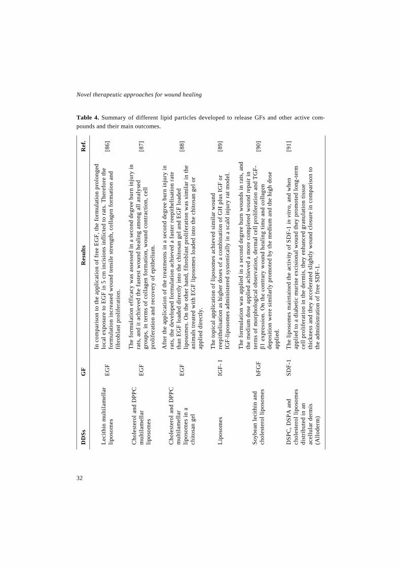
Novel therapeutic approaches for wound healing
32
Table 4. Summary of different lipid particles developed to release GFs and other active com-pounds and their main outcomes.
Ref
.
[86]
[87]
[88]
[89]
[90]
[91]
Res
ults
In c
ompa
rison
to th
e ap
plic
atio
n of
free
EG
F, th
e fo
rmul
atio
n pr
olon
ged
loca
l exp
osur
e to
EG
F in
5 c
m in
cisi
ons
infli
cted
to ra
ts. T
here
fore
the
form
ulat
ion
incr
ease
d w
ound
tens
ile s
treng
th, c
olla
gen
form
atio
n an
d fib
robl
ast p
rolif
erat
ion.
The
form
ulat
ion
effi
cacy
was
ass
esse
d in
a s
econ
d de
gree
bur
n in
jury
in
rats
, and
it a
chie
ved
the
fast
est w
ound
hea
ling
amon
g al
l ana
lyse
d gr
oups
, in
term
s of
col
lage
n fo
rmat
ion,
wou
nd c
ontra
ctio
n, c
ell
prol
ifer
atio
n an
d re
cove
ry o
f epi
thel
ium
.
Aft
er th
e ap
plic
atio
n of
the
treat
men
ts in
a s
econ
d de
gree
bur
n in
jury
in
rats
, the
dev
elop
ed fo
rmul
atio
n ac
hiev
ed a
fast
er re
epith
elis
atio
n ra
te
than
EG
F lo
aded
dir
ectly
into
the
chito
san
gel a
nd E
GF
load
ed
lipos
omes
. On
the
othe
r han
d, fi
brob
last
pro
lifer
atio
n w
as s
imila
r in
the
anim
als
treat
ed w
ith E
GF
lipos
omes
load
ed in
to th
e ch
itosa
n ge
l or
appl
ied
dire
ctly
.
The
topi
cal a
pplic
atio
n of
lipo
som
es a
chie
ved
sim
ilar w
ound
re
epith
elis
atio
n as
hig
her d
oses
of a
com
bina
tion
of G
H p
lus
IGF
or
IGF-
lipos
omes
adm
inis
tere
d sy
stem
ical
ly in
a s
cald
inju
ry ra
t mod
el.
The
form
ulat
ion
was
app
lied
in a
sec
ond
degr
ee b
urn
wou
nds
in ra
ts, a
nd
the
med
ium
dos
e ap
plie
d ac
hiev
ed a
mor
e co
mpl
eted
wou
nd re
pair
in
term
s of
mor
phol
ogic
al o
bser
vatio
n, d
erm
al c
ell p
rolif
erat
ion
and
TGF-
β1 e
xpre
ssio
n. O
n th
e co
ntra
ry w
ound
hea
ling
time
and
colla
gen
depo
sitio
n w
ere
sim
ilarly
pro
mot
ed b
y th
e m
ediu
m a
nd th
e hi
gh d
ose
appl
ied.
The
lipos
omes
mai
ntai
ned
the
activ
ity o
f SD
F-1
in v
itro,
and
whe
n ap
plie
d to
a d
iabe
tic m
urin
e ex
cisi
onal
wou
nd th
ey p
rom
oted
long
-ter
m
cell
prol
ifer
atio
n in
the
derm
is, t
hey
enha
nced
gra
nula
tion
tissu
e th
ickn
ess
and
they
acc
eler
ated
slig
htly
wou
nd c
losu
re in
com
pari
son
to
the
adm
inis
tratio
n of
free
SD
F-1.
GF
EGF
EGF
EGF
IGF-
I
bFG
F
SDF-
1
DD
Ss
Leci
thin
mul
tilam
ella
r lip
osom
es
Cho
lest
erol
and
DPP
C
mul
tilam
ella
r lip
osom
es
Cho
lest
erol
and
DPP
C
mul
tilam
ella
r lip
osom
es in
a
chito
san
gel
Lipo
som
es
Soyb
ean
leci
thin
and
ch
oles
tero
l lip
osom
es
DSP
C, D
SPA
and
ch
oles
tero
l lip
osom
es
dist
ribut
ed in
an
acel
lula
r der
mis
(A
llode
rm)

Introduction
33
Ref
.
[97,
98]
[99]
[10
0]
[31]
Res
ults
Bot
h fo
rmul
atio
ns im
prov
ed h
ealin
g in
com
paris
on to
free
EG
F in
a
diab
etic
mic
e w
ound
mod
el, i
n te
rms
of w
ound
clo
sure
, re
epith
elis
atio
n an
d re
stor
atio
n of
the
infla
mm
ator
y pr
oces
s.
Due
to h
ighe
r enc
apsu
latio
n ef
ficie
ncy
and
the
lack
of o
rgan
ic s
olve
nt
in it
s pr
oduc
tion,
NLC
s ef
fica
cy w
as a
sses
sed
in a
pig
wou
nd h
ealin
g m
odel
. In
it, th
e N
LCs
impr
oved
wou
nd c
losu
re a
nd w
ound
hea
ling
qual
ity in
com
pari
son
to a
hig
her d
ose
of fr
ee rh
EGF.
All
the
form
ulat
ions
wer
e si
mila
r in
term
s of
EG
F re
leas
e, u
ptak
e of
EG
F in
to d
amag
ed s
kin
and
min
imal
pen
etra
tion
in th
e in
tact
ski
n.
The
NLC
s pr
esen
ted
antib
acte
rial a
ctiv
ity a
nd a
nti-
infl
amm
ator
y ac
tivity
in v
itro,
and
in v
ivo
wer
e ab
le to
impr
ove
wou
nd c
losu
re,
reep
ithel
isat
ion
and
rest
orat
ion
of th
e in
flam
mat
ory
proc
ess
in a
full
thic
knes
s ex
cisi
onal
wou
nd m
odel
in d
iabe
tic m
ice.
The
form
ulat
ion
pres
ente
d an
tibac
teria
l act
ivity
aga
inst
E. c
oli a
nd S
. au
reus
, it p
rom
oted
wou
nd c
losu
re in
an
in v
itro
scra
tch
assa
y, it
pr
otec
ted
mon
olay
er c
ultu
re in
tegr
ity fr
om L
PS a
nd it
sho
wed
ant
i-in
flam
mat
ory
activ
ity a
gain
st L
PS.
GF
EGF
EGF
LL37
LL37
and
Se
rpin
-1
DD
Ss
SLN
(pre
ciro
l) an
d N
LC
(pre
ciro
l and
myg
liol)
SLN
and
NLC
em
bedd
ed in
se
mi-
solid
hyd
roge
ls (N
oveo
n A
A-1
or P
luro
nic
F-27
) or
fibrin
bas
ed s
olid
sca
ffol
d.
NLC
(pre
ciro
l and
myg
liol)
SLN
(gliy
cery
l mon
oest
eara
te
+ α-
phos
phat
idyl
chol
ine)

Novel therapeutic approaches for wound healing
34
or emulsifying the active compound into
the electrospinning solution; (ii) by coax-
ial electrospinning, a technique in which
two immiscible components are simulta-
neously electrospun through separate
feeding capillary channels to generate nan-
ofibers composed of a core-shell structure;
and (iii) immobilising the drugs on the sur-
face of the nanofibers through weak inter-
actions (hydrogen bonding or electro-
static, van de Waals or hydrophobic inter-
action) or covalent bindings that produce
chemical surface-modifications in the nan-
ofibers [105].
The three methods mentioned above
have been used to generate nanofibers
loaded with GFs and other endogenous
molecules for wound healing. Among di-
rect loading, our research group emulsi-
fied EGF and Aloe vera with PLGA to ob-
tain nanofibrous membranes with the com-
bined beneficial effect of both EGF and
Aloe vera. In vitro, the nanofibers showed
a promotion of fibroblast proliferation and
antimicrobial activity against S. aureus
and S. epidermidis; and in vivo, they accel-
erated significantly wound closure and
reepithelisation in a full thickness wound
model in diabetic mice [106]. The electro-
spinnning of emulsions has also been used
to encapsulate EGF in PCL/hyaluronan
nanofibers and bFGF in poly(ethylene gly-
col)-poly(DL-lactide) (PELA) nanofibers,
with the aim of increasing the encapsula-
tion efficiency and alleviate the initial
burst release. Both formulations devel-
oped with this strategy were able to im-
prove wound healing in vivo [107,108].
Direct blending of hydro- and lipophilic
compounds into the respective hydro- and
lipophilic electrospun solutions has also
been done to obtain wound dressings.
Among hydrophilic nanofibers, Bertoncelj
et al., developed a chitosan/PEO mem-
brane loaded with PRP that stimulated cell
proliferation, changed cell morphology
and limited cell mobility in vitro [109]. On
the other hand, the lipophilic compound β-
estradiol was loaded in a PU/dextran com-
posite nanofiber to develop a post-meno-
pausal wound dressing, which was able to
accelerate wound closure in vivo in partial
thickness wounds [34].
In order to obtain a more controlled re-
lease, nanofibers can be also directly
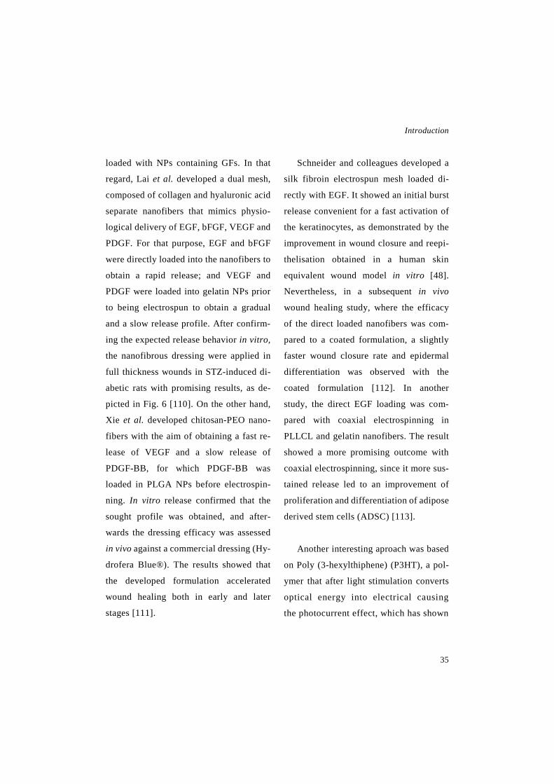
Introduction
35
loaded with NPs containing GFs. In that
regard, Lai et al. developed a dual mesh,
composed of collagen and hyaluronic acid
separate nanofibers that mimics physio-
logical delivery of EGF, bFGF, VEGF and
PDGF. For that purpose, EGF and bFGF
were directly loaded into the nanofibers to
obtain a rapid release; and VEGF and
PDGF were loaded into gelatin NPs prior
to being electrospun to obtain a gradual
and a slow release profile. After confirm-
ing the expected release behavior in vitro,
the nanofibrous dressing were applied in
full thickness wounds in STZ-induced di-
abetic rats with promising results, as de-
picted in Fig. 6 [110]. On the other hand,
Xie et al. developed chitosan-PEO nano-
fibers with the aim of obtaining a fast re-
lease of VEGF and a slow release of
PDGF-BB, for which PDGF-BB was
loaded in PLGA NPs before electrospin-
ning. In vitro release confirmed that the
sought profile was obtained, and after-
wards the dressing efficacy was assessed
in vivo against a commercial dressing (Hy-
drofera Blue®). The results showed that
the developed formulation accelerated
wound healing both in early and later
stages [111].
Schneider and colleagues developed a
silk fibroin electrospun mesh loaded di-
rectly with EGF. It showed an initial burst
release convenient for a fast activation of
the keratinocytes, as demonstrated by the
improvement in wound closure and reepi-
thelisation obtained in a human skin
equivalent wound model in vitro [48].
Nevertheless, in a subsequent in vivo
wound healing study, where the efficacy
of the direct loaded nanofibers was com-
pared to a coated formulation, a slightly
faster wound closure rate and epidermal
differentiation was observed with the
coated formulation [112]. In another
study, the direct EGF loading was com-
pared with coaxial electrospinning in
PLLCL and gelatin nanofibers. The result
showed a more promising outcome with
coaxial electrospinning, since it more sus-
tained release led to an improvement of
proliferation and differentiation of adipose
derived stem cells (ADSC) [113].
Another interesting aproach was based
on Poly (3-hexylthiphene) (P3HT), a pol-
ymer that after light stimulation converts
optical energy into electrical causing
the photocurrent effect, which has shown

Novel therapeutic approaches for wound healing
36
Fig. 6. Composite collagen-hyaluronic acid nanofibers containing free GFs (bFGF and EGF) and GFs loaded into gelatin NSs (VEGF and PDGF). (A) SEM images of the morphology of nano-fibers. (Arrows indicate gelatin NPs; the scale bar 6 µm). (B) Fluorescent images by confocal microscopy of nanofibers, collagen dyed in red and hyaluronic acid dyed in green. (C) Fluorescent images by confocal microscopy of collagen nanofibers with fluorescein(red) loaded gelatin NSs and hyaluronic acid nanofibers with rhodamine b (green) loaded gelatin NSs. (D) Wound area at different times of untreated wounds and wounds treated with collagen-hyaluronic acid nanofibers, collagen-hyaluronic acid nanofibers containing bFGF and EGF and collagen-hyaluronic acid nan-ofibers containing bFGF, EGF, VEGF and PDGF. (⁄⁄p < 0.01, ⁄P < 0.05). Reproduced and adapted with permission from Lai et al. [111] (©2014).
Introduction
37
promising results in skin regeneration. In
order to improve its biocompatibility, co-
axial electrospinning was used to encapsu-
late EGF and P3HT in the core of a gela-
tin/PLLCL nanofibrous mesh. The formu-
lation improved fibroblast proliferation
and wound closure in vitro, and it was able
to induce ADSC differentiation into
keratinocytes under light stimulation
[114].
In order to achieve a longer therapeutic
effect, EGF can be immobilised to the nan-
ofibers by covalent binding with the amine
groups exposed on their surface. In that re-
gard, Tigli and colleagues developed a
PCL and PCL/gelatin scaffold with EGF
covalently immobilised on it surface. That
immobilisation resulted in an earlier
spreading and faster proliferation of fibro-
blast seeded on top [115]. Similar results
were obtained after binding EGF cova-
lently to the surface of PCL/collagen nan-
ofibers, as an acceleration in keratinocyte
spreading and proliferation was observed.
In addition, the keratinocytes seeded on
top of the nanofibers exhibited superior
differentiation ability, evidenced by a
higher loricrin expression [116].
Choi et al., developed another DDS
based on the covalent conjugation of EGF
to the nanofibers. In this case, EGF was
immobilised on the surface of PEG/PCL
block copolymers and it efficacy in wound
healing was compared to that of nanofibers
plus EGF solution. The dressing contain-
ing conjugated EGF showed superior ther-
apeutic activity in both in vitro and in vivo
wound healing studies, due to the greater
protection of the GF. A higher enhance-
ment of keratinocyte specific gene expres-
sion was observed in vitro, and an acceler-
ated wound closure and an improvement
of EGFR expression in vivo [117]. Taking
into account the promising results ob-
tained by immobilising EGF in PEG/PCL
block copolymer, a coaxial electrospun
composed of PEG/PCL was developed. It
was loaded with EGF and bFGF, the first
one immobilised in the surface and the
second one encapsulated in the core. The
formulation exhibited an initial burst re-
lease of bFGF and no negligible release of
EGF; and in comparison to nanofibers
with the single GFs, it accelerated wound
closure achieving more mature wounds on
day 7 in burn wounds inflicted to STZ-in-
duced diabetic mice [118].

Novel therapeutic approaches for wound healing
38
As demonstrated by Song and col-
leagues, there are more ways to cova-
lently conjugate active compounds to
nanofibers surface. In that regard, they
conjugated Cys-KR12 (an antimicrobial
peptide motif originated from LL37) to
silk fibroin nanofibers, using EDC/NHS
and thiol-maleimide click chemistry. The
conjugation process did not affect the bi-
oactivity of the Cys-KR12, since in vitro
studies confirmed that it exhibited anti-
microbial and proliferative activities and
suppressed LPS induced inflammation
[119].
In a study by Patel et al., bFGF and
laminin were immobilised in the surface of
PLLA using a strategy based on the capac-
ity of heparin to associate with endoge-
nous molecules, thereby PLLA was func-
tionalised with heparin and subsequently
bFGF and laminin were conjugated. The
resulting formulation was able to enhance
wound healing in vitro in a scratch as-
say, especially when the nanofibers were
oriented perpendicularly to the gap
[120].
4. Conclusions and future perspectives
In recent years the incidence of chronic
wounds is increasing alarmingly, as the
risk population grows. Furthermore, due to
the lack of efficient treatments, chronic
wounds are becoming a clinical and eco-
nomical burden. In comparison with the
physiological healing process, a series of
abnormalities occur during chronic heal-
ing, such as the decrease of the levels of
GFs and other molecules that are involved
in healing regulation. Therefore, the exog-
enous administration of GFs is a promising
approach for the treatment of wound heal-
ing, although their low stability in vivo
limits their clinical application. An at-
tempt to cope with this is the development
of novel DDSs containing GFs, including
polymeric MP/NP, lipid nanoparticles,
nanofibrous structures and their combina-
tions. Those formulations, besides of pro-
tecting the encapsulated drug, present nu-
merous advantages. Among others they of-
fer good biocompatibility, controlled or
sustained release, high drug-loading and
good mechanical properties. In addition,
depending on the polymer and DDS used,

Introduction
39
Table 5. Summary of different nanofibrous membranes developed to release GFs and other active compounds and their main outcomes.
Ref
.
[106
]
[107
]
[108
]
[109
]
[34]
[110
]
Res
ults
In v
itro
it im
prov
ed fi
brob
last
pro
lifer
atio
n an
d sh
owed
ant
imic
robi
al a
ctiv
ity. I
n vi
vo
it ac
cele
rate
d w
ound
clo
sure
and
re
epith
elis
atio
n in
a d
iabe
tic m
ice
wou
nd
mod
el.
The
form
ulat
ion
enha
nced
ker
atin
ocyt
e an
d fib
robl
ast p
rolif
erat
ion
and
infil
trat
ion
in
vitr
o. A
nd it
enh
ance
d th
e re
gene
ratio
n of
a
fully
func
tiona
l ski
n, in
viv
o.
The
form
ulat
ion
enha
nced
fibr
obla
st
adhe
sion
, pro
lifer
atio
n an
d EC
M s
ecre
tion
in v
itro.
And
whe
n ap
plie
d to
wou
nds
infli
cted
to d
iabe
tic ra
ts, i
t im
prov
ed w
ound
cl
osur
e, a
chie
ving
a c
ompl
ete
reep
ithel
isat
ion
and
rege
nera
tion
of s
kin
appe
ndag
es in
2 w
eeks
.
The
form
ulat
ion
stim
ulat
es c
ell
prol
ifer
atio
n, c
hang
ed c
ell m
orph
olog
y an
d lim
ited
cell
mob
ility
.
The
form
ulat
ion
acce
lera
ted
wou
nd a
rea
redu
ctio
n in
an
in v
ivo
parti
al th
ickn
ess
wou
nd h
ealin
g in
rats
.
The
DD
S in
crea
sed
endo
thel
ial c
ell g
row
th
rate
and
allo
wed
a b
ette
r net
wor
k. In
w
ound
s in
flict
ed to
dia
betic
rats
it
acce
lera
ted
wou
nd c
losu
re, i
ncre
ased
co
llage
n de
posi
tion
and
enha
nced
m
atur
atio
n of
ves
sels
.
Loa
ding
met
hod
Dir
ect l
oadi
ng, b
y em
ulsi
ficat
ion
Dir
ect l
oadi
ng, b
y em
ulsi
ficat
ion
Dir
ect l
oadi
ng, b
y em
ulsi
ficat
ion
Dir
ect l
oadi
ng, b
y bl
endi
ng
Dir
ect l
oadi
ng, b
y bl
endi
ng
Dir
ect l
oadi
ng. b
FGF
and
EGF
dire
ctly
and
V
EGF
and
PDG
F lo
aded
into
gel
atin
N
Ps
GF
EGF
EGF
bFG
F
PRP
β-es
tradi
ol
VEG
F, b
FGF,
PD
GF
and
EGF
DD
Ss
PLG
A a
nd
Aloe
ver
a na
nofib
ers
PCL
and
hyal
uron
an
nano
fiber
s
PELA
na
nofib
ers
Chi
tosa
n an
d PE
O
nano
fiber
s
PU a
nd d
extr
an
nano
fiber
s
Col
lage
n an
d hy
alur
onic
aci
d du
al n
anof
iber
s
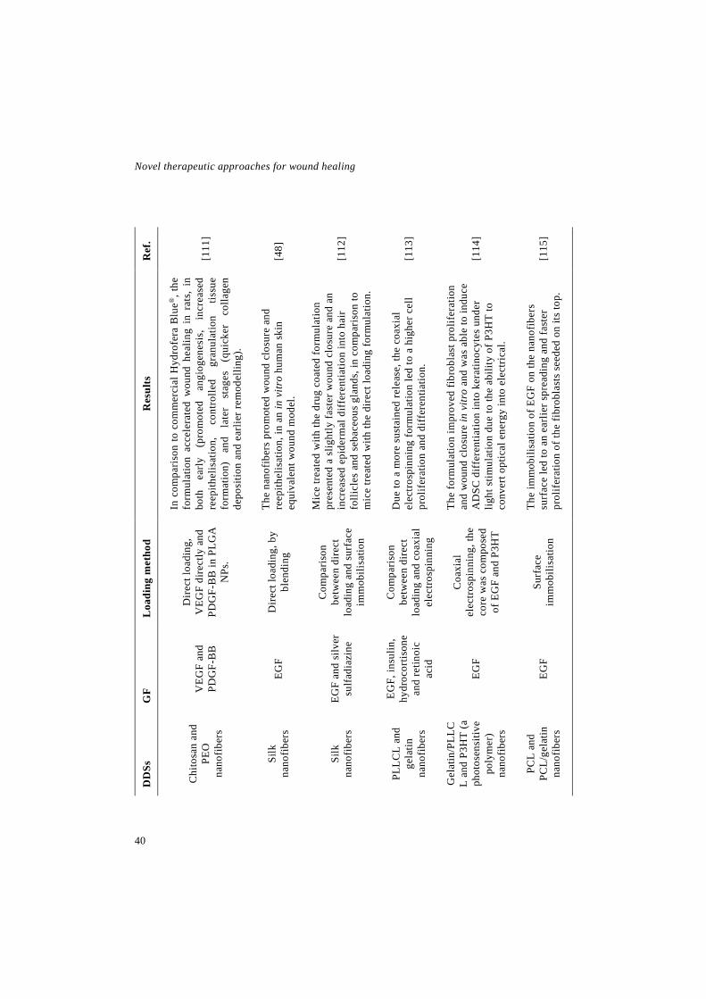
Novel therapeutic approaches for wound healing
40
Ref
.
[111
]
[48]
[112
]
[113
]
[114
]
[115
]
Res
ults
In c
ompa
rison
to c
omm
erci
al H
ydro
fera
Blu
e®, t
he
form
ulat
ion
acce
lera
ted
wou
nd h
ealin
g in
rat
s, i
n bo
th
early
(p
rom
oted
an
giog
enes
is,
incr
ease
d re
epith
elis
atio
n,
cont
rolle
d gr
anul
atio
n tis
sue
form
atio
n)
and
late
r st
ages
(q
uick
er
colla
gen
depo
sitio
n an
d ea
rlier
rem
odel
ling)
.
The
nano
fiber
s pr
omot
ed w
ound
clo
sure
and
re
epith
elis
atio
n, in
an
in v
itro
hum
an s
kin
equi
vale
nt w
ound
mod
el.
Mic
e tre
ated
with
the
drug
coa
ted
form
ulat
ion
pres
ente
d a
slig
htly
fast
er w
ound
clo
sure
and
an
incr
ease
d ep
ider
mal
diff
eren
tiatio
n in
to h
air
folli
cles
and
seb
aceo
us g
land
s, in
com
pari
son
to
mic
e tr
eate
d w
ith th
e di
rect
load
ing
form
ulat
ion.
Due
to a
mor
e su
stai
ned
rele
ase,
the
coax
ial
elec
trosp
inni
ng fo
rmul
atio
n le
d to
a h
ighe
r cel
l pr
olif
erat
ion
and
diff
eren
tiatio
n.
The
form
ulat
ion
impr
oved
fibr
obla
st p
rolif
erat
ion
and
wou
nd c
losu
re in
vitr
o an
d w
as a
ble
to in
duce
A
DSC
diff
eren
tiatio
n in
to k
erat
inoc
ytes
und
er
light
stim
ulat
ion
due
to th
e ab
ility
of P
3HT
to
conv
ert o
ptic
al e
nerg
y in
to e
lect
rica
l.
The
imm
obili
satio
n of
EG
F on
the
nano
fiber
s su
rfac
e le
d to
an
earli
er s
prea
ding
and
fast
er
prol
ifer
atio
n of
the
fibro
blas
ts s
eede
d on
its
top.
Loa
ding
met
hod
Dir
ect l
oadi
ng,
VEG
F di
rect
ly a
nd
PDG
F-B
B in
PLG
A
NPs
.
Dir
ect l
oadi
ng, b
y bl
endi
ng
Com
paris
on
betw
een
dire
ct
load
ing
and
surf
ace
imm
obili
satio
n
Com
paris
on
betw
een
dire
ct
load
ing
and
coax
ial
elec
trosp
inni
ng
Coa
xial
el
ectro
spin
ning
, the
co
re w
as c
ompo
sed
of E
GF
and
P3H
T
Surf
ace
imm
obili
satio
n
GF V
EGF
and
PDG
F-B
B
EGF
EGF
and
silv
er
sulfa
diaz
ine
EGF,
insu
lin,
hydr
ocor
tison
e an
d re
tinoi
c ac
id
EGF
EGF
DD
Ss
Chi
tosa
n an
d PE
O
nano
fiber
s
Silk
na
nofib
ers
Silk
na
nofib
ers
PLLC
L an
d ge
latin
na
nofib
ers
Gel
atin
/PLL
CL
and
P3H
T (a
ph
otos
ensi
tive
poly
mer
) na
nofib
ers
PCL
and
PCL/
gela
tin
nano
fiber
s

Introduction
41
Ref
.
[116
]
[117
]
[118
]
[119
]
[120
]
Res
ults
The
form
ulat
ion
enha
nced
ker
atin
ocyt
e sp
read
ing,
pr
olif
erat
ion
and
diff
eren
tiatio
n ab
ility
.
The
appl
icat
ion
of th
e D
DS
into
a b
urn
wou
nd
infli
cted
to d
iabe
tic m
ice
led
to a
sup
erio
r wou
nd
clos
ure
and
enha
nced
EG
FR e
xpre
ssio
n. In
ad
ditio
n, in
vitr
o it
enha
nced
ker
atin
ocyt
e-sp
ecifi
c ge
ne e
xpre
ssio
n.
The
form
ulat
ion
show
ed a
bin
ary
rele
ase
prof
ile
with
an
initi
al b
urst
of b
FGF
and
negl
igib
le re
leas
e of
EG
F, a
nd w
as a
ble
to a
ccel
erat
e w
ound
clo
sure
an
d en
hanc
e w
ound
mat
urat
ion
in a
bur
n w
ound
m
odel
in d
iabe
tic m
ice.
The
enca
psul
ated
fact
or m
aint
aine
d its
an
timic
robi
al a
nd p
rolif
erat
ive
activ
ities
, its
abi
lity
to p
rom
ote
kera
tinoc
yte
diff
eren
tiatio
n an
d to
su
ppre
ss L
PS in
duce
d in
flam
mat
ion.
The
form
ulat
ion
enha
nced
wou
nd h
ealin
g in
vitr
o in
a s
crat
ch a
ssay
, esp
ecia
lly w
hen
the
nano
fiber
s w
ere
orie
nted
per
pend
icul
arly
to th
e sc
ratc
h.
Loa
ding
met
hod
Surf
ace
imm
obili
satio
n
Surf
ace
imm
obili
satio
n
EGF
imm
obili
sed
on th
e su
rfac
e an
d bF
GF
load
ed b
y co
axia
l el
ectro
spin
ning
Surf
ace
imm
obili
satio
n
Surf
ace
imm
obili
satio
n
GF
EGF
EGF
bFG
F an
d EG
F
Cys
-KR
12
Lam
inin
and
bF
GF
DD
Ss
PCL
and
PCL/
colla
gen
nano
fiber
s
PEG
and
PC
L na
nofib
ers
PCL/
PEG
na
nofib
ers
Silk
fibr
oin
nano
fiber
s
PLLA
na
nofib
ers
func
tiona
lised
w
ith h
epar
itn
Novel therapeutic approaches for wound healing
42
specific properties can be obtained, such
as, drug targeting or an environment that
simulates native ECM.
Overall, the controlled release of GFs
incorporated into nanotechnology based
DDSs has demonstrated a great potential
for the treatment of chronic wounds and
skin regeneration. However, up to date, the
research on DDSs releasing GFs for
wound healing is still at the preclinical
phase, and further studies are needed in or-
der to advance to the clinical phase.
Among the preclinical studies needed,
there are tolerance and toxicity assays in
animal models, and efficacy assays in
larger animal models, such as, porcine
models.
Since most of the summarised DDSs
are developed as topical delivery systems,
a critical issue that need to be studied be-
fore their approval is the systemic absorp-
tion of the encapsulated drug. In addition,
the suitability of the polymers is another
key issue that need to be proved, because
some of the polymers used for the devel-
opment of these DDSs are not approved
for clinical use.
Although there are not clinical trials or
marketed products based in nanotechnol-
ogy to release GFs or other endogenous
molecules for wound healing application,
there are two marketed products based in
nanotechnology for wound healing. On the
one hand, there is Altrazeal®, a crystalline
white powder consisting of nanoparticles
of freeze dried poly-2-hydroxyethyl-
methacrylate (pHEMA) and poly-2-hy-
droxypropylmethacrylate (pHPMA). Due
to nanoflex technology, in contact with the
wound exudates the particles hydrate and
aggregate becoming a moist and flexible
film [121]. On the other hand, there is
Talymed® and advanced matrix composed
of shortened nanofibers of poly-N-acetyl
glucosamine (pGIcNAc) [122]. In addi-
tion, there is a clinical trial currently re-
cruiting participants, to evaluate the safety
and performance of SPINNER, a portable
electrospinning device that produces in-
situ nanofiber dressings [123].
5. Acknowledgments
I. Garcia-Orue thanks the Basque Gov-
ernment for the fellowship grant. This pro-
ject has been funded by the Spanish Min-

Introduction
43
istry of Economy and competitiveness
(INNPACTO, IPT-2012-0602-300000,
2012). In addition, it has been partially by
the Basque Government (ELKARTEK
2015, Nanoplatform, KK-2015/0000036).
6. References
[1] V.W. Wong, G.C. Gurtner, Tissue engi-neering for the management of chronic wounds: current concepts and future perspec-tives, Exp. Dermatol. 21 (2012) 729-734. doi: 10.1111/j.1600-0625.2012.01542.x.
[2] J. Posnett, F. Gottrup, H. Lundgren, G. Saal, The resource impact of wounds on health-care providers in Europe, J. Wound Care 18 (2009)154.doi:10.12968/jowc.2009.18.4.41607.
[3] C.K. Sen, G.M. Gordillo, S. Roy, R. Kirsner, L. Lambert, T.K. Hunt, F. Gottrup, G.C. Gurtner, M.T. Longaker, Human skin wounds: a major and snowballing threat to pub-lic health and the economy, Wound Repair Re-gen. 17 (2009) 763-771. doi: 10.1111/j.1524-475X.2009.00543.x.
[4] A.J. Whittam, Z.N. Maan, D. Duscher, V.W. Wong, J.A. Barrera, M. Januszyk, G.C. Gurtner, Challenges and opportunities in drug delivery for wound healing, Adv. Wound Care (New Rochelle) 5 (2016) 79-88. doi: //dx.doi. org/10.1089%2Fwound.2014.0600.
[5] N.B. Menke, K.R. Ward, T.M. Witten, D.G. Bonchev, R.F. Diegelmann, Impaired wound healing, Clin. Dermatol. 25 (2007) 19-25. doi: //dx.doi.org/10.1016/j.clindermatol.2006.12.05.
[6] G. Gainza, S. Villullas, J.L. Pedraz, R.M. Hernandez, M. Igartua, Advances in drug de-livery systems (DDSs) to release growth fac-tors for wound healing and skin regeneration, Nanomedicine 11 (2015) 1551-1573. doi: //dx.doi.org/10.1016/j.nano.2015.03.002.
[7] R.F. Diegelmann, M.C. Evans, Wound healing: an overview of acute, fibrotic and de-layed healing, Front. Biosci. 9 (2004) 283-289. doi: //dx.doi.org/10.2741/.
[8] S. Barrientos, H. Brem, O. Stojadinovic, M. Tomic-Canic, Clinical application of growth factors and cytokines in wound healing, Wound Repair Regen. 22 (2014) 569-578. doi: 10.1111/wrr.12205.
[9] P. Olczyk, L. Mencner, K. Komosinska-Vassev, Diverse roles of heparan sulfate and heparin in wound repair, Biomed. Res. Int. 2015(2015)549417.doi: 10.1155/2015/549417.
[10] S. Barrientos, O. Stojadinovic, M.S. Go-linko, H. Brem, M. Tomic Canic, Growth fac-tors and cytokines in wound healing, Wound Repair Regen 16 (2008) 585-601. doi: 10.1111 /j.1524-475X.2008.00410.x.
[11] S. Werner, R. Grose, Regulation of wound healing by growth factors and cytokines, Phys-iol. Rev. 83 (2003) 835-870. doi: 10.1152/phy srev.00031.2002.
[12] P.S. Briquez, J.A. Hubbell, M.M. Martino, Extracellular matrix-inspired growth factor de-livery systems for skin wound healing, Adv. Wound Care (New Rochelle) 4 (2015) 479-489. doi: 10.1089/wound.2014.0603.
[13] T. Velnar, T. Bailey, V. Smrkolj, The wound healing process: an overview of the
Novel therapeutic approaches for wound healing
44
cell-ular and molecular mechanisms, J. Int. Med. Res. 37 (2009) 1528-1542. doi: 10.1177 /147323000903700531.
[14] A. Lubkowska, B. Dolegowska, G. Banfi, Growth factor content in PRP and their ap-plicability in medicine, J. Biol. Regul. Home-ost. Agents 26 (2012) 22S.
[15] M. Zakrzewska, E. Marcinkowska, A. Wiedlocha, FGF-1: from biology through engi-neering to potential medical applications, Crit. Rev. Clin. Lab. Sci. 45 (2008) 91-135. doi: 10.1080/10408360701713120.
[16] M.G. Tonnesen, X. Feng, R.A.F. Clark, Angiogenesis in wound healing, J. Investig. Dermatol. Symp. Proc. 5 (2000) 40-46. doi: //dx.doi.org/10.1046/j.1087-0024.2000.00014.x.
[17] X. Li, C. Wang, J. Xiao, W.L. McKeehan, F. Wang, Fibroblast growth factors, old kids on the new block, Semin. Cell Dev. Biol. 53 (2016)155-167.doi: //dx.doi.org/10.1016/j.sem cdb.2015.12.014.
[18] J. Hardwicke, D. Schmaljohann, D. Boyce, D. Thomas, Epidermal growth factor therapy and wound healing — past, present and future perspectives, The Surgeon 6 (2008) 172-177. doi: //dx.doi.org/10.1016/S1479-666X (08)80114-X.
[19] K.E. Johnson, T.A. Wilgus, Vascular en-dothelial growth factor and angiogenesis in the regulation of cutaneous wound repair, Adv. Wound. Care. (New Rochelle) 3 (2014) 647-661. doi: 10.1089/wound.2013.0517.
[20] M.A. Seeger, A.S. Paller, The roles of growth factors in keratinocyte migration, Adv.
Wound. Care. (New Rochelle) 4 (2015) 213-224. doi: 10.1089/wound.2014.0540.
[21] T. Emanuelli, A. Burgeiro, E. Carvalho, Effects of insulin on the skin: possible healing benefits for diabetic foot ulcers, Arch. Derma-tol. Res. 308 (2016) 677-694. doi: 10.1007/ s00403-016-1686-z.
[22] T.K. Ho, X. Shiwen, D. Abraham, J. Tsui, D. Baker, Stromal-Cell-Derived Factor-1 (SDF-1)/CXCL12 as potential target of thera-peutic angiogenesis in critical leg ischaemia, Cardiol. Res. Pract. 2012 (2012) 143209. doi: 10.1155/2012/143209 [doi].
[23] T.T. Lau, D.A. Wang, Stromal cell-de-rived factor-1 (SDF-1): homing factor for en-gineered regenerative medicine, Expert Opin. Biol. Ther. 11 (2011) 189-197. doi: 10.1517/ 14712598.2011.546338.
[24] D. Vandamme, B. Landuyt, W. Luyten, L. Schoofs, A comprehensive summary of LL-37, the factotum human cathelicidin peptide, Cell. Immunol. 280 (2012) 22-35. doi: 10.1016/j. cellimm.2012.11.009.
[25] Y. Wang, Z. Chen, G. Luo, W. He, K. Xu, R. Xu, Q. Lei, J. Tan, J. Wu, M. Xing, In-situ-generated vasoactive intestinal peptide loaded microspheres in mussel-inspired polycaprolac-tone nanosheets creating spatiotemporal re-leasing microenvironment to promote wound healing and angiogenesis, ACS Appl. Mater. Interfaces 8 (2016) 7411-7421. doi: 10.1021/ acsami.5b11332.
[26] J. Chéret, N. Lebonvallet, V. Buhé, J.L. Carre, L. Misery, C. Le Gall-Ianotto, Influence of sensory neuropeptides on human cutaneous wound healing process, J. Dermatol. Sci. 74
Introduction
45
(2014)193-203.doi: //dx.doi.org/10.1016/j.jder msci.2014.02.001.
[27] W. Jiang, H. Wang, Y.S. Li, W. Luo, Role of vasoactive intestinal peptide in osteoarthri-tis, J. Biomed. Sci. 23 (2016) 1. doi: 10.1186/ s12929-016-0280-1 [doi].
[28] J. Drobnik, Wound healing and the effect of pineal gland and melatonin, J. Exp. Integr. Med. 2 (2012) 3-14. doi: 10.5455/jeim.040112 .ir.009.
[29] L.K. Petersen, A.S. Determan, C. West-gate, L. Bendickson, M. Nilsen-Hamilton, B. Narasimhan, Lipocalin-2-loaded amphiphilic polyanhydride microparticles accelerate cell migration, J. Biomater. Sci. Polym. Ed. 22 (2011)1237-1252.doi: 10.1163/092050610X50 2776.
[30] K. Suk, Lipocalin-2 as a therapeutic target for brain injury: An astrocentric perspective, Prog. Neurobiol. 144 (2016) 158-172. doi: //dx.doi.org/10.1016/j.pneurobio.2016.08.001.
[31] M. Fumakia, E.A. Ho, Nanoparticles en-capsulated with LL37 and serpin A1 promotes wound healing and synergistically enhances antibacterial activity, Mol. Pharm. 13 (2016) 2318-2331. doi: 10.1021/acs.molpharma-ceut.6b00099.
[32] C.A.B. Kandregula, G. Smilin Bell Aseervatham, G.T. Bentley, R. Kandasamy, Alpha-1 antitrypsin: associated diseases and therapeutic uses, Clin. Chim. Acta 459 (2016) 109-116.doi: //dx.doi.org/10.1016/j.cca.2016. 05.028.
[33] S.C. Gilliver, G.S. Ashcroft, Sex steroids and cutaneous wound healing: the contrasting
influences of estrogens and androgens, Climac-teric 10 (2007) 276-288. doi: 10.1080/ 13697130701456630.
[34] A.R. Unnithan, A.R.K. Sasikala, P. Murugesan, M. Gurusamy, D. Wu, C.H. Park, C.S. Kim, Electrospun polyurethane-dextran nanofiber mats loaded with estradiol for post-menopausal wound dressing, Int. J. Biol. Mac-romol. 77 (2015) 1-8. doi: //dx.doi.org/10. 1016/j.ijbiomac.2015.02.044.
[35] T. Garg, G. Rath, A.K. Goyal, Biomateri-als-based nanofiber scaffold: targeted and con-trolled carrier for cell and drug delivery, J. Drug Target. 23 (2015) 202-221. doi: 10.3109/1061186X.2014.992899.
[36] J.M. Dang, K.W. Leong, Natural polymers for gene delivery and tissue engineering, Adv. Drug Deliv. Rev. 58 (2006) 487-499. doi: //dx.doi.org/10.1016/j.addr.2006.03.001.
[37] T. Garg, G. Rath, A.K. Goyal, Compre-hensive review on additives of topical dosage forms for drug delivery, Drug Deliv. 22 (2015) 969-987.doi: 10.3109/10717544.2013.879355.
[38] W. Li, C.T. Laurencin, E.J. Caterson, R.S. Tuan, F.K. Ko, Electrospun nanofibrous struc-ture: A novel scaffold for tissue engineering, J. Biomed. Mater. Res. 60 (2002) 613-621. doi: 10.1002/jbm.10167.
[39] J. Gunn, M. Zhang, Polyblend nanofibers for biomedical applications: perspectives and challenges, Trends Biotechnol. 28 (2010) 189-197.doi: //dx.doi.org/10.1016/j.tibtech.2009. 12.006.
[40] S. Jain, N. Patel, M.K. Shah, P. Khatri, N. Vora, Recent advances in lipid-based vesicles

Novel therapeutic approaches for wound healing
46
and particulate carriers for topical and trans-dermal application, J. Pharm. Sci. (2016). doi: //dx.doi.org/10.1016/j.xphs.2016.10.001.
[41] S.B. Kulkarni, G.V. Betageri, M. Singh, Factors affecting microencapsulation of drugs in liposomes, J. Microencapsul. 12 (1995) 229-246. doi: 10.3109/02652049509010292.
[42] M. Geszke-Moritz, M. Moritz, Solid lipid nanoparticles as attractive drug vehicles: Com-position, properties and therapeutic strategies, Materials Science and Engineering: C 68 (2016)982-994.doi: //dx.doi.org/10.1016/j.mse c.2016.05.119.
[43] S. Doktorovova, E.B. Souto, Nanostruc-tured lipid carrier-based hydrogel formulations for drug delivery: a comprehensive review, Ex-pert. Opin. Drug Deliv. 6 (2009) 165-176. doi: 10.1517/17425240802712590.
[44] C.P. Barnes, S.A. Sell, E.D. Boland, D.G. Simpson, G.L. Bowlin, Nanofiber technology: Designing the next generation of tissue engi-neering scaffolds, Adv. Drug Deliv. Rev. 59 (2007)1413-1433.doi: //dx.doi.org/10.1016/j. addr.2007.04.022.
[45] K. Su, C. Wang, Recent advances in the use of gelatin in biomedical research, Biotech-nol. Lett. 37 (2015) 2139-2145. doi: 10.1007 /s10529-015-1907-0.
[46] T. Dai, M. Tanaka, Y.Y. Huang, M.R. Hamblin, Chitosan preparations for wounds and burns: antimicrobial and wound-healing effects, Expert Rev. Anti Infect. Ther. 9 (2011) 857-879. doi: 10.1586/eri.11.59.
[47] G.D. Mogoşanu, A.M. Grumezescu, Nat-ural and synthetic polymers for wounds and
burns dressing, Int. J. Pharm. 463 (2014) 127-136.doi: //dx.doi.org/10.1016/j.ijpharm.2013 .12.015.
[48] A. Schneider, X.Y. Wang, D.L. Kaplan, J.A. Garlick, C. Egles, Biofunctionalized elec-trospun silk mats as a topical bioactive dress-ing for accelerated wound healing, Acta Bio-mater. 5 (2009) 2570-2578. doi: //dx.doi.org/ 10.1016/j.actbio.2008.12.013.
[49] K.K. Chereddy, G. Vandermeulen, V. Préat, PLGA based drug delivery systems - promising carriers for wound healing activity, Wound Repair Regen. (2016). doi: 10. 1111/wrr.12404.
[50] B.J. Kim, H. Cheong, E. Choi, S. Yun, B. Choi, K. Park, I.S. Kim, D. Park, H.J. Cha, Ac-celerated skin wound healing using electrospun nanofibrous mats blended with mussel adhe-sive protein and polycaprolactone, J. Biomed. Mater. Res. A 105 (2017) 218-225. doi: 10. 1002/jbm.a.35903.
[51] Z. Değim, Use of microparticulate sys-tems to accelerate skin wound healing, J. Drug Target. 16 (2008) 437-448. doi: 10.1080/ 10611860802088572.
[52] M. Ye, S. Kim, K. Park, Issues in long-term protein delivery using biodegradable mi-croparticles, J. Control Release 146 (2010) 241-260. doi: //dx.doi.org/10.1016/j.jconrel. 2010.05.011.
[53] Q. Liu, Y. Huang, Y. Lan, Q. Zuo, C. Li, Y. Zhang, R. Guo, W. Xue, Acceleration of skin regeneration in full-thickness burns by in-corporation of bFGF-loaded alginate micro-spheres into a CMCS–PVA hydrogel, J. Tissue
Introduction
47
Eng. Regen. Med. (2015). doi: 10.1002/ term.2057.
[54] L.W. Place, M. Sekyi, M.J. Kipper, Ag-grecan-mimetic, glycosaminoglycan-contain-ing nanoparticles for growth factor stabiliza-tion and delivery, Biomacromolecules 15 (2014) 680-689. doi: 10.1021/bm401736c.
[55] W. Li, Y. Lan, R. Guo, Y. Zhang, W. Xue, Y. Zhang, In vitro and in vivo evaluation of a novel collagen/cellulose nanocrystals scaffold for achieving the sustained release of basic fi-broblast growth factor, J. Biomater. Appl. 29 (2015)882-893.doi: 10.1177/08853282145470 91.
[56] C.J. Park, S.G. Clark, C.A. Lichtensteiger, R.D. Jamison, A.J.W. Johnson, Accelerated wound closure of pressure ulcers in aged mice by chitosan scaffolds with and without bFGF, Acta Biomater. 5 (2009) 1926-1936. doi: //dx.doi.org/10.1016/j.actbio.2009.03.002.
[57] K. Kawai, S. Suzuki, Y. Tabata, Y. Ikada, Y. Nishimura, Accelerated tissue regeneration through incorporation of basic fibroblast growth factor-impregnated gelatin micro-spheres into artificial dermis, Biomaterials 21 (2000)489-499.doi: //dx.doi.org/10.1016/S01 42-9612(99)00207-0.
[58] K. Kawai, S. Suzuki, Y. Tabata, Y. Nishi-mura, Accelerated wound healing through the incorporation of basic fibroblast growth factor-impregnated gelatin microspheres into artifi-cial dermis using a pressure-induced decubitus ulcer model in genetically diabetic mice, Br. J. Plast. Surg. 58 (2005) 1115-1123. doi: //dx.doi.org/10.1016/j.bjps.2005.04.010.
[59] S. Huang, T. Deng, H. Wu, F. Chen, Y. Jin, Wound dressings containing bFGF-im-pregnated microspheres, J. Microencapsul. 23 (2006)277-290.doi: 10.1080/02652040500435 170.
[60] K. Ulubayram, A.N. Cakar, P. Korkusuz, C. Ertan, N. Hasirci, EGF containing gelatin-based wound dressings, Biomaterials 22 (2001) 1345-1356. doi: //dx.doi.org/10.1016/S0142-9612(00)00287-8.
[61] S. Dogan, S. Demirer, I. Kepenekci, B. Erkek, A. Kiziltay, N. Hasirci, S. Müftüoğlu, A. Nazikoğlu, N. Renda, U.D. Dincer, A. El-han, E. Kuterdem, Epidermal growth factor-containing wound closure enhances wound healing in non-diabetic and diabetic rats, Int. Wound J. 6 (2009) 107-115. doi: 10.1111/j. 1742-481X.2009.00584.x.
[62] W. Zhou, M. Zhao, Y. Zhao, Y. Mou, A fibrin gel loaded with chitosan nanoparticles for local delivery of rhEGF: preparation and in vitro release studies, J. Mater. Sci. Mater. Med. 22 (2011) 1221. doi: 10.1007/s10856-011-4304-9.
[63] X. Dong, J. Xu, W. Wang, H. Luo, X. Liang, L. Zhang, H. Wang, P. Wang, J. Chang, Repair effect of diabetic ulcers with recombi-nant human epidermal growth factor loaded by sustained-release microspheres, Sci. China C Life Sci. 51 (2008) 1039-1044. doi: 10.1007/ s11427-008-0126-5.
[64] Y. Chu, D. Yu, P. Wang, J. Xu, D. Li, M. Ding, Nanotechnology promotes the full-thick-ness diabetic wound healing effect of recombi-nant human epidermal growth factor in diabetic

Novel therapeutic approaches for wound healing
48
rats, Wound Repair Regen. 18 (2010) 499-505. doi: 10.1111/j.1524-475X.2010.00612.x.
[65] G. Gainza, J.J. Aguirre, J.L. Pedraz, R.M. Hernández, M. Igartua, rhEGF-loaded PLGA-Alginate microspheres enhance the healing of full-thickness excisional wounds in diabetised Wistar rats, Eur. J. Pharm. Sci. 50 (2013) 243-252.doi: //dx.doi.org/10.1016/j.ejps.2013.07. 003.
[66] S.M. Royce, M. Askari, K.G. Marra, In-corporation of polymer microspheres within fi-brin scaffolds for the controlled delivery of FGF-1, J. Biomater. Sci. Polym. Ed. 15 (2004) 1327-1336. doi: 10.1163/1568562041960016.
[67] K.K. Chereddy, C. Her, M. Comune, C. Moia, A. Lopes, P.E. Porporato, J. Vanacker, M.C. Lam, L. Steinstraesser, P. Sonveaux, H. Zhu, L.S. Ferreira, G. Vandermeulen, V. Préat, PLGA nanoparticles loaded with host defense peptide LL37 promote wound healing, J. Con-trol Release 194 (2014) 138-147. doi: //dx.doi.org/10.1016/j.jconrel.2014.08.016.
[68] M. Hrynyk, M. Martins-Green, A.E. Bar-ron, R.J. Neufeld, Sustained prolonged topical delivery of bioactive human insulin for poten-tial treatment of cutaneous wounds, Int. J. Pharm. 398 (2010) 146-154. doi: //dx.doi.org/ 10.1016/j.ijpharm.2010.07.052.
[69] M. Hrynyk, M. Martins-Green, A.E. Bar-ron, R.J. Neufeld, Alginate-PEG sponge archi-tecture and role in the design of insulin release dressings, Biomacromolecules 13 (2012) 1478-1485. doi: 10.1021/bm300186k.
[70] S. Dhall, J. Silva, Y. Liu, M. Hrynyk, M. Garcia, A. Chan, J. Lyubovitsky, R. Neufeld, M. Martins-Green, Release of insulin from
PLGA–alginate dressing stimulates regenera-tive healing of burn wounds in rats, Clin. Sci. 129 (2015) 1115-1129. doi: 10.1042/CS201 50393.
[71] A. Mohandas, B.S. Anisha, K.P. Chenna-zhi, R. Jayakumar, Chitosan–hyaluronic acid/VEGF loaded fibrin nanoparticles compo-site sponges for enhancing angiogenesis in wounds, Colloids and Surfaces B: Biointer-faces 127 (2015) 105-113. doi: //dx.doi.org/ 10.1016/j.colsurfb.2015.01.024.
[72] Y.M. Elcin, V. Dixit, G. Gitnick, Exten-sive in vivo angiogenesis following controlled release of human vascular endothelial cell growth factor: implications for tissue engineer-ing and wound healing, Artif. Organs 25 (2001) 558-565. doi: aor6830 [pii].
[73] K.K. Chereddy, A. Lopes, S. Kous-soroplis, V. Payen, C. Moia, H. Zhu, P. Son-veaux, P. Carmeliet, A. des Rieux, G. Vander-meulen, V. Préat, Combined effects of PLGA and vascular endothelial growth factor promote the healing of non-diabetic and diabetic wounds, Nanomedicine 11 (2015) 1975-1984. doi: //dx.doi.org/10.1016/j.nano.2015.07.006.
[74] P. Losi, E. Briganti, C. Errico, A. Lisella, E. Sanguinetti, F. Chiellini, G. Soldani, Fibrin-based scaffold incorporating VEGF- and bFGF-loaded nanoparticles stimulates wound healing in diabetic mice, Acta Biomater. 9 (2013)7814-7821.doi: //dx.doi.org/10.1016/j. actbio.2013.04.019.
[75] M.P. Ribeiro, P.I. Morgado, S.P. Miguel, P. Coutinho, I.J. Correia, Dextran-based hy-drogel containing chitosan microparticles loaded with growth factors to be used in wound
Introduction
49
healing, Mater. Sci. Eng. C Mater. Biol. Appl. 33 (2013) 2958-2966. doi: //dx.doi.org/10. 1016/j.msec.2013.03.025.
[76] F. Fontana, M. Mori, F. Riva, E. Mäkilä, D. Liu, J. Salonen, G. Nicoletti, J. Hirvonen, C. Caramella, H. Santos, Platelet ly-sate-modified porous silicon microparticles for enhanced cell proliferation in wound healing applications, ACS Appl. Mater. Interfaces 8 (2016) 988-996. doi: 10.1021/acsami.5b10950.
[77] Y. Takabayashi, M. Ishihara, Y. Sumi, M. Takikawa, S. Nakamura, T. Kiyosawa, Plate-let-rich plasma-containing fragmin-protamine micro-nanoparticles promote epithelialization and angiogenesis in split-thickness skin graft donor sites, J. Surg. Res. 193 (2015) 483-491. doi: //dx.doi.org/10.1016/j.jss.2014.08.011.
[78] W. La, H.S. Yang, Heparin-conjugated poly(lactic-co-glycolic acid) nanospheres en-hance large-wound healing by delivering growth factors in platelet-rich plasma, Artif. Organs 39 (2015) 388-394. doi: 10.1111/ aor.12389.
[79] T.S.R. Lakshmi, N. Shanmugasundaram, S. Shanmuganathan, M. Babu, Efficacy of desulfated heparin mitigating inflammation in rat burn wound model, J. Biomed. Mater. Res. B Appl. Biomater. 97B (2011) 215-223. doi: 10.1002/jbm.b.31797.
[80] F. Blažević, T. Milekić, M.D. Romić, M. Juretić, I. Pepić, J. Filipović-Grčić, J. Lovrić, A. Hafner, Nanoparticle-mediated interplay of chitosan and melatonin for improved wound epithelialisation, Carbohydr. Polym. 146 (2016) 445-454. doi: //dx.doi.org/10.1016/j. carbpol.2016.03.074.
[81] T. Tang, H. Jiang, Y. Yu, F. He, S. Ji, Y. Liu, Z. Wang, S. Xiao, C. Tang, G. Wang, Z. Xia, A new method of wound treatment: tar-geted therapy of skin wounds with reactive ox-ygen species-responsive nanoparticles contain-ing SDF-1α, Int. J. Nanomedicine 10 (2015) 6571-6585. doi: 10.2147/IJN.S88384.
[82] T.M. Allen, P.R. Cullis, Liposomal drug delivery systems: From concept to clinical ap-plications, Adv. Drug Deliv. Rev. 65 (2013) 36-48.doi: //dx.doi.org/10.1016/j.addr.2012. 09.037.
[83] D.B. Yarosh, Liposomes in investigative dermatology, Photodermatol. Photoimmunol. Photomed. 17 (2001) 203-212. doi: 10.1111/ j.1600-0781.2001.170501.x.
[84] M. Toh, G.N.C. Chiu, Liposomes as ster-ile preparations and limitations of sterilisation techniques in liposomal manufacturing, Asian Journal of Pharmaceutical Sciences 8 (2013) 88-95.doi: //dx.doi.org/10.1016/j.ajps.2013. 07.011.
[85] Y. Rahimpour, H. Hamishehkar, Lipo-somes in cosmeceutics, Expert Opin. Drug De-liv. 9 (2012) 443-455. doi: 10.1517/174252 47.2012.666968.
[86] G.L. Brown, L.J. Curtsinger, M. White, R.O. Mitchell, J. Pietsch, R. Nordquist, A. Fraunhofer, G.S. Schultz, Acceleration of ten-sile strenght of incisions treated with EGF and TGF-beta, Ann. Surg. 208 (1988) 788-794.
[87] C. Alemdaroğlu, Z. Degim, N. Celebi, M. Şengezer, M. Alömeroglu, A. Nacar, Investiga-tion of epidermal growth factor containing lip-
Novel therapeutic approaches for wound healing
50
osome formulation effects on burn wound heal-ing, J. Biomed. Mater. Res. A 85A (2008) 271-283. doi: 10.1002/jbm.a.31588.
[88] Z. Değim, N. Çelebi, C. Alemdaroğlu, M. Deveci, S. Öztürk, C. Özoğul, Evaluation of chitosan gel containing liposome-loaded epi-dermal growth factor on burn wound healing, Int. Wound J. 8 (2011) 343-354. doi: 10.1111/ j.1742-481X.2011.00795.x.
[89] E.J. Pierre, J.R. Perez-polo, A.T. Mitchell, S. Matin, H.L. Foyt, D.n. Hernfon, Insuline-like growth factor-I liposomal gene transfer and systemic growth hormone stimulate wound healing, J. Burn Care Rehabil. 18 (1997) 287-291.
[90] Q. Xiang, J. Xiao, H. Zhang, X. Zhang, M. Lu, H. Zhang, Z. Su, W. Zhao, C. Lin, Y. Huang, X. Li, Preparation and characterisation of bFGF-encapsulated liposomes and evalua-tion of wound-healing activities in the rat, Burns 37 (2011) 886-895. doi: //dx.doi.org/ 10.1016/j.burns.2011.01.018.
[91] M.A.P. Olekson, R. Faulknor, A. Ban-dekar, M. Sempkowski, H.C. Hsia, F. Berth-iaume, SDF-1 liposomes promote sustained cell proliferation in mouse diabetic wounds, Wound Repair Regen. 23 (2015) 711-723. doi: 10.1111/wrr.12334.
[92] M. Schäfer-Korting, W. Mehnert, H. Korting, Lipid nanoparticles for improved top-ical application of drugs for skin diseases, Adv. Drug Deliv. Rev. 59 (2007) 427-443. doi: //dx.doi.org/10.1016/j.addr.2007.04.006.
[93] R.H. Müller, R.D. Petersen, A. Hommoss, J. Pardeike, Nanostructured lipid carriers (NLC) in cosmetic dermal products, Adv. Drug
Deliv. Rev. 59 (2007) 522-530. doi: //dx.doi.org/10.1016/j.addr.2007.04.012.
[94] R.H. Müller, M. Radtke, S.A. Wissing, Solid lipid nanoparticles (SLN) and nanostruc-tured lipid carriers (NLC) in cosmetic and der-matological preparations, Adv. Drug Deliv. Rev. 54, Supplement (2002) S155. doi: //dx. doi.org/10.1016/S0169-409X(02)00118-7.
[95] J. Pardeike, A. Hommoss, R.H. Müller, Lipid nanoparticles (SLN, NLC) in cosmetic and pharmaceutical dermal products, Int. J. Pharm. 366 (2009) 170-184. doi: //dx.doi.org/ 10.1016/j.ijpharm.2008.10.003.
[96] S. Das, W.K. Ng, R.B.H. Tan, Are nanostructured lipid carriers (NLCs) better than solid lipid nanoparticles (SLNs): develop-ment, characterizations and comparative eval-uations of clotrimazole-loaded SLNs and NLCs?, Eur. J. Pharm. Sci. 47 (2012) 139-151. doi: //dx.doi.org/10.1016/j.ejps.2012.05.010.
[97] G. Gainza, M. Pastor, J.J. Aguirre, S. Vil-lullas, J.L. Pedraz, R.M. Hernandez, M. Igar-tua, A novel strategy for the treatment of chronic wounds based on the topical admin-istration of rhEGF-loaded lipid nanoparticles: In vitro bioactivity and in vivo effectiveness in healing-impaired db/db mice, J. Control Re-lease 185 (2014) 51-61. doi: //dx.doi.org/10. 1016/j.jconrel.2014.04.032.
[98] G. Gainza, D.C. Bonafonte, B. Moreno, J.J. Aguirre, F.B. Gutierrez, S. Villullas, J.L. Pedraz, M. Igartua, R.M. Hernandez, The topi-cal administration of rhEGF-loaded nanostruc-tured lipid carriers (rhEGF-NLC) improves healing in a porcine full-thickness excisional wound model, J. Control Release 197 (2015)

Introduction
51
41-47. doi: //dx.doi.org/10.1016/j.jconrel.2014 .10.033.
[99] G. Gainza, W.S. Chu, R.H. Guy, J.L. Pedraz, R.M. Hernandez, B. Delgado-Charro, M. Igartua, Development and in vitro evalua-tion of lipid nanoparticle-based dressings for topical treatment of chronic wounds, Int. J. Pharm. 490 (2015) 404-411. doi: //dx.doi.org/ 10.1016/j.ijpharm.2015.05.075.
[100] I. Garcia-Orue, G. Gainza, C. Girbau, R. Alonso, J.J. Aguirre, J.L. Pedraz, M. Igartua, R.M. Hernandez, LL37 loaded nanostructured lipid carriers (NLC): A new strategy for the topical treatment of chronic wounds, Eur. J. Pharm. Biopharm. (2016). doi: //dx.doi.org/10. 1016/j.ejpb.2016.04.006.
[101] K.C. Gupta, A. Haider, Y. Choi, I. Kang, Nanofibrous scaffolds in biomedical applica-tions, Biomater. Res. 18 (2014) 5. doi: 10.1186/2055-7124-18-5.
[102] M. Abrigo, S.L. McArthur, P. Kingshott, Electrospun nanofibers as dressings for chronic wound care: advances, challenges, and future prospects, Macromol. Biosci. 14 (2014) 772-792. doi: 10.1002/mabi.201300561.
[103] L. Pachuau, Recent developments in novel drug delivery systems for wound heal-ing, Expert. Opin. Drug Deliv. 12 (2015) 1895-1909. doi: 10.1517/17425247.2015.1070143.
[104] N. Bhattarai, D. Edmondson, O. Veiseh, F.A. Matsen, M. Zhang, Electrospun chitosan-based nanofibers and their cellular compatibil-ity, Biomaterials 26 (2005) 6176-6184. doi: //dx.doi.org/10.1016/j.biomaterials. 2005.03.027.
[105] J.S. Choi, H.S. Kim, H.S. Yoo, Electro-spinning strategies of drug-incorporated nano-fibrous mats for wound recovery, Drug Deliv. Transl. Res. 5 (2015) 137-145. doi: 10.1007/s13346-013-0148-9.
[106] I. Garcia-Orue, G. Gainza, F.B. Gutierrez, J.J. Aguirre, C. Evora, J.L. Pedraz, R.M. Hernandez, A. Delgado, M. Igartua, Novel nanofibrous dressings containing rhEGF and Aloe vera for wound healing applications, Int. J. Pharm. (2016). doi: //dx.doi.org/10.1016 /j.ijpharm.2016.11.006.
[107] Z. Wang, Y. Qian, L. Li, L. Pan, L.W. Njunge, L. Dong, L. Yang, Evaluation of emul-sion electrospun polycaprolactone/hyalu-ronan/epidermal growth factor nanofibrous scaffolds for wound healing, J. Biomater. Appl. 30 (2016) 686-698. doi: 10.1177/ 0885328215586907.
[108] Y. Yang, T. Xia, W. Zhi, L. Wei, J. Weng, C. Zhang, X. Li, Promotion of skin re-generation in diabetic rats by electrospun core-sheath fibers loaded with basic fibroblast growth factor, Biomaterials 32 (2011) 4243-4254. doi: //dx.doi.org/10.1016/j.biomateri-als.2011.02.042.
[109] V. Bertoncelj, J. Pelipenko, J. Kristl, M. Jeras, M. Cukjati, P. Kocbek, Development and bioevaluation of nanofibers with blood-derived growth factors for dermal wound healing, Eur. J. Pharm. Biopharm. 88 (2014) 64-74. doi: //dx.doi.org/10.1016/j.ejpb.2014.06.001.
[110] H. Lai, C. Kuan, H. Wu, J. Tsai, T. Chen, D. Hsieh, T. Wang, Tailored design of electro-spun composite nanofibers with staged release of multiple angiogenic growth factors for

Novel therapeutic approaches for wound healing
52
chronic wound healing, Acta Biomater. 10 (2014)4156-4166.doi: //dx.doi.org/10.1016/j.a ctbio.2014.05.001.
[111] Z. Xie, C.B. Paras, H. Weng, P. Punna-kitikashem, L. Su, K. Vu, L. Tang, J. Yang, K.T. Nguyen, Dual growth factor releasing multi-functional nanofibers for wound healing, Acta Biomater. 9 (2013) 9351-9359. doi: //dx. doi.org/10.1016/j.actbio.2013.07.030.
[112] E.S. Gil, B. Panilaitis, E. Bellas, D.L. Kaplan, Functionalized silk biomaterials for wound healing, Adv. Healthc. Mater. 2 (2013) 206-217. doi: 10.1002/adhm.201200192.
[113] G. Jin, M.P. Prabhakaran, D. Kai, S. Ra-makrishna, Controlled release of multiple epi-dermal induction factors through core–shell nanofibers for skin regeneration, Eur. J. Pharm. Biopharm. 85 (2013) 689-698. doi: //dx.doi. org/10.1016/j.ejpb.2013.06.002.
[114] G. Jin, M.P. Prabhakaran, S. Rama-krishna, Photosensitive and biomimetic core-shell nanofibrous scaffolds as wound dressing, Photochem. Photobiol. 90 (2014) 673-681. doi: 10.1111/php.12238.
[115] R.S. Tigli, N.M. Kazaroglu, B. Mavis, M. Gumusderelioglu, Cellular behavior on ep-idermal growth factor (EGF)-immobilized PCL/gelatin nanofibrous scaffolds, J. Bio-mater. Sci. Polym. Ed. 22 (2011) 207-223. doi: 10.1163/092050609X12591500475424.
[116] M. Gümüşderelioğlu, S. Dalkıranoğlu, R.S.T. Aydın, S. Çakmak, A novel dermal sub-stitute based on biofunctionalized electrospun PCL nanofibrous matrix, J. Biomed. Mater. Res. A 98A (2011) 461-472. doi: 10.1002/j bm.a.33143.
[117] J.S. Choi, K.W. Leong, H.S. Yoo, In vivo wound healing of diabetic ulcers using electro-spun nanofibers immobilized with human epi-dermal growth factor (EGF), Biomaterials 29 (2008) 587-596. doi: //dx.doi.org/10.1016/j.bi-omaterials.2007.10.012.
[118] J.S. Choi, S.H. Choi, H.S. Yoo, Coaxial electrospun nanofibers for treatment of dia-betic ulcers with binary release of multiple growth factors, J. Mater. Chem. 21 (2011) 5258-5267. doi: 10.1039/C0JM03706K.
[119] D.W. Song, S.H. Kim, H.H. Kim, K.H. Lee, C.S. Ki, Y.H. Park, Multi-biofunction of antimicrobial peptide-immobilized silk fibroin nanofiber membrane: Implications for wound healing, Acta Biomater. 39 (2016) 146-155. doi: //dx.doi.org/10.1016/j.actbio.2016.05. 008.
[120] S. Patel, K. Kurpinski, R. Quigley, H. Gao, B.S. Hsiao, M. Poo, S. Li, Bioactive nan-ofibers: synergistic effects of nanotopography and chemical signaling on cell guidance, Nano Lett. 7 (2007) 2122-2128. doi: 10.1021 /nl071182z.
[121] R.H. Fitzgerald, M. Bharara, J.L. Mills, D.G. Armstrong, Use of a Nanoflex powder dressing for wound management following debridement for necrotising fasciitis in the di-abetic foot, International Wound Journal 6 (2009) 133-139. doi: 10.1111/j.1742-481X. 2009.00596.x.
[122] T.J. Kelechi, M. Mueller, C.S. Hankin, A. Bronstone, J. Samies, P.A. Bonham, A ran-domized, investigator-blinded, controlled pilot study to evaluate the safety and efficacy of a poly-N-acetyl glucosamine–derived membrane

Introduction
53
material in patients with venous leg ulcers, J. Am. Acad. Dermatol. 66 (2012) e215. doi: //dx.doi.org/10.1016/j.jaad.2011.01.031.
[123] Bethesda (MD): National Library of Medicine (US). [Internet]. NCT02680106,
Evaluation of the SPINNER Device for the Ap-plication of Wound Dressing: Treatment of Split Skin Graft Donor Sites (SPINNER01); 2016. Available from:https://clinicaltrials.gov/ ct2/show/NCT02680106?term=spin-ner&rank=1 (Accessed 2017/02/10/).

Objectives


Objectives
57
As stated in the introduction, current therapies cannot provide an effective healing
for chronic wounds, which are becoming a health burden. In addition, their incidence is
growing; since the high-risk population, which comprises diabetic, obese, smokers or
elderly people, is rising alarmingly. Therefore, the development of new therapies for
delayed wound healing has gained importance in the last years. Among others, the ad-
ministration of endogenous molecules involved in the healing process is one of the most
promising new therapies. Nevertheless, due to their short stability in vivo, they need to
be protected from the proteases of the wound bed by being encapsulated into carriers,
such as lipid nanoparticles. Another alternative for wound healing is the use of nano-
fibrous membranes, since their unique structure present a high porosity and surface area
to volume ratio, which enhances wound healing. Finally, another interesting strategy is
the development of bilayer dressings, which combine the different properties and func-
tions of each layer, e.g., the dense upper layer has a protective function and the porous
lower layer is designed to absorb wound exudates.
Taking all the previous described aspects into account, the aim of the current thesis
was the development and characterisation of different therapeutic approaches for wound
healing management. Concretely, the objectives of the present study were the following:
1. Development, characterisation and in vivo assessment of the efficacy of
nanostructured lipid carriers (NLCs) containing the human peptide LL37.
2. Development, characterisation and in vivo assessment of the efficacy of electro-
spun nanofibrous dressings composed of PLGA and Aloe vera containing EGF.
3. Incorporation of NLCs to the PLGA/Aloe vera dressings in order to improve
some of their features, such as the removal, handling, elasticity and occlusivity.
4. Development, characterisation and ex vivo assessment of the efficacy of a bilayer
hydrofilm dressing composed of gelatin and chitosan.


Experimental work

CHAPTER 1
LL37 loaded nanostructured lipid carriers (NLC): a new strategy for the topical treatment of chronic wounds
I. Garcia-Oruea, G. Gainzaa,c, C. Girbaud, R. Alonsod, .J.J. Aguirree, J.L. Pedraza,b, M. Igartuaa,b and R.M.Hernandeza,b,*
a NanoBioCel Group, Laboratory of Pharmaceutics, School of Pharmacy, University of the Basque Country (UPV/EHU). b Biomedical Research Networking Centre in Bioengineering, Biomaterials and Nanomedicine (CIBER-BBN). c Biopraxis Research AIE, Miñano, Vitoria-Gasteiz. d Department of Immunology, Microbiology and Parasitology, School of Pharmacy, University of the Basque Country . e Hospital Universitario de Álava (HUA) Txagorritxu, Vitoria-Gasteiz, 01009, Spain.
*Corresponding author: Prof. Rosa Maria Hernandez.
ABSTRACT
The LL37 is a human antimicrobial peptide which not only has a broad spectrum of antimicrobial activity, but it has also been proved to modulate wound healing by partic-ipating in angiogenesis, epithelial cell migration and proliferation, and immune response. In this work, LL37 has been encapsulated in nanostructured lipid carriers (NLCs), pro-duced by the melt-emulsification method, in order to improve its effectiveness. The char-acterisation of the NLC-LL37 showed a mean size of 270 nm, a zeta potential of -26 mV and an encapsulation efficiency of 96.4%. The cytotoxicity assay performed in Human Foreskin Fibroblasts demonstrated that the NLC-LL37 did not affect cell viability. More-over, the in vitro bioactivity assay evidenced that the peptide remained active after the encapsulation, since the NLC-LL37 reversed the activation of the macrophages induced by LPS in the same way as the LL37 in solution. In addition, the in vitro antimicrobial assay revealed the NLC-LL37 activity against E. coli. The effectiveness of the nanopar-ticles was assessed in a full thickness wound model in db/db mice. The data demon-strated that NLC-LL37 significantly improved healing compared to the same concentra-tion of the LL37 solution in terms of wound closure, reepithelisation grade and restora-tion of the inflammatory process. Overall, these findings suggest a promising potential of the NLC-LL37 formulation for chronic wound healing.
Published in: European Journal of Pharmaceutics and Biopharmaceutics
108 (2016), 310-316. Doi: https://doi.org/10.1016/j.ejpb.2016.04.006


Experimental work: Chapter 1
63
1. Introduction
The incidence of suffering non-healing
chronic wounds has exponentially increa-
sed with aging of population together with
the resultant comorbidities of diabetes, ve-
nous insufficiency and associated chronic
diseases. It has been estimated that 1-2%
of the population in the developed coun-
tries would experience a chronic wound in
their lifetime, representing 2% of the Eu-
ropean health budget according to the Uni-
ted Nations [1,2].
Cutaneous wound healing is an orderly
and timely reparative process, designed to
restore the skin barrier function and home-
ostasis. It is accomplished by regulated
processes that overlap in space and time,
including an initial inflammatory res-
ponse, a proliferative phase and a final re-
modelling phase [3,4]. Chronic wounds
fail to proceed in these subsequent phases,
stagnating in a permanent inflammatory
state in which the wound do not heal. This
inflammation maintains a constant infiltra-
tion of macrophages and neutrophils,
which produce an excessive amount of co-
llagenases, proteases and reactive oxygen
species that degrade healing mediators and
hamper the formation of the extracellular
matrix and new epithelia. In addition,
these lesions usually get infected and re-
main open for extended periods of time,
occasionally endangering the life of the
patient [1,5].
Current advances in wound care have
focused on finding new treatments for
wound healing, such as the administration
of healing mediators for wound repair. In
this regard, one of the strategies that is gai-
ning a notorious interest is the administra-
tion of the human antimicrobial peptide
LL37. It should be noted that the downre-
gulation of LL37 has been associated with
an increased risk in suffering chronic ul-
cers [6].
The LL37 has been detected in an inac-
tive preform (hCAP18) in several immune
and dermal-epithelial cells. After a skin in-
jury, hCAP18 is released to the extracellu-
lar environment due to cell degranulation,
leading to its activation into the LL37 pep-
tide [4,7]. It has been proven that this mo-
lecule modulates wound healing by activa-
ting angiogenesis [8,9] and epithelial cell
Novel therapeutic approaches for wound healing
64
migration and proliferation [10,11]. Fur-
thermore, it has demonstrated a broad
spectrum of antimicrobial, antiviral and
antifungal activity [12-15].
In addition, the LL37 presents an im-
munomodulatory effect, since it is chemo-
tactic to monocytes, neutrophils and den-
dritic cells [13]. The LL37 also shows the
ability to neutralise the proinflamatory
responses exerted by macrophages (very
important in chronic wounds) by its union
to lipopolysaccharide (LPS) [13,16].
The in vivo wound healing studies con-
ducted to date, have required high doses
and dosing frequencies of free LL37 be-
cause of the rapid degradation of the pep-
tide [15,17] or gene therapy [18] to achi-
eve improved healing. With the aim of
overcoming these limitations, Chereddy
and cols. encapsulated the LL37 peptide
into PLGA nanoparticles obtaining a sig-
nificantly improvement in the wound heal-
ing activity after intradermal administra-
tion [19]. In line with this approach, the
encapsulation of LL37 into nanostructured
lipid carriers (NLC) is presented as a pro-
mising alternative to optimise the adminis-
tration of the LL37 in terms of dose, deli-
very pattern, and safety. The NLCs, besi-
des of protecting the encapsulated peptide
against the degradation of the proteases,
can be administered through the topical
route, thereby reducing the systemic
effects due to a lower systemic bioavaila-
bility of the drug. Furthermore, the NLCs
allow a sustained release of the drugs at
the site of action and present an excellent
biocompatibility, making them a suitable
option for the topical treatment of chronic
wounds [20-22].
Thus, the aim of our study is the deve-
lopment of a topical formulation based on
LL37 loaded NLCs (NLC-LL37) for the
treatment of chronic wounds. In vitro stu-
dies were carried out to determine the bio-
compatibility of the formulation and the
activity against LPS of the encapsulated
LL37. In addition, antimicrobial tests were
undertaken in E. coli to analyse the effi-
cacy of NLC-LL37 against bacteria. Fina-
lly, the wound healing activity of the
NLC-LL37 was evaluated in vivo in a full
thickness wound model in db/db mice.

Experimental work: Chapter 1
65
2. Materials and methods
2.1 NLC-LL37 preparation
The NLC-LL37 were prepared trough
the melt emulsification method and follo-
wing the procedure previously described
by our group [23,24]. Briefly, a warm
aqueous phase (heated at 40 °C, 1 min)
containing 3 ml of water, 20 mg of Polo-
xamer 188 (Panreac, Spain) and 40 mg of
Tween® 80 (Panreac, Spain) was added to
a melted lipid phase containing 200 mg of
the solid lipid Precirol® ATO 5 (Gatte-
fossé Spain, Spain) and 20 mg of the liquid
lipid Miglyol® 812N (Sasol Germany
GmbH). Then, 50 µL of an 80 mg/mL
LL37 (95.0% pure, Caslo ApS, DK) aque-
ous solution was added and immediately
after, the mixture was emulsified for 15 s
at 50 W (Branson® 250 sonifier, CT,
USA). The resulting emulsion was stored
at 4°C to allow the re-crystallisation of the
lipid for the NLC formation. On the follo-
wing day, the particles were collected
using a 100 kDa molecular weight cut-off
centrifugal filter unit (Amicon, “Ultracel-
100k”, Millipore, Spain) at 2,500 rpm for
10 minutes and washed three times with
MillliQ water. Finally, the NLC suspen-
sion was freeze dried with the cryoprotec-
tant trehalose (Sigma-Aldrich, Spain) in a
final concentration of 15% (w/w) of the
weighed lipid. The target loading of LL-37
in NLCLL37 was 2% (w/w).
2.2 Nanoparticle characterisation
The mean particle size (Z-average dia-
meter) and the polydispersity index (PDI)
were measured by Dynamic Light Scatte-
ring (DLS), and the zeta potential was de-
termined through Laser Doppler micro-
electrophoresis (Malvern® Zetasizer Nano
ZS, Model Zen 3600; Malvern instruments
Ltd., UK). The measurement medium for
zeta potential was water (pH 5.6), and the
measured electrophoretic mobility was
converted into zeta potential through the
Smoluchowski approximation. Each assay
was performed in triplicate after nanopar-
ticles lyophilisation. Nanoparticle mor-
phology was examined by transmission
electron microscopy (TEM, Philips
EM208S).
The encapsulation efficiency (EE) of
the NLC-LL37 was assessed indirectly by

Novel therapeutic approaches for wound healing
66
measuring the free LL37 (non-encapsula-
ted) in the supernatant obtained after the
filtration/centrifugation process described
in Section 2.1. The amount of free LL37
was quantified using a commercially avai-
lable ELISA kit for LL37 (human LL37
ELISA kit, Hycult® biotech, Nether-
lands). The EE (%) was determined using
the following equation (1):
EE(%)= Teoric amount -Free amount
Teoric amount of LL37×100 (1)
2.3 In vitro cell culture studies
2.3.1 Cell culture
Human Foreskin Fibroblasts (HFF)
(ATCC, Manassas, USA) were cultured on
Dulbeccos’s modified Eagle’s medium
(DMEM) (ATCC, Manassas, USA) supple-
mented with 15% (v/v) fetal bovine serum
(FBS), 1% (v/v) L-glutamine and 1% (v/v)
penicillin-streptomycin.
The RAW 264.7 cell line (Murine Ma-
crophages; ATCC, Manassas, USA) was
cultured on a specific growth medium
DMEM/F-12, GlutaMAX™ Supplement
(Gibco®, Life Technologies, Spain)
supplemented with 10% (v/v) FBS and 1%
(v/v) penicillin-streptomycin.
The cell lines were maintained at 37°C
in a humidified incubator with a 5% CO2
atmosphere. The cell passages were per-
formed every 2-3 days depending on the
cell line.
2.3.2 Cell viability studies
The effect of the NLC-LL37 on cell vi-
ability was assayed using HFF cells. 1000
cells/well were seeded into 96-well culture
plates and incubated 24 h to allow cell
attachment. Then, the medium was repla-
ced by serial concentrations of NLC-LL37
(corresponding to 5000-50 ng of LL37/mL)
and empty NLCs resuspended in 1% se-
rum-supplemented-DMEM. After 48 h of
incubation, the cell viability was measured
by adding 10 µL of CCK-8 reagent
(Sigma-Aldrich, Saint Louise, USA) to the
wells. The mixture was incubated for 4 h
and the absorbance was then read at 450
nm and at 650 nm as the reference wave-
length. The absorbance was directly pro-
portional to the number of living cells in
the culture.

Experimental work: Chapter 1
67
2.3.3 Inhibition of macrophages activation
induced by LPS
This assay was conducted in the RAW
264.7 cell line. 105 cells/well were seeded
into a 96-well culture plate and incubated
for 24 h to allow cell attachment. Then, the
medium was replaced by the following
samples (all of them were resuspended in
1% serum-supplemented DMEM/F-12 con-
taining 20 ng/ml of LPS prior to their incor-
poration into the cell culture): (i) 5000
ng/ml of free LL37, (ii) the NLC-LL37
concentration equivalent to 5000 ng/ml of
LL37 and (iii) the same concentration of
empty NLC. As negative control 1% se-
rum-supplemented medium was used and
as positive control 1% serum-supplemen-
ted medium with 20 ng/ml of LPS.
After 6 h of incubation, the supernatant
of the wells was collected to quantify the
TNF-α released from the macrophages
using a commercially available ELISA kit
(Murine TNF-α ELISA Development kit,
PeproTech, USA). The results were dis-
played as the percentage of absorbance va-
lue of each group compared to those obtai-
ned in the negative control.
2.4 Antimicrobial assay
In order to test the antimicrobial acti-
vity of the formulations, Escherichia coli
ATCC 25922 was grown overnight at
37°C in Mueller Hinton broth (Conda,
Pronadisa, Spain). Then, the bacterial sus-
pension was diluted to 105 CFU/ml. Free
LL37 (20 µg/ml), NLC-LL37 (the concen-
tration corresponding to 20 µg/ml LL37)
and empty NLC (the same nanoparticle
concentrations as the previous group) were
incubated with 1 ml of this bacterial sus-
pension at 37°C with gentle agitation. In
addition, 1 ml of the bacterial suspension
alone was also incubated, to use it as control.
At 4 h of incubation, aliquots were ta-
ken from each suspension, diluted in PBS
and 100 µl were inoculated in Mueller
Hinton agar plates (Conda/Pronadisa,
Spain). The plates were incubated for 24 h
at 37°C and subsequently, the colony for-
ming units were determined as the total
number of colonies grown on each plate.
Three independent experiments were per-
formed. The antibacterial effect was asse-
ssed as the percentage of death cells com-
pared to the control.

Novel therapeutic approaches for wound healing
68
2.5 In vivo wound healing study
2.5.1. Animals
For the in vivo experiment, 16 eight
week-old male db/db (BKS.Cg-m+/+Le-
prdb/J) mice were used (Janvier laborato-
ries, France). Every experiment was con-
ducted according to the protocols appro-
ved by the Institutional Ethical Committee
for Animal Experimentation of the Uni-
versity of the Basque Country (Procedure
number: CEBA/243/ 2012/HERNANDEZ
MARTIN). Animals were housed in indi-
vidual cages that were maintained on a 12
h light-dark cycle, and were given stan-
dard rodent chow and water ad libitum.
2.5.2 Wound healing assay
The wound healing assay was perfor-
med adapting the procedure described by
Michaels et al. [25]. Human main healing
mechanism is through granulation tissue
formation and re-epithelisation, while mice’s
one is through wound contraction. In order
to mimic human healing process and avoid
mice’s main process, two silicone rings of
1 cm in diameter were sutured on each side
of the midline using a 3-0 nylon suture
(Aragó, Spain), after anesthetising the
mice and removing their dorsal hair. Then,
two full thickness wounds of 8 mm in dia-
meter and extending through the pannicu-
lus carnosus were created using a punch
biopsy tool (Acu-Punch, Acuderm, USA).
After the administration of the treatments,
the wounds were covered with one layer of
petrolatum gauze (Tegaderm® 3M) and
two layers of adhesive.
The mice were divided in 4 groups
(n=4): (i) untreated control, (ii) 6 µg of
free LL37, (iii) 6 µg of LL37 encapsulated
into NLC-LL37 and (iv) 2 µg of LL37 en-
capsulated into NLC-LL37. Treatments,
previously resuspended in 10 µL of MilliQ
water, were administered topically allo-
wing them to spread over the wound bed,
on day 1 and 4 after the wound induction.
On day 8, the mice were sacrificed through
CO2 inhalation.
2.5.3 Evaluation of wound healing
The effectiveness of the treatments was
evaluated by measuring the wound area
(px2) on days 1, 4 and 8 after the surgery.
Experimental work: Chapter 1
69
Those days, photographs of the wounds
were taken using a digital camera (Lumix
FS16, Panasonic®, Spain), and the wound
area was assessed with an image analysis
programme (ImageJ®, Biophotonics Faci-
lity, University of McMaster, Canada).
The wound closure was expressed as the
percentage of the initial wound area.
2.5.4 Histological analysis of healing
After the sacrifice of the mice, the
wound and surrounding tissue (~1 cm)
were excised and fixed in 3.7 % parafor-
maldehyde for 24 h. Then the tissues were
bisected, embedded in paraffin, and secti-
oned in 5 µm thick layers. Samples were
processed by H&E staining for wound he-
aling evaluation and by Masson trichrome
staining for collagen deposition evaluation.
The re-epithelisation process was asse-
ssed following the scale established by Si-
nha et al., 2003 [26]. The score of each
wound was determined semi-quantitati-
vely giving to each wound a value within
a range from 0 to 4: 0, reepithelisation at
the margin of the wound; 1, reepithelisa-
tion covering less than half of the wound;
2, reepithelisation covering more than half
of the wound; 3, reepithelisation covering
the entire wound with irregular thickness,
and 4, reepithelisation covering the entire
wound with normal thickness.
The resolution of the inflammatory
process and wound maturity was determi-
ned according to the scale described by
Cotran et al., 2000 [27]: 1, acute inflam-
mation, with the formation of fibrin clot
and migration of leucocytes and poly-
nuclear neutrophils; 2, diffuse acute in-
flammation, with the predominance of gra-
nulation tissue formation and angiogenesis
and with barely presence of pyogenic
membrane; 3, chronic inflammation, with
the presence of granulation tissue and with
fibroblast proliferation and 4, resolution
and healing, disappearance of chronic in-
flammation, although occasionally round
cells can be found.
The deposition of collagen was deter-
mined following the scale described by
Gal et al, 2006. [28]: 0, absent of collagen;
1, mild content of collagen; 2, moderate
content of collagen and 3, marked content
of collagen.

Novel therapeutic approaches for wound healing
70
2.5.5 Immunohistochemistry
In order to assess neoangiogenesis in
the wound bed, immunohistochemical stu-
dies were performed using a specific mo-
noclonal anti-CD31 antibody (JC70 clon,
760-4378, Ventana-Roche). 5 µm thick ti-
ssue slides were incubated with the pri-
mary antibody for 36 min at 37 °C, and
subsequently the layers were treated with
Ultraview Universal DAB detection kit
(760-500, Ventana-Roche) to visualize the
antigen. Afterwards, the number of blood
vessels was counted per field.
2.6 Statistical analyses
All of the data are expressed as the
mean ± standard deviation. Based on the
result of the normality test and the Levene
test for the homogeneity of variances, the
means were compared through one-way
ANOVA, with the subsequent application
of Student-Newman-Keuls post-hoc; or
through Mann-Whitney U test. All the sta-
tistical calculations were performed using
SPSS 22.0.01 (SPSS®, INC., Chicago, IL,
USA).
3. Results and discussion
3.1 Nanoparticle characterisation
As illustrated in Table 1 the NLC-
LL37 and the empty NLC showed similar
mean size, however, the NLC-LL37 exhi-
bited a slightly higher size: 273.6 ± 27.64
nm and 220.6 ± 5.48 nm, respectively. The
polydispersity index (PDI) was below 0.4
in both formulations, indicating a stable
polydisperse system. The analysis of the
zeta potential revealed that both formula-
tions presented a similar surface charge
of about -31 mV in the case of NLC-LL37
Table 1.Characterisation of NLCs: Mean size, PDI, zeta potential, EE and peptide loading. Data are shown as the means ± SD.
Size (nm) PDI Zeta Potential (mV) EE % Peptide loading (µg
LL37/mg NLC)
NLC-LL37 273.6±27.64 0.3 ±0.06 -31.63±1.94 96.40±0.41 16.76 ± 0.07
Empty NLC 220.6 ± 5.48 0.27 ±0.03 -26.10 ±0.53

Experimental work: Chapter 1
71
and about -26 mV in the case of empty
NLCs. In addition, the NLC-LL37 presen-
ted high encapsulation efficiency (96.40 ±
0.41 %) and a peptide loading of 16.76 ±
0.07 µg LL37/mg nanoparticle.
According to the TEM photographs of
the nanoparticles, they showed a similar
size to that obtained with DLS (Fig. 1).
3.2 In vitro cell culture studies
3.2.1 Cell viability
In order to evaluate the cytotoxicity of
the NLCs, HFF cells were incubated for 48
h in the presence of the particles, and then
the cell viability was determined using a
CCK-8 assay. As shown in Fig. 2, neither
the NLC-LL37 nor the empty NLC did
affect the cell viability, demonstrating the
absence of toxicity of the formulations.
3.2.2 Inhibition of macrophage activation
With the aim of determining whether
the encapsulation process affects the bioac-
tivity of the peptide, the ability of the LL37
binding the LPS endotoxin was determined.
Fig. 1. TEM photographs of NLC-LL37 and empty NLCs. The scale bar indicates 200 nm.
Fig. 2. Cell viability after NLC treatment. The results are given as the mean % of living cells relative to the control ± SD. Controls are C (cells without any addition) and DMSO (cells after addition of DMSO).
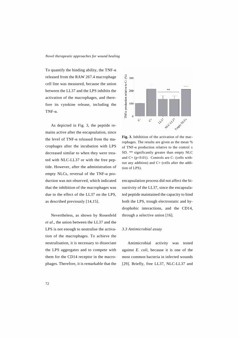
Novel therapeutic approaches for wound healing
72
To quantify the binding ability, the TNF-α
released from the RAW 267.4 macrophage
cell line was measured, because the union
between the LL37 and the LPS inhibits the
activation of the macrophages, and there-
fore its cytokine release, including the
TNF-α.
As depicted in Fig. 3, the peptide re-
mains active after the encapsulation, since
the level of TNF-α released from the ma-
crophages after the incubation with LPS
decreased similar to when they were trea-
ted with NLC-LL37 or with the free pep-
tide. However, after the administration of
empty NLCs, reversal of the TNF-α pro-
duction was not observed, which indicated
that the inhibition of the macrophages was
due to the effect of the LL37 on the LPS,
as described previously [14,15].
Nevertheless, as shown by Rosenfeld
et al., the union between the LL37 and the
LPS is not enough to neutralise the activa-
tion of the macrophages. To achieve the
neutralisation, it is necessary to dissociate
the LPS aggregates and to compete with
them for the CD14 receptor in the macro-
phages. Therefore, it is remarkable that the
Fig. 3. Inhibition of the activation of the mac-rophages. The results are given as the mean % of TNF-α production relative to the control ± SD. ** significantly greater than empty NLC and C+ (p<0.01). Controls are C- (cells with-out any addition) and C+ (cells after the addi-tion of LPS).
encapsulation process did not affect the bi-
oactivity of the LL37, since the encapsula-
ted peptide maintained the capacity to bind
both the LPS, trough electrostatic and hy-
drophobic interactions, and the CD14,
through a selective union [16].
3.3 Antimicrobial assay
Antimicrobial activity was tested
against E. coli, because it is one of the
most common bacteria in infected wounds
[29]. Briefly, free LL37, NLC-LL37 and
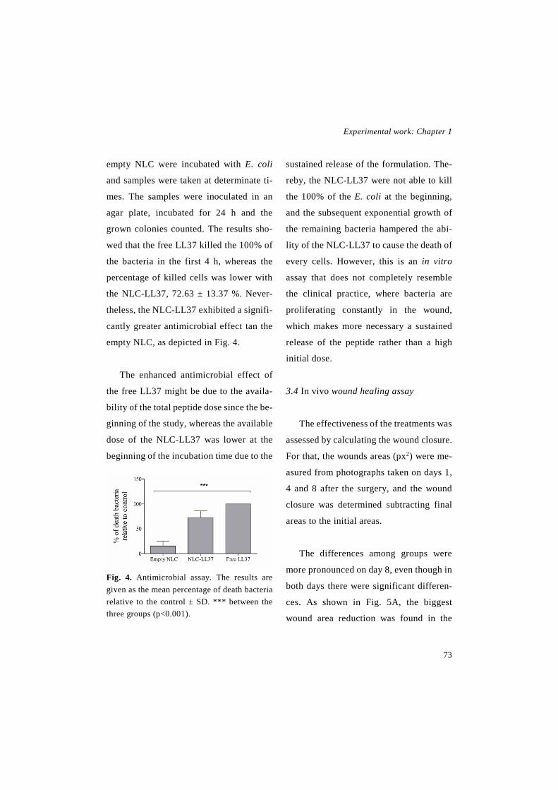
Experimental work: Chapter 1
73
empty NLC were incubated with E. coli
and samples were taken at determinate ti-
mes. The samples were inoculated in an
agar plate, incubated for 24 h and the
grown colonies counted. The results sho-
wed that the free LL37 killed the 100% of
the bacteria in the first 4 h, whereas the
percentage of killed cells was lower with
the NLC-LL37, 72.63 ± 13.37 %. Never-
theless, the NLC-LL37 exhibited a signifi-
cantly greater antimicrobial effect tan the
empty NLC, as depicted in Fig. 4.
The enhanced antimicrobial effect of
the free LL37 might be due to the availa-
bility of the total peptide dose since the be-
ginning of the study, whereas the available
dose of the NLC-LL37 was lower at the
beginning of the incubation time due to the
Fig. 4. Antimicrobial assay. The results are given as the mean percentage of death bacteria relative to the control ± SD. *** between the three groups (p<0.001).
sustained release of the formulation. The-
reby, the NLC-LL37 were not able to kill
the 100% of the E. coli at the beginning,
and the subsequent exponential growth of
the remaining bacteria hampered the abi-
lity of the NLC-LL37 to cause the death of
every cells. However, this is an in vitro
assay that does not completely resemble
the clinical practice, where bacteria are
proliferating constantly in the wound,
which makes more necessary a sustained
release of the peptide rather than a high
initial dose.
3.4 In vivo wound healing assay
The effectiveness of the treatments was
assessed by calculating the wound closure.
For that, the wounds areas (px2) were me-
asured from photographs taken on days 1,
4 and 8 after the surgery, and the wound
closure was determined subtracting final
areas to the initial areas.
The differences among groups were
more pronounced on day 8, even though in
both days there were significant differen-
ces. As shown in Fig. 5A, the biggest
wound area reduction was found in the
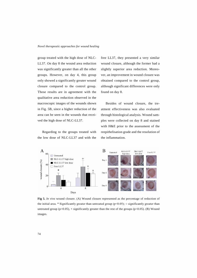
Novel therapeutic approaches for wound healing
74
group treated with the high dose of NLC-
LL37. On day 8 the wound area reduction
was significantly greater than all the other
groups. However, on day 4, this group
only showed a significantly greater wound
closure compared to the control group.
Those results are in agreement with the
qualitative area reduction observed in the
macroscopic images of the wounds shown
in Fig. 5B, since a higher reduction of the
area can be seen in the wounds that recei-
ved the high dose of NLC-LL37.
Regarding to the groups treated with
the low dose of NLC-LL37 and with the
free LL37, they presented a very similar
wound closure, although the former had a
slightly superior area reduction. Moreo-
ver, an improvement in wound closure was
obtained compared to the control group,
although significant differences were only
found on day 8.
Besides of wound closure, the tre-
atment effectiveness was also evaluated
through histological analysis. Wound sam-
ples were collected on day 8 and stained
with H&E prior to the assessment of the
reepithelisation grade and the resolution of
the inflammation.
Fig 5. In vivo wound closure. (A) Wound closure represented as the percentage of reduction of
the initial area. * Significantly greater than untreated group (p<0.05); ○ significantly greater than
untreated group (p>0.05), + significantly greater than the rest of the groups (p<0.05). (B) Wound
images.

Experimental work: Chapter 1
75
The reepithelisation is the main healing
process in humans, whereas in rodents the
main process is wound contraction. Howe-
ver, reepithelisation is an important hea-
ling process in this animal model, since the
db/db mice have impaired the wound con-
traction process, according to Fang et al.,
because of the extreme obesity of the ani-
mals reduces the looseness of the skin
[30,31]. Furthermore, the wound contrac-
tion is more impaired by the silicone splint
sutured around the wounds [25], which
makes the reepithelisation an important
healing process in this animal model.
The results obtained in the analysis of
the reepithelisation (Fig. 6A) showed that
the wounds treated with the high dose of
NLC-LL37 presented a significantly hig-
her grade of reepithelisation, with new
epithelium covering more than a half of
the wound, around 2, according to the
scale described by Sinha et al. [26]. Con-
versely, the wounds treated with free LL37
and those of the control group had reepi-
thelisation covering less than a half of the
wound (~1), and the wounds treated with
the low dose of NLC-LL37 had reepitheli-
sation only in the margin of the wounds
(~0). These results are consistent with those
obtained in wound closure, since the high
dose of NLC-LL37 improved the wound
healing compared to a lower dose of the
NLC-LL37 and the same dose of free LL37.
The analysis of the resolution of the in-
flammation was conducted following the
criteria established by Cotran et al. [27].
As shown in Fig. 6B, the groups treated
with the high dose of NLC-LL37 and with
the free LL37 presented an accelerated in-
flammation recovery, as the wounds exhi-
bited a predominance of granulation tissue
formation and angiogenesis, with barely
presence of pyogenic membrane and neu-
trophils (grade 2). However, the wounds of
the control group and of the group treated
with the low dose of NLC-LL37 were cha-
racterised by the formation of the fibrin
clot and the migration of macrophages and
polynuclear neutrophils that form the pyo-
genic membrane (grade 1). The permanent
neutrophil infiltration showed in the last
groups, is characteristic of chronic wounds
and it is involved in delayed wound hea-
ling, since neutrophils release an excessive
amount of proteases that degrade the ex-
tracellular matrix [32].
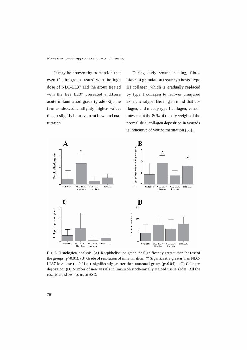
Novel therapeutic approaches for wound healing
76
It may be noteworthy to mention that
even if the group treated with the high
dose of NLC-LL37 and the group treated
with the free LL37 presented a diffuse
acute inflammation grade (grade ~2), the
former showed a slightly higher value,
thus, a slightly improvement in wound ma-
turation.
During early wound healing, fibro-
blasts of granulation tissue synthesise type
III collagen, which is gradually replaced
by type I collagen to recover uninjured
skin phenotype. Bearing in mind that co-
llagen, and mostly type I collagen, consti-
tutes about the 80% of the dry weight of the
normal skin, collagen deposition in wounds
is indicative of wound maturation [33].
Fig. 6. Histological analysis. (A) Reepithelisation grade. ** Significantly greater than the rest of the groups (p>0.01). (B) Grade of resolution of inflammation. ** Significantly greater than NLC-LL37 low dose (p<0.01); ● significantly greater than untreated group (p<0.05). (C) Collagen deposition. (D) Number of new vessels in immunohistochemically stained tissue slides. All the results are shown as mean ±SD.

Experimental work: Chapter 1
77
Accordingly, Masson Trichrome stai-
ning revealed that the high dose of NLC-
LL37 slightly improved wound matura-
tion, as it enhanced collagen deposition
compared to the rest of the treatments, as
depicted in Fig. 6C. In untreated wounds,
in the wounds that received the low dose
of NLC-LL37 and in the wounds that re-
ceived free LL37 there was a lack of colla-
gen deposition (grade 0 according to the
criteria set by Gal et al. [28]), since most
of the wounds did not show collagen and
only a few of them showed a mild deposi-
tion. The group treated with the highest
dose of NLC-LL37 presented a mild depo-
sition of collagen (grade 1), however, one
wound exhibited a marked deposition of
collagen and another one, a moderate de-
position. Nevertheless, the differences in
collagen deposition did not achieve statis-
tical significance, possibly due to the great
variability found within the groups. That
wide variability was previously described
by Trousdale et al. (2009) analysing
wound healing in db/db mice [34].
The angiogenic process was evaluated
by counting the newly formed blood ve-
ssels in tissue slides that were immunohis-
tochemically stained with antiCD31 mo-
noclonal antibody. Although the results
did not reach statistical significance, the
wounds treated with free LL37 and with
the high dose of NLC-LL37 presented a
tendency to show higher number of vessels
compared to the untreated wounds. In fact,
two of the wounds showed more than 10
new vessels in each group, whereas none
of the wounds in the untreated group sho-
wed more than 8 vessels. Furthermore, the
group treated with the low dose of NLC-
LL37 presented a slightly augmented mi-
crovascular density (one wound with more
than 10 vessels) compared to the untreated
group, as illustrated in Fig. 6D.
Overall, these results indicate that topi-
cally administered NLC-LL37 can im-
prove wound healing in terms of wound
closure, reepithelisation and resolution of
the inflammation in a full thickness wound
compared to the administration of the free
peptide. The enhanced activity of the en-
capsulated peptide can be explained by the
protective effect of the NLCs, which pro-
tect the LL37 against both, chemical de-
gradation and degradation caused by the
proteases present in the wound bed.
Novel therapeutic approaches for wound healing
78
Moreover, the NLCs allow a controlled
release profile, and thus extend the dura-
tion of the LL37 effect [20,21]. That is
why, the NLC-LL37 formulation can re-
duce the high dosing frequency needed
when the peptide is administered freely.
Previous studies have demonstrated the
usefulness of encapsulating LL37 in nano-
particles [19], however in this study the
nanoparticles encapsulating LL37 have
been administered through the topical
route for the first time, which presents se-
veral advantages. First of all, the lipidic
nature of the NLCs creates an occlusive la-
yer that increases skin hydration favouring
the penetration of the encapsulated LL37
[21,35]. Moreover, the topical administra-
tion is an easy, comfortable and safe route
for the patient.
4. Conclusion
In the current study NLCs loaded with
2% of LL37 have been developed. The na-
noparticles exhibited a mean size of 270
nm, showed lack of cytotoxicity and the
encapsulated LL37 maintained its bioacti-
vity, as it was evidenced by the immuno-
modulatory and the antimicrobial in vitro
assays.
The in vivo full thickness wound hea-
ling assay conducted in db/db mice, de-
monstrated that the administration of 6 µg
of LL37 into NLC-LL37 topically impro-
ved wound healing compared to the admi-
nistration of the free LL37. Taking into
account all these findings, it can be conclu-
ded that the topical administration of NLC-
LL37 might present an interesting strategy
for the treatment of chronic wounds.
5. Acknowledgments
I. García-Orue thanks the Basque Go-
vernment for the fellowship grant. The au-
thors thank for technical and human su-
pport provided by SGIker of UPV/EHU
and European funding (ERDF and ESF).
This project has been funded by the Spa-
nish Ministry of Economy and competiti-
veness (INNPACTO, IPT-2012-0602-
300000, 2012). In addition, it has been
partially supported by the Basque govern-
ment (Consolidated Groups, IT-407-07
and IT-528-10) and the University of the
Basque Country UPV/EHU (UFI11/32).
Experimental work: Chapter 1
79
6. References
[1] C.K. Sen, G.M. Gordillo, S. Roy, R. Kirsner, L. Lambert, T.K. Hunt, F. Gottrup, G.C. Gurtner, M.T. Longaker, Human skin wounds: A major and snowballing threat to public health and the economy, Wound Repair and Regeneration 17 (2009) 763-771.
[2] V.W. Wong, G.C. Gurtner, Tissue engi-neering for the management of chronic wounds: current concepts and future perspec-tives, Exp. Dermatol. 21 (2012) 729-734.
[3] P. Martin, Wound Healing--Aiming for Perfect Skin Regeneration, Science 276 (1997) 75-81.
[4] A.J. Singer, R.A. Clark, Cutaneous wound healing, N Engl J Med 341 (1999) 738-746.
[5] R.F. Diegelmann, M.C. Evans, Wound healing: an overview of acute, fibrotic and de-layed healing., Front Biosci. 9 (2004) 283-289.
[6] J.D. Heilborn, M.F. Nilsson, O. Sørensen, M. Ståhle-Bäckdahl, G. Kratz, G. Weber, N. Borregaard, The Cathelicidin Anti-Microbial Peptide LL-37 is Involved in Re-Epithelializa-tion of Human Skin Wounds and is Lacking in Chronic Ulcer Epithelium, J. Invest. Dermatol. 120 (2003) 379-389.
[7] M. Frohm, B. Agerberth, G. Ahangari, M. Ståhle-Bäckdahl, S. Lidén, H. Wigzell, G.H. Gudmundsson, The Expression of the Gene Coding for the Antibacterial Peptide LL-37 Is Induced in Human Keratinocytes during In-flammatory Disorders, Journal of Biological Chemistry 272 (1997) 15258-15263.
[8] R. Koczulla, G. von Degenfeld, C. Kupatt, F. Krötz, S. Zahler, T. Gloe, K. Issbrücker, P. Unterberger, M. Zaiou, C. Lebherz, A. Karl, P. Raake, A. Pfosser, P. Boekstegers, U. Welsch, P.S. Hiemstra, C. Vogelmeier, R.L. Gallo, M. Clauss, R. Bals, An angiogenic role for the hu-man peptide antibiotic LL-37/hCAP-18., J Clin Invest 111 (2003) 1665-1672.
[9] S.B. Coffelt, S.L. Tomchuck, K.J. Zwezdaryk, E.S. Danka, A.B. Scandurro, Leu-cine Leucine-37 Uses Formyl Peptide Recep-tor–Like 1 to Activate Signal Transduction Pathways, Stimulate Oncogenic Gene Expres-sion, and Enhance the Invasiveness of Ovarian Cancer Cells, Molecular Cancer Research 7 (2009) 907-915.
[10] K. Hase, L. Eckmann, J.D. Leopard, N. Varki, M.F. Kagnoff, Cell Differentiation Is a Key Determinant of Cathelicidin LL-37/Hu-man Cationic Antimicrobial Protein 18 Expres-sion by Human Colon Epithelium, Infection and Immunity 70 (2002) 953-063.
[11] R.C. Anderson, M. Rehders, P.L. Yu, An-timicrobial fragments of the pro-region of cathelicidins and other immune peptides, Bio-technol. Lett. 30 (2008) 813-818.
[12] D. Vandamme, B. Landuyt, W. Luyten, L. Schoofs, A comprehensive summary of LL-37, the factotum human cathelicidin peptide, Cell Immunol 280 (2012) 22-35.
[13] Y. Kai-Larsen, B. Agerberth, The role of the multifunctional peptide LL-37 in host de-fense, Front. Biosci 13 (2008) 3760-3767.
[14] J. Turner, Y. Cho, N. Dinh, A.J. Waring, R.I. Lehrer, Activities of LL-37, a Cathelin-Associated Antimicrobial Peptide of Human
Novel therapeutic approaches for wound healing
80
Neutrophils, Antimicrobial Agents and Chem-otherapy 42 (1998) 2206-2214.
[15] R. Ramos, J.P. Silva, A.C. Rodrigues, R. Costa, L. Guardão, F. Schmitt, R. Soares, M. Vilanova, L. Domingues, M. Gama, Wound healing activity of the human antimicrobial peptide LL37, Peptides 32 (2011) 1469-1476.
[16] Y. Rosenfeld, N. Papo, Y. Shai, Endotoxin (Lipopolysaccharide) Neutralization by Innate Immunity Host-Defense Peptides: peptide properties ad plausible modes of action, Jour-nal of Biological Chemistry 281 (2006) 1636-1643.
[17] R.E. Hancock, Cationic peptides: effec-tors in innate immunity and novel antimicrobi-als, Lancet Infect Dis 1 (2001) 156-164.
[18] M. Carretero, M. Del Río, M. García, M.J. Escámez, I. Mirones, L. Rivas, C. Balague, J.L. Jorcano, F. Larcher, A cutaneous gene therapy approach to treat infection through keratino-cyte-targeted overexpression of antimicrobial peptides., FASEB J. 18 (2004) 1931-1933.
[19] K.K. Chereddy, C. Her, M. Comune, C. Moia, A. Lopes, P.E. Porporato, J. Vanacker, M.C. Lam, L. Steinstraesser, P. Sonveaux, H. Zhu, L.S. Ferreira, G. Vandermeulen, V. Préat, PLGA nanoparticles loaded with host defense peptide LL37 promote wound healing, J Con-trol Release 194 (2014) 138-147.
[20] M. Schäfer-Korting, W. Mehnert, H. Korting, Lipid nanoparticles for improved top-ical application of drugs for skin diseases, Adv. Drug Deliv. Rev. 59 (2007) 427-443.
[21] J. Pardeike, A. Hommoss, R.H. Müller, Lipid nanoparticles (SLN, NLC) in cosmetic
and pharmaceutical dermal products, Int. J. Pharm. 366 (2009) 170-184.
[22] R.H. Müller, M. Radtke, S.A. Wissing, Solid lipid nanoparticles (SLN) and nanostruc-tured lipid carriers (NLC) in cosmetic and der-matological preparations, Adv. Drug Deliv. Rev. 54, Supplement (2002) S131-S155.
[23] G. Gainza, M. Pastor, J.J. Aguirre, S. Vil-lullas, J.L. Pedraz, R.M. Hernandez, M. Igar-tua, A novel strategy for the treatment of chronic wounds based on the topical admin-istration of rhEGF-loaded lipid nanoparticles: In vitro bioactivity and in vivo effectiveness in healing-impaired db/db mice, J Control Re-lease 185 (2014) 51-61.
[24] G. Gainza, D.C. Bonafonte, B. Moreno, J.J. Aguirre, F.B. Gutierrez, S. Villullas, J.L. Pedraz, M. Igartua, R.M. Hernandez, The topi-cal administration of rhEGF-loaded nanostruc-tured lipid carriers (rhEGF-NLC) improves healing in a porcine full-thickness excisional wound model, J Control Release 197 (2015) 41-47.
[25] J. Michaels, S.S. Churgin, K.M. Blech-man, M.R. Greives, S. Aarabi, R.D. Galiano, G.C. Gurtner, db/db mice exhibit severe wound-healing impairments compared with other murine diabetic strains in a silicone-splinted excisional wound model, Wound Re-pair Regen 15 (2007) 665-670.
[26] U.K. Sinha, L.A. Gallagher, Effects of Steel Scalpel, Ultrasonic Scalpel, CO2 Laser, and Monopolar and Bipolar Electrosurgery on Wound Healing in Guinea Pig Oral Mucosa, Laryngoscope 113 (2003) 228-236.

Experimental work: Chapter 1
81
[27] R. Cotran, G.K. Kumar, T. Collins, Repa-ración de los tejidos: proliferacion celular, fi-brosis y curación de las heridas, in: R. Cotran, G.K. Kumar, T. Collins (Eds.), Patología Es-tructural Y Funcional, McGraw-Hill, Interame-ricana, Madrid, 2000, pp. 95-120.
[28] P. Gál, T. Toporcer, B. Vidinský, M. Mokrý, M. Novotný, R. Kilík, K. Smetana, T. Gál, J. Sabo, Early changes in the tensile stregnth and morphology of primary sutured skin wounds in rats, Folia Biol. (Praha) 52 (2006) 109-115.
[29] A.R. Siddiqui, J.M. Bernstein, Chronic wound infection: Facts and controversies, Clin. Dermatol. 28 (2010) 519-526.
[30] R.C. Fang, Z.B. Kryger, D.W. Buck II, M. De La Garza, R.D. Galiano, T.A. Mustoe, Lim-itations of the db/db mouse in translational wound healing research: Is the NONcNZO10 polygenic mouse model superior?, Wound Re-pair and Regeneration 18 (2010) 605-613.
[31] V.I. Tkalcevic, S. Cužic, M.J. Parnham, I. Pašalic, K. Brajša, Differential Evaluation of Excisional Non-occluded Wound Healing in db/db Mice, Toxicologic Pathology 37 (2009) 183-192.
[32] N.B. Menke, K.R. Ward, T.M. Witten, D.G. Bonchev, R.F. Diegelmann, Impaired wound healing, Clin. Dermatol. 25 (2007) 19-25.
[33] J. Li, J. Chen, R. Kirsner, Pathophysiology of acute wound healing, Clin. Dermatol. 25 (2007) 9-18.
[34] R.K. Trousdale, S. Jacobs, D.A. Simhaee, J.K. Wu, J.W. Lustbader, Wound Closure and Metabolic Parameter Variability in a db/db Mouse Model for Diabetic Ulcers, J. Surg. Res. 151 (2009) 100-107.
[35] R.H. Müller, R.D. Petersen, A. Hommoss, J. Pardeike, Nanostructured lipid carriers (NLC) in cosmetic dermal products, Adv. Drug Deliv. Rev. 59 (2007) 522-5

CHAPTER 2
Novel nanofibrous dressings containing rhEGF and Aloe vera for wound healing applications
I. García-Oruea, G. Gainzaa, F.B. Gutierrezb, J.J. Aguirreb, C. Evorac, J.L. Pedraza,d, R.M. Hernandeza,d, A. Delgadoc*, M. Igartuaa,d** a NanoBioCel Group, Laboratory of Pharmaceutics, School of Pharmacy, University of the Basque Country (UPV/EHU). b Hospital Universitario de Álava (HUA) Txagorritxu, Vitoria-Gasteiz, 01009, Spain. c Department of Chemical Engineering and Pharmaceutical Technology. School of Pharmacy. University of La Laguna,
Tenerife, Spain. d Biomedical Research Networking Centre in Bioengineering, Biomaterials and Nanomedicine (CIBER-BBN).
*Corresponding author: Dr. A. Delgado and Dr. M. Igartua
ABSTRACT
Nanofibrous membranes produced by electrospinning possess a large surface area-to-volume ratio, which mimics the three-dimensional structure of the extracellular ma-trix. Thus, nanofibrous dressings are a promising alternative for chronic wound healing, since they can replace the natural ECM until it is repaired. Therefore, in this study we developed a PLGA nanofibrous membrane that contains recombinant human Epidermal Growth Factor (rhEGF) and Aloe vera (AV) extract. Both of them promote wound heal-ing, as EGF is a wound healing mediator and AV stimulates the proliferation and activity of fibroblast. The obtained membranes were composed of uniform and randomly oriented fibers with an average diameter of 356.03 ± 112.05 nm, they presented a porosity of 87.92 ± 11.96 % and the amount of rhEGF was 9.76 ± 1.75 µg/mg. The in vitro viability assay demonstrated that the membranes containing rhEGF and AV improved fibroblast proliferation, revealing the beneficial effect of the combination. Furthermore, these membranes accelerated significantly wound closure and reepithelisation in an in vivo full thickness wound healing assay carried out in db/db mice. Overall, these findings demon-strated the potential of PLGA nanofibers containing rhEGF and AV for the treatment of chronic wounds.
Published in: International Journal of Pharmaceutics 523 issue 2 (2017), 556-566. Doi: https://doi.org/10.1016/j.ijpharm.2016.11.006

Experimental work: Chapter 2
85
1. Introduction
Wound healing is a dynamic and com-
plex process with the aim of restoring the
normal anatomical structure and functio-
nality of the skin (Diegelmann and Evans
2004). In order to achieve that objective,
diverse immunological and biological sys-
tems participate in a coordinated way,
through three distinct but overlapping pha-
ses, including an inflammatory response
(which comprises hemostasis and inflam-
mation), a proliferative phase (consisting
of protein synthesis and wound contrac-
tion) and a remodelling phase (Monaco
and Lawrence 2003; Morton and Phillips
2016; Schreml, et al. 2010). Nevertheless,
some wounds fail to progress through he-
aling in that timely and spatially organised
manner. The disturbance of the healing
can be caused by several factors, such as,
infection, necrosis, tissue hypoxia, exuda-
tes and an excess of inflammatory cytoki-
nes. Therefore, wounds get stagnated in a
permanent inflammatory stage, with an ab-
normally high level of proinflammatory
cytokines and proteases, which lead to the
deterioration of the extracellular matrix
(ECM) and, thus, perpetuating a non healing
state (Briquez, et al. 2015; Velnar, et al.
2009).
The treatment of chronic wounds has
gained a huge impact in our society, due to
the alarming increase of its prevalence in
the last years. Moreover, the prevalence is
expected to continue growing exponentia-
lly because of the increase of the high risk
population, which includes elderly, diabe-
tics and obese (Whittam, et al. 2016). In-
deed, it has been estimated that 1-2% of
the population of developed countries will
suffer from chronic wounds during their li-
fetime, representing their associated cost
the 2% of the European health budget
according to the United Nations (Sen, et al.
2009). Current studies to overcome this
clinical problem are focused on the deve-
lopment of wound dressings able to
enhance wound healing. Among those,
nonwoven nanofibrous membranes are of
especial interest, since their unique archi-
tectural features mimic the three dimensio-
nal structure of the ECM (Choi, et al. 2015).
Nanofibers are usually produced by
electrospinning, a simple, cost effective
and versatile technique that uses an elec-
Novel therapeutic approaches for wound healing
86
tric force to spin nanometric fibers from a
polymeric solution (Abrigo, et al. 2014;
Gainza, et al. 2015b). The nanofibers ge-
nerated by this technique exhibit a high
surface area-to-volume ratio and a high
porosity, that enhance wound healing
through diverse mechanisms. For exam-
ple, they allow gas permeation, which pro-
motes cell respiration; they retain moisture
and they improve the removal of exudates
(Pachuau 2015). The dressings should pro-
mote cell migration and proliferation,
while preventing tissue ingrowth within
the nanofibrous membrane, since they are
designed to be removed once the wound
has healed (Abrigo, et al. 2014). Further-
more, nanofibers can be loaded with active
compounds to accelerate the wound hea-
ling, as it has been shown in numerous stu-
dies with the encapsulation of drugs such
as, growth factors, Aloe vera extract, cur-
cumin, metformin, ciprofloxacin and co-
llagen, among others (Choi, et al. 2008;
Kataria, et al. 2014; Lai, et al. 2014; Lee,
et al. 2014; Merrell, et al. 2009; Uslu and
Aytimur 2012). In addition, the encapsula-
tion of drugs into nanofibers allows their
topical administration, which present se-
veral advantages. Firstly, it improves pati-
ent compliance, permitting self adminis-
tration; secondly, it maximises the thera-
peutic potential by the improvement of bi-
oavailability and the maintenance of drug
concentration in the wound site; and fina-
lly, it reduces the systemic drug toxicity
(Pachuau 2015; Whittam, et al. 2016).
The nanofibrous membranes obtained
through electrospinning have also been
used in cell based therapies. The similarity
of the membranes to the ECM allows cell
penetration, differentiation and adhesion,
making them a suitable approach for deve-
loping skin substitutes or stem cell the-
rapy. Skin substitutes are scaffolds seeded
with cells to resemble the native skin
structure (Dickinson and Gerecht 2016).
In order to ease cell-matrix interactions,
scaffolds are often produced of collagen
and other naturally occurring ECM pro-
teins (Hodgkinson and Bayat 2011), as the
skin substitute developed by Powell et al.
where human dermal fibroblast and epi-
dermal keratinocytes were seeded into a
collagen electrospun scaffold (Powell, et
al. 2008). In another study, Han et al. de-
veloped a scaffold based on other natural
polymer (PHVB) that was seeded with hair
Experimental work: Chapter 2
87
follicular cells because their low immuno-
genicity is advantageous for allografts
(Han, et al. 2007). On the other hand, a va-
riety of stems cells; such as, adipose-deri-
ved stem cells (Gu, et al. 2014; Machula,
et al. 2014), mesenchymal stem cells
(Bhowmick, et al. 2016; Steffens, et al.
2014), unrestricted somatic stem cells
(Bahrami, et al. 2016; Keshel, et al. 2014)
and urine derived stem cells (Fu, et al.
2014); have been used to produce wound
healing scaffolds anchored to electrospun
membranes. Those stem cells have proven
to improve wound healing due to their ca-
pacity to differentiate into endothelial
cells and to express soluble factors and
ECM proteins needed in wound healing.
Aloe vera extract (AV) is a compound
that can benefit from the encapsulation in
nanofibers for wound healing treatment.
Aloe vera is an herbaceous succulent plant
that belongs to Liliaceae family. Besides
ist pharmacological activity, which inclu-
des anti-inflammatory, antiarthritis, anti-
bacterial, antifungal and hypoglycaemic
effects, it has been reported to promote
wound healing (Choi and Chung 2003; Ha
shemi, et al. 2015). The healing effect is
mainly mediated by a compound called
glucomannan that affects fibroblast
growth factor and stimulates the activity
and proliferation of the fibroblasts, which
improves collagen production and secre-
tion by those cells (Hashemi, et al. 2015).
Growth factors are also interesting
compounds for encapsulating in nanofi-
brous wound dressings, since they are
essential healing mediators for a success-
ful tissue repair (Choi, et al. 2012). Among
others, the Epidermal Growth Factor
(EGF) is a key signalling molecule that sti-
mulates keratinocyte and fibroblast proli-
feration and migration, hence improving
wound healing (Bodnar 2013). Further-
more, its external administration in chro-
nic wounds has been proven to promote
wound healing (Choi, et al. 2008; Gainza,
et al. 2015a). However, the clinical appli-
cation of EGF is hampered by its short
half-life, due to its degradation by the hi-
drolitic enzymes present in the wound bed.
Therefore, the encapsulation in nanofi-
brous membranes can overcome this limi-
tation, as these formulations protect the
EGF form the environment, and thus
enhance its stability (Pachuau 2015).

Novel therapeutic approaches for wound healing
88
Nanofibrous membranes can be produ-
ced from natural or synthetic polymers.
Natural polymers used in electrospinning
include ECM components, such as, colla-
gen, laminin, fibrin or elastin; and other
polymers such as, chitosan, agarose, gela-
tine, pectin, silk, etc. The properties of
those polymers (biocompatibility, non-to-
xicity, physcochemical properties and ma-
trix-cell interactions) made them adequate
for wound healing (Garg, et al. 2015). Ho-
wever, their weak mechanical properties
and difficulty for electrospinning make ne-
cessary to reinforce them with synthetic
polymers (Hodgkinson and Bayat 2011).
Among the synthetic polymers used for
electrospinning there are cellulose acetate,
polylactic-co-glycolic acid (PLGA),
poly(vinyl alcohol), poly(lactic acid), po-
lystyrene, polyglycerol, polyglycolide, po-
lyurethane, etc. Synthetic materials are
stronger and cheaper, present a well defi-
ned structure and are more easily electros-
pinable (Garg, et al. 2015).
Taking that into account, in this study,
PLGA was electrospinned to develop a na-
nofibrous dressing loaded with rhEGF and
AV for the treatment of chronic wounds.
The nanofibrous membranes were charac-
terised to evaluate their suitability for
wound dressing, and in vitro tests were un-
dertaken to analyse the bioactivity of the
encapsulated active compounds.
In addition, dressings’ efficacy was
assessed in vivo. Although pig dermis is
probably the most similar to that of hu-
mans, rodent wound models are more
common due to the difficulties in working
with large animals (Seaton, et al. 2015).
Nevertheless, rodent wound healing para-
llels worse human wound healing, because
they heal primarily by contraction due to
their loose skin. In order to avoid that di-
fference, wounds are inflicted in specific
anatomic sites as head, ears or tail, where
skin is tighter. Another approach to address
that problem is to adhere circular silicone
splints beyond the wound margin to pre-
vent wound contraction, which makes this
model the most representative of open hu-
man wounds in rodents (Fang and Mustoe
2008).
To mimic the delayed wound healing in
chronic wounds, rodent models with im-
paired healing conditions can be used,
Experimental work: Chapter 2
89
such as, ischemic (Elgharably, et al. 2014;
Magin, et al. 2016) or diabetic models.
Among diabetic models the most widely
used are chemically induced diabetes se-
condary to streptozocin injections, since it
destroys pancreatic β-cells developing a
phenotype similar to type I diabetes (Inpa-
nya, et al. 2012; Moura, et al. 2014); and
genetically diabetic db/db mice (BKS.Cg-
m +/+ Leprdb/J), which present a mutation
of the leptin receptor and thus, possess a
phenotype of type II diabetes (Guillemin,
et al. 2016; Losi, et al. 2013). A study ca-
rried out by Michaels et al. demonstrated
that wound healing was more impaired in
the db/db mice than in the streptozocin
model (Michaels, et al. 2007).
Accordingly, in this study the dressings
were topically applied in a full-thickness
excisional splinted wound model in db/db
mice, and their efficacy was assessed in
terms of wound closure, reepithelisation
grade, recovery of the inflammatory stage
and collagen deposition.
2. Materials and methods
2.1 Nanofiber preparation
An emulsion (o/w) containing rhEGF,
AV, and PLGA was electrospun to fabri-
cate a PLGA-AV-EGF nanofibrous mem-
brane. The organic phase of the emulsion
was composed of 140 mg of PLGA 50:50
(Resomer RG504, Evonik, Germany) in
650 µL of hexafluoroisopropanol (HFIP,
Fluka, Switzerland), and the aqueous
phase was 350 µL of a polyvinyl alcohol
(PVA) solution (0.5% w/v) containing 140
mg of Aloe vera powder (Agora Valencia
SL., Spain) and 5 mg of rhEGF (Centre for
Genetic Engineering and Biotechnology,
Cuba). Both phases were vortexed at level
10 for 3 min to form the emulsion (Vortex-
Genie 2, Scientific Industries Inc., USA).
The resulting emulsion was loaded into a
Luer-lock syringe (Norm-Ject) with an
18G needle attached to a pump (Harvard
Apparatus, MA) providing a flow rate of 1
mL/h and it was electrospun horizontally
on a rotating collector located at 12 cm
from the needle, under 12 kV power
supply. The collector rotating speed was
set at 200 rpm.

Novel therapeutic approaches for wound healing
90
The preparation of the PLGA-AV and
the PLGA nanofibers only differ from the
explained above in the composition of the
aqueous phase, which lacked the rhEGF,
and the rhEGF and the AV, respectively.
The injected volume was normalized to
obtain membranes with similar thickness.
The products used for producing the
membranes were previously sterilised by
gamma radiation and the syringe, the co-
llector and the needle used for electrospin-
ning were autoclaved. Moreover, the elec-
trospinning process was carried out under
aseptic conditions. Finally, before the in
vitro and in vivo studies the membranes
were sterilised by keeping them under UV
light for 30 minutes.
2.2. Nanofiber characterisation
The nanofibrous membrane morpho-
logy, i.e. membrane quality and fiber dia-
meter, was analysed using Scanning Elec-
tron Microscopy images (SEM, Jeol JSM-
6300). Porosity (P) of the membranes was
calculated through the following equation
(Eq. 1), where ρreal and ρapp are the real and
apparent densities, respectively:
P (%)=�1 - ρapp
ρreal� x 100 (1)
Real density was assessed by means of
a helium pycnometer (Micromeritics, Accu-
Pyc 1330, USA) and apparent density was
calculated dividing the mass by the calcu-
lated volume (length × width × height).
Membrane thickness was measured from
photographs obtained by stereo micros-
copy (Leica M205 C, Leica LAS, Germany).
Monotonic tensile tests of the PLGA-
AV-EGF, PLGA-AV and PLGA nanofi-
bers were performed under displacement
control on an Instron 5848 microtester,
using a 5 N full scale load cell (Instron®,
UK). The samples were loaded at a displa-
cement rate of 0.01 mm/s up to rupture.
The load-displacement curve was transfor-
med in a stress-strain curve and the ulti-
mate strength was determined to compare
the mechanical properties of the different
nanofibers. At least 5 samples of each
membrane were tested and the results were
presented as mean ± standard deviation.
Nanofibers degree of water uptake in
an aqueous environment was evaluated in

Experimental work: Chapter 2
91
vitro to compare the behaviour of the three
nanofibrous membranes. After weighing
1x1 cm nanofiber samples, they were soa-
ked in 1 ml of PBS and incubated at 32 °C
for 1 h. Then, they were removed and the
excess of water dried. Finally, the nanofi-
ber pieces were weighed to calculate the
water uptake through the following equa-
tion (Eq. 2):
Water uptake (%)=M-M0
M0×100 (2)
Where M represents the weight of the
membrane after the immersion in PBS and
M0 represents the initial weight of the na-
nofibrous mesh.
The Water Vapour Permeability Rate
(WVPR) through the membranes was de-
termined modifying the method described
by Li et al. (Li, et al. 2013). The mouth of
a cup containing silica gel dessicant (1cm
in diameter) was thoroughly covered with
the test membrane, so that water vapor
only can enter to the cup through the mem-
brane. The cup was placed in a chamber at
30°C with a constant relative humidity of
75% for 24 hours. The change of the
weight of the assembly before and after the
exposure time was recorded and WVPR
was calculated using the following equa-
tion (3):
WVPR=M1-M0
A×T (3)
Where M0 is the weight of the assem-
bly before the assay, M1 is its weight after
the assay, A is the exposure area of the
membrane and T is the exposure time.
The thermal behaviour of the nanofi-
bers, the physical blend of the components
and the components themselves were stu-
died using Differential Scanning Calori-
metry (DSC-50, Shimadzu, Japan). Each
sample was sealed in an aluminium pan
and heated from 25°C to 350°C at a hea-
ting rate of 10°C per minute.
To determine the amount of rhEGF lo-
aded into the PLGA-AV-EGF nanofibers,
a piece of 1x1 cm of the membrane was
dissolved in 200 µL of dimethyl sul-
phoxide (DMSO) (Scharlau, Spain), vorte-
xed until it became a clear solution and mi-
xed with 800 µL of phosphate buffered sa-
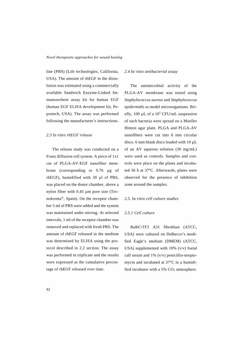
Novel therapeutic approaches for wound healing
92
line (PBS) (Life technologies, California,
USA). The amount of rhEGF in the disso-
lution was estimated using a commercially
available Sandwich Enzyme-Linked Im-
munosorbent assay kit for human EGF
(human EGF ELISA development kit, Pe-
protech, USA). The assay was performed
following the manufacturer’s instructions.
2.3 In vitro rhEGF release
The release study was conducted on a
Franz diffusion cell system. A piece of 1x1
cm of PLGA-AV-EGF nanofiber mem-
brane (corresponding to 9.76 µg of
rhEGF), humidified with 30 µl of PBS,
was placed on the donor chamber, above a
nylon filter with 0.45 µm pore size (Tec-
nokroma®, Spain). On the receptor cham-
ber 5 ml of PBS were added and the system
was maintained under stirring. At selected
intervals, 1 ml of the receptor chamber was
removed and replaced with fresh PBS. The
amount of rhEGF released in the medium
was determined by ELISA using the pro-
tocol described in 2.2 section. The assay
was performed in triplicate and the results
were expressed as the cumulative percen-
tage of rhEGF released over time.
2.4 In vitro antibacterial assay
The antimicrobial activity of the
PLGA-AV membrane was tested using
Staphylococcus aureus and Staphylococcus
epidermidis as model microorganisms. Bri-
efly, 100 µL of a 105 CFU/mL suspension
of each bacteria were spread on a Mueller
Hinton agar plate. PLGA and PLGA-AV
nanofibers were cut into 6 mm circular
discs. 6 mm blank discs loaded with 10 µL
of an AV aqueous solution (30 mg/mL)
were used as controls. Samples and con-
trols were place on the plates and incuba-
ted 36 h at 37ºC. Afterwards, plates were
observed for the presence of inhibition
zone around the samples.
2.5. In vitro cell culture studies
2.5.1 Cell culture
BalbC/3T3 A31 fibroblast (ATCC,
USA) were cultured on Dulbecco’s modi-
fied Eagle’s medium (DMEM) (ATCC,
USA) supplemented with 10% (v/v) foetal
calf serum and 1% (v/v) penicillin-strepto-
mycin and incubated at 37°C in a humidi-
fied incubator with a 5% CO2 atmosphere.

Experimental work: Chapter 2
93
2.5.2 Bioactivity assay
The effect of the released medium of
PLGA-AV-EGF nanofibers, PLGA-AV
nanofibers and PLGA nanofibers on cell
viability was assayed in the fibroblasts.
The cells were seeded in a 96-well plate at
a density of 6000 cell/well, and cultured
overnight to allow cell attachment.
The following samples were added to
the wells: (i) complete culture medium as
negative control, (ii) 20 ng/ml of free
rhEGF, (iii) 20 ng/ml of rhEGF released
from the PLGA-AV-EGF nanofibers, (iv)
the release medium of the PLGA-AV na-
nofibers and (v) the release medium of the
PLGA nanofibers. After 48 h of incuba-
tion, cell viability was evaluated using a
CCK-8 kit (cell counting kit-8, Sigma-Al-
drich, USA). Briefly, 10 µL of the CCK-8
reagent were added to the cells, and the
mixture was incubated for 4 h. Then the
absorbance was read at 450 nm and at 650
nm as the reference wavelength (Plate Re-
ader Infinite M200, Tecan, Switzerland).
The measured absorbance was directly
proportional to the number of living cells
in each well.
2.5.3 Adhesion assay
In order to evaluate the adhesion of the
cells to the nanofibrous membranes,
BalbC/3T3 A31 fibroblasts were cultured
on top of the membranes and the attached
cells counted. Briefly, 1x1 cm of PLGA,
PLGA-AV and PLGA-AV-EGF nanofi-
bers were cut and fixed to the bottom of 96
culture plate wells using 10 µl of fibrin as
adhesive. The nanofibers were incubated
for 30 min at 37 °C to allow the formation
of the fibrin clot, and subsecuently, 15,000
cells were seeded in each well. Control
wells were also seeded with 15,000 cells
and incubated with complete culture me-
dium.
Cells were incubated overnight at 37°C
to allow cell attachment. Afterwards, the
membranes were washed with PBS and the
adhered cells were detached by incubating
them for 10 min with 50 µl of trypsin-
EDTA (Lonza, Switzerland). Finally, the
trypsin was collected and the cells were
counted using an automated cell counter
(Automated Cell Counter TC20™, Bio-
Rad, California, USA). The adhesion to
the membranes was assesses as the percen-

Novel therapeutic approaches for wound healing
94
tage of counted cells compared to the con-
trol. Three independent experiments were
performed.
In addition, SEM images were taken to
observe cell adhesion. After overnight cell
incubation, cells were fixed in a 2.5%
glutaraldehyde solution followed by dehy-
dration in graded ethanol series. Finally,
images were taken using a SEM micros-
cope (Hitachi S4800, Tokyo, Japan).
2.6. In vivo wound healing study
2.6.1 Animals
For the in vivo study, 20 male db/db
(BKS.Cg-m+/+Leprdb/J) mice (8 weeks
old) were used (Janvier laboratories,
France). All the experiment were conduc-
ted following the protocols approved by
the Institutional Ethical Committee for
Animal Experimentation of the University
of the Basque Country (Procedure number:
CEBA/243/2012/HERNANDEZ MAR-
TIN). The mice were housed in individual
cages under a 12 h light-dark cycle, and
they were given free access to standard ro-
dent chow and water.
2.6.2 Wound healing assay
The wound healing assay was perfor-
med according to the procedure described
by Michaels et al. (Michaels, et al. 2007).
After anesthetising the mice with iso-
flurane (Isoflo, Esteve, Spain) and remo-
ving their dorsal hair, two silicone rings of
1 cm diameter were sutured on each side
of the midline using a 3-0 nylon suture
(Aragó, Spain). The aim of the sutured si-
licone rings was to avoid wound healing
through contraction, the main healing me-
chanism of mice, and hence mimic human
main healing mechanisms, which are gra-
nulation tissue formation and re-epitheli-
sation. Subsequently, a full thickness
wound extending through the panniculus
carnosus was created in the middle of each
ring, using an 8 mm in diameter punch bi-
opsy tool (Acu-Punch, Acuderm, USA).
The treatments were administered after the
wound induction, and finally, the wounds
were covered with one layer of petrolatum
gauze (Tegaderm®, 3M, Minnesota, USA)
and two layers of adhesive gauze.
The mice were divided in 5 groups of 4
animal each (n=4): (i) untreated control,
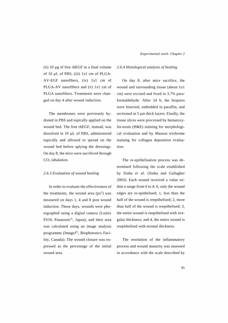
Experimental work: Chapter 2
95
(ii) 10 µg of free rhEGF in a final volume
of 10 µL of PBS, (iii) 1x1 cm of PLGA-
AV-EGF nanofibers, (iv) 1x1 cm of
PLGA-AV nanofibers and (v) 1x1 cm of
PLGA nanofibers. Treatments were chan-
ged on day 4 after wound induction.
The membranes were previously hy-
drated in PBS and topically applied on the
wound bed. The free rhEGF, instead, was
dissolved in 10 µL of PBS, administered
topically and allowed to spread on the
wound bed before aplying the dressings.
On day 8, the mice were sacrificed through
CO2 inhalation.
2.6.3 Evaluation of wound healing
In order to evaluate the effectiveness of
the treatments, the wound area (px2) was
measured on days 1, 4 and 8 post wound
induction. Those days, wounds were pho-
tographed using a digital camera (Lumix
FS16, Panasonic®, Japan), and their area
was calculated using an image analysis
programme (ImageJ®, Biophotonics Faci-
lity, Canada). The wound closure was ex-
pressed as the percentage of the initial
wound area.
2.6.4 Histological analysis of healing
On day 8, after mice sacrifice, the
wound and surrounding tissue (about 1x1
cm) were excised and fixed in 3.7% para-
formaldehyde. After 24 h, the biopsies
were bisected, embedded in paraffin, and
sectioned in 5 µm thick layers. Finally, the
tissue slices were processed by hematoxy-
lin-eosin (H&E) staining for morphologi-
cal evaluation and by Masson trichrome
staining for collagen deposition evalua-
tion.
The re-epithelisation process was de-
termined following the scale established
by Sinha et al. (Sinha and Gallagher
2003). Each wound received a value wi-
thin a range from 0 to 4: 0, only the wound
edges are re-epithelised; 1, less than the
half of the wound is reepithelised; 2, more
than half of the wound is reepithelised; 3,
the entire wound is reepithelised with irre-
gular thickness; and 4, the entire wound is
reepithelised with normal thickness.
The resolution of the inflammatory
process and wound maturity was assessed
in accordance with the scale described by

Novel therapeutic approaches for wound healing
96
Cotran et al. (Cotran, et al. 2000). Wounds
were semi-quantitatively rated according
to the following criteria: 0, absence of in-
flammation; 1, acute inflammation, com-
prising formation of the fibrin clot and the
pyogenic membrane and the migration of
leucocytes and polynuclear neutrophils; 2,
diffuse acute inflammation, comprising
the formation of granulation tissue, angio-
genesis and almost non-existence of pyo-
genic membrane; 3, chronic inflammation,
which consist on fibroblast proliferation
and 4, resolution and healing, which con-
sist on the disappearance of chronic in-
flammation, although occasionally round
cells can be found.
The deposition of collagen was evalua-
ted following the scale described by Gál et
al. (Gál, et al. 2006): 0, absent of collagen;
1, mild content of collagen; 2, moderate
content of collagen and 3, marked content
of collagen.
2.7 Statistical analysis
Data were expressed as the mean ±
standard deviation (SD). Based on the re-
sult of the normality test, the means were
compared through one-way ANOVA for
multiple comparison or Mann-Whitney U
test. Subsequently to the one-way
ANOVA test, Bonferroni or Tamhane
post-hoc were applied based on the Levene
test for the homogeneity of variances. All
the statistical calculations were performed
using SPSS 22.0.01 (SPSS®, INC., Chi-
cago, IL, USA).
3. Results
3.1 Nanofiber characterisation
The SEM photographs displayed in
Fig. 1 showed that the membranes were
composed of uniform and randomly orien-
ted nanofibers. As shown in Table 1, they
presented a high porosity above 79 %.
As expected, the thickness of the diffe-
rent membranes was similar, with a value
of 45.92 ± 0.78 µm, 56.76 ± 1.23 µm and
59.17 ± 1.83 µm, for the PLGA-AV-EGF,
the PLGA-AV and the PLGA nanofibers,
respectively. However, the nanofibers dia-
meter varies between the membranes,
being 356.03 ± 112.05 nm in the PLGA-
AV-EGF nanofibers, 486.99 ± 114.73 nm
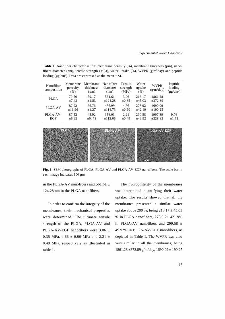
Experimental work: Chapter 2
97
Table 1. Nanofiber characterisation: membrane porosity (%), membrane thickness (µm), nano-fibers diameter (nm), tensile strength (MPa), water uptake (%), WVPR (g/m2day) and peptide loading (µg/cm2). Data are expressed as the mean ± SD.
Nanofiber composition
Membrane porosity
(%)
Membrane thickness
(µm)
Nanofiber diameter
(nm)
Tensile strength (MPa)
Water uptake
(%)
WVPR (g/m2day)
Peptide loading
(µg/cm2)
PLGA 79.50 ±7.42
59.17 ±1.83
561.61 ±124.28
3.06 ±0.35
218.17 ±45.03
1861.28 ±372.89 -
PLGA-AV 87.92 ±11.96
56.76 ±1.27
486.99 ±114.73
4.66 ±0.90
273.92 ±42.19
1690.09 ±190.25 -
PLGA-AV-EGF
87.52 ±6.62
45.92 ±0. 78
356.03 ±112.05
2.21 ±0.49
290.58 ±49.92
1907,39 ±228.82
9.76 ±1.75
Fig. 1. SEM photographs of PLGA, PLGA-AV and PLGA-AV-EGF nanofibers. The scale bar in each image indicates 100 µm.
in the PLGA-AV nanofibers and 561.61 ±
124.28 nm in the PLGA nanofibers.
In order to confirm the integrity of the
membranes, their mechanical properties
were determined. The ultimate tensile
strength of the PLGA, PLGA-AV and
PLGA-AV-EGF nanofibers were 3.06 ±
0.35 MPa, 4.66 ± 0.90 MPa and 2.21 ±
0.49 MPa, respectively as illustrated in
table 1.
The hydrophilicity of the membranes
was determined quantifying their water
uptake. The results showed that all the
membranes presented a similar water
uptake above 200 %; being 218.17 ± 45.03
% in PLGA nanofibers, 273.9 2± 42.19%
in PLGA-AV nanofibers and 290.58 ±
49.92% in PLGA-AV-EGF nanofibers, as
depicted in Table 1. The WVPR was also
very similar in all the membranes, being
1861.28 ±372.89 g/m2day, 1690.09 ± 190.25
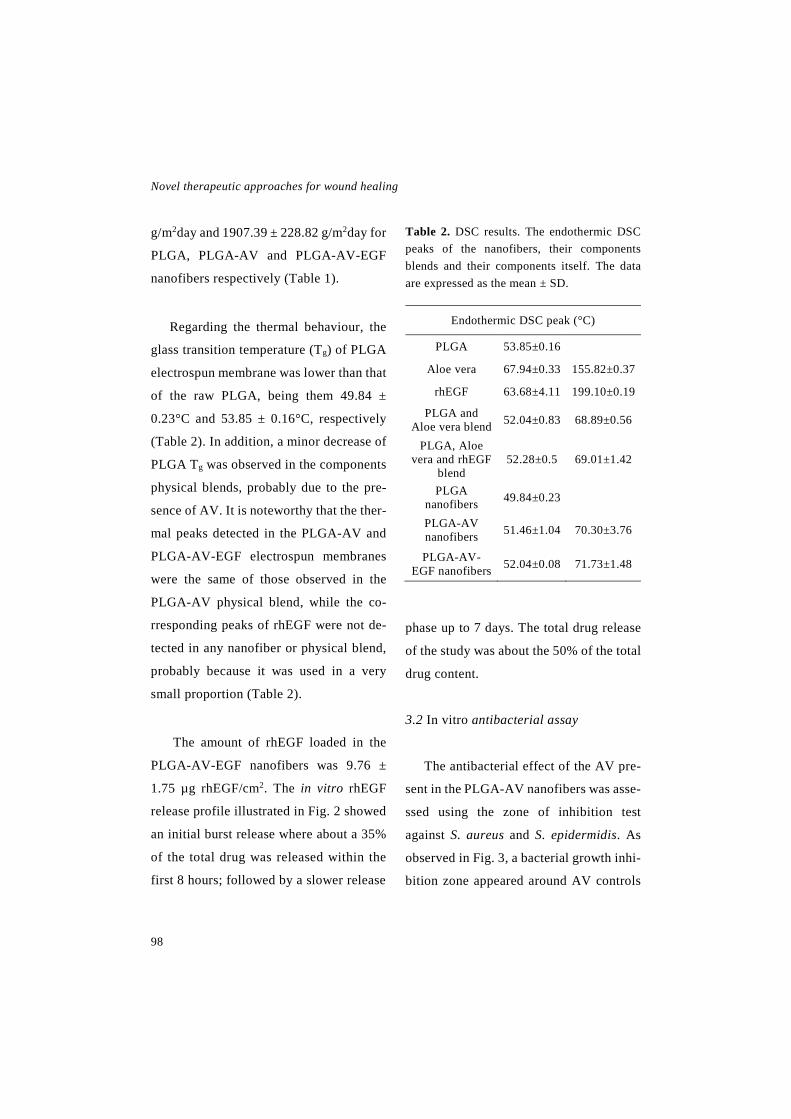
Novel therapeutic approaches for wound healing
98
g/m2day and 1907.39 ± 228.82 g/m2day for
PLGA, PLGA-AV and PLGA-AV-EGF
nanofibers respectively (Table 1).
Regarding the thermal behaviour, the
glass transition temperature (Tg) of PLGA
electrospun membrane was lower than that
of the raw PLGA, being them 49.84 ±
0.23°C and 53.85 ± 0.16°C, respectively
(Table 2). In addition, a minor decrease of
PLGA Tg was observed in the components
physical blends, probably due to the pre-
sence of AV. It is noteworthy that the ther-
mal peaks detected in the PLGA-AV and
PLGA-AV-EGF electrospun membranes
were the same of those observed in the
PLGA-AV physical blend, while the co-
rresponding peaks of rhEGF were not de-
tected in any nanofiber or physical blend,
probably because it was used in a very
small proportion (Table 2).
The amount of rhEGF loaded in the
PLGA-AV-EGF nanofibers was 9.76 ±
1.75 µg rhEGF/cm2. The in vitro rhEGF
release profile illustrated in Fig. 2 showed
an initial burst release where about a 35%
of the total drug was released within the
first 8 hours; followed by a slower release
Table 2. DSC results. The endothermic DSC peaks of the nanofibers, their components blends and their components itself. The data are expressed as the mean ± SD.
Endothermic DSC peak (°C)
PLGA 53.85±0.16
Aloe vera 67.94±0.33 155.82±0.37
rhEGF 63.68±4.11 199.10±0.19
PLGA and Aloe vera blend 52.04±0.83 68.89±0.56
PLGA, Aloe vera and rhEGF
blend 52.28±0.5 69.01±1.42
PLGA nanofibers 49.84±0.23
PLGA-AV nanofibers 51.46±1.04 70.30±3.76
PLGA-AV-EGF nanofibers 52.04±0.08 71.73±1.48
phase up to 7 days. The total drug release
of the study was about the 50% of the total
drug content.
3.2 In vitro antibacterial assay
The antibacterial effect of the AV pre-
sent in the PLGA-AV nanofibers was asse-
ssed using the zone of inhibition test
against S. aureus and S. epidermidis. As
observed in Fig. 3, a bacterial growth inhi-
bition zone appeared around AV controls
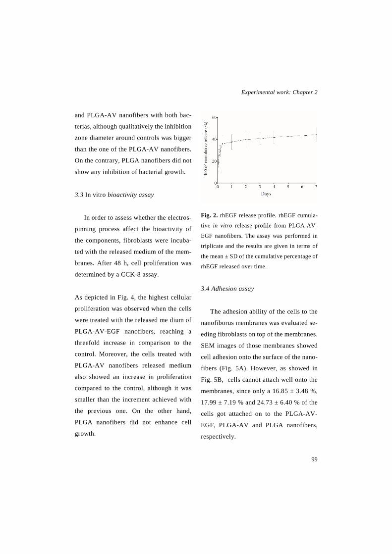
Experimental work: Chapter 2
99
and PLGA-AV nanofibers with both bac-
terias, although qualitatively the inhibition
zone diameter around controls was bigger
than the one of the PLGA-AV nanofibers.
On the contrary, PLGA nanofibers did not
show any inhibition of bacterial growth.
3.3 In vitro bioactivity assay
In order to assess whether the electros-
pinning process affect the bioactivity of
the components, fibroblasts were incuba-
ted with the released medium of the mem-
branes. After 48 h, cell proliferation was
determined by a CCK-8 assay.
As depicted in Fig. 4, the highest cellular
proliferation was observed when the cells
were treated with the released me dium of
PLGA-AV-EGF nanofibers, reaching a
threefold increase in comparison to the
control. Moreover, the cells treated with
PLGA-AV nanofibers released medium
also showed an increase in proliferation
compared to the control, although it was
smaller than the increment achieved with
the previous one. On the other hand,
PLGA nanofibers did not enhance cell
growth.
Fig. 2. rhEGF release profile. rhEGF cumula-
tive in vitro release profile from PLGA-AV-
EGF nanofibers. The assay was performed in
triplicate and the results are given in terms of
the mean ± SD of the cumulative percentage of
rhEGF released over time.
3.4 Adhesion assay
The adhesion ability of the cells to the
nanofiborus membranes was evaluated se-
eding fibroblasts on top of the membranes.
SEM images of those membranes showed
cell adhesion onto the surface of the nano-
fibers (Fig. 5A). However, as showed in
Fig. 5B, cells cannot attach well onto the
membranes, since only a 16.85 ± 3.48 %,
17.99 ± 7.19 % and 24.73 ± 6.40 % of the
cells got attached on to the PLGA-AV-
EGF, PLGA-AV and PLGA nanofibers,
respectively.

Novel therapeutic approaches for wound healing
100
3.5 In vivo wound healing assay
The effectiveness of the membranes
was evaluated in a full thickness splinted
wound healing assay carried out in db/db
mice by calculating the wound area clo-
sure. In that regard wounds areas (px2)
were measured from photographs taken on
days 1, 4 and 8 post wounding, and the
wound closure of each wound was calcu-
lated as the percentage of the initial area.
As shown in Fig. 6A the greatest
wound area reduction was observed in the
animals treated with the PLGA-AV-EGF
nanofibers. The differences with the rest
of the groups were found statistically sig-
nificant both days, being these differen-
ces more pronounced on day 8. Those re-
sults were in agreement with the gross ob-
serva tion of the wounds depicted in Fig.
6B, where a higher reduction of the area
was observed in the wounds treated with
PLGA-AV-EGF nanofibers. Moreover,
the mice that received the same dose of
free rhEGF did not show any improve-
ment of wound closure in comparison to
control.
Fig. 3. In vitro antimicrobial assay. Images of the culture plates seeded with S. aureus and S. epidermidis. The inhibition zone caused by the samples can be observed: (A) free AV as control, (B) PLGA-AV nanofibers and (C) PLGA nanofibers.
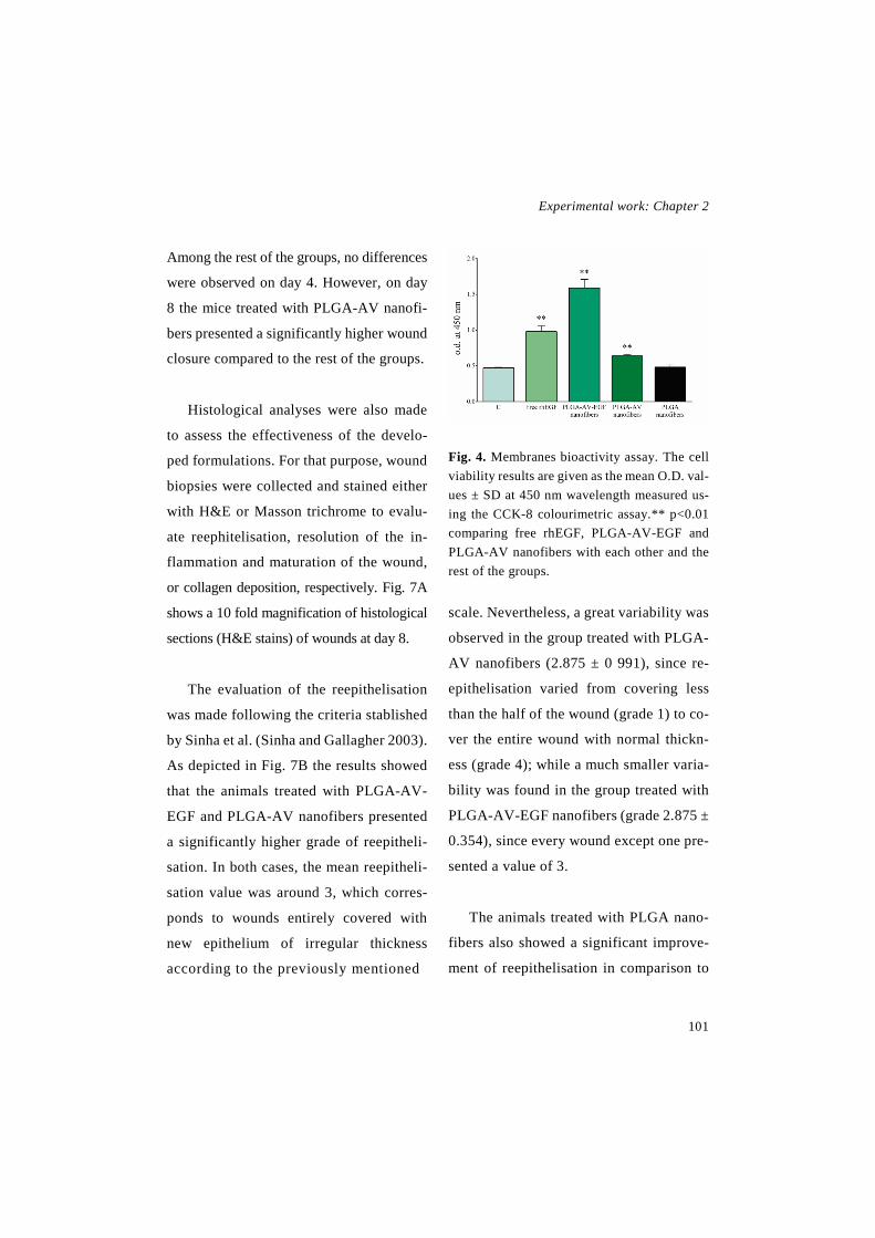
Experimental work: Chapter 2
101
Among the rest of the groups, no differences
were observed on day 4. However, on day
8 the mice treated with PLGA-AV nanofi-
bers presented a significantly higher wound
closure compared to the rest of the groups.
Histological analyses were also made
to assess the effectiveness of the develo-
ped formulations. For that purpose, wound
biopsies were collected and stained either
with H&E or Masson trichrome to evalu-
ate reephitelisation, resolution of the in-
flammation and maturation of the wound,
or collagen deposition, respectively. Fig. 7A
shows a 10 fold magnification of histological
sections (H&E stains) of wounds at day 8.
The evaluation of the reepithelisation
was made following the criteria stablished
by Sinha et al. (Sinha and Gallagher 2003).
As depicted in Fig. 7B the results showed
that the animals treated with PLGA-AV-
EGF and PLGA-AV nanofibers presented
a significantly higher grade of reepitheli-
sation. In both cases, the mean reepitheli-
sation value was around 3, which corres-
ponds to wounds entirely covered with
new epithelium of irregular thickness
according to the previously mentioned
Fig. 4. Membranes bioactivity assay. The cell viability results are given as the mean O.D. val-ues ± SD at 450 nm wavelength measured us-ing the CCK-8 colourimetric assay.** p<0.01 comparing free rhEGF, PLGA-AV-EGF and PLGA-AV nanofibers with each other and the rest of the groups.
scale. Nevertheless, a great variability was
observed in the group treated with PLGA-
AV nanofibers (2.875 ± 0 991), since re-
epithelisation varied from covering less
than the half of the wound (grade 1) to co-
ver the entire wound with normal thickn-
ess (grade 4); while a much smaller varia-
bility was found in the group treated with
PLGA-AV-EGF nanofibers (grade 2.875 ±
0.354), since every wound except one pre-
sented a value of 3.
The animals treated with PLGA nano-
fibers also showed a significant improve-
ment of reepithelisation in comparison to

Novel therapeutic approaches for wound healing
102
the control (1.857 ± 0.9 and 0.875 ± 0.641,
respectively), although the difference was
smaller than the observed with the mem-
branes containing active compounds.
The resolution of the inflammation was
assessed according to the scale described
by Cotran et al. (Cotran, et al. 2000). As
illustrated in Fig. 7C, there were no signi-
ficant differences among groups, although
a slight tendency to accelerate the inflam-
mation recovery was observed in the
groups treated with PLGA-AV-EGF nano-
fibers and PLGA-AV nanofibers. In those
groups the mean value of the inflammation
resolution was around grade 3 (3 ± 0.926
and 2.875 ± 0.835, respectively), while in
the remaining groups the mean value was
around grade 2 (free rhEGF 2.25 ± 0.07,
PLGA nanofibers 2.143 ± 0.9 and untrea-
ted control 1.75 ± 1.165), revealing a fur-
ther maturation of the wounds belonging
to the first groups, as they have advanced
from the formation of the granulation ti-
ssue to fibroblast proliferation.
In addition, the groups treated with na-
nofibers containing active compounds
were the only groups that achieved a com-
plete disappearance of chronic inflamma-
tion (grade 4), precisely, 3 wounds rea-
ched a complete resolution and healing in
the PLGA-AV-EGF nanofiber group and 2
wounds reached that grade in the PLGA-
Fig.5. In vitro adhesion assay. (A) SEM image of the nanofibers with cells attached. The scale bar indicates 300 µm. (B) The cell adhesion results are given as the mean ± SD of the % of cells counted in comparison to the control. *** p<0.001 comparing PLGA-AV-EGF, PLGA-AV and PLGA nanofibers with the control group.

Experimental work: Chapter 2
103
AV nanofiber group. Moreover, the rest of
the groups presented wounds trapped in
the acute inflammation phase (grade 1).
That permanent migration of leucocytes
and polynuclear neutrophils to the wound
bed is typical of chronic wounds, due to
the excessive amount of proteases released
by neutrophils, which degrade the ECM
delaying wound healing (Menke, et al.
2007).
In regard to collagen deposition (Fig. 7D),
no differences were observed among
groups. The greatest collagen deposition
was observed in the animals treated with
PLGA-AV-EGF nanofibers and PLGA na-
nofibers, since most of the wounds presen-
ted a mild deposition of collagen (grade 1
according to the criteria established by Gál
et al. (Gál, et al. 2006)), and a few of them
presented either a moderate content or ab-
sent of collagen, grade 2 and grade 0, res-
pectively. Most of the wounds treated with
PLGA-AV nanofibers and free rhEGF also
showed mild content of collagen (grade 1),
however, the rest of them did not present
any collagen deposition (grade 0). Finally,
in the control group a great variability was
observed, since most of the wounds did not
show any collagen (grade 0), whereas
some of them presented a marked content
of it (grade 3).
Fig. 6. In vivo wound closure. (A) Wound closure represented as the percentage of reduction of the initial area on days 4 and 8 after wound induction (n=8). ● p<0.05 comparing PLGA-AV-EGF nan-ofibers with the rest of the groups, on day 4. ○○○ p<0.001 comparing PLGA-AV-EGF nanofibers with free rhEGF, PLGA nanofibers and no treatment on day 8. * p<0.05 comparing PLGA-AV-EGF nanofibers with PLGA-AV nanofibers. + p<0.05 comparing PLGA-AV nanofibers with PLGA nan-ofibers, free rhEGF and untreated groups. (B) Wound images of each group on days 1, 4 and 8.
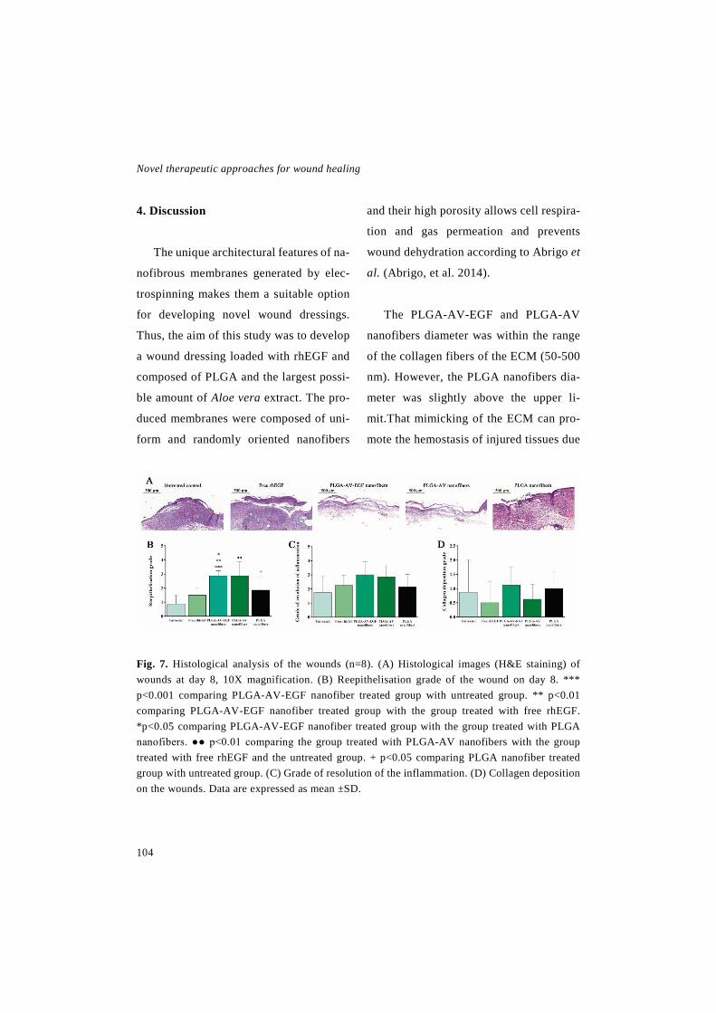
Novel therapeutic approaches for wound healing
104
4. Discussion
The unique architectural features of na-
nofibrous membranes generated by elec-
trospinning makes them a suitable option
for developing novel wound dressings.
Thus, the aim of this study was to develop
a wound dressing loaded with rhEGF and
composed of PLGA and the largest possi-
ble amount of Aloe vera extract. The pro-
duced membranes were composed of uni-
form and randomly oriented nanofibers
and their high porosity allows cell respira-
tion and gas permeation and prevents
wound dehydration according to Abrigo et
al. (Abrigo, et al. 2014).
The PLGA-AV-EGF and PLGA-AV
nanofibers diameter was within the range
of the collagen fibers of the ECM (50-500
nm). However, the PLGA nanofibers dia-
meter was slightly above the upper li-
mit.That mimicking of the ECM can pro-
mote the hemostasis of injured tissues due
Fig. 7. Histological analysis of the wounds (n=8). (A) Histological images (H&E staining) of wounds at day 8, 10X magnification. (B) Reepithelisation grade of the wound on day 8. *** p<0.001 comparing PLGA-AV-EGF nanofiber treated group with untreated group. ** p<0.01 comparing PLGA-AV-EGF nanofiber treated group with the group treated with free rhEGF. *p<0.05 comparing PLGA-AV-EGF nanofiber treated group with the group treated with PLGA nanofibers. ●● p<0.01 comparing the group treated with PLGA-AV nanofibers with the group treated with free rhEGF and the untreated group. + p<0.05 comparing PLGA nanofiber treated group with untreated group. (C) Grade of resolution of the inflammation. (D) Collagen deposition on the wounds. Data are expressed as mean ±SD.

Experimental work: Chapter 2
105
to the presence of small interstices and the
high-surface area of the fibers (Abrigo, et
al. 2014).
The mechanical properties of the mem-
branes indicated that they were adequate
for wound dressing application, since the
obtained values were very similar to the
human skin tensile strength, which ranged
from 5.7 MPa to 12.6 MPa according to
Jacquemoud et al. (Jacquemoud, et al.
2007). The slight differences found bet-
ween the membranes tensile strenght lied
in the different composition of the mem-
branes, since the Aloe vera extract gave to
the nanofibers more elasticity to endure
tensile force rising the utilmate tensile
strength of the PLGA-AV membrane,
while the salts presents in the lyophilised
rhEGF slightly decreased the PLGA-AV-
EGF nanofibers tensile strength.
Although water uptake was similar in
the three membranes, the PLGA nanofi-
bers presented the lowest value. PLGA is
a hydrophobic polymer and thus, its water
uptake is low. However, the membranes
containing AV were more hydrophilic and
therefore, they were able to absorb more
water. Those results were in agreement
with the results obtained by Son et al.
(Son, et al. 2013), who observed a higher
degree of swelling when PLGA was elec-
trospun with increasing gelatin concentra-
tions rather than electrospun alone.
Wounds dressings require water va-
pour permeability to drain exudates, but
they also need to retain moisture to help
wound healing (Abrigo, et al. 2014; Nata-
rajan, et al. 2000). In order to maintain that
specific control of moisture, WVPR of
commercial skin dressings have been re-
ported to be in the range of 426-2047
g/m2day (Tu, et al. 2015), therefore the de-
veloped dressings presented an adequate
WVPR for wound healing application.
The results obtained in the analysis of
the thermal behaviour showed that the
electrospun PLGA presented a high degree
of alignment and orientation of polymer
chains, as suggested by the decrease of the
Tg of the PLGA membrane in comparison
to the raw PLGA. Similar reduction in Tg
was observed previously (Fouad, et al.
2013). Moreover, the detection of the same
thermal peaks in the PLGA-AV and

Novel therapeutic approaches for wound healing
106
PLGA-AV-EGF electrospun membranes
and the PLGA-AV physical blend reveals
an immiscible blend morphology of this
two components in the nanofibers.
The in vitro release of the EGF present
in the PLGA-AV-EGF membrane exhibi-
ted a biphasic profile, with an initial burst
release and a slower sustained phase. The
burst release is related to the percentage of
surface-associated protein, which is im-
portant to obtain a rapid activation of the
keratinocytes of the wound edge (Schnei-
der, et al. 2009). In the following sustained
phase, only about the 50% of the total drug
was released. Nevertheless, the release of
the loaded drugs may be accelerated in
vivo, driven by a faster polymer degrada-
tion caused by the enzymes present in the
wound bed (Fredenberg, et al. 2011).
An important property of wound dre-
ssings is their ability to protect the wounds
against microbial growth to prevent
wound infections. According to the zone
inhibition test, PLGA-AV membranes
were able to inhibit bacterial growth, since
AV presented antibacterial effect through
numerous low molecular weight com-
pounds, such as, α-bisabolol, lupeol, cin-
namonic acid, salicylic acid and uric acid,
which can kill bacteria by interfering with
enzymatic processes (Ghayempour, et al.
2016; Tummalapalli, et al. 2016). Moreo-
ver, nanofibrous dressings can prevent
bacterial growth into the wound creating a
physical barrier, since the small pore size
prevents the entry of microorganisms
(Abrigo, et al. 2014).
The higher inhibition zone obtained by
the free AV used as control might be due
to the availability of the total AV dose
since the beginning of the study, while the
AV of the nanofibrous membranes needed
to be release from the nanofibers in order
to inhibit bacterial growth. Due to the lack
of antibacterial activity of EGF, the anti-
microbial activity of PLGA-AV-EGF
membrane was not analysed, since the
effect of the AV was already proven with
the PLGA-AV membranes.
The bioactivity of the rhEGF and AV
remained unchanged after the electrospin-
ning process, since the extracts from the
membranes maintained the ability to
enhance fibroblast proliferation, as both of

Experimental work: Chapter 2
107
them have proven to do previously (Bou-
dreau and Beland 2006; Gainza, et al.
2015a). Nevertheless, the most striking re-
sult from the bioactivity assay was the
great promotion of cell proliferation by the
PLGA-AV-EGF nanofibers, which revea-
led the beneficial effect of the combination
of AV and rhEGF. Moreover, the effect in
cell growth was caused exclusively by
them, as highlighted by the lack of prolife-
ration increment when cells were incuba-
ted with the PLGA nanofibers extract.
Since the membranes were designed to
be changed during the treatment, cell
adhesion or tissue ingrowth within the na-
nofibrous structure should be prevented in
order to avoid pain and damage of the
newly formed tissue after dressing remo-
val. The low percentage of cells that got
adhered onto the nanofibers proved their
inability to attach cells, and therefore de-
monstrated their suitability as temporary
wound dressings.
Wound healing was evaluated in vivo
in a full thickness splinted model in db/db
mice. Unlike in humans, reepithelisation
does not play a major role in rodent wound
healing models. However, in db/db mice
this process gains importance due to a lack
of wound contraction, making the healing
process more similar to that of humans.
Wound contraction is impaired because of
the extreme obesity of the animals, which
reduces the looseness of their skin (Fang,
et al. 2010; Tkalcevic, et al. 2009). Moreo-
ver, the impairment of contraction is enhan-
ced by the placement of silicone splints
around the wounds, increasing the impor-
tance of reepithelisation (Michaels, et al.
2007). However, as we have observed in
collagen deposition, the wound healing of
these mice presented a wide variability as
previously described by Trousdale et al.
(Trousdale, et al. 2009).
Overall, the PLGA-AV-EGF nanofi-
bers have shown that they can improve
wound healing in terms of wound closure
and reepithelisation, in comparison to free
rhEGF. The enhanced wound healing acti-
vity of the nanofibers containing rhEGF
can be partially explained by their protec-
tive effect against the proteases present in
the wound bed, as shown by multiple in
vivo studies carried out with EGF loaded
nanofibers which did not contain Aloe
Novel therapeutic approaches for wound healing
108
vera, such as the study performed by Choi
et al., where PCL-PEG electrospun nano-
fibers were chemically conjugated with
EGF (Choi, et al. 2008); or the study per-
formed by Lai et al., where a dual wound
dressing composed of hialuronic acid fi-
bers containing bFGF and gelatine nano-
particles loaded with VEGF and collagen
fibers containing EGF and gelatine nano-
particles loaded with PDGF (Lai, et al.
2014), respectively. Moreover, the dura-
tion of the rhEGF in the wound is extended
by the sustained release achieved with the
electrospun membranes, and thus its effect
is improved.
Although in a lesser extent than PLGA-
AV-EGF nanofibers, the PLGA-AV nano-
fibers also showed an improvement of
wound healing, since both Aloe vera and
electrospun nanofibers themselves have
been proven to enhance wound healing
(Abrigo, et al. 2014; Hashemi, et al. 2015).
The unique architecture of electrospun
membranes, i.e. the high surface to volume
area and the nanoporosity, allows water
and gas permeability maintaining an ade-
quate moisture of the wound and promo-
ting cell respiration. Furthermore, the
small size of the pores prevents the entry
of microorganisms to the wound. Altoge-
ther, the electrospun membranes promote
cell migration and proliferation to the
wound bed, and thus enhance the release
of wound healing mediators, such as, co-
llagen, growth factors or angiogenic fac-
tors, and thereby facilitate the formation of
granulation tissue and reepithelisation
(Abrigo, et al. 2014; Shahverdi, et al.
2014). In addition, the improvement of re-
epithelisation may be related to the chosen
polymer; since the lactate, one of the de-
gradation products of PLGA, has been
proven to accelerate wound healing (Por-
porato, et al. 2012) .
Aloe vera has been used previously to
develop nanofibrous dressings for tissue
engineering in ratios of 1:3, 1:6 and 1:8 in
comparison to the rest of the components
(Bhaarathy, et al. 2014; Karuppuswamy, et
al. 2014; Suganya, et al. 2014). However,
in the current study the proportion of Aloe
vera was considerably higher, since the ra-
tio of Aloe vera, PLGA and rhEGF was
1:1:0.4. Therefore, it is the first time
where nanofibers with that composition
and such a large proportion of Aloe vera

Experimental work: Chapter 2
109
have been developed for wound dressing
application. That use of Aloe vera high-
lights the novelty of the study, since it has
shown to improve wound closure.
Accordingly, the improvement of
wound closure was due to the combination
of the three elements, the rhEGF, the Aloe
vera extract and the PLGA nanofibers,
since all of them promoted actively wound
healing.
5. Conclusion
In the current study, an aqueous phase
of Aloe vera was emulsified into PLGA
and the emulsion was electrospun to deve-
lop a nanofibrous dressing loaded with
rhEGF. The nanofibers contained the same
amount of Aloe vera and PLGA (1:1), and
to the best of our knowledge such a high
concentration of Aloe vera for wound hea-
ling application in a nanofibrous dressing
has not been studied so far.
The membranes were composed of uni-
form fibers of 356.03 ± 112.05 nm in dia-
meter, presented a porosity of 87.52% and
a thickness of 45.92 ± 0.78 µm. The elec-
trospinning process did not affect the bio-
activity of the active compounds as de-
monstrated by the in vitro bioactivity
assay.
The in vivo full thickness wound hea-
ling assay carried out in db/db mice, sho-
wed an improvement in wound healing, in
terms of wound closure and reephitelisa-
tion, when PLGA-AV-EGF nanofibers
were applied on the wound. Accordingly,
these results showed that the topical appli-
cation of PLGA-AV-EGF nanofibers with
a high concentration of Aloe vera might be
a suitable strategy for the treatment of ch-
ronic wounds.
6. Acknowledgments
I. García-Orue thanks the Basque Go-
vernment for the fellowship grant. The au-
thors thank for technical and human su-
pport provided by SGIker of UPV/EHU
and European funding (ERDF and ESF).
This project has been funded by the Bas-
que Government (ELKARTEK 2015, Na-
noplatform, KK-2015/0000036). In addi-
tion, it has been partially supported by the
University of the Basque Country

Novel therapeutic approaches for wound healing
110
(UPV/EHU) (UFI 11/32). Authors also
wish to thank the intellectual and technical
assistance from the ICTS “NANBIOSIS”,
more specifically by the Drug Formulation
Unit (U10) of the CIBER-BBN at the
UPV/EHU.
7. References
Abrigo, M., McArthur, S.L., Kingshott, P., 2014. Electrospun Nanofibers as Dressings for Chronic Wound Care: Advances, Challenges, and Future Prospects. Macromol. Biosci., 14, 772-792. doi: 10.1002/mabi.201300561.
Bahrami, H., Keshel, S.H., Chari, A.J., Biazar, E., 2016. Human unrestricted somatic stem cells loaded in nanofibrous PCL scaffold and their healing effect on skin defects. Artif. Cells Nanomed. Biotechnol., 44, 1556-1560. doi: 10.3109/21691401.2015.1062390.
Bhaarathy, V., Venugopal, J., Gandhimathi, C., Ponpandian, N., Mangalaraj, D., Ramakrishna, S., 2014. Biologically improved nanofibrous scaffolds for cardiac tissue engineering. Mater. Sci. Eng. C. Mater. Biol. Appl., 44, 268-277. doi: http://dx.doi.org/10.1016/j.msec.2014. 08. 018.
Bhowmick, S., Scharnweber, D., Koul, V., 2016. Co-cultivation of keratinocyte-human mesenchymal stem cell (hMSC) on sericin loaded electrospun nanofibrous composite scaffold (cationic gelatin/hyaluronan/chon-droitin sulfate) stimulates epithelial differenti-ation in hMSCs: In vitro study. Biomaterials,
88, 83-96. doi: http://dx.doi.org/10.1016/j.bio-materials.2016.02.034.
Bodnar, R.J., 2013. Epidermal Growth Factor and Epidermal Growth Factor Receptor: The Yin and Yang in the Treatment of Cutaneous Wounds and Cancer. Adv. Wound Care (New Rochelle), 2, 24-29. doi: 10.1089/wound .2011.0326.
Boudreau, M.D., Beland, F.A., 2006. An eval-uation of the biological and toxicological prop-erties of Aloe barbadensis (miller), Aloe vera. J. Environ. Sci. Health C Environ. Carcinog. Ecotoxicol. Rev., 24, 103-154. doi: 10.1080/ 10590500600614303.
Briquez, P.S., Hubbell, J.A., Martino, M.M., 2015. Extracellular Matrix-Inspired Growth Factor Delivery Systems for Skin Wound Heal-ing. Adv. Wound Care (New Rochelle), 4, 479-489. doi: 10.1089/wound.2014.0603.
Choi, J.S., Kim, H.S., Yoo, H.S., 2015. Elec-trospinning strategies of drug-incorporated nanofibrous mats for wound recovery. Drug Deliv. Transl. Res., 5, 137-145. doi: 10.1007 /s13346-013-0148-9.
Choi, J.S., Leong, K.W., Yoo, H.S., 2008. In vivo wound healing of diabetic ulcers using electrospun nanofibers immobilized with hu-man epidermal growth factor (EGF). Bio-materials, 29, 587-596. doi: http://dx.doi.org/ 10.1016/j.biomaterials.2007.10.012.
Choi, J.K., Jang, J., Jang, W., Kim, J., Bae, I., Bae, J., Park, Y., Kim, B.J., Lim, K., Park, J.W., 2012. The effect of epidermal growth factor (EGF) conjugated with low-molecular-weight protamine (LMWP) on wound healing of the skin. Biomaterials, 33, 8579-8590. doi:

Experimental work: Chapter 2
111
http://dx.doi.org/10.1016/j.biomateri-als.2012.07.061.
Choi, S., Chung, M., 2003. A review on the re-lationship between aloe vera components and their biologic effects. Semin. Integr. Med., 1, 53-62. doi: http://dx.doi.org/10.1016/S1543-1150(03)00005-X.
Cotran, R., Kumar, G.K., Collins, T., 2000. Re-paración de los tejidos: proliferacion celular, fibrosis y curaicón de las heridasIn: Cotran, R., Kumar, G.K., Collins, T. (Eds.), Patología Es-tructural Y Funcional, McGraw-Hill, Interame-ricana, Madrid, pp. 95-120.
Dickinson, L.E., Gerecht, S., 2016. Engineered Biopolymeric Scaffolds for Chronic Wound Healing. Front. Physiol., 7, 341. doi: 10.3389 /fphys.2016.00341.
Diegelmann, R.F., Evans, M.C., 2004. Wound healing: an overview of acute, fibrotic and de-layed healing. Front. Biosci., 9, 283-289. doi: http://dx.doi.org/10.2741/.
Elgharably, H., Ganesh, K., Dickerson, J., Khanna, S., Abas, M., Ghatak, P.D., Dixit, S., Bergdall, V., Roy, S., Sen, C.K., 2014. A mod-ified collagen gel dressing promotes angiogen-esis in a preclinical swine model of chronic is-chemic wounds. Adv. Wound Care (New Ro-chelle), 22, 720-729. doi: 10.1111/wrr.12229.
Fang, R.C., Kryger, Z.B., Buck II, D.W., De La Garza, M., Galiano, R.D., Mustoe, T.A., 2010. Limitations of the db/db mouse in translational wound healing research: Is the NONcNZO10 polygenic mouse model superior? Wound Re-pair Regen., 18, 605-613. doi: 10.1111/j.1524-475X.2010.00634.x.
Fang, R.C., Mustoe, T.A., 2008. Animal mod-els of wound healing: uility in transgenic mice. J. Biomater. Sci. Polym. Ed., 19, 989-1005. doi: 10.1163/156856208784909327.
Fouad, H., Elsarnagawy, T., Almahjdi, F.N., Khalil, K.A., 2013. Preparation and in vitro thermo-mechanical characterization of electro-spun PLGA nanofibers for soft and hard tissue replacement. Int. J. Electrochem. Sci., 8, 2293-2304.
Fredenberg, S., Wahlgren, M., Reslow, M., Axelsson, A., 2011. The mechanisms of drug release in poly(lactic-co-glycolic acid)-based drug delivery systems—A review. Int. J. Pharm., 415, 34-52. doi: http://dx.doi. org/10.1016/j.ijpharm.2011.05.049.
Fu, Y., Guan, J., Guo, S., Guo, F., Niu, X., Liu, Q., Zhang, C., Nie, H., Wang, Y., 2014. Human urine-derived stem cells in combination with polycaprolactone/gelatin nanofibrous mem-branes enhance wound healing by promoting angiogenesis. J. Transl. Med., 12, 274. doi: 10.1186/s12967-014-0274-2.
Gainza, G., Bonafonte, D.C., Moreno, B., Aguirre, J.J., Gutierrez, F.B., Villullas, S., Pedraz, J.L., Igartua, M., Hernandez, R.M., 2015a. The topical administration of rhEGF-loaded nanostructured lipid carriers (rhEGF-NLC) improves healing in a porcine full-thick-ness excisional wound model. J. Control Re-lease, 197, 41-47. doi: http://dx.doi.org /10.1016/j.jconrel.2014.10.033.
Gainza, G., Villullas, S., Pedraz, J.L., Hernan-dez, R.M., Igartua, M., 2015b. Advances in drug delivery systems (DDSs) to release

Novel therapeutic approaches for wound healing
112
growth factors for wound healing and skin re-generation. Nanomedicine, 11, 1551-1573. doi: http://dx.doi.org/10.1016/j.nano.2015.03.002.
Gál, P., Toporcer, T., Vidinský, B., Mokrý, M., Novotný, M., Kilík, R., Smetana, K., Gál, T., Sabo, J., 2006. Early changes in the tensile stregnth and morphology of primary sutured skin wounds in rats. Folia Biol. (Praha), 52, 109-115.
Garg, T., Rath, G., Goyal, A.K., 2015. Bio-materials-based nanofiber scaffold: targeted and controlled carrier for cell and drug deliv-ery. J. Drug Target., 23, 202-221. doi: 10.3109 /1061186X.2014.992899.
Ghayempour, S., Montazer, M., Mahmoudi Rad, M., 2016. Encapsulation of Aloe Vera ex-tract into natural Tragacanth Gum as a novel green wound healing product. Int. J. Biol. Mac-romol., 93, Part A, 344-349. doi: http://dx.doi.org/10.1016/j.ijbi-omac.2016.08.076.
Gu, J., Liu, N., Yang, X., Feng, Z., Qi, F., 2014. Adiposed-derived stem cells seeded on PLCL/P123 eletrospun nanofibrous scaffold enhance wound healing. Biomed. Mater., 9 (3), 035012. doi: 10.1088/1748-6041/9/3/035012.
Guillemin, Y., Le Broc, D., Ségalen, C., Kurk-djian, E., Gouze, J.N., 2016. Efficacy of a col-lagen-based dressing in an animal model of de-layed wound healing. J. Wound Care, 25, 406-413. doi: 10.12968/jowc.2016.25.7.406.
Han, I., Shim, K.J., Kim, J.Y., Im, S.U., Sung, Y.K., Kim, M., Kang, I., Kim, J.C., 2007. Ef-fect of Poly(3-hydroxybutyrate-co-3-hy-droxyvalerate) Nanofiber Matrices Cocultured With Hair Follicular Epithelial and Dermal
Cells for Biological Wound Dressing. Artif. Organs, 31, 801-808. doi: 10.1111/j.1525-1594.2007. 00466.x.
Hashemi, S.A., Madani, S.A., Abediankenari, S., 2015. The Review on Properties of Aloe Vera in Healing of Cutaneous Wounds. Bio-med. Res. Int., 2015, 714216. doi: 10.1155/2015/714216.
Hodgkinson, T., Bayat, A., 2011. Dermal sub-stitute-assisted healing: enhancing stem cell therapy with novel biomaterial design. Arch. Dermatol. Res., 303, 301-315. doi: 10.1007/s00403-011-1131-2.
Inpanya, P., Faikrua, A., Ounaroon, A., Sitti-chokechaiwut, A., Viyoch, J., 2012. Effects of the blended fibroin/aloe gel film on wound healing in streptozotocin-induced diabetic rats. Biomed. Mater., 7, 035008. doi: 10.1088/1748-6041/7/3/035008.
Jacquemoud, C., Bruyere-Garnier, K., Coret, M., 2007. Methodology to determine failure characteristics of planar soft tissues using a dy-namic tensile test. J. Biomech., 40, 468-475. doi: http://dx.doi.org/10.1016/j.jbiomech .2005.12.010.
Karuppuswamy, P., Venugopal, J.R., Navanee-than, B., Laiva, A.L., Sridhar, S., Ramakrishna, S., 2014. Functionalized hybrid nanofibers to mimic native ECM for tissue engineering ap-plications. Appl. Surf. Sci., 322, 162-168. doi: http://dx.doi.org/10.1016/j.ap-susc.2014.10.074.
Kataria, K., Gupta, A., Rath, G., Mathur, R.B., Dhakate, S.R., 2014. In vivo wound healing performance of drug loaded electrospun com-posite nanofibers transdermal patch. Int. J.
Experimental work: Chapter 2
113
Pharm., 469, 102-110. doi: http://dx.doi.org/1 0.1016/j.ijpharm.2014.04.047.
Keshel, S.H., Biazar, E., Rezaei Tavirani, M., Rahmati Roodsari, M., Ronaghi, A., Ebrahimi, M., Rad, H., Sahebalzamani, A., Rakhshan, A., Afsordeh, K., 2014. The healing effect of unre-stricted somatic stem cells loaded in collagen-modified nanofibrous PHBV scaffold on full-thickness skin defects. Artif. Cells Nanomed. Biotechnol., 42, 210-216. doi: 10.3109/21691401.2013.800080.
Lai, H., Kuan, C., Wu, H., Tsai, J., Chen, T., Hsieh, D., Wang, T., 2014. Tailored design of electrospun composite nanofibers with staged release of multiple angiogenic growth factors for chronic wound healing. Acta Biomater., 10, 4156-4166. doi: http://dx.doi.org/10.1016/j.actbio.2014.05.001.
Lee, C., Hsieh, M., Chang, S., Lin, Y., Liu, S., Lin, T., Hung, K., Pang, J.S., Juang, J., 2014. Enhancement of Diabetic Wound Repair Using Biodegradable Nanofibrous Metformin-Elut-ing Membranes: in Vitro and in Vivo. ACS Appl. Mater. Interfaces, 6, 3979-3986. doi: 10.1021/am405329g.
Li, C., Fu, R., Yu, C., Li, Z., Guan, H., Hu, D., Zhao, D., Lu, L., 2013. Silver nanoparticle/chi-tosan oligosaccharide/poly(vinyl alcohol) nan-ofibers as wound dressings: a preclinical study. Int. J. Nanomedicine, 8, 4131-4145. doi: 10.2147/IJN.S51679 [doi].
Losi, P., Briganti, E., Errico, C., Lisella, A., Sanguinetti, E., Chiellini, F., Soldani, G., 2013. Fibrin-based scaffold incorporating
VEGF- and bFGF-loaded nanoparticles stimu-lates wound healing in diabetic mice. Acta Bio-mater., 9, 7814-7821. doi: http://dx.doi.org /10.1016/j.actbio.2013.04.019.
Machula, H., Ensley, B., Kellar, R., 2014. Electrospun Tropoelastin for Delivery of Ther-apeutic Adipose-Derived Stem Cells to Full-Thickness Dermal Wounds. Adv. Wound Care (New Rochelle), 3, 367-375. doi: 10.1089 /wound.2013.0513.
Magin, C.M., Neale, D.B., Drinker, M.C., Wil-lenberg, B.J., Reddy, S.T., La Perle, K.M., Schultz, G.S., Brennan, A.B., 2016. Evaluation of a bilayered, micropatterned hydrogel dress-ing for full-thickness wound healing. Exp. Biol. Med. (Maywood), 241, 986-995. doi: 10.1177/1535370216640943.
Menke, N.B., Ward, K.R., Witten, T.M., Bonchev, D.G., Diegelmann, R.F., 2007. Im-paired wound healing. Clin. Dermatol., 25, 19-25. doi: http://dx.doi.org/10.1016/j.clinderma-tol.2006.12.005.
Merrell, J.G., McLaughlin, S.W., Tie, L., Lau-rencin, C.T., Chen, A.F., Nair, L.S., 2009. Cur-cumin-loaded poly(epsilon-caprolactone) nan-ofibres: Diabetic wound dressing with anti-ox-idant and anti-inflammatory properties. Clin. Exp. Pharmacol. Physiol., 36, 1149-1156. doi: 10.1111/j.1440-1681.2009.05216.x.
Michaels, J., Churgin, S.S., Blechman, K.M., Greives, M.R., Aarabi, S., Galiano, R.D., Gurt-ner, G.C., 2007. db/db mice exhibit severe wound-healing impairments compared with other murine diabetic strains in a silicone-splinted excisional wound model. Wound Re-
Novel therapeutic approaches for wound healing
114
pair Regen., 15, 665-670. doi: 10.1111/j.1524-475X.2007.00273.x.
Monaco, J.L., Lawrence, W.T., 2003. Acute wound healing: An overview. Clin. Plast. Surg., 30, 1-12. doi: http://dx.doi.org/ 10.1016/S0094-1298(02)00070-6.
Morton, L.M., Phillips, T.J., 2016. Wound healing and treating wounds: Differential diag-nosis and evaluation of chronic wounds. J. Am. Acad. Dermatol., 74, 589-605. doi: http://dx.doi.org/10.1016/j.jaad.2015.08.068.
Moura, L.I.F., Dias, A.M.A., Suesca, E., Casadiegos, S., Leal, E.C., Fontanilla, M.R., Carvalho, L., de Sousa, H.C., Carvalho, E., 2014. Neurotensin-loaded collagen dressings reduce inflammation and improve wound heal-ing in diabetic mice. Biochim. Biophys. Acta, 1842, 32-43. doi: http://dx.doi.org/10.1016/j.bbadis.2013.10.009.
Natarajan, S., Williamson, D., Stiltz, A.J., Har-ding, K., 2000. Advances in Wound Care and Healing Technology. Am. J. Clin. Dermatol., 1, 269-275. doi: 10.2165/00128071-20000 1050-00002.
Pachuau, L., 2015. Recent developments in novel drug delivery systems for wound heal-ing. Expert. Opin. Drug Deliv., 12, 1895-1909. doi: 10.1517/17425247.2015.1070143.
Porporato, P.E., Payen, V.L., De Saedeleer, C.J., Préat, V., Thissen, P., Feron, O., Son-veaux, P., 2012. Lactate stimulates angiogene-sis and accelerates the healing of superficial and ischemic wounds in mice . Angiogenesis, 15, 581-592. doi: 10.1007/s10456-012-9282-0.
Powell, H.M., Supp, D.M., Boyce, S.T., 2008. Influence of electrospun collagen on wound contraction of engineered skin substitutes. Bi-omaterials, 29, 834-843. doi: http://dx.doi.org/ 10.1016/j.biomaterials.2007.10.036.
Schneider, A., Wang, X.Y., Kaplan, D.L., Gar-lick, J.A., Egles, C., 2009. Biofunctionalized electrospun silk mats as a topical bioactive dressing for accelerated wound healing. Acta Biomater., 5, 2570-2578. doi: http://dx.doi. org/10.1016/j.actbio.2008.12.013.
Schreml, S., Szeimies, R., Prantl, L., Landthaler, M., Babilas, P., 2010. Wound heal-ing in the 21st century. J. Am. Acad. Derma-tol., 63, 866-881. doi: http://dx.doi.org/10.1016/j.jaad. 2009.10.048.
Seaton, M., Hocking, A., Gibran, N.S., 2015. Porcine Models of Cutaneous Wound Healing. ILAR Journal, 56, 127-138. doi: 10.1093 /ilar/ilv016.
Sen, C.K., Gordillo, G.M., Roy, S., Kirsner, R., Lambert, L., Hunt, T.K., Gottrup, F., Gurtner, G.C., Longaker, M.T., 2009. Human skin wounds: A major and snowballing threat to public health and the economy. Wound Repair Regen., 17, 763-771. doi: 10.1111/j.1524-475X.2009.00543.x.
Shahverdi, S., Hajimiri, M., Esfandiari, M.A., Larijani, B., Atyabi, F., Rajabiani, A., Dehpour, A.R., Gharehaghaji, A.A., Dinar-vand, R., 2014. Fabrication and structure anal-ysis of poly(lactide-co-glycolic acid)/silk fi-broin hybrid scaffold for wound dressing appli-cations. Int. J. Pharm., 473, 345-355. doi: http://dx.doi.org/10.1016/j.ijpharm.2014.07.021.

Experimental work: Chapter 2
115
Sinha, U.K., Gallagher, L.A., 2003. Effects of Steel Scalpel, Ultrasonic Scalpel, CO2 Laser, and Monopolar and Bipolar Electrosurgery on Wound Healing in Guinea Pig Oral Mucosa. Laryngoscope, 113, 228-236. doi: 10.1097/ 00005537-200302000-00007.
Son, S., Franco, R., Bae, S., Min, Y., Lee, B., 2013. Electrospun PLGA/gelatin fibrous tubes for the application of biodegradable intestinal stent in rat model. Journal of Biomedical Ma-terials Research Part B: Applied Biomaterials, 101B, 1095-1105. doi: 10.1002/jbm.b.32923.
Steffens, D., Leonardi, D., Soster, P.R.d.L., Lersch, M., Rosa, A., Crestani, T., Scher, C., de Morais, M.G., Costa, J.A.V., Pranke, P., 2014. Development of a new nanofiber scaf-fold for use with stem cells in a third degree burn animal model. Burns, 40, 1650-1660. doi: http://dx.doi.org/10.1016/j.burns.2014.03.008.
Suganya, S., Venugopal, J., Ramakrishna, S., Lakshmi, B.S., Dev, V.R.G., 2014. Naturally derived biofunctional nanofibrous scaffold for skin tissue regeneration. Int. J. Biol. Macro-mol., 68, 135-143. doi: http://dx.doi.org/ 10.1016/j.ijbiomac.2014.04.031.
Tkalcevic, V.I., Cužic, S., Parnham, M.J., Pašalic, I., Brajša, K., 2009. Differential Eval-uation of Excisional Non-occluded Wound Healing in db/db Mice. Toxico. Pathol., 37, 183-192. doi: 10.1177/0192623308329280.
Trousdale, R.K., Jacobs, S., Simhaee, D.A., Wu, J.K., Lustbader, J.W., 2009. Wound Clo-sure and Metabolic Parameter Variability in a db/db Mouse Model for Diabetic Ulcers. J. Surg. Res., 151, 100-107. doi: http://dx.doi.org /10.1016/j.jss.2008.01.023.
Tu, Y., Zhou, M., Guo, Z., Li, Y., Hou, Y., Wang, D., Zhang, L., 2015. Preparation and characterization of thermosensitive artificial skin with a Sandwich structure. Mater Lett, 147, 4-7. doi: http://dx.doi.org/10.1016/ j.mat-let.2015.01.163.
Tummalapalli, M., Berthet, M., Verrier, B., Deopura, B.L., Alam, M.S., Gupta, B., 2016. Composite wound dressings of pectin and gel-atin with aloe vera and curcumin as bioactive agents. Int. J. Biol. Macromol., 82, 104-113. doi: http://dx.doi.org/10.1016/j.ijbiomac. 2015.10.087.
Uslu, I., Aytimur, A., 2012. Production and characterization of poly(vinyl alco-hol)/poly(vinylpyrrolidone) iodine/poly (eth-ylene glycol) electrospun fibers with (hydrox-ypropyl)methyl cellulose and aloe vera as promising material for wound dressing. J. Appl. Polym. Sci., 124, 3520-3524. doi: 10.1002/app.35525.
Velnar, T., Bailey, T., Smrkolj, V., 2009. The Wound Healing Process: An Overview of the Cellular and Molecular Mechanisms. J. Int. Med. Res., 37, 1528-1542. doi: 10.1177/147323000903700531.
Whittam, A.J., Maan, Z.N., Duscher, D., Wong, V.W., Barrera, J.A., Januszyk, M., Gurtner, G.C., 2016. Challenges and Opportu-nities in Drug Delivery for Wound Healing. Adv. Wound Care (New Rochelle), 5, 79-88. doi: http://dx.doi.org/10.1089%2Fwound.2014 .060


CHAPTER 3
Composite nanofibrous membranes of PLGA/Aloe vera containing li-pid nanoparticles for wound dressing applications
I. Garcia-Orue a,b, G. Gainzac, P. Garcia-Garciad, F.B. Gutierreze, J.J. Aguirree,d, R.M. Hernandeza,b, A. Delgadod*, M. Igartuaa,b* a NanoBioCel Group, Laboratory of Pharmaceutics, School of Pharmacy, University of the Basque Country (UPV/EHU). b Biomedical Research Networking Centre in Bioengineering, Biomaterials and Nanomedicine (CIBER-BBN). c Biopraxis Research AIE, Miñano, Vitoria-Gasteiz. d Department of Chemical Engineering and Pharmaceutical Technology, School of Pharmacy, Institute of Biomedical Te-
chnologies (ITB), Center for Biomedical Research of the Canary Islands (CIBICAN), University of La Laguna, Tenerife.. e Hospital Universitario de Álava (HUA) Txagorritxu, Vitoria-Gasteiz, 01009, Spain.
*Corresponding author: A. Delgado and M. Igartua
ABSTRACT
Electrospun nanofibrous dressings present suitable characteristics to be used in wound healing, such as high porosity and high surface area-to-volume ratio. In this study, a wound dressing based in PLGA and Aloe vera containing lipid nanoparticles (NLCs) was developed. NLCs were added in order to add a lipid component that could avoid the adhesion of the dressing to the wound and improve its handling. Membranes with and without NLCs were composed of uniform fibers of about 1 µm in diameter. Their poros-ity was above 80% and their thickness was about 160 µm. Both dressings showed similar water uptake and water vapour transmision rate, of 370 % and 1100 g/m2day, respec-tively. The formulation containing NLCs presented a higher ultimate tensile strength (2.61 ± 0.46 MPa). Both formulations were biocompatible in vitro. Furthermore, the cell adhesion assay demonstrated that both membranes had a low adherence profile, although it was lower with the dressing containing NLCs. Finally, their efficacy was evaluated in a full thickness wound healing assay conducted in db/db mice, where both enhanced healing similarly. Accordingly, the PLGA-AV-NLC membrane might be a promising strategy for the treatment of chronic wounds, since it improved handling in comparison to the formulation without NLCs.
Sent to: International Journal of Pharmaceutics.


Experimental work: chapter 3
119
1. Introduction
Electrospinning is a technique to ob-
tain membranes composed of polymeric
nanofibers, which uses electric force to
elute nanofibers from a polymeric solu-
tion. Due to the electrostatic repulsion pro-
duced by the application of high voltage
charges to the solution, the polymeric dro-
plet is stretched and ejected to the collec-
tor. The final nanofibers are formed during
the ejection process in which solvent is
evaporated, allowing the arrival of solid
nanofibers to the collector [1,2]. This pro-
cess produces membranes composed of
nonwoven polymeric nanofibers that mi-
mic the three dimensional structure of ex-
tracellular matrix [3]. Their distinctive
characteristics are high porosity and a high
surface area-to-volume ratio. Those pro-
perties make them suitable to develop dre-
ssings for wound healing, as they allow
gas permeation and thus cells breathing
[4]. In addition, they help to regulate
wound moisture, enhancing tissue regene-
ration [5], since they promote the removal
of exudates from the wound bed, and they
retain moisture to prevent wound desicca-
tion [6]. Furthermore, the small size of the
pores hinders the entrance of microorga-
nisms, and thus wound infection [2].
Research to develop novel wound dre-
ssings has gained importance due to the
great increase in chronic wound incidence;
in fact, only in the US, chronic wounds an-
nually affect 5.7 million people (around
2% of the population) and cost $20 billions
[7]. A factor involved in that growth is the
rise of diseases associated with wound ch-
ronicity, such as, diabetes, venous insuffi-
ciency and obesity [8,9]. Wounds occu-
rring in patients suffering those diseases,
usually fail to progress through the organi-
zed steps of physiological healing that
comprises the following subsequent but
overlapping phases: hemostasis, inflam-
mation, proliferation and remodelling
phase [10,11]. Chronic wounds remain
stagnated into the inflammatory phase,
with a constant infiltration of macrophages
and neutrophils to the wound bed [12].
Those cells secrete a great amount of
proinflammatory cytokines and proteases,
that degrade healing mediators and extra-
cellular matrix and hamper the formation
of new epithelia, leading to a delay in he-
aling [11,13].

Novel therapeutic approaches for wound healing
120
In the current study, we developed a
composite nanofibrous membrane of
PLGA (poly lactic-co-glycolide acid) and
Aloe vera extract containing lipid nanopar-
ticles (nanostructured lipid carriers or
NLC) [14]. PLGA is a synthetic polymer
which has good biocompatibility and bio-
degradability [15]. In comparison to natu-
ral polymers, it presents some advantages,
among which are lower price; well-defi-
ned structure and degradation kinetics; re-
liability [16]; better mechanical proper-
ties, that make them more easily electros-
pinable [17]; and the presence of lactate as
a degradation product, which has proven
to accelerate wound healing [18].
In order to improve the wound healing
properties of the PLGA nanofibers, they
were also composed of Aloe vera, which
has been widely used in wound healing
since ancient times [19], because it has
shown to promote healing in addition to its
anti-inflammatory, antifungal, antibacte-
rial and hypoglycemic properties [20].
Due to the action of glucomannan, Aloe
vera affects fibroblast growth factor
(FGF), stimulating the activity and proli-
feration of fibroblasts, and thus, enhancing
their collagen production and secretion, as
well as the transversal connection among
collagen chains [21,22]. In addition, Aloe
vera contains some vitamins, amino acids
and anthraquinones involved in the enha-
cement of wound healing due to their anti-
oxidant activity [23]. Finally, it is note-
worthy to mention that due to its antimi-
crobial activity, Aloe vera can help in the
prevention of wound infection [21]. For all
the above reasons, Aloe vera has been al-
ready used to develop nanofibrous wound
dressings using different polymers, such
as, silk fibroin, poly-lactic-co-ε-caprolac-
tone, poly- caprolactone, PLGA, Poly(vi-
nyl) alcohol or poly-L-lactic acid [14,24-
28].
Traditionally, primary dressings have
been impregnated in vaseline or paraffin to
prevent adhesion to the wound surface.
Nevertheless, the lipid component fre-
quently was absorbed into the secondary
dressing or in the wound, drying the pri-
mary dressing and increasing the risk of
adherence to the wound [29,30]. In order
to avoid that limitation, Urgotul™ was de-
veloped, a non-occlusive thin sheet com-
posed of a polyester net impregnated with

Experimental work: chapter 3
121
hydrocolloid particles dispersed in a petro-
leum jelly matrix. In contact with wound
exudates the hydrocolloid particles hy-
drate, and jointly with the petroleum jelly,
they form a lipido-colloid interphase that
prevent wound adherence [31,32]. Consi-
dering this, in the present study, NLCs
were incorporated to the PLGA/Aloe vera
formulation in order to add a lipid compo-
nent that could avoid adhesion to the
wound. The NLCs were distributed in the
PLGA nanofibrous structure during the
electrospinning process, in order to avoid
its diffusion to the secondary dressing and
thus, decrease the risk of adherence. Fur-
thermore, we hypothesise that the addition
of the NLCs could improve some features
of the dressing, such as, handling, elasti-
city and occlusivity. The fabricated nano-
fibrous PLGA-AV-NLC membranes were
subjected to physical, mechanical and cy-
tocompatibility evaluation. Their wound
healing efficacy was assessed in vivo in a
splinted full thickness wound model per-
formed in diabetic db/db mice.
2. Material and methods
2.1 Nanofibers preparation
PLGA-AV-NLC nanofibers were pro-
duced electrospinning an emulsion contai-
ning all the components. The organic
phase was composed of 300 mg of PLGA
(Resomer, LG824; Evonik, Germany) into
2.5 ml of hexafluoroisopropanol (HFIP,
Fluka, Switzerland), and the aqueous
phase was composed of 300 mg of Aloe
vera extract (Agora Valencia SL:, Spain)
and 30 mg of NLCs in 1.2 ml of a 0.5 %
(w/v) PVA solution. To create the emul-
sion both phases were vortexed at level 10
for 3 min (Vortex-Genie 2, Scientific In-
dustries Inc., USA). The resulting emul-
sion was loaded into a Luer-lock syringe
(Norm-Ject) containing a 14 G needle and
attached to a pump (Harvard Apparatus,
MA) that provided a flow rate of 2.7 mL/h.
The nanofibers were electrospun horizon-
tally on a rotating collector (250 rpm) lo-
cated at 8 cm from the needle, under 10 kV
power supply. Similarly, PLGA-AV nano-
fibers were prepared without adding NLCs
to the aqueous phase.
Novel therapeutic approaches for wound healing
122
The NLCs incorporated into the nano-
fibers were prepared following the proce-
dure described previously by our research
group [33-35]. Briefly, an aqueous phase
composed of 40 mg of Tween® 80 (Pan-
reac, Spain) and 20 mg of Poloxamer 188
(Panreac, Spain) in 3 ml MilliQ water and
a lipid phase composed of 200 mg of Pre-
cirol® ATO 5 (Gattefossé Spain, Spain)
and 20 mg of Mygliol 812N (Sasol Ger-
many GmbH), were heated separately until
the lipid phase melted into a clear solution
(40°C). Then, the aqueous phase was
added to the lipid phase and the mixture
was sonicated for 15 s at 50 W (Branson®
250 Sonifier, CT, USA). The resulting
emulsion was stored at 4°C overnight to
allow the re-crystallisation of the lipid.
It is noteworthy to mention that before
the in vitro and in vivo studies the mem-
branes were sterilised by keeping them un-
der UV light for 30 min.
2.2 Nanoparticle and nanofiber characte-
risation
The mean particle size (Z-average dia-
meter) and the polydispersity index (PDI)
of the NLCs incorporated into the nanofi-
bers were measured through Dynamic
Light Scattering (DLS) and their zeta po-
tential was determined by Laser Doppler
micro-electrophoresis (Malvern® Zetasi-
zer Nano ZS, Model Zen 3600; Malvern
instruments Ltd., UK). The electrophoretic
mobility was measured in water (pH 5.6)
and it was converted into zeta potential
through the Smoluchowski approximation.
The morphology of the nanofibrous
membranes, namely, fiber diameter and
membrane quality, was assessed using
Scanning Electron Microscopy photo-
graphs (SEM, Jeol JSM-6300) and their
thickness was measured using stereo mi-
croscopy photographs (Leica M205 C,
Leica LAS, v3 software, Germany). Mem-
brane’s porosity (P) was calculated using
the following equation (Eq. 1):
P (%)=�1 - ρapp
ρreal� x 100 (1)
Where ρreal is the real density that was
assessed by means of a helium pycnometer
(Micromeritics, AccuPyc 1330, USA); and
ρapp is the apparent density that was calcu-

Experimental work: chapter 3
123
lated dividing the weighed mass of the
membranes by their volume (length ×
width × height).
The monotonic tensile tests of both
dressings (PLGA-AV and PLGA-AV-
NLC membranes) were performed under
displacement control on a texture analyser,
using a 5 N full scale load cell (Instron
5848 microtester, Instron®, UK). The sam-
ples were loaded at a displacement rate of
0.01 mm/s up to rupture. The load-displa-
cement curve obtained from those tests
was transformed into a stress-strain curve
and the ultimate tensile strength was obtai-
ned from it. At least 5 samples were tested
from each membrane and the results were
shown as mean ± standard deviation (SD).
To determine the water uptake of the
different nanofibrous membranes 1.3x1.3
cm pieces of the membranes were cut and
weighed. Then, the samples were immer-
sed in 1 ml of PBS and incubated at 32 °C
for 72 h. After incubation, the excess of
water was dried blotting them with filter
paper and they were weighed again to cal-
culate the water uptake using the follo-
wing equation (Eq. 2):
Water uptake (%) = M-M0
M0×100 (2)
Where M0 and M are the mass of the
membranes before and after 72 h incuba-
tion in PBS, respectively.
The Water Vapour Transmision Rate
(WVTR) of the membranes was quantified
following a modified procedure of the me-
thod described by Li et al. [36]. The mouth
of a cup filled with silica gel dessicant (1
cm in diameter) was thoroughly sealed
with a piece of the nanofibrous membrane,
to make the membrane the only way water
vapour could enter to the cup. The assem-
bly was weighed and placed in a chamber
with a constant relative humidity of 75%
at 30 °C. After 24 h, the assembly was
weighed again to calculate the WVTR
through the following equation (Eq. 3):
WVTR=M1-M0
A×T (3)
Where M0 is the weight of the assem-
bly at the beginning of the assay, M1 is its
weight after the incubation time, T is the
exposure time (1 day) and A is the expo-
sure area (0.79 cm2).

Novel therapeutic approaches for wound healing
124
The thermal behaviour of the nanofi-
brous membranes, the physical blend of
their components and the components
themselves was analysed using Differen-
tial Scanning Calorimetry (DSC-50, Shi-
madzu, Japan). 1-2 mg of each sample was
weighed and sealed into an aluminium
pan. Then, the samples were heated from
25°C to 350°C at a heating rate of 10°C
per minute.
2.3 In vitro cell culture studies
2.3.1 Cell culture
The cell lines used in this study were
HaCaT keratinocytes and BalbC/3T3 A31
fibroblasts (ATCC, Manassas, USA). The
first one was cultured on Dulbecco’s mo-
dified Eagle’s medium (DMEM) (41965-
039, Gibco®, Ma, USA) supplemented
with 10% (v/v) foetal bovine serum (FBS)
and 1% (v/v) penicillin-streptomycin. The
fibroblasts were cultured on DMEM (30-
2202, ATCC, Manassas, USA) supple-
mented with 10% (v/v) foetal calf serum
(FCS) and 1% (v/v) penicillin-streptomy-
cin. Cell lines were incubated in a humidi-
fied incubator at 37 °C with a 5% CO2 at-
mosphere and cell passages were done
every 2-3 days depending on the cell line.
2.3.2 Cell viability studies
The effect of the nanofibrous membra-
nes on cell viability was assessed incuba-
ting their extracted medium with fibroblast
and keratinocytes. The cells were seeded
in a 96 well-plate, fibroblasts at a density
of 6000 cell/well and keratinocytes at
12000 cell/well.
Cells were cultured overnight to allow
cell attachment, and then, the following
samples were added: (i) starving medium
as negative control, (ii) medium incubated
with a 1x1 cm piece of PLGA-AV mem-
brane for 24 hours, (iii) medium incubated
with a 1x1 cm piece of PLGA-AV-NLC
membrane for 24 hours. The starving me-
dium for the control and samples incuba-
ted with HaCaT cells, was DMEM contai-
ning 0.5% (v/v) of FBS and for the sam-
ples incubated with fibroblasts was
DMEM with 0.2% (v/v) of FCS.
Cells were incubated with the samples
for 48 h, and afterwards the viability was

Experimental work: chapter 3
125
assessed using a CCK-8 kit (cell counting
kit-8, Sigma-Aldrich, Saint Louise, USA).
Briefly, 10 µL of the CCK-8 reagent was
added to the cells. After 4 hours of incuba-
tion the absorbance of the mixture was
read at 450 nm, using 650 nm as reference
wavelength (Plate Reader Infinite M200,
Tecan, Switzerland). The absorbance and
the number of living cells in each well
were directly proportional.
2.3.3 Adhesion assay
The ability of the cells to adhere to the
nanofibrous membranes was evaluated by
seeding cells on top of them and measu-
ring the number of adhered cells after an
incubation time. PLGA-AV and PLGA-
AV-NLC membranes were cut in disks of
14 mm in diameter and fixed to the bottom
of 24 well-plates using 10 µl of fibrin as
adhesive. Membranes were incubated for
30 min to allow the formation of fibrin clot
and then cells were seeded on top of them,
fibroblasts at a density of 20,000 cells/well
and keratinocytes at a density of 40,000
cells/well. Control wells were also seeded
with the same cell density.
Cells were incubated overnight to
allow their attachment to the membranes.
Then, membranes were washed with PBS
and cells were detached incubating them
with trypsin for 10 minutes. After trypsin
neutralization, cells were collected and
centrifugated for 5 minutes at 100 rpm. Fi-
nally, cells were counted using an automa-
ted cell counter (Automated Cell Counter
TC20™, Bio-Rad, California, USA). The
adhesion to the membranes was expressed
as the percentage of cells counted compa-
ring to the control. Three independent stu-
dies were performed.
In addition, SEM images were taken to
observe the adhered cells and their mor-
phology. For this assay, membranes and
cells were incubated as in the previous
one. But, after overnight incubation, ins-
tead of detaching them, cells were fixed in
a 2.5 % glutaraldehyde solution and dehy-
drated in graded ethanol series. Finally,
microphotographs of the membranes were
taken using a SEM microscope (Hitachi
S4800, Tokyo, Japan).

Novel therapeutic approaches for wound healing
126
2.4 In vivo wound healing assay
2.4.1 Animals
For the in vivo study 24 male db/db
mice (BKS.Cg-m+/+Leprdb/J) of 6 weeks
old were used (Janvier laboratories, Sain
Berthevin Cedex, France). All the experi-
ments were conducted following the pro-
tocols approved by the Institutional Ethi-
cal Committee for Animal Experimenta-
tion of the University of the Basque Coun-
try (Procedure number: M20_2015_155_
HERNÁNDEZ MARTÍN). Each mouse
was housed individually under a 12 h
light-dark cycle, and they had ad libitum
access to standard rodent chow and water.
2.4.2. Wound healing assay
This assay was performed following the
procedure described by Michaels et al. [37].
Mice were anesthetised with isoflurane
(Isoflo®, Esteve, Spain) and their dorsal
hair was removed. In order to avoid healing
through wound contraction, and thus
enhance reepithelisation, two silicone rings
of 1 cm in diameter were sutured on the
back of the mice, in each side of the midline
using a 3-0 nylon suture (Aragó, Spain). In
the middle of each splint, a full thickness
wound extending through the panniculus
carnosus was created using an 8 mm in di-
ameter punch biopsy tool (Acu-Punch,
Acuderm, USA). Afterwards, treatments
were applied and finally the wounds were
covered with petrolatum gauze (Tega-
derm®, 3M, Minnesota, USA) and adhesive.
On days 4, 8, and 11, the membranes
were removed and new treatments were
applied. On day 8, half of the mice were
sacrificed through CO2 inhalation, and the
remaining mice were sacrificed on day 15.
Mice were divided in 3 groups of 8 ani-
mal each (n=8). Each group received a di-
fferent treatment: (i) untreated control, (ii)
a dressing of 1.5x1.5 cm of PLGA-AV
membrane previously hydrated in PBS,
and (iii) a dressing of 1.5x1.5 cm of
PLGA-AV-NLC membrane previously
hydrated in PBS.
2.4.3 Evaluation of wound healing
The effectiveness of the treatments was
evaluated assessing the wound closure

Experimental work: chapter 3
127
percentage in each wound. On days 1, 4, 8,
11 and 15 photographs of the wounds were
taken using a digital camera (Lumix FS16,
Panasonic®, Japan) and the area of each
wound (px2) was measured using an image
analysis programme (ImageJ®, Biophoto-
nics Facility, University of McMaster, Ca-
nada). The wound closure percentage was
calculated using the following equation
(Eq. 4):
Wound closure (%)=Final area
Initial area ×100 (4)
2.4.4 Hystological analysis of healing
After mice sacrifice, the wound and su-
rrounding tissue (about 1x1 cm) were ex-
cised and fixed in 3.7% paraformaldehyde.
Tissue was allowed to fix during 24 h and
then, the biopsies were bisected, embe-
dded in paraffin and sectioned in layers of
a thickness of 5 µm. Those slices were pro-
cessed by hematoxylin-eosin (H&E) stai-
ning to evaluate their progress through
wound healing.
The reepithelisation process was evalu-
ated in accordance with the scale establi-
shed by Sinha et al. [38]. Each wound was
semi-quantitatively rated with a value wi-
thin a range from 0 to 4: 0, reephitelisated
area was confined to wound margins; 1,
the new epithelium covers less than half of
the wound area; 2, the new epithelium co-
vers more than half of the wound area; 3,
the entire wound is reepithelised with irre-
gular thickness; and 4, the entire wound is
reepithelised and the new epithelium has
normal thickness.
The resolution of the inflammatory
process and wound maturity was assessed
following the scale established by Cotran
et al. [39]. Wounds were scored according
to the following criteria: 0, absence of in-
flammation; 1, acute inflammation, in this
phase the fibrin clot and the pyogenic
membrane are formed and the leucocytes
and polynuclear neutrophils migrate to the
wound; 2, diffuse acute inflammation, this
phase comprises the formation of the gra-
nulation tissue, and the disappearance of
the pyogenic membrane; 3, chronic in-
flammation, this phase consists on fibro-
blast proliferation and 4; resolution and
healing: this phase consist on the disappe-
arance of chronic inflammation, although
occasionally round cells can be observed.

Novel therapeutic approaches for wound healing
128
2.4.5 Immunohistochemical analysis
In order to perform immunohistoche-
mical studies, tissue slices were deparaffi-
nised and automatically processes accor-
ding to the U Ultra View DAB detection
kit (Roche, Switzerland). First, tissue bi-
opsies were incubated with the primary an-
tibodies at 37 °C. The incubation period
varied depending on the antibody, 12 min
for the anti-CD68 (clone KP-1, Roche,
Switzerland), 32 min for the anti-CD4
(SP-35, Roche, Switzerland) and 44 min
for the anti-CD8 (SP-57, Roche, Switzer-
land). Subsequently, biopsies were stained
with DAB and hematoxylin as counters-
tain. Finally, positive cells were counted
using the QuPath analysis software (Centre
for Cancer Research & Cell Biology at Que-
en's University, UK). Immunohistochemi-
cal analyses were performed on biopsies of
day 8, because on day 15 complete healing
was achieved in some groups, and therefore
no differences were expected.
2.5 Statistical analysis
Results were expressed as the mean ±
standard deviation. Results were analysed
through student T-test to compare two
groups or through one-way ANOVA test
for multiple comparisons. Based on the
Levene test for the homogeneity of varian-
ces, Bonferroni of Tamhane post-hoc were
applied. All the statistical tests were per-
formed using SPSS 22.0.01 (SPSS®, INC.,
Chicago, IL, USA).
3. Results
3.1 Nanofiber characterisation
The mean size of the NLCs incorpora-
ted into the nanofibrous membranes was
139.92 ± 5.02 nm with a PDI of 0.185 ±
0.0025, which indicate a stable polydis-
perse system. The zeta potential revealed
that the surface charge of the NLCs was -
20.79 ± 0.87 mV.
The morphology of the nanofibrous
membranes is displayed in Fig. 1, SEM
images showed that both membranes were
composed of uniform nanofibers, with a
diameter of 0.96 ± 0.34 µm and 1.10 ±
0.42 µm for the PLGA-AV-NLC and
PLGA-AV nanofibrous membranes, res-
pectively (Table 1). Both membranes pre-
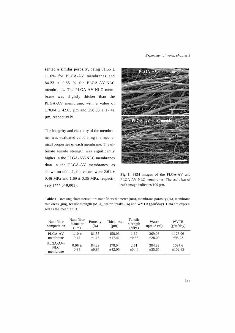
Experimental work: chapter 3
129
sented a similar porosity, being 81.55 ±
1.16% for PLGA-AV membranes and
84.23 ± 0.85 % for PLGA-AV-NLC
membranes. The PLGA-AV-NLC mem-
brane was slightly thicker than the
PLGA-AV membrane, with a value of
178.04 ± 42.05 µm and 158.03 ± 17.41
µm, respectively.
The integrity and elasticity of the membra-
nes was evaluated calculating the mecha-
nical properties of each membrane. The ul-
timate tensile strength was significantly
higher in the PLGA-AV-NLC membranes
than in the PLGA-AV membranes, as
shown on table 1, the values were 2.61 ±
0.46 MPa and 1.69 ± 0.35 MPa, respecti-
vely (*** p>0.001).
Fig 1. SEM images of the PLGA-AV and PLGA-AV-NLC membranes. The scale bar of each image indicates 100 µm.
Table 1. Dressing characterisation: nanofibers diameter (nm), membrane porosity (%), membrane thickness (µm), tensile strength (MPa), water uptake (%) and WVTR (g/m2day). Data are expres-sed as the mean ± SD.
Nanofiber composition
Nanofiber diameter
(µm)
Porosity (%)
Thickness (µm)
Tensile strength (MPa)
Water uptake (%)
WVTR (g/m2day)
PLGA-AV membrane
1.10 ± 0.42
81.55 ±1.16
158.03 ±17.41
1.69 ±0.35
369.06 ±28.09
1128.06 ±93.23
PLGA-AV-NLC
membrane
0.96 ± 0.34
84.23 ±0.85
178.04 ±42.05
2.61 ±0.46
384.32 ±35.65
1097.6 ±102.83

Novel therapeutic approaches for wound healing
130
The water uptake of the membranes
was quantified to determine their hydro-
philicity. The results showed that both
membranes presented a similar water
uptake in 72 hours, being 369.06 ± 28.09
% in PLGA-AV nanofibrous membranes
and 384.32 ± 35.65 % in PLGA-AV-NLC
membranes.
The ability of the dressings to regulate
wound moisture was assessed measuring
their WVTR. As observed in table 1, both
membranes presented similar values, al-
though it was slightly lower for PLGA-
AV-NLC nanofibers, with a value of
1128.06 ± 93.23, g/m2day while it was
1097.6 ± 102.83 g/m2day in the case of
PLGA-AV-NLC nanofibers.
Regarding to the thermal behaviour,
the thermograms of the raw materials, pre-
sented the following aspect: PLGA’s glass
transition temperature (Tg) was 156.95 ±
1.30 °C; AV did not show any marked
peak, it was a curve; and NLCs presented
an endothermic peak at 54.81 ± 0.30 °C, as
depicted in Table 2. The physical blends of
the components and the nanofibrous mem-
branes had the same peaks than the raw
Table 2. DSC peaks. The endothermic DSC
peaks of the nanofibers, their components
blends and their components itself. Data are
expressed as mean ± SD.
Endothermic DSC peak (°C)
PLGA 156.95 ± 1.30
NLC 54.81 ± 0.30
PLGA + AV blend 156.38 ± 2,88
PLGA+AV+NLC blend 56.26 ± 2.94
157.73 ±0.78
PLGA-AV membrane 153.27 ± 042
PLGA-AV-NLC membrane
59.98 ± 0.17
152.86 ± 0.08
materials, including the curve observed in
the thermogram of AV. Nevertheless, a
minor decrease on the Tg of PLGA was ob-
served on the nanofibers, and the peak of
the NLCs was slightly increased on the
physical blend and on the nanofibers.
3.2 In vitro cell viability studies
In order to assess the cytocompatibility
of the membranes, they were incubated
with culture medium for 24 h, and then the
medium was incubated with keratinocytes
and fibroblasts. After 48 h, cell viability

Experimental work: chapter 3
131
was measured through a CCK-8 assay.
Fig. 4 illustrates the results obtained in the
CCK-8 assay. None of the membranes
were cytotoxic showing percentages of
cell viability greater than 70% in in both
cell lines tested. When the extracted me-
dium was cultured with fibroblasts, both
membranes achieved a significantly higher
cell viability in comparison to the control.
Among them, the highest cell proliferation
was observed with PLGA-AV membranes,
and the difference between them was statis-
tically significant. Nevertheless, when the
extracted medium of any of the membranes
was cultured with keratinocytes, cell viabi-
lity was lower than in the control group.
3.3 Cell adhesion studies
To assess whether the cells were able to
adhere to the nanofibrous membranes or
not, they were seeded on top of the mem-
branes and SEM images were taken or
adhered cell number counted. Although
the counting process showed that some
cells were able to adhere to the membra-
nes, they could not be observed in SEM
images, as illustrated in Fig. 3A. As dep-
Fig 2. Cell viability study. (A) CCK-8 results after culturing the membranes´ extracted medium with
fibroblast. *** p<0.001 comparing PLGA-AV-NLC and PLGA-AV membranes with the control;
and ** p<0.01 comparing PLGA-AV membranes with PLGA-AV-NLC membranes. (B) CCK-8 re-
sults after culturing the membranes´ extracted medium with keratinocytes. *** p<0.001 comparing
PLGA-AV nmemebranes with the control; and ** p<0.01 comparing PLGA-AV-NLC membranes
with the control. Results are given as the mean % of living cells regarding to the control ± SD.

Novel therapeutic approaches for wound healing
132
icted in Fig. 3B and 3C, in comparison to
the bottom of the wells, both cell lines had
a significantly impaired adhesion to the
membranes. It is noteworthy to mention,
that in HaCaT cells, the adhesion was sig-
nificantly lower when the cells were see-
ded on top of PLGA-AV-NLC membranes
than on top of PLGA-AV membranes,
since the percentage of adhered cells were
25.45 ± 11.89 % and 56.23 ± 23.59%, res-
pectively.
3.4 In vivo wound healing assay
The efficacy of the membranes was
assessed in full thickness wounds inflicted
to db/db mice. One of the parameters me-
asured was the wound closure, expressed
Fig. 3. In vitro adhesion assay. (A) SEM images of membranes with cells seeded on top: 1, PLGA-
AV-NLC nanofibers incubated with keratinocytes; 2, PLGA-AV-NLC membranes incubated with
fibroblasts; 3, PLGA-AV membranes incubated with keratinocytes; and 4, PLGA-AV membranes
incubated with fibroblasts. The scale bar in each image indicates 500 µm. (B) Keratinocytes adhe-
sion percentage. *** p<0.001 comparing PLGA-AV-NLC membranes with control group; **
p<0.01 comparing PLGA-AV membranes with control group; * p<0.05 comparing both membra-
nes. (C) Fibroblasts adhesion percentage. *** p<0.001 comparing PLGA-AV-NLC and PLGA-
AV membranes with control group.
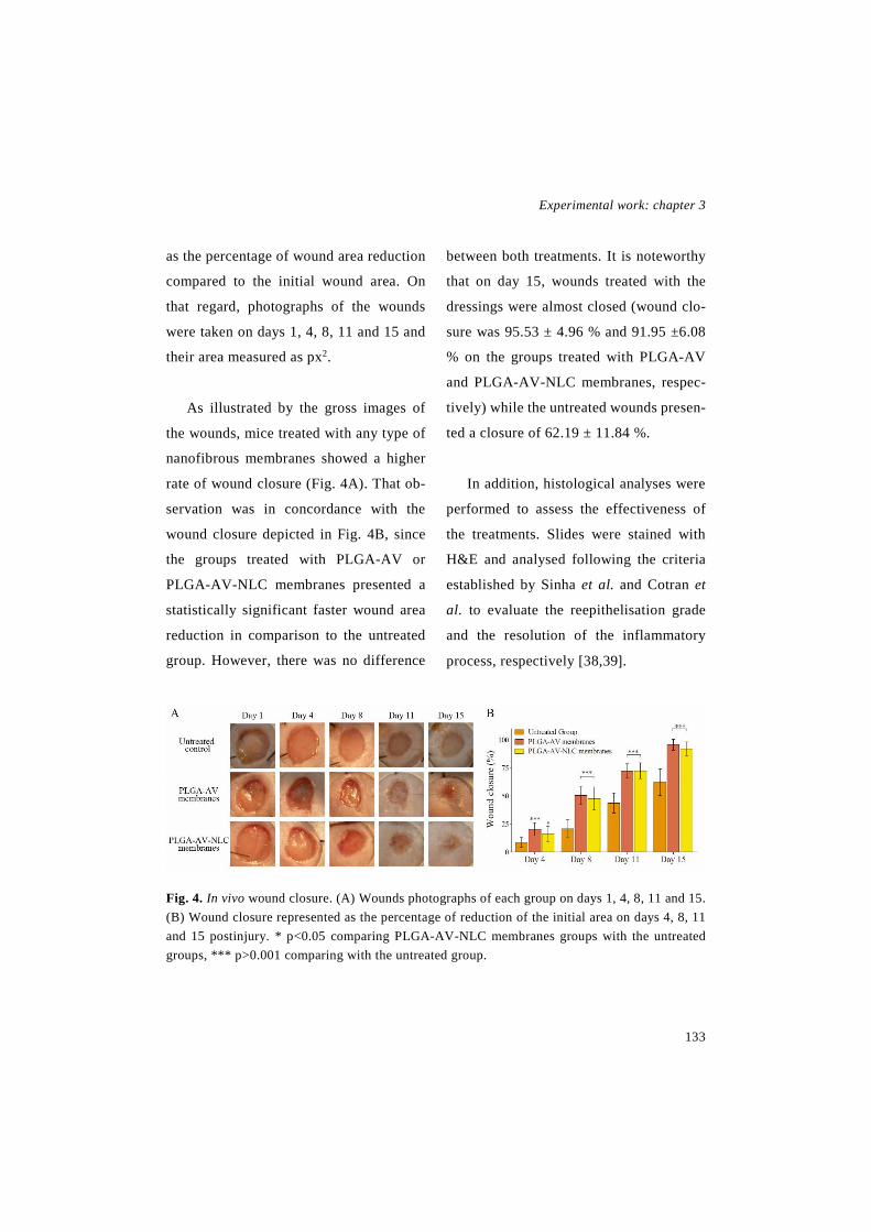
Experimental work: chapter 3
133
as the percentage of wound area reduction
compared to the initial wound area. On
that regard, photographs of the wounds
were taken on days 1, 4, 8, 11 and 15 and
their area measured as px2.
As illustrated by the gross images of
the wounds, mice treated with any type of
nanofibrous membranes showed a higher
rate of wound closure (Fig. 4A). That ob-
servation was in concordance with the
wound closure depicted in Fig. 4B, since
the groups treated with PLGA-AV or
PLGA-AV-NLC membranes presented a
statistically significant faster wound area
reduction in comparison to the untreated
group. However, there was no difference
between both treatments. It is noteworthy
that on day 15, wounds treated with the
dressings were almost closed (wound clo-
sure was 95.53 ± 4.96 % and 91.95 ±6.08
% on the groups treated with PLGA-AV
and PLGA-AV-NLC membranes, respec-
tively) while the untreated wounds presen-
ted a closure of 62.19 ± 11.84 %.
In addition, histological analyses were
performed to assess the effectiveness of
the treatments. Slides were stained with
H&E and analysed following the criteria
established by Sinha et al. and Cotran et
al. to evaluate the reepithelisation grade
and the resolution of the inflammatory
process, respectively [38,39].
Fig. 4. In vivo wound closure. (A) Wounds photographs of each group on days 1, 4, 8, 11 and 15. (B) Wound closure represented as the percentage of reduction of the initial area on days 4, 8, 11 and 15 postinjury. * p<0.05 comparing PLGA-AV-NLC membranes groups with the untreated groups, *** p>0.001 comparing with the untreated group.

Novel therapeutic approaches for wound healing
134
Regarding the reepithelisation grade,
on day 8 there were no significant diffe-
rences among groups, although a small in-
crease in the reepithelisation was observed
in the groups treated with the dressings in
comparison with the untreated control, as
illustrated in Fig. 5A. The groups treated
with the developed formulations achieved
a value around 2 in the scale described by
Sinha et al. (2 ± 0.82 and 1.75 ± 1.04 for
PLGA-AV-NLC membranes and for
PLGA-AV membranes, respectively),
which indicates that the new epithelium
covered more than half of the wound. The
untreated group, instead, obtained a value
around 1, 1.36 ± 1.06 exactly, which me-
ans that less than half of the wound was
covered by new epithelium. On day 15, the
same tendency was maintained, since a
higher reepithelisation grade was observed
with the treated groups. In this case, the
difference between the treated groups and
the untreated control was statistically sig-
nificant. A complete reepithelisation was
observed in the treated groups (4 on the
scale described by Sinha et al.); while the
untreated wounds were not completely co-
vered by new epithelium, since a value
around 2 was obtained (1.875 ± 0.64).
The evaluation of the resolution of the
inflammatory process showed that mice
treated with the dressings achieved a faster
wound maturation than the untreated
group as depicted in Fig. 5B. On day 8, no
statistically significant differences were
observed among groups. PLGA-AV-NLC
and PLGA-AV membranes reached a va-
lue higher than 2 in the in the scale descri-
bed by Cotran et al., which means that
wound were in the diffuse acute inflamma-
tion phase, the values were 2.5 ± 1.10 and
2.25 ± 0.46, respectively. Untreated mice,
on the contrary, were on the acute inflam-
mation phase, since they did not achieve a
value of 2 (1.88 ± 0.99). On day 15, mice
treated with the dressings reached a value
around 3, which means that the main pro-
cess occurring on the wounds was fibro-
blast proliferation. In untreated mice, on
the contrary, the wounds were still on the
diffuse acute inflammation phase. The dif-
ference between the PLGA-AV mem-
branes group and the untreated control was
statistically significant.
Furthermore, immunohistological anal-
yses were conducted to evaluate the num-
ber of immune cells on the wound on day
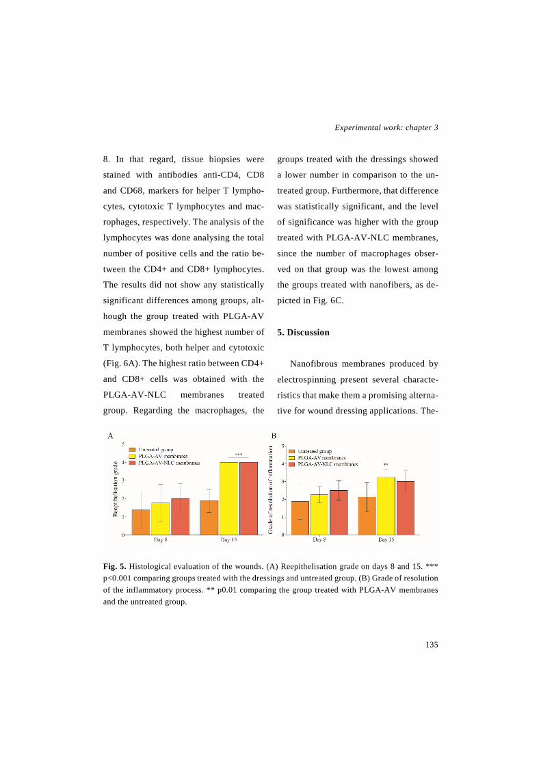
Experimental work: chapter 3
135
8. In that regard, tissue biopsies were
stained with antibodies anti-CD4, CD8
and CD68, markers for helper T lympho-
cytes, cytotoxic T lymphocytes and mac-
rophages, respectively. The analysis of the
lymphocytes was done analysing the total
number of positive cells and the ratio be-
tween the CD4+ and CD8+ lymphocytes.
The results did not show any statistically
significant differences among groups, alt-
hough the group treated with PLGA-AV
membranes showed the highest number of
T lymphocytes, both helper and cytotoxic
(Fig. 6A). The highest ratio between CD4+
and CD8+ cells was obtained with the
PLGA-AV-NLC membranes treated
group. Regarding the macrophages, the
groups treated with the dressings showed
a lower number in comparison to the un-
treated group. Furthermore, that difference
was statistically significant, and the level
of significance was higher with the group
treated with PLGA-AV-NLC membranes,
since the number of macrophages obser-
ved on that group was the lowest among
the groups treated with nanofibers, as de-
picted in Fig. 6C.
5. Discussion
Nanofibrous membranes produced by
electrospinning present several characte-
ristics that make them a promising alterna-
tive for wound dressing applications. The-
Fig. 5. Histological evaluation of the wounds. (A) Reepithelisation grade on days 8 and 15. *** p<0.001 comparing groups treated with the dressings and untreated group. (B) Grade of resolution of the inflammatory process. ** p0.01 comparing the group treated with PLGA-AV membranes and the untreated group.

Novel therapeutic approaches for wound healing
136
refore, in a previous study, our research
group developed a composite membrane
of PLGA RG508 and the largest possible
amount of Aloe vera extract, within which
EGF was uniformly distributed [14]. In the
present study, EGF was not added since
we aimed to develop a medical device; ins-
tead lipid nanoparticles were included to
improve the removal of the membrane du-
ring the dressing change. In addition, ano-
ther polymer was used, PLGA 824 LG,
which has a higher molecular weight, be-
cause it allowed a better incorporation of
the nanoparticles. The average diameter of
the nanofibers was increased with increa-
sing the polymer molecular weight, from
about 500 nm in the previous study to
about 1 µm in the current study. The rise
of the diameter was probably due to the
greater viscosity of the polymer solution
prepared with the high molecular weight
PLGA. In this regard, previous studies
have shown that solution viscosity is criti-
cal to control the nanofibers morphology,
including their diameter [40]. The high po-
rosity is one of the characteristics that ma-
kes electrospun membranes a suitable
approach for wound dressing, since it
allows gas permeation, and thus cell respi-
ration. Although that high porosity, nano-
fibers present a very small pore size,
which creates a physical barrier that can
prevent bacterial growth [3]. The charac-
terisation of the nanofibers showed that
the addition of the NLCs into the formula-
tion improved their mechanical properties,
and therefore, the handling of the membra-
nes. The PLGA-AV-NLC membranes pre-
Fig 6. Immunohistological analysis. (A) Number of lymphocytes (CD4+ and CD8+ cells) on day 8. (B) Ratio between CD8+ and CD4+ cells on day 8. (C) Number of CD68+ cells on day 8. ** p<0.01 comparing the group treated with PLGA-AV membranes with the untreated group. *** p<0.001 comparing the group treated with PLGA-AV-NLC membranes with the untreated group.
Experimental work: chapter 3
137
sented an increased thickness and tensile
strength, which makes more difficult to
break or bend the dressing during the
application, and therefore makes easier to
apply it. The increased thickness of the
membrane was probably due to the higher
solids percentage of the solution contai-
ning the NLCs. The increment of the ten-
sile strength may be due to the uniform
distribution of the nanoparticles in the po-
lymer matrix, as observed by Thomas et al.
Therefore, the stress might be uniformly
distributed into the nanofibers, minimi-
zing the stress concentration centres, and
increasing the interfacial area to transfer
that stress from the polymer matrix to the
NLCs, which result in improved mechani-
cal properties [41]. The tensile strength of
both formulations was below the tensile
strength of human skin, which ranged
from 5.7 MPa to 12.6 MPa [42]. However,
this does not weaken their potential as
wound dressings, because wounds are im-
mobilized during healing and thus dre-
ssings are rarely under a high tensile
strength [43]. Moreover, nanofibrous dre-
ssings with a low tensile strength ranging
from 1-3 MPa have been previously deve-
loped for wound healing [44-46].
As described previously the water
uptake (%) increased with rising concen-
tration of the hydrophilic compound
[14,47]. Therefore, since Aloe vera con-
centration was very similar in both formu-
lations, it was expected that they would
have a similar water uptake. Their swe-
lling ability was very high, about 380 %,
which helps to drain the exudates while re-
taining moisture to help wound healing. In
addition to water uptake, moisture was
also controlled by the water vapour trans-
mision of the dressing [3,48]. The WVTR
of the developed formulations was within
the range of commercial wound dressings
(426-2047 g/m2day), thus they presented
an adequate ability to allow most of the
exudate to evaporate, while maintaining
some moisture into the wound [49]. The
PLGA-AV-NLC membranes showed a
slightly lower WVTR value which can be
translated into a slightly higher occlusivity.
The analysis of the thermal behaviour
showed a decrease of the PLGA Tg, in both
nanofibers in comparison to the raw PLGA
and the physical blend of the components.
That decrease can be due to a higher de-
gree of alignment and orientation of the

Novel therapeutic approaches for wound healing
138
polymer chains into the electrospun mem-
brane [14,50]. Every formulation contai-
ning NLCs presented a peak around 55°C
that presumably corresponds to Precirol
[51]. The NLCs thermogram showed a
slight lower melting point of the Precirol
in comparison to the physical blend or
PLGA-AV-NLC membranes, which could
indicate an interaction between the NLCs
and the AV. It is noteworthy to mention
that the peaks corresponding to PLGA and
NLCs and the curve pattern of AV were
found in both PLGA-AV-NLC nanofibers
and physical blend, which reveals that the
components of the nanofibers presented an
immiscible blend morphology.
Regarding the biocompatibility of the
dressings, none of them showed indirect
cytotoxicity after the incubation of their
extracted medium along with keratinocy-
tes or fibroblasts, since their viability was
maintained above the 70%, which indica-
ted a good biocompatibility. The results
obtained with fibroblasts, showed an in-
creased viability after the incubation with
the extracted medium of the nanofibers,
probably due to the proliferative effect of
the Aloe vera into fibroblasts [14,21,23].
The higher fibroblasts viability observed
with the PLGA-AV nanofibers might be due
to a faster release of the AV from the nano-
fibers, since the interaction between the AV
and NLCs exposed in the DSC analysis
could have aminorate the rate of AV release.
The main reasons to include NLCs into
the nanofibers were the hypothesis that
they could improve their handling and to
ease the removal of the dressing from the
wound, avoiding pain and damage of the
newly formed tissue during dressing
change. The characterisation of the mem-
branes showed that NLCs were able to im-
prove the handling and mechanical strength
of the nanofibers. Therefore, cell adhesion
was analysed and it revealed a lower kera-
tinocyte adhesion to the formulation con-
taining NLCs. Nevertheless, no differen-
ces were found in fibroblast attachment,
since both membranes with or without
NLCs presented a very low adhesion. Con-
sidering those results, more studies should
be performed to assess dressing attach-
ment into wounded tissue, analysing the
dressing removal in vivo or using a texture
analyzer to evaluate the adhesion strength
to the wounded tissue.

Experimental work: chapter 3
139
Finally, the efficacy of the membranes
was evaluated in vivo in a full thickness
splinted wound model carried out in db/db
mice. db/db mice were chosen because
they present an impaired wound healing
secondary to diabetes, and thus, they re-
semble better a chronic wound model. In
addition, they mimic better human wound
healing than other rodent models, since
they have impaired wound contraction due
to their obesity, and therefore their healing
occurs mainly via reepithelisation, as hu-
man healing [52,53]. In order to impair
even more contraction and enhance ree-
phitelisation, silicone splints were sutured
around the wounds [37].
Overall, both membranes achieved si-
milar improvement in wound healing.
Comparable results were obtained in
wound closure and reepithelisation, as
both were able to accelerate healing in
comparison to untreated control. Regar-
ding the resolution of the inflammatory
process, on day 15 only PLGA-AV mem-
branes presented an improved outcome in
comparison to the control group. On day 8,
no differences were observed in the histo-
logical analysis, although the developed
formulations, and especially the PLGA-
AV-NLC membranes, were able to reduce
the macrophage infiltration in the wound
bed that usually is augmented in rodent
wound models with diabetes or impaired
healing. [54,55]. In addition, the macro-
phages found on those wounds present an
impaired ability to phagocyte apoptotic
cells, increasing the level of proinflamma-
toy cytokines, and thus, perpetuating a
continuous inflammatory state [56].
Accordingly, both formulations showed an
enhancement of wound maturation, the
PLGA-AV-NLC membranes on macro-
phage infiltration on the early stage of he-
aling and the PLGA-AV membranes on
the general inflammatory state of the later
stage.
The effect of the nanofibrous dressings
on wound healing can be partially explai-
ned by the incorporation of Aloe vera,
since it has shown to improve wound hea-
ling, mainly by affecting fibroblast growth
factor, and thus, improving their activity
and proliferation [23]. In addition, the cha-
racteristics of the nanofibrous structure
also contribute to the improvement of
wound healing. In fact, the high surface to

Novel therapeutic approaches for wound healing
140
volume area and the nanoporosity create
an adequate environment for cell migra-
tion and proliferation to the wound bed.
Moreover, that proliferating environment
is involved in the improvement of the gra-
nulation tissue formation and reepithelisa-
tion by enhancing the release of healing
mediators, such as growth factors, angio-
genic factors or collagen [3,40]. Finally,
the chosen polymer is also involved in the
enhancement of reepithelisation, since one
of its degradation products, lactate, has
shown to be able to induce a faster wound
healing [18].
Regarding to the inclusion of NLCs
into the membranes, their effect on the re-
moval of the dressing could not be obser-
ved in vivo, since both dressings were hu-
mected with PBS prior to their elimina-
tion, in order to avoid any possible damage
on the newly formed tissue. Nevertheless,
an improvement on the handling of the
membranes containing NLCs was obser-
ved along the characterisation of the for-
mulations. Hence, the nanofibrous dressing
with NLCs showed a benefit concerning
handling, although more studies are needed
to assess their effect in dressing removal.
4. Conclusion
In the current study two composite
electrospun dressings were developed, the
first one was composed of an emulsion of
PLGA and Aloe vera (1:1), and in the se-
cond one lipid nanoparticles (NLCs) were
added to the aforementioned emulsion.
Both dressings showed a similar characte-
risation, although an enhanced handling
was observed in the PLGA-AV-NLC for-
mulation regarding to elasticity and thick-
ness. Finally, their effectivity in wound
healing was assessed in a full thickness
wound healing assay performed in db/db
mice, achieving similar results with both
formulations. Accordingly, the PLGA-
AV-NLC nanofibrous membrane might be
a promising strategy for the treatment of
chronic wound, since it improved handling
in comparison to the formulation without
NLCs.
6. Acknowledgments
I. García-Orue thanks the Basque Go-
vernment for the fellowship grant. The au-
thors thank for technical and human su-
pport provided by SGIker of UPV/EHU
Experimental work: chapter 3
141
and European funding (ERDF and ESF).
This project has been funded by the Bas-
que Government (ELKARTEK 2015, Na-
noplatform, KK-2015/0000036 and Con-
solidated Groups, IT-428-10 and IT-528-
10).
7. References
[1] M. Liu, X. Duan, Y. Li, D. Yang, Y. Long, Electrospun nanofibers for wound healing, Ma-ter. Sci. Eng. C Mater. Biol. Appl. 76 (2017) 1413-1423. doi: 10.1016/j.msec.2017.03.034.
[2] H.P. Felgueiras, M.T.P. Amorim, Functio-nalization of electrospun polymeric wound dressings with antimicrobial peptides, Colloids Surf. B Biointerfaces 156 (2017) 133-148. doi: //doi.org/10.1016/j.colsurfb.2017.05.001.
[3] M. Abrigo, S.L. McArthur, P. Kingshott, Electrospun nanofibers as dressings for chronic wound care: advances, challenges, and future prospects, Macromol. Biosci. 14 (2014) 772-792. doi: 10.1002/mabi.201300561.
[4] I. Garcia-Orue, J.L. Pedraz, R.M. Hernan-dez, M. Igartua, Nanotechnology-based deli-very systems to release growth factors and ot-her endogenous molecules for chronic wound healing, J. Drug Deliv. Sci. Technol. 42 (2017) 2-17. doi: 10.1016/j.jddst.2017.03.002.
[5] Matthew S. Brown, Brandon Ashley, Ahyeon Koh, Wearable Technology for Chro-nic Wound Monitoring: Current Dressings, Advancements, and Future Prospects, Frontiers
in Bioengineering and Biotechnology 6 (2018). doi: 10.3389/fbioe.2018.00047.
[6] L. Pachuau, Recent developments in novel drug delivery systems for wound healing, Ex-pert. Opin. Drug Deliv. 12 (2015) 1895-1909. doi: 10.1517/17425247.2015.1070143.
[7] K. Järbrink, G. Ni, H. Sönnergren, A. Sch-midtchen, C. Pang, R. Bajpai, J. Car, The hu-manistic and economic burden of chronic wounds: a protocol for a systematic review, Syst. Rev. 6 (2017). doi: 10.1186/s13643-016-0400-8.
[8] C.K. Sen, G.M. Gordillo, S. Roy, R. Kirs-ner, L. Lambert, T.K. Hunt, F. Gottrup, G.C. Gurtner, M.T. Longaker, Human skin wounds: a major and snowballing threat to public health and the economy, Wound Repair Regen. 17 (2009) 763-771. doi: 10.1111/j.1524-475X.2009.00543.x.
[9] G. Han, R. Ceilley, Chronic Wound Healing: A Review of Current Management and Treatments, Adv. Ther. 34 (2017) 599-610. doi: 10.1007/s12325-017-0478-y.
[10] R.F. Diegelmann, M.C. Evans, Wound healing: an overview of acute, fibrotic and de-layed healing, Front. Biosci. 9 (2004) 283-289. doi: //dx.doi.org/10.2741/.
[11] T. Velnar, T. Bailey, V. Smrkolj, The wound healing process: an overview of the ce-llular and molecular mechanisms, J. Int. Med. Res. 37 (2009) 1528-1542. doi: 10.1177/147323000903700531.
[12] S. Schreml, R. Szeimies, L. Prantl, M. Landthaler, P. Babilas, Wound healing in the 21st century, J. Am. Acad. Dermatol. 63 (2010)

Novel therapeutic approaches for wound healing
142
866-881. doi: //dx.doi.org/10.1016/j. jaad.2009 .10.048.
[13] P.S. Briquez, J.A. Hubbell, M.M. Martino, Extracellular matrix-inspired growth factor de-livery systems for skin wound healing, Adv. Wound Care (New Rochelle) 4 (2015) 479-489. doi: 10.1089/wound.2014.0603.
[14] I. Garcia-Orue, G. Gainza, F.B. Gutierrez, J.J. Aguirre, C. Evora, J.L. Pedraz, R.M. Her-nandez, A. Delgado, M. Igartua, Novel nanofi-brous dressings containing rhEGF and Aloe vera for wound healing applications, Int. J. Pharm. 53 (2016) 556-566. doi: //dx.doi.org/ 10.1016/j.ijpharm.2016.11.006.
[15] K.K. Chereddy, G. Vandermeulen, V. Préat, PLGA based drug delivery systems: Pro-mising carriers for wound healing activity, Wound Repair Regen. 24 (2016) 223-236. doi: 10.1111/wrr.12404.
[16] T. Garg, G. Rath, A.K. Goyal, Com-prehensive review on additives of topical do-sage forms for drug delivery, Drug Deliv. 22 (2015) 969-987. doi: 10.3109/10717544. 2013.879355.
[17] T. Garg, G. Rath, A.K. Goyal, Biomate-rials-based nanofiber scaffold: targeted and controlled carrier for cell and drug delivery, J. Drug Target. 23 (2015) 202-221. doi: 10.3109/1061186X.2014.992899.
[18] P. Porporato, V. Payen, C. De Saedeleer, V. Préat, J. Thissen, O. Feron, P. Sonveaux, Lactate stimulates angiogenesis and accelera-tes the healing of superficial and ischemic wounds in mice, Angiogenesis 15 (2012) 581-592. doi: 10.1007/s10456-012-9282-0.
[19] A.D. Dat, F. Poon, K.B. Pham, J. Doust, Aloe vera for treating acute and chronic wounds, Cochrane Database Syst. Rev. (2012). doi: 10.1002/14651858.CD008762.pub2.
[20] S. Choi, M. Chung, A review on the rela-tionship between aloe vera components and their biologic effects, Semin. Integr. Med. 1 (2003) 53-62. doi: //dx.doi.org/10.1016/S1543 -1150(03)00005-X.
[21] S.A. Hashemi, S.A. Madani, S. Abedian-kenari, The Review on Properties of Aloe Vera in Healing of Cutaneous Wounds, Biomed. Res. Int. 2015 (2015) 714216. doi: 10.1155 /2015/714216.
[22] A. Surjushe, R. Vasani, D.G. Saple, Aloe vera: a short review, Indian J. Dermatol. 53 (2008) 163-166. doi: 10.4103/0019-5154.4478
[23] M.D. Boudreau, F.A. Beland, An evalua-tion of the biological and toxicological proper-ties of Aloe barbadensis (miller), Aloe vera., J. Environ. Sci. Health C Environ. Carcinog. Ecotoxicol. Rev. 24 (2006) 103-154. doi: 10.1080/10590500600614303.
[24] S. Suganya, J. Venugopal, S. Rama-krishna, B.S. Lakshmi, V.R.G. Dev, Naturally derived biofunctional nanofibrous scaffold for skin tissue regeneration, Int. J. Biol. Macro-mol. 68 (2014) 135-143. doi: //dx.doi.org/10. 1016/j.ijbiomac.2014.04.031.
[25] V. Bhaarathy, J. Venugopal, C. Gandhi-mathi, N. Ponpandian, D. Mangalaraj, S. Ra-makrishna, Biologically improved nanofibrous scaffolds for cardiac tissue engineering, Mater. Sci Eng C Mater. Biol. Appl. 44(2014)268-277 doi: //dx.doi.org/10.1016/j.msec.2014.08. 018

Experimental work: chapter 3
143
[26] P. Karuppuswamy, J.R. Venugopal, B. Navaneethan, A.L. Laiva, S. Sridhar, S. Rama-krishna, Functionalized hybrid nanofibers to mimic native ECM for tissue engineering ap-plications, Appl. Surf. Sci. 322 (2014) 162-168. doi: //dx.doi.org/10.1016/j.apsusc.2014. 10.074.
[27] S.A. Kheradvar, J. Nourmohammadi, H. Tabesh, B. Bagheri, Starch nanoparticle as a vitamin E-TPGS carrier loaded in silk fibroin-poly(vinyl alcohol)-Aloe vera nanofibrous dressing, Colloids Surf. B Biointerfaces 166 (2018) 9-16. doi: //doi.org/10.1016/j.colsu-rfb.2018.03.004.
[28] A. Jouybar, E. Seyedjafari, A. Ardeshiry-lajimi, A. Zandi-Karimi, N. Feizi, M. Khani, I. Pousti, Enhanced Skin Regeneration by Herbal Extract-Coated Poly-L-Lactic Acid Nanofi-brous Scaffold, Artif. Organs 41 (2017) E307. doi: 10.1111/aor.12926.
[29] F. David, J. Wurtz, N. Breton, O. Bisch, P. Gazeu, J. Kerihuel, O. Guibon, A randomi-sed, controlled, non-inferiority trial comparing the performance of a soft silicone-coated wound contact layer (Mepitel One) with a lipi-docolloid wound contact layer (UrgoTul) in the treatment of acute wounds, Int. Wound J. 15 (2018) 159-169. doi: 10.1111/iwj.12853.
[30] M. Benbow, Urgotul™: alternative to con-ventional non-adherence dressings, Br. J. Nurs. 11 (2002) 135-138. doi: 10.12968/bjon .2002.11.2.9315.
[31] M. Benbow, G. Iosson, A clinical evalua-tion of Urgotul to treat acute and chronic wounds, Br. J. Nurs. 13 (2004) 105-109. doi: 10.12968/bjon.2004.13.2.12042.
[32] P.W.W. Tan, W.C. Ho, C. Song, The use of Urgotul in the treatment of partial thickness burns and split-thickness skin graft donor sites: a prospective control study, Int. Wound J. 6 (2009) 295-300. doi: 10.1111/j.1742-481X.2009.00611.x.
[33] G. Gainza, M. Pastor, J.J. Aguirre, S. Vi-llullas, J.L. Pedraz, R.M. Hernandez, M. Igar-tua, A novel strategy for the treatment of chro-nic wounds based on the topical administration of rhEGF-loaded lipid nanoparticles: In vitro bioactivity and in vivo effectiveness in healing-impaired db/db mice, J. Control Re- lease 185 (2014) 51-61. doi: //dx.doi.org/10. 1016/j.jconrel.2014.04.032.
[34] G. Gainza, D.C. Bonafonte, B. Moreno, J.J. Aguirre, F.B. Gutierrez, S. Villullas, J.L. Pedraz, M. Igartua, R.M. Hernandez, The topi-cal administration of rhEGF-loaded nanostruc-tured lipid carriers (rhEGF-NLC) improves healing in a porcine full-thickness excisional wound model, J. Control Release 197 (2015) 41-47. doi: //dx.doi.org/10.1016/j.jconrel. .10.033.
[35] I. Garcia-Orue, G. Gainza, C. Girbau, R. Alonso, J.J. Aguirre, J.L. Pedraz, M. Igartua, R.M. Hernandez, LL37 loaded nanostructured lipid carriers (NLC): A new strategy for the to-pical treatment of chronic wounds, Eur. J. Pharm. Biopharm. (2016). doi: //dx.doi.org/10. 1016/j.ejpb.2016.04.006.
[36] C. Li, R. Fu, C. Yu, Z. Li, H. Guan, D. Hu, D. Zhao, L. Lu, Silver nanoparticle/chitosan oligosaccharide/poly(vinyl alcohol) nanofibers as wound dressings: a preclinical study, Int. J. Nanomedicine 8 (2013) 4131-4145. doi: 10.2147/IJN.S51679 [doi].
Novel therapeutic approaches for wound healing
144
[37] J. Michaels, S.S. Churgin, K.M. Blech-man, M.R. Greives, S. Aarabi, R.D. Galiano, G.C. Gurtner, db/db mice exhibit severe wound-healing impairments compared with ot-her murine diabetic strains in a silicone-splin-ted excisional wound model, Wound Repair Regen. 15 (2007) 665-670. doi: 10.1111/j.1524-475X.2007.00273.x.
[38] U.K. Sinha, L.A. Gallagher, Effects of Steel Scalpel, Ultrasonic Scalpel, CO2 Laser, and Monopolar and Bipolar Electrosurgery on Wound Healing in Guinea Pig Oral Mucosa, Laryngoscope 113 (2003) 228-236. doi: 10.1097/00005537-200302000-00007.
[39] R. Cotran, G.K. Kumar, T. Collins, Repa-ración de los tejidos: proliferacion celular, fi-brosis y curaicón de las heridas (2000) 95-120.
[40] S. Shahverdi, M. Hajimiri, M.A. Esfan-diari, B. Larijani, F. Atyabi, A. Rajabiani, A.R. Dehpour, A.A. Gharehaghaji, R. Dinarvand, Fabrication and structure analysis of poly(lac-tide-co-glycolic acid)/silk fibroin hybrid scaf-fold for wound dressing applications, Int. J. Pharm. 473 (2014) 345-355. doi: //dx.doi.org/10.1016/j.ijpharm.2014.07.021.
[41] R. Thomas, K. Soumya, J. Mathew, E. Radhakrishnan, Electrospun Polycaprolactone Membrane Incorporated with Biosynthesized Silver Nanoparticles as Effective Wound Dres-sing Material, Appl. Biochem. Biotechnol. 176 (2015) 2213-2224. doi: 10.1007/s12010-015-1709-9.
[42] C. Jacquemoud, K. Bruyere-Garnier, M. Coret, Methodology to determine failure cha-racteristics of planar soft tissues using a dyna-mic tensile test, J. Biomech. 40 (2007) 468-
475. doi: //dx.doi.org/10.1016/j.jbiomech.2005 .12.010.
[43] G. Jin, M.P. Prabhakaran, D. Kai, S.K. Annamalai, K.D. Arunachalam, S. Rama-krishna, Tissue engineered plant extracts as na-nofibrous wound dressing, Biomaterials 34 (2013) 724-734. doi: //doi.org/10.1016/j.bio-materials.2012.10.026.
[44] A.M. Loordhuswamy, V.R. Krishna-swamy, P.S. Korrapati, S. Thinakaran, G.D.V. Rengaswami, Fabrication of highly aligned fi-brous scaffolds for tissue regeneration by cent-rifugal spinning technology, Materials Science and Engineering: C 42 (2014) 799-807. doi: //doi.org/10.1016/j.msec.2014. 06.011.
[45] U. Dashdorj, M.K. Reyes, A.R. Unnithan, A.P. Tiwari, B. Tumurbaatar, C.H. Park, C.S. Kim, Fabrication and characterization of elec-trospun zein/Ag nanocomposite mats for wound dressing applications, Int. J. Biol. Ma-cromol. 80 (2015) 1-7. doi: //dx.doi.org/ 10.1016/j.ijbiomac.2015.06.026.
[46] S. Tort, F. Acartürk, A. Beşikci, Evalua-tion of three-layered doxycycline-collagen loa-ded nanofiber wound dressing, International Journal of Pharmaceutics 529 (2017) 642-653. doi: //doi.org/10.1016/j.ijpharm.2017.07.027.
[47] S. Son, R. Franco, S. Bae, Y. Min, B. Lee, Electrospun PLGA/gelatin fibrous tubes for the application of biodegradable intestinal stent in rat model, J. Biomed. Mater. Res. 101B (2013) 1095-1105. doi: 10.1002/jbm.b.32923.
[48] S. Natarajan, D. Williamson, A.J. Stiltz, K. Harding, Advances in Wound Care and Healing Technology, Am. J. Clin. Dermatol. 1

Experimental work: chapter 3
145
(2000) 269-275. doi: 10.2165/00128071-200001050-00002.
[49] Y. Tu, M. Zhou, Z. Guo, Y. Li, Y. Hou, D. Wang, L. Zhang, Preparation and characteriza-tion of thermosensitive artificial skin with a Sandwich structure, Mater. Lett. 147(2015)4-7 doi: //dx.doi.org/10.1016/j.matlet.2015.01.163
[50] H. Fouad, T. Elsarnagawy, F.N. Almahjdi, K.A. Khalil, Preparation and in vitro thermo-mechanical characterization of electrospun PLGA nanofibers for soft and hard tissue re-placement, Int. J. Electrochem. Sci. 8 (2013) 2293-2304.
[51] J. Hamdani, A.J. Moës, K. Amighi, Physi-cal and thermal characterisation of Precirol ® and Compritol ® as lipophilic glycerides used for the preparation of controlled-release matrix pellets, Int. J. Pharm. 260 (2003) 47-57. doi: 10.1016/S0378-5173(03)00229-1.
[52] R.C. Fang, T.A. Mustoe, Animal models of wound healing: uility in transgenic mice, J. Biomater. Sci. Polym. Ed. 19 (2008) 989-1005. doi: 10.1163/156856208784909327.
[53] V.I. Tkalcevic, S. Cužic, M.J. Parnham, I. Pašalic, K. Brajša, Differential Evaluation of Excisional Non-occluded Wound Healing in db/db Mice, Toxico. Pathol. 37 (2009) 183-192. doi: 10.1177/0192623308329280.
[54] Tsubame Nishikai-Yan Shen, Shigeyuki Kanazawa, Makiko Kado, Kayoko Okada, Lin Luo, Ayato Hayashi, Hiroshi Mizuno, Rica Ta-naka, Interleukin-6 stimulates Akt and p38 MAPK phosphorylation and fibroblast migra-tion in non-diabetic but not diabetic mice, PLoS One 12 (2017) e0178232. doi: 10.1371/journal.pone.0178232.
[55] J. Yeh, L. Yeh, S. Jung, T. Chang, H. Wu, T. Shiu, C. Liu, W.W. Kao, P. Chu, Impaired skin wound healing in lumican-null mice, Br. J. Dermatol. 163 (2010) 1174. doi: 10.1111/j.1365-2133.2010.10008.x.
[56] Savita Khanna, Sabyasachi Biswas, Yingli Shang, Eric Collard, Ali Azad, Courtney Kauh, Vineet Bhasker, Gayle M Gordillo, Chandan K Sen, Sashwati Roy, Macrophage Dysfunction Impairs Resolution of Inflammation in the Wounds of Diabetic Mice, PLoS One 5 (2010) e9539. doi: 10.1371/journal.pone.0009539.
[43] G. Jin, M.P. Prabhakaran, D. Kai, S.K. Annamalai, K.D. Arunachalam, S. Rama-krishna, Tissue engineered plant extracts as nanofibrous wound dressing, Biomaterials 34 (2013) 724-734. doi: //doi.org/10.1016/j.bio-materials.2012.10.026.
[44] A.M. Loordhuswamy, V.R. Krish-naswamy, P.S. Korrapati, S. Thinakaran, G.D.V. Rengaswami, Fabrication of highly aligned fibrous scaffolds for tissue regenera-tion by centrifugal spinning technology, Mate-rials Science and Engineering: C 42 (2014) 799-807. doi: //doi.org/10.1016/j.msec.2014. 06.011.
[45] U. Dashdorj, M.K. Reyes, A.R. Unnithan, A.P. Tiwari, B. Tumurbaatar, C.H. Park, C.S. Kim, Fabrication and characterization of elec-trospun zein/Ag nanocomposite mats for wound dressing applications, Int. J. Biol. Mac-romol. 80 (2015) 1-7. doi: //dx.doi.org/ 10.1016/j.ijbiomac.2015.06.026.
[46] S. Tort, F. Acartürk, A. Beşikci, Evalua-tion of three-layered doxycycline-collagen
Novel therapeutic approaches for wound healing
146
loaded nanofiber wound dressing, International Journal of Pharmaceutics 529 (2017) 642-653. doi: //doi.org/10.1016/j.ijpharm.2017.07.027.
[47] S. Son, R. Franco, S. Bae, Y. Min, B. Lee, Electrospun PLGA/gelatin fibrous tubes for the application of biodegradable intestinal stent in rat model, J. Biomed. Mater. Res. 101B (2013) 1095-1105. doi: 10.1002/jbm.b.32923.
[48] S. Natarajan, D. Williamson, A.J. Stiltz, K. Harding, Advances in Wound Care and Healing Technology, Am. J. Clin. Dermatol. 1 (2000) 269-275. doi: 10.2165/00128071-200001050-00002.
[49] Y. Tu, M. Zhou, Z. Guo, Y. Li, Y. Hou, D. Wang, L. Zhang, Preparation and characteriza-tion of thermosensitive artificial skin with a Sandwich structure, Mater. Lett. 147 (2015) 4-7. doi: //dx.doi.org/10.1016/j.matlet.2015.01. 163.
[50] H. Fouad, T. Elsarnagawy, F.N. Almahjdi, K.A. Khalil, Preparation and in vitro thermo-mechanical characterization of electrospun PLGA nanofibers for soft and hard tissue re-placement, Int. J. Electrochem. Sci. 8 (2013) 2293-2304.
[51] J. Hamdani, A.J. Moës, K. Amighi, Phys-ical and thermal characterisation of Precirol ® and Compritol ® as lipophilic glycerides used for the preparation of controlled-release matrix pellets, Int. J. Pharm. 260 (2003) 47-57. doi: 10.1016/S0378-5173(03)00229-1.
[52] R.C. Fang, T.A. Mustoe, Animal models of wound healing: uility in transgenic mice, J. Biomater. Sci. Polym. Ed. 19 (2008) 989-1005. doi: 10.1163/156856208784909327.
[53] V.I. Tkalcevic, S. Cužic, M.J. Parnham, I. Pašalic, K. Brajša, Differential Evaluation of Excisional Non-occluded Wound Healing in db/db Mice, Toxico. Pathol. 37 (2009) 183-192. doi: 10.1177/0192623308329280.
[54] Tsubame Nishikai-Yan Shen, Shigeyuki Kanazawa, Makiko Kado, Kayoko Okada, Lin Luo, Ayato Hayashi, Hiroshi Mizuno, Rica Tanaka, Interleukin-6 stimulates Akt and p38 MAPK phosphorylation and fibroblast migra-tion in non-diabetic but not diabetic mice, PLoS One 12 (2017) e0178232. doi: 10.1371/journal.pone.0178232.
[55] J. Yeh, L. Yeh, S. Jung, T. Chang, H. Wu, T. Shiu, C. Liu, W.W. Kao, P. Chu, Impaired skin wound healing in lumican-null mice, Br. J. Dermatol. 163 (2010) 1174. doi: 10.1111/ .1365-2133.2010.10008.x.
[56] Savita Khanna, Sabyasachi Biswas, Yingli Shang, Eric Collard, Ali Azad, Courtney Kauh, Vineet Bhasker, Gayle M Gordillo, Chandan K Sen, Sashwati Roy, Macrophage Dysfunction Impairs Resolution of Inflammation in the Wounds of Diabetic Mice, PLoS One 5 (2010) e9539. doi: 10.1371/journal.pone.0009539.
CHAPTER 4
Development of a gelatin/chitosan bilayer hydrofilm for wound healing
I. Garcia-Oruea,b+; E. Santos-Vizcainoa,b+; A. Etxabidec; J. Urangac; A. Bayatd*; P. Guerreroc; M. Igartuaa,b; K. de la Cabac; R.M. Hernandeza,b* a NanoBioCel Group, Laboratory of Pharmaceutics, School of Pharmacy, University of the Basque Country (UPV/EHU),
Paseo de la Universidad 7, 01006 Vitoria-Gasteiz, Spain b Biomedical Research Networking Centre in Bioengineering, Biomaterials and Nanomedicine (CIBER-BBN), Vitoria-Gas-
teiz, Spain c BIOMAT Research Group, Chemical and Environmental Engineering Department, Engineering College of Gipuzkoa, Uni-
versity of the Basque Country (UPV/EHU), Plaza de Europa 1, 20018 Donostia-San Sebastián, Spain d Plastic & Reconstructive Surgery Research, Division of Musculoskeletal & Dermatological Sciences, School of Biological
Sciences, University of Manchester, Manchester, UK.
+ These two authors contributed equally to this work.
* Corresponding author: Dr. A. Bayat and Dr. R.M. Hernandez
ABSTRACT
In the current study, we developed a novel gelatin-based bilayer wound dressing. We used different crosslinking agents to confer unique properties to each layer, obtaining a multifunctional hydrofilm able to fulfil the requirements for wound healing applications. First, we produced a resistant and non-degradable upper layer by lactose-mediated cross-linking of gelatin, which provided mechanical support and protection to overall design. For the lower layer, gelatin was crosslinked with citric acid, resulting in a porous matrix with a great swelling ability. In addition, we incorporated chitosan into the lower layer to harness its wound healing ability. The influence of the crosslinker was demonstrated by FTIR and SEM analysis, lactose addition changed the secondary structure of gelatin leading to a more compact and smoother structure. Results showed that the hydrofilm was able to swell 407.3 ± 108.6 % of its dry weight while maintaining mechanical integ-rity. Besides, its water vapour transmission rate was 1381.5 ± 108.6 g/m2day. In vitro, it showed a good biocompatibility. Finally, its efficacy was tested through an ex vivo wound healing assay in human skin. The hydrofilm achieved similar results to the con-trol. Altogether, the developed bilayer hydrofilm showed suitable characteristics to be used as a wound dressing.
Sent to: European Journal of Pharmaceutics and Biopharmaceutics


Experimental work: Chapter 4
149
1. Introduction
Bilayer dressings are a promising
approach for wound healing, since they are
able to provide a structure that mimics the
bilayer structure of skin, with an upper
protective layer resembling the epidermis,
and a thicker flexible lower layer like the
dermis. Taking that into account, the dense
upper layer is designed to cover the
wound, giving mechanical strength to the
dressing. In addition, it needs to control
moisture transmission to prevent fluid loss
and dehydration, while allowing exudate
removal. Furthermore, the upper layer pre-
vents bacterial penetration and thus wound
infection. On the other hand, the lower la-
yer is a porous or sponge like structure that
is able to absorb wound exudate and smo-
othly adhere to the wet wound bed to
accommodate newly formed tissue [1-3].
Usually, both layers are composed of
different polymers; however, gelatin is
able to exhibit the characteristics needed
to constitute both layers, depending on its
crosslinking degree and nature. Gelatin is
a natural polymer derived from collagen,
and in comparison to it, gelatin is more
inexpensive and less antigenic, since it is
partially denatured [4,5]. Moreover, it has
been extensively used in medical and phar-
maceutical applications due to its biocom-
patibility and biodegradability, and it has
been recognized as GRAS (Generally Re-
cognized As Safe) by the FDA [6]. In addi-
tion, it has multiple characteristics that
make it a suitable option to develop wound
dressings, such as an excellent ability to
form films and hydrogels. Secondly, the
gelatin chains contain arginine-glycine-as-
partic (RGD) motifs, an important se-
quence in the promotion of cell adhesion,
which gives gelatin an improved biologi-
cal behaviour in comparison to other poly-
mers [7]. Lastly, the flexible amino acidic
structure of gelatin presents diverse free
functional groups that allow chemical con-
jugation and thus the modification of the
gelatin structure.
Gelatin presents an excellent ability to
absorb large volumes of water and thereby
create hydrogels. They show a variety of
advantages for their use as wound dre-
ssings, such as fluid absorption or wound
hydration, since they can absorb or donate
water depending on their environment; co-
Novel therapeutic approaches for wound healing
150
oling of the wound surface; pain control;
permeability to water vapour and oxygen
without water leaks; and suitability for
unusual wound shapes due to their jelly-
like nature [8].
In order to increase gelatin mechanical
properties and reduce its solubility and de-
gradation rate in aqueous environment,
crosslinking is essential when it comes to
developing a gelatin wound dressing. It is
noteworthy to mention that depending on
the crosslinking degree and nature (e.g.
sensitivity of the bound to hydrolytic de-
gradation), gelatin can adopt physical
forms ranging from amorphous gels to
semi-stiff sheets. Therefore, a correct
choice of crosslinking agents and proto-
cols may enable gelatin to constitute both
layers of a bilayer dressing [8]. Crosslin-
king methods include physical, biological
and chemical methods [9]. Among chemi-
cal methods, aldehydes such as glutaralde-
hyde are the most commonly used rea-
gents, although unreacted toxic products
can get trapped into the hydrogel [10]. In
this sense, genipin, a natural crosslinking
compound has gained importance due to
its lower citotoxycity and higher biocom-
patibility [11].
On the other hand, in order to accele-
rate wound healing, natural polymers such
as chitosan can be added to the dressings.
Chitosan is a biocompatible and biodegra-
dable polymer derived from chitin, a com-
pound of the exoskeleton of insects and
crustaceans [12]. It has shown to accele-
rate wound healing through diverse me-
chanisms. It presents antibacterial and ha-
emostatic activity, acting in the early pha-
ses of healing [13]. In addition, it promo-
tes the migration of polymorphonuclear
neutrophils and the activity of macropha-
ges. Lastly, it enhances the formation of
the granulation tissue by inducing the pro-
liferation of dermal fibroblasts [14].
The efficacy of wound dressings, is
usually tested in wound healing assays
conducted in animals. However, animal
models do not accurately mimic the struc-
ture of human skin or the process of
wound healing [15]. Moreover, ethical
concerns discourage their use [16]. An al-
ternative to reduce the use of animal ex-
Experimental work: Chapter 4
151
perimentation are in vitro and ex vivo mo-
dels. Among in vitro models single cell
culture, co-culture and organotypic cul-
ture can be distinguished. Single monola-
yer cell cultures of ke-ratinocytes, fibro-
blasts or endothelial cells are used to ana-
lyse basic physiological processes
through scratch, chemotaxis or tube for-
mation assays [17]. Co-cultures of kerati-
nocytes and fibroblast using transwell
systems are used to analyse the interac-
tion between those cell types [18]. Fina-
lly, organotypic culture consists on see-
ding keratinocytes on top of a collagen
gel containing fibroblast. Those systems
have been used to investigate scar patho-
logy [19]. Nevertheless, the use of all
those models is limited due to the lack of
extracellular matrix (ECM) components
and the skin native structure. To over-
come those limitations, whole skin biop-
sies in culture can be used. Those ex vivo
models mimic more closely normal skin,
and they enable an evaluation of wound
healing process both in implicated cells
and in the surrounding ECM components.
These models have already been establi-
shed as a useful tool to evaluate wound
healing. For that purpose, partial or full-
thickness wounds are made in the centre
of the biopsies, and wound healing asse-
ssed after an incubation period [20,21].
Accordingly, the aim of this study was
to develop a bilayer wound dressing ba-
sed on gelatin. In order to regulate the
crosslinking degree and nature of each la-
yer, and thereby their properties, different
crosslinking agents were used. The upper
layer was crosslinked with lactose, to ob-
tain a rigid layer able to provide mecha-
nical strength and protection to the dre-
ssing. The lower layer was crosslinked
with citric acid, and the obtained hydro-
gel was able to absorb a larger volume of
water and degrade. In addition, chitosan
was added to the lower layer to enhance
wound healing. Firstly, both layers and
the resulting bilayer hydrofilm were cha-
racterised in terms of protein structure,
morphology, crosslinking degree, struc-
ture, swelling ability and oclussivity.
Additionally, cytotoxicity studies were
performed to evaluate the safety of the
developed formulation. Finally, the effi-
cacy of the dressing was evaluated using
an ex vivo model of a full thickness
wound.
Novel therapeutic approaches for wound healing
152
2. Materials and methods
2.1 Hydrofilm production
Type A fish gelatin, with a 240 bloom
value and average molar mass of 125-250
kDa (Healan Ingredients, UK), was used
as the main component of film forming
formulations. Lactose and anhydrous citric
acid (Panreac, Spain) were used as cross-
linkers and glycerol with a purity of
99.01% (Panreac, Spain) was used as plas-
ticizer. Chitosan, with a deacetylation de-
gree higher than 75% and molecular
weight of 375 kDa (Sigma Aldrich, Spain),
was used as a bioactive compound.
On the one hand, gelatin films with lac-
tose (Lac) were prepared. Hence, 5 g gela-
tin and 20 wt % lactose (on gelatin dry ba-
sis) were dissolved in 100 ml distilled wa-
ter for 30 min at 80 °C under continuous
stirring to obtain a good blend. After that,
20 wt % glycerol (on gelatin dry basis) was
added to the solution, pH was adjusted to
10 with NaOH (0.1 M), and solution was
maintained at 80 °C for other 30 min under
stirring. Finally, 17 ml of film forming so-
lution were poured into each Petri dish and
left drying 48 h at room temperature to ob-
tain films. The films peeled from the Petri
dishes were heated at 105 °C for 24 h to
obtain lactose-crosslinked films.
On the other hand, gelatin films with
citric acid (CA) and chitosan (Chit) were
prepared. Firstly, 10 wt % (on gelatin ba-
sis) citric acid solutions were prepared.
Then, 9 wt % chitosan (on gelatin basis)
was dissolved in 100 ml of citric acid so-
lution and it was maintained under contin-
uous stirring for 30 min. After that, 5 g
gelatin were added and the resultant solu-
tion was heated at 80 °C for 30 min and
stirred at 200 rpm. Finally, 20 wt % glyc-
erol (on gelatin basis) was added, pH was
adjusted to 4.5 using NaOH (0.1 M), and
the solution was stirred for other 30 min at
80 °C and 200 rpm. Finally, 17 ml of film
forming solution were poured into each
Petri dish and left drying 48 h at room tem-
perature to obtain chitosan-incorporated
films crosslinked with citric acid. Films
without chitosan were also prepared.
Additionally, films without glycerol
were produced. Furthermore, bilayer films
were prepared with lactose-crosslinked

Experimental work: Chapter 4
153
Fig. 1. Scheme and photograph of gelatin bilayer films.
films as the upper layer and citric acid-
crosslinked films as the lower layer, as
shown in Fig. 1. Both layers were glued
together spraying ethanol on them and le-
tting them air dry.
All films were conditioned in an ACS
Sunrise 700 V bio-chamber (Alava Ingeni-
eros, Madrid, Spain) at 25 °C and 50% re-
lative humidity before testing. The develo-
ped formulations are summarised in table 1.
2.2 Fourier transform infrared (FTIR)
spectroscopy
Fourier transform infrared (FTIR) spec-
tra were recorded on a Nicolet 380 FTIR
spectrometer equipped with horizontal at-
tenuated total reflectance (ATR) crystal
(ZnSe). The spectra were collected in ab-
sorbance mode on sample films. The meas-
urements were recorded between 4000 and
800 cm-1. A total of 32 scans were made for
each sample, at 4 cm-1 resolution. All spec-
tra were smoothed using the Savitzky–Go-
lay function. Second-derivative spectra of
the amide region were used at peak position
guides for the curve fitting procedure, using
OriginPro 9.1 software.
2.3 Scanning electron microscopy (SEM)
The morphology of the cross-section of
the films was visualized using a Hitachi S-
4800 scanning electron microscopy (Hita-
chi, Japan). The cross-section was prepared
using mechanical means like conventional
cutter. Then, samples were mounted on a
metal stub with double-side adhesive tape
and coated under vacuum with gold, using
a JEOL fine-coat ion sputter JFC-1100
(Izasa, Spain) in an argon atmosphere prior
to observation. All samples were examined
using an accelerating voltage of 15 kV.
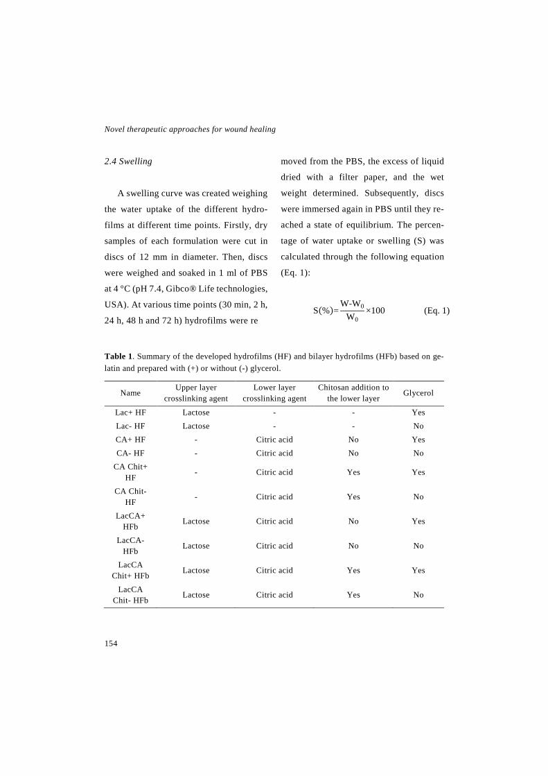
Novel therapeutic approaches for wound healing
154
2.4 Swelling
A swelling curve was created weighing
the water uptake of the different hydro-
films at different time points. Firstly, dry
samples of each formulation were cut in
discs of 12 mm in diameter. Then, discs
were weighed and soaked in 1 ml of PBS
at 4 °C (pH 7.4, Gibco® Life technologies,
USA). At various time points (30 min, 2 h,
24 h, 48 h and 72 h) hydrofilms were re
moved from the PBS, the excess of liquid
dried with a filter paper, and the wet
weight determined. Subsequently, discs
were immersed again in PBS until they re-
ached a state of equilibrium. The percen-
tage of water uptake or swelling (S) was
calculated through the following equation
(Eq. 1):
S(%)=W-W0
W0×100 (Eq. 1)
Table 1. Summary of the developed hydrofilms (HF) and bilayer hydrofilms (HFb) based on ge-latin and prepared with (+) or without (-) glycerol.
Name Upper layer
crosslinking agent Lower layer
crosslinking agent Chitosan addition to
the lower layer Glycerol
Lac+ HF Lactose - - Yes Lac- HF Lactose - - No CA+ HF - Citric acid No Yes CA- HF - Citric acid No No
CA Chit+ HF
- Citric acid Yes Yes
CA Chit- HF - Citric acid Yes No
LacCA+ HFb
Lactose Citric acid No Yes
LacCA- HFb
Lactose Citric acid No No
LacCA Chit+ HFb Lactose Citric acid Yes Yes
LacCA Chit- HFb
Lactose Citric acid Yes No

Experimental work: Chapter 4
155
Where, W is the weight of the wet sam-
ple at every time point and W0 is the
weight of the dry samples.
2.5 Hydrolytic degradation
Discs of 12 mm in diameter were im-
mersed in PBS at 4 °C until they reached
the absorption equilibrium determined in
2.4 section. Then, they were washed with
MilliQ water and they were left to dry for
7 days. When they were completely dried,
discs were weighed and the percentage of
the remaining weight was calculated using
the following equation (Eq. 2):
RM (%)=Dry weight
Initial weight×100 (Eq. 2)
2.6 Water vapour transmission rate
(WVTR)
In order to analyse the ability of hydro-
films to regulate moisture, WVTR was
calculated. The WVTR of the CA HF (with
and without chitosan and glycerol) were
not measured since wet hydrofilms lose
the structural stability to seal the Franz
diffusion cell due to their great ability to
swell water. The WVTR was quantified
following the method described by Etxa-
bide et al. [22]. Briefly, the receptor com-
partment of a Franz diffusion cell was
completely filled with MilliQ water and its
receptor arm was sealed with parafilm.
Then, hydrofilm discs slightly bigger than
the aperture between compartments (10
mm) were placed between them, to make
the hydrofilm the only way for water va-
pour to leave the system. The complete
assembly was weighed at the beginning of
the study and after a 48 h incubation at
room temperature. The WVTR was calcu-
lated using the following equation (Eq. 3):
WVTR=M1-M0
A×T (Eq. 3)
Where M0 is the weight of the assem-
bly at the beginning of the assay, M1 is its
weight after the incubation time, A is the
exposure area (0.79 cm2) and T is the ex-
posure time (2 days).
2.7 Cytotoxicity study
Cytotoxicity studies were performed
using the L-929 fibroblasts (ATCC, Mana-

Novel therapeutic approaches for wound healing
156
ssas, USA), since it is the cell line recom-
mended by the ISO 10993-5:2009 guide-
line for biological evaluation of medical
devices. Cells were cultured on Eagle’s
Minimum Essential Medium (EMEM;
ATCC, Manassas, USA) supplemented
with 10 % (v/v) inactivated Horse Serum
and 1 % (v/v) penicillin-streptomycin, and
incubated at 37 °C in a humidified incuba-
tor with a 5 % CO2 atmosphere. Cell pa-
ssages were performed every 2-3 days de-
pending on cell confluence.
Indirect cytotoxicity was assessed in-
cubating cells with the extracted medium
of the hydrofilms. Firstly, the hydrofilms
were sterilized by impregnating both sides
with 70 % ethanol and exposing them to
UV light for 20 min. Subsequently, the hy-
drofilms were divided into two groups that
received a different processing method.
Hydrofilms in the first group were dia-
lyzed in 1 l of MilliQ water for 72 h and
then they were maintained in culture me-
dium for 24 h to achieve an osmotic equi-
librium. The second group was simply hy-
drated for 15 min in culture medium. Af-
terwards, 16 mm discs of each hydrofilm
were incubated with 0.5 ml of culture me-
dium for 24 h at 37 °C, to obtain the ex-
tracted medium.
Meanwhile, cells were seeded on 96
well plates at a density of 5000 cell/well
and incubated overnight to allow cell
attachment. Then, the medium was repla-
ced by the extracted medium of the hydro-
films, although some wells were replaced
by fresh culture medium to be used as con-
trols. After 24 h of incubation, cell viabi-
lity was assessed using the CCK-8 colori-
metric assay (Cell Counting Kit-8, Sigma-
Aldrich, Saint Louise, USA). Briefly, 10
µL of the CCK-8 reagent was added to the
cells and incubated for 4 h. Then, the ab-
sorbance of the wells was read at 450 nm,
using 650 nm as reference wavelength
(Plate Reader Infinite M200, Tecan, Swi-
tzerland). The absorbance value was direc-
tly proportional to the number of living
cells in each well. Results were given as
the percentage of living cells regarding to
control.
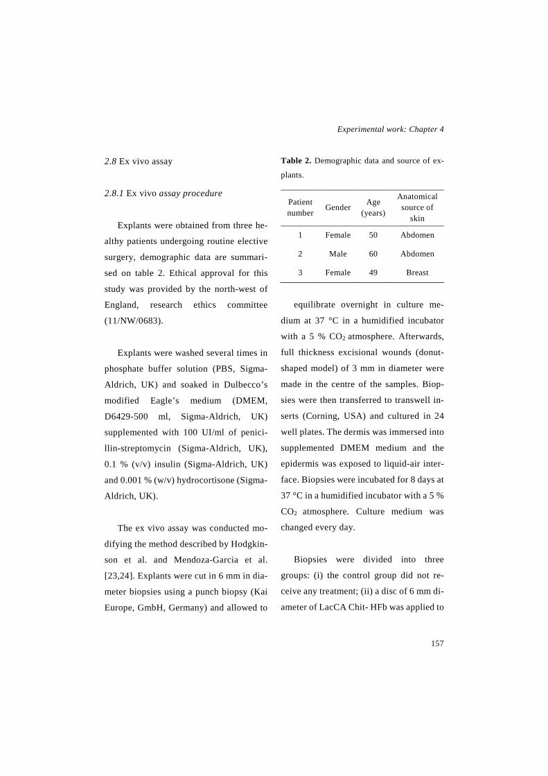
Experimental work: Chapter 4
157
2.8 Ex vivo assay
2.8.1 Ex vivo assay procedure
Explants were obtained from three he-
althy patients undergoing routine elective
surgery, demographic data are summari-
sed on table 2. Ethical approval for this
study was provided by the north-west of
England, research ethics committee
(11/NW/0683).
Explants were washed several times in
phosphate buffer solution (PBS, Sigma-
Aldrich, UK) and soaked in Dulbecco’s
modified Eagle’s medium (DMEM,
D6429-500 ml, Sigma-Aldrich, UK)
supplemented with 100 UI/ml of penici-
llin-streptomycin (Sigma-Aldrich, UK),
0.1 % (v/v) insulin (Sigma-Aldrich, UK)
and 0.001 % (w/v) hydrocortisone (Sigma-
Aldrich, UK).
The ex vivo assay was conducted mo-
difying the method described by Hodgkin-
son et al. and Mendoza-Garcia et al.
[23,24]. Explants were cut in 6 mm in dia-
meter biopsies using a punch biopsy (Kai
Europe, GmbH, Germany) and allowed to
Table 2. Demographic data and source of ex-
plants.
Patient number
Gender Age (years)
Anatomical source of
skin
1 Female 50 Abdomen
2 Male 60 Abdomen
3 Female 49 Breast
equilibrate overnight in culture me-
dium at 37 °C in a humidified incubator
with a 5 % CO2 atmosphere. Afterwards,
full thickness excisional wounds (donut-
shaped model) of 3 mm in diameter were
made in the centre of the samples. Biop-
sies were then transferred to transwell in-
serts (Corning, USA) and cultured in 24
well plates. The dermis was immersed into
supplemented DMEM medium and the
epidermis was exposed to liquid-air inter-
face. Biopsies were incubated for 8 days at
37 °C in a humidified incubator with a 5 %
CO2 atmosphere. Culture medium was
changed every day.
Biopsies were divided into three
groups: (i) the control group did not re-
ceive any treatment; (ii) a disc of 6 mm di-
ameter of LacCA Chit- HFb was applied to

Novel therapeutic approaches for wound healing
158
the biopsies; and (iii) a disc of 6 mm dia-
meter of Lac- HF was applied to the biop-
sies. Before their application, the hydro-
films were sprayed with ethanol in both si-
des, kept under UV light for 30 min and
hydrated for 15 min. Treatments were
changed on day 4.
2.8.2 LDH assay
On days 4 and 8, 50 µl of the medium
were collected from the wells to perform a
LDH (lactate dehydrogenase) assay (Pi-
erce™ LDH Cytotoxicity Assay Kit, Ther-
moFisher Scientific Inc., USA), in order to
assess the viability of the biopsies. The
LDH assay was conducted according to
the manufacturer instructions. Briefly, 50
µL of lysis buffer was added to an extra
biopsy included to be used as the control
of this assay and the mixture was incuba-
ted for 45 min. Then, 50 µl of each biopsy
(including the one treated with the lysis
buffer) were transferred into a 96 well
plate and 50 µl of the LDH reagent were
added to the wells. The mixture was incu-
bated for less than 30 min at room tempe-
rature and protected from light. The reac-
tion was stopped with 50 µl of the stop re-
agent and the absorbance was read at 492
nm, using 680 nm as reference wave-
length. Cell viability was expressed using
the equation 4.
Cell viability (%)=AL-AS
AL×100 (Eq. 4)
Where, AL is the absorbance of the
samples incubated with the lysis buffer and
AS is the absorbance of the tested samples.
Fig. 2. Ex vivo assay scheme
Experimental work: Chapter 4
159
2.8.3 Tissue processing
On days 1 and 8, samples were proce-
ssed to assess wound healing. Tissues bi-
opsied were fixed in 3.7 % paraformalde-
hyde for 24 h and then they were embe-
dded in paraffin and sectioned in layers of
5 µm in thickness.
Regarding histogical analyses, tissue
sections were stained with Hematoxylin-
Eosin (H&E) and with Herovici staining to
evaluate wound closure and collagen de-
position, respectively.
In addition, immunohistochemical ana-
lyses were conducted using antibodies
against α-SMA (1:250 dilution, ab5696,
Abcam, UK), proliferating cell nuclear an-
tigen (PCNA) (ab181797, Abcam, UK),
cytokeratin 10 (1:5000, ab 76318, Abcam,
UK) and cytokeratin 14 (1:250, Abcam,
UK). Briefly, sections were deparaffini-
sed, and after the blocking step, they were
incubated with the primary antibodies
overnight at 4 °C. Thereafter, sections
were revealed using the ImmPRESS™ Pe-
roxidase Detection Kit (Vector Laborato-
ries LTD., UK) and ImmPACT DAB subs-
trate (Vector Laboratories LTD., UK) follo-
wing the manufacturer's instructions. Fina-
lly, sections were counterstained with hema-
toxylin (Vector Laboratories LTD., UK), de-
hydrated and mounted.
Images were acquired in a 3D-Histech
Pannoramic-250 microscope slide-scanner
using a 20x objective (Zeiss, Germany).
Snapshots of the slide-scans were taken
using the Case Viewer software (3D-His-
tech, Hungary). The stained area in each ti-
ssue section or the positive cell numbers
were measured using the Tissue Studio ana-
lysis software (Definiens AG, Germany).
2.9 Statistical analysis
Results were expressed as the mean ±
standard deviation (SD), except the results
of the histological and immunohistochemi-
cal analysis that were expressed as the mean
± standard error of the mean. Results were
analysed through one-way ANOVA test for
multiple comparisons. Based on the Levene
test for the homogeneity of variances, Bon-
ferroni or Tamhane post-hoc were applied.
All the statistical tests were performed
using SPSS 22.0.01 (SPSS® INC., USA).
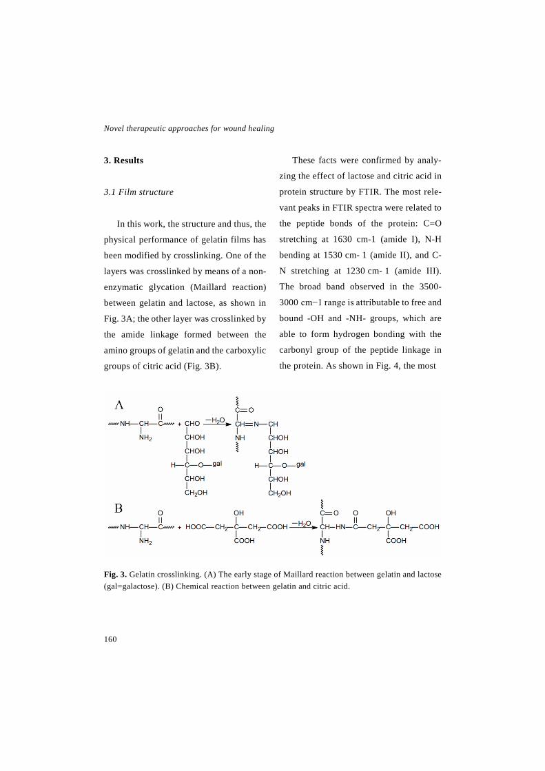
Novel therapeutic approaches for wound healing
160
3. Results
3.1 Film structure
In this work, the structure and thus, the
physical performance of gelatin films has
been modified by crosslinking. One of the
layers was crosslinked by means of a non-
enzymatic glycation (Maillard reaction)
between gelatin and lactose, as shown in
Fig. 3A; the other layer was crosslinked by
the amide linkage formed between the
amino groups of gelatin and the carboxylic
groups of citric acid (Fig. 3B).
These facts were confirmed by analy-
zing the effect of lactose and citric acid in
protein structure by FTIR. The most rele-
vant peaks in FTIR spectra were related to
the peptide bonds of the protein: C=O
stretching at 1630 cm-1 (amide I), N-H
bending at 1530 cm- 1 (amide II), and C-
N stretching at 1230 cm- 1 (amide III).
The broad band observed in the 3500-
3000 cm−1 range is attributable to free and
bound -OH and -NH- groups, which are
able to form hydrogen bonding with the
carbonyl group of the peptide linkage in
the protein. As shown in Fig. 4, the most
Fig. 3. Gelatin crosslinking. (A) The early stage of Maillard reaction between gelatin and lactose (gal=galactose). (B) Chemical reaction between gelatin and citric acid.
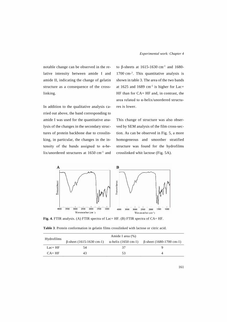
Experimental work: Chapter 4
161
notable change can be observed in the re-
lative intensity between amide I and
amide II, indicating the change of gelatin
structure as a consequence of the cross-
linking.
In addition to the qualitative analysis ca-
rried out above, the band corresponding to
amide I was used for the quantitative ana-
lysis of the changes in the secondary struc-
tures of protein backbone due to crosslin-
king, in particular, the changes in the in-
tensity of the bands assigned to α-he-
lix/unordered structures at 1650 cm-1 and
to β-sheets at 1615-1630 cm-1 and 1680-
1700 cm-1. This quantitative analysis is
shown in table 3. The area of the two bands
at 1625 and 1689 cm-1 is higher for Lac+
HF than for CA+ HF and, in contrast, the
area related to α-helix/unordered structu-
res is lower.
This change of structure was also obser-
ved by SEM analysis of the film cross-sec-
tion. As can be observed in Fig. 5, a more
homogeneous and smoother stratified
structure was found for the hydrofilms
crosslinked whit lactose (Fig. 5A).
Fig. 4. FTIR analysis. (A) FTIR spectra of Lac+ HF. (B) FTIR spectra of CA+ HF.
Table 3. Protein conformation in gelatin films crosslinked with lactose or citric acid.
Hydrofilms Amide I area (%)
β-sheet (1615-1630 cm-1) α-helix (1650 cm-1) β-sheet (1680-1700 cm-1)
Lac+ HF 54 37 9 CA+ HF 43 53 4

Novel therapeutic approaches for wound healing
162
3.2 Swelling
Water uptake assay showed that after
120 h in PBS all the hydrofilms reached an
equilibrium. As depicted in Fig. 6, the
swelling curve demonstrated the same ten-
dency in almost every time point tested.
The hydrofilms with the greatest swelling
ability were CA+ HF and CA+ Chit HF; in
equilibrium they showed a swelling ability
of 800 %, and 600-650 % in the case of
CA- HF and CA- Chit HF. Lac- and Lac+
HF absorbed 4-5-fold less water than the
previous ones, with a swelling value about
130-180 %.
Bilayer hydrofilms presented interme-
diate values, LacCA Chit+ HFb showed a
swelling of 504 ± 4 %, LacCA+ HFb sho-
wed a value of 401 ± 29 %, LacCA Chit-
HFb showed a value of 407 ± 15 % and the
LacCA- HFb showed a value of 345 ± 9
%.
In equilibrium and comparing CA HF
and LacCA HFb with the same composi-
tion, the statistical analysis showed signi-
ficant differences among CA Chit+ HF
and LacCA Chit+ HFb (** p<0.01), CA+
HF and LacCA+ HFb (* p<0.05) and CA-
HF and LacCA- HFb (* p<0.05). In addi-
tion, the difference between Lac HF (with
and without glycerol) and the rest of hy-
drofilms was significant.
The addition of chitosan did not pro-
duce any difference in the ability to absorb
water. The addition of glycerol, on the
Fig. 5. SEM analysis. (A) Lac+ HF cross-section. (B) CA+ HF cross-section.
Experimental work: Chapter 4
163
contrary, increased significantly the abi-
lity to absorb water on the CA HF without
chitosan (* p<0.05) and on the LacCA
HFb without chitosan (** p<0.01). The
same tendency was observed on CA Chit
HF and LacCA Chit HFb.
3.3 Hydrolytic degradation
In order to assess the degradation of the
hydrofilms in an aqueous environment,
they were immersed in PBS until they rea-
ched the water absorption equilibrium
(120 h). The remaining weight of all the
formulations was above the 80 % of the
initial weight (Fig. 7). Two groups could
be observed, the hydrofilms containing
glycerol showed a remaining weight of
about 88 % and the hydrofilms without
glycerol showed a weight of about 96 %,
that difference was statistically significant
in every hydrofilm type, except in the Lac
HF.
3.3 WVTR
The occlusivity of the hydrofilms was
assessed trough the WVTR. As observed
in Fig. 4, the addition of glycerol did not
affect the permeability to water vapour of
each hydrofilm type. In addition, there was
no difference among bilayer hydrofilms
(LacCA and LacCA Chit HFb with and wi-
thout glycerol) as all of them showed simi-
lar values. Lac HF, on the contrary, sho-
wed a significantly less occlusive charac-
ter as they achieved higher WVTR values.
3.5 Cytotoxicity assay
Cytotoxicity assay was performed to
evaluate the effect of the hydrofilms on
cells. Two processing methods were used
before the assay. The first method was a 72
h dialysis, to remove the glycerol and the
rest of compounds (e.g. unreacted crosslin-
kers) released from the hydrofilms. The se-
cond method consisted in hydrating the hy-
drofilms for 15 min. In addition, hydrofilms
with and without glycerol were tested.
Results of the CCK-8 are shown in Fig. 9,
and they proved that none of the hydro-
films was cytotoxic, since cell viability
was above 70 % after fibroblast incubation
with the release medium of each hydro-
film. There were no differences between
hydrofilms with different processing met-
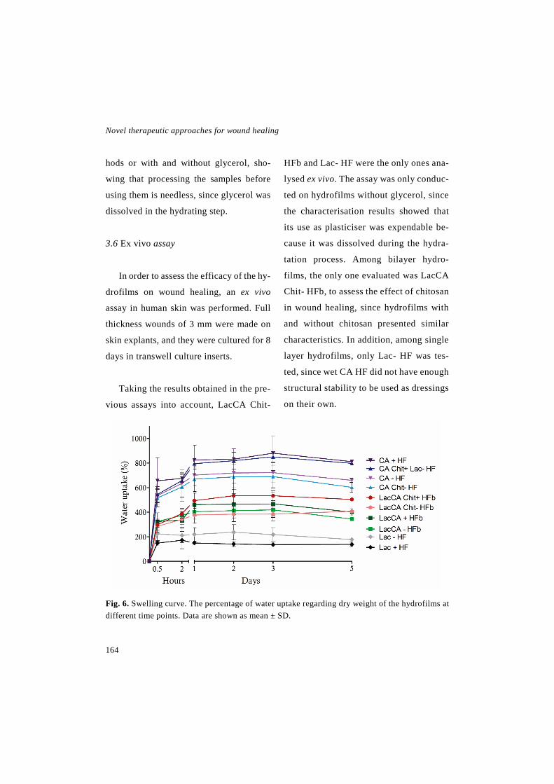
Novel therapeutic approaches for wound healing
164
hods or with and without glycerol, sho-
wing that processing the samples before
using them is needless, since glycerol was
dissolved in the hydrating step.
3.6 Ex vivo assay
In order to assess the efficacy of the hy-
drofilms on wound healing, an ex vivo
assay in human skin was performed. Full
thickness wounds of 3 mm were made on
skin explants, and they were cultured for 8
days in transwell culture inserts.
Taking the results obtained in the pre-
vious assays into account, LacCA Chit-
HFb and Lac- HF were the only ones ana-
lysed ex vivo. The assay was only conduc-
ted on hydrofilms without glycerol, since
the characterisation results showed that
its use as plasticiser was expendable be-
cause it was dissolved during the hydra-
tation process. Among bilayer hydro-
films, the only one evaluated was LacCA
Chit- HFb, to assess the effect of chitosan
in wound healing, since hydrofilms with
and without chitosan presented similar
characteristics. In addition, among single
layer hydrofilms, only Lac- HF was tes-
ted, since wet CA HF did not have enough
structural stability to be used as dressings
on their own.
Fig. 6. Swelling curve. The percentage of water uptake regarding dry weight of the hydrofilms at different time points. Data are shown as mean ± SD.
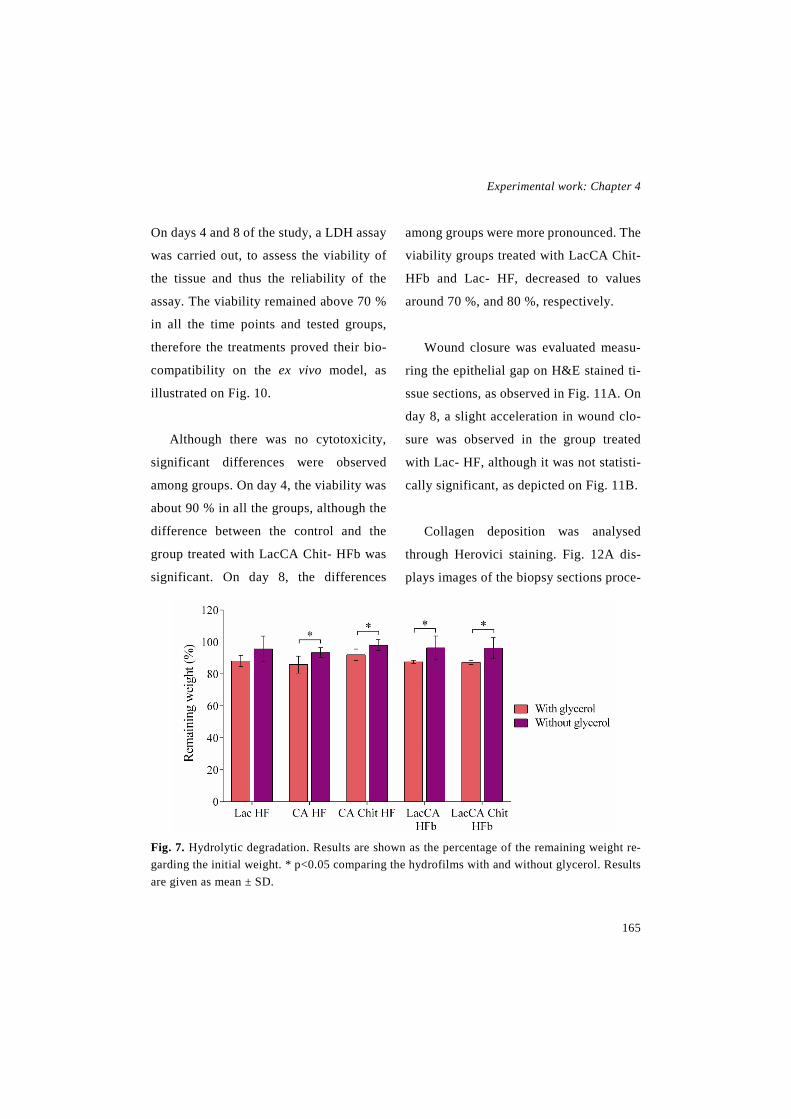
Experimental work: Chapter 4
165
On days 4 and 8 of the study, a LDH assay
was carried out, to assess the viability of
the tissue and thus the reliability of the
assay. The viability remained above 70 %
in all the time points and tested groups,
therefore the treatments proved their bio-
compatibility on the ex vivo model, as
illustrated on Fig. 10.
Although there was no cytotoxicity,
significant differences were observed
among groups. On day 4, the viability was
about 90 % in all the groups, although the
difference between the control and the
group treated with LacCA Chit- HFb was
significant. On day 8, the differences
among groups were more pronounced. The
viability groups treated with LacCA Chit-
HFb and Lac- HF, decreased to values
around 70 %, and 80 %, respectively.
Wound closure was evaluated measu-
ring the epithelial gap on H&E stained ti-
ssue sections, as observed in Fig. 11A. On
day 8, a slight acceleration in wound clo-
sure was observed in the group treated
with Lac- HF, although it was not statisti-
cally significant, as depicted on Fig. 11B.
Collagen deposition was analysed
through Herovici staining. Fig. 12A dis-
plays images of the biopsy sections proce-
Fig. 7. Hydrolytic degradation. Results are shown as the percentage of the remaining weight re-garding the initial weight. * p<0.05 comparing the hydrofilms with and without glycerol. Results are given as mean ± SD.

Novel therapeutic approaches for wound healing
166
ssed with Herovici staining, while Fig.
12B shows the ratio between the stained
area of collagen III and collagen I, since
collagen III corresponds to the newly pro-
duced collagen that forms the new ECM
and collagen I corresponds to mature co-
llagen [25]. The results showed that Lac-
HF presented a similar collagen III/I ratio
as the control group, achieving values hig-
her than baseline. The collagen ratio of the
wounds treated with LacCA Chit- HFb, on
the contrary, was significantly lower than
that of the other two groups.
In addition, immunohistological analy-
ses were performed. Tissue sections were
stained against proliferating cell nuclear
antigen (PCNA), a marker for cellular pro-
liferation, which gives an insight about the
cellular proliferation into the wound [26].
Results were represented as the percentage
of positive cells in regards to baseline of
each patient, in order to decrease the inte-
rindividual variability. Results depicted in
Fig. 13A show non-significant differences
in cell proliferation.
Wound contraction was analysed using
antibodies against α-smooth muscle actin
(α-SMA), a marker of myofibroblast diffe-
rentiation and thus wound contraction
[27]. Unfortunately, no differences were
found among groups, analysing the per-
centage of the stained area, although a 2-
fold increase regarding baseline was ob-
served (Fig. 13B).
Fig 8. WVTR. Graphical representation of the WVTR of the hydrofilms with and without glycerol. *** p<0.001 comparing Lac HF (with and without glycerol) with LacCA and LacCA Chit HFb. Results are given as mean ± SD.

Experimental work: Chapter 4
167
The expression of two cytokeratins was
also evaluated, cytokeratin 14 and 10, pre-
cisely. Cytokeratin 14 is a marker for stra-
tifying keratinocytes into the epithelial
tongue. Cytokeratin 10, on the contrary, is
a marker found in differentiated keratinocy-
tes, which is not found on epithelial tongue,
just in the mature epithelium [24,28,29].
The analysis of the stratifying keratinocytes
(cytokeratin 14) showed similar values in
all the groups on day 8 (Fig. 13C). Results
obtained from the analysis of cytokeratin 10
are illustrated on Fig. 13D, showing a lower
stained area on the group treated with the
Lac- HF, while the group treated with La-
cCA Chit- HFb presented a similar value to
the control group.
4. Discussion
The aim of the current study was to de-
velop a bilayer dressing composed of ge-
latin. In order to mimic the natural struc-
ture of the skin, the characteristics of both
layers varied, since different crosslinkers
were used. The upper layer was composed
of gelatin crosslinked with lactose, to ob-
tain a stiffer and non-hydrolytically labile
layer. Lactose and gelatin react in pre-
sence of heat leading to a non-enzymatic
glycation of the protein chains, known as
Maillard reaction [30]. This hydrofilm was
previously characterised by our research
group [22]. It forms a homogeneous and
transparent layer of about 50 µm of thick-
Fig. 9 Cytotoxicity assay. (A) Cell viability after incubating fibroblasts with the release medium of hydrofilms containing glycerol. (B) Cell viability after incubating fibroblasts with the release medium of hydrofilms without glycerol. Results are shown as mean ± SD.

Novel therapeutic approaches for wound healing
168
ness that maintained its initial appearance
after being immersed in PBS at 37 °C or
incubated with cells for 8 days. The lower
layer was composed of gelatin crosslinked
with citric acid, due to its reaction with the
amine group of the protein chains [31].
These hydrofilms presented a more jelly-
like structure, as they were able to absorb
a considerable larger volume of water and
they lost their integrity after a few hours
immersed in PBS at 37 °C (data not
shown). The influence of the crosslinker
was demonstrated by FTIR results, which
showed that a progressive conversion of
residual regular structures and unordered
segments into intermolecular β-sheets
occurred for the films crosslinked by lac-
tose compared to citric acid-crosslinked
films. This change in the secondary struc-
ture of gelatin led to a more compact and
smoother microstructure when crosslin-
king was promoted by lactose addition, as
shown by SEM analysis.
Chitosan was added to the lower layer
in order to promote wound healing pro-
cess, as chitosan has previously demons-
trated various beneficial effects in wound
Fig. 10. LDH assay of the ex vivo assay. The results were represented as the viability of the three groups on different time points. * p<0.05 comparing the control group and the group treated with LAcCA Chit- HFb, on day 4; *** p>0.001 comparing each group with the other two, on day 8; ** p<0.001 comparing the control group with the group treated with Lac- HF on day 11; ●●● p<0.001 comparing the control group and the group treated with LacCA Chit- HFb, on day 11 and 15. Results are shown as mean ± SD.

Experimental work: Chapter 4
169
healing. In fact, it presents antibacterial
and hemostatic activity, acting in the early
phases of healing [13]; it promotes the ac-
tivity of macrophages and the migration of
polymorphonuclear neutrophils [14]; it is
able to induce the proliferation of fibro-
blasts and keratinocytes both in vitro and
in vivo [32,33]; and it regulates in a bipha-
sic manner the expression of TGFβ1, indu-
cing an increment in its expression during
the early healing phases, which lead to an
increment in collagen production, while
downregulating its expression in the later
phases, thereby reducing the scar forma-
tion [34]. It is noteworthy, that none of the
developed layers showed cytotoxicity.
Both layers were glued together spray-
ing ethanol on them and letting them air
dry. The swelling ability of the resulting
bilayer was evaluated in order to compare
the behaviour of the different hydrofilms.
As swelling studies were performed only
to determine the differences among hydro-
films, they were performed at 4 °C instead
of being performed at skin temperature,
since CA HF lost their structural stability
in a warm aqueous environment.
As expected, hydrofilms with the hig-
hest crosslinking degree (Lac HF) had the
lowest ability to absorb water, and the
ones with the lowest crosslinking degree
(CA HF) had the highest water uptake
[35]. Bilayer hydrofilms had intermediate
values since they were composed of both
layers. The addition of chitosan did not
produce any change in swelling. The addi-
Fig. 11. Wound closure. (A) Histological images of tissue sections of each group on day 8, pro-cessed with H&E. The scale bar indicates 500 µm. (B) Percentage of wound closure on days 8. Results are shown as the mean ± standard error of the mean.

Novel therapeutic approaches for wound healing
170
tion of glycerol increased the water uptake
in CA HF and LacCA HFb probably be-
cause of an increased hydrophilicity of the
hydrofilms [36].
The hydrolitical stability of the hydro-
films was also carried out at 4 °C to allow
a longer study time where compare the
characteristics of the different formulati-
ons. Two groups of hydrofilms could be
distinguished, the ones without glycerol
and the ones containing glycerol. Hydro-
films in the first group maintained about
the 96 % of their dry weight after being
immersed in PBS. This weight loss may be
due to the dissolution of the unreacted
crosslinker, as described previously in ge-
latin films crosslinked with lactose. On the
other hand, the remaining weight in hydro-
films containing glycerol was about 88 %.
In this case, besides of releasing the unre-
acted crosslinker, glycerol was probably
also dissolved in the aqueous medium,
since it interacts with gelatin through hy-
drogen bonding [37].
The occlusivity of the hydrofilms was
evaluated measuring the WVTR, in order
to assess if they were able to maintain an
acceptable level of moisture into the
wounds. The WVTR of the bilayer hydro-
films was within the range of commercial
wound dressings (426-2047 g/m2day).
Thus, they presented an adequate control
of moisture; allowing exudate to evapo-
rate, while maintaining some moisture into
Fig. 12. Collagen deposition. (A) Tissue section images processed with Herovici staining. Colla-gen I or mature fibres are stained with red and collagen III or new fibres are stained with blue. The scale bar indicates 1000 µm. (B) Quantitative analysis of the collagen deposition. ** p<0.01 and comparing the group treated with LacCA Chit- HFb with the other groups. Results are shown as the mean ± standard error of the mean.

Experimental work: Chapter 4
171
the wound to prevent tissue dehydration,
help in reepithelisation and angiogenesis,
and reduce pain [38-40]. Lac HF, however,
showed a higher WVTR value, which can
be translated into a lower occlusivity, be-
cause it allowed a larger volume of water
vapour to be evaporated from the wound.
The higher permeability could be explai-
ned by the lower thickness of the Lac HF
in comparison to bilayer hydrofilms, as
previously reported [41].
Prior to evaluating the efficacy of the
dressings ex vivo, their cytocompatibility
was assessed. The results obtained in the
indirect cytotoxicity assay with all the hy-
drofilms tested showed viability values
above 70 % with respect to the control
group, which suggest an excellent biocom-
patibility according to the ISO 10992-
5:2009 guidelines for biological evalua-
tion of medical devices. The direct cytoto-
xicity assay was not performed, since CA
HF partially dissolve at 37 °C, thereby in-
creasing the viscosity of the medium and
impeding the removal of the hydrofilms to
perform the CCK-8 assay.
With the viability assay, the effect of
glycerol on cell viability and the need of a
previous conditioning step were analysed.
Regarding the effect of glycerol, the re-
sults showed that hydrofilms containing
glycerol presented similar biocompatibi-
lity than the hydrofilms without it. In addi-
tion, the assay demonstrated that there was
no need of processing the hydrofilms
through dialysis, as the same viability re-
Fig. 13. Immunohistochemical analysis. (A) Representation of PCNA positive cells percentage regarding to baseline. (B) Representation of quantitative analysis of α-SMA stained area. (C) Re-presentation of quantitative analysis of cytokeratin 14 stained area. (D) Representation of quanti-tative analysis of cytokeratin 10 stained area. ** p>0.001 comparing the group treated with the Lac- HF and the other two groups on day 8. Results are shown as the mean ± standard error of the mean.

Novel therapeutic approaches for wound healing
172
sults were obtained hydrating them for 15
min prior to the assay. The biocompatibi-
lity of dry hydrofilms was not tested, since
in a previous study carried out in Lac+ HF,
incubating cells with the extracted me-
dium of dry hydrofilms lead to a decrease
in cell viability due to an increase in me-
dium osmolarity caused by the rapid rele-
ase of unreacted lactose and glycerol [22].
Thereby, a conditioning step of 15 min hy-
dration was still needed in order to remove
the unreacted crosslinker and glycerol.
Dressings efficacy was evaluated using
an ex vivo assay in order to reduce the use
of animals in research and to use human
skin instead of animal skin, thus avoiding
the differences in wound healing among
species [15]. Although it is not standardi-
zed, ex vivo human full thickness wound
models have been already used to evaluate
the effect on wound healing of different
dressings and formulations such as, a hya-
luronic acid/alginate dressing containing
platelet lysate and vancomycin [42], a chi-
tosan oleate nanoemulsion containing α-
tocopherol [43], an electrospun silk fibroin
dressing [23], a cellular (containing fibro-
blasts and mesenchymal stem cells) or ace-
llular collagen-glycosaminoglican flo-
wable matrix [44] and Integra® dermal
template or Integra® supplemented with
hyaluronan [45].
In order to evaluate the viability of the
biopsies a LDH assay was conducted on
days 4 and 8. All groups presented viabi-
lity values above 70 %. On one hand, those
values demonstrated that cells of the biop-
sies remained alive during the whole
study, which highlights the reliability and
suitability of the model. On the other hand,
this study proved the lack of cytotoxicity
of the developed hydrofilms, as previously
observed in the cytotoxicity assay carried
out on fibroblasts. Moreover, assessing the
viability assay into the ex vivo model gives
an insight of the behaviour of the hydro-
films in a more complex structure than a
monolayer single cell culture.
In addition, the wound healing process
was assessed to analyse the effect of the
hydrofilms. In general, the application of
the hydrofilms, did not hinder healing,
since no differences were observed among
Experimental work: Chapter 4
173
groups in the majority of parameters eva-
luated, such as wound closure, cellular
proliferation, wound contraction and ex-
pression of new undifferentiated keratino-
cytes. Nevertheless, collagen deposition
was decreased in the group treated with
Lac- HF and the expression of new mature
keratinocytes was lower in the group trea-
ted with LacCA Chit- HFb. Those negative
results could be explained by the great
swelling ability of the dressings, which led
to the absorption of some of the culture
medium, decreasing the quantity of nutri-
ents available for cells. In addition, the
great water uptake of the hydrofilms and
the incubation in a humidified incubator,
presented an overly moist environment for
the epidermis, which do not favour hea-
ling. Therefore, the hydrofilms and speci-
ally the CA HF should be optimised, in or-
der to create a more adequate environment
for healing.
Accordingly, the results obtained in the
ex vivo assay, showed the suitability of the
model to assess wound healing, as a scree-
ning method prior to conduct an in vivo
study. In addition, it showed that the develo-
ped hydrofilms could be useful as wound
dressings. In order to improve the efficacy of
the hydrofilms, in future studies active mo-
lecules such as growth factors will be encap-
sulated or immobilised into the lower layer.
5. Conclusion
In the current study a bilayer wound
dressing has been developed based on ge-
latin. Firstly both layers and the resulting
bilayer hydrofilms were characterised,
showing that the upper layer (Lac HF) was
more resistant and that the lower layer (CA
and CA Chit HF) had a greater ability to
absorb water. Overall, the dressing presen-
ted good swelling and occlusivity charac-
teristics and it did not show cytotoxicity
against fibroblasts. In order to evaluate its
efficacy an ex vivo wound healing assay
was performed, which demonstrated on one
hand the suitability of the model to assess
wound healing, and on the other hand that
the hydrofilms did not hinder healing in the
majority of the evaluated parameters.
6. Acknowledgments
I. García-Orue and J. Uranga thank the
Basque Government for the fellowship
Novel therapeutic approaches for wound healing
174
grant. This project has been partially su-
pported by the Basque Government (Con-
solidated Groups, IT-428-10 and IT-528-
10). The Bioimaging Facility Slide Scan-
ning Service used in this study was pur-
chased with grants from BBSRC, Well-
come and the University of Manchester
Strategic Fund. Special thanks goes to Ro-
ger Meadows and Steve Marsden for their
help with the microscopy. BIOMAT group
thanks the Provincial Council of Gipuzkoa
for the financial support.
7. References
[1] R.A. Franco, Y. Min, H. Yang, B. Lee, Fa-brication and biocompatibility of novel bilayer scaffold for skin tissue engineering applica-tions, J. Biomater. Appl. 27 (2013) 605-615. doi: 10.1177/0885328211416527.
[2] M. Zilberman, D. Egozi, M. Shemesh, A. Keren, E. Mazor, M. Baranes-Zeevi, N. Golds-tein, I. Berdicevsky, A. Gilhar, Y. Ullmann, Hybrid wound dressings with controlled re-lease of antibiotics: Structure-release profile effects and in vivo study in a guinea pig burn model, Acta Biomater. 22 (2015) 155-163. doi: 10.1016/j.actbio.2015.04.029.
[3] L. Ding, X. Shan, X. Zhao, H. Zha, X. Chen, J. Wang, X. Wang, C. Cai, G. Li, J. Hao, G. Yu, Spongy bilayer dressing composed of chitosan–Ag nanoparticles and chitosan–Bleti-lla striata polysaccharide for wound healing
applications, Carbohydr. Polym. 157 (2017) 1538-1547. doi: 10.1016/j.carbpol.2016.11 .040.
[4] P.B. Malafaya, G.A. Silva, R.L. Reis, Na-tural–origin polymers as carriers and scaffolds for biomolecules and cell delivery in tissue en-gineering applications, Adv. Drug Deliv. Rev. 59 (2007) 207-233. doi: 10.1016/j.addr.2007. 03.012.
[5] G.D. Mogoşanu, A.M. Grumezescu, Natu-ral and synthetic polymers for wounds and burns dressing, Int. J. Pharm. 463 (2014) 127-136. doi: //dx.doi.org/10.1016/j.ijpharm.2013. 12.015.
[6] A.O. Elzoghby, Gelatin-based nanoparti-cles as drug and gene delivery systems: Revie-wing three decades of research, J. Control. Re-lease 172 (2013) 1075-1091. doi: 10.1016/j. jconrel.2013.09.019.
[7] H. Wang, O.C. Boerman, K. Sariibrahimo-glu, Y. Li, J.A. Jansen, S.C.G. Leeuwenburgh, Comparison of micro- vs. nanostructured co-lloidal gelatin gels for sustained delivery of os-teogenic proteins: Bone morphogenetic pro-tein-2 and alkaline phosphatase, Biomaterials 33 (2012) 8695-8703. doi: 10.1016/j.biomate-rials.2012.08.024.
[8] D. Eisenbud, H. Hunter, L. Kessler, K. Zulkowski, Hydrogel wound dressings: where do we stand in 2003?, Ostomy Wound Manage. 49 (2003) 52.
[9] M. Foox, M. Zilberman, Drug delivery from gelatin-based systems, Expert Opin. Drug Deliv. 12 (2015) 1547-1563. doi: 10.1517/ 17425247.2015.1037272.
Experimental work: Chapter 4
175
[10] N. Reddy, R. Reddy, Q. Jiang, Crosslin-king biopolymers for biomedical applications, Trends Biotechnol. 33 (2015) 362-369. doi: 10.1016/j.tibtech.2015.03.008.
[11] V. Chiono, E. Pulieri, G. Vozzi, G. Ciar-delli, A. Ahluwalia, P. Giusti, Genipin-cross-linked chitosan/gelatin blends for biomedical applications, J. Mater. Sci. : Mater. Med. 19 (2008) 889-898. doi: 10.1007/s10856-007-3212-5.
[12] T. Dai, M. Tanaka, Y.Y. Huang, M.R. Hamblin, Chitosan preparations for wounds and burns: antimicrobial and wound-healing effects, Expert Rev. Anti Infect. Ther. 9 (2011) 857-879. doi: 10.1586/eri.11.59.
[13] M.B. Dreifke, A.C. Jayasuriya, A.A. Jaya-suriya, Current wound healing procedures and potential care, Mater. Sci. Eng. C Mater. Biol. Appl. 48 (2015) 651-662. doi: 10.1016/j.msec.2014.12.068.
[14] V. Patrulea, V. Ostafe, G. Borchard, O. Jordan, Chitosan as a starting material for wound healing applications, Eur. J. Pharm. Biopharm. 97 (2015) 417-426. doi: 10.1016/j.ejpb.2015.08.004.
[15] S.H. Mathes, H. Ruffner, U. Graf-Haus-ner, The use of skin models in drug develop-ment, Adv. Drug Deliv. Rev. 69-70 (2014) 81-102. doi: 10.1016/j.addr.2013.12.006.
[16] S. Pfuhler, R. Fautz, G. Ouedraogo, A. La-til, J. Kenny, C. Moore, W. Diembeck, N.J. He-witt, K. Reisinger, J. Barroso, The Cosmetics Europe strategy for animal-free genotoxicity testing: Project status up-date, Toxicol. in Vi-tro 28 (2014) 18-23. doi: 10.1016/j.tiv. 2013.06.004.
[17] Nicola J Hewitt, Robert J Edwards, Ellen Fritsche, Carsten Goebel, Pierre Aeby, Julia Scheel, Kerstin Reisinger, Gladys Ouédraogo, Daniel Duche, Joan Eilstein, Alain Latil, Julia Kenny, Claire Moore, Jochen Kuehnl, Joao Ba-rroso, Rolf Fautz, Stefan Pfuhler, Use of hu-man in vitro skin models for accurate and ethi-cal risk assessment: metabolic considerations, Toxicol. Sci. 133 (2013) 209-217. doi: 10.1093/toxsci/kft080.
[18] P.D. Butler, D.P. Ly, M.T. Longaker, G.P. Yang, Use of organotypic coculture to study keloid biology, Am. J. Surg. 195 (2008) 144-148. doi: 10.1016/j.amjsurg.2007.10.003.
[19] Wei Xu, Seok Jong Hong, Shengxian Jia, Yanan Zhao, Robert D Galiano, Thomas A Mustoe, Application of a partial-thickness hu-man ex vivo skin culture model in cutaneous wound healing study, Lab. Invest. 92 (2012) 584-599. doi: 10.1038/labinvest.2011.184.
[20] K.Q. Zhu, L.H. Engrav, R.N. Tamura, J.A. Cole, P. Muangman, G.J. Carrougher, N.S. Gi-bran, Further similarities between cutaneous scarring in the female, red Duroc pig and hu-man hypertrophic scarring, Burns 30 (2004) 518-530. doi: 10.1016/j.burns.2004.02.005.
[21] S. Ud-Din, A. Bayat, Non-animal models of wound healing in cutaneous repair: In silico, in vitro, ex vivo, and in vivo models of wounds and scars in human skin, Wound Repair Regen. 25 (2017) 164-176. doi: 10.1111/wrr.12513.
[22] A. Etxabide, C. Vairo, E. Santos-Viz-caino, P. Guerrero, J.L. Pedraz, M. Igartua, K. de la Caba, R.M. Hernandez, Ultra thin hydro-films based on lactose-crosslinked fish gelatin for wound healing applications, Int. J. Pharm.
Novel therapeutic approaches for wound healing
176
530 (2017) 455-467. doi: 10.1016/j.ijpharm. 2017.08.001.
[23] T. Hodgkinson, X. Yuan, A. Bayat, Elec-trospun silk fibroin fiber diameter influences in vitro dermal fibroblast behavior and promotes healing of ex vivo wound models, J. Tissue Eng. 5 (2014) 2041731414551661. doi: 10.1177/2041731414551661.
[24] J. Mendoza-Garcia, A. Sebastian, T. Alonso-Rasgado, A. Bayat, Optimization of an ex vivo wound healing model in the adult hu-man skin: Functional evaluation using photo-dynamic therapy, Wound Repair Regen. 23 (2015) 685-702. doi: 10.1111/wrr.12325.
[25] P. Bainbridge, Wound healing and the role of fibroblasts, J. Wound Care 22 (2013) 407-412. doi: 10.12968/jowc.2013.22.8.407.
[26] J. Chen, B. Lin, H. Hu, C. Lin, W. Jin, F. Zhang, Y. Zhu, C. Lu, X. Wei, R. Chen, NGF accelerates cutaneous wound healing by pro-moting the migration of dermal fibroblasts via the PI3K/Akt-Rac1-JNK and ERK pathways, Biomed Res. Int. 2014 (2014) 547187.
[27] B.M.d. Almeida, M.F.d. Nascimento, R.N. Pereira-Filho, G.C.d. Melo, J.C.d. Santos, C.R.d. Oliveira, M.Z. Gomes, S.O. Lima, Al-buquerque-Júnior, Ricardo Luiz Cavalcanti de, Immunohistochemical profile of stromal cons-tituents and lymphoid cells over the course of wound healing in murine model, Acta Cir. Bras. 29 (2014) 596-602. doi: 10.1590/S0102-8650201400150007.
[28] R. Moll, M. Divo, L. Langbein, The hu-man keratins: biology and pathology, Histo-chem. Cell. Biol. 129 (2008) 705-733. doi: 10.1007/s00418-008-0435-6.
[29] M.L. Usui, J.N. Mansbridge, W.G. Carter, M. Fujita, J.E. Olerud, Keratinocyte Migration, Proliferation, and Differentiation in Chronic Ulcers From Patients With Diabetes and Nor-mal Wounds, J. Histochem. Cytochem. 56 (2008) 687-696. doi: 10.1369/jhc.2008.951194
[30] A. Etxabide, M. Urdanpilleta, P. Guerrero, K. de la Caba, Effects of cross-linking in na-nostructure and physicochemical properties of fish gelatins for bio-applications, React. Funct. Polym. 94 (2015) 55-62. doi: 10.1016/j. reactfunctpolym.2015.07.006.
[31] A. Oryan, A. Kamali, A. Moshiri, H. Baharvand, H. Daemi, Chemical crosslinking of biopolymeric scaffolds: Current knowledge and future directions of crosslinked engineered bone scaffolds, Int. J. Biol. Macromol. 107 (2018) 678-688. doi: 10.1016/j.ijbio-mac.2017.08.184.
[32] G.I. Howling, P.W. Dettmar, P.A. Goddard, F.C. Hampson, M. Dornish, E.J. Wood, The effect of chitin and chitosan on the proliferation of human skin fibroblasts and ke-ratinocytes in vitro, Biomaterials 22 (2001) 2959-2966. doi: 10.1016/S0142-9612(01) 00042-4.
[33] A.K. Azad, N. Sermsintham, S. Chan-drkrachang, W.F. Stevens, Chitosan membrane as a wound-healing dressing: Characterization and clinical application, J. Biomed. Mater. Res. B Appl. Biomater. 69B (2004) 216-222. doi: 10.1002/jbm.b.30000.
[34] R.M. Baxter, T. Dai, J. Kimball, E. Wang, M.R. Hamblin, W.P. Wiesmann, S.J. McCarthy, S.M. Baker, Chitosan dressing pro-motes healing in third degree burns in mice:

Experimental work: Chapter 4
177
Gene expression analysis shows biphasic ef-fects for rapid tissue regeneration and de-creased fibrotic signaling, J. Biomed. Mater. Res. A 101A (2013) 340-348. doi: 10.1002/jbm.a.34328.
[35] A. Saarai, V. Kasparkova, T. Sedlacek, P. Saha, On the development and characterisation of crosslinked sodium alginate/gelatine hydro-gels, J. Mech. Behav. Biomed. Mater. 18 (2013) 152-166. doi: 10.1016/j. jmbbm.2012.11.010.
[36] D. Prabu, A.F. Majdalawieh, I.A. Abu-Yousef, K. Inbasekaran, T. Balasubramaniam, N. Nallaperumal, C.J. Gunasekar, Preparation and characterization of gatifloxacin-loaded so-dium alginate hydrogel membranes supple-mented with hydroxypropyl methylcellulose and hydroxypropyl cellulose polymers for wound dressing, J. Pharm. Investig. 6 (2016) 86. doi: 10.4103/2230-973X.177810.
[37] A. Etxabide, J. Uranga, P. Guerrero, K. de la Caba, Improvement of barrier properties of fish gelatin films promoted by gelatin glyca-tion with lactose at high temperatures, LWT - Food Sci. Technol. 63 (2015) 315-321. doi: 10.1016/j.lwt.2015.03.079.
[38] Y. Tu, M. Zhou, Z. Guo, Y. Li, Y. Hou, D. Wang, L. Zhang, Preparation and characteriza-tion of thermosensitive artificial skin with a Sandwich structure, Mater. Lett. 147 (2015) 4-7. doi: //dx.doi.org/10.1016/j.matlet.2015.01.163.
[39] N. Mayet, Y.E. Choonara, P. Kumar, L.K. Tomar, C. Tyagi, L.C. Du Toit, V. Pillay, A Comprehensive Review of Advanced Biopoly-
meric Wound Healing Systems, J. Pharm. Sci. 103 (2014) 2211-2230. doi: 10.1002/jps.24068
[40] B. Singh, S. Sharma, A. Dhiman, Design of antibiotic containing hydrogel wound dres-sings: Biomedical properties and histological study of wound healing, Int. J. Pharm. 457 (2013) 82-91. doi: 10.1016/j.ijp-harm.2013.09.028.
[41] V. Morillon, F. Debeaufort, G. Blond, M. Capelle, A. Voilley, Factors Affecting the Moi-sture Permeability of Lipid-Based Edible Films: A Review, Crit. Rev. Food Sci. Nutr. 42 (2002) 67-89. doi: 10.1080/10408690290 825466.
[42] S. Rossi, M. Mori, B. Vigani, M.C. Bon-feroni, G. Sandri, F. Riva, C. Caramella, F. Fe-rrari, A novel dressing for the combined deli-very of platelet lysate and vancomycin hydro-chloride to chronic skin ulcers: Hyaluronic acid particles in alginate matrices, Eur. J. Pharm. Sci. 118 (2018) 87-95. doi: 10.1016/j.ejps.2018.03.024.
[43] M.C. Bonferoni, F. Riva, A. Invernizzi, E. Dellera, G. Sandri, S. Rossi, G. Marrubini, G. Bruni, B. Vigani, C. Caramella, F. Ferrari, Alpha tocopherol loaded chitosan oleate na-noemulsions for wound healing. Evaluation on cell lines and ex vivo human biopsies, and stabilization in spray dried Trojan microparti-cles, Eur. J. Pharm. Biopharm. 123 (2018) 31-41. doi: 10.1016/j.ejpb.2017.11.008.
[44] T. Hodgkinson, A. Bayat, Ex vivo evalua-tion of acellular and cellular collagen-glyco-saminoglycan flowable matrices, Biomed. Ma-ter. 10 (2015) 041001.

Novel therapeutic approaches for wound healing
178
[45] T. Hodgkinson, A. Bayat, In vitro and ex vivo analysis of hyaluronan supplementation of Integra® dermal template on human dermal
fibroblasts and keratinocytes, J. Appl. Bioma-ter. Funct. Mater. 14 (2016) 0. doi: 10.5301/jabfm.5000259..

Discussion


Discussion
181
The treatment of chronic wounds has gained a great importance due to the alarming in-
crease in their incidence. They are becoming a challenging clinical problem, in fact, only in
the US, chronic wounds annually affect 5.7 million people (around 2% of the population) and
cost $20 billion [1]. In Europe, 6000-10000 € are spent in wound associated expenses, such
as nursing time, hospitalisation, dressing changes and wound infections management [2,3].
The growth of chronic wounds incidence is related to the rise of diseases associated
with wound chronicity, such as, obesity, venous insufficiency and diabetes [4]. Those
wounds fails to progress through the physiological phases of healing (hemostasis, inflam-
mation, proliferation and remodelling), delaying the restoration of skin integrity, and fre-
quently relapsing [5-7]. The main reason of this impaired healing is their continuous in-
flammatory state, since neutrophils are present during the whole process, releasing an ex-
cessive amount of proinflammatory cytokines and proteases, which creates a proteolytic
microenvironment that degrades healing mediators and extracellular matrix (ECM) [8,9].
Currently, the therapies for chronic wounds cannot guarantee an effective healing.
Therefore, the search of an effective treatment has gained a huge importance, and signi-
ficant efforts have been made in that search to develop new treatments or improve the
current ones. The topical administration of endogenous molecules involved in wound
healing is one of those novel treatments. Unfortunately, most of those molecules present
a very short stability in vivo due to their proteic nature. To overcome this limitation, they
can be encapsulated into different systems, such as lipid nanoparticles, and more speci-
fically in nanostructured lipid carriers (NLCs). NLCs, besides protecting the encapsula-
ted peptide, present numerous advantages for wound healing: they allow controlled re-
lease, due to their adhesiveness and occlusivity they increase skin hydration, and their
small size ensures a close contact with the skin [10].
Another approach to fulfil the requirements for wound care application includes na-
nofibrous membranes. Those dressings are usually generated by electrospinning, a tech-

Novel therapeutic approaches for wound healing
182
nique that draws nanometric fibers from a polymeric solution using the electric force
[11]. Nanofibers produced using this technique exhibit a unique structure, that consist of
a high surface are to volume ratio and a high porosity. Those characteristics enhance
wound healing by promoting cell breathing, retaining moisture and allowing the removal
of exudates, since they allow gas permeation and they control wound moisture [12]. Fur-
thermore, their structure mimics the native ECM three dimensional structure, which ma-
kes them an interesting option for tissue engineering [13,14].
Finally, bilayer dressings have received significant interest for wound healing applica-
tion, due to their outstanding characteristics. Their structure mimics the bilayer structure of
the skin, with an upper protective layer resembling the epidermis, and a thicker flexible lower
layer like the dermis. Taking that into account, the dense upper layer is designed to cover the
wound, giving mechanical strength to the dressing. In addition, it needs to control moisture
transmission to prevent fluid loss and dehydration, while allowing exudate removal. Further-
more, the upper layer prevents bacterial penetration and thus wound infection. On the other
hand, the lower layer is a porous or sponge like structure that is able to absorb wound exudate
and smoothly adhere to the wet wound bed to accommodate newly formed tissue [15-17].
Bearing these considerations in mind, in the current doctoral thesis we have focused
on the development of different approaches to promote wound healing. In a first step, we
developed a formulation containing the human peptide LL37, since it has been proven
that this antimicrobial molecule modulates wound healing by activating angiogenesis
[18,19], enhancing epithelial cell migration and proliferation [20,21] and acting as che-
motactic to monocytes, neutrophils and dendritic cells [22]. As the LL37 presents a short
stability in vivo, it was encapsulated into NLCs in order to protect it. Thereby, the first
formulation developed in this thesis were NLCs loaded with LL37 (NLC-LL37).
The NLCs were produced emulsifying a melted lipid phase containing a solid (Preci-
rol ATO5) and a liquid lipid (Mygliol® 812N) with a warm aqueous phase containing

Discussion
183
surfactants. Both phases were mixed, the LL37 added to the mixture, and then they were
emulsified by sonication. The NLCs obtained using this preparation method had a mean
size of 273.6 ± 27.64 nm and a zeta potential of about -31 mV. The encapsulation efficiency
of the LL37 was 96.4 ± 0.41 %, with a peptide loading of 16.76 ± 0.07 µg LL37/mg NLC.
Prior to the in vitro activity assay, the biocompatibility of the NLC-LL37 was determi-
ned, assaying the viability of fibroblast that were incubated with NLCs. Once it was de-
monstrated that the formulation was not cytotoxic, the activity of the formulation was eva-
luated to determine whether the encapsulation process affects the bioactivity of the peptide.
In that regard, the RAW 267.4 macrophage cell line was activated with lipopolysaccharide
(LPS) and treated with free LL37, NLC-LL37 and empty NLCs. To quantify if the formu-
lations were able to inhibit the macrophage activation, the release of TNF-α was measured
by ELISA. As depicted in Fig. 1, the NLC-LL37 was able to reduce the TNF-α production
in the same extent as the free LL37, which suggest that the peptide remained active after
the encapsulation process. Moreover, the empty NLCs did not show any effect, which high-
lights that the effect was due to the LL37 of the NLC-LL37. To achieve that neutralisation,
the LL37 needs to bind both the LPS, through electrostatic and hydrophobic interactions,
and the CD14 receptor of the macrophages through a selective union [23-25].
Fig. 1. Inhibition of the activation of the macrophages. The results are given as the mean % of TNF-α production relative to the control ± SD. ** significantly greater than empty NLC and C+ (p<0.01). Controls are C- (cells without any addition) and C+ (cells after the addition of LPS).
Novel therapeutic approaches for wound healing
184
Subsequently, the antimicrobial activity of the NLC-LL37 was tested against E. coli,
because it is one of the most common bacteria in infected wounds [26]. Briefly, free
LL37, NLC-LL37 and empty NLCs were incubated with E. coli for 4 h. Then, samples
were taken, inoculated in an agar plate, incubated for 24 h and the grown colonies coun-
ted. The results showed that the free LL37 killed all the bacteria, whereas the percentage
of cells killed by the NLC-LL37 was lower. The most plausible explanation for the lower
effect observed in the group treated with NLC-LL37 is that the LL37 was released in a
sustained manner from the NLC-LL37, and thus the available dose at the beginning of
the study was lower. Thereby, the NLC-LL37 were not able to kill all the bacteria at the
beginning, and the subsequent exponential growth of the remaining bacteria hampered
the effect of the NLC-LL37. However, in vivo the bacteria are proliferating constantly
in the wound, which makes more necessary a sustained release of the peptide rather than
a high initial dose.
Finally, the efficacy of the formulation was analysed in vivo in a full thickness splin-
ted model in db/db mice. A rodent model was chosen, since it is easier to work with
small animals, although pig dermis is more similar to that of humans [27]. Nevertheless,
db/db mice present some advantages over other rodent models. Firstly, db/db mice pre-
sent a mutation of the leptin receptor and thus, they possess a type II diabetes phenotype,
which leads to an impaired healing, and to a better resemblance of a chronic wound
[28,29]. Secondly, contraction is impaired in these mice, because their extreme obesity
reduces the looseness of their skin [30,31]. Therefore, reepithelisation is enhanced, ma-
king the healing process more similar to human’s one. In addition, wound contraction is
even more hampered by placing silicone splint around the wounds [32]. The animals
received two dosis of NLC-LL37 topically, one group received 6 µg of LL37 encpasula-
ted into NLCs and the other group received 2 µg of LL37 into NLCs.
Wound healing was evaluated measuring the following parameters: (i) wound clo-
sure, it was evaluated measuring wound areas from photographs; (ii) reepithelisation

Discussion
185
grade, it was evaluated semi-quantitatively applying the scale described by Sinha et al.
to tissue sections processed with H&E [33]; (iii) the resolution of the inflammatory pro-
cess, as the previous one, it was evaluated from H&E stained tissue sections, but this
time according to the scale described by Cotran et al. [34]; (iv) collagen deposition, it
was assessed from tissue sections stained with Masson trichrome following the scale
described by Gal et al. [35]; and (v) angiogenesis, which was evaluated counting the
newly formed blood vessels in tissue sections immunohistochemically stained with an-
tiCD31 monoclonal antibody.
The results showed that in both wound closure and reepithelisation, the administra-
tion of NLC-LL37 accelerated wound healing in comparison to empty NLCs, free LL37
and untreated group, as observed in Fig. 2. The administration of NLC-LL37 also enhan-
ced the resolution of the inflammatory process, but in this case in the same extent as the
administration of free LL37. Nevertheless, collagen deposition and angiogenesis did not
show any differences among groups.
Fig. 2. In vivo wound closure. (A) Wound closure represented as the percentage of reduction of the initial area. * Significantly greater than untreated group (p<0.05); ○ significantly greater than untreated group (p>0.05), + significantly greater than the rest of the groups (p<0.05). (B) Wound images.
Novel therapeutic approaches for wound healing
186
Overall, the encapsulated peptide presented an enhanced activity that might be due
to the protective effect of the NLCs against both, chemical and enzymatic degradation.
In addition, the NLCs alloweede a controlled release profile, extending the duration of
the effect of the LL37 [36,37]. Moreover, in comparison to other nanoparticles loaded
with LL37 previously developed [38], the NLC-LL37 allowed a topical administration,
an easy, comfortable and safe route for the patient. Taking into account all these findings,
it can be concluded that the topical administration of NLC-LL37 might present an inte-
resting strategy for the treatment of chronic wounds.
In the second step of the thesis, a nanofibrous membrane dressing was developed to
encapsulate the Epidermal Growth Factor (EGF). This trophic factor is a key signalling
molecule in wound healing due to its effect stimulating keratinocyte and fibroblast pro-
liferation and migration [39]. It has been proven that its exogenous application promotes
wound healing, however its short half-life hampers its clinical application [40,41].
Therefore, we hypothesized that the encapsulation of EGF in nanofibrous membranes
can protect the peptide from the harmful environment, moreover their structure can
enhance wound healing. The nanofibrous dressing was produced by electrospinning an
emulsion composed of of PLGA dissolved in hexafluoroisopropanol and an aqueous
phase containing Aloe vera extract and EGF. The emulsion was loaded into a syringe,
which needle was attached to a power supply. The high voltage charged the emulsion,
creating an electrostatic repulsion that stretched the droplet of the needle ejecting it to
the collector. The final nanofibers were formed during the ejection process in which the
solvent was evaporated, allowing the arrival of solid nanofibers to the collector [42,43].
This setup enables the production of nanofibrous membranes composed of PLGA and
Aloe vera (1:1) loaded with EGF (PLGA-AV-EGF membranes). Aloe vera was added to
the formulation, since it has been reported to promote wound healing inducing fibroblast
growth factor, and thereby, stimulating fibroblast’s activity and proliferation and their
collagen production [44,45]. To the best of our knowledge, that was the first time were

Discussion
187
electrospun membranes containing such a large proportion of Aloe vera were developed
for wound dressing applications. In addition, nanofibrous membranes without EGF
(PLGA-AV membrane) and without EGF and Aloe vera (PLGA membrane) were pro-
duce. The characterisation of the electrospun nanofibers is summarised in table 1 and
shown in Fig. 3. The electrospinning process produces membranes composed of nonwo-
ven uniform and randomly oriented fibres. The structure presents a great porosity (above
79%) and a high area to volume ratio, allowing cell respiration and moisture control [11].
Fig. 3. SEM photographs of PLGA, PLGA-AV and PLGA-AV-EGF membranes. The scale bar in each image indicates 100 µm.
Table 1. Nanofiber characterisation: membrane porosity (%), membrane thickness (µm), nano-fibers diameter (nm), tensile strength (MPa), water uptake (%), WVPR (g/m2day) and peptide loading (µg/cm2). Data are expressed as the mean ± SD.
Nanofiber composition
Membrane porosity
(%)
Membrane thickness
(µm)
Nanofiber diameter
(nm)
Tensile strength (MPa)
Water uptake
(%)
WVPR (g/m2day)
Peptide loading
(µg/cm2)
PLGA 79.50 ±7.42
59.17 ±1.83
561.61 ±124.28
3.06 ±0.35
218.17 ±45.03
1861.28 ±372.89 -
PLGA-AV 87.92 ±11.96
56.76 ±1.27
486.99 ±114.73
4.66 ±0.90
273.92 ±42.19
1690.09 ±190.25 -
PLGA-AV-EGF
87.52 ±6.62
45.92 ±0. 78
356.03 ±112.05
2.21 ±0.49
290.58 ±49.92
1907,39 ±228.82
9.76 ±1.75
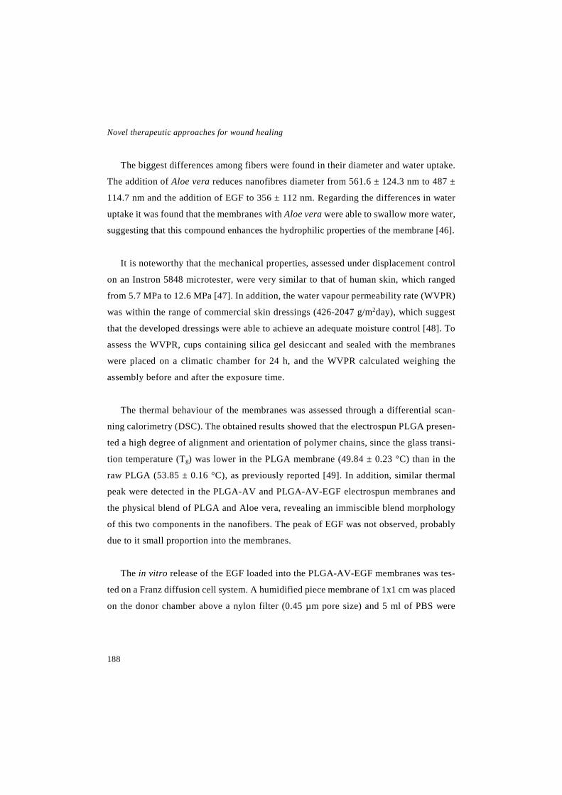
Novel therapeutic approaches for wound healing
188
The biggest differences among fibers were found in their diameter and water uptake.
The addition of Aloe vera reduces nanofibres diameter from 561.6 ± 124.3 nm to 487 ±
114.7 nm and the addition of EGF to 356 ± 112 nm. Regarding the differences in water
uptake it was found that the membranes with Aloe vera were able to swallow more water,
suggesting that this compound enhances the hydrophilic properties of the membrane [46].
It is noteworthy that the mechanical properties, assessed under displacement control
on an Instron 5848 microtester, were very similar to that of human skin, which ranged
from 5.7 MPa to 12.6 MPa [47]. In addition, the water vapour permeability rate (WVPR)
was within the range of commercial skin dressings (426-2047 g/m2day), which suggest
that the developed dressings were able to achieve an adequate moisture control [48]. To
assess the WVPR, cups containing silica gel desiccant and sealed with the membranes
were placed on a climatic chamber for 24 h, and the WVPR calculated weighing the
assembly before and after the exposure time.
The thermal behaviour of the membranes was assessed through a differential scan-
ning calorimetry (DSC). The obtained results showed that the electrospun PLGA presen-
ted a high degree of alignment and orientation of polymer chains, since the glass transi-
tion temperature (Tg) was lower in the PLGA membrane (49.84 ± 0.23 °C) than in the
raw PLGA (53.85 ± 0.16 °C), as previously reported [49]. In addition, similar thermal
peak were detected in the PLGA-AV and PLGA-AV-EGF electrospun membranes and
the physical blend of PLGA and Aloe vera, revealing an immiscible blend morphology
of this two components in the nanofibers. The peak of EGF was not observed, probably
due to it small proportion into the membranes.
The in vitro release of the EGF loaded into the PLGA-AV-EGF membranes was tes-
ted on a Franz diffusion cell system. A humidified piece membrane of 1x1 cm was placed
on the donor chamber above a nylon filter (0.45 µm pore size) and 5 ml of PBS were

Discussion
189
added to the receptor chamber. On selected time points, samples from the receptor cham-
ber were taken and the released EGF determined through a commercial ELISA. The re-
sults showed a biphasic prolife; with an initial burst release that lasted 8 h where 35% of
the drug was released, and a slower release phase were 50% of the total EGF was released
on 7 days. The initial burst release was related to the surface-associated protein and was
advantageous to obtain a rapid activation of the keratinocytes of the wound edge [50]. It
is noteworthy that although only the 50% of EGF was released, the release may be acce-
lerated in vivo, since the enzymes of the wound can lead to a faster polymer degradation
[51].
Once the dressings were characterised their bioactivity was evaluated. On the one
hand, the antimicrobial activity of Aloe vera was assessed [52], determining the inhibi-
tion zone created after placing discs of PLGA-AV and PLGA membranes on culture
plates seeded with Staphilococcus aureus and Staphilococcus epidermidis. Around
PLGA membranes there was not inhibition zone, highlighting that the one observed
around PLGA-AV membranes was due to the Aloe vera . However, a bigger inhibitory
zone was observed around commercial discs ambedded with Aloe vera solution, probably
due to the immediate availability of the whole Aloe vera dose, as observed in the results
obtained with the NLC-LL37 formulation. Moreover, nanofibrous dressings prevent mi-
croorganism entry, due to their small pore size.
On the other hand, the effect of EGF and Aloe vera on fibroblast proliferation was
assessed, in order to determine if the electrospinning process affects their activity
[53,54]. Proliferation was evaluated incubating fibroblasts with the medium released
from the nanofibers for 48 h, and then measuring cell viability through a CCK-8 assay.
The highest cell proliferation was achieved with PLGA-AV-EGF membranes. In fact, it
was higher than incubating cells with the same concentration of free EGF, revealing the
beneficial effect of the combination. In addition, an increased proliferation was observed
with PLGA-AV membranes, but not with PLGA membranes. Therefore, these results

Novel therapeutic approaches for wound healing
190
suggests that the effect was caused exclusively by the EGF and the Aloe vera, but not by
the PLGA.
Since the membranes were designed to be changed during the treatment, cells adhe-
sion or tissue ingrowth should be prevented in order to avoid damage of the newly formed
tissue or pain during the dressing removal [11]. Thereby, we decided to evaluate if cells
were able to adhere to the membranes, incubating fibroblasts on their top for 24 h, and
then detaching and counting the cells adhered or taking SEM photographs of them as
depicted in Fig. 4A. As can be observed in the graph (Fig. 4B), cell adhesion was highly
hindered by the membranes in comparison to the bottom of the plates, demonstrating
their suitability as temporary wound dressings.
Finally, the efficacy of the membranes was studied using the splinted full thickness
wound healing model in db/db mice. For that purpose, wound area reduction, reepitheli-
sation grade, the resolution of the inflammatory process and collagen deposition were
evaluated, as in the previous study. On day 4, the PLGA-AV-EGF membranes acceler-
ated wound closure in comparison to the rest of the groups, namely, free EGF, PLGA-
Fig. 4. In vitro adhesion assay. (A) SEM image of the nanofibers with cells attached. The scale bar indicates 300 µm. (B) The cell adhesion results are given as the mean ± SD of the % of cells counted in comparison to the control. *** p<0.001 comparing PLGA-AV-EGF, PLGA-AV and PLGA membranes with the control group.

Discussion
191
AV membranes, PLGA membranes and untreated control. On day 8, the highest impro-
vement of wound closure was achieved with PLGA-AV-EGF membranes, but PLGA-
AV membranes were also able to enhance wound closure. Reepithelisation grade was
also improved applying both PLGA-AV-EGF and PLGA-AV membranes, since accor-
ding to the scale used to measured reepithelisation those group presented the highest
value, which correspond to a complete reepithelisation with irregular thickness [33]. No
differences were observed in the resolution of the inflammatory process and in collagen
deposition. However, a slight tendency in agreement with the rest of the evaluated para-
meters was detected.
Overall, the results obtained in the in vivo study showed that the application of PLGA-
AV-EGF membranes was more advantageous than the application of free EGF or PLGA-
AV membranes. That can be partially explained by the protective effect of the nanofi-
brous membranes against proteases present in the wound bed, as previously reported
[40,55]. In addition, the sustained release from the nanofibrous membranes extended the
duration of the EGF in the wound and thus its effect was improved.
Both, the Aloe vera and the nanofibrous membranes themselves, were also involved
in the wound healing effect. On the one hand, Aloe vera has proven to enhance wound
healing, mainly by inducing fibroblast growth factor, and thus, improving their activity
and proliferation [53]. On the other hand, as previously commented, the unique structure
of electrospun membranes promote wound closure, allowing gas and water permeability
[56]. Accordingly, the improvement of wound closure was due to the combination of the
three elements, the EGF, the Aloe vera extract and the nanofibrous structure, leading to
a dressing that might be a suitable strategy for the treatment of chronic wounds.
Taking into account that the nanofibrous membranes without EGF also promoted
wound healing, in the following step, we focused on the development of a nanofibrous
dressing based on PLGA and Aloe vera, since the lack of EGF brings a new approach to

Novel therapeutic approaches for wound healing
192
develop a medical device. In order to improve some of the membranes properties NLCs
were added during the electrospinning process, assuming that the lipid component could
avoid the adhesion to the wound, easing the removal of the dressing. Moreover, we hy-
pothesize that the addition of NLCs could improve some features of the dressing, such
as, handling, elasticity and occlusivity.
Nanofibrous membranes with and without NLCs (PLGA-AV-NLC and PLGA-AV)
were developed to assess the effect of adding the NLCs. In this case, a PLGA with a
higher molecular weight was used, since it allowed an easier incorporation of the NLCs.
Therefore, their characterisation results were slightly different, as showed in table 2.
Porosity values were similar to the ones obtained in the previous study, around 80%,
which allows gas permeation and thus cell breathing. On the other hand, nanofibers dia-
meter increased from about 500 nm obtained in to around 1 µm in the current one. That
increment was probably due to the greater viscosity of the polymer solution prepared
with the high molecular weight PLGA, since solution viscosity is critical to control the
nanofibers morphology [56]. Moreover, water uptake was higher than in the previous
study, about 380%, which helps to drain the exudates while retaining moisture [3,46].
Table 2. Dressings characterisation: nanofibers diameter (nm), membrane porosity (%), mem-brane thickness (µm), tensile strength (MPa), water uptake (%) and WVTR (g/m2day). Data are expressed as the mean ± SD.
Nanofiber composition
Nanofiber diameter
(µm)
Porosity (%)
Thickness (µm)
Tensile strength (MPa)
Water uptake (%)
WVTR (g/m2day)
PLGA-AV membrane 1.10±0.42 81.55
±1.16 158.03 ±17.41
1.69 ±0.35
369.06 ±28.09
1128.06 ±93.23
PLGA-AV-NLC
membrane 0.96±0.34 84.23
±0.85 178.04 ±42.05
2.61 ±0.46
384.32 ±35.65
1097.6 ±102.83
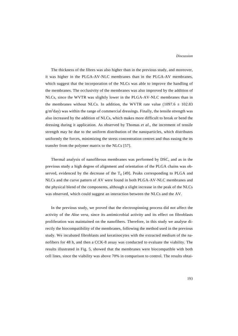
Discussion
193
The thickness of the fibres was also higher than in the previous study, and moreover,
it was higher in the PLGA-AV-NLC membranes than in the PLGA-AV membranes,
which suggest that the incorporation of the NLCs was able to improve the handling of
the membranes. The occlusivity of the membranes was also improved by the addition of
NLCs, since the WVTR was slightly lower in the PLGA-AV-NLC membranes than in
the membranes without NLCs. In addition, the WVTR rate value (1097.6 ± 102.83
g/m2day) was within the range of commercial dressings. Finally, the tensile strength was
also increased by the addition of NLCs, which makes more difficult to break or bend the
dressing during it application. As observed by Thomas et al., the increment of tensile
strength may be due to the uniform distribution of the nanoparticles, which distributes
uniformly the forces, minimizing the stress concentration centres and thus easing the its
transfer from the polymer matrix to the NLCs [57].
Thermal analysis of nanofibrous membranes was performed by DSC, and as in the
previous study a high degree of alignment and orientation of the PLGA chains was ob-
served, evidenced by the decrease of the Tg [49]. Peaks corresponding to PLGA and
NLCs and the curve pattern of AV were found in both PLGA-AV-NLC membranes and
the physical blend of the components, although a slight increase in the peak of the NLCs
was observed, which could suggest an interaction between the NLCs and the AV.
In the previous study, we proved that the electrospinning process did not affect the
activity of the Aloe vera, since its antimicrobial activity and its effect on fibroblasts
proliferation was maintained on the nanofibers. Therefore, in this study we analyse di-
rectly the biocompatibility of the membranes, following the method used in the previous
study. We incubated fibroblasts and keratinocytes with the extracted medium of the na-
nofibers for 48 h, and then a CCK-8 assay was conducted to evaluate the viability. The
results illustrated in Fig. 5, showed that the membranes were biocompatible with both
cell lines, since the viability was above 70% in comparison to control. The results obtai-
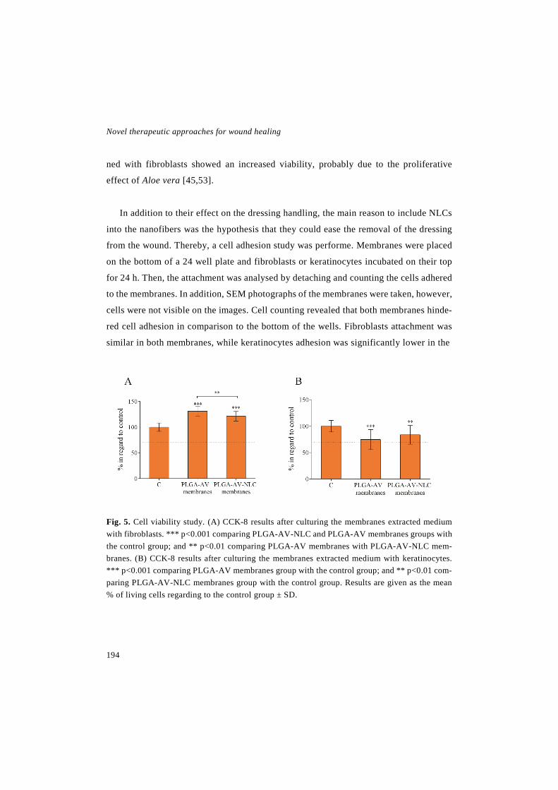
Novel therapeutic approaches for wound healing
194
ned with fibroblasts showed an increased viability, probably due to the proliferative
effect of Aloe vera [45,53].
In addition to their effect on the dressing handling, the main reason to include NLCs
into the nanofibers was the hypothesis that they could ease the removal of the dressing
from the wound. Thereby, a cell adhesion study was performe. Membranes were placed
on the bottom of a 24 well plate and fibroblasts or keratinocytes incubated on their top
for 24 h. Then, the attachment was analysed by detaching and counting the cells adhered
to the membranes. In addition, SEM photographs of the membranes were taken, however,
cells were not visible on the images. Cell counting revealed that both membranes hinde-
red cell adhesion in comparison to the bottom of the wells. Fibroblasts attachment was
similar in both membranes, while keratinocytes adhesion was significantly lower in the
Fig. 5. Cell viability study. (A) CCK-8 results after culturing the membranes extracted medium with fibroblasts. *** p<0.001 comparing PLGA-AV-NLC and PLGA-AV membranes groups with the control group; and ** p<0.01 comparing PLGA-AV membranes with PLGA-AV-NLC mem-branes. (B) CCK-8 results after culturing the membranes extracted medium with keratinocytes. *** p<0.001 comparing PLGA-AV membranes group with the control group; and ** p<0.01 com-paring PLGA-AV-NLC membranes group with the control group. Results are given as the mean % of living cells regarding to the control group ± SD.
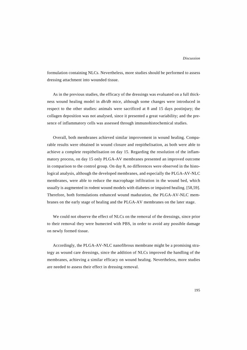
Discussion
195
formulation containing NLCs. Nevertheless, more studies should be performed to assess
dressing attachment into wounded tissue.
As in the previous studies, the efficacy of the dressings was evaluated on a full thick-
ness wound healing model in db/db mice, although some changes were introduced in
respect to the other studies: animals were sacrificed at 8 and 15 days postinjury; the
collagen deposition was not analysed, since it presented a great variability; and the pre-
sence of inflammatory cells was assessed through immunohistochemical studies.
Overall, both membranes achieved similar improvement in wound healing. Compa-
rable results were obtained in wound closure and reepithelisation, as both were able to
achieve a complete reepithelisation on day 15. Regarding the resolution of the inflam-
matory process, on day 15 only PLGA-AV membranes presented an improved outcome
in comparison to the control group. On day 8, no differences were observed in the histo-
logical analysis, although the developed membranes, and especially the PLGA-AV-NLC
membranes, were able to reduce the macrophage infiltration in the wound bed, which
usually is augmented in rodent wound models with diabetes or impaired healing. [58,59].
Therefore, both formulations enhanced wound maduration, the PLGA-AV-NLC mem-
branes on the early stage of healing and the PLGA-AV membranes on the later stage.
We could not observe the effect of NLCs on the removal of the dressings, since prior
to their removal they were humected with PBS, in order to avoid any possible damage
on newly formed tissue.
Accordingly, the PLGA-AV-NLC nanofibrous membrane might be a promising stra-
tegy as wound care dressings, since the addition of NLCs improved the handling of the
membranes, achieving a similar efficacy on wound healing. Nevertheless, more studies
are needed to assess their effect in dressing removal.

Novel therapeutic approaches for wound healing
196
For the last step of the thesis we developed a bilayer dressing composed of gelatin
and chitosan. Bilayer dressings represent an interesting alternative since they are able to
provide a structure similar to that of human skin, with an upper protective layer re-
sembling the epidermis, and a thicker flexible lower layer as the dermis [16]. Depending
on its crosslinking degree, gelatin is able to present the characteristics required for both
layers. This natural polymer has been extensively used in pharmaceutical applications
due to its biocompatibility and biodegradability [60]. Its chains contain arginine-glycine-
aspartic (RGD) motifs, an important sequence in the promotion of cell adhesion [61]. In
addition, gelatin presents an excellent ability to absorb large volumes of water and the-
reby create hydrogels, which present multiple advantages to be used as wound dressings.
For instance they are able to maintain an adequate moisture on the wound since they can
absorb or donate water depending on their environment, they cool the wound surface and
they reduce pain [62].
Thereby, two gelatin hydrofilms were developed, one of them to be the upper layer
of the dressing and the other to act as the lower layer. Both layers were produced by
casting lipid gels into suitable molds. To produce the upper layer, a gelatin solution was
mixed with glycerol and lactose. The glycerol acted as a plasticiser and the lactose as the
crosslinker, since it glycated the protein chains of gelatin in presence of heat, which is
known as the Maillard reaction [63]. The obtained hydrofilm was stiff and resistant to
water degradation [64]. It was designed to cover the wound, giving mechanical strength
to the dressing. In addition, it needs to control moisture transmission and prevent bacte-
rial penetration [15-17].
The lower layer was produced mixing the gelatin solution with glycerol and citric
acid. The citric acid was added since it reacts with the amine groups of the protein chains,
crosslinking the gelatin [65]. The resulting hydrofilm was more porous and had a great
swelling ability to absorb wound exudates [15-17]. In addition, chitosan was added to
the lower layer to promote the healing process, since it has shown to accelerate wound
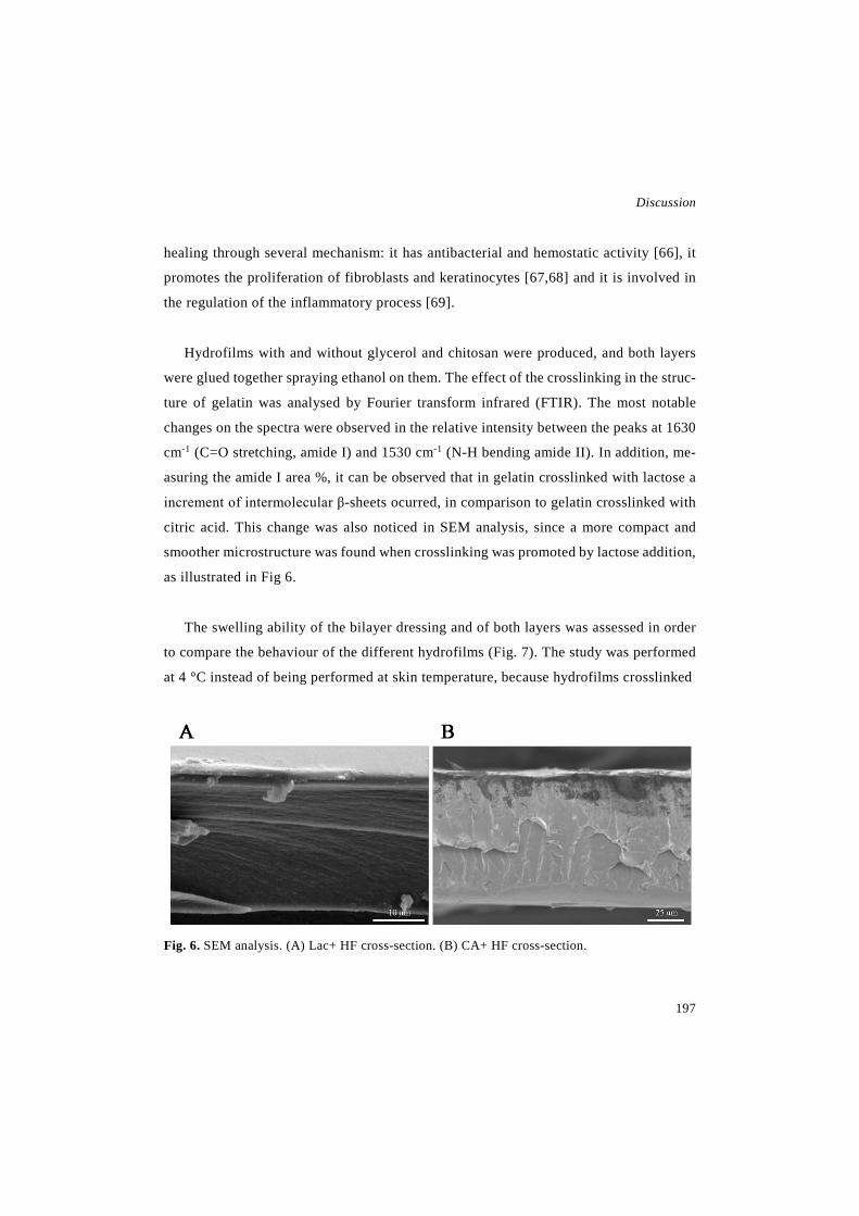
Discussion
197
healing through several mechanism: it has antibacterial and hemostatic activity [66], it
promotes the proliferation of fibroblasts and keratinocytes [67,68] and it is involved in
the regulation of the inflammatory process [69].
Hydrofilms with and without glycerol and chitosan were produced, and both layers
were glued together spraying ethanol on them. The effect of the crosslinking in the struc-
ture of gelatin was analysed by Fourier transform infrared (FTIR). The most notable
changes on the spectra were observed in the relative intensity between the peaks at 1630
cm-1 (C=O stretching, amide I) and 1530 cm-1 (N-H bending amide II). In addition, me-
asuring the amide I area %, it can be observed that in gelatin crosslinked with lactose a
increment of intermolecular β-sheets ocurred, in comparison to gelatin crosslinked with
citric acid. This change was also noticed in SEM analysis, since a more compact and
smoother microstructure was found when crosslinking was promoted by lactose addition,
as illustrated in Fig 6.
The swelling ability of the bilayer dressing and of both layers was assessed in order
to compare the behaviour of the different hydrofilms (Fig. 7). The study was performed
at 4 °C instead of being performed at skin temperature, because hydrofilms crosslinked
Fig. 6. SEM analysis. (A) Lac+ HF cross-section. (B) CA+ HF cross-section.

Novel therapeutic approaches for wound healing
198
with citric acid lost their integrity in a warm aqueous environment. Therefore, weighed
hydrofilm discs were immersed in cold PBS for determined time points, and then the
excess water was dried and the wet discs weighed to measure the water uptake. As ex-
pected, water absorption was controlled by the crosslinking degree of the gelatin. The-
reby, hydrofilms crosslinked with lactose had the lowest ability to absorb water, the ones
crosslinked with citric acid the highest and bilayer hydrofilms presented intermediate
values, since they were composed of both layers [70].
The hydrolytical stability of the hydrofilms was assessed immersing discs in cold
PBS and letting them dry once they reached an equilibrium. Results showed that hydro-
films with glycerol maintained about 88% of their initial weight, while hydrofilms wi-
thout glycerol maintained the 96 %. The weight lost can be easily explained by the di-
ssolution of glycerol and the unreacted crosslinker in the aqueous medium.
Fig. 7. Swelling curve. The percentage of water uptake regarding dry weight of the hydrofilms at different time points. Data are shown as mean ± SD.

Discussion
199
Afterwards, the occlusivity of the hydrofilms was evaluated measuring the WVTR,
but the method used was slightly different from the method used to analyse the nanofi-
brous dressings. Franz diffusion cells were filled with water and the receptor arm sealed
with parafilm. The hydrofilms were placed between both compartments, this way water
vapour could only leave the system through the hydrofilms. The difference in weight of
the assembly after 48 hours was recorded to determine the WVTR. The occlusivity of
the hydrofilms crosslinked with citric acid was not evaluated, since wet hydrofilms loss
the ability to seal the aperture between both compartments. The results of the bilayer
hydrofilms showed that they could present an adequate control of moisture into the
wounds, since their WVTR value was within the range of commercial dressings afore-
mentioned. The hydrofilms crosslinked with lactose, however, showed a higher WVTR
value, which indicated a lower occlusivity. That higher permeability could be explained
by the lower thickness of those hydrofilms [71].
As in the previous studies, once the formulations were characterised, their biocom-
patibility was assessed, following the guidelines of the ISO 10992-5:2009 for biological
evaluation of medical devices. That guideline proposes the direct and the indirect cyto-
toxicity assays, but we could not perform the direct assay, since hydrofilms crosslinked
with citric acid were partially dissolved at 37°C, hampering the removal of the hydro-
films to perform the CCK-8 assay. Therefore, we only performed the indirect assay, in-
cubating together the extracted medium of the hydrofilms with fibroblasts for 24 hours
and then measuring cell viability by a CCK-8 assay. All the hydrofilms presented a via-
bility above 70 % in the indirect assay, demonstrating that they were biocompatible. In
addition, it was demonstrated that there was no need of a previous conditioning step,
since similar viability results were obtained dialyzing hydrofilms for 72 h and hydrating
them for 15 min prior to conduct the assay.
Finally, the efficacy of the dressings on wound healing was analysed. The assay was
only conducted on hydrofilms without glycerol since the characterisation results showed
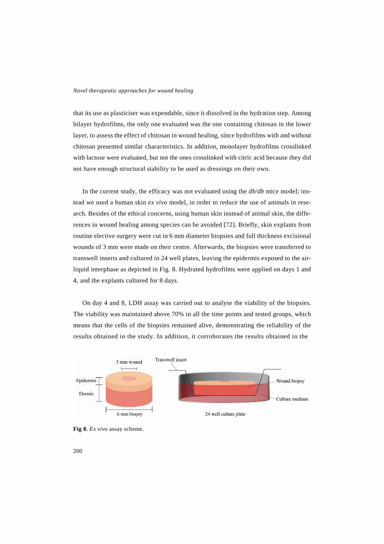
Novel therapeutic approaches for wound healing
200
that its use as plasticiser was expendable, since it dissolved in the hydration step. Among
bilayer hydrofilms, the only one evaluated was the one containing chitosan in the lower
layer, to assess the effect of chitosan in wound healing, since hydrofilms with and without
chitosan presented similar characteristics. In addition, monolayer hydrofilms crosslinked
with lactose were evaluated, but not the ones crosslinked with citric acid because they did
not have enough structural stability to be used as dressings on their own.
In the current study, the efficacy was not evaluated using the db/db mice model; ins-
tead we used a human skin ex vivo model, in order to reduce the use of animals in rese-
arch. Besides of the ethical concerns, using human skin instead of animal skin, the diffe-
rences in wound healing among species can be avoided [72]. Briefly, skin explants from
routine elective surgery were cut in 6 mm diameter biopsies and full thickness excisional
wounds of 3 mm were made on their centre. Afterwards, the biopsies were transferred to
transwell inserts and cultured in 24 well plates, leaving the epidermis exposed to the air-
liquid interphase as depicted in Fig. 8. Hydrated hydrofilms were applied on days 1 and
4, and the explants cultured for 8 days.
On day 4 and 8, LDH assay was carried out to analyse the viability of the biopsies.
The viability was maintained above 70% in all the time points and tested groups, which
means that the cells of the biopsies remained alive, demonstrating the reliability of the
results obtained in the study. In addition, it corroborates the results obtained in the
Fig 8. Ex vivo assay scheme.
Discussion
201
cytotoxicity assay carried out in fibroblasts, demonstrating the biocompatibility of the
hydrofilms in a more complex structure than a monolayer single cell culture.
In order to assess the efficacy of the hydrofilms, wound healing was evaluated from
histological and immunohistochemical analyses performed on biopsies sections. Overall,
similar results were obtained in the groups treated with the hydrofilms and in the untre-
ated groups, since no differences were observed in wound closure, cellular proliferation,
wound contraction and the expression of new undifferentiated keratinocytes. Neverthe-
less, collagen deposition was decreased in the group treated with the gelatin hydrofilm
crosslinked with lactose and the expression of mature keratinocytes was hindered in the
group treated with the bilayer hydrofilm containing chitosan. Those negative results
could be explained by the overly moist environment created on the wound, due to the
high humidity of the incubator and the ability of the hydrofilms to absorb a portion of
the culture medium. Therefore, the hydrofilms, and specially the gelatin hydrofilms
crosslinked with citric acid, should be optimised in order to create a more adequate en-
vironment for healing.
Accordingly, the results obtained in the ex vivo assay, showed the suitability of the
model to assess wound healing, as a screening method prior to conduct an in vivo study.
In addition, it showed that the developed hydrofilms could be useful as wound dressings.
In order to improve the efficacy of the hydrofilms, in future studies active molecules
such as growth factors will be encapsulated or immobilised into the lower layer.
In summary, considering all the results obtained in this thesis, we can conclude that
with this work we have taken a step forward in the development of new strategies for the
treatment of chronic wounds. In these years, we have developed different therapeutic
approaches to promote healing on chronic wounds, such as NCLs containing the LL37
peptide, PLGA-Aloe vera nanofibrous membranes containing EGF or NLCs and bilayer
dressings based on gelatin and chitosan.

Novel therapeutic approaches for wound healing
202
REFERENCES
[1] K. Järbrink, G. Ni, H. Sönnergren, A. Schmidtchen, C. Pang, R. Bajpai, J. Car, The humanistic and economic burden of chronic wounds: a protocol for a systematic review, Syst. Rev. 6 (2017). doi: 10.1186/s13643-016-0400-8.
[2] V.W. Wong, G.C. Gurtner, Tissue engineering for the management of chronic wounds: current concepts and future perspectives, Exp. Dermatol. 21 (2012) 729-734. doi: 10.1111/j.1600-0625.2012.01542.x.
[3] J. Posnett, F. Gottrup, H. Lundgren, G. Saal, The resource impact of wounds on health-care providers in Europe, J. Wound Care 18 (2009) 154. doi: 10.12968/jowc.2009.18.4.41607.
[4] G. Han, R. Ceilley, Chronic Wound Healing: A Review of Current Management and Treat-ments, Adv. Ther. 34 (2017) 599-610. doi: 10.1007/s12325-017-0478-y.
[5] C.K. Sen, G.M. Gordillo, S. Roy, R. Kirsner, L. Lambert, T.K. Hunt, F. Gottrup, G.C. Gurtner, M.T. Longaker, Human skin wounds: a major and snowballing threat to public health and the economy, Wound Repair Regen. 17 (2009) 763-771. doi: 10.1111/j.1524-475X.2009.00543.x.
[6] P.S. Briquez, J.A. Hubbell, M.M. Martino, Extracellular matrix-inspired growth factor deli-very systems for skin wound healing, Adv. Wound Care (New Rochelle) 4 (2015) 479-489. doi: 10.1089/wound.2014.0603.
[7] T. Velnar, T. Bailey, V. Smrkolj, The wound healing process: an overview of the cellular and molecular mechanisms, J. Int. Med. Res. 37 (2009) 1528-1542. doi: 10.1177/147323000903700531.
[8] G. Gainza, S. Villullas, J.L. Pedraz, R.M. Hernandez, M. Igartua, Advances in drug delivery systems (DDSs) to release growth factors for wound healing and skin regeneration, Nanomedicine 11 (2015) 1551-1573. doi: //dx.doi.org/10.1016/j.nano.2015.03.002.
[9] N.B. Menke, K.R. Ward, T.M. Witten, D.G. Bonchev, R.F. Diegelmann, Impaired wound healing, Clin. Dermatol. 25 (2007) 19-25. doi: //dx.doi.org/10.1016/j.clindermatol.2006.12.005.
[10] J. Pardeike, K. Schwabe, R.H. Müller, Influence of nanostructured lipid carriers (NLC) on the physical properties of the Cutanova Nanorepair Q10 cream and the in vivo skin hydration effect, Int. J. Pharm. 396 (2010) 166-173. doi: //dx.doi.org/10.1016/j.ijpharm.2010.06.007.
[11] M. Abrigo, S.L. McArthur, P. Kingshott, Electrospun nanofibers as dressings for chronic wound care: advances, challenges, and future prospects, Macromol. Biosci. 14 (2014) 772-792. doi: 10.1002/mabi.201300561.
Discussion
203
[12] L. Pachuau, Recent developments in novel drug delivery systems for wound healing, Expert. Opin. Drug Deliv. 12 (2015) 1895-1909. doi: 10.1517/17425247.2015.1070143.
[13] C.P. Barnes, S.A. Sell, E.D. Boland, D.G. Simpson, G.L. Bowlin, Nanofiber technology: Designing the next generation of tissue engineering scaffolds, Adv. Drug Deliv. Rev. 59 (2007) 1413-1433. doi: //dx.doi.org/10.1016/j.addr.2007.04.022.
[14] N. Bhattarai, D. Edmondson, O. Veiseh, F.A. Matsen, M. Zhang, Electrospun chitosan-based nanofibers and their cellular compatibility, Biomaterials 26 (2005) 6176-6184. doi: //dx.doi.org/10.1016/j.biomaterials.2005.03.027.
[15] R.A. Franco, Y. Min, H. Yang, B. Lee, Fabrication and biocompatibility of novel bilayer scaffold for skin tissue engineering applications, J. Biomater. Appl. 27 (2013) 605-615. doi: 10.1177/0885328211416527.
[16] M. Zilberman, D. Egozi, M. Shemesh, A. Keren, E. Mazor, M. Baranes-Zeevi, N. Goldstein, I. Berdicevsky, A. Gilhar, Y. Ullmann, Hybrid wound dressings with controlled release of anti-biotics: Structure-release profile effects and in vivo study in a guinea pig burn model, Acta Bio-mater. 22 (2015) 155-163. doi: 10.1016/j.actbio.2015.04.029.
[17] L. Ding, X. Shan, X. Zhao, H. Zha, X. Chen, J. Wang, X. Wang, C. Cai, G. Li, J. Hao, G. Yu, Spongy bilayer dressing composed of chitosan–Ag nanoparticles and chitosan–Bletilla striata polysaccharide for wound healing applications, Carbohydr. Polym. 157 (2017) 1538-1547. doi: 10.1016/j.carbpol.2016.11.040.
[18] R. Koczulla, G. von Degenfeld, C. Kupatt, F. Krötz, S. Zahler, T. Gloe, K. Issbrücker, P. Unterberger, M. Zaiou, C. Lebherz, A. Karl, P. Raake, A. Pfosser, P. Boekstegers, U. Welsch, P.S. Hiemstra, C. Vogelmeier, R.L. Gallo, M. Clauss, R. Bals, An angiogenic role for the human peptide antibiotic LL-37/hCAP-18., J Clin Invest 111 (2003) 1665-1672.
[19] S.B. Coffelt, S.L. Tomchuck, K.J. Zwezdaryk, E.S. Danka, A.B. Scandurro, Leucine Leucine-37 Uses Formyl Peptide Receptor–Like 1 to Activate Signal Transduction Pathways, Stimulate Oncogenic Gene Expression, and Enhance the Invasiveness of Ovarian Cancer Cells, Molecular Cancer Research 7 (2009) 907-915. doi: 10.1158/1541-7786.MCR-08-0326.
[20] K. Hase, L. Eckmann, J.D. Leopard, N. Varki, M.F. Kagnoff, Cell Differentiation Is a Key Determinant of Cathelicidin LL-37/Human Cationic Antimicrobial Protein 18 Expression by Hu-man Colon Epithelium, Infection and Immunity 70 (2002) 953-063. doi: 10.1128/IAI.70.2.953-963.2002.
[21] R.C. Anderson, M. Rehders, P.L. Yu, Antimicrobial fragments of the pro-region of catheli-cidins and other immune peptides, Biotechnol. Lett. 30 (2008) 813-818.
Novel therapeutic approaches for wound healing
204
[22] Y. Kai-Larsen, B. Agerberth, The role of the multifunctional peptide LL-37 in host defense, Front. Biosci 13 (2008) 3760-3767.
[23] J. Turner, Y. Cho, N. Dinh, A.J. Waring, R.I. Lehrer, Activities of LL-37, a Cathelin-Asso-ciated Antimicrobial Peptide of Human Neutrophils, Antimicrobial Agents and Chemotherapy 42 (1998) 2206-2214.
[24] R. Ramos, J.P. Silva, A.C. Rodrigues, R. Costa, L. Guardão, F. Schmitt, R. Soares, M. Vila-nova, L. Domingues, M. Gama, Wound healing activity of the human antimicrobial peptide LL37, Peptides 32 (2011) 1469-1476. doi: //dx.doi.org/10.1016/j.peptides.2011.06.005.
[25] Y. Rosenfeld, N. Papo, Y. Shai, Endotoxin (Lipopolysaccharide) Neutralization by Innate Immunity Host-Defense Peptides: peptide properties ad plausible modes of action, Journal of Bio-logical Chemistry 281 (2006) 1636-1643. doi: 10.1074/jbc.M504327200.
[26] A.R. Siddiqui, J.M. Bernstein, Chronic wound infection: Facts and controversies, Clin. Der-matol. 28 (2010) 519-526. doi: //dx.doi.org/10.1016/j.clindermatol.2010.03.009.
[27] M. Seaton, A. Hocking, N.S. Gibran, Porcine Models of Cutaneous Wound Healing, ILAR Journal 56 (2015) 127-138. doi: 10.1093/ilar/ilv016.
[28] P. Losi, E. Briganti, C. Errico, A. Lisella, E. Sanguinetti, F. Chiellini, G. Soldani, Fibrin-based scaffold incorporating VEGF- and bFGF-loaded nanoparticles stimulates wound healing in diabetic mice, Acta Biomater. 9 (2013) 7814-7821. doi: //dx.doi.org/10.1016/j.ac-tbio.2013.04.019.
[29] Y. Guillemin, D. Le Broc, C. Ségalen, E. Kurkdjian, J.N. Gouze, Efficacy of a collagen-based dressing in an animal model of delayed wound healing, J. Wound Care 25 (2016) 406-413. doi: 10.12968/jowc.2016.25.7.406.
[30] R.C. Fang, Z.B. Kryger, D.W. Buck II, M. De La Garza, R.D. Galiano, T.A. Mustoe, Limi-tations of the db/db mouse in translational wound healing research: Is the NONcNZO10 polygenic mouse model superior?, Wound Repair Regen. 18 (2010) 605-613. doi: 10.1111/j.1524-475X.2010.00634.x.
[31] V.I. Tkalcevic, S. Cužic, M.J. Parnham, I. Pašalic, K. Brajša, Differential Evaluation of Ex-cisional Non-occluded Wound Healing in db/db Mice, Toxico. Pathol. 37 (2009) 183-192. doi: 10.1177/0192623308329280.
[32] J. Michaels, S.S. Churgin, K.M. Blechman, M.R. Greives, S. Aarabi, R.D. Galiano, G.C. Gurtner, db/db mice exhibit severe wound-healing impairments compared with other murine dia-betic strains in a silicone-splinted excisional wound model, Wound Repair Regen. 15 (2007) 665-670. doi: 10.1111/j.1524-475X.2007.00273.x.

Discussion
205
[33] U.K. Sinha, L.A. Gallagher, Effects of Steel Scalpel, Ultrasonic Scalpel, CO2 Laser, and Monopolar and Bipolar Electrosurgery on Wound Healing in Guinea Pig Oral Mucosa, Laryngos-cope 113 (2003) 228-236. doi: 10.1097/00005537-200302000-00007.
[34] R. Cotran, G.K. Kumar, T. Collins, Reparación de los tejidos: proliferacion celular, fibrosis y curaicón de las heridas (2000) 95-120.
[35] P. Gál, T. Toporcer, B. Vidinský, M. Mokrý, M. Novotný, R. Kilík, K. Smetana, T. Gál, J. Sabo, Early changes in the tensile stregnth and morphology of primary sutured skin wounds in rats, Folia Biol. (Praha) 52 (2006) 109-115.
[36] M. Schäfer-Korting, W. Mehnert, H. Korting, Lipid nanoparticles for improved topical ap-plication of drugs for skin diseases, Adv. Drug Deliv. Rev. 59 (2007) 427-443. doi: //dx.doi.org/10.1016/j.addr.2007.04.006.
[37] J. Pardeike, A. Hommoss, R.H. Müller, Lipid nanoparticles (SLN, NLC) in cosmetic and pharmaceutical dermal products, Int. J. Pharm. 366 (2009) 170-184. doi: //dx.doi.org/10.1016/j.ijpharm.2008.10.003.
[38] K.K. Chereddy, C. Her, M. Comune, C. Moia, A. Lopes, P.E. Porporato, J. Vanacker, M.C. Lam, L. Steinstraesser, P. Sonveaux, H. Zhu, L.S. Ferreira, G. Vandermeulen, V. Préat, PLGA nanoparticles loaded with host defense peptide LL37 promote wound healing, J. Control Release 194 (2014) 138-147. doi: //dx.doi.org/10.1016/j.jconrel.2014.08.016.
[39] R.J. Bodnar, Epidermal Growth Factor and Epidermal Growth Factor Receptor: The Yin and Yang in the Treatment of Cutaneous Wounds and Cancer, Adv. Wound Care (New Rochelle) 2 (2013) 24-29. doi: 10.1089/wound.2011.0326.
[40] J.S. Choi, K.W. Leong, H.S. Yoo, In vivo wound healing of diabetic ulcers using electrospun nanofibers immobilized with human epidermal growth factor (EGF), Biomaterials 29 (2008) 587-596. doi: //dx.doi.org/10.1016/j.biomaterials.2007.10.012.
[41] G. Gainza, D.C. Bonafonte, B. Moreno, J.J. Aguirre, F.B. Gutierrez, S. Villullas, J.L. Pedraz, M. Igartua, R.M. Hernandez, The topical administration of rhEGF-loaded nanostructured lipid carriers (rhEGF-NLC) improves healing in a porcine full-thickness excisional wound model, J. Control Release 197 (2015) 41-47. doi: //dx.doi.org/10.1016/j.jconrel.2014.10.033.
[42] M. Liu, X. Duan, Y. Li, D. Yang, Y. Long, Electrospun nanofibers for wound healing, Mater. Sci. Eng. C Mater. Biol. Appl. 76 (2017) 1413-1423. doi: 10.1016/j.msec.2017.03.034.
[43] H.P. Felgueiras, M.T.P. Amorim, Functionalization of electrospun polymeric wound dres-sings with antimicrobial peptides, Colloids Surf. B Biointerfaces 156 (2017) 133-148. doi: //doi.org/10.1016/j.colsurfb.2017.05.001.

Novel therapeutic approaches for wound healing
206
[44] S. Choi, M. Chung, A review on the relationship between aloe vera components and their biologic effects, Semin. Integr. Med. 1 (2003) 53-62. doi: //dx.doi.org/10.1016/S1543-1150(03)00005-X.
[45] S.A. Hashemi, S.A. Madani, S. Abediankenari, The Review on Properties of Aloe Vera in Healing of Cutaneous Wounds, Biomed. Res. Int. 2015 (2015) 714216. doi: 10.1155/2015/714216.
[46] S. Son, R. Franco, S. Bae, Y. Min, B. Lee, Electrospun PLGA/gelatin fibrous tubes for the application of biodegradable intestinal stent in rat model, J. Biomed. Mater. Res. 101B (2013) 1095-1105. doi: 10.1002/jbm.b.32923.
[47] C. Jacquemoud, K. Bruyere-Garnier, M. Coret, Methodology to determine failure characte-ristics of planar soft tissues using a dynamic tensile test, J. Biomech. 40 (2007) 468-475. doi: //dx.doi.org/10.1016/j.jbiomech.2005.12.010.
[48] Y. Tu, M. Zhou, Z. Guo, Y. Li, Y. Hou, D. Wang, L. Zhang, Preparation and characterization of thermosensitive artificial skin with a Sandwich structure, Mater. Lett. 147 (2015) 4-7. doi: //dx.doi.org/10.1016/j.matlet.2015.01.163.
[49] H. Fouad, T. Elsarnagawy, F.N. Almahjdi, K.A. Khalil, Preparation and in vitro thermo-mechanical characterization of electrospun PLGA nanofibers for soft and hard tissue replacement, Int. J. Electrochem. Sci. 8 (2013) 2293-2304.
[50] A. Schneider, X.Y. Wang, D.L. Kaplan, J.A. Garlick, C. Egles, Biofunctionalized electrospun silk mats as a topical bioactive dressing for accelerated wound healing, Acta Biomater. 5 (2009) 2570-2578. doi: //dx.doi.org/10.1016/j.actbio.2008.12.013.
[51] S. Fredenberg, M. Wahlgren, M. Reslow, A. Axelsson, The mechanisms of drug release in poly(lactic-co-glycolic acid)-based drug delivery systems—A review, Int. J. Pharm. 415 (2011) 34-52. doi: //dx.doi.org/10.1016/j.ijpharm.2011.05.049.
[52] S. Ghayempour, M. Montazer, M. Mahmoudi Rad, Encapsulation of Aloe Vera extract into natural Tragacanth Gum as a novel green wound healing product, Int. J. Biol. Macromol. 93, Part A (2016) 344-349. doi: //dx.doi.org/10.1016/j.ijbiomac.2016.08.076.
[53] M.D. Boudreau, F.A. Beland, An evaluation of the biological and toxicological properties of Aloe barbadensis (miller), Aloe vera., J. Environ. Sci. Health C Environ. Carcinog. Ecotoxicol. Rev. 24 (2006) 103-154. doi: 10.1080/10590500600614303.
[54] G. Gainza, W.S. Chu, R.H. Guy, J.L. Pedraz, R.M. Hernandez, B. Delgado-Charro, M. Igar-tua, Development and in vitro evaluation of lipid nanoparticle-based dressings for topical treat-ment of chronic wounds, Int. J. Pharm. 490 (2015) 404-411. doi: //dx.doi.org/10.1016/j.ijp-harm.2015.05.075.
Discussion
207
[55] H. Lai, C. Kuan, H. Wu, J. Tsai, T. Chen, D. Hsieh, T. Wang, Tailored design of electrospun composite nanofibers with staged release of multiple angiogenic growth factors for chronic wound healing, Acta Biomater. 10 (2014) 4156-4166. doi: //dx.doi.org/10.1016/j.actbio.2014.05.001.
[56] S. Shahverdi, M. Hajimiri, M.A. Esfandiari, B. Larijani, F. Atyabi, A. Rajabiani, A.R. Dehpour, A.A. Gharehaghaji, R. Dinarvand, Fabrication and structure analysis of poly(lactide-co-glycolic acid)/silk fibroin hybrid scaffold for wound dressing applications, Int. J. Pharm. 473 (2014) 345-355. doi: //dx.doi.org/10.1016/j.ijpharm.2014.07.021.
[57] R. Thomas, K. Soumya, J. Mathew, E. Radhakrishnan, Electrospun Polycaprolactone Mem-brane Incorporated with Biosynthesized Silver Nanoparticles as Effective Wound Dressing Mate-rial, Appl. Biochem. Biotechnol. 176 (2015) 2213-2224. doi: 10.1007/s12010-015-1709-9.
[58] Tsubame Nishikai-Yan Shen, Shigeyuki Kanazawa, Makiko Kado, Kayoko Okada, Lin Luo, Ayato Hayashi, Hiroshi Mizuno, Rica Tanaka, Interleukin-6 stimulates Akt and p38 MAPK phosphorylation and fibroblast migration in non-diabetic but not diabetic mice, PLoS One 12 (2017) e0178232. doi: 10.1371/journal.pone.0178232.
[59] J. Yeh, L. Yeh, S. Jung, T. Chang, H. Wu, T. Shiu, C. Liu, W.W. Kao, P. Chu, Impaired skin wound healing in lumican-null mice, Br. J. Dermatol. 163 (2010) 1174. doi: 10.1111/j.1365-2133.2010.10008.x.
[60] A.O. Elzoghby, Gelatin-based nanoparticles as drug and gene delivery systems: Reviewing three decades of research, J. Control. Release 172 (2013) 1075-1091. doi: 10.1016/j.jcon-rel.2013.09.019.
[61] H. Wang, O.C. Boerman, K. Sariibrahimoglu, Y. Li, J.A. Jansen, S.C.G. Leeuwenburgh, Comparison of micro- vs. nanostructured colloidal gelatin gels for sustained delivery of osteoge-nic proteins: Bone morphogenetic protein-2 and alkaline phosphatase, Biomaterials 33 (2012) 8695-8703. doi: 10.1016/j.biomaterials.2012.08.024.
[62] D. Eisenbud, H. Hunter, L. Kessler, K. Zulkowski, Hydrogel wound dressings: where do we stand in 2003?, Ostomy Wound Manage. 49 (2003) 52.
[63] A. Etxabide, M. Urdanpilleta, P. Guerrero, K. de la Caba, Effects of cross-linking in nanos-tructure and physicochemical properties of fish gelatins for bio-applications, React. Funct. Polym. 94 (2015) 55-62. doi: 10.1016/j.reactfunctpolym.2015.07.006.
[64] A. Etxabide, C. Vairo, E. Santos-Vizcaino, P. Guerrero, J.L. Pedraz, M. Igartua, K. de la Caba, R.M. Hernandez, Ultra thin hydro-films based on lactose-crosslinked fish gelatin for wound healing applications, Int. J. Pharm. 530 (2017) 455-467. doi: 10.1016/j.ijpharm.2017.08.001.
Novel therapeutic approaches for wound healing
208
[65] A. Oryan, A. Kamali, A. Moshiri, H. Baharvand, H. Daemi, Chemical crosslinking of bio-polymeric scaffolds: Current knowledge and future directions of crosslinked engineered bone sca-ffolds, Int. J. Biol. Macromol. 107 (2018) 678-688. doi: 10.1016/j.ijbiomac.2017.08.184.
[66] M.B. Dreifke, A.C. Jayasuriya, A.A. Jayasuriya, Current wound healing procedures and po-tential care, Mater. Sci. Eng. C Mater. Biol. Appl. 48 (2015) 651-662. doi: 10.1016/j.msec.2014.12.068.
[67] G.I. Howling, P.W. Dettmar, P.A. Goddard, F.C. Hampson, M. Dornish, E.J. Wood, The effect of chitin and chitosan on the proliferation of human skin fibroblasts and keratinocytes in vitro, Biomaterials 22 (2001) 2959-2966. doi: 10.1016/S0142-9612(01)00042-4.
[68] A.K. Azad, N. Sermsintham, S. Chandrkrachang, W.F. Stevens, Chitosan membrane as a wound-healing dressing: Characterization and clinical application, J. Biomed. Mater. Res. B Appl. Biomater. 69B (2004) 216-222. doi: 10.1002/jbm.b.30000.
[69] R.M. Baxter, T. Dai, J. Kimball, E. Wang, M.R. Hamblin, W.P. Wiesmann, S.J. McCarthy, S.M. Baker, Chitosan dressing promotes healing in third degree burns in mice: Gene expression analysis shows biphasic effects for rapid tissue regeneration and decreased fibrotic signaling, J. Biomed. Mater. Res. A 101A (2013) 340-348. doi: 10.1002/jbm.a.34328.
[70] A. Saarai, V. Kasparkova, T. Sedlacek, P. Saha, On the development and characterisation of crosslinked sodium alginate/gelatine hydrogels, J. Mech. Behav. Biomed. Mater. 18 (2013) 152-166. doi: 10.1016/j.jmbbm.2012.11.010.
[71] V. Morillon, F. Debeaufort, G. Blond, M. Capelle, A. Voilley, Factors Affecting the Moisture Permeability of Lipid-Based Edible Films: A Review, Crit. Rev. Food Sci. Nutr. 42 (2002) 67-89. doi: 10.1080/10408690290825466.
[72] S.H. Mathes, H. Ruffner, U. Graf-Hausner, The use of skin models in drug development, Adv. Drug Deliv. Rev. 69-70 (2014) 81-102. doi: 10.1016/j.addr.2013.12.006.
Conclusions

Conclusions
211
On the basis of the results obtained in the experimental studies of this Doctoral The-
sis, the following conclusions were derived:
1. LL37 was successfully encapsulated into NLCs, achieving suitable characteristics
for wound healing applications and maintaining the activity of the peptide after the
encapsulation process.
2. The topical administration of NLC-LL37 significantly improved wound healing in a
full thickness wound model in db/db mice in comparison to the same concentration
of LL37 in solution; in terms of wound closure, reepithelisation grade and restora-
tion of the inflammatory process.
3. The developed nanofibrous membranes composed of PLGA and Aloe vera contai-
ning EGF showed adequate characteristics to be used as wound dressings. In addi-
tion, the EGF and the Aloe vera remained active after the electrospinning process
and they accelerated significantly wound closure and reepithelisation in a full thick-
ness wound model carried out in db/db mice.
4. The incorporation of NLCs to the PLGA and Aloe vera nanofibrous dressings im-
proved their handling. Those membranes were also able to improve wound healing
but in a lesser extent.
5. Bilayer gelatin and chitosan dressings were developed using lactose or citric acid as
crosslinkers to prepare a resistant upper layer and a porous lower layer, respectively.
The developed dressings presented suitable characteristics to be used as a wound
dressings, and their biocompatibility was proven ex vivo.


EUSKARAZKO BERTSIOA

Sarrera

Orbaintzean erabiltzeko nanoteknologian oinarritutako askatze siste-mak, zeintzuk hazkuntza faktoreak edo bestelako molekula endoge-noak kapsularatuta dituzten
SARRERA
Hazkuntza Faktoreen (Growth Factor edo GF) eta bestelako molekula endogenoen (intsulina, intsulinaren antzeko GF-1, zelula estromaletatik eratorritako faktorea, LL37, hesteko peptide basoaktiboa, heparina, melatonina, lipokalina, serpin-A1 eta β-estradi-ola) administrazio topikoak zauri kronikoen orbaintzea hobetzen dutela frogatuta dago. Dena den, haien in vivo egonkortasun txikia dela eta, administrazio topikoa hobetu beha-rra dago dosiari, askatze sistemari eta segurtasunari dagokionez. Horren harira, Farma-koak Askatzeko Sistema (Drug Delivery System edo DDS) berriak erabili izan dira, izan ere, DDSak farmakoak era lokalizatu eta kontrolatuan askatzeko gai dira, zaurian dauden proteasetaz babestuz. Haien artean, nanoteknologian oinarritutako DDSak aipagarriak dira, besteak beste, mikro edo nanopartikula polimerikoak, nanopartikula lipidikoak eta nanozuntzezko mintzak. Berrikuspen honen helburua DDS hauei buruzko ikuspegi oro-kor bat eskaintzea da. Horretaz gain, berrikuspenak GFek zaurietan duten eginkizunari eta DDSetan erabilitako biomaterial ohikoenei buruzko ideia bat ematen du ere. Formu-lazio horiek abantaila ugari dituzte, hala nola, farmakoaren babesa, biokonpatibilitate ona, kontrolatutako edo luzatutako askapena, farmako karga handia eta ezaugarri me-kaniko onak. Orokorrean, GFen kapsularatzea nanoteknologian oinarritutako DDSen barne potentzial handia erakutsi du zauri kronikoen orbaintzerako.


Sarrera
219
Zauri kronikoak erronka bat bilakatzen
ari dira praktika klinikoan, nahiz eta pato-
logia oso arrunta diren. Izatez, 2012. ur-
tean AEBan gutxi gorabehera 6.5 milioi
pertsonek zauri kronikoak pairatu zituz-
ten, eta 25 bilioi dolar gastatu ziren zau-
rien orbaintzearekin harremandutako ara-
zoetan. Europan, zaurien zainketak batez-
besteko 6.000-10.000 € balio du pertsona
eta urteko, eta gastu horiek honakoei lo-
tuta daude: erizaintza denborari, ospitali-
zazioei, zauri aposituen aldaketei eta zau-
rien infekzioei [1,2]. Gainera, uste da biz-
tanleriaren %1-2-ak zauri kronikoak ja-
sango dituela bizitzan zehar. Izan ere,
zauri hauen intzidentzia handitzen ari da,
arrisku handiko populazioaren igoeraren
ondorioz, hauen artean, pertsona diabe-
tiko, zahar, erretzaile eta obesoak daudela-
rik [3-5].
Gaur egungo terapiek ezin dute orbain-
tze efektibo bat bermatu, eta ondorioz,
sendaketa denbora asko luzatzen da eta be-
rragerpenak ohikoak dira. Hori dela eta,
zauriak era eraginkorrean eta denbora la-
burrean sendatuko dituen tratamendu bat
garatzea beharrezkoa bilakatu da. Zentzu
horretan, tratamendu berrien bilaketan eta
egungo tratamenduen hobekuntzan ahale-
gin esanguratsuak egin dira, horien artean,
hazkuntza faktoreen (Growth Factor edo
GF) eta bestelako konposatu endogenoen
administrazioa dagoelarik. GFen trata-
mendua hobetzeko estrategia itxaropentsu
bat farmakoak askatzeko sistema (drug de-
livery system edo DDS) berrien garapena
da, GFak modu kontrolatu eta lokalean la-
gatzeko [6].
Berrikuspen honen helburua bi baldin-
tza hauek betetzen dituzten DDSei bu-
ruzko ikuspegi orokor bat eskaintzea da:
modu kontrolatuan orbaintzerako GFak
edo bestelako konposatu endogenoak as-
katzea eta nanoteknologian oinarrituta
egotea. Horien artean, mikro- eta nanopar-
tikula (MP/NP) polimerikoei, nanoparti-
kula lipidikoei eta nanozuntzezko egiturei
buruz arituko da zehazki. Horretaz gain,
berrikuspenean DDSetan erabilitako bio-
material ohikoenak aipatzen dira.

Zaurien orbaintzerako baliabide terapeutiko berriak
220
1. Hazkuntza faktoreen eta bestelako
molekula endogenoen eginkizuna zau-
rien orbaintzean
Fisiologikoki, zauriak era antolatu eta
eraginkor batean orbaintzen dira, zeinean
4 fase bereizi baina gainjarri dauden: he-
mostasia, hantura, proliferazioa eta bir-
moldatzea, 1. irudian ikus daitekeen be-
zala [7]. Prozesu hau oso estuki araututa
egon behar du, orbaintzea ahalbidetzen
duen ingurune molekular orekatu bat lor-
tzeko. Erregulazioa GF eta zitokina ugari-
ren menpe dago, zeinek seinale sare kon-
plexu bat osatzen duten itu zelulen haz-
kuntza, desberdintzea eta metabolismoa
aldatzeko [8,9]. GFak haien ekintza bete-
tzeko hartzaile espezifikoetara lotzen dira,
zenbait ur-jauzi molekularren aktibazioa
eraginez [10,11].
Hala eta guztiz ere, zenbait kasutan
zauriak ez dira gai izaten orbaintze prozesu
normalaren faseetan zehar igarotzeko, or-
baintze denbora luzatuz eta maiz zauriaren
berragerpena eraginez. Horren adibide
dira, ultzera diabetikoak, baskularrak edo
presio-ultzerak [3,12,13]. Zauri kroniko ho-
rien fisiopatologiak zenbait desberdintasun
ditu zauri akutuenekin alderatuz. Haien ar-
teko desberdintasun nagusia zauri kroni-
koen etengabeko hantura egoera da, kon-
trolik gabeko hantura seinaleen atzeraeli-
kadura (feedback) positiboaren ondorioz
gertatzen dena. Hori dela eta, orbaintze
prozesu osoan zehar neutrofiloak zaurian
egoten dira, matrizearen proteinasa meta-
liko (matrix metallo proteinase edo
MMPak) ugari jariatuz, zauriaren matrizea
degradatzen dutenak. Gainera, lortutako
mikroingurune proteolitikoak GFen degra-
dazioa eragiten du, haien funtzioa inhibi-
tuz eta beraz, orbaintze prozesuaren erre-
gulazioa aldatuz. Horretaz gain, fibroblas-
toen jokaera ere asaldatuta agertzen da, or-
baintzea are gehiago atzeratuz; fibroblas-
toek alde batetik, migratzeko ahalmena
kaltetuta izaten dute eta bestetik, GFen es-
timuluei erantzuteko gaitasuna murriztuta.
Azkenik, zauri kronikoak infekzioetara
sentikorragoak dira, orbaintze denboraldi
luzeen ondorioz [5,6].
Ingurune proteolitikoaren ondorioz,
zauri hauetan GFen maila txikituta dagoe-
nez haien tratamendurako erabili den es-
trategia erakargarri bat GFen administra-
zio exogenoa da [8]. Izatez, zenbait GF

Sarrera
221
1. Irudia. Zauriaren orbaintze prozesua. (A) Hemostasia. Azalaren lesioaren ostean, basouzkurtze labur bat gertatzen da odol jarioa saihesteko, eta ondoren plaketak aktibatzen dira koagulazioa eragiteko eta fibrina odolbatua sortzekoa. (B) Hanturazko fasea. Fase honetan neutrofiloak, ma-krofagoak eta linfozitoak zaurira infiltratzen dira, infekzioak galarazteko eta kaltetutako ehuna ezabatzeko. (C) Fase proliferatiboa. Aurreko faseetan askatutako seinale kimiotaktikoei erantzu-nez, fibroblastoak eta keratinozitoak zaurira migratzen dute, non proliferatzen duten. Horretaz gain, fibrobastoek ECM berria eratzeko proteinak askatzen dituzte, eta ECM berri horrek fibrina odolbatua ordezkatzen du. Azkenik angiogenesia gertatzen da zauriari behar dituen oxigeno eta nutrienteak helarazteko. (D) Erremodelazio fasea. Fase honetan epitelio berria eta orbain ehunaren garapena gertatzen da. Horretarako, aurreko fasean eratu den behin behineko ECMaren osaketa eta antolaketa aldatzen da ehun normalarena lortzeko.
.
Zaurien orbaintzerako baliabide terapeutiko berriak
222
merkaturatu dira zauri kronikoen trata-
mendurako, besteak beste, PDGFa (Regra-
nex®), EGFa (Heberprot-P®, Regen-D™
eta Easyef®) eta bFGFa (Fiblast®) [6]. Ho-
rretaz gain, GFen eta kimiozinen konbina-
zio fisiologikoa imitatzeko asmoz, plake-
tetan aberatsa den plasma (platelets rich
plasma edo PRP) aztertu da zaurien or-
baintzerako. PRPa giza plaketa kontzen-
trazio handia duen plasma autologoaren
bolumen txiki bat da, zeinean zaurien or-
baintzerako garrantzizkoak diren GF eta
bestelako proteina aktibo ugari dauden
[14].
Zoritxarrez, GFak proteinak direnez
oso egonkortasun eskasa daukate in vivo,
maiztasun handiko administrazioak behar
dituztelarik. Muga hori gainditzeko,
GFak DDStan barneratu dira, haietan
kapsularatuta daudenean, zauriko protea-
setaz babestuta baitaude. Orbaintzea erre-
gulatzen duten bestelako molekula endo-
geno batzuk ere DDSen barne formulatu
dira. 1. taulan DDSetan kapsularatu diren
GFen eta bestelako molekulen zerrenda
dago, haien jatorria eta funtzio nagusiak
laburbilduz.
2. Orbaintzerako nanoteknologian oina-
rritutako DDSak garatzeko biomateria-
lak
DDS berriak garatzeko askotariko bio-
materialak erabili izan dira, baina denek
honako ezaugarriak betetzea komenigarria
da, egonkortasun ona, biobateragarritasun
eta biodegradazio onak, farmako karga
handia, ezaugarri mekaniko onak, farma-
koaren lagapen luzatu edo kontrolatua eta
farmakoaren babesa [6,35].
Polimeroak, natural zein sintetikoak,
oso erabiliak dira nanozuntz, MP eta NPen
garapenean. 2. taulan aplikazio honetarako
gehien erabiltzen diren polimeroak zerren-
datuta daude, haien abantaila eta mugekin.
Naturalen artean hurrengoak daude: algi-
natoa, gelatina, fibrina, kitosanoa, kolage-
noa, azido hialuronikoa… Polimero horiek
oso erabiliak izan dira, giza gorputzak eza-
gutzen dituen makromolekulen antzekoak
direnez biobateragarritasun handia dute-
lako. Gainera, haietako zenbait kaltetutako
ehunen konponketan parte hartzen dute eta
zelulentzako lotura gune eta seinale bio-
molekularrak dituzte, horrek balio erantsia
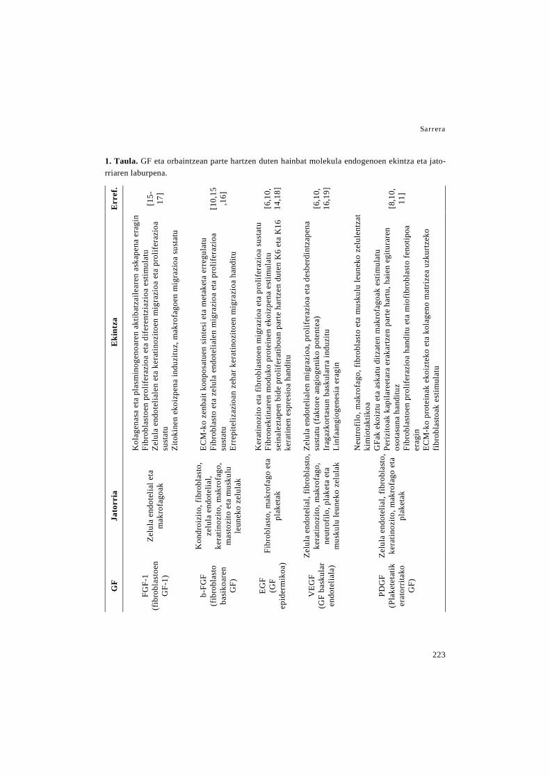
Sarrera
223
1. Taula. GF eta orbaintzean parte hartzen duten hainbat molekula endogenoen ekintza eta jato-rriaren laburpena.
Err
ef.
[15-
17]
[10,
15,1
6]
[6,1
0,14
,18]
[6,1
0,16
,19]
[8,1
0,11
]
Eki
ntza
Kol
agen
asa
eta
plas
min
ogen
oare
n ak
tibat
zaile
aren
ask
apen
a er
agin
Fi
brob
last
oen
prol
ifera
zioa
eta
dife
rent
ziaz
ioa
estim
ulat
u Ze
lula
end
otel
iale
n et
a ke
ratin
ozito
en m
igra
zioa
eta
pro
lifer
azio
a su
stat
u Zi
toki
nen
ekoi
zpen
a in
duzi
tuz,
mak
rofa
goen
mig
razi
oa s
usta
tu
ECM
-ko
zenb
ait k
onpo
satu
en s
inte
si e
ta m
etak
eta
erre
gula
tu
Fibr
obla
sto
eta
zelu
la e
ndot
elia
len
mig
razi
oa e
ta p
rolif
eraz
ioa
sust
atu
Erre
pite
lizaz
ioan
zeh
ar k
erat
inoz
itoen
mig
razi
oa h
andi
tu
Ker
atin
ozito
eta
fibr
obla
stoe
n m
igra
zioa
eta
pro
lifer
azio
a su
stat
u Fi
bron
ektin
aren
mod
uko
prot
eine
n ek
oizp
ena
estim
ulat
u se
inal
ezta
pen
bide
pro
lifer
atib
oan
part
e ha
rtzen
dut
en K
6 et
a K
16
kera
tinen
esp
resi
oa h
andi
tu
Zelu
la e
ndot
elia
len
mig
razi
oa, p
rolif
eraz
ioa
eta
desb
erdi
ntza
pena
su
stat
u (f
akto
re a
ngio
geni
ko p
oten
tea)
Ir
agaz
kort
asun
bas
kula
rra
indu
zitu
Li
nfaa
ngio
gene
sia
erag
in
Neu
trofil
o, m
akro
fago
, fib
robl
asto
eta
mus
kulu
leun
eko
zelu
lent
zat
kim
iota
ktik
oa
GFa
k ek
oizt
u et
a as
katu
ditz
aten
mak
rofa
goak
est
imul
atu
Periz
itoak
kap
ilare
etar
a er
akar
tzen
par
te h
artu
, hai
en e
gitu
rare
n os
otas
una
hand
ituz
Fibr
obla
stoe
n pr
olife
razi
oa h
andi
tu e
ta m
iofib
robl
asto
feno
tipoa
er
agin
EC
M-k
o pr
otei
nak
ekoi
ztek
o et
a ko
lage
no m
atri
zea
uzku
rtze
ko
fibro
blas
toak
est
imul
atu
Jato
rria
Zelu
la e
ndot
elia
l eta
m
akro
fago
ak
Kon
droi
zito
, fib
robl
asto
, ze
lula
end
otel
ial,
kera
tinoz
ito, m
akro
fago
, m
asto
zito
eta
mus
kulu
le
unek
o ze
lula
k
Fibr
obla
sto,
mak
rofa
go e
ta
plak
etak
Zelu
la e
ndot
elia
l, fib
robl
asto
, ke
ratin
ozito
, mak
rofa
go,
neut
rofil
o, p
lake
ta e
ta
mus
kulu
leun
eko
zelu
lak
Zelu
la e
ndot
elia
l, fib
robl
asto
, ke
ratin
ozito
, mak
rofa
go e
ta
plak
etak
GF
FGF-
1 (f
ibro
blas
toen
G
F-1)
b-FG
F (f
ibro
blas
to
basi
koar
en
GF)
EGF
(GF
epid
erm
ikoa
)
VEG
F (G
F ba
skul
ar
endo
telia
la)
PDG
F (P
lake
teta
tik
erat
orrit
ako
GF)

Zaurien orbaintzerako baliabide terapeutiko berriak
224
Err
ef.
[8]
[21]
[11,
22,2
3]
[24]
[20,
25-
27]
[9]
Eki
ntza
Ker
atin
ozito
en m
igra
zioa
eta
pro
lifer
azio
a ha
nditu
Fi
brob
last
oen
prol
ifera
zioa
est
imul
atu
Ker
atin
ozito
en m
igra
zioa
, pro
lifer
azio
a et
a de
sber
dint
zape
na e
stim
ulat
u Fi
brob
last
oen
prol
ifera
zioa
eta
EC
M-k
o pr
otei
nen
ekoi
zpen
a su
stat
u Zi
toki
na in
flam
ator
ioen
ask
apen
a m
odul
atu
Zelu
la e
ndot
elia
l mik
roba
skul
arre
n ap
opto
sia
inhi
bitu
Pr
olif
eraz
io e
ndot
elia
la e
ta h
odie
n fo
rmaz
ioa
sust
atu
Hez
ur m
uine
ko a
ma
zelu
len
aktib
azio
a, m
ugik
orta
suna
eta
lesi
o gu
nean
ge
ratz
ea e
stim
ulat
u
Pato
geno
en a
urre
an le
hene
ngo
defe
ntsa
da,
akt
ibita
te a
ntib
akte
rian
o,
antib
iofil
m, a
ntib
iriko
eta
ant
ifung
ikoa
due
larik
M
onoz
ito, n
eutro
filo,
zel
ula
dent
ritik
o, m
akro
fago
, fib
robl
asto
eta
ke
ratin
ozito
entz
ako
kim
iota
ktik
oa d
a Zi
toki
na p
ro- e
ta a
nti-i
nfla
mat
orio
en e
koiz
pena
ore
katu
K
erat
inoz
itoen
eta
zel
ula
endo
telia
len
aurr
ekar
ien
mig
razi
oa e
ta
TGF-
α-re
n si
ntes
ia e
stim
ulat
u A
ngio
gene
sia
sust
atu
Fibr
obla
sto
eta
kera
tinoz
itoen
pro
lifer
azio
a su
stat
u
Han
tura
zko
fase
err
egul
atze
n du
age
nte
pro-
eta
ant
i-inf
lam
ator
ioei
lo
tuz
Fibr
obla
sto,
gih
ar e
hun
leun
eko
zelu
len
eta
kera
tinoz
itoen
pro
lifer
azio
a su
stat
u G
F ug
arir
ekin
elk
arre
kint
zak
ditu
, hai
en e
fekt
ua s
usta
tuz
Jato
rria
Fibr
obla
sto,
hep
atoz
ito,
mak
rofa
go, n
eutro
filo
eta
giha
r esk
elet
ikok
o ze
lula
k
Pank
reak
o β-
zelu
lak
Fibr
obla
sto
derm
al e
ta z
elul
a en
dote
liala
k
Zelu
la d
endr
itiko
, ke
ratin
ozito
, lin
fozi
to,
mak
rofa
go, m
asto
zito
, ne
utro
filo
eta
NK
zel
ulak
SNZ-
ko n
euro
na e
ta
birik
ieta
ko e
ta h
este
meh
eko
zelu
lak
Bas
ofilo
eta
mas
tozi
toak
GF
IGF-
I (in
tsul
inar
en
antz
eko
GF-
I)
Ints
ulin
a
SDF-
1α
(est
rom
ako
zelu
leta
tik
erat
orrit
ako
fakt
orea
-1α)
LL37
VIP
(h
este
ko
pept
ido
baso
aktib
oa)
Hep
arin
a de
sulfa
tatu
a
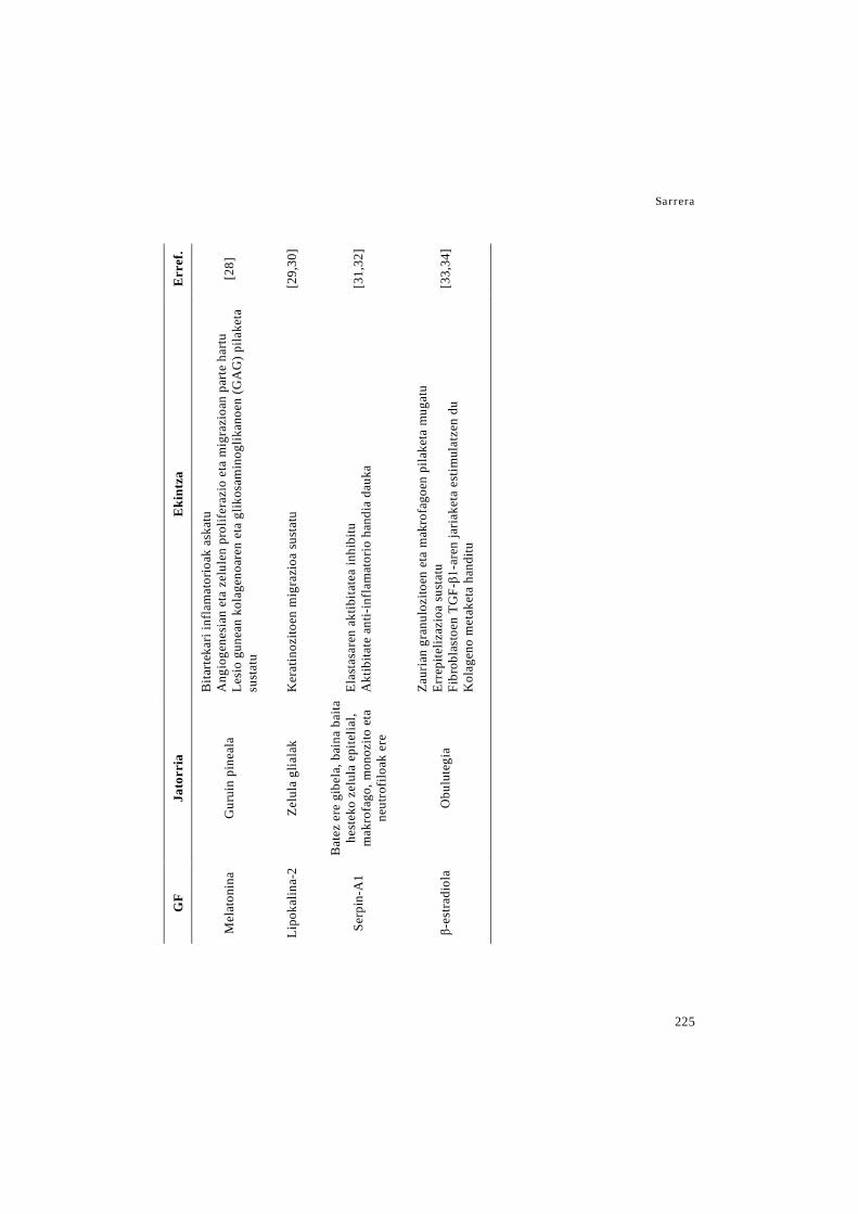
Sarrera
225
Err
ef.
[28]
[29,
30]
[31,
32]
[33,
34]
Eki
ntza
Bita
rtek
ari i
nfla
mat
orio
ak a
skat
u A
ngio
gene
sian
eta
zel
ulen
pro
lifer
azio
eta
mig
razi
oan
part
e ha
rtu
Lesi
o gu
nean
kol
agen
oare
n et
a gl
ikos
amin
oglik
anoe
n (G
AG
) pila
keta
su
stat
u
Ker
atin
ozito
en m
igra
zioa
sus
tatu
Elas
tasa
ren
aktib
itate
a in
hibi
tu
Akt
ibita
te a
nti-i
nfla
mat
orio
han
dia
dauk
a
Zaur
ian
gran
uloz
itoen
eta
mak
rofa
goen
pila
keta
mug
atu
Erre
pite
lizaz
ioa
sust
atu
Fibr
obla
stoe
n TG
F-β1
-are
n ja
riake
ta e
stim
ulat
zen
du
Kol
agen
o m
etak
eta
hand
itu
Jato
rria
Gur
uin
pine
ala
Zelu
la g
liala
k
Bat
ez e
re g
ibel
a, b
aina
bai
ta
hest
eko
zelu
la e
pite
lial,
mak
rofa
go, m
onoz
ito e
ta
neut
rofil
oak
ere
Obu
lute
gia
GF
Mel
aton
ina
Lipo
kalin
a-2
Serp
in-A
1
β-es
tradi
ola

Zaurien orbaintzerako baliabide terapeutiko berriak
226
ematen dielarik [36]. Hala eta guztiz ere,
zenbait muga dituzte, besteak beste, lote
arteko aldakortasuna, gurutzatutako kutsa-
durari sentikortasuna, immunogenizitatea,
atal immunogeno edo patogenoen ager-
pena eta garestiak izatea. Horretaz gain,
haien ezaugarri mekaniko ezegokiek elek-
troirute prozesua eta nanozuntzen erabi-
lera zailtzen dute [35].
Nanozuntzezko mintzen garapenari da-
gokionez, polimero sintetikoek ezaugarri
mekaniko hobeak dituzte, elektroirute pro-
zesua erraztuz. Horretaz gain, polimero
naturalekin alderatuz honako abantailak
dituzte: lote arteko errepikakortasuna,
ezaugarri fisiko-kimiko kontrolagarriak,
prezio baxuagoa eta ondo zehaztutako egi-
tura zein degradazio zinetika [37]. Dena
den, badituzte zenbait muga, hala nola, ez
dute zelulentzako lotura-gune aproposik eta
zelulekiko afinitate eskasa daukate. Poli-
mero sintetiko erabilienak azido polilaktikoa
(PLA), poli(ε-kaprolaktona) (PCL), azido
poliglikolikoa (PGA) eta haien arteko konbi-
nazioak (PLGA eta PLLCL) dira [35,38].
Jatorri sintetiko eta naturaleko polime-
roak elkarrekin erabili daitezke DDSak
garatzeko, bien abantailez baliatzeko as-
moz. Era horretan lortutako formulazioek
osagai guztien indarguneak izango dituzte,
esaterako, polimero naturalen bioaktibita-
tea eta sintetikoen degradazio abiadura
[39].
NP lipidikoetan erabiltzen diren lipi-
doek polimeroen antzeko ezaugarriak izan
behar dituzte, hots, biobateragarritasun eta
biodegradazio egokiak, askapen kontrola-
tua, bideratutako farmako lagapena, far-
mako karga handia eta farmakoaren ba-
besa. Horretaz gain, azaleko lipidoak neu-
rri batean fluidotzen dituzte eta farmakoen
banaketa handitzen dute, horrela, farmako-
aren garraioa erraztuz [40].
DDS motaren arabera lipido desberdi-
nak erabiltzen dira. Alde batetik, liposo-
mak normalean fosfolipidoez, kolesterolez
eta inguru urtsu batez osatuta daude. Fos-
folipidoak liposomen osagai nagusia dira
eta azalaren tenperaturan likidoak dira.
Haien artean erabilienak fosfatidilkolina
naturalak dira, profil toxikologikoagatik
eta prezioagatik. Kolesterola bikapa lipidi-
koari zurruntasuna emateko erabiltzen da,
baina farmako hidrofilikoen kapsuralatze

Sarrera
227
eraginkortasuna txikitu dezake eta azalean
zeharreko barneraketa zailagotu [40,41].
Beste aldetik, nanopartikula solido lipidi-
koak (solid lipid nanoparticle edo SLN) li-
pido solidoak erabiliz ekoizten dira, bes-
teak beste, mono-, di- eta triglizeridoak,
gantz azidoak, argizariak eta esteroideak.
Gainera, surfaktanteak gehitzen zaizkie
egonkortasun esterikoa lortzeko, haien ar-
tean fosfolipidoak, poloxameroak eta poli-
sorbatoak daudelarik [42]. Azkenik, ga-
rraiatzaile lipidiko nanoegituratuak (na-
nostructured lipid carried edo NLC)
ekoizteko, lipido solidoaz gain, giro tenpe-
raturan likidoa den lipido bat erabiltzen
da, eduki osoaren %30-a izaten dena [43].
3. Nanoteknologian oinarritutako aska-
tze sistemak zaurien orbaintzerako
3.1 MP eta NP polimerikoak
Proteinen administrazioak dituen
zenbait arazo gainditzeko erabili izan den
metodo bat, proteinak sistema polimeriko
koloidalen barnean kapsularatzea izan da.
Esaterako, erdibizitza in vivo asko hobe-
tzen da kapsularazioaren ondoren, MP eta
NPek zaurian ageri diren proteasen aurrean
duten efektu babesgarriari esker. Gainera,
partikulek kapsularatutako osagaiaren as-
kapen kontrolatua lortzen dute, maiztasun
handiko administrazioak saihestuz, eta
zenbait kasutan dosia txikitzea ahalbide-
tuz. Dosifikazioaren hobekuntza horrek
eta administrazio lokalak, tratamenduaren
eraginkortasuna hobetzen dute, izan ere,
dosi altuek edo esposizio sistemikoek era-
gindako bigarren mailako efektuak saihes-
ten dira horrela [6,51,52]. Atal honen
amaieran dagoen 3. taulan orbaintzerako
garatu diren MP eta NP polimerikoen la-
burpena dago.
bFGFa sarritan kapsularatu da MPen
barnean. Adibidez, Liu et. al.-ek alginato
mikroesferetan bFGFa kapsularatu zuten
eta ondoren hidrogel batean barneratu,
zeina karboximetil kitosano eta alkohol
polibiniliko (polyvinyl alcohol edo PVA)
konposite batez osatuta zegoen. Garatu-
tako formulazio horrek, hidrogel soila eta
bFGF askea zuen hidrogelarekin konpara-
tuz gero, orbaintze tasa azkartu zuen, erre-
pitelizazioa eta dermisaren leheneratzea
bizkortuz [53]. Horretaz gain, Place et.
al.-ek bFGFz kargatutako formulazio bat
garatu zuten polielektrolito konplexuetan
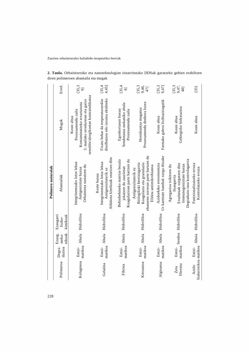
Zaurien orbaintzerako baliabide terapeutiko berriak
228
2. Taula. Orbaintzerako eta nanoteknologian oinarritutako DDSak garatzeko gehien erabiltzen diren polimeroen abantaila eta mugak
Polim
ero
natu
rala
k
Erre
f.
[35,
39]
[35,
44,
45]
[35,
44]
[35,
39,
46,
47]
[35,
39,
47]
[35,
39,
47,
48]
[35]
Mug
ak
Kos
te a
ltua
Proz
esam
endu
zai
la
Kon
tam
inat
zeko
err
azta
suna
3.
mai
lako
err
edur
etan
eta
gai
xo
sens
ible
/ale
rgik
oeta
n ko
ntra
indi
katu
a
Fixa
tu b
ehar
da
tenp
erat
urar
ekin
di
solb
atze
a ed
o iz
ozte
a ek
idite
ko
Egon
korta
sun
baxu
a Se
ndot
asun
mek
anik
o ah
ula
Proz
esam
endu
zai
la
Hor
niku
ntza
mug
atua
Pr
ozes
amen
du d
enbo
ra lu
zea
Kos
te a
ltua
Farm
ako
gale
ra li
xibi
azio
agat
ik
Kos
te a
ltua
Geh
iegi
zko
hidr
ataz
ioa
Kos
te a
ltua
Aba
ntai
lak
Inte
grin
entz
ako
lotz
e le
kua
Ant
igen
izita
te b
axua
O
rbai
ntze
a su
stat
zen
du
Kos
te b
axua
In
tegr
inen
tzak
o lo
tze
leku
a A
ntig
eniz
itate
rik e
z A
ldak
eta
kim
ikoa
k on
artz
en d
itu
Beh
inbe
hine
ko m
atri
ze b
ezal
a jo
katz
en d
u za
urie
tan
Koa
gula
zioa
n pa
rte h
artz
en d
u A
ntig
eniz
itate
rik e
z B
iolo
giko
ki b
erriz
taga
rria
K
oagu
lazi
o et
a gr
anul
azio
eh
unar
en s
orre
ran
parte
har
tzen
du
Efek
tu a
ntim
ikro
bian
oa
Azi
doek
iko
erre
sist
entz
ia
Ur k
antit
ate
hand
iak
xurg
a di
tzak
e
Agr
egaz
ioa
txik
itzen
du
Itsa
sgar
ria
Exud
atua
k xu
rgat
zen
ditu
Im
mun
ogen
izita
te b
axua
D
egra
dazi
o ta
sa k
ontro
laga
rria
Fu
ntzi
onal
izat
zeko
err
aza
Kon
trola
tzek
o er
raza
Ezau
garr
i fi
siko
-ki
mik
oak
Hid
rofil
oa
Hid
rofil
oa
Hid
rofil
oa
Hid
rofil
oa
Hid
rofil
oa
Hid
rofil
oa
Hid
rofil
oa
Ezau
g.
mek
a-ni
koak
Ahu
la
Ahu
la
Ahu
la
Ahu
la
Ahu
la
Send
oa
Ahu
la
Deg
ra-
dazi
oa
Entz
i-m
atik
oa
Entz
i-m
atik
oa
Entz
i-m
atik
oa
Entz
i-m
atik
oa
Entz
i-m
atik
oa
Entz
i-m
atik
oa
Entz
i-m
atik
oa
Polim
eroa
Kol
agen
oa
Gel
atin
a
Fibr
ina
Kito
sano
a
Alg
inat
oa
Zeta
fib
roin
a
Azi
do
hial
uron
ikoa
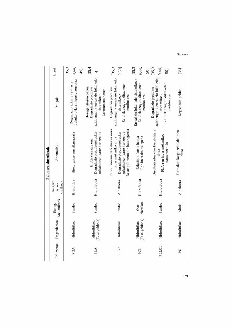
Sarrera
229
Polim
ero
sint
etik
oak
Erre
f.
[35,
3
9,44
,
49]
[35,
4
4]
[35,
3
9,50
]
[35,
3
9,44
,
50]
[35,
3
9,44
,
50]
[35]
Mug
ak
Deg
rada
zio
azka
rra
(2-4
ast
e)
Leku
ko p
Har
en ig
oera
zor
rotz
a
Hez
egar
ritas
un b
axua
D
egra
dazi
o pr
oduk
tu
azid
oeng
atik
err
eakz
io lo
kal e
do
sist
emik
oak
Zu
rrun
tasu
n ba
xua
Deg
rada
zio
prod
uktu
az
idoe
ngat
ik e
rrea
kzio
loka
l edo
si
stem
ikoa
k Ze
lule
k ez
agut
u di
tzak
eten
m
otib
o ez
a
Erre
akzi
o lo
kal e
do s
iste
mik
oak
Zelu
lek
ezag
utu
ditz
aket
en
mot
ibo
eza
Deg
rada
zio
prod
uktu
az
idoe
ngat
ik e
rrea
kzio
loka
l edo
si
stem
ikoa
k Ze
lule
k ez
agut
u di
tzak
eten
m
otib
o ez
a
Deg
rada
zio
geld
oa
Aba
ntai
lak
Bio
xurg
atze
aur
reik
usga
rria
Bio
birx
urga
tze
ona
Deg
rada
zio
prod
uktu
ei e
sker
or
bain
tzea
n pa
rte
hartz
en d
u
Endo
-liso
som
etat
ik ih
es a
zkar
ra
Inda
r mek
anik
o al
tua
Deg
rada
zio
prod
uktu
ei e
sker
or
bain
tzea
n pa
rte
hartz
en d
u B
este
pol
imer
oeki
n ba
tera
garr
ia
Erla
tibok
i kos
te b
axua
Ep
e lu
zera
ko a
skap
ena
Dis
olba
tzai
leek
iko
flex
ibili
tate
al
tua
PLA
-ren
inda
r mek
anik
oa
man
tent
zen
du
Farm
akoa
kar
gatz
eko
ahal
men
al
tua
Ezau
garr
i fi
siko
-ki
mik
oak
Hid
rofil
oa
Hid
rofo
boa
Ald
akor
ra
Hid
rofo
boa
Hid
rofo
boa
Ald
akor
ra
Ezau
g.
Mek
anik
oak
Send
oa
Send
oa
Send
oa
Oso
el
astik
oa
Send
oa
Ahu
la
Deg
rada
zioa
Hid
rolit
ikoa
Hid
rolit
ikoa
(T
asa
geld
oak)
Hid
rolit
ikoa
Hid
rolit
ikoa
(T
asa
geld
oak)
Hid
rolit
ikoa
Hid
rolit
ikoa
Polim
eroa
PGA
PLA
PLG
A
PCL
PLLC
L
PU

Zaurien orbaintzerako baliabide terapeutiko berriak
230
oinarrituta, zehazki polisakarido kationiko
(kitosano eta N,N,n-trimetil kitosano) eta
glikosaminoglikano anionikoez (heparina
eta kondroitin sulfatoa) osatutako NPak
garatu zituzten. NP horiek agrekanoa imi-
tatzeko diseinatuta zeuden, proteoglikano
horrek GFentzako gordailu moduan joka-
tzen baitu. Hori dela eta, GFa proteasen
aurrean egonkortzeaz gain, formulazio ho-
nek bFGFa testuinguru biomimetiko ba-
tean aurkezten du, inguruko ehunekin
duen elkarrekintza erraztuz. Formulazioa-
ren eraginkortasuna in vitro entseguen bi-
dez erakutsi zen, izan ere formulazio ho-
nek muineko zelula estromalen prolifera-
zioa eta jarduera metabolikoa handitu zi-
tuen bFGF askearekin eta agrekanoari lo-
tutako bFGFarekin konparatuz [54].
Li eta bere kideek bFGFa gelatina
MPetan kapsularatu zuten, haien arteko el-
karrekintza elektrostatikoak MPen efektu
babesgarria areagotu dezakelako. Partiku-
lak kolageno/zelulosa nanokristalez osatu-
tako egitura porotsu batean barneratu zi-
ren. Lortutako formulazioa angiogenesia
handitzeko gai izan zen, in vitro zein in
vivo [55]. Park et. al.-ek bFGF-z kargatu-
tako gelatina MPak garatu zituzten ere, eta
kasu honetan kitosano bio-aldamio poro-
tsu batean sartu zituzten. Aposituaren era-
ginkortasuna in vivo aztertu zen, sagu za-
harrei egindako presio ultzeretan, non zau-
rien itxiera tasa handitu zuen. Horretaz
gain kitosanoari esker, animaliek proteasa
maila baxuagoak zituzten, eta horren on-
dorioz bFGF endogeno zein exogeno
maila handiagoak [56]. Beste ikerketa ba-
tean, Kawai eta kideek bFGFdun MPak
merkaturatutako dermis artifizial (Pel-
nak™) batean barneratu zituzten. Pel-
nak™ bi geruza dituen dermis artifizial bat
da, barrualdean kolageno esponja bat eta
kanpoaldean silikona geruza bat. Formula-
zio honen eraginkortasuna bi animalia ere-
dutan ikertu zuten, akurietan egindako lo-
diera osoko zauri eszisionaletan eta sagu
diabetikoetan egindako decubito presio-
ultzeretan. Bi kasuetan formulazioak fi-
broblastoen proliferazioa eta kapilareen
sorrera azkartu zituen. Gainera, sagu dia-
betikoen kasuan infekzioen aurrean erre-
sistentzia handitua behatu zen [57,58].
Huang eta kolaboratzaileek beste bige-
ruzadun apositu bat garatu zuten, zeinaren
barne bFGFdun MPak sartu zituzten. Bio-
aldamioaren barne geruza gelatinazko

Sarrera
231
esponja bat zen, bere egitura porotsuari es-
ker proliferatzen dauden zelulentzako os-
talari modura jokatzen duena; eta kanpo
geruza poliuretano (PU) elastomerikoz
osatuta zegoen, zeinak bio-aldamioaren
ezaugarri mekanikoak hobetzen dituen.
Apositua york txerrietan egindako lodiera
osoko zaurietan ezarri zen, eta emaitzek
erakutsi zutenez, zaurien itxiera bizkortu
zuen eta azalaren birmoldaketa hobetu
zuen [59]. Uluybaram et. al.-ek, bigeruza-
dun apositu horren barnean EGFaz karga-
tutako MPak sartu zituzten eta in vivo zu-
ten eraginkortasuna aztertu zuten untxie-
tan egindako zauri eszisionaletan. Ikertu-
tako dosi altuenarekin, apositua izan zuten
animalietan EGF disoluzioa izan zutenetan
baino askoz gehiago txikitu zen zauriaren
azalera. Gainera, azterketa histologikoek
erakutsi zutenez, ehun berriak azal osasun-
tsuaren egitura ia berdina eskuratu zuen
[60]. Ondoren, bio-aldamioaren barne ge-
ruza (gelatinazko esponja EGFdun gela-
tina MPekin) zauri eszisionaletan aplikatu
zuten, arratoi normal zein estreptozotozi-
nak eragindako arratoi diabetikoetan.
Arratoi ez-diabetikoetan zaurien itxiera
bereziki handitu zen, nahiz eta diabetikoe-
tan hobekuntza txiki bat ere somatu zen.
Analisi histologikoei dagokienez, apositua-
ren efektua esanguratsuki desberdina izan
zen arratoi normal eta diabetikoetan, baina
orokorrean granulazio ehunaren sorrera eta
errepitelizazioa hobetu zituen [61].
Horretaz gain, Zhou eta kideek EGFa
beste polimero natural batean kapsularatu
zuten, kitosanoan, zehazki. EGFa zuten ki-
tosano NPak fibrina gel batera gehitu zi-
tuzten, eta horrela faktorearen askapen lu-
zatua lortu zuten, 7 egunez mantenduz fi-
broblastoen proliferazioa estimulatzeko
gaitasuna [62]. EGFa polimero sintetikoez
osatutako MPetan ere kapsularatua izan
da, adibidez PLGA MPtan, zeinekin fibro-
blastoen hazkuntza tasa hobetu zen in vitro
[63]. Beste ikerketa batean, EGFdun
PLGA NPek lesioaren itxiera bizkortu zu-
ten arratoi diabetikoetan egindako lodiera
osoko zaurietan, izan ere, NPek askatutako
EGFak fibroblastoen proliferazioa sustatu
zuen [64]. Gure ikerketa taldeak burutu-
tako ikerketa batean EGFa PLGA-alginato
MPtan barneratu zen. Alginatoa kapsula-
razio efizientzia hobetzeko erabili zen,
izan ere, MPen barneko fase urtsuaren li-
katasuna handituz, GFen barreiadura mu-
gatu daiteke. MPetan kapsularatu ostean
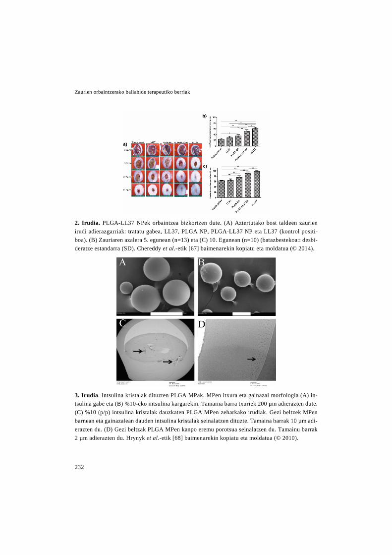
Zaurien orbaintzerako baliabide terapeutiko berriak
232
2. Irudia. PLGA-LL37 NPek orbaintzea bizkortzen dute. (A) Aztertutako bost taldeen zaurien irudi adierazgarriak: tratatu gabea, LL37, PLGA NP, PLGA-LL37 NP eta LL37 (kontrol positi-boa). (B) Zauriaren azalera 5. egunean (n=13) eta (C) 10. Egunean (n=10) (batazbestekoa± desbi-deratze estandarra (SD). Chereddy et al.-etik [67] baimenarekin kopiatu eta moldatua (© 2014).
3. Irudia. Intsulina kristalak dituzten PLGA MPak. MPen itxura eta gainazal morfologia (A) in-tsulina gabe eta (B) %10-eko intsulina kargarekin. Tamaina barra txuriek 200 µm adierazten dute. (C) %10 (p/p) intsulina kristalak dauzkaten PLGA MPen zeharkako irudiak. Gezi beltzek MPen barnean eta gainazalean dauden intsulina kristalak seinalatzen dituzte. Tamaina barrak 10 µm adi-erazten du. (D) Gezi beltzak PLGA MPen kanpo eremu porotsua seinalatzen du. Tamainu barrak 2 µm adierazten du. Hrynyk et al.-etik [68] baimenarekin kopiatu eta moldatua (© 2010).

Sarrera
233
EGFaren aktibitatea mantentzen zela fro-
gatu zen in vitro. Ondoren, in vivo injekzio
intralesional bakarraren ostean, orbaintze
azkarragoa eta eraginkorragoa eragin zu-
ten, zauriaren itxierari, errepitelizazioari
eta hantura prozesuaren ebazpenari dago-
kionez. In vivo entsegua estreptozotozinari
dela eta diabetiko ziren arratoietan egin-
dako lodiera osoko zaurietan burutu zen
[65].
PLGA beste konposatu aktibo batzuk
kapsularatzeko erabilia izan da ere bai,
hala nola, FGF-1, LL37 eta intsulina. Alde
batetik, FGF-1ez kargatutako MPak fi-
brina gel batean sartu ziren, eta formulazi-
oak fibroblastoen proliferazioa hobetu
zuen [66]. Beste aldetik, LL37a zuten
NPak lodiera osoko zaurietan ezarri ziren,
non ondorengoak hobetu zituzten: granu-
lazio ehunaren sorrera, errepitelizazioa,
kolagenoaren metaketa eta neoangiogene-
sia, 2. irudian ikus daitekeen modura [67].
Intsulinaren kapsularatzeari dagokionez
(3. irudia), intsulina zuten PLGA MPek
zauriaren itxiera hobetu zuten keratinozi-
toen migrazio entsegu batean, in vitro
[68]. Ondoren MP horiek alginato edo al-
ginato-polietilenglikol (PEG) esponjetan
sartu ziren, prozesu horren ostean intsuli-
naren bioaktibitatzea mantendu zelarik
[69]. Jarraian, aposituaren eraginkorta-
suna arratoiei eragindako erredura eredu
batean frogatu zen. Aposituarekin tratatu-
tako lesioek orbaintze azkarragoa eta erre-
generatiboagoa lortu zuten, honakoei es-
ker: hildako ehunaren desagertze tasaren
handitzea, estres oxidatiboaren txikitzea,
kolageno metaketaren estimulazioa susta-
tzea eta angiogenesiaren areagotzea [70].
VEGFa NPen barne sartzeak angioge-
nesia hobetzen duela in vitro zein in vivo
frogatu da. Alde batetik, VEGFa zuten fi-
brinazko NPak kitosano-azido hialuroniko
esponja batean barneratu ziren. Apositua
medioarekin inkubatu zen eta zelula endo-
telialei in vitro gehitu ostean, angiogene-
sia areagotu zen [71]. Beste aldetik,
VEGFa zuten alginato kaltzikozko MPak
in vivo larruazalpean ezarri ziren, non an-
giogenesia handitu zuten [72]. Angiogene-
sia aztertzeaz gain, kapsularatutako
VEGFak orbaintzean duen eragina ere az-
tertu da. Horretarako VEGFa zuten PLGA
NPak sagu ez-diabetiko eta diabetikoetan
egindako lodiera osoko zaurietan aplikatu
ziren, non kolageno metaketa, granulazio
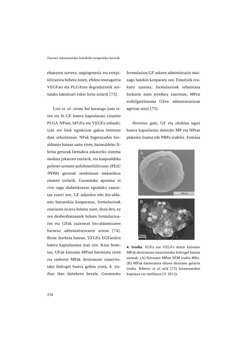
Zaurien orbaintzerako baliabide terapeutiko berriak
234
ehunaren sorrera, angiogenesia eta errepi-
telizazioa hobetu zuten, efektu onuragarria
VEGFari eta PLGAren degradaziotik sor-
tutako laktatoari esker lortu zelarik [73].
Losi et. al. urrats bat haratago joan zi-
ren eta bi GF batera kapsularatu zituzten
PLGA NPtan, bFGFa eta VEGFa zehazki,
izan ere biek eginkizun gakoa betetzen
dute orbaintzean. NPak bigeruzadun bio-
aldamio batean sartu ziren, barnealdeko fi-
brina geruzak farmakoa askatzeko sistema
modura jokatzen zuelarik, eta kanpoaldeko
polieter-uretano-polidimetilsiloxano (PEtU
/PDM) geruzak sendotasun mekanikoa
ematen ziolarik. Garatutako apositua in
vivo sagu diabetikoetan egindako zaurie-
tan ezarri zen. GF askeekin edo bio-alda-
mio hutsarekin konparatuz, formulazioak
zauriaren itxiera hobetu zuen, dena den, ez
zen desberdintasunik behatu formulazioa-
ren eta GFak zuzenean bio-aldamioaren
barnean administratzearen artean [74].
Beste ikerketa batean, VEGFa EGFarekin
batera kapsularatua izan zen. Kasu hone-
tan, GFak kitosano MPtan barneratu ziren
eta ondoren MPak dextranoan oinarritu-
tako hidrogel batera gehitu ziren, 4. iru-
dian ikus daitekeen bezala. Garatutako
formulazioa GF askeen administrazio mai-
zago batekin konparatu zen. Emaitzek era-
kutsi zutenez, formulazioak orbaintzea
bizkortu zuen erredura zaurietan, MPen
erabilgarritasuna GFen administrazioan
agerian utziz [75].
Horretaz gain, GF eta zitokina ugari
batera kapsularatu daitezke MP eta NPtan
plaketen lisatua edo PRPa erabiliz. Fontana
4. Irudia. EGFa eta VEGFa duten kitosano MPak dextranoan oinarritutako hidrogel batean sartuak. (A) Kitosano MPen SEM irudia 400x. (B) MPak barneratuta dituen dextrano gelaren irudia. Ribeiro et al.-etik [75] baimenarekin kopiatua eta moldatua (© 2013).
Sarrera
235
et. al.-ek plaketen lisatua silikona poro-
tsuzko MPtan sartu zuten, emaitza desira-
garriak lortuz ex vivo zauri eredu batean.
Formulazioak kontrol positiboak (plake-
ten lisatua partikula modura) baino efektu
proliferatibo handiago lortu zuen eta ko-
lageno fibren azidofilia labur baina efek-
tiboa ere bai, erregenerazioa abian dago-
elaren seinale dena [76]. PRP edo plaketa
lisatuetatik GFen askapen luzatuagoa lor-
tzeko estrategia bat heparina daramaten
NPen ekoizpena da, izan ere, heparinak
GFak lotzen ditu, haien gordailu modura
jokatuz. Horren harira, PRPa daramaten
fragmina-protamina MP/NPak garatu zi-
ren, haien oinarria protamina fragminare-
kin (pisu molekular baxuko heparina) lo-
tzean sortzen diren konplexu solugaitzak
direlarik. Partikula horiek arratoiei egin-
dako zaurietan administratu ziren, erabi-
litako ereduarekin azal-mentuak lortzeko
emaileei egindako lodiera partzialeko
zauriak islatzen dira. Partikulek zaurien
itxiera bizkorragoa eta angiogenesi han-
ditua eragin zuten [77]. Emaitza horiek
La eta kideek lortutakoekin bat datoz,
haiek ere berdina behatu baitzuten, garatu
zuten formulazioa timorik gabeko saguei
egindako zauri handietan ezarri ostean.
Garatu zuten formulazioa heparinarekin
konjugatutako PLGA NPez osatuta ze-
goen, zeintzuk PRPa kapsularatuta zuten
eta fibrina gel batean barneratuta zeuden
[78].
Heparinak GFak egonkortzeaz gain,
eragin antiinflamatorioa ere badu, erredu-
retan erabilgarria dena. Helburu horrekin
Lakshmi et. al.-ek heparina desulfatatua
kitosanozko NPtan kapsularatu zuten, eta
ondoren NPak kolageno matrize batean
sartu zituzten. Garatutako formulazioa
arratoietan egindako erredura zaurietan
hantura erregulatzeko gai izan zen, ondo-
rioz zauriaren itxiera eta granulazio ehu-
naren sorrera bizkorragoak lortuz [79].
Lezitina moduko lipido negatiboek ki-
tosanoarekin duten elkarrekintza elektros-
tatikoei esker NPak ekoiztu daitezke, Bla-
zevik eta kideek egin zutena, hain zuzen.
NP horiek melatoninarekin kargatu zituz-
ten, eta in vitro entseguetan orbaintzea
sustatu zezaketela frogatu zuten [80].
Zenbait kasutan, DDSak garatzeko
ez-ohiko polimeroak erabili izan dira,
haien ezaugarri bereziez baliatzeko asmoz.
Zaurien orbaintzerako baliabide terapeutiko berriak
236
Horren harira, Petersen eta kolaboratzai-
leek ondorengo polianhidrido anfifilikoez
osatutako MPak garatu zituzten: 1,6-bis(p-
karboxifenoxi)hexanoa (CPH) eta 1,6-
bis(p-karboxifenoxi)-3,6-dioxaoktanoa
(CPTEG). Polimero hauek beste polimero
sintetiko batzuek ez bezala, egitura osoko
zein gainazaleko higadura aurkeztu deza-
kete eta haien degradazio produktuek azi-
dotasun gutxiago dute. Hori dela eta, heze-
tasunak eragindako agregazioaren arriskua
txikitzen dute eta proteinentzako ingurune
egokiago bat sortzen dute. Polimero haue-
kin ekoiztutako MPetan lipokalin-2 pro-
teina barneratu zen. Ondoren, proteina
kapsularatuaren eta askearen in vitro akti-
bitateak konparatu zituzten, eta MPen bar-
nean zegoen lipokalin-2-ak zelulen migra-
zio tasa handitzen zuela nabarmendu zen,
ziurrenik MPek eragindako askapen luza-
tuari esker [29].
Horretaz gain, zenbait polimero erabil-
tzearen arrazoia kapsularatutako farma-
koa itu ehun edo zeluletara bideratzeko
duten gaitasuna da. Horren adibide poli-
(1,4-fenileneazetona dimetilene tioketala)
(PPADT) da, izan ere, polimero hau oxi-
geno espezie erreaktiboekiko (reactive
oxygen species edo ROS) sentikorra da eta
zaurian dauden ROS maila altuak direla
eta bertan depolimerizatzen da batez ere.
Hori dela eta, PPADT NPak garatu ziren
SDF-1α askatzeko. Markatutako NPak
erabiliz, PPADTa zaurietan bereziki depo-
limerizatzen zela frogatu zen, bertan SDF-
1α askatuz. Gainera, formulazio honekin
tratatutako saguen zauriek itxiera azkarra-
goa eta handitutako baskularizazioa izan
zuten [81].
Beste aldetik, polimeroen erabilgarri-
tasuna handitzeko asmoz funtzionalizatu
daitezke, Wang eta kideek egin zuten be-
zala, VIP askatzeko in situ sortutako MPak
ekoiztuz. Horretarako, lehenbizi PCL na-
nozuntzak dopaminarekin funtzionalizatu
zituzten. Ondoren, VIPa nanozuntzetan
absorbatu zuten, dopaminaren ezaugarri
itsaskorrei esker. Azkenik, nanozuntzak
azetonan denbora labur batez murgildu zi-
tuzten, partzialki disolbatutako PCLaren
hauspeatzea eraginez, eta horrela in situ
MPak lortuz. In vivo zauri eredu batean ad-
ministratu ostean, garatutako formulazi-
oak orbaintzea era esanguratsuan sustatu
zuen, granulazio ehunaren sorrera eta an-
giogenesia hobetuz [25].

Sarrera
237
3. Taula. GF eta beste konposatu aktiboak askatzeko garatutako MP eta NP polimerikoen labur-pena eta haiekin lortutako emaitza nagusiak
Erre
f.
[53]
[54]
[55]
[56]
[57,
58]
[59]
[60]
[61]
Emai
tzak
Arr
atoi
ei e
ngin
dako
lodi
era
osok
o er
redu
ra z
auri
etan
azt
ertu
zire
n. F
orm
u-la
zioa
k za
uria
ren
berr
esku
ratz
e ta
sa a
zkar
rago
a lo
rtu z
uen,
err
epite
lizaz
io
eta
derm
isar
en e
rreg
ener
azio
azk
arra
goar
ekin
.
Form
ulaz
ioak
bG
FGar
en a
kitib
itite
a m
ante
ndu
zuen
in v
itro,
akt
ibita
te m
e-ta
bolik
o et
a pr
olif
eraz
ioar
en e
ntse
gu b
anat
an.
Form
ulaz
io h
onek
in z
elul
a en
dote
liale
n pr
olif
eraz
ioa
hand
itu z
en. E
ta a
rra-
toie
tan
larr
uaza
lpea
n ez
arri
oste
an a
ngio
gene
sia
sust
atu
zen
in v
ivo.
Form
ulaz
ioa
sagu
zah
arre
i egi
ndak
o pr
esio
ultz
erat
an a
zter
tu z
en. 7
. egu
-ne
ra a
rte, b
io-a
ldam
io h
utsa
k ze
in b
FGFd
un b
io-a
ldam
ioak
zau
riare
n itx
i-er
a bi
zkor
tu z
uten
. Hor
reta
z ga
in, b
FGFd
un b
io-a
ldam
ioak
pro
teas
en m
aila
tx
ikitu
zue
n 10
. egu
nera
arte
.
Form
ulaz
ioa
akur
ieta
n eg
inda
ko lo
dier
a os
oko
zaur
i esz
isio
nale
tan
eta
db/d
b sa
guet
an e
gind
ako
decu
bito
pre
sio
ultz
erat
an a
zter
tu z
en. M
Peki
n bF
GFa
ren
in v
ivo
erre
tent
zioa
luza
tu z
en. H
orre
taz
gain
, bi e
redu
etan
for-
mul
azio
ak fi
brob
last
oen
prol
ifera
zioa
eta
kap
ilare
en s
orre
ra h
andi
tu z
uen,
do
siar
en m
enpe
ko e
ran.
Yor
k tx
erri
etan
egi
ndak
o lo
dier
a os
oko
zaur
ieta
n az
tertu
zen
. Bio
-ald
amio
-ar
en a
plik
azio
ak z
auri
aren
itxi
era
azka
rtu z
uen
eta
erre
mod
elaz
ioa
hobe
tu.
Form
ulaz
ioa
untx
ieta
n eg
inda
ko lo
dier
a os
oko
zaur
i esz
iona
leta
n az
tertu
ze
n. E
rabi
litak
o do
si a
ltuen
ak z
auria
ren
azal
erar
en m
urriz
keta
han
dien
a er
agin
zue
n. A
zter
keta
his
tolo
giko
ek e
raku
tsi z
uten
ez, e
hun
berr
iare
n eg
i-tu
ra a
zal n
orm
alar
enar
en ia
ber
dina
zen
. Fo
rmul
azio
a lo
dier
a os
oko
zaur
i ere
duet
an a
zter
tu z
en a
rrat
oi d
iabe
tiko
eta
ez-d
iabe
tikoe
tan.
For
mul
azio
ak z
auri
aren
itxi
era
sust
atu
zuen
, ber
ezik
i ar
rato
i ez-
diab
etik
oeta
n. G
aine
ra, b
i ani
mal
ia e
redu
etan
kal
ifika
zio
hist
o-lo
giko
ak h
obet
u zi
tuen
(err
epite
lizaz
ioa,
gra
nula
zio
ehun
aren
sor
rera
eta
ne
obas
kula
rizaz
ioa)
.
GF
bFG
F
bFG
F
bFG
F
bFG
F
bFG
F
bFG
F
EGF
EGF
DD
S
Alg
inat
o M
Pak
karb
oxim
etil
ki-
tosa
no-P
VA
hid
roge
lean
Agr
ekan
oa im
itatz
en d
uten
po-
liele
ktro
lito
konp
lexu
en N
Pak
Gel
atin
a M
Pak
kola
geno
/zel
u-lo
sa p
orot
suzk
o na
nokr
ista
len
bio-
alda
mio
etan
Gel
atin
a M
Pak
kito
sano
por
o-ts
uzko
bio
-ald
amio
etan
Gel
atin
a M
Pak
derm
is a
rtifiz
ial
(Pel
nac®
)bat
ean
Gel
atin
a M
Pak
bige
ruza
dun
bio-
alda
mio
bat
ean
(gel
atin
a es
ponj
a et
a po
liure
tano
min
tza)
Gel
atin
a M
Pak
bige
ruza
dun
Bio
-ald
amio
bat
ean
(gel
atin
a es
ponj
a et
a po
liure
tano
min
tza)
Gel
atin
a M
Pak
gela
tina
es-
ponj
etan

Zaurien orbaintzerako baliabide terapeutiko berriak
238
Erre
f.
[62]
[63]
[64]
[65]
[66]
[67]
[68]
[69,
70]
[71]
[72]
[73]
Emai
tzak
Form
ulaz
ioak
eza
ugar
ri bi
o-its
asga
rria
k er
akut
si z
ituen
, eta
NPa
k ed
o ge
la b
akar
rik
erab
ilita
bai
no a
skap
en lu
zatu
agoa
lortu
zue
n. H
orre
taz
gain
, kap
sula
razi
o pr
ozes
ua-
ren
oste
an E
GFa
ren
bioa
ktib
itate
a m
ante
ndu
zen,
fibr
obla
stoe
n pr
olif
eraz
ioa
sust
a-tz
eko
gai i
zan
baitz
en.
Form
ulaz
ioek
fibr
obla
stoe
n ha
zkun
tza
tasa
hob
etu
zute
n in
vitr
o.
Arr
atoi
dia
betik
oen
zaur
i ere
du b
atea
n, N
Pek
zaur
iare
n itx
iera
biz
kortu
zut
en E
GF
aske
arek
in k
onpa
ratu
z.
Form
ulaz
ioa
arra
toi d
iabe
tikoe
tan
egin
dako
lodi
era
osok
o za
uri e
redu
bat
ean
azte
rtu
zen.
For
mul
azio
ak o
rbai
ntze
azk
arra
go e
ta e
ragi
nkor
rago
a er
agin
zue
n, e
rrep
iteliz
a-zi
o et
a ha
ntur
a pr
ozes
uare
n eb
azpe
nari
dago
kioe
nez.
Bio
-ald
amio
ak fi
brob
last
oen
prol
ifer
azio
a su
stat
u zu
en in
vitr
o.
NPe
k za
urie
n itx
iera
hob
etu
zute
n N
MR
I arr
atoi
etan
, err
epite
lizaz
io, k
olag
eno
me-
take
ta, g
ranu
lazi
o eh
unar
en s
orre
ra e
ta n
eoan
giog
enes
iari
dago
kion
ez.
Form
ulaz
ioa
kera
tinoz
itoet
an e
gind
ako
urra
dura
ent
segu
bat
ean
azte
rtu z
en, n
on
zaur
iare
n itx
iera
azk
artu
zue
n.
Apo
situ
a ar
rato
ieta
n eg
inda
ko e
rred
ura
lesi
oeta
n ap
likat
u ze
n. O
rbai
ntze
azk
arra
go
eta
erre
gene
ratib
oago
a lo
rtu z
uen
hona
koen
gatik
: itx
iera
biz
kortu
a, h
ildak
o eh
una-
ren
desi
nteg
razi
o ta
sa a
ltuag
oa, e
stre
s ox
idat
ibo
txik
itua,
eta
kol
agen
oare
n m
etak
eta
eta
heltz
e ho
betu
ak.
Form
ulaz
ioak
in v
itro
kapi
lare
-gis
ako
tubu
luen
form
azio
a (a
ngio
gene
sia)
han
ditu
zu
en.
MPa
k la
rrua
zalp
ean
ezar
ri zi
ren,
eta
ang
ioge
nesi
a su
stat
u zu
ten
ingu
ruko
ehu
nare
n 0,
6-0,
8 m
m-k
o lu
zera
n.
NPe
k, in
vitr
o ke
ratin
ozito
en p
rolif
eraz
ioa
eta
mig
razi
oa h
obet
u zu
ten
eta
VEG
FR-
2-ar
en e
spre
sioa
gai
n-er
regu
latu
zut
en. O
ndor
en, s
agu
diab
etik
o et
a ez
dia
betik
oeta
n eg
inda
ko z
auri
ered
ueta
n az
tertu
zir
en, n
on g
ranu
lazi
o eh
unar
en s
orre
ra, k
olag
eno
eduk
ia, e
rrep
itelia
zioa
eta
ang
ioge
nesi
a ho
betu
zitu
zten
.
GF
EGF
EGF
EGF
EGF
FGF-
1
LL37
Ints
ulin
a
Ints
ulin
a
VEG
F
VEG
F
VEG
F
DD
S
Kito
sano
NPa
k fib
rina
gel
bate
an
PLG
A M
Pak
PLG
A N
Pak
PLG
A-A
lgin
ato
MPa
k
PLG
A M
Pak
fibrin
a bi
o-al
-da
mio
bat
ean
PLG
A N
Pak
PLG
A M
Pak
PLG
A M
Pak
algi
nato
edo
al
gina
to-P
EG e
spon
jeta
n
Fibr
ina
NPa
k ki
tosa
no-a
zido
hi
alur
onik
o es
ponj
etan
Kal
tzio
alg
inat
ozko
MPa
k
PLG
A N
Pak

Sarrera
239
Erre
f.
[74]
[75]
[76]
[77]
[78]
[79]
[80]
[29]
[81]
[25]
Emai
tzak
Form
ulaz
ioak
sag
u di
abet
ikoe
tan
egin
dako
lodi
era
osok
o za
uri e
redu
bat
ean
az-
tertu
zire
n. F
orm
ulaz
ioek
GF
aske
ekin
kon
para
tuta
orb
aint
zea
hobe
tu z
uten
, ba
ina
ez z
en a
ldak
etar
ik s
umat
u G
Fak
zuze
nean
bio
-ald
amio
etan
bar
nera
tuta
ko
apos
ituek
in k
onpa
ratu
z ge
ro.
Hid
roge
lak
arra
toie
tan
egin
dako
err
edur
a za
urie
tan
aplik
atu
zire
n. G
F as
keen
ad-
min
istra
zio
mai
zago
bat
ekin
kon
para
tuta
, hid
roge
lek
orba
intz
ea a
zkar
tu z
uten
.
Form
ulaz
ioak
in v
itro
egin
dako
zau
rien
itxie
ra h
obet
u zu
en. E
x vi
vo e
gind
ako
zaur
ieta
n er
rege
nera
zioa
sus
tatu
zue
n, g
erta
tuta
ko e
fekt
u pr
olif
erat
iboa
k et
a az
i-do
filia
k fr
ogat
u zu
tene
z.
Form
ulaz
ioa
arra
toie
tan
egin
dako
lodi
era
partz
iale
ko z
aurie
tan
adm
inis
tratu
zen
, no
n er
repi
teliz
azio
a et
a an
giog
enes
ia s
usta
tu z
uen.
Form
ulaz
ioar
ekin
trat
atut
ako
zaur
iek
itxie
ra a
zkar
rago
a et
a su
stat
utak
o an
giog
e-ne
sia
erak
utsi
zut
en, s
agu
atim
ikoe
tan.
Form
ulaz
ioa
erre
dura
zau
riet
an a
plik
atu
oste
an, o
rbai
ntze
a az
kartu
zue
n. Iz
an e
re
hant
ura
lehe
nago
mur
riztu
zen
eta
gra
nula
zio
ehun
a bi
zkor
rago
sor
tu z
en.
In v
itro
urra
dura
ent
segu
bat
ean,
HaC
aT z
elul
en m
igra
zioa
hob
etu
zute
n.
Prot
eina
ask
ea a
dmin
istra
tzea
reki
n ko
npar
atut
a, fo
rmul
azio
ak z
elul
en m
igra
zioa
ha
nditu
zue
n, lu
zatu
tako
ask
apen
a de
la e
ta.
NPe
k fa
rmak
oare
n bi
dera
tze
eta
aska
tze
erag
inko
rra
egin
zut
en z
aurir
a, b
erta
n da
goen
RO
S ed
uki a
ltuag
atik
. Gai
nera
, NPe
kin
trat
atut
ako
zaur
iek
itxie
ra a
zka-
rrag
oa e
ta h
andi
tuta
ko b
asku
lariz
azio
a er
akut
si z
uten
sag
u er
edu
bate
an.
Sagu
etan
egi
ndak
o za
uri e
szis
iona
l bat
ean,
form
ulaz
ioak
orb
aint
zea
era
esan
gura
-ts
uan
hobe
tu z
uen,
gra
nula
zio
ehun
aren
sor
rera
eta
ang
ioge
nesi
a su
stat
uz.
GF
VEG
F et
a bF
GF
EGF
eta
VEG
F
Plak
eten
li-
satu
a
PRP
PRP
Hep
arin
a de
sulfa
tatu
a
Mel
aton
ina
Lipo
kalin
a-2
SDF-
1α
VIP
DD
S
PLG
A N
Pak
PEtU
/PD
M
edo
fibrin
an o
inar
ritut
ako
Bio
-ald
amio
etan
Kito
sano
MPa
k de
xtra
noan
oi
narr
ituta
ko h
idro
gele
tan
Silik
ona
poro
tsuz
ko M
Pak
Frag
min
a-pr
otam
ina
MP/
NPa
k
Hep
arin
arek
in k
onju
gatu
-ta
ko P
LGA
NPa
k fib
rina
gel b
atea
n.
Kito
sano
MPa
k ko
lage
no
mat
rize
bat
ean
Lezi
tina/
kito
sano
NPa
k
CPH
eta
CPT
EGea
n oi
na-
rritu
tako
, pol
ianh
idrid
o an
-fi
filik
ozko
MPa
k
PPA
DT
NPa
k (R
OSa
ri se
n-tik
orra
den
nan
omat
eria
la)
In s
itu s
ortu
tako
PC
L M
Pak
PCL
nano
xafl
atan

Zaurien orbaintzerako baliabide terapeutiko berriak
240
3.2 Nanopartikula lipidikoak
4. taulan laburtuta ageri denez, NP li-
pidikoak GF eta beste molekula endoge-
noak askatzeko DDS egokiak dira. Haien
artean, liposomak sakonki ikertuak izan
dira, 1960-ko hamarkadan aurkitu zirene-
tik [82]. Liposomak bigeruza lipidikozko
mintz batez osatutako xixku esferikoak
dira [83]. Lipidoek ematen dizkieten ezau-
garrietaz gain (biobateragarritasuna eta bi-
odegradazioa), zenbait abantaila dituzte
haien egitura dela eta: (i) konposatu hidro-
filo zein lipofiloak kapsularatzeko gaita-
suna, hidrofiloak konpartimentu urtsuan
eta lipofiloak bigeruza lipidikoan; (ii) lu-
zatutako askapena lortzeko gaitasuna; eta
(iii) epidermiseko konposaketa imitatzen
dutenez, azalean farmako pilaketa handi-
tzeko gaitasuna. Dena den, liposomek
muga ugari dauzkate, besteak beste, far-
makoen kontrolik gabeko askapena eragin
dezakeen egonkortasun baxua, kapsulara-
tze efizientzia baxua eta biltegiratzean ze-
har sedimentazio, agregazio edo fusioa
pairatzeko aukera [84, 85].
1988an, Brown eta kideek zaurien
orbaintzerako GF bat liposoma batean
barneratu zuten lehendabiziz, aukeratuta-
koa EGFa izan zelarik. EGFa lezitina lipo-
soma geruzanitzetan kapsularatu zuten eta
liposoma horiek arratoiei egindako 5 cm-
ko ebakiduretan aplikatu zituzten. Liposo-
mek EGFaren esposizio denbora luzatu zu-
ten, eta EGF askearen administrazioarekin
konparatuz honako hobekuntzak lortu zi-
tuzten: %200-ean handitutako trakzio in-
darra, kolageno sorreraren sustapena eta
fibroblastoen proliferazioaren igoera [86].
Ondoren, beste ikerketa talde batek EGFa
zuten beste liposoma geruzanitz batzuk
garatu zituzten. Formulazio berri honen
eraginkortasuna in vivo aztertu zuten biga-
rren mailako erreduretan, EGF askearekin
eta liposoma hutsekin konparatuz. Formu-
lazioak orbaintze bizkorrena lortu zuen
hurrengoei dagokienez: kolageno forma-
zioa, zauriaren uzkurdura, fibroblastoen
proliferazioa eta epitelioaren berreskura-
tzea [87]. Liposomen administrazioa erraz-
teko asmoz, kitosano gel batean sartu zi-
ren, zeinak zauria heze mantentzen zuen.
Formulazio berri hau bigaren mailako
erreduretan aztertu zen ere, liposomekin
izandako antzeko emaitzak lortuz. Horrek
liposomak kitosano gel batean barneatzea-
ren onura erakusten duelarik [88].
Sarrera
241
Pierre et al.-en arabera, IGF-Iez karga-
tutako liposomak topikoki administratuz
gero, orbaintzea IGF eta hazkuntza hormo-
naren dosi altuagoak sistemikoki admis-
tratuta bezain sustatuta egongo litzateke.
Arratoietan egindako orbaintze entsegu
bati esker haien hipotesia frogatu zuten,
errepitelizazioa neurtuz, hain zuzen ere
[89].
Xiang eta kideek liposomek farmakoak
barneratzeko duten ahalmena ikertu zuten.
Zentzu horretan, bFGFa liposometan kap-
sularatzeko 4 metodoen egokitasuna az-
tertu zuten. Metodo denek antzeko aban-
tailak eskaintzen zituzten, prestaketa
erraza eta ekoizpen-baldintza arinak, esa-
terako. Baina, zenbait desberdintasun zi-
tuzten, besteak beste, kapsularatze tasa,
bioaktibitatea eta egonkortasun fisikoa.
Aztertutako metodoen artean, pH gradien-
tearen metodoa egokiena zela erabaki zu-
ten. Horregatik jarraian egindako in vivo
entsegurako, metodo honen bidez ekoitzi
ziren liposomak. In vivo entsegua arratoiei
egindako bigarren mailako erreduretan bu-
rutu zen, eta bertan, formulazioak orbain-
tzea sustatzeko, proliferazio dermala biz-
kortzeko eta kolagenoa sortzeko gaitasuna
erakutsi zuen [90]. Azkenik, SDF-1α zuen
liposoma formulazio bat garatu zen, eta
merkaturatutako dermis azelular baten
(Alloderm®) barne dispertsatu zen. SDF-
1αa bi eratan barneratu zen dermis artifizi-
alean, liposometan kapsularatua edo aske.
Liposometan sartuta dispertsatzea estrate-
gia hobea dela frogatu zen, izan ere, sagu
diabetikoetan egindako lodiera osoko zau-
rietan, liposometako SDF-1αak zelulen
proliferazio iraunkorragoa, granulazio
ehun lodiagoa eta zauriaren itxiera hobe-
tua lortu zituen [91].
Liposomek dituzten mugak gaindi-
tzeko asmoz, NP lipidikoen belaunaldi be-
rri bat garatu zen, nanopartikula solido li-
pidikoak (solid lipid nanoparticle edo
SLN). SLNak giro tenperaturan solido di-
ren lipidoez eta egitura egonkortzen duten
surfaktanteez osatutako NPk dira [92]. Li-
posomekin alderatuz gero, askapena erre-
gulatzeko malgutasun handiagoa dute eta
barneratutako farmakoen babes hobea es-
kaintzen dute, haien egoera solidoak ingu-
ruko fase urtsuarekin farmako hidrofiloen
trukea txikitzen baitu [93,94]. Hala eta
guztiz ere, SLNek egonkortasun mugatua
dute, izan ere, biltegiratzean zehar sare
Zaurien orbaintzerako baliabide terapeutiko berriak
242
kristalinoaren akatsak murrizten dira, far-
makoen kanporaketa eraginez [40]. Arazo
hau gainditzeko, garraiatzaile lipidiko na-
noegituratuak (nanostructured lipid ca-
rrier edo NLC) garatu ziren, hauek ma-
trize amorfoa duten NP lipidikoak dira eta
lipido solido eta likidoen nahasketez osa-
tuta daude [95,96]. Horretaz gain, NLCek
SLNek baino farmako gehiago kargatzeko
ahalmena dute eta amaierako dispertsioan
ur eduki txikiagoa dute. SLNen mugen
arren, bi NP mota hauek abantaila ugari di-
tuzte orbaintzean erabiliak izateko, haue-
tako zenbait liposomekin partekatzen di-
tuztelarik: (i) kontrolatutako askapena
ahalbidetzen dute; (ii) azalaren hidratazio
efektua handitzen dute, haien itsaskorta-
sun eta oklusibitateari esker; (iii) haien ta-
maina txikiak azalarekin kontaktu estua
ahalbidetzen du; eta (iv) administrazio to-
pikoa baimentzen dute, esposizio sistemi-
koa ekidinez [95].
Esandakoa kontuan izanda, gure iker-
keta taldeak EGFa SLN eta NLCtan kap-
sularatu zuen. Garatutako formulazioek
EGFaren bioaktibitatea mantendu zuten
ekoizte prozesuaren zein gamma esterili-
zazio prozesuaren ondoren. Orduan, haien
eraginkortasuna sagu diabetikoetan (db/db
saguak) egindako loditaera osoko zauri es-
zisional eredu batean frogatu zen. Formu-
lazio biek orbaintzea antzeko neurrian sus-
tatu zuten, EGFaren dosi altuago batekin
konparatuz gero. Formulazioek orbaintze-
aren ondorengo atalak hobetu zituzten:
zauriaren itxiera, errepitelizazioa eta han-
tura prozesuaren ebazpena. Dena den,
NLCak egokiagoak zirela erabaki zen
haien abantailengatik, hots, kapsularatze
efikazia altuagoa eta disolbatzaile organi-
koen erabilera eza ekoizpen prozesuan ze-
har [97]. Hori dela eta, txerrietan egindako
in vivo entseguan EGFdun NLCak erabili
ziren soilik (5. irudia). Marruskariekin
konparatuz, txerriak maneiatzeko zailago
dira, haien tamaina handia dela eta. Baina,
beste aldetik, haien orbaintze prozesua gi-
zakiarenaren antzekoagoa da, eta zauri
handiagoak egitea baimentzen dute, kasu
honetan zauriek 30 cm2-ko azalera zuten.
Entsegu honetan lortutako emaitzak aurre-
koekin bat datoz, izan ere, NLCek zauria-
ren itxiera bizkortu zuten eta orbaintzea-
ren kalitatea hobetu zuten EGF askearen
dosi altuago batekin konparatuz [98]. Az-
kenik, haien administrazio topikoa erraz-
teko asmoz, EGFz kargatutako SLN eta

Sarrera
243
5. Irudia. In vivo zauri eredua txerrietan EGFa duten NLCak administratzeko. (A-C) Zauria egi-teko prozesu-kirurgikoa: (A) Zauriaren azalera estandarizatzeko plastikozko markoa. (B) Zauria-ren perimetroaren tatuajea. (C) Diatermia monopolarra erabiliz lodiera osoko zauriak egiteko pro-zedura kirurgikoa. (D) Zauriaren itxieraren irudikapen grafikoa bigarren astetik entseguaren amai-eraraino. Datuak batazbesteko ± SD moduan daude. Esangura estatistikoa *p<0,05 NLC hutsekin konparatuta. (E) Talde experimental bakoitzaren zaurien irudiak. Gainza et al.-etik [99] baimena-rekin kopiatua eta moldatua (© 2014).
NLCak hidrogel erdisolidoetan (Noveon
AA-1 eta Pluronic F-27an oinarrituta) edo
fibrinan oinarritutako bio-aldamioetan
barneratu ziren. Formulazio denek EGFa-
ren askapen luzatua erakutsi zuten gutxie-
nez 48 orduz. Horretaz gain, barneraketa
minimoa erakutsi zuten azal osoko stratum
corneumean zehar eta antzeko xurgapena
kaltetutako azalean zehar in vitro entse-
guek erakutsi zuten moduan. Haien artean,
fibrinan oinarritutako formulazioak aban-
tailatsuenak zirela erabaki zen, honakoei
esker: biobateragarritasun ona, egonkorta-
sun hobea, fibrinaren gaitasuna bitartekari
hemostatikoa izateko eta ehun berria sor-
tzen den bitartean matrize moduan ari-
tzeko duen gaitasuna [99].
EGFa NLCtan kapsularatuz lortutako
emaitza onen ondorioz, ikerketa berri
Zaurien orbaintzerako baliabide terapeutiko berriak
244
batean LL37 peptido antimikrobianoa
NLtan barneratzea erabaki genuen. kapsu-
laratze prozesuak ez zuen farmakoaren bi-
oaktibitatea aldatu, izan ere, bere ekintza
antimikrobianoa mantendu zuen E. coli-
ren aurka eta baita bere ekintza antiinfla-
matorioa ere, lipopolisakarikoak (LPS)
eragindako makrofagoen aktibazioa ekidi-
nez. Formulazioaren eraginkortasuna az-
tertzeko db/db sagu diabetikoetan egin-
dako lodiera osoko zauri eszisional eredu
bat erabili zen. Emaitzek erakutsi zutenez,
garatutako formulazioa orbaintzea hobe-
tzeko gai izan zen zauriaren itxiera, erre-
pitelizatioa eta hantura prozesuaren ebaz-
penari dagokienez [100]. Fumakia et al.-
ek LL37a SLNtan barneratu zuten, serpin-
A1arekin batera. Garatutako formulazioak
E. coli eta S. aureusen aurrean aktibitate
antibakterianoa erakutsi zuen, in vitro fi-
broblasto eta keratinozitoetan egindako
zaurien itxiera sustatu zuen eta in vitro
LPSaren aurkako makrofagoen ekintza in-
flamatorioa ekidin zuen [31].
3.3 Nanozuntzezko egiturak
Nanozuntz sareak, haien egitura bereiz-
garria dela eta, apositu moduan erabiliak
izateko oso aproposak dira, 5. taulan la-
burtzen den bezala. Orokorrean, elektroi-
rutearen bitartez ekoizten dira. Teknika
honek indar elektrostatikoa erabiltzen du
tanta batetik nanozuntzak sortzeko [101].
Horrela sortutako nanozuntz polimerikoak
biodegradagarriak eta ehundu gabeak dira,
eta haien diametroa nanometro gutxi ba-
tzuetatik mikrometrotara bitartekoa da
[35,102]. Orbaintzean erabiltzeko zenbait
ezaugarri onuragarri dituzte, haien artean,
bolumenaren araberako azalera handia eta
porositate altua. Bi ezaugarri horiek gasen
sarrera eta exudatuen garbiketa ahalbide-
tzen dute, horrela zelulen arnasketa bai-
menduz eta zaurian hezetazuna mantenduz
deshidratatu ez dadin [103]. Horretaz gain,
nanozuntzezko mintzak oso erakargarriak
dira ehun ingeniaritzarako, izan ere, ma-
trize extrazelularrarekin (extracellular
matrix edo ECM) duten antzekotasuna
dela eta, zelulen migrazio eta prolifera-
zioa sustatzen dute eta ehunaren sorrera-
rako euskarri moduan jokatzen dute
[44,104].
Farmakoen garraioari dagokionez,
apositu hauek farmakoak kargatzeko ahal-
men altua daukate, eta gainera, inguruneko
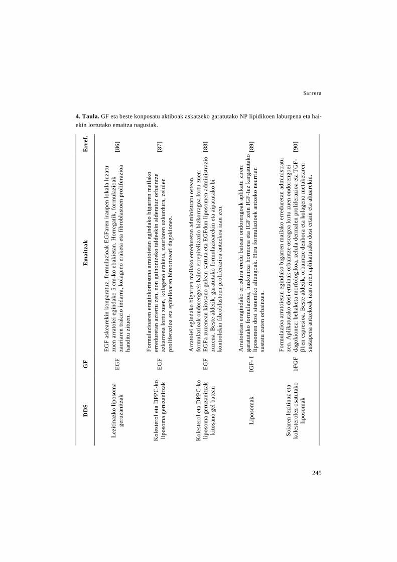
Sarrera
245
4. Taula. GF eta beste konposatu aktiboak askatzeko garatutako NP lipidikoen laburpena eta hai-ekin lortutako emaitza nagusiak.
Err
ef.
[86]
[87]
[88]
[89]
[90]
Em
aitz
ak
EGF
aske
arek
in k
onpa
ratu
z, fo
rmul
azio
ak E
GFa
ren
irau
pen
loka
la lu
zatu
zu
en a
rrat
oiei
egi
ndak
o 5
cm-k
o eb
akie
tan.
Hor
rega
tik, f
orm
ulaz
ioak
za
uria
ren
trakz
io in
darr
a, k
olag
eno
erak
eta
eta
fibro
blas
toen
pro
lifer
azio
a ha
nditu
zitu
en.
Form
ulaz
ioar
en e
ragi
nkor
tasu
na a
rrat
oiet
an e
gind
ako
biga
rren
mai
lako
er
redu
reta
n az
tertu
zen
, non
gai
nont
zeko
tald
eeki
n al
dera
tuz
orba
intz
e az
karr
ena
lortu
zue
n, k
olag
eno
erak
eta,
zau
riare
n uz
kurd
ura,
zel
ulen
pr
olif
eraz
ioa
eta
epite
lioar
en b
irsor
tzea
ri da
goki
onez
.
Arr
atoi
ei e
gind
ako
biga
rren
mai
lako
err
edur
etan
adm
inis
trat
u os
tean
, fo
rmul
azio
ak o
ndor
engo
ek b
aino
err
epite
lizaz
io b
izko
rrag
oa lo
rtu z
uen:
EG
Fa z
uzen
ean
kito
sano
gel
ean
sartu
ta e
ta E
GFd
un li
poso
men
adm
inis
trazi
o zu
zena
. Bes
te a
ldet
ik, g
arat
utak
o fo
rmul
azio
arek
in e
ta a
ipat
utak
o bi
ko
ntro
leki
n fib
robl
asto
en p
rolif
eraz
ioa
antz
ekoa
izan
zen
.
Arr
atoi
etan
era
gind
ako
erre
dura
ere
du b
atea
n on
dore
ngoa
k ap
likat
u zi
ren:
ga
ratu
tako
form
ulaz
ioa,
haz
kunt
za h
orm
ona
eta
IGF
zein
IGF-
Iez
karg
atut
ako
lipos
omen
dos
i sis
tem
iko
altu
agoa
k. H
iru fo
rmul
azio
ek a
ntze
ko n
eurr
ian
sust
atu
zute
n or
bain
zea.
Form
ulaz
ioa
arra
toie
tan
egin
dako
big
arre
n m
aila
ko e
rred
uret
an a
dmin
istr
atu
zen.
Apl
ikat
utak
o do
si e
rtain
ak o
rbai
ntze
oso
agoa
lortu
zue
n on
dore
ngoe
i da
goki
onez
: beh
aket
a m
orfo
logi
koa,
zel
ula
derm
alen
pro
lifer
azio
a et
a TG
F-β1
en e
spre
sioa
. Bes
te a
ldet
ik, o
rbai
ntze
den
bora
eta
kol
agen
o m
etak
etar
en
sust
apen
a an
tzek
oak
izan
zir
en a
plik
atut
ako
dosi
erta
in e
ta a
ltuar
ekin
.
GF
EGF
EGF
EGF
IGF-
I
bFG
F
DD
S
Lezi
tinaz
ko li
poso
ma
geru
zani
tzak
Kol
este
rol e
ta D
PPC
-ko
lipos
oma
geru
zani
tzak
Kol
este
rol e
ta D
PPC
-ko
lipos
oma
geru
zani
tzak
ki
tosa
no g
el b
atea
n
Lipo
som
ak
Soia
ren
lezi
tinaz
eta
ko
lest
erol
ez o
satu
tako
lip
osom
ak

Zaurien orbaintzerako baliabide terapeutiko berriak
246
Err
ef.
[91]
[97,
98]
[99]
[100
]
[31]
Em
aitz
ak
Lipo
som
ek S
DF-
1are
n ak
tibita
tea
man
tend
u zu
ten
in v
itro.
In
vivo
zau
ri es
zisi
onal
ere
du m
urin
o di
abet
iko
bate
an a
dmin
istr
atu
zire
n, n
on S
DF-
1 as
kear
en
adm
inis
traz
ioar
ekin
ko
npar
atuz
ho
nako
ak
hobe
tu
zitu
zten
: de
rmis
ean
epe
luze
rako
zel
ulen
pro
lifer
azio
a su
stat
u zu
ten,
gra
nula
zio
ehun
a lo
ditu
zut
en e
ta z
auri
aren
itxi
era
apur
bat
azk
artu
zut
en.
Sagu
dia
betik
oen
zaur
ieta
n ad
min
istra
tu o
stea
n, b
i fo
rmul
azio
ek o
rbai
ntze
a ho
betu
zut
en E
GF
aske
arek
in k
onpa
ratu
z, z
auri
aren
itx
iera
, er
repi
teliz
azio
a et
a ha
ntur
a pr
ozes
uare
n eb
azpe
nari
dago
kion
ez.
Kap
sula
ratz
e er
agin
korta
suna
altu
agoa
izan
zut
elak
o et
a ek
oizp
en p
roze
suan
ez
dir
elak
o di
solb
atza
ile o
rgan
ikor
ik b
ehar
, N
LCak
era
bili
zire
n on
dore
n eg
inda
ko i
n vi
vo e
ntse
guan
txe
rri
ered
u ba
tean
. B
erta
n, N
LCek
zau
riare
n itx
iera
eta
kal
itate
a ho
betu
zut
en E
GF-
aren
dos
i altu
ago
bate
kin
konp
arat
uz.
Form
ulaz
io d
enak
ant
zeko
ak iz
an z
iren
ondo
reng
oeta
n: E
GF
aska
pena
, EG
F sa
rrer
a ka
ltetu
tako
aza
lean
eta
sar
rera
min
imoa
aza
l osa
sunt
suan
. N
LCak
in
vitro
era
gin
antim
ikro
bian
o et
a an
ti-in
flam
ator
ioa
izan
zut
en.
In
vivo
sa
gu
diab
etik
oeta
n eg
inda
ko
lodi
era
osok
o za
uri
eszi
sion
alet
an
adm
inis
tratu
ost
ean,
zau
riar
en it
xier
a, e
rrep
iteliz
azio
a et
a ha
ntur
a pr
ozes
uare
n eb
azpe
na h
obet
zeko
gai
izan
zire
n.
Form
ulaz
ioak
E. c
oli e
ta S
. aur
euse
n au
rrea
n er
agin
ant
imik
robi
anoa
aur
kezt
u zu
en. I
n vi
tro e
gind
ako
urra
dura
ent
segu
bat
ean
zaur
iare
n itx
iera
sus
tatu
zue
n.
LPS-
ren
aurr
ean,
haz
kunt
zare
n ge
ruza
baka
rrar
en o
sota
suna
bab
estu
zue
n et
a er
agin
ant
i-inf
lam
ator
ioa
erak
utsi
zue
n.
GF
SDF-
1
EGF
EGF
LL37
LL37
eta
Se
rpin
-A1
DD
S
DSP
C, D
SPA
eta
ko
lest
erol
ezko
lipo
som
ak
derm
is a
zelu
lar b
atea
n (A
llode
rm)
SLN
ak (p
reci
rol)
eta
N
LCak
(pre
ciro
l eta
m
yglio
l)
SLN
eta
NLC
ak
hidr
ogel
erd
isol
idoe
tan
(Nov
eon
AA
-1 e
ta
Plur
onic
F-2
7) e
do
fibrin
an o
inar
ritut
ako
bio-
alda
mio
etan
NLC
ak (p
reci
rol e
ta
myg
liol)
SLN
ak (g
lizer
il m
onoe
ster
arat
oa e
ta α
-fo
sfat
idilk
olin
a
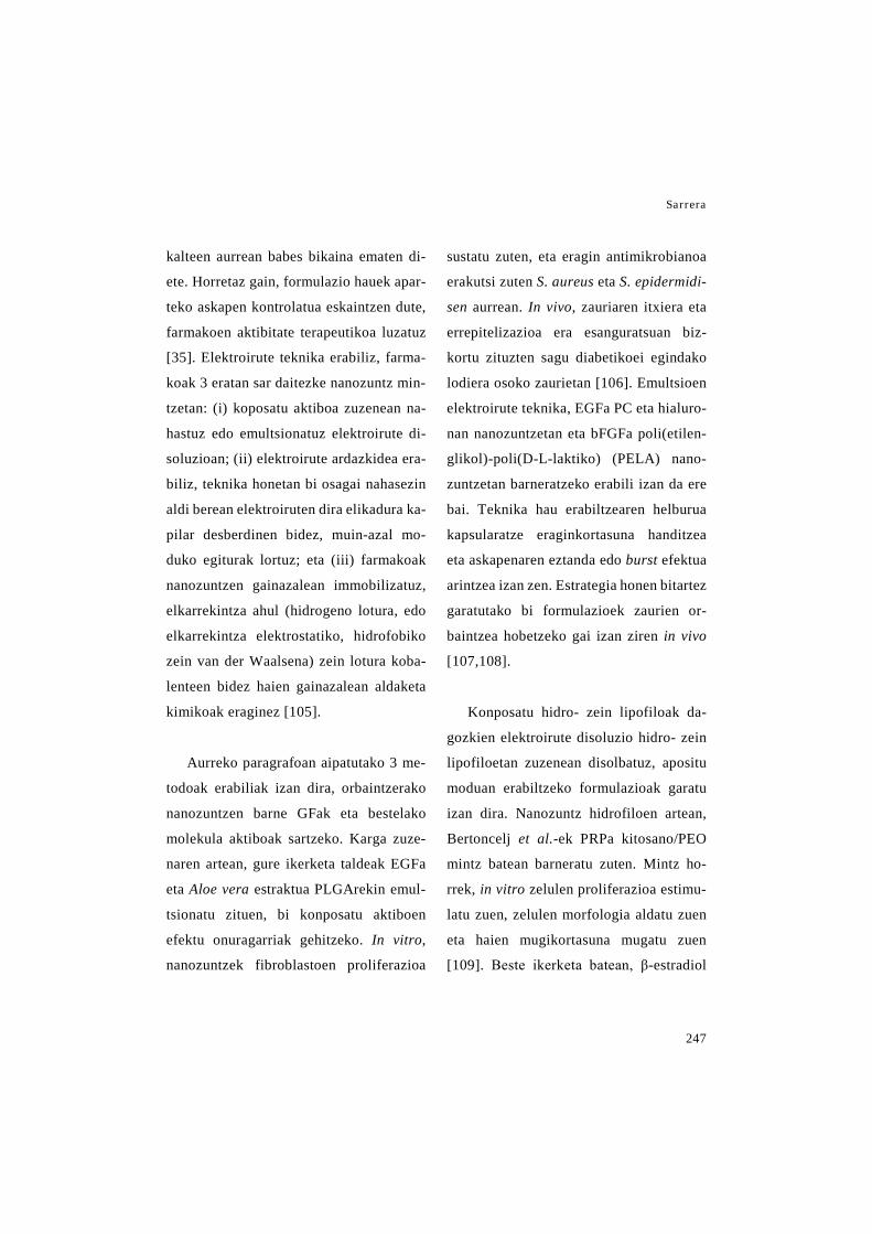
Sarrera
247
kalteen aurrean babes bikaina ematen di-
ete. Horretaz gain, formulazio hauek apar-
teko askapen kontrolatua eskaintzen dute,
farmakoen aktibitate terapeutikoa luzatuz
[35]. Elektroirute teknika erabiliz, farma-
koak 3 eratan sar daitezke nanozuntz min-
tzetan: (i) koposatu aktiboa zuzenean na-
hastuz edo emultsionatuz elektroirute di-
soluzioan; (ii) elektroirute ardazkidea era-
biliz, teknika honetan bi osagai nahasezin
aldi berean elektroiruten dira elikadura ka-
pilar desberdinen bidez, muin-azal mo-
duko egiturak lortuz; eta (iii) farmakoak
nanozuntzen gainazalean immobilizatuz,
elkarrekintza ahul (hidrogeno lotura, edo
elkarrekintza elektrostatiko, hidrofobiko
zein van der Waalsena) zein lotura koba-
lenteen bidez haien gainazalean aldaketa
kimikoak eraginez [105].
Aurreko paragrafoan aipatutako 3 me-
todoak erabiliak izan dira, orbaintzerako
nanozuntzen barne GFak eta bestelako
molekula aktiboak sartzeko. Karga zuze-
naren artean, gure ikerketa taldeak EGFa
eta Aloe vera estraktua PLGArekin emul-
tsionatu zituen, bi konposatu aktiboen
efektu onuragarriak gehitzeko. In vitro,
nanozuntzek fibroblastoen proliferazioa
sustatu zuten, eta eragin antimikrobianoa
erakutsi zuten S. aureus eta S. epidermidi-
sen aurrean. In vivo, zauriaren itxiera eta
errepitelizazioa era esanguratsuan biz-
kortu zituzten sagu diabetikoei egindako
lodiera osoko zaurietan [106]. Emultsioen
elektroirute teknika, EGFa PC eta hialuro-
nan nanozuntzetan eta bFGFa poli(etilen-
glikol)-poli(D-L-laktiko) (PELA) nano-
zuntzetan barneratzeko erabili izan da ere
bai. Teknika hau erabiltzearen helburua
kapsularatze eraginkortasuna handitzea
eta askapenaren eztanda edo burst efektua
arintzea izan zen. Estrategia honen bitartez
garatutako bi formulazioek zaurien or-
baintzea hobetzeko gai izan ziren in vivo
[107,108].
Konposatu hidro- zein lipofiloak da-
gozkien elektroirute disoluzio hidro- zein
lipofiloetan zuzenean disolbatuz, apositu
moduan erabiltzeko formulazioak garatu
izan dira. Nanozuntz hidrofiloen artean,
Bertoncelj et al.-ek PRPa kitosano/PEO
mintz batean barneratu zuten. Mintz ho-
rrek, in vitro zelulen proliferazioa estimu-
latu zuen, zelulen morfologia aldatu zuen
eta haien mugikortasuna mugatu zuen
[109]. Beste ikerketa batean, β-estradiol
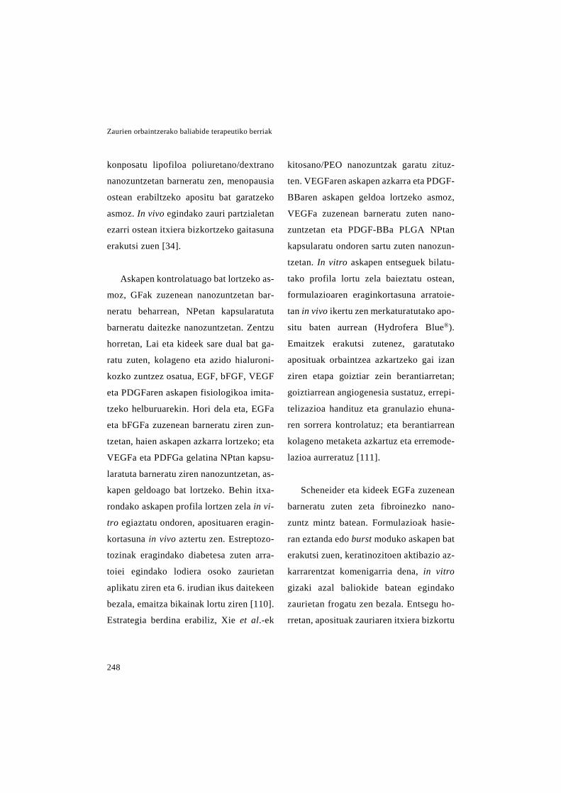
Zaurien orbaintzerako baliabide terapeutiko berriak
248
konposatu lipofiloa poliuretano/dextrano
nanozuntzetan barneratu zen, menopausia
ostean erabiltzeko apositu bat garatzeko
asmoz. In vivo egindako zauri partzialetan
ezarri ostean itxiera bizkortzeko gaitasuna
erakutsi zuen [34].
Askapen kontrolatuago bat lortzeko as-
moz, GFak zuzenean nanozuntzetan bar-
neratu beharrean, NPetan kapsularatuta
barneratu daitezke nanozuntzetan. Zentzu
horretan, Lai eta kideek sare dual bat ga-
ratu zuten, kolageno eta azido hialuroni-
kozko zuntzez osatua, EGF, bFGF, VEGF
eta PDGFaren askapen fisiologikoa imita-
tzeko helburuarekin. Hori dela eta, EGFa
eta bFGFa zuzenean barneratu ziren zun-
tzetan, haien askapen azkarra lortzeko; eta
VEGFa eta PDFGa gelatina NPtan kapsu-
laratuta barneratu ziren nanozuntzetan, as-
kapen geldoago bat lortzeko. Behin itxa-
rondako askapen profila lortzen zela in vi-
tro egiaztatu ondoren, aposituaren eragin-
kortasuna in vivo aztertu zen. Estreptozo-
tozinak eragindako diabetesa zuten arra-
toiei egindako lodiera osoko zaurietan
aplikatu ziren eta 6. irudian ikus daitekeen
bezala, emaitza bikainak lortu ziren [110].
Estrategia berdina erabiliz, Xie et al.-ek
kitosano/PEO nanozuntzak garatu zituz-
ten. VEGFaren askapen azkarra eta PDGF-
BBaren askapen geldoa lortzeko asmoz,
VEGFa zuzenean barneratu zuten nano-
zuntzetan eta PDGF-BBa PLGA NPtan
kapsularatu ondoren sartu zuten nanozun-
tzetan. In vitro askapen entseguek bilatu-
tako profila lortu zela baieztatu ostean,
formulazioaren eraginkortasuna arratoie-
tan in vivo ikertu zen merkaturatutako apo-
situ baten aurrean (Hydrofera Blue®).
Emaitzek erakutsi zutenez, garatutako
aposituak orbaintzea azkartzeko gai izan
ziren etapa goiztiar zein berantiarretan;
goiztiarrean angiogenesia sustatuz, errepi-
telizazioa handituz eta granulazio ehuna-
ren sorrera kontrolatuz; eta berantiarrean
kolageno metaketa azkartuz eta erremode-
lazioa aurreratuz [111].
Scheneider eta kideek EGFa zuzenean
barneratu zuten zeta fibroinezko nano-
zuntz mintz batean. Formulazioak hasie-
ran eztanda edo burst moduko askapen bat
erakutsi zuen, keratinozitoen aktibazio az-
karrarentzat komenigarria dena, in vitro
gizaki azal baliokide batean egindako
zaurietan frogatu zen bezala. Entsegu ho-
rretan, aposituak zauriaren itxiera bizkortu
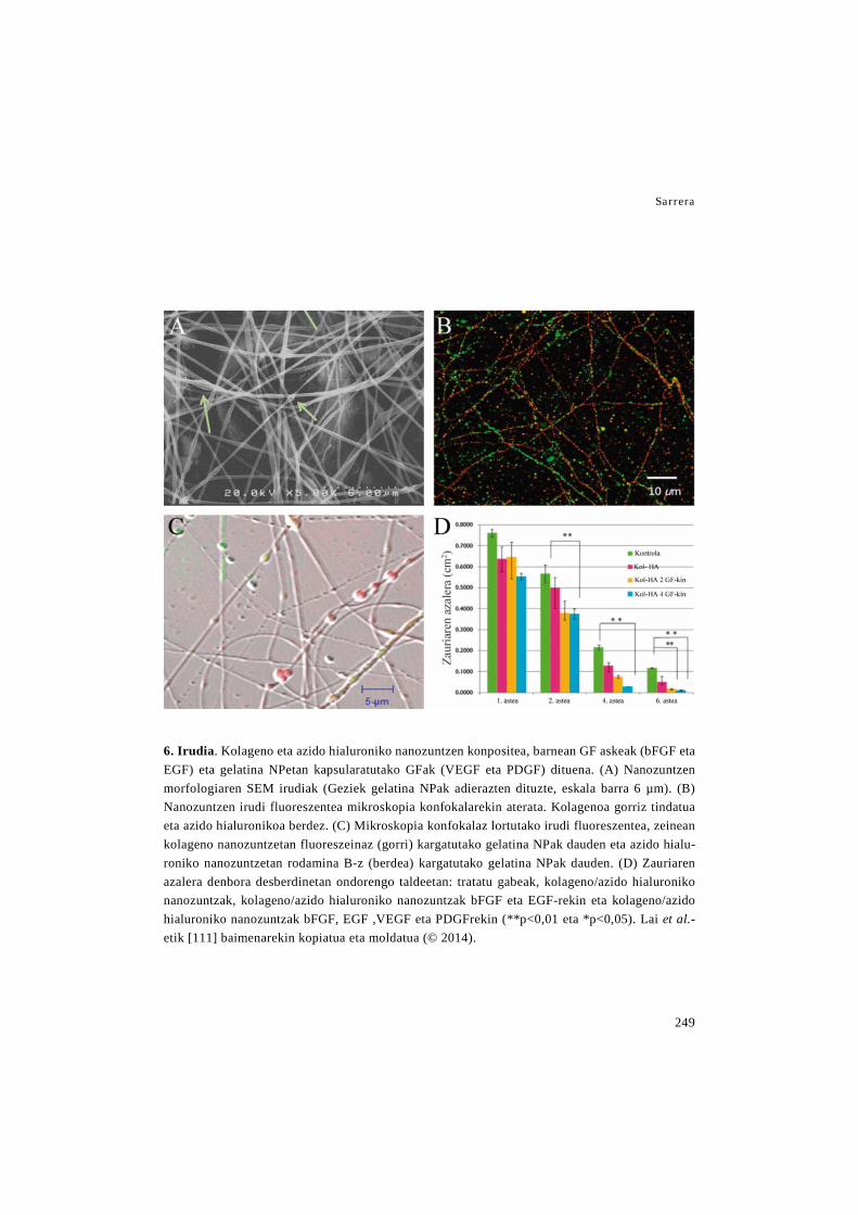
Sarrera
249
6. Irudia. Kolageno eta azido hialuroniko nanozuntzen konpositea, barnean GF askeak (bFGF eta EGF) eta gelatina NPetan kapsularatutako GFak (VEGF eta PDGF) dituena. (A) Nanozuntzen morfologiaren SEM irudiak (Geziek gelatina NPak adierazten dituzte, eskala barra 6 µm). (B) Nanozuntzen irudi fluoreszentea mikroskopia konfokalarekin aterata. Kolagenoa gorriz tindatua eta azido hialuronikoa berdez. (C) Mikroskopia konfokalaz lortutako irudi fluoreszentea, zeinean kolageno nanozuntzetan fluoreszeinaz (gorri) kargatutako gelatina NPak dauden eta azido hialu-roniko nanozuntzetan rodamina B-z (berdea) kargatutako gelatina NPak dauden. (D) Zauriaren azalera denbora desberdinetan ondorengo taldeetan: tratatu gabeak, kolageno/azido hialuroniko nanozuntzak, kolageno/azido hialuroniko nanozuntzak bFGF eta EGF-rekin eta kolageno/azido hialuroniko nanozuntzak bFGF, EGF ,VEGF eta PDGFrekin (**p<0,01 eta *p<0,05). Lai et al.-etik [111] baimenarekin kopiatua eta moldatua (© 2014).
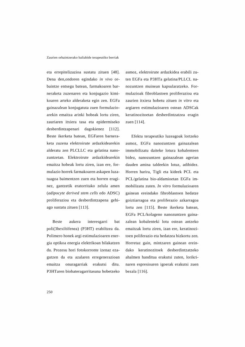
Zaurien orbaintzerako baliabide terapeutiko berriak
250
eta errepitelizazioa sustatu zituen [48].
Dena den,ondoren egindako in vivo or-
baintze entsegu batean, farmakoaren bar-
neraketa zuzenaren eta konjugazio kimi-
koaren arteko alderaketa egin zen. EGFa
gainazalean konjugatuta zuen formulazio-
arekin emaitza arinki hobeak lortu ziren,
zauriaren itxiera tasa eta epidermiseko
desberdintzapenari dagokienez [112].
Beste ikerketa batean, EGFaren barnera-
keta zuzena elektroirute ardazkidearekin
alderatu zen PLCLLC eta gelatina nano-
zuntzetan. Elektroirute ardazkidearekin
emaitza hobeak lortu ziren, izan ere, for-
mulazio horrek farmakoaren askapen luza-
tuagoa baimentzen zuen eta horren eragi-
nez, gantzetik eratorritako zelula amen
(adipocyte derived stem cells edo ADSC)
proliferazioa eta desberdintzapena gehi-
ago sustatu zituen [113].
Beste aukera interesgarri bat
poli(3hexiltifenea) (P3HT) erabiltzea da.
Polimero honek argi estimulazioaren ener-
gia optikoa energia elektrikoan bilakatzen
du. Prozesu hori fotokorronte izenaz eza-
gutzen da eta azalaren erregenerazioan
emaitza onuragarriak erakutsi ditu.
P3HTaren biobateragarritasuna hobetzeko
asmoz, elektroirute ardazkidea erabili zu-
ten EGFa eta P3HTa gelatina/PLLCL na-
nozuntzen muinean kapsularatzeko. For-
mulazioak fibroblastoen proliferazioa eta
zaurien itxiera hobetu zituen in vitro eta
argiaren estimulazioaren ostean ADSCak
keratinozitoetan desberdintzatzea eragin
zuen [114].
Efektu terapeutiko luzeagoak lortzeko
asmoz, EGFa nanozuntzen gainazalean
immobilizatu daiteke lotura kobalenteen
bidez, nanozuntzen gainazalean agerian
dauden amina taldeekin lotuz, adibidez.
Horren harira, Tigli eta kideek PCL eta
PCL/gelatina bio-aldamioetan EGFa im-
mobilizatu zuten. In vitro formulazioaren
gainean ereindako fibroblastoen hedatze
goiztiarragoa eta proliferazio azkarragoa
lortu zen [115]. Beste ikerketa batean,
EGFa PCL/kolageno nanozuntzen gaina-
zalean kobalenteki lotu ostean antzeko
emaitzak lortu ziren, izan ere, keratinozi-
toen poliferazio eta hedatzea bizkortu zen.
Horretaz gain, mintzaren gainean erein-
dako keratinozitoek desberdintzatzeko
ahalmen handitua erakutsi zuten, lorikri-
naren espresioaren igoerak erakutsi zuen
bezala [116].

Sarrera
251
Choi et al.-ek EGFa kobalenteki lotuta
zuen beste formulazio bat garatu zuten.
Kasu honetan, EGFa PEG/PCL kopoli-
mero blokeez osautako nanozuntzetan im-
mobilizatu zuten. Formulazio honen era-
ginkortasuna nanozuntzen gain EGF diso-
luzioa zuen formulazioarekin konparatu
zen. EGFa kobalenteki lotuta zuen formu-
lazioak aktibitate terapeutiko hobea eraku-
tsi zuen in vitro zein in vivo orbaintze en-
tseguetan, immobilizazioak EGFari es-
kaintzen dion babes handiagoa dela eta. In
vitro, keratinozitoen gene espezifikoen es-
presioaren igoera behatu zen, eta in vivo
zauriaren itxiera azkartua eta EGFRaren
espresio handitua ikusi ziren [117]. Iker-
keta horietan lortutako emaitza onuraga-
rriak kontuan hartuta, ondorengo lan ba-
tean PEG/PC kopolimero blokeez osatu-
tako nanozuntzen gainazalean EGFa im-
mobilizatzeaz gain, bFGFa muinean kap-
sularatu zuten elektroirute ardazkidea era-
biliz. Fomulazio honetan bFGFak hasie-
rako eztanda moduko askapena erakutsi
zuen, EGFaren askapenik, ordea, ez zen
behatu. GF bakarra zuten formulazioekin
konparatuta, bi GFak zituen formulazioak
erredura zaurien itxiera bizkortu zuen, zauri
helduagoak lortuz zazpigarren egunerako,
estreptozotozinak eragindako diabetesa
zuten saguetan [117].
Song eta kideek erakutsi zutenez, na-
nozuntzen gainazalean konposatu akti-
boak kobalenteki lotzeko era gehiago
daude. Zentzu horretan, Cys-KR12 (LL37
peptido antimikrobianotik eratorritako
motiboa) zeta fibroinazko nanozuntzekin
konjugatu zuten, EDC/NSH eta tiol-malei-
mida klick kimikaren bitartez. Konjugazio
prozesuak ez zuen Cys-KR12aren bioakti-
bitatea kaltetu, izan ere, in vitro eragin an-
timikrobianoa eta proliferatiboa manten-
tzen zituen, baita LPSak eragindako han-
tura deuzesteko gaitasuna ere [119].
Patel et al.-ek burututako ikerketa ba-
tean, bFGFa eta laminina PLLaren gaina-
zalean immobilizatuak izan ziren, hepari-
nak molekula endogenoekin lotzeko duen
gaitasunean oinarrituta. Horrretarako,
PLLa heparinarekin funtzionalizatu zen
eta ondoren bFGFa eta laminina lotu zi-
tzaizkion. Ondoriozko formulazioak or-
baintzea hobetzeko gai izan zen in vitro
urradura saio batean, batez ere, nanozun-
tzak zauriraen elkarzut kokatuta zeude-
nean [120].

Zaurien orbaintzerako baliabide terapeutiko berriak
252
5. Taula. GF eta beste konposatu aktiboak askatzeko garatutako nanozuntz mintzen laburpena eta haiekin lortutako emaitza nagusiak
Err
ef.
[106
]
[107
]
[108
]
[109
]
[34]
[110
]
[111
]
Em
aitz
ak
In v
itro
fibro
blas
toen
pro
lifer
azio
a ho
betu
zut
en e
ta e
ragi
n an
timik
robi
anoa
izan
zut
en. S
agu
diab
etik
oeta
n eg
inda
ko
zaur
i ere
du b
atea
n za
uria
ren
itxie
ra e
ta e
rrep
iteliz
azio
a az
kartu
zitu
zten
.
Form
ulaz
ioak
ker
atin
ozito
en e
ta fi
brob
last
oen
prol
ifer
azio
a et
a in
filtra
zioa
sus
tatu
zitu
en in
vitr
o. E
ta in
viv
o, a
zal
funt
zion
al b
aten
bir
sort
zea
bultz
atu
zuen
.
Form
ulaz
ioak
fibr
obla
stoe
n ad
hesi
oa, p
rolif
eraz
ioa
eta
ECM
ko p
rote
inen
jari
aket
a su
stat
u zi
tuen
. Arr
atoi
dia
betik
oei
egin
dako
zau
riet
an e
zart
zean
orb
aint
zea
hobe
tu z
uten
, 2
aste
etan
err
epite
lizaz
io o
soa
eta
azal
eko
apen
dize
en b
irsor
tzea
lo
rtuz.
Form
ulaz
ioak
zel
ulen
pro
lifer
azio
a es
timul
atu
zuen
, hai
en
mor
folo
gia
alda
tu z
uen
eta
haie
n m
ugik
orta
suna
mug
atu.
Form
ulaz
ioa
arra
toie
i egi
ndak
o lo
dier
a pa
rtzi
alek
o za
urie
tan
azte
rtu z
en, n
on z
auria
ren
itxie
ra b
izko
rtu z
uen.
Form
ulaz
ioak
zel
ula
endo
telia
len
hazk
untz
a ta
sa h
andi
tu z
uen,
et
a ha
ien
arte
ko s
aret
zea
hobe
tu z
uen.
Arr
atoi
dia
betik
oei
egin
dako
zau
riet
an, f
orm
ulaz
ioak
zau
riar
en it
xier
a bi
zkor
tu
zuen
, kol
agen
o m
etak
eta
hand
itu z
uen
eta
hodi
en h
eltz
ea
sust
atu
zuen
.
Hyd
rofe
ra B
lue®
kom
ertz
iala
reki
n ko
npar
atuz
, for
mul
azio
ak
zaur
ien
itxie
ra b
izko
rtu z
uen
arra
toie
tan,
fase
goi
ztia
r (a
ngio
gene
sia
sust
atuz
, err
epite
lizaz
ioa
hand
ituz
eta
gran
ulaz
io e
huna
ren
sorr
era
kont
rola
tuz)
, zei
n be
rant
iarr
etan
(k
olag
eno
met
aket
a az
kartu
z et
a er
rem
odel
azio
a au
rrer
atuz
).
Kar
ga m
etod
oa
Kar
ga z
uzen
a,
emul
tsifi
kazi
oz
Kar
ga z
uzen
a,
emul
tsifi
kazi
oz
Kar
ga z
uzen
a,
emul
tsifi
kazi
oz
Kar
ga z
uzen
a,
naha
stuz
Kar
ga z
uzen
a,
naha
stuz
Kar
ga z
uzen
a.
bFG
F et
a EG
F zu
zene
an e
ta V
EGF
eta
PDG
F ge
latin
a N
Pen
barn
e
Kar
ga z
uzen
a.
VEG
F zu
zene
an e
ta
PDG
F-B
B P
LGA
N
Pen
barn
e
GF
EGF
EGF
bFG
F
PRP
β-es
tradi
ola
VEG
F,
bFG
F, P
DG
F et
a EG
F
VEG
F et
a PD
GF-
BB
DD
S
PLG
A e
ta A
loe
vera
na
nozu
ntza
k
PCL
eta
hial
uron
an
nano
zunt
zak
PELA
na
nozu
ntza
k
Kito
sano
/PEO
na
nozu
ntza
k
PU e
ta d
extr
ano
nano
zunt
zak
Kol
agen
o et
a az
ido
hial
uron
iko
nano
zunt
z du
alak
Kito
sano
eta
PE
O
nano
zunt
zak
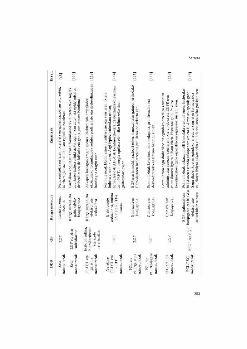
Sarrera
253
Err
ef.
[48]
[112
]
[113
]
[114
]
[115
]
[116
]
[117
]
[118
]
Em
aitz
ak
Nan
ozun
tzek
zau
riar
en it
xier
a et
a er
repi
teliz
azio
a su
stat
u zu
ten,
in
vitr
o gi
za a
zal b
alio
kide
an e
gind
ako
zaur
ieta
n
Farm
akoa
kon
juga
tua
zuen
form
ulaz
ioar
ekin
trat
atut
ako
sagu
ek
zaur
iare
n itx
iera
arin
ki a
zkar
rago
a iz
an z
uten
eta
epi
derm
isar
en
desb
erdi
ntze
a ile
folik
ulu
eta
gant
z gu
ruin
etar
a ha
nditu
a.
Ask
apen
luze
agoa
era
gin
zuen
ez, e
lekt
roiru
te a
rdaz
kide
z ek
oizt
utak
o fo
rmul
azio
ak z
elul
en p
rolif
eraz
io e
ta d
esbe
rdin
tzap
en
hand
iago
a er
agin
zue
n.
Form
ulaz
ioak
fibr
obla
stoe
n pr
olif
eraz
io e
ta z
auri
aren
itxi
era
hobe
tu z
ituen
in v
itro.
Arg
i izp
iez
estim
ulat
u os
tean
, fo
rmul
azio
ak A
DSC
ak k
erat
inoz
itoet
ara
desb
erdi
ntze
ko g
ai iz
an
zen,
P3H
Tak
ener
gia
optik
oa e
lekt
riko
bihu
rtze
ko d
uen
gaita
suna
ri es
ker.
EGFa
ren
imm
obili
zazi
oari
eske
r, na
nozu
ntze
n ga
inea
n er
eind
ako
fibro
blas
toen
hed
atze
a et
a pr
olife
razi
oa a
zkar
tu z
en.
Form
ulaz
ioak
ker
atin
ozito
en h
edap
ena,
pro
lifer
azio
a et
a de
sber
dint
zeko
aha
lmen
a ha
nditu
zitu
en.
Form
ulaz
ioa
sagu
dia
betik
oeta
n eg
inda
ko e
rred
ura
zaur
ieta
n ad
min
istra
tu z
enea
n, z
auri
aren
itxi
era
hobe
a et
a EG
FRar
en
espr
esio
aren
igoe
ra b
ehat
u zi
ren.
Hor
reta
z ga
in, i
n vi
tro
kera
tinoz
itoen
gen
e es
pezi
fikoe
n es
pres
ioa
sust
atu
zuen
.
Form
ulaz
ioak
ask
atze
pro
fil b
imod
ala
erak
utsi
zue
n, h
asie
rako
bF
GFa
ren
ezta
nda
aska
pena
reki
n et
a EG
Fren
ask
apen
ik g
abe.
Sa
gu d
iabe
tikoe
tan
egin
dako
err
edur
a za
urie
tan
form
ulaz
ioa
zaur
iare
n itx
iera
azk
artz
eko
eta
heltz
ea s
usta
tzek
o ga
i iza
n ze
n.
Kar
ga m
etod
oa
Kar
ga z
uzen
a,
naha
stuz
Kar
ga z
uzen
a et
a ga
inaz
alek
o ko
njug
azio
a
Kar
ga z
uzen
a et
a el
ektro
irute
ar
dazk
idea
Elek
troiru
te
arda
zkid
ea, m
uina
EG
F et
a P3
HT-
z os
atua
Gai
naza
lean
ko
njug
atua
Gai
naza
lean
ko
njug
atua
Gai
naza
lean
ko
njug
atua
EGFa
gai
naza
lean
ko
njug
atua
eta
bFG
F a
elek
troiru
te
arda
zkid
eaz
sartu
ta
GF
EGF
EGF
eta
zila
r su
lfadi
azin
a
EGF,
ints
ulin
a,
hidr
okor
tison
a et
a az
ido
erre
tinoi
koa
EGF
EGF
EGF
EGF
bFG
F et
a EG
F
DD
S
Zeta
na
nozu
ntza
k
Zeta
na
nozu
ntza
k
PLLC
L et
a ge
latin
a na
nozu
ntza
k
Gel
atin
a/
PLLC
L et
a P3
HT
nano
zunt
zak
PCL
eta
PCL/
gela
tina
nano
zunt
zak
PCL
eta
PCL/
kola
geno
na
nozu
ntza
k
PEG
eta
PC
L na
nozu
ntza
k
PCL/
PEG
na
nozu
ntza
k
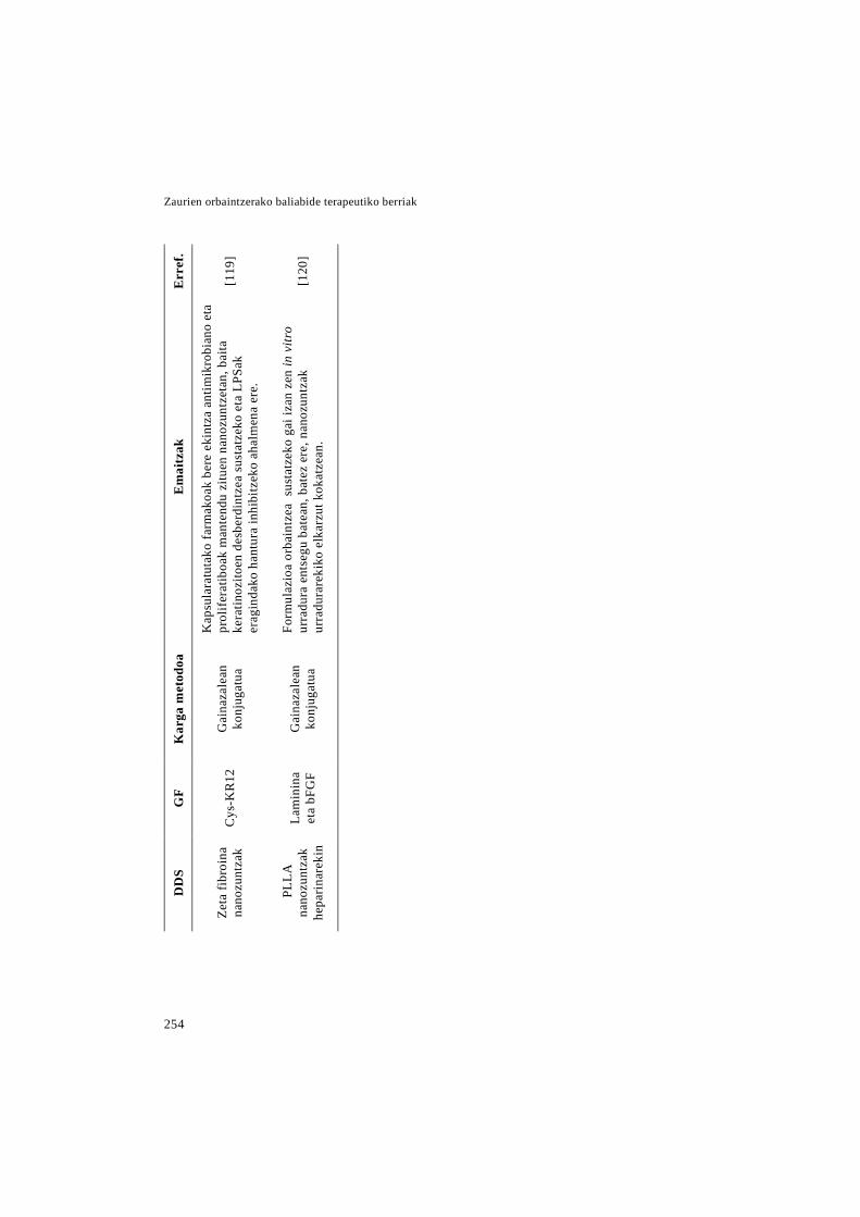
Zaurien orbaintzerako baliabide terapeutiko berriak
254
Err
ef.
[119
]
[120
]
Em
aitz
ak
Kap
sula
ratu
tako
farm
akoa
k be
re e
kint
za a
ntim
ikro
bian
o et
a pr
olif
erat
iboa
k m
ante
ndu
zitu
en n
anoz
untz
etan
, bai
ta
kera
tinoz
itoen
des
berd
intz
ea s
usta
tzek
o et
a LP
Sak
erag
inda
ko h
antu
ra in
hibi
tzek
o ah
alm
ena
ere.
Form
ulaz
ioa
orba
intz
ea s
usta
tzek
o ga
i iza
n ze
n in
vitr
o ur
radu
ra e
ntse
gu b
atea
n, b
atez
ere
, nan
ozun
tzak
ur
radu
rare
kiko
elk
arzu
t kok
atze
an.
Kar
ga m
etod
oa
Gai
naza
lean
ko
njug
atua
Gai
naza
lean
ko
njug
atua
GF
Cys
-KR
12
Lam
inin
a et
a bF
GF
DD
S
Zeta
fibr
oina
na
nozu
ntza
k
PLL
A
nano
zunt
zak
hepa
rinar
ekin

Sarrera
255
4. Ondorioak eta etorkizuneko
ikuspegia
Azken urteotan, zauri kronikoen intzi-
dentzia era kezkagarrian handitu da, arris-
kuan dagoen populazioa hazi den heinean.
Horretaz gain, tratamendu eraginkorren
gabezia dela eta, zauri kronikoak zama
ekonomiko zein kliniko handia bilakatzen
ari dira. Prozesu fisiologikoarekin konpa-
ratuz, zauri kronikoen orbaintzean hainbat
asaldura gertatzen dira, hala nola, GFen
eta prozesuaren erregulazioan parte har-
tzen duten beste molekula batzuen maila-
ren jaitsiera. Hori dela eta, konposatu ho-
rien administrazio exogenoa estrategia
itxaropentsua da orbaintzerako, baina
haien erabilera klinikoa murriztuta dago,
in vivo daukaten egonkortasun baxua dela
eta. Arazo hau gainditzeko bide bat, kon-
posatu bioaktibo horiek DDS berrietan
kapsularatzea da, zeinen artean, MP/NP
polimerikoak, NP lipidikoak, nanozun-
tzezko egiturak eta haien arteko konbina-
zioak daudelarik. DDS horiek, kapsulara-
tutako farmakoa babesteaz gain, honako
abantailak dituzte: biobateragarritasun ona,
askapen kontrolatu edota luzatua, farmako
karga handia eta ezaugarri mekaniko onak.
Horretaz gain, erabilitako DDS mota eta
polimeroaren arabera ezaugarri zehatzak
lor daitezke, hala nola, zuzendutako ga-
rraioa edo ECMa imitatzen duen ingurunea.
Laburbilduz, nanoteknologian oinarri-
tutako DDSetan GFen eta bestelako mole-
kula endogeno aktiboen barneraketak po-
tentzial handia erakutsi du zauri kronikoen
tratamenduan eta azalaren leheneratzean.
Dena den, momentura arte, GFak askatzen
dituzten DDSen ikerketak fase preklini-
koan jarraitzen du, eta ikerketa gehiago
behar dira fase klinikora heltzeko. Egiteke
dauden ikerketa preklinikoen artean hu-
rrengoak daude: animalia ereduetan tole-
rantzia eta toxizitate saioak eta txerrien
moduko animalia eredu handiagoetan era-
ginkortasun saioak.
Aipatutako DDS gehienak sistema to-
pikoak direnez, haien onarpenaren aurretik
gai kritiko bat ikertu behar da, kapsulara-
tutako farmakoaren xurgapen sistemikoa,
alegia. Horretaz gain, polimeroen egokita-
suna aztertu behar den beste gai gako bat
da, izan ere, deskribatutako zenbait DDS
garatzeko erabili diren polimeroak ez
daude onartuta klinikan erabiliak izateko.

Zaurien orbaintzerako baliabide terapeutiko berriak
256
Nahiz eta entsegu klinikorik edo mer-
katuratutako produkturik ez dagoen GFak
dituzten DDS hauekin, badaude nanotek-
nologian oinarritutako bi produktu orbain-
tzerako merkaturatuak. Horietatik lehena
Altrazeal® da, hauts txuri kristalino bat, li-
ofilizatutako poli-2-hidroxietilmetakrila-
tozko (pHEMA) eta poli-2hidroxipropil-
metakrilatozko (pHPMA) NPtaz osatuta
dagoena. Nanoflex teknologiari esker,
zauriaren exudatuarekin kontaktuan sar-
tzean NPak hidratatu eta agregatzen dira
mintz heze eta malgu bat bilakatuz [121].
Bigarren produktua Talymed® da, poli-N-
azetil glukosaminaz (pGIcNAc) osatutako
nanozuntz laburtuez osatutako matrize au-
rreratu bat [122]. Horietaz gain, gaur egun
partehartzaileak aukeratzeko fasean da-
goen entsegu kliniko bat dago, zeinean,
SPINNERaren jokaera eta segurtasuna az-
tertuko diren. SPINNERa eskuko elektroi-
rute tresna bat da, in situ nanozuntzezko
aposituak ekoizteko balio duena [123].
5. Eskerrak
I. Garcia-Oruek Eusko Jaurlaritzari
doktoratu aurreko laguntza eskertzen dio.
Projektu hau neurri batean Espainiako
Ekonomia eta Lehiakortasun Ministerioak
finantziatu du (INNPACTO, IPT-2012-
0602-300000, 2012). Horretaz gain, par-
tzialki Eusko Jaurlaritzak finantziatu du
(ELKARTEK 2015, Nanoplatform, KK-
2015/0000036).
Erreferentzien zerrenda 43-53 orrialdeetan aurkitzen da.
Helburuak


Helburuak
259
Sarreran zehaztu denez, zauri kronikoak osasun arazo bat bilakatzen ari dira, izan ere
egungo terapiek ezin dute orbaintze eraginkor bat ziurtatu. Gainera, haien intzidentzia
hazten ari da, arrisku-handiko populazioa, pertsona diabetiko, obeso, erretzaile edota za-
harrez osatua dagoena, era kezkagarrian hazten ari delako. Hori dela eta, atzeratutako
orbaintzea tratatzeko terapia berrien garapenak garrantzia irabazi du azken urteotan.
Beste batzuen artean, orbaintze prozesuan parte hartzen duten molekula endogenoen ad-
ministrazioa terapia horietako bat da. Dena den, in vivo egonkortasun laburra dute, eta
ondorioz zaurian dauden proteasetaz babestuko dituen eramaile baten barne kapsularatu
behar dira, hala nola nanopartikula lipidikoetan. Orbaintzerako beste alternatiba interesga-
rri bat nanozuntzezko mintzen erabilera da, haien egitura bereizgarriak orbaintzea susta-
tzen baitu, dituen porositate eta bolumenarekiko gainazal azalera altuak direla eta. Azke-
nik, beste aukera prometigarri bat bigeruzadun aposituen garapena da, geruza bakoitzaren
ezaugarri eta funtzioak bateratzen baititu, hain zuzen ere, gaineko geruza trinkoak funtzio
babesgarria du eta azpiko geruza porotsua zauriaren exudatua xurgatzeko diseinatua dago.
Azaldutakoa kontuan hartuz, tesi honen helburua zauri kronikoen tratamendurako ba-
liabide terapeutiko berrien garapena eta karakterizazioa izan zen. Zehazki, ikerketa ho-
nen helburuak honakoak dira:
1. LL37 giza peptidoa kapsularatua duten nanoegituratutako eramaile lipidikoen
(NLCs) garapena, karakterizazioa eta haien eraginkortasunaren in vivo ebaluazioa.
2. EGFa kapsularatuta duten PLGA eta Aloe verako elektroirundako nanozuntz aposituen
garapena, karakterizazioa eta haien eraginkortasunaren in vivo ebaluazioa.
3. PLGA/Aloe vera aposituetan NLCen barneratzea, haien ezaugarrietako batzuk ho-
betzeko asmoz, hala nola, zauritik kentzea, elastizitatea, oklusibitatea eta maneiua.
4. Gelatinaz eta kitosanoz osatutako bigeruzadun hidrofilm baten garapena, karakteri-
zazioa eta bere eraginkortasunaren ex vivo ebaluazioa.


Lan experimentala


1. KAPITULUA
LL37a duten nanoegituratutako eramaile lipidikoak (NLC): zauri kronikoen tratamendu topikorako estrategia berri bat.
LABURPENA
LL37a giza peptido antimikrobiano bat da, zeinak espektro antimikrobiano zabala izateaz gain, orbaintzea modulatzen duen, angiogenesian, zelula epitelialen migrazio eta proliferazioan eta erantzun immunean parte hartuz. Ikerketa honetan, LL37a nanoegitu-ratutako eramaile lipidikoetan (NLC) barneratu zen, bere eraginkortasuna hobetzeko as-moz. NLCak urtze-emulsifikazio metodoaren bidez ekoiztu ziren, eta haien karakteriza-zioak erakutsi zuenez, 270 nm-ko batazbesteko tamaina, -26 mV-ko zeta potzentziala eta %96,4-ko kapsularatze eraginkortasuna zuten. Giza prepuzioko fibroblastoetan egindako zitotoxikotasun entseguak frogatu zuen formulazioak ez zuela zelulen bideragarritasu-nean eraginik. Horretaz gain, peptidoa kapsularatze prozesuaren ostean aktibo man-tentzen zela in vitro egindako bioaktibitate entseguaren bidez frogatu zen. Izan ere, kap-sularatutako LL37ak soluzioan zegoen LL37aren hein berean inhibitu zuen LPSak era-gindako makrofagoen aktibazioa. In vitro egindako entsegu antimikrobianoan NLC-LL37ek E. coliren aurkako eraginkortasuna erakutsi zuten. Nanopartikulen eraginkorta-suna db/db sagu ereduan egindako lodiera osoko zauri eszisionaletan aztertu zen. NLC-LL37ak esanguratsuki hobetu zuen orbaintzea kontzentrazio berdina zuen LL37 disolu-zioarekin konparatuz, zauriaren itxierari, errepitelizazio graduari eta hantura prozesua-ren ebazpenari dagokionez. Orokorrean, aurkikuntza hauek garatutako formulazioa zauri kronikoen orbaintzean potentzial handia duela iradokitzen dute.

Lan experimentala: 1. kapitulua
265
1. Sarrera
Orbaintzen ez diren zauri kronikoen in-
tzidentzia esponentzialki handitu da. Ho-
rren arrazoi nagusiak populazioaren zahar-
tzea eta horrek dakartzan komorbilitateak
dira, hots, diabetesa, bena-gutxiegitasuna
eta haiei lotutako gaixotasun kronikoak.
Estimatu denez, herrialde garatuen popu-
lazioaren %1-2-ak zauri kronikoak paira-
tuko ditu, Nazio Batuen arabera Europako
osasun aurrekontuaren %2-a dena [1,2].
Azaleko zaurien orbaintzea prozesu or-
denatua da, bere helburua azalaren hesi
funtzioa eta homeostasia berreskuratzea
delarik. Denboran eta espazioan gainjar-
tzen diren honako prozesuen bidez gauza-
tzen da: hasierako hantura erantzuna, fase
proliferatiboa eta amaierako erremodela-
zio fasea [3,4]. Zauri kronikoek ezin dute
fase hauetan zehar igaro, hantura fase
iraunkor batean harrapatuta geratuz, zei-
nean zauria ezin den orbaindu. Hantura
honek etengabeko makrofago eta neutrofi-
loen infiltrazioa eragiten du, zelula horiek
gehiegizko kolagenasa, proteasa eta oxige-
noarekiko espezie erreaktiboak ekoizten
dituztelarik. Entzima horiek orbaintzearen
bitartekariak degradatzen dituzte eta ma-
trize extrazelular zein epitelio berriaren
sorkuntza oztopatzen dute. Horretaz gain,
zauri hauek maiz infektatzen dira eta ix-
teko denbora luzea behar dute, zenbaitetan
gaixoaren bizitza arriskuan jarriz [1,5].
Gaur egun, orbaintzeari buruzko iker-
keten arreta tratamendu berrien bilaketan
jarrita dago, adibidez, orbaintzearen bitar-
tekarien administrazioan. Horren harira,
LL37 giza peptido antimikrobianoaren ad-
ministrazioa garrantzia irabazi duen au-
kera bat da. Izan ere, LL37aren azpi erre-
gulazioa ultzera kronikoak pairatzeko
arrisku handituarekin erlazionatu da [6].
LL37aren preforma inaktiboa (hCAP-
18) zelula immune eta dermal-epiteliale-
tan aurkitu da. Azal lesio baten ostean, ze-
lulen degranulazioa dela eta hCAP18a in-
guru extrazelularrera jariatzen da, aktiba-
tuz eta LL37 peptidoa emanez [4,7]. Mo-
lekula honek orbaintzea modulatzen duela
ikusi da angiogenesia aktibatuz [8,9] eta
zelula epitelialen migrazioa eta prolifera-
zioa sustatuz [10,11]. Are gehiago, espek-
tro antimikrobiano, antibiral eta antifun-
giko zabala dituela frogatu da [12-15].
Zaurien orbaintzerako baliabide terapeutiko berriak
266
Horretaz gain, LL37ak eragin immuno-
modulatzailea dauka, izan ere, monozito,
neutrofilo eta zelula dendritikoentzat ki-
miotaktikoa da [13]. Gainera, makrofa-
goek lipopolisakaridora (LPS) lotzean du-
ten erantzun proinflamatorioa neutraliza-
tzeko gai da ere [13,16].
Gaur egun arte burutu diren in vivo or-
baintze entseguek LL37 askearen dosi eta
administrazio maiztasun handiak [15,17]
edo terapia genikoa [18] behar izan dituzte
orbaintzea hobetzeko, peptidoaren degra-
dazioa azkarra dela eta. Muga horiek gain-
ditzeko asmoz, Chereddy eta kolaboratzai-
leek LL37a PLGA nanopartikulen barnean
kapsularatu zuten, orbaintzearen hobekun-
tza esanguratsua lortuz formulazioaren de-
rmisbarneko administrazioaren ondoren
[19]. Saiakuntza horren harira, LL37a na-
noegituratutako eramaile lipidikoetan (na-
nostructured lipid carriers edo NLC) kap-
sularatzea aukera itxaropentsu bat izan
daiteke bere administrazioa hobetzeko,
dosiari, administrazio maiztasunari eta se-
gurtasunari dagokionez. NLCak, peptidoa
proteasen degradaziotik babesteaz gain,
bide topikotik administratu daitezke, ho-
rrela peptidoaren bioerabilgarritasun siste-
mikoa, eta beraz, efektu sistemikoak txiki-
tuz. Horretaz gain, NLCak farmakoaren
askapen luzatua ahalbidetzen dute eragite
lekuan eta biobateragarritasun ezin hobea
daukate, ondorioz zauri kronikoen trata-
mendurako aukera egokia direlarik [20-22].
Hori dela eta, ikerketa honen helburua
zauri kronikoen tratamendurako LL37dun
NLCen (NLC-LL37) formulazio topiko
bat garatzea da. In vitro entseguak formu-
lazioaren biobateragarritasuna eta kapsu-
laratutako LL37aren bioaktibitatea froga-
tzeko egin ziren. Horretaz gain, E. coliren
aurka saio antimikrobianoak burutu ziren,
NLC-LL37en eraginkortasuna bakterioen
aurrean aztertzeko. Azkenik, NLC-LL37-
aren eragina orbaintzean in vivo ebaluatu
zen db/db saguetan egindako lodiera
osoko zauri eredu batean.
2. Material eta metodoak
2.1 NLC-LL37en ekoizpena
NLC-LL37ak urtze-emulsifikazio me-
todoaren bidez prestatu ziren, gure iker-
keta taldeak aurretik deskribatutako proze-
dura erabiliz [23,24]. Laburki, 3 ml ur, 20
Lan experimentala: 1. kapitulua
267
mg Poloxamer 188 (Panreac, Espainia) eta
40 mg Tween® 80 (Panreac, Espainia) zi-
tuen fase urtsu epel bat (40°C-tara berotua
minutu batez), urtutako fase lipidiko ba-
tera gehitu zen, fase lipidikoak 200 mg
Precirol® ATO 5 (Gattefossé Spain, Espai-
nia) eta 20 mg Miglyol 812N (Sasol Ger-
many, GmbH) zituelarik. Orduan, LL37
disoluzio baten 50 µL (80 mg/ml; 95,0 %
purutasuna, Caslo ApS, Danimarka)
gehitu ziren eta jarraian nahastea 15 se-
gunduz emulsifikatu zen 50 W-etara
(Branson® 250 sonifier, CT, AEB). Lortu-
tako emultsioa 4°C-tara biltegiratu zen li-
pidoaren ber-kristalizazioa eta NLCen era-
keta ahalbidetzeko. Hurrengo egunean,
partikulak 100 kDa-eko pisu molekula-
rreko atalasea zuten zentrifuga filtro uni-
tatetan (Amicon, Ultracell 100k, Milli-
pore, Espainia) zentrifugatuz berreskuratu
ziren. Hiru zentrifugazio egin zire 2500
rpm-tara 10 minutuz, haien artean partiku-
lak MilliQ urarekin garbituz. Azkenik,
NLC esekidura liofilizatu zen kriobabesle
modura trehalosa (Sigma-Aldrich, Espai-
nia) erabiliz, pisatutako lipidoaren %15-
eko (p/p) amaierako kontzentrazioan.
LL37aren karga teorikoa NLC-LL37etan
%2-koa (p/p) zen.
2.2 Nanopartikulen karakterizazioa
Partikulen batezbesteko tamaina eta
polidispertsio indizea (PDI) argi dispertsio
dinamikoaren (Dynamic Light Scattering
edo DLS) bidez neurtu ziren eta zeta po-
tentziala Laser Doppler mikroelektrofore-
siaren bidez (Malvern® Zetasizer Nano
ZS, Zen 3600 modeloa, Malvern instru-
ments Ltd. EB). Zeta potentziala neur-
tzeko ingurunea ura izan zen (pH 5,6) eta
neurtutako mugikortasun elektroforetikoa
zeta potentzialean bihurtu zen Smolu-
chowski hurbilketaren bitartez. Saio ba-
koitza hirutan burutu zen nanopartikulen
liofilizazioaren ostean. Haien morfologia
transmisioko mikroskopia elektronikoaren
bidez aztertu zen (TEM, Philips EM208S).
NLC-LL37en kapsularatze eraginkor-
tasuna (EE) zeharka neurtu zen, 2.1 ata-
lean azaldutako iragazpen/zentrifugazio
prozesuan lortutako gainjalkinaren LL37
askea neurtuz. LL37 askearen kopurua
merkaturatutako LL37arentzako ELISA
kit baten bidez kuantifikatu zen (human
LL37 ELISA kit, Hycult® biotech, Ho-
landa). EEa (%) hurrengo ekuazioaren bi-
dez kalkulatu zen (1):
Zaurien orbaintzerako baliabide terapeutiko berriak
268
EE= kantitate teorikoa-LL37 askea
LL37 kantitatea teorikoa×100 (1)
2.3 In vitro kultibo zelularreko entseguak
2.3.1 Kultibo zelularra
Giza prepuzioko fibroblastoak (Hu-
man Foreskin Fibroblast edo HFF;
ATCC, Manassas, AEB) DMEM inguru-
nean (Dulbeccos’s Modified Eagle’s me-
dium; ATCC, Manassas, AEB) kultibatu
ziren, honako gehigarriekin: % 15-eko
(b/b) behi fetuen seruma (fetal bovine
serum edo FBS), % 1-eko (b/b) L-gluta-
mina eta % 1-eko (b/b) penizilina-es-
treptomizina.
RAW 264.7 lerro zelularra (karraska-
rien makrofagoak; ATCC, Manassas,
AEB) berariazko kultibo ingurune ba-
tean kultibatu zen DMEM/F-12, Gluta-
MAX™ Supplement-ean zehazki
(Gibco®, Life Technologies, Espainia).
Ingurune honi ere zenbait gehigarri eran-
tsi zitzaizkion, % 10-eko (b/b) FBSa eta
% 1-eko (b/b) penizilina-estreptomizina,
hain zuzen ere.
Lerro zelularrak 37°C-tan mantendu
ziren, % 5-eko CO2 atmosferadun hezetu-
tako inkubagailu batean. Zelula paseak 2-
3 egunetik behin egin ziren, lerro zelula-
rraren arabera.
2.3.2 Zelulen bideragarritasun entsegua
NLC-LL37en eragina zelulen bidera-
garritasunean HFF zeluletan aztertu zen.
96 putzuko kultibo plaketan 1000 zelula
putzuko erein ziren eta 24 orduz inkubatu
ziren zelulen eranstea ahalbidetzeko. Or-
duan, ingurunea aldatu egin zen, %1
FBSdun DMEM ingurunean egindako
NLC-LL37ren disoluzio seriatuengatik
(LL37-ren 5000-50 ng/ml-ren balioki-
deak) eta NLC hutsen disoluzio berdinen-
gatik ordezkatuz. 48 ordu ostean, zelulen
bideragarritasuna neurtu zen, putzuetara
CCK-8 erreaktiboaren 10 µL gehituz
(Sigma-Aldrich, Saint Louise, AEB). Na-
hastea 4 orduz inkubatu zen eta bere ab-
sorbantzia 450 nm-tan neurtu zen, 650nm
erreferentziazko uhin luzera modura era-
biliz. Lortutako absorbantzia kultiboan
bizirik zeuden zelulei zuzenki proportzi-
onala zen.

Lan experimentala: 1. kapitulua
269
2.3.3 LPSak eragindako makrofagoen ak-
tibazioaren inhibizioa
Entsegu hau RAW 264.7 lerro zelula-
rrean burutu zen. 105 zelula putzuko erein
ziren 96 putzuko kultibo plaka batean eta
24 orduz inkubatu ziren zelulen eranstea
ahalbidetzeko. Orduan, ingurunea ondo-
rengo laginengatik ordezkatu zen (denak
LPSaren 20 ng/ml zituen 1% FBSdun
DMEM/F-12 ingurunean esekita zeuden):
(i) 5000 ng/ml LL37 askea, (ii) 5000 ng/ml
LL37ren baliokidea den NLC-LL37 kon-
tzentrazioa, (iii) NLC hutsen kontzentra-
zio berdina. Kontrol negatibo bezala
LPSrik gabeko % 1 FBSdun DMEM/F-12
ingurunea erabili zen eta kontrol positibo
bezala LPSaren 20 ng/ml zituen 1% FBS-
dun DMEM/F-12 ingurunea.
6 orduko inkubazioaren ostean, pu-
tzuen gainjalkia batu zen makrofagoek as-
katutako TNF-α kantitatea ELISA kit ba-
ten bidez neurtzeko (Murine TNF-α ELISA
development Kit, PeproTech, EB). Emai-
tzak kontrol negatiboarekiko konparatuz,
talde bakoitzak lortutako absorbantziaren
ehuneko modura irudikatu ziren.
2.4 Entsegu antimikrobianoa
Formulazioen eragin antimikrobianoa
aztertzeko, Escherichia coliren ATCC
25922 anduia gauean zehar hazi zen 37°C-
tara Mueller Hinton ingurunean (Conda,
Pronadisa, Espainia). Orduan, esekidura
bakterianoa 105 CFU (kolonia formatzaile
unitate edo colony forming unit)/ml –tara
diluitu zen. Esekidura honen 1 ml-rekin
honako laginak inkubatu ziren 37°C-tara:
LL37 askea (20 µg/ml), NLC-LL37
(LL37ren 20 µg/ml-ren baliokidea) eta
NLC hutsak (aurreko taldearen kontzen-
trazio berdina). Horretaz gain, esekidura
bakterianoaren 1 ml ere inkubatu zen, kon-
trol moduan erabiltzeko.
Inkubazioaren 4. orduan, esekidura ba-
koitzetik laginak hartu ziren, PBSan di-
luitu eta haien 100 µl inokulatu ziren Mu-
eller Hinton agar plaketan (Conda/Prona-
disa, Espainia). Plakak 24 orduz inkubatu
ziren 37° C-tara eta ondoren CFUak neurtu
ziren, plaka bakoitzean hazitako kolonia
kopurua zenbatuz. Hiru entsegu indepen-
dente burutu ziren eta eragin antibakteria-
noa, kontrolarekin konparatuz, plaka ba-
Zaurien orbaintzerako baliabide terapeutiko berriak
270
koitzean hildako zelulen ehuneko modura
adierazi zen.
2.5 Zauriaren orbaintze in vivo entsegua
2.5.1 Animaliak
In vivo entsegurako 16 asteko adina zu-
ten 8 db/db (BKS.Cg-m+/+Leprdb/J) sagu ar
erabili ziren (Janvier laborategiak, Fran-
tzia). Saio guztiak Euskal Herriko Uniber-
tsitateko Animaliekin egiten den Esperi-
mentaziorako Etika Batzordeak onartu-
tako protokoloak jarraituz burutu ziren
(Prozedura zenbakia: CEBA/243/2012/
HERNANDEZ MARTIN). Animaliak kai-
oletan banaka ostatatu ziren, 12 orduko
argi-ilun zikloan mantendu ziren eta ka-
rraskari pentsua zein ura ad libitum eman
zitzaien.
2.5.2 Orbaintze entsegua
Entsegu hau Michaels et al. [25] des-
kribatutako prozedura moldatuz burutu
zen. Gizakien orbaintze mekanismo nagu-
siak granulazio ehunaren sorrera eta erre-
pitelizazioa dira, saguena, aldiz, zaurien
uzkurdura da. Azken prozesu hori ekidi-
teko eta, beraz, gizakien orbaintze proze-
sua hobeto islatzeko, saguak anestesiatu
eta bizkarreko ilea kendu ostean, bizkarra-
ren bi aldeetan silikona eraztun bana josi
zitzaien, 3-0 nylon hari bat erabiliz
(Aragó, Espainia). Orduan, eraztunen er-
dian, 8 mm-ko diametroko lodiera osoko
zauri bana egin zitzaien panniculus carno-
susera helduz biopsietarako puntzoi (Acu-
Punch, Acuderm, AEB) baten bidez. Tra-
tamenduen administrazioaren ostean, zau-
riak baselinadun gazarekin (Tegaderm®
3M) eta itsasgarriaren bi geruzekin estali
ziren.
Saguak lau taldetan banatu ziren (n=4):
(i) tratatu gabeko kontrola, (ii) LL37 aske-
aren 6 µg jaso zuen taldea, (iii) NLC-
LL37etan kapsularatutako LL37aren 6 µg-
rekin tratatutako taldea, eta (iv) NLC-
LL37etan kapsularatutako LL37aren 2 µg-
rekin tratatutako taldea. Tratamenduak
zaurien indukzioaren osteko lehenengo eta
laugarren egunean administratu ziren. Ho-
rretarako, MilliQ uraren 10 µL-tan eseki
ziren eta topikoki administratu ziren, zau-
rian zehar barreiatzea ahalbidetuz. Zortzi-
garren egunean saguak CO2 inhalazioaren
bidez sakrifikatu ziren.

Lan experimentala: 1. kapitulua
271
2.5.3 Orbaintzearen ebaluazioa
Tratamenduen eraginkortasuna, kirur-
giaren osteko 1., 4. eta 8. egunetan, zau-
rien azalera (px2) neurtuz ebaluatu zen.
Egun horietan zaurien argazkiak kamera
digital batekin atera ziren (Lumix FS16,
Panasonic®, Espainia) eta zaurien azalera
irudi analisi programa batekin neurtu ziren
(ImageJ®, Biophotonics Facility, Univer-
sity of McMaster, Canada). Zaurien itxiera
hasierako azaleraren ehuneko modura adi-
erazi zen.
2.5.4 Orbaintzearen analisi histologikoa
Saguen sakrifizioaren ostean, zauria
eta inguruko ehuna (~1 cm) erauzi zen eta
% 3,7 formaldehidoan fixatu zen 24 orduz.
Orduan, ehuna parafinean murgildu zen
eta 5 µm lodierako xaflatan ebaki zen. La-
ginak H&E tindaketaren bidez prozesatu
ziren orbaintzea ebaluatzeko eta Masson
trikromiko tindaketaren bidez kolagenoa-
ren metaketa ebaluatzeko.
Errepitelizazio prozesua neurtzeko Si-
nha et al.-ek [26] ezarritako eskala erabili
zen. Zauri bakoitzari 0-4 bitarteko puntua-
zio erdi-kuantitatiboa egotzi zitzaion: 0,
errepitelizazioa zauriaren ertzetan; 1, erre-
pitelizazioa zauri erdia baino gutxiago es-
taliz; 2, errepitelizazioa zauri erdia baino
gehiago estaliz; 3, lodiera irregularreko
epitelio berria zauri osoa estaliz; eta 4,
zauri osoa lodiera arrunteko epitelio be-
rriaz estalia.
Hantura prozesuaren ebazpena eta zau-
riaren heldutasuna Cotran et al.-ek deskri-
batutako eskala erabiliz neurtu zen [27]: 1,
hantura akutua, fibrinaren odolbatuaren
eraketa eta leukozito zein neutrofilo poli-
nuklearren migrazioa gertatzen dira fase
honetan; 2, hedatutako hantura akutua,
granulazio ehunaren eraketa eta angioge-
nesia gertatzen dira fase honetan, mintz pi-
ogenikoa desagertzen doalarik; 3, hantura
kronikoa, granulazio ehuna eta fibroblas-
toen proliferazioa dira fase honetan na-
gusi; eta 4, ebazpena eta sendaketa, han-
tura kronikoa desagertzen da fase honetan,
nahiz eta zenbaitetan zelula biribilak aur-
kitu daitezkeen.
Kolagenoaren metaketa Gal et al.-ek
deskribatutako eskalaren arabera zehaztu
zen [28]: 0, kolageno eza; 1, kolageno

Zaurien orbaintzerako baliabide terapeutiko berriak
272
eduki eskasa; 2, kolageno eduki ertaina;
eta, 3, kolageno eduki handia.
2.5.5 Immunohistokimika
Zaurietan gertatutako neoangiogenesia
aztertzeko, ikerketa immunohistokimi-
koak egin ziren anti-CD31 antigorputz
monoklonal zehatza erabiliz (JC70 klona,
760-4378, Ventana-Roche). 5 µm lodierako
ehun xaflak antigorputz primarioarekin in-
kubatu ziren 36 minutuz 37°C-tara. Ondo-
ren, xaflei Ultraview Universal DAB de-
tection kit-a (760-500, Ventana-Roche)
gehitu zitzaien antigenoa errebelatzeko.
Azkenik, odol hodien kopurua zenbatu
zen.
2.6 Analisi estatistikoa
Datu guztiak batezbesteko ± desbidera-
keta estandar (SD) modura adierazi ziren.
Normaltasun testaren eta bariantzen ber-
dintasunaren probaren (Levene test) emai-
tzetan oinarrituz, batezbestekoak bide-ba-
karreko ANOVA probaren edo Mann-
Whitney U testaren bidez konparatu ziren.
ANOVA probaren jarraian Student-New-
man-Keuls post-hoc-a aplikatu zen. Kal-
kulu estatistiko guztiak SPSS 22.0.01 pro-
grama erabi-liz burutu ziren (SPSS® INC.,
Chicago, AEB).
3. Emaitzak eta eztabaida
3.1 Nanopartikulen karakterizazioa
1. taulan ageri den bezala, NLC-
LL37ek eta NLC hutsek antzeko batezbes-
teko neurria zuten, nahiz eta NLC-
LL37ena apur bat handiagoa zen: 273,6 ±
27,64 nm eta 220,6 ± 5,48 nm, hurrenez
hurren. Polidispertsio indizea (PDI) 0,4-
ren azpitik zegoen bi formulazioetan, sis-
1. Taula. NLCen karakterizazioa: tamaina, PDI, zeta potentziala, EE eta peptido karga. Datuak bastezbesteko ± desbideraketa estandar bezala adierazten dira.
Tamaina (nm) PDI
Zeta Potentziala
(mV) EE % Peptido karga (µg
LL37/mg NLC)
NLC-LL37
273,6 ± 27,64 0,31 ± 0,06 -31,63 ± 1,94 96,40 ± 0,41 16,76 ± 0,07
NLC hutsak 220,6 ± 5,48 0,27 ±
0,03 -26,10 ± 0,53
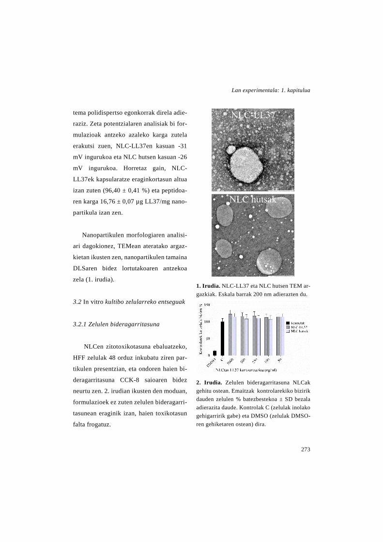
Lan experimentala: 1. kapitulua
273
tema polidispertso egonkorrak direla adie-
raziz. Zeta potentzialaren analisiak bi for-
mulazioak antzeko azaleko karga zutela
erakutsi zuen, NLC-LL37en kasuan -31
mV ingurukoa eta NLC hutsen kasuan -26
mV ingurukoa. Horretaz gain, NLC-
LL37ek kapsularatze eraginkortasun altua
izan zuten (96,40 ± 0,41 %) eta peptidoa-
ren karga 16,76 ± 0,07 µg LL37/mg nano-
partikula izan zen.
Nanopartikulen morfologiaren analisi-
ari dagokionez, TEMean ateratako argaz-
kietan ikusten zen, nanopartikulen tamaina
DLSaren bidez lortutakoaren antzekoa
zela (1. irudia).
3.2 In vitro kultibo zelularreko entseguak
3.2.1 Zelulen bideragarritasuna
NLCen zitotoxikotasuna ebaluatzeko,
HFF zelulak 48 orduz inkubatu ziren par-
tikulen presentzian, eta ondoren haien bi-
deragarritasuna CCK-8 saioaren bidez
neurtu zen. 2. irudian ikusten den moduan,
formulazioek ez zuten zelulen bideragarri-
tasunean eraginik izan, haien toxikotasun
falta frogatuz.
1. Irudia. NLC-LL37 eta NLC hutsen TEM ar-gazkiak. Eskala barrak 200 nm adierazten du.
2. Irudia. Zelulen bideragarritasuna NLCak gehitu ostean. Emaitzak kontrolarekiko bizirik dauden zelulen % batezbestekoa ± SD bezala adierazita daude. Kontrolak C (zelulak inolako gehigarririk gabe) eta DMSO (zelulak DMSO-ren gehiketaren ostean) dira.
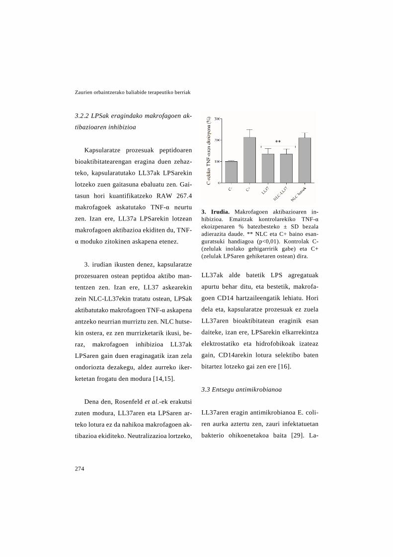
Zaurien orbaintzerako baliabide terapeutiko berriak
274
3.2.2 LPSak eragindako makrofagoen ak-
tibazioaren inhibizioa
Kapsularatze prozesuak peptidoaren
bioaktibitatearengan eragina duen zehaz-
teko, kapsularatutako LL37ak LPSarekin
lotzeko zuen gaitasuna ebaluatu zen. Gai-
tasun hori kuantifikatzeko RAW 267.4
makrofagoek askatutako TNF-α neurtu
zen. Izan ere, LL37a LPSarekin lotzean
makrofagoen aktibazioa ekiditen du, TNF-
α moduko zitokinen askapena etenez.
3. irudian ikusten denez, kapsularatze
prozesuaren ostean peptidoa aktibo man-
tentzen zen. Izan ere, LL37 askearekin
zein NLC-LL37ekin tratatu ostean, LPSak
aktibatutako makrofagoen TNF-α askapena
antzeko neurrian murriztu zen. NLC hutse-
kin ostera, ez zen murrizketarik ikusi, be-
raz, makrofagoen inhibizioa LL37ak
LPSaren gain duen eraginagatik izan zela
ondoriozta dezakegu, aldez aurreko iker-
ketetan frogatu den modura [14,15].
Dena den, Rosenfeld et al.-ek erakutsi
zuten modura, LL37aren eta LPSaren ar-
teko lotura ez da nahikoa makrofagoen ak-
tibazioa ekiditeko. Neutralizazioa lortzeko,
3. Irudia. Makrofagoen aktibazioaren in-hibizioa. Emaitzak kontrolarekiko TNF-α ekoizpenaren % batezbesteko ± SD bezala adierazita daude. ** NLC eta C+ baino esan-guratsuki handiagoa (p<0,01). Kontrolak C- (zelulak inolako gehigarririk gabe) eta C+ (zelulak LPSaren gehiketaren ostean) dira.
LL37ak alde batetik LPS agregatuak
apurtu behar ditu, eta bestetik, makrofa-
goen CD14 hartzaileengatik lehiatu. Hori
dela eta, kapsularatze prozesuak ez zuela
LL37aren bioaktibitatean eraginik esan
daiteke, izan ere, LPSarekin elkarrekintza
elektrostatiko eta hidrofobikoak izateaz
gain, CD14arekin lotura selektibo baten
bitartez lotzeko gai zen ere [16].
3.3 Entsegu antimikrobianoa
LL37aren eragin antimikrobianoa E. coli-
ren aurka aztertu zen, zauri infektatuetan
bakterio ohikoenetakoa baita [29]. La-
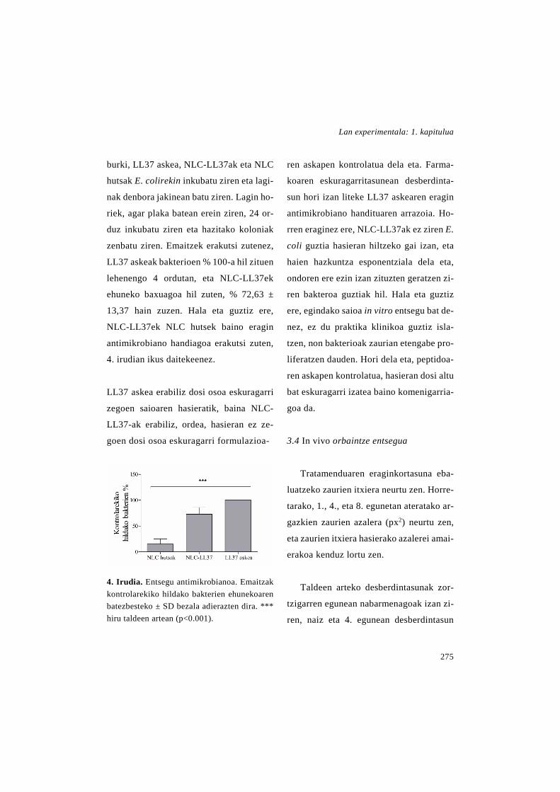
Lan experimentala: 1. kapitulua
275
burki, LL37 askea, NLC-LL37ak eta NLC
hutsak E. colirekin inkubatu ziren eta lagi-
nak denbora jakinean batu ziren. Lagin ho-
riek, agar plaka batean erein ziren, 24 or-
duz inkubatu ziren eta hazitako koloniak
zenbatu ziren. Emaitzek erakutsi zutenez,
LL37 askeak bakterioen % 100-a hil zituen
lehenengo 4 ordutan, eta NLC-LL37ek
ehuneko baxuagoa hil zuten, % 72,63 ±
13,37 hain zuzen. Hala eta guztiz ere,
NLC-LL37ek NLC hutsek baino eragin
antimikrobiano handiagoa erakutsi zuten,
4. irudian ikus daitekeenez.
LL37 askea erabiliz dosi osoa eskuragarri
zegoen saioaren hasieratik, baina NLC-
LL37-ak erabiliz, ordea, hasieran ez ze-
goen dosi osoa eskuragarri formulazioa-
4. Irudia. Entsegu antimikrobianoa. Emaitzak kontrolarekiko hildako bakterien ehunekoaren batezbesteko ± SD bezala adierazten dira. *** hiru taldeen artean (p<0.001).
ren askapen kontrolatua dela eta. Farma-
koaren eskuragarritasunean desberdinta-
sun hori izan liteke LL37 askearen eragin
antimikrobiano handituaren arrazoia. Ho-
rren eraginez ere, NLC-LL37ak ez ziren E.
coli guztia hasieran hiltzeko gai izan, eta
haien hazkuntza esponentziala dela eta,
ondoren ere ezin izan zituzten geratzen zi-
ren bakteroa guztiak hil. Hala eta guztiz
ere, egindako saioa in vitro entsegu bat de-
nez, ez du praktika klinikoa guztiz isla-
tzen, non bakterioak zaurian etengabe pro-
liferatzen dauden. Hori dela eta, peptidoa-
ren askapen kontrolatua, hasieran dosi altu
bat eskuragarri izatea baino komenigarria-
goa da.
3.4 In vivo orbaintze entsegua
Tratamenduaren eraginkortasuna eba-
luatzeko zaurien itxiera neurtu zen. Horre-
tarako, 1., 4., eta 8. egunetan ateratako ar-
gazkien zaurien azalera (px2) neurtu zen,
eta zaurien itxiera hasierako azalerei amai-
erakoa kenduz lortu zen.
Taldeen arteko desberdintasunak zor-
tzigarren egunean nabarmenagoak izan zi-
ren, naiz eta 4. egunean desberdintasun

Zaurien orbaintzerako baliabide terapeutiko berriak
276
esanguratsuak egon ziren ere. 5A. irudian
ikusten denez, azalera murrizketa nagusi-
ena NLC-LL37aren dosi altuenarekin tra-
tatutako saguetan gertatu zen. Zortziga-
rren egunean, talde horren azaleraren mu-
rrizketa gainontzeko taldeena baino esan-
guratsuki handiagoa izan zen. Laugarren
egunean, aldiz, kontrol taldearekin aldera-
tuta bakarrik izan zen esanguratsua des-
berdintasun hori. Emaitza horiek 5.B iru-
diko zaurien irudi makroskopikoetan be-
hatzen den azaleraren murrizketa kualita-
tiboarekin bat datoz, izan ere, txikiagotze
handiena NLC-LL37aren dosi altuarekin
ikusten zen.
NLC-LL37aren dosi baxuarekin eta
LL37 askearekin tratatutako taldeei dago-
kienez, antzeko zauri itxiera izan zuten,
nahiz eta lehenengoak azalera murrizketa
apur bat handiagoa izan zuen. Horretaz
gain, kontrol taldearekin alderatuz itxieran
hobekuntza lortu zuten, nahiz eta 8. egu-
nean bakarrik izan zen esanguratsua.
Zaurien itxieraz gain, tratamenduen
eraginkortasuna analisi histologikoen bi-
dez ebaluatu zen. Horretarako, laginak 8.
egunean batu ziren eta H&Erekin tindatu
ziren errepitelizazio gradua eta hanturaren
ebazpena aztertzeko asmoz.
5. Irudia. In vivo zauriaren itxiera. (A) Zaurien itxiera hasierako gaianazal azalerarekiko murriz-keta ehuneko bezala adierazita. * Tratatu gabeko taldearekiko esanguratsua. (p<0.05); ○ Tratatu gabeko taldearekiko esanguratsua. (p<0.05); + gainontzeko taldeekiko esanguratsua (p<0.05). (B) Zaurien irudiak.

Lan experimentala: 1. kapitulua
277
Errepitelizazioa gizakien orbaintze
prozesu nagusia da, karraskarietan aldiz,
prozesu nagusia zaurien uzkurtzea da.
Hala eta guztiz ere, hautatutako animalia
ereduan errepitelizazioak paper garrantzi-
tsua dauka, db/db saguek uzkurtze proze-
sua kaltetuta daukatelako, haien gizenta-
sunak azalaren malgutasuna txikitzen due-
lako Fang et al.-en arabera [30,31]. Gai-
nera, zaurien inguruan silikonazko eraztun
bana josi zirenez, zaurien uzkurdura are
gehiago oztopatu zen [25]. Guzti horren
ondorioz, errepitelizazioa prozesu garran-
tzitsua da animalia eredu honetan.
Errepitelizazioaren analisitik lortutako
emaitzek (6A. irudia) erakutsi zutenez,
NLC-LL37en dosi altuarekin tratatutako
zaurietan errepitelizazio gradua altuagoa
lortu zen. Sinha et al.-ek deskribatutako
eskalan, 2 inguruko balioa lortu zen bataz-
besteko, hau da, zauri gehienetan epitelio
berriak azaleraren erdia baino gehiago es-
taltzen zuen [26]. LL37 askearekin tratatu-
tako saguetan eta kontrol taldeko saguetan
errepitelizazioak zaurien azaleraren erdia
baino gutxiago estaltzen zuen (1 balioa),
eta NLC-LL37aren dosi baxuarekin trata-
tutako saguetan errepitelizazioa zaurien
ertzetan baino ez zen behatzen (~0). Emai-
tza hauek zaurien itxieran lortutakoekin
bat datoz, izan ere bi kasuetan NLC-
LL37aren dosi altuak orbaintzea sustatu
zuen, NLC-LL37aren dosi baxuago bate-
kin edo LL37 askearekin alderatuz gero.
Hanturaren ebazpenaren analisia Cotran
et al.-ek ezarri zuten irizpidearen arabera
burutu zen [27]. 6B. irudian ikusten denez,
NLC-LL37aren dosi altuarekin eta LL37
askearekin tratatutako zaurietan hantura-
ren ebazpena bizkortuta ageri zen, haietan
granulazio ehunaren sorrera eta angioge-
nesia nagusi baitziren, ia ez zeudelarik
mintz piogeniko ezta neutroliforik ere (2
gradua). Beste bi taldeetan (NLC-LL37en
dosi baxua eta kontrol taldea), ordea, zau-
riak beste fase batean zeuden, zeinean fi-
brina odolbatua sortzen ari zen eta leuko-
zitoak zein neutrofilo polimorfonuklearrak
zaurira migratzen zeuden (1 gradua). Az-
keneko talde hauetan behatzen den neutro-
filoen infiltrazio iraunkorra zauri kroni-
koen ezaugarri bereizgarria da, eta orbain-
tzea atzeratzea eragiten du, izan ere, neu-
trofilo horiek gehiegizko proteasa kopurua
jariatzen dute, matrize extrazelularra de-
gradatzen dutenak [32].

Zaurien orbaintzerako baliabide terapeutiko berriak
278
Aipagarria da, NLC-LL37aren dosi al-
tuarekin zein LL37 askearekin tratatutako
saguek hedatutako hantura akutuaren fa-
sean (2 gradua) egon arren, nanopartikulen
taldean lortutako balioa arinki altuagoa
zen, ondorioz, zauriaren heldutasunean
hobekuntza arina lortuz.
Orbaintzearen hasieran, granulazio ehu-
neko fibroblastoek III. motako kolagenoa
sintetizatzen dute, zeina apurka I. motako
kolagenoaz ordezkatua izaten den azal
osasuntsuaren fenotipoa berreskuratzeko.
Kolagenoa, eta batez ere I. motakoa, azal
osasuntsuaren pisu lehorraren % 80-a da,
6. irudia. Analisi histologikoa. (A) Errepitelizazio gradua. ** Gainontzeko taldeekiko esangura-
tsuki handiagoa (p>0.01). (B) Hanturaren ebazpenaren gradua. **NLC-LL37aren dosi baxua
baino esanguratsuki handiagoa (p<0.01); ● tratatu gabeko taldea baino esanguratsuki handiagoa
(p<0.05). (C) Kolageno metaketa. (D) Odol hodixka berrien zenbakia tindatutako ehun xafletan.
Emaitza denak batezbesteko ±SD bezala adierazita daude.
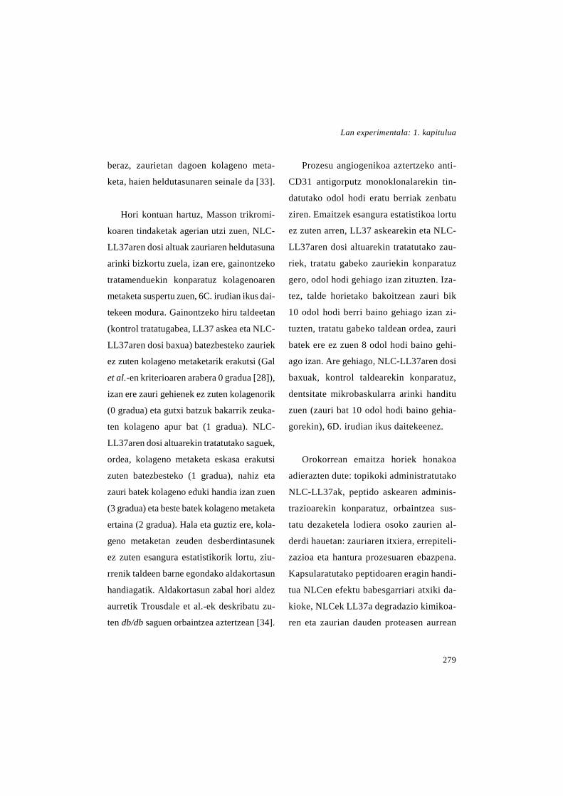
Lan experimentala: 1. kapitulua
279
beraz, zaurietan dagoen kolageno meta-
keta, haien heldutasunaren seinale da [33].
Hori kontuan hartuz, Masson trikromi-
koaren tindaketak agerian utzi zuen, NLC-
LL37aren dosi altuak zauriaren heldutasuna
arinki bizkortu zuela, izan ere, gainontzeko
tratamenduekin konparatuz kolagenoaren
metaketa suspertu zuen, 6C. irudian ikus dai-
tekeen modura. Gainontzeko hiru taldeetan
(kontrol tratatugabea, LL37 askea eta NLC-
LL37aren dosi baxua) batezbesteko zauriek
ez zuten kolageno metaketarik erakutsi (Gal
et al.-en kriterioaren arabera 0 gradua [28]),
izan ere zauri gehienek ez zuten kolagenorik
(0 gradua) eta gutxi batzuk bakarrik zeuka-
ten kolageno apur bat (1 gradua). NLC-
LL37aren dosi altuarekin tratatutako saguek,
ordea, kolageno metaketa eskasa erakutsi
zuten batezbesteko (1 gradua), nahiz eta
zauri batek kolageno eduki handia izan zuen
(3 gradua) eta beste batek kolageno metaketa
ertaina (2 gradua). Hala eta guztiz ere, kola-
geno metaketan zeuden desberdintasunek
ez zuten esangura estatistikorik lortu, ziu-
rrenik taldeen barne egondako aldakortasun
handiagatik. Aldakortasun zabal hori aldez
aurretik Trousdale et al.-ek deskribatu zu-
ten db/db saguen orbaintzea aztertzean [34].
Prozesu angiogenikoa aztertzeko anti-
CD31 antigorputz monoklonalarekin tin-
datutako odol hodi eratu berriak zenbatu
ziren. Emaitzek esangura estatistikoa lortu
ez zuten arren, LL37 askearekin eta NLC-
LL37aren dosi altuarekin tratatutako zau-
riek, tratatu gabeko zauriekin konparatuz
gero, odol hodi gehiago izan zituzten. Iza-
tez, talde horietako bakoitzean zauri bik
10 odol hodi berri baino gehiago izan zi-
tuzten, tratatu gabeko taldean ordea, zauri
batek ere ez zuen 8 odol hodi baino gehi-
ago izan. Are gehiago, NLC-LL37aren dosi
baxuak, kontrol taldearekin konparatuz,
dentsitate mikrobaskularra arinki handitu
zuen (zauri bat 10 odol hodi baino gehia-
gorekin), 6D. irudian ikus daitekeenez.
Orokorrean emaitza horiek honakoa
adierazten dute: topikoki administratutako
NLC-LL37ak, peptido askearen adminis-
trazioarekin konparatuz, orbaintzea sus-
tatu dezaketela lodiera osoko zaurien al-
derdi hauetan: zauriaren itxiera, errepiteli-
zazioa eta hantura prozesuaren ebazpena.
Kapsularatutako peptidoaren eragin handi-
tua NLCen efektu babesgarriari atxiki da-
kioke, NLCek LL37a degradazio kimikoa-
ren eta zaurian dauden proteasen aurrean
Zaurien orbaintzerako baliabide terapeutiko berriak
280
babesten baitute. Horretaz gain, NLCek
askapen profil kontrolatua daukate, ho-
rrela LL37aren eragina luzatuz [20,21].
Hori dela eta, NLC-LL37ak erabiliz, pep-
tido askea administratzean beharrezkoa
den dosi maiztasun handia murriztu dai-
teke.
Aldez aurretik egindako ikerketek
LL37a kapsularatzearen erabilgarritasuna
frogatu dute [19], dena den, oraingo iker-
keta honetan LL37a duten nanopartikulak
bide topikotik administratuta dira lehen al-
diz, zeinak abantaila ugari dauzkan. Lehe-
nik, NLCen izaera lipidikoak geruza oklu-
sibo bat eratzen du, azalaren hidratazioa
handituz eta kapsularatutako LL37aren
barneratzea lagunduz [21,35]. Gainera, ad-
ministrazio topikoa gaixoarentzat bide
erraza, erosoa eta segurua da.
4. Ondorioak
Ikerketa honetan %2 LL37 zuten
NLCak garatu ziren. Haien batezbesteko
neurria 270 nm-koa zen eta ez zuten zito-
toxizkotasunik erakutsi. Kapsularatutako
peptidoak bere bioaktibitatea mantentzen
zuen kapsularatze prozesuaren ostean, in
vitro egindako saio immunomodulatzai-
lean eta antimikrobianoan ikusi zenez.
Db/db saguetan egindako lodiera
osoko orbaintze in vivo entseguan frogatu
zen, orbaintzean eragin handiagoa zuela
LL37aren 6 µg NLCen barne topikoki ad-
ministratzeak, aske administrazeak baino.
Aurkikuntza guztiak kontuan hartuz, NLC-
LL37en administrazio topikoa zauri kroni-
koen tratamendurako estrategia interesga-
rria izan daitekeela ondoriozta daiteke.
5. Eskerrak
I. Garcia-Oruek Eusko Jaurlaritzari
doktoratu aurreko laguntza eskertzen dio.
Egileek UPV/EHU-ko SGIkerren laguntza
teknikoa, giza babesa eta Europako finan-
tziazioa (FEDER eta FSE) eskertzen di-
tuzte. Proiektu hau neurri batean Espaini-
ako Ekonomia eta Lehiakortasun Ministe-
rioak finantziatu du (INNPACTO, IPT-
2012-0602-300000, 2012) eta baita, Eusko
Jaurlaritzak (Talde finkatuak, IT-407-07
eta IT-528-10) eta Euskal Herriko Uniber-
tsitateak ere (UFI11/32).
Erreferentzien zerrenda 79-81 orrialdeetan aurkitzen da.
2. KAPITULUA
rhEGF eta Aloe vera dituzten nanozuntzezko apositu berriak zaurien orbaintzean erabiltzeko.
LABURPENA
Elektroirutearen bidez ekoiztutako nanozuntzezko mintzek bolumenaren araberako gai-nazal azalera handia dute, zeinak matrize extrazelularraren (extracellular matrix edo ECM) hiru-dimentsioetako egitura imitatzen duen. Hori dela eta, nanozuntzezko aposi-tuak zauri kronikoen orbaintzerako aukera itxaropentsua dira, izan ere, ECM naturala ordezkatu dezakete zauria sendatzen den bitartean. Ondorioz, ikerketa honetan PLGAz eginiko nanozuntzezko mintz bat garatu dugu, zeinak giza hazkuntza faktore epidermal errekonbinantea (recombinant human Epidermal Growth Factor edo rhEGF) eta Aloe veraren (AV) erauzkina dituen. Bi konposatuek orbaintzea sustatzen dute, alde batetik, EGFa orbaintzearen bitartekaria delako, eta bestetik, AVa fibroblastoen proliferazioa eta aktibitatea estimulatzen dituelako. Lortutako mintzak ausaz orientatutako zuntz unifor-mez osatuta zeuden, haien batazbesteko diametroa 356,03 ± 112,05 nm-ko zen, porosi-tatea %87,92 ± 11,96-koa zen eta rhEGF edukia 9,76 ± 1,75 µg/mg-koa zen. In vitro bideragarritasun entseguak erakutsi zuenez, rhEGFa eta AVa zuten mintzak fibroblas-toen proliferazioa hobetzeko gai ziren, haien elkarketa onuragarria dela frogatuz. Are gehiago, mintz hauek zaurien itxiera eta errepitelizazioa era esanguratsuan bizkortu zu-ten, db/db saguetan egindako lodiera osoko zauri eszisionaletan. Orokorrean, aurkikun-tza hauek rhEGFa eta AVa kapsularatuta dituzten PLGA nanozuntzen erabilgarritasuna erakusten dute zauri kronikoen orbaintzean erabiltzeko.


Lan experimentala: 2. kapitulua
283
1. Sarrera
Orbaintzea prozesu dinamiko eta kon-
plexua da, zeinaren helburua azalaren egi-
tura anatomiko eta funtzionaltasun norma-
lak berreskuratzea den (Diegelmann eta
Evans 2004). Helburu hori lortzeko as-
moz, zenbait sistema biologikok eta im-
munologikok era koordinatuan parte har-
tzen dute orbaintzea osatzen duten 3 fase
bereizi baina gainjarrietan. Fase horiek
hanturazko erantzuna (non hemostasia eta
hantura bereiz daitezkeen), fase prolifera-
tiboa (zeinean proteinen sintesia eta zauri-
aren uzkurdura gertatzen diren) eta erre-
modelazio fasea dira (Monaco eta Law-
rence 2003; Morton eta Phillips 2016; Sch-
reml, et al. 2010). Hala eta guztiz ere, zen-
bait zaurik huts egiten diote denboran zein
espazioan zehar antolatutako prozesu ho-
rretan zehar igarotzeari. Orbaintzearen
asaldura zenbait faktorek eragin dezakete,
hala nola, infekzioek, nekrosiak, ehunen
hipoxiak, exudatuak eta gehiegizko hantu-
razko zitokinak. Horren ondorioz, zauriak
hanturazko fase iraunkor batean harrapa-
tuta geratzen dira. Fase horretan ezohiko
zitokinen maila altua egoten da, zeinak
matrize extrazelularraren (extracellular
matrix edo ECM) hondaketa eragiten
duen, sendaketa are gehiago kaltetuz (Bri-
quez, et al. 2015; Velnar, et al, 2009).
Gure gizartean, zauri kronikoen trata-
menduak garrantzia handia lortu du, azken
urteotan gertatu den prebalentziaren igo-
era kezkagarria dela eta. Gainera, preba-
lentziak igotzen jarraituko duela uste da,
arrisku handiko biztanleria hazten ari de-
lako, haien artean, adindunak, diabetikoak
eta obesoak daudelarik (Whittam, et al.
2016). Izatez, garatutako herrialdeetan
biztanleriaren % 1-2-ak zauri kronikoak
pairatuko dituela zenbatetsi da, Nazio Ba-
tuen arabera, haiei elkartutako kostuak Eu-
ropako osasun aurrekontuaren %2-a dire-
larik (Sen, et al. 2009). Arazo kliniko hori
gainditzeko egiten ari diren ikerketak, or-
baintzea sustatzeko gai diren aposituen ga-
rapenera bideratuta daude. Apositu horien
artean, ehundu gabeko nanozuntzezko
mintzak daude, zeintzuengan arreta bere-
zia jarrita dagoen, haien egiturak ECMa-
ren hiru dimentsioetako egitura imitatzen
dutelako (Choi, et al. 2015).
Nanozuntzak orokorrean elektroirutea-
ren bitartez ekoizten dira, teknika sinple,

Zaurien orbaintzerako baliabide terapeutiko berriak
284
kostu-efektibo eta erabilera anitzekoa
dena. Teknika honekin disoluzio polime-
riko batetatik zuntz nanometrikoak ardaz-
katzen dira indar elektrostatikoen bidez
(Abrigo, et al. 2014; Gainza, et al. 2015b).
Horrela lortutako nanozuntzak bolumena-
ren araberako gainazal azalera eta porosi-
tate handia dute, horrek orbaintzea meka-
nismo desberdinen bidez sustatzen duela-
rik. Adibidez, gasen sarrera ahalbidetzen
dute, zelulen arnasketa ahalbidetuz; eta
hezetasuna mantentzeko gai dira, exuda-
tuen irteera erraztuz (Pachuau 2015). Apo-
situek zelulen migrazioa eta proliferazioa
sustatu beharko lukete, baina aldi berean,
ehuna nanozuntzezko mintzaren gain haz-
tea ekidin, apositu hauek zauria orbain-
tzean kentzeko diseinatuta baitaude
(Abrigo, et a. 2014). Horretaz gain, nano-
zuntzak konposatu aktiboak barneratzeko
gai dira orbaintzea bizkortzeko asmoz. Es-
trategia hori hainbat ikerketetan erabilia
izan da, besteak beste honako farmakoak
kapsularatuz: hazkuntza faktoreak, Aloe
vera extraktua, kurkumina, metformina,
ziprofloxazinoa eta kolagenoa (Choi, et al.
2008; Kataria, et al. 2014; Lai, et al. 2014;
Lee, et al. 2014; Merrell, et al. 2009; Uslu
and Aytimur 2012). Gainera, nanozuntze-
tan kapsularatzeak farmakoen administra-
zio topikoa ahalbidetzen du, abantaila
ugari daukana. Lehenik eta bien, gaixoek
hobeto onartzen dituzte tratamendu topi-
koak, nork bere buruari administra die-
zaizkiokelako; bigarrenez, tratamenduaren
ahalmen terapeutikoa hobetzen dute, far-
makoaren bioerabilgarritasuna eta zaurian
mantentzea handitzen baitituzte; eta azke-
nik farmakoaren toxikotasun sistemikoa
txikitzen dute (Pachuau 2015; Whittam, et
al. 2016).
Elektroirutearen bitartez lortutako na-
nozuntzezko mintzak zeluletan oinarritu-
tako terapietan erabiliak izan dira ere.
Mintzen eta ECMaren arteko antzekotasu-
nak zelulen sarrera, desberdintzatzea eta
atxikipena ahalbidetzen ditu, ordezko aza-
lak edo zelula amen terapiak garatzeko
aproposak egiten dituena. Ordezko azalak
azal osasuntsuaren egitura antzekoa duten
zeluladun bio-aldamioak dira (Dickinson
adn Gerecht 2016). Zelula-matrize elka-
rrekintzak hobetzeko asmoz, bio-aldami-
oak kolagenoz edo bestez ECMko protei-
nez ekoizten dira (Hodgkinson and Bayat
2011). Horren harira, Powell et al.-ek
elektroirundako kolageno bio-aldamio bat
Lan experimentala: 2. kapitulua
285
garatu zuten, zeinean gizakien fibroblasto
dermalak eta keratinozito epitelialak erein
zituzten ( Powell, et al. 2008). Beste iker-
keta batean, Han et al.-ek PHVB polimero
naturalean oinarritutako bio-aldamio bat
garatu zuten, zeinean ileko zelula folikula-
rrak erein zituzten, haien immunogenizi-
tate baxuak alo-transplanteetarako aban-
tailatsu egiten baititu (Han, et al 2007).
Beste aldetik, elektroirundako mintzetan
zenbait zelula ama mota finkatu dira apo-
situak garatzeko, zelula horien artean hu-
rrengoak daudelarik: gantzetik eratorri-
tako zelula amak (Gu, et al. 2014; Ma-
chula, et al. 2014), zelula ama mesenkima-
lak (Bhowmick, et al. 2016; Steffens, et al.
2014), murriztu gabeko zelula ama soma-
tikoak (Bahrami, et al. 2016, Keshel, et al.
2014) eta gernutik eratorritako zelula
amak (Fu, et al. 2014). Zelula ama horiek
orbaintzea hobetzeko gai direla frogatu
dute, zelula endotelialetara desberdintza-
tzeko duten gaitasunagatik eta baita
ECMko proteinak zein orbaintzearen bi-
tartekariak expresatzeko duten gaitasuna-
gatik ere.
Aloe vera extraktua (AV) apositutzat
erabiltzeko nanozuntzetan kapsularatu
daitekeen konposatu bat da. Aloe vera
hosto zukutzuak dituen Liliaceae famili-
ako landare bat da. Bere aktibitate farma-
kologikoaren barne, eragin anti-inflamato-
rioak, antiartritikoak, antibakterianoak,
antifungikoak eta hipogluzemikoak daude,
baina horietaz gain, orbaintzea sustatu de-
zakela ikusi da ere (Choi and Chung, 2003,
Hashemi, et al. 2015). Orbaintzean duen
ekintza, glukomanano deritzon konposatu-
aren bidez gauzatzen du batez ere, honek
fibroblastoen hazkuntza faktorean eragina
baitu, flibroblastoen aktibitatea eta proli-
ferazioa sustatuz eta zelula hauen kola-
geno ekoizte eta jariapena handituz (Ha-
shemi, et al. 2015).
Hazkuntza faktoreak nanozuntzezko
aposituetan kapsularatzea interesgarria da,
izan ere, orbaintzearen ezinbesteko bitar-
tekariak dira (Choi, et al. 2012). Horien ar-
tean, hazkuntza faktore epidermikoa (epi-
dermal growth factor edo EGF) dago,
zeina seinalizazio molekula gakoa den eta
fibroblasto eta keratinozitoen proliferazio
eta migrazioa estimulatzen dituen, orbain-
tzea sustatuz (Bodnar 2013). Horretaz
gain, zauri kronikoetan administratu os-
tean orbaintzea bultzatzen duela ikusi da
Zaurien orbaintzerako baliabide terapeutiko berriak
286
(Choi, et al. 2008, Gainza, et al. 2015a).
Hala ere, EGFaren erabilera klinikoak
arazo ugari ditu, bere erdibizitza laburra
dela eta, zauriko entzima hidrolitikoek de-
gradatzen baitute. Nanozuntzezko mintze-
tan kapsularatuz muga horiek gainditu dai-
tezke, formulazio hauen barnean EGFa in-
gurunetik babestuta egongo litzatekee-
lako, bere egonkortasuna hobetuz
(Pachuau, 2015).
Nanozuntzezko mintzak polimero na-
tural zein sintetikoak erabiliz ekoiztu dai-
tezke. Elektroirutean erabilitako polimero
naturalak ECMko osagaiak izaten dira as-
kotan, hala nola, kolagenoa, laminina, fi-
brina edo elastina, baina beste polimero
batzuk izan daitezke ere, besteak beste, ki-
tosanoa, agarosa, gelatina, pektina, zeta,
etab. Polimero horiek orbaintzean erabil-
tzeko egokiak dira honako ezaugarriak di-
rela eta: biobateragarritasuna, toxikotasun
eza, ezaugarri fisikokimiko aproposak eta
matrize-zelula elkarrekintzak (Garg, et al.
2015). Hala eta guztiz ere, haien ezaugarri
mekaniko ahulak eta elektroiruteko dituz-
ten zailtasunak direla eta, askotan poli-
mero sintetikoen bidez sendotu egiten dira
(Hodgkinson and Bayat, 2011). Elektroi-
rutean erabilitako polimero sintetikoen ar-
tean, zelulosa azetatoa, azido polilaktiko-
ko-glikolikoa (PLGA), alkohol polibinili-
koa, azido polilaktikoa, poliestirenoa, po-
liglizerola, azido poliglikolikoa, poliureta-
noa, etab. daude. Material hauek sendoa-
goak eta merkeagoak dira, ondo zehaztu-
tako egitura daukate eta errazago elektroi-
ruten dira (Garg, et al. 2015).
Esandako guztia kontuan hartuz, lan
honetan PLGA elektroirun zen rhEGFaz
eta AVaz kargatutako nanozuntzezko apo-
situ bat garatzeko. Mintzak karakterizatu
ziren orbaintzean erabiltzeko zuten egoki-
tasuna aztertzeko, eta ondoren, in vitro sai-
oak burutu ziren kapsularatutako konposa-
tuen bioaktibitatea ebaluatzeko.
Horretaz gain, aposituen eraginkorta-
suna in vivo aztertu zen. Laborategiko ani-
malien artean, ziurrenik txerrien azala da
gizakien azalaren antzekoena, baina ani-
malia handiekin lan egiteko zailtasunenga-
tik karraskarien ereduak askoz erabilia-
goak dira (Seaton, et al. 2015). Baina, esan
bezala, karraskarien orbaintzeak ez du ho-
rren ondo imitatzen du gizakiena, haien
azal soltea dela eta, duten sendatze meka-

Lan experimentala: 2. kapitulua
287
nismo nagusia zauriaren uzkurtzea baita.
Desberdintasun hori ekiditeko asmoz, zau-
riak leku anatomiko zehatzetan egiten
dira, hala nola buruan, belarrietan edo buz-
tanean, non azala tinkoago dagoen. Arazo
hau gainditzeko beste aukera bat zaurien
inguruan silikonazko eraztunak jostea da,
horrela uzkurdura saihesteko. Azken eredu
honek gizakien zaurien antzekotasun
gehien duen karraskarien eredua da (Fang
and Mustoe, 2008).
Zauri kronikoetan gertatzen den senda-
ketaren atzerapena imitatzeko asmoz, or-
baintzea kaltetuta duten karraskarien ere-
duak erabili daitezke, hala nola, eredu is-
kemikoak (Elgharably, et al. 2014; Magin,
et al. 2016) edo diabetikoak. Eredu diabe-
tikoen artean erabilienak kimikoki eragin-
dako diabetea dutenak edo animalia gene-
tikoki diabetikoak dira. Kimikoki diabetea
estreptozotozina injekzioen bidez eragiten
da, konposatu horrek areko β-zelulak hon-
datzen baititu, I. motako diabetesaren an-
tzeko fenotipo bat lortuz (Inpanya, et al.
2012; Moura, et al. 2014). Genetikoki dia-
betikoak diren animaliak db/db saguak
dira (BKS.Cg-m +/+ Leprdbj/J), zeintzuek
leptinaren hartzailean mutazio bat duten,
eta horren ondorioz II. motako diabetearen
antzeko fenotipoa duten (Guillemin, et al.
2016; Losi, et al. 2013). Michaels et al.-ek
burutako ikerketa batean, orbaintzea db/db
saguetan estreptozotozina ereduan baino
kaltetuagoa zegoela ikusi zen (Michaels,
et al. 2007).
Hori dela eta, ikerketa honetan garatu-
tako formulazioen eraginkortasuna azter-
tzeko, db/db sagu ereduan zauri eszisiona-
lak egin ziren eta haien inguruan siliko-
nazko eraztunak josi ziren. Orbaintzea
ebaluatzeko, zaurien itxiera, errepiteliza-
zio gradua, hanturaren ebazpena eta kola-
geno metaketa aztertu ziren.
2. Material eta metodoak
2.1 Nanozuntzen prestaketa
rhEGF, AV eta PLGA zituen o/w emul-
tsio bat elektroirun zen PLGA-AV-EGF
nanozuntzezko mintza eratzeko. Emultsio-
aren fase organikoa 140 mg PLGA 50:50
(Resomer RG504, Evonik, Alemania) zi-
tuen hexafluoroisopropanolaren 650 µL-
tan (HFIP, Fluka, Suitza). Fase urtsua, al-
diz, 140 mg Aloe vera hauts (Agora Va-

Zaurien orbaintzerako baliabide terapeutiko berriak
288
lencia SL., Espainia) eta 5 mg rhEGF (Cen-
tre for Genetic Engineering and Biotechno-
logy, Cuba) zituen % 0,5-eko (p/b) alkohol
polibiniliko disoluzio baten barne. Bi fa-
seak 3 minutuz nahastu ziren potentzia ma-
ximoan emultsioa eratzeko (Vortex-Genie
2, Scientific Industries Inc., AEB).
Lortutako emultsioa Luer-lock xiringa
(Norm-Ject) batean kargatu zen, zeinak 18
G-ko orratz bat zeukan. Xiringa ponpa ba-
tera atxiki zen, 1ml/h-ko emaria eragiten
zuenak, horrela elektroirutea abiaraziz.
Emultsioa horizontalki elektroirun zen 12
kV-ko korrontearen bidez eta orratzaren
12 cm-tara zegoen kolektore birakari ba-
tean jaso zen (200 r/min).
PLGA-AV eta PLGA nanozuntzen
prestaketa, azaldutakoaren oso antzekoa
izan zen, desberdintasun bakarra fase urtsu-
aren osagaietan zegoelarik. PLGA-AV na-
nozuntzek ez zuten rhEGFrik eta PLGA na-
nozuntzek ez zuten rhEGF ezta AVrik ere.
Antzeko lodierako mintzak lortzeko injek-
tatutako bolumenak normalizatu egin ziren.
Mintzak ekoizteko erabilitako lehen-
gaiak gamma erradiazioaren bidez esterili-
zatu ziren erabili aurretik. Beste aldetik,
elektroirutean erabilitako xiringa, kolekto-
rea eta orratza autoklabatu egin ziren, eta
are gehiago, elektroirute prozesua baldin-
tza aseptikoetan burutu zen. Azkenik, in
vitro eta in vivo ikerketen aurretik mintzak
esterilizatu egin ziren, 30 minutuz UV ar-
gitan mantenduz.
2.2 Nanozuntzen karakterizazioa
Nanozuntzezko mintzen morfologia,
hau da, mintzen kalitatea eta zuntzen dia-
metroa, ekorketazko mikroskopio elektro-
nikoko irudien bidez aztertu zen (SEM,
Jeol JSM-6300). Haien porositatea (P) hu-
rrengo ekuazioaren bidez kalkulatu zen
(Ek. 1), non ρreal and ρapp dentsitate erreala
eta itxurazkoa diren, hurrenez hurren:
P (%)=�1 - ρapp
ρreal� x 100 (1)
Dentsitate erreala helio piknometro ba-
ten bidez neurtu zen (Micromeritics,
AccuPyc 1330, AEB), itxurazko dentsita-
tea, berriz, pisatutako masa eta kalkulatu-
tako bolumena (Luzera × zabalera × altu-
era) zatituz kalkulatu zen. Mintzaren lodi-

Lan experimentala: 2. kapitulua
289
era estereo mikroskopio (Leica M205 C.
Leica LAS, Alemania) baten bitartez lor-
tutako argazkietatik neurtu zen.
Mintzen ezaugarri mekanikoen probak
desplazamendu kontrolaren arabera egin
ziren 5 N-eko eskala osoko karga zelula
bat erabiliz Instron 5848 Microtester eki-
poan (Instron®, EB). Laginak 0,01 mm/s-
ko desplazamendu abiaduraz luzatu ziren
apurtu arte. Ondoren, karga-desplaza-
mendu kurbak tentsio-deformazio kurbe-
tan bihurtu ziren, eta hortik erresistentzia
maximoa atera zen, nanozuntz desberdi-
nen ezaugarri mekanikoak alderatzeko.
Nanozuntz bakoitzeko gutxienez 5 lagin az-
tertu ziren eta emaitzak batezbesteko ± des-
bideraketa estandar bezala aurkeztu ziren.
Nanozuntzek inguru urtsu batean ura
xurgatzeko zuten ahalmena in vitro aztertu
zen, 3 mintzen jokaera alderatzeko asmoz.
Nanozuntzen 1x1 cm-ko laginak pisatu on-
doren, PBS 1 ml-tan murgildu ziren eta
32°C-tara inkubatu ziren ordu batez. On-
doren, PBStik atera ziren, gehiegizko ura
lehortu zen eta berriro pisatu ziren. Hu-
rrengo ekuazioa erabiliz mintzek xurgatu-
tako ura kalkulatu zen (Ek. 2):
Ur xurgapena (%)=M-M0
M0×100 (2)
Non, M nanozuntzen amaierako pisua
den eta M0 nanozuntzen hasierako pisua
den.
Nanozuntzen ur-lurrunaren iragazkor-
tasun abiadura (Water Vapour Permeabi-
lity Rate edo WVPR) Li et al.-ek deskri-
batutako metodoa moldatuz burutu zen
(Li, et al. 2013). Silika gel lehorgarria
zuen ontzi baten ahoa (1 cm diametro) az-
tergai zegoen mintzarekin erabat estali
zen, ur-lurruna mintzean zehar bakarrik
sar zitekeelarik ontzi barnera. Ontzia 30°C
eta % 75-eko hezetasun erlatibo konstan-
tea zuen ganbera batean 24 orduz man-
tendu zen. Muntaiaren hasierako eta amai-
erako pisuak eta honako ekuazioa erabiliz
WVPRa kalkulatu zen (Ek. 3):
WVPR=M1-M0
A×T (3)
Non, M0 saioaren hasieran muntaiak
zuen pisua den, M1 saioaren amaieran zuen
pisua, A mintzaren esposizio azalera den
eta T esposizio denbora.
Zaurien orbaintzerako baliabide terapeutiko berriak
290
Nanozuntzen, haien osagaien nahaste
fisikoen eta lehengaien jokabide termikoa
ekorketazko kalorimetria diferentzialaren
(differential scanning calorimetry edo
DSC) bidez aztertu zen (DSC-50, Shi-
madzu, Japonia). Lagin bakoitza alumini-
kozko platertxo batean hermetikoki itxi
zen eta 25°C-tatik 350°C-tara berotu zen,
10°C/minutuko abiaduraz.
PLGA-AV-EGF nanozuntzen barnean
kargatutako rhEGF kantitatea neurtzeko
hurrengo prozedura erabili zen. Lehenik
mintzaren 1x1 cm-ko zati bat dimetil sul-
foxidoaren (DMSO, Scharlau, Espainia)
200 µL-tan disolbatu zen, disoluzio gar-
dena lortu arte irabiatuz eta ondoren
PBSaren (Life thecnologies, California,
AEB) 800 µL-rekin diluituz. Lortutako di-
soluzio horretan zegoen rhEGF kantitatea
giza EGFarentzako ELISA kit komertzial
bat erabiliz neurtu zen, ekoizlearen jarrai-
bideak jarraituz (human EGF ELISA deve-
lopment kit, Peprotech, AEB).
2.3 rhEGFaren in vitro askapena
Askapen entsegua Franz difusio gelax-
ketan burutu zen. 1x1 cm-ko PGA-AV-
EGF nanountz mintz zati bat (9,76 µg
rhEGF zituena) 30 µL PBSrekin hezetu
zen eta ganbera emailean kokatu zen, 0,45
µm-ko poro tamaina zuen nylonezko ira-
gazki baten gainean (Tecnokroma®, Espai-
nia). Ganbera hartzailean 5 ml PBS jarri
ziren. Sistema irabiatzen mantendu zen eta
aukeratutako denbora tarteetan ganbera
hartzailearen 1 ml hartu zen eta PBS berri-
arekin ordezkatu. Ingurunera askatutako
rhEGF kopurua 2.2 atalean azaldutako
ELISA protokoloa erabiliz neurtu zen.
Saioa hiru aldiz burutu zen eta emaitzak
denboran zehar metatutako rhEGF ehu-
neko modura adierazi ziren.
2.4 In vitro entsegu antibakterianoa
PLGA-AV mintzaren eragin antimi-
krobianoa Staphilococcus aureus eta Sta-
philococcus epidermidisen aurrean aztertu
zen. Laburki, bakterio bakoitzaren 105
CFU (unitate kolonia eratzaile edo colony
forming unit)/mL zituen disoluzio banaren
100 µL Mueller Hinton agar plaketan he-
datu ziren. PLGA eta PGA-AV nanozun-
tzak 6 mm-ko disko biribiletan moztu zi-
ren. 6 mm-ko disko txurietan 10 µL AV-
aren estraktu urtsua kargatu zen (30
Lan experimentala: 2. kapitulua
291
mg/mL) kontrol bezala erabiltzeko. Lagi-
nak eta kontrolak plaketan ezarri ziren eta
37°C-tara inkubatu ziren 36 orduz. Azke-
nik, laginen inguruan inhibizio halorik ze-
goen behatu zen.
2.3 In vitro kultibo zelularreko entseguak
2.3.1 Kultibo zelularra
BalbC/3T3 A31 fibroblastoak (ATCC,
Manassas, AEB) DMEM ingurunean (dul-
beccos’s modified Eagle’s medium;
ATCC, Manassas, AEB) kultibatu ziren,
eta inguruneari honako gehigarriak jarri
zitzaizkion: % 10-eko (b/b) zekor seruma
eta % 1-eko (b/b) penizilina-estreptomi-
zina. Zelulak 37°C-tan mantendu ziren, %
5-eko CO2 atmosferadun hezetutako inku-
bagailu batean. Zelula paseak 2-3 egunetik
behin egin ziren.
2.3.2. Bioaktibitate entsegua
PLGA-AV-EGF, PLGA-AV eta PLGA
nanozuntzen erauzkinek zelulen bideraga-
rritasunean duten eragina fibroblastoetan
aztertu zen. Zelulak 96 putzuko plaka ba-
tean erein ziren, 6000 zelula/putzu dentsi-
tatearekin eta gauean zehar inkubatu ziren
zelulen atxikimendua ahalbidetzeko.
Orduan, honako laginak gehitu ziren
putzuetara: (i) kultibo ingurunea kontrol
negatibo bezala, (ii) 20 ng/ml rhEGF as-
kea, (iii) 20 ng/ml rhEGFren baliokidea
den PLGA-AV-EGF nanozuntzen erauz-
kina, (iv) PLGA-AV nanozuntzen erauz-
kina eta (v) PLGA nanozuntzen erauzkina.
48 orduko inkubazioaren ostean zelulen
bideragarritasuna CCK-8 kita erabiliz
neurtu zen (cell counting kit-8, sigma-Al-
drich, AEB). Laburki, CCK-8 erreaktiboa-
ren 10 µL zeluletara gehitu ziren eta na-
hastea 4 orduz inkubatu zen. Orduan, ab-
sorbantzia 450 nm-tara irakurri zen, 650
nm erreferentziazko uhin luzera modura
erabiliz (Plate Reader Infinite M200, Te-
can, Suitza). Lortutako absorbantzia bizi-
rik zeuden zelulei zuzenki proportzionala
zen.
2.5.3 Atxikipen entsegua
Zelulen atxikipena nanozuntzezko
mintzetara aztertzeko asmoz, BalbC/3T3
A31 fibroblastoak mintzen gainean erein
ziren eta lotutako zelulak zenbatu ziren.

Zaurien orbaintzerako baliabide terapeutiko berriak
292
Laburki, PLGA, PLGA-AV eta PLGA-
AV-EGF mintzen 1x1 cm zatiak 96 pu-
tzuko plaka batean finkatu ziren, itsasgarri
bezala fibrinaren 10 µL erabiliz. Odolba-
tua eratzea ahalbidetzeko nanozuntzak 30
minutuz inkubatu ziren 37°C-tara, eta on-
doren 15000 zelula erein ziren putzu ba-
koitzean. Kontrol putzuetan ere 15000 ze-
lula erein ziren kultibo ingurune osoan.
Zelulak gauean zehar inkubatu ziren
37°C-tara zelulen atxikipena ahalbide-
tzeko. Ondoren, mintzak PBSarekin gar-
bitu ziren eta atxikitako zelulak 10 minu-
tuz tripsina-EDTaren (Lonza, Suitza) 50
µL-rekin inkubatuz askatu ziren. Azkenik,
tripsina batu zen eta zelulak kontagailu au-
tomatiko bat erabiliz zenbatu ziren (Auto-
mated Cell Counter TC20™, Bio-Rad, Ca-
lifornia, AEB). Atxikipena adierazteko
kontrolarekin alderatuz mintz bakoitzaren
gainean zenbatutako zelulen ehunekoa era-
bili zen. 3 saio independente burutu ziren.
Horretaz gain, SEM argazkiak atera zi-
ren zelulen atxikipena ikusteko. Kasu ho-
netan, gauean zeharreko inkubazioaren os-
tean zelulak %2,5-eko glutaraldehido di-
soluzio batekin fixatu ziren eta etanol serie
graduatuak erabiliz deshidratatu zen. Az-
kenik, irudiak SEM mikroskopio bat era-
biliz atera ziren (Hitachi S4800, Tokio, Ja-
ponia).
2.6 Zauriaren orbaintze in vivo entsegua
2.5.1 Animaliak
In vivo entsegurako 20 db/db (BKS.Cg-
m+/+Leprdb/J) sagu ar (8 asteko adine-
koak) erabili ziren (Janvier laborategiak,
Frantzia). Saio guztiak Euskal Herriko
Unibertsitateko Animaliekin egiten den
Esperimentaziorako Etika Batzordeak
onartutako protokoloak jarraituz burutu zi-
ren (Prozedura zenbakia: CEBA/243/
2012/HERNANDEZ MARTIN). Anima-
liak banaka ostatatu ziren kaioletan, 12 or-
duko argi-ilun zikloan. Saguek karraskari
pentsua zein ura nahierara izan zuten.
2.5.2 Orbaintze entsegua
Orbaintze entsegua Michaels et al.-ek
(Michaels, et al. 2007) deskribatutako pro-
zedura jarraituz burutu zen. Saguak iso-
fluranoarekin (Isoflo, Esteve, Espainia)
anestesiatu eta bizkarreko ilea kendu os-
Lan experimentala: 2. kapitulua
293
tean, bizkarraren bi aldeetan 1 cm-ko dia-
metroko silikona eraztun bana josi zitzaien
3-0 nylon hari bat erabiliz (Aragó, Espai-
nia). Eraztunen helburua zaurien uzkur-
dura ekiditea zen, saguen orbaintze meka-
nismo nagusia baita. Horrela gizakien or-
baintze mekanismo nagusiek, granulazio
ehunaren sorrera eta errepitelizazioa, ale-
gia, garrantzia har zezaten. Ondoren, eraz-
tunen erdian, lodiera osoko zauri bana egin
zitzaien panniculus carnosumera helduz 8
mm-ko diametroko biopsia puntzoi (Acu-
Punch, Acuderm, AEB) baten bidez. Tra-
tamenduak zaurien gain ezarri ziren eta az-
kenik zauriak baselinadun gaza batekin
(Tegaderm® 3M) eta itsasgarriaren bi ge-
ruzekin estali ziren.
Saguak lau animaliako 5 taldetan ba-
natu ziren jaso zuten tratamenduaren ara-
bera (n=4): (i) tratatu gabeko kontrola; (ii)
10 µg rhEGF askea, 10 µL-tan disolbatuta;
(iii) 1x1 cm-ko PLGA-AV-EGF nanozun-
tzen zati bat; (iv) 1x1 cm-ko PLGA-AV
nanozuntzen zati bat eta (v) 1x1 cm-ko
PLGA nanozuntzen zati bat.
Mintzak zaurian ezarri aurretik, PBSa-
rekin hidratatu ziren. rhEGF askea, aldiz,
PBSren 10 µL-tan disolbatu zen eta topi-
koki admininistratu zen, zaurian zehar ba-
rreiatzea ahalbidetuz. Zortzigarren egu-
nean saguak CO2 inhalazioaren bidez sa-
krifikatu ziren.
2.5.3 Orbaintzearen ebaluazioa
Tratamenduen eraginkortasuna neur-
tzeko asmoz, kirurgiaren osteko 1., 4. eta 8.
egunetan zaurien azalera (px2) neurtu zen.
Egun horietan zaurien argazkiak atera
ziren kamera digital bat erabiliz (Lumix
FS16, Panasonic®, Espainia) eta haien aza-
lera irudien analisi programa batekin
neurtu zen (ImageJ®, Biophotonics Faci-
lity, University of McMaster, Canada).
Zaurien itxiera hasierako azaleraren ehu-
neko modura adierazi zen.
2.5.4 Orbaintzearen analisi histologikoa
Saguen sakrifizioaren ostean, zauriak
eta inguruko ehunak (1x1 cm inguru)
erauzi ziren eta % 3,7 formaldehidoan fi-
xatu ziren. 24 ordu ondoren, biopsiak pa-
rafinan murgildu ziren eta 5 µm lodierako
xaflatan ebaki ziren. Laginak H&E tinda-

Zaurien orbaintzerako baliabide terapeutiko berriak
294
ketaren eta Masson trikromiko tindaketa-
ren bidez prozesatu ziren, orbaintzea eta
kolagenoaren metaketa ebaluatzeko, hu-
rrenez hurren.
Errepitelizazio prozesua Sinha et al.-ek
(Sinha eta Gallager 2003) ezarritako es-
kala erabiliz ebaluatu zen. Zauri bakoitzak
0-4 bitarteko puntuazio erdi-kuantitatiboa
lortu zuen: 0, zauri ertzak bakarrik daude
errepitelizatuta; 1, zauri erdia baino gutxi-
ago dago errepitelizatuta; 2, zauri erdia
baino gehiago dago errepitelizatuta; 3,
zauri osoa dago errepitelizatuta, baina epi-
telio berriak lodiera irregularra du; eta 4,
zauri osoa dago errepitelizatuta lodiera
arrunteko epitelio berriarekin.
Hantura prozesuaren ebazpena eta zau-
riaren heldutasuna Cotran et al.-ek deskri-
batutako eskalaren arabera aztertu zen
(Cotran, et al. 2000). Zauriei balio erdi-ku-
antitatiboak eman zitzaizkien, honako iriz-
pidea jarraituz: 1, hantura akutua, fase ho-
netan fibrina odolbatua eta mintz piogeni-
koa eratzen dira eta leukozito zein neutro-
filo polinuklearren migrazioa gertatzen da;
2, hedatutako hantura akutua, granulazio
ehunaren eraketa eta angiogenesia dira
fase honetan nagusi, horretaz gain, mintz
piogenikoa desagertzen hasten da; 3, han-
tura kronikoa, fibroblastoen proliferazioa
gertatzen da fase honetan; eta 4, ebazpena
eta sendaketa, hantura kronikoaren desa-
gerpena gertatzen da fase honetan, nahiz
eta zenbait zelula biribilak aurkitu daitez-
keen.
Kolagenoaren metaketa ebaluatzeko
Gal et al.-ek deskribatu zuten eskala era-
bili zen (Gál, et al. 2006): 0, kolageno eza;
1, kolageno eduki eskasa; 2, kolageno
eduki ertaina; eta, 3, kolageno eduki han-
dia.
2.6 Analisi estatistikoa
Datuak batezbesteko ± desbideraketa
estandar modura adierazi ziren. Normalta-
sun testan oinarrituz, batezbestekoak bide-
bakarreko ANOVA probaren edo Mann-
Whitney U testaren bidez konparatu ziren.
ANOVAren ostean, Bonferroni edo Tam-
hane post-hocak aplikatu ziren bariantzen
berdintasunaren probaren (Levene test)
emaitzaren arabera. Kalkulu estatistikoak
SPSS 22.0.01 programa erabiliz burutu zi-
ren (SPSS® INC., Chicago, IL, AEB).
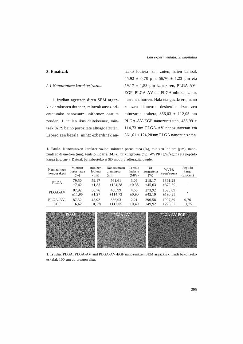
Lan experimentala: 2. kapitulua
295
3. Emaitzak
2.1 Nanozuntzen karakterizazioa
1. irudian agertzen diren SEM argaz-
kiek erakusten dutenez, mintzak ausaz ori-
entatutako nanozuntz uniformez osatuta
zeuden. 1. taulan ikus daitekeenez, min-
tzek % 79 baino porositate altuagoa zuten.
Espero zen bezala, mintz ezberdinek an-
tzeko lodiera izan zuten, haien balioak
45,92 ± 0,78 µm; 56,76 ± 1,23 µm eta
59,17 ± 1,83 µm izan ziren, PLGA-AV-
EGF, PLGA-AV eta PLGA mintzentzako,
hurrenez hurren. Hala eta guztiz ere, nano
zuntzen diametroa desberdina izan zen
mintzaren arabera, 356,03 ± 112,05 nm
PLGA-AV-EGF nanozuntzetan, 486,99 ±
114,73 nm PLGA-AV nanozuntzetan eta
561,61 ± 124,28 nm PLGA nanozuntzetan.
1. Taula. Nanozuntzen karakterizazioa: mintzen porositatea (%), mintzen lodiera (µm), nano-zuntzen diametroa (nm), tentsio indarra (MPa), ur xurgapena (%), WVPR (g/m2egun) eta peptido karga (µg/cm2). Datuak batazbesteko ± SD modura adierazita daude.
Nanozuntzen konposaketa
Mintzen porositatea
(%)
mintzen lodiera (µm)
Nanozuntzen diametroa (nm)
Tentsio indarra (MPa)
Ur xurgapena
(%)
WVPR (g/m2egun)
Peptido karga
(µg/cm2)
PLGA 79,50 ±7,42
59,17 ±1,83
561,61 ±124,28
3,06 ±0,35
218,17 ±45,03
1861,28 ±372,89 -
PLGA-AV 87,92 ±11,96
56,76 ±1,27
486,99 ±114,73
4,66 ±0,90
273,92 ±42,19
1690,09 ±190,25 -
PLGA-AV-EGF
87,52 ±6,62
45,92 ±0, 78
356,03 ±112,05
2,21 ±0,49
290,58 ±49,92
1907,39 ±228,82
9,76 ±1,75
1. Irudia. PLGA, PLGA-AV and PLGA-AV-EGF nanozuntzen SEM argazkiak. Irudi bakoitzeko eskalak 100 µm adierazten ditu.

Zaurien orbaintzerako baliabide terapeutiko berriak
296
Mintzen osotasuna baieztatzeko as-
moz, haien ezaugarri mekanikoak aztertu
ziren. PLGA, PLGA-AV eta PLGA-AV-
EGF nanozutzen erresistentzia maximoak
hurrenez hurren, honakoak izan ziren: 3,06
± 0,35 MPa, 4,66 ± 0,90 MPa and 2,21 ±
0,49 MPa (1. taula).
Mintzen hidrofilotasuna haien ur-
xurgapena neurtuz ebaluatu zen. Emai-
tzek erakutsi zuten mintzek antzeko ur-
xurgapena zutela, kasu denetan % 200
baino altuagoa zelarik: % 218,17 ± 45,03
PLGA nanozuntzetan; % 273,9 2 ± 42,19
PLGA-AV nanozuntzetan eta % 290,58
± 49,92 PLGA-AV-EGF nanozuntzetan,
1. taulan ageri den modura. WVPRa ere,
oso antzekoa izan zen mintz guztietan,
1861,28 ± 372,89 g/m2egun, 1690,09 ±
190,25 g/m2egun eta 1907,39 ± 228,82
g/m2egun PLGA, PLGA-AV eta PLGA-
AV-EGF nanozuntzentzako, hurrenez
hurren.
Jokabide termikoari dagokionez,
PLGA nanozuntzen beira-trantsiziozko ten-
peratura (Tg) PLGA lehengaiarena baino
baxuagoa izan zen, haien balioak 49,84 ±
0,23°C eta 53,85 ± 0,16°C izan zirelarik,
2. Taula. DSC emaitzak. Nanozuntzen, osagaien nahastearen eta lehengaien DSC ton-torrak. Datuak batazbesteko ± SD modura adierazita daude.
DSCaren tontor endotermikoak (°C)
PLGA 53.85±0.16
Aloe vera 67.94±0.33 155.82±0.37
rhEGF 63.68±4.11 199.10±0.19
PLGA eta Aloe vera nahastea 52.04±0.83 68.89±0.56
PLGA, Aloe vera eta rhEGF
nahastea 52.28±0.5 69.01±1.42
PLGA nanozuntzak 49.84±0.23
PLGA-AV nanozuntzak 51.46±1.04 70.30±3.76
PLGA-AV-EGF
nanozuntzak 52.04±0.08 71.73±1.48
hurrenez hurren (2. taula). Horretaz gain,
PLGAren Tg-an jaitsiera txiki bat behatu
zen ere nanozuntzen osagaien nahaske-
tan, ziurrenik AVaren presentzia dela eta.
Aipagarria da, PLGA-AV eta PLGA-AV-
EGF nanozuntzen termogrametan ageri
ziren tontorrak PLGA eta AVaren nahas-
keta fisikoaren termograman ageri ziren
berdinak zirela. rhEGFaren tontorra, al-
diz, ez zen nanozuntzetan ezta osagaien
nahaste fisikoetan ageri, ziurrenik pro-
portzio oso txikian erabili zelako (2.
taula).

Lan experimentala: 2. kapitulua
297
PLGA-AV-EGF nanozuntzetan karga-
tutako rhEGF kantitatea 9,76 ± 1,75
µg/cm2 zen. In vitro rhEGFaren askapen
profila 2. irudian ageri da. Hasierako bat-
bateko askapena edo burst efektua izan zu-
ten nanozuntzek, izan ere, lehen 8 ordue-
tan rhEGF totalaren %35a askatu zen. On-
doren, askapen fase geldoago bat egon zen
7 egunez, farmakoaren % 50a askatu zela-
rik entsegu honen amaierarako.
3.2 In vitro entsegu antibakterianoa
PLGA-AV nanozuntzetan zegoen
AVaren eragin antibakterianoa aztertzeko
inhibizio eremuaren testa erabili zen S. au-
2. Irudia. rhEGFaren askapen profila. PLGA-
AV-NLC nanozuntzetatik rhEGFaren metatze
askapen profila in vitro. Emaitzak denboran ze-
har rhEGFaren metatze ehunekoaren batazbas-
teko ±SD moduan adierazita daude.
reus eta S. epidermidisaren aurka. 3. iru-
dian ikus daitekeen modura, erabilitako bi
mikroorganismoetan, AV kontrolen eta
PLGA-AV nanozuntzen inguruan hazkun-
tzaren inhibizio guneak agertu ziren. Hala
eta guztiz ere, kualitatiboki begiratuta
kontrolen inguruan ageri zen inhibizio gu-
nearen diametroa handiagoa zen. PLGA
mintzetan, aldiz, ez zen inolako hazkun-
tzaren inhibiziorik ikusi.
3.3 In vitro bioaktibitate entsegua
Elektroirute prozesuak osagaien bioak-
tibitatean eragina zuen aztertzeko fibro-
blastoak mintzen erauzkinarekin inkubatu
ziren. 48 ordu ostean, zelulen proliferazioa
CCk-8 entsegu bat eginez neurtu zen.
4. Irudian ageri den bezala, proliferazio
zelular handiena PLGA-AV-EGF nano-
zuntzen erauzkinarekin lortu zen, kontrol
taldearekin konparatuz hazkuntza hiru-
koiztu baitzen. Horretaz gain, PLGA-AV
nanozuntzen erauzkinarekin tratatutako
zeluletan ere proliferazioa handitu zen
kontrolarekin alderatuz, dena den, mintz
honekin lortutako igoera PLGA-AV-EGF
mintzarekin lortutakoa baino txikiagoa
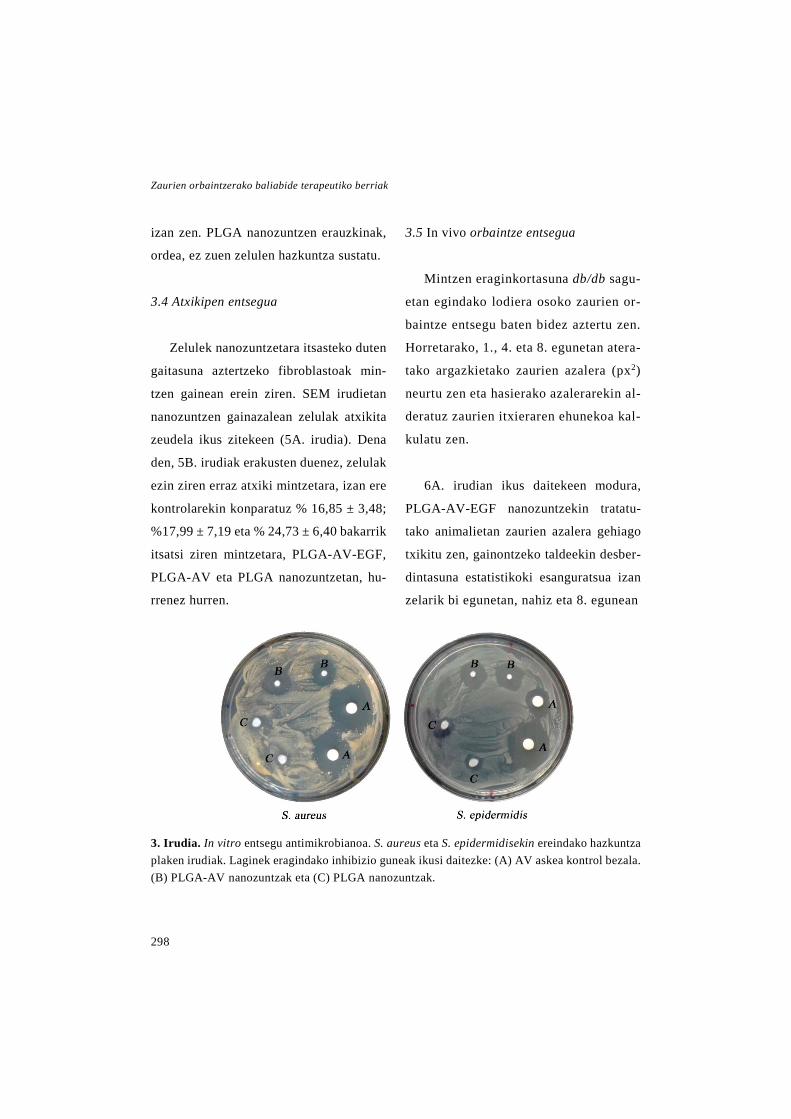
Zaurien orbaintzerako baliabide terapeutiko berriak
298
izan zen. PLGA nanozuntzen erauzkinak,
ordea, ez zuen zelulen hazkuntza sustatu.
3.4 Atxikipen entsegua
Zelulek nanozuntzetara itsasteko duten
gaitasuna aztertzeko fibroblastoak min-
tzen gainean erein ziren. SEM irudietan
nanozuntzen gainazalean zelulak atxikita
zeudela ikus zitekeen (5A. irudia). Dena
den, 5B. irudiak erakusten duenez, zelulak
ezin ziren erraz atxiki mintzetara, izan ere
kontrolarekin konparatuz % 16,85 ± 3,48;
%17,99 ± 7,19 eta % 24,73 ± 6,40 bakarrik
itsatsi ziren mintzetara, PLGA-AV-EGF,
PLGA-AV eta PLGA nanozuntzetan, hu-
rrenez hurren.
3.5 In vivo orbaintze entsegua
Mintzen eraginkortasuna db/db sagu-
etan egindako lodiera osoko zaurien or-
baintze entsegu baten bidez aztertu zen.
Horretarako, 1., 4. eta 8. egunetan atera-
tako argazkietako zaurien azalera (px2)
neurtu zen eta hasierako azalerarekin al-
deratuz zaurien itxieraren ehunekoa kal-
kulatu zen.
6A. irudian ikus daitekeen modura,
PLGA-AV-EGF nanozuntzekin tratatu-
tako animalietan zaurien azalera gehiago
txikitu zen, gainontzeko taldeekin desber-
dintasuna estatistikoki esanguratsua izan
zelarik bi egunetan, nahiz eta 8. egunean
3. Irudia. In vitro entsegu antimikrobianoa. S. aureus eta S. epidermidisekin ereindako hazkuntza plaken irudiak. Laginek eragindako inhibizio guneak ikusi daitezke: (A) AV askea kontrol bezala. (B) PLGA-AV nanozuntzak eta (C) PLGA nanozuntzak.

Lan experimentala: 2. kapitulua
299
desberdintasuna nabarmenagoa izan zen.
Emaitza horiek zaurien behaketa kualitati-
boarekin bat datoz (6B. irudia), izan ere
PLGA-AV-EGF nanozuntzekin tratatutako
zaurietan azalera txikiago ikusten baita.
Horretaz gain, rhEGF askearen dosi ber-
dina jaso zuten saguetan ez zen zauriaren
itxieraren hobekuntzarik ikusi kontrolare-
kin alderatuz. Laugarren egunean, ez ziren
desberdintasunik behatu gainontzeko tal-
deen artean. Zortzigarren egunean, aldiz,
beste taldeekin konparatuz, PLGA-AV na-
nozuntzekin tratatuko saguetan zaurien
itxiera esanguratsuki handiagoa izan zen.
Garatutako formulazioen eraginkorta-
suna aztertzeko analisi histologikoak bu-
rutu ziren ere bai. Horretarako, zaurien
biopsiak H&E edo Masson trikromiko
tindaketekin prozesatu ziren, errepiteliza-
zioa, hanturaren ebazpena edo kolageno-
aren metaketa aztertzeko, hurrenez hu-
rren. 7A. irudian 8. eguneko ebaketa his-
tologikoen (H&E tindaketa) argazkiak
ikus daitezke.
Errepitelizazioren ebaluaketa Sinha et
al.-ek (Sinha eta Gallagher 2003) ezarri-
tako irizpidearen arabera burutu zen. 7B.
4. Irudia. Mintzen bioaktibitate entsegua. Zelulen bideragarritasunaren emaitzak CCK-8 saioan neurtutako dentsitate optikoen ba-tazbesteko ± SD bezala adierazita daude. ** p<0,01 rhEGF askea, PLGA-AV-EGF eta PLGA-AV mintzak elkarrekin eta gainontzeko taldeak alderatuz.
irudian ikus daitekeen moduan, PLGA-
AV-EGF eta PLGA-AV nanozuntzekin
tratatutako animalien errepitelizazio gra-
dua esanguratsuki handiagoa izan zen. Bi
kasuetan, errepitelizazioaren batezbesteko
balioa 3 ingurukoa izan zen, aipatutako es-
kalaren arabera, lodiera irregularreko epi-
telio berriaz guztiz estalirik dauden zauriei
dagokiena. Hala eta guztiz ere, PLGA-AV
nanozuntzekin tratatutako zauriek alda-
kortasun handia izan zuten (2,875 ±
0,991), zenbait zaurietan errepitelizazioak
ez baitzuen zauri erdia estaltzen (1 gradua)
eta beste batzuetan zauri osoa estaltzen zu-
elako lodiera arruntarekin (4 gradua).
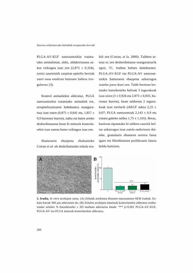
Zaurien orbaintzerako baliabide terapeutiko berriak
300
PLGA-AV-EGF nanozuntzekin tratatu-
tako animalietan, aldiz, aldakortasuna as-
koz txikiagoa izan zen (2,875 ± 0,354),
zortzi zaurietatik zazpitan epitelio berriak
zauri osoa estaltzen baitzuen lodiera irre-
gularrez (3).
Kontrol animaliekin alderatuz, PLGA
nanozuntzekin tratatutako animaliek ere,
errepitelizazioaren hobekuntza esangura-
tsua izan zuten (0,875 ± 0,641 eta, 1,857 ±
0,9 hurrenez hurren), nahiz eta haien arteko
desberdintasuna beste bi mintzek kontrola-
rekin izan zutena baino txikiagoa izan zen.
Hanturaren ebazpena ebaluatzeko
Cotran et al.-ek deskribatutako eskala era-
bili zen (Cotran, et la. 2000). Taldeen ar-
tean ez zen desberdintasun esanguratsurik
egon, 7C. irudian behatu daitekeenez.
PLGA-AV-EGF eta PLGA-AV nanozun-
tzekin hanturaren ebazpena azkarragoa
izateko joera ikusi zen. Talde horietan lor-
tutako batezbesteko balioak 3 ingurukoak
izan ziren (3 ± 0,926 eta 2,875 ± 0,835, hu-
rrenez hurren), beste taldeetan 2 inguru-
koak izan zirelarik (rhEGF askea 2,25 ±
0,07; PLGA nanozuntzak 2,143 ± 0,9 eta
tratatu gabeko taldea 1,75 ± 1,165). Beraz,
hasieran aipatutako bi taldeen zauriek hel-
tze azkarragoa izan zutela ondoriozta dai-
teke, granulazio ehunaren sorrera fasea
igaro eta fibroblastoen proliferazio fasera
heldu baitziren.
5. Irudia. In vitro atxikipen saioa. (A) Zelulak atxikituta dituzten nanozuntzen SEM irudiak. Es-kala barrak 300 µm adierazten du. (B) Zelulen atxikipen emaitzak kontrolarekin alderatuz zenba-tutako zelulen % batazbesteko ± SD moduan adierazita daude. *** p<0,001 PLGA-AV-EGF, PLGA-AV eta PLGA mintzak kontrolarekin alderatuz.
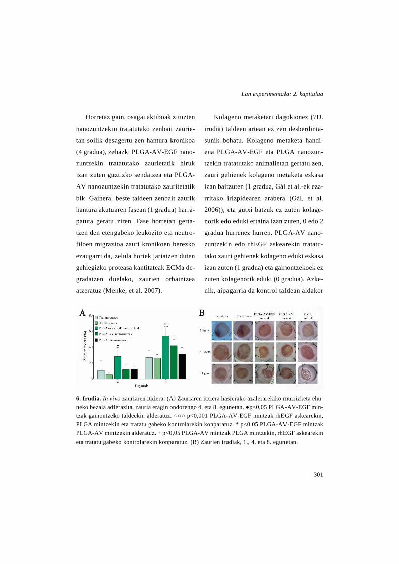
Lan experimentala: 2. kapitulua
301
Horretaz gain, osagai aktiboak zituzten
nanozuntzekin tratatutako zenbait zaurie-
tan soilik desagertu zen hantura kronikoa
(4 gradua), zehazki PLGA-AV-EGF nano-
zuntzekin tratatutako zaurietatik hiruk
izan zuten guztizko sendatzea eta PLGA-
AV nanozuntzekin tratatutako zauritetatik
bik. Gainera, beste taldeen zenbait zaurik
hantura akutuaren fasean (1 gradua) harra-
patuta geratu ziren. Fase horretan gerta-
tzen den etengabeko leukozito eta neutro-
filoen migrazioa zauri kronikoen berezko
ezaugarri da, zelula horiek jariatzen duten
gehiegizko proteasa kantitateak ECMa de-
gradatzen duelako, zaurien orbaintzea
atzeratuz (Menke, et al. 2007).
Kolageno metaketari dagokionez (7D.
irudia) taldeen artean ez zen desberdinta-
sunik behatu. Kolageno metaketa handi-
ena PLGA-AV-EGF eta PLGA nanozun-
tzekin tratatutako animalietan gertatu zen,
zauri gehienek kolageno metaketa eskasa
izan baitzuten (1 gradua, Gál et al.-ek eza-
rritako irizpidearen arabera (Gál, et al.
2006)), eta gutxi batzuk ez zuten kolage-
norik edo eduki ertaina izan zuten, 0 edo 2
gradua hurrenez hurren. PLGA-AV nano-
zuntzekin edo rhEGF askearekin tratatu-
tako zauri gehienek kolageno eduki eskasa
izan zuten (1 gradua) eta gainontzekoek ez
zuten kolagenorik eduki (0 gradua). Azke-
nik, aipagarria da kontrol taldean aldakor
6. Irudia. In vivo zauriaren itxiera. (A) Zauriaren itxiera hasierako azalerarekiko murrizketa ehu-neko bezala adierazita, zauria eragin ondorengo 4. eta 8. egunetan. ●p<0,05 PLGA-AV-EGF min-tzak gainontzeko taldeekin alderatuz. ○○○ p<0,001 PLGA-AV-EGF mintzak rhEGF askearekin, PLGA mintzekin eta tratatu gabeko kontrolarekin konparatuz. * p<0,05 PLGA-AV-EGF mintzak PLGA-AV mintzekin alderatuz. + p<0,05 PLGA-AV mintzak PLGA mintzekin, rhEGF askearekin eta tratatu gabeko kontrolarekin konparatuz. (B) Zaurien irudiak, 1., 4. eta 8. egunetan.

Zaurien orbaintzerako baliabide terapeutiko berriak
302
tasun handia egon zela, izan ere zauri gehi-
enek kolageno edukirik izan ez zuten arren
(0 gradua), gutxi batzuk kolageno eduki
handia izan zutelako (3 gradua).
4. Eztabaida
Elektroirutearen bidez ekoiztutako na-
nozuntzezko mintzak zaurientzako aposi-
tuak garatzeko aukera egokia dira, haien
ezaugarri arkitektural bereizgarriak direla
eta. Horregatik, ikerketa honen helburua
PLGAz eta ahal izan zen AV kantitate han-
dienaz osatutako nanozuntzezko apositu
bat garatzea izan zen, zeinean rhEGFa
kapsularatu zen.
Ekoiztutako mintzak ausaz orientatu-
tako nanozuntz uniformez osatuta zeuden
eta porositate altua zuten. Abrigo et al.-en
arabera, ezaugarri horiek zelulen arnas-
keta eta gasen sarrera ahalbidetzen dute
eta baita zaurien deshidratazioa ekidin ere
(Abrigo, et al. 2014).
7. Irudia. Zaurien analisi histologikoa (n=8). (A) Zaurien irudi histologikoak (H&E) 8. egunean, 10x handipena. (B) Zaurien errepitelizazio gradua 8. egunean. *** p<0,001 PLGA-AV-EGF mintzekin tratatutako taldea tratatu gabeko taldearekin alderatuz. ** p<0,01 PLGA-AV-EGF mintzekin tratatutako taldea rhEGF askearekin tratatutakoarekin konparatuz. * p<0,05 PLGA-AV-EGF mintzekin tratatutako taldea PLGA mintzekin tratatutakoarekin alderatuz. ●● p<0,01 PLGA-AV mintzekin tratatutako taldea rhEGF askearekin eta tratatu gabeko kontrolarekin alderatuz. + p<0,05 PLGA mintzekin tratatutko taldea tratatu gabeko taldearekin alderatuz. (C) Hantura pro-zesuaren ebazpen maila. (D) Kolageno metaketa maila zaurietan. Emaitzak batazbesteko ± SD moduan adierazita daude.

Lan experimentala: 2. kapitulua
303
PLGA-AV-EGF eta PLGA-AV nano-
zuntzen diametroa ECMko kolageno zun-
tzen tartean zegoen (50-500 nm). PLGA na-
nozuntzen diametroa ordea, goiko mugaren
apur bat gainetik zegoen. ECMarekin an-
tzekotasun horrek kaltetutako ehunen he-
mostasia sustatu dezake, zuntzen gainazal
azalera altuagatik eta mintzean zehar zirri-
kitu txikiak daudelako (Abrigo, et al. 2014)
Mintzen ezaugarri mekanikoek haien
egokitasuna apositu bezala erabiliak iza-
teko erakutsi zuten, izan ere, lortutako ten-
tsio indarraren balioak giza azalaren bali-
oen antzekoak izan ziren, zeinak 5,7 MPa-
tik 12,6 MPa-ra bitartera doazen, Jacque-
moud et al.-en arabera (Jacquemoud, et al.
2007). Mintzen artean egon ziren desber-
dintasun txikiak haien osaerarengatik izan
ziren. Alde batetik, AVak nanozuntzei
malgutasuna eman zien, tentsio indarra
handituz eta PLGA-AV mintzaren trakzio-
ari erresistentzia maximoa handituz. Beste
aldetik, rhEGFaren hauts liofilizatuak zi-
tuen gatzek PLGA-AV-EGF nanozuntzen
tentsio indarra txikitu zuten.
Hiru mintzen ur xurgapena antzekoa
izan zen arren, PLGA nanotzuntzek balio
baxuena izan zuten, PLGA polimero hi-
drofoboa delako. AVa zuten mintzak, al-
diz, hidrofiloagoak ziren, eta beraz ur
gehiago xurgatzeko gai izan ziren. Emai-
tza horiek Son et al.-ek (Son, et al 2013)
lortutakoekin bat datoz, PLGA nanozun-
tzetan gelatina kontzentrazio gorakorrak
jartzean ur xurgapenaren igoera behatu
baitzuten.
Zaurien aposituek ur-lurrunarekiko ira-
gazkortasuna izan behar dute, exudatuak
drainatzeko, baina horretaz gain, hezeta-
suna mantendu behar dute orbaintzea
errazteko (Abrigo, et al. 2014; Natarajan,
et al. 2000). Hezetasunaren kontrol hori
mantendu ahal izateko merkaturatutako
mintzek WVPRa balio zehatz batzuen ar-
tean daukate (426-2047 g/m2egun) (Tu, et
la. 2015). Tarte hori kontuan hartuta, gara-
tutako formulazioek WVPR egokia zeuka-
ten orbaintzean erabiliak izateko.
Mintzen jokabide termikoari dagokio-
nez, PLGA nanozuntzen Tg-a PLGA le-
hengaiarena baino txikiagoa izateak, elek-
troirundako PLGAn polimeroaren kateek
lerrokatze eta orientazio maila handia du-
tela iradokitzen du. Aldez aurretiko iker-

Zaurien orbaintzerako baliabide terapeutiko berriak
304
ketetan ere, Tg-aren antzeko txikipena be-
hatu izan da (Fouad, et al. 2013). Horretaz
gain, PLGA-AV eta PLGA-AV-EGF min-
tzetan eta PLGA-AV nahaste fisikoan ton-
tor termiko berdinak antzeman izanak na-
nozuntzetan bi osagaiak ez zirela nahasten
esan nahi du.
PLGA-AV-EGF nanozuntzetan, EGF
aren in vitro askapenak profil bifasikoa
erakutsi zuen, hasieran eztanda moduko
askapena aurkeztuz eta ondoren askapen
luzatutako fase bat aurkeztuz. Hasierako
askapena nanozuntzen gainazalean zegoen
proteinari zegokion, zauriaren ertzetan
dauden keratinozitoen aktibazio azkarra-
rentzako garrantzitsua dena (Schneider, et
al. 2009). Ondorengo fase luzatuan, far-
makoaren %50 inguru baino ez zen askatu.
Dena den, kargatutako farmakoaren aska-
pena bizkorragoa izan daiteke in vivo, zau-
rian dauden entzimek polimeroaren degra-
dazio azkartu dezaketelako (Fredenberg,
et al. 2011).
Zauriak mikroorganismoetatik babes-
teko ahalmena aposituek izan behar duten
ezaugarri garrantzitsu bat da, horrela zau-
rien infekzioak saihestu daitezkelako.
Inhibizio gunearen saioan ikusi zen be-
zala, PLGA-AV nanozuntzak hazkuntza
bakterianoa ekiditeko gai izan ziren, AVak
eragin antibakterianoa baitu pisu moleku-
lar baxuko osagaiei esker. Konposatu ho-
rien artean α-bisabolola, lupeola, azido zi-
namonikoa, azido salizilikoa eta azido uri-
koa daude, besteak beste. Haien ekintza
mekanismoa bakterioen prozesu entzima-
tikoak oztopatuz haiek hiltzean datza
(Ghayempour, et al. 2016; Tummalapalli,
et al. 2016). Horretaz gain, aposituek
muga fisiko bat eratzen dute bakterioen
aurrean, haien poro tamaina txikiak mikro-
organismoen sarrera ekiditen baitu
(Abrigo, et al. 2014).
AV askea erabiliz, emandako dosi
osoa saioaren hasieratik eskuragarri ze-
goen, nanozuntzetan zegoen AVa, aldiz,
haietatik askatu behar zen bakterioen haz-
kuntza inhibitu ahal izateko. Hori izan li-
teke AV askearekin inhibizio diametro
handiagoa lortu izanaren arrazoia. EGFak
eragin antibakterianorik ez duenez, ez zi-
ren PLGA-AV-EGF mintzak aztertu saio
honetan, AVaren eragina jadanik froga-
tuta baitzegoen PLGA-AV mintzen emai-
tzekin.

Lan experimentala: 2. kapitulua
305
In vitro entseguekin rhEGFaren eta
AV aren bioaktibitatea elektroirute pro-
zesuaren ostean aldatu gabe mantentzen
zela frogatu zen. Izan ere, mintzetatik
eratorritako erauzkinek fibroblastoen
proliferazioa sustatzeko gaitasuna zuten,
lehengaiek bezala (Boudreau and Beland
2006; Gainza, et al. 2015a). Hala eta guz-
tiz ere, entsegu honetan lortutako emaitza
deigarriena PLGA-AV-EGF nanozuntze-
kin lortutako proliferazio handia izan zen,
AVaren eta rhEGFaren konbinazioa onu-
ragarria dela erakutsi zuena. Gainera,
proliferazioa soilik AVaren eta rhEGFa-
ren eraginez izan zela esan daiteke, izan
ere, PLGA nanozuntzen erauzkinarekin
tratatutako zeluletan ez zen proliferazio-
rik behatu.
Aposituak tratamenduan zehar alda-
tzeko diseinatuta zeudenez, zelulen atxiki-
pena edo nanozuntzetan zehar ehunaren
hazkuntza ekidin behar ziren, apositua
kentzean mina eta eratu berri den ehuna
kaltetzea saihesteko. Nanozuntzetan atxi-
kitutako zelulen ehuneko baxua zela eta,
garatutako formulazioak aldi baterako
apositu modura erabiltzeko baliagarriak
zirela ikusi zen.
Orbaintzea in vivo ebaluatu zen db/db
saguetan egindako lodiera osoko zaurie-
tan. Karraskarien orbaintze ereduetan
errepitelizazioak ez dauka garrantzia han-
dirik, nahiz eta gizakietan badaukan. Dena
den, db/db saguetan prozesu horrek ga-
rrantzia hartzen du, zaurien uzkurdura oz-
topatua dagoelako, animalien gizentasu-
nak azalaren malgutasuna txikitzen baitu
(Fang, et al. 2010; Tkalcevic, et al. 2009).
Horretaz gain, zaurien inguruan siliko-
nazko eraztun bana josi zen, uzkurdura are
gehiago eragotziz (Michales, et al. 2007).
Errepitelizazioak garrantzia hartzen due-
nez, animalia hauen orbaintze prozesua gi-
zakienaren antzekoagoa da. Hala eta guz-
tiz ere, kolageno metaketaren emaitzetan
ikusi zenez, sagu hauen orbaintzeak alda-
kortasun altua zeukan, aldez aurretik
Trousdale et al.-ek deskribatu zuten bezala
(Trousdale, et al. 2009).
Orokorrean, PLGA-AV-EGF nanozun-
tzek, rhEGF askearekin konparatuz, or-
baintzea hobetu zezaketela erakutsi zuten,
zauriaren itxiera eta errepitelizazioari da-
gokionez. PLGA-AV-EGF nanozuntzek
orbaintzean izan zuten eragina, nanozun-
tzek kapsularatutako proteinengan duten

Zaurien orbaintzerako baliabide terapeutiko berriak
306
eragin babesleari atxiki dakioke, izan ere,
aldez aurretiko zenbait ikerketetan behatu
den modura, nanozuntzek EGFa zaurian
dauden proteasen aurrean babesten dute.
Ikerketa horien artean, Choi et al.-ek eta
Lai et al.-ek burututakoak daude. Lehen-
nengoan, PCL-PG nanozuntzak EGFare-
kin konjugatu zituzten (Choi, et al. 2008).
Lai et al.-ek bi nanozuntz motaz osatutako
apositu bat garatu zuten, zeinak, alde bate-
tik bFGFa eta VEGFaz kargatutako gela-
tina nanopartikulak zituzten azido hialuro-
nikozko zuntzez eta, bestetik, EGFa eta
PDGFdun gelatina nanopartikulak zituz-
ten kolagenozko zuntzez osatuta zegoen
(Lai, et al. 2014). Horretaz gain, nanozun-
tzen barne, rhEGFa denbora gehiago man-
tentzen da zaurian, mintzek luzatutako as-
kapena ahalbidetzen baitute.
PLGA-AV-EGF nanozuntzek baino
hein txikiagoan izan arren, PLGA-AV na-
nozuntzek orbaintzea sustatu zuten ere bai,
AVak eta nanozuntzek haiek berak orbain-
tzea sustatu baidezakete (Abrigo, et al.
2014; Hashemi, et al. 2015). Mintzen egi-
tura arkitektural bereizgarriak, hau da, du-
ten bolumenerako gainazal azalera handia
eta nanoporositateak, urari eta gasei ira-
gazkor egiten ditu, zaurian hezetasuna
maila egokia mantentzea ahalbidetuz eta
arnasketa zelularra sustatuz. Horretaz
gain, poroen tamaina txikiak mikroorga-
nismoen sarrera ekiditen du. Aipatutako
guztia dela eta, nanozuntzezko mintzek
zelulen migrazioa eta proliferazioa susta-
tzen dute eta orbaintzearen bitartekarien
askapena areagotzen dute, hala nola, kola-
genoarena, hazkuntza faktoreena eta fak-
tore angiogenikoena, era horretan, errepi-
telizazioa eta granulazio ehunaren sorrera
erraztuz (Abrigo, et al. 2014; Shahverdi, et
al. 2014). Honekin batera, errrepitelizazi-
oaren hobekuntzan aukeratutako polime-
roa eragina izan dezake, laktatoa, PLGA-
ren degradazio produktu bat, orbaintzea
bizkortzen duela ikusi baita (Porporato, et
al. 2012).
Ikerketa honen aurretik, bazeuden Aloe
veradun nanozuntzezko aposituei buruzko
beste ikerketa batzuk, baina horietan gai-
nontzeko osagaiekin konparatuz AVaren
proportzioa 1:3, 1:6 eta 1:8 zen (Bhaara-
thy, et al. 2014; Karuppuswamy, et al.
2014; Suganya, et al. 2014). Ikerketa ho-
netan, aldiz, AVren proportzioa handiagoa
zen, AV, PLGA eta rhEGF proportzioa
Lan experimentala: 2. kapitulua
307
1:1:0,4 baitzen hurrenez hurren. Beraz,
ikerketa honetan, lehen aldiz Aloe vera-
ren proportzio horren handia zuten nano-
zuntzak garatu ziren apositu modura era-
biltzeko. Aloe veraren erabilerak ikerke-
taren berritasuna azpimarratzen du, bere
erabilerak orbaintzea hobetzen duela era-
kutsi baitu.
Orokorrean, orbaintzearen hobekun-
tza hiru elementu hauen baturagatik izan
zen, rhEGFrena, AVrena eta PLGA nano-
zuntzena, alegia, izan ere, aldez aurretik
hirurek frogatu dute orbaintzea sustatzen
dutela.
5. Ondorioak
Honako entsegu honetan, Aloe vera
zeukan fase urtsu bat, azido poli-laktiko-
ko-glikolikoarekin (PLGA) emultsionatu
zen eta lortutako emultsioa elektroirun
zen, rhEGFa kargatuta zuen nanozun-
tzezko apositu bat garatzeko. Nanozuntzek
PLGAren eta AVaren proportzio berdina
zuen (1:1), eta gure jakintzaren arabera ho-
rren Aloe vera kontzentrazio handia ez da
orain arte erabili orbaintzean erabiltzeko
nanozuntzezko aposituak garatzeko.
Mintzak nanozuntz uniformez osatuta
zeuden, haien diametroa 356,03 ± 112,05
nm zen, porositatea % 87,52 zen eta min-
tzen lodiera 45,92 ± 0,78 µm zen. Elektroi-
rute prozesuak ez zuen osagai aktiboen bi-
oaktibitatean eraginik izan, in vitro bioak-
tibitate entseguak frogatu zuenez.
Db/db saguetan burutu zen in vivo lo-
diera osoko orbaintze entseguan, PLGA-
AV-EGF nanozuntzek orbaintzearen sus-
tapena eragin zuten, zauriaren itxiera eta
errepitelizazioari dagokionez. Orokorrean,
lortutako emaitza guztietatik atera daite-
keen ondorioa honakoa da: AVaren pro-
portzio handia duten PLGA-AV-EGF na-
nozuntzezko mintzak zauri kronikoetan
erabiltzeko aukera aproposa direla.
6. Eskerrak
I. Garcia-Oruek Eusko Jaurlaritzari
doktoratu aurreko laguntza eskertzen dio.
Egileek UPV/EHU-ko SGIkerren laguntza
teknikoa, giza babesa eta Europako finan-
tziazioa (FEDER eta FSE) eskertzen di-
tuzte. Proiektu hau neurri batean Eusko
jaurlaritzak (ELKARTEK 2015, Nano-
platform, KK-2015/0000036). Horretaz

Zaurien orbaintzerako baliabide terapeutiko berriak
308
gain, partzialki Euskal Herriko Unibertsi-
tateak finantziatu du (UFI11/32). Egileek
ICTS “NANBIOSIS”-en eta zehazki
UPV/EHUan dagoen CIBER-BBNren Far-
makoen Formulazio Unitateari (U10) la-
guntza tekniko eta intelektuala eskertu
nahi diote.
Erreferentzien zerrenda 110-115 orrialdeetan aurkitzen da.

3. KAPITULUA
Nanopartikula lipidikoak barneratuta dituen PLGA/Aloe veraz osa-tutako nanozuntz mintz konpositearen garapena, zaurien apositu mo-duan erabiltzeko.
LABURPENA
Elektroirutearen bidez ekoitzitako nanozuntzezko aposituek zenbait ezaugarri apro-pos dituzte orbaintzean erabiliak izateko, hala nola, porositate eta bolumenaren ara-berako gainazal azalera handia. Hori dela eta, ikerketa honetan PLGA eta Aloe veraren erauzkinaz osatutako apositu bat garatu zen. Horretaz gain, nanopartikula lipidikoak (NLCak) gehitu zitzaizkion, osagai lipidikoaren helburua apositua zaurietara atxikitzea ekiditea eta aposituaren maneiua, oklusibitatea eta elastikotasuna hobetzea zirelarik. NLCdun eta NLC gabeko mintzak 1 µm diametro inguru zuten zuntz uniformez osatuta zeuden. Haien porositatea %80 baino altuagoa zen eta lodiera 160 µm, NLCak zituzten apositoak loditasun handiagoa zutelarik. Apositu biek antzeko ur-xurgapena eta ur-lurrunaren iragazkortasun abiadura (WVPR) izan zituzten, haien balioak % 370 eta 1100 g/m2egun izan zirelarik, hurrenez hurren. Ezaugarri mekaniko desberdinak izan zituzten bi mintzek, izan ere NLCak zituen mintza elastikoagoa zen, bere erresistentzia maximoa 2,61 ± 0,46 MPa-koa zelarik. In vitro, bi fomulazioek biobateragarriak zirela erakutsi zuten, fibroblasto eta keratinozitoen aurrean. Horretaz gain, zelulen atxikipen entseguan behatu zen bi mintzek itsaspen perfil baxua zutela, NLCdun apositoak baxuagoa zuelarik. Azkenik, mintzen eraginkortasuna db/db saguetan egindako lodiera oso zauri eszision-aletan aztertu zen. Orbaintzea antzeko eran hobetu zuten bi formulazioek, zaurien itxi-erari, errepitelizazioari eta hanturari zegokienez. Ondorioz, PLGA-AV-NLC nano-zuntzezko mintzak zauri kronikoen tratamendurako estrategia aproposa izan daitezkeela esan dezakegu, izan ere, NLC gabeko formulazioarekin alderatuz aposituaren maneiua hobetzen zuten.

4. KAPITULUA
Orbaintzea sustatzeko gelatina/kitosano bigeruzadun hidrofilm baten garapena
LABURPENA
Ikerketa honetan, gelatinan oinarritutako bigeruzadun apositu berri bat garatu genuen. Elkargurutzaketa erreaktibo desberdinak erabiliz geruza bakoitzari ezaugarri bereizgarriak eman genizkion, horrela orbaintzean erabilgarri izan zitekeen hidrofilm multifuntzional bat lortuz. Lehenik, gaineko geruza ekoitzi genuen, zeina laktosaz elkargurutzatutako gelatinaz osatuta zegoen. Geruza hau erresistentea eta ez-degradagar-ria zen, apositu osoari euskarri mekaniko eta babesa emateko. Azpiko geruza ekoizteko, gelatina azido zitrikoarekin elkargurutzatu zen, ura xurgatzeko ahalmen handia zuen matrize porotsu bat lortuz. Horretaz gain, azpiko geruzari kitosanoa gehitu genion, or-baintzea estimulatzeko duen ahalmenaz baliatzeko. Elkargurutuzatzailearen eragina FTIR eta SEM analisien bidez frogatu zen, izan ere, laktosa gelatinaren bigarren mailako egitura aldatzeko gai zela behatu zen, egitura trinkoago eta lauagoa eraginez. Emaitzek erakutsi zuten bigeruzadun hidrofilma bere pisu lehorraren % 407,3 ± 108,6 ur xur-gatzeko gai zela, osotasun mekanikoa mantenduz. Horretaz gain, bere ur lurrunaren transmisio abiadura 1381,5 ± 108,6 g/m2egun zen. In vitro, fibroblastoekin biobateragar-ritasun ona erakutsi zuten garatutako hidrofilmek. Azkenik, haien eraginkortasuna giza azalean burututako ex vivo orbaintze saio batean aztertu zen. Hidrofilmek kontrol taldearen antzeko emaitzak lortu zituzten. Beraz, orokorrean esan daiteke garatutako big-eruzadun hidrofilmek apositu moduan erabiliak izateko ezaugarri egokiak izan zituztela.

Eztabaida

Eztabaida
315
Zauri kronikoen tratamenduak garrantzia handia irabazi du, bere intzidentziaren igo-
era kezkagarria dela eta. Erronka kliniko bat bilakatzen ari dira, izan ere, AEBtan baka-
rrik, urtero 5,7 millioi pertsonek (populazioaren % 2 inguru) pairatzen dituzte eta 20
bilioi dolar kostatzen dute zauri kronikoek [1]. Europan, 6000-10000 € xahutzen dira
zauriei lotutako gastuetan, hots, erizain denboran, ospitalizazioetan, aposituen aldakete-
tan eta zaurien infekzioen kudeaketan [2,3].
Zauri kronikoen intzidentziaren igoera zaurien kronizitatearekin lotuta dauden gai-
xotasunen hazkuntzarekin erlazionatuta dago, gaixotasun horien artean, obesitatea, dia-
betesa eta gutxiegitasun benosoa daudelarik [4]. Zauri horiek orbaintzearen fase fisiolo-
gikoetan (hemostasia, hantura, proliferazioa eta erremodelazioa) zehar igarotzeari huts
egiten diote, azalaren integritatearen berreskurapena atzeratuz eta maiz berriro agertuz
[5-7]. Oztopatutako orbaintzearen arrazoi nagusia haien etengabeko hantura egoera da,
izan ere prozesu osoan zehar neutrofiloak zaurian egoten dira, gehiegizko zitokina proin-
flamatorioak eta proteasak askatuz. Horiek mikroingurune proteolitiko bat eratzen dute,
orbaintzearen bitartekariak eta matrize extrazelularra (extracellular matrix edo ECM)
degradatzen dituena [8,9].
Gaur egun, zauri kronikoen terapiek ezin dute orbaintze eraginkor bat ziurtatu. Hori
dela eta, tratamendu eraginkor baten bilaketak garrantzia handia irabazi du, ahalegin
handiak egin direlarik tratamendu berrien garapenean edo egungo tratamenduen hobe-
kuntzan. Orbaintzean parte hartzen duten molekula endogenoen administrazio topikoa
tratamendu berri horietako bat da. Zoritxarrez, molekula horietako gehienek egonkorta-
sun oso eskasa dute in vivo, haien izaera proteikoa dela eta. Muga hau gainditzeko, sis-
tema desberdinetan kapsularatuak izan daitezke, hala nola, nanopartikula lipidikoetan
eta zehazki nanoegituratutako eramaile lipidikoetan (NLCak). NLCak kapsularatutako
peptidoa babesteaz gain, orbaintzerako abantaila ugari dituzte: askapen kontrolatua ahal-
bidetzen dute, haien itsasgarritasun eta oklusibotasunari esker azalaren hidratazioa han-
ditzen dute, eta haien tamaina txikiak azalarekin kontaktu estua ziurtatzen du [10].

Zaurien orbaintzerako baliabide terapeutiko berriak
316
Zaurien zainketak dituen eskakizunak betetzeko beste aukera bat nanozuntzezko min-
tzak dira. Apositu hauek orokorrean elektroirutearen bidez eratzen dira. Teknika honek,
indar elektrikoa erabiliz, disoluzio polimeriko batetik zuntz nanometrikoak erauzten ditu
[11]. Elektroirutearen bidez lortutako nanozuntzek egitura bereizgarria daukate, zeinak
bolumenarekiko gainazal azalera handia eta porositate handia dituen. Ezaugarri horiek
orbaintzea sustatzen dute arnasketa zelularra bultzatuz eta exudatuak xurgatzeaz gain
zaurian hezetasuna mantenduz, izan ere, gasen iragazkortasuna ahalbidetzen dute eta
zauriko hezetasuna kontrolatzen dute [12]. Horretaz gain, haien egiturak ECMaren hiru
dimentsioetako egitura imitatzen du, ehun ingeniaritzarako aukera interesgarria bihur-
tzen dituena [13,14].
Azkenik, bigeruzadun aposituek interes handia jaso dute zaurien orbaintzean erabi-
liak izateko, haien ezaugarri nabarmenak direla eta. Haien egiturak azalaren bigeruzadun
egitura imitatzen du, epidermisa antza duen gaineko geruza babesle batekin eta dermisa-
ren antzekoa den azpiko geruza malgu eta lodiarekin. Hori kontuan hartuz, aposituaren
gaineko geruza dentsoa zauria estaltzeko diseinatua dago, aposituari indar mekanikoa
emanez. Horretaz gain, hezetasunaren transmisioa kontrolatu behar du, zauriaren deshi-
dratazioa ekiditen duen biartzean, exudatuen garbiketa ahalbidetuz. Horretaz gain, gai-
neko geruzak bakterioen sarrera ekidin behar du, horrela zaurien infekzioa saihestuz.
Beste aldetik, azpiko geruzak egitura porotsua izan behar du, zeinak zauriko exudatua
xurgatzeko gai izan behar duen eta zaurira leunki atxiki behar den ehun eratu berriaren-
tzat inguru egokia sortuz [15-17].
Faktore horiek buruan edukita, doktorego honetan orbaintzea sustatzeko formulazio
desberdinen garapenean zentratu gara. Lehenengo urrats batean, LL37 giza peptidoan
oinarritutako nanopartikula formulazio bat garatzea erabaki genuen, molekula antimi-
krobiano honek orbaintzea zenbait bidez modulatzen duela frogatu baita, hala nola, an-
giogenesia aktibatuz [18,19], zelula epitelialen migrazioa eta proliferazioa bultzatuz
[20,21] eta monozito, neutrofilo eta zelula dendritikoentzako kimiotaktikoa izanez [22].

Eztabaida
317
LL37ak in vivo egonkortasun eskasa duenez, babesteko asmoz NLCetan kapsula-ratua
izan zen. Beraz, tesi honetan garatutako lehen formulazioa LL37a kapsularatuta zuten
NLCak izan ziren (NLC-LL37).
NLCak ekoizteko lipido solido (Precirol ATO5) eta likido (Mygliol® 812N) bana zi-
tuen urtutako fase lipidiko bat surfaktanteak zituen fase urtsu epel batekin emultsionatu
zen. Bi faseak nahastu ondoren, LL37a gehitu eta sonikazioaren bidez emultsionatu zi-
ren. Metodo honen bidez lortutako NLCak 273,6 ± 27,6 nm-ko batazbesteko tamaina
zuten eta -31 mV inguruko zeta potentziala. LL37aren kapsularatze eraginkortasuna %
96,4 ± 0,4-koa izan zen eta peptido karga 16,8 ± 0,1 µg LL37/mg NLC.
In vitro aktibitate entseguaren aurretik, NLC-LL37en biobateragarritasuna zehaztu
zen, haiekin batera inkubatutako fibroblastoen bideragarritasuna neurtuz. Formulazioa
zitotoxikoa ez zela frogatu ostean, bere ekintza ebaluatu zen, kapsularatze prozesuak
peptidoaren bioaktibitatean eragina zuen aztertzeko. Horretarako, RAW 267.4 makro-
fago lerro zelularra lipopolisakaridoarekin (LPS) aktibatu zen eta LL37 askearekin,
NLC-LL37ekin edo NLC hutsekin tratatu zen. Formulazioak makrofagoen aktibazioa
inhibitzeko gai ote ziren kuantifikatzeko, makrofagoek askatutako TNF-α kantitatea
ELISA baten bidez neurtu zen. 1. irudian ikus daitekeenez, NLC-LL37ek LL37 askearen
antzeko neurrian murriztu zuten TNF-αren ekoizpena, peptidoa kapsularatze prozesuaren
ostean aktibo mantentzen zela iradokitzen duelarik. Horretaz gain, NLC hutsek ez zuten
efekturik izan, NLC-LL37en eragina kapsularatutako LL37ari esker izan zela nabarmen-
duz. Makrofagoen neutralizazioa lortzeko, LL37a LPSari elkarrekintza eletrostatiko eta
hidrofoboen bidez lotu behar da eta baita makrofagoen CD14 hartzaileari ere lotura se-
lektibo baten bidez [23-25].
Ondoren, NLC-LL37en ekintza antimikrobianoa E. coliren aurka aztertu zen, infektatu-
tako zaurietan bakterio arruntenetakoa baita [26]. Laburki azalduta, LL37askea, NLC-
LL37ak eta NLC hutsak E. colirekin batera inkubatu ziren 4 orduz. Orduan, laginak
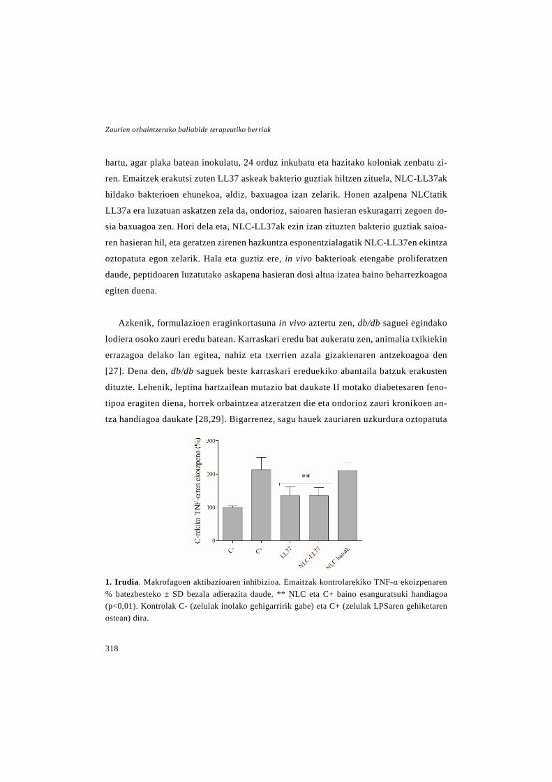
Zaurien orbaintzerako baliabide terapeutiko berriak
318
hartu, agar plaka batean inokulatu, 24 orduz inkubatu eta hazitako koloniak zenbatu zi-
ren. Emaitzek erakutsi zuten LL37 askeak bakterio guztiak hiltzen zituela, NLC-LL37ak
hildako bakterioen ehunekoa, aldiz, baxuagoa izan zelarik. Honen azalpena NLCtatik
LL37a era luzatuan askatzen zela da, ondorioz, saioaren hasieran eskuragarri zegoen do-
sia baxuagoa zen. Hori dela eta, NLC-LL37ak ezin izan zituzten bakterio guztiak saioa-
ren hasieran hil, eta geratzen zirenen hazkuntza esponentzialagatik NLC-LL37en ekintza
oztopatuta egon zelarik. Hala eta guztiz ere, in vivo bakterioak etengabe proliferatzen
daude, peptidoaren luzatutako askapena hasieran dosi altua izatea baino beharrezkoagoa
egiten duena.
Azkenik, formulazioen eraginkortasuna in vivo aztertu zen, db/db saguei egindako
lodiera osoko zauri eredu batean. Karraskari eredu bat aukeratu zen, animalia txikiekin
errazagoa delako lan egitea, nahiz eta txerrien azala gizakienaren antzekoagoa den
[27]. Dena den, db/db saguek beste karraskari ereduekiko abantaila batzuk erakusten
dituzte. Lehenik, leptina hartzailean mutazio bat daukate II motako diabetesaren feno-
tipoa eragiten diena, horrek orbaintzea atzeratzen die eta ondorioz zauri kronikoen an-
tza handiagoa daukate [28,29]. Bigarrenez, sagu hauek zauriaren uzkurdura oztopatuta
1. Irudia. Makrofagoen aktibazioaren inhibizioa. Emaitzak kontrolarekiko TNF-α ekoizpenaren % batezbesteko ± SD bezala adierazita daude. ** NLC eta C+ baino esanguratsuki handiagoa (p<0,01). Kontrolak C- (zelulak inolako gehigarririk gabe) eta C+ (zelulak LPSaren gehiketaren ostean) dira.

Eztabaida
319
daukate, haien gizentasunak azalaren malgutasuna murrizten baitu [30,31]. Horren on-
dorioz, errepitelizazioak garrantzia handiagoa hartzen du, orbaintze prozesua gizakiena-
ren antzekoagoa eginez. Horretaz gain, zaurien uzkurdura are gehiago oztopatu genuen
haien inguruan silikonazko eraztun bana josiz [32]. Animaliei NLC-LL37aren bi dosi
desberdin administratu zitzazkien bide topikotik, talde bati LL37aren 6 µg-ri zegokion
NLC-LL37aren dosia administratu zitzaion eta beste taldeari LL37aren 2 µg-ri zegoki-
ona.
Orbaintzea ebaluatzeko ondorengo parametroak neurtu ziren: (i) zaurien itxiera, zau-
rien argazkietatik azalerak neurtuz kalkulatu zena; (ii) errepitelizazio gradua, erdi-kuan-
titatiboki neurtu zen, H&Eren bidez prozesatutako ehun sekzioetan Sinha et. al.-ek des-
kribatutako eskala aplikatuz [33]; (iii) hantura prozesuaren ebazpena, aurrekoa bezala
H&Eren bidez tindatutako ehun sekzioak erabiliz ebaluatu zen, kasu honetan Contran et
al.-ek deskribatutako eskalaren arabera [34]; (iv) kolageno metaketa, Masson trikomiko
tindaketaren bidez prozesatutako ehun sekzioak erabiliz aztertu zen, Gal et al.-ek ezarri-
tako eskala jarraituz [35], eta (v) angiogenesia, immunohistokimikoki antiCD31 antigor-
putz monoklonalaz tindatutako ehun sekzioetan odol hodi eratu berriak zenbatuz ebalu-
atu zen.
2. irudiko emaitzek erakusten dutenez, NLC-LL37en administrazioak zaurien itxiera
zein errepitelizazioa orbaintzea bizkortu zuen NLC hutsekin, LL37 askearekin eta tratatu
gabeko taldearekin alderatuz. Horretaz gain, NLC-LL37ak hantura prozesuaren ebaz-
pena ere sustatu zuen, baina kasu honetan LL37 askearen neurri berean. Kolageno meta-
ketari eta angiogenesiari dagokienez, ez zen desberdintasunik behatu taldeen artean.
Orokorrean, kapsularatutako peptidoak areagotutako ekintza erakutsi zuen, ziurrenik
NLCek degradazio kimiko zein entzimatikoaren aurrean izan zuten efektu babesgarriaren
ondorioz. Horretaz gain, NLCek luzatutako askapena ahalbidetzen dute, LL37aren era-
gina luzatuz [36,37]. Gainera, aurretik garatutako beste LL37dun nanopartikulekin alde-

Zaurien orbaintzerako baliabide terapeutiko berriak
320
ratuz [38], NLCek administrazio topikoa alhalbidetzen dute, pazienteentzako bide
erraza, eroso eta segurua dena. Aurkikuntza guzti horiek kontuan hartuz, NLC-LL37en
administrazio topikoa zauri kronikoen tratamendurako estrategia interesgarria izan dai-
tekeela ondoriozta dezakegu.
Tesi honen bigarren urratsean, hazkuntza faktore epidermikoa (epidermal growth fac-
tor edo EGF) kapsularatuta zuen nanozuntzezko apositu bat garatu genuen. Faktore tro-
fiko hau orbaintzearen molekula gakoa da, keratinozito eta fibroblastoen proliferazio eta
migrazioa estimulatzen dituelako [39]. Bere administrazio topikoak orbaintzea sustatu
dezakeela frogatu da jadanik, dena den, duen erdibizitza laburrak bere erabilpen klinikoa
zailtzen du [40,41].
Hori dela eta, EGFa nanozuntzezko mintzen barne kapsularatuz muga hori gaindi
daitekela hipotetizatu genuen, izan ere, mintzek peptidoa ingurune kaltegarritik babestu
dezakete eta gainera haien egiturak orbaintzea sustatu dezake. Nanozuntzezko mintzak
2. Irudia. In vivo zauriaren itxiera. (A) Zaurien itxiera hasierako gainazal azalerarekiko murriz-keta ehuneko bezala adierazita. * Tratatu gabeko taldearekiko esanguratsua (p<0.05); ○ Tratatu gabeko taldearekiko esanguratsua (p<0.05); + gainontzeko taldeekiko esanguratsua (p<0.05). (B) Zaurien irudiak.
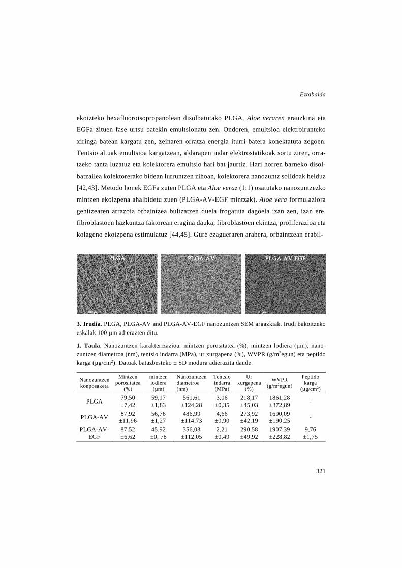
Eztabaida
321
ekoizteko hexafluoroisopropanolean disolbatutako PLGA, Aloe veraren erauzkina eta
EGFa zituen fase urtsu batekin emultsionatu zen. Ondoren, emultsioa elektroirunteko
xiringa batean kargatu zen, zeinaren orratza energia iturri batera konektatuta zegoen.
Tentsio altuak emultsioa kargatzean, aldarapen indar elektrostatikoak sortu ziren, orra-
tzeko tanta luzatuz eta kolektorera emultsio hari bat jaurtiz. Hari horren barneko disol-
batzailea kolektorerako bidean lurruntzen zihoan, kolektorera nanozuntz solidoak helduz
[42,43]. Metodo honek EGFa zuten PLGA eta Aloe veraz (1:1) osatutako nanozuntzezko
mintzen ekoizpena ahalbidetu zuen (PLGA-AV-EGF mintzak). Aloe vera formulaziora
gehitzearen arrazoia orbaintzea bultzatzen duela frogatuta dagoela izan zen, izan ere,
fibroblastoen hazkuntza faktorean eragina dauka, fibroblastoen ekintza, proliferazioa eta
kolageno ekoizpena estimulatuz [44,45]. Gure ezagueraren arabera, orbaintzean erabil-
3. Irudia. PLGA, PLGA-AV and PLGA-AV-EGF nanozuntzen SEM argazkiak. Irudi bakoitzeko eskalak 100 µm adierazten ditu.
1. Taula. Nanozuntzen karakterizazioa: mintzen porositatea (%), mintzen lodiera (µm), nano-zuntzen diametroa (nm), tentsio indarra (MPa), ur xurgapena (%), WVPR (g/m2egun) eta peptido karga (µg/cm2). Datuak batazbesteko ± SD modura adierazita daude.
Nanozuntzen konposaketa
Mintzen porositatea
(%)
mintzen lodiera (µm)
Nanozuntzen diametroa (nm)
Tentsio indarra (MPa)
Ur xurgapena
(%)
WVPR (g/m2egun)
Peptido karga
(µg/cm2)
PLGA 79,50 ±7,42
59,17 ±1,83
561,61 ±124,28
3,06 ±0,35
218,17 ±45,03
1861,28 ±372,89 -
PLGA-AV 87,92 ±11,96
56,76 ±1,27
486,99 ±114,73
4,66 ±0,90
273,92 ±42,19
1690,09 ±190,25 -
PLGA-AV-EGF
87,52 ±6,62
45,92 ±0, 78
356,03 ±112,05
2,21 ±0,49
290,58 ±49,92
1907,39 ±228,82
9,76 ±1,75
Zaurien orbaintzerako baliabide terapeutiko berriak
322
tzeko horren Aloe vera kontzentrazio handia duten aposituak garatu ziren lehen aldia
izan zen hau. Horretaz gain, nanozuntzezko mintz hauek garatu ziren ere: EGFrik ez
zutenak (PLGA-AV mintzak) eta EGF zein Aloe verarik ez zutenak (PLGA mintzak).
Hiru mintz moten karakterizazioa 1. taulan eta 3. irudian laburtuta ageri da. Elektroi-
rute prozesuaren bidez ausaz orientatutako nanozuntz uniformez osatutako mintzak
ekoiztu ziren. Egiturak porositate handia zuen (%79 baino altuagoa) eta bolumenare-
kiko azalera handia ere bai, zelulen arnasketa eta hezetasunaren kontrola baimentzen
dituena [11].
Zuntzen arteko desberdintasun handiena haien diametroan eta ur xurgapenean ze-
goen. Aloe vera gehitzeak nanozuntzen diametroa 561,6 ± 124,3 nm-tatik 487 ± 114,7
nm-tara txikitu zuen eta EGFa gehitzeak 356 ± 112 nm-tara. Ur xurgapenean behatutako
desberdintasunei dagokienez, Aloe veradun mintzek ur gehiago xurgatzeko gaitasuna zu-
tela ikusi zen, konposatu honek mintzen hidrofilotasuna handitzen zuela iradokiz [46].
Mintzen ezaugarri mekanikoak proba desplazamendu kontrolaren arabera aztertu zi-
ren Instron 5848 mikrotestagailu batean. Aipagarria da giza azalaren antzeko balioak
izan zituztela, horiek 5,7 MPa 12.6 MPa-ra bitartea doazelarik [47]. Horretaz gain, ur-
lurrunarekiko iragazpen abiadura (water vapour permeability rate edo WVPR) apositu
komertzialen mailan zegoen (426-2047 g/m2egun), horrek garatutako aposituak hezeta-
sunaren kontrol egokia lortzeko gai zirela iradokitzen duelarik [48]. WVPRa neurtzeko,
silika gel lehorgarria zuten ontzien ahoak garatutako mintzekin erabat estali ziren. On-
doren ontziak ganbera klimatiko batean sartu ziren 24 orduz, eta muntaia esposizio den-
boraren aurretik eta ondoren pisatuz WVPRa kalkulatu zen.
Mintzen jokabide termikoa ekorketazko kalorimetria diferentzialaren (differential
scanning calorimetry edo DSC) bidez aztertu zen. Emaitzek erakutsi zuten elektroirun-
dako PLGAk polimero kateen lerrokatze eta orientazio maila handia zutela, izan ere,
beira-trantsiziozko tenperatura (Tg) PLGA mintzetan (49,84 ± 0,23°C) PLGA lehengaian
Eztabaida
323
(53,85 ± 0,16°C) baino baxuagoa zen, aurretik deskribatu den moduan [48]. Horretaz
gain, antzeko tontor termikoak antzeman ziren PLGA-AV eta PLGA-AV-EGF elektroi-
rundako mintzetan eta baita PLGA eta Aloe vera nahaste fisikoan ere, nanozuntzen barne
bi osagaien izaera nahastezina agerian utziz. EGFaren tontorra ez zen inon behatu, ziu-
rrenik mintzetan zegoen proportzio txikia zela eta.
PLGA-AV-EGF mintzetatik EGFaren in vitro askapen saioa Franz difusio gelaxketan
burutu zen. Hezetutako 1x1 cm-ko mintz zati bana ganbara emailean kokatu zen nylo-
nezko iragazki (0,45 µm-ko poro tamaina) baten gainean eta ganbera hartzailean 5 ml
PBS kokatu ziren. Aurretik hautatutako uneetan, ganbera hartzailetik laginak hartu ziren
eta askatutako EGFa ELISA komertzial baten bidez neurtu zen. Mintzek askapen profil
bifasikoa izan zuten, hasierako eztanda edo burst askapena gertatu zen, non lehenengo 8
orduetan farmakoaren % 35a askatu zen eta ondoren askapen fase geldoago bat gertatu
zen, 7 egunetan EGF totalaren % 50a askatu zelarik. Hasierako eztanda askapena nano-
zuntzen gainazalean zegoen EGFari zegokion eta zauri ertzeko keratinozitoen aktibazio
azkarra lortzeko balio du [49]. Aipagarri da, EGFaren % 50a soilik askatu zen arren, in
vivo askapena bizkortu daitekela, zaurian dauden entzimek polimeroaren degradazioa
azkartu dezaketelako [50].
Behin aposituak karakterizatu zirela haien bioaktibitatea ebaluatu zen. Alde batetik,
Aloe veraren aktibibatea aztertu zen [51]. Horretarako PLGA-AV eta PLGA mintzetatik
hartutako diskoak Staphilococcus aureus eta S. epidermidisekin ereindako hazkuntza
plaketan jarri ziren, eta inkubatu osean mintzek eratu zuten inhibizio gunea neurtu zen.
PLGA mintzen inguruan ez zen inhibizio gunerik agertu, PLGA-AV mintzen inguruan
agertutako inhibizio gunea Aloe veraren eraginez izan zela nabarmenduz. Hala eta guztiz
ere, Aloe vera disoluzioa zuten disko komertzialen inguruan inhibizio gune handiagoa
agertu zen, ziurrenik bigarren kasu honetan Aloe vera dosi osoa hasieratik eskuragarri

Zaurien orbaintzerako baliabide terapeutiko berriak
324
zegoelako, NLC-LL37 formulazioarekin gertatu zen bezala. Gainera, ekintza antimikro-
bianoari dagokionez, nanozuntzezko mintzen mikroorganismoen sarrera ekiditen dute,
duten poro tamaina txikia dela eta.
Beste aldetik, EGFaren eta Aloe veraren efektua fibroblastoen proliferazioan aztertu
genuen, elektroirute prozesuak haien ekintzaren gain eragina zuen behatzeko [52,53].
Horren harira, fibroblastoak nanozuntzezko mintzek askatutako ingurunearekin inkubatu
ziren, eta haien bideragarritasuna CCK-8 saioaren bidez neurtu zen. Proliferazio zelular
altuena PLGA-AV-EGF mintzekin lortu zen, izatez, EGF askearen kontzentrazio berdi-
narekin inkubatutako zelulena baino altuagoa zen, konbinazioaren efektu onuragarria na-
barmenduz. Horretaz gain, PLGA-AV mintzetan proliferazio igoera behatu zen, baina ez
PLGA mintzetan. Horrek, ekintza EGF eta Aloe veraren eraginez baino ez zela iradoki-
tzen du.
Mintzak tratamenduan zehar aldatzeko diseinatuak zeudenez, zelulen atxikipena edo
nanozuntzetan zehar ehunaren hazkuntza ekidin behar dira, apositua kentzean mina eta
eratu berri den ehuna kaltetzea saihesteko [11]. Hori dela eta, zelulak mintzetara atxiki-
tzeko gai ziren ebaluatzea erabaki genuen, fibroblastoak haien gainean 24 orduz inkuba-
tuz, eta orduan, atxikitutako zelulak askatuz eta zenbatuz edo haien SEM argazkiak ate-
raz, 4.A irudian ageri den modura. Grafikoan ikus daitekeenez (4.B irudia), zelulen atxi-
kipena mintzetara oso oztopatuta zegoen hazkuntza plaken hondoarekin konparatuz, aldi
baterako apositu bezala haien egokitasuna erakutsiz.
Azkenik, mintzen eraginkortasuna db/db saguei egindako lodiera osoko zauri ereduan
aztertu zen. Horretarako, aurreko ikerketan bezala, zaurien azaleraren murrizketa, erre-
pitelizazio gradua, hantura prozesuaren ebazpena eta kolageno metaketa aztertu ziren. 4.
egunean, PLGA-AV-EGF mintzek zaurien itxiera azkartu zuten gainontzeko taldeekin
konparatuta, talde horiek EGF askea, PLGA-AV mintzak, PLGA mintzak eta tratatu ga-
beko kontrola zirelarik. 8. egunean zaurien itxieraren hobekuntza handiena PLGA-AV-

Eztabaida
325
EGF mintzekin lortu zen ere, nahiz eta kasu honetan PLGA-AV mintzek zaurien itxiera
sustatzeko gai izan ziren ere. Horretaz gain, PLGA-AV-EGF eta PLGA-AV mintzak eza-
rriz, zaurien errepitelizazio gradua hobetu zen. Izan ere, errepitelizazioa ebaluatzeko era-
bili zen eskalaren arabera zauri osoa errepitelizatuta zuten, baina lodiera irregularrez
[33]. Hantura prozesuaren ebazpenari eta kolageno metaketari dagokionez, ez ziren des-
berdintasunik behatu taldeen artean, dena den, aurreko parametroekin bat zetorren ten-
dentzia arin bat nabari zitekeen.
Orokorrean, in vivo entseguan lortutako emaitzek erakutsi zuten zaurietan PLGA-
AV-EGF mintzen ezarpena, PLGA-AV mintzen ezarpena eta EGF askearen administra-
zioa baino abantailatsuagoa zela. Efektu onuragarri hau, neurri batean nanozuntzezko
mintzek zauriko proteasen aurka duten eragin babesgarriak azaltzen du, aurretik deskri-
batu den modura [40,55]. Horretaz gain, mintzetatik gertatzen den askapen luzatuak,
EGFaren iraupena luzatzen du, horrela bere eragina hobetuz.
Aloe vera eta nanozuntzek haiek berak orbaintzea sustatu zuten. Alde batetik, Aloe
verak orbaintzea bultzatzen duela frogatua dago, fibroblastoen hazkuntza faktorearengan
4 irudia. In vitro atxikipen saioa. (A) Zelulak atxikita dituzten nanozuntzen SEM irudiak. Eskala barrak 300 µm adierazten du. (B) Zelulen atxikipen emaitzak kontrolarekin alderatuz zenbatutako zelulen % batazbestekoa ± SD moduan adierazita daude. *** p<0,001 PLGA-AV-EGF, PLGA-AV eta PLGA mintzak kontrolarekin alderatuz.
Zaurien orbaintzerako baliabide terapeutiko berriak
326
eraginez, fibroblastoen ekintza eta proliferazioa sustatzen baitu [53]. Beste aldetik, au-
rretik azaldu denez, elektroirundako mintzen egiturak zaurien itxieran eragina dauka,
gasen eta uraren iragazkortasuna ahalbidetzen baitu [56]. Ondorioz, orbaintzearen hobe-
kuntza EGFaren, Aloe vera erauzkinaren eta nanozuntzen baturaren ondorioz izan zen,
haien konbinazioz lortutako apositua zauri kronikoen tratamendurako aproposa izan dai-
tekeela erakutsiz.
EGFrik ez zuten nanozuntzezko mintzek ere orbaintzea sustatu zutenez, ondorengo
urratsean, PLGA eta Aloe veran oinarritutako nanozuntzezko apositu baten garapenari
ekin genion. EGFa ez eramateak osasun-produktu baten garapenerako bidea zabaltzen
baitu. Mintzen ezaugarrietako batzuk hobetzeko asmoz, elektroirute prozesuan zehar
NLCak gehitu ziren, osagai lipidiko horrek mintzak zaurira atxikitzea ekidin eta haien
kentzea erraztuko zuelakoan. Horretaz gain, NLCen gehitzeak aposituaren maneiua,
elastikotasuna eta oklusibitatea hobetuko zituela ere hipotetizatu genuen.
NLCdun eta NLC gabeko nanozuntzezko mintzak (PLGA-AV-NLC eta PLGA-AV)
garatu ziren, NLCak gehitzeak duen eragina ebaluatzeko. Kasu honetan, pisu moleku-
lar handiagoko PLGA bat erabili zen, NLCen gehitzea errazten baitzuen. Hori dela eta,
mintz hauen karakterizazioa aurrekoenen arinki desberdina izan zen, 2. taulan ikus
daiteekenez. Porositatearen balioak aurreko ikerketan lorutako balioen antzekoak izan
ziren, % 80 ingurukoak, alegia, horrek gasen iragazketa eta beraz, zelulen arnasketa
ahalbidetzen zuelarik. Beste aldetik, nanozuntzen diametroa 500 nm-tatik 1 µm ingu-
rura handitu zuen, ziurrenik pisu molekular handiko PLGArekin prestatutako polimero
soluzioaren biskositate handiagoa zela eta, izan zere, soluzioaren biskositatea nano-
zuntzen morfologia kontrolatzeko faktore kritikoa da [56]. Horretaz gain, mintzen ur
xurgapena aurreko ikerketan baino altuagoa izan zen, % 380 ingurukoa, zaurian heze-
tasuna mantentzen duen bitartean, exudatuak drainatzen laguntzeko baliagarria dena
[3,46].
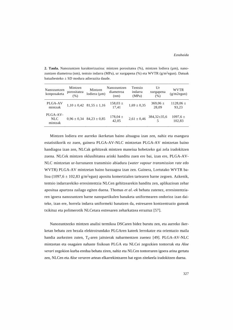
Eztabaida
327
2. Taula. Nanozuntzen karakterizazioa: mintzen porositatea (%), mintzen lodiera (µm), nano-zuntzen diametroa (nm), tentsio indarra (MPa), ur xurgapena (%) eta WVTR (g/m2egun). Datuak batazbesteko ± SD modura adierazita daude.
Nanozuntzen konposaketa
Mintzen porositatea
(%)
Mintzen lodiera (µm)
Nanozuntzen diametroa
(nm)
Tentsio indarra (MPa)
Ur xurgapena
(%)
WVTR (g/m2egun)
PLGA-AV mintzak 1,10 ± 0,42 81,55 ± 1,16 158,03 ±
17,41 1,69 ± 0,35 369,06 ± 28,09
1128,06 ± 93,23
PLGA-AV-NLC
mintzak 0,96 ± 0,34 84,23 ± 0,85 178,04 ±
42,05 2,61 ± 0,46 384,32±35,65
1097,6 ± 102,83
Mintzen lodiera ere aurreko ikerketan baino altuagoa izan zen, nahiz eta esangura
estatistikorik ez zuen, gainera PLGA-AV-NLC mintzetan PLGA-AV mintzetan baino
handiagoa izan zen, NLCak gehitzeak mintzen maneiua hobetzeko gai zela iradokitzen
zuena. NLCek mintzen oklusibitatea arinki handitu zuen ere bai, izan ere, PLGA-AV-
NLC mintzetan ur-lurrunaren transmisio abiadura (water vapour transmission rate edo
WVTR) PLGA-AV mintzetan baino baxuagoa izan zen. Gainera, Lortutako WVTR ba-
lioa (1097,6 ± 102,83 g/m2egun) apositu komertzialen tartearen barne zegoen. Azkenik,
tentsio indarrarekiko erresistentzia NLCen gehitzearekin handitu zen, aplikazioan zehar
apositua apurtzea zailago egiten duena. Thomas et al.-ek behatu zutenez, erresistentzia-
ren igoera nanozuntzen barne nanopartikulen banaketa uniformearen ondorioz izan dai-
teke, izan ere, horrela indarra uniformeki banatzen da, estresaren kontzentrazio guneak
txikituz eta polimerotik NLCetara estresaren zeharkatzea erraztuz [57].
Nanozuntzezko mintzen analisi termikoa DSCaren bidez burutu zen, eta aurreko iker-
ketan behatu zen bezala elektroirundako PLGAren kateek lerrokatze eta orientazio maila
handia aurkezten zuten, Tg-aren jaitsierak nabarmentzen zuenez [49]. PLGA-AV-NLC
mintzetan eta osagaien nahaste fisikoan PLGA eta NLCei zegozkien tontorrak eta Aloe
verari zegokion kurba eredua behatu ziren, nahiz eta NLCen tontorraren igoera arina gertatu
zen, NLCen eta Aloe veraren artean elkarrekintzaren bat egon zitekeela iradokitzen duena.

Zaurien orbaintzerako baliabide terapeutiko berriak
328
Aurreko ikerketan, elektroirute prozesuak Aloe veraren ekintzan eraginik ez zuela
frogatu genuen, bere ekintza antimikrobianoa eta fibroblastoen proliferazioaren gaine-
koa mantendu baitzen nanozuntzetan. Hori dela eta, ikerketa honetan zuzenean mintzen
biobateragarritasuna aztertu genuen, aurreko ikerketan erabilitako metodo berdina jarrai-
tuz. Fibroblastoak eta keratinozitoak nanozuntzetatik erauzitako ingurunearekin 48 or-
duz inkubatu genituen eta, orduan CCK-8 saioa burutu genuen zelulen bideragarritasuna
neurtzeko. Emaitzak 5. irudian ageri dira, eta mintzak bi lerro zelularrekin biobateraga-
rriak zirela erakusten dute, kontrolarekin alderatuz bideragarritasuna % 70-a baino altu-
agoa baitzen. Gainera, fibroblastoetan mintzek bideragarritasuna igo zuten, ziurrenik
Aloe verak haien gain duen efektu proliferatiboagoa dela eta [45,53].
Aposituaren maneiuan zuten efektuaz gain, NLCak nanozuntzetan barneratzeko arra-
zoi nagusia, aposituaren kentzea erraztu zezaketela hipotesia zen. Horregatik, zelulen
atxikipen entsegua burutu genuen. Mintzak 24 putzutako plaken hondoan kokatu ziren
5 Irudia. Zelulen bideragarritasun saioa. (A) CCK-8 saioaren emaitzak mintzen erauzkina fibro-blastoekin inkubatu ostean. *** p<0.001 PLGA-AV-NLC eta PLGA-AV mintzak kontrolarekin konparatuz; eta ** p<0.01 PLGA-AV mintza PLGA-AV-NLC mintzarekin alderatuz. (B) CK-8 saioaren emaitzak mintzen erauzkina fibroblastoekin inkubatu ostean. *** p<0.001 PLGA-AV mintza kontrol taldearekin alderatuz; eta ** p<0.01 PLGA-AV-NLC mintza kontrol taldearekin alderatuz. Emaitzak kontrolarekiko bizirik dauden zelulen % batezbestekoa ± SD bezala adierazita daude.
Eztabaida
329
eta fibroblastoak edo keratinozitoak haien gainean inkubatu ziren 24 orduz. Orduan, atxi-
kitako zelulak mintzetatik askatu eta zenbatu ziren atxikipena aztertzeko. Horretaz gain,
mintzen SEM argazkiak atera ziren, baina irudietan ez zen zelularik behatzen. Zelulen
zenbaketak erakutsi zuen bi mintzek zelulen atxikipena oztopatzen zutela, putzuen hon-
doarekin konparatuz. Fibroblastoen atxikipena antzekoa izan zen bi mintzetan. Kerati-
nozitoetan ordea, NLCak zituen mintzaren gain itsaspen esanguratsuki txikiagoa gertatu
zen. Hala eta guztiz ere, ikerketa gehiago burutu beharko lirateke, zauritutako ehunen
gain aposituen itsaspena aztertzeko.
Aurreko ikerketetan bezala, aposituen eraginkortasuna db/db saguei egindako lodiera
osoko zauri ereduan aztertu zen, nahiz eta aurrekoekin alderatuz zenbait aldaketa egin
ziren: animaliak 8. eta 15. egunetan sakrifikatu ziren; kolageno metaketa ez zen aztertu,
aurreko entseguetan aldakortasun handia baitzeukan; eta hantura zelulen presentzia iker-
keta immunohistokimikoen bidez ebaluatu zen.
Orokorrean, bi mintzek antzera hobetu zuten orbaintzea, zaurien itxiera eta errepiteli-
zazioari dagokionez, izan ere biek errepitelizazio osoa lortu zuten 15. egunerako. Hantura
prozesuaren ebazpenari dagokionez, 15. egunean PLGA-AV mintzek bakarrik hobetu zu-
ten orbaintzea kontrol taldearekin alderatuz. 8. egunean, aldiz, ez zen desberdintasunik
behatu analisi histologikoan, dena den, garatutako mintzek, eta batez ere PLGA-AV-EGF
mintzek, zaurietako makrofago infiltrazioa murrizteko gai izan ziren, zeina handituta egon
ohi den atzeratutako orbaintzea edo diabetesa duten karraskarien zauri ereduetan [58,59].
Beraz, esan daiteke bi formulazioek zauriaren heltzea sustatu zutela, PLGA-AV-NLC min-
tzek soilik orbaintzearen fase goiztiarrean eta PLGA-AV mintzek fase berantiarrean. Ezin
izan genuen NLCen eragina aposituen kentzean aztertu, izan ere, kendu aurretik PBSarekin
hezetu genituen, ehun eratu berriari inolako kalterik ez eragiteko asmotan.
Ondorioz, esan dezakegu PLGA-AV-NLC nanozuntzezko mintzak zaurien zainketa-
rako apositu bezala erabiltzeko estrategia aproposa izan daitezkeela, NLCen gehitzeak
Zaurien orbaintzerako baliabide terapeutiko berriak
330
mintzen maneiua hobetzen baitu, antzeko eraginkortasunarekin. Dena den, ikerketa gehi-
ago burutu beharko lirateke aposituak kentzerakoan NLCen eragina aztertzeko.
Tesi honen azken urratsean gelatina eta kitosanoz osatutako bigeruzadun apositu bat
garatu genuen. Bigeruzadun aposituek alternatiba interesgarri bat aurkezten dute giza aza-
laren antzeko egitura bat eskaintzen dutelako, epidermisa bezalako gaineko geruza babes-
garriarekin eta dermisa bezalako azpiko geruza malguago batekin [16]. Bere elkargurutza-
keta mailaren arabera, gelatina bi geruzak osatzeko beharrezko diren ezaugarriak aurkez-
teko gai da. Polimero natural hau zabalki erabili da erabilera farmazeutikoetan, bere bio-
bateragarritasun eta biodegradazio onak direla eta [60]. Bere kateek zelulen itsaspenaren
sustapenerako sekuentzia garrantzitsua daukate, arginina-glizina-aspartiko (RGD) motiboa
hain zuzen [61]. Horretaz gain, gelatinak ur kantitate handia xurgatzeko ahalmena du, hi-
drogelak sortuz, zeinek apositu moduan erabiltzeko abantaila ugari dituzten. Adibidez,
zaurietan hezetasun maila egokia mantentzeko gai dira, inguruaren arabera ura xurgatzeko
edo askatzeko gai baitira, zaurien azalera freskatzeko gai dira eta mina gutxitzen dute [62].
Horren ondorioz, bi gelatina hidrofilm garatu ziren, bata aposituaren gaineko geruza
izateko eta bestea azpiko geruza izateko. Bi geruzak gelatina likidoa molde egokietan isuriz
ekoitzi ziren. Gaineko geruza ekoizteko gelatina disoluzioa glizerol eta laktosarekin na-
hastu zen. Glizerola plastifikatzaile moduan gehitu zen eta laktosa elkargurutzatzaile mo-
duan. Izan ere, gelatina eta laktosa beroaren presentzian nahasten direnean, Maillard erre-
akzioa deritzona gertatzen da, hau da, gelatinaren proteina kateak glikatzen dira [63]. Lor-
tutako hidrofilma zurruna eta uraren degradazioarekiko erresistentea zen [64]. Zauria es-
taltzeko eta aposituaren erresistentzia mekanikoa handitzeko diseinatuta zegoen. Horretaz
gain, hezetasunaren transmisioa kontrolatu behar zuen eta bakterioen sarrera ekidin [15-17].
Azpiko geruza gelatina disoluzioa, glizerola eta azido zitrikoarekin nahastuz ekoitzi
zen. Azido zitrikoak gelatina elkargurutzatzeko gehitu zen, proteina kateen amina talde-
ekin erreakzionatzen baitu [65]. Lortutako hidrofilma porotsuagoa zen eta ura xurga-

Eztabaida
331
tzeko gaitasun handia zuen, zauriko exudatuak xurgatzeko ezaugarri egokiak [15-17].
Horretaz gain, azpiko geruzari kitosanoa gehitu genion orbaintzea sustatzeko asmoare-
kin, izan ere, kitosanoak orbaintzea zenbait mekanismoen bidez bultzatzen du: ekintza
hemostatikoa eta antibakterianoa du [66], fibroblasto eta keratinozitoen proliferazioa
bultzatzen du [67,68], eta hantura prozesuaren erregulazioan parte hartzen du [69].
Glizerola eta kitosanoa zuten zein ez zuten hidrofilmak ekoiztu ziren eta bi geruzak
etanolaren bidez elkarrekin itsatsi ziren. Gelatinaren egituran elkargurutzaketaren era-
gina Fourieren transformatu bidezko espektroskopio infragorriaren (Fourier-transform
infrared spectroscopy edo FITR) bidez aztertu zen. Infragorri espektroen artean zeuden
aldaketa nabarmenenak 1630 cm-1-an (C=O tentsioa, amida I) eta 1530 cm-1-an (N-H
deformazioa, amida II) ageri ziren tontorren intentsitate erlatiboan ikus zitezkeen. Ho-
rretaz gain, amida I-aren azaleraren ehunekoa neurtuz, laktosarekin elkargurutzatutako
gelatinan β-orri intermolekularren igoera behatzen zen, azido zitrikoarekin elkargurutza-
tutakoarekin alderatuz. Aldaketa hau SEM analisian agerikoa zen ere, izan ere elkargu-
rutzaketa laktosaren eraginez gertatzen zenean mikroegitura trinkoago eta leunagoa ikus-
ten zen, 6. irudian irudikatzen den bezala.
6. irudia. SEM analisia. (A) Lac+ HF zeharkako ebaketa. (B) CA+ HF zeharkako ebaketa.
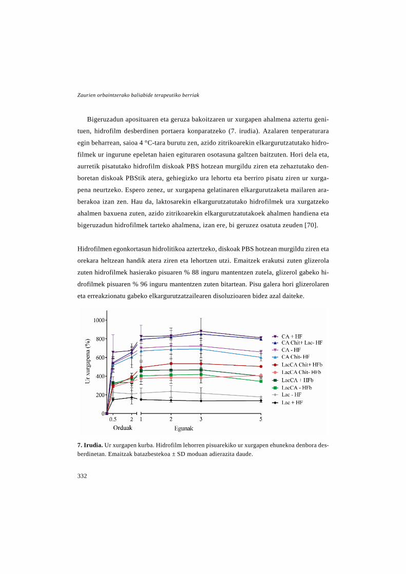
Zaurien orbaintzerako baliabide terapeutiko berriak
332
Bigeruzadun aposituaren eta geruza bakoitzaren ur xurgapen ahalmena aztertu geni-
tuen, hidrofilm desberdinen portaera konparatzeko (7. irudia). Azalaren tenperaturara
egin beharrean, saioa 4 °C-tara burutu zen, azido zitrikoarekin elkargurutzatutako hidro-
filmek ur ingurune epeletan haien egituraren osotasuna galtzen baitzuten. Hori dela eta,
aurretik pisatutako hidrofilm diskoak PBS hotzean murgildu ziren eta zehaztutako den-
boretan diskoak PBStik atera, gehiegizko ura lehortu eta berriro pisatu ziren ur xurga-
pena neurtzeko. Espero zenez, ur xurgapena gelatinaren elkargurutzaketa mailaren ara-
berakoa izan zen. Hau da, laktosarekin elkargurutzatutako hidrofilmek ura xurgatzeko
ahalmen baxuena zuten, azido zitrikoarekin elkargurutzatutakoek ahalmen handiena eta
bigeruzadun hidrofilmek tarteko ahalmena, izan ere, bi geruzez osatuta zeuden [70].
Hidrofilmen egonkortasun hidrolitikoa aztertzeko, diskoak PBS hotzean murgildu ziren eta
orekara heltzean handik atera ziren eta lehortzen utzi. Emaitzek erakutsi zuten glizerola
zuten hidrofilmek hasierako pisuaren % 88 inguru mantentzen zutela, glizerol gabeko hi-
drofilmek pisuaren % 96 inguru mantentzen zuten bitartean. Pisu galera hori glizerolaren
eta erreakzionatu gabeko elkargurutzatzailearen disoluzioaren bidez azal daiteke.
7. Irudia. Ur xurgapen kurba. Hidrofilm lehorren pisuarekiko ur xurgapen ehunekoa denbora des-berdinetan. Emaitzak batazbestekoa ± SD moduan adierazita daude.

Eztabaida
333
Ondoren, hidrofilmen oklusibitatea ebaluatu zen WVTR zehaztuz, baina erabilitako
metodoa nanozuntzezko aposituena neurtzeko erabilitakoaren arinki desberdina izan zen.
Franz difusio zelulak urarekin bete ziren eta haien beso hartzailea parafilmarekin estali
zen. Hidrofilmak bi ganberen artean kokatu ziren, horrela ur lurruna hidrofilmak zehar-
katuz soilik atera zitekeen sistematik. Saioaren hasieran eta 48 ordu ondoren muntaia
pisatu zen WVTRa zehazteko. Azido zitrikoarekin elkargurutzatutako hidrofilmen oklu-
sibitatea ez zen neurtu, izan ere hezetutakoan bi ganberen arteko irekidura estaltzeko
ahalmena galtzen zuten. Bigeruzadun hidrofilmen emaitzek erakutsi zuten zaurien heze-
tasuna era egokian kontrola zezaketela, haien WVTRaren balioa aurretik aipatutako apo-
situ komertzialen balioen tartean baitzegoen. Laktosarekin elkargurutzatutako hidrofil-
mek, ordea, WVTR balio altuagoa izan zuten, oklusibitate baxuagoa adierazten duena.
Laktosarekin elkargurutzatutako hidrofilmen iragazkortasun handiagoa, haien lodiera
txikiagoagatik azal daiteke [71].
Aurreko ikerketetan bezala, behin formulazioa karakterizatuta zegoela, haien bioba-
teragarritasuna aztertu zen, osasun-produktuen ebaluaketa biologikorako ISO 10992-
5:2009 jarraibideen arabera. Jarraibide horiek zitotoxikotasun zuzena eta zeharkakoa
neurtzeko saioak proposatzen dituzte. Hala ere, azido zitrikoarekin elkargurutzatutako
hidrofilmak 37 °C-tara partzialki disolbatzen zirenez, ezin genituen hidrofilm horiek
hazkuntza plaka putzuetatik atera, eta beraz, ezinezkoa izan zitzaigun saio zuzena egitea.
Hori dela eta, zeharkako saioa burutu genuen soilik, hidrofilmetatik askatutako inguru-
nea fibroblastoekin batera 24 orduz inkubatuz, eta orduan haien bideragarritasuna CCK-
8 entseguaren bidez neurtuz. Hidrofilm guztiekin bideragarritasuna % 70-aren gainetik
zegoen, biobateragarriak zirela frogatuz. Horretaz gain, saio honen bidez frogatu genuen
ez zela beharrezkoa aurretiko egokitze pausurik burutzea, saioaren aurretik hidrofilmak
72 orduz dializatuz edo 15 minutuz hidratatuz emaitza berdinak lortu baitziren.
Azkenik, aposituen eraginkortasuna zaurien orbaintzean aztertu zen. Glizerol gabeko hi-
drofilmetan bakarrik burutu zen, izan ere glizerola hidratazio pausuan disolbatzen zenez, bere

Zaurien orbaintzerako baliabide terapeutiko berriak
334
erabilera arbuiagarria zen. Bigeruzadun hidrofilmen artean, azpiko geruzan kitosanoa zuena
soilik ebaluatu zen, kitosanoa eduki edo ez antzeko ezaugarriak zituztenez, kitosanoaren era-
gina aztertzeko. Horretaz gain, laktosarekin elkargurutzatutako geruza bakarreko hidrofilmak
aztertu ziren, baina ez azido zitrikoarekin elkargurutzatutakoak, azken horiek ez zutelako
nahiko egiturazko egonkortasunik haien kabuz apositu moduan erabiliak izateko.
Kasu honetan, eraginkortasuna ez zen db/db sagu ereduaren bidez ebaluatu, giza aza-
lean egindako ex vivo eredu baten bidez baizik, ikerkuntzan animalien erabilera murriz-
teko asmoz. Ardura etikoetaz gain, animalia azala erabili beharrean gizakiena erabiliz,
espezieen arteko orbaintzean dauden desberdintasunak ekidin daitezke [72]. Laburki,
ohiturazko hautazko kirurgietatik lortutako azal biopsiak 6 mm diametroko diskoetan
moztu ziren eta haien erdialdean 3 mm-ko lodiera osoko zauri eszisionalak egin ziren.
Ondoren, biopsiak 24 putzuetako plaketan kultibatu ziren, Transwell sistemetan, epider-
misa aire-likido interfasean mantenduz, 8. irudian ikus daitekeenez. Aldez aurretik hidra-
tatutako hidrofilmak 1. eta 4. egunean ezarri ziren eta biopsiak 8 egunez kultibatu ziren.
4. eta 8. egunetan LDH saio bana burutu zen biopsien bideragarritasuna aztertzeko. Kasu
guztietan bideragarritasuna % 70 baino handiagoa izan zen, biopsietako zelulak bizirik
mantendu zirela esan nahi duena, ex vivo entseguan lortutako emaitzen fidagarritasuna
erakutsiz. Emaitzek fibroblastoetan egindako zitotoxikotasun entseguan lortutako emai-
tzak berretsi zituzten, hidrofilmen biobateragarritasuna geruza bakarreko zelula hazkun-
tza baino egitura konplexuago batean frogatuz.
8. Irudia. Ex vivo saioaren eskema.
Eztabaida
335
Hidrofilmen eraginkortasuna aztertzeko, orbaintzea ebaluatu zen azal laginetan bu-
rututako analisi histologiko eta immunohistokimikoen bidez. Orokorrean, antzeko emai-
tzak lortu ziren hidrofilmekin tratatutako taldeetan eta tratatu gabeko kontroletan, izan
ere, ez zen desberdintasunik behatu zauriaren itxieran, zelulen proliferazioan, zauriaren
uzkurduran eta keratinozito heldugabeen espresioan. Hala eta guztiz ere, laktosarekin
elkargurutzatutako gelatina hidrofilmekin tratatutako taldean kolageno metaketa txikia-
goa behatu zen eta bigeruzadun hidrofilmek keratinozito helduen espresioa oztopatu zu-
ten. Inkubagailuaren hezetasun maila altuak eta hazkuntza medioaren poportzio bat xur-
gatzeko hidrofilmen gaitasunak, zaurian gehiegizko hezetasuna lortzea eragin dezakete,
emaitza negatibo horiek azal ditzakeena. Hori dela eta, hidrofilmak eta batez ere, azido
zitrikoarekin elkagurutzatutako gelatina hidrofilmak, optimizatu beharko lirateke orbain-
tzea sustatu dezakeen ingurune aproposagoa lortzeko.
Ondorioz, ex vivo entseguak erabilitako ereduaren bailagarritasuna erakutsi zuen in
vivo entseguak burutu aurretik orbaintzea era orokorrean aztertzeko metodo bezala. Ho-
rretaz gain, garatutako hidrofilmek apositu moduan erabilgarri izan zitekeela erakutsi
zuen. Haien eraginkortasuna hobetzeko asmoz, etorkizuneko ikerketen helburua azpiko
geruzan hazkuntza faktoreen moduko molekula aktiboak kapsularatzea edo immobiliza-
tzea da.
Laburbilduz, tesi honetan lortutako emaitza denak kontuan hartuz, esan dezakegu lan
honetan zauri kronikoen tratamendurako estrategia berrien garapenean aurrerapausu bat
eman dugula. Urte hauetan, baliabide terapeutiko berriak garatu ditugu zauri kronikoen
orbaintzea sustatzeko, hala nola, LL37 peptidoa kapsularatuta duten NLCak, EGF edo
NLCak kapsularatuta zituzten PLGA-Aloe vera nanozuntzezko mintzak eta gelatina eta
kitosanoan oinarritutako bigeruzadun aposituak.
Erreferentzien zerrenda 202-208 orridaldeetan aurkitzen da.


Ondorioak


Ondorioak
339
Doktoretza tesi honetako ikerketa experimentaletan lortutako emaitzetatik honako
ondorioak eratorri daitezke:
1. LL37a arrakastaz kapsularatu zen NLCen barne, orbaintzean erabiltzeko ezaugarri
egokiak zituen formulazioa lortuz eta peptidoaren ekintza mantenduz kapsularatze
prozesuaren ostean.
2. db/db saguetan burututako lodiera osoko zauri ereduan, NLC-LL37en administrazio
topikoak orbaintzea esanguratsuki hobetu zuen LL37 askearen kontzentrazio berdi-
narekin alderatuz, zauriaren itxiera, errepitelizazio gradua eta hantura prozesuaren
ebazpenari dagokionez.
3. Garatutako EGFdun PLGA eta Aloe veraz osatutako nanozuntzezko mintzek apositu
moduan erabiltzeko ezaugarri egokiak erakutsi zituzten. Horretaz gain, EGFa zein
Aloe vera aktibo mantendu ziren elektroirute prozesuaren ostean. Gainera EGFa kap-
sularatuta zuten mintzek zaurien itxiera eta errepitelizazioa esanguratsuki bizkortu
zuten db/db saguetan burututako lodiera osoko zaurietan.
4. PLGA/Aloe vera nanozuntzezko mintzetan NLCen barneraketak mintzen maneiua
hobetu zuen. Gainera, mintz horiek ere orbaintzea sustatzeko gai ziren, nahiz eta
hein txikiagoan.
5. Gelatinaz osatutako bigeruzadun aposituak garatu ziren, laktosa edo azido zitrikoa
erabiliz elkargurutzatzaile moduan, goiko geruza erresistentea eta beheko geruza po-
rotsua prestatzeko, hurrenez hurren. Garatutako aposituek ezaugarri egokiak zituz-
ten apositu moduan erabiliak izateko eta biobateragarriak ziren, ex vivo entseguak
frogatu zuen bezala.


APPENDIX/ IRUZKINA


APPENDIX 1
Nanotechnological approaches for skin wound regeneration using drug delivery systems
I. Garcia-Orue1,2*, G. Gainza1,2*, S. Villullas3, J.L. Pedraz1,2, R.M. Hernandez1,2, Ma-noli Igartua1,2,+
*These two authors contributed equally to this work. 1 NanoBioCel Group, Laboratory of Pharmaceutics, School of Pharmacy, University of the Basque Country (UPV/EHU). 2 Biomedical Research Networking Centre in Bioengineering, Biomaterials and Nanomedicine (CIBER-BBN). 3 Biopraxis Research AIE, Miñano, Vitoria-Gasteiz.
+Corresponding author: Dr. M. Igartua
ABSTRACT
The application of nanotechnology in medicine represents a great opportunity to en-hance the effectiveness of currently available medical treatments, especially focused on challenging healthcare issues, like skin wound regeneration. Hence, in the last few dec-ades, nanobiomaterials have been extensively studied and optimised for the development of nanoscale drug delivery systems (DDSs) releasing drugs, such as growth factors, cy-tokines or antimicrobials, for skin wound repair. In this regard, several natural or syn-thetic materials have been studied due to their similarities to the skin and biocompatible properties, or due to their antibacterial or antiseptic effect. In addition, materials can also be engineered as scaffolds for the development of novel wound dressings. Thus, this chapter presents an overview of the current nanotechnological approaches used for the controlled release of drugs in the field of skin wound regeneration, particularly empha-sising on polymeric and lipid nanoparticles, silver nanoparticles, nanofibrous structures, nanosheets and nanohybrids.
Published in: Applications of Nanobiomaterials. Volume 5, Nanobiomaterials in Soft Tissue Engineering. William Andrew Publishing. 2016. 31-55
1. ERANSKINA
Farmakoen askatze sistemak erabiliz azaleko zaurien erregenerazio-
rako aukera nanoteknologikoak
LABURPENA
Nanoteknologiaren aplikazioak aukera paregabea dakar medikuntzan, erabilgarri dauden tratamenduen eraginkortasuna hobetzeko, batez ere, azal zaurien erregenerazioaren moduko arazo kliniko erronkarietan. Hori dela eta, azkeneko hamarkadetan, nanobi-omaterialei buruz asko ikertu da, zaurien orbaintzean erabiltzeko nanoeskalan egindako farmakoen askatze sistemak garatzeko, askatzen diztuzten farmakoak hazkuntza faktoreak, zitokinak edo antimikrobianoak izaten direlarik. Horren harira, material sin-tetiko edo natural ugari ikertu dira, azalarekin duten antzekotasunari, biobateragarrita-sunari edo haien eragin antibakterial edo antiseptikoari esker. Horretaz gain, materialak bio-aldamioak garatzeko disenatuak izan daitezke zaurien apositu moduan erabiltzeko. Ondorioz, kapitulo honek orbaintzerako egungo aukera nanoteknologikoei buruzko ikus-pegi orokor bat ematen du, ondorengo farmakoen askatze sistemetan zentratuz: nanopar-tikula polimeriko eta lipidikoetan, zilarrezko nanopartikuletank, egitura nanofibrotsue-tan, nano-orrietan eta nanohibridoetank.

Appendix 1
345
1. Introduction
Non healing wounds have dramatically
increased and have become a great pro-
blem for health care professionals and pa-
tients as pain, diminished quality of life,
frequent hospitalisation, and increased
morbidity and mortality are associated to
chronic ulceration. In developed countries,
up to 2% of the population may be affected
by a chronic wound at least once in their
lifetime, which results in a major health
care and economic burden, representing
around 2% of the health care expenditure
(Menke et al. 2007). In fact, chronic
wounds are a current and future challenge
for health care systems as the demographic
change is leading to a much older popula-
tion. Many other factors than age, associa-
ted with a delay in wound healing, can in-
crease the risk of developing chronic
wounds such as obesity, smoking and ch-
ronic diseases, including diabetes mellitus
and vascular disorders (Menke et al.
2007). In addition, infection is a common
complication that also needs to be addre-
ssed when dealing with chronic wounds as
it can delay or impair the wound healing
process.
Current therapies involve costly and
long-lasting treatments that are often insu-
fficient and are associated with high ulcer
recurrence (estimated up to 70%). Thus,
more effective novel treatments are requi-
red to solve this unmet need so that the re-
lated tremendous health care costs and re-
sources can be optimised. For that pur-
pose, the scientific community has focu-
sed many of its efforts on developing new
therapies to promote wound healing and
on improving already available treatments
to, ultimately, accelerate and achieve
wound closure. To this aim, the use of na-
notechnology has provided a great tool to
develop new drug delivery systems
(DDSs) for the sustained release of thera-
peutic agents in order to achieve more
cost-effective therapies.
This chapter will provide a general
overview of useful therapeutic agents,
such as growth factors (GFs) and antimi-
crobials, employed in wound healing the-
rapy, and the nanotechnological appro-
aches developed so far to improve tre-
atment effectiveness. A detailed insight of
the current advances on DDS development
for wound healing therapy will be given,
Novel therapeutic approaches for wound healing
346
mainly focusing on polymeric and lipid
nanoparticles, silver nanoparticles, nanofi-
brous structures, nanosheets and nano-
hybrids.
2. Cutaneous wound healing and skin le-
sions
The skin, the outer tissue protecting
against the environment, is exposed to
chemical, physical and biological harms
as well as subject of genetic, inflamma-
tory or metabolic diseases. Aging is asso-
ciated with a loss or decrease of some of
the protective properties of the skin as it
becomes dryer, less elastic, thinner, and
thus being more likely to suffer damage
and infection. Senescent cells accumula-
ted in aging tissues lead to a decline in
several functions of the human skin, such
as barrier efficacy, sensory perception,
wound healing, immune response and
DNA repair. Furthermore, changes in co-
llagen formation, flattening of the base-
ment membrane, a reduction in blood
supply to the skin and a slower inflamma-
tory response reduce the ability to achi-
eve skin repair (Farage et al. 2009).
Cutaneous wound healing is a well-co-
ordinated biological process that involves
the integrated interaction of different
growth factors, cytokines, enzymes and
cell types, such as inflammatory cells, ke-
ratinocytes, fibroblasts and endothelial
cells. Tissue repair is accomplished by
tightly regulated processes that overlap in
space and time, including an initial inflam-
matory response, a proliferative phase and
a final remodelling phase (Martin 1997,
Singer, Clark 1999). Chronic wounds fail
to proceed through those sequential pha-
ses, subsequently resulting in delayed or
incomplete healing.
Multiple factors contribute to poor
wound healing. Among them, a major role
has been attributed to an abnormal and
persistent inflammatory response, a hall-
mark of non-healing skin wounds, that le-
ads to an excessive proteolytic activity due
to protease inhibitor degradation, and ulti-
mately to inflammation-mediated tissue
damage (Trengove et al, 1999). A decrea-
sed vascular supply is also a common
cause of ulcer formation. In addition, a re-
duction in the amount of available and biolog-

Appendix 1
347
ically active growth factors in the wound en-
vironment particularly affects wound repair.
One of the most common complicati-
ons of chronic wounds is infection. In
open wounds there is a disruption of the
intact skin that is the first mechanical de-
fence line against infection. Elevated le-
vels of bacterial load are typically present
on open wounds and can stimulate a proin-
flamatory environment. Hence, the wound
healing process can be impaired partially
due to an elongation of the inflammatory
phase that suppresses the regenerative
phase (Hernandez 2006, Lipsky, Hoey
2009). Bacteria not only compete for the
limited oxygen and nutrients present in the
wound, they also release toxins and induce
an increased production of enzymes that
can further lead to cellular failure.
Chronic wounds include pressure, vas-
cular (venous or arterial) and diabetic foot
ulcers. A pressure ulcer is defined as an
area of localised damage on the skin
and/or underlying tissue, usually over a
bony prominence, occurred as a conse-
quence of pressure or shear and/or a com-
bination of those (Black et al. 2007). Vasc-
ular ulcers, usually localised on the lower
limbs, are caused by a circulation disorder
that leads to a reduction of the arterial
blood flow or an impaired venous blood
return. Vascular ulcers constitute a group
of lesions of particular relevance due to
their high prevalence. In addition, these ul-
cers are a leading cause of morbidity
among patients suffering from peripheral
vascular disease (Markova, Mostow
2012). Lastly, diabetic foot ulcer (DFU) is
a major complication of diabetes because
of the high consequences produced on the
patients’ quality of life. DFUs are caused
due to a number of contributing factors,
such as mechanical changes in conforma-
tion of the bony architecture of the foot,
peripheral neuropathy (damaged nerves)
and peripheral vascular disease (block ar-
teries), all of which occurred with higher
frequency and intensity in the diabetic po-
pulation (Eldor et al. 2004). In addition,
DFUs are highly associated with non-trau-
matic lower extremity amputations in the
industrialised world.
On the other hand, major burns produ-
ced by heat, chemicals, electricity or radi-
ation, can also became chronic wounds. A

Novel therapeutic approaches for wound healing
348
burn injury affects the skin integrity lea-
ding to fluid loss and being a portal for
bacterial infection. Severity of a burn de-
pends on the depth, location and the body
surface area injured.
3. Therapeutic agents for wound healing
therapy
3.1 Growth Factors
Growth factors (GF) are biologically
active polypeptides which regulate cell
growth, differentiation, proliferation and
migration, as well as, protein expression
and enzyme production. In addition, GFs
have a potential ability to heal wounds by
stimulating angiogenesis, modulating the
inflammatory response and intervening in
the production and degradation of the ex-
tracellular matrix and the granulation tis-
sue (Barrientos et al. 2008, Bodnar 2013).
The main GFs involved in the healing
process and skin regeneration are the
platelet derived growth factor (PDGF),
epidermal growth factor (EGF), basic fi-
broblast growth factor (bFGF), insulin-
like growth factor (IGF) and vascular en-
dothelial growth factor (VEGF) families.
Their functions are summarised in Table
1.
The external administration of GFs has
become one of the most interesting strate-
gies to promote wound healing and skin
regeneration, as a GF level decrease has
been reported in chronic wounds (Barrien-
tos et al. 2008, Barrientos et al. 2014).
Nevertheless, the proteases present in the
lesion, able to degrade the GFs, along with
the low in vivo stability of these molecules
have limited their clinical application.
(Mast, Schultz 1996, Ulubayram et al.
2001, Baldwin, Mark Saltzman 1998). In
this regard, in the last years the nanotech-
nological approaches for the release of
GFs have been expansively investigated in
order to improve the stability of the GFs at
the wound site, allowing their sustained
release, and ultimately, optimising treat-
ment effectiveness.
3.2 Antimicrobial agents
One of the most common complica-
tions in the treatment of chronic wounds
are bacterial infections. In fact, 75% of the
mortality associated to burn injuries are
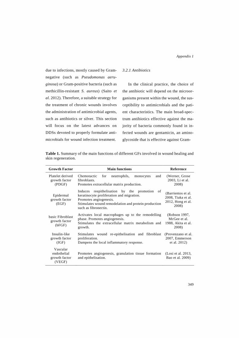
Appendix 1
349
due to infections, mostly caused by Gram-
negative (such as Pseudomonas aeru-
ginosa) or Gram-positive bacteria (such as
methicillin-resistant S. aureus) (Saito et
al. 2012). Therefore, a suitable strategy for
the treatment of chronic wounds involves
the administration of antimicrobial agents,
such as antibiotics or silver. This section
will focus on the latest advances on
DDSs devoted to properly formulate anti-
microbials for wound infection treatment.
3.2.1 Antibiotics
In the clinical practice, the choice of
the antibiotic will depend on the microor-
ganisms present within the wound, the sus-
ceptibility to antimicrobials and the pati-
ent characteristics. The main broad-spec-
trum antibiotics effective against the ma-
jority of bacteria commonly found in in-
fected wounds are gentamicin, an amino-
glycoside that is effective against Gram-
Table 1. Summary of the main functions of different GFs involved in wound healing and skin regeneration.
Growth Factor Main functions Reference
Platelet derived growth factor
(PDGF)
Chemotactic for neutrophils, monocytes and fibroblasts. Promotes extracellular matrix production.
(Werner, Grose 2003, Li et al.
2008)
Epidermal growth factor
(EGF)
Induces reepithelisation by the promotion of keratinocyte proliferation and migration. Promotes angiogenesis. Stimulates wound remodelation and protein production such as fibronectin.
(Barrientos et al. 2008, Tiaka et al. 2012, Hong et al.
2008)
basic Fibroblast growth factor
(bFGF)
Activates local macrophages up to the remodelling phase. Promotes angiogenesis. Stimulates the extracellular matrix metabolism and growth.
(Robson 1997, McGee et al.
1988, Akita et al. 2008)
Insulin-like growth factor
(IGF)
Stimulates wound re-epithelisation and fibroblast proliferation. Dampens the local inflammatory response.
(Provenzano et al. 2007, Emmerson
et al. 2012)
Vascular endothelial
growth factor (VEGF)
Promotes angiogenesis, granulation tissue formation and epithelisation.
(Losi et al. 2013, Bao et al. 2009)
Novel therapeutic approaches for wound healing
350
negative bacteria, especially Pseudomo-
nas spp and certain Gram-positive (such as
S. aureus) (Chen et al. 2012b); mupirocin
commonly used in wound care prophylaxis
against S. aureus and against some Gram-
negative flora (Thakur et al. 2008); tetra-
cycline, a broad spectrum antibiotic that,
besides being active against a wide range
of Gram-positive and Gram-negative bac-
teria, is effective against Chlamydia, My-
coplasma, Rickettsia and protozoan para-
sites (Chopra, Roberts 2001); and ciprof-
loxacin, the most potent fluoroquinolone
active against a wide range of bacteria, be-
ing the most susceptible the aerobic Gram-
negative bacilli (Sharma et al. 2010).
Narrow range antibiotics such as peni-
cillin G, vancomycin and lysostaphin, are
employed for wound healing treatment.
On the one hand, penicillin G is active
against non-β-lactamase-producing Gram-
positive bacteria, anaerobes and Gram-
negative cocci from the Neisseria spp
(Doi, Chambers 2015). On the other hand,
vancomycin is a glycopeptide active
against Gram-positive bacteria and is
mainly used in recalcitrant staphylococcal
infections that are resistant to penicillin or
cephalosporin (Chen et al. 2012b). Fi-
nally, lysostaphin is a cell lytic enzyme se-
creted by Staphylococcus simulans that is
highly effective and specific against S. au-
reus (Miao et al. 2011).
Another encouraging strategy to over-
come wound bacterial infection is the ad-
ministration of antimicrobial peptides
(AMP). AMP are part of the innate im-
munity providing a first defence line
against a wide range of pathogenic or-gan-
isms (Vandamme et al. 2012). Currently,
there are more than 600 AMP classified in
two families: defensins and cathelicidins
(Hancock 2001). The LL37 peptide, also
known as hCAP-18, is the only human
cathelicidin identified to date. This pep-
tide exerts antimicrobial activity towards
various microorganisms, such as Gram-
positive and Gram-negative bacteria,
fungi, parasites and enveloped virus. Fur-
thermore, LL37 is chemotactic (Vanda-
mme et al. 2012), stimulates the produc-
tion of proinflammatory cytokines, neu-
tralises the immune response through the
interaction with the LPS endotoxin (Kai-
Larsen, Agerberth 2008), promotes new
vessel formation (Koczulla et al. 2003)
Appendix 1
351
and stimulates the proliferation and migra-
tion of epithelial cells (Anderson, Rehders
& Yu 2008). Therefore, LL37 is emerging
as a potential therapeutic tool for promot-
ing wound healing and inhibiting bacterial
growth. However, peptides have shown
limited stability in vivo due to the prote-
ases present in the wound. Thus, optimisa-
tion of the LL37 administration using ad-
vanced DDSs can enhance its biological
function and thus improve treatment effec-
tiveness (Hancock 2001).
Finally, it should be noted that most of
the antimicrobials employed in the treat-
ment of infected wounds are administered
through the systemic route. However, this
approach can lead to the appearance of un-
desirable side effects or to an insufficient
dosage on the lesion as the antibiotics can
barely penetrate into ischemic tissues (Xu
et al. 2010). In addition, the extensive use
and misuse of antibiotics can contribute to
the development of antibiotic-resistant
bacteria. In order to overcome these disad-
vantages, antibiotics formulated in ad-
vanced DDSs can be locally administered
to obtain higher concentrations at the
wound site due to a high initial drug re-
lease followed by a sustained release that
can achieve a long-lasting antimicrobial
effect (De Cicco et al. 2014).
3.2.2 Silver
The use of silver is a widely employed
strategy to overcome bacterial infection
and prevent wound sepsis. Sil-ver has
shown several interesting properties, such
as wide spectrum against several microor-
ganisms; multiple mechanisms of action to
inhibit bacterial colonisation, which re-
duces the risk of developing resistance;
great effectiveness against multi-drug-re-
sistant organisms and low systemic tox-
icity (Gunasekaran, Nigusse & Dhanaraju
2011).
Only the soluble form of silver is bio-
logically active, i.e. Ag+ or Ag0 clusters.
Ag+ ions are present in silver nitrate, sil-
ver sulphadiazine (SSD) and other ionic
silver compounds. On the other hand, me-
tallic silver (Ag0) is found in two different
crystalline forms, i.e. the nanocrystalline
(<20 nm) or the subcrystalline (less than

Novel therapeutic approaches for wound healing
352
eight atoms of silver) forms (Dunn, Ed-
wards-Jones 2004).
The mechanism of action of soluble sil-
ver is still unknown; however, the most
widely accepted mode of action is closely
related to the interaction of silver with the
bacterial cell-wall and cell-membrane that
inhibits the bacteria respiration process
(Klasen 2000). Further-more, silver is in-
volved in the bacterial DNA condensation,
which leads to the loss of the replication
ability and therefore, to the cell death
(Feng et al. 2000). As a result of the mul-
tiple bactericidal mechanisms of soluble
silver, this noble metal does not produce
bacterial resistance; thus, becoming an in-
teresting strategy for the treatment of in-
fected wounds.
4. Nanotechnological approaches for the
release of therapeutic agents for wound
healing
Currently, several research groups have
developed different nanotechnological
approaches for the release of therapeutic
agents as a novel strategy in the wound he-
aling therapy. Due to the distinct characte-
ristics of wounds and healing stages, diffe-
rent wound dressings and therapeutic
agents can be employed to meet most of
the needs in a particular wound stage. The-
refore, in this section, the current nano-
technology based strategies used for the
controlled release of drugs involved in
skin wound regeneration are summarised
(Figure 1).
4.1 Nanotechnological approaches for
growth factor delivery
As previously mentioned nanotechno-
logy provides the opportunity to enhance
the in vivo effectiveness of GFs, preven-
ting their proteolytic degradation and pro-
longing their release at the lesion (Kubi-
nová, Syková 2010). A wide type of na-
noscale-delivery systems have been desig-
ned to promote wound healing, mainly po-
lymeric nanoparticles (NPs), lipid NPs and
nanofibrous structures.
4.1.1 Polymeric nanoparticles
Biocompatible polymeric devices have
currently become a promising alternative
for the controlled release of active com-

Appendix 1
353
Figure 1. Schematic illustration of different DDSs used for the treatment of chronic wounds.
pounds, including GFs. These nanometric
colloidal systems can be employed in mul-
tiple diseases, administration routes and
dosage ranges. Therefore, polymeric carri-
ers have been developed for chronic
wound therapy because apart from allo-
wing controlled drug release over time,
they may also reduce drug degradation by
the prote-ases present in the wound envi-
ronment (Ulubayram et al. 2001, Gainza et
al. 2013), and thus, the administration fre-
quency and dose can be decreased. Nanos-
pheres can be prepared using natural or
synthetic biomaterials, as well as other po-
lymer combinations (Makadia, Siegel
2011, Değim 2008, Ye, Kim & Park 2010).
One of the most widely used polymer
to prepare nanospheres as DDSs is the syn-
thetic poly lactic-co-glycolic acid
(PLGA), because this polymer allows drug
sustained release, is biocompatible and bi-
odegradable (Makadia, Siegel 2011). In
this regard, Chu and co-workers developed
rhEGF-loaded PLGA NPs using a modi-
fied double-emulsion method and assessed
their effectiveness during 21 days after to-
pical administration in full-thickness
Novel therapeutic approaches for wound healing
354
wounds created on diabetic rats. The
rhEGF-NPs (size around 193.5 nm) sho-
wed an encapsulation efficiency (EE) of
85.6% and a rhEGF release for 24 h; the-
refore, treatments were locally administe-
red once daily. After 7, 14 and 21 days
wounds treated with rhEGF-NPs achieved
the greatest wound area reduction among
all the experimental groups. In addition,
the histopathological examination of lesi-
ons showed that the rhEGF-NPs promoted
the highest level of fibroblast proliferation
and PCNA expression. These results su-
pport that the controlled release of rhEGF
allows continuous contact of the GF with
the wound surface to maintain an effective
concentration able to promote healing; and
suggest a more appropriate administration
for rhEGF (Chu et al. 2010).
Latest advances in drug delivery invol-
ved the combination of different technolo-
gical approaches that may improve dre-
ssing integration. In this regard, Losi et al.
have developed poly-(ether)urethane-po-
lydimethylsilxane/fibrin-based scaffolds
containing VEGF and bFGF loaded PLGA
NPs that have demonstrated to be effective
in enhancing wound closure by day 15 in
db/db mice compared with scaffolds not
containing GFs. In addition, as observed in
the histological evaluation, complete re-
epithelisation, improved collagen deposi-
tion and more mature granulation tis-sue
were observed in scaffolds containing
GFs-loaded NPs, even though no differ-
ence between scaffolds containing free
GFs and GFs-loaded NPs were found.
Overall, these results suggest that fibrin-
based scaffolds containing GFs-loaded
NPs are an interesting strategy for the sus-
tained release of VEGF and bFGF because
they acted as a double DDS for GF deliv-
ery; and therefore, the initial burst release
of the GFs and the leak out of the particles
from the wound site after topical admin-
istration might be reduced providing a more
sustained release of the GFs (Losi et al.
2013).
4.1.2 Lipid nanoparticles
Lipid nanoparticles have been inten-
sively studied for dermal application,
pharmaceutical and cosmetic uses (Par-
deike, Schwabe & Müller 2010). These li-
pid carriers look promising due to their bi-
ocompatibility, ease of preparation and su-

Appendix 1
355
perior efficacy compared with non-encap-
sulated drugs (Fan, Zhang 2013).
First developed lipid nanocarriers were
liposomes, comprising an aqueous core
encapsulated by natural or synthetic phos-
pholipids. This structure allows entrap-
ment of hydrophilic drugs in the core,
while hydrophobic drugs are entrapped
within the lipid bilayers (Fan, Zhang
2013). Since the first liposomal pharma-
ceutical product, Doxil® [doxorubicin-
polyethylenglycol (PEG)ylated liposome]
approved in 1995 for Kaposi's sarcoma,
ovarian and breast cancer, to date, there
are a number of commercially available
products. (Allen, Cullis 2013).
Few publications have described the
use of liposomes as GF carriers for wound
healing. First, Brown and collaborators re-
ported the benefits of locally administered
EGF loaded multilamellar liposomes on
the tensile strength of experimental inci-
sions on rats. As observed in the measure-
ment of the rate of rhEGF loss from inci-
sions in vivo, significant higher levels of
the GF were retained by the multilammelar
liposomes (62% after 1 day, 48% after 2
days and 11% after 5 days). In addition, a
single dose of EGF in liposomes signifi-
cantly prolonged the exposure of EGF,
produc-ing a 200 % increase in wound ten-
sile strength from days 7 to 14, and in-
creasing collagen formation and fibroblast
proliferation. These studies clearly proved
that the incorporation of the EGF in an ap-
propriate vehicle induced more efficient
wound healing than local administration of
the free GF, since EGF-loaded liposomes
improved early tensile strength and there-
fore promoted wound remodelation and
healing (Brown et al. 1988). In a different
study published by Pierre and colleagues,
very low doses of IGF-I encapsulated in lip-
osomes (0.9 μg/kg/week) administered sub-
cutaneously were as effective improving re-
epithelisation as higher doses of IGF-I (5.0
μg/kg/week) administered together with the
growth hormone (GH) and more effective
than 5.0 μg/kg/week IGF-I without GH ad-
ministration (Pierre et al. 1997). These data
suggest that nanoencapsulation may im-
prove treatment effectiveness compared
with the free GF administration because
this technological approach may protect the
GFs from the wound environment and deg-
radation (Almeida, Souto 2007).

Novel therapeutic approaches for wound healing
356
However, despite liposomes are well
established and extensively investigated,
the total number of products in the market
is still limited. One of the reasons may be
related to the low in vivo stability that may
induce a rapid uncontrolled release of the
drug. Therefore, at the beginning of the
1990s solid lipid nanoparticles (SLN) and
later, nanostructured lipid carriers (NLC)
were developed as alternative carrier sys-
tems to liposomes (Müller, Mäder &
Gohla 2000). SLN preparation requires
solid lipids as matrix material while NLC
are prepared using a blend of solid lipids
and oils. In addition, both nanoparticu-
lated formulations are ideal for dermal ad-
ministration because they are able to re-
main on the skin, improve skin hydration
and release drugs in a sustained manner.
However, since SLN have shown drug ex-
pulsion during the storage, NLC may be a
more suitable alternative for hydrophilic
drug encapsulation due to their enhanced
stability (Almeida, Souto 2007, Pardeike,
Schwabe & Müller 2010).
Based on this nanotechnology, our re-
search group has demonstrated that
rhEGF-loaded SLN and NLC topically ad-
ministered were more effective improving
Figure 2: rhEGF-loaded nanostructured lipid carriers (rhEGF-NLC) (A) Wound images obtained from each experimental group on days 15, 25, 36 and 43. (B) Representative pictures of endothe-lial cells marked with anti-CD 31 antibody on days 15, 25 and 43 (x10 magnification). Reproduced and adapted with permission from Gainza et al. 2015 (©2014)

Appendix 1
357
healing than a greater dose of free rhEGF
intralesionally administered. First, we as-
sessed the effectiveness of both formula-
tions in healing-impaired db/db mice. The
healing process was improved in terms of
wound closure, re-epithelisation and resto-
ration of the inflammatory process in those
wounds treated with two weekly doses of
topical rhEGF-SLN and rhEGF-NLC over
controls, including a significantly higher
dose of intralesional free rhEGF (20 μg vs
75 μg)) (Gainza et al. 2014). Secondly, we
assessed the effectiveness of the rhEGF
developed nanoformulations in a more rel-
evant preclinical model, such as Large
White pigs. Because rhEGF-NLC showed
an improved EE compared with rhEGF-
SLN, the lack of organic solvent needed
for their preparation and the same in
vitro/vivo effectiveness as rhEGF-SLN;
the effect tiveness of two weekly topical
administrations of 20 μg rhEGF-NLC was
assessed in a full-thickness swine exci-
sional wound model compared with
empty NLC and two weekly intralesional
administrations of 75 μg free rhEGF. Data
obtained from the swine model supported
the results previously obtained in rodents,
since by day 25, lower doses of rhEGF-
NLC increased the wound closure and per-
centage of healed wounds as much as that
exerted by free rhEGF, but more than
empty NLC (Figure 2A). In addition, the
histopathological examination confirmed
that rhEGF-NLC promoted fibroblast mi-
gration and proliferation, collagen deposi-
tion, resolution of the inflammatory re-
sponse and, as observed in Figure 2B, ne-
oangiogenesis (Gainza et al. 2015). Over-
all, these findings demonstrated that lower
doses of topically administered rhEGF-
NLC may stimulate healing of full thick-
ness wounds in rodents and pigs, thereby
confirming their potential clinical applica-
tion for wound management.
4.1.3 Nanofibrous structures
Nanofibrous materials are gaining in-
terest for tissue engineering applications,
including skin regeneration. Nanofibrous
structures mimic the native extracellular
matrix and promote the adhesion of vari-
ous cells and soluble factors that may pro-
mote cell function and tissue regeneration.
Nanofibers (NFs) for skin tissue regenera-
tion are mainly developed using the elec-
trospinning technique because it allows

Novel therapeutic approaches for wound healing
358
the production of micro and nanometric
continuous fibres that have shown a uni-
form adhesion to the lesion without fluid
accumulation (Chaudhury et al. 2014).
In the last few years several studies
have been performed to assess the effect of
NFs in the controlled release of GFs for
wound repair. Different materials, natural
or synthetic, can be used for the fabrica-
tion of electrospun NFs. On the one hand,
silk fibroin from Bombyx mori silkworm,
a natural polymer, has demonstrated ade-
quate mechanical strength and excellent
biocompatibility for skin regeneration ap-
plications. Silk mats containing EGF have
shown to maintain their structure during
the healing process and to enhance healing
by inducing the keratinocyte tongue pro-
liferation and thus, promoting re-epitheli-
sation. Therefore, the silk mats offered not
only physical protection of the lesion, but
also sustained release of EGF to the wound
(Schneider et al. 2009). In addition, the ef-
fectiveness of silk-protein biomaterials
with EGF and silver sulphadiazine has
been studied in lesions created on mice.
Wound biopsies revealed that those dress-
ings, as well as the other lamellar porous
films evaluated in that study, provided the
most rapid wound closure, also showing
higher healing rate, re-epithelisation, der-
mis proliferation and collagen synthesis
compared with the air-permeable Te-
gaderm® (Gil et al. 2013).
Synthetic biomaterials are also widely
employed for the preparation of NFs for
wound repair. For example, bFGF-loaded
electrospun nanofibers developed using
the synthetic poly(ethylene glycol)-
poly(DL-lactide) (PELA) polymer for the
treatment of diabetic skin ulcers, com-
pared with NFs infiltrated with free bFGF,
significantly improved the healing rate by
promoting re-epithelisation and regenera-
tion of skin appendages and the ECM re-
modellation. In addition, the obtained sus-
tained release of bFGF enhanced collagen
deposition and improved the density and
maturity of capillaries during two weeks
(Yang et al. 2011).
Due to the practicality, easy fabrication
and the successful results NFs have
demonstrated, several research groups are
currently working on the development of
advanced nanofibrous structures combin-

Appendix 1
359
ing two or more polymers. In addition, due
to the increased recognition of photother-
apy to promote wound healing, photosen-
sitive polymers are also used for NF prep-
aration. In this regard, poly(3-hexylthio-
phene) (P3HT), a photosensitive polymer,
and EGF encapsulated in the core-shell-
structured gelatin/poly(L-lactic acid)-co-
poly-(ε-caprolactone) (PLLCL) NFs
(EGF-Gel/PLLCL/P3GF(cs) prepared by
coaxial spinning have been evaluated as
skin graft. EGF-Gel/PLLCL /P3GF(cs)
significantly improved human dermal fi-
broblast proliferation and wound closure
under light stimulation in an in vitro
wound model compared with fibroblasts
treated with those NFs but under dark con-
ditions. Moreover, EGF-
Gel/PLLCL/P3GF(cs) treatment in adi-
pose-stem cells under light stimulation in-
duced keratinocyte differentiation (Jin,
Prabhakaran & Ramakrishna 2014).
Combinations of poly(ε-caprolactone)
(PCL) and polyethylenglycol (PEG) poly-
mers for NF preparation have also been
studied. For example, the healing effec-
tiveness of rhEGF surface-immobilised
NFs assessed in diabetic burns created on
the back of C57BL/6 female mice showed
an increased healing rate on days 7 and 14
compared with empty NFs and the un-
treated control (Choi, Leong & Yoo 2008).
Remarkably, wound closure in those
wounds treated with rhEGF-NFs was sig-
nificantly greater than in those treated with
a mixture of rhEGF and NFs. These data
suggest that the entrapment of rhEGF
within NFs may prevent the GF degrada-
tion from wound proteases. Moreover, the
histological examination also confirmed
that lesions treated with rhEGF-conju-
gated NFs showed increased keratinocyte
specific genes and EGF receptor (EGFR)
expression; thus, promoting wound heal-
ing (Choi, Leong & Yoo 2008). Moreover,
two different growth factors, EGF and
bFGF, were also incorporated in the coax-
ial NFs to obtain a bi-phasic release of the
GFs (Choi, Choi & Yoo 2011). EGF was
surface immobilised and minimal release,
related to the NFs mesh degradation, was
shown during 7 days while bFGF, immo-
bilised on the core, showed an initial burst
and an approximately 30% release of the
encapsulated bFGF in the first 12 h. In ad-
dition, studies undertaken in diabetic
burns created on the back of C57BL/6 fe-

Novel therapeutic approaches for wound healing
360
male mice clearly demonstrated that con-
trolled release of EGF and bFGF from NFs
further accelerated proliferation of epider-
mal cells and wound closure than the con-
trol groups, EGF-NFs and bFGF-NFs.
Moreover, animals treated with
EGF/bFGF NFs improved collagen and
keratin accumulation; confirming the nan-
ofibrous matrix as a promising wound
dressing to improve wound healing (Choi,
Choi & Yoo 2011).
Combinations of different DDSs have
been also used as multifunctional wound
dressings. As an example, a biomimetic
nanofibrous scaffold containing PDGF-
BB loaded PLGA NPs and VEGF loaded
chitosan, [poly(ethylene oxide)] (PEO)
NFs were developed to achieve dual re-
lease of the two GFs (fast and sustained re-
lease) (Xie et al. 2013). This system al-
lowed the quick release of VEGF to pro-
mote new vessel formation and provided
PDGF-BB sus-tained release throughout
the whole healing process (Figure 3A).
Studies performed on a rat full-thickness
wound model, depicted in Figure 3B, re-
vealed an accelerated wound closure of
Figure 3: Biomimetic nanofibrous scaffold containing PDGF-BB loaded PLGA NPs and VEGF loaded CS-PEO NFs (A) Release kinetics of VEGF from nanofibers and PDGF-nanoparticles within fibers. (B) Representative macroscopic appearance of wound closure on weeks 0, 1, 2, and 4 after wound treatment with two controls (open wound and Hydrofera Blue®) and nanofiber meshes (2:1 CS/PEO) without GFs and nanofiber meshes with nanoparticles and GFs (2:1 CS/PEO-NPs). Reproduced and adapted with permission from Xie et al. 2013 (©2013).

Appendix 1
361
those wounds treated with the biomimetic
nanofibrous scaffold compared with the
untreated group, empty NFs and the com-
mercial dressing Hydrofera Blue®. In ad-
dition, as observed in the histological ex-
amination, VEGF may be responsible of
the increased angiogenesis observed in the
early stage of the healing process, while
PDFG-BB may improve re-epithelisation,
collagen deposition and tissue remodel-
ling (Xie et al. 2013).
Finally, a more complex NP in-fiber
structures have also been developed using
collagen and hyaluronic acid for the pro-
grammable multi angiogenic GF release
(Lai et al. 2014). In this occasion, bFGF
and EGF were directly embedded in the
nanofibrous skin equivalent and PDGF-
BB and VEGF were encapsulated into gel-
atine NPs and embedded in the NF meshes.
Results obtained from lesions created on
STZ-diabetised rats treated with this com-
plex structure revealed that the initial de-
livery of bFGF and EGF may mimic the
early stage of healing by accelerating epi-
thelisation and angiogenesis, while the
sustained release of PDGF and VEGF may
promote tissue remodelation, blood vessel
maturation and collagen deposition. Over-
all, these findings demonstrated that this
advanced DDS could be a promising bio-
engineered construct for wound therapy
(Lai et al. 2014).
4.2 Nanotechnological approaches for an-
timicrobial agents delivery
4.2.1 Antibiotics
Among the current nanotechnological
approaches for chronic wound treatment,
nanoparticles, nanofibers and nanosheets
to release antibiotics can be highlighted.
4.2.1 Polymeric nanoparticles
Polymeric NPs are one of the most
promising DDSs for antibiotics (Cortivo et
al. 2010). As previously mentioned, it is
widely assumed that drug nanoencapsula-
tion can improve the drug stability within
the wounded area. In this regard, penicillin
G loaded polyacrylate NPs have proven to
protect penicillin against β-lactamase,
since they present an equipotent in vitro
antibacterial activity against methicillin-
susceptible and methicillin-resistant

Novel therapeutic approaches for wound healing
362
strains of S. aureus (Turos et al. 2007).
Furthermore, the application of the NP
emulsion directly to skin abrasions in mice
accelerated wound healing com-pared
with saline administration and showed no
discernible toxicity, i.e. no redness, irrita-
tion or inflammation. In addition, the pol-
ymeric film formed by the NP emulsion
applied over the wound protected the le-
sion from microorganisms; so that these
wound dressings appeared to be an integral
part of the forming skin layers, maintain-
ing the skin mechanical properties, pre-
serving the moisture and allowing oxygen-
ation of the lesion (Greenhalgh, Turos
2009).
A different wound dressing based on
an in situ gelling nanoparticulated dry-
powder composed of alginate-pectin
loaded with gentamicin sulphate has been
developed. Remarkably, due to the rapid
gelation of the nanoparticulated powder,
this nanoformulation was able to absorb
large quantities of wound fluid and there-
fore microorganism colonisation could be
reduced. Moreover, since the formed hy-
drogel exhibited proper adhesiveness, easy
dressing removal from the wound was al-
lowed. The antimicrobial activity of the
gentamicin-sulphate loaded NPs incorpo-
rated into the hydrogel was tested against
S. aureus and P. aeruginosa the most com-
mon bacteria present in infected wounds,
revealing a higher antibacterial activity
compared with the free drug (De Cicco et
al. 2014).
Finally, regarding the controlled deliv-
ery of AMPs, Chereddy and collaborators
incorporated LL37 in PLGA nanospheres
in order to improve the in vivo stability of
the peptide. As observed in the in vitro
studies, both PLGA-LL37 and free LL37
showed to be no cytotoxic and to improve
wound healing in the HaCaT cell line; and
presented anti-inflammatory activity by
downregulating TNF-α in macrophages.
However, no significant increase of prolif-
eration was observed in HaCaT and in the
fibroblast cell line BJ. In addition, PLGA-
LL37 nanospheres improved the wound
healing rate compared with free LL37 in a
full-thickness wound model in mice, and
presented a similar wound closure than
wounds treated with the plasmid encoding
hCAP18. Moreover, PLGA-LL37 pro-
moted angiogenesis and diminished the in-

Appendix 1
363
flammatory infiltration, since IL-6 and
VEGF were upregulated and the myelop-
eroxidase activity decreased (Chereddy et
al. 2014).
4.2.1.2 Nanofibrous structures
Wound dressings based on antibiotic-
loaded nanofibrous scaffolds are another
encouraging approach for wound treat-
ment.
In a study published by Miao and col-
laborators, NFs were prepared by electro-
spinning cellulose, cellulose-chitosan or
cellulose-poly(methylmethacrylate)
(PMMA). NFs were then functionalised by
conjugation with lysostaphin. In addition,
the method of conjugation, which depends
on the polymer used, may have a great im-
pact on the antibacterial activity of the an-
tibiotic-loaded NFs. In this regard, the
conjugation of lysostaphin and cellulose-
nanofibers by covalent bonds produced the
most bioestable formulation and the best
anti-infective properties, as this formula-
tion showed complete neutralisation of S.
aureus. Furthermore, minimal cytotoxicity
in the keratinocyte cell line HaCaT was
shown in biocompati-bility assays and an-
timicrobial activity was revealed in a skin-
like model consisting of a monolayer cul-
tured keratinocytes infected with S. aureus
(Miao et al. 2011).
Additionally, NFs prepared with so-
dium alginate have also shown to be suita-
ble for wound treatment due to the bio-
compatible and biodegradable properties
of alginate. Sodium alginate NFs have
been prepared by the electrospinning tech-
nique, incorporating PVA into the polymer
solution in order to avoid the early gela-
tion of sodium alginate. In this occasion,
ciprofloxacin, loaded during the electro-
spinning process, was employed as the ac-
tive compound of the wound dressing.
Studies carried out in a full thickness
model in rabbits demonstrated that ciprof-
loxacin loaded PVA-sodium alginate NFs
enhanced wound epithelisation compared
with PVA-NFs and a commercial ciprof-
loxacin formulation (Kataria et al. 2014).
As previously mentioned, NFs can also
be prepared using synthetic polymers. For
that purpose, a PEG-PLLA nanofibrous
membrane loaded with tetracycline had
Novel therapeutic approaches for wound healing
364
been successfully developed, showing no
loss of the tetracycline bioactivity, since
the membranes were effective inhibiting S.
aureus for 5 days (Xu et al. 2010).
Electrospun scaffolds can also be
loaded with several drugs. As an example,
a PLLA scaffold containing an antibiotic,
mupirocin, and an anesthetic, lidocaine hy-
drochloride had been developed. In order to
achieve a different release for each drug, a
dual spinneret electrospinning apparatus
was used to prepare a single scaffold com-
posed of two types of NFs, i.e. mupirocin-
loaded NFs and lidocaine hydrochloride-
loaded NFs. The resulting wound dressing
presented an initial burst release of lido-
caine, for immediate pain relief, and a sus-
tained release of mupirocin, for continuous
antibacterial activity. Furthermore, as ob-
served in the in vitro activity assays, the an-
tibiotic remained active in the scaffold for
at least 3 days (Thakur et al. 2008).
NFs, besides being used as DDSs, can
also act as a scaffold for cellular growth
and proliferation. In this regard, a sand-
wich-structured membrane was developed
comprising a core layer of PLGA for the
release of vancomycin, gentamicin and li-
docaine; and an outer layer of PLGA and
collagen, devoted to act as a scaffold for
cellular growth and differentiation. As ob-
served in the in vitro antibacterial studies
performed in S. aureus and Escherichia
coli, gentamicin and vancomycin NFs had
above 30% and 37%, respectively, of the
maximum activity of each free antibiotic
after 3 weeks. Moreover, as observed in
the MTT assay, the viability of human fi-
broblasts was not affected in 21 days
(Chen et al. 2012b). The sandwich-struc-
tured nanofibrous membranes were effec-
tive for the treatment of S. aureus and E.
coli infected wounds in a full-thickness
wound model in rats, especially in the
early stages of healing. As described by
authors, two and three weeks after graft-
ing, wounds treated with the developed
sandwich-structured membrane presented
a newly synthesised fibrous tissue with
small infiltration of inflammatory cells,
covered by a re-epithelised epidermis, in
contrast with the incomplete re-epithelisa-
tion and prominent inflammatory cells in-
filtration shown in those wounds treated
with drug free scaffolds (Chen et al.
2012a).

Appendix 1
365
Similar sandwich-structured mem-
branes had also been developed for antibi-
otic release, such as NFs prepared using a
needleless electrospinning technology
(Nanospider™). Those NFs were com-
posed of a core layer of PVA loaded with
gentamicin and cover layers of polyure-
thane (PUR). The in vitro antibacterial as-
say conducted to prove the bioactivity of
the gentamicin-loaded NFs did not show
any loss of the gentamicin activity after its
incorporation in the nanofibrous structure
(Sirc et al. 2012).
4.2.1.3 Nanosheets
Nanotechnology has also allowed the
development of nanosheets. Remarkably,
these polymeric ultra-thin two dimen-
sional films have demonstrated to be use-
ful as wound dressing. In this regard, Saito
and collaborators developed a nanosheet
consisting of three functional layers; a
layer-by-layer nanosheet composed of chi-
tosan and sodium alginate, a tetracycline
layer and a poly(vinyl acetate) (PVAc)
layer, that acted as a hydrophobic protect-
ing layer. The nanosheets improved the
survival rate of P. aeruginosa infected
burn injured mice up to 100%. Moreover,
as observed by the viable bacterial count
in the wound site and in the liver biopsies,
the P. aeruginosa infection was sup-
pressed 3 days after wound induction. Fi-
nally, the histopathological examinations
revealed the resolution of the inflamma-
tory response as the infiltration of inflam-
matory cells significantly decreased by
day 3 in the tetracycline-loaded nanosheet
treated group (Saito et al. 2012).
4.2.2 Silver NP
Initially, silver was employed in the
ionic form and was directly administered
on burns. However, one of the major limi-
tations of ionic silver is that it may be neu-
tralised by anions in the body fluids, limi-
tating its effectiveness, and may show sev-
eral side effects, such as cosmetic abnor-
malities and impaired healing after pro-
longed topical use (Gunasekaran, Nigusse
& Dhanaraju 2011).
In order to improve the effectiveness of
ionic silver, silver nanoparticles (AgNPs)
composed of nanocrystalline metallic sil-
ver were developed. Their improved ef-
Novel therapeutic approaches for wound healing
366
fecttiveness can be attributed to the de-
creased size of the NP that provides a
larger surface area to volume ratio and
thus, improve the antibacterial activity
(Rai, Yadav & Gade 2009, Gunasekaran,
Nigusse & Dhanaraju 2011). Moreover, in
order to ease their administration and re-
duce the side effects associated to an exces-
sive exposure to silver, AgNPs are usually
formulated in both, traditional dressings
and novel DDSs.
AgNPs can be prepared in different
manners, such as wet chemical, physical
and biological methods, all of them based
on a redox reaction, where silver ions
(Ag+) are reduced to metallic silver (Ag0).
The resulting silver NPs are effective
against a wide range of microorganisms,
such as Gram-positive and Gram-negative
bacteria including methicillin-resistant S.
aureus and vancomycin-resistant entero-
cocci, yeast and mould (Atiyeh et al.
2007).
In order to avoid high concentrations of
silver in the lesion and thus, reduce asso-
ciated side effects, Hebeish and collegues
developed silver NPs loaded in cotton fab-
rics through the pad-dry-cure technique.
The 250 ppm AgNP cotton dressing
showed antimicrobial activity against S.
aureus, E. coli and in lesser extent against
Candida albicans. Furthermore, studies in
rats revealed that the dressing presented
nearly similar wound healing rate to that
induced by the marketed 1% micronised
SSD cream (Dermazin®) in a burn injury
model. Furthermore, the dressing pos-
sessed similar anti-inflammatory activity
to indomethacin, since the inhibition of
carrageenan-induced oedema was similar
in animals that received a single oral dose
of either formulations (Hebeish et al.
2014).
AgNPs-loaded nanofiber membranes
can also be prepared in situ using bacterial
cellulose, a natural biopolymer, with high
water content, high wet strength and
chemical purity. Those membranes were
biocompatible with epidermal cells, since
cell growth was observed microscopically,
and were able to inhibit bacterial growth
of S. aureus, E. coli and P. aeruginosa in
a similar extent to the commercially avail-
able silver containing dressing Coloplast®
(Wu et al. 2014b). The subsequent in vivo
Appendix 1
367
studies, performed in a second-degree skin
burn wound model in Wistar rats, showed
that the membranes reduced bacterial pro-
liferation in the wound, reaching to pre-
operative levels on day 21, as observed in
the culture of swab samples collected from
the wound bed. Furthermore, the mem-
brane demonstrated excellent healing ef-
fects as complete healing was reached by
day 28 (Wu et al. 2014a).
Guar gum alkylamine (GGAA), a
novel cationic biopolymer, can also be em-
ployed to develop an antimicrobial wound
dressing embedding AgNPs. This polymer
presents great water-absorbing capacity
that may facilitate wound exudate absorp-
tion. The resulting nanobiocomposite pre-
sented an enhanced wound healing rate in
a full-thickness wound rat model, com-
pared with a silver alginate cream, as both
achieved complete healing on days 10 and
12, respectively. Moreover, the biochemi-
cal markers, such as increased total DNA
and protein content, indicated an increase
of cellular proliferation, and thus, en-
hanced healing. Remarkably, the AgNPs-
GGAA formulation achieved an improved
healing quality since the tensile strength
and the hydroxyproline content, both di-
rectly related to the collagen content of the
wound, were higher than that induced by
the silver alginate cream. Additionally, a
primary skin irritation study conducted in
rabbits showed no irritation after 4 hours
of dermal exposure to AgNPs-GGAA
(Ghosh Auddy et al. 2013).
Chitosan is a suitable polymer widely
used due to its remarkable properties for
wound healing, such as pain reliever, bac-
terial growth inhibitor, and haemostasis
and epidermal cell growth inducer. In this
regard, Lu and co-workers developed a
novel wound dressing composed of
AgNPs and chitosan (SNC), by self-as-
sembly technology. SNC dressing treated
wounds, in a deep-partial thickness
wound model in Sprague-Dawley rats,
were almost recovered 10 days after the
injury, whereas wounds treated with SSD
presented a pseudo-scar and wounds
treated with a chitosan film presented
some postoperative infection. Further-
more, the SNC group presented an in-
creased wound healing rate since the av-
erage healing time was 3,94 days shorter
than in the SSD group. Additionally, the

Novel therapeutic approaches for wound healing
368
rats treated with SNC presented a lower
silver systemic content than those rats
treated with SSD at any time, reaching
normal levels on day 13, as observed by
blood sample determination (Lu, Gao &
Gu 2008).
Other chitosan based wound dressings
have been developed, such as a nanobi-
ocomposite consisted of chitosan and
sargo starch impregnated with AgNPs,
with and without gentamicin. In addition,
alginate and sago starch dress-ing impreg-
nated with AgNPs and a regenerated cellu-
lose-chitosan biocomposite impregnated
with AgNP, both with and without gen-
tamicin, have been developed. The wound
dressings were evaluated in an open exci-
sion wound in Wistar rats obtaining simi-
lar results in the three studies, a faster
healing of wounds treated with the tested
biocomposites in comparison to the con-
trol wounds, as observed in Figure 4 for
chitosan and sago starch biocomposites.
No significant differences were observed
in the gross healing pattern among the
dressings containing or lacking gentami-
cin, and the presence of the antibiotic only
reduced the number of inflammatory cells.
However, as confirmed by the histological
Figure 4. Photographic representation of wound contraction rate on different days of healing: A, C & E are control, chitosan-sago starch-AgNP and chitosan-sago starch-AgNP-Gentamicin treated groups on 0th day, respectively. B, D & F are control, chitosan-sago starch-AgNP and chitosan-sago starch-AgNP-Gentamicin treated groups on 16th day, respectively. Reproduced and adapted with permission from Arockianathan et al. 2012 (©2012).

Appendix 1
369
studies, the formulations containing gen-
tamicin achieved complete healing before
the control groups (rats treated with a
gauze dipped with gentamicin). The bio-
chemical parameters, such as the increase
of collagen, DNA, total protein account,
hexosamine and uronic acid, demonstrated
that the experimental groups presented
faster wound healing than the controls
(Arockianathan et al. 2012b, Arockian-
athan et al. 2012a, Ahamed et al. 2015).
AgNPs can be formulated by eco-
friendly procedures. Bhuvaneswari and
collaborators prepared AgNPs using plant
extracts, as the aqueous extract of N. cren-
ulata, due to the great affinity of silver to
sulphur or phosphorus-containing biomol-
ecules of the plant cells. The resulting
AgNPs possessed various phytochemical
constituents and were included in an oint-
ment base for topical application in exci-
sional wounds created on Wistar rats. The
ointment presented astringent and antimi-
crobial properties, which enhanced wound
contraction and the epithelialisation rate,
leading to complete wound healing in 15
days (faster than the standard formulation
of betadine) (Bhuvaneswari et al. 2014).
AgNPs using either chondroitin sulphate
or acharan sulphate had also been pro-
duced with a green synthetic route. Both
composites showed bactericidal activity in
vitro against some of the bac teria that usu-
ally infect wounds (S. aureus, S. pyogenes,
P. aeruginosa, E. coli
Salmonella typhimurium, Klebsiella oxy-
toca, K. aerogenes and Enterobacter cloa-
cae). Moreover, in an incisional wound
model in mice, these AgNPs showed com-
parable results with Silmazin® (1% SSD
cream) in terms of the wound healing rate
and histological findings, achieving nor-
mal skin morphology by day 21 in all cases
(Im et al. 2013).
Nanofibers are another encouraging
wound dressing for AgNP incorporation.
In that regard, Jin and colleagues devel-
oped an electrospun composite of PLLCL
containing AgNPs. The scaffolds loaded
with 0.25 wt % of AgNPs were not cyto-
toxic for human skin fibroblasts, and re-
markably, maintained the bactericidal ac-
tivity against S. aureus and Salmonella en-
terica after incorporation in the wound
dressing (Jin et al. 2012).
Novel therapeutic approaches for wound healing
370
Li et al. published the development of
two different wound dressing, the first one
consisted of AgNPs-loaded PVA and chi-
tosan-oligosaccharides electrospun NFs
(PVA/COS-AgNPs), whereas the second
one consisted of AgNO3-loaded in the
same type of NFs irradiated with ultravio-
let light to form the AgNPs
(PVA/COS/AgNO3). Both NFs demon-
strated to be effective in a full-thickness
wound model in Sprague-Dawley rats;
however, the histological examination
showed a slightly superior wound healing
in the early stages for the PVA/COS-
AgNP formulation. Moreover, that scaf-
fold had slightly superior antibacterial ac-
tivity against S. aureus and E. coli, and in
vitro biocompatibility in human skin fibro-
blasts (Li et al. 2013).
Wound dressings can also be prepared
using modified clay minerals. Nanohy-
brids composed of AgNPs and a nanoscale
silicate platelet produced from montmoril-
lonite clay presented lower cytotoxicity
than commercial SSD in a primary human
foreskin fibroblast cell line (Hs68), and no
genotoxicity in Chinese hamster ovary
cells at concentrations lower than 70 ppm.
The effectiveness of those nanohybrids
was tested in S. aureus infected acute burn
wounds and full-thickness wounds cre-
ated on Balb/C mice. By day 7, the nano-
hybrid treated groups (both burns and ex-
cision wounds) showed similar healing
rates and VEGF and TGF-β1 concentra-
tions, than lesions treated with commer-
cially available products (Aquacel®, a sil-
ver containing-wound dressing, and a SSD
commercial product). Furthermore, it
should be noted that those burns treated
with nanohybrids showed a cleaner sur-
face, an improved appearance and less scar
formation (Chu et al. 2012).
5. Conclusions
Chronic wounds represent a major he-
alth problem from an epidemiologic, eco-
nomic and social point of view. Current
therapies devoted to promote wound hea-
ling are often insufficient, showing the
great and increasing demand for the im-
provement of chronic wound management.
External administration of therapeutic
agents, such as GFs and antimicrobials, to
stimulate wound healing and tackle wound
infection has shown to be successful
Appendix 1
371
enhancing skin regeneration. Neverthe-
less, stability problems of these agents
associated to the presence of proteases in
the wound bed needs to be addressed. For
that purpose, development of novel DDSs
that allow drug protection, controlled rele-
ase to prolong the drug effect on the
wound site, reduction or eradication of the
wound bacterial load and improvement of
re-epithelisation is a very encouraging
strategy to ultimately achieve wound clo-
sure.
Different approaches involving the use
of polymeric, lipid or silver NPs, nanofi-
brous structures, nanosheets and nano-
hybrids have been developed and are pro-
mising tools to obtain more cost-effective
therapies to improve the wound healing
therapy, with the final aim of improving
patient quality of life. However, more
efforts should be taken as only few of them
have reached the use in humans.
6. Declaration of interest
The authors report no conflicts of inte-
rest. The authors alone are responsible for
the content and writing of the paper.
7. References
Ahamed, M.I.N., Sankar, S., Kashif, P.M., Ba-sha, S.K.H., Sastry, T.P. 2015. Evaluation of Biomaterial Containing Regenerated Cellulose and Chitosan Incorporated with Silver Nano-particles. Int J Biol Macromol. 72, 680-686.
Akita, S., Akino, K., Imaizumi, T., Hirano, A. 2008. Basic Fibroblast Growth Factor Acceler-ates and Improves Second-Degree Burn Wound Healing. Wound Repair Regen. 16(5), 635-641.
Allen, T.M., Cullis, P.R. 2013. Liposomal Drug Delivery Systems: From Concept to Clin-ical Applications. Adv Drug Deliv Rev. 65(1), 36-48.
Almeida, A.J., Souto, E. 2007. Solid Lipid Na-noparticles as a Drug Delivery System for Pep-tides and Proteins. Adv Drug Deliv Rev. 59(6), 478-490.
Anderson, R.C., Rehders, M., Yu, P.L. 2008. Antimicrobial Fragments of the Pro-Region of Cathelicidins and Other Immune Peptides. Bi-otechnol Lett. 30(5), 813-818.
Arockianathan, P.M., Sekar, S., Kumaran, B., Sastry, T.P. 2012a. Preparation, Characteriza-tion and Evaluation of Biocomposite Films Containing Chitosan and Sago Starch Impreg-nated with Silver Nanoparticles. Int J Biol Macromol. 50(4), 939-946.
Arockianathan, P.M., Sekar, S., Sankar, S., Kumaran, B., Sastry, T.P. 2012b. Evaluation of Biocomposite Films Containing Alginate and Sago Starch Impregnated with Silver Nano Particles. Carbohydr Polym. 90(1), 717-724.

Novel therapeutic approaches for wound healing
372
Atiyeh, B.S., Costagliola, M., Hayek, S.N., Dibo, S.A. 2007. Effect of Silver on Burn Wound Infection Control and Healing: Review of the Literature. Burns. 33(2), 139-148.
Baldwin, S.P., Mark Saltzman, W. 1998. Ma-terials for Protein Delivery in Tissue Engineer-ing. Adv Drug Deliv Rev. 33(1–2), 71-86.
Bao, P., Kodra, A., Tomic-Canic, M., Golinko, M.S., Ehrlich, H.P., Brem, H. 2009. The Role of Vascular Endothelial Growth Factor in Wound Healing. J Surg Res. 153(2), 347-358.
Barrientos, S., Brem, H., Stojadinovic, O., Tomic-Canic, M. 2014. Clinical Application of Growth Factors and Cytokines in Wound Heal-ing. Wound Repair Regen. 22(5), 569-578.
Barrientos, S., Stojadinovic, O., Golinko, M.S., Brem, H., Tomic Canic, M. 2008. Growth Factors and Cytokines in Wound Heal-ing. Wound Repair Regen. 16(5), 585-601.
Bhuvaneswari, T., Thiyagarajan, M., Geetha, N., Venkatachalam, P. 2014. Bioactive Com-pound Loaded Stable Silver Nanoparticle Syn-thesis from Microwave Irradiated Aqueous Ex-tracellular Leaf Extracts of Naringi Crenulata and its Wound Healing Activity in Experi-mental Rat Model. Acta Trop. 135, 55-61.
Black, J., Baharestani, M., Cuddigan, J., Dorner, B., Edsberg, L., Langemo, D., Post-hauer, M.E., Ratliff, C., Taler, G., National Pressure Ulcer Advisory Panel 2007. National Pressure Ulcer Advisory Panel's Updated Pres-sure Ulcer Staging System. Dermatol Nurs. 19(4), 343-350.
Bodnar, R.J. 2013. Epidermal Growth Factor and Epidermal Growth Factor Receptor: The
Yin and Yang in the Treatment of Cutaneous Wounds and Cancer. Adv Wound Care (New Rochelle). 2(1), 24-29.
Brown, G.L., Curtsinger, L.J., White, M., Mitchell, R.O., Pietsch, J., Nordquist, R., Fraunhofer, A., Schultz, G.S. 1988. Accel-eartion of Tensile Strenght of Incisions Treated with EGF and TGF-Beta. Ann Surg. 208(6), 788-794.
Chaudhury, K., Kumar, V., Kandasamy, J., RoyChoudhury, S. 2014. Regenerative Nano-medicine: Current Perspectives and Future Di-rections. Int J Nanomedicine. 9, 4153-4167.
Chen, D.W., Liao, J.Y., Liu, S.J., Chan, E.C. 2012a. Novel Biodegradable Sandwich-Struc-tured Naofibrous Drug-Eluting Membranes for Repair of Infected Wounds: an in Vitro and in Vivo study. Int J Nanomedicine. 7, 763-771.
Chen, D.W., Hsu, Y., Liao, J., Liu, S., Chen, J., Ueng, S.W. 2012b. Sustainable Release of Vancomycin, Gentamicin and Lidocaine from Novel Electrospun Sandwich-Structured PLGA/Collagen Nanofibrous Membranes. Int J Pharm. 430(1–2), 335-341.
Chereddy, K.K., Her, C., Comune, M., Moia, C., Lopes, A., Porporato, P.E., Vanacker, J., Lam, M.C., Steinstraesser, L., Sonveaux, P., Zhu, H., Ferreira, L.S., Vandermeulen, G., Préat, V. 2014. PLGA Nanoparticles Loaded with Host Defense Peptide LL37 Promote Wound Healing. J Control Release. 194, 138-147.
Choi, J.S., Choi, S.H., Yoo, H.S. 2011. Coaxial Electrospun Nanofibers for Treatment of Dia-betic Ulcers with Binary Release of Multiple Growth Factors. J Mater Chem. 21, 5258-5267.

Appendix 1
373
Choi, J.S., Leong, K.W., Yoo, H.S. 2008. In Vivo Wound Healing of Diabetic Ulcers using Electrospun Nanofibers Immobilized with Hu-man Epidermal Growth Factor (EGF). Bio-materials. 29(5), 587-596.
Chopra, I., Roberts, M. 2001. Tetracycline An-tibiotics: Mode of Action, Applications, Mo-lecular Biology, and Epidemiology of Bacterial Resistance. Microbiol Mol Biol Rev. 65(2), 232-260.
Chu, C.Y., Peng, F.C., Chiu, Y.F., Lee, H.C., Chen, C.W., Wei, J.C., Lin, J.J. 2012. Nanohy-brids of Silver Particles Immobilized on Sili-cate Platelet for Infected Wound Healing. PLoS One. 7(6), e38360.
Chu, Y., Yu, D., Wang, P., Xu, J., Li, D., Ding, M. 2010. Nanotechnology Promotes the Full-Thickness Diabetic Wound Healing Effect of Recombinant Human Epidermal Growth Fac-tor in Diabetic Rats. Wound Repair Regen. 18(5), 499-505.
Cortivo, R., Vindigni, V., Iacobellis, L., Aba-tangelo, G., Pinton, P., Zavan, B. 2010. Nanos-cale Particle Therapies for Wounds and Ulcers. Nanomedicine. 5(4), 641-656.
De Cicco, F., Porta, A., Sansone, F., Aquino, R.P., Del Gaudio, P. 2014. Nanospray Tech-nology for an in Situ Gelling Nanoparticulate Powder as a Wound Dressing. Int J Pharm. 473(1–2), 30-37.
Değim, Z. 2008. Use of Microparticulate Sys-tems to Accelerate Skin Wound Healing. J Drug Target. 16(6), 437-448.
Doi, Y. & Chambers, H.F. 2015, Penicillins and β-Lactamase Inhibitors in: J.E. Bennett,
R. Dolin & M.J. Blaser (Eds.), Mandell, Doug-las, and Bennett’s Principles and Practice of Infectious Diseases. 8th ed, Elsevier Saunders, Canada, pp. 263-277.
Dunn, K., Edwards-Jones, V. 2004. The Role of Acticoat™ with Nanocrystalline Silver in the Management of Burns. Burns. 30, Supple-ment 1(0), S1-S9.
Eldor, R., Raz, I., Ben Yehuda, A., Boulton, A.J. 2004. New and Experimental Approaches to Treatment of Diabetic Foot Ulcers: A Com-prehensive Review of Emerging Treatment Strategies. Diabet Med. 21, 1161-1173.
Emmerson, E., Campbell, L., Davies, F.C., Ross, N.L., Ashcroft, G.S., Krust, A., Cham-bon, P., Hardman, M.J. 2012. Insulin-Like Growth Factor-1 Promotes Wound Healing in Estrogen-Deprived Mice: New Insights into Cutaneous IGF-1R/ERalpha Cross Talk. J In-vest Dermatol. 132(12), 2838-2848.
Fan, Y., Zhang, Q. 2013. Development of Lip-osomal Formulations: From Concept to Clini-cal Investigations. Asian Journal of Pharma-ceutical Sciences. 8(2), 81-87.
Farage, M.A., Miller, K.W., Berardesca, E., Maibach, H.I. 2009. Clinical implica-tions of aging Skin: cutaneous Disorders in The elderly. Am J Clin Dermatol. 10(2), 73-86.
Feng, Q.L., Wu, J., Chen, G.Q., Cui, F.Z., Kim, T.N., Kim, J.O. 2000. A mechanistic study of The antibacterial effect Of silver ions on Esch-erichia Coli and Staphylococcus Aureus. J Bi-omed Mater Res. 52(4), 662-668.
Gainza, G., Aguirre, J.J., Pedraz, J.L., Hernán-dez, R.M., Igartua, M. 2013. rhEGF-Loaded

Novel therapeutic approaches for wound healing
374
PLGA-Alginate Microspheres Enhance the Healing of Full-Thickness Excisional Wounds in Diabetised Wistar Rats. Eur J Pharm Sci. 50(3–4), 243-252.
Gainza, G., Bonafonte, D.C., Moreno, B., Aguirre, J.J., Gutierrez, F.B., Villullas, S., Pedraz, J.L., Igartua, M., Hernandez, R.M. 2015. The Topical Administration of rhEGF-Loaded Nanostructured Lipid Carriers (rhEGF-NLC) Improves Healing in a Porcine Full-Thickness Excisional Wound Model. J Control Release. 197(0), 41-47.
Gainza, G., Pastor, M., Aguirre, J.J., Villullas, S., Pedraz, J.L., Hernandez, R.M., Igartua, M. 2014. A Novel Strategy for the Treatment of Chronic Wounds Based on the Topical Admin-istration of rhEGF-Loaded Lipid Nanoparti-cles: In Vitro Bioactivity and in Vivo Effec-tiveness in Healing-Impaired Db/Db Mice. J Control Release. 185(0), 51-61.
Ghosh Auddy, R., Abdullah, M.F., Das, S., Roy, P., Datta, S., Mukherjee, A. 2013. New Guar Biopolymer Silver Nanocomposites for Wound Healing Applications. Biomed Res Int. 2013, 912458.
Gil, E.S., Panilaitis, B., Bellas, E., Kaplan, D.L. 2013. Functionalized Silk Biomaterials for Wound Healing. Adv Healthc Mater. 2(1), 206-217.
Greenhalgh, K., Turos, E. 2009. In Vivo Stud-ies of Polyacrylate Nanoparticle Emulsions for Topical and Systemic Applications. Nanomed-icine. 5(1), 46-54.
Gunasekaran, T., Nigusse, T., Dhanaraju, M. 2011. Silver Nanoparticles as Real Topical
Bullets for Wound Healing. J Am Coll Clin Wound Spec. 3(4), 82-96.
Hancock, R.E. 2001. Cationic Peptides: Effec-tors in Innate Immunity and Novel Antimicro-bials. Lancet Infect Dis. 1(3), 156-164.
Hebeish, A., El-Rafie, M.H., EL-Sheikh, M.A., Seleem, A.A., El-Naggar, M.E. 2014. Antimi-crobial Wound Dressing and Anti-Inflamma-tory Efficacy of Silver Nanoparticles. Int J Biol Macromol. 65, 509-515.
Hernandez, R. 2006. The use of Systemic An-tibiotics in the Treatment of Chronic wounds. Dermatol Ther. 19, 326-337.
Hong, J.P., Kim, Y.W., Lee, S.K., Kim, S.H., Min, K.H. 2008. The Effect of Continuous Re-lease of Recombinant Human Epidermal Growth Factor (Rh-EGF) in Chitosan Film on Full Thickness Excisional Porcine Wounds. Ann Plast Surg. 61(4), 457-462.
Im, A.R., Kim, J.Y., Kim, H.S., Cho, S., Park, Y., Kim, Y.S. 2013. Wound Healing and Anti-bacterial Activities of Chondroitin Sulfate- and Acharan Sulfate-Reduced Silver Nanoparti-cles. Nanotechnology. 24(39), 395102.
Jin, G., Prabhakaran, M.P., Nadappuram, B.P., Singh, G., Kai, D., Ramakrishna, S. 2012. Electrospun Poly(L-Lactic Acid)-Co-Poly(Varepsilon-Caprolactone) Nanofibres Containing Silver Nanoparticles for Skin-Tis-sue Engineering. J Biomater Sci Polym Ed.
Jin, G., Prabhakaran, M.P., Ramakrishna, S. 2014. Photosensitive and Biomimetic Core-Shell Nanofibrous Scaffolds as Wound Dress-ing. Photochem Photobiol. 90(3), 673-681.

Appendix 1
375
Kai-Larsen, Y., Agerberth, B. 2008. The Role of the Multifunctional Peptide LL-37 in Host Defense. Front Biosci. 13, 3760-3767.
Kataria, K., Gupta, A., Rath, G., Mathur, R.B., Dhakate, S.R. 2014. In Vivo Wound Healing Performance of Drug Loaded Electrospun Composite Nanofibers Transdermal Patch. Int J Pharm. 469(1), 102-110.
Klasen, H.J. 2000. A Historical Review of the use of Silver in the Treatment of Burns. II. Re-newed Interest for Silver. Burns. 26(2), 131-138.
Koczulla, R., von Degenfeld, G., Kupatt, C., Krötz, F., Zahler, S., Gloe, T., Issbrücker, K., Unterberger, P., Zaiou, M., Lebherz, C., Karl, A., Raake, P., Pfosser, A., Boekstegers, P., Welsch, U., Hiemstra, P.S., Vogelmeier, C., Gallo, R.L., Clauss, M., Bals, R. 2003. An An-giogenic Role for the Human Peptide Antibi-otic LL-37/hCAP-18. J Clin Invest. 111(11), 1665-1672.
Kubinová, S., Syková, E. 2010. Nanotechnolo-gies in Regenerative Medicine. Minim Inva-sive Ther Allied Technol. 19(3), 144-156.
Lai, H., Kuan, C., Wu, H., Tsai, J., Chen, T., Hsieh, D., Wang, T. 2014. Tailored Design of Electrospun Composite Nanofibers with Staged Release of Multiple Angiogenic Growth Factors for Chronic Wound Healing. Acta Biomater. 10(10), 4156-4166.
Li, C., Fu, R., Yu, C., Li, Z., Guan, H., Hu, D., Zhao, D., Lu, L. 2013. Silver Nanoparti-cle/Chitosan Oligosaccharide/Poly(Vinyl Al-cohol) Nanofibers as Wound Dressings: A Pre-clinical Study. Int J Nanomedicine. 8, 4131-4145.
Li, H., Fu, X., Zhang, L., Huang, Q., Wu, Z., Sun, T. 2008. Research of PDGF-BB Gel on the Wound Healing of Diabetic Rats and its Pharmacodynamics. J Surg Res. 145(1), 41-48.
Lipsky, B.A., Hoey, C. 2009. Topical Antimi-crobial Therapy for Treating Chronic Wounds. Clin Infect Dis. 49, 1541-1549.
Losi, P., Briganti, E., Errico, C., Lisella, A., Sanguinetti, E., Chiellini, F., Soldani, G. 2013. Fibrin-Based Scaffold Incorporating VEGF- and bFGF-Loaded Nanoparticles Stimulates Wound Healing in Diabetic Mice. Acta Bio-mater. 9(8), 7814-7821.
Lu, S., Gao, W., Gu, H.Y. 2008. Construction, Application and Biosafety of Silver Nanocrys-talline Chitosan Wound Dressing. Burns. 34(5), 623-628.
Makadia, H.K., Siegel, S.J. 2011. Poly Lactic-Co-Glycolic Acid (PLGA) as Biodegrada-ble Controlled Drug Delivery Carrier. Poly-mers (Basel). 3(3), 1377-1397.
Markova, A., Mostow, E.N. 2012. US Skin Disease Assessment: Ulcer and Wound Care. Dermatol Clin. 30, 107-111.
Martin, P. 1997. Wound Healing--Aiming for Perfect Skin Regeneration. Science. 276(5309), 75-81.
Mast, B.A., Schultz, G.S. 1996. Interactions of Cytokines, Growth Factors, and Proteases in Acute and Chronic Wounds. Wound Repair Regen. 4(4), 411-420.
McGee, G.S., Davidson, J.M., Buckley, A., Sommer, A., Woodward, S.C., Aquino, A.M., Barbour, R., Demetriou, A.A. 1988. Recombi-

Novel therapeutic approaches for wound healing
376
nant Basic Fibroblast Growth Factor Acceler-ates Wound Healing. J Surg Res. 45(1), 145-153.
Menke, N.B., Ward, K.R., Witten, T.M., Bonchev, D.G., Diegelmann, R.F. 2007. Im-paired Wound Healing. Clin Dermatol. 25(1), 19-25.
Miao, J., Pangule, R.C., Paskaleva, E.E., Hwang, E.E., Kane, R.S., Linhardt, R.J., Dordick, J.S. 2011. Lysostaphin-Functional-ized Cellulose Fibers with Antistaphylococcal Activity for Wound Healing Applications. Bi-omaterials. 32(36), 9557-9567.
Müller, R.H., Mäder, K., Gohla, S. 2000. Solid Lipid Nanoparticles (SLN) for Controlled Drug Delivery – a Review of the State of the Art. Eur J Pharm Biopharm. 50(1), 161-177.
Pardeike, J., Schwabe, K., Müller, R.H. 2010. Influence of Nanostructured Lipid Carriers (NLC) on the Physical Properties of the Cuta-nova Nanorepair Q10 Cream and the in Vivo Skin Hydration Effect. Int J Pharm. 396(1–2), 166-173.
Pierre, E.J., Perez-polo, J.R., Mitchell, A.T., Matin, S., Foyt, H.L., Hernfon, D.n. 1997. In-suline-Like Growth Factor-I Liposomal Gene Transfer and Systemic Growth Hormone Stim-ulate Wound healing. J Burn Care Rehabil. 18(4), 287-291.
Provenzano, P.P., Alejandro-Osorio, A.L., Grorud, K.W., Martinez, D.A., Vailas, A.C., Grindeland, R.E., Vanderby, R.J. 2007. Sys-temic Administration of IGF-I Enhances Heal-ing in Collagenous Extracellular Matrices: Evaluation of Loaded and Unloaded Liga-ments. BMC Physiol. 7, 2.
Rai, M., Yadav, A., Gade, A. 2009. Silver Na-noparticles as a New Generation of Antimicro-bials. Biotechnol Adv. 27(1), 76-83.
Robson, M.C. 1997. The Role of Growth Fac-tors in the Healing of Chronic Wounds. Wound Repair and Regen. 5(1), 12-7.
Saito, A., Miyazaki, H., Fujie, T., Ohtsubo, S., Kinoshita, M., Saitoh, D., Takeoka, S. 2012. Therapeutic Efficacy of an Antibiotic-Loaded Nanosheet in a Murine Burn-Wound Infection Model. Acta Biomater. 8(8), 2932-2940.
Schneider, A., Wang, X.Y., Kaplan, D.L., Gar-lick, J.A., Egles, C. 2009. Biofunctionalized Electrospun Silk Mats as a Topical Bioactive Dressing for Accelerated Wound Healing. Acta Biomater. 5(7), 2570-2578.
Sharma, P.C., Jain, A., Jain, S., Pahwa, R., Yar, M.S. 2010. Ciprofloxacin: Review on Devel-opments in Synthetic, Analytical, and Medici-nal Aspects. J Enzyme Inhib Med Chem. 25(4), 577-589.
Singer, A.J., Clark, R.A. 1999. Cutaneous Wound Healing. N Engl J Med. 341(10), 738-746.
Sirc, J., Kubinova, S., Hobzova, R., Stranska, D., Kozlik, P., Bosakova, Z., Marekova, D., Holan, V., Sykova, E., Michalek, J. 2012. Con-trolled Gentamicin Release from Multi-Lay-ered Electrospun Nanofibrous Structures of various Thicknesses. Int J Nanomedicine. 7, 5315-5325.
Thakur, R.A., Florek, C.A., Kohn, J., Mich-niak, B.B. 2008. Electrospun Nanofibrous Pol-ymeric Scaffold with Targeted Drug Release
Appendix 1
377
Profiles for Potential Application as Wound Dressing. Int J Pharm. 364(1), 87-93.
Tiaka, E.K., Papanas, N., Manolakis, A.C., Georgiadis, G.S. 2012. Epidermal Growth Fac-tor in the Treatment of Diabetic Foot Ulcers: An Update. Perspect Vasc Surg Endovasc Ther. 24, 37-44.
Trengove, N.J., Stacey, M.C., Macauley, S., Bennett, N., Gibson, J., Burslem, F., Murphy, G., Schultz, G. 1999. Analysis of the Acute and Chronic Wound Environments: The Role of Proteases and their Inhibitors. Wound Repair and Regen. 7(6), 442-452.
Turos, E., Reddy, G.S.K., Greenhalgh, K., Ramaraju, P., Abeylath, S.C., Jang, S., Dickey, S., Lim, D.V. 2007. Penicillin-Bound Poly-acrylate Nanoparticles: Restoring the Activity of Β-Lactam Antibiotics Against MRSA. Bioorg Med Chem Lett. 17(12), 3468-3472.
Ulubayram, K., Cakar, A.N., Korkusuz, P., Er-tan, C., Hasirci, N. 2001. EGF Containing Gel-atin-Based Wound Dressings. Biomaterials. 22(11), 1345-1356.
Vandamme, D., Landuyt, B., Luyten, W., Schoofs, L. 2012. A Comprehensive Summary of LL-37, the Factotum Human Cathelicidin Peptide. Cell Immunol. 280(1), 22-35.
Werner, S., Grose, R. 2003. Regulation of Wound Healing by Growth Factors and Cyto-kines. Physiol Rev. 83(3), 835-870.
Wu, J., Zheng, Y., Wen, X., Lin, Q., Chen, X., Wu, Z. 2014a. Silver Nanoparticle/Bacterial
Cellulose Gel Membranes for Antibacterial Wound Dressing: Investigation in Vitro and in Vivo. Biomed Mater. 9(3), 035005.
Wu, J., Zheng, Y., Song, W., Luan, J., Wen, X., Wu, Z., Chen, X., Wang, Q., Guo, S. 2014b. In Situ Synthesis of Silver-Nanoparticles/Bacte-rial Cellulose Composites for Slow-Released Antimicrobial Wound Dressing. Carbohydr Polym. 102, 762-771.
Xie, Z., Paras, C.B., Weng, H., Punnakitika-shem, P., Su, L., Vu, K., Tang, L., Yang, J., Nguyen, K.T. 2013. Dual Growth Factor Re-leasing Multi-Functional Nanofibers for Wound Healing. Acta Biomater. 9(12), 9351-9359.
Xu, X., Zhong, W., Zhou, S., Trajtman, A., Alfa, M. 2010. Electrospun PEG–PLA Nano-fibrous Membrane for Sustained Release of Hydrophilic Antibiotics. J Appl Polym Sci. 118, 588-595.
Yang, Y., Xia, T., Zhi, W., Wei, L., Weng, J., Zhang, C., Li, X. 2011. Promotion of Skin Re-generation in Diabetic Rats by Electrospun Core-Sheath Fibers Loaded with Basic Fibro-blast Growth Factor. Biomaterials. 32(18), 4243-4254.
Ye, M., Kim, S., Park, K. 2010. Issues in Long-Term Protein Delivery using Biodegradable Microparticles. J Control Release. 146(2), 241-260.

Top Related
Copyright © 2022 FDOKUMEN

