


Bahasa
Halaman
Hukum

Prenatal Inflammation-Induced Hypoferremia AltersDopamine Function in the Adult Offspring in Rat:Relevance for SchizophreniaArgel Aguilar-Valles, Cecilia Flores, Giamal N. Luheshi*
Douglas Mental Health University Institute, Department of Psychiatry, McGill University, Montreal, Quebec, Canada
Abstract
Maternal infection during pregnancy has been associated with increased incidence of schizophrenia in the adult offspring.Mechanistically, this has been partially attributed to neurodevelopmental disruption of the dopamine neurons, as aconsequence of exacerbated maternal immunity. In the present study we sought to target hypoferremia, a cytokine-induced reduction of serum non-heme iron, which is common to all types of infections. Adequate iron supply to the fetus isfundamental for the development of the mesencephalic dopamine neurons and disruption of this following maternalinfection can affect the offspring’s dopamine function. Using a rat model of localized injury induced by turpentine, whichtriggers the innate immune response and inflammation, we investigated the effects of maternal iron supplementation onthe offspring’s dopamine function by assessing behavioral responses to acute and repeated administration of the dopamineindirect agonist, amphetamine. In addition we measured protein levels of tyrosine hydroxylase, and tissue levels ofdopamine and its metabolites, in ventral tegmental area, susbtantia nigra, nucleus accumbens, dorsal striatum and medialprefrontal cortex. Offspring of turpentine-treated mothers exhibited greater responses to a single amphetamine injectionand enhanced behavioral sensitization following repeated exposure to this drug, when compared to control offspring.These behavioral changes were accompanied by increased baseline levels of tyrosine hydroxylase, dopamine and itsmetabolites, selectively in the nucleus accumbens. Both, the behavioral and neurochemical changes were prevented bymaternal iron supplementation. Localized prenatal inflammation induced a deregulation in iron homeostasis, which resultedin fundamental alterations in dopamine function and behavioral alterations in the adult offspring. These changes arecharacteristic of schizophrenia symptoms in humans.
Citation: Aguilar-Valles A, Flores C, Luheshi GN (2010) Prenatal Inflammation-Induced Hypoferremia Alters Dopamine Function in the Adult Offspring in Rat:Relevance for Schizophrenia. PLoS ONE 5(6): e10967. doi:10.1371/journal.pone.0010967
Editor: Lisa F. P. Ng, Singapore Immunology Network, Singapore
Received March 28, 2010; Accepted May 16, 2010; Published June 4, 2010
Copyright: � 2010 Aguilar-Valles et al. This is an open-access article distributed under the terms of the Creative Commons Attribution License, which permitsunrestricted use, distribution, and reproduction in any medium, provided the original author and source are credited.
Funding: This study was supported by the Canadian Institute of Health Research (http://www.cihr-irsc.gc.ca/) and the Natural Sciences and Engineering ResearchCouncil of Canada (http://www.nserc-crsng.gc.ca/). AAV was the recipient of a Government of Canada Award from the Canadian Bureau for InternationalEducation. The funders had no role in study design, data collection and analysis, decision to publish, or preparation of the manuscript.
Competing Interests: The authors have declared that no competing interests exist.
* E-mail: [email protected]
Introduction
Environmental factors, combined with genetic predisposition, are
now recognized as key events underlying a number of psychiatric
disorders of neurodevelopmental origin, including schizophrenia. Of
the environmental events, maternal infection during critical stages
of human gestation has been associated with increased incidence of
schizophrenia in the adult progeny [1–4]. Studies in animal models
of maternal infection have demonstrated a plethora of behavioral,
molecular and structural alterations relevant to schizophrenia in the
adult offspring [5–16]. The mechanisms responsible for these
alterations are unknown. However, because a wide variety of viral
and bacterial pathogens are implicated [2,17], it is thought that a
response common to all forms of infection is involved in the etiology
of the disorder [1,18]. One such response is hypoferremia, a cytokine-
mediated reduction of circulating non-heme iron [19–22]. In normal
individuals, this response is triggered to limit the availability of this
essential nutrient to the invading pathogens and is thus considered an
inherent protective mechanism [23–25]. Hypoferremia results from
the hepcidin (HAMP)-mediated interruption of iron trafficking into
the blood from body stores such as macrophages, hepatocytes and
from duodenal enterocytes, which mediate dietary absorption of iron
[25,26]. Hypoferremia during pregnancy may have serious reper-
cussions for the developing fetus. Sufficient iron supply is necessary
for neurodevelopmental processes; in fact, reduction in iron supply at
several stages of development results in enduring changes in
dopamine (DA) neurotransmission [27–30] that outlast the iron
deficient periods [28,31].
We hypothesized that inflammation-induced hypoferremia
causes a disruption of fetal brain development, which leads to
functional defects in adulthood, synonymous with psychiatric
disorders such as schizophrenia. The goal of this study was,
therefore, to investigate whether changes in iron traffic induced by
maternal inflammation would have an impact on DA function and
DA-related behaviors in the adult offspring. To this end, we
conducted studies using a rat model of localized injury and
inflammation induced by an intramuscular (i.m.) injection of
turpentine (TURP). In contrast to the more commonly used
models of systemic bacterial [lipopolysaccharide (LPS)] or viral
(poly I:C) infection, TURP remains localized at the injection site
[32]. This feature allows us to study the role of endogenous
inflammatory mediators on fetal development, in the absence of
PLoS ONE | www.plosone.org 1 June 2010 | Volume 5 | Issue 6 | e10967
possible confounding factors triggered by systemically injected
immunogens, which could act directly on the fetal compartment
[33–37] and often lead to high maternal mortality [7]. In recent
studies, we demonstrated that treatment of pregnant rats with
TURP induces reproducible behavioral changes in the adult
offspring, similar to those induced by maternal LPS or poly I:C
treatments [7]. The aim of the present study was to investigate
whether these effects are due to alteration in DA function resulting
from reduced iron levels during a critical neurodevelopmental
period (i.e. gestational day [GD] 15). Our results strongly support
a major role for inflammation-induced hypoferremia in the
development of enhanced DA function in the offspring.
Results
TURP induces an acute-phase response andhypoferremia in GD 15 pregnant dams
In order to characterize the maternal inflammatory response to
TURP and the alterations that this may induce in systemic iron
trafficking, we determined the kinetics of several inflammatory
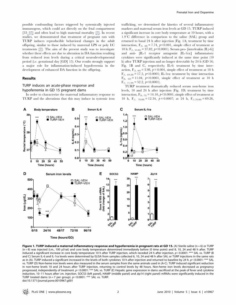
markers and maternal serum iron levels at GD 15. TURP induced
a significant increase in core body temperature at 10 hours, with a
1.9uC difference in comparison to the saline (SAL) group and
returned to basal 24 h after injection (Fig. 1A; treatment by time
interaction, F(4, 48) = 7.74, p,0.001, simple effect of treatment at
10 h: F(1, 53.84) = 37.82, p,0.0001). Serum pro- [interleukin (IL)-6)]
and anti- [IL-1 receptor antagonist (IL-1ra)] inflammatory
cytokines were significantly induced at the same time point (10
h) after TURP injection and no longer detectable by 24 h (GD 16;
Fig. 1B and C, respectively; IL-6: treatment by time inter-
action, F(5, 60) = 3.98, p = 0.004, simple effect of treatment at 10 h
F(1, 69.28) = 17.5, p,0.0001; IL-1ra: treatment by time interaction
F(6, 62) = 11.04, p,0.0001, simple effect of treatment at 10 h
F(1, 71.39) = 52.2, p,0.0001).
TURP treatment dramatically reduced serum non-heme iron
levels, 10 and 24 h after injection (Fig. 1D; treatment by time
interaction, F(6, 72) = 14.10, p,0.0001; simple effect of treatment at
10 h, F(1, 78.88) = 12.34, p = 0.0007; at 24 h, F(1,78.88) = 69.26,
Figure 1. TURP induced a maternal inflammatory response and hypoferremia in pregnant rats at GD 15. (A) Sterile saline (n = 6) or TURP(n = 8) was injected (i.m., 100 ml/rat) and core body temperature determined immediately before (0 time point) and 8, 10, 24 and 48 h after. TURPinduced a significant increase in core body temperature 10 h after TURP injection, which receded 24 h after injection. p,0.0001: *** SAL vs. TURP (Band C) Serum IL-6 and IL-1ra levels were determined by ELISA from samples collected 6, 10, 24 and 48 h after SAL or TURP injections in the same ratsas in (A). TURP induced a significant increased in the levels of both cytokines 10 h after injection and returned to baseline by 24 h. p,0.0001: *** SALvs. TURP (D) Non-heme iron levels were also measured in the serum samples from the same animals used in (A-C). TURP induced significant reductionin non-heme levels 10 and 24 hours after TURP injection, returning to control levels by 48 hours. Non-heme iron levels decreased as pregnancyprogressed, independently of treatment. p,0.0001: *** SAL vs. TURP (E) Hepatic gene expression in dams sacrificed at the peak of fever and cytokineinduction, 10–11 hours after i.m. injection. SOCS3 (left panel), HAMP (middle panel) and zip14 (right panel) mRNAs were significantly induced in theTURP treated dams (n = 7 per group). p,0.0001: *** SAL vs. TURP.doi:10.1371/journal.pone.0010967.g001
Prenatal Iron and Dopamine
PLoS ONE | www.plosone.org 2 June 2010 | Volume 5 | Issue 6 | e10967
p,0.0001), returning to control levels at 48 h (GD 17) after
treatment. No further differences in non-heme iron levels were
observed between TURP and control groups. However, there was
a reduction in the serum levels of iron with the progress of
pregnancy towards term, with non-heme iron levels at GD 19
becoming significantly lower than those at GD 15, 16, 17 and 18
(Fig. 1D; main effect of time, F(6, 72) = 16.36, p,0.0001, 96 h vs. 0,
24, 48 or 72 h Fisher’s LSDs p, 0.05). This decrease in maternal
serum non-heme iron towards term is in fact normally observed, as
iron is used to sustain increased requirement for this nutrient due
to expansion of the maternal erythrocyte mass and the high
demand of the growing fetus [38,39].
TURP’s effect on circulating iron levels was accompanied by an
induction of hepatic expression levels of HAMP mRNA by 25-fold
(Fig. 1E; t(12) = 5.27, p = 0.0002) and zip14 mRNA by 5.5-fold
(Fig. 1E; t(12) = 4.48, p = 0.0007). Zip14 is a dual iron/zinc
importer involved in the cellular uptake of iron [40]. In addition,
we observed an 11.6-fold increase in the expression levels of
suppressor of cytokine signaling (SOCS) 3 mRNA after TURP
treatment (Fig. 1E; t(12) = 7.6, p,0.0001), indicating the activation
of the JAK/STAT signaling pathway, most likely by IL-6 [41],
which is involved in the induction of HAMP and zip14 mRNA
expression.
Maternal iron supplementation reverses hypoferremia,but has no effect on other inflammatory responses
We determined if parenteral iron supplementation had any side
effect on the inflammatory responses induced by TURP, namely
cytokine production or fever. Maternal febrile response, as well as
induction of IL-6 and IL-1ra by TURP remained intact following
the iron supplementation schedule, with no significant effects of
iron supplementation detectable on all three parameters (Fig. S1A-
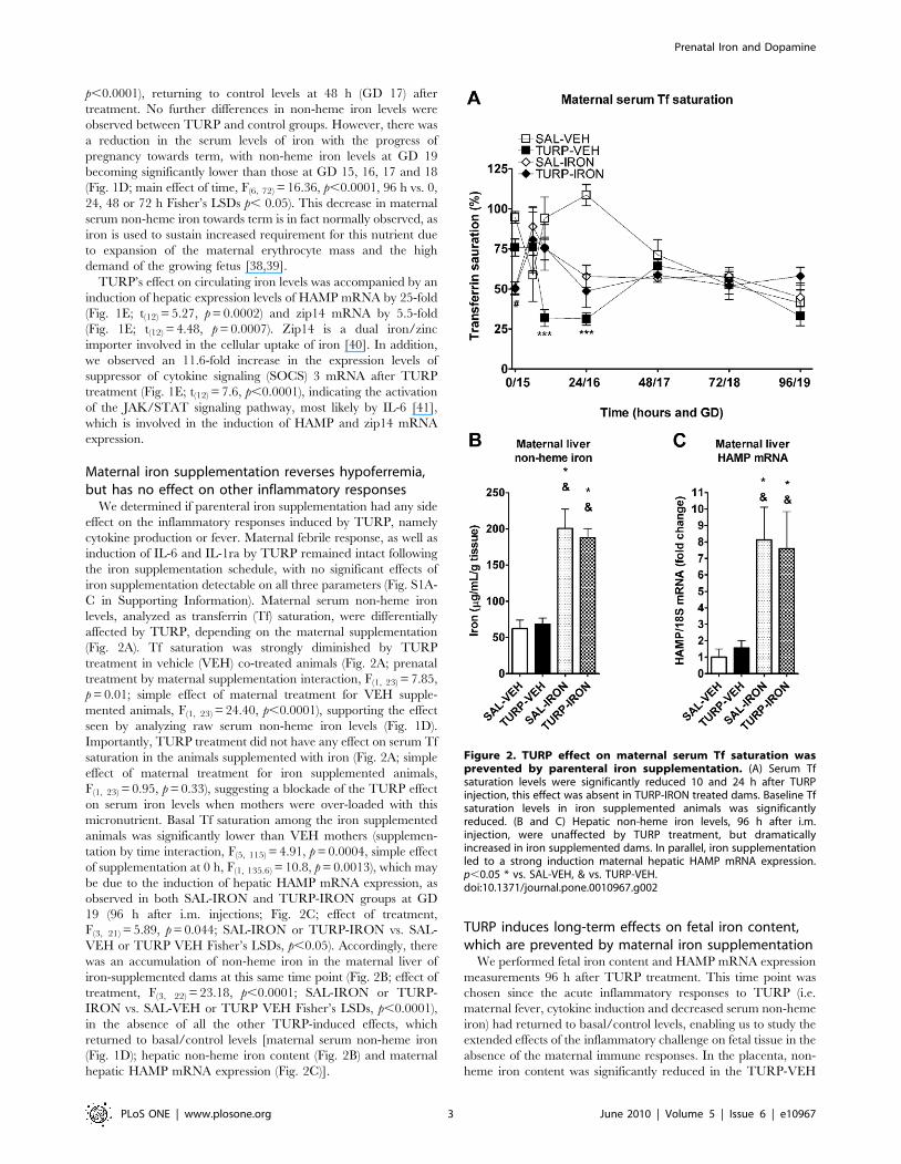
C in Supporting Information). Maternal serum non-heme iron
levels, analyzed as transferrin (Tf) saturation, were differentially
affected by TURP, depending on the maternal supplementation
(Fig. 2A). Tf saturation was strongly diminished by TURP
treatment in vehicle (VEH) co-treated animals (Fig. 2A; prenatal
treatment by maternal supplementation interaction, F(1, 23) = 7.85,
p = 0.01; simple effect of maternal treatment for VEH supple-
mented animals, F(1, 23) = 24.40, p,0.0001), supporting the effect
seen by analyzing raw serum non-heme iron levels (Fig. 1D).
Importantly, TURP treatment did not have any effect on serum Tf
saturation in the animals supplemented with iron (Fig. 2A; simple
effect of maternal treatment for iron supplemented animals,
F(1, 23) = 0.95, p = 0.33), suggesting a blockade of the TURP effect
on serum iron levels when mothers were over-loaded with this
micronutrient. Basal Tf saturation among the iron supplemented
animals was significantly lower than VEH mothers (supplemen-
tation by time interaction, F(5, 115) = 4.91, p = 0.0004, simple effect
of supplementation at 0 h, F(1, 135.6) = 10.8, p = 0.0013), which may
be due to the induction of hepatic HAMP mRNA expression, as
observed in both SAL-IRON and TURP-IRON groups at GD
19 (96 h after i.m. injections; Fig. 2C; effect of treatment,
F(3, 21) = 5.89, p = 0.044; SAL-IRON or TURP-IRON vs. SAL-
VEH or TURP VEH Fisher’s LSDs, p,0.05). Accordingly, there
was an accumulation of non-heme iron in the maternal liver of
iron-supplemented dams at this same time point (Fig. 2B; effect of
treatment, F(3, 22) = 23.18, p,0.0001; SAL-IRON or TURP-
IRON vs. SAL-VEH or TURP VEH Fisher’s LSDs, p,0.0001),
in the absence of all the other TURP-induced effects, which
returned to basal/control levels [maternal serum non-heme iron
(Fig. 1D); hepatic non-heme iron content (Fig. 2B) and maternal
hepatic HAMP mRNA expression (Fig. 2C)].
TURP induces long-term effects on fetal iron content,which are prevented by maternal iron supplementation
We performed fetal iron content and HAMP mRNA expression
measurements 96 h after TURP treatment. This time point was
chosen since the acute inflammatory responses to TURP (i.e.
maternal fever, cytokine induction and decreased serum non-heme
iron) had returned to basal/control levels, enabling us to study the
extended effects of the inflammatory challenge on fetal tissue in the
absence of the maternal immune responses. In the placenta, non-
heme iron content was significantly reduced in the TURP-VEH
Figure 2. TURP effect on maternal serum Tf saturation wasprevented by parenteral iron supplementation. (A) Serum Tfsaturation levels were significantly reduced 10 and 24 h after TURPinjection, this effect was absent in TURP-IRON treated dams. Baseline Tfsaturation levels in iron supplemented animals was significantlyreduced. (B and C) Hepatic non-heme iron levels, 96 h after i.m.injection, were unaffected by TURP treatment, but dramaticallyincreased in iron supplemented dams. In parallel, iron supplementationled to a strong induction maternal hepatic HAMP mRNA expression.p,0.05 * vs. SAL-VEH, & vs. TURP-VEH.doi:10.1371/journal.pone.0010967.g002
Prenatal Iron and Dopamine
PLoS ONE | www.plosone.org 3 June 2010 | Volume 5 | Issue 6 | e10967
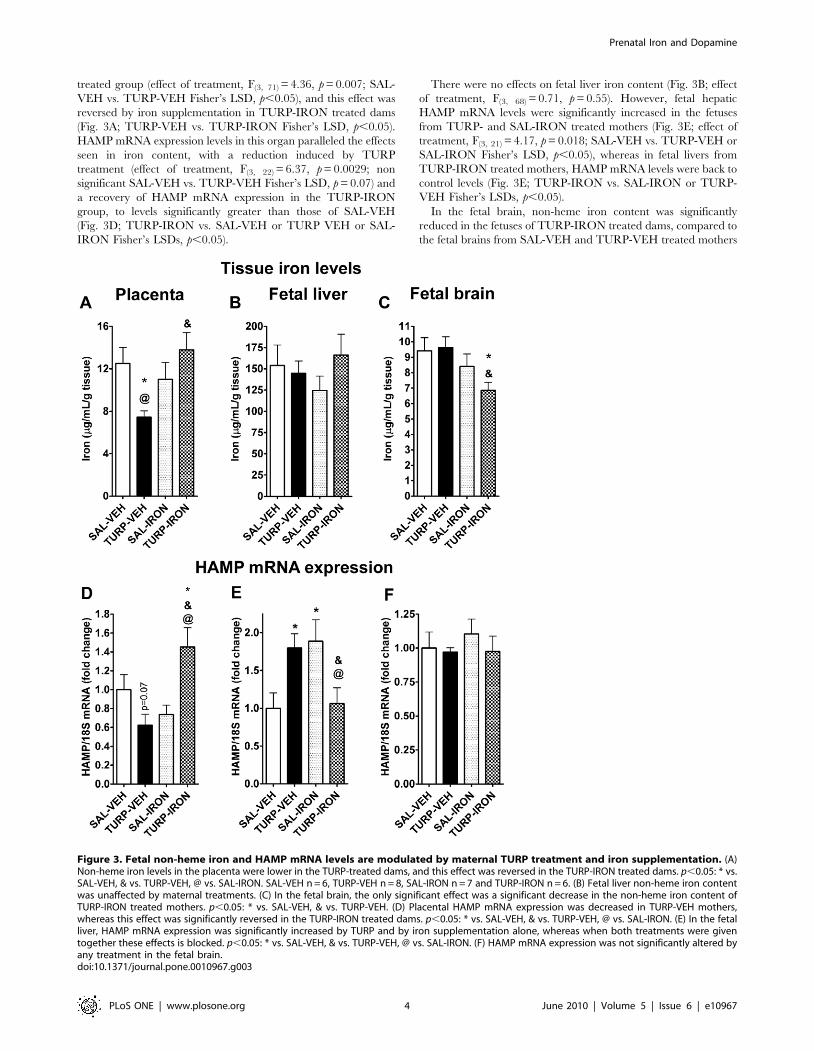
treated group (effect of treatment, F(3, 71) = 4.36, p = 0.007; SAL-
VEH vs. TURP-VEH Fisher’s LSD, p,0.05), and this effect was
reversed by iron supplementation in TURP-IRON treated dams
(Fig. 3A; TURP-VEH vs. TURP-IRON Fisher’s LSD, p,0.05).
HAMP mRNA expression levels in this organ paralleled the effects
seen in iron content, with a reduction induced by TURP
treatment (effect of treatment, F(3, 22) = 6.37, p = 0.0029; non
significant SAL-VEH vs. TURP-VEH Fisher’s LSD, p = 0.07) and
a recovery of HAMP mRNA expression in the TURP-IRON
group, to levels significantly greater than those of SAL-VEH
(Fig. 3D; TURP-IRON vs. SAL-VEH or TURP VEH or SAL-
IRON Fisher’s LSDs, p,0.05).
There were no effects on fetal liver iron content (Fig. 3B; effect
of treatment, F(3, 68) = 0.71, p = 0.55). However, fetal hepatic
HAMP mRNA levels were significantly increased in the fetuses
from TURP- and SAL-IRON treated mothers (Fig. 3E; effect of
treatment, F(3, 21) = 4.17, p = 0.018; SAL-VEH vs. TURP-VEH or
SAL-IRON Fisher’s LSD, p,0.05), whereas in fetal livers from
TURP-IRON treated mothers, HAMP mRNA levels were back to
control levels (Fig. 3E; TURP-IRON vs. SAL-IRON or TURP-
VEH Fisher’s LSDs, p,0.05).
In the fetal brain, non-heme iron content was significantly
reduced in the fetuses of TURP-IRON treated dams, compared to
the fetal brains from SAL-VEH and TURP-VEH treated mothers
Figure 3. Fetal non-heme iron and HAMP mRNA levels are modulated by maternal TURP treatment and iron supplementation. (A)Non-heme iron levels in the placenta were lower in the TURP-treated dams, and this effect was reversed in the TURP-IRON treated dams. p,0.05: * vs.SAL-VEH, & vs. TURP-VEH, @ vs. SAL-IRON. SAL-VEH n = 6, TURP-VEH n = 8, SAL-IRON n = 7 and TURP-IRON n = 6. (B) Fetal liver non-heme iron contentwas unaffected by maternal treatments. (C) In the fetal brain, the only significant effect was a significant decrease in the non-heme iron content ofTURP-IRON treated mothers. p,0.05: * vs. SAL-VEH, & vs. TURP-VEH. (D) Placental HAMP mRNA expression was decreased in TURP-VEH mothers,whereas this effect was significantly reversed in the TURP-IRON treated dams. p,0.05: * vs. SAL-VEH, & vs. TURP-VEH, @ vs. SAL-IRON. (E) In the fetalliver, HAMP mRNA expression was significantly increased by TURP and by iron supplementation alone, whereas when both treatments were giventogether these effects is blocked. p,0.05: * vs. SAL-VEH, & vs. TURP-VEH, @ vs. SAL-IRON. (F) HAMP mRNA expression was not significantly altered byany treatment in the fetal brain.doi:10.1371/journal.pone.0010967.g003
Prenatal Iron and Dopamine
PLoS ONE | www.plosone.org 4 June 2010 | Volume 5 | Issue 6 | e10967
(Fig. 3C; effect of treatment, F(3, 73) = 2.96, p = 0.038; TURP-
IRON vs. SAL-VEH or TURP-VEH Fisher’s LSDs, p,0.05).
However, there were no effects on HAMP mRNA expression
(Fig. 3F; effect of treatment, F(3, 23) = 0.46, p = 0.71).
Maternal inflammation and iron supplementationdifferentially alter sensitivity to the effects of AMPH onlocomotor activity in the adult offspring
We first assessed the effects on litter size and body weight at
birth, and found that none of the prenatal treatments had a
significant effect on these variables (data not shown), suggesting
that neither TURP and/or iron supplementation induced fetal
mortality nor gross physical changes at birth.
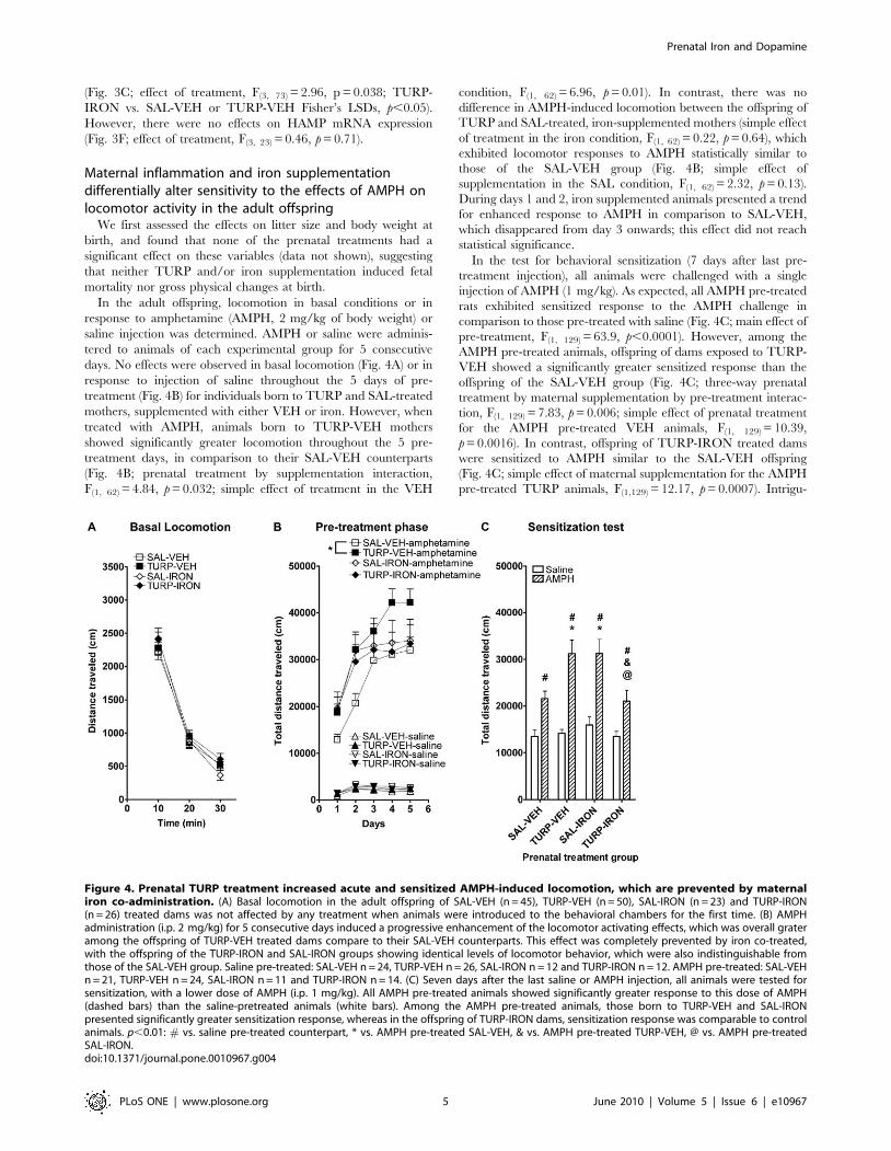
In the adult offspring, locomotion in basal conditions or in
response to amphetamine (AMPH, 2 mg/kg of body weight) or
saline injection was determined. AMPH or saline were adminis-
tered to animals of each experimental group for 5 consecutive
days. No effects were observed in basal locomotion (Fig. 4A) or in
response to injection of saline throughout the 5 days of pre-
treatment (Fig. 4B) for individuals born to TURP and SAL-treated
mothers, supplemented with either VEH or iron. However, when
treated with AMPH, animals born to TURP-VEH mothers
showed significantly greater locomotion throughout the 5 pre-
treatment days, in comparison to their SAL-VEH counterparts
(Fig. 4B; prenatal treatment by supplementation interaction,
F(1, 62) = 4.84, p = 0.032; simple effect of treatment in the VEH
condition, F(1, 62) = 6.96, p = 0.01). In contrast, there was no
difference in AMPH-induced locomotion between the offspring of
TURP and SAL-treated, iron-supplemented mothers (simple effect
of treatment in the iron condition, F(1, 62) = 0.22, p = 0.64), which
exhibited locomotor responses to AMPH statistically similar to
those of the SAL-VEH group (Fig. 4B; simple effect of
supplementation in the SAL condition, F(1, 62) = 2.32, p = 0.13).
During days 1 and 2, iron supplemented animals presented a trend
for enhanced response to AMPH in comparison to SAL-VEH,
which disappeared from day 3 onwards; this effect did not reach
statistical significance.
In the test for behavioral sensitization (7 days after last pre-
treatment injection), all animals were challenged with a single
injection of AMPH (1 mg/kg). As expected, all AMPH pre-treated
rats exhibited sensitized response to the AMPH challenge in
comparison to those pre-treated with saline (Fig. 4C; main effect of
pre-treatment, F(1, 129) = 63.9, p,0.0001). However, among the
AMPH pre-treated animals, offspring of dams exposed to TURP-
VEH showed a significantly greater sensitized response than the
offspring of the SAL-VEH group (Fig. 4C; three-way prenatal
treatment by maternal supplementation by pre-treatment interac-
tion, F(1, 129) = 7.83, p = 0.006; simple effect of prenatal treatment
for the AMPH pre-treated VEH animals, F(1, 129) = 10.39,
p = 0.0016). In contrast, offspring of TURP-IRON treated dams
were sensitized to AMPH similar to the SAL-VEH offspring
(Fig. 4C; simple effect of maternal supplementation for the AMPH
pre-treated TURP animals, F(1,129) = 12.17, p = 0.0007). Intrigu-
Figure 4. Prenatal TURP treatment increased acute and sensitized AMPH-induced locomotion, which are prevented by maternaliron co-administration. (A) Basal locomotion in the adult offspring of SAL-VEH (n = 45), TURP-VEH (n = 50), SAL-IRON (n = 23) and TURP-IRON(n = 26) treated dams was not affected by any treatment when animals were introduced to the behavioral chambers for the first time. (B) AMPHadministration (i.p. 2 mg/kg) for 5 consecutive days induced a progressive enhancement of the locomotor activating effects, which was overall grateramong the offspring of TURP-VEH treated dams compare to their SAL-VEH counterparts. This effect was completely prevented by iron co-treated,with the offspring of the TURP-IRON and SAL-IRON groups showing identical levels of locomotor behavior, which were also indistinguishable fromthose of the SAL-VEH group. Saline pre-treated: SAL-VEH n = 24, TURP-VEH n = 26, SAL-IRON n = 12 and TURP-IRON n = 12. AMPH pre-treated: SAL-VEHn = 21, TURP-VEH n = 24, SAL-IRON n = 11 and TURP-IRON n = 14. (C) Seven days after the last saline or AMPH injection, all animals were tested forsensitization, with a lower dose of AMPH (i.p. 1 mg/kg). All AMPH pre-treated animals showed significantly greater response to this dose of AMPH(dashed bars) than the saline-pretreated animals (white bars). Among the AMPH pre-treated animals, those born to TURP-VEH and SAL-IRONpresented significantly greater sensitization response, whereas in the offspring of TURP-IRON dams, sensitization response was comparable to controlanimals. p,0.01: # vs. saline pre-treated counterpart, * vs. AMPH pre-treated SAL-VEH, & vs. AMPH pre-treated TURP-VEH, @ vs. AMPH pre-treatedSAL-IRON.doi:10.1371/journal.pone.0010967.g004
Prenatal Iron and Dopamine
PLoS ONE | www.plosone.org 5 June 2010 | Volume 5 | Issue 6 | e10967
ingly, iron supplementation alone during pregnancy, also resulted
in enhanced behavioral sensitization in the adult offspring (Fig. 4C;
simple effect of maternal supplementation for the AMPH pre-
treated SAL animals, F(1,129) = 9.94, p = 0.02; simple effect of
treatment for the AMPH pre-treated IRON animals, F(1, 129) =
8.77, p = 0.0037).
Maternal inflammation leads to biochemical alterationsin the adult DA system, which are partly reversed bymaternal iron supplementation
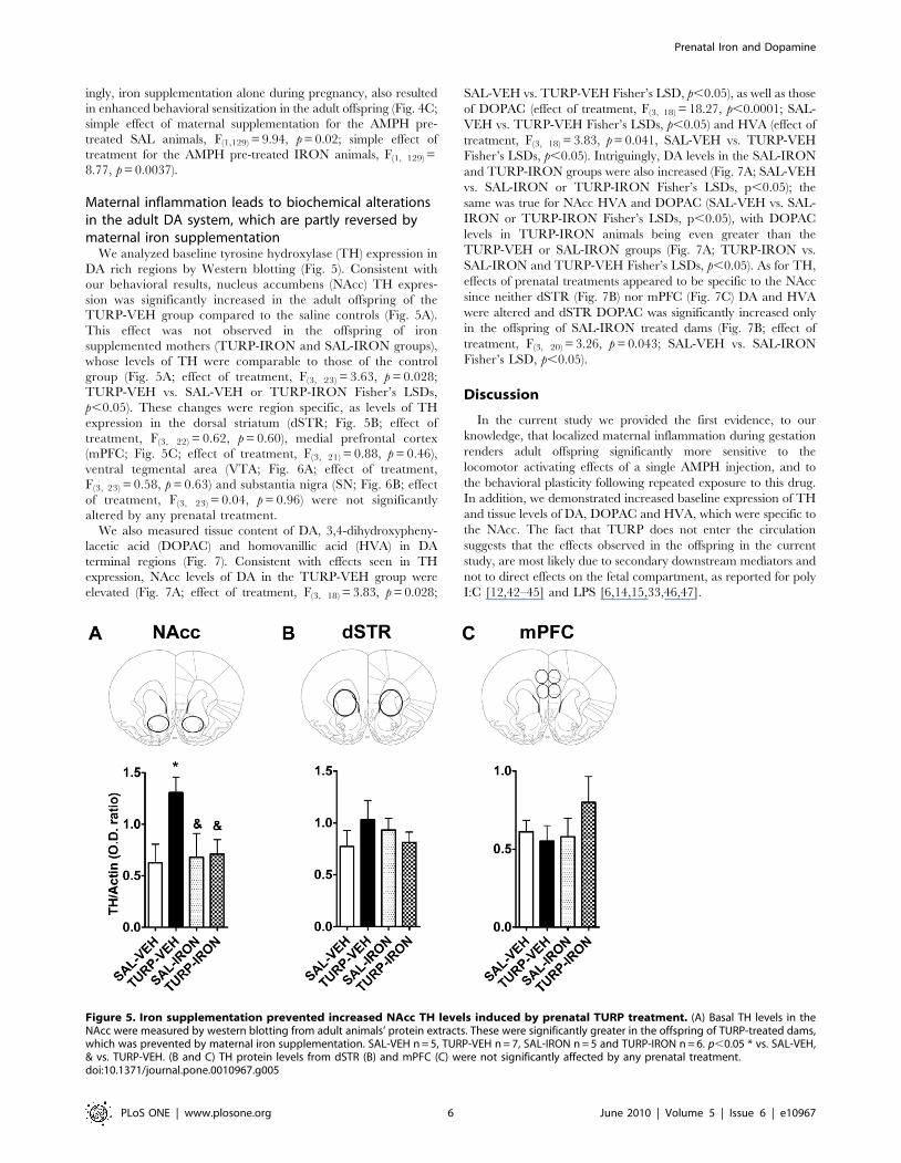
We analyzed baseline tyrosine hydroxylase (TH) expression in
DA rich regions by Western blotting (Fig. 5). Consistent with
our behavioral results, nucleus accumbens (NAcc) TH expres-
sion was significantly increased in the adult offspring of the
TURP-VEH group compared to the saline controls (Fig. 5A).
This effect was not observed in the offspring of iron
supplemented mothers (TURP-IRON and SAL-IRON groups),
whose levels of TH were comparable to those of the control
group (Fig. 5A; effect of treatment, F(3, 23) = 3.63, p = 0.028;
TURP-VEH vs. SAL-VEH or TURP-IRON Fisher’s LSDs,
p,0.05). These changes were region specific, as levels of TH
expression in the dorsal striatum (dSTR; Fig. 5B; effect of
treatment, F(3, 22) = 0.62, p = 0.60), medial prefrontal cortex
(mPFC; Fig. 5C; effect of treatment, F(3, 21) = 0.88, p = 0.46),
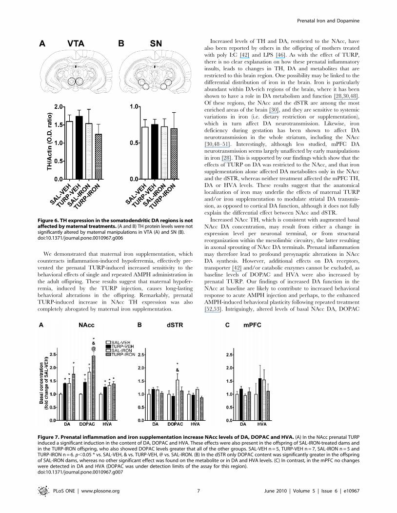
ventral tegmental area (VTA; Fig. 6A; effect of treatment,
F(3, 23) = 0.58, p = 0.63) and substantia nigra (SN; Fig. 6B; effect
of treatment, F(3, 23) = 0.04, p = 0.96) were not significantly
altered by any prenatal treatment.
We also measured tissue content of DA, 3,4-dihydroxypheny-
lacetic acid (DOPAC) and homovanillic acid (HVA) in DA
terminal regions (Fig. 7). Consistent with effects seen in TH
expression, NAcc levels of DA in the TURP-VEH group were
elevated (Fig. 7A; effect of treatment, F(3, 18) = 3.83, p = 0.028;
SAL-VEH vs. TURP-VEH Fisher’s LSD, p,0.05), as well as those
of DOPAC (effect of treatment, F(3, 18) = 18.27, p,0.0001; SAL-
VEH vs. TURP-VEH Fisher’s LSDs, p,0.05) and HVA (effect of
treatment, F(3, 18) = 3.83, p = 0.041, SAL-VEH vs. TURP-VEH
Fisher’s LSDs, p,0.05). Intriguingly, DA levels in the SAL-IRON
and TURP-IRON groups were also increased (Fig. 7A; SAL-VEH
vs. SAL-IRON or TURP-IRON Fisher’s LSDs, p,0.05); the
same was true for NAcc HVA and DOPAC (SAL-VEH vs. SAL-
IRON or TURP-IRON Fisher’s LSDs, p,0.05), with DOPAC
levels in TURP-IRON animals being even greater than the
TURP-VEH or SAL-IRON groups (Fig. 7A; TURP-IRON vs.
SAL-IRON and TURP-VEH Fisher’s LSDs, p,0.05). As for TH,
effects of prenatal treatments appeared to be specific to the NAcc
since neither dSTR (Fig. 7B) nor mPFC (Fig. 7C) DA and HVA
were altered and dSTR DOPAC was significantly increased only
in the offspring of SAL-IRON treated dams (Fig. 7B; effect of
treatment, F(3, 20) = 3.26, p = 0.043; SAL-VEH vs. SAL-IRON
Fisher’s LSD, p,0.05).
Discussion
In the current study we provided the first evidence, to our
knowledge, that localized maternal inflammation during gestation
renders adult offspring significantly more sensitive to the
locomotor activating effects of a single AMPH injection, and to
the behavioral plasticity following repeated exposure to this drug.
In addition, we demonstrated increased baseline expression of TH
and tissue levels of DA, DOPAC and HVA, which were specific to
the NAcc. The fact that TURP does not enter the circulation
suggests that the effects observed in the offspring in the current
study, are most likely due to secondary downstream mediators and
not to direct effects on the fetal compartment, as reported for poly
I:C [12,42–45] and LPS [6,14,15,33,46,47].
Figure 5. Iron supplementation prevented increased NAcc TH levels induced by prenatal TURP treatment. (A) Basal TH levels in theNAcc were measured by western blotting from adult animals’ protein extracts. These were significantly greater in the offspring of TURP-treated dams,which was prevented by maternal iron supplementation. SAL-VEH n = 5, TURP-VEH n = 7, SAL-IRON n = 5 and TURP-IRON n = 6. p,0.05 * vs. SAL-VEH,& vs. TURP-VEH. (B and C) TH protein levels from dSTR (B) and mPFC (C) were not significantly affected by any prenatal treatment.doi:10.1371/journal.pone.0010967.g005
Prenatal Iron and Dopamine
PLoS ONE | www.plosone.org 6 June 2010 | Volume 5 | Issue 6 | e10967
We demonstrated that maternal iron supplementation, which
counteracts inflammation-induced hypoferremia, effectively pre-
vented the prenatal TURP-induced increased sensitivity to the
behavioral effects of single and repeated AMPH administration in
the adult offspring. These results suggest that maternal hypofer-
remia, induced by the TURP injection, causes long-lasting
behavioral alterations in the offspring. Remarkably, prenatal
TURP-induced increase in NAcc TH expression was also
completely abrogated by maternal iron supplementation.
Increased levels of TH and DA, restricted to the NAcc, have
also been reported by others in the offspring of mothers treated
with poly I:C [42] and LPS [46]. As with the effect of TURP,
there is no clear explanation on how these prenatal inflammatory
insults, leads to changes in TH, DA and metabolites that are
restricted to this brain region. One possibility may be linked to the
differential distribution of iron in the brain. Iron is particularly
abundant within DA-rich regions of the brain, where it has been
shown to have a role in DA metabolism and function [28,30,48].
Of these regions, the NAcc and the dSTR are among the most
enriched areas of the brain [30], and they are sensitive to systemic
variations in iron (i.e. dietary restriction or supplementation),
which in turn affect DA neurotransmission. Likewise, iron
deficiency during gestation has been shown to affect DA
neurotransmission in the whole striatum, including the NAcc
[30,48–51]. Interestingly, although less studied, mPFC DA
neurotransmission seems largely unaffected by early manipulations
in iron [28]. This is supported by our findings which show that the
effects of TURP on DA was restricted to the NAcc, and that iron
supplementation alone affected DA metabolites only in the NAcc
and the dSTR, whereas neither treatment affected the mPFC TH,
DA or HVA levels. These results suggest that the anatomical
localization of iron may underlie the effects of maternal TURP
and/or iron supplementation to modulate striatal DA transmis-
sion, as opposed to cortical DA function, although it does not fully
explain the differential effect between NAcc and dSTR.
Increased NAcc TH, which is consistent with augmented basal
NAcc DA concentration, may result from either a change in
expression level per neuronal terminal, or from structural
reorganization within the mesolimbic circuitry, the latter resulting
in axonal sprouting of NAcc DA terminals. Prenatal inflammation
may therefore lead to profound presynaptic alterations in NAcc
DA synthesis. However, additional effects on DA receptors,
transporter [42] and/or catabolic enzymes cannot be excluded, as
baseline levels of DOPAC and HVA were also increased by
prenatal TURP. Our findings of increased DA function in the
NAcc at baseline are likely to contribute to increased behavioral
response to acute AMPH injection and perhaps, to the enhanced
AMPH-induced behavioral plasticity following repeated treatment
[52,53]. Intriguingly, altered levels of basal NAcc DA, DOPAC
Figure 6. TH expression in the somatodendritic DA regions is notaffected by maternal treatments. (A and B) TH protein levels were notsignificantly altered by maternal manipulations in VTA (A) and SN (B).doi:10.1371/journal.pone.0010967.g006
Figure 7. Prenatal inflammation and iron supplementation increase NAcc levels of DA, DOPAC and HVA. (A) In the NAcc prenatal TURPinduced a significant induction in the content of DA, DOPAC and HVA. These effects were also present in the offspring of SAL-IRON-treated dams andin the TURP-IRON offspring, who also showed DOPAC levels greater that all of the other groups. SAL-VEH n = 5, TURP-VEH n = 7, SAL-IRON n = 5 andTURP-IRON n = 6. p,0.05 * vs. SAL-VEH, & vs. TURP-VEH, @ vs. SAL-IRON. (B) In the dSTR only DOPAC content was significantly greater in the offspringof SAL-IRON dams, whereas no other significant effect was found on the metabolite or in DA and HVA levels. (C) In contrast, in the mPFC no changeswere detected in DA and HVA (DOPAC was under detection limits of the assay for this region).doi:10.1371/journal.pone.0010967.g007
Prenatal Iron and Dopamine
PLoS ONE | www.plosone.org 7 June 2010 | Volume 5 | Issue 6 | e10967
and HVA remained increased in the offspring of TURP-IRON
treated mothers, despite normalization of TH expression. This
suggests additional alterations in the synthetic and/or catabolic
pathways of DA that may account for the observed alterations. It is
important to note however, that prenatal TURP treatment caused
two mechanistically differentiable effects in the offspring. One was
the increased response to a single AMPH injection, which may
stem from changes in the release of DA in the NAcc [54], therefore
is more directly related to increased basal expression of TH and to
the increased basal content of DA in the NAcc. In contrast,
behavioral plasticity after repeated AMPH administration is a
phenomenon that requires long-term neural adaptations that
occur in several brain structures, notably the VTA, where the cell
bodies of the mesolimbic DA neurons lie [55–57], therefore
TURP may affect additional cellular and molecular targets. In this
regard, iron supplementation to TURP-treated mothers seems to
have prevented only some of the effects of TURP that impact the
response to acute AMPH injection (i.e. increased NAcc TH
expression, but no greater DA and metabolites) as well as those
mechanisms underlying the sensitized response after repeated
AMPH administration.
Interestingly, iron supplementation alone (i.e. SAL-IRON
group) induced several behavioral and biochemical changes in
the offspring, which resembled the effects of prenatal inflammation
(i.e. enhanced sensitivity to acute and repeated AMPH injection
and increased basal levels of DA, DOPAC and HVA in the NAcc).
SAL-IRON group also exhibited a greater AMPH-induced
stereotypy during the pre-treatment phase (data not shown) and
increased basal levels of DOPAC in the dSTR. This surprising
finding is most likely due to iron overload in iron sufficient animals
[58], although the doses of iron used in our study were chosen to
be below those that cause toxic side effects [59,60]. This is further
exacerbated by the fact that parenteral administration of iron
bypasses the regulatory mechanisms that control its dietary
absorption [26], therefore iron administered through dietary
supplementation, which normally does not cause overload [61],
may spare the offspring from the undesired effects described in the
present study. To the best of our knowledge, there are no studies
on the effects of maternal-iron supplementation on the DA
function of the adult offspring, however it is normally considered
that excess of iron results in toxic effects due to oxidative stress
[26]. These findings, although unexpected, nevertheless emphasize
the critical importance of this micronutrient to normal develop-
ment of mesolimbic DA neurons.
Given that maternal circulation constitutes the only source of
iron to the developing fetus [38], and that HAMP down-regulates
FPN1 [62], which is essential for the transport of iron through the
materno-fetal interface [63–67], we rationalized that the inflam-
mation-induced reduction in maternal iron will restrict the
amounts being transported through the materno-fetal interface.
To confirm this, we measured iron levels in the placenta, fetal liver
and brain collected from all the treatment groups. As expected,
placental iron levels were dramatically reduced, paralleling the
maternal supply of this nutrient and completely recovered by iron
replenishment in TURP treated dams. The TURP-induced
reduction in placental iron content may reflect either the reduced
availability of maternal serum iron and/or the demand to sustain
the fetal development during the hypoferremia [68].
The changes in iron levels in the placenta were very closely
paralleled by the levels of HAMP expression, which may be
merely responding to the tissue’s fluctuation in iron concentration
[69] or conversely, responding to a signal from outside the
placenta, and in turn modulating iron export rates from this tissue
to the fetal compartment [68]. Interestingly, regulation of HAMP
mRNA expression in fetal liver was the reverse of that seen in the
placenta. In this tissue, TURP-treatment resulted in induced
expression of HAMP mRNA at a time point when TURP-induced
maternal circulating cytokines are no longer elevated, suggesting
that mediators other than cytokines may be involved in this effect.
One likely possibility is that iron flow from the placental stores into
the fetus resulted in higher than normal iron levels in the fetal
circulation, thus triggering fetal liver HAMP mRNA expression
[68]. In fact, fetuses of iron supplemented (SAL-IRON) dams also
showed increased fetal liver HAMP mRNA expression, which is
presumably due to a direct effect of the supplemented micronu-
trient [68], as no inflammatory pathways were activated in this
condition.
Iron levels in the fetal brains taken from all four treatment
groups did not show any alteration, other than a small but
significant reduction in TURP-IRON group. This latter observa-
tion is rather puzzling and it possibly reflects a lower than normal
supply of iron in the TURP-IRON group. The latter effect can be
mediated by the increased placental HAMP in the TURP-IRON
group, which would lead to iron trapping within this organ, at the
expense of the fetal brain, similar to the effects of maternal hepatic
HAMP on maternal liver iron content [70]. However, it is
important to note that in the present study we measured whole
brain iron levels, and that marked regional differences in iron
acquisition capabilities of the brain are possible and have indeed
been reported [30,71,72]. In fact, brain DA regions are known to
be enriched in iron [73,74] and susceptible to systemic variations
of this nutrient [75], suggesting that specific effects of inflamma-
tion and iron supplementation may be found in the mesolimbic
DA neurons, which would then be involved in the induction of the
marked effects on DA function observed in the adult offspring.
Altogether our data are consistent with a scenario where
inflammation-induced maternal hypoferremia leads to placental
deficiency in this nutrient. This could be due, at least partly, to a
putative increase in iron transport to the fetal compartment that
keeps whole-tissue levels of iron normal. In addition, placental iron
deficiency may in turn result in altered function of this tissue. For
example, dietary iron deficiency leads to increased cytokine
expression in the placenta [76] and altered transport of amino
acids towards the fetal compartment [77,78]. This may, along with
direct restriction of iron supply in specific brain areas, impact
aspects of neuronal development, resulting in the effects observed
in the adult offspring.
Hypoferremia has long been recognized as one of the main
components of the host’s response to infection/inflammation, and
the mechanisms involved in the induction of this process are fairly
well understood. The strongest evidence to date suggests that the
pro-inflammatory cytokine IL-6, which is readily detectable in the
circulation of infected individuals regardless of the pathogenic
trigger, is the main circulating mediator of hepatic HAMP mRNA
expression [19–22]. IL-6 has been suggested to be strongly
involved in the etiology of the offspring alterations induced by
maternal inflammation [79]. We cannot rule out the direct
involvement of either other cytokines [45] or other mediators, such
as inflammation-induced alteration in zinc homeostasis (hypozin-
cemia) [80]. However, our results strongly indicate that during
prenatal inflammation, hypoferremia plays a fundamental role in
the developmental effects of this maternal insult on the mesolimbic
DA function, and that the effect of IL-6 on the developing fetus
may be mediated trough this mechanism.
Our results clearly demonstrate that a localized inflammatory
insult during gestation has profound affects on DA function,
which may be relevant for schizophrenia, where increased
striatal DA is proposed to underlie the so-called positive
Prenatal Iron and Dopamine
PLoS ONE | www.plosone.org 8 June 2010 | Volume 5 | Issue 6 | e10967
symptoms of the disorder [81–84]. In addition, we showed for
the first time that this risk factor for schizophrenia also increased
the animal’s vulnerability to develop sensitized behavioral
response to an AMPH challenge given one week after repeated
exposure. Repeated administration of drugs of abuse, including
AMPH, has been shown to also result in sensitization to the
rewarding effects of these drugs [52]. Thus, our findings further
strengthen the link between these two psychiatric disorders,
whose co-morbidity has been proposed to stem from DA
dysfunction [85]. Finally, we provided new evidence for the
involvement of fetal/maternal iron homeostasis in the develop-
mental processes that render the offspring more susceptible to
enhanced DA function.
Materials and Methods
AnimalsTime pregnant primiparous Sprague-Dawley rats (Charles
Rivers, QC, Canada) were used in all experiments. On GD 7 or
8 animals were individually housed in a controlled environment at
an ambient temperature of 2161uC on 12:12 h light-dark cycle
(lights on at 0800 hours) with free access to food and water. Only
male offspring were used, which were housed under the same
environmental conditions. Mothers were treated with the
inflammatory stimulus at GD 15. This day of pregnancy was
used since it has been proposed to be roughly equivalent to the late
1st trimester of human pregnancy [86], which has been
significantly linked with the increased risk of developing
schizophrenia in the offspring of infected mothers [1]. Important-
ly, our previous studies in pregnant rats treated with TURP at GD
10, 15 or 18, suggested a window of vulnerability for the adult
offspring treated at GD 15 [7].
Ethics statement. Experimental procedures were approved by the
Animal Care Committee from the Douglas Mental Health
University Institute and McGill University pursuant of the
Canadian Council of Animal Care (Animal Use Protocol #4306). All efforts were made to minimize the number of animals
used.
Treatments and experimental protocolsPregnant dams were handled daily from GD 11 onwards and
habituated to a rectal probe to determine core temperature
(Physitemp Instruments, NJ, U.S.A.). Some animals (13 dams)
received an intraperitoneal (i.p.) injection of 10 mg/kg of the
aqueous complex of poly-nuclear iron (III)-hydroxide in sucrose
(Venofer, American Reagent, NY, U.S.A.) or an equivalent
volume of vehicle (either saline [7 dams] or 30% sucrose [7 dams])
at GD 13 and 14. Iron was administered to the dams from GD 15
until GD 18 at larger doses (i.p., 20 mg/kg). At GD 15 basal core
temperature was recorded and a small sample of tail blood was
collected to determine basal levels of cytokines and serum iron.
Subsequently, a maternal inflammatory response was elicited by
injecting intramuscularly with 100 mL of purified TURP (Riedel-
deHaen, Sleeze, Germany); control dams received an equivalent
volume of saline. All injections were administered between 0900
and 1000 hours. Using a rectal probe core body temperature was
measured at 8, 10, 24, 48 and 72 hours after i.m. treatment and
tail blood collected at each time point. The animals were then
sacrificed at GD 19 (96 hours after the i.m. treatment) with a lethal
dose of pentobarbital sodium (i.p., 60 mg/kg) and a final blood
sample was collected via cardiac puncture. Maternal liver,
placentas, fetal livers and brains were excised and immersed in
2-methylbutane (Fisher Scientific, Hampton, NH, U.S.A.) and
chilled with dry ice. Serum cytokine, iron and Tf saturation levels
were determined, as well as maternal liver, placenta, fetal liver and
brain tissue iron levels.
In a separate experiment, SAL and TURP treated dams (i.m., 7
rats per group) were sacrificed 10–11 hours after i.m. treatment,
which represents the peak of fever and cytokine responses [87],
and livers collected to measure mRNA expression of molecules
involved in iron homeostasis.
To determine the effects of prenatal TURP and iron
supplementation on DA function and related behaviors in the
adult offspring, pregnant dams (6–12 per group) were treated as
described above, with the exception that tail blood collection was
not performed. The male offspring from each mother were
marked, weighed and cross-fostered with surrogate dams in mixed
litters [6,7]. Offspring were weaned from their foster mothers at
postnatal day (P) 22 and used at P 60–62, for either behavioral
testing or biochemical analyses. Behavioral sensitization to AMPH
was used to gauge midbrain DA function and plasticity in the adult
offspring. In addition, protein expression levels of TH were
determined by Western blotting from extracts obtained from the
VTA, SN, NAcc, dSTR and mPFC. Tissue levels of DA and
metabolites (DOPAC and HVA) were determined by High
Performance Liquid Chromatography (HPLC) from NAcc, dSTR
and mPFC.
Behavioral testingWe measured locomotor responses to either single or repeated
injections of d-amphetamine sulphate salt (AMPH, Sigma-Aldrich,
Dorset, UK) in adult male offspring, using our well-established
protocol [57]. Briefly, locomotor activity was quantified with an
infrared activity-monitoring apparatus for rats (AccuScan Instru-
ments, Columbus, OH, U.S.A.). On day 1 all rats (n = 23–50 per
group) were habituated to the boxes for 30 minutes (basal
locomotor activity). On day 2 all animals received a saline
injection (1 ml/g, i.p.) and were placed back in the boxes for 30
minutes. Immediately after, one half of the animals (saline pre-
treatment group, 11–26 per group) received another saline
injection, and the other half was injected with AMPH (2 mg/kg,
i.p.); locomotor activity was monitored for an additional 90
minutes. On days 3, 4, 5 and 6, animals received an injection of
either saline (saline pre-treatment group) or AMPH (2 mg/kg,
AMPH pre-treatment group) and their locomotor activity was
measured for 90 minutes. Finally, on day 14, a week after
termination of saline or AMPH pre-treatment, a test for
behavioral sensitization was conducted, where all animals,
regardless of the pre-treatment condition, received a single
injection of AMPH (1 mg/kg, i.p.). Locomotor activity was
monitored for 90 minutes. This lower dose of AMPH for the
sensitization test was chosen to avoid stereotypy. All behavioral
measurements were performed between 0900 and 1600 hours.
Serum cytokine and iron determinationAll blood samples were allowed to cloth at room temperature
for 1 hour, spun at 4000 rpm for 20 min at 4uC to obtain serum
and stored at 280uC until used. Maternal serum samples were
analyzed for IL-6 and IL-1ra (the main cytokines released into the
circulation by TURP treatment [87]) using species specific ELISA
(NIBSC, Potters Bar, UK) as described previously [88]. Serum
iron (SI) was measured using an iron kit (RANDOX, Mississauga,
ON, Canada). In parallel, a direct measure of total iron binding
capacity (TIBC) of the serum was determined using a TIBC kit
(RANDOX). For both procedures the manufacturer’s protocols
were followed. Once both measurements were obtained (expressed
as mg/dL of serum), serum T) saturation was calculated by
Prenatal Iron and Dopamine
PLoS ONE | www.plosone.org 9 June 2010 | Volume 5 | Issue 6 | e10967
expressing the serum iron content as percentage of TIBC (Tf
saturation = SI/TIBC6100).
Three fetuses per dam were collected, and the results from
their individual measurements averaged to obtain one value per
mother. For tissue iron content, maternal liver, placenta, fetal
liver and brain were homogenized in 1:10 (w/v) high-purity
water. One volume of protein-precipitation solution (1 N HCl
and 10% (v/v) trichloroacetic acid in high purity water) was then
added to the samples, blank and iron standards and incubated for
1 hour at 95uC. The samples were then allowed to cool at room
temperature for 2 min, vortex mixed and centrifuged at 10
0006g for 10 min at room temperature [89,90] and assayed for
iron content.
Quantitative RT-PCRTotal RNA was extracted and reverse transcribed from
maternal livers, placenta, fetal liver and brain as described
previously [91]. Real-time PCR was performed in duplicate using
pre-optimized primer/probe mixture (TaqMan Gene Expression
Assays, Applied Biosystems, ON, Canada) and TaqMan universal
PCR master mix (Applied Biosystems). The housekeeping gene
18S was used to normalize levels of cDNA expression for each
sample. Levels of gene expression were calculated as the X-fold
difference from the control groups (SAL-VEH group). The mRNA
levels of the following genes was assessed: SOCS3, used as a
marker of activity of the JAK/STAT3 signaling pathway activated
by IL-6 [92]; HAMP and zip14, a dual iron/zinc importer that has
been shown to be induced by TURP and may be involved in the
cellular sequestration of iron [40,93]. The assay ID for each gene
is as follows: SOCS3: Rn00585674-s1, HAMP: Rn00584987-m1,
zip14 (slc39a14): Rn01468335-g1 and 18S: EUK-18S-
rRNA4352930.
Western blotting and HPLCWe explored biochemical markers of DA neurons, including
protein expression of TH and tissue levels of DA and its
metabolites, DOPAC and HVA. Brains collected from adult male
offspring (P 60–62) by decapitation were immersed in 2-
methylbutane (Fisher Scientific, Hampton, NH, U.S.A.) and
chilled with dry ice. 300 mm thick cryostat sections were obtained.
There were no behavioral differences between the animals born to
mothers supplemented with either SAL or sucrose. Thus, to
minimize the number of animals used, only SAL-SAL and TURP-
SAL groups were included. Bilateral punches from the VTA, SN,
NAcc (including core and shell), dSTR and mPFC (including
cingulated areas 1 and 2) were excised using our previously
described procedures [57] using the Paxinos and Watson rat brain
atlas [94].
Western blots were conducted as previously described [57,87].
Membranes were incubated with antibodies against TH (1:5000,
Chemicon, Temicula, CA, U.S.A.) and actin (HRP-coupled,
1:5000, Santa Cruz Biotechnology, Santa Cruz, CA, U.S.A.). Data
are expressed as a ratio of TH over actin optical densities.
HPLC analysis was conducted as we recently reported [95].
Briefly, tissue punches were re-suspended in 100 mL 0.1 m
phosphate buffer, pH 7.0, and filtered using 0.45-mm syringe
filters. A 10-mL volume of this filtrate was loaded onto a 15-cm C-
18 reverse-phase column via manual injection ports (20-mL loop).
Dual-channel coulometric III detectors (model 5100A; ESA, Inc.,
Bedford, MA, U.S.A.) were used to measure the reduction and
oxidation currents for DA and DA metabolites (one channel was
used for DA and the other for DA metabolites). EZChrom Data
Chromatography Data System (Scientific Software, Inc., San
Ramon, CA, U.S.A) was used to analyzed and quantify DA,
DOPAC and HVA concentrations.
Data analysesAll data were analyzed as raw values, except for HPLC results,
where the data were analyzed as percentage of the average value
for the SAL-VEH group, due to large inter-assay variability
between two different batches of samples assayed. All data are
presented as mean values 6 s.e.m. p values,0.05 values were
deemed significant.
Maternal fever, serum cytokines and iron: data were analyzed
using a repeated measures 2-way ANOVA, with time as a within-
group variable and pre-natal treatment (SAL vs. TURP) as a
between-groups variable.
Maternal liver PCR data: data from the 10–11 hours
experiment were analyzed using two-tailed Student’s t-tests to
compare data from the SAL-VEH and TURP-VEH groups.
Maternal serum Tf saturation: data were analyzed using a 3-
way ANOVA with prenatal treatment (SAL vs. TURP) and
maternal supplementation (VEH vs. IRON) used as between-
groups variables in all cases and time point used as a within-group
variable. When the interactions of main effects were significant,
analysis was followed by performing simple-effects ANOVA and
Fisher’s LSD post hoc tests.
Tissue iron content, HAMP mRNA levels (96 h time point),
western blotting and HPLC: data were analyzed using 1-way
ANOVA, with maternal treatment (SAL-VEH vs. TURP-VEH vs.
SAL-IRON vs. TURP-IRON) as between-groups variable and
furthered with Fisher’s LSD post hoc tests when ANOVA was
significant.
Offspring’s basal and AMPH-induced locomotion: data were
analyzed using 3-way ANOVAs, with prenatal treatment (SAL vs.
TURP) and maternal supplementation (VEH vs. IRON) as
between-groups variables and time as a within-group variable.
When interactions were significant, analyses were followed by
simple-effects ANOVA and Fisher’s LSD post hoc tests.
AMPH sensitization test: data were analyzed using a 3-way
ANOVA, with prenatal treatment (SAL vs. TURP), maternal
supplementation (VEH vs. IRON) and pre-treatment (saline vs.
AMPH) used as a between-groups variables, followed by simple-
effects ANOVA and Fisher’s LSD post hoc tests when interactions
were significant.
Supporting Information
Figure S1 Inflammatory response remained intact in iron
supplemented mothers. (A) Febrile response followed the same
kinetics in vehicle and iron supplemented mothers, peaking at 10 h
after TURP injection and returning to baseline 24 h later. Basal
temperature was not affected by iron supplementation. (B and C)
Serum IL-6 and IL-1ra levels followed the same kinetics in the
TURP-IRON group compared to the TURP-VEH group.
Found at: doi:10.1371/journal.pone.0010967.s001 (4.70 MB TIF)
Acknowledgments
We thank Dr. S Poole (NIBSC, UK) for kindly providing the sandwich
ELISA reagents. We also thank Ms. G Somay, E Dai and S Jung for their
excellent technical assistance and Mrs. E Matta-Camacho for helpful
discussion of this manuscript.
Author Contributions
Conceived and designed the experiments: AAV CF GL. Performed the
experiments: AAV. Analyzed the data: AAV CF. Wrote the paper: AAV
CF GL.
Prenatal Iron and Dopamine
PLoS ONE | www.plosone.org 10 June 2010 | Volume 5 | Issue 6 | e10967
References
1. Brown AS, Derkits EJ (2010) Prenatal Infection and Schizophrenia: A Review of
Epidemiologic and Translational Studies. Am J Psychiatry.
2. Brown AS (2006) Prenatal infection as a risk factor for schizophrenia. Schizophr
Bull 32: 200–202.
3. Babulas V, Factor-Litvak P, Goetz R, Schaefer CA, Brown AS (2006) Prenatal
exposure to maternal genital and reproductive infections and adult schizophre-
nia. Am J Psychiatry 163: 927–929.
4. Sorensen HJ, Mortensen EL, Reinisch JM, Mednick SA (2009) Association
between prenatal exposure to bacterial infection and risk of schizophrenia.
Schizophr Bull 35: 631–637.
5. Cui K, Ashdown H, Luheshi GN, Boksa P (2009) Effects of prenatal immune
activation on hippocampal neurogenesis in the rat. Schizophr Res 113: 288–297.
6. Fortier ME, Joober R, Luheshi GN, Boksa P (2004) Maternal exposure to
bacterial endotoxin during pregnancy enhances amphetamine-induced locomo-
tion and startle responses in adult rat offspring. J Psychiatr Res 38: 335–345.
7. Fortier ME, Luheshi GN, Boksa P (2007) Effects of prenatal infection on
prepulse inhibition in the rat depend on the nature of the infectious agent and
the stage of pregnancy. Behav Brain Res 181: 270–277.
8. Lowe GC, Luheshi GN, Williams S (2008) Maternal infection and fever during
late gestation are associated with altered synaptic transmission in the
hippocampus of juvenile offspring rats. Am J Physiol Regul Integr Comp
Physiol 295: R1563–1571.
9. Meyer U, Feldon J, Schedlowski M, Yee BK (2005) Towards an immuno-
precipitated neurodevelopmental animal model of schizophrenia. Neurosci
Biobehav Rev 29: 913–947.
10. Meyer U, Engler A, Weber L, Schedlowski M, Feldon J (2008) Preliminary
evidence for a modulation of fetal dopaminergic development by maternal
immune activation during pregnancy. Neuroscience 154: 701–709.
11. Baharnoori M, Brake WG, Srivastava LK (2009) Prenatal immune challenge
induces developmental changes in the morphology of pyramidal neurons of the
prefrontal cortex and hippocampus in rats. Schizophr Res 107: 99–109.
12. Ozawa K, Hashimoto K, Kishimoto T, Shimizu E, Ishikura H, et al. (2006)
Immune activation during pregnancy in mice leads to dopaminergic hyper-
function and cognitive impairment in the offspring: a neurodevelopmental
animal model of schizophrenia. Biol Psychiatry 59: 546–554.
13. Zuckerman L, Weiner I (2005) Maternal immune activation leads to behavioral
and pharmacological changes in the adult offspring. J Psychiatr Res 39:
311–323.
14. Borrell J, Vela JM, Arevalo-Martin A, Molina-Holgado E, Guaza C (2002)
Prenatal immune challenge disrupts sensorimotor gating in adult rats.
Implications for the etiopathogenesis of schizophrenia. Neuropsychopharma-
cology 26: 204–215.
15. Romero E, Ali C, Molina-Holgado E, Castellano B, Guaza C, et al. (2006)
Neurobehavioral and Immunological Consequences of Prenatal Immune
Activation in Rats. Influence of Antipsychotics. Neuropsychopharmacology.
16. Shi L, Fatemi SH, Sidwell RW, Patterson PH (2003) Maternal influenza
infection causes marked behavioral and pharmacological changes in the
offspring. J Neurosci 23: 297–302.
17. Ellman LM, Susser ES (2009) The promise of epidemiologic studies:
neuroimmune mechanisms in the etiologies of brain disorders. Neuron 64:
25–27.
18. Patterson PH (2009) Immune involvement in schizophrenia and autism:
etiology, pathology and animal models. Behav Brain Res 204: 313–321.
19. Lee P, Peng H, Gelbart T, Wang L, Beutler E (2005) Regulation of hepcidin
transcription by interleukin-1 and interleukin-6. Proc Natl Acad Sci U S A 102:
1906–1910.
20. Nemeth E, Valore EV, Territo M, Schiller G, Lichtenstein A, et al. (2003)
Hepcidin, a putative mediator of anemia of inflammation, is a type II acute-
phase protein. Blood 101: 2461–2463.
21. Nemeth E, Rivera S, Gabayan V, Keller C, Taudorf S, et al. (2004) IL-6
mediates hypoferremia of inflammation by inducing the synthesis of the iron
regulatory hormone hepcidin. J Clin Invest 113: 1271–1276.
22. Nicolas G, Chauvet C, Viatte L, Danan JL, Bigard X, et al. (2002) The gene
encoding the iron regulatory peptide hepcidin is regulated by anemia, hypoxia,
and inflammation. J Clin Invest 110: 1037–1044.
23. Kluger MJ, Rothenburg BA (1979) Fever and reduced iron: their interaction as a
host defense response to bacterial infection. Science 203: 374–376.
24. Grieger TA, Kluger MJ (1978) Fever and survival: the role of serum iron.
J Physiol 279: 187–196.
25. Nemeth E, Ganz T (2006) Regulation of iron metabolism by hepcidin. Annu
Rev Nutr 26: 323–342.
26. Hentze MW, Muckenthaler MU, Andrews NC (2004) Balancing acts: molecular
control of mammalian iron metabolism. Cell 117: 285–297.
27. Beard JL, Felt B, Schallert T, Burhans M, Connor JR, et al. (2006) Moderate
iron deficiency in infancy: biology and behavior in young rats. Behav Brain Res
170: 224–232.
28. Kwik-Uribe CL, Gietzen D, German JB, Golub MS, Keen CL (2000) Chronic
marginal iron intakes during early development in mice result in persistent
changes in dopamine metabolism and myelin composition. J Nutr 130:
2821–2830.
29. Unger EL, Paul T, Murray-Kolb LE, Felt B, Jones BC, et al. (2007) Early iron
deficiency alters sensorimotor development and brain monoamines in rats. J Nutr
137: 118–124.
30. Beard JL, Connor JR (2003) Iron status and neural functioning. Annu Rev Nutr
23: 41–58.
31. Felt BT, Beard JL, Schallert T, Shao J, Aldridge JW, et al. (2006) Persistent
neurochemical and behavioral abnormalities in adulthood despite early iron
supplementation for perinatal iron deficiency anemia in rats. Behav Brain Res
171: 261–270.
32. Wusteman M, Wight DG, Elia M (1990) Protein metabolism after injury with
turpentine: a rat model for clinical trauma. Am J Physiol 259: E763–769.
33. Ashdown H, Dumont Y, Ng M, Poole S, Boksa P, et al. (2006) The role of
cytokines in mediating effects of prenatal infection on the fetus: implications for
schizophrenia. Mol Psychiatry 11: 47–55.
34. Cai Z, Pan ZL, Pang Y, Evans OB, Rhodes PG (2000) Cytokine induction in
fetal rat brains and brain injury in neonatal rats after maternal lipopolysaccha-
ride administration. Pediatr Res 47: 64–72.
35. Gayle DA, Beloosesky R, Desai M, Amidi F, Nunez SE, et al. (2004) Maternal
LPS induces cytokines in the amniotic fluid and corticotropin releasing hormone
in the fetal rat brain. Am J Physiol Regul Integr Comp Physiol 286:
R1024–1029.
36. Liverman CS, Kaftan HA, Cui L, Hersperger SG, Taboada E, et al. (2006)
Altered expression of pro-inflammatory and developmental genes in the fetal
brain in a mouse model of maternal infection. Neurosci Lett 399: 220–225.
37. Meyer U, Nyffeler M, Engler A, Urwyler A, Schedlowski M, et al. (2006) The
time of prenatal immune challenge determines the specificity of inflammation-
mediated brain and behavioral pathology. J Neurosci 26: 4752–4762.
38. Millard KN, Frazer DM, Wilkins SJ, Anderson GJ (2004) Changes in the
expression of intestinal iron transport and hepatic regulatory molecules explain
the enhanced iron absorption associated with pregnancy in the rat. Gut 53:
655–660.
39. Murray MJ, Stein N (1971) Contribution of maternal rat iron stores to fetal iron
in maternal iron deficiency and overload. J Nutr 101: 1583–1587.
40. Liuzzi JP, Aydemir F, Nam H, Knutson MD, Cousins RJ (2006) Zip14
(Slc39a14) mediates non-transferrin-bound iron uptake into cells. Proc Natl
Acad Sci U S A 103: 13612–13617.
41. Lebel E, Vallieres L, Rivest S (2000) Selective involvement of interleukin-6 in the
transcriptional activation of the suppressor of cytokine signaling-3 in the brain
during systemic immune challenges. Endocrinology 141: 3749–3763.
42. Vuillermot S, Weber L, Feldon J, Meyer U (2010) A longitudinal examination of
the neurodevelopmental impact of prenatal immune activation in mice reveals
primary defects in dopaminergic development relevant to schizophrenia.
J Neurosci 30: 1270–1287.
43. Meyer U, Yee BK, Feldon J (2007) The neurodevelopmental impact of prenatal
infections at different times of pregnancy: the earlier the worse? Neuroscientist
13: 241–256.
44. Zuckerman L, Rehavi M, Nachman R, Weiner I (2003) Immune activation
during pregnancy in rats leads to a postpubertal emergence of disrupted latent
inhibition, dopaminergic hyperfunction, and altered limbic morphology in the
offspring: a novel neurodevelopmental model of schizophrenia. Neuropsycho-
pharmacology 28: 1778–1789.
45. Meyer U, Murray PJ, Urwyler A, Yee BK, Schedlowski M, et al. (2008) Adult
behavioral and pharmacological dysfunctions following disruption of the fetal
brain balance between pro-inflammatory and IL-10-mediated anti-inflammatory
signaling. Mol Psychiatry 13: 208–221.
46. Romero E, Guaza C, Castellano B, Borrell J (2008) Ontogeny of sensorimotor
gating and immune impairment induced by prenatal immune challenge in rats:
implications for the etiopathology of schizophrenia. Mol Psychiatry.
47. Feleder C, Tseng KY, Calhoon GG, O’Donnell P (2010) Neonatal
intrahippocampal immune challenge alters dopamine modulation of prefrontal
cortical interneurons in adult rats. Biol Psychiatry 67: 386–392.
48. Lozoff B, Beard J, Connor J, Barbara F, Georgieff M, et al. (2006) Long-lasting
neural and behavioral effects of iron deficiency in infancy. Nutr Rev 64: S34–43;
discussion S72-91.
49. Burhans MS, Dailey C, Wiesinger J, Murray-Kolb LE, Jones BC, et al. (2006)
Iron deficiency affects acoustic startle response and latency, but not prepulse
inhibition in young adult rats. Physiol Behav 87: 917–924.
50. Burhans MS, Dailey C, Beard Z, Wiesinger J, Murray-Kolb L, et al. (2005) Iron
deficiency: differential effects on monoamine transporters. Nutr Neurosci 8:
31–38.
51. Beard J (2003) Iron deficiency alters brain development and functioning. J Nutr
133: 1468S–1472S.
52. Vezina P (2004) Sensitization of midbrain dopamine neuron reactivity and the
self-administration of psychomotor stimulant drugs. Neurosci Biobehav Rev 27:
827–839.
53. Featherstone RE, Kapur S, Fletcher PJ (2007) The amphetamine-induced
sensitized state as a model of schizophrenia. Prog Neuropsychopharmacol Biol
Psychiatry 31: 1556–1571.
54. Sulzer D, Chen TK, Lau YY, Kristensen H, Rayport S, et al. (1995)
Amphetamine redistributes dopamine from synaptic vesicles to the cytosol and
promotes reverse transport. J Neurosci 15: 4102–4108.
Prenatal Iron and Dopamine
PLoS ONE | www.plosone.org 11 June 2010 | Volume 5 | Issue 6 | e10967
55. Flores C, Samaha AN, Stewart J (2000) Requirement of endogenous basic
fibroblast growth factor for sensitization to amphetamine. J Neurosci 20: RC55.
56. Flores C, Manitt C, Rodaros D, Thompson KM, Rajabi H, et al. (2005) Netrin
receptor deficient mice exhibit functional reorganization of dopaminergic
systems and do not sensitize to amphetamine. Mol Psychiatry 10: 606–612.
57. Yetnikoff L, Labelle-Dumais C, Flores C (2007) Regulation of netrin-1 receptors
by amphetamine in the adult brain. Neuroscience 150: 764–773.
58. Archer T, Fredriksson A (2007) Functional consequences of iron overload in
catecholaminergic interactions: the Youdim factor. Neurochem Res 32:
1625–1639.
59. Jiang H, Song N, Wang J, Ren LY, Xie JX (2007) Peripheral iron dextran
induced degeneration of dopaminergic neurons in rat substantia nigra.
Neurochem Int 51: 32–36.
60. Legssyer R, Geisser P, McArdle H, Crichton RR, Ward RJ (2003) Comparison
of injectable iron complexes in their ability to iron load tissues and to induce
oxidative stress. Biometals 16: 425–433.
61. Casanueva E, Viteri FE (2003) Iron and oxidative stress in pregnancy. J Nutr
133: 1700S–1708S.
62. Nemeth E, Tuttle MS, Powelson J, Vaughn MB, Donovan A, et al. (2004)
Hepcidin regulates cellular iron efflux by binding to ferroportin and inducing its
internalization. Science 306: 2090–2093.
63. Donovan A, Lima CA, Pinkus JL, Pinkus GS, Zon LI, et al. (2005) The iron
exporter ferroportin/Slc40a1 is essential for iron homeostasis. Cell Metab 1:
191–200.
64. Mok H, Mendoza M, Prchal JT, Balogh P, Schumacher A (2004) Dysregulation
of ferroportin 1 interferes with spleen organogenesis in polycythaemia mice.
Development 131: 4871–4881.
65. Mok H, Jelinek J, Pai S, Cattanach BM, Prchal JT, et al. (2004) Disruption of
ferroportin 1 regulation causes dynamic alterations in iron homeostasis and
erythropoiesis in polycythaemia mice. Development 131: 1859–1868.
66. Donovan A, Brownlie A, Zhou Y, Shepard J, Pratt SJ, et al. (2000) Positional
cloning of zebrafish ferroportin1 identifies a conserved vertebrate iron exporter.
Nature 403: 776–781.
67. Gruper Y, Bar J, Bacharach E, Ehrlich R (2005) Transferrin receptor co-
localizes and interacts with the hemochromatosis factor (HFE) and the divalent
metal transporter-1 (DMT1) in trophoblast cells. J Cell Physiol 204: 901–912.
68. Gambling L, Czopek A, Andersen HS, Holtrop G, Srai SK, et al. (2009) Fetal
iron status regulates maternal iron metabolism during pregnancy in the rat.
Am J Physiol Regul Integr Comp Physiol 296: R1063–1070.
69. Pigeon C, Ilyin G, Courselaud B, Leroyer P, Turlin B, et al. (2001) A new mouse
liver-specific gene, encoding a protein homologous to human antimicrobial
peptide hepcidin, is overexpressed during iron overload. J Biol Chem 276:
7811–7819.
70. De Domenico I, Ward DM, Kaplan J (2007) Hepcidin regulation: ironing out
the details. J Clin Invest 117: 1755–1758.
71. Connor JR, Menzies SL, Burdo JR, Boyer PJ (2001) Iron and iron management
proteins in neurobiology. Pediatr Neurol 25: 118–129.
72. Moos T, Rosengren Nielsen T, Skjorringe T, Morgan EH (2007) Iron trafficking
inside the brain. J Neurochem 103: 1730–1740.
73. Hill JM, Switzer RC, 3rd (1984) The regional distribution and cellular
localization of iron in the rat brain. Neuroscience 11: 595–603.
74. Youdim MB, Ashkenazi R, Ben-Shachar D, Yehuda S (1984) Modulation of
dopamine receptor in the striatum by iron: behavioral and biochemical
correlates. Adv Neurol 40: 159–170.
75. Connor JR, Wang XS, Allen RP, Beard JL, Wiesinger JA, et al. (2009) Altered
dopaminergic profile in the putamen and substantia nigra in restless leg
syndrome. Brain 132: 2403–2412.
76. Gambling L, Charania Z, Hannah L, Antipatis C, Lea RG, et al. (2002) Effect of
iron deficiency on placental cytokine expression and fetal growth in the pregnantrat. Biol Reprod 66: 516–523.
77. McArdle HJ, Danzeisen R, Fosset C, Gambling L (2003) The role of the
placenta in iron transfer from mother to fetus and the relationship between ironstatus and fetal outcome. Biometals 16: 161–167.
78. McArdle HJ, Andersen HS, Jones H, Gambling L (2006) Fetal programming:causes and consequences as revealed by studies of dietary manipulation in rats –
a review. Placenta 27 Suppl A: S56–60.
79. Smith SE, Li J, Garbett K, Mirnics K, Patterson PH (2007) Maternal immuneactivation alters fetal brain development through interleukin-6. J Neurosci 27:
10695–10702.80. Coyle P, Tran N, Fung JN, Summers BL, Rofe AM (2009) Maternal dietary zinc
supplementation prevents aberrant behaviour in an object recognition task inmice offspring exposed to LPS in early pregnancy. Behav Brain Res 197:
210–218.
81. Abi-Dargham A, Gil R, Krystal J, Baldwin RM, Seibyl JP, et al. (1998) Increasedstriatal dopamine transmission in schizophrenia: confirmation in a second
cohort. Am J Psychiatry 155: 761–767.82. Abi-Dargham A, Rodenhiser J, Printz D, Zea-Ponce Y, Gil R, et al. (2000)
Increased baseline occupancy of D2 receptors by dopamine in schizophrenia.
Proc Natl Acad Sci U S A 97: 8104–8109.83. Laruelle M, Abi-Dargham A, Gil R, Kegeles L, Innis R (1999) Increased
dopamine transmission in schizophrenia: relationship to illness phases. BiolPsychiatry 46: 56–72.
84. Breier A, Su TP, Saunders R, Carson RE, Kolachana BS, et al. (1997)Schizophrenia is associated with elevated amphetamine-induced synaptic
dopamine concentrations: evidence from a novel positron emission tomography
method. Proc Natl Acad Sci U S A 94: 2569–2574.85. Chambers RA, Krystal JH, Self DW (2001) A neurobiological basis for substance
abuse comorbidity in schizophrenia. Biol Psychiatry 50: 71–83.86. Clancy B, Darlington RB, Finlay BL (2001) Translating developmental time
across mammalian species. Neuroscience 105: 7–17.
87. Aguilar-Valles A, Poole S, Mistry Y, Williams S, Luheshi GN (2007) Attenuatedfever in rats during late pregnancy is linked to suppressed interleukin-6
production after localized inflammation with turpentine. J Physiol 583: 391–403.88. Rees GS, Ball C, Ward HL, Gee CK, Tarrant G, et al. (1999) Rat interleukin 6:
expression in recombinant Escherichia coli, purification and development of anovel ELISA. Cytokine 11: 95–103.
89. Rebouche CJ, Wilcox CL, Widness JA (2004) Microanalysis of non-heme iron in
animal tissues. J Biochem Biophys Methods 58: 239–251.90. Hofer T, Marzetti E, Xu J, Seo AY, Gulec S, et al. (2008) Increased iron content
and RNA oxidative damage in skeletal muscle with aging and disuse atrophy.Exp Gerontol 43: 563–570.
91. Pohl J, Woodside B, Luheshi GN (2009) Changes in Hypothalamically Mediated
Acute-Phase Inflammatory Responses to Lipopolysaccharide in Diet-InducedObese Rats. Endocrinology.
92. Lebel E, Valleres L, Rivest S (2000) Selective involvement of interleukin-6 in thetranscriptional activation of the suppressor of cytokine signaling-3 in the brain
during systemic immune challenges. Endocrinology 141: 3749–3763.93. Liuzzi JP, Lichten LA, Rivera S, Blanchard RK, Aydemir TB, et al. (2005)
Interleukin-6 regulates the zinc transporter Zip14 in liver and contributes to the
hypozincemia of the acute-phase response. Proc Natl Acad Sci U S A 102:6843–6848.
94. Paxinos G, Watson C (1998) The Rat Brain in Stereotaxic Coordinates, 4th edn.San Diego: Academic Press.
95. Grant A, Speed Z, Labelle-Dumais C, Flores C (2009) Post-pubertal emergence
of a dopamine phenotype in netrin-1 receptor-deficient mice. Eur J Neurosci 30:1318–1328.
Prenatal Iron and Dopamine
PLoS ONE | www.plosone.org 12 June 2010 | Volume 5 | Issue 6 | e10967
Top Related
Copyright © 2022 FDOKUMEN

