


Bahasa
Halaman
Hukum

JOURNAL OF BACTERIOLOGY, Jan. 2004, p. 366–373 Vol. 186, No. 20021-9193/04/$08.00�0 DOI: 10.1128/JB.186.2.366–373.2004Copyright © 2004, American Society for Microbiology. All Rights Reserved.
Phage Shock Protein PspA of Escherichia coli Relieves Saturation ofProtein Export via the Tat Pathway
Matthew P. DeLisa,1,2 Philip Lee,3 Tracy Palmer,3 and George Georgiou1,2,4*Department of Chemical Engineering,1 Institute for Cell and Molecular Biology,2 and Department of Biomedical
Engineering,4 University of Texas, Austin, Texas 78712, and Department of Molecular Microbiology, JohnInnes Centre, Norwich, Norfolk NR4 7UH, United Kingdom3
Received 15 July 2003/Accepted 10 October 2003
Overexpression of either heterologous or homologous proteins that are routed to the periplasm via thetwin-arginine translocation (Tat) pathway results in a block of export and concomitant accumulation of therespective protein precursor in the cytoplasm. Screening of a plasmid-encoded genomic library for mutantsthat confer enhanced export of a TorA signal sequence (ssTorA)-GFP-SsrA fusion protein, and thus result inhigher cell fluorescence, yielded the pspA gene encoding phage shock protein A. Coexpression of pspA relievedthe secretion block observed with ssTorA-GFP-SsrA or upon overexpression of the native Tat proteins SufI andCueO. A similar effect was observed with the Synechocystis sp. strain PCC6803 PspA homologue, VIPP1,indicating that the role of PspA in Tat export may be phylogenetically conserved. Mutations in Tat componentsthat completely abolish export result in a marked induction of PspA protein synthesis, consistent with itsproposed role in enhancing protein translocation via Tat.
Secretion of proteins across lipid bilayer membranes is aprocess fundamental to life. The bacterial general secretory(Sec) pathway and its eukaryotic counterpart are responsiblefor the membrane translocation of the majority of secretedproteins. However, 6 years ago a fundamentally different path-way for protein translocation was discovered, first in plants andthen in bacteria. In the latter organisms, the new pathway wastermed the twin-arginine translocation (Tat) pathway becauseof the signature RR dipeptide found in most of the leaderpeptides of proteins that use this mode of export (5). A com-bination of biochemical and genetic studies has identified thekey features that distinguish the Tat pathway from the Secmechanism of protein export. (i) The Tat pathway is able totransport proteins that have attained a substantial degree oftertiary or even quaternary structure in the cytoplasm prior toexport (16, 33, 34). (ii) The Tat translocase consists of theTat(A/E)BC proteins, which are completely distinct and share,at most, little homology with the components of the Sec trans-locon (SecYEG) (7). (iii) Tat-specific leader peptides possess anumber of significant differences relative to Sec sorting signals(6, 7, 13). (iv) Whereas the translocation of proteins by the Secpathway requires ATP hydrolysis, the Tat pathway is solelydependent on the proton motive force, ��H� (26, 28).
Recently, in vitro translocation assays with purified (in-verted) inner membrane vesicles from Escherichia coli weredeveloped by Yahr and Wickner (47) and independently byAlami et al. (2). It was shown that disruption of the H� gra-dient abolishes export whereas ATP has little effect. Moreover,Alami et al. showed that functional membrane association of aTat precursor with the Tat apparatus requires an intact Arg-Arg signal but is independent of the H� gradient. However,these groups reported that translocation of SufI could not be
observed in inside-out inner membrane vesicles prepared fromwild-type (WT) E. coli cells. Translocation could only be de-tected in membrane vesicles that had been prepared from cellsoverexpressing TatABC from a strong T7 promoter, and evenunder these conditions, the efficiency was low. The same au-thors also reported that the precursor proteins became trans-location incompetent as a function of time, a rather peculiarfinding for a dedicated posttranslational pathway. For nearlyall heterologous protein fusions to Tat leaders, e.g., greenfluorescent protein (GFP) (15, 16, 41), chloramphenicol acetyl-transferase (40), and even certain native Tat substrates (11,24), the efficiency of export in vivo is also well below 100%. Asa result, upon subcellular fractionation, a sizable fraction of thepreprotein is retained in the cytoplasm. For example, withssTorA-GFP fusions, about 50% of the preprotein is found inthe spheroplast fraction of exponential-phase cells (4, 41).
Collectively, these observations suggest that while Tat(A/E)BC are the only essential components of the translocon,factors other than these proteins might help maintain exportcompetence and/or enhance translocation efficiency in vivo.This would be analogous to the Sec pathway, where soluble(e.g., SecB) and membrane (SecD and SecF) proteins playdistinct roles in the secretion process but do not representessential components of the translocon (18). To date, the onlyauxiliary factors found to affect Tat transport are substrate-specific chaperones such as DmsD (29). DmsD binds to theleader peptides of precursor DmsA and TorA but not to ma-ture DmsA and TorA. On the basis of this observation, a dualrole for this chaperone was initially proposed in which DmsDassists in molybdopterin cofactor attachment and guides thepreprotein to the translocation channel (29, 35). While DmsDis found associated with the inner membrane through an in-teraction with TatB and TatC (30), it is not required for effi-cient translocation of a DmsA signal peptide-GFP chimera orthe authentic Tat substrate TorA (32). Thus, it appears that
* Corresponding author. Mailing address: Department of ChemicalEngineering, University of Texas, Austin, TX 78712. Phone: (512)471-6975. Fax: (512) 471-7963. E-mail: [email protected].
366
on January 11, 2016 by guesthttp://jb.asm
.org/D
ownloaded from
DmsD is a substrate-specific chaperone that does not play ageneralized role in Tat pathway transport efficiency.
We have developed a genetic system for the isolation ofmulticopy E. coli genes that enhance the export of Tat sub-strate proteins in vivo. As was discussed above, a significantportion of ssTorA-GFP remains in the cytoplasm in a folded,fluorescent conformation. Fusion of an SsrA C-terminal exten-sion to ssTorA-GFP results in the degradation of export-in-competent cytoplasmic protein. Therefore, in cells expressingssTorA-GFP-SsrA, only protein that is exported from the cy-toplasm via the Tat pathway is rescued from degradation andcontributes to cell fluorescence (15). We sought to isolatechromosomal genes that confer enhanced cell fluorescence,and thus improved Tat export, when expressed from a multi-copy plasmid. A gene fragment encoding phage shock proteinA (pspA) was found to markedly increase the export efficiencyof not only TorA-GFP-SsrA but also native Tat protein sub-strates (SufI, CueO) that accumulate in an export-incompetentform when expressed from multicopy plasmids. A similar effectwas observed with the Synechocystis sp. pspA homologue,VIPP1, indicating that the role of PspA in Tat export may bephylogenetically conserved. Finally, we show that mutations inTat components that completely abolish export result in amarked induction of PspA synthesis.
The pspA gene is the first gene in the pspABCDE operonthat is induced upon infection by filamentous phage and nu-merous other stresses. pspA encodes a 26-kDa polypeptide thatis approximately equally distributed between the cytoplasmand the inner membrane fraction (9). The PspA protein ex-hibits multiple functions. First, it serves as a negative regulatorof transcriptional enhancer protein PspF, and thus, it nega-
tively regulates its own expression (25). Second, PspA assists inmaintenance of the proton motive force, which is thought tohelp the cell cope with membrane-related stresses (i.e., os-motic shock, lipid depletion, and blockage of the Sec pore) (21,25). Most importantly, the transport of various Sec pathwayprecursors is less efficient in vivo and in vitro in the absence ofPspA while expression of the pspA gene stimulates efficient Secprotein export (22). In accordance, its role in protein secretionand ��H� maintenance may be a direct result of its associa-tion with the inner membrane.
MATERIALS AND METHODS
Bacterial strains, plasmids, and growth conditions. The bacterial strains andplasmids used in this study are described in Table 1. E. coli strain XL1-Blue(recA1 endA1 gyrA96 thi-1 hsdR17 supE44 relA1 lac [F� proAB lacIqZ�M15Tn10(Tetr)]) was used for screening of the genomic library by fluorescence-activatedcell sorter (FACS). Plasmids pSufI-FLAG and pCueO-FLAG were constructedby PCR amplification of E. coli K-12 genomic DNA with primers SufI-For(5�-GCGATGGAGCTCTTAAAGAGGAGAAAGGTCATGTCACTC AGTCGGCGT-3�) and SufI-Rev (5�-GCTCTAGATTATCCCTTGTCGTCATCGTCC TTGTAGTCTGCTCCCGGTACCGGATTGACCAA-3�) and primersCueO-For (5�-GCGATGGAGCTCTTAAAGAGGAGAAAGGTCATGCAACGTCGTGATTTC-3�) and CueO-Rev (5�-GCTCTAGATTATCCCTTGTCGTCATCGTCCTTGT AGTCTGCTCCTACCGTAAACCCTAACAT-3�), wherethe sequence for the FLAG affinity tag was incorporated into the reverse prim-ers. PCR products were digested with SacI and XbaI and ligated into the samesites of pBAD33. Plasmids pVipp1 and pV-236 were constructed by PCR am-plification of Synechocystis sp. strain PCC6803 genomic DNA with primers Vfor(5�-GCGGCGTCATGATAGGATTATTTGACCGTTTAGGC-3�) and eitherVrev (5�-GCGGCGCCCGGGTTATCCGTGATG GTGATGATGATGTGCTCCCAGATTATTTAACCGACG-3�) or V236rev (5�-GCGGCGCCCGGGTTAAGAGGTTCCCGGTAATGC-3�). PCR products were digested with BspHI andHindIII and ligated into the same sites of pTrc99. All plasmid constructs wereconfirmed by sequencing.
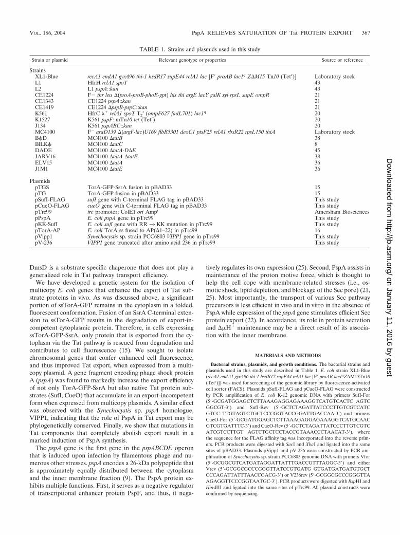
TABLE 1. Strains and plasmids used in this study
Strain or plasmid Relevant genotype or properties Source or reference
StrainsXL1-Blue recA1 endA1 gyrA96 thi-1 hsdR17 supE44 relA1 lac [F� proAB lac1q Z�M15 Tn10 (Tetr)] Laboratory stockL1 HfrH relA1 spoT 43L2 L1 pspA::kan 43CE1224 F� thr leu �(proA-proB-phoE-gpt) his thi argE lacY galK xyl rpsL supE ompR 21CE1343 CE1224 pspA::kan 21CE1419 CE1224 �pspB-pspC::kan 21K561 HfrC �� relA1 spoT T2
r (ompF627 fadL701) lac1q 20K1527 K561 pspF::mTn10-tet (Tetr) 20J134 K561 pspABC::kan 20MC4100 F� araD139 �(argF-lac)U169 flbB5301 deoC1 ptsF25 relA1 rbsR22 rpsL150 thiA Laboratory stockB�D MC4100 �tatB 38BlLK� MC4100 �tatC 8DADE MC4100 �tatA-D�E 45JARV16 MC4100 �tatA �tatE 38ELV15 MC4100 �tatA 36J1M1 MC4100 �tatE 36
PlasmidspTGS TorA-GFP-SsrA fusion in pBAD33 15pTG TorA-GFP fusion in pBAD33 15pSufI-FLAG sufI gene with C-terminal FLAG tag in pBAD33 This studypCueO-FLAG cueO gene with C-terminal FLAG tag in pBAD33 This studypTrc99 trc promoter; ColE1 ori Ampr Amersham BiosciencespPspA E. coli pspA gene in pTrc99 This studypKK-SufI E. coli sufl gene with RR 3 KK mutation in pTrc99 This studypTorA-AP E. coli TorA ss fused to AP(�1–22) in pTrc99 16pVipp1 Synechocystis sp. strain PCC6803 VIPP1 gene in pTrc99 This studypV-236 VIPP1 gene truncated after amino acid 236 in pTrc99 This study
VOL. 186, 2004 PspA RELIEVES SATURATION OF Tat PROTEIN EXPORT 367
on January 11, 2016 by guesthttp://jb.asm
.org/D
ownloaded from
Cultures were routinely grown aerobically at 30°C in Luria-Bertani (LB) me-dium, and antibiotic selection was maintained at the following concentrations, asrequired: ampicillin, 100 �g/ml; chloramphenicol, 25 �g/ml; kanamycin, 50 �g/ml. Protein synthesis was induced by adding isopropyl-�-D-thiogalactopyranoside(IPTG; 0.1 mM) and/or arabinose (0.2%) when the cells reached an opticaldensity at 600 nm of 0.5.
Generation of a genomic library and library screening. A library of random 2-to 3-kbp genomic fragments was constructed by digestion of XL1-Blue genomicDNA with Sau3AI, gel purification of the 2- to 3-kbp products, and ligation intothe BamHI site of plasmid pTrc99 (Amersham Biosciences). The ligation prod-ucts were transformed into XL1-Blue cells carrying pTGS (15) and plated on LBagar containing 0.2% glucose and ampicillin and chloramphenicol at the con-centrations indicated above. The resulting cell library (105 clones) was har-vested from plates and subcultured directly into liquid LB medium containingthe appropriate antibiotics. Cells were grown at 30°C until mid-log phase (opticaldensity at 600 nm, 0.5), and synthesis of ssTorA-GFP-SsrA and of polypeptidesencoded within genomic DNA inserts was induced with arabinose and IPTG,respectively. Following 3 h of induction, the cells were washed once with phos-phate-buffered saline and a 5-�l aliquot was diluted into 1 ml of phosphate-buffered saline and labeled with propidium iodide for flow cytometric detectionof nonviable cells (14). FACS sorting was performed with a Becton-DickinsonFACSort, and the desired cell population was gated by setting appropriate SSC,FL1, and FL2 windows (side scatter is used to trigger the cell events, whereasFL1 is used to monitor GFP fluorescence and FL2 is used to monitor propidiumiodine fluorescence). Typically, ca. 3 � 106 cells were examined in 30 min and250 to 1,000 events were collected. The collected solution was sterilely filtered(0.45-�m pore size; Millipore), and the filters were placed on LB medium plateswith ampicillin and chloramphenicol. After 12 h of incubation at 30°C, individualcolonies were inoculated into LB medium with ampicillin and chloramphenicolin triplicate 96-well plates. Following 12 h of growth at 30°C, cells were similarlysubcultured in triplicate into 96-well plates containing LB medium with ampi-cillin, chloramphenicol, 0.1 mM IPTG, and 0.2% arabinose and grown for 6 h at30°C. Individual clones were screened via flow cytometry and on a fluorescentplate reader (Bio-Tek FL600; Bio-Tek Instruments, Winooski, Vt.) for verifica-tion of the fluorescent phenotype.
Cell fractionation. Periplasmic and spheroplast fractions were prepared bysubjecting equivalent amounts of cells to the lysozyme-EDTA-cold osmoticshock procedure (31). The resulting spheroplasts were resuspended in 10 ml ofTE (10 mM Tris-Cl [pH 7.5], 2.5 mM Na-EDTA) and lysed by sonication, andintact cells and cellular debris were removed by centrifugation (5 min at 10,000� g). Lysed spheroplasts, including soluble and insoluble fractions, were dilutedin sodium dodecyl sulfate-polyacrylamide gel electrophoresis buffer and sub-jected to electrophoretic analysis, or alternatively, they were centrifuged and thesupernatant was retained as the soluble cytoplasmic fraction. For isolation ofmembrane fractions, the lysate of E. coli cells was centrifuged at 27,000 � g for20 min at 4°C. The membrane fraction was obtained by further centrifugation ofthe 27,000 � g supernatant at 100,000 � g for 1 h at 4°C. The supernatant wascarefully removed, and the membranes were gently resuspended in morpho-linepropanesulfonic acid (MOPS) buffer (50 mM, adjusted to pH 8.0 with KOH)containing 5 mM �-mercaptoethanol and 10 mM MgCl2 at a protein concentra-tion of 20 mg/ml. Protein concentrations were determined with a Bio-Rad pro-tein assay reagent kit with bovine serum albumin as the standard. �-Galactosi-dase activity was used as a cytoplasmic marker of fractionation efficiency (17).Only data from fractionation experiments in which �95% of the �-galactosidaseactivity was in the cytoplasmic fraction are reported. To analyze total cellularproteins, collected cells were resuspended in TE and homogenized in a Frenchpress cell (Carver) at 20,000 lb/in2.
Western blotting analysis. Western blotting was performed as described pre-viously (12). The following primary antibodies were used: polyclonal rabbitanti-PspA (gift from J. Tommassen), monoclonal mouse anti-GFP (Clontech)diluted 1:5,000, monoclonal mouse anti-FLAG (Sigma) diluted 1:3,000, poly-clonal rabbit anti-SufI (10) diluted 1:3,000, monoclonal rabbit anti-DsbC (giftfrom John Joly, Genentech) diluted 1:10,000, and monoclonal rabbit anti-GroEL(Sigma) diluted 1:10,000. The secondary antibody was 1:10,000 goat anti-mouse–horseradish peroxidase or goat anti-rabbit–horseradish peroxidase. Membranescontaining fractionated samples were first probed with anti-GFP, anti-FLAG, oranti-SufI antibodies and then, following development, stripped in Tris-bufferedsaline–2% sodium dodecyl sulfate–0.7 M �-mercaptoethanol. Stripped mem-branes were reblocked and probed with anti-DsbC and anti-GroEL antibodiessimultaneously. Relative band intensities of Western blots were calculated withImageJ v1.29, which was obtained from http://rsb.info.nih.gov/ij/.
RESULTS
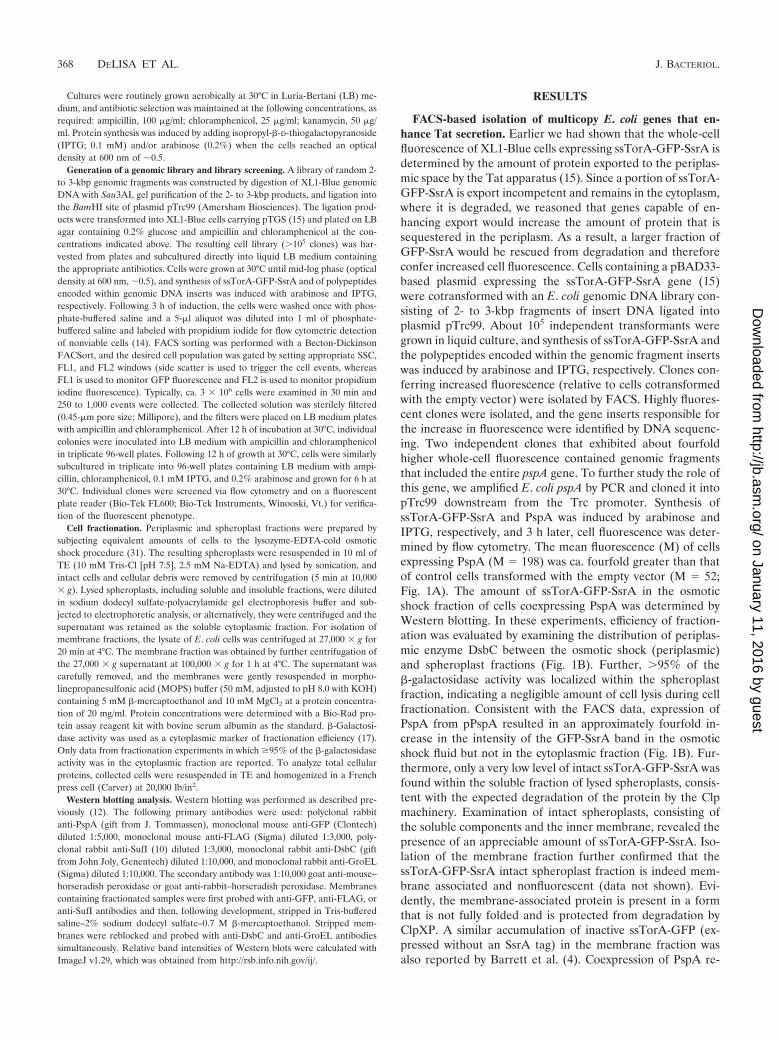
FACS-based isolation of multicopy E. coli genes that en-hance Tat secretion. Earlier we had shown that the whole-cellfluorescence of XL1-Blue cells expressing ssTorA-GFP-SsrA isdetermined by the amount of protein exported to the periplas-mic space by the Tat apparatus (15). Since a portion of ssTorA-GFP-SsrA is export incompetent and remains in the cytoplasm,where it is degraded, we reasoned that genes capable of en-hancing export would increase the amount of protein that issequestered in the periplasm. As a result, a larger fraction ofGFP-SsrA would be rescued from degradation and thereforeconfer increased cell fluorescence. Cells containing a pBAD33-based plasmid expressing the ssTorA-GFP-SsrA gene (15)were cotransformed with an E. coli genomic DNA library con-sisting of 2- to 3-kbp fragments of insert DNA ligated intoplasmid pTrc99. About 105 independent transformants weregrown in liquid culture, and synthesis of ssTorA-GFP-SsrA andthe polypeptides encoded within the genomic fragment insertswas induced by arabinose and IPTG, respectively. Clones con-ferring increased fluorescence (relative to cells cotransformedwith the empty vector) were isolated by FACS. Highly fluores-cent clones were isolated, and the gene inserts responsible forthe increase in fluorescence were identified by DNA sequenc-ing. Two independent clones that exhibited about fourfoldhigher whole-cell fluorescence contained genomic fragmentsthat included the entire pspA gene. To further study the role ofthis gene, we amplified E. coli pspA by PCR and cloned it intopTrc99 downstream from the Trc promoter. Synthesis ofssTorA-GFP-SsrA and PspA was induced by arabinose andIPTG, respectively, and 3 h later, cell fluorescence was deter-mined by flow cytometry. The mean fluorescence (M) of cellsexpressing PspA (M � 198) was ca. fourfold greater than thatof control cells transformed with the empty vector (M � 52;Fig. 1A). The amount of ssTorA-GFP-SsrA in the osmoticshock fraction of cells coexpressing PspA was determined byWestern blotting. In these experiments, efficiency of fraction-ation was evaluated by examining the distribution of periplas-mic enzyme DsbC between the osmotic shock (periplasmic)and spheroplast fractions (Fig. 1B). Further, 95% of the�-galactosidase activity was localized within the spheroplastfraction, indicating a negligible amount of cell lysis during cellfractionation. Consistent with the FACS data, expression ofPspA from pPspA resulted in an approximately fourfold in-crease in the intensity of the GFP-SsrA band in the osmoticshock fluid but not in the cytoplasmic fraction (Fig. 1B). Fur-thermore, only a very low level of intact ssTorA-GFP-SsrA wasfound within the soluble fraction of lysed spheroplasts, consis-tent with the expected degradation of the protein by the Clpmachinery. Examination of intact spheroplasts, consisting ofthe soluble components and the inner membrane, revealed thepresence of an appreciable amount of ssTorA-GFP-SsrA. Iso-lation of the membrane fraction further confirmed that thessTorA-GFP-SsrA intact spheroplast fraction is indeed mem-brane associated and nonfluorescent (data not shown). Evi-dently, the membrane-associated protein is present in a formthat is not fully folded and is protected from degradation byClpXP. A similar accumulation of inactive ssTorA-GFP (ex-pressed without an SsrA tag) in the membrane fraction wasalso reported by Barrett et al. (4). Coexpression of PspA re-
368 DELISA ET AL. J. BACTERIOL.
on January 11, 2016 by guesthttp://jb.asm
.org/D
ownloaded from
sulted in a reduction in the amount of membrane-associatedssTorA-GFP-SsrA (Fig. 1C). Densitometric analysis of theprotein band in intact spheroplasts with and without PspAexpression revealed a fourfold greater amount in the lattersamples.
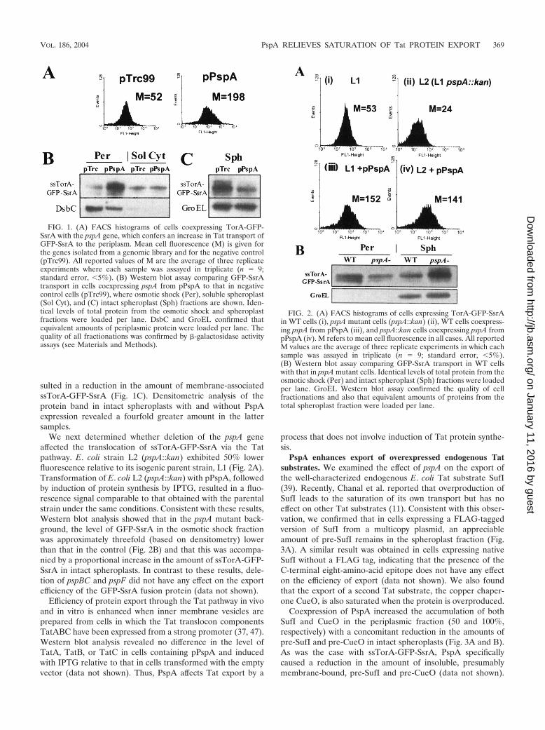
We next determined whether deletion of the pspA geneaffected the translocation of ssTorA-GFP-SsrA via the Tatpathway. E. coli strain L2 (pspA::kan) exhibited 50% lowerfluorescence relative to its isogenic parent strain, L1 (Fig. 2A).Transformation of E. coli L2 (pspA::kan) with pPspA, followedby induction of protein synthesis by IPTG, resulted in a fluo-rescence signal comparable to that obtained with the parentalstrain under the same conditions. Consistent with these results,Western blot analysis showed that in the pspA mutant back-ground, the level of GFP-SsrA in the osmotic shock fractionwas approximately threefold (based on densitometry) lowerthan that in the control (Fig. 2B) and that this was accompa-nied by a proportional increase in the amount of ssTorA-GFP-SsrA in intact spheroplasts. In contrast to these results, dele-tion of pspBC and pspF did not have any effect on the exportefficiency of the GFP-SsrA fusion protein (data not shown).
Efficiency of protein export through the Tat pathway in vivoand in vitro is enhanced when inner membrane vesicles areprepared from cells in which the Tat translocon componentsTatABC have been expressed from a strong promoter (37, 47).Western blot analysis revealed no difference in the level ofTatA, TatB, or TatC in cells containing pPspA and inducedwith IPTG relative to that in cells transformed with the emptyvector (data not shown). Thus, PspA affects Tat export by a
process that does not involve induction of Tat protein synthe-sis.
PspA enhances export of overexpressed endogenous Tatsubstrates. We examined the effect of pspA on the export ofthe well-characterized endogenous E. coli Tat substrate SufI(39). Recently, Chanal et al. reported that overproduction ofSufI leads to the saturation of its own transport but has noeffect on other Tat substrates (11). Consistent with this obser-vation, we confirmed that in cells expressing a FLAG-taggedversion of SufI from a multicopy plasmid, an appreciableamount of pre-SufI remains in the spheroplast fraction (Fig.3A). A similar result was obtained in cells expressing nativeSufI without a FLAG tag, indicating that the presence of theC-terminal eight-amino-acid epitope does not have any effecton the efficiency of export (data not shown). We also foundthat the export of a second Tat substrate, the copper chaper-one CueO, is also saturated when the protein is overproduced.
Coexpression of PspA increased the accumulation of bothSufI and CueO in the periplasmic fraction (50 and 100%,respectively) with a concomitant reduction in the amounts ofpre-SufI and pre-CueO in intact spheroplasts (Fig. 3A and B).As was the case with ssTorA-GFP-SsrA, PspA specificallycaused a reduction in the amount of insoluble, presumablymembrane-bound, pre-SufI and pre-CueO (data not shown).
FIG. 1. (A) FACS histograms of cells coexpressing TorA-GFP-SsrA with the pspA gene, which confers an increase in Tat transport ofGFP-SsrA to the periplasm. Mean cell fluorescence (M) is given forthe genes isolated from a genomic library and for the negative control(pTrc99). All reported values of M are the average of three replicateexperiments where each sample was assayed in triplicate (n � 9;standard error, 5%). (B) Western blot assay comparing GFP-SsrAtransport in cells coexpressing pspA from pPspA to that in negativecontrol cells (pTrc99), where osmotic shock (Per), soluble spheroplast(Sol Cyt), and (C) intact spheroplast (Sph) fractions are shown. Iden-tical levels of total protein from the osmotic shock and spheroplastfractions were loaded per lane. DsbC and GroEL confirmed thatequivalent amounts of periplasmic protein were loaded per lane. Thequality of all fractionations was confirmed by �-galactosidase activityassays (see Materials and Methods).
FIG. 2. (A) FACS histograms of cells expressing TorA-GFP-SsrAin WT cells (i), pspA mutant cells (pspA::kan) (ii), WT cells coexpress-ing pspA from pPspA (iii), and pspA::kan cells coexpressing pspA frompPspA (iv). M refers to mean cell fluorescence in all cases. All reportedM values are the average of three replicate experiments in which eachsample was assayed in triplicate (n � 9; standard error, 5%).(B) Western blot assay comparing GFP-SsrA transport in WT cellswith that in pspA mutant cells. Identical levels of total protein from theosmotic shock (Per) and intact spheroplast (Sph) fractions were loadedper lane. GroEL Western blot assay confirmed the quality of cellfractionations and also that equivalent amounts of proteins from thetotal spheroplast fraction were loaded per lane.
VOL. 186, 2004 PspA RELIEVES SATURATION OF Tat PROTEIN EXPORT 369
on January 11, 2016 by guesthttp://jb.asm
.org/D
ownloaded from
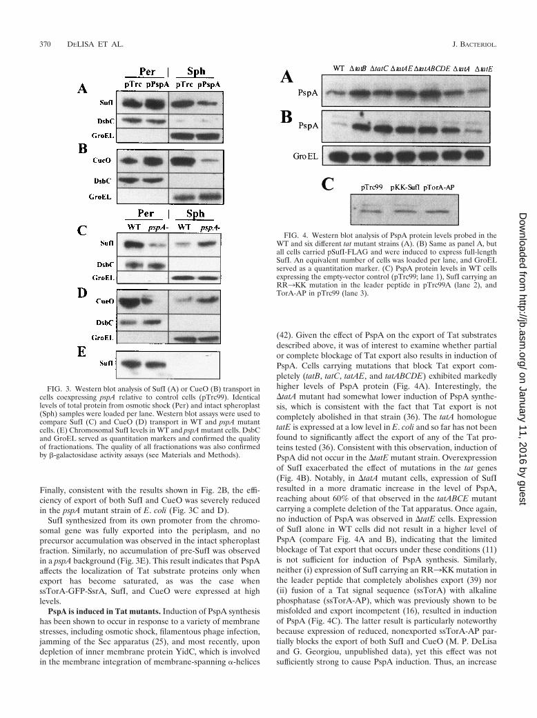
Finally, consistent with the results shown in Fig. 2B, the effi-ciency of export of both SufI and CueO was severely reducedin the pspA mutant strain of E. coli (Fig. 3C and D).
SufI synthesized from its own promoter from the chromo-somal gene was fully exported into the periplasm, and noprecursor accumulation was observed in the intact spheroplastfraction. Similarly, no accumulation of pre-SufI was observedin a pspA background (Fig. 3E). This result indicates that PspAaffects the localization of Tat substrate proteins only whenexport has become saturated, as was the case whenssTorA-GFP-SsrA, SufI, and CueO were expressed at highlevels.
PspA is induced in Tat mutants. Induction of PspA synthesishas been shown to occur in response to a variety of membranestresses, including osmotic shock, filamentous phage infection,jamming of the Sec apparatus (25), and most recently, upondepletion of inner membrane protein YidC, which is involvedin the membrane integration of membrane-spanning �-helices
(42). Given the effect of PspA on the export of Tat substratesdescribed above, it was of interest to examine whether partialor complete blockage of Tat export also results in induction ofPspA. Cells carrying mutations that block Tat export com-pletely (tatB, tatC, tatAE, and tatABCDE) exhibited markedlyhigher levels of PspA protein (Fig. 4A). Interestingly, the�tatA mutant had somewhat lower induction of PspA synthe-sis, which is consistent with the fact that Tat export is notcompletely abolished in that strain (36). The tatA homologuetatE is expressed at a low level in E. coli and so far has not beenfound to significantly affect the export of any of the Tat pro-teins tested (36). Consistent with this observation, induction ofPspA did not occur in the �tatE mutant strain. Overexpressionof SufI exacerbated the effect of mutations in the tat genes(Fig. 4B). Notably, in �tatA mutant cells, expression of SufIresulted in a more dramatic increase in the level of PspA,reaching about 60% of that observed in the tatABCE mutantcarrying a complete deletion of the Tat apparatus. Once again,no induction of PspA was observed in �tatE cells. Expressionof SufI alone in WT cells did not result in a higher level ofPspA (compare Fig. 4A and B), indicating that the limitedblockage of Tat export that occurs under these conditions (11)is not sufficient for induction of PspA synthesis. Similarly,neither (i) expression of SufI carrying an RR3KK mutation inthe leader peptide that completely abolishes export (39) nor(ii) fusion of a Tat signal sequence (ssTorA) with alkalinephosphatase (ssTorA-AP), which was previously shown to bemisfolded and export incompetent (16), resulted in inductionof PspA (Fig. 4C). The latter result is particularly noteworthybecause expression of reduced, nonexported ssTorA-AP par-tially blocks the export of both SufI and CueO (M. P. DeLisaand G. Georgiou, unpublished data), yet this effect was notsufficiently strong to cause PspA induction. Thus, an increase
FIG. 3. Western blot analysis of SufI (A) or CueO (B) transport incells coexpressing pspA relative to control cells (pTrc99). Identicallevels of total protein from osmotic shock (Per) and intact spheroplast(Sph) samples were loaded per lane. Western blot assays were used tocompare SufI (C) and CueO (D) transport in WT and pspA mutantcells. (E) Chromosomal SufI levels in WT and pspA mutant cells. DsbCand GroEL served as quantitation markers and confirmed the qualityof fractionations. The quality of all fractionations was also confirmedby �-galactosidase activity assays (see Materials and Methods).
FIG. 4. Western blot analysis of PspA protein levels probed in theWT and six different tat mutant strains (A). (B) Same as panel A, butall cells carried pSufI-FLAG and were induced to express full-lengthSufI. An equivalent number of cells was loaded per lane, and GroELserved as a quantitation marker. (C) PspA protein levels in WT cellsexpressing the empty-vector control (pTrc99; lane 1), SufI carrying anRR3KK mutation in the leader peptide in pTrc99A (lane 2), andTorA-AP in pTrc99 (lane 3).
370 DELISA ET AL. J. BACTERIOL.
on January 11, 2016 by guesthttp://jb.asm
.org/D
ownloaded from
in PspA synthesis is only caused by mutations that abolishexport nearly completely.
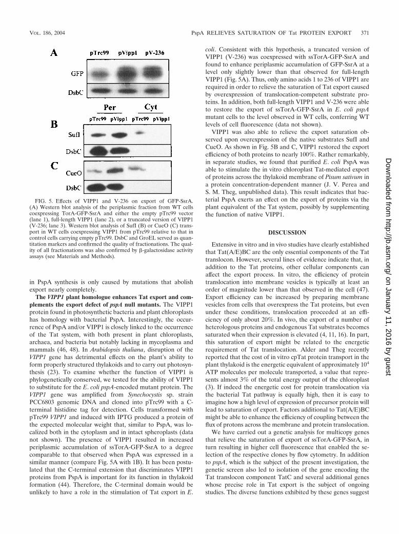
The VIPP1 plant homologue enhances Tat export and com-plements the export defect of pspA null mutants. The VIPP1protein found in photosynthetic bacteria and plant chloroplastshas homology with bacterial PspA. Interestingly, the occur-rence of PspA and/or VIPP1 is closely linked to the occurrenceof the Tat system, with both present in plant chloroplasts,archaea, and bacteria but notably lacking in mycoplasma andmammals (46, 48). In Arabidopsis thaliana, disruption of theVIPP1 gene has detrimental effects on the plant’s ability toform properly structured thylakoids and to carry out photosyn-thesis (23). To examine whether the function of VIPP1 isphylogenetically conserved, we tested for the ability of VIPP1to substitute for the E. coli pspA-encoded mutant protein. TheVIPP1 gene was amplified from Synechocystis sp. strainPCC6803 genomic DNA and cloned into pTrc99 with a C-terminal histidine tag for detection. Cells transformed withpTrc99 VIPP1 and induced with IPTG produced a protein ofthe expected molecular weight that, similar to PspA, was lo-calized both in the cytoplasm and in intact spheroplasts (datanot shown). The presence of VIPP1 resulted in increasedperiplasmic accumulation of ssTorA-GFP-SsrA to a degreecomparable to that observed when PspA was expressed in asimilar manner (compare Fig. 5A with 1B). It has been postu-lated that the C-terminal extension that discriminates VIPP1proteins from PspA is important for its function in thylakoidformation (44). Therefore, the C-terminal domain would beunlikely to have a role in the stimulation of Tat export in E.
coli. Consistent with this hypothesis, a truncated version ofVIPP1 (V-236) was coexpressed with ssTorA-GFP-SsrA andfound to enhance periplasmic accumulation of GFP-SsrA at alevel only slightly lower than that observed for full-lengthVIPP1 (Fig. 5A). Thus, only amino acids 1 to 236 of VIPP1 arerequired in order to relieve the saturation of Tat export causedby overexpression of translocation-competent substrate pro-teins. In addition, both full-length VIPP1 and V-236 were ableto restore the export of ssTorA-GFP-SsrA in E. coli pspAmutant cells to the level observed in WT cells, conferring WTlevels of cell fluorescence (data not shown).
VIPP1 was also able to relieve the export saturation ob-served upon overexpression of the native substrates SufI andCueO. As shown in Fig. 5B and C, VIPP1 restored the exportefficiency of both proteins to nearly 100%. Rather remarkably,in separate studies, we found that purified E. coli PspA wasable to stimulate the in vitro chloroplast Tat-mediated exportof proteins across the thylakoid membrane of Pisum sativum ina protein concentration-dependent manner (J. V. Perea andS. M. Theg, unpublished data). This result indicates that bac-terial PspA exerts an effect on the export of proteins via theplant equivalent of the Tat system, possibly by supplementingthe function of native VIPP1.
DISCUSSION
Extensive in vitro and in vivo studies have clearly establishedthat Tat(A/E)BC are the only essential components of the Tattranslocon. However, several lines of evidence indicate that, inaddition to the Tat proteins, other cellular components canaffect the export process. In vitro, the efficiency of proteintranslocation into membrane vesicles is typically at least anorder of magnitude lower than that observed in the cell (47).Export efficiency can be increased by preparing membranevesicles from cells that overexpress the Tat proteins, but evenunder these conditions, translocation proceeded at an effi-ciency of only about 20%. In vivo, the export of a number ofheterologous proteins and endogenous Tat substrates becomessaturated when their expression is elevated (4, 11, 16). In part,this saturation of export might be related to the energeticrequirement of Tat translocation. Alder and Theg recentlyreported that the cost of in vitro cpTat protein transport in theplant thylakoid is the energetic equivalent of approximately 104
ATP molecules per molecule transported, a value that repre-sents almost 3% of the total energy output of the chloroplast(3). If indeed the energetic cost for protein translocation viathe bacterial Tat pathway is equally high, then it is easy toimagine how a high level of expression of precursor protein willlead to saturation of export. Factors additional to Tat(A/E)BCmight be able to enhance the efficiency of coupling between theflux of protons across the membrane and protein translocation.
We have carried out a genetic analysis for multicopy genesthat relieve the saturation of export of ssTorA-GFP-SsrA, inturn resulting in higher cell fluorescence that enabled the se-lection of the respective clones by flow cytometry. In additionto pspA, which is the subject of the present investigation, thegenetic screen also led to isolation of the gene encoding theTat translocon component TatC and several additional geneswhose precise role in Tat export is the subject of ongoingstudies. The diverse functions exhibited by these genes suggest
FIG. 5. Effects of VIPP1 and V-236 on export of GFP-SsrA.(A) Western blot analysis of the periplasmic fraction from WT cellscoexpressing TorA-GFP-SsrA and either the empty pTrc99 vector(lane 1), full-length VIPP1 (lane 2), or a truncated version of VIPP1(V-236; lane 3). Western blot analysis of SufI (B) or CueO (C) trans-port in WT cells coexpressing VIPP1 from pTrc99 relative to that incontrol cells carrying empty pTrc99. DsbC and GroEL served as quan-titation markers and confirmed the quality of fractionations. The qual-ity of all fractionations was also confirmed by �-galactosidase activityassays (see Materials and Methods).
VOL. 186, 2004 PspA RELIEVES SATURATION OF Tat PROTEIN EXPORT 371
on January 11, 2016 by guesthttp://jb.asm
.org/D
ownloaded from
that several distinct processes might be contributing to thesaturation of protein export.
We found that expression of PspA relieves the export satu-ration that occurs upon high-level expression of heterologous(ssTorA-GFP-SsrA) and native (SufI, CueO) Tat substrates.Saturation of export is manifest with the accumulation of thepreprotein in the insoluble portion of the spheroplast fraction.As a result of the action of PspA, the amount of insolubleprecursor is reduced concomitant with an increase in the pro-tein in the periplasmic fraction. We also found that, for all ofthe proteins tested, the export saturation was exacerbated instrains deficient in pspA. It is important to note that pspAaffects efficiency of protein export only under conditions inwhich the transport machinery is saturated. When SufI is ex-pressed at a low level from the chromosomal copy, no accu-mulation of precursor protein is observed in the spheroplastfraction either in WT cells or in a pspA mutant.
The precise mechanism by which PspA is able to mediatetransfer of the protein from the insoluble spheroplast fractionto the periplasm is not clear. PspA may be affecting any of anumber of processes, including, for example, the folding of theprotein into a form competent for export, improved couplingof the proton flux with secretion, proteolysis of membrane-associated translocation-incompetent precursors, or even re-lease of the newly translocated protein from the periplasmicside of the membrane.
Whatever the precise mechanism of the action of PspAmight be, it is clearly not related to the regulatory role of theprotein as an inhibitor of the enhancement of transcription byPspF (25). Deletion of the other genes in the psp operon,including the transcriptional activator pspF or the two positiveregulators pspBC, did not exacerbate the saturation of ssTorA-GFP-SsrA export. Thus, although PspA has been shown tobind PspB, PspC, and PspF in vivo (1, 19), these interactionsare not involved in the observed PspA-dependent increase inTat export efficiency.
The level of PspA was markedly increased when Tat exportwas nearly or completely abolished as a result of the inactiva-tion of Tat genes (i.e., in �tatA, �tatB, �tatC, or �tatABCDEmutant strains). Consistent with these findings, Kleerebezumet al. found that depletion of proteins involved in Sec translo-cation occurring after the protein reaches the membrane (i.e.,in SecD, SecF, and SecA) led to induction of PspA. Theseauthors postulated that the PspA-inducing signal was dissipa-tion of the ��H�, such as occurs upon entrance of a precursorprotein into and subsequent blockage of the export apparatusin the inner membrane. Similarly, Van der Laan et al. demon-strated that depletion of YidC also resulted in elevated levelsof PspA (42). Induction of PspA expression upon depletion ofYidC proved to be a reliable indicator of a reduced protonmotive force, which is in line with previous suggestions thatPspA senses membrane damage and/or a reduction of theproton motive force. Since the translocation defects observedin a psp mutant strain are caused by a drop in proton motiveforce (��H�) and at least one function of PspA appears to bemaintenance of ��H� under these stress conditions (21), it istempting to speculate that stimulation of Tat transport byPspA might be through maintenance of ��H� during translo-cation of Tat precursors.
In conclusion, we note that the identification of factors that
can increase protein flux and enhance the yield of secretedproteins via the Tat pathway is significant from a biotechnologystandpoint. The Sec pathway has served as the primary conduitfor the secretion of many industrially important proteins. How-ever, the observation that many Sec substrates can becomestuck in the translocation pore is problematic when seekinghigh recombinant yields. To date, there have been no reportedcases of precursors becoming jammed in the Tat machinery. Infact, efforts to develop genetic constructs that form membrane-spanning translocation intermediates have proved largely un-successful (27). The primary limitation of the Tat pathwayrelative to the Sec pathway is the relatively poor export effi-ciency of proteins targeted to the Tat pathway. Therefore,strategies whereby cellular factors such as those identifiedherein are coexpressed with recombinant proteins of interestshould help alleviate the inefficiency of Tat transport.
ACKNOWLEDGMENTS
We are especially grateful to P. Model for the generous gift of pspmutants and to J. Tommassen for anti-PspA antisera. We thank G.Buchanan for assistance with pulse-chase assays.
This work was supported by a grant from the Foundation for Re-search to G.G. P.L. is supported by a BBSRC-funded Ph.D. student-ship, and T.P. is a Royal Society Research Fellow.
REFERENCES
1. Adams, H., W. Teertstra, J. Demmers, R. Boesten, and J. Tommassen. 2003.Interactions between phage-shock proteins in Escherichia coli. J. Bacteriol.185:1174–1180.
2. Alami, M., D. Trescher, L. F. Wu, and M. Muller. 2002. Separate analysis oftwin-arginine translocation (Tat)-specific membrane binding and transloca-tion in Escherichia coli. J. Biol. Chem. 277:20499–20503.
3. Alder, N. N., and S. M. Theg. 2003. Energetics of protein transport acrossbiological membranes. a study of the thylakoid �pH-dependent/cpTat path-way. Cell 112:231–242.
4. Barrett, C. M., N. Ray, J. D. Thomas, C. Robinson, and A. Bolhuis. 2003.Quantitative export of a reporter protein, GFP, by the twin-arginine trans-location pathway in Escherichia coli. Biochem. Biophys. Res. Commun. 304:279–284.
5. Berks, B. C. 1996. A common export pathway for proteins binding complexredox cofactors? Mol. Microbiol. 22:393–404.
6. Berks, B. C., T. Palmer, and F. Sargent. 2003. The Tat protein translocationpathway and its role in microbial physiology. Adv. Microb. Physiol. 47:187–254.
7. Berks, B. C., F. Sargent, and T. Palmer. 2000. The Tat protein exportpathway. Mol. Microbiol. 35:260–274.
8. Bogsch, E. G., F. Sargent, N. R. Stanley, B. C. Berks, C. Robinson, and T.Palmer. 1998. An essential component of a novel bacterial protein exportsystem with homologues in plastids and mitochondria. J. Biol. Chem. 273:18003–18006.
9. Brissette, J. L., M. Russel, L. Weiner, and P. Model. 1990. Phage shockprotein, a stress protein of Escherichia coli. Proc. Natl. Acad. Sci. USA87:862–866.
10. Buchanan, G., E. Leeuw, N. R. Stanley, M. Wexler, B. C. Berks, F. Sargent,and T. Palmer. 2002. Functional complexity of the twin-arginine translocaseTatC component revealed by site-directed mutagenesis. Mol. Microbiol.43:1457–1470.
11. Chanal, A., C. L. Santini, and L. F. Wu. 2003. Specific inhibition of thetranslocation of a subset of Escherichia coli TAT substrates by the TorAsignal peptide. J. Mol. Biol. 327:563–570.
12. Chen, G., A. Hayhurst, J. G. Thomas, B. R. Harvey, B. L. Iverson, and G.Georgiou. 2001. Isolation of high-affinity ligand-binding proteins by periplas-mic expression with cytometric screening (PECS). Nat. Biotechnol. 19:537–542.
13. Cristobal, S., J. W. de Gier, H. Nielsen, and G. von Heijne. 1999. Competi-tion between Sec- and TAT-dependent protein translocation in Escherichiacoli. EMBO J. 18:2982–2990.
14. Daugherty, P. S., G. Chen, B. L. Iverson, and G. Georgiou. 2000. Quantita-tive analysis of the effect of the mutation frequency on the affinity maturationof single chain Fv antibodies. Proc. Natl. Acad. Sci. USA 97:2029–2034.
15. DeLisa, M. P., P. Samuelson, T. Palmer, and G. Georgiou. 2002. Geneticanalysis of the twin arginine translocator secretion pathway in bacteria.J. Biol. Chem. 277:29825–29831.
16. DeLisa, M. P., D. Tullman, and G. Georgiou. 2003. Folding quality control
372 DELISA ET AL. J. BACTERIOL.
on January 11, 2016 by guesthttp://jb.asm
.org/D
ownloaded from
in the export of proteins by the bacterial twin-arginine translocation path-way. Proc. Natl. Acad. Sci. USA 100:6115–6120.
17. Derman, A. I., J. W. Puziss, P. J. Bassford, Jr., and J. Beckwith. 1993. Asignal sequence is not required for protein export in prlA mutants of Esch-erichia coli. EMBO J. 12:879–888.
18. Driessen, A. J., P. Fekkes, and J. P. van der Wolk. 1998. The Sec system.Curr. Opin. Microbiol. 1:216–222.
19. Elderkin, S., S. Jones, J. Schumacher, D. Studholme, and M. Buck. 2002.Mechanism of action of the Escherichia coli phage shock protein PspA inrepression of the AAA family transcription factor PspF. J. Mol. Biol. 320:23–37.
20. Jovanovic, G., L. Weiner, and P. Model. 1996. Identification, nucleotidesequence, and characterization of PspF, the transcriptional activator of theEscherichia coli stress-induced psp operon. J. Bacteriol. 178:1936–1945.
21. Kleerebezem, M., W. Crielaard, and J. Tommassen. 1996. Involvement ofstress protein PspA (phage shock protein A) of Escherichia coli in mainte-nance of the protonmotive force under stress conditions. EMBO J. 15:162–171.
22. Kleerebezem, M., and J. Tommassen. 1993. Expression of the pspA genestimulates efficient protein export in Escherichia coli. Mol. Microbiol. 7:947–956.
23. Kroll, D., K. Meierhoff, N. Bechtold, M. Kinoshita, S. Westphal, U. C.Vothknecht, J. Soll, and P. Westhoff. 2001. VIPP1, a nuclear gene of Arabi-dopsis thaliana essential for thylakoid membrane formation. Proc. Natl.Acad. Sci. USA 98:4238–4242.
24. Mikhaleva, N. I., C. L. Santini, G. Giordano, M. A. Nesmeyanova, and L. F.Wu. 1999. Requirement for phospholipids of the translocation of the trim-ethylamine N-oxide reductase through the Tat pathway in Escherichia coli.FEBS Lett. 463:331–335.
25. Model, P., G. Jovanovic, and J. Dworkin. 1997. The. Escherichia coli phage-shock-protein (psp) operon. Mol. Microbiol. 24:255–261.
26. Mori, H., and K. Cline. 2002. A twin arginine signal peptide and the pHgradient trigger reversible assembly of the thylakoid �pH/Tat translocase.J. Cell Biol. 157:205–210.
27. Musser, S. M., and S. M. Theg. 2000. Characterization of the early steps ofOE17 precursor transport by the thylakoid �pH/Tat machinery. Eur. J. Bio-chem. 267:2588–2598.
28. Musser, S. M., and S. M. Theg. 2000. Proton transfer limits protein trans-location rate by the thylakoid �pH/Tat machinery. Biochemistry 39:8228–8233.
29. Oresnik, I. J., C. L. Ladner, and R. J. Turner. 2001. Identification of atwin-arginine leader-binding protein. Mol. Microbiol. 40:323–331.
30. Papish, A. L., C. L. Ladner, and R. J. Turner. 2003. The twin-arginineleader-binding protein, DmsD, interacts with the TatB and TatC subunits ofthe Escherichia coli twin-arginine translocase. J. Biol. Chem. 278:32501–32506.
31. Randall, L. L., and S. J. Hardy. 1986. Correlation of competence for exportwith lack of tertiary structure of the mature species: a study in vivo ofmaltose-binding protein in E. coli. Cell 46:921–928.
32. Ray, N., J. Oates, R. J. Turner, and C. Robinson. 2003. DmsD is required forthe biogenesis of DMSO reductase in Escherichia coli but not for the inter-action of the DmsA signal peptide with the Tat apparatus. FEBS Lett.534:156–160.
33. Rodrigue, A., A. Chanal, K. Beck, M. Muller, and L. F. Wu. 1999. Co-translocation of a periplasmic enzyme complex by a hitchhiker mechanismthrough the bacterial tat pathway. J. Biol. Chem. 274:13223–13228.
34. Sanders, C., N. Wethkamp, and H. Lill. 2001. Transport of cytochrome cderivatives by the bacterial Tat protein translocation system. Mol. Microbiol.41:241–246.
35. Sargent, F., B. C. Berks, and T. Palmer. 2002. Assembly of membrane-boundrespiratory complexes by the Tat protein-transport system. Arch. Microbiol.178:77–84.
36. Sargent, F., E. G. Bogsch, N. R. Stanley, M. Wexler, C. Robinson, B. C.Berks, and T. Palmer. 1998. Overlapping functions of components of abacterial Sec-independent protein export pathway. EMBO J. 17:3640–3650.
37. Sargent, F., U. Gohlke, E. De Leeuw, N. R. Stanley, T. Palmer, H. R. Saibil,and B. C. Berks. 2001. Purified components of the Escherichia coli Tatprotein transport system form a double-layered ring structure. Eur. J. Bio-chem. 268:3361–3367.
38. Sargent, F., N. R. Stanley, B. C. Berks, and T. Palmer. 1999. Sec-indepen-dent protein translocation in Escherichia coli. A distinct and pivotal role forthe TatB protein. J. Biol. Chem. 274:36073–36082.
39. Stanley, N. R., T. Palmer, and B. C. Berks. 2000. The twin arginine consensusmotif of Tat signal peptides is involved in Sec-independent protein targetingin Escherichia coli. J. Biol. Chem. 275:11591–11596.
40. Stanley, N. R., F. Sargent, G. Buchanan, J. Shi, V. Stewart, T. Palmer, andB. C. Berks. 2002. Behaviour of topological marker proteins targeted to theTat protein transport pathway. Mol. Microbiol. 43:1005–1021.
41. Thomas, J. D., R. A. Daniel, J. Errington, and C. Robinson. 2001. Export ofactive green fluorescent protein to the periplasm by the twin-arginine trans-locase (Tat) pathway in Escherichia coli. Mol. Microbiol. 39:47–53.
42. Van Der Laan, M., M. L. Urbanus, C. M. Ten Hagen-Jongman, N. Nouwen,B. Oudega, N. Harms, A. J. Driessen, and J. Luirink. 2003. A conservedfunction of YidC in the biogenesis of respiratory chain complexes. Proc.Natl. Acad. Sci. USA 100:5801–5806.
43. Weiner, L., J. L. Brissette, and P. Model. 1991. Stress-induced expression ofthe Escherichia coli phage shock protein operon is dependent on sigma 54and modulated by positive and negative feedback mechanisms. Genes Dev.5:1912–1923.
44. Westphal, S., L. Heins, J. Soll, and U. C. Vothknecht. 2001. Vipp1 deletionmutant of Synechocystis: a connection between bacterial phage shock andthylakoid biogenesis? Proc. Natl. Acad. Sci. USA 98:4243–4248.
45. Wexler, M., F. Sargent, R. L. Jack, N. R. Stanley, E. G. Bogsch, C. Robinson,B. C. Berks, and T. Palmer. 2000. TatD is a cytoplasmic protein with DNaseactivity. No requirement for TatD family proteins in sec-independent proteinexport. J. Biol. Chem. 275:16717–16722.
46. Wu, L. F., B. Ize, A. Chanal, Y. Quentin, and G. Fichant. 2000. Bacterialtwin-arginine signal peptide-dependent protein translocation pathway: evo-lution and mechanism. J. Mol. Microbiol. Biotechnol. 2:179–189.
47. Yahr, T. L., and W. T. Wickner. 2001. Functional reconstitution of bacterialTat translocation in vitro. EMBO J. 20:2472–2479.
48. Yen, M. R., Y. H. Tseng, E. H. Nguyen, L. F. Wu, and M. H. Saier, Jr. 2002.Sequence and phylogenetic analyses of the twin-arginine targeting (Tat)protein export system. Arch. Microbiol. 177:441–450.
VOL. 186, 2004 PspA RELIEVES SATURATION OF Tat PROTEIN EXPORT 373
on January 11, 2016 by guesthttp://jb.asm
.org/D
ownloaded from
Top Related
Copyright © 2022 FDOKUMEN

