


Bahasa
Halaman
Hukum


1
One-step Sustainable Preparation of
Superparamagnetic Iron Oxide Nanoparticles
Supported on Mesoporous SiO2
Elena Chamorro,a M. José Tenorio,b,† Lourdes Calvo,a M. José Torralvo,c Regino Sáez-Puchec
and Albertina Cabañasb*
aDep. Chemical Engineering, bDep. Physical Chemistry, cDep. Inorganic Chemistry, Universidad
Complutense de Madrid, Avda. Complutense s/n, 28040 Madrid, Spain
†Current address: Chemical, Energy and Mechanical Technology Dep, Universidad Rey Juan
Carlos, c/Tulipán s/n, 28933 Móstoles, Madrid, Spain
* Corresponding author:
Albertina Cabañas, e-mail: [email protected]

2
ABSTRACT
Superparamagnetic iron oxide nanoparticles (SPIONs) supported on high surface area mesoporous
SiO2 are advanced materials of great interest in catalysis, adsorption and biomedicine. Here we
present a new process to prepare SPION/SiO2 materials by the impregnation and insitu
decomposition of Fe(NO3)3.9H2O on mesoporous SiO2 supports in a 25-50% mol ethanol + CO2
mixture at 523 K and 25.0 MPa. -Fe2O3 nanoparticles (NPs) of average size between 6-9 nm were
distributed homogeneously on the supports. NPs deposited into the SBA-15 mesopores but mostly
on the external surface of MCM-41. Materials prepared with the highest ethanol content were very
homogeneous. Magnetic measurements confirmed the superparamagnetic nature of the materials
at room temperature. The process proposed is sustainable and scalable, avoids tedious preparations
and the additional high temperature treatment under a controlled atmosphere, as the metal
decomposition is performed insitu in the CO2-expanded liquid mixture.
KEYWORDS: SPION, Supercritical CO2, Maghemite, Mesoporous Materials, CO2-expanded
liquids.

3
1. Introduction
Magnetic iron oxide nanoparticles (NPs) have great potential as magnetic recyclable nano-
catalysts[1, 2], information storage materials[3], in adsorption/separation processes[4], and in
biomedical applications[5, 6]. From the different iron oxides, maghemite (-Fe2O3) and magnetite
(Fe3O4) present an inverse spinel structure and exhibit ferrimagnetic behaviour at room
temperature with a net magnetic moment in absence of magnetic field, whilst hematite (-Fe2O3)
presents a corundum structure being weakly ferromagnetic. Magnetic NPs smaller than 20 nm
have single magnetic domains and normally show superparamagnetic behaviour at room
temperature (superparamagnetic magnetic iron oxide nanoparticles, SPIONs) with no remnant
magnetization that could lead to agglomeration.
Magnetic NPs supported on high surface area mesoporous SiO2 materials do not aggregate, their
stability to oxidation is enhanced and they are much easier to handle. The high surface and pore
volume of ordered mesoporous SiO2 materials such as SBA-15 and MCM-41, their tunable
periodic structures, uniform pore sizes and biocompatibility, turn them into ideal supports for
catalysts, absorbents for pollutants or even excipients for drugs, expanding their applicability [7,8].
Furthermore, silica can be easily functionalized imparting new properties to the materials.
Supported iron oxide NPs have proved to be very active catalysts in the oxidation of alcohols,
sulphides and thiols[9,10]. Similarly, surface modified mesoporous silica materials containing
magnetic iron oxide NPs are efficient metal adsorbers[11]. These materials can be easily recovered
by application of magnetic fields. Modern drug delivery formulations incorporating SPIONs and
mesoporous SiO2 have been also proposed[12, 13]. These systems can be monitored in the
organism by Magnetic Resonance Imaging (MRI) and directed by magnetic fields to the target

4
organs. Because magnetic fields are weakly absorbed by life tissues, they can be applied to internal
regions of the body. Local heating and hyperthermia can be also induced by application of an
alternating magnetic field.
There are numerous publications on the preparation of SPION/mesoporous SiO2 composite
materials by the wet impregnation of SiO2 supports followed by heat treatment[14-17]. In this
method, uptake of the liquid metal precursor solution into the pores occurs thanks to the capillary
pressure and for wetting liquids it happens spontaneously[18]. However, the relatively high surface
tension and viscosity of most liquid solvents oftentimes hinders wetting of the support surface[19]
and makes difficult the penetration of the metal precursor into the pores leading to non-
homogenous materials, making the process non-scalable. Drying of the impregnated support is
also critical and can influence adversely the precursor distribution. Surface modification of the
supports has been also performed prior to the impregnation to favour wetting and therefore
precursor penetration[20]. Better results have been obtained using the two solvent approach[21].
In every method, the impregnated precursor must be further decomposed. Thus a strict control of
the decomposition conditions and relatively high temperatures are employed in order to yield
magnetic iron oxide, being difficult to get single phase materials[16]. Alternatively, aerogels
generated by the simultaneous sol gel reaction of Si and Fe precursors have been also prepared[22-
24]. However, the saturation magnetization values of these materials in some cases can be quite
low[25]. If structure directing agents are used, the presence of the Fe precursor may interfere with
the silica formation and, at the same time, it may be difficult to reach single phase materials.
Magnetic aerogels showing good properties have been also prepared incorporating preformed
magnetic nanoparticles [26, 27]. Summarizing, most of these processes are multi-steps and require

5
the use of relatively high temperatures as well as a strict control of the heating atmosphere, which
turn the process complicated, difficult to scale-up and not sustainable.
In contrast, supercritical fluids have emerged as sustainable media in the preparation and
processing of materials[28, 29]. Their tuneable density, lower viscosity and much higher
diffusivity in comparison to liquids allow the preparation of advanced materials in a more
sustainable way[30-32]. In particular supercritical CO2 is considered a sustainable solvent because
it is non-toxic, inert, cheap and abundant (residue of the chemical industry), it can be recycled and
it has low critical pressure and temperature (304.2 K, 7.38 MPa)[33]. Supercritical water, ethanol
and methanol are other supercritical fluids frequently used.
Iron oxide NPs have been processed using different supercritical fluids[34]. Magnetic iron oxide
NPs were successfully synthesized from different Fe salts using supercritical water[35-37] and
alcohols[38-40] at temperatures from 523-673 K and pressures above 20 MPa. With respect to the
preparation of iron oxide composite materials using supercritical CO2, Crowley et al. reported the
preparation of Fe3O4/silica materials from the decomposition of Fe carbonyl in CO2 + methanol
mixtures at 773 K[41]. Carbon–Fe3O4 coaxial nanofibres were also prepared by the thermal
decomposition of ferrocene in supercritical CO2 at 673 K[42]. Other authors have used hydrated
metal nitrate precursors. These compounds are insoluble in pure CO2 but are soluble in mixtures
of ethanol + CO2[43, 44]. Sun et al. prepared α-Fe2O3 nanotubes by impregnation of
Fe(NO3)3.9H2O on carbon nanotubes in a mixture of CO2 and ethanol at 398 K followed by
calcination in air at 773 K[45]. The same precursor was also employed to anchor Fe3O4 NPs on
graphene foams[46] and on porous carbon[47,48].

6
Here we report the one-step preparation of mesoporous SPION/SiO2 composite materials from the
simultaneous impregnation and decomposition of Fe(NO3)3.9H2O in a mixture of CO2 and ethanol
at 523 K and 25.0 MPa. The solubility of the precursor in ethanol + CO2 was also studied. The
precursor used is cheap and the reaction conditions are mild, making the process sustainable.
Furthermore, the materials obtained are very homogeneous and superparamagnetic.
2. Materials and methods
2.1 Materials
Tetraethylorthosilicate (TEOS, 99+%), poly(ethyleneglycol)–block–poly(propylene glycol)–
block–poly(ethylenegly-col) (Mw =5800) (PEO–PPO–PEO), hexadecyltrimethylammonium
bromide (98+%) or CTAB and (Fe(NO3)3·9H2O) (99+%) were obtained from Sigma–Aldrich and
used as received. CO2 (purity > 99.99%) was supplied by Air Liquide. Two different mesoporous
SiO2 supports were used: SBA-15 and spherical MCM-41.
Mesoporous silica SBA-15 was prepared following a procedure similar to that described by Zhao
et al.[49, 50]. This material was composed of large micron size particles with an ordered hexagonal
cylindrical mesopore distribution. BET surface area and pore size were 730 m2/g and 7.2 nm,
respectively.
Mesoporous silica MCM-41 nanoparticles were also prepared according to literature
procedures[51]. The sample was composed of 50-100 nm spherical particles with hexagonally
ordered cylindrical mesopores. BET surface area was 830 m2/g for MCM-41. Pore size was 2.3
nm, smaller than that for SBA-15.

7
2.2 View Cell experiments
The solubility of Fe(NO3)3·9H2O in the CO2 + ethanol mixture was assessed using a custom made
high-pressure variable volume view cell (maximum volume ca. 10 mL) following the procedure
previously described[44]. Briefly, ca. 50 mg of Fe(NO3)3·9H2O was dissolved in 3.5-7 mL of
ethanol and introduced into the view cell using a syringe. Then the cell was closed and liquid CO2
was introduced into the reactor from a 20 mL high-pressure sample cylinder by pressure drop.
Composition was determined gravimetrically. Mole fraction of Fe(NO3)3·9H2O in the mixture was
kept at 3.3-4.3 x 10⁻⁴ and ethanol content varied between 22 and 34% mol. The cell was heated up
to 333 K by means of a heating tape connected to a PID controller. Then the sample was
compressed by a movable piston to a single phase. Contents of the cell were continuously stirred
by a magnetic flea to assure homogeneity. Solubilisation of the solid was established visually.
Pressure and temperature conditions were above the bubble points of the system CO2 + ethanol at
333 K [52, 53] in the CO2-expanded liquid region. The solubilisation of the precursor in the
reaction mixture is essential in order to achieve the homogeneous impregnation of the SiO2
supports.
2.3. Composite preparation
The materials were synthesized using a 100 mL stirred high-pressure Bolted Closure reactor
(Autoclave Eng.). A high pressure syringe pump (ISCO 260D), thermostated at 333 K, was used
to introduced CO2 into the reactor. A given amount of the SiO2 support (typically 100 mg) was
placed in contact into the reactor with a volume of 0.18% mol solution of Fe(NO3)3·9H2O (between
5 and 20 mL) in ethanol. An extra amount of ethanol (up to 20 mL) was added in some experiments
to reach a concentration 50% mol in ethanol, while keeping constant the metal concentration. The

8
reactor was then closed, heated up to 333 K and filled with CO2 from the syringe pump up to 12.0
MPa.
The mixture was kept at these conditions under constant stirring for 2 hours. The precursor
dissolved in the CO2 + ethanol mixture and adsorbed onto the support. Afterwards, the reactor
temperature was increased up to 523 K to produce the insitu decomposition of the precursor. To
avoid exceeding the reactor pressure rating (27.0 MPa), a small amount of fluid was vented from
the reactor during heating. Materials were kept at these conditions for 2 hours. Then the heater was
turn off and the reactor was slowly depressurized. After cooling, the reactor was opened and the
dark brown/grey solid material was collected, washed several times with small amounts of ethanol
and dried at room temperature. Experiments at 473-500 K led to weakly magnetic light
brown/orange samples. Then the choice of experimental conditions was based on the solubility of
the precursor in the CO2 + ethanol mixtures, their magnetic behaviour and the temperature and
pressure ratings of the stirred high-pressure reactor (573 K and 27.0 MPa)
For comparative purposes an SBA-15 sample was first impregnated with Fe(NO3)3·9H2O at 333
K and 12.0 MPa in the 25% mol ethanol + CO2 solution for 2 hours under stirring and slowly
depressurized in 0.5 hours. The sample was then heated in a tubular furnace under N2 or N2/H2 at
10 K/min up to different temperatures (523, 673, 773, 873 and 973 K) and kept at these conditions
for 1 hour. A reducing atmosphere was employed in an effort to promote formation of the highly
magnetic iron oxide phases (Fe3O4 and -Fe2O3) and avoid formation of the low magnetic phase
-Fe2O3[16].

9
2.4 Materials characterization
The materials were characterized by Transmission Electron Microscopy (TEM) and X-Ray
Diffraction (XRD). Metal content was measured by Energy Dispersive X-ray Spectroscopy (EDX)
and Inductively Coupled Plasma Optical Emission Spectroscopy (ICP-OES).
N2 adsorption-desorption experiments of the SiO2 supports at 77 K were also performed using a
Micromeritics ASAP-2020. SiO2 samples were out-gassed at 383 K for 6 h before the
measurement. The BET equation and the BJH method were used for the specific surface area and
pore size distributions calculation[54, 55].
TEM was carried out using a JEOL JEM 2100 electron microscope working at 200 kV equipped
with a double tilting Be sample holder (±25º) and EDX (Oxford INCA). Samples were dispersed
in 1-butanol over copper grids and dried in air.
Metal loading of all the samples was quantified by ICP-OES. Samples were dissolved in mixtures
of HNO3 and HF. Wide angle XRD patterns of the composite materials were collected using a
X´PERT MPD diffractometer with Cu Kα radiation at 2θ values between 20 and 70º. The Scherrer
equation was employed to estimate the crystallite size.
A confocal Raman microscope (NT-MDT Ntegra Spectra) with an excitation source of 532 nm
was used to collect Raman spectra. Samples were focused with an Olympus BXFM microscope
equipped with a 50 x objective and were measured in 4 independent points. Spectra were base line
corrected, normalized and averaged.
Magnetic measurements were performed using a Superconducting Quantum Interference Device
XL-SQUID magnetometer in the temperature range of 4−300 K. Magnetic susceptibility () was

10
measured after cooling the sample at 5 K in zero-field cooling (ZFC), whereas in the case of field-
cooling measurements (FC), the sample was cooled in the presence of a 500 Oe field down to 5 K.
was measured as a function of temperature up to 300 K. Magnetization cycles were also
measured at 5 and 250 K, from -5T to 5T.
3. Results and discussion
3.1. Solubility measurements
Fe(NO3)3·9H2O is soluble in mixtures of ethanol + CO2[43]. Figure 1 shows images of the view
cell containing Fe(NO3)3·9H2O in a 22 and 34 % mol ethanol + CO2 mixtures. Fe(NO3)3.9H2O
mole fraction was 3.3-4.3 x 10⁻⁴. At 295 K and 6 MPa two different phases were clearly observed
(Fig. 1b), whilst at 333 K and 12 MPa (impregnation conditions), the ethanol + CO2 mixtures
formed a single fluid phase (Fig. 1a and c). The colour of the solutions turned orange/light brown
due to the solubilisation of the Fe salt, although some solid particles were observed at the lower
ethanol concentration (Fig. 1a). Increasing the ethanol concentration, the solubility of
Fe(NO3)3·9H2O increased.
Figure 1. Fe(NO3)3.9H2O in ethanol + CO2 mixtures: (a) 22 % mol ethanol and (b and c) 34 %
mol ethanol at different pressure and temperature conditions.
11
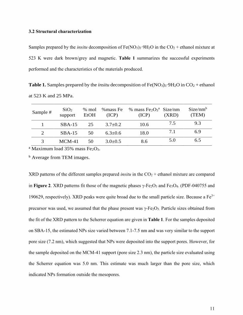
3.2 Structural characterization
Samples prepared by the insitu decomposition of Fe(NO3)3·9H2O in the CO2 + ethanol mixture at
523 K were dark brown/grey and magnetic. Table 1 summarizes the successful experiments
performed and the characteristics of the materials produced.
Table 1. Samples prepared by the insitu decomposition of Fe(NO3)3·9H2O in CO2 + ethanol
at 523 K and 25 MPa.
Sample # SiO2
support
% mol
EtOH
%mass Fe
(ICP)
% mass Fe2O3a
(ICP)
Size/nm
(XRD)
Size/nmb
(TEM)
1 SBA-15 25 3.7±0.2 10.6 7.5 9.3
2 SBA-15 50 6.3±0.6 18.0 7.1 6.9
3 MCM-41 50 3.0±0.5 8.6 5.0 6.5
a Maximum load 35% mass Fe2O3.
b Average from TEM images.
XRD patterns of the different samples prepared insitu in the CO2 + ethanol mixture are compared
in Figure 2. XRD patterns fit those of the magnetic phases -Fe2O3 and Fe3O4. (PDF-040755 and
190629, respectively). XRD peaks were quite broad due to the small particle size. Because a Fe3+
precursor was used, we assumed that the phase present was -Fe2O3. Particle sizes obtained from
the fit of the XRD pattern to the Scherrer equation are given in Table 1. For the samples deposited
on SBA-15, the estimated NPs size varied between 7.1-7.5 nm and was very similar to the support
pore size (7.2 nm), which suggested that NPs were deposited into the support pores. However, for
the sample deposited on the MCM-41 support (pore size 2.3 nm), the particle size evaluated using
the Scherrer equation was 5.0 nm. This estimate was much larger than the pore size, which
indicated NPs formation outside the mesopores.

12
TEM images of the SPION/SBA-15 samples are shown in Figure 3. Images showed clearly the
SiO2 mesopores along with the presence of darker nanoparticles corresponding to the metal oxide.
Sample 1 prepared using the 25% mol ethanol + CO2 mixture showed NPs of sizes between 4-15
nm (Figure 3a-c). The larger particles seem to be deposited on the external surface of the support.
In contrast, sample 2 prepared from the 50% mol ethanol + CO2 mixture showed smaller NPs,
most between 5 and 8 nm within the support pores, very well dispersed, yielding a very
homogeneous material (Figure 3d-f). TEM images proved that the deposition of SPIONs into the
SBA-15 mesopores was successful in the ethanol + CO2 mixtures. Metal content by ICP-OES was
3.7 and 6.3 % mass Fe for samples 1 and 2, respectively.
The differences in particle size and NPs dispersion of samples 1 and 2 seem to be related to the
different solubility of the precursor in the reaction mixtures. When the 25% ethanol + CO2 mixture
was employed, not all the precursor dissolved in the reaction medium and most NPs deposit on the
external surface of the support. However, in the sample prepared using the 50% ethanol + CO2
mixture, the precursor fully dissolved, favouring deposition of the NPs within the support pores,
leading to a larger metal loading. Therefore thanks to the higher solubility of Fe(NO3)3·9H2O in
the 50% mol ethanol + CO2 mixture, metal oxide deposition was more homogeneous at these
conditions and a larger metal content was obtained in sample 2. For this sample, metal
incorporation on the support with respect to the amount loaded into the reactor was close to 40%.

13
Figure 2- XRD patterns of SPION/SiO2 samples prepared by the insitu decomposition of
Fe(NO3)3·9H2O at 523 K on the different supports in the ethanol + CO2 mixtures.

14
Figure 3- TEM images and particle size distributions of SPION/SBA-15 samples prepared by the
insitu decomposition of Fe(NO3)3·9H2O at 523 K in 25% mol ethanol + CO2 (a-c) and 50% mol
ethanol + CO2 (d-f).
In view of the previous results, experiments on MCM-41 were only performed using the highest
ethanol content. TEM images of the SPION/MCM-41 sample are shown in Figure 4. Images
showed small mesoporous SiO2 particles with sizes ranging from 40-100 nm, along with the darker
iron oxide NPs of sizes between 3-12 nm (average 6.5 nm). NPs seemed to be attached mostly to
the external surface of the support and particle size distribution was wide. Particle size controlled
is only achieved when NPs deposit into the mesopores. Metal content of this sample by ICP-OES
revealed a Fe contents equal to 6.0% mass, slightly lower than that measured for the SPION/SBA-
15 sample prepared at the same conditions (sample 2).

15
The small pore size of MCM-41 makes difficult the impregnation of the support in the ethanol +
CO2 mixture. At the concentrations and conditions of these experiments, the ethanol + CO2 mixture
is in the expanded liquid region. Although the reduction in density and viscosity of the mixture
and the increased diffusivity of the precursor in this medium in comparison to pure ethanol is high,
mass transfer limitations seem to preclude the effectively filling of the small MCM-41 mesopores
in the short times used.
Figure 4- TEM images and particle size distribution of the SPION/MCM-41 sample prepared by
the insitu decomposition of Fe(NO3)3·9H2O at 523 K in 50% mol ethanol + CO2 (a-e).
Figure 5 shows XRD patterns of the SPION/SBA-15 samples prepared by the impregnation of
Fe(NO3)3·9H2O in the 25% mol ethanol + CO2 solution at 333 K and 12.0 MPa and reduced in a
tubular furnace at different conditions. When the sample was heated in N2 at 523 K (temperature
used in the insitu experiments), no crystalline phase was observed in the XRD. Furthermore,
mixtures of phases including Fe and α-Fe2O3 besides -Fe2O3 were obtained after decomposition

16
in H2/N2 and N2 at higher temperatures. In order to maximize the presence of the magnetic -Fe2O3
phase, impregnated samples were treated in N2 at 973 K. At this temperature a small amount of α-
Fe2O3 was still present. The possible oxidation of Fe3O4 to Fe2O3 under exposure of the samples
to air cannot be ruled out. XRD peaks were narrower than those obtained for the materials prepared
insitu. TEM images of some samples reduced in N2 are provided as supporting information. Iron
oxide NPs were larger than those observed in the samples prepared by the insitu decomposition of
Fe(NO3)3·9H2O in CO2 + ethanol at 523 K, which is certainly related to the higher temperatures
employed in the thermal treatment and the different decomposition method. Furthermore, particle
distribution was also less homogeneous. If the precursor adsorption is not very strong, the
precursor may migrate with the solvent during depressurization, leading to an inhomogeneous
metal distribution.
Raman spectra of selected samples were also measured. Sample 2 did not give any significant
Raman signal probably due to the small iron content and the good metal dispersion within the SiO2
support. Spectrum of the sample impregnated in ethanol + CO2 and heated in N2 at 973 K
confirmed the presence of α-Fe2O3 and -Fe2O3 (see supporting information).
Comparison of the SPION/SBA-15 samples prepared by the insitu decomposition of
Fe(NO3)3·9H2O in ethanol + CO2 with samples previously prepared by wet impregnation of the
same precursor in liquid ethanol followed by solvent evaporation and thermal treatment [16, 17]
revealed that our method allows a much easier control of the oxidation state leading to single phase
magnetic materials at much lower temperatures and shorter times in one single step.

17
Figure 5- XRD patterns of SPION/SBA-15 samples prepared by impregnation of Fe(NO3)3·9H2O
in a 25% mol ethanol + CO2 solution and reduced in N2 and H2/N2 at different temperatures
showing the different crystal phases.
3.3. Reaction mechanism
The mechanism operating in the deposition of supported nanoparticles by the supercritical fluid
reactive deposition (SFRD) technique is well known and it has been recently reviewed [56]. Here
we propose a similar mechanism for the reactive deposition of inorganic salts on mesoporous
supports using expanded liquid solvents.

18
The precursor Fe(NO3)3·9H2O was dissolved in the ethanol + CO2 mixture at 333 K and 12.0 MPa.
This expanded liquid mixture was brought into contact with the mesoporous SiO2 support. The
mixture diffused into the support pores and the metal precursor adsorbed on their surface
impregnating the support. In contrast to CO2 that adsorbs weakly on SiO2[57], competitive
adsorption of ethanol on the SiO2 surface is possible. Then the temperature was raised to 523 K in
order to promote the insitu decomposition of the precursor in the CO2 + ethanol mixture. At these
conditions -Fe2O3 NPs were produced within the SiO2 mesopores. Ming at al. have shown that
the decomposition of hydrous metal nitrates in CO2 expanded ethanol proceeds through an
intermedium coordinated compound before the corresponding oxide is formed [43]. Then the
heater was disconnected and the reactor was vented. When the 50% ethanol + CO2 mixture was
used, the solubility of the precursor was high leading to very homogeneous SPION/SBA-15
materials. In contrast, the precursor solubility in the 25% ethanol + CO2 mixture was lower and
some metal NPs may have deposited directly on the external SiO2 surface from the solid phase.
Because air was not deliberately excluded from the support pores before adding the precursor
solution and filling the reactor with CO2, an interphase may have been formed at the initial stages
of the filling process. Mass transfer limitations make difficult the filling of the smaller MCM-41
mesopores.
On the other hand, the samples impregnated with Fe(NO3)3·9H2O in ethanol + CO2 but
decomposed after depressurization in the tubular furnace were less homogeneous. If the precursor
adsorption is not very strong, the precursor may be transported along with the solvent during
depressurization, leading to an inhomogeneous metal distribution.

19
3.4. Magnetic characterization
Figure 6 shows the temperature dependence of ZFC and FC magnetic susceptibility () for
samples 1-3. Due to the small particle size of the -Fe2O3 NPs, the material turned
superparamagnetic from the blocking temperature (TB) to ambient temperature. At temperatures
lower than TB, when the samples were cooled in the presence of a 500 Oe field down to 5K (FC),
the samples remained magnetized. By contrast, in the ZFC regime decreased with the
temperature. The inverse dependence of with temperature in the FC measurements confirmed
the ferrimagnetic behaviour of the samples. ZFC and FC curves merged together above TB and
decreased with temperature due to thermal fluctuations[58]. The higher values were measured
for samples 3 and 1, being much smaller for sample 2. The larger particle size of samples 1 and 3
would explain the large values. A similar behaviour is exhibited by nanoparticulated spinel
ferrites [59-62].

20
Figure 6- Mass susceptibility measurements up to 300 K of the SPION/SiO2 samples: □, sample
1; o, sample 2 and ∆, sample 3.
Magnetization (M) versus magnetic field (H) curves measured at 5 and 250 K for the different
samples are shown in Figure 7. M increased with H up to the saturation value (Ms). At 250 K, no
hysteresis was observed confirming the superparamagnetic nature of the materials. In contrast at
5 K (below TB) not all the magnetic domains returned to their original orientations at H=O and the
material exhibited a remnant magnetization (MR), being necessary to apply a coercive field (Hc)
in the opposite direction to reverse the process. Hysteresis loops at 5K were characterized by low
MR and HR, whist Ms were high.

21
Figure 7- Magnetic susceptibility versus magnetic field curves measurements of the SPION/SiO2
samples at 5 and 250 K: □, sample 1; o, sample 2 and ∆, sample 3.
Table 2 shows the values of Ms, MR, Hc and TB for the different samples. TB was obtained from
the maxima of the ZFC measurement versus temperature curve and ranged from 56-91 K. Samples
1 and 2 showed the largest TB values, whilst the smallest TB was obtained for sample 3. TB is
directly related to the particle volume. Maxima were relatively narrow for sample 2 in agreement
to their particle size distribution. However, the sample deposited on MCM-41 showed a wider

22
particle size distribution and the maximum was broader. The smallest value of TB obtained for
sample 3 could indicate the presence of very small magnetic NPs. These values are similar to those
reported for other SPION/SiO2 composite materials[20].
Table 2. Magnetic parameters for SPION/SiO2 samples prepared by the insitu decomposition of
Fe(NO3)3·9H2O in ethanol + CO2 at 523 K. Parameters reported per gram of -Fe2O3 based on
ICP-OES data.
Sample # TB (K)
HC,5K (Oe)
MR,5 K (emu/g)
MS,5 K (emu/g)
MS,250 K (emu/g)
1 91 369 11.4 >54 31
2 70 322 9.0 30 24
3 56 250 12.5 39 31
Hc and MR at 5 K were small in all the samples. Ms at 5 and 250 K ranged from 30-55 and 24-31
(emu/g Fe2O3), respectively. As expected, Ms decreased as the temperature increased. At 5 K, M
values for sample 1 did not reach saturation at the highest H employed (50k Oe) most likely due
to interparticle interactions between the surface layer of canted spins[60]. In sample 2 most of the
particles were inside the pores of the matrix and these interactions were minimized. Similarly, the
good NP dispersion on the support of sample 3 prevented a strong interparticle interaction.
However, NPs at the support surface in sample 1 were agglomerated and interparticle surface
interactions increased the surface anisotropy and the hardness of the material as the Ms values
indicate. Ms,250 K were lower than those reported for bulk -Fe2O3 (76 emu/g) [58] related to the
small particle size and the presence of magnetic anisotropy at the particle surface. These values

23
were however larger than those previously reported for similar -Fe2O3/SBA-15 materials[20] but
slightly lower than the values reported by Yiu et al. for Fe3O4/SBA-15 materials[16].
4. Conclusions
SPION/SiO2 materials were prepared by the impregnation and insitu decomposition of
Fe(NO3)3·9H2O into mesoporous SiO2 supports at 523 K and 25.0 MPa using ethanol + CO2
mixtures. Ethanol was required to solubilize the salt in CO2 and helped in the decomposition
process. At the concentrations and impregnation conditions of these experiments, the ethanol +
CO2 mixture was in the expanded liquid region. The method was simple and allowed the
preparation of the composite materials in one step using CO2 as solvent and reaction medium. -
Fe2O3 nanoparticles of average size between 6-9 nm were deposited on the SiO2 supports with
loadings up to 18.0% mass. In the 50% ethanol + CO2 mixture, -Fe2O3 NPs deposited into the
support pores of SBA-15 but mostly on the external surface of MCM-41. Materials were very
homogeneous due to the large solubility of the precursor in the ethanol + CO2 mixture. Mass
transfer limitations seemed to preclude the effectively filling of the small MCM-41 mesopores in
the short times used. The very small TB obtained for the MCM-41 sample, however, suggest the
presence of very small magnetic NPs. Larger impregnation times and/or higher impregnation
temperatures may be required to promote extensive deposition into the smaller mesopores.
Composite materials were superparamagnetic at room temperature with TB below 91 K for all the
samples. Hysteresis loops at 5 K were small with high Ms and low HR. The materials prepared
have many potential technological applications.

24
Acknowledgements
We acknowledge financial support from the Spanish Ministry of Science and Innovation, research
projects MAT2017-84385-R and RTI2018-097230-B-I00 and from UCM-Santander, research
project PR75/18-21583. We thank the Centres of Scientific Instrumentation at UCM (X-ray
diffraction, Physical techniques, Geological Techniques and ICTS National Microscopy Centre)
and their staff, for use of the technical facilities.
Appendix A. Supplementary data
TEM images of SPION/SBA-15 samples obtained by the impregnation of Fe(NO3)3.9H2O in
ethanol + CO2 and further decomposed in N2 (Figure S1).
Raman spectrum of a SiO2 support impregnated with Fe(NO3)3·9H2O in ethanol + CO2 and heated
in N2 at 973 K (Figure S2).
References
[1] M.D. Marquez-Medina, P. Prinsen, H.K. Li, K.M. Shih, A.A. Romero, R. Luque,
Continuous-Flow Synthesis of Supported Magnetic Iron Oxide Nanoparticles for Efficient
Isoeugenol Conversion into Vanillin, ChemSusChem, 11 (2018) 389-396.
[2] L.M. Rossi, N.J.S. Costa, F.P. Silva, R. Wojcieszak, Magnetic nanomaterials in
catalysis: advanced catalysts for magnetic separation and beyond, Green Chem., 16 (2014)
2906-2933.
[3] B. Duong, H. Khurshid, P. Gangopadhyay, J. Devkota, K. Stojak, H. Srikanth, L. Tetard,
R.A. Norwood, N. Peyghambarian, M.H. Phan, J. Thomas, Enhanced Magnetism in Highly
Ordered Magnetite Nanoparticle-Filled Nanohole Arrays, Small, 10 (2014) 2840-2848.

25
[4] R. Kaur, A. Hasan, N. Iqbal, S. Alam, M.K. Saini, S.K. Raza, Synthesis and surface
engineering of magnetic nanoparticles for environmental cleanup and pesticide residue
analysis: A review, J. Sep. Sci., 37 (2014) 1805-1825.
[5] B. Julian-Lopez, C. Boissiere, C. Chaneac, D. Grosso, S. Vasseur, S. Miraux, E. Duguet,
C. Sanchez, Mesoporous maghemite-organosilica microspheres: a promising route towards
multifunctional platforms for smart diagnosis and therapy, J. Mater. Chem., 17 (2007)
1563-1569.
[6] Q.A. Pankhurst, J. Connolly, S.K. Jones, J. Dobson, Applications of magnetic
nanoparticles in biomedicine, Journal of Physics D-Applied Physics, 36 (2003) R167-
R181.
[7] J. Liu, S.Z. Qiao, Q.H. Hu, G.Q. Lu, Magnetic Nanocomposites with Mesoporous
Structures: Synthesis and Applications, Small, 7 (2011) 425-443.
[8] M. Manzano, M. Vallet-Regí, Mesoporous silica nanoparticles in nanomedicine
applications, J. Mater. Sci. Mater. Med., 29 (2018) 65.
[9] H. Atashin, R. Malakooti, Magnetic iron oxide nanoparticles embedded in SBA-15
silica wall as a green and recoverable catalyst for the oxidation of alcohols and sulfides, J.
Saudi Chem. Soc., 21 (2017) S17-S24.
[10] F. Rajabi, T. Kakeshpour, M.R. Saidi, Supported iron oxide nanoparticles:
Recoverable and efficient catalyst for oxidative S-S coupling of thiols to disulfides, Catal.
Commun., 40 (2013) 13-17.
[11] S. Egodawatte, A. Datt, E.A. Burns, S.C. Larsen, Chemical Insight into the Adsorption
of Chromium(III) on Iron Oxide/Mesoporous Silica Nanocomposites, Langmuir, 31 (2015)
7553-7562.
[12] E. Guisasola, L. Asín, L. Beola, J.M. de la Fuente, A. Baeza, M. Vallet-Regí, Beyond
Traditional Hyperthermia: In Vivo Cancer Treatment with Magnetic-Responsive
Mesoporous Silica Nanocarriers, ACS Appl. Mater. Interfaces, 10 (2018) 12518-12525.

26
[13] A. Baeza, E. Guisasola, E. Ruiz-Hernández, M. Vallet-Regí, Magnetically Triggered
Multidrug Release by Hybrid Mesoporous Silica Nanoparticles, Chem. Mater., 24 (2012)
517-524.
[14] N.I. Cuello, V.R. Elias, C.E.R. Torres, M.E. Crivello, M.I. Oliva, G.A. Eimer,
Development of iron modified MCM-41 as promising nano-composites with specific
magnetic behavior, Microporous Mesoporous Mater., 203 (2015) 106-115.
[15] A. Zelenakova, V. Zelenak, J. Bednarcik, P. Hrubovcak, J. Kovac, Magnetic
nanocomposites of periodic mesoporous silica: The influence of the silica substrate
dimensionality on the inter-particle magnetic interactions, J. Alloy Compd., 582 (2014)
483-490.
[16] H.H.P. Yiu, M.A. Keane, Z.A.D. Lethbridge, M.R. Lees, A.J. El Haj, J. Dobson,
Synthesis of novel magnetic iron metal-silica (Fe-SBA-15) and magnetite-silica (Fe3O4-
SBA-15) nanocomposites with a high iron content using temperature-programed reduction,
Nanotechnology, 19 (2008).
[17] H.H.P. Yiu, S.C. McBain, A.J. El Haj, J. Dobson, A triple-layer design for
polyethyleneimine-coated, nanostructured magnetic particles and their use in DNA binding
and transfection, Nanotechnology, 18 (2007).
[18] P. Munnik, P.E. de Jongh, K.P. de Jong, Recent Developments in the Synthesis of
Supported Catalysts, Chem. Rev., 115 (2015) 6687-6718.
[19] G. Collins, K. Rahme, J. O'Connell, J.D. Holmes, Embedding colloidal nanoparticles
inside mesoporous silica using gas expanded liquids for high loading recyclable catalysts,
Catal. Sci. Technol., 6 (2016) 7212-7219.
[20] H.A. Lin, C.H. Liu, W.C. Huang, S.C. Lion, M.W. Chu, C.H. Chen, J.F. Lee, C.M.
Yang, Novel Magnetically Separable Mesoporous Fe2O3@SBA-15 Nanocomposite with
Fully Open Mesochannels for Protein Immobilization, Chem. Mater., 20 (2008) 6617-6622.

27
[21] E. Delahaye, V. Escax, N. El Hassan, A. Davidson, R. Aquino, V. Dupuis, R.
Perzynski, Y.L. Raikher, "Nanocasting": Using SBA-15 silicas as hard templates to obtain
ultrasmall monodispersed gamma-Fe2O3 nanoparticles, J. Phys. Chem. B, 110 (2006)
26001-26011.
[22] R.G. Digigow, J.-F. Dechezelles, H. Dietsch, I. Geissbuehler, D. Vanhecke, C. Geers,
A.M. Hirt, B. Rothen-Rutishauser, A. Petri-Fink, Preparation and characterization of
functional silica hybrid magnetic nanoparticles, J. Magn. Magn. Mater., 362 (2014) 72-79.
[23] P. Fabrizioli, T. Burgi, M. Burgener, S. van Doorslaer, A. Baiker, Synthesis, structural
and chemical properties of iron oxide-silica aerogels, J. Mater. Chem., 12 (2002) 619-630.
[24] D. Niznansky, N. Viart, J.L. Rehspringer, Nanocomposites Fe2O3/SiO2-preparation
by sol-gel method and physical properties, J. Sol-Gel Sci. Technol., 8 (1997) 615-618.
[25] D. Niznansky, J.L. Rehspringer, M. Drillon, Preparation of magentic nanoaprtilces
(gamma-Fe2O3) in the silica matrix, IEEE Trans. Magn., 30 (1994) 821-823.
[26] C.A. Garcia-Gonzalez, E. Carenza, M.L. Zeng, I. Smirnova, A. Roig, Design of
biocompatible magnetic pectin aerogel monoliths and microspheres, RSC Advances, 2
(2012) 9816-9823.
[27] E. Taboada, R. Solanas, E. Rodriguez, R. Weissleder, A. Roig, Supercritical-Fluid-
Assisted One-Pot Synthesis of Biocompatible Core(gamma-Fe2O3)/Shell(SiO2)
Nanoparticles as High Relaxivity T-2-Contrast Agents for Magnetic Resonance Imaging,
Adv. Funct. Mater., 19 (2009) 2319-2324.
[28] C. Aymonier, A. Loppinet-Serani, H. Reverón, Y. Garrabos, F. Cansell, Review of
supercritical fluids in inorganic materials science, J. Supercrit. Fluids, 38 (2006) 242-251.
[29] A.M. López-Periago, C. Domingo, Features of supercritical CO2 in the delicate world
of the nanopores, J. Supercrit. Fluids, 134 (2018) 204-213.

28
[30] A. Castro, J. Morere, A. Cabañas, L.P. Ferreira, M. Godinho, P. Ferreira, P.M.
Vilarinho, Designing nanocomposites using supercritical CO2 to insert Ni nanoparticles
into the pores of nanopatterned BaTiO3 thin films, J. Mater. Chem. C, 5 (2017) 1083-1089.
[31] E. Sanchez-Miguel, M.J. Tenorio, J. Morere, A. Cabañas, Green preparation of PtRu
and PtCu/SBA-15 catalysts using supercritical CO2, J. CO2 Util., 22 (2017) 382-391.
[32] Y. Sanchez-Vicente, L.A. Stevens, C. Pando, M. Jose Torralvo, C.E. Snape, T.C.
Drage, A. Cabañas, A new sustainable route in supercritical CO2 to functionalize silica
SBA-15 with 3-aminopropyltrimethoxysilane as material for carbon capture, Chem. Eng.
J., 264 (2015) 886-898.
[33] https://webbook.nist.gov/, in.
[34] U.T. Lam, R. Mammucari, K. Suzuki, N.R. Foster, Processing of iron oxide
nanoparticles by supercritical fluids, Ind. Eng. Chem. Res., 47 (2008) 599-614.
[35] A. Cabañas, M. Poliakoff, The continuous hydrothermal synthesis of nano-particulate
ferrites in near critical and supercritical water, J. Mater. Chem., 11 (2001) 1408-1416.
[36] A.S. Teja, P.Y. Koh, Synthesis, properties, and applications of magnetic iron oxide
nanoparticles, Prog. Cryst. Growth. Ch., 55 (2009) 22-45.
[37] Y.L. Hao, A.S. Teja, Continuous hydrothermal crystallization of alpha-Fe2O3 and
Co3O4 nanoparticles, J. Mater. Res., 18 (2003) 415-422.
[38] O. Pascu, S. Marre, C. Aymonier, A. Roig, Ultrafast and continuous synthesis of
crystalline ferrite nanoparticles in supercritical ethanol, Nanoscale, 5 (2013) 2126-2132.
[39] B. Veriansyah, J.D. Kim, B.K. Min, J. Kim, Continuous synthesis of magnetite
nanoparticles in supercritical methanol, Mater. Lett., 64 (2010) 2197-2200.

29
[40] X.T. Liu, S.N. Jiang, M.J. Niu, S.M. Li, W.X. Li, S.S. Yu, Synthesis and Magnetic
Properties of Ferroferric Oxide in Supercritical Methanol, J. Nanosci. Nanotechnol.,, 19
(2019) 833-838.
[41] T.A. Crowley, K.J. Ziegler, D.M. Lyons, D. Erts, H. Olin, M.A. Morris, J.D. Holmes,
Synthesis of metal and metal oxide nanowire and nanotube arrays within a mesoporous
silica template, Chem. Mater., 15 (2003) 3518-3522.
[42] F.Y. Cao, C.L. Chen, Q. Wang, Q.W. Chen, Synthesis of carbon-Fe3O4 coaxial
nanofibres by pyrolysis of ferrocene in supercritical carbon dioxide, Carbon, 45 (2007)
727-731.
[43] J. Ming, C. Wu, H. Cheng, Y. Yu, F. Zhao, Reaction of hydrous inorganic metal salts
in CO2 expanded ethanol: Fabrication of nanostructured materials via supercritical
technology, J. Supercrit. Fluids, 57 (2011) 137-142.
[44] M.J. Tenorio, S. Ginés, C. Pando, J.A.R. Renuncio, A. Cabañas, Solubility of the Metal
Precursor Ni(NO3)2·6H2O in High-Pressure CO2 + Ethanol Mixtures, J. Chem. Eng. Data,
63 (2018) 1065-1071.
[45] Z. Sun, H. Yuan, Z. Liu, B. Han, X. Zhang, A Highly Efficient Chemical Sensor
Material for H2S: α-Fe2O3 Nanotubes Fabricated Using Carbon Nanotube Templates,
Adv. Mater., 17 (2005) 2993-2997.
[46] X. Hu, M. Ma, M. Zeng, Y. Sun, L. Chen, Y. Xue, T. Zhang, X. Ai, R.G. Mendes,
M.H. Rümmeli, L. Fu, Supercritical Carbon Dioxide Anchored Fe3O4 Nanoparticles on
Graphene Foam and Lithium Battery Performance, ACS Appl. Mater. Interfaces, 6 (2014)
22527-22533.
[47] L.Y. Wang, L.H. Zhuo, C. Zhang, F.Y. Zhao, Supercritical Carbon Dioxide Assisted
Deposition of Fe3O4 Nanoparticles on Hierarchical Porous Carbon and Their
LithiumStorage Performance, Chem.: Eur. J., 20 (2014) 4308-4315.

30
[48] L. Wang, L. Zhuo, F. Zhao, Carbon dioxide-expanded ethanol-assisted synthesis of
carbon-based metal composites and their catalytic and electrochemical performance in
lithium-ion batteries, Chinese J. Catal., 37 (2016) 218-226.
[49] D.Y. Zhao, J.L. Feng, Q.S. Huo, N. Melosh, G.H. Fredrickson, B.F. Chmelka, G.D.
Stucky, Triblock copolymer syntheses of mesoporous silica with periodic 50 to 300
angstrom pores, Science, 279 (1998) 548-552.
[50] D.Y. Zhao, Q.S. Huo, J.L. Feng, B.F. Chmelka, G.D. Stucky, Nonionic triblock and
star diblock copolymer and oligomeric surfactant syntheses of highly ordered,
hydrothermally stable, mesoporous silica structures, J. Am. Chem. Soc., 120 (1998) 6024-
6036.
[51] H. Chen, J. He, Fine control over the morphology and structure of mesoporous silica
nanomaterials by a dual-templating approach, Chem. Comm., (2008) 4422-4424.
[52] Ž. Knez, M. Škerget, L. Ilič, C. Lütge, Vapor–liquid equilibrium of binary CO2–
organic solvent systems (ethanol, tetrahydrofuran, ortho-xylene, meta-xylene, para-
xylene), J. Supercrit. Fluids,, 43 (2008) 383-389.
[53] C. Secuianu, V. Feroiu, D. Geană, Phase behavior for carbon dioxide+ethanol system:
Experimental measurements and modeling with a cubic equation of state, J. Supercrit.
Fluids, 47 (2008) 109-116.
[54] M. Jaroniec, M. Kruk, J.P. Olivier, Standard Nitrogen Adsorption Data for
Characterization of Nanoporous Silicas, Langmuir, 15 (1999) 5410-5413.
[55] E. Barret, L.G. Joyner, P.P. Halenda, The determination of pore volume and area
distributions in porous substances. I. Computations for nitrogen isotherms, J. Am. Chem.
Soc., 73 (1951) 373-380.
[56] M. Türk, C. Erkey, Synthesis of supported nanoparticles in supercritical fluids by
supercritical fluid reactive deposition: Current state, further perspectives and needs, J.
Supercrit Fluids, 134 (2018) 176-183.

31
[57] S. Reiser, M. Türk, Influence of temperature and high-pressure on the adsorption
behavior of scCO2 on MCM-41 and SBA-15, J. Supercrit. Fluids, 144 (2019) 122-133.
[58] B.D. Cullity, C.D. Graham, Introduction to Magnetic Materials, 2nd ed., Wiley,
Cambridge, 1972.
[59] V. Blanco-Gutierrez, R. Saez-Puche, M.J. Torralvo-Fernandez, Superparamagnetism
and interparticle interactions in ZnFe2O4 nanocrystals, J. Mater. Chem., 22 (2012) 2992-
3003.
[60] V. Blanco-Gutierrez, M. Virumbrales, R. Saez-Puche, M.J. Torralvo-Fernandez,
Superparamagnetic Behavior of MFe2O4 Nanoparticles and MFe2O4/SiO2 Composites
(M: Co, Ni), J. Phys. Chem. C, 117 (2013) 20927-20935.
[61] M. Virumbrales, R. Saez-Puche, V. Blanco-Gutierrez, M.J. Torralvo-Fernandez,
Discussion on the Interparticle Interactions in NiFe2O4 and ZnFe2O4 Nanosized Systems
Based on the Matrix Effects in the Magnetic Behavior, J. Phys. Chem. Cb, 121 (2017)
4029-4036.
[62] M. Virumbrales, R. Saez-Puche, M. Jose Torralvo, V. Blanco-Gutierrez, Mesoporous
Silica Matrix as a Tool for Minimizing Dipolar Interactions in NiFe2O4 and ZnFe2O4
Nanoparticles, Nanomaterials, 7 (2017).

32
Supplementary Material
One-step Sustainable Preparation of Superparamagnetic
Iron Oxide Nanoparticles Supported on Mesoporous SiO2
Elena Chamorro,a M. José Tenorio,b,† Lourdes Calvo,a M. José Torralvo,c Regino Sáez-Puchec
and Albertina Cabañasb*
aDep. Chemical Engineering, bDep. Physical Chemistry, cDep. Inorganic Chemistry, Universidad
Complutense de Madrid, Avda. Complutense s/n, 28040 Madrid, Spain
†Current address: Chemical, Energy and Mechanical Technology Dep, Universidad Rey Juan
Carlos, c/Tulipán s/n, 28933 Móstoles, Madrid, Spain
* Corresponding author: Albertina Cabañas, e-mail: [email protected]
TEM images of the SPION/S samples obtained by impregnation of the SBA-15 support using
Fe(NO3)3·9H2O in ethanol + CO2 mixtures at 333 K and 12.0 MPa and further decomposed in a
tubular furnace in N2 are shown in Figure S1. Iron oxide nanoparticles (NPs) are larger than those
observed in the samples prepared by insitu decomposition of Fe(NO3)3·9H2O in CO2-expanded
ethanol at 523 K (Figure 3 of the manuscript). Furthermore, NP distribution is also less
homogeneous. If the precursor adsorption to the support is not very strong, the precursor may be
transported along with the solvent during depressurization, leading to an inhomogeneous metal
distribution.

33
Figure S1- TEM of iron oxide/SBA-15 samples prepared by impregnation of the support
with Fe(NO3)3·9H2O in CO2-expanded ethanol at 333 K and 12.0 MPa: (a) 25% mol
ethanol + CO2 mixture, heated in N2 at 873 K; (b) 50% mol ethanol + CO2 mixture, heated
in N2 at 973 K
Raman spectra of the SiO2 SBA-15 support and the SPION/SiO2 sample obtained from
impregnation of the support using Fe(NO3)3·9H2O in a 25% mol ethanol + CO2 mixture at
333 K and 12.0 MPa and further heated in N2 at 973 K is shown in Figure S2. Spectra were
base line corrected and normalized. SiO2 support showed broad bands at ca. 500, 585 and
650 cm-1. Peak assignment for the different iron oxide phases was based on a previous
report by de Faria et al. [1] Peaks at 221 and 289 cm-1 were assigned to α-Fe2O3. The
presence of two Raman bands of similar intensity at ca. 670, 715 indicated the presence of
-Fe2O3 and not Fe3O4. The band at ca. 715 cm-1 is related to the vibrational modes of local
FeO near the cation vacancies and it is very weak in Fe3O4. The intensity ratio of these
two bands has been previously used to estimate the -Fe2O3 content in partially oxidized
Fe3O4 samples.[2] Broad bands at ca. 350 and 500 cm-1 associated to -Fe2O3 were also
expected. Raman spectrum of this sample confirmed the presence of α-Fe2O3 and -Fe2O3
in the sample in agreement with the XRD data.

34
200 300 400 500 600 700 800
585
Inte
nsity (
a.u
.)
Wavenumber (cm-1)
500
585650
350
715
670
289
221
Figure S2- Raman spectra of SiO2 SBA-15 (black line) and an iron oxide/SiO2 sample (red
line) prepared by impregnation of the support with Fe(NO3)3·9H2O in CO2-expanded
ethanol at 333 K and 12.0 MPa and heated in N2 at 973 K.
[1] D.L.A. deFaria, S.V. Silva, M.T. deOliveira, Raman microspectroscopy of some iron oxides
and oxyhydroxides, J. Raman Spectrosc 28(11) (1997) 873-878.
[2] J.W. Long, M.S. Logan, C.P. Rhodes, E.E. Carpenter, R.M. Stroud, D.R. Rolison,
Nanocrystalline iron oxide aerogels as mesoporous magnetic architectures, J. Am. Chem. Soc.
126(51) (2004) 16879-16889.
Top Related
Copyright © 2022 FDOKUMEN

