


Bahasa
Halaman
Hukum

Human Reproduction vol.11 no 12 pp.2713-2718, 1996
Immunohistochemical localization of extracellularmatrix proteins in luteal phase endometrium of fertileand infertile patients
D.A.Bilalis1, L.D.Klentzeris2 and S.Fleming3
Department of Obstetrics and Gynaecology, University Hospital,Nottingham NG7 2UH, UK
'Present address' Academic Department of Obstetrics andGynaecology, Birmingham Maternity Hospital, BirminghamB15 2TG, UK2Present address Departments of Obstetrics and Gynaecology andBiological Sciences, University of Warwick, Coventry CV4 7AL,UK
•'To whom correspondence should be addressed
The lack of expression of certain components involved incell adhesion and migration is believed to contribute toendometrial dysfunction and implantation failure. Thepurpose of this study was to investigate whether lutealphase endometrium in women with unexplained infertilitydiffers, with respect to specific extracellular matrix (ECM)proteins, from endometrium of normal fertile women.A panel of monoclonal antibodies to collagen type IV,fibronectin and laminin was used to characterize the localiz-ation of ECM components in the different endometriaJcompartments. Precisely timed endometrial biopsiesobtained at 4, 7, 10 and 13 days following the luteinizinghormone surge were obtained from 22 normal fertilewomen (group 1) and 24 women suffering from unexplainedinfertility (group 2). Paraffin-embedded sections werelabelled using the streptavidin-biotin alkaline phosphatasetechnique. In group 1, collagen type IV, fibronectin andlaminin were absent from the luminal epithelium butpresent in stromal cells and the basement membrane ofglands and blood vessels. In group 2, these componentswere absent from all endometrial regions using equivalenttitres of antibody to those used in group 1. This suggeststhat the endometrium of women with unexplained infertilitydemonstrates defects in the distribution of certain ECMglycoproteins. A possible consequence of this defect maybe implantation failure.Key words: endometrium/extracellular matrix proteins/fertile/infertile/luteal phase
Introduction
There is no doubt that the endometrium plays an importantrole in the process of implantation and the establishment of asuccessful pregnancy (Lejeune et al., 1986; Psychoyos, 1986;Yoshinaga, 1988). The molecular events that accompanyembryo attachment and implantation are incompletely under-stood. The lack of knowledge in this field may be partly
© European Society for Human Reproduction and Embryology
because the process of implantation in the human is unique,and because results from animal studies cannot be extrapolatedto human beings
Over the past decade, investigators have come to recognizethe importance of the extracellular matrix (ECM) in directinggrowth, differentiation and function of the underlying epithe-lium (Getzenberg et al., 1990). The major components ofbasement membranes within human endometrium have beenidentified (Aplin et al., 1988). Vascular and glandular basementmembranes have been shown to be immunoreactive for colla-gen IV, laminin and heparan sulphate proteoglycan during theproliferative, mid- and late secretory stages of the menstrualcycle. Recently, these components have been shown to beexpressed even during the menstrual phase of the cycle (Kellyet al., 1995). During the proliferative phase the most abundantinterstitial matrix component is collagen I (Glasser et al.,1987). The mam change in the intershtium in humans is theprogressive loss of collagen IV, which starts in the mid-secretory phase and continues after implantation (Aplin et al.,1988). The absence of collagen IV may be one factor whichallows oedema and expansion of the extracellular space.
Electron microscopic studies of human decidua haverevealed that around stromal and mainly decidual cells there isa pericellular matrix which demonstrates structural similaritieswith the basement membrane of glandular and luminal epithe-lium (Wynn, 1974). Later studies using monoclonal antibodieshave shown that the epithelial and peridecidual basementmembrane have a common biochemical identity (Aphn et al.,1988). In the early luteal phase lamimn, collagen IV andheparan sulphate begin to appear around stromal cells. Lamininand fibronectin appear first, whereas collagen VI and heparansulphate proteoglycan appear in the late luteal phase when thestromal cells are fully transformed into predecidual cells.The components of the pericellular matrix can be used asimmunocytochemical markers of cellular differentiation.
For most studies, the uterine tissue obtained by biopsy hasbeen the material of choice. The biopsy has to be properlytimed in relation to ovulation, as reflected by the luteinizinghormone (LH) surge. Endometnal markers are required tomonitor the development of a normally functioning endomet-rium. However, no morphological or biochemical featurehas yet been unequivocally associated with any endometrialfunction and implantation (Anderson, 1989; Rogers andMurphy, 1989).
In this study a panel of three monoclonal antibodies was usedto investigate and compare the presence of ECM components inthe endometrium of fertile and infertile women. Differencesin staining intensity between the two groups were noted. Apossible consequence of the differences may be 'incomplete'
2713
by guest on April 14, 2012
http://humrep.oxfordjournals.org/
Dow
nloaded from
D.A.BIlalis, LD.Klentzeris and S.Fleming
embryo-matemal recognition during the peri-implantationphase and subsequent failure of blastocyst implantation.
Materials and methods
Subjects
A group of 22 normal fertile women (group 1) who were requestingsterilization and another group of 24 women with unexplainedinfertility (group 2) were studied Both groups were recruited fromthe gynaecological outpatient clinic of the University Department,Queen's Medical Centre, Nottingham, UK. Women were defined asnormal fertile if they (l) had had one or more successful pregnanciesin the past, (ii) were aged between 20 and 40 years, (lii) had a regularmenstrual pattern (25-35 days), and (IV) had not received any steroidhormone or had not had an intrauterine contraceptive device for 3months prior to collection of the biopsy. Infertility was characterizedas unexplained if the following criteria were fulfilled: (i) a history ofinvoluntary infertility of 3=2 years; (ii) an anatomically normal uterusand patent tubes were demonstrated by hysterosalpingogram andlaparoscopy to have chromotubation; (iii) a history of normal men-strual cycles (25-35 days); (iv) evidence of ovulation, as judged bynud-luteal progesterone concentrations (>18 nmol/1) and a secretoryendometrium; (v) a normal semen analysis, as defined by the WorldHealth Organization (Belsey et al, 1980), and finally (vi) absence ofany ejaculatory problems
Endometrial biopsy
Each woman had a smgle endometrial biopsy taken during the relevantday of the luteal phase Chronological dating was based on the LHsurge, which was determined by daily LH assays on specimens ofearly morning urine or plasma starting on day 9 of the menstrualcycle. The day of the LH surge was designated LH+0. Biopsy wasperformed with informed consent at 4, 7, 10 and 13 days followingthe LH surge (group 1- n = 5 for days LH+4 and LH+10, n = 6for days LH+7 and LH+13; group 2: n = 6 for each post-LH day).The endometrial biopsy was performed as described previously(Klentzeris et al., 1992). As soon as the specimen was obtained, itwas transferred to a thin strip of wax and subsequently fixed informol calcium at 40°C for 24 h. The blocks of tissue were dehydratedfor 2 h in each ethanol solution (70, 90 and 100%) and then placedin three washes of xylene. Embedding was performed in paraffin,and sections were cut using an Anglia scientific microtome and a dryglass knife The sections were stained with acid fuchsin for 1 min,washed with distilled water and counterstained with Toluidine Bluefor 2 min. After further washes with distilled water, the sections wereair dried and mounted with Polymount and coverslips for microscopy.All endometrial biopsies from fertile women and women withunexplained infertility were examined by a pathologist. The criteriafor histological dating were those described by Noyes et al. (1950).The endometnum was considered retarded if the result of thehistological dating was > 2 days behind that of chronological dating.
Immunohistochemistry
All antibodies were of the immunoglobulin (Ig) Gl isotype and aredetailed in Table I Primary antibody binding was visualized using acommercial streptavidin-biotin alkaline phosphatase technique (Dako,High Wycombe, UK). Tissue sections were deparaffinized and rehy-drated as follows. Initially they were placed in xylene for threewashes, then in 99% ethanol for three more washes and one morewash in 95% ethanol. Afterwards they were washed in slowly runningtap water, placed in 0.01 M HC1 containing 0.4% pepsin for 20 min(to expose the antigenic sites of the different components of the
2714
Table L Mouse monoclonal annbodies used for the immunolocahzation ofextracellular matrix components within the endometnum of fertile andinfertile women
Antigen Clone no. Form Titre Supplier
FibronecnnLarmninCollagen IVLeukocytecommon antigen
FN-15LAM-89COL-94PDF-2672811
AscitesAscuesAscitesSupernatant
1 10015001 10001 5
Sigma*SigmaSigmaDakob
•Sigma, Poole, UKbDako, High Wycombe, UK.
ECM) and finally washed twice in Tris-buffered saline (TBS), pH7 6, for 5 min. All the slides were incubated for 20 min with normalrabbit serum (Dako) diluted 1:5 in TBS for blocking non-specificbackground staining. The serum was tapped off, the excess was wipedaway and the slides were incubated for 1 h with the mouse monoclonalantibody diluted optimally in TBS. After further washes in TBS, thetissues were incubated for 30 mm in biotinylated rabbit anti-mouseIg (Dako). Following two more washes and incubation for 20-30min with streptavidin-ABC complex/AP (Dako), the reaction wasdeveloped by incubation for 1 h with an alkaline phosphatase substratesolution. The solution was prepared using naphthol AS-BI phosphate(Sigma, Poole, UK) and Fast Red TR salt (Sigma) in veronal acetatebuffer (pH 9.2) containing 2 mM levamisole (Sigma) to inhibit anyendogenous alkaline phosphatases. Sections were counterstained withMayer's haematoxylin, 'blued' in Scotts tap water substitute andsealed with Apathy's mounting medium (BDH, Poole, UK) and acovershp. Two negative controls were included for each immunostainby omitting the primary antibody and substituting it with a normalmouse Ig of the same isotype The latter, a monoclonal anti-humanleukocyte common antigen (LCA) antibody (1:5 dilution), could alsobe used as a positive control for non-specific staining because it onlystains leukocytes. All incubations were performed in a moist chamberat room temperature to avoid edge artefacts caused by drying out
The reactivity of the different antibodies with surface epithelium,endometrial glands, stromal cells and blood vessels was assessed.The intensity of staining of the endometrial components was evaluatedby a semiquantitative scoring system. The binding was graded asabsent (-), equivocal (±) , weak (+) , moderate ( + + ) and strong(+ + +). In all, 10 microscopic fields of view per section wereexamined (magnification -X400).
Results
General data
The mean ± SD age of the normal fertile women (group 1)was 34.6 ± 2.4 years (range 23-40). The mean ± SD age ofthe women with unexplained infertility (group 2) was 34.4 ±1.6 years (range 25—43). The duration of infertility rangedfrom 3 to 16 years, with a mean of 8 years.
Immunohistochemistry
Negative controlsNo staining was observed in the control tissues of humanendometrium when normal mouse Ig of the same isotype(IgGl) at the equivalent concentration was substituted forlaminin, fibronectin or collagen IV.
by guest on April 14, 2012
http://humrep.oxfordjournals.org/
Dow
nloaded from
Endometrial extracellular matrix proteins and fertility
Table IL Localization of laminin within the endometnum according to binding intensity*
Day biopsy taken following luteinizing hormone surge
+4 +7 + 10 + 13
Groupb
No of samples
Glandular epitheliumLumenCytoplasm
StromaSurface epitheliumBlood vessels
•Binding intensity (-) none, (±) equivocal, (+) weak; ( + + ) moderate, (+ + +) strongbGroup 1 included 22 normal fertile women, group 2 included 24 women with unexplained fertility
Table DL Localization of fibronectrn within the endometnum according to binding intensity*
Day biopsy taken following luteinizing hormone surge
+4 + 7 + 10 + 13
Groupb
No of samples
Glandular epitheliumLumenCytoplasm
StromaSurface epitheliumBlood vessels
•Binding intensity (-) none; (±) equivocal, (+) weak, ( + + ) moderate, (+ + +) strong""Group 1 included 22 normal fertile women, group 2 included 24 women with unexplained fertility
Table IV. Localization of collagen IV widiin the endometnum according to binding intensity*
Day biopsy taken following luteinizing hormone surge
+4 + 7 + 10 + 13
Groupb
No. of samples
Glandular epitheliumLumenCytoplasm
StromaSurface epitheliumBlood vessels
•Binding intensity: (-) none, (±) equivocal, (+) weak; ( + + ) moderate, (+ + +) strong•"Group 1 included 22 normal fertile women, group 2 included 24 women with unexplained fertility
Positive controlsOnly leukocytes in the relevant endometnal tissues stainedpositively when LCA was added. The results of individualECM component localization in the endometnum of thefertile and infertile groups are summarized in Tables II-IV.Endometrial samples from group 1 demonstrated a degree ofheterogeneity in the intensity of staining of ECM componentswith interindividual and intra-individual variations. In contrast,endometrial samples from group 2 exhibited homogeneity offailed localization in all cases for all three ECM componentsused in the study In both groups, surface epithelia did not
react with any of the ECM component antibodies used fromdays LH+4 toLH+13
LamininThe intensity of staining with the anti-lamirun monoclonalantibody differed between the groups throughout the lutealphase of the menstrual cycle (Table II). In group 1, thereactivity of endometnal components with the anti-lamininmonoclonal antibody varied throughout the luteal phase. Onday LH+4, stromal cells demonstrated moderate staining andblood vessels showed mostly a strong staining pattern. On day
2715
by guest on April 14, 2012
http://humrep.oxfordjournals.org/
Dow
nloaded from

D.AJJilalis, LJXKIentzeris and S-Fleming
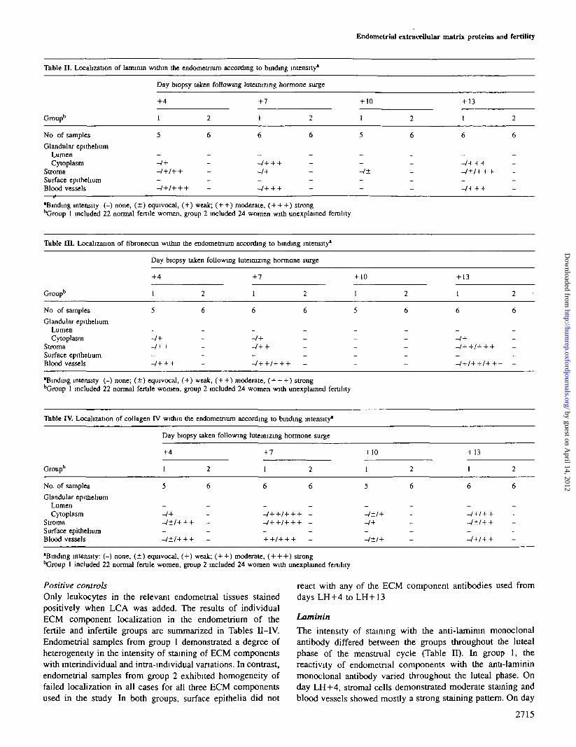
Figure 1. Immunolocalization of laminin (darker areas) within the endometnal stroma from a day luteinizing hormone +7 fertile woman.Original magnification (a) X100; (b) X400.
LH + 7, staining of the stromal cells varied equally fromnegative to weak, and blood vessels and glands showed mostlya strong staining pattern (Figure 1). On day LH+10, bloodvessels did not exhibit any staining, and stromal cells showedonly an equivocal staining pattern. On day LH+13, stromalcells stained mostly strongly, with a few exceptions of eitherweak or no staining, whilst blood vessels showed mostly astrong reaction to the antibody. In group 2, laminin failed tostain from days LH+4 to LH+13 in both stromal cells andblood vessels.
Fibronectin
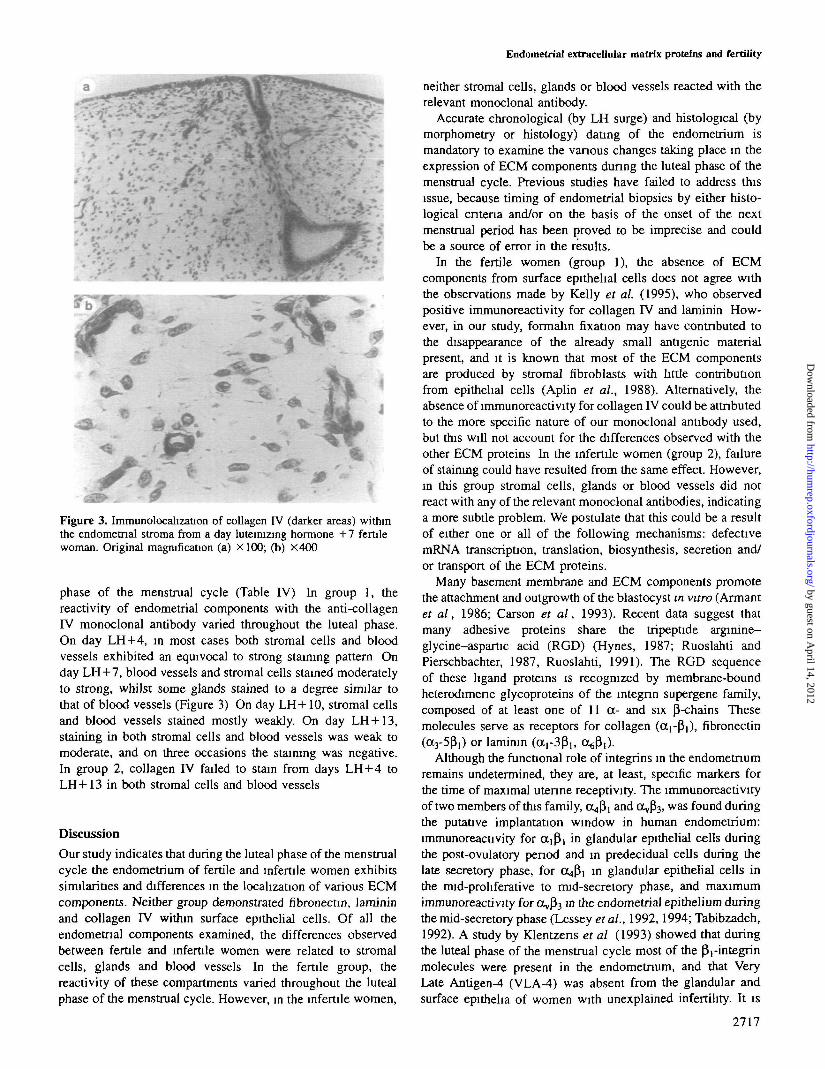
The intensity of staining with the anti-fibronectin monoclonalantibody differed between the groups throughout the lutealphase of the menstrual cycle (Table HI). In group 1, thereactivity of endometnal components with the anti-fibronectinmonoclonal antibody varied throughout the luteal phase. Onday LH+4, the majority of cases showed a moderate stainingin the stromal cells, and only a few failed to demonstrate anystaining; the blood vessels exhibited a strong pattern of stainingin most cases. On day LH+7, stromal cells showed a moderateintensity of staining and the majority of blood vessels demon-strated a moderate to strong staining. On day LH+10, however,no staining was observed in either stromal cells or bloodvessels. On day LH+13, the staining intensity of stroma] cellsand blood vessels varied from no staining to strong staining(Figure 2). In group 2, fibronectin failed to stain from daysLH+4 to LH+13 in both stromal cells and blood vessels.
Collagen IV
The intensity of staining with the anti-collagen IV monoclonalantibody differed between the groups throughout the luteal
2716
Figure 2. Immunolocalization of fibronectin (darker areas) withinthe endometnal stroma from a day luteinizing hormone +13 fertilewoman. Original magnification (a) X100; (b) X400.
by guest on April 14, 2012
http://humrep.oxfordjournals.org/
Dow
nloaded from
Endometrial extracellular matrix proteins and fertility
* • ' . < - •."» r' 7 —. •. -<
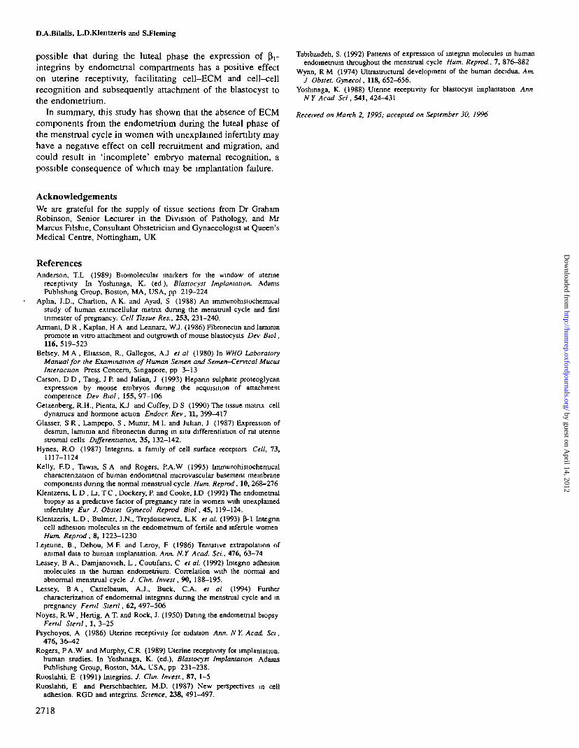
Figure 3. Immunolocalization of coUagen IV (darker areas) withinthe endometnal stroma from a day luteituzing hormone +7 fertilewoman. Original magnification (a) X100; (b) X400
phase of the menstrual cycle (Table IV) In group 1, thereactivity of endometrial components with the anti-collagenIV monoclonal antibody varied throughout the luteal phase.On day LH+4, in most cases both stromal cells and bloodvessels exhibited an equivocal to strong staining pattern Onday LH+7, blood vessels and stromal cells stained moderatelyto strong, whilst some glands stained to a degree similar tothat of blood vessels (Figure 3) On day LH+10, stromal cellsand blood vessels stained mostly weakly. On day LH+13,staining in both stroma] cells and blood vessels was weak tomoderate, and on three occasions the staining was negative.In group 2, collagen IV failed to stain from days LH + 4 toLH+13 in both stromal cells and blood vessels
Discussion
Our study indicates that during the luteal phase of the menstrualcycle the endometrium of fertile and infertile women exhibitssimilarities and differences in the localization of various ECMcomponents. Neither group demonstrated fibronectin, lamininand collagen IV within surface epithelial cells. Of all theendometnal components examined, the differences observedbetween fertile and infertile women were related to stromalcells, glands and blood vessels In the fertile group, thereactivity of these compartments varied throughout the lutealphase of the menstrual cycle. However, in the infertile women,
neither stromal cells, glands or blood vessels reacted with therelevant monoclonal antibody.
Accurate chronological (by LH surge) and histological (bymorphometry or histology) dating of the endometrium ismandatory to examine the various changes taking place in theexpression of ECM components during the luteal phase of themenstrual cycle. Previous studies have failed to address thisissue, because timing of endometrial biopsies by either histo-logical criteria and/or on the basis of the onset of the nextmenstrual period has been proved to be imprecise and couldbe a source of error in the results.
In the fertile women (group 1), the absence of ECMcomponents from surface epithelial cells does not agree withthe observations made by Kelly et al. (1995), who observedpositive immunoreactivity for collagen IV and laminin How-ever, in our study, formalin fixation may have contributed tothe disappearance of the already small anOgenic materialpresent, and it is known that most of the ECM componentsare produced by stromal fibroblasts with little contributionfrom epithelial cells (Aplin et al., 1988). Alternatively, theabsence of immunoreactivity for collagen IV could be attributedto the more specific nature of our monoclonal antibody used,but this will not account for the differences observed with theother ECM proteins In the infertile women (group 2), failureof staining could have resulted from the same effect. However,in this group stromal cells, glands or blood vessels did notreact with any of the relevant monoclonal antibodies, indicatinga more subtle problem. We postulate that this could be a resultof either one or all of the following mechanisms: defectivemRNA transcription, translation, biosynthesis, secretion and/or transport of the ECM proteins.
Many basement membrane and ECM components promotethe attachment and outgrowth of the blastocyst in vitro (Armantet al, 1986; Carson et al, 1993). Recent data suggest thatmany adhesive proteins share the tripeptide arginine-glycine-aspartic acid (RGD) (Hynes, 1987; Ruoslahti andPierschbachter, 1987, Ruoslahti, 1991). The RGD sequenceof these hgand proteins is recognized by membrane-boundheterodimenc glycoproteins of the integnn supergene family,composed of at least one of 11 a- and six P-chains Thesemolecules serve as receptors for collagen (apPi), fibronectin(a3-5P!) or laminin ( a r 3P , , o^fi,)-
Although the functional role of integrins in the endometriumremains undetermined, they are, at least, specific markers forthe time of maximal uterine receptivity. The immunoreactivityof two members of this family, o^pj and 0^3 , was found duringthe putative implantation window in human endometrium:immunoreactivity for ot|Pi in glandular epithelial cells duringthe post-ovulatory period and in predecidual cells during thelate secretory phase, for OtP, in glandular epithelial cells inthe mid-proliferative to mid-secretory phase, and maximumimmunoreactivity for OvP3 in the endometrial epithelium duringthe mid-secretory phase (Lessey et al., 1992,1994; Tabibzadeh,1992). A study by Klentzens et al (1993) showed that duringthe luteal phase of the menstrual cycle most of the p,-integrinmolecules were present in the endometrium, and that VeryLate Antigen-4 (VLA-4) was absent from the glandular andsurface epitheha of women with unexplained infertility. It is
2717
by guest on April 14, 2012
http://humrep.oxfordjournals.org/
Dow
nloaded from
DA.Bilalis, L.D.KIentzeris and SJleming
possible that during the luteal phase the expression of pVintegrins by endometnal compartments has a positive effecton uterine receptivity, facilitating cell-ECM and cell-cellrecognition and subsequently attachment of the blastocyst tothe endometrium.
In summary, this study has shown that the absence of ECMcomponents from the endometrium during the luteal phase ofthe menstrual cycle in women with unexplained infertility mayhave a negative effect on cell recruitment and migration, andcould result in 'incomplete' embryo maternal recognition, apossible consequence of which may be implantation failure.
Tabibzadeh, S. (1992) Patterns of expression of integnn molecules in humanendometrium throughout the menstrual cycle Hum. Reprod, 7, 876-882
Wynn, R M (1974) Ultrastructural development of the human decidua. Am.J Obstei. Gynecol, 118, 652-656.
Yoshinaga, K. (1988) Uterine receptivity for blastocyst implantation AnnN Y Acad Sci, 541, 424-^*31
Received on March 2, 1995; accepted on September 30, 1996
AcknowledgementsWe are grateful for the supply of tissue sections from Dr GrahamRobinson, Senior Lecturer in the Division of Pathology, and MrMarcus Filshie, Consultant Obstetrician and Gynaecologist at Queen'sMedical Centre, Nottingham, UK
ReferencesAnderson, T.L (1989) Biomolecular markers for the window of uterine
receptivity In Yoshinaga, K. (ed), Blastocyst Implantation. AdamsPublishing Group, Boston, MA, USA, pp 219-224
Aphn, J.D., Charlton, A K. and Ayad, S (1988) An lmmunohistochemicalstudy of human extracellular matrix during the menstrual cycle and firsttrimester of pregnancy. Cell Tissue Res., 253, 231-240.
Armant, D R , Kaplan, H A and Lennarz, WJ. (1986) Fibronectin and lamimnpromote in vitro attachment and outgrowth of mouse blastocysts Dev Bwl,116,519-523
Belsey, M A , Eliasson, R., Gallegos, AJ et al (1980) In WHO LaboratoryManual for the Examination of Human Semen and Semen—Cervical MucusInteraction Press Concern, Singapore, pp 3-13
Carson, D D , Tang, J P. and Julian, J (1993) Hepann sulphate proteoglycanexpression by mouse embryos during the acquisition of attachmentcompetence Dev Biol, 155, 97-106
Getzenberg, R.H., Pienta, KJ and Coffey, D S (1990) The tissue matrix celldynamics and hormone action Endocr. Rev, 11, 399-417
Glasser, S R , Lampepo, S , Munir, M I. and Julian, J (1987) Expression ofdesmin, lamimn and fibronectin during in situ differentiation of rat utennestromal cells Differentiation, 35, 132-142.
Hynes, R.O (1987) Integnns. a family of cell surface receptors Cell, 73,1117-1124
Kelly, ED , Tawia, S A and Rogers, P.A.W (1995) lmmunohistochemicalcharacterization of human endometnal microvascular basement membranecomponents during the normal menstrual cycle. Hum. Reprod, 10, 268—276
Klentzens, L D , Li, TC , Dockery, P. and Cooke, I.D (1992) The endometnalbiopsy as a predictive factor of pregnancy rate in women with unexplainedinfertility Eur J. Obstet Gynecol Reprod Biol, 45, 119-124.
Klentzeris, L.D , Bulmer, J.N., Trejdosiewicz, L.K et al. (1993) fM Integnncell adhesion molecules in the endometnum of fertile and infertile womenHum. Reprod, 8, 1223-1230
Lejeune, B., Dehou, M F. and Leroy, F (1986) Tentative extrapolation ofanimal data to human implantation. Ann. N.Y Acad. ScL, 476, 63-74
Lessey, B A., Damjanovich, L , Coutifans, C et al (1992) Integnn adhesionmolecules in the human endometrium. Correlation with the normal andabnormal menstrual cycle J. Clin. Invest, 90, 188-195.
Lessey, B A , Castelbaum, AJ., Buck, C.A. et al (1994) Furthercharacterization of endometnal integnns during the menstrual cycle and inpregnancy Fertil Stenl, 62, 497-506
Noyes, R.W, Hertig, A T. and Rock, J. (1950) Dating the endometnal biopsyFertil Stenl, 1, 3-25
Psychoyos, A (1986) Uterine receptivity for mdation Ann. NY. Acad Set,476, 36-42
Rogers, PA.W and Murphy, C.R (1989) Uterine receptivity for implantation.human studies. In Yoshinaga, K. (ed.), Blastocyst Implantation AdamsPublishing Group, Boston, MA, USA, pp 231-238.
Ruoslahti, E (1991) Integnns. J. Clm. Invest., 87, 1-5Ruoslahti, E and Pierschbachter, M.D. (1987) New perspectives in cell
adhesion. RGD and integrins. Science, 238, 491^97.
2718
by guest on April 14, 2012
http://humrep.oxfordjournals.org/
Dow
nloaded from
Top Related
Copyright © 2022 FDOKUMEN

