


Bahasa
Halaman
Hukum

Biochimica et Biophysica Acta 1810 (2011) 150–161
Contents lists available at ScienceDirect
Biochimica et Biophysica Acta
j ourna l homepage: www.e lsev ie r.com/ locate /bbagen
Human galectin-3 (Mac-2 antigen): Defining molecular switches of affinity to naturalglycoproteins, structural and dynamic aspects of glycan binding by flexible liganddocking and putative regulatory sequences in the proximal promoter region
Mickaël Krzeminski a,1, Tanuja Singh b,c,1, Sabine André b, Martin Lensch b, Albert M. Wu c,⁎,Alexandre M.J.J. Bonvin a,⁎, Hans-Joachim Gabius b,⁎a Bijvoet Center for Biomolecular Research, Faculty of Science, Utrecht University, Padualaan 8, 3584 CH Utrecht, The Netherlandsb Institute of Physiological Chemistry, Faculty of Veterinary Medicine, Ludwig-Maximilians-University, Veterinärstr. 13, 80539 Munich, Germanyc Glyco-Immunochemistry Research Laboratory, Institute of Molecular and Cellular Biology, College of Medicine, Chang-Gung University, Kwei-san, Tao-yuan 333, Taiwan
Abbreviations: BSM, bovine submaxillary mucin; Gal,D-galactosamine; Glc, D-glucose; GlcNAc, N-acetyl-D-gHADDOCK, High Ambiguity Driven DOCKing; hGal-3,acetyllactosamine; LacdiNAc, di-N-acetylated lactose;submaxillary mucin; PSM, porcine submaxillary mucin;glycoprotein; TBS-T, Tris/HCl-buffered saline containingFriedenreich antigen (Galβ1–3GalNAcα)⁎ Corresponding authors. A.M. Wu is to be contacted
+886 3 211 8456, +886 3 211 8700. A.M.J.J. Bonvin, tel.:2537623. H.-J. Gabius, tel.: +49 89 2180 2290; fax: +4
E-mail addresses: [email protected] (A.M. Wu(A.M.J.J. Bonvin), [email protected] (H.-J. Gabius).
1 These authors contributed equally to this manuscrip
0304-4165/$ – see front matter © 2010 Elsevier B.V. Adoi:10.1016/j.bbagen.2010.11.001
a b s t r a c t
a r t i c l e i n f oArticle history:
Received 27 August 2010Received in revised form 29 October 2010Accepted 2 November 2010Available online 8 November 2010Keywords:GlycosylationLectinMacrophageModelingMucinSialylation
Background: Human galectin-3 (Mac-2 antigen) is a cell-type-specific multifunctional effector owing toselective binding of distinct cell-surface glycoconjugates harboring β-galactosides. The structural basisunderlying the apparent preferences for distinct glycoproteins and for expression is so far unknown.Methods: We strategically combined solid-phase assays on 43 natural glycoproteins with a new statisticalapproach to fully flexible computational docking and also processed the proximal promoter region in silico.Results: The degree of branching in N-glycans and clustering of core 1 O-glycans are positive modulators foravidity. Sialylation of N-glycans in α2–6 linkage and of core 1 O-glycans in α2–3 linkage along with core 2branching was an unfavorable factor, despite the presence of suited glycans in the vicinity. The lectin–ligandcontact profile was scrutinized for six natural di- and tetrasaccharides enabling a statistical grading byanalyzing flexible docking trajectories. The computational analysis of the proximal promoter regiondelineated putative sites for Lmo2/c-Ets-1 binding and new sites with potential for RUNX binding.General significance: These results identify new features of glycan selectivity and ligand contact by combining
solid-phase assays with in silico work as well as of reactivity potential of the promoter.© 2010 Elsevier B.V. All rights reserved.
1. Introduction
Protein–glycan recognition is being delineated as a key molecularinteraction route based on the exceptional capacity of oligosacchar-ides for high-density information coding and the emergence ofvarious families of lectins [1]. Despite the glycomic diversity on cellsurfaces this process has an exquisite target selectivity whichunderlies the precision of the post-binding biosignaling [2]. To
D-galactose; GalNAc, N-acetyl-lucosamine; gp, glycoprotein;human galectin-3; LacNAc, N-Man, D-mannose; OSM, ovineTHGP, Tamm–Horsfall urinary0.05% Tween 20; TF, Thomsen–
at tel.: +886 3 211 8966; fax:+31 30 2533850; fax: +31 309 89 2180 2508.), [email protected]
t.
ll rights reserved.
define the nature of the mechanisms and the structural basis for theinherent selectivity are thus questions with conspicuous biorele-vance. Here, we present a combined approach toward this end,studying binding preferences of a multifunctional human lectin. Wetested various ligand structures and topology of presentation incombination with flexible ligand docking and statistical analysis ofcontact.
The Mac-2 antigen was originally defined as rat macrophagemarker present abundantly on thioglycollate-induced peritonealexudate cells [3]. It was later confirmed to be expressed in themonocyte–macrophage lineage and detected also in epithelial andstromal cells as well as in diverse tumor cell types [4–6]. The humanMac-2 protein, known as major non-integrin binding partner forlaminin and IgE receptor, was then identified to be a β-galactoside-specific lectin [7]. The characteristics of the C-terminal sequencesection, the lectin part, led to its assignment to the galectin family,herein as galectin-3 [8,9]. Its lectin property plays an important role indefense mechanisms. For instance, it enables macrophages to targetdi-N-acetylated lactose (LacdiNAc) of helminth parasites and themajor xenoantigen (α-Gal) [10,11]. Evidently, distinct glycan epitopesserve as sugar-encoded signals for the lectin in immune defense. This
151M. Krzeminski et al. / Biochimica et Biophysica Acta 1810 (2011) 150–161
also holds true for adhesion/growth-regulatory events triggered bygalectin-3, observed for example with thymocytes or carcinoma/neuroblastoma cells [2,12–15].
To exert its specific functions, human galectin-3 (hGal-3) requiresa discriminatory reactivity to the different types of oligosaccharidesbased on a substituted β-galactoside core in natural glycoconjugates.Binding and inhibition assays, frontal affinity chromatography, glycanarrays and titration calorimetry revealed a strong reactivity with β1–4-linked galactosides, especially oligomeric N-acetyllactosamine(LacNAc), and histo-blood group ABH epitopes with indications fora minor influence of α2–3-sialylation and N-glycan branching in freeglycans, and assay-dependent variation of avidity for N- vs O-glycans[16–25]. Due to the rather common presence of theoretically suitedepitopes at the termini of glycan branches, binding of hGal-3 to manycell surface glycoconjugates can be expected. But, however, this lectinis very selective when engaging in contacts with cell surfaceglycoconjugates.
Despite the abundance of glycoconjugates with terminal β-galactosides only a limited set of glycoproteins/glycolipids qualifiesas physiological galectin-3 ligand, and this with cell-type selectiv-ity. Testing for example colon cancer cells, carcinoembryonicantigen, laminin and lysosome-associated membrane proteins 1/2as well as mucins (especially the mucin MUC1 via O-glycans and theN-glycan of MUC1-C at Asn36) were bioactive [15,26–28]. Theglycoproteins CD29, CD43, CD45, CD71, and CD98 (H-chain) and theMac-2-binding protein were counterreceptors in the case ofthymocytes, for hippocampal neurons the neural cell adhesionmolecule L1 and for neuroblastoma cells ganglioside GM1[13,14,29]. Thus, the presence of a cognate carbohydrate structurecombined with other, not yet precisely defined topological factorsappears to underlie the distinct binding properties. It is noteworthyin this context that hGal-3's tridomain structure, especially its fivecollagenase-sensitive tandem-repeat modules, endows the onlychimera-type member of this lectin family with unique propertiesfor cooperativity of binding and oligomerization in the presence ofmultivalent ligands [30–33]. This characteristic gives special reasonto test natural glycoproteins with different degrees of valency andbranch-end structure. Compared to free N- and O-glycans thespatial distribution of the glycan chains attached to a protein carrieris assumed to partake in regulation of lectin affinity [34–36]. Towhat extent factors imposed by glycan presentation on a naturalprotein affect the binding of this potent effector in the immunesystem and beyond has not yet been addressed. In order to take thefine-specificity analysis of hGal-3 to the topological level, probingwell-characterized glycoproteins comparatively under identicalconditions is a promising approach.
We analyze here 43 glycoprotein preparations with differences inbranch-end and core structures including sialylation and in degree ofbranching as well as a bacterial polysaccharide. These binding assaysare supplemented with fine-specificity monitoring using 41 glycancompounds as inhibitors of hGal-3/glycoprotein binding. The exper-imental part is combined with in silico calculations to monitor therange of interactions in complexes with six cognate di- andtetrasaccharides. Using HADDOCK version 2.1 [37,38], a flexibledocking approach that has been introduced previously to the studyof protein–carbohydrate interactions [39], we visualize the dynamicswithin the interaction profile of a lectin for the first time at a statisticallevel. The presented results contribute to the efforts to explain theorigin of affinity, specificity and eventually intrafamily differences inphysiological ligand selection, with implications for drug design.These differences are also manifested on the level of the expressionpatterns of galectins, for example detected by immunohistochemicalfingerprinting of human tumors with prognostic relevance [40–42].To help delineate the underlying sequence signals, we present acomparative in silico dissection of the proximal promoter region of thehGal-3 gene with those of three proto-type galectins, a step to spot
putative sites for transcription factors and discern non-uniformfeatures.
2. Materials and methods
2.1. Galectin-3
Human galectin-3 was obtained by recombinant production andpurified by affinity chromatography on lactosylated Sepharose 4B,prepared after resin activation with divinyl sulfone, as crucial step. Itwas subjected to rigorous quality controls by one- and two-dimensional gel electrophoresis, gel filtration and mass spectrometry,then biotinylated under activity-preserving conditions with the N-hydroxysuccinimide ester derivative of biotin [12,31]. The degree ofbiotinylation was determined by two-dimensional gel electrophoresisandmass spectrometric fingerprinting [43,44]. Lack of influence of thelabeling on lectin activity was ascertained by solid-phase and cell-binding assays using asialofetuin and human SW480 colon carcinomacells, respectively [45,46].
2.2. Glycoproteins and saccharides
Sources and further processing of the glycoproteins have beengiven previously in the first reports on galectin reactivity [47,48]. Thepredominant carbohydrate determinants at branch ends with affinityto galectins are listed in Table 1. Due to its importance for galectinbinding, information on branch-end sialylation is as follows for thetested glycoproteins:
• N-glycans of bovine lactoferrin are exclusively α2–6-sialylated, andin addition terminal LacdiNAc and Galα1–3Galβ1–4GlcNAc (B-like)epitopes are present [49]. The same applies to human glycophorin,which can naturally present terminal galactose in one arm due toincomplete sialylation [50].
• Porcine thyroglobulin, human α1-acid glycoprotein and bovinefetuin all contain α2–3/6-sialylated N-acetyllactosamine withbranch specificity, e.g. α2–3 sialylation resides in the β1–4-branchof the α1–3-arm (for further information, please see [51]).
The Pneumococcus type 14 polysaccharidewas a generous gift fromthe late Dr. E. A. Kabat (Department of Microbiology, ColumbiaUniversity, NY, USA). Mono-, di- and oligosaccharides used wereobtained from Dextra (Reading, Berkshire, UK) or Sigma (Munich,Germany).
2.3. Enzyme-linked lectinosorbent assay
The assay was performed under conditions deliberately keptconstant to allow comparison of the different galectins tested[39,47,48,52]. In detail, the volume of each reagent solution appliedto wells of the plate was 50 μl/well, and all incubations, except forcoating, were performed at 20 °C. The reagents, if not indicatedotherwise, were diluted with Tris-buffered saline (TBS; 0.05 M Tris–HCl, 0.15 M NaCl, pH 7.35) containing 0.05% Tween 20 (TBS-T). TBS-Twas used for washing the plates between incubation steps.
The surface of the 96-well microtiter plate wells (Nunc-Immuno,Kamstrupvej, Denmark) was coated with glycoproteins dissolved in0.05 M sodium carbonate buffer (0.05 M NaHCO3/0.05 M Na2CO3, pH9.6) overnight at 4 °C. After washing the plate, solution withbiotinylated hGal-3 (5 μg/ml) was added and the plate was incubatedfor 30 min. The plates were then carefully washed to remove any freelectin. The ExtrAvidin/alkaline phosphatase solution (diluted1:10,000; Sigma) was added thereafter to detect the specificallybound hGal-3 by its biotin moieties. Carbohydrate dependence ofbindingwas routinely ascertained by haptenic inhibition. After 1 h theplates were washed at least four times to remove free conjugate andincubated with a solution of p-nitrophenyl phosphate (Sigma
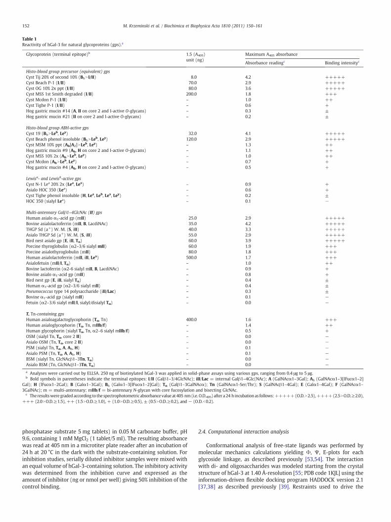
Table 1Reactivity of hGal-3 for natural glycoproteins (gps).a
Glycoprotein (terminal epitope)b 1.5 (A405)unit (ng)
Maximum A405 absorbance
Absorbance readingc Binding intensityc
Histo-blood group precursor (equivalent) gpsCyst Tij 20% of second 10% (BhN I/II) 8.0 4.2 +++++Cyst Beach P-1 (I/II) 70.0 2.9 +++++Cyst OG 10% 2x ppt (I/II) 80.0 3.6 +++++Cyst MSS 1st Smith degraded (I/II) 200.0 1.8 +++Cyst Mcdon P-1 (I/II) – 1.0 ++Cyst Tighe P-1 (I/II) – 0.6 +Hog gastric mucin #14 (A, II on core 2 and I-active O-glycans) – 0.3 ±Hog gastric mucin #21 (II on core 2 and I-active O-glycans) – 0.2 ±
Histo-blood group ABH-active gpsCyst 19 (BhNLeb, Ley) 32.0 4.1 +++++Cyst Beach phenol insoluble (BhNLeb, Ley) 120.0 2.9 +++++Cyst MSM 10% ppt (Ah[A2]NLeb, Ley) – 1.3 ++Hog gastric mucin #9 (Ah, H on core 2 and I-active O-glycans) – 1.1 ++Cyst MSS 10% 2x (AhNLeb, Ley) – 1.0 ++Cyst Mcdon (AhNLeb, Ley) – 0.7 +Hog gastric mucin #4 (Ah, H on core 2 and I-active O-glycans) – 0.5 +
Lewisa- and Lewisb-active gpsCyst N-1 Lea 20% 2x (Lea, Lex) – 0.9 +Asialo HOC 350 (Lea) – 0.6 +Cyst Tighe phenol insoluble (H, Lea, Leb, Lex, Ley) – 0.2 ±HOC 350 (sialyl Lea) – 0.1 −
Multi-antennary Galβ1–4GlcNAc (II) gpsHuman asialo α1-acid gp (mII) 25.0 2.9 +++++Bovine asialolactoferrin (mII, B, LacdiNAc) 35.0 4.2 +++++THGP Sd (a+) W. M. (S, iII) 40.0 3.3 +++++Asialo THGP Sd (a+) W. M. (S, iII) 55.0 2.9 +++++Bird nest asialo gp (E, iII, Tα) 60.0 3.9 +++++Porcine thyroglobulin (α2–3/6 sialyl mII) 60.0 1.9 +++Porcine asialothyroglobulin (mII) 80.0 1.8 +++Human asialolactoferrin (mII, iII, Lex) 500.0 1.7 +++Asialofetuin (mII/I, Tα) – 1.0 ++Bovine lactoferrin (α2-6 sialyl mII, B, LacdiNAc) – 0.9 +Bovine asialo α1-acid gp (mII) – 0.8 +Bird nest gp (E, iII, sialyl Tα) – 0.4 ±Human α1-acid gp (α2–3/6 sialyl mII) – 0.4 ±Pneumococcus type 14 polysaccharide (iII/Lac) – 0.3 ±Bovine α1-acid gp (sialyl mII) – 0.1 −Fetuin (α2–3/6 sialyl mII/I, sialyl/disialyl Tα) – 0.0 −
T, Tn-containing gpsHuman asialoagalactoglycophorin (Tα, Tn) 400.0 1.6 +++Human asialoglycophorin (Tα, Tn, mIIb/f) – 1.4 ++Human glycophorin (sialyl Tα, Tn, α2–6 sialyl mIIb/f) – 0.5 +OSM (sialyl Tn, Tα, core 2 II) – 0.0 −Asialo OSM (Tn, Tα, core 2 II) – 0.0 −PSM (sialyl Tn, Tα, A, Ah, H) – 0.1 −Asialo PSM (Tn, Tα, A, Ah, H) – 0.1 −BSM (sialyl Tn, GlcNAcβ1–3Tn, Tα) – 0.0 −Asialo BSM (Tn, GlcNAcβ1–3Tn, Tα) – 0.0 −a Analyses were carried out by ELLSA. 250 ng of biotinylated hGal-3 was applied in solid-phase assays using various gps, ranging from 0.4 μg to 5 μg.b Bold symbols in parentheses indicate the terminal epitopes: I/II (Galβ1–3/4GlcNAc); iII/Lac = internal Galβ1–4Glc(NAc); A (GalNAcα1–3Gal); Ah (GalNAcα1–3[lFucα1–2]
Gal); H (lFucα1–2Gal); B (Galα1–3Gal); Bh (Galα1–3[lFucα1–2]Gal); Tα (Galβ1–3GalNAcα); Tn (GalNAcα1-Ser/Thr); S (GalNAcβ1–4Gal); E (Galα1–4Gal); F (GalNAcα1–3GalNAc); m = multi-antennary; mIIb/f = bi-antennary N-glycan with core fucosylation and bisecting GlcNAc.
c Theresultsweregraded according to the spectrophotometricabsorbancevalue at 405 nm(i.e. O.D.405) after a 24 h incubationas follows:+++++(O.D.N2.5),++++(2.5NO.D.≥2.0),+++ (2.0NO.D.≥1.5), ++ (1.5NO.D.≥1.0), + (1.0NO.D.≥0.5), ± (0.5NO.D.≥0.2), and − (O.D.b0.2).
152 M. Krzeminski et al. / Biochimica et Biophysica Acta 1810 (2011) 150–161
phosphatase substrate 5 mg tablets) in 0.05 M carbonate buffer, pH9.6, containing 1 mM MgCl2 (1 tablet/5 ml). The resulting absorbancewas read at 405 nm in a microtiter plate reader after an incubation of24 h at 20 °C in the dark with the substrate-containing solution. Forinhibition studies, serially diluted inhibitor samples were mixed withan equal volume of hGal-3-containing solution. The inhibitory activitywas determined from the inhibition curve and expressed as theamount of inhibitor (ng or nmol per well) giving 50% inhibition of thecontrol binding.
2.4. Computational interaction analysis
Conformational analysis of free-state ligands was performed bymolecular mechanics calculations yielding Φ, Ψ, E-plots for eachglycoside linkage, as described previously [53,54]. The interactionwith di- and oligosaccharides was modeled starting from the crystalstructure of hGal-3 at 1.40 Å-resolution [55; PDB code 1KJL] using theinformation-driven flexible docking program HADDOCK version 2.1[37,38] as described previously [39]. Restraints used to drive the
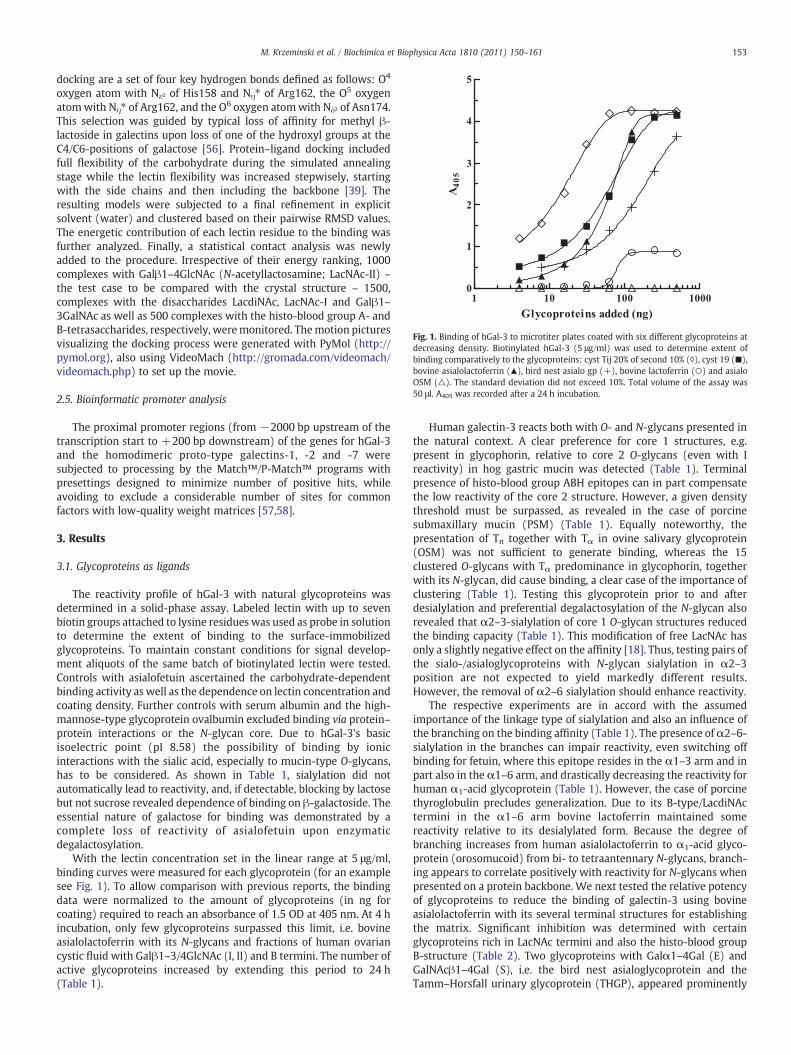
Fig. 1. Binding of hGal-3 to microtiter plates coated with six different glycoproteins atdecreasing density. Biotinylated hGal-3 (5 μg/ml) was used to determine extent ofbinding comparatively to the glycoproteins: cyst Tij 20% of second 10% (◊), cyst 19 (■),bovine asialolactoferrin (▲), bird nest asialo gp (+), bovine lactoferrin (○) and asialoOSM (△). The standard deviation did not exceed 10%. Total volume of the assay was50 μl. A405 was recorded after a 24 h incubation.
153M. Krzeminski et al. / Biochimica et Biophysica Acta 1810 (2011) 150–161
docking are a set of four key hydrogen bonds defined as follows: O4
oxygen atom with Nε2 of His158 and Nη⁎ of Arg162, the O5 oxygenatomwith Nη⁎ of Arg162, and the O6 oxygen atomwith Nδ2 of Asn174.This selection was guided by typical loss of affinity for methyl β-lactoside in galectins upon loss of one of the hydroxyl groups at theC4/C6-positions of galactose [56]. Protein–ligand docking includedfull flexibility of the carbohydrate during the simulated annealingstage while the lectin flexibility was increased stepwisely, startingwith the side chains and then including the backbone [39]. Theresulting models were subjected to a final refinement in explicitsolvent (water) and clustered based on their pairwise RMSD values.The energetic contribution of each lectin residue to the binding wasfurther analyzed. Finally, a statistical contact analysis was newlyadded to the procedure. Irrespective of their energy ranking, 1000complexes with Galβ1–4GlcNAc (N-acetyllactosamine; LacNAc-II) –
the test case to be compared with the crystal structure – 1500,complexes with the disaccharides LacdiNAc, LacNAc-I and Galβ1–3GalNAc as well as 500 complexes with the histo-blood group A- andB-tetrasaccharides, respectively, weremonitored. Themotion picturesvisualizing the docking process were generated with PyMol (http://pymol.org), also using VideoMach (http://gromada.com/videomach/videomach.php) to set up the movie.
2.5. Bioinformatic promoter analysis
The proximal promoter regions (from −2000 bp upstream of thetranscription start to +200 bp downstream) of the genes for hGal-3and the homodimeric proto-type galectins-1, -2 and -7 weresubjected to processing by the Match™/P-Match™ programs withpresettings designed to minimize number of positive hits, whileavoiding to exclude a considerable number of sites for commonfactors with low-quality weight matrices [57,58].
3. Results
3.1. Glycoproteins as ligands
The reactivity profile of hGal-3 with natural glycoproteins wasdetermined in a solid-phase assay. Labeled lectin with up to sevenbiotin groups attached to lysine residues was used as probe in solutionto determine the extent of binding to the surface-immobilizedglycoproteins. To maintain constant conditions for signal develop-ment aliquots of the same batch of biotinylated lectin were tested.Controls with asialofetuin ascertained the carbohydrate-dependentbinding activity as well as the dependence on lectin concentration andcoating density. Further controls with serum albumin and the high-mannose-type glycoprotein ovalbumin excluded binding via protein–protein interactions or the N-glycan core. Due to hGal-3's basicisoelectric point (pI 8.58) the possibility of binding by ionicinteractions with the sialic acid, especially to mucin-type O-glycans,has to be considered. As shown in Table 1, sialylation did notautomatically lead to reactivity, and, if detectable, blocking by lactosebut not sucrose revealed dependence of binding on β-galactoside. Theessential nature of galactose for binding was demonstrated by acomplete loss of reactivity of asialofetuin upon enzymaticdegalactosylation.
With the lectin concentration set in the linear range at 5 μg/ml,binding curves were measured for each glycoprotein (for an examplesee Fig. 1). To allow comparison with previous reports, the bindingdata were normalized to the amount of glycoproteins (in ng forcoating) required to reach an absorbance of 1.5 OD at 405 nm. At 4 hincubation, only few glycoproteins surpassed this limit, i.e. bovineasialolactoferrin with its N-glycans and fractions of human ovariancystic fluid with Galβ1–3/4GlcNAc (I, II) and B termini. The number ofactive glycoproteins increased by extending this period to 24 h(Table 1).
Human galectin-3 reacts both with O- and N-glycans presented inthe natural context. A clear preference for core 1 structures, e.g.present in glycophorin, relative to core 2 O-glycans (even with Ireactivity) in hog gastric mucin was detected (Table 1). Terminalpresence of histo-blood group ABH epitopes can in part compensatethe low reactivity of the core 2 structure. However, a given densitythreshold must be surpassed, as revealed in the case of porcinesubmaxillary mucin (PSM) (Table 1). Equally noteworthy, thepresentation of Tn together with Tα in ovine salivary glycoprotein(OSM) was not sufficient to generate binding, whereas the 15clustered O-glycans with Tα predominance in glycophorin, togetherwith its N-glycan, did cause binding, a clear case of the importance ofclustering (Table 1). Testing this glycoprotein prior to and afterdesialylation and preferential degalactosylation of the N-glycan alsorevealed that α2–3-sialylation of core 1 O-glycan structures reducedthe binding capacity (Table 1). This modification of free LacNAc hasonly a slightly negative effect on the affinity [18]. Thus, testing pairs ofthe sialo-/asialoglycoproteins with N-glycan sialylation in α2–3position are not expected to yield markedly different results.However, the removal of α2–6 sialylation should enhance reactivity.
The respective experiments are in accord with the assumedimportance of the linkage type of sialylation and also an influence ofthe branching on the binding affinity (Table 1). The presence ofα2–6-sialylation in the branches can impair reactivity, even switching offbinding for fetuin, where this epitope resides in the α1–3 arm and inpart also in the α1–6 arm, and drastically decreasing the reactivity forhuman α1-acid glycoprotein (Table 1). However, the case of porcinethyroglobulin precludes generalization. Due to its B-type/LacdiNActermini in the α1–6 arm bovine lactoferrin maintained somereactivity relative to its desialylated form. Because the degree ofbranching increases from human asialolactoferrin to α1-acid glyco-protein (orosomucoid) from bi- to tetraantennary N-glycans, branch-ing appears to correlate positively with reactivity for N-glycans whenpresented on a protein backbone. We next tested the relative potencyof glycoproteins to reduce the binding of galectin-3 using bovineasialolactoferrin with its several terminal structures for establishingthe matrix. Significant inhibition was determined with certainglycoproteins rich in LacNAc termini and also the histo-blood groupB-structure (Table 2). Two glycoproteins with Galα1–4Gal (E) andGalNAcβ1–4Gal (S), i.e. the bird nest asialoglycoprotein and theTamm–Horsfall urinary glycoprotein (THGP), appeared prominently
Table 2Inhibitory potency of various gps on binding of hGal-3 (125 ng/50 μl) to a II-containing gp (bovine asialolactoferrin, 250 ng/50 μl).a
Inhibitor (terminal epitope)b Quantity leading to 50% inhibition (ng) Relative potencyc
Multi-antennary Galβ1–4GlcNAc (II) gpsPorcine thyroglobulin (α2–3/6 sialyl mII) 100.0 6.8×10³
Porcine asialothyroglobulin (mII) 200.0 3.4×10³
Human asialo α1-acid gp (mII) 300.0 2.3×10³
Bird nest asialo gp (E, iII, Tα) 600.0d 1.1×10³
THGP Sd (a+) W. M. (S, iII) 1500.0 4.5×10²
Asialo THGP Sd (a+) W. M. (S, iII) 2000.0 3.4×10²
Human asialolactoferrin (mII, iII, Lex) 2000.0 3.4×10²
Bovine asialolactoferrin (mII, B, LacdiNAc) 8000.0 85.5Bovine asialo α1-acid gp (mII) N1388.9 (47.1%)e –
Asialofetuin (mII/I, Tα) N1388.9 (38.2%) –
Pneumococcus type 14 polysaccharide (iII/Lac) N1388.9 (33.9%) –
Human α1-acid gp (α2–3/6 sialyl mII) N1388.9 (22.8%) –
Bovine lactoferrin (α2–6 sialyl mII, B, LacdiNAc) N2777.8 (13.0%) –
Bovine α1-acid gp (sialyl mII) N1388.9 (4.9%) –
Fetuin (α2–3/6 sialyl mII/I, sialyl/disialyl T) N1388.9 (0.0%) –
Histo-blood group precursor (equivalent) gpsCyst Beach P-1 (I/II) 1800.0 3.8×10²
Cyst OG 10% 2x ppt (I/II) 2000.0 3.4×10²
Cyst Tij 20% of second 10% (BhN I/II) 3000.0 2.3×10²
Cyst MSS 1st Smith degraded (I/II) 8000.0d 85.5Cyst Mcdon P-1 (I/II) N1388.9 (21.8%) –
Hog gastric mucin #14 (A, II on core 2 and I-active O-glycans) N1388.9 (11.9%) –
Hog gastric mucin #21 (II on core 2 and I-active O-glycans) N1388.9 (11.5%) –
Cyst Tighe P-1 (I/II) N1388.9 (2.8%) –
T, Tn-containing gpsHuman asialoglycophorin (Tα, Tn, mIIb/f) 2000.0 3.4×10²
Human glycophorin (sialyl Tα, Tn, α2–6 sialyl mIIb/f) N2777.8 (14.4%) –
Asialo PSM (Tn, Tα, A, Ah, H) N555.6 (2.0%) –
Histo-blood group ABH-active gpsCyst 19 (BhNLeb, Ley) 3000.0 2.3×10²
Cyst Beach phenol insoluble (BhNLeb, Ley) 4000.0 1.7×10²
Hog gastric mucin #4 (Ah, H on core 2 and I-active O-glycans) N1388.9 (34.8%) –
Hog gastric mucin #9 (Ah, H on core 2 and I-active O-glycans) N1388.9 (26.0%) –
Cyst Mcdon (AhNLeb, Ley) N1388.9 (20.7%) –
Lewisa- and Lewisb-active gpsCyst N-1 Lea 20% 2x (Lea, Lex) N1388.9 (21.0%) –
Cyst Tighe phenol insoluble (H, Lea, Leb, Lex, Ley) N1388.9 (10.0%) –
Asialo HOC 350 (Lea) N138.9 (1.9%) –
SaccharidesTri-II 8316.0 82.3Galβ1–4GlcNAc (II) 684,600.0d 1.0Gal 2,000,000.0 0.3GalNAc 2,000,000.0 0.3
a The inhibitory activity is expressed as the amount of inhibitor leading to 50% inhibition of the control lectin binding. Total volume was 50 μl.b See footnote of Table 1.c Relative potency (RP)=quantity of Galβ1–4GlcNAc (II) required for 50% inhibition (taken as 1.0) /quantity of sample required for 50% inhibition.d Extrapolation.e The inhibitory potency of inactive gps is expressed as the maximum amount of glycans tested that yield inhibition (in parenthesis) below 50%. Other glycans that did not reach
50% inhibition were: OSM (sialyl Tn, Tα, core 2 II) and asialo OSM (Tn, Tα, core 2 II); BSM (sialyl Tn, GlcNAcβ1–3Tn, Tα) and asialo BSM (Tn, GlcNAcβ1–3Tn, Tα); humanasialoagalactoglycophorin (Tα, Tn); cyst 14 phenol insoluble (AhNLeb, Ley); HOC 350 (sialyl Lea).
154 M. Krzeminski et al. / Biochimica et Biophysica Acta 1810 (2011) 150–161
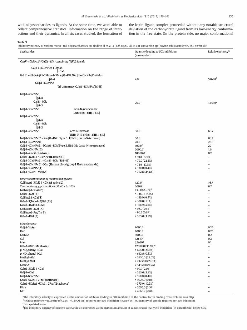
on this list (Table 2). In order to elucidate whether these epitopes arekey contact sites for the lectin we next investigated a series of mono-,di- and oligosaccharides for inhibitory capacity.
3.2. Free glycans as ligands
Bovine asialolactoferrinwasmaintained as ligand-bearingmatrix,and the free sugar compound tested was added to the lectin-containing solution to compete with the immobilized glycoprotein'sglycans for entry into the lectin site. The two disaccharides (E) and(S) were only weakly active (Table 3). Evidently, other disaccharidessuch as (T), its extended version Tβ1–4L or the Galα1–3-cappedLacNAc (B active II) were also rather weak inhibitors. In comparison,branching yielded enhanced inhibition (Table 3). Chain elongation,
too, was effective toward this end (Table 3). This set of experimentsthus further solidifies the notion that the local density of glycanbranches in a glycoprotein is a modulator for binding to this type ofnatural glycoconjugate. The actual contact can apparently bemade toII/I structures, which can be extended to histo-blood group ABHepitopes, as well as to LacdiNAc and Tα. To explain in structural termsthe reactivity of galectin-3 in solution to these epitopes, we nextperformed computational modeling.
3.3. Computational interaction analysis
In docking with HADDOCK version 2.1 [37,38], both the lectin andits carbohydrate ligand can be treated as flexible. This procedureenables us to detect any ligand-dependent adaptations, especially
155M. Krzeminski et al. / Biochimica et Biophysica Acta 1810 (2011) 150–161
with oligosaccharides as ligands. At the same time, we were able tocollect comprehensive statistical information on the range of inter-actions and their dynamics. In all six cases studied, the formation of
Table 3Inhibitory potency of various mono- and oligosaccharides on binding of hGal-3 (125 ng/50
aThe inhibitory activity is expressed as the amount of inhibitor leading to 50% inhibitiobRelative potency=quantity of Galβ1–4GlcNAc (II) required for 50% inhibition is takencExtrapolated value.dThe inhibitory potency of inactive saccharides is expressed as the maximum amount o
the lectin–ligand complex proceeded without any notable structuraldeviation of the carbohydrate ligand from its low-energy conforma-tion in the free state. On the protein side, no major conformational
μl) to a II-containing gp (bovine asialolactoferrin, 250 ng/50 μl).a
n of the control lectin binding. Total volume was 50 μl.as 1.0 /quantity of sample required for 50% inhibition.
f sugars tested that yield inhibition (in parenthesis) below 50%.
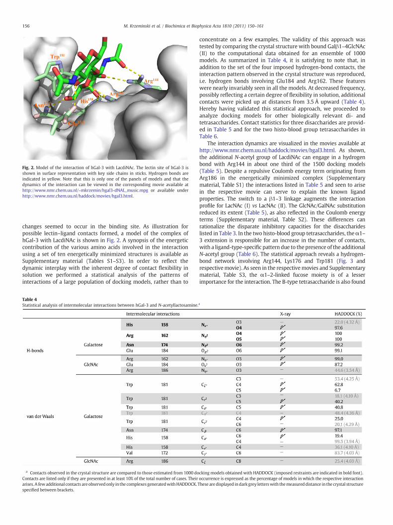
Fig. 2. Model of the interaction of hGal-3 with LacdiNAc. The lectin site of hGal-3 isshown in surface representation with key side chains in sticks. Hydrogen bonds areindicated in yellow. Note that this is only one of the panels of models and that thedynamics of the interaction can be viewed in the corresponding movie available athttp://www.nmr.chem.uu.nl/~mkrzemin/hgal3-dNAL_music.mpg or available underhttp://www.nmr.chem.uu.nl/haddock/movies/hgal3.html.
156 M. Krzeminski et al. / Biochimica et Biophysica Acta 1810 (2011) 150–161
changes seemed to occur in the binding site. As illustration forpossible lectin–ligand contacts formed, a model of the complex ofhGal-3 with LacdiNAc is shown in Fig. 2. A synopsis of the energeticcontribution of the various amino acids involved in the interactionusing a set of ten energetically minimized structures is available asSupplementary material (Tables S1–S3). In order to reflect thedynamic interplay with the inherent degree of contact flexibility insolution we performed a statistical analysis of the patterns ofinteractions of a large population of docking models, rather than to
Table 4Statistical analysis of intermolecular interactions between hGal-3 and N-acetyllactosamine.
a Contacts observed in the crystal structure are compared to those estimated from 1000 doContacts are listed only if they are presented in at least 10% of the total number of cases. Theirarises. A fewadditional contacts are observedonly in the complexes generatedwithHADDOCK.specified between brackets.
concentrate on a few examples. The validity of this approach wastested by comparing the crystal structure with bound Galβ1–4GlcNAc(II) to the computational data obtained for an ensemble of 1000models. As summarized in Table 4, it is satisfying to note that, inaddition to the set of the four imposed hydrogen-bond contacts, theinteraction pattern observed in the crystal structure was reproduced,i.e. hydrogen bonds involving Glu184 and Arg162. These featureswere nearly invariably seen in all the models. At decreased frequency,possibly reflecting a certain degree of flexibility in solution, additionalcontacts were picked up at distances from 3.5 Å upward (Table 4).Hereby having validated this statistical approach, we proceeded toanalyze docking models for other biologically relevant di- andtetrasaccharides. Contact statistics for three disaccharides are provid-ed in Table 5 and for the two histo-blood group tetrasaccharides inTable 6.
The interaction dynamics are visualized in the movies available athttp://www.nmr.chem.uu.nl/haddock/movies/hgal3.html. As shown,the additional N-acetyl group of LacdiNAc can engage in a hydrogenbond with Arg144 in about one third of the 1500 docking models(Table 5). Despite a repulsive Coulomb energy term originating fromArg186 in the energetically minimized complex (Supplementarymaterial, Table S1) the interactions listed in Table 5 and seen to arisein the respective movie can serve to explain the known ligandproperties. The switch to a β1–3 linkage augments the interactionprofile for LacNAc (I) vs LacNAc (II). The GlcNAc/GalNAc substitutionreduced its extent (Table 5), as also reflected in the Coulomb energyterms (Supplementary material, Table S2). These differences canrationalize the disparate inhibitory capacities for the disaccharideslisted in Table 3. In the two histo-blood group tetrasaccharides, theα1–3 extension is responsible for an increase in the number of contacts,with a ligand-type-specific pattern due to the presence of the additionalN-acetyl group (Table 6). The statistical approach reveals a hydrogen-bond network involving Arg144, Lys176 and Trp181 (Fig. 3 andrespectivemovie). As seen in the respectivemovies and Supplementarymaterial, Table S3, the α1–2-linked fucose moiety is of a lesserimportance for the interaction. The B-type tetrasaccharide is also found
a
cking models obtained with HADDOCK (imposed restraints are indicated in bold font).occurrence is expressed as the percentage of models in which the respective interactionThese aredisplayed indarkgrey letterswith themeasured distance in the crystal structure
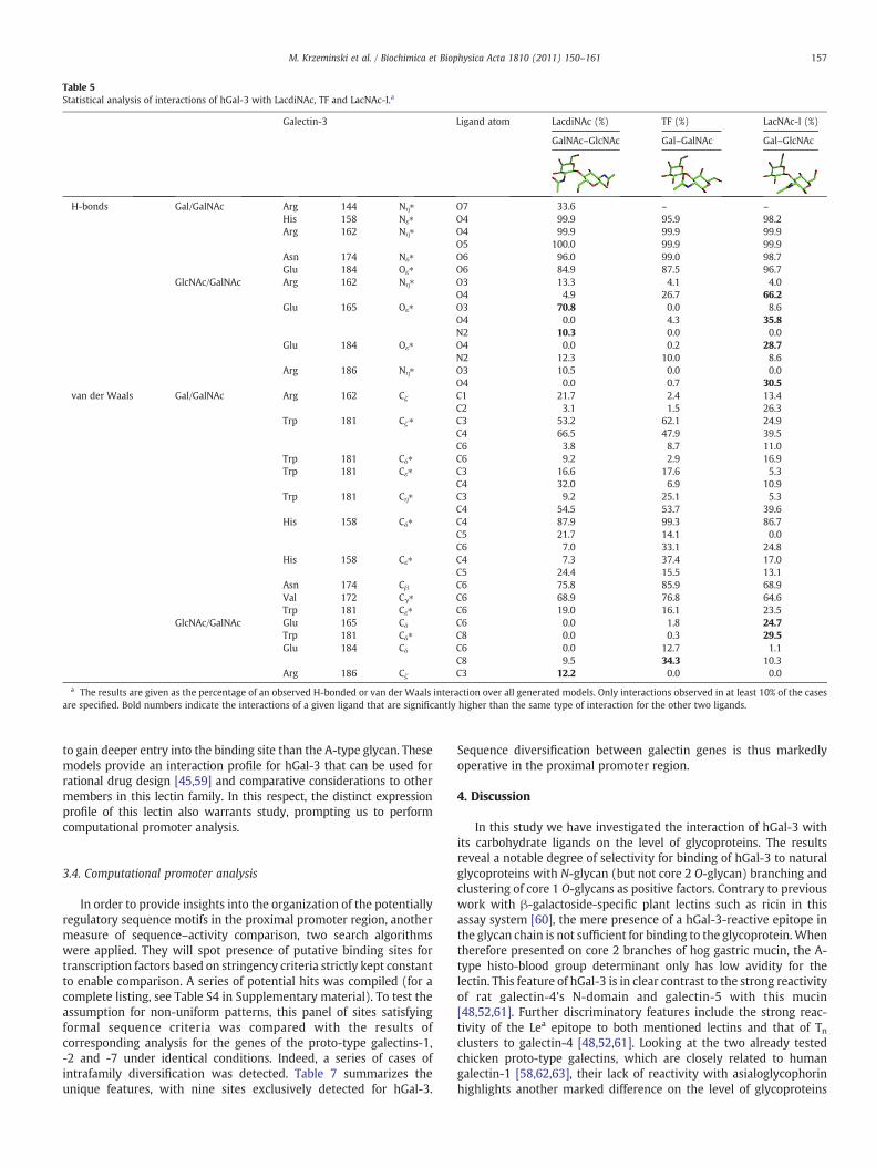
Table 5Statistical analysis of interactions of hGal-3 with LacdiNAc, TF and LacNAc-I.a
Galectin-3 Ligand atom LacdiNAc (%) TF (%) LacNAc-I (%)
GalNAc–GlcNAc Gal–GalNAc Gal–GlcNAc
H-bonds Gal/GalNAc Arg 144 Nη⁎ O7 33.6 – –
His 158 Nε⁎ O4 99.9 95.9 98.2Arg 162 Nη⁎ O4 99.9 99.9 99.9
O5 100.0 99.9 99.9Asn 174 Nδ⁎ O6 96.0 99.0 98.7Glu 184 Oε⁎ O6 84.9 87.5 96.7
GlcNAc/GalNAc Arg 162 Nη⁎ O3 13.3 4.1 4.0O4 4.9 26.7 66.2
Glu 165 Oε⁎ O3 70.8 0.0 8.6O4 0.0 4.3 35.8N2 10.3 0.0 0.0
Glu 184 Oε⁎ O4 0.0 0.2 28.7N2 12.3 10.0 8.6
Arg 186 Nη⁎ O3 10.5 0.0 0.0O4 0.0 0.7 30.5
van der Waals Gal/GalNAc Arg 162 Cζ C1 21.7 2.4 13.4C2 3.1 1.5 26.3
Trp 181 Cζ⁎ C3 53.2 62.1 24.9C4 66.5 47.9 39.5C6 3.8 8.7 11.0
Trp 181 Cδ⁎ C6 9.2 2.9 16.9Trp 181 Cε⁎ C3 16.6 17.6 5.3
C4 32.0 6.9 10.9Trp 181 Cη⁎ C3 9.2 25.1 5.3
C4 54.5 53.7 39.6His 158 Cδ⁎ C4 87.9 99.3 86.7
C5 21.7 14.1 0.0C6 7.0 33.1 24.8
His 158 Cε⁎ C4 7.3 37.4 17.0C5 24.4 15.5 13.1
Asn 174 Cβ C6 75.8 85.9 68.9Val 172 Cγ⁎ C6 68.9 76.8 64.6Trp 181 Cε⁎ C6 19.0 16.1 23.5
GlcNAc/GalNAc Glu 165 Cδ C6 0.0 1.8 24.7Trp 181 Cδ⁎ C8 0.0 0.3 29.5Glu 184 Cδ C6 0.0 12.7 1.1
C8 9.5 34.3 10.3Arg 186 Cζ C3 12.2 0.0 0.0
a The results are given as the percentage of an observed H-bonded or van der Waals interaction over all generated models. Only interactions observed in at least 10% of the casesare specified. Bold numbers indicate the interactions of a given ligand that are significantly higher than the same type of interaction for the other two ligands.
157M. Krzeminski et al. / Biochimica et Biophysica Acta 1810 (2011) 150–161
to gain deeper entry into the binding site than the A-type glycan. Thesemodels provide an interaction profile for hGal-3 that can be used forrational drug design [45,59] and comparative considerations to othermembers in this lectin family. In this respect, the distinct expressionprofile of this lectin also warrants study, prompting us to performcomputational promoter analysis.
3.4. Computational promoter analysis
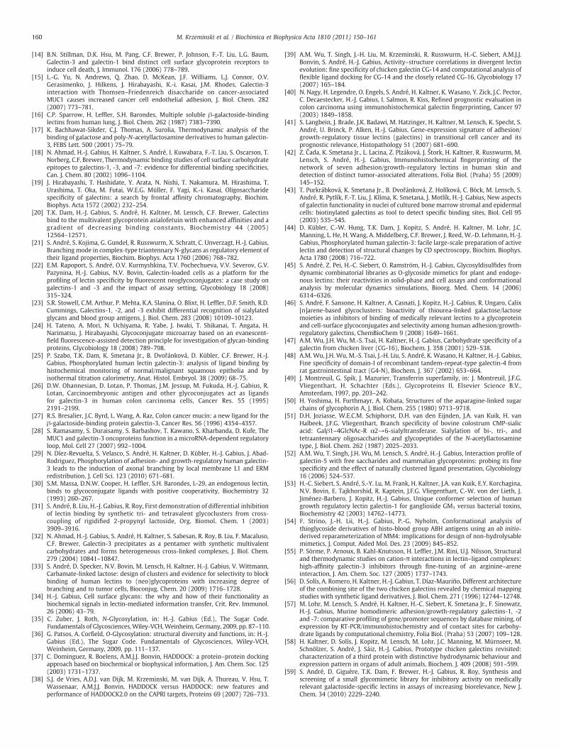
In order to provide insights into the organization of the potentiallyregulatory sequence motifs in the proximal promoter region, anothermeasure of sequence–activity comparison, two search algorithmswere applied. They will spot presence of putative binding sites fortranscription factors based on stringency criteria strictly kept constantto enable comparison. A series of potential hits was compiled (for acomplete listing, see Table S4 in Supplementary material). To test theassumption for non-uniform patterns, this panel of sites satisfyingformal sequence criteria was compared with the results ofcorresponding analysis for the genes of the proto-type galectins-1,-2 and -7 under identical conditions. Indeed, a series of cases ofintrafamily diversification was detected. Table 7 summarizes theunique features, with nine sites exclusively detected for hGal-3.
Sequence diversification between galectin genes is thus markedlyoperative in the proximal promoter region.
4. Discussion
In this study we have investigated the interaction of hGal-3 withits carbohydrate ligands on the level of glycoproteins. The resultsreveal a notable degree of selectivity for binding of hGal-3 to naturalglycoproteins with N-glycan (but not core 2 O-glycan) branching andclustering of core 1 O-glycans as positive factors. Contrary to previouswork with β-galactoside-specific plant lectins such as ricin in thisassay system [60], the mere presence of a hGal-3-reactive epitope inthe glycan chain is not sufficient for binding to the glycoprotein.Whentherefore presented on core 2 branches of hog gastric mucin, the A-type histo-blood group determinant only has low avidity for thelectin. This feature of hGal-3 is in clear contrast to the strong reactivityof rat galectin-4's N-domain and galectin-5 with this mucin[48,52,61]. Further discriminatory features include the strong reac-tivity of the Lea epitope to both mentioned lectins and that of Tnclusters to galectin-4 [48,52,61]. Looking at the two already testedchicken proto-type galectins, which are closely related to humangalectin-1 [58,62,63], their lack of reactivity with asialoglycophorinhighlights another marked difference on the level of glycoproteins
Table 6Statistical analysis of interactions of hGal-3 with histo-blood group A/B-tetrasaccharides.a
Residue Monosaccharide A-type (%) B-type (%)
H-bonds Asn 160 Oδ⁎ Gal O4 0.8 21.8
Glu 184 Oε⁎ Glc O2 59.2 32.2
Arg 144 Nη⁎ GalNAc/Gal O7 32.2 –
O3 0.2 33.0Lys 176 Nζ GalNAc/Gal O6 19.8 44.8Trp 181 Nε
⁎ 85.0 43.5van der Waals His 158 Cε
⁎ GalNAc/Gal C1 41.8 94.0C2 1.2 35.0
Asn 160 Cγ GalNAc/Gal C8 78.0 –
a The results are given as the percentage of an observed H-bonded or van der Waals interaction over all generated models. Only interactions observed in at least 10% of the casesare specified.
158 M. Krzeminski et al. / Biochimica et Biophysica Acta 1810 (2011) 150–161
[39,47]. Although these lectins together with hGal-3 can interact withthe Tβ determinant in the context of the GM1 pentasaccharide [64],the 15 O-glycans on asialoglycophorin fail to form a platform forbinding the proto-type galectins [39]. Obviously, a route towardsselectivity for glycoproteins is the way glycan epitopes are presentedspatially, as is the case for ganglioside GM1 in microdomains on themembrane [65]. Clustered presentation of the sugar headgroup ofgangliosides, in microdomains together with glycoproteins such asintegrins, can thus form high-affinity aggregates [66], provided thereare no spatial restrictions to letting the glycan chains adopt theconformer suitable for binding [53,64,67]. Our experiments withnatural glycoproteins presented on a surface thus prove instrumentalto decipher rules of physiological selectivity, considering spatialparameters. A closer look on topological aspects of ligand contact isprovided by the docking analysis.
In general, a common property of galectins is the binding ofdistinct low-energy conformers of the carbohydrate countereceptor.Previous rigid-body docking studies on mammalian galectin-3,without allowing full flexibility on both sides, had indicated that nodeviations from an energetically privileged position are required[68,69]. This also holds true for binding of histo-blood group tri- andtetrasaccharides in crystals to a fungal galectin (PDB codes 1ULD,1ULE, and 1ULF), the E. coli enterotoxin and its hybrid with choleratoxin (202L, 3EFX), a Streptococcus pneumoniae virulence factor(2J1U) and the Norwalk virus (2ZL7). Our results from flexibledocking extend the respective data basis significantly. We present aview on the individual energy contributions in the complex, derivedfrom energy-minimized models after allowing mutual flexibility.Going beyond the static view, we have introduced a statisticalapproach to the analysis of the dynamics of interactions from
Fig. 3. Schematic representation of the cooperativeness of interaction between Arg144, Lys17(panel A) and B (panel B) epitopes. The diameter of the circles reflects the occurrence of abetween the saccharide and the specified residue (and not the two others). Interactions exibetween the edges of the triangle. Finally, interactions simultaneously occurring between althe O3 and O6 oxygen atoms are shifted to reflect their interaction preferences.
HADDOCK-derived data on large ensembles of docking models. Itgives net advantages compared to common, energy-based-onlyanalysis approaches, since it reflects the probability of a giveninteraction to exist, provided the interaction space has beensufficiently sampled. It also offers the possibility to better appreciateligand flexibility in the binding pocket, which facilitates transientcontacts to certain amino acids.
Our results provide explanations for distinct specificity traits,uncovering the importance of Arg144 for LacdiNAc binding (Asn is atthis place in galectin-1, which is only weakly reactive with thisdisaccharide in a cell assay [22]). Also, the free enthalpy gains by 2.75and 8.44 kcal/mol by adding the Fuc and the GalNAc units to the β-galactoside core, measured by titration calorimetry at 279.6 or 280.4 K[17], can be rationalized by the presented interaction analysis. Of note,the favorable changes in the protein's overall conformational entropyupon ligand binding add to the overall thermodynamic balance [70].The detailed description of the extended binding site and theinteraction profile of flexible lectin/ligand pairs refine the structuralbasis in order to inspire the rational design of substitutions insynthetic inhibitors, thereby opening the route to the exploitation ofthe sequence differences between galectins for the benefit of reducingintrafamily cross-reactivity. This modeling approach and the derivedstructural models will thus be helpful in automated screening forglycomimetics such as (glyco)peptides [71] and estimating theirrelative potencies.
In addition to delineating intrafamily differences in carbohydratespecificity the definition of the regulatory mechanisms underlying thedistinct gene expression profiles, e.g. also with upregulation ininflammation [72,73], is a further challenge. Using two current searchalgorithms we have mapped the proximal promoter region with
6 and Trp181 of hGal-3 and theα1–3-linked GalNAc/Gal part of the histo-blood group An interaction. A circle at a vertex of the triangle represents an interaction present onlysting simultaneously with two residues are depicted with circles located in the middlel three residues are indicated by the circle in the middle of the triangle. The positions of
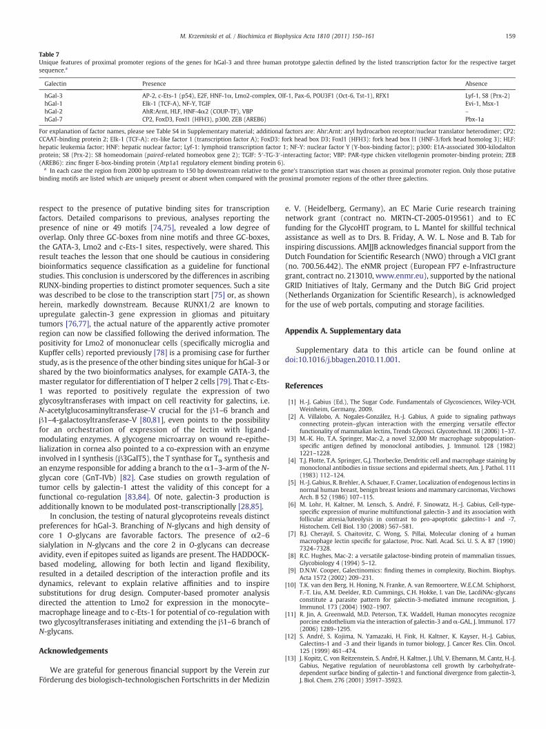
Table 7Unique features of proximal promoter regions of the genes for hGal-3 and three human prototype galectin defined by the listed transcription factor for the respective targetsequence.a
Galectin Presence Absence
hGal-3 AP-2, c-Ets-1 (p54), E2F, HNF-1α, Lmo2-complex, Olf-1, Pax-6, POU3F1 (Oct-6, Tst-1), RFX1 Lyf-1, S8 (Prx-2)hGal-1 Elk-1 (TCF-A), NF-Y, TGIF Evi-1, Msx-1hGal-2 AhR:Arnt, HLF, HNF-4α2 (COUP-TF), VBP –
hGal-7 CP2, FoxD3, FoxI1 (HFH3), p300, ZEB (AREB6) Pbx-1a
For explanation of factor names, please see Table S4 in Supplementary material; additional factors are: Ahr:Arnt: aryl hydrocarbon receptor/nuclear translator heterodimer; CP2:CCAAT-binding protein 2; Elk-1 (TCF-A): ets-like factor 1 (transcription factor A); FoxD3: fork head box D3; FoxI1 (HFH3): fork head box I1 (HNF-3/fork head homolog 3); HLF:hepatic leukemia factor; HNF: hepatic nuclear factor; Lyf-1: lymphoid transcription factor 1; NF-Y: nuclear factor Y (Y-box-binding factor); p300: E1A-associated 300-kilodaltonprotein; S8 (Prx-2): S8 homeodomain (paired-related homeobox gene 2); TGIF: 5′-TG-3′-interacting factor; VBP: PAR-type chicken vitellogenin promoter-binding protein; ZEB(AREB6): zinc finger E-box-binding protein (Atp1a1 regulatory element binding protein 6).
a In each case the region from 2000 bp upstream to 150 bp downstream relative to the gene's transcription start was chosen as proximal promoter region. Only those putativebinding motifs are listed which are uniquely present or absent when compared with the proximal promoter regions of the other three galectins.
159M. Krzeminski et al. / Biochimica et Biophysica Acta 1810 (2011) 150–161
respect to the presence of putative binding sites for transcriptionfactors. Detailed comparisons to previous, analyses reporting thepresence of nine or 49 motifs [74,75], revealed a low degree ofoverlap. Only three GC-boxes from nine motifs and three GC-boxes,the GATA-3, Lmo2 and c-Ets-1 sites, respectively, were shared. Thisresult teaches the lesson that one should be cautious in consideringbioinformatics sequence classification as a guideline for functionalstudies. This conclusion is underscored by the differences in ascribingRUNX-binding properties to distinct promoter sequences. Such a sitewas described to be close to the transcription start [75] or, as shownherein, markedly downstream. Because RUNX1/2 are known toupregulate galectin-3 gene expression in gliomas and pituitarytumors [76,77], the actual nature of the apparently active promoterregion can now be classified following the derived information. Thepositivity for Lmo2 of mononuclear cells (specifically microglia andKupffer cells) reported previously [78] is a promising case for furtherstudy, as is the presence of the other binding sites unique for hGal-3 orshared by the two bioinformatics analyses, for example GATA-3, themaster regulator for differentiation of T helper 2 cells [79]. That c-Ets-1 was reported to positively regulate the expression of twoglycosyltransferases with impact on cell reactivity for galectins, i.e.N-acetylglucosaminyltransferase-V crucial for the β1–6 branch andβ1–4-galactosyltransferase-V [80,81], even points to the possibilityfor an orchestration of expression of the lectin with ligand-modulating enzymes. A glycogene microarray on wound re-epithe-lialization in cornea also pointed to a co-expression with an enzymeinvolved in I synthesis (β3GalT5), the T synthase for Tα synthesis andan enzyme responsible for adding a branch to the α1–3-arm of the N-glycan core (GnT-IVb) [82]. Case studies on growth regulation oftumor cells by galectin-1 attest the validity of this concept for afunctional co-regulation [83,84]. Of note, galectin-3 production isadditionally known to be modulated post-transcriptionally [28,85].
In conclusion, the testing of natural glycoproteins reveals distinctpreferences for hGal-3. Branching of N-glycans and high density ofcore 1 O-glycans are favorable factors. The presence of α2–6sialylation in N-glycans and the core 2 in O-glycans can decreaseavidity, even if epitopes suited as ligands are present. The HADDOCK-based modeling, allowing for both lectin and ligand flexibility,resulted in a detailed description of the interaction profile and itsdynamics, relevant to explain relative affinities and to inspiresubstitutions for drug design. Computer-based promoter analysisdirected the attention to Lmo2 for expression in the monocyte–macrophage lineage and to c-Ets-1 for potential of co-regulation withtwo glycosyltransferases initiating and extending the β1–6 branch ofN-glycans.
Acknowledgements
We are grateful for generous financial support by the Verein zurFörderung des biologisch-technologischen Fortschritts in der Medizin
e. V. (Heidelberg, Germany), an EC Marie Curie research trainingnetwork grant (contract no. MRTN-CT-2005-019561) and to ECfunding for the GlycoHIT program, to L. Mantel for skillful technicalassistance as well as to Drs. B. Friday, A. W. L. Nose and B. Tab forinspiring discussions. AMJJB acknowledges financial support from theDutch Foundation for Scientific Research (NWO) through a VICI grant(no. 700.56.442). The eNMR project (European FP7 e-Infrastructuregrant, contract no. 213010, www.enmr.eu), supported by the nationalGRID Initiatives of Italy, Germany and the Dutch BiG Grid project(Netherlands Organization for Scientific Research), is acknowledgedfor the use of web portals, computing and storage facilities.
Appendix A. Supplementary data
Supplementary data to this article can be found online atdoi:10.1016/j.bbagen.2010.11.001.
References
[1] H.-J. Gabius (Ed.), The Sugar Code. Fundamentals of Glycosciences, Wiley-VCH,Weinheim, Germany, 2009.
[2] A. Villalobo, A. Nogales-González, H.-J. Gabius, A guide to signaling pathwaysconnecting protein–glycan interaction with the emerging versatile effectorfunctionality of mammalian lectins, Trends Glycosci. Glycotechnol. 18 (2006) 1–37.
[3] M.-K. Ho, T.A. Springer, Mac-2, a novel 32,000 Mr macrophage subpopulation-specific antigen defined by monoclonal antibodies, J. Immunol. 128 (1982)1221–1228.
[4] T.J. Flotte, T.A. Springer, G.J. Thorbecke, Dendritic cell and macrophage staining bymonoclonal antibodies in tissue sections and epidermal sheets, Am. J. Pathol. 111(1983) 112–124.
[5] H.-J. Gabius, R. Brehler, A. Schauer, F. Cramer, Localization of endogenous lectins innormal human breast, benign breast lesions and mammary carcinomas, VirchowsArch. B 52 (1986) 107–115.
[6] M. Lohr, H. Kaltner, M. Lensch, S. André, F. Sinowatz, H.-J. Gabius, Cell-type-specific expression of murine multifunctional galectin-3 and its association withfollicular atresia/luteolysis in contrast to pro-apoptotic galectins-1 and -7,Histochem. Cell Biol. 130 (2008) 567–581.
[7] B.J. Cherayil, S. Chaitovitz, C. Wong, S. Pillai, Molecular cloning of a humanmacrophage lectin specific for galactose, Proc. Natl. Acad. Sci. U. S. A. 87 (1990)7324–7328.
[8] R.C. Hughes, Mac-2: a versatile galactose-binding protein of mammalian tissues,Glycobiology 4 (1994) 5–12.
[9] D.N.W. Cooper, Galectinomics: finding themes in complexity, Biochim. Biophys.Acta 1572 (2002) 209–231.
[10] T.K. van den Berg, H. Honing, N. Franke, A. van Remoortere, W.E.C.M. Schiphorst,F.-T. Liu, A.M. Deelder, R.D. Cummings, C.H. Hokke, I. van Die, LacdiNAc-glycansconstitute a parasite pattern for galectin-3-mediated immune recognition, J.Immunol. 173 (2004) 1902–1907.
[11] R. Jin, A. Greenwald, M.D. Peterson, T.K. Waddell, Human monocytes recognizeporcine endothelium via the interaction of galectin-3 and α-GAL, J. Immunol. 177(2006) 1289–1295.
[12] S. André, S. Kojima, N. Yamazaki, H. Fink, H. Kaltner, K. Kayser, H.-J. Gabius,Galectins-1 and -3 and their ligands in tumor biology, J. Cancer Res. Clin. Oncol.125 (1999) 461–474.
[13] J. Kopitz, C. von Reitzenstein, S. André, H. Kaltner, J. Uhl, V. Ehemann, M. Cantz, H.-J.Gabius, Negative regulation of neuroblastoma cell growth by carbohydrate-dependent surface binding of galectin-1 and functional divergence from galectin-3,J. Biol. Chem. 276 (2001) 35917–35923.
160 M. Krzeminski et al. / Biochimica et Biophysica Acta 1810 (2011) 150–161
[14] B.N. Stillman, D.K. Hsu, M. Pang, C.F. Brewer, P. Johnson, F.-T. Liu, L.G. Baum,Galectin-3 and galectin-1 bind distinct cell surface glycoprotein receptors toinduce cell death, J. Immunol. 176 (2006) 778–789.
[15] L.-G. Yu, N. Andrews, Q. Zhao, D. McKean, J.F. Williams, L.J. Connor, O.V.Gerasimenko, J. Hilkens, J. Hirabayashi, K.-i. Kasai, J.M. Rhodes, Galectin-3interaction with Thomsen–Friedenreich disaccharide on cancer-associatedMUC1 causes increased cancer cell endothelial adhesion, J. Biol. Chem. 282(2007) 773–781.
[16] C.P. Sparrow, H. Leffler, S.H. Barondes, Multiple soluble β-galactoside-bindinglectins from human lung, J. Biol. Chem. 262 (1987) 7383–7390.
[17] K. Bachhawat-Sikder, C.J. Thomas, A. Surolia, Thermodynamic analysis of thebinding of galactose and poly-N-acetyllactosamine derivatives to human galectin-3, FEBS Lett. 500 (2001) 75–79.
[18] N. Ahmad, H.-J. Gabius, H. Kaltner, S. André, I. Kuwabara, F.-T. Liu, S. Oscarson, T.Norberg, C.F. Brewer, Thermodynamic binding studies of cell surface carbohydrateepitopes to galectins-1, -3, and -7: evidence for differential binding specificities,Can. J. Chem. 80 (2002) 1096–1104.
[19] J. Hirabayashi, T. Hashidate, Y. Arata, N. Nishi, T. Nakamura, M. Hirashima, T.Urashima, T. Oka, M. Futai, W.E.G. Müller, F. Yagi, K.-i. Kasai, Oligosaccharidespecificity of galectins: a search by frontal affinity chromatography, Biochim.Biophys. Acta 1572 (2002) 232–254.
[20] T.K. Dam, H.-J. Gabius, S. André, H. Kaltner, M. Lensch, C.F. Brewer, Galectinsbind to the multivalent glycoprotein asialofetuin with enhanced affinities and agradient of decreasing binding constants, Biochemistry 44 (2005)12564–12571.
[21] S. André, S. Kojima, G. Gundel, R. Russwurm, X. Schratt, C. Unverzagt, H.-J. Gabius,Branchingmode in complex-type triantennary N-glycans as regulatory element oftheir ligand properties, Biochim. Biophys. Acta 1760 (2006) 768–782.
[22] E.M. Rapoport, S. André, O.V. Kurmyshkina, T.V. Pochechueva, V.V. Severov, G.V.Pazynina, H.-J. Gabius, N.V. Bovin, Galectin-loaded cells as a platform for theprofiling of lectin specificity by fluorescent neoglycoconjugates: a case study ongalectins-1 and -3 and the impact of assay setting, Glycobiology 18 (2008)315–324.
[23] S.R. Stowell, C.M. Arthur, P. Mehta, K.A. Slanina, O. Blixt, H. Leffler, D.F. Smith, R.D.Cummings, Galectins-1, -2, and -3 exhibit differential recognition of sialylatedglycans and blood group antigens, J. Biol. Chem. 283 (2008) 10109–10123.
[24] H. Tateno, A. Mori, N. Uchiyama, R. Yabe, J. Iwaki, T. Shikanai, T. Angata, H.Narimatsu, J. Hirabayashi, Glycoconjugate microarray based on an evanescent-field fluorescence-assisted detection principle for investigation of glycan-bindingproteins, Glycobiology 18 (2008) 789–798.
[25] P. Szabo, T.K. Dam, K. Smetana Jr., B. Dvořánková, D. Kübler, C.F. Brewer, H.-J.Gabius, Phosphorylated human lectin galectin-3: analysis of ligand binding byhistochemical monitoring of normal/malignant squamous epithelia and byisothermal titration calorimetry, Anat. Histol. Embryol. 38 (2009) 68–75.
[26] D.W. Ohannesian, D. Lotan, P. Thomas, J.M. Jessup, M. Fukuda, H.-J. Gabius, R.Lotan, Carcinoembryonic antigen and other glycoconjugates act as ligandsfor galectin-3 in human colon carcinoma cells, Cancer Res. 55 (1995)2191–2199.
[27] R.S. Bresalier, J.C. Byrd, L. Wang, A. Raz, Colon cancer mucin: a new ligand for theβ-galactoside-binding protein galectin-3, Cancer Res. 56 (1996) 4354–4357.
[28] S. Ramasamy, S. Duraisamy, S. Barbashov, T. Kawano, S. Kharbanda, D. Kufe, TheMUC1 and galectin-3 oncoproteins function in a microRNA-dependent regulatoryloop, Mol. Cell 27 (2007) 992–1004.
[29] N. Díez-Revuelta, S. Velasco, S. André, H. Kaltner, D. Kübler, H.-J. Gabius, J. Abad-Rodriguez, Phosphorylation of adhesion- and growth-regulatory human galectin-3 leads to the induction of axonal branching by local membrane L1 and ERMredistribution, J. Cell Sci. 123 (2010) 671–681.
[30] S.M. Massa, D.N.W. Cooper, H. Leffler, S.H. Barondes, L-29, an endogenous lectin,binds to glycoconjugate ligands with positive cooperativity, Biochemistry 32(1993) 260–267.
[31] S. André, B. Liu, H.-J. Gabius, R. Roy, First demonstration of differential inhibitionof lectin binding by synthetic tri- and tetravalent glycoclusters from cross-coupling of rigidified 2-propynyl lactoside, Org. Biomol. Chem. 1 (2003)3909–3916.
[32] N. Ahmad, H.-J. Gabius, S. André, H. Kaltner, S. Sabesan, R. Roy, B. Liu, F. Macaluso,C.F. Brewer, Galectin-3 precipitates as a pentamer with synthetic multivalentcarbohydrates and forms heterogeneous cross-linked complexes, J. Biol. Chem.279 (2004) 10841–10847.
[33] S. André, D. Specker, N.V. Bovin, M. Lensch, H. Kaltner, H.-J. Gabius, V. Wittmann,Carbamate-linked lactose: design of clusters and evidence for selectivity to blockbinding of human lectins to (neo)glycoproteins with increasing degree ofbranching and to tumor cells, Bioconjug. Chem. 20 (2009) 1716–1728.
[34] H.-J. Gabius, Cell surface glycans: the why and how of their functionality asbiochemical signals in lectin-mediated information transfer, Crit. Rev. Immunol.26 (2006) 43–79.
[35] C. Zuber, J. Roth, N-Glycosylation, in: H.-J. Gabius (Ed.), The Sugar Code.Fundamentals of Glycosciences,Wiley-VCH,Weinheim, Germany, 2009, pp. 87–110.
[36] G. Patsos, A. Corfield, O-Glycosylation: structural diversity and functions, in: H.-J.Gabius (Ed.), The Sugar Code. Fundamentals of Glycosciences, Wiley-VCH,Weinheim, Germany, 2009, pp. 111–137.
[37] C. Dominguez, R. Boelens, A.M.J.J. Bonvin, HADDOCK: a protein–protein dockingapproach based on biochemical or biophysical information, J. Am. Chem. Soc. 125(2003) 1731–1737.
[38] S.J. de Vries, A.D.J. van Dijk, M. Krzeminski, M. van Dijk, A. Thureau, V. Hsu, T.Wassenaar, A.M.J.J. Bonvin, HADDOCK versus HADDOCK: new features andperformance of HADDOCK2.0 on the CAPRI targets, Proteins 69 (2007) 726–733.
[39] A.M. Wu, T. Singh, J.-H. Liu, M. Krzeminski, R. Russwurm, H.-C. Siebert, A.M.J.J.Bonvin, S. André, H.-J. Gabius, Activity–structure correlations in divergent lectinevolution: fine specificity of chicken galectin CG-14 and computational analysis offlexible ligand docking for CG-14 and the closely related CG-16, Glycobiology 17(2007) 165–184.
[40] N. Nagy, H. Legendre, O. Engels, S. André, H. Kaltner, K. Wasano, Y. Zick, J.C. Pector,C. Decaestecker, H.-J. Gabius, I. Salmon, R. Kiss, Refined prognostic evaluation incolon carcinoma using immunohistochemical galectin fingerprinting, Cancer 97(2003) 1849–1858.
[41] S. Langbein, J. Brade, J.K. Badawi, M. Hatzinger, H. Kaltner, M. Lensch, K. Specht, S.André, U. Brinck, P. Alken, H.-J. Gabius, Gene-expression signature of adhesion/growth-regulatory tissue lectins (galectins) in transitional cell cancer and itsprognostic relevance, Histopathology 51 (2007) 681–690.
[42] Z. Čada, K. Smetana Jr., L. Lacina, Z. Plzáková, J. Štork, H. Kaltner, R. Russwurm, M.Lensch, S. André, H.-J. Gabius, Immunohistochemical fingerprinting of thenetwork of seven adhesion/growth-regulatory lectins in human skin anddetection of distinct tumor-associated alterations, Folia Biol. (Praha) 55 (2009)145–152.
[43] T. Purkrábková, K. Smetana Jr., B. Dvořánková, Z. Holíková, C. Böck, M. Lensch, S.André, R. Pytlík, F.-T. Liu, J. Klíma, K. Smetana, J. Motlík, H.-J. Gabius, New aspectsof galectin functionality in nuclei of cultured bone marrow stromal and epidermalcells: biotinylated galectins as tool to detect specific binding sites, Biol. Cell 95(2003) 535–545.
[44] D. Kübler, C.-W. Hung, T.K. Dam, J. Kopitz, S. André, H. Kaltner, M. Lohr, J.C.Manning, L. He, H. Wang, A. Middelberg, C.F. Brewer, J. Reed, W.-D. Lehmann, H.-J.Gabius, Phosphorylated human galectin-3: facile large-scale preparation of activelectin and detection of structural changes by CD spectroscopy, Biochim. Biophys.Acta 1780 (2008) 716–722.
[45] S. André, Z. Pei, H.-C. Siebert, O. Ramström, H.-J. Gabius, Glycosyldisulfides fromdynamic combinatorial libraries as O-glycoside mimetics for plant and endoge-nous lectins: their reactivities in solid-phase and cell assays and conformationalanalysis by molecular dynamics simulations, Bioorg. Med. Chem. 14 (2006)6314–6326.
[46] S. André, F. Sansone, H. Kaltner, A. Casnati, J. Kopitz, H.-J. Gabius, R. Ungaro, Calix[n]arene-based glycoclusters: bioactivity of thiourea-linked galactose/lactosemoieties as inhibitors of binding of medically relevant lectins to a glycoproteinand cell-surface glycoconjugates and selectivity among human adhesion/growth-regulatory galectins, ChemBioChem 9 (2008) 1649–1661.
[47] A.M. Wu, J.H. Wu, M.-S. Tsai, H. Kaltner, H.-J. Gabius, Carbohydrate specificity of agalectin from chicken liver (CG-16), Biochem. J. 358 (2001) 529–538.
[48] A.M. Wu, J.H. Wu, M.-S. Tsai, J.-H. Liu, S. André, K. Wasano, H. Kaltner, H.-J. Gabius,Fine specificity of domain-I of recombinant tandem-repeat-type galectin-4 fromrat gastrointestinal tract (G4-N), Biochem. J. 367 (2002) 653–664.
[49] J. Montreuil, G. Spik, J. Mazurier, Transferrin superfamily, in: J. Montreuil, J.F.G.Vliegenthart, H. Schachter (Eds.), Glycoproteins II, Elsevier Science B.V.,Amsterdam, 1997, pp. 203–242.
[50] H. Yoshima, H. Furthmayr, A. Kobata, Structures of the asparagine-linked sugarchains of glycophorin A, J. Biol. Chem. 255 (1980) 9713–9718.
[51] D.H. Joziasse, W.E.C.M. Schiphorst, D.H. van den Eijnden, J.A. van Kuik, H. vanHalbeek, J.F.G. Vliegenthart, Branch specificity of bovine colostrum CMP-sialicacid: Galβ1–4GlcNAc-R α2→6-sialyltransferase. Sialylation of bi-, tri-, andtetraantennary oligosaccharides and glycopeptides of the N-acetyllactosaminetype, J. Biol. Chem. 262 (1987) 2025–2033.
[52] A.M. Wu, T. Singh, J.H. Wu, M. Lensch, S. André, H.-J. Gabius, Interaction profile ofgalectin-5 with free saccharides and mammalian glycoproteins: probing its finespecificity and the effect of naturally clustered ligand presentation, Glycobiology16 (2006) 524–537.
[53] H.-C. Siebert, S. André, S.-Y. Lu, M. Frank, H. Kaltner, J.A. van Kuik, E.Y. Korchagina,N.V. Bovin, E. Tajkhorshid, R. Kaptein, J.F.G. Vliegenthart, C.-W. von der Lieth, J.Jiménez-Barbero, J. Kopitz, H.-J. Gabius, Unique conformer selection of humangrowth regulatory lectin galectin-1 for ganglioside GM1 versus bacterial toxins,Biochemistry 42 (2003) 14762–14773.
[54] F. Strino, J.-H. Lii, H.-J. Gabius, P.-G. Nyholm, Conformational analysis ofthioglycoside derivatives of histo-blood group ABH antigens using an ab initio-derived reparameterization of MM4: implications for design of non-hydrolysablemimetics, J. Comput. Aided Mol. Des. 23 (2009) 845–852.
[55] P. Sörme, P. Arnoux, B. Kahl-Knutsson, H. Leffler, J.M. Rini, U.J. Nilsson, Structuraland thermodynamic studies on cation-π interactions in lectin–ligand complexes:high-affinity galectin-3 inhibitors through fine-tuning of an arginine–areneinteraction, J. Am. Chem. Soc. 127 (2005) 1737–1743.
[56] D. Solís, A. Romero, H. Kaltner, H.-J. Gabius, T. Díaz-Mauriño, Different architectureof the combining site of the two chicken galectins revealed by chemical mappingstudies with synthetic ligand derivatives, J. Biol. Chem. 271 (1996) 12744–12748.
[57] M. Lohr, M. Lensch, S. André, H. Kaltner, H.-C. Siebert, K. Smetana Jr., F. Sinowatz,H.-J. Gabius, Murine homodimeric adhesion/growth-regulatory galectins-1, -2and -7: comparative profiling of gene/promoter sequences by database mining, ofexpression by RT-PCR/immunohistochemistry and of contact sites for carbohy-drate ligands by computational chemistry, Folia Biol. (Praha) 53 (2007) 109–128.
[58] H. Kaltner, D. Solís, J. Kopitz, M. Lensch, M. Lohr, J.C. Manning, M. Mürnseer, M.Schnölzer, S. André, J. Sáiz, H.-J. Gabius, Prototype chicken galectins revisited:characterization of a third protein with distinctive hydrodynamic behaviour andexpression pattern in organs of adult animals, Biochem. J. 409 (2008) 591–599.
[59] S. André, D. Giguère, T.K. Dam, F. Brewer, H.-J. Gabius, R. Roy, Synthesis andscreening of a small glycomimetic library for inhibitory activity on medicallyrelevant galactoside-specific lectins in assays of increasing biorelevance, New J.Chem. 34 (2010) 2229–2240.
161M. Krzeminski et al. / Biochimica et Biophysica Acta 1810 (2011) 150–161
[60] J.H. Wu, T. Singh, A. Herp, A.M. Wu, Carbohydrate recognition factors of the lectindomains present in the Ricinus communis toxic protein (Ricin), Biochimie 88(2006) 201–217.
[61] A.M. Wu, J.H. Wu, J.-H. Liu, T. Singh, S. André, H. Kaltner, H.-J. Gabius, Effects ofpolyvalency of glycotopes and natural modifications of human blood group ABH/Lewis sugars at the Galβ1-terminated core saccharides on the binding of domain-Iof recombinant tandem-repeat-type galectin-4 from rat gastrointestinal tract(G4-N), Biochimie 86 (2004) 317–326.
[62] M.F. López-Lucendo, D. Solís, S. André, J. Hirabayashi, K.-i. Kasai, H. Kaltner, H.-J.Gabius, A. Romero, Growth-regulatory human galectin-1: crystallographic char-acterisation of the structural changes induced by single-site mutations and theirimpact on the thermodynamics of ligand binding, J. Mol. Biol. 343 (2004) 957–970.
[63] M.F. López-Lucendo, D. Solís, J.L. Sáiz, H. Kaltner, R. Russwurm, S. André, H.-J.Gabius, A. Romero, Homodimeric chicken galectin CG-1B (C-14): crystal structureand detection of unique redox-dependent shape changes involving inter- andintrasubunit disulfide bridges by gel filtration, ultracentrifugation, site-directedmutagenesis and peptide mass fingerprinting, J. Mol. Biol. 386 (2009) 366–378.
[64] S. André, H. Kaltner, M. Lensch, R. Russwurm, H.-C. Siebert, C. Fallsehr, E.Tajkhorshid, A.J.R. Heck, M. von Knebel-Döberitz, H.-J. Gabius, J. Kopitz,Determination of structural and functional overlap/divergence of five proto-type galectins by analysis of the growth-regulatory interaction with gangliosideGM1 in silico and in vitro on human neuroblastoma cells, Int. J. Cancer 114 (2005)46–57.
[65] J. Kopitz, M. Bergmann, H.-J. Gabius, How adhesion/growth-regulatory galectins-1and -3 attain cell specificity: case study defining their target on neuroblastomacells (SK-N-MC) and marked affinity regulation by affecting microdomainorganization of the membrane, IUBMB Life 62 (2010) 624–628.
[66] A.R. Todeschini, S.-i. Hakomori, Functional role of glycosphingolipids andgangliosides in control of cell adhesion, motility, and growth, throughglycosynaptic microdomains, Biochim. Biophys. Acta 1780 (2008) 421–433.
[67] C.-W. von der Lieth, H.-C. Siebert, T. Kožár, M. Burchert, M. Frank, M. Gilleron, H.Kaltner, G. Kayser, E. Tajkhorshid, N.V. Bovin, J.F.G. Vliegenthart, H.-J. Gabius, Lectinligands: new insights into their conformations and their dynamic behavior and thediscovery of conformer selection by lectins, Acta Anat. 161 (1998) 91–109.
[68] K. Henrick, S. Bawumina, E.A.M. Barboni, B. Mehul, R.C. Hughes, Evidence forsubsites in the galectins involved in sugar binding at the nonreducing end of thecentral galactose of oligosaccharide ligands: sequence analysis, homologymodelingand mutagenesis studies of hamster galectin-3, Glycobiology 8 (1998) 45–57.
[69] T.K. Mandal, C. Mukhopadhyay, Binding free energy calculations of galectin-3–ligand interactions, Protein Eng. 15 (2002) 979–986.
[70] C. Diehl, S. Genheden, K. Modig, U. Ryde, M. Akke, Conformational entropychanges upon lactose binding to the carbohydrate recognition domain of galectin-3, J. Biol. NMR 45 (2009) 157–169.
[71] S. André, C. Elizabeth, C.E.P. Maljaars, K.M. Halkes, H.-J. Gabius, J.P. Kamerling,Discovery of galectin ligands in fully randomized combinatorial one-bead-one-compound (glyco)peptide libraries, Bioorg. Med. Chem. Lett. 17 (2007)793–798.
[72] L. Frol'ová, K. Smetana Jr., D. Borovská, A. Kitanovičová, K. Klimešová, I.Janatková, K. Malíčková, M. Lukáš, P. Drastich, Z. Beneš, L. Tučková, J.C. Manning,S. André, H.-J. Gabius, H. Tlaskalová-Hogenová, Detection of galectin-3 inpatients with inflammatory bowel diseases: new serum marker of active formsof IBD? Inflamm. Res. 58 (2009) 503–512.
[73] R. Schwartz-Albiez, Inflammation and glycosciences, in: H.-J. Gabius (Ed.), TheSugar Code. Fundamentals of Glycosciences, Wiley-VCH, Weinheim, Germany,2009, pp. 447–467.
[74] M.M. Kadrofske, K.P. Openo, J.L. Wang, The human LGALS3 (galectin-3) gene:determination of the gene structure and functional characterization of thepromoter, Arch. Biochem. Biophys. 349 (1998) 7–20.
[75] M. Stock, H. Schäfer, S. Stricker, G. Gross, S. Mundlos, F. Otto, Expression ofgalectin-3 in skeletal tissues is controlled by Runx2, J. Biol. Chem. 278 (2003)17360–17367.
[76] V. Vladimirova, A. Waha, K. Lückerath, P. Pesheva, R. Probstmeier, Runx2 isexpressed in human glioma cells and mediates the expression of galectin-3, J.Neurosci. Res. 86 (2008) 2450–2461.
[77] H.-Y. Zhang, L. Jin, G.A. Stilling, K.H. Ruebel, K. Coonse, Y. Tanizaki, A. Raz, R.V.Lloyd, RUNX1 and RUNX2 upregulate galectin-3 expression in human pituitarytumors, Neuroendocrinology 35 (2009) 101–111.
[78] D. Gratzinger, S. Zhao, R. West, R.V. Rouse, H. Vogel, E. Cubedo Gil, R. Levy, I.S.Lossos, Y. Natkunam, The transcription factor LMO2 is a robust marker of vascularendothelium and vascular neoplasms and selected other entities, Am. J. Clin.Pathol. 131 (2009) 264–278.
[79] I.-C. Ho, T.-S. Tai, S.-Y. Pai, GATA3 and the T-cell lineage: essential functions beforeand after T-helper-2-cell differentiation, Nat. Rev. Immunol. 9 (2009) 125–135.
[80] J.H. Ko, E. Miyoshi, K. Noda, A. Ekuni, R. Kang, Y. Ikeda, N. Taniguchi, Regulation ofthe GnT-V promoter by transcription factor Ets-1 in various cancer cell lines, J.Biol. Chem. 274 (1999) 22941–22948.
[81] T. Sato, K. Furukawa, Sequential action of Ets-1 and Sp1 in the activation of thehuman β-1,4-galactosyltransferase V gene involved in abnormal glycosylationcharacteristic of cancer cells, J. Biol. Chem. 282 (2007) 27702–27712.
[82] C. Saravanan, Z. Cao, S.R. Head, N. Panjwani, Analysis of differential expression ofglycosyltransferases in healing corneas by glycogene microarrays, Glycobiology20 (2010) 13–23.
[83] S. André, H. Sanchez-Ruderisch, H. Nakagawa, M. Buchholz, J. Kopitz, P. Forberich,W. Kemmner, C. Böck, K. Deguchi, K.M. Detjen, B. Wiedenmann, M. von KnebelDoeberitz, T.M. Gress, S.-I. Nishimura, S. Rosewicz, H.-J. Gabius, Tumor suppressorp16INK4a: modulator of glycomic profile and galectin-1 expression to increasesusceptibility to carbohydrate-dependent induction of anoikis in pancreaticcarcinoma cells, FEBS J. 274 (2007) 3233–3256.
[84] H.-J. Gabius, Glycans: bioactive signals decoded by lectins, Biochem. Soc. Trans. 36(2008) 1491–1496.
[85] H. Sanchez-Ruderisch, C. Fischer, K.M. Detjen, M. Welzel, A. Wimmel, J.C.Manning, S. André, H.-J. Gabius, Tumor suppressor p16INK4a: downregulation ofgalectin-3, an endogenous competitor of the pro-anoikis effector galectin-1, in apancreatic carcinoma model, FEBS J. 277 (2010) 3552–3563.
Top Related
Copyright © 2022 FDOKUMEN

