Mechanical properties of epoxy reinforced with homogeneously dispersed carbon nanofibre

Engineering Yarrowia lipolytica to ProduceGlycoproteins Homogeneously Modified with theUniversal Man3GlcNAc2 N-Glycan CoreKaren De Pourcq1,3, Petra Tiels1,2, Annelies Van Hecke1,3, Steven Geysens1,3,4, Wouter Vervecken1,3,4,
Nico Callewaert1,2*
1Unit for Medical Biotechnology, Department for Molecular Biomedical Research, VIB, Ghent, Belgium, 2 L-Probe, Department of Biochemistry and Microbiology, Ghent
University, Ghent, Belgium, 3Department of Biomedical Molecular Biology, Ghent University, Ghent, Belgium, 4Oxyrane Belgium, Ghent, Belgium
Abstract
Yarrowia lipolytica is a dimorphic yeast that efficiently secretes various heterologous proteins and is classified as ‘‘generallyrecognized as safe.’’ Therefore, it is an attractive protein production host. However, yeasts modify glycoproteins with non-human high mannose-type N-glycans. These structures reduce the protein half-life in vivo and can be immunogenic in man.Here, we describe how we genetically engineered N-glycan biosynthesis in Yarrowia lipolytica so that it producesMan3GlcNAc2 structures on its glycoproteins. We obtained unprecedented levels of homogeneity of this glycanstructure.This is the ideal starting point for building human-like sugars. Disruption of the ALG3 gene resulted in modification ofproteins mainly with Man5GlcNAc2 and GlcMan5GlcNAc2 glycans, and to a lesser extent with Glc2Man5GlcNAc2 glycans. Toavoid underoccupancy of glycosylation sites, we concomitantly overexpressed ALG6. We also explored several approachesto remove the terminal glucose residues, which hamper further humanization of N-glycosylation; overexpression of theheterodimeric Apergillus niger glucosidase II proved to be the most effective approach. Finally, we overexpressed an a-1,2-mannosidase to obtain Man3GlcNAc2 structures, which are substrates for the synthesis of complex-type glycans. The finalYarrowia lipolytica strain produces proteins glycosylated with the trimannosyl core N-glycan (Man3GlcNAc2), which is thecommon core of all complex-type N-glycans. All these glycans can be constructed on the obtained trimannosyl N-glycanusing either in vivo or in vitro modification with the appropriate glycosyltransferases. The results demonstrate the highpotential of Yarrowia lipolytica to be developed as an efficient expression system for the production of glycoproteins withhumanized glycans.
Citation: De Pourcq K, Tiels P, Van Hecke A, Geysens S, Vervecken W, et al. (2012) Engineering Yarrowia lipolytica to Produce Glycoproteins HomogeneouslyModified with the Universal Man3GlcNAc2 N-Glycan Core. PLoS ONE 7(6): e39976. doi:10.1371/journal.pone.0039976
Editor: Mikael Rørdam Andersen, Technical University of Denmark, Denmark
Received January 12, 2012; Accepted May 30, 2012; Published June 29, 2012
Copyright: � 2012 De Pourcq et al. This is an open-access article distributed under the terms of the Creative Commons Attribution License, which permitsunrestricted use, distribution, and reproduction in any medium, provided the original author and source are credited.
Funding: This work was supported by Marie Curie Excellence Grant MEXT-014292 under EU Framework Program 6, Oxyrane UK Ltd. (Manchester UK) and theFund for Scientific Research Flanders grant (FWO- G.0.541.08.N.10). The funders had no role in study design, data collection and analysis, decision to publish, orpreparation of the manuscript.
Competing Interests: The authors have read PLoS ONE’s policy on sharing materials, and the materials (plasmids, strains) described in the study will beavailable to other researchers under a Material Transfer Agreement.
* E-mail: [email protected]
Introduction
There is increasing demand for efficient expression systems for
the economical production of biopharmaceuticals. The properties
of recombinant biopharmaceutical proteins can be fine-tuned by
manipulating the glycan structures attached to them. However,
versatile production methods for producing specific glycoforms are
few and involve mostly laborious in vivo pathway engineering.
To rapidly generate different glycoforms of a particular
biopharmaceutical for functional studies and subsequent pro-
duction, it would be valuable to have a microbial expression
system that produces N-glycoproteins homogenously modified
with the Man3GlcNAc2 N-glycan core. This core is common to all
mammalian N-glycan structures, and any complex type N-glycan
can be built in vitro on this core using the appropriate
glycosyltranferases and sugar-nucleotide donors. However, no
convenient expression system producing this Man3GlcNAc2 core is
currently available. Our objective was to engineer the yeast
Yarrowia lipolytica for this purpose.
Yeasts combine the ease of genetic manipulation and up-scaling
of microbial cultures with the ability to secrete and modify proteins
with the major eukaryotic post-translational modifications. Saccha-
romyces cerevisiae and the methylotrophic yeasts Pichia pastoris and
Hansenula polymorpha are the most frequently used yeast hosts for
recombinant protein production, but there is growing interest in
the dimorphic yeast Yarrowia lipolytica. This yeast can grow to high
cell density on long-chain fatty acids. The promoters of acyl-CoA
oxidase (POX) genes are strongly induced on this carbon source
and are therefore used to drive heterologous gene expression.
Moreover, Y. lipolytica has long been used for the production of
lipases for the agro-food industry and is therefore classified as
GRAS (generally regarded as safe).
To generate a Y. lipolytica strain producing Man3GlcNAc2 on
its glycoproteins, we engineered the ER-localized components of
the N-glycosylation pathway. At the cytoplasmic side of the ER
membrane, N-glycosylation starts with the synthesis of a dolichol
linked glycan precursor (Figure 1A). The intermediate Man5-
GlcNAc2-PP-Dol structure flips to the luminal side of the ER,
PLoS ONE | www.plosone.org 1 June 2012 | Volume 7 | Issue 6 | e39976

where it is further elongated, first by the a-1,3-mannosyltransfer-
ase Alg3p, and then by other mannosyltransferases until
Man9GlcNAc2 is formed. This dolichol linked sugar is then
glucosylated by the a-1,3-glucosyltransferase Alg6p, after which
two more glucoses are added. The resultant glycan
(Glc3Man9GlcNAc2) is transferred to the nascent polypeptide
chain (Figure 1A) [1].
In a process of quality control for protein folding [2], all glucose
residues are trimmed sequentially. The first two glucose molecules
are removed rapidly by the consecutive action of glucosidase I and
II, whereas the last a-1,3-linked glucose residue is removed more
slowly by glucosidase II (GII). Monoglucosylated proteins are
recognized by calnexin and/or calreticulin. These ER chaperones
aid the folding of the glycoprotein and do not reassociate with the
glycoprotein once the last glucose residue is removed by GII. If the
glycoprotein does not fold properly, it is glucosylated again by the
UDP-glucose:glycoprotein glucosyltransferase, after which it again
binds calnexin and/or calreticulin and reenters the folding cycle.
When the glycoprotein is correctly folded and the sugars are
trimmed to Man8GlcNAc2 by ER mannosidase I, the protein
proceeds along the secretory pathway. In the Golgi apparatus of
yeasts, the Man8GlcNAc2 N-glycans are further extended by the
addition of mannose and phospho-mannose residues. This
elongation is initiated by the a-1,6-mannosyltransferase Och1p
[3,4]. In contrast, higher eukaryotes first trim the glycans to
Man5GlcNAc2 by Golgi mannosidases I and then further modify
them to complex type glycans [5–7].
Several methods can be envisioned to engineer yeast for the
production of homogeneous, universal glycan ‘scaffolds’ on which
different types of eukaryotic N-glycans can be built [8]. One
approach is to engineer only Golgi-localized processes so that the
more essential ER-localized steps of the N-glycosylation pathway
are not affected. This has been successfully implemented in
P. pastoris [9,10]. Another approach is to interfere with the ER
steps of the pathway. This is particularly attractive at the ALG3
step: disruption of ALG3 is expected to lead to the glycosylation of
proteins with Man5GlcNAc2 N-glycans, which should be easy to
trim to Man3GlcNAc2 with an a-1,2-mannosidase (Figure 1B).
Man3GlcNAc2 is the common core of all types of eukaryotic N-
glycans and provides an ideal scaffold for in vitro or in vivo synthesis
of different glycoforms.
However, at least in S. cerevisiae [11,12], the situation is
complicated because Man5GlcNAc2-PP-Dol is glucosylated by
Alg6p less efficiently than Man9GlcNAc2-PP-Dol. Glucosylation of
the N-glycan precursor is important for its efficient transfer to
nascent proteins by oligosaccharyltransferase, and reduced
glucosylation diminishes this transfer. Previous studies have not
addressed this shortcoming of this otherwise attractive engineering
approach. Here, we report that the glucose residues on
glycoproteins produced in alg3 strains are not removed efficiently
by Yarrowia GII, and we describe the engineering strategy we used
to solve this problem (Figure 1B). Through this integrated ‘systems
engineering’ approach, we succeeded in creating a glyco-en-
gineered Y. lipolytica strain that produces glycoproteins homoge-
neously modified with the trimannosyl core N-glycan (Man3-
GlcNAc2).
Results
ALG3 Gene Knock-outIn order to alter Y. lipolytica to produce heterologous proteins
glycosylated with Man3GlcNAc2, we interfered with biosynthesis
of the core N-glycan (Figure 1B, step1). Elimination of Alg3p a-1,3-mannosyltransferase prevents the addition of an a-1,3-
mannose to the a-1,6-arm of the ER Man5-PP-Dol structure.
Knock-out of ALG3 should lead to accumulation of its substrate,
Man5GlcNAc2 [13,14].
To disrupt the Y. lipolytica ALG3 gene, we constructed a plasmid
that includes parts of the promoter and terminator of ALG3 and
has a URA3 selection marker cassette (pYLalg3PUT). The NotI
and PacI sites were used to linearize the vector in order to remove
the E. coli related DNA elements before transformation of wild
type (WT) Y. lipolytica MTLY60 (Table 1). Double homologous
recombination at the promoter and terminator sites replaced
ALG3 with the URA3 selectable marker, which resulted in the
alg3::URA3 mutant strain YLA3 (Table 1). To study the effect of
this mutation, we analyzed the N-glycan profile of proteins that
completely traverse the yeast’s secretory system, i.e. cell wall
mannoproteins. Whereas the wild type mannoproteins contained
mainly Man8GlcNAc2 and Man9GlcNAc2 N-glycans (Figure 2,
panel C), the alg3 mutants proteins had three glycan structures
(Figure 2, panel D). As expected, one of these structures ran at
about the same position in electrophoresis as the Man5GlcNAc2sugar structure of RNaseB, but two others ran at positions
corresponding to one and two extra monosaccharide units
(Figure 2, panel D). This was the case for all transformants that
were confirmed by PCR on gDNA to be alg3 knock-outs.
To further elucidate the structures of the two additional N-
glycans, we performed exoglycosidase digests with a-1,2-manno-
sidase, Jack Bean (JB) a-mannosidase and purified rat liver GII
and analysed the products using capillary electrophoresis. The
peak that had reached the same position as Man5GlcNAc2 of the
RNaseB marker shifted two glucose units after a-1,2-mannosidase
treatment (Figure 2, panel E) and four glucose units after broad-
specificity a-mannosidase (JB) digestion (Figure 2, panel F). This
fits with the dolichol-linked Man5GlcNAc2 structure, as expected.
The additional two glycans are not affected by a-1,2-mannosidase
digestion. Also, both peaks shifted only one glucose unit upon a-mannosidase (JB) digestion. However, both glycans were sensitive
to GII digestion and were converted to Man5GlcNAc2 (Figure 2,
panel G). In the light of what is known about the canonical
eukaryotic N-glycosylation pathway, these findings are consistent
with the three observed N-glycan structures in the alg3 mutant
being Man5GlcNAc2, Glca1,3Man5GlcNAc2 and Glca1,3Gl-
ca1,3Man5GlcNAc2 (Figure 2, panel D).
Compensation for Underoccupancy of the N-glycan Sitesby Overexpressing ALG6The alg3 mutation in S. cerevisiae causes underoccupancy of N-
glycosylation sites [12,13,15–17]. Efficient transfer of the dolichol
linked N-glycan precursor to a protein by the oligosaccharyl-
transferase complex (OST) requires the triglucosyl glycotope on
the dolichol-linked precursor [1,18]. The first glucosyltransferase,
Alg6p, can glucosylate the Man5GlcNAc2-PP-Dol structure in alg3
S. cerevisiae [12], but with low efficiency. This results in under-
glucosylation of the dolichol linked precursor, poor transfer by
OST, and reduced occupancy of N-glycosylation sites. Anticipat-
ing this problem, we incorporated an Alg6p constitutive over-
expression cassette in the alg3 knock-out vector (Figure 1B, step2).
The resultant vector (pYLalg3PUT-ALG6) was transformed into
WT Y. lipolytica MTLY60, yielding strain YLA3–A6 (Table 1).
Upon DSA-FACE analysis of the N-glycans derived from
mannoproteins, all transformants in which alg3 knock-out was
confirmed by PCR exhibited a change in glycosylation pattern.
The proportion of glucosylated Man5GlcNAc2 increased sub-
stantially (Figure 3, panel E) compared to the alg3 mutant without
Alg6p overexpression (Figure 3, panel D). This indicates that
Alg6p activity was indeed augmented and clearly shows that the
Engineering Y. lipolytica for Man3GlcNAc2-Proteins
PLoS ONE | www.plosone.org 2 June 2012 | Volume 7 | Issue 6 | e39976

Engineering Y. lipolytica for Man3GlcNAc2-Proteins
PLoS ONE | www.plosone.org 3 June 2012 | Volume 7 | Issue 6 | e39976

endogenous Y. lipolytica GII activity was insufficient to deglucosy-
late its suboptimal Glc1-2Man5GlcNAc2 substrates.
To evaluate the underoccupancy of N-glycosylation sites in our
different strains, we examined the N-glycosylation of overexpressed
Y. lipolytica lipase 2 (LIP2), which has two glycosylation sites [19,20].
We analyzed the pattern of secreted proteins before and after N-
deglycosylation with PNGaseF. For the wild type strain, a single
LIP2 band with a smear of hyper-N-glycosylation is observed
(Figure 4, lane 3). In the alg3 knock-out strain, LIP2 is found in two
bands (Figure 4, lane 7), the top one at the same MW as the non-
hyperglycosylated wild type-produced protein, and the bottom one
at an intermediate position between the wild type-produced protein
and the fully de-N-glycosylated protein. The bottom band is much
less abundant in the preparation from the alg3 mutant strain
overexpressing Alg6p (Figure 4, lane 5). The bands are separated by
1–2 kDa and they collapse into one band when the N-glycans are
removed by PNGaseF digestion (Figure 4, lane 4, 6 and 8). These
results indicate that the N-glycosylation sites are underoccupied in
the alg3 mutant. As intended, overexpression of Alg6p largely
compensated for this underoccupancy, because only one band is
visible on the protein gel (Figure 4, lane 5). It should be noted that
this phenotype was observed in cells inmid-log phase of growth, and
that it was much less pronounced in stationary-phase cells (data not
shown). The difference is probably due to the considerably slower
flux of proteins through the N-glycosylation pathway in stationary
phase.
Interestingly, no hyperglycosylation of LIP2 was seen in the alg3
and alg3ALG6 strains, which means that our strategy need not
involve knocking out any Golgi mannosyltransferases to obtain
homogeneous glycosylation, contrary to previous approaches
[9,10].
Consequently, we solved the underglycosylation problem of the
alg3 mutant by overexpressing Alg6p, but this was at the expense
of further augmenting the fraction of undesired glucosylated
Man5GlcNAc2 derivatives.
Removal of Capping GlucosesIn strains in which alg3 is disrupted, the N-glycans are capped
by GII-hydrolyzable glucose residues. This type of capping is more
pronounced when the ALG6 gene is overexpressed. Since the
presence of these glucose residues prevents conversion of
Man5GlcNAc2 to Man3GlcNAc2 by an introduced a-1,2-manno-
sidase (Figure 1B, step 4), our next objective was to eliminate those
glucose residues by further in vivo engineering.
Removal of capping glucose residues: mutanase and T.
brucei GII. We examined the possibility of using the mutanase
of Trichoderma harzianum to remove the capping glucose residues on
the Man5GlcNAc2 glycans. Both unwanted glucose residues are a-1,3-linked to the rest of the sugar, and this mutanase has a-1,3-glucosidase activity. A dilution series of the Novozyme 234
mutanase preparation was added to the oligosaccharides derived
from the YLA3-A6 strain (Man5GlcNAc2, GlcMan5GlcNAc2 and
Glc2Man5GlcNAc2). The DSA-FACE profile (Figure 5B, panel G)
shows that Glc2Man5GlcNAc2 was effectively hydrolyzed to
GlcMan5GlcNAc2. However, GlcMan5GlcNAc2 was not deglu-
cosylated further. It should be noted that Man5GlcNAc2 was also
trimmed, most probably by a contaminating mannosidase in the
crude enzyme mixture. Since complete deglucosylation could not
be obtained with this mutanase, we abandoned this approach.
Figure 1. N-glycosylation and engineering thereof in yeast. (A) N-glycosylation in wild type yeast and (B) The approach used toengineer the yeast specific pathway. A: Standard N-glycosylation pathway in the ER. The early steps in N-glycosylation start with the synthesis ofa dolichol-linked Man5GlcNAc2 glycan precursor that flips to the ER lumen, where it is further elongated with mannoses starting with the activity ofAlg3p mannosyltransferase. The resulting dolichol-linked Man9GlcNAc2 precursor is then also glucosylated starting with the activity of Alg6pglucosyltransferase. When complete, the Glc3Man9GlcNAc2 glycan is transferred en bloc to the nascent polypeptide chain. These glycans are thensubjected to a protein folding quality control process involving de-glucosylation by glucosidases I and II (GI, GII) and re-glucosylationglucosyltransferase. B: The engineering strategies used to obtain a Y. lipolytica strain that produces glycoproteins homogeneously modified with thetrimannosyl core N-glycan (Man3GlcNAc2). First, ALG3 was knocked out (1), then Alg6p was overexpressed (2), then GII was overexpressed (3), andfinally a-1,2-mannosidase was overexpressed (4). Conforming to the representation proposed by the Consortium for Functional GlycomicsNomenclature Committee, the green and blue spheres represent mannose (Man) and glucose (Glc), respectively, and blue squares represent N-acetylglucosamine residues (GlcNAc). C: Man3GlcNAc2-glycans can be further modified to any complex-type N-glycan structure using a combinationof glycosyl-transferases, either in vitro or in vivo.doi:10.1371/journal.pone.0039976.g001
Table 1. Y. lipolytica strains used in this study.
Y.l. strains Genotype Reference
MTLY60 MatA ura3-302 leu2-270 xpr2-322_lip2_lip7_lip8 Fickers et al., 2005
YLA3 MTLY60 with alg3::URA3 This work
YLA3–A6 MTLY60 with alg3::ALG6-URA3 This work
YLTBGIIA As YLA3-A6+overexpr of Tb GII a This work
YLTBGIIAHDEL As YLA3-A6+overexpr of Tb GII aHDEL This work
YLTBpreGIIAHDEL As YLA3-A6+overexpr of LIP2pre Tb GII a HDEL This work
YLYLGIIA As YLA3-A6+overexpr of Yl GII a This work
YLYLGIIAHDEL As YLA3-A6+overexpr of Yl GII aHDEL This work
YLYLGIIAB As YLA3-A6+overexpr of Yl GII a, ba, b This work
YLANGIIA As YLA3-A6+overexpr of An GII a This work
YLANGIIAB As YLA3-A6+overexpr of An GII a,b This work
YLMAN As YLANGIIAB+overexpr of a-1,2-mannosidase This work
doi:10.1371/journal.pone.0039976.t001
Engineering Y. lipolytica for Man3GlcNAc2-Proteins
PLoS ONE | www.plosone.org 4 June 2012 | Volume 7 | Issue 6 | e39976

As an alternative strategy, we overexpressed the T. brucei GII a-subunit. T. brucei uses a dual N-glycosylation system that can
transfer both Man9GlcNAc2 and Man5GlcNAc2 to proteins
(Figure 5A) [21]. Furthermore, unlike organisms that exclusively
transfer Glc3Man9GlcNAc2, the GII enzyme in T. brucei uses
GlcMan5GlcNAc2 as a preferred substrate [22]. Therefore, we
tested whether the T. brucei enzyme can deglucosylate these
structures in our engineered strains. We transformed the YLA3–
A6 strain with pYLHmAXTbGIIa, which resulted in a YLTBGIIA
strain (Table 1) and analyzed its cell wall mannoprotein glycans.
No deglucosylation was observed (Figure 5B, panel D). As GII is
heterodimeric [23], we considered the possibility that the a-subunit of T. brucei GII cannot dimerize with the b-subunit of
Y. lipolytica GII and would thus not be retained in the endoplasmic
reticulum. So we introduced an HDEL ER-retrieval tag at the C-
terminus of the a-subunit of T. brucei GII. Moreover, we expressed
the T. brucei enzyme once with its own signal peptide and once
with the Y. lipolytica LIP2 signal peptide in the YLA3–A6 strain
(yielding strains YLTBGIIAHDEL and YLTBpreGIIAHDEL,
respectively) (Table 1). N-glycan analysis of the clones over-
expressing the HDEL-tagged a-subunit showed reduced abun-
dance of the mono-glucosylated Man5GlcNAc2 peak (Figure 5B,
panel E and F), whereas the di-glucosylated Man5GlcNAc2structure was not hydrolyzed. Evidently, this latter structure is
not a substrate for the T. brucei GII. Consequently, this engineering
approach also did not solve our problem, so we abandoned it.
Removal of capping glucoses by overexpression of the
endogenous GII. To eliminate mono- and di-glucosylated
Man5GlcNAc2 structures in vivo, the YLA3–A6 strain was
genetically engineered to overexpress the Y. lipolytica GII. This
enzyme is a heterodimer consisting of two subunits, of which the a-subunit is catalytically active [23] and contains a GH31 family
domain [24]. We started by overexpressing the a-subunit in our
YLA3–A6 strain. Glucosylation of the various glycans in the
resultant strain, YLYLGIIA, was not reduced (Figure 3, panel F
versus panel E).
It is believed that the b-subunit, which contains an HDEL tag,
serves primarily to retain the a-subunit in the ER [23,25–27].
Therefore, first we tried mimicking the b-subunit’s function by
adding an HDEL tag to the C-terminus of the a-subunit of theY. lipolytica GII. This way, the tag would serve to retrieve the
enzyme from the Golgi apparatus to the ER via COPI vesicles and
thereby help to maintain the enzyme at its site of action. Again, a-glucose removal was not improved in any of the transformation
clones of the resultant YLYLGIIAHDEL strain (Figure 3, panel
G).
Several studies have indicated the necessity of the b-subunit ofthe GII complex for maturation, solubility, stability and enzymatic
activity on natural substrates [25–29]. Overexpression of the
a subunit of Y. lipolytica GII alone was not sufficient to reduce the
unwanted glucosylation on the Man5GlcNAc2 glycan. Therefore
we simultaneously overexpressed the b-subunit in two strains that
Figure 2. Identification of N-glycans by exoglycosidase digestion and DSA-FACE analysis. A: Oligomaltose reference. B, N-glycans fromRNaseB reference. C–G, N-glycans from different strains: C, MTLY60 wild type strain; D, alg3 knock-out strain; E, The same as D but treated with a-1,2-mannosidase; F, The same as D but treated with JB a-mannosidase; G, The same as D but treated with glucosidase II. The N-glycan structures in thealg3 knock-out strain are consistent with Man5GlcNAc2, GlcMan5GlcNAc2 and Glc2Man5GlcNAc2.doi:10.1371/journal.pone.0039976.g002
Engineering Y. lipolytica for Man3GlcNAc2-Proteins
PLoS ONE | www.plosone.org 5 June 2012 | Volume 7 | Issue 6 | e39976

overexpress the Y. lipolytica GII a-subunit with or without HDEL
tag and we tested both the hp4d and the TEF promoter. We
retained the clone with the best glycan profile, i.e. the one that
removed a-glucose most efficiently. The best result was obtained
in a strain that overexpressed the Y. lipolytica GII a-subunit withthe HDEL tag, with a slightly improved effect when the Y. lipolytica
GII b-subunit was expressed from the TEF promoter compared to
the hp4d promoter. Therefore, we created a strain that over-
expressed both the Y. lipolytica GII a and b-subunit driven by the
TEF promoter. The strain was named YLYLGIIAB (Figure 3,
panel H). However, though overexpression of both a- and b-subunits of Y. lipolytica GII significantly reduced the proportion of
glucosylated Man5GlcNAc2, it was still insufficiently effective for
homogeneous glycoprotein production.
Removal of capping glucoses by overexpression of the A.
niger GII. Kainz and colleagues [30] recently reported that
knockout of ALGC, the ALG3 homologue in the filamentous fungus
A. niger, leads to the synthesis of Man3-6GlcNAc2 glycans. In vitro
digestion of these glycans with a-1,2-mannosidase gave almost
exclusively Man3GlcNAc2 [30]. Hence, no glucosylated glycan
structures were detected when the ALG3 gene was disrupted in A.
niger. Therefore, we assumed that the GII of A. niger can cope better
with the alterations in N-glycan substrate structures caused by
inactivation of the ER-mannosyltransferase Alg3p. Indeed, over-
expression of the HDEL-tagged a-subunit of A. niger GII alone in
our Y. lipolytica alg3 strain overexpressing ALG6, i.e. YLA3–A6,
resulted in trimming of the glucosylated Man5GlcNAc2 forms in
the newly made YLANGIIA strain (Figure 3, panel I). No
differences were seen between the strains that overexpressed the a-subunit of A. niger GII under control of the TEF or under control
of the hp4d promoter (data not shown). We subsequently
overexpressed the b-subunit of A. niger GII in the YLANGIIA
strain that overexpressed the HDEL-tagged a-subunit of A. nigerGII, also under control of the TEF promoter. The resultant strain
was named YLANGIIAB. Analysis of the glycan structures on
glycoproteins produced by this strain showed very efficient
conversion of glucosylated to non-glucosylated Man5GlcNAc2glycan structures (Figure 3, panel J), which represented about 80%
of the total cell wall mannoprotein N-glycan pool.
Overexpression of ER-targeted a-1,2-mannosidase Leadsto Production of Man3GlcNAc2As a final step in our N-glycan engineering (Figure 1B, step 4),
we aimed at converting Man5GlcNAc2 to core Man3GlcNAc2glycan structures. Therefore, we overexpressed a Y. lipolytica-
optimized ER-targeted T. reesei a-1,2-mannosidase [31,32] in the
alg3 knock-out strain overexpressing Alg6p and the A. niger GIIa/b, i.e. YLANGIIAB. The resulting strain, YLMAN, produces
homogeneous Man3GlcNAc2 (.85%) (Figure 3, panel K).
Discussion
Y. lipolytica has emerged as a suitable system for heterologous
protein expression [33]. With the increasing importance of yeasts
Figure 3. DSA-FACE analysis of engineered Y. lipolytica strains.A, oligomaltose reference. B–K, N-glycans derived from differentsources: B, bovine RNaseB reference; C, MTLY60 wild type strain; D,alg3 knock-out strain; E, alg3 mutant strain overexpressing Alg6p. F–J,the alg3 mutant strain overexpressing Alg6p engineered with: F, Y.lipolytica GIIa; G, Y. lipolytica GIIa HDEL-tagged; H, both a and b subunitsof Y. lipolytica GII; I, the HDEL-tagged A. niger GIIa; J, both a andb subunits of A. niger GII. K, The latter strain engineered with an HDEL-tagged T. reesei a-1,2-mannosidase. This fully engineered strainproduces glycoproteins with more than 85% trimannosyl core N-glycans.doi:10.1371/journal.pone.0039976.g003
Figure 4. SDS-PAGE evaluation of underoccupancy of N-glycansites in lipase 2 after inactivation of alg3. 1, Wild-type strain (WT,MTLY60). 3, The same as lane 1 but overexpressing lipase2. 5, The alg3knock-out strain overexpressing lipase 2 and Alg6p. 7, The alg3 knock-out strain overexpressing lipase2. Lanes 2, 4, 6 and 8, the same as 1, 3, 5,and 7, respectively, but treated with PNGaseF. A hyperglycosylationsmear is observed when lipase2 is overexpressed in the WT strain. Forthe alg3 mutant strain expressing lipase2, two distinct bands are visible,which is consistent with site underoccupancy largely compensated forby Alg6p overexpression. Lane 9: PNGaseF preparation used for thedigestions shown in Lane 2, 4, 6 and 8.doi:10.1371/journal.pone.0039976.g004
Engineering Y. lipolytica for Man3GlcNAc2-Proteins
PLoS ONE | www.plosone.org 6 June 2012 | Volume 7 | Issue 6 | e39976

Engineering Y. lipolytica for Man3GlcNAc2-Proteins
PLoS ONE | www.plosone.org 7 June 2012 | Volume 7 | Issue 6 | e39976

as an alternative host for recombinant protein production, it has
become important to glyco-engineer yeasts for production of
humanized glycans for therapeutic purposes. We aimed to
engineer the Yarrowia ER glycosylation pathway for the production
of the Man3GlcNAc2 core N-glycan structure, which can be
converted to any desired mammalian N-glycan using Golgi
glycosyltransferases (Figure 1C).
Upon disruption of the ALG3 gene in Y. lipolytica, we observed
the expected Man5GlcNAc2 (dolichol-linked type) as well as two
additional glycan structures: GlcMan5GlcNAc2 and
Glc2Man5GlcNAc2. Both glucose residues could be removed in
vitro by purified rat liver GII. It has also been reported that N-
glycosylation sites of secretory proteins are underoccupied in alg3
mutants [12,13,15–17]. Various studies have shown that the
glucose residues on the lipid-linked oligosaccharide facilitate the
transfer of the oligosaccharide to protein [1,18]. Nonglucosylated
or partially glucosylated oligosaccharides can be transferred to
protein, but with a reduced efficiency. In alg3 mutants of baker’s
yeast, the resulting Man5GlcNAc2 lipid-linked glycan is not
glucosylated efficiently [12]. Apparently, the 69 branch of the
oligosaccharide is a major structural determinant in the specificity
and activity of the Alg6p, dolichol-P-Glc:Man9GlcNAc2-PP-Dol
glucosyl transferase, which is the first glucosyltransferase in the ER
[16,34]. We anticipated this problem and avoided it by
constitutively overexpressing the Y. lipolytica ALG6 gene. Indeed,
overexpression of ALG6 largely remedied the defect in N-
glycosylation site occupancy in the lipase secreted by the alg3
mutant. However, this complemented strain secreted proteins with
more Man5GlcNAc2 glucosylation, most likely because of the
transfer of a larger fraction of nonglucosylated Man5GlcNAc2 to
proteins.
Remarkably and beneficially, Y. lipolytica Golgi glycosyltrans-
ferases does not seem to further modify the glycans upon
disruption of the ALG3 gene. This was also reflected in the
increased homogeneity of secreted LIP2 lipase on SDS-PAGE
gels. Most likely, YlOch1p does not recognize the ER-type
Man5GlcNAc2 or its glucosylated derivates.
In contrast, N-glycans released from an alg3och1mutant strain of
P. pastoris contain the expected Hex5GlcNAc2 structure, as well as
large quantities of glycans of higher molecular weight ranging
from Hex6GlcNAc2 to Hex12GlcNAc2 [35]. Upon treatment with
a-1,2-mannosidase, the Man5GlcNAc2 was converted to Man3-
GlcNAc2, which is consistent with the alg3 Man5 structure. The
other glycans, however, were mostly resistant to treatment with
broad-specificity a-mannosidase. Amongst these, only Hex6Glc-
NAc2 was shown to contain glucose, which is consistent with
a GlcMan5GlcNAc2 structure [35]. The presence of larger
structures implies the existence of P. pastoris Golgi glycosyltrans-
ferases capable of acting on these substantially truncated
substrates. This is clearly different from the situation in Yarrowia.
In a S. cerevisiae alg3sec18 mutant, a substantial proportion of the
glycan chains on the model protein invertase were the mono-, di-
and triglucosylated Man5GlcNAc2 structures [12,16,36].
In contrast, in the plant Arabidopsis thaliana, an alg3cgl mutant
yielded Man3-4GlcNAc2 glycans, which led to the hypothesis that
an aberrant Man5GlcNAc2 structure, once it is transferred to
a protein, is trimmed by the Golgi a-1,2-mannosidase [37].
Similarly, analysis of whole cell extracts from the filamentous
fungus A. niger algC knock-out (the ALG3 homologue) revealed the
presence of Man3-6GlcNAc2 N-glycans [30]. Moreover, proteins
secreted by an alg3mutant of the yeast Hansenula polymorpha contain
almost no glucosylated glycans [38]: model glycoproteins contain
predominantly Man5GlcNAc2. The less abundant Hex6-8GlcNAc2structures can be almost completely converted to Man3GlcNAc2by in vitro digests with a-1,2- and a-1,6-mannosidases. Deletion of
the endogenous OCH1 gene encoding the initiating a-1,6-mannosyltransferase decreases the overall abundance of Hex6-
8GlcNAc2 structures and only a minor fraction of Hex6GlcNAc2remains. This Hex6GlcNAc2 glycan quite likely contains a capping
glucose residue [38].
The presence of glucose residues on the alg3 Man5GlcNAc2glycans implies either the existence of an endogenous glucosyl-
transferase or, more likely, insufficient activity of ER-resident GII,
which normally cleaves both a1,3-linked glucose residues succes-
sively from Glc2Man9GlcNAc2. GII’s substrate specificity includes
the 69 pentamannosyl branch of its glucose-containing oligosac-
charide substrates. Its activity seems to decrease with reduction of
the number of mannoses on the 69 branch of the N-glycan
substrate. Mammalian GII activity was several times higher with
Glc1-2Man9GlcNAc2 as substrate than with Glc1-2Man7GlcNAc2.
Moreover, oligosaccharides lacking the four outermost mannose
residues on the 69 branch were very poor substrates [39]. Similar
results were obtained by other investigators [40–43]. More
recently, it was found that the rate of GII-mediated trimming is
specifically dependent on the presence of the a-1,2-linkedmannose on the C-arm [44]. The b-subunit of GII contains
a mannose-6-phosphate-homology (MRH) domain that recognizes
carbohydrates and contributes to substrate recognition [45].
Sequence alignments indicated that all residues involved in
mannose binding in the MRH domain are conserved in GII b,except for those that interact with the phosphate group. Indeed,
there is evidence that the GII b-subunit plays a key role in
enhancing the specific activity of the heterodimeric GII enzyme
towards natural N-glycan substrates [28,29,46–49].
From all the above observations, it can be concluded that, GII
of Y. lipolytica is much more specific for its natural substrate than,
for example, the GII of A. thaliana or A. niger. Here, we used this
broader substrate specificity of A. niger GII to reduce the
glucosylation of our YLA3–A6 strain.
The feasibility of our integrated system’s engineering approach
illustrates the current level of understanding of the N-glycosylation
pathway’s intricacies. We anticipate that this strain will find use in
the structure-function analysis of N-glycan modifications in many
settings, such as in the fine-tuning of biopharmaceutical protein N-
glycans to particular therapeutic goals.
Materials and Methods
Strains, Reagents and Culture ConditionsEscherichia coli strains MC1061, TOP10, and DH5a were used
for the amplification of recombinant plasmid DNA.
Figure 5. T. brucei GII and mutanase tested as engineering approach. (A) The dual N-glycosylation system in T. brucei. BothMan9GlcNAc2 and Man5GlcNAc2 can be transferred to proteins. Next, these proteins are reglucosylated and deglucosylated in the folding cycle byglucosyltransferase and GII, respectively. (B) DSA-FACE analysis of reference N-glycans and N-glycans derived from strains engineeredwith T. brucei GII or treated with mutanase. A, Oligomaltose reference. B, N-glycans from RNaseB reference. C, N-glycans from the alg3 mutantstrain overexpressing Alg6p. D-F, N-glycan from the alg3 mutant strain overexpressing Alg6p and engineered in different ways: D, engineered with T.brucei GII; E, engineered with T. brucei GII with HDEL tag; F, engineered with T. brucei GII with HDEL tag and pre-lip2 signal. G, N-glycans derived fromthe alg3 mutant strain overexpressing Alg6p treated with mutanase.doi:10.1371/journal.pone.0039976.g005
Engineering Y. lipolytica for Man3GlcNAc2-Proteins
PLoS ONE | www.plosone.org 8 June 2012 | Volume 7 | Issue 6 | e39976

Yarrowia lipolytica MTLY60 (Table 1) [50] was used as parent
strain. All yeast strains were cultured at 28uC. They were grown
on YPD (20 g/L dextrose, 20 g/L bacto-peptone and 10 g/L
yeast extract) or MM (1.7 g/L yeast nitrogen base (YNB) without
amino acids and ammonium sulfate, 10 g/L glucose, 5 g/L
NH4Cl, 50 mM K+/Na+ phosphate buffer pH 6.8, and 7.7 g/L
Complex Serum-free Medium (CSM)); for selection of Ura+ and
Leu+ transformants, 7.7 g/L CSM –ura or CSM –leu was added
instead of CSM.
Standard Genetic TechniquesFor transformation of Y. lipolytica, competent cells were prepared
as described [51]. Briefly, cells were pretreated with lithium
acetate and incubated with the DNA to be transformed together
with salmon sperm carrier DNA. PEG 4000 was added, and after
a heat shock at 42uC, cells are plated on selective plates.
Genomic DNA was isolated using the MasterPureTM Yeast
DNA Purification Kit according to the instructions of the
manufacturer (Epicenter Biotechnologies). PCR amplification
was performed in a volume of 50 mL containing 20 mM Tris-
HCl pH 8.4, 50 mM KCl, different concentrations of MgCl2,
0.4 mM of dNTPs, 50 ng of template DNA, 50 pmol of primers,
and 2.5 units of either Taq or Pfu DNA polymerase. Cycling
conditions were as follows: denaturation at 94uC for 10 min
followed by hot start at 80uC and 30 cycles of 94uC for 45 s,
suitable annealing temperature for 45 s, and extension at 72uC for
1 min per kbp, followed by 10 min of final extension at 72uC.DNA fragments in PCR reactions and those recovered from gels
were purified using NucleoSpin extract II (Macherey-Nagel).
Vector ConstructionKnocking out the ALG3 gene. We used a knock-out strategy
that makes use of the Cre-lox recombination system, which
facilitates efficient marker rescue [52]. The genomic region
upstream of the ALG3 ORF (GenBank Accession No:
XM_503488; Genolevures: YALI0E3190g) was amplified from
genomic DNA of Y. lipolytica MTLY60 by PCR with primers
ALG3Pfw and ALG3Prv (Table 2) using Taq polymerase
(Invitrogen, Carlsbad, CA, USA). The overhanging A was
removed with T4 DNA polymerase (Fermentas, Burlington,
Ontario, Canada). The genomic region downstream of the ALG3
ORF was amplified from genomic DNA of Y. lipolytica MTLY60
by PCR with primers ALG3Tfw and ALG3Trv (Table 2) using Pfu
DNA polymerase (Fermentas). The presence of overlapping
primer sequences containing I-SceI restriction sites allowed the
linking of the fragments by PCR with primers ALG3Pfw and
ALG3Trv using Taq polymerase. This co-amplicon was then
subcloned in pCR-2.1-TOPO-TA (Invitrogen, Carlsbad, CA,
USA) and sequenced. It was then cloned between the NotI and PacI
sites in a derivative of pBluescriptIISK (Stratagene, Cedar Creek,
Texas, USA) to yield pBLUYLalg3PT. Next, the URA3 selection
marker flanked by lox sites originating from pKS-LPR-URA3 [52]
(a gift from J.M. Nicaud, INRA) was inserted in the introduced I-
SceI site between upstream and downstream regions, yielding
pYlalg3PUT. Similarly, pYlalg3PLT was constructed by exchang-
ing the URA3 cassette in pYlalg3PUT with the LEU2 selection
marker from pKS-LPR-LEU2 [52] by means of I-SceI digestion.
Cloning the ALG6 gene. The ORF (1725 bp) of ALG6
together with the 415-bp downstream region (GenBank Accession
No: XM_502922; Genolevures: YALI0D17028g) was cloned from
genomic DNA of Y. lipolytica MTLY60 by PCR with primers
ALG6fw and ALG6rv (Table 2) using Pfu DNA polymerase. The
amplified fragment was cloned in pCR-Blunt-II-TOPO (Invitro-
gen, Carlsbad, CA, USA) and sequenced. Next, it was cloned
between the BamHI and AvrII sites of pYLHmA (pINA1291) [53],
which contains the hp4d promoter [54] and the LIP2 terminator.
It was then subcloned in the intermediate vector pBLUYLalg3PT
in the unique ClaI and HindIII restriction sites present in the
downstream region of ALG3. The URA3 selection marker flanked
by lox sites, which was obtained from pKS-LPR-URA3, was
inserted in the introduced I-SceI site between promoter and
terminator fragments of the ALG3 gene. The resultant plasmid was
named pYlalg3PUT-ALG6. Similarly, pYlalg3PLT-ALG6 was
made by exchanging the URA3 cassette in pYlalg3PUT-ALG6
with the LEU2 selection marker from pKS-LPR-LEU2 be means
of I-SceI digestion.
Cloning the GII alpha-subunit of Y. lipolytica with and
without HDEL tag. The ORF (2766 bp) of the Y. lipolytica GII
a-subunit gene (GenBank Accession No: XM_500574) was
amplified from genomic DNA of Y. lipolytica MTLY60 by PCR
with primers YlGlucIIafw and YlGlucIIarv (Table 2) using Pfu
DNA polymerase. The PCR fragment was cloned in pCR-Blunt-
II-TOPO (Invitrogen, Carlsbad, CA, USA) and confirmed by
Sanger sequencing. Next, it was cloned (BglII/BamHI and AvrII)
under control of the hp4d promoter in pYLHmAX (pYLHmA
carrying the URA3 selection marker) yielding pYLHmAXYlGIIa.
To add the HDEL coding sequence to the ORFof GII a-subunt ofY. lipolytica, a PCR was performed on the obtained plasmid
pYLHmAXYlGIIa with primers YlGlucIIafw and YlGlucIIaH-
DELrv (Table 2), and the amplified fragment was cloned as
described above for the version without HDEL tag.
Cloning the GII alpha-subunit of Trypanosoma brucei
with and without HDEL tag. The ORF (2421 bp) of the GII
a-subunit gene was amplified from genomic DNA of T. brucei
(GenBank Accession No: AJ865333; a gift from Stijn Roge,
Institute of Tropical Medicine, Antwerp) by PCR with primers
TbGlucIIafw and TbGlucIIarv (Table 2) using Pfu DNA poly-
merase. The amplified fragment was cloned in pCR-Blunt-II-
TOPO (Invitrogen, Carlsbad, CA, USA) and confirmed by
sequencing. Next, it was subcloned BamHI–AvrII in pYLHmAX,
which contains the hp4d promoter and the URA3 marker, yielding
pYLHmAXTbGIIa. To add an HDEL tag to the T. brucei GII a-subunit, PCR was performed on the obtained plasmid with
primers TbGlucIIafw and TbGlucIIaHDELrv (Table 2), and the
amplified fragment was cloned in the same way as without HDEL
tag.
Cloning the GII beta-subunit of Y. lipolytica. The ORF
(1288 bp) of the GII b-subunit gene was cloned from genomic
DNA of Y. lipolytica MTLY60 (GenBank Accession No:
XM_500467; Genolevures: YALI0B03652g) by PCR with primers
YlGlucIIbfw and YlGlucIIbrv (Table 2) and Pfu DNA polymerase.
Two other vectors (pYLHL and pYLTL) carrying the LEU2
selection marker were constructed for protein expression con-
trolled by the hp4d or TEF promoter, respectively. Next, the ORF
of Y. lipolytica GII b-subunit was cloned BamHI–AvrII in these
vectors, yielding pYLHLYlGIIb and pYLTLYlGIIb.
Cloning the GII alpha-subunit of Aspergillus
niger. cDNA for a fusion of the ORF of the a-subunit of A.niger GII and an HDEL tag, flanked by SnaBI and AvrII, was
synthesized by Geneart AG (Regensburg, Germany). The
sequence was codon-optimized for expression in Y. lipolytica.
First, two intermediate vectors were constructed, pYLTUXL2pre
and pYLHUXL2pre, by introducing the pre sequence of LIP2 in
pYLHmAX and pYLTmAX. The latter was derived from
pYLHmAX by replacing the hp4d promoter by the TEF
promoter. The introduction of the pre sequence of LIP2 was
performed by annealing two primers (Table 2) and cloning them
BamHI-AvrII in pYLHmAX and pYLTmAX. The above-
Engineering Y. lipolytica for Man3GlcNAc2-Proteins
PLoS ONE | www.plosone.org 9 June 2012 | Volume 7 | Issue 6 | e39976
mentioned cDNA of the glucosidase a-subunit of A. niger flankedby SnaBI and AvrII was cloned in the corresponding restriction
sites of pYLTUXL2pre and pYLHUXL2pre after SacII digestion
+ T4 DNA polymerase blunting and AvrII digestion. The
resultant plasmids (pYLTUXL2preAnGlucIIa and pYLHUXL2-
preAnGlucIIa, respectively) were confirmed by sequencing.
Cloning the GII beta-subunit of A. niger. The coding
sequence for the b-subunit of A. niger GII flanked by Eco47III and
AvrII restriction sites was synthesized by Geneart AG (Regensburg,
Germany) as cDNA codon-optimized for expression in Y. lipolytica.
Two intermediate vectors (pYLTLL2pre and pYLHLL2pre) were
constructed by introducing the pre sequence of LIP2 in pYLTL
and pYLHL, respectively, as described above. The resultant
plasmids were named pYLTLL2pre and pYLHLL2pre. The
above-mentioned synthesized cDNA was then cloned in the
Eco47III and AvrII sites of these vectors by using SacII digestion +T4 DNA polymerase blunting and AvrII digestion. The resultant
plasmids (pYLTLL2preAnGlucIIb and pYLHLL2preAnGlucIIb,
respectively) were confirmed by sequencing.
Cloning the Trichoderma reesei a-1,2-mannosidase with
HDEL tag. We used an expression plasmid derived from
pYLTUXL2preManHDEL [32] by digestion with I-SceI followed
by replacement of the URA3 selection marker with the hygromycin
selection marker (obtained from pKS-LPR-HYG, a gift from J.M.
Nicaud, INRA) [52]. The resultant plasmid, pYLTHygL2pre-
ManHDEL, contains the T. reesei a-1,2-mannosidase coding
sequence codon-optimized for Y. lipolytica, under control of
a TEF promoter, preceded by the Y. lipolytica LIP2 pre signal
sequence, and C-terminally tagged with an HDEL retrieval
sequence.
Selection marker rescue. In all plasmids, the selection
marker cassette is flanked by loxP and loxR sites to facilitate
marker rescue by transient overexpression of the Cre recombinase.
For overexpression of Cre recombinase, we used pRRQ2 (a gift
from J.M. Nicaud, INRA) [52], which expresses the enzyme under
control of the hp4d promoter and carries the LEU2 resistance
gene.
Preparation of Mannoproteins, N-glycan Analysis andExoglycosidase DigestsYeast strains were inoculated and grown overnight in 10 mL of
standard YPD medium in 50 mL Falcon tubes rotating at
250 rpm in a 28uC incubator. The cells were then pelleted at
4000 rpm in a cooled Eppendorf 5810R centrifuge. The super-
natants were removed, and the cells were first washed with 2 mL
of 0.9% NaCl solution followed by two washes with 2 mL of water
and subsequently resuspended in 1.5 mL of 0.02 M sodium citrate
pH 7 in an Eppendorf tube. After autoclaving for 90 min at
121uC, they were vortexed and the cellular debris was spun down.
Then the supernatants were collected and the mannoproteins were
precipitated overnight with four volumes of methanol at 4uC on
a rotating wheel. After centrifugation, the pellets were allowed to
dry and then dissolved in 50 mL of water.
The whole 50 mL of the cell wall protein solution was used to
prepare N-glycans labeled with 8-aminopyrene-1.3.6-trisulphonic
acid (APTS) according to a published method [55]. Then,
fluorophore-assisted carbohydrate electrophoresis (FACE) was
performed with an ABI 3130 DNA sequencer.
For the exoglycosidase digests, one tenth of the prepared APTS-
labeled N-glycans was used. Exoglycosidase treatment of APTS-
labeled glycans with Jack bean a-mannosidase (20 mU/digest,
Sigma Biochemicals, Bornem, Belgium) or a-1,2-mannosidase
(0.33 mg/digest, made in house) was performed overnight at 37uCin 50 mM ammonium acetate (pH 5.0). GII treatment of APTS-
labeled glycans was performed with a purified rat liver mixture of
alpha and beta (5 mU/mL, a gift from Dr. Terry Butters,
Glycobiology Institute, Department of Biochemistry, Oxford, UK)
[56]. Equal volumes of enzyme (in 80 mM triethylamine buffer,
pH 7, containing 0.15 M NaCl and 10% glycerol) and sample
were incubated together at 37uC overnight. The samples were
then vacuum dried, resuspended in 10 mL of water, and analyzed
on the ABI 3130 DNA sequencer.
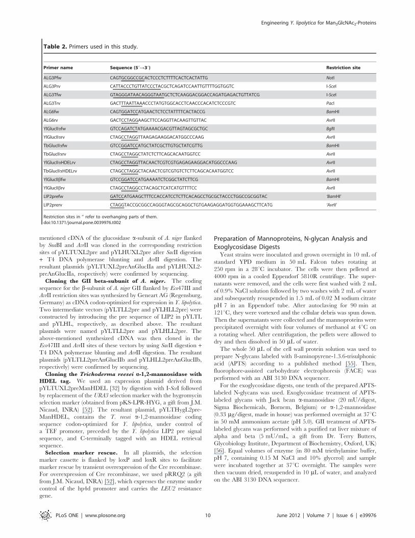
Table 2. Primers used in this study.
Primer name Sequence (59R39) Restriction site
ALG3Pfw CAGTGCGGCCGCACTCCCTCTTTTCACTCACTATTG NotI
ALG3Prv CATTACCCTGTTATCCCTACGCTCAGATCCAATTGTTTTGGTGGTC I-SceI
ALG3Tfw GTAGGGATAACAGGGTAATGCTCTCAAGGACGGACCAGATGAGACTGTTATCG I-SceI
ALG3Trv GACTTTAATTAAACCCTATGTGGCACCTCAACCCACATCTCCCGTC PacI
ALG6fw CAGTGGATCCATGAACTCTCCTATTTTCACTACCG BamHI
ALG6rv GACTCCTAGGAAGCTTCCAGGTTACAAGTTGTTAC AvrII
YlGlucIIafw GTCCAGATCTATGAAAACGACGTTAGTAGCGCTGC BglII
YlGlucIIarv CTAGCCTAGGTTAAGAGAAGGACATGGCCCAAG AvrII
TbGlucIIafw GTCCGGATCCATGCTATCGCTTGTGCTATCGTTG BamHI
TbGlucIIarv CTAGCCTAGGCTATCTCTTCAGCACAATGGTCC AvrII
YlGlucIIaHDELrv CTAGCCTAGGTTACAACTCGTCGTGAGAGAAGGACATGGCCCAAG AvrII
TbGlucIIaHDELrv CTAGCCTAGGCTACAACTCGTCGTGTCTCTTCAGCACAATGGTCC AvrII
YlGlucIIbfw GTCCGGATCCATGAAAATCTCGGCTATCTTCG BamHI
YlGlucIIbrv CTAGCCTAGGCCTACAGCTCATCATGTTTTCC AvrII
LIP2prefw GATCCATGAAGCTTTCCACCATCCTCTTCACAGCCTGCGCTACCCTGGCCGCGGTAC ‘BamHI’
LIP2prerv CTAGGTACCGCGGCCAGGGTAGCGCAGGCTGTGAAGAGGATGGTGGAAAGCTTCATG ‘AvrII’
Restriction sites in ‘’ refer to overhanging parts of them.doi:10.1371/journal.pone.0039976.t002
Engineering Y. lipolytica for Man3GlcNAc2-Proteins
PLoS ONE | www.plosone.org 10 June 2012 | Volume 7 | Issue 6 | e39976

PNGaseF Treatment of GlycoproteinsProteins in the Yarrowia culture medium were precipitated with
two volumes of ice-cold acetone. After incubation on ice for
20 min and centrifugation at 14,000 rpm for 5 min, the superna-
tant was removed and the protein pellet was resuspended in
100 mL of 50 mM Tris-HCl, pH 8. SDS and b-mercaptoethanol
were added to a final concentration of 0.5% and 1%, respectively.
Samples were incubated for 5 min at 100uC, after which G7 buffer
(106buffer, New England Biolabs), NP-40 (final concentration of
1%), complete protease inhibitor (Roche) and in-house produced
PNGaseF (15 IUBMB milliunits) were added. After overnight
incubation at 37uC, proteins were precipitated by the deoxycho-
late/trichloroacetic acid (DOC/TCA) procedure, resuspended in
26Laemmli buffer, and analyzed by SDS-PAGE.
In vitro Digestion with Trichoderma Harzianum MutanaseT. harzianum mutanase Novozyme 234, L1412 was obtained
from Sigma-Aldrich Corporation, Spruce St., St. Louis, MO,
USA. A stock solution of the enzyme (10 g/L) was prepared by
dissolving 40 mg in 4 mL of 5 mM NH4Ac pH5 buffer. Five serial
five-fold dilutions were made, and the final dilution (0.2 mL) wasused to treat 0.5 mL of APTS-labeled N-glycans in a total volume
of 10 mL buffered to a final concentration of 50 mM NH4Ac pH5.
This reaction mixture was incubated overnight at 37uC and
analyzed on an ABI 3130 DNA sequencer after desalting on
a Sephadex G10 column [55].
Acknowledgments
The authors thank Dr. Amin Bredan for the help in preparing the
manuscript. We also thank Dr. Jean-Marc Nicaud and Dr. Jean-Marie
Beckerich (CNRS-INRA) Laboratoire Microbiologie et Genetique Mole-
culaire INRA, France, for providing us with plasmids and strains.
Author Contributions
Conceived and designed the experiments: KDP PT SG WV NC.
Performed the experiments: KDP AVH. Analyzed the data: KDP PT
NC. Wrote the paper: KDP.
References
1. Kornfeld R, Kornfeld S (1985) Assembly of asparagine-linked oligosaccharides.
Annu Rev Biochem 54: 631–664.
2. Ellgaard L, Helenius A (2003) Quality control in the endoplasmic reticulum. Nat
Rev Mol Cell Biol 4: 181–191.
3. Nakayama K, Nagasu T, Shimma Y, Kuromitsu J, Jigami Y (1992) OCH1
encodes a novel membrane bound mannosyltransferase: outer chain elongation
of asparagine-linked oligosaccharides. EMBO J 11: 2511–2519.
4. Song Y, Choi MH, Park JN, Kim MW, Kim EJ, et al. (2007) Engineering of the
yeast Yarrowia lipolytica for the production of glycoproteins lacking the outer-chain
mannose residues of N-glycans. Appl Environ Microbiol 73: 4446–4454.
5. Bause E, Bieberich E, Rolfs A, Volker C, Schmidt B (1993) Molecular cloning
and primary structure of Man9-mannosidase from human kidney. Eur J Biochem
217: 535–540.
6. Tremblay LO, Campbell Dyke N, Herscovics A (1998) Molecular cloning,
chromosomal mapping and tissue-specific expression of a novel human a1,2-mannosidase gene involved in N-glycan maturation. Glycobiology 8: 585–595.
7. Tremblay LO, Herscovics A (2000) Characterization of a cDNA encoding
a novel human Golgi a1, 2-mannosidase (IC) involved in N-glycan biosynthesis.
J Biol Chem 275: 31655–31660.
8. De Pourcq K, De Schutter K, Callewaert N (2010) Engineering of glycosylation
in yeast and other fungi: current state and perspectives. Appl Microbiol
Biotechnol 87: 1617–1631.
9. Choi BK, Bobrowicz P, Davidson RC, Hamilton SR, Kung DH, et al. (2003)
Use of combinatorial genetic libraries to humanize N-linked glycosylation in the
yeast Pichia pastoris. Proc Natl Acad Sci USA 100: 5022–5027.
10. Jacobs PP, Geysens S, Vervecken W, Contreras R, Callewaert N (2009)
Engineering complex-type N-glycosylation in Pichia pastoris using GlycoSwitch
technology. Nat Protoc 4: 58–70.
11. Burda P, Jakob CA, Beinhauer J, Hegemann JH, Aebi M (1999) Ordered
assembly of the asymmetrically branched lipid-linked oligosaccharide in the
endoplasmic reticulum is ensured by the substrate specificity of the individual
glycosyltransferases. Glycobiology 9: 617–625.
12. Verostek MF, Atkinson PH, Trimble RB (1993) Glycoprotein biosynthesis in the
alg3 Saccharomyces cerevisiae mutant. I. Role of glucose in the initial glycosylation of
invertase in the endoplasmic reticulum. J Biol Chem 268: 12095–12103.
13. Aebi M, Gassenhuber J, Domdey H, te Heesen S (1996) Cloning and
characterization of the ALG3 gene of Saccharomyces cerevisiae. Glycobiology 6:
439–444.
14. Sharma CB, Knauer R, Lehle L (2001) Biosynthesis of lipid-linked oligosacchar-
ides in yeast: the ALG3 gene encodes the Dol-P-Man: Man5GlcNAc2-PP-Dol
mannosyltransferase. Biol Chem 382: 321–328.
15. Huffaker TC, Robbins PW (1983) Yeast mutants deficient in protein
glycosylation. Proc Natl Acad Sci U S A 80: 7466–7470.
16. Verostek MF, Atkinson PH, Trimble RB (1993) Glycoprotein biosynthesis in the
alg3 Saccharomyces cerevisiae mutant. II. Structure of novel Man6–10GlcNAc2processing intermediates on secreted invertase. J Biol Chem 268: 12104–12115.
17. Zufferey R, Knauer R, Burda P, Stagljar I, te Heesen S, et al. (1995) STT3,
a highly conserved protein required for yeast oligosaccharyl transferase activity in
vivo. EMBO J 14: 4949–4960.
18. Trimble RB, Verostek MF (1995) Glycoprotein oligosaccharide synthesis and
processing in Yeast. Trends Glycosci and Glycotechnol 7: 1–30.
19. Pignede G, Wang H, Fudalej F, Gaillardin C, Seman M, et al. (2000)
Characterization of an extracellular lipase encoded by LIP2 in Yarrowia lipolytica.
J Bacteriol 182: 2802–2810.
20. Jolivet P, Bordes F, Fudalej F, Cancino M, Vignaud C, et al. (2007) Analysis ofYarrowia lipolytica extracellular lipase Lip2p glycosylation. FEMS Yeast Res 7:
1317–1327.
21. Jones D, Mehlert A, Ferguson MA (2004) The N-glycan glucosidase system in
Trypanosoma brucei. Biochem Soc Trans 32: 766–768.
22. Jones DC, Mehlert A, Guther MLS, Ferguson MAJ (2005) Deletion of theglucosidase II gene in Trypanosoma brucei reveals novel N-glycosylation
mechanisms in the biosynthesis of variant surface glycoprotein. J Biol Chem280: 35929–35942.
23. Trombetta ES, Simons JF, Helenius A (1996) Endoplasmic reticulum
glucosidase II is composed of a catalytic subunit, conserved from yeast tomammals, and a tightly bound noncatalytic HDEL-containing subunit. J Biol
Chem 271: 27509–27516.
24. Henrissat B (1991) A classification of glycosyl hydrolases based on amino-acid
sequence similarities. Biochem J 280: 309–316.
25. D’Alessio C, Fernandez F, Trombetta ES, Parodi AJ (1999) Genetic evidence for
the heterodimeric structure of glucosidase II. The effect of disrupting the
subunit-encoding genes on glycoprotein folding. J Biol Chem 274: 25899–25905.
26. Pelletier MF, Marcil A, Sevigny G, Jakob CA, Tessier DC, et al. (2000) The
heterodimeric structure of glucosidase II is required for its activity, solubility, andlocalization in vivo. Glycobiology 10: 815–827.
27. Treml K, Meimaroglou D, Hentges A, Bause E (2000) The a- and b-subunitsare required for expression of catalytic activity in the hetero-dimeric glucosidaseII complex from human liver. Glycobiology 10: 493–502.
28. Watanabe T, Totani K, Matsuo I, Maruyama J, Kitamoto K, et al. (2009)Genetic analysis of glucosidase II b-subunit in trimming of high-mannose-type
glycans. Glycobiology 19: 834–840.
29. Stigliano ID, Caramelo JJ, Labriola CA, Parodi AJ, D’Alessio C (2009)Glucosidase II b subunit modulates N-glycan trimming in fission yeasts and
mammals. Mol Biol Cell 20: 3974–3984.
30. Kainz E, Gallmetzer A, Hatzl C, Nett JH, Li H, et al. (2008) N-glycan
modification in Aspergillus species. Appl Environ Microbiol 74: 1076–1086.
31. Callewaert N, Laroy W, Cadirgi H, Geysens S, Saelens X, et al. (2001) Use ofHDEL-tagged Trichoderma reesei mannosyl oligosaccharide 1,2-a-D-mannosidase
for N-glycan engineering in Pichia pastoris. FEBS Lett 503: 173–178.
32. De Pourcq K, Vervecken W, Dewerte I, Valevska A, Van Hecke A, et al. (2012)
Engineering the yeast Yarrowia lipolytica for the production of therapeutic proteinshomogeneously glycosylated with Man8GlcNAc2 and Man5GlcNAc2. Microb
Cell Fact 11: 53 doi:10.1186/1475–2859–11–53.
33. Madzak C, Gaillardin C, Beckerich JM (2004) Heterologous protein expressionand secretion in the non-conventional yeast Yarrowia lipolytica: a review.
J Biotechnol 109: 63–81.
34. Cipollo JF, Trimble RB (2000) The accumulation of Man6GlcNAc2-PP-dolichol
in the Saccharomyces cerevisiae Dalg9 mutant reveals a regulatory role for the Alg3p
a1,3-Man middle-arm addition in downstream oligosaccharide-lipid andglycoprotein glycan processing. J Biol Chem 275: 4267–4277.
35. Davidson RC, Nett JH, Renfer E, Li H, Stadheim TA, et al. (2004) Functionalanalysis of the ALG3 gene encoding the Dol-P-Man: Man5GlcNAc2-PP-Dol
mannosyltransferase enzyme of P. pastoris. Glycobiology 14: 399–407.
36. Verostek MF, Atkinson PH, Trimble RB (1991) Structure of Saccharomyces
cerevisiae alg3, sec18 mutant oligosaccharides. J Biol Chem 266: 5547–5551.
37. Henquet M, Lehle L, Schreuder M, Rouwendal G, Molthoff J, et al. (2008)Identification of the gene encoding the a1,3-mannosyltransferase (ALG3) in
Arabidopsis and characterization of downstream n-glycan processing. Plant Cell
20: 1652–1664.
Engineering Y. lipolytica for Man3GlcNAc2-Proteins
PLoS ONE | www.plosone.org 11 June 2012 | Volume 7 | Issue 6 | e39976

38. Oh DB, Park JS, Kim MW, Cheon SA, Kim EJ, et al. (2008) Glycoengineering
of the methylotrophic yeast Hansenula polymorpha for the production ofglycoproteins with trimannosyl core N-glycan by blocking core oligosaccharide
assembly. Biotechnol J 3: 659–668.
39. Grinna LS, Robbins PW (1980) Substrate specificities of rat liver microsomalglucosidases which process glycoproteins. J Biol Chem 255: 2255–2258.
40. Spiro MJ, Spiro RG, Bhoyroo VD (1979) Glycosylation of proteins byoligosaccharide-lipids. Studies on a thyroid enzyme involved in oligosaccharide
transfer and the role of glucose in this reaction. J Biol Chem 254: 7668–7674.
41. Spiro RG, Spiro MJ, Bhoyroo VD (1979) Processing of carbohydrate units ofglycoproteins. Characterization of a thyroid glucosidase. J Biol Chem 254:
7659–7667.42. Michael JM, Kornfeld S (1980) Partial purification and characterization of the
glucosidases involved in the processing of asparagine-linked oligosaccharides.Arch Biochem Biophys 199: 249–258.
43. Saunier B, Kilker RDJr, Tkacz JS, Quaroni A, Herscovics A (1982) Inhibition of
N-linked complex oligosaccharide formation by 1-deoxynojirimycin, an inhibitorof processing glucosidases. J Biol Chem 257: 14155–14161.
44. Totani K, Ihara Y, Matsuo I, Ito Y (2006) Substrate specificity analysis ofendoplasmic reticulum glucosidase II using synthetic high mannose-type glycans.
J Biol Chem 281: 31502–31508.
45. Munro S (2001) The MRH domain suggests a shared ancestry for the mannose6-phosphate receptors and other N-glycan-recognising proteins. Curr Biol 11:
R499–501.46. Wilkinson BM, Purswani J, Stirling CJ (2006) Yeast GTB1 encodes a subunit of
glucosidase II required for glycoprotein processing in the endoplasmic reticulum.J Biol Chem 281: 6325–6333.
47. Totani K, Ihara Y, Matsuo I, Ito Y (2008) Effects of macromolecular crowding
on glycoprotein processing enzymes. J Am Chem Soc 130: 2101–2107.
48. Quinn RP, Mahoney SJ, Wilkinson BM, Thornton DJ, Stirling CJ (2009) A
novel role for Gtb1p in glucose trimming of N-linked glycans. Glycobiology 19:
1408–1416.
49. Hu D, Kamiya Y, Totani K, Kamiya D, Kawasaki N, et al. (2009) Sugar-
binding activity of the MRH domain in the ER a-glucosidase II b subunit is
important for efficient glucose trimming. Glycobiology 19: 1127–1135.
50. Fickers P, Fudalej F, Le Dall MT, Casaregola S, Gaillardin C, et al. (2005)
Identification and characterisation of LIP7 and LIP8 genes encoding two
extracellular triacylglycerol lipases in the yeast Yarrowia lipolytica. Fungal Genet
Biol 42: 264–274.
51. Barth G, Gaillardin C (1997) Physiology and genetics of the dimorphic fungus
Yarrowia lipolytica. Fems Microbiol Rev 19: 219–237.
52. Fickers P, Le Dall MT, Gaillardin C, Thonart P, Nicaud JM (2003) New
disruption cassettes for rapid gene disruption and marker rescue in the yeast
Yarrowia lipolytica. J Microbiol Methods 55: 727–737.
53. Nicaud JM, Madzak C, van den Broek P, Gysler C, Duboc P, et al. (2002)
Protein expression and secretion in the yeast Yarrowia lipolytica. FEMS Yeast Res
2: 371–379.
54. Madzak C, Treton B, Blanchin-Roland S (2000) Strong hybrid promoters and
integrative expression/secretion vectors for quasi-constitutive expression of
heterologous proteins in the yeast Yarrowia lipolytica. J Mol Microbiol Biotechnol
2: 207–216.
55. Laroy W, Contreras R, Callewaert N (2006) Glycome mapping on DNA
sequencing equipment. Nat Protoc 1: 397–405.
56. Alonzi DS, Neville DCA, Lachmann RH, Dwek RA, Butters TD (2006)
Glucosylated free oligosaccharides are biomarkers of endoplasmic reticulum
alpha-glucosidase inhibition. Biochem J 409: 571–580.
Engineering Y. lipolytica for Man3GlcNAc2-Proteins
PLoS ONE | www.plosone.org 12 June 2012 | Volume 7 | Issue 6 | e39976
Copyright © 2022 FDOKUMEN