


Bahasa
Halaman
Hukum


Enhanced Interleukin-8 Release and Gene Expression in Macrophagesafter Exposure to Mycobacterium tuberculosis and Its ComponentsYihong Zhang, Mindy Broser, Henry Cohen, Marion Bodkin, Kevin Law, Joan Reibman, and William N. RomDivision of Pulmonary and Critical Care Medicine, Departments of Medicine and Environmental Medicine, and Chest Service,Bellevue Hospital Center, New York University Medical Center, New York 10016
Abstract
Mycobacterium tuberculosis infection is accompanied byacute and chronic inflammatory infiltrates associated withnecrotizing granulomas in lung tissue. The cellular infiltrateis characterized by inflammatory cells which include neutro-phils, lymphocytes, and macrophages. In animal and in vitromodels of mycobacterial infection, cytokines including tu-mor necrosis factor-c (TNF-a), interferon gamma (IFN-y), and interleukin-1lf (IL-1p) participate in granuloma-tous inflammation. We hypothesized that interleukin-8, apotent chemoattractant for neutrophils and lymphocytes,could be released by activated alveolar macrophages afterexposure to M. tuberculosis or its components and contributeto granulomatous lung inflammation. A quantitative immu-noassay revealed that IL-8 protein release was significantlyelevated in supernatants of macrophages and in lavage fluidobtained from patients with pulmonary tuberculosis com-pared to normal controls. In addition, Northern blots dem-onstrated striking up-regulation of IL-8 mRNA in macro-phages from these patients. M. tuberculosis and its cell wallcomponents lipoarabinomannan (LAM), lipomannan(LM), and phosphoinositolmannoside (PIM) stimulated IL-8 protein release and mRNA expression in vitro from alveo-lar macrophages, but deacylated LAM did not. Neutralizingantibodies to TNF-a and/or IL-1-a and P blocked 83% ofthe stimulation. IL-8 synthesis and release is an early re-sponse of macrophages after phagocytosis ofM. tuberculosis.Its production serves to attract both acute and chronic in-flammatory cells of active infection and thus participates inthe process of containment of the pathogen. (J. Clin. Invest.95:586-592.) Key words: tuberculosis * cytokines - Interleu-kin-8
Introduction
Tuberculosis is a major cause of morbidity and mortality withapproximately 25 million infectious cases reported throughoutthe world annually (1-3). The decline in tuberculosis in theU. S. has been reversed by a new tuberculosis epidemic fueled
Address correspondence to William N. Rom, MD, MPH, Division ofPulmonary and Critical Care Medicine, Bellevue Hospital Center, Rm7N24, New York University Medical Center, 550 First Avenue, NewYork, NY 10016. Phone: 212-263-6479; FAX: 212-263-8442.
Received for publication 24 March 1994 and in revised form 29August 1994.
by the crises of AIDS, intravenous drug use, homelessness, anddismantling of the public health infrastructure for tuberculosisprograms (4). To develop new strategies to treat and preventtuberculosis, we and others have focused on cytokine networksin the lung to understand more about the human host response(5-8). We have previously demonstrated that Mycobacteriumtuberculosis and its cell wall components stimulate both mRNAfor IL-1,6 and TNF-a and protein release. Transcription is en-hanced by the nuclear transcription factor, NF-1L6 (5, 9-10).These data are supported by numerous other investigations onthe release of these cytokines by M. tuberculosis, yet it is un-clear whether in vivo, enhancement or inhibition of select cyto-kines is propitious (11-17) for containment of the infection.Friedland and colleagues (18) reported that IL-8 was producedafter phagocytosis of M. tuberculosis by a monocytic cell line(THP-1 cells) and proposed that this cytokine was involved ingranuloma formation possibly by acting as a T cell chemoattrac-tant. We now demonstrate the in vivo release of IL-8 fromalveolar macrophages obtained from patients with active tuber-culosis and investigate the mechanism of this release.
IL-8 is a recently characterized cytokine that functions as achemotactic factor for neutrophils (27), T lymphocytes (28)and basophils (29). It belongs to a family of 8-kD polypeptides(30, 31 ). Because IL-8 is a chemoattractant for both neutrophilsand lymphocytes, it may modulate both acute and chronic in-flammation. Neutrophils are sequestered within the lung in gran-ulomatous as well as fibrotic interstitial lung diseases. Seques-tration may be induced by IL-8 as well as a number of otherchemoattractants including C5a (24), leukotriene B4 (25) andtransforming growth factor 3 (26). Granulomatous diseases arealso characterized by an abundance of lymphocytes, many ofwhich are found around the periphery of the granuloma. ThusIL-8, as both a neutrophil and lymphocyte chemoattractant, maybe a pivotal cytokine in the control of the inflammatory responseto M. tuberculosis. Therefore, overexpression of IL-8 during M.tuberculosis infection may be responsible for the neutrophil andlymphocyte infiltrations which lead to granuloma formation. Inaddition, overexpression of 11L-8 may activate inflammatorycells and thus contribute to the necrotic destruction of the lung.
IL-8 is expressed by many monocytes/macrophages (32),as well as endothelial cells (33), fibroblasts, keratinocytes, andlymphocytes (for review see reference 34). Most cell typesproduce little, if any, IL-8 constitutively (34). However, a widevariety of cells have been shown to produce a large amount ofIL-8 after stimulation with LPS, TNF-a, and IL-1 (33-35).We now demonstrate IL-8 production by alveolar macrophages(AMs)' derived from human patients with tuberculosis. Alveo-
1. Abbreviations used in this paper: AM, alveolar macrophage; HRP,horseradish peroxidase; Hsp, heat-shock protein; LAM, lipoarabinoman-nan; LM, lipomannan; PIM, phosphoinositolmannoside.
586 Zhang et al.
J. Clin. Invest.© The American Society for Clinical Investigation, Inc.0021-9738/95/02/0586/07 $2.00Volume 95, February 1995, 586-592

lar macrophages from patients infected with M. tuberculosis hadincreased spontaneous IL-8 release; this increase was associatedwith an alveolitis in the lower respiratory tract. We have alsodemonstrated that mycobacterial components lipoarabinoman-nan (LAM), phosphoinositolmannoside (PIM), and lipoman-nan (LM) (36) are potent inducers for IL-8 production fromalveolar macrophages and peripheral blood monocytes. A re-combinant heat-shock protein (Hsp 65kD) is also a weak stimu-lus for IL-8 production.
Methods
Study population. The clinical protocol was approved by the HumanSubjects Review Committees of New York University and BellevueHospital Center. There were 15 patients with pulmonary tuberculosisconfirmed by a positive culture for M. tuberculosis. The patients, 14males and I female, included 7 African-American, 3 Hispanic, and 5Asian individuals. There were 8 nonsmokers, and 7 current smokers.All TB patients were HIV-. Their mean age was 41±4 yr. There were5 normal volunteers with normal chest radiographs, pulmonary functiontests, and physical examinations. They included 4 males, 1 female; 4Caucasian, 1 Asian; 4 nonsmokers, 1 ex-smoker; mean age 28±5 yrand no HIV risk factors.
Materials. LPS was purchased from Sigma Chemical Co. (St. Louis,MO). Recombinant human TNF-a (specific activity, 4.8 X 10⁷ U/ml) was kindly supplied by Dr. M. Tsujimoto (Suntory Institute forBiomedical Research, Osaka, Japan); recombinant human IL-l/, (spe-cific activity 3 x 10⁷ U/mg) was kindly provided by Hoffman-LaRoche(Nutley, NJ). LAM, PIM, and LM deacylated LAM were gifts of P.Brennan (Colorado State University, Ft. Collins, CO). They were de-rived from a laboratory attenuated strain of Mycobacterium sp. (37).These reagents had been eluted through Detoxi-Gel columns using ster-ile, pyrogen-free water and stored in pyrogen-free vials that had removedany contaminating LPS. Only pyrogen-free water was used in reconstitu-tion of this material. M. bovis heat-shock protein 65 kD (recombinant)was kindly provided by R. Van der Zee (National Institute of PublicHealth and Environmental Protection, Bilthoren, The Netherlands).Evaluation of tuberculosis reagents and lavage fluid for the presence ofgram-negative bacterial endotoxin was done with the Amebocyte Lysateassay (E-toxate kit, Sigma Chemical Co.). Neutralizing polyclonal anti-bodies anti-IL-la, anti-IL-l/B, and anti-TNF-a (R & D Systems,Minneapolis, MN), and anti-IgG2 isotype control antibodies (CoulterImmunology, Hialeah, IL) were used at a concentration of 10 mg/ml.
Bronchoalveolar lavage. Bronchoalveolar lavage was performedwith a flexible fiberoptic bronchoscope with local Xylocaine anesthesia.Normal saline (6, 50-ml aliquots) was instilled and suctioned sequen-tially from two or three sites (including radiographically involved sites).The recovered fluid was filtered through sterile gauze. A total cell countwas done in a hematocytometer, and cell differentials performed oncytocentrifuge slides stained with Diff-Quick and 500 cells werecounted. Cell viability was determined by trypan blue exclusion, and inall cases, recovered cells were > 90% viable. AMs were placed in 175-cc plastic culture flasks in RPMI, allowed to adhere for 1 h, and washedthree times with RPMI to remove nonadherent cells. Adherent cellswere scraped off with a sterile rubber policeman, and placed in 24-welltissue culture dishes at 106 AM/ml under serum-free conditions for 24h. Viability remained > 90% after culture and > 98% of adherent cellswere macrophages.
Isolation ofhuman monocytes and cell culture. Mononuclear cells inbuffy coat were separated by centrifugation over lymphocyte separationmedium (Flow Laboratories, McLean, VA). The cells were washed,suspended in RPMI 1640 medium supplemented with 10% fetal bovineserum and seeded onto the plastic surface of a 175-cm2 flask for 2 h at37°C to let monocytes adhere. The flask was washed three times withRPMI 1640 to remove nonadherent cells and the monocyte-enrichedpopulation was detached by scraping with the aid of a rubber policeman.The isolated human peripheral monocytes were placed in 24-well plastic
tissue culture plates (Falcon) with a density of 106 cells/well. The cellswere stimulated with different reagents for 24 h. The culture supernatantwas then collected and frozen at -70'C.
THP-1 cells (myelomonocytic leukemia cell line) were obtainedfrom American Type Culture Collection (Rockville, MD) and culturedin a 175-cm2 flask in 10% fetal bovine serum until reaching a densityof 5 x 10⁵/ml. The cells were washed three times with RPM 1640 andcultured in 24-well plastic culture plates at a density of 106 cells/wellfor 24 h with various stimuli.
ELISA assay for IL-8. The assays were carried out with commer-cially available kits from R & D System (Minneapolis, MN). For theassay, the frozen supernatants were thawed out at room temperature andadded to wells of rigid flat-bottomed microliter plates coated with mu-rine monoclonal antibody to IL-8. After incubation of the samples,horseradish peroxidase (HRP)-conjugated antibody was added to thetest wells. After a second incubation, the excess of the HRP-conjugatedantibody was removed by washing. IL-8 was quantitated by a microtiterplate reader.
Isolation ofRNA and Northern blot analysis. Normal alveolar mac-rophages were treated with test reagents for 4 h, collected by centrifuga-tion and lysed by addition of 5.5 M guanidinium isothiocyanate buffer.Fresh alveolar macrophages from patients or controls were also pro-cessed directly for RNA extraction. Cytoplasmic RNA was isolatedthrough CsC12 gradient ultracentrifugation. An equal amount of theobtained RNA was fractionated by electrophoresis through a 1% agar-ose-6% formaldehyde denaturing gel and transferred onto a nitrocellu-lose filter (BA 85; Schleicher and Schuell, Inc., Keene, NH). The bakedfilter was prehybridized in a solution containing 50% formamide, 0.5%SDS, lOx Denhardt, 2.5% herring sperm DNA, and 4x SSPE at 420Cfor 6 h. For hybridization, the IL-8 cDNA (kindly provided by JanVilcek M.D. [New York University Medical Center]) was labeled with(alpha-32P) dCTP (specific activity 3,000 Ci/mmol from DuPont-NEN,Boston, MA) by the random-priming technique using the kit purchasedfrom Boehringer Mannheim (Indianapolis, IN). The hybridization wascarried out at 42°C overnight. The filter was then washed in a solutioncontaining 2x SSC and 0.5% SDS at room temperature for 20 minfollowed by O.1 X SSC plus 0.5% SDS at 65°C for 30 min. The filterwas exposed at -70°C for 1-3 d.
Statistics. Data were assessed for normal distribution and analyzedwith the Student's t test (unpaired) to compare mean IL-8 release andpercent neutrophils from patients versus controls. Data was expressed asmean±standard error of the mean. A P value of < 0.05 was consideredsignificant. When data were not normally distributed, nonparametricmethods were used including the Wilcoxon test for BAL parameters.
Results
Evaluation of bronchoalveolar lavage for cell profile and IL-8release. Both the cell concentration and cell differential differedbetween patients with active pulmonary tuberculosis and nor-mals. Patients with active tuberculosis showed a twofold in-crease in cellularity (641±165 X 103 cells/ml) versus normals(314±39 X 103 cells/ml). A subgroup of the tuberculosis pa-tients displayed increases in the percentage of lymphocytes orneutrophils (Fig. 1). Most of the patients with tuberculosis hada striking increase in neutrophils compared to controls (TBpatients 37±10% versus normals 1±1%, P < 0.05).
To determine whether alveolar macrophages spontaneouslyreleased more IL-8 in patients with tuberculosis, AMs (106/ml) were cultured for 24 h and supernatants analyzed for IL-8release. Alveolar macrophages from patients with tuberculosisreleased significantly more IL-8 into culture supernatants com-pared with normals (TB patients 87±15 ng/ 106 cells versusnormals 9±1 ng/106 cells, P < 0.01, Fig. 2 A). IL-8 wasalso increased in the BAL fluid recovered from patients withtuberculosis (2,510±692 pg/mg protein versus normals 549±81
IL-8 in Tuberculosis 587

0
88
00
00
000
Normals TuberculisLymphocytes
0
00~~ o500 2
01Ec.CL
-J0.
0 U
0 m
Normals TubercuosisNeutrophils
Figure 1. Percent cells recovered by bronchoalveolar lavage from pa-tients with active tuberculosis and normal individuals. (A) Normals;(o)TB patients.
0 500 1000 1500 2000 2500Neutrophils (x 10-3)
Figure 3. Linear association between IL-8 (pg/mg protein) in BALfluid and neutrophils/ml recovered. The association was significant (r2= 0.74, P < 0.01).
pg/mg protein, P = 0.06, Fig. 2 B). LPS contamination ofBAL fluid was negligible (14-50 pg/ml) and was not differentbetween normals and TB patients. The linear association be-tween the IL-8 in the BAL fluid and the neutrophils per milliliterrecovered in the lavage was significant (r2 = 0.74, P < 0.01,Fig. 3). In most TB patients there was a dramatic increase inthe percentage of neutrophils, and 11L-8 release and content inBAL fluid was significantly elevated consistent with the conceptthat 11L-8 was a chemotactic factor for neutrophils in TB. Sincealveolar macrophages were isolated by adherence to plastic andcontaminating neutrophils and lymphocytes were removed bywashing, and since adherent cells were confirmed to be AMs,the cell source of the 11L-8 was most likely the alveolar macro-phage.
To determine whether the increase in spontaneous IL-8 re-lease was controlled at the gene level, we evaluated 11L-8 mRNAexpression in fresh alveolar macrophages from two normal indi-viduals and three patients with TB who had > 80% macro-
phages in their BAL. Northern blot analysis revealed a singleband of 1.8 kb in AM from each individual. In accordancewith the measurement of IL-8 protein, alveolar macrophagesconsistently expressed increased mRNA for IL-8 in patientswith TB compared with normal controls (Fig. 4). Thus AMsfrom patients with tuberculosis secrete increased amounts ofIL-8, and this increase was determined in part, by enhancedmRNA expression.
Stimulation of IL-8 protein release and mRNA expressionfrom alveolar macrophages with M. tuberculosis and its cellwall components. To determine the specific mechanism bywhich IL-8 release was stimulated, we evaluated the ability ofM. tuberculosis H37Ra strain to stimulate the release of IL-8
1 2A. Macrophage Supematants B. BAL Fluid
00
0
0
0
0
19,
Normals Tuberuois
_10
xc 8
2Ca
6A
4-
79
20jt2.£
0
0
1.8 kb --
0
0
0
0
ceoA R9
Normals Tuberculosis
Figure 2. Spontaneous release of IL-8. (A) Macrophage supernatantsfrom patients with active tuberculosis and normal individuals. (B) BALfluid from patients with active TB and normals. (A) Normals; (0) TBpatients. The spontaneous release of IL-8 was evaluated using an ELISAassay. (n = 15, one patient with 98% neutrophils had insufficient macro-phages for assay.)
Figure 4. IL-8 steadystate mRNA levels in al-veolar macrophages fromnormal individuals or pa-tients with active tuber-
3 4 5 culosis. Alveolar macro-phages were collected byBAL from normal volun-teers or active tuberculo-sis patients (> 80% mac-rophages) and total RNAwas extracted. Northernblot analysis was carriedout using a 32P-labeledIL-8 cDNA probe and 30Mtg of total RNA wasplaced in each lane. Onthe bottom of each figureare ethidium bromidestained RNA gels whichdemonstrated equalamounts of RNA in eachlane. There is a strikingincrease in IL-8 mRNAexpression in TB patients(lanes 3-5) comparedwith normals (lanes1-2).
588 Zhang et al.
100
- AA
l A
Ia.8c
L
90
80
70
60
50
40
30
20
10Al-
A 00A629 AA6
Normanls TubercuosisMacrophages
I.E
~0-S
c
02IC

A. M. Tuberculols H37Ra Dose Response B. Uposrabinomannan Dose se
= $o
_ 600
x 400
3200
v0 10' 102 103 104 105 10 107 10' 0 0.01 0.1 1 10 1001000
M Tubewusls H37Rs (BacIllI/ml) Llpoarablnomennan H37Rs (ng/ml)
Figure 5. Dose-dependent stimulation of IL-8 production from alveolarmacrophages by M. tuberculosis H37Ra or LAM. Alveolar macrophagesfrom normal individuals (106/ml) were stimulated for 24 h. The culturesupernatant was collected and IL-8 release was measured by ELISA.(A) M. tuberculosis H37Ra. (B) LAM.
from alveolar macrophages from normal volunteers. Becauseof the similarity in function between M. tuberculosis H37Ra,and the cell wall component LAM derived from a laboratoryattenuated strain of Mycobacterium sp. (37), we also examinedthe effect of LAM on IL-8 production. M. tuberculosis andLAM (H37Ra) stimulated a dramatic release of IL-8 from nor-
mal alveolar macrophages (24 h) in a dose-dependent manner
(Fig. 5 A and B). Unstimulated AMs released 10-30 ng/106cells of IL-8. LAM at a dose of 10-100 ng/ml enhanced IL-8release to levels observed in BAL supernatants from patientswith pulmonary tuberculosis. The decreased potency of LAMalone for IL-8 release compared to the whole M. tuberculosisbacillus suggests that multiple components are capable of stimu-lating IL-8 release. We also performed experiments with theErdman strain which was a far weaker stimulus of IL-8 releasefrom AMs compared to H37Ra strain (10-fold less, data not
shown).To test whether other cell wall components of M. tuberculo-
sis also induced IL-8 release, we evaluated the following: PIM,LM, and deacylated LAM derived from Mycobacterium sp. andheat-shock protein 65 kD (Hsp-65kD). M. tuberculosis H37Ra,LAM, LM, and PIM were potent inducers of IL-8 release fromperipheral blood monocytes (Fig. 6 A) and THP-1 cells (Fig.6 B). There was a sixfold greater release of IL-8 from bloodmonocytes compared with THP-1 cells. LAM and PIM fromthe M. tuberculosis Erdman strain stimulated minimal releaseof IL-8. This is consistent with previous reports demonstratingthat extensive mannosyl capping is associated with less cytokinerelease from macrophages (38). The Hsp-65kD from M. bovisalso stimulated minimal release of IL-8. Last, deacylated LAMwas unable to stimulate IL-8 release, a finding consistent withprevious reports demonstrating the necessity of the phosphoino-sitol cell wall anchor of LAM to stimulate the release of cyto-kines (8, 38).
To determine whether IL-8 mRNA level was also increasedby M. tuberculosis and its components, we performed Northernblot analysis of normal AM. Whole M. tuberculosis and itscomponents LAM, PIM, and LM from H37Ra as well as Hsp-65kD stimulated a significant increase in IL-8 mRNA level inalveolar macrophages after 4 h of stimulation (Fig. 7).
To demonstrate that these results were not due to contamina-tion with LPS, all test reagents were evaluated for LPS contami-nation using the Limulus Amebocyte assay. Less than 10 pg/mg was found for each test reagent (LAM, LM, PIM, Hsp-
t 300
200
100-~~~~~~~~~2-c
.
Figure 6. Stimulation of IL-8 production from peripheral blood mono-
cytes (A) and THP-1 cells (B) with various stimuli. Cells (106/ml)were stimulated with different stimuli: M. tuberculosis (105/ml), LAMH37Ra (100 ng/ml), LAM Erdman (100 ng/ml), PIM H37Ra (100ng/ml), LM (100 ng/ml), Hsp-65kD (1 ,ug/ml), and Deacyl LAM(100 ng/ml) for 24 h.
65kD) in all lots tested. To further guard against LPS contami-nation, the experiments in Figs. 5 and 6 were performed in thepresence of polymyxin B 10-100 mg/ml with no significantalteration of the results (data not shown). These doses of poly-myxin B completely blocked the stimulatory effect of LPS on
IL-8 (14).Effect ofanti-cytokine antibodies on IL-8 release stimulated
by M. tuberculosis. We (6) and others (7-8, 14-15) havepreviously described that LAM, PIM, LM, or Hsp-65kD stimu-
.(Z3
Q
C) -Jo-
n
(I
-i
1.8 kb-*^-
Figure 7. Northern anal-ysis of IL-8 steady-statemRNA from alveolarmacrophages after stimu-lation. Alveolar macro-
phages from normal indi-viduals were stimulatedwith M. tuberculosis( 105/ml), LAM H37Ra(100 ng/ml), PIMH37Ra (100 ng/ml), LMH37Ra (100 ng/ml), or
Hsp 65kD (1 qtg/ml) for4 h. RNA was extracted,30 ,g was placed in eachlane, and Northern blotanalysis was carried outas described in Methods.On the bottom of eachfigure are ethidium bro-mide stained RNA gelswhich demonstratedequal amounts ofRNA ineach lane.
IL-8 in Tuberculosis 589
200
ISO -
1ooso-
10I .,.......
B. THP- I CellsA. Peripheral Blood Monocytes
I.
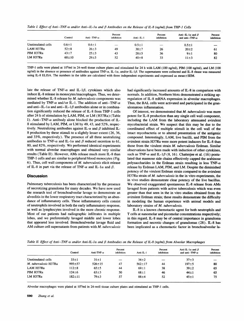
Table L Effect ofAnti-TNF-a and/or Anti-IL-Ia and /3 Antibodies on the Release of IL-8 (ng/ml) from THP-I Cells
Percent Percent Anti-IL-a and B PercentControl Anti-TNF-a inhibition Anti-IL-I inhibition and anti-TNF-a inhibition
Unstimulated cells 0.6±1 0.6+1 - 0.5±1 0.5±1LAM H37Ra 52±8 26±3 49 38±7 26 20±2 61PIM H37Ra 43±7 25±3 43 28±5 36 9±1 80LM H37Ra 60±10 29±5 52 40±8 33 11±3 82
THP-1 cells were plated at 106/ml in 24-well tissue culture plates and stimulated for 24 h with LAM (100 ng/ml), PIM (100 ng/ml), and LM (100ng/ml) in the absence or presence of antibodies against TNF-a, IL-la, and/or IL-1Ip. The supernatants were collected and IL-8 rlease was measuredusing IL-8 ELISA. The numbers in the table are calculated with three independent experiments and expressed as mean±SEM.
late the release of TNF-a and IL-1I3, cytokines which alsoinduce IL-8 release in monocytes/macrophages. Thus, we deter-mined whether IL-8 release by M. tuberculosis components wasmediated by TNF-a and/or IL-1. The addition of anti-TNF-aand anti-IL-la and anti-IL-l:3 antibodies alone or in combina-tion significantly reduced the release of IL-8 from THP-1 cellsafter 24 h of stimulation by LAM, PIM, or LM (H37Ra) (TableI). Anti-TNF-a antibody alone blocked the production of IL-8 stimulated by LAM, PIM, or LM by 49, 43, and 52%, respec-tively. Neutralizing antibodies against IL-a and ,/ inhibited IL-8 production by these stimuli to a slightly lesser extent (26, 36,and 33%, respectively). The presence of all three neutralizingantibodies to TNF-a and IL-la and /3 reduced secretion to 61,80, and 82%, respectively. We performed identical experimentswith normal alveolar macrophages and obtained very similarresults (Table II). However, AMs release much more IL-8 thanTHP- l cells and are similar to peripheral blood monocytes (Fig.6). Thus, cell wall components of M. tuberculosis elicit releaseof IL-8 in part via the release of TNF-a and IL-la and /3.
Discussion
Pulmonary tuberculosis has been characterized by the presenceof necrotizing granuloma for many decades. We have now usedthe research tool of bronchoalveolar lavage to demonstrate analveolitis in the lower respiratory tract characterized by an abun-dance of inflammatory cells. These inflammatory cells consistof neutrophils involved in both the early inflammatory response,as well as lymphocytes involved in the more chronic response.Most of our patients had radiographic infiltrates in multiplelobes, and we preferentially lavaged middle and lower lobesthat appeared less involved. Bronchoalveolar lavage fluid andAM culture cell supernatants from patients with M. tuberculosis
had significantly increased amounts of IL-8 in comparison withnormals. In addition, Northern blots demonstrated a striking up-regulation of IL-8 mRNA expression in alveolar macrophages.Thus, the BAL cells were activated and participated in the gran-ulomatous inflammation.
Of interest, we demonstrated that M. tuberculosis was morepotent for IL-8 production than any single cell wall component,including the LAM from the laboratory attenuated avirulentmycobacterial strain. We suspect that this may be due to thecoordinated effect of multiple stimuli in the cell wall of theintact mycobacteria or to altered presentation of the antigeniccompound. Interestingly, LAM, live bacilli, and PIM from theavirulent strain H37Ra were more potent inducers for IL-8 thanthose from the virulent strain M. tuberculosis Erdman. Similarobservations have been made with induction of other cytokines,such as TNF-a and IL-l1/ (8, 16). Chatterjee et al. (16) postu-lated that mannose side chains effectively capped the arabinosepolysaccharides in the Erdman strain resulting in less TNF-arelease by Erdman LAM, PIM, and LM. Despite the diminishedpotency of the virulent Erdman strain compared to the avirulentH37Ra strain of M. tuberculosis in the in vitro experiments, thein vivo studies demonstrate clear potency of the live bacillus.We observed exaggerated spontaneous IL-8 release from AMslavaged from patients with active tuberculosis which was evengreater than that seen in the in vitro studies obtained from theavirulent Erdman strain; these results demonstrate the difficultyin modeling the human experience with animal models andlaboratory strains of M. tuberculosis.
IL-8 is a known chemotactic agent for both neutrophils andT cells at nanomolar and picomolar concentrations respectively;in this regard, IL-8 may be of central importance in granulomaformation and necrotic changes of granulomas (28). IL-8 hasbeen implicated as a chemotactic factor in bronchoalveolar la-
Table II. Effect of Anti-TNF-a and/or Anti-IL-la and /8 Antibodies on the Release of IL-8 (ng/ml) from Alveolar Macrophages
Percent Percent Anti-IL-la and (3 PercentControl Anti-TNF-a inhibition Anti-IL-I inhibition and anti-TNF-a inhibition
Unstimulated cells 33±1 31±1 34±2 37±3M. tuberculosis H37Ra 995±57 526±15 47 562±17 44 197±5 80LAM H37Ra 112±8 65±5 44 69±1 38 39±2 65PIM H37Ra 124±6 63±3 50 68±1 46 44±1 65LM H37Ra 182±11 79±3 57 88±4 52 45±1 75
Alveolar macrophages were plated at 106/ml in 24-well tissue culture plates and stimulated as THP-1 cells.
590 Zhang et al.

vage fluid or supernatants in a variety of inflammatory andinterstitial lung diseases ranging from idiopathic pulmonary fi-brosis (22, 23), pneumocystis carinii pneumonia (40), nonspe-cific interstitial pneumonitis associated with HIV infection(40), and inorganic dusts. IL-1 and TNF-a both stimulate IL-8 release and correlate with increased BAL neutrophils (41).These two cytokines have also been implicated as stimuli forIL-8 release from mononuclear phagocyte and bronchial epithe-lial cells (42, 43). Not only can bronchial epithelial cells releaseIL-8, but McCain et al have shown that neutrophils can synthe-size and release IL-8 after stimulation by leukotriene B4 (44).IL-1, TNF-a, and PMA activate the IL-8 gene rapidly and di-rectly in the absence of new protein synthesis probably by acommon serine kinase (41). Thus several cytokines and celltypes contribute to IL-8 release in the locally inflamed lung.
M. tuberculosis has previously been demonstrated to elicitthe release of IL-8. Friedland et al. demonstrated a 10-foldgreater release of IL-8 by THP-1 cells following phagocytosisof M. tuberculosis than after LPS ( 18). Whereas these investi-gators observed IL-8 mRNA accumulated gradually over 24 hin THP-1 cells, they found that secretion was identical usingvirulent or avirulent strains. Using a monoclonal antibody toTNF-a, they also failed to demonstrate a change in IL-8 release.We have now demonstrated that both AM and PBM release IL-8 in response to M. tuberculosis and Mycobacterium sp. cellwall components. In contrast to Friedland et al., our polyclonalantibody elicited a 47-57% decrease in IL-8 release. Thus AMand PBM release IL-8 in a paracrine/autocrine manner in re-sponse to M. tuberculosis. Subcomponents LAM, LM, and PIM,had similar stimulatory capabilities, but removing the phospho-inositol anchor in the cell wall by deacylation abrogated theseactivities. We found AM released more IL-8 than PBM in re-sponse to live M. tuberculosis but similar amounts of IL-8 werereleased in response to using cell wall components. Both AMand PBM released approximately sixfold more IL-8 than THP-1 cells with each of the stimuli. The difference in the responseto LAM between THP-1 cells and AM or PBM may be dueto culture differences; THP- 1 cells are cultured in suspensionwhereas AM and PBM are adherent. In addition, cell lines maybe less responsive than primary cultures.
Transcripts for mRNA from two receptors for IL-8 havebeen described in PMN (45-47) and IL-8 binds to two proteins(p70 and p44) with high affinity. Thus the enhanced presenceof IL-8 in response to M. tuberculosis may function to recruitand activate neutrophils at sites of pulmonary infections. Jonesand colleagues have demonstrated that neutrophils kill M. tuber-culosis (48). The toxic effect is mediated by nonoxidativemeans because neither free radical inhibitors (catalase, superox-ide dismutase) nor deferoxamine impaired killing (48). More-over, neutrophils from a patient with chronic granulomatousdisease with defective NADPH pathway, were able to kill M.tuberculosis (48). M. tuberculosis is also capable of producingsuperoxide dismutase (the 23-kD antigen), and catalase (49).The 23-kD superoxide dismutase is the major protein releasedby M. tuberculosis in logarithmic growth constituting a veryimportant factor for virulence (49). In addition, LAM is a po-tent scavenger of oxygen radicals (50). Although neutrophilnonoxidative mechanisms including defensins and bactericidalpermeability increasing protein may contribute to host defense,they may be inadequate to control infection of mycobacteria.Neutrophils in an animal model of pleural TB release chemotax-
ins for monocytes that may then participate in granuloma forma-tion (51).
In contrast to neutrophils, lymphocytes express only a singlemRNA transcript for IL-8R2 receptor (46) and its expressionis less than that in neutrophils. In addition, only a subpopulationof lymphocytes respond to IL-8. These lymphocytes requireprior activation and are enriched for CD45RO (47). Indeed,preincubation of lymphocytes to purified protein derivative ofM. tuberculosis results in a phenotypically distinct populationof lymphocytes with an enhanced response to IL-8 (52).
IL-8 may also mediate the healing of granuloma. In a man-ner similar to basic and acidic fibroblast growth factor, IL-8 isangiogenic and also binds to heparin (53). Thus IL-8, like TNF-a, may participate in the angiogenesis observed in the healingprocess, particularly that seen at the margin of tuberculosiscavities.
We propose that the release of IL-8, as well as that of TNF-a and IL-1,6 from alveolar macrophages is rapid upon ingestionof the M. tuberculosis. These cytokines form an autocrine regu-latory loop. Although the neutrophils are rapidly recruited, oxi-dant and nonoxidant mechanisms of killing may be over-whelmed by M. tuberculosis, LAM and scavenging enzymes.These data demonstrate the enhanced presence of IL-8 in pulmo-nary tuberculosis in humans. The potent effect of IL-8 on bothneutrophils and lymphocytes make this a pivotal cytokine.
Acknowledgments
The authors thank Natalie Little for editorial assistance.This work was supported by grants M0100096 (General Clinical
Research Center) from National Institutes of Health (NIH), Aaron Dia-mond Foundation, NIH grants AI-35233, HL-51494, HL-51631, andNIOSH Cooperative Agreement U60/CCU206153.
References
1. Elliot, A. M., N. Luo, G. Tembo, B. Halwiindi, G. Steenbergen, L. Machiels,J. Pobee, P. Nunn, R. J. Hayes, and K. P. McAdam. 1990. Impact of HIV onTuberculosis in Zambia: a cross sectional study. Br. Med. J. 301:412-415.
2. Barnes, P. F., A. B. Bloch, P. T. Davidson, and D. E. Snider. 1991.Tuberculosis in patients with human immunodeficiency virus infection. N. Engl.J. Med. 324:1644-1650.
3. Bloom, B. R., and C. J. L. Murray. 1992. Tuberculosis: commentary on areemergent killer. Science (Wash. DC). 257:1055-1064.
4. Rom, W. N., and Y. Zhang. 1993. The rising tide of tuberculosis and thehuman host response to Mycobacterium tuberculosis. J. Lab. Clin. Med. 121:737-741.
5. Neville, K., A. Bromberg, R. Bromberg, S. Bonk, B. A. Hanna, and W. N.Rom. 1994. The third epidemic--multidrug resistant tuberculosis. Chest. 105:45-48.
6. Zhang, Y., M. Doerfler, T. C. Lee, B. Guillemin, and W. N. Rom. 1993.Mechanisms of stimulation of interleukin-1,1 and tumor necrosis factor-a byMycobacterium tuberculosis components. J. Clin. Invest. 91:2076-2083.
7. Barnes, P. F., S.-J. Fong, P. J. Brennan, P. E. Twomey, A. Mazumder, and R.Modlin. 1990. Local production of tumor necrosis factor and IFN-y in tuberculouspleuritis. J. Immunol. 145:149-154.
8. Barnes, P. F., D. Chatterjee, J. S. Abrams, S. Lu, E. Wang, M. Yamamura,P. J. Brennan, and R. L. Modlin. 1992. Cytokine production induced by Mycobac-terium tuberculosis lipoarabinomannan. J. Immunol. 149:541-547.
9. Kindler, V., A.-P. Sappino, G. E. Grau, P.-F. Piguet, and P. Vassalli. 1989.The inducing role of tumor necrosis factor in the development of bactericidalgranulomas during BCG infection. Cell. 56:731-740.
10. Zhang, Y., and W. N. Rom. 1993. Regulation of the interleukin-1,/ (IL-1/3) gene by mycobacterial components and lipopolysaccharide is mediated bytwo nuclear factor-1L6 motifs. Mol. Cell. Biol. 13:3831-3837.
11. Takashima, T., C. Ueta, I. Tsuyuguch, and S. Kishimoto. 1990. Productionof tumor necrosis factor alpha by monocytes from patients with pulmonary tuber-culosis. Infect. Immun. 58:3286-3292.
12. Moreno, C., J. Taverne, A. Mehlert, C. A. W. Bate, R. J. Brealey, A.Meager, G. A. W. Rook, and J. H. L. Playfair. 1989. Lipoarabinomannan from
IL-8 in Tuberculosis 591

M. tuberculosis induces the production of tumour necrosis factor from humanand murine macrophages. Clin. Exp. Immunol. 76:240-245.
13. Chensue, S. W., M. P. Davey, D. G. Resnick, and S. L. Kunkel. 1986.Release of interleukin-l by peripheral blood mononuclear cells in patients withtuberculosis and active inflammation. Infect. Immun. S2:341-343.
14. Wallis, R. S., H. Fujiwara, and J. J. Ellner. 1986. Direct stimulation ofmonocyte release of interleukin-1 by mycobacterial protein antigens. J. Immunol.136: 193-196.
15. Wallis, R. S., M. Amir-Tahmasseb, and J. J. Ellner. 1990. Induction ofinterleukin-l and tumor necrosis factor by mycobacterial proteins: the monocyteWestern blot. Proc. Natl. Acad. Sci. USA. 87:3348-3352.
16. Chatterjee, D., A. D. Roberts, K. Lowell, P. J. Brennan, and I. M. Orme.1992. Structural basis of capacity of lipoarabinomannan to induce secretion oftumor necrosis factor. Infect. Immun. 60:1249-1253.
17. Cadranel, J., C. Philippe, J. Perez, B. Milleron, G. Akoun, R. Ardaillon,and L. Band. 1990. In vitro production of tumor necrosis factor and prostaglandinE2 by peripheral blood mononuclear cells from tuberculosis patients. Clin. Exp.Inmunol. 81:319-324.
18. Friedland, J. S., D. G. Remick, R. Shattock, and G. E. Griffin. 1992.Secretion of interleukin-8 following phagocytosis of Mycobacterium tuberculosisby human monocyte cell lines. Eur. J. Immunol. 22:1373-1378.
19. Weiland, J. E., W. B. Davis, J. F. Holter, J. R. Mohammed, P. M. Dorinsky,and J. E. Gadek. 1986. Lung neutrophils in the adult respiratory distress syndrome.Clinical and pathophysiologic significance. Am. Rev. Respir. Dis. 133:218-225.
20. Hunninghake, G. W., K. C. Garrett, H. B. Richerson, J. C. Fantone, P. A.Ward, S. I. Rennard, P. B. Bitterman, and R. G. Crystal. 1984. Pathogenesis ofthe granulomatous lung diseases. Am. Rev. Respir. Dis. 130:476-496.
21. Hunninghake, G. W., J. E. Gadek, T. J. Lawley, and R. G. Crystal. 1981.Mechanisms of neutrophil accumulation in the lungs of patients with idiopathicpulmonary fibrosis. J. Clin. Invest. 68:259-269.
22. Carfe, P., R. L. Mortenson, T. E. King, P. W. Noble , C. L. Sable, andD. W. H. Riches. 1991. Increased expression of the interleukin-8 gene by alveolarmacrophages in idiopathic pulmonary fibrosis. J. Clin. Invest. 88:1802-1810.
23. Lynch, J. P., T. J. Standiford, M. W. Rolfe, S. L. Kunkel, and R. M.Strieter. 1992. Neutrophilic alveolitis in idiopathic pulmonary fibrosis. The roleof interleukin-8. Am. Rev. Respir. Dis. 145:1433-1439.
24. Fernandez, H. N., P. M. Henson, A. Otani, and T. E. I. Hugh. 1978.Chemotactic response to human C3a and C5a anaphylatoxins. I. Evaluation ofC3a and C5a leukotaxis in vitro and under stimulated in vivo conditions. J.Immunol. 120:109- 115.
25. Samuelsson, B. 1983. Leukotrienes: mediators of immediate hypersensitiv-ity reactions and inflammation. Science (Wash. DC). 220:568-575.
26. Reibman, J., S. Meixler, T. C. Lee, L. I. Gold, B. N. Cronstein, K. A.Haines, S. L. Kolasinski, and G. Weissmann. 1991. Transforming growth factorbl, a potent chemoattractant for human neutrophils, bypasses classic signal-trans-duction pathways. Proc. Natl. Acad. Sci. USA. 88:6805-6809.
27. Baggiolini, M., A. Walz, and S. L. Kunkel. 1989. Neutrophil-activatingpeptide-1 /interleukin 8, a novel cytokine that activates neutrophils. J. Clin. Invest.84:1045-1049.
28. Larsen, C. A., A. 0. Anderson, E. Appella, J. J. Oppenheim, and K.Matsushima. 1989. The neutrophil-activating protein (NAP-i ) is also chemotacticfor T lymphocytes. Science (Wash. DC). 243:1464-1466.
29. Leonard, E. J., A. Skeel, T. Yoshimura, K. Noer, S. Kutvirt, and D. VanEpps. 1990. Leukocyte specificity and binding of human neutrophil attractant/activating protein-i. J. Immunol. 144:1323-1330.
30. Oppenheim, J. J., C. O. C. Zachariae, N. Mukaida, and K. Matsushima.1991. Properties of the novel proinflammatory supergene 'intercrine' cytokinefamily. Ann. Rev. Immunol. 9:617-648.
31. Wolpe, S.D. and A. Cerami. 1989. Macrophage inflammatory proteins 1and 2: members of a novel superfamily of cytokines. FASEB J. 3:2565-2573.
32. Yoshimura, T., K. Matsushima, S. Tanaka, E.A. Robinson, E. Appella,J. J. Oppenheim, and E. J. Leonard. 1987. Purification of a human monocyte-derived neutrophil chemotactic factor that has peptide sequence similarity to otherhost defense cytokines. Proc. Natl. Acad. Sci. USA. 84:9233-9237.
33. Strieter, R. M., S. L. Kunkel, H. J. Showell, D. G. Remick, S. H. Phan,P. A. Ward, and R. M. Marks. 1989. Endothelial cell gene expression of a
neutrophil chemotactic factor by TNF alpha, LPS, and IL-I beta. Science (Wash.DC). 243:1467-1469.
34. Matsushima, K., and J. J. Oppenheim. 1989. Interleukin 8 and MCAF:novel inflammatory cytokines inducible by IL-1 and TNF. Cytokine. 1:2-13.
35. Strieter, R. M., S. W. Chensue, M. A. Basha, T. J. Standiford, J. P. Lynch,M. Baggiolini, and S. L. Kunkel. 1990. Human alveolar gene expression ofinterleukin-8 by tumor necrosis factor alpha, lipopolysaccharide and interleukin1 beta. Am J. Respir. Cell. Mol. Biol. 2:321-326.
36. Brennan, P. J., S. W. Hunter, M. McNeil, D. Chatterjee, and M. Daffe.1990. Reappraisal of the chemistry of mycobacterial cell walls, with a view tounderstanding the roles of individual entities in disease processes. In MicrobialDeterminants of Virulence and Host Response. E. M. Ayoub, G. H. Cassell,W. C. Branche, Jr., and T. J. Henry, editors. American Society for Microbiology,Washington, DC. 55-75.
37. Prinzis, S., D. Chatterdee, P. J. Brennan. 1993. Structure and antigenicityof lipoarabinomannan from Mycobacterium bovis BCG. J. Gen. Microbiol.139:2649-2658.
38. Chatterjee, D., A. D. Roberts, K. Lowell, P. J. Brennan, and I. M. Orme.1992. Structural basis of capacity of lipoarabinomannan to induce secretion oftumor necrosis factor. Infect. Immun. 60:1249-1253.
39. Wright, S., R. Ramos, P. Tobias, R. Ulevitch, and J. Mathison. 1990.CD14, a receptor for complexes of lipopolysaccharides (LPS) and LPS bindingprotein. Science (Wash. DC). 249:1431-1433.
40. Lipschik, G. Y., M. E. Doerfler, J. A. Kovacs, W. D. Travis, M. G.Lawrence, J. R. Dichter, F. P. Ognibene, and J. H. Shelhamer. 1993. LeukotrieneB4 and interleukin 8 in AIDS-related pulmonary disease. Chest. 104:763-769.
41. Matsushima, K., E.T. Baldwin, and N. Mukaida. 1992. Interleukin-8 andMCAF: novel leukocyte recruitment and activating cytokines. In Interleukins:Molecular Biology and Immunology. T. Kishimoto, editor. Chem. Immunol. Ba-sel, Karger. 51:236-265.
42. DeForge, L. E., J. S. Kenney , M. L. Jones , J. S. Warren, and D. G.Remick. 1992. Biphasic production of IL-8 in lipopolysaccharide (LPS)-stimu-lated human whole blood. J. Immunol. 148:2133-2141.
43. Standiford, T. J., S. L. Kunkel, M. A. Basha, S. W. Chensue, J. P. Lynch,HI, G. B. Toews, J. Westwick, and R. M. Strieter. 1990. Interleukin-8 geneexpression by a pulmonary epithelial cell line. J. Clin. Invest. 86:1945-1953.
44. McCain, R. W., E. P. Holden,T. R. Blackwell, and J. W. Christman. 1994.Leukotriene B4 stimulates human polymorphonuclear leukocytes to synthesizeand release interleukin-8 in vitro. Am. J. Respir. Cell. Mol. Biol. 10:651-657.
45. Bazzoni, F., M. A. Cassatella, F. Rossi, M. Ceska, B. Dewald, and M.Baggiolini. 1991. Phagocytosing neutrophils produce and release high amountsof the neutrophil-activating peptide 1/interleukin 8. J. Exp. Med. 173:771-774.
46. Wilkinson, P. C. 1992. Identification of IL-8 as a locomotor attractant foractivated human lymphocytes in mononuclear cell cultures with anti-CD3 orpurified protein derivative Mycobacterium tuberculosis. J. Immunol. 149(8):2689-2694.
47. Moser, B., L. Barella, S, Mattei, C. Schumacher, F. Boulay, M. P. Co-lombo, and M. Baggiolini. 1993. Expression of transcripts for two interleukin 8receptors in human phagocytes, lymphocytes and melanoma cells. Biochem. J.294:285-292.
48. Jones, G. S., H. S. Amirault, and B. R. Anderson. 1990. Killing of Myco-bacterium tuberculosis by neutrophils. A nonoxidative process. J. Infect. Dis.162:700-704.
49. Zhang, Y., R. Lathigra, T. Garbe, D. Cathy, and D. Young. 1991. Geneticanalysis of superoxide dismutase, the 23 kilodalton antigen of Mycobacteriumtuberculosis. Mol. Mircobiol. 5(2):381-391.
50. Chan, J., X. Fan, S. W. Hunter, P. J. Brennan, and B. R. Bloom. 1991.Lipoarabinomannan, a possible virulence factor involved in persistence of Myco-bacterium tuberculosis. Infect. Irmuun. 59:1755-1761.
51. Antony, V. B., S. A. Sahn, A. C. Antony, and J. E. Repine. 1985. BacillusCalmette-Guerin-stimulated neutrophils release chemotaxins for monocytes inrabbit pleural spaces and in vitro. J. Clin. Invest. 76:1514-1521.
52. Barnes, P. F., S. D. Mistry, C. L. Cooper, C. Pirmez, T. H. Rea, and R. L.Modlin. 1989. Compartmentalization of a CD4+ T lymphocyte subpopulation intuberculous pleuritis. J. Immunol. 142(4):1114-1119.
53. Koch, A. E., P. J. Polverini, S. L. Kunkel, L. A. Harlow, L. A. DiPietro,V. M. Elner, S. G. Elner, and R. M. Strieter. 1992. Interleukin-8 as a macrophage-derived mediator of angiogenesis. Science (Wash. DC). 258:1798-1801.
592 Zhang et al.
Top Related
Copyright © 2022 FDOKUMEN

