


Bahasa
Halaman
Hukum

RSC Advances
PAPER
Publ
ishe
d on
05
Febr
uary
201
5. D
ownl
oade
d by
Ind
ian
Inst
itute
of
Scie
nce
on 1
1/02
/201
5 15
:56:
56.
View Article OnlineView Journal | View Issue
Effect of solvent
aCrystal Growth Centre, Anna University, Ch
[email protected]; [email protected] of Materials Engineering, India
Karnataka, IndiacCentre for Biotechnology, Anna University,dDepartment of Chemistry, Anna University,
Cite this: RSC Adv., 2015, 5, 18301
Received 15th November 2014Accepted 5th February 2015
DOI: 10.1039/c4ra14584d
www.rsc.org/advances
This journal is © The Royal Society of C
; enhancing the wettability andengineering the porous structure of a calciumphosphate/agarose composite for drug delivery
Elayaraja Kolanthai,ab V. Sivaraj Dikeshwar Colon,c P. Abinaya Sindu,c
V. Sarath Chandra,a K. R. Karthikeyan,a M. Surendar Babu,c S. Meenakshi Sundaram,c
M. Palanichamyd and S. Narayana Kalkura*a
Tissue engineering deals with the regeneration of tissues for bone repair, wound healing, drug delivery, etc.,
and a highly porous 3D artificial scaffold is required to accommodate the cells and direct their growth. We
prepared 3D porous calcium phosphate ((hydroxyapatite/b-tricalcium phosphate)/agarose, (HAp/b-TCP)/
agarose) composite scaffolds by sol–gel technique with water (WBS) and ethanol (EBS) as solvents. The
crystalline phases of HAp and b-TCP in the scaffolds were confirmed by X-ray diffraction (XRD) analysis.
The EBS had reduced crystallinity and crystallite size compared to WBS. WBS and EBS revealed
interconnected pores of 1 mm and 100 nm, respectively. The swelling ratio was higher for EBS in water
and phosphate buffered saline (PBS). An in vitro drug loading/release experiment was carried out on the
scaffolds using gentamicin sulphate (GS) and amoxicillin (AMX). We observed initial burst release followed
by sustained release from WBS and EBS. In addition, GS showed more extended release than AMX from
both the scaffolds. GS and AMX loaded scaffolds showed greater efficacy against Pseudomonas than
Bacillus species. WBS exhibited enhanced mechanical properties, wettability, drug loading and
haemocompatibility compared to EBS. In vitro cell studies showed that over the scaffolds, MC3T3 cells
attached and proliferated and there was a significant increase in live MC3T3 cells. Both scaffolds
supported MC3T3 proliferation and mineralization in the absence of osteogenic differentiation
supplements in media which proves the scaffolds are osteoconducive. Microporous scaffolds (WBS)
could assist the bone in-growth, whereas the presence of nanopores (EBS) could enhance the
degradation process. Hence, WBS and EBS could be used as scaffolds for tissue engineering and drug
delivery. This is a cost effective technique to produce scaffolds of degradable 3D ceramic–polymer
composites.
Introduction
Highly bioactive and biocompatible scaffolds are synthesizedfor regeneration of large bone defects caused by bone diseasessuch as bone infections and bone cancer.1 The current trend isto repair large bone defects by bone graing materials such asautogras and allogras.2 However, these have limitations dueto their limited availability and possibility of disease trans-mission.2 Synthetic hydroxyapatite shows poor bioactivity whenimplanted in the body. Hence, a bioactive material whichmimics the natural cell environment is necessary for preparingfunctional scaffolds. Hydrogels/polymers are used for bone
ennai 600025, Tamil Nadu, India. E-mail:
n Institute of Science, Bangalore-560012,
Chennai 600 025, Tamil Nadu, India
Chennai 600025, Tamil Nadu, India
hemistry 2015
tissue engineering, as they mimic the physico-chemical prop-erties of the extracellular matrix with a highly hydrated contenthaving a three dimensional structure.3 Synthetic polymers suchas poly(acrylic acid), poly(vinyl alcohol), polypeptides andnatural polymers (collagen, agarose, chitosan) are used asscaffolds.3 Such scaffolds have micropores to assist the drugdelivery, cell attachment and fast tissue regeneration.4 Inaddition, the surface wettability helps cell growth, as well asprotein and drug loading on biomedical implants. However,polymer-based scaffolds display low mechanical properties.Hence, polymer/ceramic composites are fabricated to overcomethis limitation.5–7
Agarose hydrogel is a biodegradable organic polymer matrixused for tissue engineering and drug delivery applications.8
Agarose has gained acceptance in tissue engineering applica-tions such as cell–hydrogel hybrids, nerve guidance scaffoldsand micro patterned stamping arrays.9 Tabata et al.8 reportedthat HAp/agarose composites showed high osteoconductivity aswell as biodegradability. Freeze dried b-TCP/agarose and
RSC Adv., 2015, 5, 18301–18311 | 18301
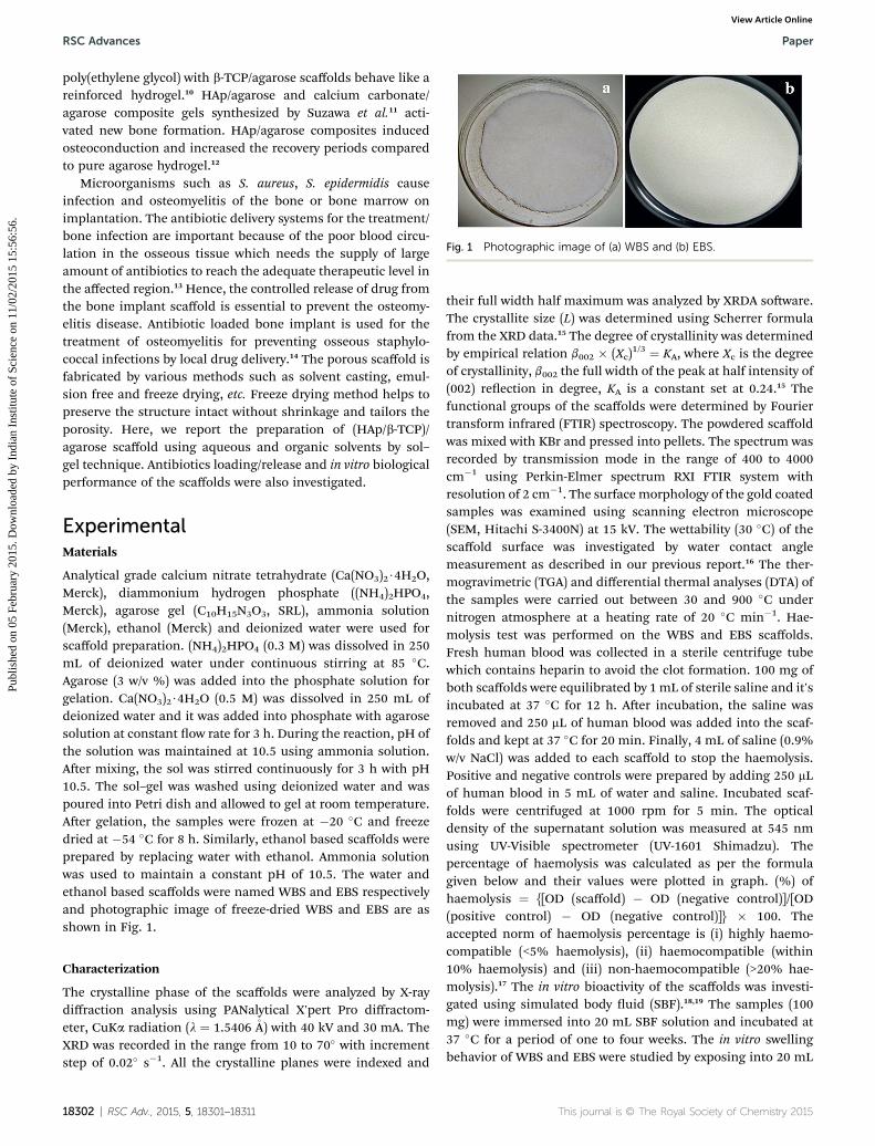
Fig. 1 Photographic image of (a) WBS and (b) EBS.
RSC Advances Paper
Publ
ishe
d on
05
Febr
uary
201
5. D
ownl
oade
d by
Ind
ian
Inst
itute
of
Scie
nce
on 1
1/02
/201
5 15
:56:
56.
View Article Online
poly(ethylene glycol) with b-TCP/agarose scaffolds behave like areinforced hydrogel.10 HAp/agarose and calcium carbonate/agarose composite gels synthesized by Suzawa et al.11 acti-vated new bone formation. HAp/agarose composites inducedosteoconduction and increased the recovery periods comparedto pure agarose hydrogel.12
Microorganisms such as S. aureus, S. epidermidis causeinfection and osteomyelitis of the bone or bone marrow onimplantation. The antibiotic delivery systems for the treatment/bone infection are important because of the poor blood circu-lation in the osseous tissue which needs the supply of largeamount of antibiotics to reach the adequate therapeutic level inthe affected region.13 Hence, the controlled release of drug fromthe bone implant scaffold is essential to prevent the osteomy-elitis disease. Antibiotic loaded bone implant is used for thetreatment of osteomyelitis for preventing osseous staphylo-coccal infections by local drug delivery.14 The porous scaffold isfabricated by various methods such as solvent casting, emul-sion free and freeze drying, etc. Freeze drying method helps topreserve the structure intact without shrinkage and tailors theporosity. Here, we report the preparation of (HAp/b-TCP)/agarose scaffold using aqueous and organic solvents by sol–gel technique. Antibiotics loading/release and in vitro biologicalperformance of the scaffolds were also investigated.
ExperimentalMaterials
Analytical grade calcium nitrate tetrahydrate (Ca(NO3)2$4H2O,Merck), diammonium hydrogen phosphate ((NH4)2HPO4,Merck), agarose gel (C10H15N3O3, SRL), ammonia solution(Merck), ethanol (Merck) and deionized water were used forscaffold preparation. (NH4)2HPO4 (0.3 M) was dissolved in 250mL of deionized water under continuous stirring at 85 �C.Agarose (3 w/v %) was added into the phosphate solution forgelation. Ca(NO3)2$4H2O (0.5 M) was dissolved in 250 mL ofdeionized water and it was added into phosphate with agarosesolution at constant ow rate for 3 h. During the reaction, pH ofthe solution was maintained at 10.5 using ammonia solution.Aer mixing, the sol was stirred continuously for 3 h with pH10.5. The sol–gel was washed using deionized water and waspoured into Petri dish and allowed to gel at room temperature.Aer gelation, the samples were frozen at �20 �C and freezedried at �54 �C for 8 h. Similarly, ethanol based scaffolds wereprepared by replacing water with ethanol. Ammonia solutionwas used to maintain a constant pH of 10.5. The water andethanol based scaffolds were named WBS and EBS respectivelyand photographic image of freeze-dried WBS and EBS are asshown in Fig. 1.
Characterization
The crystalline phase of the scaffolds were analyzed by X-raydiffraction analysis using PANalytical X'pert Pro diffractom-eter, CuKa radiation (l ¼ 1.5406�A) with 40 kV and 30 mA. TheXRD was recorded in the range from 10 to 70� with incrementstep of 0.02� s�1. All the crystalline planes were indexed and
18302 | RSC Adv., 2015, 5, 18301–18311
their full width half maximum was analyzed by XRDA soware.The crystallite size (L) was determined using Scherrer formulafrom the XRD data.15 The degree of crystallinity was determinedby empirical relation b002 � (Xc)
1/3 ¼ KA, where Xc is the degreeof crystallinity, b002 the full width of the peak at half intensity of(002) reection in degree, KA is a constant set at 0.24.15 Thefunctional groups of the scaffolds were determined by Fouriertransform infrared (FTIR) spectroscopy. The powdered scaffoldwas mixed with KBr and pressed into pellets. The spectrum wasrecorded by transmission mode in the range of 400 to 4000cm�1 using Perkin-Elmer spectrum RXI FTIR system withresolution of 2 cm�1. The surface morphology of the gold coatedsamples was examined using scanning electron microscope(SEM, Hitachi S-3400N) at 15 kV. The wettability (30 �C) of thescaffold surface was investigated by water contact anglemeasurement as described in our previous report.16 The ther-mogravimetric (TGA) and differential thermal analyses (DTA) ofthe samples were carried out between 30 and 900 �C undernitrogen atmosphere at a heating rate of 20 �C min�1. Hae-molysis test was performed on the WBS and EBS scaffolds.Fresh human blood was collected in a sterile centrifuge tubewhich contains heparin to avoid the clot formation. 100 mg ofboth scaffolds were equilibrated by 1mL of sterile saline and it'sincubated at 37 �C for 12 h. Aer incubation, the saline wasremoved and 250 mL of human blood was added into the scaf-folds and kept at 37 �C for 20 min. Finally, 4 mL of saline (0.9%w/v NaCl) was added to each scaffold to stop the haemolysis.Positive and negative controls were prepared by adding 250 mLof human blood in 5 mL of water and saline. Incubated scaf-folds were centrifuged at 1000 rpm for 5 min. The opticaldensity of the supernatant solution was measured at 545 nmusing UV-Visible spectrometer (UV-1601 Shimadzu). Thepercentage of haemolysis was calculated as per the formulagiven below and their values were plotted in graph. (%) ofhaemolysis ¼ {[OD (scaffold) � OD (negative control)]/[OD(positive control) � OD (negative control)]} � 100. Theaccepted norm of haemolysis percentage is (i) highly haemo-compatible (<5% haemolysis), (ii) haemocompatible (within10% haemolysis) and (iii) non-haemocompatible (>20% hae-molysis).17 The in vitro bioactivity of the scaffolds was investi-gated using simulated body uid (SBF).18,19 The samples (100mg) were immersed into 20 mL SBF solution and incubated at37 �C for a period of one to four weeks. The in vitro swellingbehavior of WBS and EBS were studied by exposing into 20 mL
This journal is © The Royal Society of Chemistry 2015
Paper RSC Advances
Publ
ishe
d on
05
Febr
uary
201
5. D
ownl
oade
d by
Ind
ian
Inst
itute
of
Scie
nce
on 1
1/02
/201
5 15
:56:
56.
View Article Online
of deionized water, phosphate buffer saline (PBS, pH 7.4) andsimulated body uid (SBF) at 37 �C. Swelling of the scaffoldswas monitored gravimetrically before and aer immersion insolutions by measuring weight with respect to time for 24 h. Theexperiment was carried out in triplicate and the wet weight ofthe scaffolds was determined by removing the sample from theswelling media and wipes the surface water of the scaffold usingWhattmann lter paper. The swelling ratio E(sr) in percentage,which corresponds to the average hydration degree has beendetermined according to the following equation.10
E(sr) (%) ¼ ((Ws � Wd)/Wd) � 100
where, E(sr) is the amount of water absorbed (weight percent) bythe scaffolds and Ws, Wd are the weights of the scaffolds inswollen and dried state respectively.
Mechanical testing
Compression testing of the scaffolds was carried out usingMechanism micro universal testing machine (UTM) with 500 Nload cell and data acquisition soware. Circular disc of (10 mmdia with �3 mm thick) WBS and EBS scaffolds were used formechanical analysis. The samples were placed between twoparallel palates and loaded at a rate of 1 mm min�1 which isapproximately strain rate of 2% (0.016 s�1). All the scaffoldswere tested at pre-load of 0.03 N and each specimen was sub-jected to 60% strain. The compressive modulus was calculatedfrom the slope at strain value of 10%, 25% and 50% of theresulting stress–strain curve.
Biocompatibility test
MC3T3-E1 subclone 4 mouse osteoblast cell lines (ATCC) werecultured in eagles minimum essential medium (a-MEM, Invi-trogen), supplemented with 10% Fetal Bovine Serum (FBS,Invitrogen), 10 mg mL�1 streptomycin (Invitrogen), 10 mg mL�1
penicillin and culture was incubated at 37 �C in a humidiedatmosphere with 5% CO2. The medium was changed every twodays until 80% to 90% conuency then sub-culture the cells.The cells monolayer was washed twice with PBS solution anddetached from their culture ask by incubating with 0.25%Trypsin–EDTA (Gibco) solution for 3 min. Detached MC3T3cells of 1 � 104 (passage 16) were seeded in each well of 48 welltissue culture plates containing three time washed with PBSsamples of WBS and EBS (size 10 mm of disc and �3 mm thick)to remove the un-reacted products.
The cell proliferation was studied using MTT assay on 1, 3and 7 days. At each time point, 400 mL of MTT reagent (1 mgmL�1) was added to each well and incubated for 4 h at the samecondition. Finally, MTT reagent was removed and 400 mL ofdimethyl sulfoxide (DMSO) (Sigma-Aldrich) was added for dis-solving the formasan crystals and the absorbance wasmeasuredat 570 nm in an ELISA (Thermo Scientic) reader. For statisticalanalyses, 1-way ANOVA (analysis of variance) with Tukey's testfor multiple comparisons was used and differences wereconsidered statistically signicant if p < 0.05.
This journal is © The Royal Society of Chemistry 2015
Aer each culture time point, cells on the samples werestained using live/dead assay kit (Molecular Probes), containingcalcein AM and ethidium homodimer. Non-uorescent cell-permeant calcein AM will be enzymatically converted tointense green uorescent calcein in live cells, while ethidiumhomodimer enters cells with damaged membranes, binding tonucleic acids and producing a bright red uorescence in thedead cells. At each time point, the media was discarded and thesamples were washed with PBS, then live/dead solution con-taining 2 mM calcein AM and 4 mM ethidium homodimer wasadded to each well and were incubated at 37 �C for about20 min. The stained cultures were viewed using uorescentmicroscope.
The attachment and spreading of MC3T3 cells on the scaf-folds were examined using Alexa Fluor 546 Phalloidin/DAPIstaining. Aer 1, 3 and 7 days of incubation, cell seededcomposite scaffolds were xed with 3.7% formaldehyde for 30min and rinsed with water. Aer cell xing, 0.2% Triton X-100 inwater was added on scaffolds for permeabilization and incu-bated for 15 min. Then the scaffolds were washed with waterand stained with 200 ml of Alexa Fluor 546 Phalloidin for 45 minand 200 ml of DAPI dyes commonly used for F-actin and nucleiimaging respectively for 10 min in the dark environment. Thescaffolds were then washed with deionized water and viewedunder uorescent microscope (Olympus-BX-51) aer removingthe excess water.
In vitro mineralization was studied before and aer cellcultured on WBS and EBS scaffolds for 7 and 14 days usingAlizarin red S assay. Aer each time point, cells were xed with3.7% formaldehyde for 30 min and stained with 2% Alizarin redS dye (AR) for 20 min and washed several times with deionizedwater to remove excess stain. Digital images of stained scaffoldswere recorded. To retain the bound stain from scaffolds,samples were treated with 0.5 mL of 0.5 N HCL containing 5%SDS for 30 min. The absorbance of the dissolved AR stain wasmeasured at 415 nm in an ELISA (Thermo Scientic) reader andquantied.
Drug loading/release
250 mg of gentamicin sulphate (GS) and 150 mg of amoxicillin(AMX) drugs were dissolved separately in 10 mL of deionizedwater. 500 mg of WBS and EBS scaffolds were soaked in drugsolutions. These solutions were kept at room temperature for 24h in incubator cum orbital shaker with a shaking speed of 100rpm. Subsequently, the supernatant solution was removed andthe absorbance was measured at l ¼ 248 and 230 nm using UV-Vis spectrophotometer (Shimadzu UV-1601). Percentage of drugloading was calculated using the formula, percentage of drugloading ¼ [(Y � X)/X] � 100, where, X represents the initialconcentration of the drug and Y, the nal concentration of drugaer removing the scaffold from the drug solution. The drug(GS, AMX) loaded scaffolds were dried at 37 �C and named asWBS-GS, EBS-GS, WBS-AMX and EBS-AMX. The drug releasestudy was carried out using PBS (pH-7.4). The drug loadedscaffolds in triplicates was kept in 100 mL of dissolutionmedium (PBS) and was incubated at 37 �Cwith shaking speed of
RSC Adv., 2015, 5, 18301–18311 | 18303
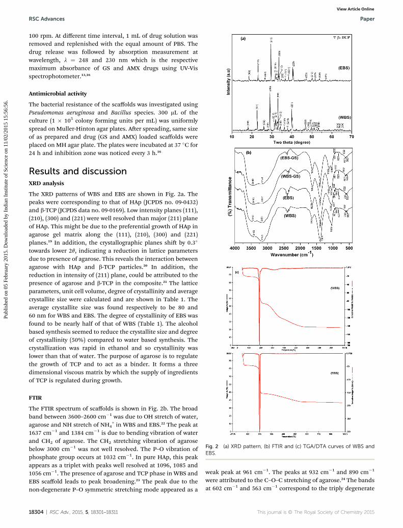
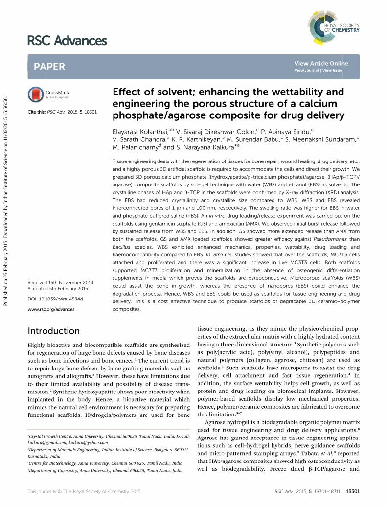
Fig. 2 (a) XRD pattern, (b) FTIR and (c) TGA/DTA curves of WBS andEBS.
RSC Advances Paper
Publ
ishe
d on
05
Febr
uary
201
5. D
ownl
oade
d by
Ind
ian
Inst
itute
of
Scie
nce
on 1
1/02
/201
5 15
:56:
56.
View Article Online
100 rpm. At different time interval, 1 mL of drug solution wasremoved and replenished with the equal amount of PBS. Thedrug release was followed by absorption measurement atwavelength, l ¼ 248 and 230 nm which is the respectivemaximum absorbance of GS and AMX drugs using UV-Visspectrophotometer.13,16
Antimicrobial activity
The bacterial resistance of the scaffolds was investigated usingPseudomonas aeruginosa and Bacillus species. 300 mL of theculture (1 � 105 colony forming units per mL) was uniformlyspread on Muller-Hinton agar plates. Aer spreading, same sizeof as prepared and drug (GS and AMX) loaded scaffolds wereplaced on MH agar plate. The plates were incubated at 37 �C for24 h and inhibition zone was noticed every 3 h.16
Results and discussionXRD analysis
The XRD patterns of WBS and EBS are shown in Fig. 2a. Thepeaks were corresponding to that of HAp (JCPDS no. 09-0432)and b-TCP (JCPDS data no. 09-0169). Low intensity planes (111),(210), (300) and (221) were well resolved than major (211) planeof HAp. This might be due to the preferential growth of HAp inagarose gel matrix along the (111), (210), (300) and (221)planes.19 In addition, the crystallographic planes shi by 0.3�
towards lower 2q, indicating a reduction in lattice parametersdue to presence of agarose. This reveals the interaction betweenagarose with HAp and b-TCP particles.20 In addition, thereduction in intensity of (211) plane, could be attributed to thepresence of agarose and b-TCP in the composite.21 The latticeparameters, unit cell volume, degree of crystallinity and averagecrystallite size were calculated and are shown in Table 1. Theaverage crystallite size was found respectively to be 80 and60 nm for WBS and EBS. The degree of crystallinity of EBS wasfound to be nearly half of that of WBS (Table 1). The alcoholbased synthesis seemed to reduce the crystallite size and degreeof crystallinity (50%) compared to water based synthesis. Thecrystallization was rapid in ethanol and so crystallinity waslower than that of water. The purpose of agarose is to regulatethe growth of TCP and to act as a binder. It forms a threedimensional viscous matrix by which the supply of ingredientsof TCP is regulated during growth.
FTIR
The FTIR spectrum of scaffolds is shown in Fig. 2b. The broadband between 3600–2600 cm�1 was due to OH stretch of water,agarose and NH stretch of NH4
+ in WBS and EBS.22 The peak at1637 cm�1 and 1384 cm�1 is due to bending vibration of waterand CH2 of agarose. The CH2 stretching vibration of agarosebelow 3000 cm�1 was not well resolved. The P–O vibration ofphosphate group occurs at 1032 cm�1. In pure HAp, this peakappears as a triplet with peaks well resolved at 1096, 1085 and1056 cm�1. The presence of agarose and TCP phase in WBS andEBS scaffold leads to peak broadening.23 The peak due to thenon-degenerate P–O symmetric stretching mode appeared as a
18304 | RSC Adv., 2015, 5, 18301–18311
weak peak at 961 cm�1. The peaks at 932 cm�1 and 890 cm�1
were attributed to the C–O–C stretching of agarose.24 The bandsat 602 cm�1 and 563 cm�1 correspond to the triply degenerate
This journal is © The Royal Society of Chemistry 2015
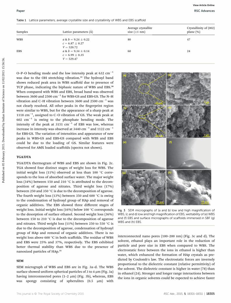
Table 1 Lattice parameters, average crystallite size and crystallinity of WBS and EBS scaffold
Samples Lattice parameters (A)Average crystallitesize (�1 nm)
Crystallinity of (002)plane (%)
WBS a & b ¼ 9.24 � 0.22 80 47c ¼ 6.87 � 0.27V ¼ 520.72
EBS a & b ¼ 9.34 � 0.14 60 24c ¼ 6.99 � 0.35V ¼ 529.47
Fig. 3 SEM micrographs of (a and b) low and high magnification ofWBS, (c and d) low and highmagnification of EBS, wettability of (e) WBSand (f) EBS and surface micrographs of scaffolds immersed in SBF (g)WBS and (h) EBS.
Paper RSC Advances
Publ
ishe
d on
05
Febr
uary
201
5. D
ownl
oade
d by
Ind
ian
Inst
itute
of
Scie
nce
on 1
1/02
/201
5 15
:56:
56.
View Article Online
O–P–O bending mode and the low intensity peak at 632 cm�1
was due to the OH stretching vibration.22 The hydroxyl bandshows reduced peak area in WBS scaffold due to presence ofTCP phase, indicating the biphasic nature of WBS and EBS.20
When compared with WBS and EBS, broad band was observedbetween 3600 and 2500 cm�1 for WBS-GS and EBS-GS. The N–Hvibration and C–H vibration between 3600 and 2500 cm�1 wasnot clearly resolved. All other peaks in the ngerprint regionwere similar to WBS, but for the appearance of a sharp peak at1118 cm�1, assigned to C–O vibration of GS. The weak peak at602 cm�1 is owing to the phosphate bending mode. Theintensity of the peak at 3151 cm�1 of EBS was low, whereasincrease in intensity was observed at 3440 cm�1 and 1122 cm�1
for EBS-GS. The variation of intensities and appearance of newpeaks in WBS-GS and EBS-GS compared with WBS and EBScould be due to the loading of GS. Similar features wereobserved for AMX loaded scaffolds (spectra not shown).
TGA/DTA
TGA/DTA thermogram of WBS and EBS are shown in Fig. 2c.TGA showed four distinct stages of weight loss for WBS. Theinitial weight loss (11%) observed at less than 100 �C corre-sponds to the loss of absorbed surface water. The major weightloss (34%) between 150 and 210 �C is attributed to the decom-position of agarose and nitrates. Third weight loss (17%)between 250 and 350 �C is due to the decomposition of agarose.The fourth weight loss (13%) between 350 and 600 �C is owingto the condensation of hydroxyl group of HAp and removal oforganic additives. The EBS showed three different stages ofweight loss. Initial weight loss (16%) below 100 �C correspondsto the desorption of surface ethanol. Second weight loss (36%)between 150 to 210 �C is due to the decomposition of agaroseand nitrates. Third weight loss (11%) between 250 to 550 �C isdue to the decomposition of agarose, condensation of hydroxylgroup of HAp and removal of organic additives. There is noweight loss above 600 �C in both scaffolds. The residue of WBSand EBS were 25% and 37%, respectively. The EBS exhibitedbetter thermal stability than WBS due to the presence ofnanosized particles of HAp.25
SEM
SEM micrograph of WBS and EBS are in Fig. 3a–d. The WBSsurface showed uniform spherical particles of 3 to 4 mm (Fig. 3a)having interconnected pores (1–2 mm) (Fig. 3b), whereas, EBSwas spongy consisting of spherulites (0.5 mm) with
This journal is © The Royal Society of Chemistry 2015
interconnected nano pores (100–200 nm) (Fig. 3c and d). Thesolvent, ethanol plays an important role in the reduction ofparticle and pore size in EBS when compared to WBS. Theelectrostatic force between the ions in ethanol is higher thanwater, which enhanced the formation of HAp crystals as pre-dicted by Coulomb's law. The electrostatic forces are inverselyproportional to the dielectric constant (relative permittivity) ofthe solvent. The dielectric constant is higher in water (78) thanin ethanol (24). Stronger and longer range interactions betweenthe ions in organic solvents could be expected to achieve faster
RSC Adv., 2015, 5, 18301–18311 | 18305
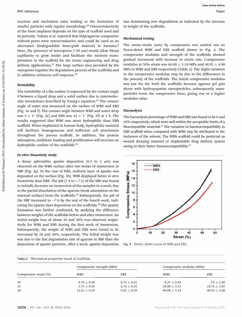
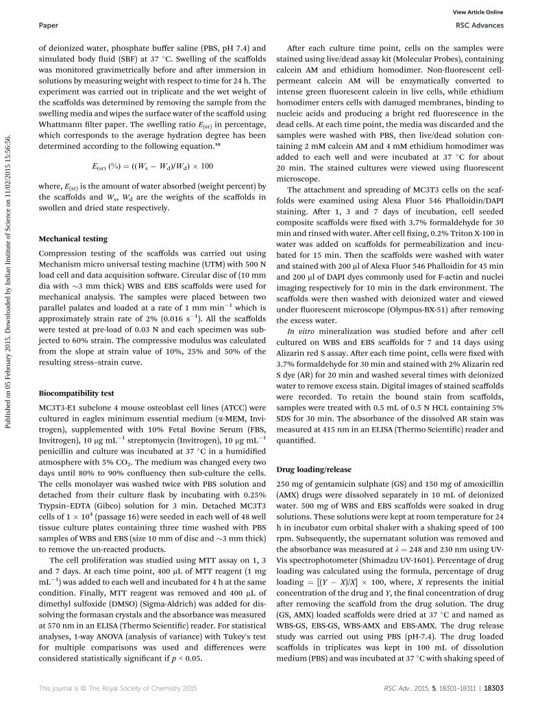
Fig. 4 Stress–strain curve of WBS and EBS.
RSC Advances Paper
Publ
ishe
d on
05
Febr
uary
201
5. D
ownl
oade
d by
Ind
ian
Inst
itute
of
Scie
nce
on 1
1/02
/201
5 15
:56:
56.
View Article Online
reaction and nucleation rates leading to the formation ofsmaller particles with regular morphology.26 Osteoconductivityof the bone implants depends on the type of scaffold used andits porosity. Tabata et al. reported that HAp/agarose compositeswithout pores were osteoconductive and could be used as analternative biodegradable bone-gra material in humans.8
Here, the presence of micropores (<50 mm) would allow bloodcapillaries to grow inside and facilitate the nutrient trans-portation in the scaffold for the tissue engineering and drugdelivery applications.27 The large surface area provided by thenanopores regulate the degradation process of the scaffolds andin addition enhances cell response.28
Wettability
The wettability of a at surface is expressed by the contact angleq between a liquid drop and a solid surface due to intermolec-ular interactions described by Young's equation.16 The contactangle of water was measured on the surface of WBS and EBS(Fig. 3e and f). The contact angle between WBS and water dropwas 5 � 1� (Fig. 2e) and EBS was 42 � 1� (Fig. 2f) at 1 S. Theresults suggested that WBS was more hydrophilic than EBSscaffold. When implanted in human body, hydrophilic materialwill facilitate homogeneous and sufficient cell attachmentthroughout the porous scaffold. In addition, the proteinadsorption, antibiotic loading and proliferation will increase onhydrophilic surface of the scaffolds.29
In vitro bioactivity study
A dense spherulitic apatite deposition (0.5 to 2 mm) wasobserved on the WBS surface aer two weeks of immersion inSBF (Fig. 3g). In the case of EBS, uniform layer of apatite wasdeposited on the surface (Fig. 3h). WBS displayed better in vitrobioactivity than EBS. The pH (7.4 to �7.1) of the SBF was foundto initially decrease on immersion of the samples in a week, dueto the partial dissolution of the agarose (weak adsorption on theexternal surface) from the scaffolds.19 Subsequently, the pH ofthe SBF increased to �7.6 by the end of the fourth week, indi-cating the apatite layer deposition on the scaffolds.19 The apatiteformation was further conrmed, by studying the differencebetween weights of the scaffolds before and aer immersion. Aninitial weight loss of about 30 and 50% was observed respec-tively for WBS and EBS during the rst week of immersion.Subsequently, the weight of WBS and EBS were found to beincreased by 28 and 20%, respectively. The initial weight losswas due to the fast degradation rate of agarose in SBF than thedeposition of apatite particles. Aer a week, apatite deposition
Table 2 Mechanical properties result of scaffolds
Compressive strain (%)
Compressive strength (MPa)
WBS E
10 0.70 � 0.40 025 3.75 � 0.51 250 12.23 � 0.39 9
18306 | RSC Adv., 2015, 5, 18301–18311
was dominating over degradation as indicated by the increasein weight of the scaffolds.
Mechanical testing
The stress–strain curve by compressive test carried out onfreeze-dried WBS and EBS scaffold shown in Fig. 4. Thecompressive modulus and strength of the scaffolds showedgradual increment with increase in strain rate. Compressivemodulus at 50% strain was 60.88 � 1.54 MPa and 48.95 � 4.98MPa in WBS and EBS respectively (Table 2). The slight variationin the compressive modulus may be due to the differences inthe porosity of the scaffolds. The initial compressive moduluswas low for the both the scaffolds because agarose gel getsdense with hydroxyapatite nanoparticles, subsequently nano-particles resist the compressive force, giving rise to a highermodulus value.
Haemolysis
The haemolysis percentage ofWBS and EBS was found to be 6 and10% respectively, which were well within the acceptable limits of abiocompatible material.17 The variation in haemocompatibility inEBS scaffold when compared with WBS may be attributed to theinclusion of the solvent. The WBS scaffold could be preferred aswound dressing material or implantable drug delivery systemowing to their better haemocompatibility.17
Compressive modulus (MPa)
BS WBS EBS
.72 � 0.31 8.57 � 0.92 7.9 � 1.00
.74 � 0.35 24.80 � 2.51 18.74 � 3.95
.45 � 0.59 60.88 � 1.54 48.95 � 4.98
This journal is © The Royal Society of Chemistry 2015
Paper RSC Advances
Publ
ishe
d on
05
Febr
uary
201
5. D
ownl
oade
d by
Ind
ian
Inst
itute
of
Scie
nce
on 1
1/02
/201
5 15
:56:
56.
View Article Online
In vitro swelling behavior
The percentage of swelling ratio (E(sr)) versus time of immersionof both the scaffolds in different solutions is shown in Fig. 5a.We observed initial rapid increase in E(sr) upto 4 h on immer-sion in water, PBS and SBF solutions, followed by a slow rate ofincrement in E(sr) upto 20 h, before reaching equilibrium.Equilibrium E(sr) was found to be respectively between 10 and60% for WBS and EBS. When compared with immersion in PBS(42%) and SBF (28%) solutions, swelling ratio signicantlyincreased in water (60%) for EBS. EBS showed higher E(sr) thanWBS in PBS and water, due to the presence of large number ofnanosized pores and particles. In addition, agarose behaves likea hydrogel and is responsible for the enhanced absorption ofwater and PBS. The physical properties of agarose are similar tothose of living tissues which assist in fast healing of woundarea.8 During swelling process, the volume of the scaffoldsincreased without any signicant change in shape. The photo-graphic image of native and 24 h PBS soaked swollen WBS andEBS is shown in Fig. 5b and c. This property will help to ensure agood t between the sample and osseous defect. The scaffoldscould adsorb physiological uids and yield controllableaugmentation on implantation, having a benecial effect onosteoblast growth and differentiation. Further, this study mayprovide an insight into the degradation mechanism of thescaffolds during implantation in living organisms.10
Drug loading and release
GS and AMX can be loaded into porousWBS and EBS scaffold byimpregnation process by immersing them in drug solution. TheOH groups on theWBS and EBS surface react with NH2 group inGS and carboxyl group in AMX to form hydrogen bonding,
Fig. 5 Percentage of swelling ratio (Esr) (W-water) (a), photographicimage of before and after 24 h swollen in PBS of WBS (b) and EBS (c).
This journal is © The Royal Society of Chemistry 2015
leading to higher encapsulation of the drugs.21 The weightpercentage loading of the AMX was 45� 1, 40� 1 and GS was 70� 1, 54 � 1 on WBS and EBS respectively. WBS exhibitedenhanced loading of AMX and GS. The drug encapsulationdepends on the porosity, size, shape, distribution, connectivity,potential functionalization of their walls and wettability of thesurface.30 In addition; size, nature and dimensions of the drugto be introduced also play a role. Here, the respective molecularsize of GS and AMX were 0.9 nm and 1.1 nm. SEM imagesrevealed macro porous WBS surface (1–2 mm), whereas EBSconsisted of nanosized pores (100–200 nm) (Fig. 2b and d). Inaddition, WBS was more hydrophilic than EBS. Therefore, largesize of pores and hydrophilic nature of surface, play amajor rolein enhancing the encapsulation of drug in WBS.31 The drugloading capacity of EBS was low due to the small pore size andlow hydrophilic surface compared with WBS.30
Hydrogel scaffolds are preferred as biomedical implants anddrug delivery systems. The drug loaded samples were subjectedto drug release in PBS. During the drug release process, PBSenters into the drug loaded samples through the pores. Thedrug is then slowly released into PBS from the surface andthrough the pores. The cumulative drug release proles as afunction of release time in PBS is shown in Fig. 6. We observed80 and 76% drug release respectively from WBS-AMX and EBS-AMX in 1 h. The drug release from WBS-GS and EBS-GS wasrespectively 68 and 66%. Both the drug release proles showedinitial burst release which may be attributed to the phys-isorption of GS and AMX on the outer surface of the scaffoldsand the high degradation rate and rapid swelling of thehydrogel inWBS and EBS. In addition, the burst release of watersoluble drugs from dehydrated agarose with HAp matrixgenerally involves the simultaneous absorption of water, anddesorption of drug via a swelling-controlled diffusion mecha-nism.31 The drug release in hydrophilic samples occurs at a raterelative to sample swelling and inuenced by factors such aspresence of pores and the entrapment of drugs. The burstrelease is observed during rst 1 h, which agree with the fast
Fig. 6 GS and AMX drugs release profile of WBS and EBS.
RSC Adv., 2015, 5, 18301–18311 | 18307
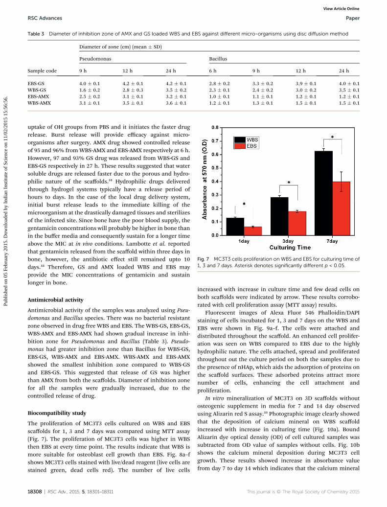
Table 3 Diameter of inhibition zone of AMX and GS loaded WBS and EBS against different micro-organisms using disc diffusion method
Sample code
Diameter of zone (cm) (mean � SD)
Pseudomonas Bacillus
9 h 12 h 24 h 6 h 9 h 12 h 24 h
EBS-GS 4.0 � 0.1 4.2 � 0.1 4.2 � 0.1 2.8 � 0.2 3.3 � 0.2 3.9 � 0.1 4.0 � 0.1WBS-GS 1.6 � 0.2 2.8 � 0.3 3.5 � 0.2 2.3 � 0.1 2.4 � 0.2 3.0 � 0.2 3.5 � 0.1EBS-AMX 2.5 � 0.2 3.1 � 0.1 3.2 � 0.1 1.0 � 0.1 1.1 � 0.1 1.2 � 0.1 1.2 � 0.1WBS-AMX 3.1 � 0.1 3.5 � 0.1 3.6 � 0.1 1.2 � 0.1 1.3 � 0.1 1.5 � 0.1 1.5 � 0.1
Fig. 7 MC3T3 cells proliferation on WBS and EBS for culturing time of1, 3 and 7 days. Asterisk denotes significantly different p < 0.05.
RSC Advances Paper
Publ
ishe
d on
05
Febr
uary
201
5. D
ownl
oade
d by
Ind
ian
Inst
itute
of
Scie
nce
on 1
1/02
/201
5 15
:56:
56.
View Article Online
uptake of OH groups from PBS and it initiates the faster drugrelease. Burst release will provide efficacy against micro-organisms aer surgery. AMX drug showed controlled releaseof 95 and 96% fromWBS-AMX and EBS-AMX respectively at 6 h.However, 97 and 93% GS drug was released from WBS-GS andEBS-GS respectively in 27 h. These results suggested that watersoluble drugs are released faster due to the porous and hydro-philic nature of the scaffolds.31 Hydrophilic drugs deliveredthrough hydrogel systems typically have a release period ofhours to days. In the case of the local drug delivery system,initial burst release leads to the immediate killing of themicroorganism at the drastically damaged tissues and sterilizesof the infected site. Since bone have the poor blood supply, thegentamicin concentrations will probably be higher in bone thanin the buffer media and consequently sustain for a longer timeabove the MIC at in vivo conditions. Lambotte et al. reportedthat gentamicin released from the scaffold within three days inbone, however, the antibiotic effect still remained upto 10days.32 Therefore, GS and AMX loaded WBS and EBS mayprovide the MIC concentrations of gentamicin and sustainlonger in bone.
Antimicrobial activity
Antimicrobial activity of the samples was analyzed using Pseu-domonas and Bacillus species. There was no bacterial resistantzone observed in drug free WBS and EBS. The WBS-GS, EBS-GS,WBS-AMX and EBS-AMX had shown gradual increase in inhi-bition zone for Pseudomonas and Bacillus (Table 3). Pseudo-monas had greater inhibition zone than Bacillus for WBS-GS,EBS-GS, WBS-AMX and EBS-AMX. WBS-AMX and EBS-AMXshowed the smallest inhibition zone compared to WBS-GSand EBS-GS. This suggested that release of GS was higherthan AMX from both the scaffolds. Diameter of inhibition zonefor all the samples were gradually increased, due to thecontrolled release of drug.
Biocompatibility study
The proliferation of MC3T3 cells cultured on WBS and EBSscaffolds for 1, 3 and 7 days was compared using MTT assay(Fig. 7). The proliferation of MC3T3 cells was higher in WBSthen EBS at every time point. The results indicate that WBS ismore suitable for osteoblast cell growth than EBS. Fig. 8a–fshows MC3T3 cells stained with live/dead reagent (live cells arestained green, dead cells red). The number of live cells
18308 | RSC Adv., 2015, 5, 18301–18311
increased with increase in culture time and few dead cells onboth scaffolds were indicated by arrow. These results corrobo-rated with cell proliferation assay (MTT assay) results.
Fluorescent images of Alexa Fluor 546 Phalloidin/DAPIstaining of cells incubated for 1, 3 and 7 days on the WBS andEBS were shown in Fig. 9a–f. The cells were attached anddistributed throughout the scaffold. An enhanced cell prolifer-ation was seen on WBS compared to EBS due to the highlyhydrophilic nature. The cells attached, spread and proliferatedthroughout out the culture period on both the samples due tothe presence of nHAp, which aids the adsorption of proteins onthe scaffold surfaces. These adsorbed proteins attract morenumber of cells, enhancing the cell attachment andproliferation.
In vitro mineralization of MC3T3 on 3D scaffolds withoutosteogenic supplement in media for 7 and 14 day observedusing Alizarin red S assay.33 Photographic image clearly showedthat the deposition of calcium mineral on WBS scaffoldincreased with increase in culturing time (Fig. 10a). BoundAlizarin dye optical density (OD) of cell cultured samples wassubtracted from OD value of samples without cells. Fig. 10bshows the calcium mineral deposition during MC3T3 cellgrowth. These results showed increase in absorbance valuefrom day 7 to day 14 which indicates that the calcium mineral
This journal is © The Royal Society of Chemistry 2015
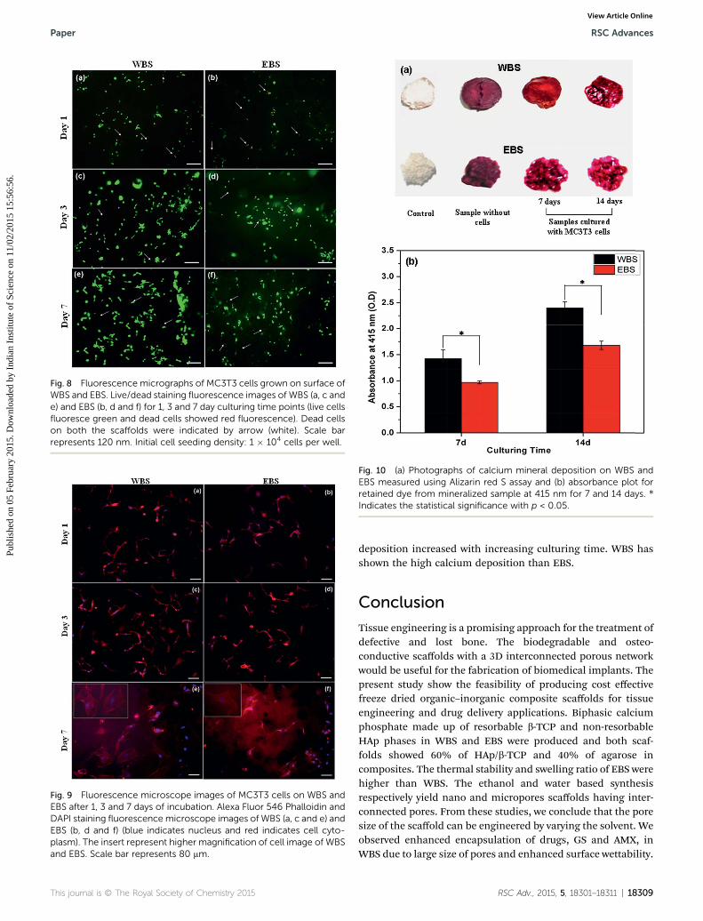
Fig. 10 (a) Photographs of calcium mineral deposition on WBS andEBS measured using Alizarin red S assay and (b) absorbance plot forretained dye from mineralized sample at 415 nm for 7 and 14 days. *Indicates the statistical significance with p < 0.05.
Fig. 8 Fluorescence micrographs of MC3T3 cells grown on surface ofWBS and EBS. Live/dead staining fluorescence images of WBS (a, c ande) and EBS (b, d and f) for 1, 3 and 7 day culturing time points (live cellsfluoresce green and dead cells showed red fluorescence). Dead cellson both the scaffolds were indicated by arrow (white). Scale barrepresents 120 nm. Initial cell seeding density: 1 � 104 cells per well.
Fig. 9 Fluorescence microscope images of MC3T3 cells on WBS andEBS after 1, 3 and 7 days of incubation. Alexa Fluor 546 Phalloidin andDAPI staining fluorescence microscope images of WBS (a, c and e) andEBS (b, d and f) (blue indicates nucleus and red indicates cell cyto-plasm). The insert represent higher magnification of cell image of WBSand EBS. Scale bar represents 80 mm.
This journal is © The Royal Society of Chemistry 2015
Paper RSC Advances
Publ
ishe
d on
05
Febr
uary
201
5. D
ownl
oade
d by
Ind
ian
Inst
itute
of
Scie
nce
on 1
1/02
/201
5 15
:56:
56.
View Article Online
deposition increased with increasing culturing time. WBS hasshown the high calcium deposition than EBS.
Conclusion
Tissue engineering is a promising approach for the treatment ofdefective and lost bone. The biodegradable and osteo-conductive scaffolds with a 3D interconnected porous networkwould be useful for the fabrication of biomedical implants. Thepresent study show the feasibility of producing cost effectivefreeze dried organic–inorganic composite scaffolds for tissueengineering and drug delivery applications. Biphasic calciumphosphate made up of resorbable b-TCP and non-resorbableHAp phases in WBS and EBS were produced and both scaf-folds showed 60% of HAp/b-TCP and 40% of agarose incomposites. The thermal stability and swelling ratio of EBS werehigher than WBS. The ethanol and water based synthesisrespectively yield nano and micropores scaffolds having inter-connected pores. From these studies, we conclude that the poresize of the scaffold can be engineered by varying the solvent. Weobserved enhanced encapsulation of drugs, GS and AMX, inWBS due to large size of pores and enhanced surface wettability.
RSC Adv., 2015, 5, 18301–18311 | 18309
RSC Advances Paper
Publ
ishe
d on
05
Febr
uary
201
5. D
ownl
oade
d by
Ind
ian
Inst
itute
of
Scie
nce
on 1
1/02
/201
5 15
:56:
56.
View Article Online
Burst release followed by the controlled drug release wasexhibited by both types of scaffolds. Pseudomonas was moreresistant in GS and AMX than Bacillus species. WBS showedenhanced wettability, interconnected pores, bioactivity, hae-mocompatibility, drug loading and mechanical properties thanEBS. Cell studies proved that WBS was most effective inpromoting MC3T3 mouse osteoblast cell attachment, prolifer-ation and osteoconductivity. WBS and EBS seem to be respec-tively suitable for tissue engineering and drug deliveryapplication. This fabrication technique which is cost effectiveand fast, yield 3D scaffolds with uniformly distributed bio-ceramics in a degradable polymer matrix.
Acknowledgements
One of the authors (K.E) acknowledges CSIR, India for theaward of SRF fellowship (File no. 09/468(0413)2009-EMR-I). Thiswork was supported by the Department of Biotechnology, NewDelhi through a research project (no. BT/PR11799/MED/32/2009).
References
1 M. Braddock, P. Houston, C. Campbell and P. Ashcro, BornAgain Bone: Tissue Engineering for Bone Repair, NewsPhysiol. Sci., 2001, 16, 208–213.
2 G. Wei and P. X. Ma, Structure and properties of nano-hydroxyapatite/polymer composite scaffolds for bone tissueengineering, Biomaterials, 2004, 25, 4749–4757.
3 K. Y. Lee and D. J. Mooney, Hydrogels for TissueEngineering, Chem. Rev., 2001, 101, 1869–1879.
4 E. Sachlos, N. Reis, C. Ainsley and J. T. Czernuszka, Novelcollagen scaffolds with predened internal morphologymade by solid free form fabrication, Biomaterials, 2003, 24,1487–1497.
5 K. Rezwana, Q. Z. Chena, J. J. Blakera and A. R. Boccaccinia,Biodegradable and bioactive porous polymer/inorganiccomposite scaffolds for bone tissue engineering,Biomaterials, 2006, 27, 3413–3431.
6 M. I. Sabir, X. Xu and L. Li, A review on biodegradablepolymeric materials for bone tissue engineeringapplications, J. Mater. Sci., 2009, 44, 5713–5724.
7 I. Armentano, M. Dottori, E. Fortunati, S. Mattioli andJ. M. Kenny, Biodegradable polymer matrixnanocomposites for tissue engineering: A review, Polym.Degrad. Stab., 2010, 95, 2126–2146.
8 M. Tabata, T. Shimoda, K. Sugihara, D. Ogomi, T. Serizawaand M. Akashi, Osteoconductive and Hemostatic Propertiesof Apatite Formed on/in Agarose Gel as a Bone- GraingMaterial, J. Biomed. Mater. Res., Part B, 2003, 67, 680–688.
9 Y. Lin, S. Tang, X. Mao and L. Bao, Protein recognition viamolecularly imprinted agarose gel membrane, J. Biomed.Mater. Res., Part A, 2008, 85, 573–581.
10 J. Roman, M. V. Cabanas, J. Pena, J. C. Doadrio andM. Vallet-Regi, An optimized b-tricalcium phosphate and agarosescaffold fabrication technique, J. Biomed. Mater. Res., PartA, 2008, 84, 99–107.
18310 | RSC Adv., 2015, 5, 18301–18311
11 Y. Suzawa, T. Funaki, J. Watanabe, S. Iwai, Y. Yura,T. Nakano, Y. Umakoshi and M. Akashi, Regenerativebehavior of biomineral/agarose composite gels as bonegraing materials in rat cranial defects, J. Biomed. Mater.Res., Part A, 2010, 93, 965–975.
12 J. Watanabe, M. Kashii, M. Hirao, K. Oka, K. Sugamoto,H. Yoshikawa and M. Akashi, Quick-forminghydroxyapatite/agarose gel composites induce boneregeneration, J. Biomed. Mater. Res., Part A, 2007, 83, 845–852.
13 V. Mourino and A. R. Boccaccini, Bone tissue engineeringtherapeutics: controlled drug delivery in three-dimensionalscaffolds, J. R. Soc., Interface, 2010, 7, 209–227.
14 M. E. Husseiny, S. Patel, R. J. MacFarlane and F. S. Haddad,Biodegradable antibiotic delivery systems, J. Bone Jt. Surg.,2011, 93, 151–157.
15 V. M. Rusu, C. H. Ng, M. Wilke, B. Tiersch, P. Fratzl andM. G. Peter, Size-controlled hydroxyapatite nanoparticles asself-organized organic–inorganic composite materials,Biomaterials, 2005, 26, 5414–5426.
16 K. Elayaraja, P. Rajesh, M. I. Ahymah Joshy, V. SarathChandara, R. V. Suganthi, J. Kennedy, P. K. Kulriya,I. Sulania, K. Asokan, D. Kanjilal, D. K. Avasthi,H. K. Varma and S. N. Kalkura, Enhancement of wettabilityand antibiotic loading/release of hydroxyapatite thin lmmodied by 100 MeV Ag7+ ion irradiation, Mater. Chem.Phys., 2012, 134, 464–477.
17 V. Sarath Chandra, B. Ganga, R. V. Suganthi, K. Elayaraja,M. I. Ahymah Joshy, W. S. Beaula, R. Mythili, V. Ganesh,K. Sivakumar and S. Narayana Kalkura, BloodCompatibility of Iron-Doped Nanosize Hydroxyapatite andIts Drug Release, ACS Appl. Mater. Interfaces, 2012, 4, 1200–1210.
18 T. Kokubo and H. Takadama, How useful is SBF inpredicting in vivo bone bioactivity?, Biomaterials, 2006, 27,2907–2915.
19 M. I. Ahymah Joshy, K. Elayaraja, R. V. Suganthi andS. Narayana Kalkura, Mineralization of oriented nanohydroxyapatite in photopolymerized polyacrylamide gelmatrix, Cryst. Res. Technol., 2010, 45, 551–556.
20 V. Siva Kumar and K. P. Rao, Preparation, characterizationand in vitro release of gentamicin from coralline HAp-gelatin composite microspheres, Biomaterials, 2002, 23,3175–3181.
21 P. Kumar, K. H. Prakash, P. Cheang, L. Gower and K. A. Khor,Chitosan-mediated crystallization and assembly ofhydroxyapatite nanoparticles into hybrid nanostructuredlms, J. R. Soc., Interface, 2008, 5, 427–439.
22 T. K. Anee, M. Ashok, M. Palanichamy and S. NarayanaKalkura, A novel technique to synthesize hydroxyapatite atlow temperature, Mater. Chem. Phys., 2003, 80, 725–730.
23 I. Manjubala andM. Sivakumar, In-situ synthesis of biphasiccalcium phosphate ceramics using microwave irradiation,Mater. Chem. Phys., 2001, 71, 271–278.
24 V. Sanchez-Vaquero, C. Satriano, N. Tejera-Sanchez,L. Gonzalez Mendez, J. P. Garcia Ruiz and M. MansoSilvan, Characterization and cytocompatibility of hybrid
This journal is © The Royal Society of Chemistry 2015
Paper RSC Advances
Publ
ishe
d on
05
Febr
uary
201
5. D
ownl
oade
d by
Ind
ian
Inst
itute
of
Scie
nce
on 1
1/02
/201
5 15
:56:
56.
View Article Online
aminosilane-agarose hydrogel scaffolds, Biointerphases,2010, 5, 23–29.
25 R. Murugan and S. Ramakrishna, Bioresorbable compositebone paste using polysaccharide based nanohydroxyapatite, Biomaterials, 2004, 25, 3829–3835.
26 H. Alobeedallah, J. L. Ellis, R. Rohanizadeh, H. Coster andF. Dehghani, Preparation of Nanostructured Hydroxyapatitein Organic Solvents for Clinical Applications, TrendsBiomater. Artif. Organs, 2011, 25, 12–19.
27 P. Fabbri, V. Cannillo, A. Sola, A. Dorigato and F. Chiellini,Highly porous polycaprolactone-45S5 Bioglass scaffolds forbone tissue engineering, Compos. Sci. Technol., 2010, 70,1869–1878.
28 S. Wang, M. M. Falk, A. Rashed, M. M. Saad, A. C. Marques,R. M. Almeida, M. K. Marei and H. Jain, Evaluation of 3Dnano–macro porous bioactive glass scaffold for hard tissueengineering, J. Mater. Sci.: Mater. Med., 2011, 22, 1195–1203.
29 L. Sania, K. Wilson and A. Bismark, Atmospheric PlasmaTreatment of Porous Polymer Constructs for Tissue
This journal is © The Royal Society of Chemistry 2015
Engineering Applications, Macromol. Biosci., 2007, 7, 315–327.
30 M. Vallet-Regi, Ordered Mesoporous Materials in theContext of Drug Delivery Systems and Bone TissueEngineering, Eur. J., 2006, 12, 5934–5943.
31 Z. N. Al-Sokanee, A. A. H. Toabi, M. J. Al-assadi andE. A. Alassadi, The Drug Release Study of Ceriaxone fromPorous Hydroxyapatite Scaffolds, AAPS PharmSciTech, 2009,10, 772–779.
32 J. C. Lambotte, H. Thomazeau, G. Cathelineau, G. Lancien,J. Minet and F. Langlais, Tricalcium phosphate, anantibiotic carrier: A study focused on experimentalosteomyelitis in rabbits, Chirurgie, 1998, 123, 572–579.
33 Y. Xia, P. Zhou, X. Cheng, Y. Xie, C. Liang, C. Li and S. Xu,Selective laser sintering fabrication of nano-hydroxyapatite/poly-3-caprolactone scaffolds for bone tissue engineeringapplications, Int. J. Nanomed., 2013, 8, 4197–4213.
RSC Adv., 2015, 5, 18301–18311 | 18311
Top Related
Copyright © 2022 FDOKUMEN

