


Bahasa
Halaman
Hukum


1996, 178(3):831. J. Bacteriol.
J M Boyd and S Lory subunit gene.
pilinactivation of the Pseudomonas aeruginosa Dual function of PilS during transcriptional
http://jb.asm.org/content/178/3/831Updated information and services can be found at:
These include:
CONTENT ALERTS more»cite this article),
Receive: RSS Feeds, eTOCs, free email alerts (when new articles
http://journals.asm.org/site/misc/reprints.xhtmlInformation about commercial reprint orders: http://journals.asm.org/site/subscriptions/To subscribe to to another ASM Journal go to:
on May 28, 2014 by guest
http://jb.asm.org/
Dow
nloaded from
on May 28, 2014 by guest
http://jb.asm.org/
Dow
nloaded from
JOURNAL OF BACTERIOLOGY, Feb. 1996, p. 831–839 Vol. 178, No. 30021-9193/96/$04.0010Copyright q 1996, American Society for Microbiology
Dual Function of PilS During Transcriptional Activation of thePseudomonas aeruginosa Pilin Subunit Gene
JESSICA M. BOYD* AND STEPHEN LORY
Department of Microbiology, University of Washington, Seattle, Washington 98195-7242
Received 15 September 1995/Accepted 24 November 1995
The polar pili of Pseudomonas aeruginosa are composed of subunits encoded by the pilA gene. Expression ofpilA requires the alternative sigma factor RpoN and a pair of regulatory elements, PilS and PilR. These twoproteins are members of the two-component regulatory family, in which PilS is the sensory component and PilRis the response regulator. By using expression and localization analyses, in this work we show that PilS issynthesized as a 59-kDa polypeptide located in the P. aeruginosa cytoplasmic membrane. When the pilS geneis expressed in Escherichia coli, aberrant translational initiation results in a smaller, 40-kDa polypeptide.Unexpectedly, overexpression of pilS in P. aeruginosa results in decreased transcription of the pilA gene.Moreover, fully functional PilS was not required for this inhibitory effect. A mutation in the histidine residueessential for kinase activity resulted in a protein unable to activate transcription, yet when overexpressed in thepresence of the wild-type PilS protein, this protein still repressed pilin synthesis. A shorter form of PilS,lacking its transmembrane segments, was active and fully capable of stimulating pilA transcription but whenoverexpressed did not show the inhibitory effect on pilin expression seen with full-length PilS. We also showthat overexpression of pilR can activate transcription of pilA even in the absence of PilS. On the basis of ourstudies, we propose a complex mechanism of regulation of PilS function, involving other cellular factors thatcontrol PilS and its activities during the phosphorelay mechanism of signal transduction.
Pseudomonas aeruginosa is a common gram-negative envi-ronmental microorganism that can also be an important op-portunistic human pathogen. P. aeruginosa infections of muco-sal surfaces, such as the respiratory tracts of patients with cysticfibrosis, are very likely initiated by a specific interaction ofadhesins on the bacterial surface with epithelial cell or mucinreceptors (43, 46). This colonization step is followed by elab-oration of a number of extracellular toxic factors which causetissue damage and elicit a strong inflammatory response. It isapparent that bacterial adhesion to epithelial cells is mediatedby the pilus organelle (52), which is composed of a majorsubunit protein, pilin, encoded by the pilA gene, and assembledwith the assistance of a number of accessory proteins (reviewedin references 18 and 47).The mechanism for expression and assembly of P. aeruginosa
pili is complex. Previous work in this laboratory has shown thatpilA is transcribed by RNA polymerase containing the alterna-tive sigma factor, s54 (RpoN) (48). As with other genes tran-scribed with s54, pilA requires an additional transcriptionalactivator, PilR. This transcriptional activator is a member of atwo-component signal transduction system. Such systems con-tain, in addition to the transcriptional regulator, a sensor pro-tein which transmits environmental signals to the regulator bya phosphorelay mechanism. PilS is the sensory componentinvolved in activation of pilA through the regulator PilR. Onceactivated by a specific environmental signal, the sensor proteinautophosphorylates at a conserved histidine residue and sub-sequently transfers the phosphate to the response regulator. Inthe absence of continuous signal, dephosphorylation of theresponse regulator leads to reduced transcription from theregulated promoter. Response regulators seem to have intrin-sic autophosphatase activity but are generally aided in this
respect by other proteins. In some systems this function isfulfilled by the sensory component itself (19) while other sys-tems use a third protein (16). Neither the nature of the signalwhich stimulates autophosphorylation of PilS nor the mecha-nism of the removal of the phosphate from phospho-PilR isknown at this time.The purpose of this study was to characterize the signal
transduction system composed of PilS and PilR. We show thatPilS is a 58,997-Da inner membrane protein predicted to havesix transmembrane helices in its N terminus. The C-terminalportion of PilS is cytoplasmic and contains all the conserveddomains indicative of histidine kinase sensors. We have con-structed several truncated and full-length variants of PilS andexamined their effects on pilin expression. Interestingly, whenoverexpressed, pilS inhibits transcription of pilA, and this in-hibitory effect is independent of its kinase activity. We alsoshow that overexpression of pilR in the absence of PilS canactivate pilin transcription. These data provide evidence for afine modulation of the activity of the sensor, perhaps involvinganother bacterial component.
MATERIALS AND METHODS
Bacterial strains and plasmids. The bacterial strains and plasmids used in thisstudy are described in Table 1 and diagrammed schematically in Fig. 1. Bacteriawere grown in L broth or in minimal medium A (9) supplemented with 1%glycerol, 1%monosodium glutamate, and 1 mMMgCl2. The following antibioticswere used: against Escherichia coli, ampicillin (100 mg/ml), tetracycline (20 mg/ml), kanamycin (25 mg/ml), and streptomycin (30 mg/ml); against P. aeruginosa,carbenicillin (100 to 150 mg/ml), tetracycline (75 to 200 mg/ml), gentamicin (10 to80 mg/ml), streptomycin (200 mg/ml), and spectinomycin (200 mg/ml). IPTG(isopropyl-b-D-thiogalactopyranoside) was added at concentrations indicated.DNA manipulations. All plasmids were isolated by the alkaline lysis method
(3). Standard recombinant DNA techniques were used (2). Enzymes were pur-chased either from Bethesda Research Laboratories (Gaithersburg, Md.) orfrom New England Biolabs (Beverly, Mass.). Mutations were made with single-stranded template derived from plasmid pJB300 in the dut ung E. coli mutatorstrain, RZ1032, following the method described by Venkitaraman (50). Muta-tions were made at the conserved histidine residue, His-319, with the followingoligonucleotide: 59 GTTGCGGATCTCAVGGGCGATGCCG 39 (V 5 G, C, orA). This method created three mutations, His-319 to Arg (His319Arg),
* Corresponding author. Mailing address: Department of Microbi-ology, University of Washington, Box 357242, Seattle, WA 98195-7242.Phone: (206) 616-4378. Fax: (206) 543-8297. Electronic mail address:[email protected].
831
on May 28, 2014 by guest
http://jb.asm.org/
Dow
nloaded from
His319Leu, and His319Pro. An EcoRI site was introduced between the pilSribosome binding site and start codon with the following oligonucleotide: 59GTTGAGCGCGCACGAATTCCCTGGTCCTGGCG 39. This enabled the po-sitioning of the pilSGTG start codon to within 10 bp of the ribosome binding siteof the tac promoter carried on the pMMB67EH vector. Two plasmids were builtthis way, pJB226, which carries both pilS and pilR, and pJB228, which carries onlypilS. A cytoplasmic version of PilS, PilS-cyt, was constructed in the pMMB67HEvector by deleting the 59 end of the pilS gene up to the SphI site; this construct,pJB224, uses the tac promoter and is presumed to initiate at methionine 205 offull-length PilS.DNA sequencing was performed by the dideoxy-chain termination method
(44) with the Sequenase DNA sequencing kit (United States Biochemical Corp.,Cleveland, Ohio). Single-stranded template was prepared from the E. coli strainXL1-Blue with the helper phage VCSM13 (both from Stratagene, La Jolla,Calif.). Triparental spot matings (29, 39) with the mobilizing plasmid pRK2013were performed to transfer plasmids from E. coli to P. aeruginosa.Immunoblotting procedures. Overnight cultures of P. aeruginosa grown in
broth with or without IPTG were harvested, resuspended in 23 reducing buffer(3% sodium dodecyl sulfate [SDS], 20% glycerol, 5% b-mercaptoethanol, 100mM Tris-HCl [pH 8.0], 0.01% bromophenol blue), boiled 5 min, and sonicated3 s to shear DNA. Cellular fractions were also prepared for electrophoresis byboiling in 23 reducing buffer. All samples were separated by SDS-polyacryl-amide gel electrophoresis (PAGE) (28) and electroblotted to Protran nitrocel-lulose membranes (Schleicher and Schuell, Keene, N.H.) (49). Blots were incu-bated with appropriate antibodies, and bound immunoglobulins were identifiedwith 125I-labeled protein A (New England Nuclear, Boston, Mass.).Construction and purification of the PilS-MBP and PilR-MBP fusion proteins
and antibody production. Maltose-binding protein (MBP) fusions to both PilSand PilR were made with the MBP system from New England Biolabs. The PilSfusion was made with the pmal-cR1 vector that directs synthesis of the fusionprotein to the cytoplasm. Only the cytoplasmic portion of the pilS gene was used.
The 1,030-bp SphI-EcoRV fragment of pJB300 was cloned into the StuI site ofthe pmal-cR1 vector by blunt-end ligation after treatment with T4 polymerase.The entire pilR gene was used to create the PilR-MBP fusion; the constructionof this plasmid has been previously described (23). Fusion proteins were ex-pressed in E. coli DH5a induced with IPTG and purified on an amylose resincolumn as described in the New England Biolabs protocol. Attempts to cleaveMBP from PilS and PilR were unsuccessful, so the entire fusions were used.Antibodies to the PilS-MBP and PilR-MBP fusions were made in New ZealandWhite rabbits (Pocono Rabbit Farm, Canadensis, Pa.). The crude sera fromthese rabbits were purified by ammonium sulfate precipitation (50% saturation)followed by isolation of immunoglobulin G on a protein A affinity column(Pierce, Rockford, Ill.). Specific anti-PilS and anti-PilR antibodies were purifiedby passage over an E. coli lysate column (Pierce) and adsorption to an acetonepowder of PAK-RA (pMMB67EH).Construction and purification of the PilS-His tag fusion and N-terminal
sequencing. The purification strategy for full-length PilS involved the addition ofa C-terminal hexahistidine tag followed by nickel affinity chromatography. Thesequence encoding the His tag was added to the pilS gene in plasmid pJB304 byPCR with the following primers: His tag, 59 GTACGTACGTGAATTCAATGATGATGATGATGGTGGCTGAGTTTGCGCGG 39; and KS, 59 CGAGGTCGACGGTATCG 39. The His tag primer inserted six histidines between the lastamino acid codon of pilS and the stop codon. It also introduced an EcoRI sitejust after the stop codon. The KS primer is a commercially available sequencingprimer which hybridizes to the pBluescript II vector (both from Stratagene). The1-kb PCR fragment generated from this amplification was cloned into pBlue-script II KS2 with EcoRI and HindIII, giving pJB307, and the His tag end wassequenced to assure that the tag was complete and in frame. Since pJB307 doesnot contain the whole pilS gene and to obviate the need to sequence the entirePCR product, the His tag was added to pilS-containing plasmid pJB210 with a200-bp SstI-EcoRI fragment from pJB307 that contained the last 31 codons ofpilS, the six histidine codons, and a stop codon, creating pJB222.
TABLE 1. Bacterial strains and plasmids used in this study
Strain or plasmid Relevant trait(s)a Source or reference
E. coliDH5a hsdR recA lacZYA f80 lacZDM15 Bethesda Research LaboratoriesXL-1 Blue recA1 endA1 gyrA96 thi1 hsdR17 supE44 relA1 lac [F9 proAB lacIq
ZDM15 Tn10 (Tetr)]Stratagene
RZ1032 dut ung Hfr thi1 relA1 supE44 Zbd-279::Tn10 Tetr 27
P. aeruginosaPAK Wild-type D. BradleyPAK-R1 pilR::Tn5G Gmr 21PAK-RA pilS::Tn5G Gmr 21PAK-DS2 In-frame deletion of pilS, Strr This study
Plasmidsb
pBluescript II SK2 Apr phagemid cloning vector StratagenepMMB67EH/HE Apr broad-host-range cloning vector IncQ lacIq ptac 13pMSZ5 Tetr IncP with ppilA-lacZ fusion 21pRK2013 Kanr mobilizer 10pJB300 pilS and pilR in pBluescript II SK2 5pJB300 GTG445 pJB300 with GTG445 changed to ATC 5pJB300 TTG1042 pJB300 with TTG1042 changed to GGG 5pJB300 ATG1057 pJB300 with ATG1057 changed to ATC 5pJB304 Cytoplasmic domain of pilS pilR in pBluescript SK2 This studypJB307 Cytoplasmic domain of pilS with His tag in pBluescript SK2 This studypJB306VpBR In-frame deletion of pilS pilR in Bluescript SK2, fused to pBR322 This studypJB207 pilS and pilR with pilS in pMMB67EH 5pJB207 GTG445 pJB207 with GTG445 changed to ATC 5pJB207 TTG1042 pJB207 with TTG1042 changed to GGG 5pJB207 ATG1057 pJB207 with ATG1057 changed to ATC 5pJB210 pilS with ptac ppilS in pMMB67HE This studypJB211 pilS with ppilS in pMMB67EH This studypJB222 pJB210 with C-terminal His tag This studypJB224 Cytoplasmic domain of pilS with ptac in pMMB67HE This studypJB226 pilS pilR with ptac in pMMB67EH This studypJB228 pilS with ptac in pMMB67EH This studypJB228HR pJB228 with pilS His-319 changed to Arg This studypJB228HL pJB228 with pilS His-319 changed to Leu This studypJB228HP pJB228 with pilS His-319 changed to Pro This studypKI23 pilR with ptac in pMMB67EH 21
a Abbreviations: Tetr, Kanr, Apr, and Gmr, resistance to tetracycline, kanamycin, ampicillin, and gentamicin, respectively.b The structures of the plasmids constructed in the course of this study are outlined in Fig. 1.
832 BOYD AND LORY J. BACTERIOL.
on May 28, 2014 by guest
http://jb.asm.org/
Dow
nloaded from
The PilS-His tag was purified by nickel affinity chromatography with Probondresin (Invitrogen, San Diego, Calif.). Briefly, 2-liter cultures of P. aeruginosaPAK-RA(pJB222) were grown overnight in L broth with 1 mM IPTG. Cells wereharvested and resuspended in 50 mM Tris (pH 8.0)–10 mMMgCl2. DNase I andRNase I were added to 50 mg/ml each, and the cells were lysed by French pressat 1,200 lb/in2. Phenylmethylsulfonyl fluoride was added to 100 mg/ml, lysozymewas added to 0.5 mg/ml, and the solution was incubated at room temperature for20 min. After a low-speed centrifugation step was performed (10,000 3 g for 10min) to remove unlysed cells, the supernatant was layered onto a 15% sucrosestep gradient over a 70% sucrose cushion and centrifuged for 1 h at 35,000 3 g.The membranes were removed from the 15% sucrose–70% sucrose interface.Total protein was measured by the Bio-Rad DC detergent-compatible proteinassay (Bio-Rad, Hercules, Calif.), and the sample was diluted to a proteinconcentration of 5 mg/ml in TBS (50 mM Tris-HCl (pH 8.0), 150 mM NaCl).PilS-His was solubilized from the inner membrane by a 1-h incubation on ice inthe presence of 0.25% Sarkosyl. The insoluble membrane fragments were re-moved by centrifugation at 100,000 3 g for 1 h. After the NaCl concentration ofthe supernatant was increased to 500 mM, the supernatant was loaded onto theProbond resin following the manufacturer’s instructions, except that 0.25% Sar-kosyl was added to all column buffers. Bound protein was eluted from the columnwith 200 mM imidazole–0.25% Sarkosyl.N-terminal sequencing was performed at the University of Washington Mo-
lecular Pharmacology Department on PilS-His tag that had been purified byPAGE and transferred to polyvinylidene difluoride membranes.Cellular fractionation. P. aeruginosa cells were separated into membrane and
cytosolic-periplasmic fractions. Cells were grown and lysed as described above,except that total membranes were harvested (100,000 3 g for 1 h) instead ofbeing separated on a sucrose gradient. To remove contaminating proteins fromthe membrane and cytosolic-periplasmic fractions, the supernatant was collectedand the membrane pellet was resuspended in TBS. These two fractions wereagain centrifuged at 100,000 3 g for 1 h. This final step was repeated once morebefore immunoblot analysis. The blots were probed with anti-b-lactamase (5
Prime 3 3 Prime Inc., Boulder, Colo.) as the marker for soluble material andanti-OprF (8) as the membrane marker.Construction of a nonpolar deletion in pilS. An SstI site was introduced into
plasmid pJB300 at amino acids 38 and 39 of PilS by oligonucleotide mutagenesiswith the following oligonucleotide: 59 GACCTGATCTTCGAGCTCGCTGGAGATC 39. This restriction site was in frame with a preexisting SstI site at aminoacids 498 and 499. The resulting 1,380-bp SstI fragment was deleted, and theplasmid was recircularized, resulting in pJB306. This left the pilS promoter andpilR gene intact but removed 460 of 531 amino acids from PilS. The regionupstream of pilS was reconstructed by addition of an 800-bp XhoI-StuI fragmentfrom cosmid pKIR2 (21); the streptomycin resistance and transcriptional termi-nator omega cassette from plasmid pUC19V (26) was inserted in the StuI site tomake plasmid pJB306V (Fig. 1). Plasmid pJB306V was fused to pBR322 (4) atthe ScaI site in the bla gene of each plasmid, creating pJB306VpBR. PlasmidpJB306VpBR has transfer functions but cannot replicate in P. aeruginosa. It wasmobilized into P. aeruginosa PAK-RA which has a Tn5G insertion in pilS at theregion to be deleted. The gentamicin resistance transposon of PAK-RA was re-placed with the deletion and the streptomycin-resistant V cassette by marker ex-change. Southern blots were performed to confirm the success of the marker ex-change (data not shown). An individual clone was chosen and designated PAK-DS2.
b-Galactosidase assay. b-Galactosidase activity of P. aeruginosa grown over-night in L broth with or without the addition of 1 mM IPTG was measured asdescribed by Miller (35).Accession number. The DNA sequence of the pilSR operon has been assigned
GenBank accession no. L22436 and NCBI sequence ID 347992. The amino acidsequence of PilS has been assigned NCBI sequence ID 508715.
RESULTS
Expression of pilS and its membrane localization in P.aeruginosa. We had previously demonstrated that when ex-
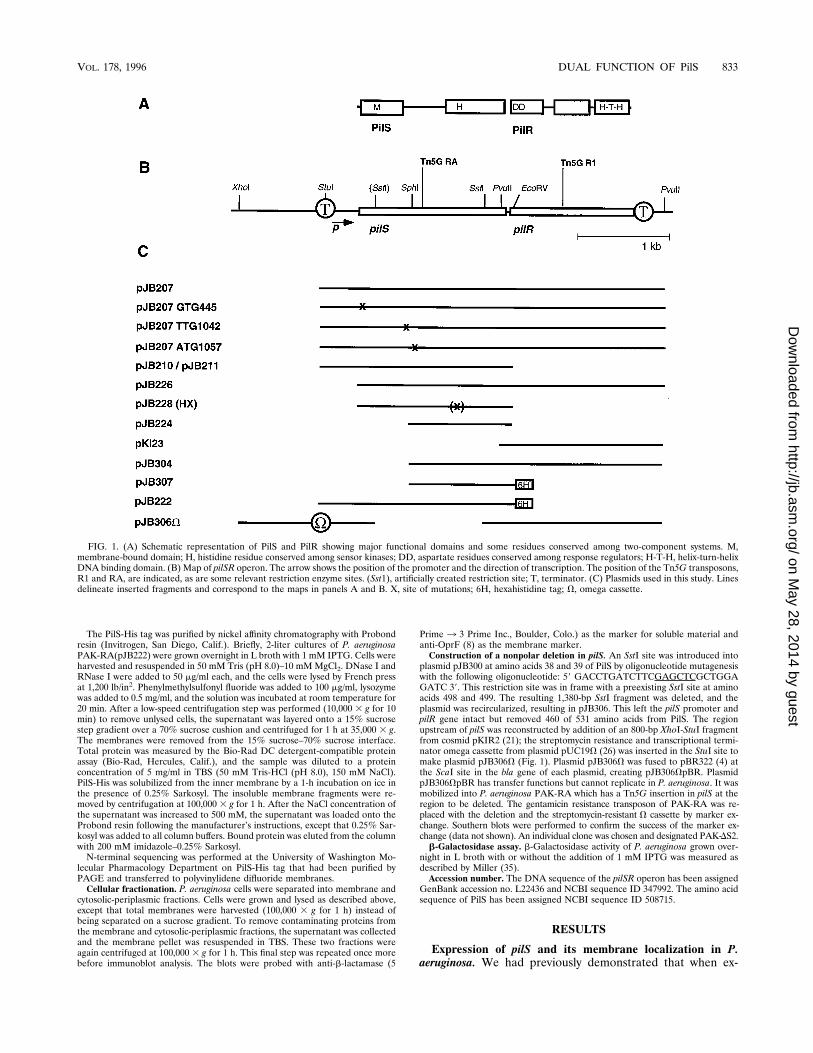
FIG. 1. (A) Schematic representation of PilS and PilR showing major functional domains and some residues conserved among two-component systems. M,membrane-bound domain; H, histidine residue conserved among sensor kinases; DD, aspartate residues conserved among response regulators; H-T-H, helix-turn-helixDNA binding domain. (B) Map of pilSR operon. The arrow shows the position of the promoter and the direction of transcription. The position of the Tn5G transposons,R1 and RA, are indicated, as are some relevant restriction enzyme sites. (Sst1), artificially created restriction site; T, terminator. (C) Plasmids used in this study. Linesdelineate inserted fragments and correspond to the maps in panels A and B. X, site of mutations; 6H, hexahistidine tag; V, omega cassette.
VOL. 178, 1996 DUAL FUNCTION OF PilS 833
on May 28, 2014 by guest
http://jb.asm.org/
Dow
nloaded from
pressed in E. coli under control of the T7 promoter-RNApolymerase, pilS is synthesized as a 40-kDa polypeptide (5).This raised the possibility that PilS, lacking putative transmem-brane domains, is cytoplasmic. This result was further sup-ported when a site-directed mutation of the predicted transla-tional start site (TTG at nucleotide position 1042 [TTG1042])failed to produce the 40-kDa protein in T7 expression exper-iments with E. coli. We have since produced antibody to PilSwhich has made it possible to examine the expression of pilSand the localization of its protein product in P. aeruginosa.Western blot (immunoblot) analysis of P. aeruginosa whole-
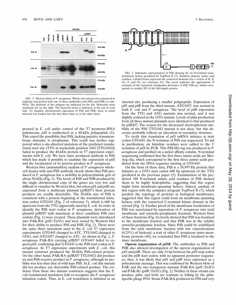
cell lysates with anti-PilS antibody clearly shows that PilS pro-duced in P. aeruginosa has a mobility in polyacrylamide gels ofabout 50-kDa (Fig. 2). The levels of PilS and PilR produced bythe single chromosomal copy of the genes are very low anddifficult to visualize by Western blot, but when pilS and pilR areexpressed from a multicopy plasmid (pJB207) their proteinproducts are readily detectable. The 50-kDa size of PilS isconsistent with a protein beginning at the translational initia-tion codon GTG445 (Fig. 2 of reference 5), which is 600 bpupstream from the TTG apparently used by E. coli. In order toidentify the PilS start codon in P. aeruginosa, derivatives ofplasmid pJB207 with mutations at three candidate PilS startcodons (Fig. 1) were created. These plasmids were introducedinto PAK-RA (pilS::Tn5G) and were tested by Western blotfor their ability to produce PilS and PilR (Fig. 2). These werethe same three mutations used in the E. coli T7 expressionexperiments: GTG445 changed to ATC, TTG1042 changed toGGG, and ATG1057 changed to ATC. As shown in Fig. 2, P.aeruginosa PAK-RA carrying pJB207 GTG445 failed to ex-press pilS, confirming that GTG445 is the PilS start codon in P.aeruginosa. In T7 expression experiments with E. coli, thismutant construct produced the 40-kDa PilS-related protein.On the other hand, PAK-RA (pJB207 TTG1042) did producean anti-PilS-reactive product in P. aeruginosa, although its mo-bility was less than that of wild-type PilS. In E. coli this mutantdid not produce any detectable PilS-related product. The evi-dence from these two mutant constructs suggests that the E.coli translational machinery fails to recognize the P. aeruginosainitiation codon. Thus, in E. coli translation is initiated at an
internal site, producing a smaller polypeptide. Expression ofpilS and pilR from the third mutant, ATG1057, was normal inboth E. coli and P. aeruginosa. The level of pilR expressionfrom the TTG and ATG mutants was normal, and it wasslightly reduced in the GTG mutant. Levels of pilin productionfrom all three mutant plasmids were identical to that producedby pJB207. The reason for the decreased electrophoretic mo-bility of the PilS TTG1042 mutant is not clear, but this de-crease probably reflects an alteration in secondary structure.To verify that translation of pilS mRNA initiates at start
codon GTG445, the N terminus of PilS was sequenced. To aidin purification, six histidine residues were added to the Cterminus of pilS by PCR. This PilS-His tag was produced in P.aeruginosa and purified on a nickel affinity column. N-terminalsequencing confirmed that the first three amino acids are Met-Arg-Ala, which correspond to the first three amino acids pre-dicted from the DNA sequence starting at GTG445.On the basis of these data, PilS is a 58,997-Da protein that
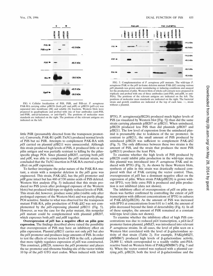
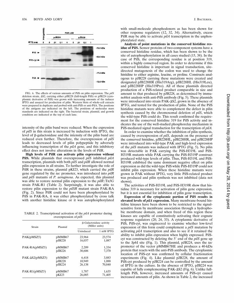
initiates at a GTG start codon 600 bp upstream of the TTGpredicted in the previous paper (5). Examination of the pre-dicted 180 N-terminal amino acid residues of PilS showedthem to be highly hydrophobic, suggesting that this regionmight form membrane-spanning helices. Indeed, analysis ofthis region with the computer program TopPred II (7), whichpredicts the topology of proteins in biological membranes,suggested that this region could form six membrane-spanninghelices, with the conserved C-terminal kinase domain in thecytosol (Fig. 3). Further proof of the membrane localization ofPilS was ascertained by separation of P. aeruginosa into totalmembrane and cytosolic-periplasmic fractions. Western blotsof these fractions (Fig. 4) clearly showed that PilS was localizedto the membrane fraction and that PilR was in the solublecytosolic-periplasmic fraction. Since PilS could be solubilizedfrom the total membrane fraction with low concentrations(0.25%) of Sarkosyl, a trait of other P. aeruginosa inner-mem-brane proteins (40), we concluded that PilS is localized to theinner membrane.Operon organization of pilSR. The antibodies to PilS and
PilR also allowed investigation of the operon organization ofpilS and pilR. There are only 14 bp between the pilS stop codonand the pilR start codon, with no apparent promoter sequenc-es; thus, it was likely that pilS and pilR were expressed as apolycistronic message. This was confirmed by Western blots ofPAK and the two transposon mutants PAK-RA (pilS::Tn5G)and PAK-R1 (pilR::Tn5G) (Fig. 2). Neither of these strains canproduce pilin, and both are resistant to killing by the pilin-specific phage PO4. Strain PAK-RA produced no PilS and very
FIG. 2. Western blots of P. aeruginosa. Whole-cell extracts were prepared intriplicate and probed with one of three antibodies (anti-PilS, anti-PilR, or anti-PilA). The positions of the antigens are indicated on the left. Molecular massstandards are on the right. The bacterial strain is indicated at the top of eachlane. To visualize chromosomal expression of PilS and PilR, twice as muchmaterial was loaded into the first three lanes as in the other lanes.
FIG. 3. Schematic representation of PilS showing the six N-terminal trans-membrane helices predicted by TopPred II (7). Numbers indicate amino acidresidues. Labeled boxes represent the conserved domains (for a review of H, N,G1, F, and G2, see reference 42). The arrow indicates the approximate Nterminus of the truncated cytoplasmic derivative of PilS, PilS-cyt, which corre-sponds to residue 205 of the full-length protein.
834 BOYD AND LORY J. BACTERIOL.
on May 28, 2014 by guest
http://jb.asm.org/
Dow
nloaded from
little PilR (presumably directed from the transposon promot-er). Conversely, PAK-R1 (pilR::Tn5G) produced normal levelsof PilS but no PilR. Attempts to complement PAK-RA withpilS carried on plasmid pJB211 were unsuccessful. Althoughthis strain produced high levels of PilS, it produced little or nopilin antigen and was partially resistant to killing by the pilus-specific phage PO4. Since plasmid pJB207, carrying both pilSand pilR, was able to complement the pilS mutant strain, weconcluded that the Tn5G insertion in PAK-RA exerted a polareffect on pilR expression.To further investigate the polar nature of the PAK-RA mu-
tant, a strain with a nonpolar deletion in the pilS gene wasengineered. This strain, PAK-DS2, has the pilS promoter andpilR gene intact but has 460 of 530 amino acids of PilS deleted.Western blot analysis (Fig. 5) indicated that this strain pro-duced no PilS (even after prolonged exposure of the Westernblots) but produced wild-type or slightly reduced levels of PilR.This strain did, however, produce some pilin (visible only afterprolonged exposure of the Western blot) and was partly phagePO4 sensitive. Similar to what was observed for the transposonmutant PAK-RA, pilin production of PAK-DS2 was not com-plemented by the pilS-expressing plasmid pJB211 (Fig. 5).However, as was the case for the polar mutant, this nonpolarpilS mutant could be complemented with plasmid pJB207,which expresses both pilS and pilR together.Overexpression of pilS has a negative effect on pilin gene
expression. The findings described above raised the possibilitythat overexpression of PilS may have an inhibitory effect onpilin expression. Plasmid pJB211 carries not only pilS but alsothe pilS promoter and produces large amounts of PilS. In orderto test the effects of various levels of PilS in the cell, a plasmidthat more tightly regulates expression of pilS was constructed.This construct, pJB228, removes the pilS promoter and placesthe tac promoter and ribosome binding site of the vector within10 bp of the pilS GTG start codon. When induced with 1mM
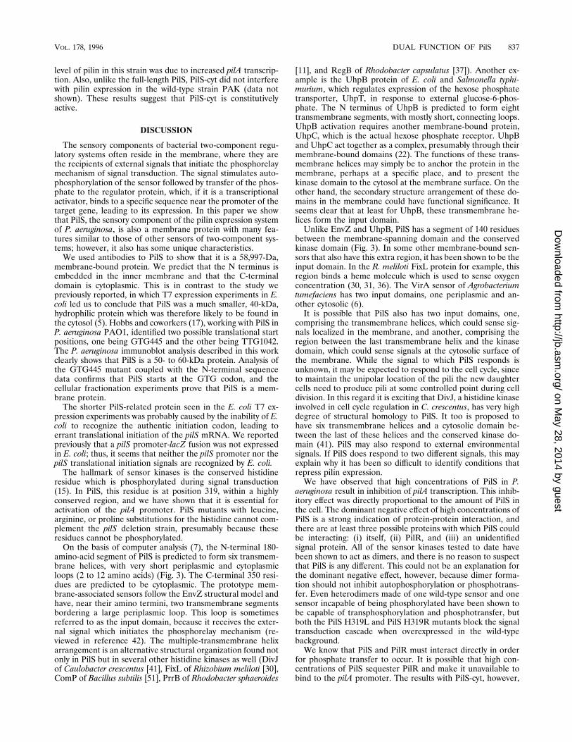
IPTG, P. aeruginosa(pJB228) produced much higher levels ofPilS (as visualized by Western blot [Fig. 5]) than did the samestrain carrying plasmids pJB207 or pJB211. When uninduced,pJB228 produced less PilS than did plasmids pJB207 andpJB211. The low level of expression from the uninduced plas-mid is presumably due to leakiness of the tac promoter. Incontrast to pJB211, the small amount of PilS produced byuninduced pJB228 was sufficient to complement PAK-DS2(Fig. 5). The only difference between these two strains is theamount of PilS, and the strain that produces the most PilS(pJB211) produces the least PilA.To examine whether the high levels of PilS produced by
pJB228 could inhibit pilin production in the wild-type strain,this plasmid was introduced into P. aeruginosa PAK and in-duced with IPTG (Fig. 5). As shown by these Western blots,synthesis of pilin in this strain was drastically reduced com-pared with that of PAK carrying the vector control. Thus,overexpression of pilS has a dominant negative effect on theexpression of pilin. When strain PAK(pJB228) is grown with-out IPTG, very little extra PilS is produced and pilin produc-tion is not inhibited (data not shown).The inhibitory effect of overexpression of pilS on pilin syn-
thesis was further confirmed by controlling the extent of pilStranscription with different concentrations of IPTG in culturesof PAK-DS2(pJB228). As the amount of PilS was increasedwith IPTG at concentrations from 0.01 to 1 mM, the amount ofpilin decreased beyond the limit of detectability (Fig. 6). In allof these samples, the amount of PilR remained at a constantwild-type level (data not shown).To examine whether the inhibitory effect of high PilS con-
centrations was due to reduced pilA transcription, a pilA-lacZpromoter fusion plasmid, pMSZ5, was introduced into relevantP. aeruginosa strains. In all cases, the level of pilin seen on aWestern blot correlated with the level of b-galactosidase ac-tivity of that strain (Table 2). For example, PAK(pMSZ5,pMMB67) produced high levels of b-galactosidase activity,24,000 U, which corresponded to a readily visible anti-PilA-reactive band on Western blots of PAK(pMMB67) (Fig. 5 and6). When the vector control was replaced with a plasmid car-rying pilS, pJB228, both the level of b-galactosidase and the
FIG. 4. Cellular localization of PilS, PilR, and PilS-cyt. P. aeruginosaPAK-RA carrying either pJB226 (both pilS and pilR) or pJB224 (pilS-cyt) wasseparated into membrane (M) and soluble (S) fractions. Western blots wereprepared in quadruplicate and probed with one of four antibodies (anti-PilS,anti-PilR, anti-b-lactamase, or anti-OprF). The positions of molecular massstandards are indicated on the right. The positions of the relevant antigens areindicated on the left.
FIG. 5. Complementation of P. aeruginosa pilS mutants. The wild-type P.aeruginosa PAK or the pilS in-frame deletion mutant PAK-DS2 carrying variouspilS plasmids was grown under noninducing or inducing conditions and assayedfor the production of pilin. Western blots of whole-cell extracts were prepared intriplicate and probed with one of three antibodies (anti-PilS, anti-pilR, or anti-PilA). The positions of the relevant antigens are indicated on the left. Thepositions of molecular mass standards are indicated on the right. The bacterialstrain and growth condition are indicated at the top of each lane. —, strainwithout a plasmid.
VOL. 178, 1996 DUAL FUNCTION OF PilS 835
on May 28, 2014 by guest
http://jb.asm.org/
Dow
nloaded from
intensity of the pilin band were reduced. When the expressionof pilS in this strain is increased by induction with IPTG, thelevel of b-galactosidase and the intensity of the pilin band arereduced even further. Therefore, the overexpression of pilSleads to decreased levels of pilin polypeptide by adverselyinfluencing transcription of the pilA gene, and this inhibitoryeffect does not involve alterations in the levels of PilR.High levels of PilR can activate pilin expression without
PilS. While plasmids that overexpressed pilS inhibited pilAtranscription, plasmids with both pilS and pilR allowed normalpilin expression in all strains. To further investigate the role ofPilR in these strains, plasmid pKI23, which carried the pilRgene regulated by the tac promoter, was introduced into pilRand pilS mutants of P. aeruginosa. As expected, this plasmidwas able to restore normal pilin expression to the pilR::Tn5Gstrain PAK-R1 (Table 2). Surprisingly, it was also able torestore pilin expression to the pilSR mutant strain PAK-RA(Fig. 2). Since PilR could not have been phosphorylated byPilS in PAK-RA, it was either phosphorylated by cross talkwith another histidine kinase or it was autophosphorylated
with small-molecule phosphodonors as has been shown forother response regulators (12, 32, 34). Alternatively, excessPilR may be able to activate pilA transcription in the unphos-phorylated state.Analysis of point mutations in the conserved histidine res-
idue of PilS. Sensor proteins of two-component systems have aconserved histidine residue, which has been shown to be thesite of autophosphorylation in all cases studied (15, 38). In thecase of PilS, the corresponding residue is at position 319,within a highly conserved region. In order to determine if thisconserved histidine is important in signal transduction, site-directed mutagenesis of the codon was used to change thehistidine to either arginine, leucine, or proline. Constructs anal-ogous to pJB228 carrying these mutations were created anddesignated pJB228HR (His319Arg), pJB228HL (His319Leu),and pJB228HP (His319Pro). All of these plasmids directedproduction of a PilS-related product comparable in size andamount to that produced by pJB228, as determined by immu-noblot analysis with anti-PilS antibody (Fig. 5). These plasmidswere introduced into strain PAK-DS2, grown in the absence ofIPTG, and tested for the production of pilin. None of the PilShistidine mutants were able to complement the defect in pilinsynthesis caused by the chromosomal deletion of pilS, whichthe wild-type PilS could do. This result confirmed the require-ment for the conserved histidine 319 for PilS activity and in-dicates the use of the well-studied phosphorelay mechanism inPilS-mediated signal transduction for the transcription of pilA.In order to examine whether the inhibition of pilin synthesis,
caused by overexpression of pilS, depends on the presence ofthe conserved histidine, pJB228HL, pJB228HR, and pJB228HPwere introduced into wild-type PAK and high-level expressionof the pilS mutants was induced with IPTG (Fig. 5). No pilinwas detectable in PAK carrying the PilS-H319L and PilS-H319R mutants while PAK carrying the PilS-H319P constructproduced wild-type levels of pilin. Thus, PilS-H319L and PilS-H319R exhibited the same dominant negative effect on pilinexpression as did the wild-type PilS while PilS-H319P could notinhibit pilin expression. When these histidine mutants weregrown in PAK without IPTG, very little PilS-related productwas produced and pilin synthesis was not inhibited (data notshown).The activities of PilS-H319L and PilS-H319R show that his-
tidine 319 is necessary for activation of pilin gene expression,but it is not essential for inhibition of pilin gene transcription.Expression of the cytoplasmic portion of PilS results in
elevated levels of pilA expression.Many membrane-bound his-tidine kinases have been shown to be restricted to the signal-sensitive form by membrane association through a hydropho-bic membrane domain, and when freed of this region thesekinases are capable of constitutively activating their cognateresponse regulators (20, 24, 33). A cytoplasmic derivative ofPilS, PilS-cyt, was engineered to examine whether low-levelexpression of this form could complement a pilS mutation byactivating pilA transcription and also to see if it retained theability to inhibit pilin expression when highly expressed. PilS-cyt was constructed by deleting the 59 end of the pilS gene upto the SphI site (Fig. 1). This plasmid, pJB224, uses the tacpromoter of the vector pMMB67HE and produces a 40-kDaprotein that reacts with the anti-PilS antibody. The cytoplasmiclocation of PilS-cyt was confirmed by cellular fractionationexperiments (Fig. 4). Like plasmid pJB228, the amount ofPilS-cyt produced by pJB224 can be controlled by the amountof IPTG in the culture. In the absence of IPTG, pJB224 wascapable of fully complementing PAK-DS2 (Fig. 6). Unlike full-length PilS, however, increased amounts of PilS-cyt causedincreased amounts of pilin. As shown in Table 2, the increased
FIG. 6. The effects of various amounts of PilS on pilin expression. The pilSdeletion strain, DS2, carrying either pJB228 (full-length PilS) or pJB224 (cyto-plasmic derivative of PilS) was grown with increasing amounts of the inducerIPTG and assayed for production of pilin. Western blots of whole-cell extractswere prepared in duplicate and probed with anti-PilS or anti-PilA. The positionsof the antigens are indicated on the left. The positions of molecular massstandards are indicated on the right. The bacterial strain, plasmid, and growthcondition are indicated at the top of each lane.
TABLE 2. Transcriptional activation of the pilA promoter duringoverexpression of pilS
Strain Plasmid
b-Galactosidase activity(Miller units)
Uninduced 1 mM IPTG
PAK(pMSZ5) pMMB67 23,964 23,574pJB228 16,037 1,087
PAK-RA(pMSZ5) pMMB67 2,209 1,334pJB226 30,606 7,370
PAK-DS2(pMSZ5) pMMB67 6,418 3,883pJB228 10,949 1,088pJB224 26,138 70,840
PAK-R1(pMSZ5) pMMB67 1,787 1,635pKI23 26,085 71,405
836 BOYD AND LORY J. BACTERIOL.
on May 28, 2014 by guest
http://jb.asm.org/
Dow
nloaded from
level of pilin in this strain was due to increased pilA transcrip-tion. Also, unlike the full-length PilS, PilS-cyt did not interferewith pilin expression in the wild-type strain PAK (data notshown). These results suggest that PilS-cyt is constitutivelyactive.
DISCUSSION
The sensory components of bacterial two-component regu-latory systems often reside in the membrane, where they arethe recipients of external signals that initiate the phosphorelaymechanism of signal transduction. The signal stimulates auto-phosphorylation of the sensor followed by transfer of the phos-phate to the regulator protein, which, if it is a transcriptionalactivator, binds to a specific sequence near the promoter of thetarget gene, leading to its expression. In this paper we showthat PilS, the sensory component of the pilin expression systemof P. aeruginosa, is also a membrane protein with many fea-tures similar to those of other sensors of two-component sys-tems; however, it also has some unique characteristics.We used antibodies to PilS to show that it is a 58,997-Da,
membrane-bound protein. We predict that the N terminus isembedded in the inner membrane and that the C-terminaldomain is cytoplasmic. This is in contrast to the study wepreviously reported, in which T7 expression experiments in E.coli led us to conclude that PilS was a much smaller, 40-kDa,hydrophilic protein which was therefore likely to be found inthe cytosol (5). Hobbs and coworkers (17), working with PilS inP. aeruginosa PAO1, identified two possible translational startpositions, one being GTG445 and the other being TTG1042.The P. aeruginosa immunoblot analysis described in this workclearly shows that PilS is a 50- to 60-kDa protein. Analysis ofthe GTG445 mutant coupled with the N-terminal sequencedata confirms that PilS starts at the GTG codon, and thecellular fractionation experiments prove that PilS is a mem-brane protein.The shorter PilS-related protein seen in the E. coli T7 ex-
pression experiments was probably caused by the inability of E.coli to recognize the authentic initiation codon, leading toerrant translational initiation of the pilS mRNA. We reportedpreviously that a pilS promoter-lacZ fusion was not expressedin E. coli; thus, it seems that neither the pilS promoter nor thepilS translational initiation signals are recognized by E. coli.The hallmark of sensor kinases is the conserved histidine
residue which is phosphorylated during signal transduction(15). In PilS, this residue is at position 319, within a highlyconserved region, and we have shown that it is essential foractivation of the pilA promoter. PilS mutants with leucine,arginine, or proline substitutions for the histidine cannot com-plement the pilS deletion strain, presumably because theseresidues cannot be phosphorylated.On the basis of computer analysis (7), the N-terminal 180-
amino-acid segment of PilS is predicted to form six transmem-brane helices, with very short periplasmic and cytoplasmicloops (2 to 12 amino acids) (Fig. 3). The C-terminal 350 resi-dues are predicted to be cytoplasmic. The prototype mem-brane-associated sensors follow the EnvZ structural model andhave, near their amino termini, two transmembrane segmentsbordering a large periplasmic loop. This loop is sometimesreferred to as the input domain, because it receives the exter-nal signal which initiates the phosphorelay mechanism (re-viewed in reference 42). The multiple-transmembrane helixarrangement is an alternative structural organization found notonly in PilS but in several other histidine kinases as well (DivJof Caulobacter crescentus [41], FixL of Rhizobium meliloti [30],ComP of Bacillus subtilis [51], PrrB of Rhodobacter sphaeroides
[11], and RegB of Rhodobacter capsulatus [37]). Another ex-ample is the UhpB protein of E. coli and Salmonella typhi-murium, which regulates expression of the hexose phosphatetransporter, UhpT, in response to external glucose-6-phos-phate. The N terminus of UhpB is predicted to form eighttransmembrane segments, with mostly short, connecting loops.UhpB activation requires another membrane-bound protein,UhpC, which is the actual hexose phosphate receptor. UhpBand UhpC act together as a complex, presumably through theirmembrane-bound domains (22). The functions of these trans-membrane helices may simply be to anchor the protein in themembrane, perhaps at a specific place, and to present thekinase domain to the cytosol at the membrane surface. On theother hand, the secondary structure arrangement of these do-mains in the membrane could have functional significance. Itseems clear that at least for UhpB, these transmembrane he-lices form the input domain.Unlike EnvZ and UhpB, PilS has a segment of 140 residues
between the membrane-spanning domain and the conservedkinase domain (Fig. 3). In some other membrane-bound sen-sors that also have this extra region, it has been shown to be theinput domain. In the R. meliloti FixL protein for example, thisregion binds a heme molecule which is used to sense oxygenconcentration (30, 31, 36). The VirA sensor of Agrobacteriumtumefaciens has two input domains, one periplasmic and an-other cytosolic (6).It is possible that PilS also has two input domains, one,
comprising the transmembrane helices, which could sense sig-nals localized in the membrane, and another, comprising theregion between the last transmembrane helix and the kinasedomain, which could sense signals at the cytosolic surface ofthe membrane. While the signal to which PilS responds isunknown, it may be expected to respond to the cell cycle, sinceto maintain the unipolar location of the pili the new daughtercells need to produce pili at some controlled point during celldivision. In this regard it is exciting that DivJ, a histidine kinaseinvolved in cell cycle regulation in C. crescentus, has very highdegree of structural homology to PilS. It too is proposed tohave six transmembrane helices and a cytosolic domain be-tween the last of these helices and the conserved kinase do-main (41). PilS may also respond to external environmentalsignals. If PilS does respond to two different signals, this mayexplain why it has been so difficult to identify conditions thatrepress pilin expression.We have observed that high concentrations of PilS in P.
aeruginosa result in inhibition of pilA transcription. This inhib-itory effect was directly proportional to the amount of PilS inthe cell. The dominant negative effect of high concentrations ofPilS is a strong indication of protein-protein interaction, andthere are at least three possible proteins with which PilS couldbe interacting: (i) itself, (ii) PilR, and (iii) an unidentifiedsignal protein. All of the sensor kinases tested to date havebeen shown to act as dimers, and there is no reason to suspectthat PilS is any different. This could not be an explanation forthe dominant negative effect, however, because dimer forma-tion should not inhibit autophosphorylation or phosphotrans-fer. Even heterodimers made of one wild-type sensor and onesensor incapable of being phosphorylated have been shown tobe capable of transphosphorylation and phosphotransfer, butboth the PilS H319L and PilS H319R mutants block the signaltransduction cascade when overexpressed in the wild-typebackground.We know that PilS and PilR must interact directly in order
for phosphate transfer to occur. It is possible that high con-centrations of PilS sequester PilR and make it unavailable tobind to the pilA promoter. The results with PilS-cyt, however,
VOL. 178, 1996 DUAL FUNCTION OF PilS 837
on May 28, 2014 by guest
http://jb.asm.org/
Dow
nloaded from
counter this argument. We know that PilS-cyt also interactswith PilR because PilS-cyt can complement a pilS deletionstrain, but this mutant does not demonstrate the dominantnegative effect when overexpressed, and instead it has a dom-inant positive effect on pilin expression. This suggests that thedominant negative effect of overproduced PilS is not due toaltered stoichiometry with PilR.The third possibility is that PilS interacts with another cel-
lular component which controls its stimulatory or inhibitoryactivities. This cellular component may be the actual PilS ac-tivation signal, or it may be the receptor for the signal. Webelieve that it is the altered stoichiometry of PilS and thisputative factor that leads to inhibition of pilin synthesis.Many sensors of two-component systems, in addition to
functioning as kinases, are also phosphatases specific to theircognate regulators (1). These two enzymatic activities deter-mine the steady-state level of the active (phosphorylated) formof the regulators in the cell. We hypothesize that PilS carriesout both of these activities: it is a kinase when activated by asignal, and it is a phosphatase in the absence of such a signal.Presumably the amount of signal is limited, so that most of theexcess PilS cannot interact with it, leading to higher levels ofPilS in the phosphatase state than in the kinase state. This inturn results in accumulation of inactive, unphosphorylatedPilR. This hypothesis also explains the unexpected result thatP. aeruginosa with a nonpolar deletion in pilS can still producelow levels of pilin. Like other response regulators, PilR canpresumably be phosphorylated by other means (cross talk withother kinases or autophosphorylation with small phosphodo-nors), and without its cognate phosphatase the phospho-PilRform will accumulate, leading to transcriptional activation.Availability of purified PilS and PilR makes it possible to testthis hypothesis biochemically, and such work is in progress.In support of this model, overexpression of uhpB, the sensor
kinase of the E. coli hexose phosphate uptake system, reducesthe level of the hexose transporter, UhpT. This may be aconsequence of the altered ratio of UhpB to the glucose-6-phosphate receptor, UhpC (25).The PilS-mediated inhibition of pilA transcription does not
depend on the autokinase activity of PilS, since the histidinesubstitution mutants PilS-H319L and PilS-H319R also inhibitpilA transcription when overexpressed in the wild-type back-ground. This effect can be explained if these mutant proteinsmaintain their phosphatase activity and their ability to interactwith the putative signal. In support of this hypothesis, it hasbeen shown that the conserved histidine residue of sensorkinases is not required for phosphatase activity (reviewed inreference 42). PilS-H319P did not demonstrate the dominantnegative effect, suggesting either that it cannot interact withthe putative signal or that it does not have phosphatase activity.It is likely that the structural changes introduced by the prolinesubstitution would drastically alter its ability to interact withother proteins. The dominant positive activity of the PilS-cytmutant suggests that it does not require interaction with thesignaling component and that it is constitutively a kinase. In-deed, this is the case when other membrane-bound sensorkinases have been freed of their transmembrane domains (20,24, 33).We have shown that overexpression of pilR in the absence of
PilS allows transcription of pilA. Although this effect has beenseen with a number of other response regulators (14, 45), thereason for it is not clear. Phosphorylation of response regula-tors modulates transcription, but whether this modulation isdue to alteration in DNA binding affinity, oligomerizationrates, or other functions is the source of some debate, and thecause of modulation may not be the same for all members of
this class. Presumably there is an equilibrium between activeand inactive forms that is normally controlled by phosphory-lation, but even in the absence of phosphorylation there arestill a few active forms in the cell. When response regulatorgenes are overexpressed, the total number of active forms isincreased and transcription can proceed. Alternatively, in theabsence of their kinases, regulators may be phosphorylated byother means. Experiments are under way to determine the casefor PilR.In conclusion, this study shows that PilS, the sensor kinase of
a two-component system responsible for regulation of pilingene transcription in P. aeruginosa, is a 58,997-Da inner mem-brane protein. We propose that PilS interacts not only with itscognate regulator, PilR, but also with another cellular compo-nent, most likely a protein, located either in the inner mem-brane or at the cytosolic surface of the inner membrane. Thisputative component may be responsible for switching PilS fromthe PilR kinase state to the phospho-PilR-phosphatase stateand may be either the actual PilS activation signal or an inter-mediary signal-binding protein.
ACKNOWLEDGMENTS
This work was supported by NIH grant AI 32624. J.B. was supportedby postdoctoral fellowship grant F494 from the Cystic Fibrosis Foun-dation.We thank Leah Turner and Tim Motley for critical reading of the
manuscript.
REFERENCES
1. Aiba, H., F. Nakasai, S. Mizushima, and T. Mizuno. 1989. Evidence for thephysiological importance of the phosphotransfer between the two regulatorycomponents, EnvZ and OmpR, in osmoregulation in Escherichia coli. J. Biol.Chem. 264:14090–14094.
2. Ausubel, F., R. Brent, R. Kingston, D. Moore, J. Seidman, and J. Smith.1990. Current protocols in molecular biology. John Wiley and Sons, NewYork.
3. Birnboim, H. C., and J. Doly. 1979. A rapid alkaline extraction procedure forscreening recombinant plasmid DNA. Nucleic Acids Res. 7:1513–1523.
4. Bolivar, F., R. L. Rodriguez, P. J. Greene, M. C. Betlach, H. L. Heynecker,H. W. Boyer, and J. H. Crosa. 1977. Construction and characterization ofnew cloning vehicles. II. A multipurpose cloning system. Gene 2:95–101.
5. Boyd, J. M., T. Koga, and S. Lory. 1994. Identification and characterizationof PilS, an essential regulator of pilin expression in Pseudomonas aeruginosa.Mol. Gen. Genet. 243:565–574.
6. Chang, C. H., and S. C. Winans. 1992. Functional roles assigned to theperiplasmic, linker, and receiver domains of the Agrobacterium tumefaciensVirA protein. J. Bacteriol. 174:7033–7039.
7. Claros, M. G., and G. von Heijne. 1994. TopPred II: an improved softwarefor membrane protein structure predictions. Comput. Appl. Biosci. 10:685–686.
8. Counts, G. W., R. W. Schwartz, B. K. Ulness, D. J. Hamilton, M. J. Rosok,M. D. Cunningham, M. R. Tam, and R. P. Darveau. 1977. Evaluation of animmunofluorescent-antibody test for rapid identification of Pseudomonasaeruginosa in blood cultures. J. Clin. Microbiol. 26:1161–1165.
9. Davis, B. D., and E. S. Mingioli. 1950. Mutants of Escherichia coli requiringvitamin B-12. J. Bacteriol. 60:17–28.
10. Ditta, G. S., S. Stanfield, D. Corbin, and D. R. Helinski. 1980. Broad hostrange DNA cloning system for gram-negative bacteria: construction of agene bank of Rhizobium meliloti. Proc. Natl. Acad. Sci. USA 77:7347–7351.
11. Eraso, J. M., and S. Kaplan. 1995. Oxygen-insensitive synthesis of the pho-tosynthetic membranes of Rhodobacter sphaeroides: a mutant histidine ki-nase. J. Bacteriol. 177:2695–2706.
12. Feng, J., M. R. Atkinson, W. McCleary, J. B. Stock, B. L. Wanner, and A. J.Ninfa. 1992. Role of phosphorylated metabolic intermediates in the regula-tion of glutamine synthetase synthesis in Escherichia coli. J. Bacteriol. 174:6061–6070.
13. Furste, J. P., W. Pansegrau, R. Frank, H. Bloecker, P. Scholz, M. Bagdasar-ian, and E. Lanka. 1986. Molecular cloning of the plasmid RP4 primaseregion in a multi-host-range tacP expression vector. Gene 48:119–131.
14. Hertig, C., R. Y. Li, A. M. Louarn, A. M. Garnerone, M. David, J. Batut, D.Kahn, and P. Boistard. 1989. Rhizobium meliloti regulatory gene fixJ acti-vates transcription of R. meliloti nifA and fixK genes in Escherichia coli. J.Bacteriol. 171:1736–1738.
15. Hess, J. F., R. B. Bourret, and M. I. Simon. 1988. Histidine phosphorylationand phosphoryl group transfer in bacterial chemotaxis. Nature (London)
838 BOYD AND LORY J. BACTERIOL.
on May 28, 2014 by guest
http://jb.asm.org/
Dow
nloaded from
336:139–143.16. Hess, J. F., K. Oosawa, N. Kaplan, and M. I. Simon. 1988. Phosphorylation
of three proteins in the signaling pathway of bacterial chemotaxis. Cell53:79–87.
17. Hobbs, M., E. S. Collie, P. D. Free, S. P. Livingston, and J. S. Mattick. 1993.PilS and PilR, a two-component transcriptional regulatory system controllingexpression of type 4 fimbriae in Pseudomonas aeruginosa. Mol. Microbiol.7:669–682.
18. Hobbs, M., and J. S. Mattick. 1993. Common components in the assembly oftype 4 fimbriae, DNA transfer systems, filamentous phage and protein-secretion apparatus: a general system for the formation of surface-associatedprotein complexes. Mol. Microbiol. 10:233–243.
19. Igo, M. M., A. J. Ninfa, J. B. Stock, and T. J. Silhavy. 1989. Phosphorylationand dephosphorylation of a bacterial transcriptional activator by a trans-membrane receptor. Genes Dev. 3:1725–1734.
20. Igo, M. M., and T. J. Silhavy. 1988. EnvZ, a transmembrane environmentalsensor of Escherichia coli K-12, is phosphorylated in vitro. J. Bacteriol.170:5971–5973.
21. Ishimoto, K. S., and S. Lory. 1992. Identification of pilR, which encodes atranscriptional activator of the Pseudomonas aeruginosa pilin gene. J. Bac-teriol. 174:3514–3521.
22. Island, M. D., and R. J. Kadner. 1993. Interplay between the membrane-associated UhpB and UhpC regulatory proteins. J. Bacteriol. 175:5028–5034.
23. Jin, S., K. S. Ishimoto, and S. Lory. 1994. PilR, a transcriptional regulator ofpiliation in Pseudomonas aeruginosa, binds to a cis-acting sequence upstreamof the pilin gene promoter. Mol. Microbiol. 14:1049–1057.
24. Jin, S., T. Roitsch, R. G. Ankenbauer, M. P. Gordon, and E. W. Nester. 1990.The VirA protein of Agrobacterium tumefaciens is autophosphorylated and isessential for vir gene regulation. J. Bacteriol. 172:525–530.
25. Kadner, R. J., M. D. Island, J. L. Dahl, and C. A. Webber. 1994. A trans-membrane signalling complex controls transcription of the Uhp sugar phos-phate transport system. Res. Microbiol. 145:381–387.
26. Koga, T., K. Ishimoto, and S. Lory. 1993. Genetic and functional character-ization of the gene cluster specifying expression of Pseudomonas aeruginosapili. Infect. Immun. 61:1371–1377.
27. Kunkel, T. A., J. D. Roberts, and R. A. Zakour. 1987. Rapid and efficientsite-specific mutagenesis without phenotypic selection. Methods Enzymol.154:367–382.
28. Laemmli, U. K. 1970. Cleavage of structural proteins during the assembly ofthe head of bacteriophage T4. Nature (London) 227:680–685.
29. Leong, S. A., G. S. Ditta, and D. R. Helinski. 1982. Heme biosynthesis inRhizobium. Identification of a cloned gene coding for delta-aminolevulinicacid synthetase from Rhizobium meliloti. J. Biol. Chem. 257:8724–8730.
30. Lois, A. F., G. S. Ditta, and D. R. Helinski. 1993. The oxygen sensor FixL ofRhizobium meliloti is a membrane protein containing four possible trans-membrane segments. J. Bacteriol. 175:1103–1109.
31. Lois, A. F., M. Weinstein, G. S. Ditta, and D. R. Helinski. 1993. Autophos-phorylation and phosphatase activities of the oxygen-sensing protein FixL ofRhizobium meliloti are coordinately regulated by oxygen. J. Biol. Chem.268:4370–4375.
32. Lukat, G. S., W. R. McCleary, A. M. Stock, and J. B. Stock. 1992. Phosphor-ylation of bacterial response regulator proteins by low molecular weightphospho-donors. Proc. Natl. Acad. Sci. USA 89:718–722.
33. Makino, K., H. Shinagawa, M. Amemura, T. Kawamoto, M. Yamada, and A.Nakata. 1989. Signal transduction in the phosphate regulon of Escherichiacoli involves phosphotransfer between PhoR and PhoB proteins. J. Mol.Biol. 210:551–559.
34. McCleary, W. R., and J. B. Stock. 1994. Acetyl phosphate and the activationof two-component response regulators. J. Biol. Chem. 269:31567–31572.
35. Miller, J. H. 1972. Experiments in molecular genetics. Cold Spring HarborLaboratory, Cold Spring Harbor, N.Y.
36. Monson, E. K., G. S. Ditta, and D. R. Helinski. 1995. The oxygen sensorprotein, FixL, of Rhizobium meliloti. Role of histidine residues in hemebinding, phosphorylation, and signal transduction. J. Biol. Chem. 270:5243–5250.
37. Mosley, C. S., J. Y. Suzuki, and C. E. Bauer. 1994. Identification and mo-lecular genetic characterization of a sensor kinase responsible for coordi-nately regulating light harvesting and reaction center gene expression inresponse to anaerobiosis. J. Bacteriol. 176:7566–7573. (Erratum, 177:3359,1995.)
38. Ninfa, A. J., and R. L. Bennett. 1991. Identification of the site of autophos-phorylation of the bacterial protein kinase/phosphatase NRII. J. Biol. Chem.266:6888–6893.
39. Nunn, D., S. Bergman, and S. Lory. 1990. Products of three accessory genes,pilB, pilC, and pilD, are required for biogenesis of Pseudomonas aeruginosapili. J. Bacteriol. 172:2911–2919.
40. Nunn, D. N., and S. Lory. 1993. Cleavage, methylation, and localization ofthe Pseudomonas aeruginosa export proteins XcpT, -U, -V, and -W. J. Bac-teriol. 175:4375–4382.
41. Ohta, N., T. Lane, E. G. Ninfa, J. M. Sommer, and A. Newton. 1992. Ahistidine protein kinase homologue required for regulation of bacterial celldivision and differentiation. Proc. Natl. Acad. Sci. USA 89:10297–10301.
42. Parkinson, J. S., and E. C. Kofoid. 1992. Communication modules in bac-terial signaling proteins. Annu. Rev. Genet. 26:71–112.
43. Ramphal, R., L. Koo, K. S. Ishimoto, P. A. Totten, J. C. Lara, and S. Lory.1991. Adhesion of Pseudomonas aeruginosa pilin-deficient mutants to mucin.Infect. Immun. 59:1307–1311.
44. Sanger, F. S., S. Nicklen, and A. R. Coulson. 1977. DNA sequencing withchain-terminating inhibitors. Proc. Natl. Acad. Sci. USA 74:5463–5467.
45. Shattuck, E. D. M., and R. J. Kadner. 1983. Molecular cloning of the uhpregion and evidence for a positive activator for expression of the hexosephosphate transport system of Escherichia coli. J. Bacteriol. 155:1062–1070.
46. Simpson, D. A., R. Ramphal, and S. Lory. 1992. Genetic analysis of Pseudo-monas aeruginosa adherence: distinct genetic loci control attachment toepithelial cells and mucins. Infect. Immun. 60:3771–3779.
47. Strom, M. S., and S. Lory. 1993. Structure-function and biogenesis of thetype IV pili. Annu. Rev. Microbiol. 47:565–596.
48. Totten, P. A., J. C. Lara, and S. Lory. 1990. The rpoN gene product ofPseudomonas aeruginosa is required for expression of diverse genes, includ-ing the flagellin gene. J. Bacteriol. 172:389–396.
49. Towbin, H., T. Staehelin, and J. Gordon. 1979. Electrophoretic transfer ofproteins from polyacrylamide gels to nitrocellulose sheets: procedure andsome applications. Proc. Natl. Acad. Sci. USA 76:4350–4354.
50. Venkitaraman, A. R. 1989. Use of modified T7 DNA polymerase (sequenaseversion 2.0) for oligonucleotide site-directed mutagenesis. Nucleic AcidsRes. 17:3314. (Erratum, 18:400, 1990.)
51. Weinrauch, Y., R. Penchev, E. Dubnau, I. Smith, and D. Dubnau. 1990. ABacillus subtilis regulatory gene product for genetic competence and sporu-lation resembles sensor protein members of the bacterial two-componentsignal-transduction systems. Genes Dev. 4:860–872.
52. Woods, D. E., D. C. Straus, W. J. Johanson, V. K. Berry, and J. A. Bass. 1980.Role of pili in adherence of Pseudomonas aeruginosa to mammalian buccalepithelial cells. Infect. Immun. 29:1146–1151.
VOL. 178, 1996 DUAL FUNCTION OF PilS 839
on May 28, 2014 by guest
http://jb.asm.org/
Dow
nloaded from
Top Related
Copyright © 2022 FDOKUMEN

