


Bahasa
Halaman
Hukum

Determinants of TRPV4 Activity following SelectiveActivation by Small Molecule Agonist GSK1016790AMin Jin, Zizhen Wu, Ling Chen, Jose Jaimes, Diana Collins, Edgar T. Walters, Roger G. O’Neil*
Department of Integrative Biology and Pharmacology, The University of Texas Health Science Center, Houston, Texas, United States of America
Abstract
TRPV4 (Transient Receptor Potential Vanilloid 4) channels are activated by a wide range of stimuli, including hypotonicstress, non-noxious heat and mechanical stress and some small molecule agonists (e.g. phorbol ester 4a-PDD).GSK1016790A (GSK101) is a recently discovered specific small molecule agonist of TRPV4. Its effects on physicaldeterminants of TRPV4 activity were evaluated in HeLa cells transiently transfected with TRPV4 (HeLa-TRPV4). GSK101(10 nM) causes a TRPV4 specific Ca2+ influx in HeLa-TRPV4 cells, but not in control transfected cells, which can be inhibitedby ruthenium red and Ca2+-free medium more significantly at the early stage of the activation rather than the late stage,reflecting apparent partial desensitization. Western blot analysis showed that GSK101 activation did not induce an increasein TRPV4 expression at the plasma membrane, but caused an immediate and sustained downregulation of TRPV4 on theplasma membrane in HeLa-TRPV4 cells. Patch clamp analysis also revealed an early partial desensitization of the channelwhich was Ca2+-independent. FRET analysis of TRPV4 subunit assembly demonstrated that the GSK101-induced TRPV4channel activation/desensitization was not due to alterations in homotetrameric channel formation on the plasmamembrane. It is concluded that GSK101 specifically activates TRPV4 channels, leading to a rapid partial desensitization anddownregulation of the channel expression on the plasma membrane. TRPV4 subunit assembly appears to occur duringtrafficking from the ER/Golgi to the plasma membrane and is not altered by agonist stimulation.
Citation: Jin M, Wu Z, Chen L, Jaimes J, Collins D, et al. (2011) Determinants of TRPV4 Activity following Selective Activation by Small Molecule AgonistGSK1016790A. PLoS ONE 6(2): e16713. doi:10.1371/journal.pone.0016713
Editor: Henning Ulrich, University of Sao Paulo, Brazil
Received September 10, 2010; Accepted January 11, 2011; Published February 14, 2011
Copyright: � 2011 Jin et al. This is an open-access article distributed under the terms of the Creative Commons Attribution License, which permits unrestricteduse, distribution, and reproduction in any medium, provided the original author and source are credited.
Funding: Funding was received from National Institutes of Health R01 DK70950 and NIH R21 DE018522. The funders had no role in study design, data collectionand analysis, decision to publish, or preparation of the manuscript.
Competing Interests: The authors have declared that no competing interests exist.
* E-mail: [email protected]
Introduction
TRPV4 is a non-selective Ca2+ - permeable cation channel that
belongs to the TRP superfamily. It is ubiquitously expressed in
various tissues such as renal epithelia, lung epithelia, vascular
endothelia, and nervous systems [1–4]. Studies show that TRPV4
is activated by hypotonic stress, moderate heat, mechanical stress,
phorbol ester (4a-PDD) and arachidonic acid metabolites [5–14].
GSK101 (GSK1016790A) is a novel activator of TRPV4, which
has been shown to be a more specific and potent activator (at
nanomolarlevels) as compared to the traditional 4a-PDD[15,16].
Recent studies show that GSK101 stimulates TRPV4 in multiple
cell types including endothelial cells, urinary smooth muscle cells,
urothelial cells and HEK-293 cells over-expressing TRPV4 [15–
18]. Being a novel TRPV4 agonist, the signaling pathway of
GSK101 is not well understood. In addition to the various
signaling pathways that may modulate the channel activity, ion
channel activation also involves subunit assembly/disassembly,
trafficking, insertion and endocytosis of functional channel to/
from the plasma membrane. Limited studies on TRP channel
trafficking have shown, however, that some stimuli can cause the
exocytosis and insertion of the channel into the plasma membrane,
thus contributing to channel activity[19–23], while other studies
have shown that TRPV4 is down regulated under angiotensin
stimulation in rat smooth muscle cells [24]. It has also been
demonstrated that TRPV4 channels at the plasma membrane
typically reflect a homotetrameric assembly [25,26], but hetero-
tetramer structures can form with other TRP family isoforms
which lead to altered channel function [27,28]. Indeed, a recent
study of TRPP2 subunit structure demonstrated that subunit
disassembly might be an important component of channel
inactivation [29,30]. Hence, the activity of TRPV4 at the plasma
membrane is likely a dynamic process reflecting both abundance
and subunit assembly. This regulation may, of course, also include
the role of more traditional modulating pathways, including both
phosphorylation and nitrosylation events, which can contribute to
channel regulation [31,32].
In this study, we set out to investigate the relationship between
GSK101-induced TRPV4 activation and its expression and
subunit assembly at the plasma membrane as the physical
determinants of TRPV4 activity. It was found that agonist
stimulation did not alter the apparent subunit assembly within
the plasma membrane, but it induced an early rapid downregu-
lation of TRPV4 expression at the plasma membrane that was
associated with a rapid desensitization of the TRPV4 channel in a
Ca2+-independent manner.
Results
GSK101 stimulates Ca2+ influx in HeLa-TRPV4 cellsRT-PCR using HeLa cell mRNA and primer pairs designed for
TRPV channels showed that HeLa cells do not expressed TRPV4
channels at the mRNA level. Likewise, Western blot using anti-
TRPV4 antibody also could not detect TRPV4 at the protein level
PLoS ONE | www.plosone.org 1 February 2011 | Volume 6 | Issue 2 | e16713
in wild type HeLa cells (data not shown). Therefore, HeLa cells
were used as an overexpression cell model when transiently
transfected with a TRPV4 containing plasmid similar to that done
previously in HEK and CHO cells [5,13].
In HeLa-TRPV4 cells, Ca2+ influx was stimulated by GSK101
in a dose-dependent manner as shown in Figure 1. The GSK101 -
Ca2+ influx relationship could be fitted by asigmoidal dose-
responsefunction, which yielded an EC50 of 3.3 nM for GSK101
stimulation. GSK101 at 10 nM displayed a near maximum
stimulation and was, therefore, used as the preferred concentration
to activate TRPV4 channels in this study.
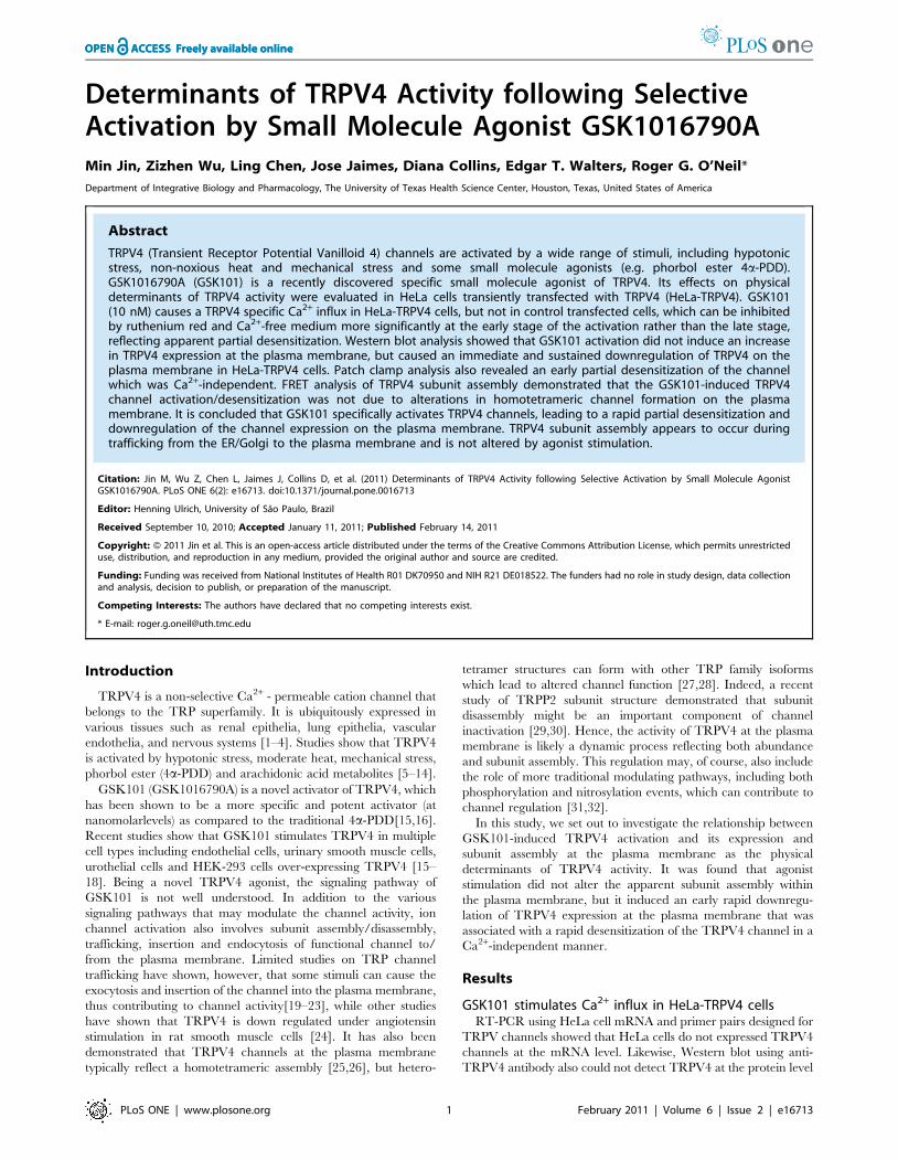
In order to test the specificity of GSK101 on TRPV4 activation,
HeLa cells transfected with mVenus-N1 vector were compared
with HeLa cells transfected with TRPV4-mVenus. Only cells
positively identified as expressing TRPV4-mVenus (via excitation
at 488-nm wavelength) were chosen for the study. GSK101 did
not cause an increase in intracellular Ca2+ concentration in HeLa
cells transfected with mVenus-N1 vector, while HeLa cells
transfected with TRPV4-mVenus responded rapidly to GSK101
with an immediate increase in intracellular Ca2+ concentration
(Figure 2A). As shown in Figure 2B, the change in intracellular
Ca2+ in HeLa TRPV4-mVenus cells averaged 267.5644.3 nM
(n = 6) while a significant change was not detected in HeLamVe-
nus cells (4.261.1 nM, n = 3). This result further confirmed that
GSK101 is a specific activator of TRPV4 that induces Ca2+ influx
intoHeLa TRPV4-mVenus (HeLa-TRPV4) cells.
The effect of GSK101 on intracellular Ca2+ in HeLa-TRPV4
cells was sustained over many minutes. While GSK101 induced an
early peak Ca2+ influx, the intracellular Ca2+ levels only displayed
a modest decay over time in the continued presence of the agonist,
as shown by the average time course of the response in Figure 2C
(n = 8). The increase in intracellular Ca2+ concentration in HeLa-
TRPV4 reached its peak at 1.960.3 minutes (n = 8) and was
maintained for over 30 minutes.
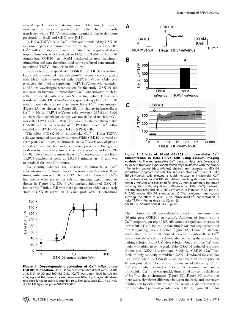
To identify whether the increase in intracellular Ca2+
concentration came from extracellular sources and/or intracellular
stores, ruthenium red (RR), a TRPV channel inhibitor, and Ca2+-
free media were utilized in calcium imaging experiments. As
shown in Figure 3A, RR (3 mM) partially inhibited GSK101-
induced Ca2+ influx. RR was more potent when added at an early
stage of GSK101 activation (1–3 min post GSK101 activation).
The inhibition by RR was reduced if added at a later time point
(10 min post GSK101 activation). Addition of ionomycin, a
Ca2+ionophore, on top of RR still caused a significant increase in
intracellular Ca2+, indicating that fura 2 was not sequestered and
fura 2 signaling was still active (Figure 3A). Figure 3B demon-
strates that the GSK101-induced increase in intracellular Ca2+
was almost abolished immediately after replacing the extracellular
bathing solution with a Ca2+-free solution, but only if the Ca2+-free
media was added near the peak of the GSK101-induced responses
(3 min post GSK101 activation). Similarly, GSK101/Ca2+-free
medium only modestly diminished GSK101-induced intracellular
Ca2+ levels when the GSK101/Ca2+-free medium was applied at
10 min post GSK101activation. Ionomycin added on top of the
Ca2+-free medium caused a moderate but transient increase in
intracellular Ca2+ that was quickly diminished due to the depletion
of Ca2+ in the environment (Figure 3B). Figure 3C shows that
there was a significant difference between the early and late stages
of inhibition by either RR or Ca2+-free media, as demonstrated by
the normalized percentage inhibition (n = 4–7, Figure 3C). This
Figure 1. Dose-dependent activation of Ca2+ influx underGSK101 stimulation. HeLa-TRPV4 cells were stimulated with GSK101at 1, 3, 5, 10, 20 and 100 nM. Delta [Ca2+]i was determined by calciumimaging and the dose-response curve was fitted by a sigmoidal dose-response function using SigmaPlot 10.0. The calculated EC50 = 3.3 nM.doi:10.1371/journal.pone.0016713.g001
Figure 2. Effects of 10 nM GSK101 on intracellular Ca2+
concentration in HeLa-TRPV4 cells using calcium imaginganalysis. A. The representative Ca2+ trace of HeLa cells (average of15-20 cells from one experiment) transiently transfected with the emptymVenus-N1 vector (HeLamVenus) showed no response to GSK101stimulation (negative control). The representative Ca2+ trace of HeLaTRPV4-mVenus cells showed a rapid increase in intracellular Ca2+
concentration under GSK101 stimulation, reaching its maximum levelwithin 2 minutes and sustained for over 30 min. B.Summary bar graphshowing statistically significant difference in delta [Ca2+]i betweenHeLamVenus cells and HeLa TRPV4-mVenus cells (Mean 6 SE, n = 3–6,P,0.05) under GSK101 stimulation. C. The averaged time courseshowing the effect of GSK101 on intracellularCa2+ concentration inHeLa TRPV4-mVenus (Mean 6 SE, n = 8).doi:10.1371/journal.pone.0016713.g002
Determinants of TRPV4 Activity
PLoS ONE | www.plosone.org 2 February 2011 | Volume 6 | Issue 2 | e16713
likely indicates that the increase in intracellular Ca2+ concentra-
tion was mainly due to Ca2+ influx through the TRPV4 channels
at the early stage of the activation, and that the TRPV4 channel
appeared to have partially desensitized following an extended
activation period. The reason for the continued elevated Ca2+
levels under these conditions is not known, but likely reflects
changes in other pathways of Ca2+ entry/exit, such as release from
stores, or inhibition of Ca2+ efflux from the cytoplasm.
GSK101 activates TRPV4 channel in whole-cell patchclamp analysis
Although calcium imaging detected significant Ca2+ influx in
response to GSK101 in HeLa-TRPV4 cells, it was necessary to
determine the specific role of TRPV4 channels in regulating
intracellular Ca2+ levels. The whole cell patch clamp technique
was utilized to study the channel currents under GSK101
stimulation using either Ca2+-containing or Ca2+-free media in
HeLa-TRPV4 cells. Whole-cell recordings in Ca2+-containing
media are shown in Figure 4A–C. A representative current trace
following GSK101 stimulation is shown in Figure 4A. At a holding
potential of 290 mV, the GSK101-induced current occurredim-
mediately, reached a peak current at 1 min after activation, and
relaxed to a pseudosteady state for the remainder of the recording
(20 min). A similar time course was observed for four separate
cells, with each demonstrating an early rapid activation, a peak
near 1 min, followed by an apparent partial desensitization of the
channel (Figure 4B). To further characterize the GSK101-induced
current, a whole-cell current-voltage relation was generated using
a voltage ramp protocol (2100 mV to +100 mV) over 200 msec
duration. The IV-plot shows the GSK101-induced inward and
Figure 3. Effects of ruthenium red (3 mM) and Ca2+ free media on GSK101-induced intracellular Ca2+ elevation in HeLa-TRPV4 cells.A. Ruthenium red (RR) at 3 mM partially inhibited GSK101-induced intracellular Ca2+ elevation when added early (1–3 min post stimulation), whilehaving less inhibitory effect if added at a later time point (10 min post stimulation). Ionomycin (5 mM) added on top of RR caused a significant Ca2+
influx in these cells. B. Ca2+ free media abolished GSK101-induced intracellular Ca2+ elevation when added early (3 min post stimulation), while havinga much smaller effect if added at a later time point (10 min post stimulation). Ionomycin (5 mM) added while the cells were in Ca2+ free media causeda transient increase in intracellular Ca2+ concentration followed by the trending down of intracellular Ca2+. C.Summary bar graph showing thepercentage inhibition of RR and Ca2+ free media added at early or late stage of GSK101 activation, respectively. The inhibition at the late stagedisplayed a much smaller percentage inhibition for either RR or Ca2+ free media(Mean 6 SE, P,0.05, n = 4–7). * Significant difference between earlyand late inhibition by RR; ** significant difference between early and late inhibition by Ca2+ free media.doi:10.1371/journal.pone.0016713.g003
Determinants of TRPV4 Activity
PLoS ONE | www.plosone.org 3 February 2011 | Volume 6 | Issue 2 | e16713
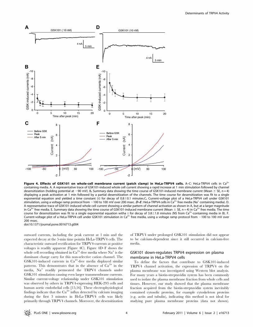
outward currents, including the peak current at 1 min and the
expected decay at the 5-min time pointin HeLa-TRPV4 cells. The
characteristic outward rectification for TRPV4-currents at positive
voltages is readily apparent (Figure 4C). Figure 4D–F shows the
whole cell recording obtained in Ca2+-free media where Na+ is the
dominant charge carry for this non-selective cation channel. The
GSK101-induced currents in Ca2+-free media displayed similar
patterns. This demonstrates that in the absence of Ca2+ in the
media, Na+ readily permeated the TRPV4 channels under
GSK101 stimulation causing even larger transmembrane currents.
Similar current-voltage relationship under GSK101 stimulation
was observed by others in TRPV4-expressing HEK-293 cells and
human aortic endothelial cells [15,16]. These electrophysiological
findings indicate that the Ca2+ influx detected by calcium imaging
during the first 3 minutes in HeLa-TRPV4 cells was likely
primarily through TRPV4 channels. Moreover, the desensitization
of TRPV4 under prolonged GSK101 stimulation did not appear
to be calcium-dependent since it still occurred in calcium-free
media.
GSK101 down-regulates TRPV4 expression on plasmamembrane in HeLa-TRPV4 cells
To define the factors that contribute to GSK101-induced
TRPV4 channel activation, the expression of TRPV4 on the
plasma membrane was investigated using Western blot analysis.
For many years a biotin-streptavidin system has been commonly
used to isolate the plasma membrane fraction from whole cells and
tissues. However, our study showed that the plasma membrane
fraction acquired from the biotin-streptavidin system inevitably
contained cytosolic proteins, for example cytoskeleton proteins
(e.g. actin and tubulin), indicating this method is not ideal for
studying pure plasma membrane proteins (data not shown).
Figure 4. Effects of GSK101 on whole-cell membrane current (patch clamp) in HeLa-TRPV4 cells. A–C: HeLa-TRPV4 cells in Ca2+
containing media. A. A representative trace of GSK101-induced whole cell current showing a rapid increase at 1 min stimulation followed by channeldesensitization (holding potential at 290 mV). B. Summary data showing the time course of GSK101-induced membrane current (Mean 6 SE, n = 4)displaying a peak activation at 1 min followed by a partial desensitization of the channels. The time course for desensitization was fit to a singleexponential equation and yielded a time constant (t) for decay of 0.860.1 minutes.C. Current-voltage plot of a HeLa-TRPV4 cell under GSK101stimulation, using a voltage ramp protocol from 2100 to 100 mV over 200 msec. D–E : HeLa-TRPV4 cells in Ca2+ free media (Na+ containing media). D.A representative trace of GSK101-induced whole cell current showing a similar pattern of channel activation as shown in A, but at a larger magnitudein Ca2+ free media. E. Summary data showing the time course of GSK101-induced membrane current (Mean 6 SE, n = 4) in Ca2+ free media. The timecourse for desensitization was fit to a single exponential equation witha t for decay of 3.861.8 minutes (NS from Ca2+-containing media in B). F.Current-voltage plot of a HeLa-TRPV4 cell under GSK101 stimulation in Ca2+ free media, using a voltage ramp protocol from 2100 to 100 mV over200 msec.doi:10.1371/journal.pone.0016713.g004
Determinants of TRPV4 Activity
PLoS ONE | www.plosone.org 4 February 2011 | Volume 6 | Issue 2 | e16713
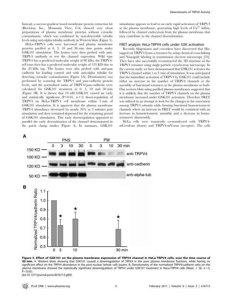
Instead, a sucrose-gradient based membrane protein extraction kit
(Biovision, Inc., Mountain View, CA) showed very clean
preparations of plasma membrane proteins without cytosolic
contaminants, which was confirmed by non-detectable tubulin
levels using anti-alpha tubulin antibody in Western blots (Figure 5).
HeLa-TRPV4 cells were harvested and plasma membrane
proteins purified at 0, 3, 10 and 30 min time points under
GSK101 stimulation. The lysates were then probed with anti-
TRPV4 antibody to test the channel expression. Wild type
TRPV4 has a predicted molecular weight of 98 kDa; the TRPV4-
mVenus then has a predicted molecular weight of 123 KD due to
the 25 kDa tag. The lysates were also probed with anti-pan
cadherin for loading control and with anti-alpha tubulin for
detecting cytosolic contamination (Figure 5A). Densitometry was
preformed by scanning the TRPV4 and pan-cadherin protein
levels, and the normalized ratios of TRPV4/pan-cadherin were
calculated for GSK101 treatment at 0, 3, 10 and 30 min
(Figure 5B). It is shown that 10 nM GSK101 caused an early
and statistically significant (P,0.05, n = 5) down-regulation of
TRPV4 on HeLa-TRPV4 cell membrane within 3 min of
GSK101 stimulation. It is apparent that the plasma membrane
TRPV4 abundance decreased by nearly 30% at 3 minutes post
stimulation and then remained depressed for the remaining period
of GSK101 stimulation. This early downregulation appeared to
parallel the early desensitization of the channel demonstrated in
the patch clamp studies (Figure 4). In summary, GSK101
stimulation appears to lead to an early rapid activation of TRPV4
at the plasma membrane, generating high levels of Ca2+ influx,
followed by channel endocytosis from the plasma membrane that
may contribute to the channel desensitization.
FRET analysis HeLa-TRPV4 cells under GSK activationRecently Shigematsu and coworkers have discovered that His-
tagged rat TRPV4 forms a tetramer by using chemical cross-linking
and Nanogold labeling in transmission electron microscopy [33].
They have also successfully reconstructed the 3D structure of the
TRPV4 tetramer using single-particle cryoelectron microscopy. In
the current study, we have demonstrated that GSK101 activates the
TRPV4 channel within 1 to 3 min of stimulation. It was anticipated
that the immediate activation of TRPV4 by GSK101 could include
either an increase in the number of TRPV4 channels or the
assembly of functional tetramers at the plasma membrane or both.
Our western blots using purified plasma membranes suggested that
it is unlikely that the number of TRPV4 channels on the plasma
membrane increased under GSK101 activation. Therefore FRET
was utilized in an attempt to look for the changes in the association
among TRPV4 subunits while forming functional homotetrameric
channels where an increase in FRET would be consistent with an
increase in homotetrameric assembly and a decrease in homo-
tetrameric disassembly.
HeLa cells were transiently co-transfected with TRPV4-
mCerulean (donor) and TRPV4-mVenus (acceptor). The cells
Figure 5. Effect of GSK101 on the plasma membrane expression of TRPV4 channel in HeLa-TRPV4 cells, over the time course of30 min. A. Western blots showing that GSK101 caused a downregulation of TRPV4 in the pure plasma membrane fractions, while having nosignificant effect on the TRPV4 abundance in the post-nuclear (whole cell) lysates. B. Densitometry of the normalized TRPV4/cadherin ratio on theplasma membrane showed the statistically significant downregulation of TRPV4 under GSK101 treatment in HeLa-TRPV4 cells (Mean 6 SE, n = 5,P,0.05).doi:10.1371/journal.pone.0016713.g005
Determinants of TRPV4 Activity
PLoS ONE | www.plosone.org 5 February 2011 | Volume 6 | Issue 2 | e16713
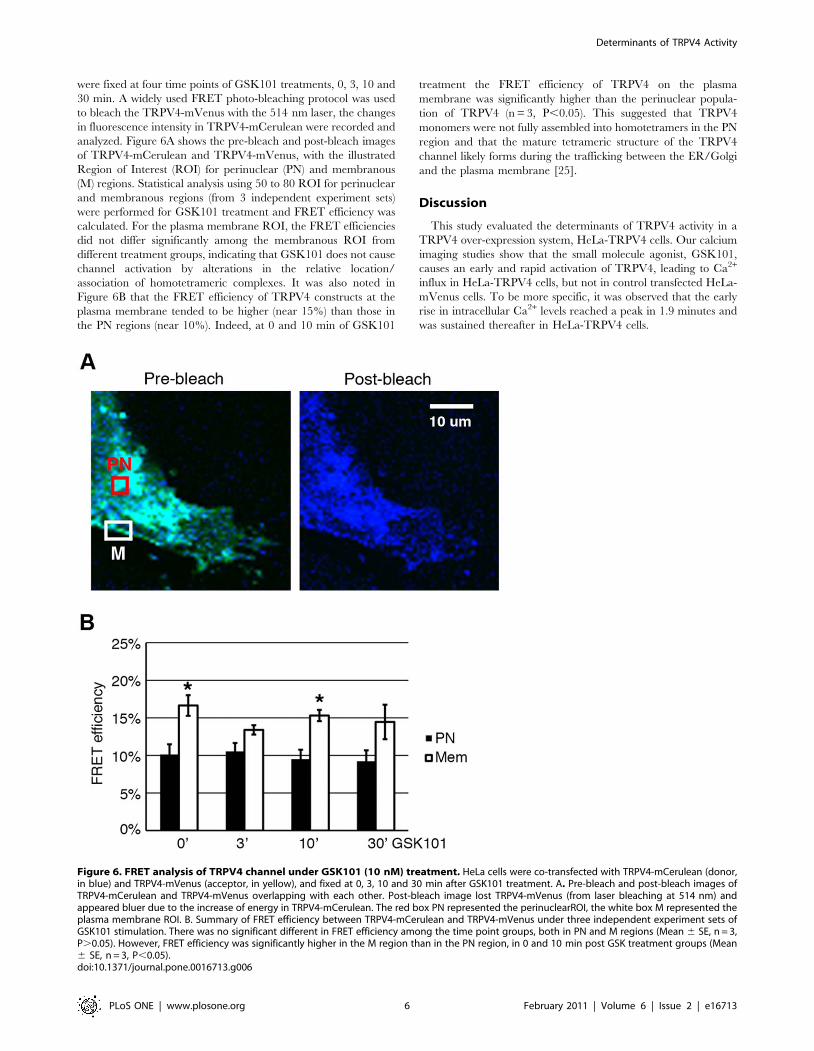
were fixed at four time points of GSK101 treatments, 0, 3, 10 and
30 min. A widely used FRET photo-bleaching protocol was used
to bleach the TRPV4-mVenus with the 514 nm laser, the changes
in fluorescence intensity in TRPV4-mCerulean were recorded and
analyzed. Figure 6A shows the pre-bleach and post-bleach images
of TRPV4-mCerulean and TRPV4-mVenus, with the illustrated
Region of Interest (ROI) for perinuclear (PN) and membranous
(M) regions. Statistical analysis using 50 to 80 ROI for perinuclear
and membranous regions (from 3 independent experiment sets)
were performed for GSK101 treatment and FRET efficiency was
calculated. For the plasma membrane ROI, the FRET efficiencies
did not differ significantly among the membranous ROI from
different treatment groups, indicating that GSK101 does not cause
channel activation by alterations in the relative location/
association of homotetrameric complexes. It was also noted in
Figure 6B that the FRET efficiency of TRPV4 constructs at the
plasma membrane tended to be higher (near 15%) than those in
the PN regions (near 10%). Indeed, at 0 and 10 min of GSK101
treatment the FRET efficiency of TRPV4 on the plasma
membrane was significantly higher than the perinuclear popula-
tion of TRPV4 (n = 3, P,0.05). This suggested that TRPV4
monomers were not fully assembled into homotetramers in the PN
region and that the mature tetrameric structure of the TRPV4
channel likely forms during the trafficking between the ER/Golgi
and the plasma membrane [25].
Discussion
This study evaluated the determinants of TRPV4 activity in a
TRPV4 over-expression system, HeLa-TRPV4 cells. Our calcium
imaging studies show that the small molecule agonist, GSK101,
causes an early and rapid activation of TRPV4, leading to Ca2+
influx in HeLa-TRPV4 cells, but not in control transfected HeLa-
mVenus cells. To be more specific, it was observed that the early
rise in intracellular Ca2+ levels reached a peak in 1.9 minutes and
was sustained thereafter in HeLa-TRPV4 cells.
Figure 6. FRET analysis of TRPV4 channel under GSK101 (10 nM) treatment. HeLa cells were co-transfected with TRPV4-mCerulean (donor,in blue) and TRPV4-mVenus (acceptor, in yellow), and fixed at 0, 3, 10 and 30 min after GSK101 treatment. A. Pre-bleach and post-bleach images ofTRPV4-mCerulean and TRPV4-mVenus overlapping with each other. Post-bleach image lost TRPV4-mVenus (from laser bleaching at 514 nm) andappeared bluer due to the increase of energy in TRPV4-mCerulean. The red box PN represented the perinuclearROI, the white box M represented theplasma membrane ROI. B. Summary of FRET efficiency between TRPV4-mCerulean and TRPV4-mVenus under three independent experiment sets ofGSK101 stimulation. There was no significant different in FRET efficiency among the time point groups, both in PN and M regions (Mean 6 SE, n = 3,P.0.05). However, FRET efficiency was significantly higher in the M region than in the PN region, in 0 and 10 min post GSK treatment groups (Mean6 SE, n = 3, P,0.05).doi:10.1371/journal.pone.0016713.g006
Determinants of TRPV4 Activity
PLoS ONE | www.plosone.org 6 February 2011 | Volume 6 | Issue 2 | e16713
It is of note that HeLa-TRPV4 cells did not return the
GSK101-induced intracellular Ca2+ levels back towards basal
values as typically observed for most Ca2+ signaling events since
many TRP channels, including TRPV4 as discussed below, often
desensitize following activation [2,34,35]. Since TRPV4 was
observed to partially desensitize in patch clamp measurements, the
sustained elevation of cytosolic Ca2+ is not likely the result of Ca2+
influx only. The apparent sustained elevation in cytosolic Ca2+
levels is not an artifact relating to fura 2 being quenched or
sequestered because ionomycin was still able to elicit a rise in
intracellular Ca2+ levels when added at the end of the experiments.
Hence, other factors controlling cytosolic Ca2+ homeostasis must
be invoked in the HeLa-TRPV4 overexpression system, possibly
induced calcium release from stores or impaired calcium extrusion
from the cytoplasm. The reason for this late effect remains to be
determined in future studies.
Whole-cell patch clamp analysis provides more direct assess-
ment of TRPV4 activity in HeLa-TRPV4 cells. As shown in
Figure 4A–C, GSK101 addition leads to an early rapid activation
of TRPV4 channels. The current can peak within seconds, but
then rapidly decays to a pseudosteady state over approximately 3
minutes. The early rapid activation of the channel likely reflects a
Ca2+-induced sensitization of the channel similar to that shown by
others [36,37]. The subsequent decay of the TRPV4 currents over
the following 3 minutes reflects an early, but partial, desensitiza-
tion of the channel in the continued presence of GSK101. This
desensitization can be due to numerous factors, including channel
endocytosis from the plasma membrane (see below), but could also
be related to other factors such as a Ca2+-induced or Ca2+-
calmodulin induced desensitization as noted for other channels,
including TRP channels [35–38]. However, the whole cell
recording of HeLa-TRPV4 cells in Ca2+-free extracellular solution
showed an even larger transmembrane current with the similar
activation pattern and outward rectification under GSK101
stimulation (Figure 4D–E). This is likely due to Na+ influx as
TRPV4 is a non-selective cation channel permeable to Ca2+ and
Na+. Since the GSK101 stimulated channel currents still displayed
a similar desensitization to that observed in Ca2+ containing
media, it follows that the desensitization of the channel following
GSK101 stimulation is not Ca2+-dependent.
The desensitization of TRPV4 following GSK101 stimulation is
also supported by the calcium imaging data showing that the
application of ruthenium red (3 mM) and Ca2+-free medium
following GSK101 stimulation leads to a diminished increase in
intracellular Ca2+ concentration, but significantly more effective
when applied in the early stage of GSK101 stimulation (1–3 min
post activation); Ca2+ levels fell only minimally in the late stage
(10 min activation). These studies again point to an early partial
desensitization of TRPV4. Therefore, in the late stage of calcium
imaging in HeLa-TRPV4 cells, the sustained intracellular Ca2+
concentrations above baseline is unlikely to be the direct effect of
continued Ca2+ influx through TRPV4 channels alone, but rather
the result of Ca2+ release from stores or diminished extrusion of
Ca2+ from the cytosol via the plasma membrane or sarcoplasmic
reticulum Ca2+ pumps, as heretofore described.
We directly evaluated the role of TRPV4 channel endocytosis
from the plasma membrane as a determinant of TRPV4 activity.
Ion channel trafficking to and from the plasma membrane is
difficult to study, partly due to the difficulty in extracting pure
plasma membrane proteins and the complex steps during vesicle
trafficking. In this study, we utilized a sucrose gradient-based
extraction method that has produced pure plasma membrane
fractions in HeLa-TRPV4 cells. Anti-alpha tubulin antibody in
Western blotting is a useful marker to check whether the plasma
membrane fraction is contaminated with cytosolic proteins
because cytoskeletal proteins are the most common contaminants
during plasma membrane extraction. Acomparison of the
traditional biotin-streptavidin system with the sucrose gradient-
based extraction kit shows that the sucrose gradient-based
extraction method yields a highly enriched, pure, plasma
membrane fraction, while the biotin-streptavidin method produces
a crude plasma membrane fraction with considerable contamina-
tion of cytoskeletal proteins. However, it is worth noting that
sucrose gradient-based extraction has a lower yield in the final
protein quantity than biotin-streptavidin system.
Few studies have evaluated TRP channel trafficking and most
have focused on translocation and insertion of TRP channel onto
the plasma membrane during a stimulus. The hypothesis is that a
stimulus causes channel exocytosis and insertion into the plasma
membrane, resulting in an increased number of channels, leading
to enhanced Ca2+ entry.The examples include insulin-induced
TRPV2 channel insertion onto the plasma membrane in
pancreatic b-cells [20], Rab11a-induced insertion of TRPV5
and TRPV6 in epithelial cells [22,23,39], and epidermal growth
factor-induced translocation of TRPC4 and TRPC5 in HEK-293
cells overexpressing TRPC4 or TRPC5, respectively [19,21].
However, Shukla and coworkers [24] recently reported that
angiotensin induces internalization of TRPV4 in rat vascular
smooth muscle cells based on confocal microscopy analysis. They
also showed that in HEK-293 cells overexpressing TRPV4,
angiotensin treatment induced an immediate upregulation of
TRPV4 at 5 min post stimulation, followed by an approximately
50% decrease in TRPV4 plasma membrane expression over the
next 1 hr using a surface biotinylation assay [24]. In the current
study, we discovered that TRPV4, while being activated by
GSK101, does not lead to an early insertion or elevated
membrane abundance of TRPV4, but rather the activation is
associated with an early and rapid downregulation of TRPV4
from the plasma membrane, i.e., TRPV4 endocytosis, within
3 min of GSK101 stimulation. A pure plasma membrane protein
extraction method, as well as normalized TRPV4 expression using
pan-cadherin as loading control for densitometry, was utilized to
ensure the accuracy of the observation. It is likely that the increase
in intracellular Ca2+ serves as a negative modulator of TRPV4
membrane expression and initiates the endocytosis of TRPV4
from the plasma membrane, likely leading to reduced Ca2+ entry.
Although the endocytosis mechanism is not known, the usual
machinery for vesicle trafficking, such as Rab small GTPases,
SNAREs and phosphoinisitides are likely to be involved and
require further investigation [40,41]. It is also noteworthy that
Shukla et al. recently discovered a novel role of b-arrestin 1 in
TRPV4 downregulation by mediating ubiquitination[24]. This
expands the endocytosis pathways of ion channels, and points to
potential mechanisms of TRPV4 downregulationin the plasma
membrane under GSK101 stimulation for future study.
To provide a more in depth assessment of the physical
determinants of TRPV4 activation, we attempted to decipher
the mechanism of GSK101 activation/desensitization of TRPV4
channels using a FRET analysis to evaluate the physical
association among subunits of the TRPV4 homotetramer.
Alterations in the physical associations between channel subunits
are thought to play an important role in activation/modulation of
TRP channels [27,42–44]. Indeed, channel subunit dissociation
may be an important regulatory process underlying channel
inactivation for some TRP channels as shown for TRPP2 [29,30].
A recent study by Shigematsu et al confirms that TRPV4 forms
functional homotetramers[33] while others have shown that
TRPV4 heterotetramers can form [26,27] and that subunit
Determinants of TRPV4 Activity
PLoS ONE | www.plosone.org 7 February 2011 | Volume 6 | Issue 2 | e16713
mixing among various TRP channel family members can lead to
altered channel functions [26–28]. Our FRET analysis utilizes
TRPV4-mCerulean and TRPV4-mVenus as donor and acceptor
to evaluate homotetramericassembly. The results show that
GSK101 does not lead to an increased FRET transfer efficiency
between the TRPV4 subunits, or more specifically at the tagged
C-termini, on the plasma membrane, indicating that the GSK101-
induced TRPV4 activation does not involve theassembly of
TRPV4 subunits into new homotetrameric structures. Likewise,
with continued exposure to GSK101, the desensitization of
TRPV4 also does not lead to a change in plasma membrane
FRET efficiency, indicating that desensitization does not appear to
involve a dissociation of TRPV4 subunits from homotetrameric
structures. We conclude that the activation and desensitization of
TRPV4 following GSK101 stimulation does not appear to involve
alterations in the homotetrameric structure of the TRPV4 channel
at the plasma membrane. Activation of the channel at the plasma
membrane would, therefore, appear to reflect activation of existing
channel structures with conformational changes within the
homotetrameric structure that lead to channel opening. With the
3D structure of TRPV4 available [33], it will be interesting to
define the changes within the homotetrameric structure itself
that lead to alterations in channel gating, potentiation, and
desensitization.
Materials and Methods
Cloning and chemicalsMouse TRPV4 coding sequence (Accession #NM_022017) was
inserted between Sal I and Bam HI sites of mCerulean-N1 and
mVenus-N1 vectors (Roger Tsien laboratory, University of
California at San Diego), so that the mCerulean or mVenus
fusion protein was at the C-terminus of recombinant TRPV4
protein. To simplify the terminology in this work, TRPV4-
mCerulean stands for mCerulean-N1-TRPV4 and TRPV4-
mVenus stands for mVenus-N1-TRPV4. Both plasmids went
through sequencing to verify the correct DNA sequences. The
recombinant TRPV4-mCerulean and TRPV4-mVenus proteins
were also characterized by western blots using both anti-TRPV4
antibody (Alomone Labs, Jerusalem, Israel) and anti-GFP
antibody (AbCam, Cambridge, MA) (data not shown). The
mCerulean or mVenus fusion protein added to the recombinant
TRPV4 did not alter the amino acid sequence at the C-ternimus
of TRPV4, therefore leaving the epitopes intact for antibody
recognition.
GSK1016790A (GSK101) was either a gift from GlaxoSmithK-
line or purchased from Sigma-Aldrich (St. Louis, MO). GSK101
was dissolved in DMSO at 10 mM stock concentration and stored
in aliquots at 220uC. It was then diluted into various working
concentrations right before use.
Cell culture and transfectionHeLa cells (human cervical cancer) were grown in RPMI
medium supplemented with 10% FBS, 1 IU/ml penicillin and
1 mg/ml streptomycin in a 37uC and 5% CO2 incubator. HeLa
cells were seeded onto glass coverslips or tissue culture dishes
within 24 hrs of transfection, then an Effectene kit (Qiagene,
Valencia, CA) was used to transfect HeLa cells with different
plasmids. Cells were used for various experiments 20 to 24 hrs post
transfection.
Calcium imagingHeLa-TRPV4 cells grown on glass coverslips were loaded with
2 mM fura-2/AM for 1 hr at RT, then washed in the isotonic
MBSS buffer (containing 140 mMNaCl, 5.4 mMKCl, 0.5 mM
MgCl2, 0.4 mM MgSO4, 3.3 mM NaHCO3, 2 mM CaCl2,
5.5 mM glucose, 10 mM HEPES, pH 7.4). The coverslip was
attached to a perfusion chamber, which was then attached to the
stage of an InCyt imaging workstation (Intracellular Imaging) for
imaging of intracellular Ca2+ levels [5,13]. For HeLa-TRPV4
cells, only those cells positively identified for TRPV4-mVenus
expression with epifluorescence at 488 nm wavelength were
selected for imaging. Cells were bathed in isotonic MBSS buffer
and 10 to 20 cells of interest were selected using a freehand ROI
(Regions of Interest) drawing tool. Then the cells were treated with
10 nM GSK101 and recorded for different periods. Ca2+-free
experiments were carried out by washing away the Ca2+-
containing MBSS with the Ca2+-free MBSS (MBSS without
CaCl2, with the addition of 1 mM EGTA). Intracellular Ca2+ was
calculated using the fura-2 fluorescence ratio method (excitation at
340 over 380 nm, emission at 510 nm) which was converted to
intracellular Ca2+ concentration as described by Gadzikowska
et al. [45] using the calibration methods as done before [5,13,46].
To better compare changes in [Ca2+]i between methods of
TRPV4 inhibition, in some studies the intracellular Ca2+
concentration was normalized for each data point. Delta GSK101 =
[Ca2+]i with GSK101 – [Ca2+]i at basal level. Delta inhibitor =
[Ca2+]i with inhibitor – [Ca2+]i at basal level. Percentage
inhibition = [(Delta GSK101 - Delta inhibitor)/Delta GSK101] x
100%.
ElectrophysiologyPatch electrodes with a resistance of <2 MV were pulled from
borosilicate micropipettes (Sutter Instrument Company, Novato,
CA) using a micropipette puller (P-97 Sutter Instrument Co.,
Novato, CA) [5]. HeLa cells transfected with TRPV4-mVenus
were identified using a combination of epifluorescence illumina-
tion and differential interference contrast optics (20X–40X) on an
inverted Axiovert 200M microscope (Carl Zeiss, Germany). Cells
were recorded in the whole-cell configuration using an EPC-10
amplifier (HEKA Instruments, Germany). The junction potential
was 13 mV (n = 14), which was compensated using Pulse software.
After whole-cell configuration was established, the cell membrane
capacitance (32.762.4 pF) and series resistance (7.160.8 MV,
n = 14) were electronically compensated. Whole-cell currents were
not normalized to capacitance. To determine the exponential
decay of the currents, data were fitted with SigmaPlot using an
exponential decay function Y = y0 + Ae-t/t, where Y is the current
at time t, y0 is the residual current.
All experiments were performed at room temperature (<25uC).
Signals were filtered at 1 kHz, digitized at 10 kHz, and acquired
using the Pulse program (HEKA Instruments, Germany). The
Ca2+-containing extracellular solution consisted of 136 mMNaCl,
5.4 mMKCl, 0.5 mM MgCl2, 0.4 mM MgSO4, 3 mM NaHCO3,
2 mM CaCl2, 5 mM glucose and 10 mM HEPES (pH 7.4, 300
mOsm). The Ca2+-free extracellular solution consisted of
145 mMNaCl, 1 mM MgCl2, 10 mM HEPES, 10 mM glucose
and 1 mM EGTA (pH 7.4, 300 mOsm). The pipette internal
solution contained 20 mMCsCl, 100 mM Cs aspartate, 1 mM
MgCl2, 5 mM EGTA, 10 mM HEPES, 1 mM Mg-ATP, and
0.1 mM Na-GTP (pH 7.2, 290 mOsm). The holding potential was
290 mV for most experiments. The current-voltage relation was
determined using a voltage ramp protocol from 2100 to 100 mV
over 200 msec.
GSK101 stock in DMSO (10 mM) was diluted 1:1000 in
extracellular solution just before use and held in a series of
independent syringes connected to an array of corresponding
fused silica columns (inner diameter, 200 mm). The exchange of
Determinants of TRPV4 Activity
PLoS ONE | www.plosone.org 8 February 2011 | Volume 6 | Issue 2 | e16713
solutions was achieved by rapidly by shifting the tubes horizontally
with a micromanipulator. The distance from the column mouth to
the cell examined was about 100 mm. Cells in the recording
chamber were continuously bathed in the extracellular solution.
Each solution was delivered to the recording chamber by gravity.
Western blotting and densitometryHeLa-TRPV4 cells were grown on 10-cm tissue culture plates
until ready for harvest. The cells were rinsed twice with cold PBS,
and scraped off of the plates using a spatula. Then a sucrose
gradient-based membrane protein extraction kit (Biovision, Inc.,
Mountain View, CA) was utilized to purify plasma membrane
proteins. Whole cell lysates were the intermediate products before
the plasma membrane fraction was purified from the sucrose
gradient. The final pellets containing pure plasma membrane
proteins were achieved at the end of the extraction process. The
resultant pellets were solubilized in a modified RIPA buffer (0.1%
Na deoxycholate, 0.01% SDS, 1% NP-40 and 20 mM Mg acetate
in PBS) containing protease inhibitor cocktail (Sigma-Aldrich, St.
Louis, MO). Protein concentrations were measured using the BCA
method. Standard Western blotting (AbCam, Cambridge, MA)
was performed to characterize and quantify TRPV4 channel
expression. Primary antibodies used in this work include anti-
TRPV4 (Alomone Labs, Jerusalem, Israel) for detecting TRPV4 as
well as TRPV4-mVenus/Cerulean, anti-pan cadherin antibody
(AbCam, Cambridge, MA) as internal loading control, and anti
alpha-tubulin antibody (Sigma-Aldrich, St. Louis, MO) for
verifying the purity of plasma membrane preparation. Densitom-
etry (Image J 1.42q, NIH) was utilized to quantify the abundance
of a TRPV4 channel in the plasma membrane under different
treatments. Normalized TRPV4 channel quantity was achieved by
calculating the ratio of TRPV4/pan cadherin, as pan cadherin
was the loading control for all the plasma membrane samples. All
experiments were repeated 4 to 5 times.
Fluorescent Resonance Energy Transfer (FRET)FRET microscopy was performed as described by Kenworthy
group [5,47,48]. HeLa cells grown on glass coverslips were co-
transfected with TRPV4-mCerulean and TRPV4-mVenus with
fluorescent fusion protein at the carboxyl terminus of the TRPV4
insert. Twenty hours after the transfection, the cells were washed
in PBS and fixed in 4% paraformaldehyde for 20 minutes at room
temperature. The coverslips were then washed and mounted onto
glass slides using Prolong mounting media (Invitrogen). Images
were captured on a Nikon A1 confocal microscope. Three pre-
bleach images of both mCerulean and mVenus were acquired
(mCeruleanpre and mVenuspre, respectively). Acceptor photo-
bleach was achieved by laser excitation at 514-nm set at 100%
transmission for duration of 30 seconds (1 pulse/second). Then
three post-bleach images of both mCerulean and mVenus were
acquired (mCeruleanpost and mVenuspost, respectively). Regions of
interest (ROI) were manually selected over the perinuclear region,
designated as perinuclear (PN), or plasma membrane region,
designated as membranous (M). A background ROI was also
selected. The fluorescent intensity of the ROI was measured,
background-subtracted and exported from the NIS software as an
Excel file. The average of the pre-bleach and post-bleach
fluorescence intensities were calculated from three images. The
FRET efficiency was calculated as E = (mCeruleanpost -
mCeruleanpre)/mCeruleanpost. A chart graph was generated after
averaging 50 to 80 ROI measurements from three sets of GSK101
time-course experiments.
Statistical AnalysisAll data are presented as mean 6 SE. Student t-test was used to
compare the means between two treatment groups. P-values less
than 0.05 were considered statistically significant.
A GSK101 dose-response curve was generated using the mean
response (n = 4–6) at various GSK101 concentrations in SigmaPlot
10.0. The sigmoidal dose-response curve employing EC50 and
Hillslope was chosen for calculation.
Acknowledgments
The authors wish to thank Dr. Roger Tsien for the gift of mVenus-N1 and
mCerulean-N1 vectors and GlaxoSmithKline for the initial gift of
GSK1016790A.
Author Contributions
Conceived and designed the experiments: MJ ZW ETW RGO. Performed
the experiments: MJ ZW LC JJ DC RGO. Analyzed the data: MJ ZW LC
JJ DC ETW RGO. Contributed reagents/materials/analysis tools: MJ
ETW RGO. Wrote the paper: MJ RGO.
References
1. Liedtke W, Choe Y, Marti-Renom MA, Bell AM, Denis CS, et al. (2000)
Vanilloid receptor-related osmotically activated channel (VR-OAC), a candidate
vertebrate osmoreceptor. Cell 103: 525–535.
2. Strotmann R, Harteneck C, Nunnenmacher K, Schultz G, Plant TD (2000)
OTRPC4, a nonselective cation channel that confers sensitivity to extracellular
osmolarity. Nat Cell Biol 2: 695–702.
3. Wissenbach U, Bodding M, Freichel M, Flockerzi V (2000) Trp12, a novel Trp
related protein from kidney. FEBS Lett 485: 127–134.
4. Kunert-Keil C, Bisping F, Kruger J, Brinkmeier H (2006) Tissue-specific
expression of TRP channel genes in the mouse and its variation in three different
mouse strains. BMC Genomics 7: 159.
5. Gao X, Wu L, O’Neil RG (2003) Temperature-modulated diversity of TRPV4
channel gating: activation by physical stresses and phorbol ester derivatives
through protein kinase C-dependent and -independent pathways. J Biol Chem
278: 27129–27137.
6. Guler AD, Lee H, Iida T, Shimizu I, Tominaga M, et al. (2002) Heat-evoked
activation of the ion channel, TRPV4. J Neurosci 22: 6408–6414.
7. Liedtke W, Friedman JM (2003) Abnormal osmotic regulation in trpv4-/- mice.
Proc Natl Acad Sci U S A 100: 13698–13703.
8. Lorenzo IM, Liedtke W, Sanderson MJ, Valverde MA (2008) TRPV4 channel
participates in receptor-operated calcium entry and ciliary beat frequency regulation
in mouse airway epithelial cells. Proc Natl Acad Sci U S A 105: 12611–12616.
9. Mendoza SA, Fang J, Gutterman DD, Wilcox DA, Bubolz AH, et al. (2009)
TRPV4-mediated endothelial Ca2+ influx and vasodilation in response to shear
stress. Am J Physiol Heart Circ Physiol 298: H466–476.
10. Vriens J, Watanabe H, Janssens A, Droogmans G, Voets T, et al. (2004) Cell
swelling, heat, and chemical agonists use distinct pathways for the activation of
the cation channel TRPV4. Proc Natl Acad Sci U S A 101: 396–401.
11. Watanabe H, Davis JB, Smart D, Jerman JC, Smith GD, et al. (2002) Activation
of TRPV4 channels (hVRL-2/mTRP12) by phorbol derivatives. J Biol Chem
277: 13569–13577.
12. Watanabe H, Vriens J, Prenen J, Droogmans G, Voets T, et al. (2003)
Anandamide and arachidonic acid use epoxyeicosatrienoic acids to activate
TRPV4 channels. Nature 424: 434–438.
13. Wu L, Gao X, Brown RC, Heller S, O’Neil RG (2007) Dual role of the TRPV4
channel as a sensor of flow and osmolality in renal epithelial cells. Am J Physiol
Renal Physiol 293: F1699–1713.
14. Watanabe H, Vriens J, Suh SH, Benham CD, Droogmans G, et al. (2002) Heat-
evoked activation of TRPV4 channels in a HEK293 cell expression system and
in native mouse aorta endothelial cells. J Biol Chem 277: 47044–47051.
15. Thorneloe KS, Sulpizio AC, Lin Z, Figueroa DJ, Clouse AK, et al. (2008) N-
((1S)-1-{[4-((2S)-2-{[(2,4-dichlorophenyl)sulfonyl]amino}-3-hydroxypropa noyl)-
1-piperazinyl]carbonyl}-3-methylbutyl)-1-benzothiophene-2-carboxamid e
(GSK1016790A), a novel and potent transient receptor potential vanilloid 4
channel agonist induces urinary bladder contraction and hyperactivity: Part I.
J Pharmacol Exp Ther 326: 432–442.
16. Willette RN, Bao W, Nerurkar S, Yue TL, Doe CP, et al. (2008) Systemic
activation of the transient receptor potential vanilloid subtype 4 channel causes
endothelial failure and circulatory collapse: Part 2. J Pharmacol Exp Ther 326:
443–452.
Determinants of TRPV4 Activity
PLoS ONE | www.plosone.org 9 February 2011 | Volume 6 | Issue 2 | e16713
17. Mendoza SA, Fang J, Gutterman DD, Wilcox DA, Bubolz AH, et al. (2010)
TRPV4-mediated endothelial Ca2+ influx and vasodilation in response to shearstress. Am J Physiol Heart Circ Physiol 298: H466–476.
18. Xu X, Gordon E, Lin Z, Lozinskaya IM, Chen Y, et al. (2009) Functional
TRPV4 channels and an absence of capsaicin-evoked currents in freshly-isolated, guinea-pig urothelial cells. Channels (Austin) 3: 156–160.
19. Bezzerides VJ, Ramsey IS, Kotecha S, Greka A, Clapham DE (2004) Rapidvesicular translocation and insertion of TRP channels. Nat Cell Biol 6: 709–720.
20. Hisanaga E, Nagasawa M, Ueki K, Kulkarni R, Mori M, et al. (2009)
Regulation of calcium-permeable TRPV2 channel by insulin in pancreatic beta-cells. Diabetes 58: 174–184.
21. Odell AF, Scott JL, Van Helden DF (2005) Epidermal growth factor inducestyrosine phosphorylation, membrane insertion, and activation of transient
receptor potential channel 4. J Biol Chem 280: 37974–37987.22. van de Graaf SF, Chang Q, Mensenkamp AR, Hoenderop JG, Bindels RJ
(2006) Direct interaction with Rab11a targets the epithelial Ca2+ channels
TRPV5 and TRPV6 to the plasma membrane. Mol Cell Biol 26: 303–312.23. van de Graaf SF, Hoenderop JG, Bindels RJ (2006) Regulation of TRPV5 and
TRPV6 by associated proteins. Am J Physiol Renal Physiol 290: F1295–1302.24. Shukla AK, Kim J, Ahn S, Xiao K, Shenoy SK, et al. (2010) Arresting a TRP
channel [beta]-arrestin 1 mediates ubiquitination and functional downregulation
of TRPV4. J Biol Chem.25. Hellwig N, Albrecht N, Harteneck C, Schultz G, Schaefer M (2005) Homo- and
heteromeric assembly of TRPV channel subunits. J Cell Sci 118: 917–928.26. Stewart AP, Smith GD, Sandford RN, Edwardson JM (2010) Atomic force
microscopy reveals the alternating subunit arrangement of the TRPP2-TRPV4heterotetramer. Biophys J 99: 790–797.
27. Cheng W, Yang F, Takanishi CL, Zheng J (2007) Thermosensitive TRPV
channel subunits coassemble into heteromeric channels with intermediateconductance and gating properties. J Gen Physiol 129: 191–207.
28. Kottgen M, Buchholz B, Garcia-Gonzalez MA, Kotsis F, Fu X, et al. (2008)TRPP2 and TRPV4 form a polymodal sensory channel complex. J Cell Biol
182: 437–447.
29. Petri ET, Celic A, Kennedy SD, Ehrlich BE, Boggon TJ, et al. (2010) Structureof the EF-hand domain of polycystin-2 suggests a mechanism for Ca2+-dependent regulation of polycystin-2 channel activity. Proc Natl Acad Sci U S A107: 9176–9181.
30. Schumann F, Hoffmeister H, Bader R, Schmidt M, Witzgall R, et al. (2009)Ca2+-dependent conformational changes in a C-terminal cytosolic domain of
polycystin-2. J Biol Chem 284: 24372–24383.
31. Fan HC, Zhang X, McNaughton PA (2009) Activation of the TRPV4 ionchannel is enhanced by phosphorylation. J Biol Chem 284: 27884–27891.
32. Yoshida T, Inoue R, Morii T, Takahashi N, Yamamoto S, et al. (2006) Nitricoxide activates TRP channels by cysteine S-nitrosylation. Nat Chem Biol 2:
596–607.
33. Shigematsu H, Sokabe T, Danev R, Tominaga M, Nagayama K (2010) A 3.5-
nm structure of rat TRPV4 cation channel revealed by Zernike phase-contrast
cryoelectron microscopy. J Biol Chem 285: 11210–11218.
34. Gordon-Shaag A, Zagotta WN, Gordon SE (2008) Mechanism of Ca(2+)
-dependent desensitization in TRP channels. Channels (Austin) 2: 125–129.
35. Nilius B, Vriens J, Prenen J, Droogmans G, Voets T (2004) TRPV4 calcium
entry channel: a paradigm for gating diversity. Am J Physiol Cell Physiol 286:
C195–205.
36. Strotmann R, Schultz G, Plant TD (2003) Ca2+-dependent potentiation of the
nonselective cation channel TRPV4 is mediated by a C-terminal calmodulin
binding site. J Biol Chem 278: 26541–26549.
37. Strotmann R, Semtner M, Kepura F, Plant TD, Schoneberg T (2010)
Interdomain interactions control Ca2+-dependent potentiation in the cation
channel TRPV4. PLoS One 5: e10580.
38. Watanabe H, Vriens J, Janssens A, Wondergem R, Droogmans G, et al. (2003)
Modulation of TRPV4 gating by intra- and extracellular Ca2+. Cell Calcium
33: 489–495.
39. van de Graaf SF, Hoenderop JG, van der Kemp AW, Gisler SM, Bindels RJ
(2006) Interaction of the epithelial Ca2+ channels TRPV5 and TRPV6 with the
intestine- and kidney-enriched PDZ protein NHERF4. Pflugers Arch 452:
407–417.
40. Dong XP, Wang X, Xu H (2010) TRP channels of intracellular membranes.
J Neurochem 113: 313–328.
41. Stenmark H (2009) Rab GTPases as coordinators of vesicle traffic. Nat Rev Mol
Cell Biol 10: 513–525.
42. Alfonso S, Benito O, Alicia S, Angelica Z, Patricia G, et al. (2008) Regulation of
the cellular localization and function of human transient receptor potential
channel 1 by other members of the TRPC family. Cell Calcium 43: 375–387.
43. Alicia S, Angelica Z, Carlos S, Alfonso S, Vaca L (2008) STIM1 converts
TRPC1 from a receptor-operated to a store-operated channel: moving TRPC1
in and out of lipid rafts. Cell Calcium 44: 479–491.
44. Becker D, Muller M, Leuner K, Jendrach M (2008) The C-terminal domain of
TRPV4 is essential for plasma membrane localization. Mol Membr Biol 25:
139–151.
45. Gadzikowska M, Grynkiewicz G (2002) Tropane alkaloids in pharmaceutical
and phytochemical analysis. Acta Pol Pharm 59: 149–160.
46. Zhang L, Yang KH, King AI (2001) Biomechanics of neurotrauma. Neurol Res
23: 144–156.
47. Kenworthy A (2001) Imaging protein-protein interaction using fluorescence
resonance energy transfer microscopy. Methods 24: 289–296.
48. Roland JT, Kenworthy AK, Peranen J, Caplan S, Goldenring JR (2007) Myosin
Vb interacts with Rab8a on a tubular network containing EHD1 and EHD3.
Mol Biol Cell 18: 2828–2837.
Determinants of TRPV4 Activity
PLoS ONE | www.plosone.org 10 February 2011 | Volume 6 | Issue 2 | e16713
Top Related
Copyright © 2022 FDOKUMEN

