


Bahasa
Halaman
Hukum

Journal of Chromatography B, 726 (1999) 25–35
Composition of the peptide fraction in human blood plasma:database of circulating human peptidesa , b b a* ¨Rudolf Richter , Peter Schulz-Knappe , Michael Schrader , Ludger Standker ,
b a a¨Michael Jurgens , Harald Tammen , Wolf-Georg ForssmannaLower Saxony Institute for Peptide Research, Feodor-Lynen-Strasse 31, D-30625 Hannover, Germany
bBioVisioN, Feodor-Lynen-Strasse 5, D-30625 Hannover, Germany
Received 7 September 1998; received in revised form 28 December 1998; accepted 28 December 1998
Abstract
A database was established from human hemofiltrate (HF) that consisted of a mass database and a sequence database, withthe aim of analyzing the composition of the peptide fraction in human blood. To establish a mass database, all 480 fractionsof a peptide bank generated from HF were analyzed by MALDI-TOF mass spectrometry. Using this method, over 20 000molecular masses representing native, circulating peptides were detected. Estimation of repeatedly detected masses suggeststhat approximately 5000 different peptides were recorded. More than 95% of the detected masses are smaller than 15 000,indicating that HF predominantly contains peptides. The sequence database contains over 340 entries from 75 differentprotein and peptide precursors. 55% of the entries are fragments from plasma proteins (fibrinogen A 13%, albumin 10%,b2-microglobulin 8.5%, cystatin C 7%, and fibrinogen B 6%). Seven percent of the entries represent peptide hormones,growth factors and cytokines. Thirty-three percent belong to protein families such as complement factors, enzymes, enzymeinhibitors and transport proteins. Five percent represent novel peptides of which some show homology to known peptide andprotein families. The coexistence of processed peptide fragments, biologically active peptides and peptide precursorssuggests that HF reflects the peptide composition of plasma. Interestingly, protein modules such as EGF domains (meprinAa-fragments), somatomedin-B domains (vitronectin fragments), thyroglobulin domains (insulin like growth factor-bindingproteins), and Kazal-type inhibitor domains were identified. Alignment of sequenced fragments to their precursor proteinsand the analysis of their cleavage sites revealed that there are different processing pathways of plasma proteins in vivo. 1999 Elsevier Science B.V. All rights reserved.
Keywords: Human hemofiltrate; Peptides; Circulating human peptides
1. Introduction cessed gene products. There are different means toidentify the consecutive products of a gene, the
Progress in genome sequencing results in an mRNA, the protein, and the processed protein frag-increasing demand for sequence data of the pro- ments. mRNA analysis is used to identify open
reading frames, transcription starts, signal sequencesand stop codons and is therefore useful to postulatetranslated proteins. The translated protein itself is of
*Corresponding author. interest to verify a postulated protein. Processed
0378-4347/99/$ – see front matter 1999 Elsevier Science B.V. All rights reserved.PI I : S0378-4347( 99 )00012-2
26 R. Richter et al. / J. Chromatogr. B 726 (1999) 25 –35
protein and peptide fragments often bear the bio- 2. Experimentallogical activity. To elucidate these products of agene, different strategies are applied. 2.1. Hemofiltrate
Approaches to identify protein coding regions(exons) from the genome are performed by generat- Hemofiltrate was obtained from the Nephro-ing a large number of expressed sequence tags logisches Zentrum Niedersachsen, Hannoversch-
¨(ESTs) resulting from the partial sequencing of Munden, Germany, in quantities of 1600 to 2000 lcDNAs, using bioinformatic computer programs per week. Patients with chronic renal failure were[1–3], or exon trapping/ in vivo processing of gen- subjected to routine arterio–venous hemofiltrationomic DNA [4,5]. three times per week. The following hemofiltration
Most of these methods consider neither post-trans- equipment was used routinely: Hemoprozessor (Sar-¨lational modification of proteins and peptides (e.g., torius, Gottingen, Germany) and AK 10 HFM (Gam-
glycosylation, amidation) nor proteolytic processing bro, Hechingen, Germany). The filters used wereof proteins and peptides to biologically active pep- Hemoflow F 60S and Hemoflow HF 80S (Fresenius,tides. Many peptide hormones show their biological Bad Homburg, Germany), Hemofilter FH 77 H andactivity after specific proteolytic processing. Exam- Hemofilter FH 88 H (Gambro, Martinsried, Ger-ples of this are insulin that is produced after cleavage many). All ultrafilters used had a specified molecu-of the C-peptide and proglucagon which is processed lar-mass cut-off around 20 000. For Hemoflow Fto at least three different hormones: glucagon, and 60S, and Hemoflow HF 80S polysulphone mem-glucagon-like peptides 1 and 2. Furthermore, the branes, and for Hemofilter FH 77 H, and Hemofilterprimary structure of a peptide deduced from a FH 88 H polyamide membranes are used. The
2 2mRNA/cDNA sequence does not give final infor- effective membrane surfaces were 1.3 m , 1.8 m ,2 2mation concerning the disulfide bridges of the re- 1.4 m , and 2.0 m , respectively. Filtration was
sulting protein or peptide. For definitive structural driven by a transmembranous pressure gradient of 60and functional studies, it is necessary to isolate the to 100 mmHg at a blood flow-rate of 250 to 350native proteins or peptides of interest. ml /min (1 mmHg5133.322 Pa). 20 to 30 l of filtrate
Therefore, we started to identify peptides from were recovered per patient and treatment.human blood. To obtain sufficient amounts of plasmapeptides, extracts of human hemofiltrate (HF) from 2.2. Peptide extractionpatients with chronic renal failure were produced.Using chromatographic methods, a reproducible The sterile filtrate was immediately cooled to 48Clarge-scale procedure to generate a peptide bank and acidified to pH 3 to prevent bacterial growth andfrom up to 10 000 l of HF was developed [6]. The proteolysis. Extraction of the peptides from HF wasproteins and peptides of each of 480 fractions from either performed using alginic acid or a strong cationthis peptide bank are characterized by their molecu- exchanger (Fractogel TSK SP 650(M), Merck,lar mass using matrix-assisted laser desorption ioni- Darmstadt, Germany; 25310 cm, Vantage VA 250zation time-of-flight mass spectometry (MALDI– column, Amicon, Witten, Germany) [6]. Alginic acidTOF-MS). These data were used to generate a mass extraction was performed using a modified methoddatabase. Furthermore, using MS–MS sequencing described by Mutt [7]. Briefly, batches of 400 l HFand during our attempts to isolate circulating hor- are adjusted to pH 2.7, 2.5 kg alginic acid weremones, we also determined the amino acid sequences added to the bath and stirred for 8 to 12 h. Then,of many of these peptides. These data were accumu- alginic acid was sedimented and separated from thelated in our sequence database, which now contains HF, washed with ethanol (10 l) and 0.005 M
¨more than 340 entries. Mass- and sequence databases hydrochloric acid on a Buchner funnel. The peptidesform our peptide database. Here we present an were eluted with 0.2 M hydrochloric acid (10 l). Theanalysis of these data, giving insight into the com- eluate was adjusted to pH 4.0 and a peptide precipi-position of the peptide fraction in human blood. tation was performed with 5.5 M sodium chloride at
R. Richter et al. / J. Chromatogr. B 726 (1999) 25 –35 27
48C for 20 h. Peptide extraction using a strong matrix solution was composed of 5 mg/ml fucose,cation-exchange column (Fractogel SP650 (M)) was and 5 mg/ml SIN or CHC in acetonitrile–0.1% TFAperformed as described by Schulz-Knappe et al. [6]. (1:1, v /v). The crystallization process was performedBriefly, 1000 l HF were conditioned to pH 2.7. with accelerated ambient temperature air-dryingThese batches were applied onto the strong cation using a microventilator. Measurements were per-exchanger using an Autopilot chromatography sys- formed in linear mode with a LaserTec RBT IItem (PerSeptive Biosystems, Wiesbaden, Germany). MALDI–TOF-MS system (Perseptive /Vestec, Hous-Then, batch elution was performed with 10 l 0.5 M ton, TX, USA). The instrument was equipped with aammonium acetate (two column volumes). The 1.2 m flight tube and a 337 nm nitrogen laser.eluate was stored at 2208C or lyophilized until Positive ions were accelerated at 25 kV and up to 30further use. laser shots were automatically accumulated per
sample position. The Voyager RP BioSpectrometry2.3. Preparation of a peptide bank from human Workstation Version 3.07.1 (PerSeptive Biosystems,blood Framingham, MA, USA) was used as control soft-
ware. The automatic measurement included a searchThe peptide bank was produced as described by pattern of 18 spots per sample position. The laser
Schulz-Knappe et al. [6]. Briefly, for the first sepa- intensity was adjusted to signal intensity in a presetration step the extracts of 5000 l HF were pooled and mass range. From the 18 spots per sample position,loaded on a 10-l cation-exchange column (Fractogel only the best measurement, i.e., that with the highestSP 650(M)). Bound peptides were eluted using seven signal intensity, was saved to the hard disk. Valuesbuffers with increasing pH. The seven buffers were for laser intensity, signal intensity and preset masscomposed as follow: I: 0.1 M citric acid monohy- range had to be differently adjusted for CHC anddrate, pH 3.6; II: 0.1 M acetic acid10.1 M sodium SIN according to their specific properties [8]. Theacetate, pH 4.5; III: 0.1 M malic acid, pH 5.0; IV: time-of-flight data were externally calibrated for each0.1 M succinic acid, pH 5.6; V: 0.1 M sodium sample plate and sample preparation. Calibration anddihydrogenphosphate, pH 6.6; 0.1 M dis- further data processing were performed with theodiumhydrogenphosphate, pH 7.4; VII: 0.1 M am- Voyager RP BioSpectrometry Workstation processingmonium carbonate, pH 9.0. The seven pools (pH software (Version 3.07.1 PerSeptive Biosystems,pools) were collected and each of them was loaded Framingham, MA, USA) based on Grams/386 Ver-onto a 12.5310 cm reverse-phase column (Source sion 3.0 (Galactic Industries, Salem, NH, USA).RPC, 15 mm, Pharmacia, Freiburg, Germany) andeluted in a 8 l gradient from 100% A (0.01 M HCl)
2.5. Isolation of peptidesto 60% B (80% acetonitrile, 0.01 M HCl). Fractionsof 200 ml were collected (see Fig. 1).
Peptides were isolated with a variety of liquidchromatographic methods such as cation-exchange2.4. MALDI–TOF-MSand reversed-phase chromatography. For cation-ex-change chromatography, Fractogel TSK SP 650 SLyophilized aliquots of 1 l HF-equivalent of the(Merck, Darmstadt, Germany) or Parcosil PepKat orpeptide bank fractions were dissolved in 0.1 to 4 ml
˚ ¨acetonitrile–0.1% aqueous trifluororacetic acid ProKat material (5 mm, 300 A, Biotek, Ostringen,(TFA) (1:1, v /v) according to their absorption profile Germany) with sodium chloride gradients were used.in reversed-phase chromatography (optical density at For preparative reversed-phase purification Source214 nm). The resulting analyte solutions contained RPC material (15 mm, Pharmacia, Freiburg, Ger-an equivalent of 0.25 to 10 ml HF/ml. A 1-ml sample many) was used and for analytical chromatography
˚solution was applied on a stainless steel multiple RP-C and -C material (5 mm, 300 A, Vydac,4 18
sample tray as admixture to either sinapinic acid Hesperia, USA) for example were used with standard(SIN) or a-cyanohydroxycinamic acid (CHC). The gradients of acetonitrile. Detailed information for

28 R. Richter et al. / J. Chromatogr. B 726 (1999) 25 –35
Fig. 1. Production of a human peptide bank and mass database from hemofiltrate. 2000 l hemofiltrate was collected per week. Extraction ofthe peptides was performed using a strong cation exchanger. Subsequently extracts of 5000 l HF were pooled and loaded on a 10-lcation-exchange column. Bound peptides were eluted using seven buffers with increasing pH. Each of the seven pH pools wasrechromatographed on a reversed-phase column. Lyophilized aliquots of the fractions were analyzed on a MALDI–TOF-MS system. TheMALDI–MS data of the fractions of each chromatography were accumulated in a one two-dimensional (2D) peptide map with the m /zvalues in the x-dimension and the fractions in the y-dimension. For each reversed-phase chromatography one 2D peptide map is produced.

R. Richter et al. / J. Chromatogr. B 726 (1999) 25 –35 29
different isolation procedures is given by Bensch et tides were searched in the dbEST-data bank. Data-al. [9], Hess et. al. [10], Kuhn et al. [11], Schepky et bases were searched using the basic blast mode
¨al. [12], Schulz-Knappe et al. [13], and Standker and [18–20]. Multiple alignment studies of peptide frag-co-workers [14,15]. ments from proteins were performed using the
MacMolly software package (Soft Gene, Berlin,2.6. Sequence analysis Germany). Predicted signal peptide processing sites
from cDNA sequences were determined by the useMass spectrometric sequencing from complex of the SignalP V1.1 WorldWideWeb Server [21].
peptides mixtures was performed on an API III1triple quadropol-MS system (PE Sciex Instruments,Toronto, Canada). The MS–MS experiments were 3. Results and discussionperformed using loop injection. Argon was used ascollision gas in the collision-induced dissociation 3.1. Peptide mass database(CID) experiments.
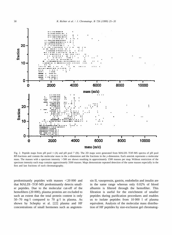
The amino acid sequence of purified peptides was The peptide mass database contains one 2D pep-determined by automated Edman degradation using a tide map for each of the seven pH pool fractionsgas-phase sequencer, Model 473 A or 494 (Applied (Fig. 2). Each map contains approximately 3500Biosystems). entries. Isolation and analysis of diverse molecular
masses suggest that the detected masses predomi-2.7. Production of the mass- and sequence nantly represent proteins and peptides. The standarddatabases deviation for detected molecular masses using Laser-
Tec RBT II MALDI–TOF-MS is 2‰. ConsideringThe peptide mass database was generated as this mass deviation, most molecules are repeatedly
¨described by Jurgens et al. [16]. It was established detected in consecutive fractions. Peaks are checkedfrom the 480 fractions of the peptide bank. Each for multiply charged and multimeric ions. CHC has afraction was analyzed by MALDI–TOF-MS using tendency to produce more multiply charged ions thanCHC and SIN as matrices. The 2D maps were SIN. However, in these complex mixtures, doublygenerated from MALDI–TOF-MS spectra of pH and rarely triply charged species are observed onlypool RP-fractions and contain the molecular mass in for larger (approx. .7000) and/or abundant pep-the x-dimension and the fractions in the y-dimension. tides. The most abundant peptides sometimes formPeak tables are processed in spreadsheet programs multimeric species. These findings suggest that ap-(Microsoft Excel and Microcal Origin) to result in a proximately 5000 different peptides are detected in2D peptide map for every pH pool. HF subjecting 0.25 ml to 10 ml HF-equivalent to
The sequence database is the list of isolated MALDI–TOF-MS. The detection limit of differentpeptides. The amino acid sequence and the N- and pure peptides (e.g., insulin, GLP-1, CDD/ANP-99-C-terminally adjacent amino acids of a peptide are 126, HCC-1) is between 30 fmol /ml and 3 pmol /ml.given. The peptides are arranged according to their This suggests minimal concentrations for the de-molecular mass (average mass). If the peptide was tection of these molecules in a range from 3 to 300isolated from the HF peptide bank [6], the fraction pM, but detection of a peptide by MALDI–TOF-MSused and the pH pool is shown. The functions of the is dependent on the concomitant components in theisolated peptides are given in the last column as analyte which may dramatically increase the de-indicated by the SwissProt database [17]. tection limit for such peptides. Therefore detection of
molecules such as insulin, CDD/ANP-99-126, or2.8. Data interpretation and database research GLP-1 by their molecular mass, and in consideration
of their chromatographic characteristics in the pep-Identification and assessment of the function of tide bank, was unsuccessful.
sequenced peptides was performed using the Ninety-five percent of the detected masses areSwissProt database [17]. cDNAs of unknown pep- ,15 000. This reflects that hemofiltrate contains
30 R. Richter et al. / J. Chromatogr. B 726 (1999) 25 –35
Fig. 2. Peptide maps from pH pool 1 (A) and pH pool 7 (B). The 2D maps were generated from MALDI–TOF-MS spectra of pH poolRP-fractions and contain the molecular mass in the x-dimension and the fractions in the y-dimension. Each asterisk represents a molecularmass. The masses with a spectrum intensity .500 are shown resulting in approximately 1500 masses per map. Without restriction of thespectrum intensity each map contains approximately 3500 masses. Maps demonstrate repeated detection of the same masses especially in thefirst and last fractions of each chromatography.
predominantly peptides with masses ,20 000 and sin II, vasopressin, gastrin, endothelin and insulin arethat MALDI–TOF-MS predominantly detects small- in the same range whereas only 0.02% of blooder peptides. Due to the molecular cut-off of the albumin is filtered through the hemofilter. Thishemofilters (20 000), plasma proteins are excluded to filtration is useful for the enrichment of smallersuch an extent that the total protein content is only peptides during purification procedures and enables50–70 mg/ l compared to 70 g/ l in plasma. As us to isolate peptides from 10 000 l of plasmashown by Schepky et al. [22] plasma and HF equivalent. Analysis of the molecular mass distribu-concentrations of small hormones such as angioten- tion of HF peptides by size-exclusion gel chromatog-

R. Richter et al. / J. Chromatogr. B 726 (1999) 25 –35 31
raphy reveals that approximately 45% of the peptides peptides with M .10 000 reflects peak spreadingr
in HF are in the range .15 000 and contain 15% due to the increasing number of isotopes and saltalbumin [22]. Analysis of fractions from pH pool 6 adducts and a decreased sensitivity of the detector.and pH pool 7 by gel electrophoresis reveals diverse In our institute the mass database is a useful toolbands in the size range between M 15 000 and with which to map each hemofiltrate peptide bankr
66 000 (data not shown). This suggests that HF for identification of known peptides of interest (e.g.,contains a diversity of peptides and proteins HCC-1 [13], guanylin [11], and b-defensin [9]).
´.15 000 but due to the hemofilters molecular cut-off Subsequently, these peptides are purified for furtherof 20 000 in significantly lower concentrations than biological testing. Furthermore, the mass spec-in blood plasma. trometry of peptide fractions is a method by which to
Another explanation for the detection of predomi- identify peptides with special molecular characteris-nantly smaller peptides (see Table 1) is that in tics (e.g., cysteine-rich peptides, amidation) byMALDI–TOF-MS the intensity of a signal is most chemical modification of these groups with sub-dominant at about M 2000 to 3000. For molecules sequent mass spectrometric analysis.r
.3000 there is a continuous decrease in the intensity Contrary to our MALDI–TOF-MS mass database,of the MALDI–TOF-MS signal, e.g., the MALDI– 2D gel plasma maps [23,24] predominantly detectTOF-MS S /N ratio for 1 pmol of GLP-1 (M 3298) proteins of M .10 000. The smallest proteins foundr r
ranges from 30 to 60, whereas the S /N ratio for 1 in this 2D gel map are kininogen light chain andpmol of albumin (M 66 000) ranges from 3 to 4. apolipoprotein A-II with an M |10 000. This showsr r
Low intensity of the M signal for proteins and that our mass data base contains information com-r
Table 1aRegulatory peptides and protein fragments from HF
Peptide hormones Angiotensin 1, guanylin-22-115, uroguanylin-89-112, cardiodilatin /atrialnatriuretic peptide (CDD/ANP 99-126), b-defensin 1 (h-BD1),neutrophil defensin 1, neutrophil defensin 3, kininogen (LMW chain)
Cytokines, HCC-1, IGF-1, IGF-2, osteoinductive factor, platelet derivedgrowth factors, growth factor (PDGF), osteopontin, CTAP III, pigment endo-growth inhibitors: thelium derived factor, angiogenin I, collagen XVIII
Complement factors: Complement factor C3, complement factor C4A (Anaphylatoxin)complement factor C9, complement factor D (CFAD)
Enzymes, Lysozyme, carboxypeptidase N, pancreatic trypsin inhibitor,enzyme inhibitors: cystatin C, plasminogen, a-2-antiplasmin, inter-a-trypsin
inhibitor complex component II (ITI2), a-1-antitrypsin,hexokinase type II, ribonuclease
Transport proteins: Transthyretin, serotransferrin, retinol binding protein (RBP),transforming growth factor-binding protein (TGF-BP), insulin-like growth factor-binding protein (IBP3)
Plasma proteins: Albumin, fibrinogen A (RGD Peptides), fibrinogen B, a-1-microglobulin, b-2-microglobulin, zinc-a-2-glycoprotein (ZAG),a-2-HS-glycoprotein (fetuin), serum amyloid A protein (SAA),haptoglobin, profilin, vitronectin, desmocollin, thymosin b4,apolipoprotein C-III, uteroglobin, ubiquitin, gelsolin,somatomedin B, hemopexin
a The fragments belong to approximately 75 precursor peptides and proteins. The known 60 peptides and proteins are shown. For peptidehormones and growth factors biologically active sequences as well as other prohormone fragments were found.

32 R. Richter et al. / J. Chromatogr. B 726 (1999) 25 –35
plementary to a 2D gel plasma map for predominant Identified peptides are in an M from 500 tor
detection of peptides M ,10 000. 30 000. The largest isolated protein is albumin-309-r
585, with an M of 31 493. The sequence data baser
3.2. Peptide sequence data base shows that proteins and peptides with a M ,8500r
represent predominantly protein fragments and pro-Our sequence database was produced in the last cessed peptides. Twenty-five percent of the identified
five years. Up to now, the sequence data base (Fig. peptides with an M .8500 are precursor peptidesr
3) contains over 340 entries from about 75 different and proteins.protein and peptide precursors. Fifty-five percent of Concerning possible non-specific degradation ofthe entries are fragments from plasma proteins the peptides in HF during collection and transport,(fibrinogen A 13%, albumin 10%, b2-microglobulin there are a number of different findings. (1) Most of8.5%, cystatin C 7%, and fibrinogen B 6%). Thirty- the identified hormones (e.g., CDD/ANP, IGF-1,three percent belong to protein families such as IGF-2) show 100% identity with postulated circulat-complement factors, enzymes, enzyme inhibitors and ing hormone sequences. The processing sites of thesetransport protein fragments. Four percent of the hormones are identical to those identified in earlierentries are contributable to peptide hormones, 3% to investigations [25,26]. (2) Biologically active factorsgrowth factors and cytokines. such as guanylin [11], HCC-1 [13], or uteroglobin
Five percent are new sequences, most of which do [27] are isolated from HF as precursor peptides. (3)not show homology to known peptide and protein Alignments of over 150 sequenced fragments offamilies. Due to the progress in genome sequencing, albumin, fibrinogen A, fibrinogen B, cystatin C,increasing numbers of these novel peptides are found b2-microglobulin, and complement factor C3 within the Expressed Sequence Tags database (dbEST). their precursor proteins reveal a specific tryptic or
Fig. 3. Structure of the peptide sequence database. The sequence database is a collection of sequenced peptides from HF. The five C- andN-terminal amino acids of a peptide sequence and the N- and C-terminally adjacent amino acids of a peptide sequence are shown. Thepeptides are arranged according to their molecular mass (average mass). The measured mass is given. If the peptide is isolated from the HFpeptide bank, the fraction and the pH pool is shown. The (putative) function of the isolated peptides are given in the last column as indicatedby the Swiss-Prot database or other literature (NT AA5N-terminal amino acid; CT AA5C-terminal amino acid). References, accessionnumbers and data base entries of the precursor proteins and peptides are given in the last column.

R. Richter et al. / J. Chromatogr. B 726 (1999) 25 –35 33
chymotryptic endoproteolysis with subsequent ex-oproteolytic digestion. Twenty percent of the frag-ment cleavages are C-terminal to Lys or Arg,suggesting tryptic digestion. Forty-four percent ofthe fragment cleavages are C-terminal to Phe, Tyr orLeu suggesting a chymotryptic digestion. Sixteen, 6,4 and 4% of the fragment cleavages are at a distanceof 1, 2, 3 and 4 amino acids, respectively, to trypticor chymotryptic cleavage sites, suggesting exop-roteinase digestion after tryptic or chymotrypticdigestion (Table 2). (4) Predicted signal peptideprocessing sites from cDNA sequences are comparedto the processing sites determined by sequencing theN-terminus of circulating peptides using SignalP Fig. 4. Comparison of predicted signal peptide processing sites to
the processing sites determined by sequencing of the N-terminusV1.1 WorldWideWeb Server. Twelve percent of theof seven peptides isolated from HF. Prediction of the signalsequenced peptides were either N-terminal fragmentscleavage site was performed using SignalP V1.1 WorldWideWeb
of the propeptides or non-processed peptides. Com- Server. Twelve percent of the database entries are N-terminalparison revealed 100% identity of the predicted fragments of propeptides or non-processed propeptides, demon-processing site with the processing site identified by strating 100% identity with the predicted signal cleavage site.
sequencing the peptides (Fig. 4). These findingsstrongly suggest that HF peptides do not undergonon-specific degradation and reflect the peptide fragments containing the thrombin cleavage site andcomposition in human blood. polymerization site are found (Fig. 5).
Alignment of identified fragments with their pro- Assessment of post-translational modifications wastein precursors revealed that processing of plasma achieved by comparison of the theoretical averageproteins is not unidirectional but there are different mass and the detected mass. This suggests thatprocessing pathways in vivo. Furthermore, this anal- approximately 20% of the peptides are post-transla-ysis suggests different proteolytic rates for different tionally modified. Due to a mass deviation of 2‰,degradation products of plasma proteins, e.g., pro- salt adducts and detection of different isotopes of acessing of N-terminal fibrinogen B does not only molecule specification of the post-translational modi-result in activation of fibrinogen B by cleavage of the fication is difficult by the use of MALDI–MS. Up tofibrinopeptide B at the cleavage site between AS 14 now glycosylation of 1% of the isolated peptides wasand AS 15, but 8 different fragments spanning this identified and elucidated (e.g., HCC-1 [28], C-termi-cleavage site are also found. Identification of exclu- nal domain of IGFBP-5 [29]). The disulfide bridgessively N-terminal fibrinogen B fragments spanning of different proteins and peptides were identifiedthis cleavage site raises the question of a physiologi- (e.g., kazal-type inhibitor domain, C-terminal domaincal function of these fragments. Only N-terminal of IGFBP-4, endostatin [15]).
As the knowledge of protein sequences grows, it isbecoming apparent that many proteins are con-
Table 2 structed from relatively few molecular units, whichaProcessing sites of the isolated peptide fragmentsoccurs repeatedly. These modules often correspond
Tryptic digestion (C-terminal to Lys and Arg) 20% to single exons. Modules can act as growth factors,Chymotryptic digestion (C-terminal to Phe, Tyr, Leu) 44% in receptor-growth factor interactions, or tightly
control cascades of enzyme-catalysed proteolysis.Cleavage site at a distance of 1–4 AS from a 30%Interestingly, we have identified different modulartryptic or chymotryptic cleavage site
a peptides such as vitronectin fragmentsThey were identified from over 150 fragments of albumin,(somatomedin-B domain) [14], meprin Aa-fragmentsfibrinogen A, fibrinogen B, cystatin C, b2-microglobulin and
complement factor C3. (EGF-domain), insulin-like growth factor-binding

34 R. Richter et al. / J. Chromatogr. B 726 (1999) 25 –35
Fig. 5. Alignment of isolated fibrinogen B fragments with the precursor protein. Signal sequence is amino acid 230 to 21. Fibrinogen B isactivated by cleavage between amino acid 44 and 45 releasing fibrinopeptide B. Some identified fibrinogen fragments are carrying thecleavage sites, suggesting different processing pathways for fibrinogen B.
proteins (thyroglobulin domain), and a kazal-type plasma proteins present in the circulation may haveinhibitor domain. The identified vitronectin module important biological activities [30,32].(residues 1 to 44–50) is an effective competitor ofthe interaction of plasmin activator inhibitor (PAI)-1with intact vitronectin or extracellular matrix. This 4. Conclusionssuggests the regulation of PAI-1 in part byproteolytic processing of vitronectin modules [30]. Our database of human circulating peptides dem-The newly identified meprin Aa-fragments show a onstrates that hemofiltrate reveals a source to char-94% homology to human meprin and this is a soluble acterize the composition of the peptide fraction inEGF-like protein, suggesting that the isolated peptide human blood. The mass database is a useful tool tohas growth modulatory function as demonstrated for identify chromatographically and mass spectrometri-other EGF domains. cally characterized peptides in each newly prepared
Concerning the biological relevance of HF peptide hemofiltrate peptide bank. As the mass deviation ofhormones, growth factors and cytokines were iso- the MALDI–TOF-MS technique improves, the at-lated in the biologically active form (e.g., angiotensin tachment of molecular masses to their moleculesI, CDD/ANP [31], insulin like growth factor I and becomes more reliable. Isolation of peptides fromII). Using HF, new regulatory peptides such as HF appears to be of general help to identify bio-guanylin [11], HCC-1 [13] or b-defensin-1 [9] could logically active peptides and proteins to understandbe identified (Table 1). Isolation of proteolytic the processing and metabolism of peptides andproducts of fibrinogen and vitronectin containing cell plasma proteins and to identify their post-translation-attachment sites (RGD sequences) that carry antit- al modifications. The mass database is a goodhrombotic activity or antagonistic activity to entire complement to a 2D gel plasma map by predominantvitronectin demonstrate that degradation products of detection of peptides ,10 000.

R. Richter et al. / J. Chromatogr. B 726 (1999) 25 –35 35
[17] A. Bairoch, B. Boeckmann, Nucleic Acids Res. 19 (Suppl.)References(1991) 2247–2249.
[18] M. Boguski, T. Lowe, C. Tolstoshev, Nat. Genet. 4 (1993)[1] M. Zhang, Proc. Natl. Acad. Sci. USA, 94 (1997) 565-568 332–333.[2] E. Uberbacher, R. Mural, Proc. Natl. Acad. Sci. USA 88 [19] S. Altschul, T. Madden, A. Schaffer, J. Zhang, Z. Zhang, W.
(1991) 11261–11265. Miller, D. Lipman, Nucleic Acids Res. 25 (1997) 3389–[3] V. Solovyev, A. Salamov, C. Lawrence, Nucl. Acids Res. 22 3402.
(1994) 5156–5163. [20] S. Karlin, S. Altschul, Proc. Natl. Acad. Sci. USA 87 (1990)[4] M. Nehls, D. Pfeifer, T. Boehm, Oncogene 9 (1994) 2169– 2264–2268.
2175. [21] H. Nielsen, J. Engelbrecht, S. Brunak, G. von Heijne, Protein[5] M. Nehls, D. Pfeifer, G. Micklem, C. Schmoor, T. Boehm, Eng. 10 (1997) 1–6.
Curr. Biol. 4 (1994) 883–989. [22] A. Schepky, K. Bensch, P. Schulz-Knappe, W.G. Forssmann,¨[6] P. Schulz-Knappe, M. Schrader, L. Standker, R. Richter, R. Biomed. Chromatogr. 8 (1994) 90–94.
¨Hess, M. Jurgens, W.-G. Forssmann, J. Chromatogr. A 776 [23] C. Hoogland, J. Sanchez, L. Tonella, A. Bairoch, D. Hoch-(1997) 125–132. strasser, R. Appel, Nucleic Acids Res. 26 (1998) 332–333.
[7] V. Mutt, Gut Hormones, in: S.R. Bloom (Ed.), C. Living- [24] J. Sanchez, R. Appel, O. Golaz, C. Pasquali, F. Ravier, A.stone, Edinburgh, London, New York, 1978, pp. 21–27. Bairoch, D. Hochstrasser, Electrophoresis 16 (1995) 1131–
[8] P. Juhasz, C. Costello, K. Biemann, J. Am. Soc. Mass 1151.Spectrom. 4 (1993) 399–409. [25] R. Humbel, Hormonal Proteins and Peptides, in: C. Li (Ed.),
¨[9] K. Bensch, M. Raida, J. Magert, P. Schulz-Knappe, W.G. Academic Press, New York, 1984, p. 57.Forssmann, FEBS Lett. 368 (1995) 331–335. [26] C. Glembotski, C. Irons, A. Sprenkle, C. Sei, Can. J. Physiol.
[10] R. Hess, M. Kuhn, P. Schulz-Knappe, M. Raida, M. Fuchs, Pharmacol. 69 (1991) 1525–1536.K. Adermann, V. Kaever, Y. Cetin, W.-G. Forssmann, FEBS [27] A. Aoki, H. Pasolli, M. Raida, M. Meyer, P. Schulz-Knappe,Lett. 374 (1995) 34–38. H. Mostafavi, A. Schepky, R. Znottka, J. Elia, D. Hock, H.
[11] M. Kuhn, M. Raida, K. Adermann, P. Schulz-Knappe, R. Beier, W.-G. Forssmann, Mol. Hum. Reprod. 2 (1996) 489–Gerzer, J. Heim, W.G. Forssmann, FEBS Lett. 318 (1993) 497.205–209. [28] R. Richter, M. Schrader, M. Hagmann, W.-G. Forssmann, P.
[12] A. Schepky, P. Schulz-Knappe, W.-G. Forssmann, J. Chroma- Schulz-Knappe, in preparation.togr. A 691 (1995) 255–261. [29] L. Standker, P. Wobst, S. Mark, W.-G. Forssmann, FEBS
¨[13] P. Schulz-Knappe, H. Magert, B. Dewald, M. Meyer, Y. Lett. 441 (1998) 281–286.Cetin, M. Kubbies, J. Tomeczkowski, K. Kirchhoff, M. ¨[30] L. Standker, A. Enger, P. Schulz-Knappe, K. Wohn, M.Raida, K. Adermann, A. Kist, M. Reineke, R. Sillard, A. Germer, M. Raida, W.G. Forssmann, K. Preissner, Eur. J.Pardigol, M. Uguccioni, M. Baggiolini, W.G. Forssmann, J. Biochem. 241 (1996) 557–563.Exp. Med. 183 (1996) 295–299. [31] K. Forssmann, D. Hock, F. Herbst, P. Schulz-Knappe, J.
¨[14] L. Standker, G. Heine, E. Hartmann, R. Hess, P. Schulz- Talartschik, F. Scheler, W.G. Forssmann, Klin. WochenschriftKnappe, W.G. Forssmann, Biol. Chem. Hoppe-Seyler 376 64 (1986) 1276–1280.(1995) 156. ¨[32] L. Standker, R. Sillard, K. Bensch, A. Ruf, M. Raida, P.
¨[15] L. Standker, M. Schrader, S. Kanse, M. Jurgens, W.G. Schulz-Knappe, A. Schepky, H. Patscheke, W.G. Forssmann,Forssmann, K.T. Preissner, FEBS Lett. 420 (1997) 129–133. Biochem. Biophys. Res. Commun. 215 (1995) 896–902.
¨[16] M. Jurgens, M. Schrader, M. Raida, W.G. Forssmann, P.Schulz-Knappe, J. Biol. Tech. http: / /www.medstv.unimelb.edu.au /JBT/Articles / JBT0006/JBT0006.html, submitted forpublication
Top Related
Copyright © 2022 FDOKUMEN

