


Bahasa
Halaman
Hukum


Early View
Original research article
Awake prone positioning and oxygen therapy in
patients with COVID-19: The APRONOX study
Orlando R. Perez-Nieto, Diego Escarraman-Martinez, Manuel A. Guerrero-Gutierrez, Eder I. Zamarron-
Lopez, Javier Mancilla-Galindo, Ashuin Kammar-García, Miguel A. Martinez-Camacho, Ernesto
Deloya-Tomás, Jesús S. Sanchez-Diaz, Luis A. Macías-García, Raúl Soriano-Orozco, Gabriel Cruz-
Sánchez, José D. Salmeron-Gonzalez, Marco A. Toledo-Rivera, Ivette Mata-Maqueda, Luis A.
Morgado-Villaseñor, Jenner J. Martinez-Mazariegos, Raymundo Flores Ramirez, Josue L. Medina-
Estrada, Silvio A. Ñamendys-Silva, on behalf of the APRONOX group
Please cite this article as: Perez-Nieto OR, Escarraman-Martinez D, Guerrero-Gutierrez MA, et
al. Awake prone positioning and oxygen therapy in patients with COVID-19: The APRONOX
study. Eur Respir J 2021; in press (https://doi.org/10.1183/13993003.00265-2021).
This manuscript has recently been accepted for publication in the European Respiratory Journal. It is
published here in its accepted form prior to copyediting and typesetting by our production team. After
these production processes are complete and the authors have approved the resulting proofs, the article
will move to the latest issue of the ERJ online.
Copyright ©The authors 2021. This version is distributed under the terms of the Creative Commons Attribution Non-Commercial Licence 4.0. For commercial reproduction rights and permissions contact [email protected]
Awake prone positioning and oxygen therapy in patients with COVID-19:
The APRONOX study
Writing Committee: Orlando R. Perez-Nieto MD https://orcid.org/0000-0001-8817-7000
(1), Diego Escarraman-Martinez MD MSc https://orcid.org/0000-0003-3190-0258 (2),
Manuel A. Guerrero-Gutierrez MD https://orcid.org/0000-0002-0645-1833 (3), Eder I.
Zamarron-Lopez (4), Javier Mancilla-Galindo MBBS [https://orcid.org/0000-0002-0718-
467X] (5,6), Ashuin Kammar-García PhD [https://orcid.org/0000-0002-3875-0945] (7),
Miguel A. Martinez-Camacho (8), Ernesto Deloya-Tomás MD https://orcid.org/0000-0002-
9623-5263 (1), Jesús S. Sanchez-Diaz MD MSc https://orcid.org/0000-0003-1744-9077 (9),
Luis A. Macías-García (10), Raúl Soriano-Orozco (11), Gabriel Cruz-Sánchez (12), José D.
Salmeron-Gonzalez (13), Marco A. Toledo-Rivera (14), Ivette Mata-Maqueda (15), Luis A.
Morgado-Villaseñor (16), Jenner J. Martinez-Mazariegos (17), Raymundo Flores Ramirez
(18), Josue L. Medina-Estrada (19), Silvio A. Ñamendys-Silva MD MSc FCCP FCCM
https://orcid.org/0000-0003-3862-169X (3,20), on behalf of the APRONOX group*
* A complete list of members of the APRONOX Group, with authors’ full names, academic
degrees, and affiliations, is provided in Appendix 1.
1. Intensive Care Unit. Hospital General San Juan del Rio, Querétaro, Mexico
2. Department of Anaesthesia. Hospital de Especialidades Centro Médico Nacional “LaRaza”, Mexico
City, Mexico
3. Department of Critical Care Medicine. Instituto Nacional de Cancerología, Mexico City.
4. Intensive Care Unit. Hospital CEMAIN Tampico, Tamaulipas, Mexico
5. Unidad de Investigación UNAM-INC, Instituto Nacional de Cardiología Ignacio Chávez, Mexico
City, Mexico
6. Respiratory Medicine. Instituto Nacional de Enfermedades Respiratorias, Mexico City, Mexico
7. Emergency Department. Instituto Nacional de Ciencias Médicas y Nutrición “Salvador Zubirán”,
Mexico City, Mexico
8. Intensive Care Unit. Hospital General de México, Mexico City, Mexico
9. Intensive Care Unit. Hospital de Alta Especialidad IMSS “Adolfo Ruiz Cortines” Veracruz,
Veracruz, Mexico
10. Intensive Care Unit. Hospital Regional ISSSTE “Fernando Quiroz Gutiérrez”, Mexico City,
Mexico
11. Intensive Care Unit. Hospital de Alta Especialidad T1 IMSS, León, Guanajuato, Mexico
12. Intensive Care Unit. Clínica Hospital Mérida ISSSTE, Yucatán, Mexico
13. Intensive Care Unit. Hospital General “Miguel Silva”, Morelia, Michoacán, Mexico
14. Intensive Care Unit. Hospital SEDNA, Mexico City, Mexico
15. Secretaría de Salud del Estado de Querétaro, Ethics and Research Committee. Mexico
16. Intensive Care Unit. Hospital General de Zona IMSS No.15 Reynosa, Tamaulipas, Mexico
17. Intensive Care Unit. Hospital Vida Mejor ISSSTECH Tuxtla Gutiérrez, Chiapas, Mexico
18. Intensive Care Unit. Hospital de Especialidades “5 de Mayo” ISSSTEP. Puebla, Puebla, Mexico
19. Intensive Care Unit. Hospital Regional No. 1 IMSS “Vicente Guerrero”, Acapulco, Guerrero,
Mexico
20. Division of Pulmonary, Anaesthesia and Critical Care Medicine. Instituto Nacional de Ciencias
Médicas y Nutrición “Salvador Zubirán”, Mexico City, Mexico.

2
Corresponding Author: Orlando R. Pérez-Nieto MD. https://orcid.org/0000-0001-8817-
7000 Hospital General San Juan del Rio. Blvd. Luis Donaldo Colosio No. 422 Col. Sagrado
Corazon, San Juan del Rio, Querétaro. Mexico. Intensive Care Unit. E-mail:
Ethical statement: This study was approved by the Health Services Research Committee of the
State of Querétaro (registration number 1178/SESEQ-HGSJR/08-05-20) and all other
participating centres.
Conflicts of Interest: The authors declare no conflicts of interest.
Funding: None.
Data availability: All data that support the findings of this study will be available from the
corresponding author upon reasonable request.

ABSTRACT
The awake prone position (AP) strategy for patients with acute respiratory distress syndrome
(ARDS) is a safe, simple, and cost-effective technique used to improve hypoxemia. We aimed
to evaluate intubation and mortality risk in patients with coronavirus disease (COVID-19) who
underwent AP during hospitalisation.
In this retrospective, multicentre observational study conducted between 1 May and 12 June
2020 in 27 hospitals in Mexico and Ecuador, non-intubated patients with COVID-19 managed
with AP or supine positioning were included to evaluate intubation and mortality risk through
logistic regression models; multivariable and centre adjustment, propensity score analyses, and
E-values were calculated to limit confounding. This study was registered at
https://clinicaltrials.gov/ct2/show/NCT04407468
827 non-intubated patients with COVID-19 in the AP (n=505) and supine (n=322) groups were
included for analysis. Less patients in the AP group required endotracheal intubation (23.6% vs
40.4%) or died (20% vs 37.9%). AP was a protective factor for intubation even after
multivariable adjustment (OR=0.39, 95%CI:0.28-0.56, p<0.0001, E-value=2.01), which
prevailed after propensity score analysis (OR=0.32, 95%CI:0.21-0.49, p<0.0001, E-
value=2.21), and mortality (adjusted OR=0.38, 95%CI:0.25-0.57, p<0.0001, E-value=1.98).
The main variables associated with intubation amongst AP patients were increasing age, lower
baseline SpO2/FiO2, and management with a non-rebreather mask.
AP in hospitalised non-intubated patients with COVID-19 is associated with a lower risk of
intubation and mortality.
Keywords: Acute respiratory distress syndrome – ARDS – prone – COVID-19 – SARS-CoV-
2 – oxygen – high-flow nasal cannula.
Take-home message: Awake prone positioning in non-intubated hospitalised patients with
COVID-19 was associated with a lower risk of intubation and mortality in this multicentre
observational study.

4
INTRODUCTION
The awake prone position (AP) in non-intubated patients with acute hypoxemic respiratory
failure results in improved oxygenation, as demonstrated by an increase in arterial partial
pressure of oxygen (PaO2), peripheral arterial oxygen saturation (SpO2), and PaO2/inspired
oxygen fraction (PaO2/FiO2), without deleterious effects on the level of partial arterial pressure
of carbon dioxide (PaCO2), pH, respiratory rate (RR), or haemodynamics [1, 2]. The
physiological mechanism by which prone positioning is useful for ARDS is by increasing
functional residual capacity, reducing dead space, reducing intrapulmonary shunts, increasing
ventilation in areas dependent of gravity, and relieving the weight that the heart exerts over the
lungs [3].
The coronavirus disease (COVID-19) pandemic has unleashed a high global demand for
respiratory support, a reason why AP in non-intubated patients has become popular and clinical
interest has rapidly increased. AP combined with non-invasive ventilation (NIV) or high-flow
nasal cannula (HFNC) in patients with moderate to severe acute respiratory distress syndrome
(ARDS) [4, 5] and COVID-19 [6–8] has been shown to be safe and may prevent intubation. One
further advantage of AP is that it allows patients to interact with their family during
hospitalisation, thereby favouring humanisation of healthcare [9]. Nonetheless, few
observational studies have evaluated AP against control groups (i.e. awake supine patients
managed with NIV or HFNC) with conflicting findings [10–12]. Thus, the utility of AP remains
to be further elucidated in larger observational or randomised studies.
In this multicentre retrospective observational study, we sought to evaluate intubation and
mortality risk in conscious patients with COVID-19 who underwent AP during hospitalisation.
METHODS
Study design

A multicentre retrospective cohort study was conducted with patients diagnosed with COVID-
19 admitted to 27 hospitals in Mexico and Ecuador (Appendix 2) from the emergency
department. The study was approved by the Health Services Research Committee of the State of
Querétaro (registration number 1178/SESEQ-HGSJR/08-05-20) and all other participating
centres. This study was prospectively registered in ClinicalTrials.gov (NCT04407468);
STROBE recommendations were followed during the reporting of this study.
Study population and data collection
In each participating hospital centre, data collection was carried out by medical specialists in
emergency medicine, respiratory medicine, anaesthesiology, and intensive care medicine, who
collected information from patients’ medical records. A separate group of physicians were
appointed to review the data obtained and check for plausibility. In cases of doubt physicians in
charge at each centre were contacted. All patients were followed-up during their entire in-
hospital stay, until discharge or in-hospital death.
Patients were deidentified by assigning them a code. All patients admitted to the emergency
department during the period between 1 May and 12 June 2020 who met the following criteria
were considered for inclusion in the study: 1. Age >18 years; 2. Positive test for SARS-CoV-2
or imaging study compatible with COVID-19 (see section ahead); 3. clinical record available in
accordance with the official Mexican standard NOM-004-SSA3-2012
(http://dof.gob.mx/nota_detalle_popup.php?codigo=5272787) or equivalent in Ecuador; and 4.
Room-air peripheral arterial oxygen saturation (SpO2) <94% upon admission to the emergency
department, and 5. two or more of the following symptoms: eye pain, cough, fever, dyspnoea,
headache, myalgia, arthralgia, or odynophagia.
Due to the differences in funding and infrastructure between centres, two criteria were employed
to standardise COVID-19 diagnosis: 1. A positive RT-PCR test for SARS-CoV-2 from a
respiratory tract sample; or 2. Chest computed tomography (CT) scan with a COVID-19
Reporting and Data System (CO-RADS) score >3 (Appendix 3) [13]. The latter imaging
criterion was applied only for patients in whom RT-PCR was not performed.

6
Exclusion criteria included: 1. Patients who were voluntarily discharged; 2. patients referred to
another hospital prior to outcome ascertainment, and 3. those with incomplete clinical records
(insufficient information to calculate SpO2/FiO2 ratio, or when unable to ascertain if the patient
was managed in a prone or supine position).
Data recorded were demographic (age, sex) and clinical variables including comorbidities
(diabetes, systemic arterial hypertension, obesity, heart disease, lung disease, cancer, liver
disease, chronic kidney disease), pre-prone SpO2/FiO2 ratio (SpO2/FiO2 ratios of 235 and 315
correlate with SpO2/FiO2 ratios of 200 and 300) [14], post-prone SpO2/FiO2 (within one hour
after proning), time-to-initiation of prone positioning (defined as the time elapsed from hospital
admission to first successful attempt in prone lasting >2 hours), total time in AP, type of care
(emergency room, hospitalisation, or intensive care unit [ICU]), medications, supplemental
oxygen delivery device used, need for orotracheal intubation, and lethal outcome. FiO2 was
calculated based on the type of supplemental oxygen delivery device employed: low-flow nasal
cannula, high-flow nasal cannula or non-rebreather mask (Appendix 4) [15].
Exposures and outcomes
Awake, spontaneously breathing patients managed with non-invasive oxygen devices who were
able to remain in the prone position for at least 2 continuous hours were considered as patients
in the AP group (main exposure); those not meeting this criterion or in whom prone positioning
was not attempted at all, were considered as the comparison group (awake supine). The primary
outcome was successful orotracheal intubation for invasive mechanical ventilation and the
secondary outcome was death during in-hospital follow-up. Factors associated with intubation
amongst patients in the AP group were also evaluated.
The decision to place patients in the prone position and perform orotracheal intubation were
based on individualised medical criteria and were not priorly defined or standardised. Patients
were managed with low-flow nasal cannula, non-rebreather mask, or high-flow nasal cannula;
other non-invasive ventilation devices were either not used or unavailable across all centres.

Sample size
Sample size was calculated to observe a 10% difference of the incidence of intubation based on
that reported by Argenziano et al [16]. The calculated sample size was 309 subjects per group
(Appendix 5). Convenience sampling for the original cohort was employed, with further
propensity score-matched sampling performed to reduce bias.
Statistical analysis
The clinical and demographic characteristics of the patients were examined for all patients and
for those in the AP or awake supine groups. Descriptive results for quantitative variables are
presented as mean with standard deviation (SD) or median with interquartile range (IQR), and
frequencies with percentage (%) for qualitative variables. Asymmetry and kurtosis were
calculated for quantitative variables. Quantitative comparisons were performed with the
independent-samples t-test; qualitative comparisons were done with chi-squared, chi-squared of
trend, or Fisher’s exact test. Baseline and post-AP SpO2/FiO2 ratios were compared with the
dependent-samples t-test. The PH-Covid19 mortality score was calculated as described in the
original model development and validation study [17].
To reduce the risk of bias due to unbalanced groups, propensity score analysis was performed
through a logistic regression model adjusted for age, sex, the presence of 3 or more
comorbidities, baseline SpO2/FiO2 ratio, supplemental oxygen device, ICU attention, and
treatment with systemic steroids, enoxaparin, tocilizumab, or ceftriaxone. Patients were matched
in a 1:1 ratio according to the nearest-neighbour matching algorithm; changes in density
functions are shown in Appendix 6. All inferential analyses were performed for all patients in
the original cohort and for the propensity score-matched cohorts.
Distinct multivariable logistic regression analyses were performed to determine the risk of
orotracheal intubation and mortality associated with AP. Variables included in the models were
selected by the Enter method; adjustment variables were those which had a p value <0.1 in

8
univariate analyses which have been reported to be associated with higher (or lesser) risk for
adverse events (age, sex [men], ICU attention, diabetes, systemic arterial hypertension, obesity,
heart disease, cancer, chronic kidney disease), pre-prone SpO2/FiO2 ratio, supplemental oxygen
delivery device, ceftriaxone, enoxaparin, tocilizumab, oseltamivir, and systemic steroids). A
multivariable logistic regression model was subsequently created to determine the risk of
intubation amongst patients who tolerated AP; the variables included in this model were
selected with the Stepwise Forward method, including those with a p<0.1 in the final model.
Odds ratios (OR) with their 95% confidence interval (95%CI) were calculated. The goodness of
fit of the final models were evaluated with the Hosmer-Loemeshow statistic, and the
discrimination of the model was determined by calculating the area under the curve (AUC). The
risk of intubation amongst AP patients according to age and baseline SpO2/FiO2 ratio were
graphed through the smoothing spline method.
Sub-analyses of intubation and mortality risk for patients who had a positive RT-PCR for
SARS-CoV-2 (excluding patients in whom RT-PCR was not available but had a compatible
CO-RADS study) were performed in the unmatched and propensity-score matched cohorts
through logistic regression models; the size of effect was adjusted for the same variables as the
main analyses.
E-values for the lower bound of the confidence intervals were calculated to determine the value
at which an unmeasured confounding factor could potentially alter the observed effect of AP on
the outcomes and drive them to a non-significant value [18]. Regression analyses were verified
through residual analysis.
To determine the variability of the association between AP and intubation rates across different
centres, multicentre adjustment was performed through generalized estimating equations (GEE);
the centre with the lowest intubation rate throughout the entire study period was set as the
reference. The main effect of every centre and AP were calculated in the same model, as well as
their interaction within the model.

A systematic search of studies of AP was conducted; the search strategy and inclusion criteria
for studies are provided in Appendix 7. Results of eligible studies were summarised alongside
the propensity score-matched cohort of APRONOX through a random-effects model in a forest
and funnel plot of the overall risk of intubation for patients in AP vs supine position.
Missing values were not imputed. A p-value <0.05 was used to define bilateral statistical
significance. All analyses and graphs were created with the SPSS software v.21, R software
v.3.4.2, and RevMan 5.3.
RESULTS
Out of 932 patients identified across all 27 hospital centres, 827 patients were ultimately
included for analysis (Figure 1). Descriptive results for all patients are provided in Table 1.
Amongst all 927 patients, 227 (27.4%) were female and mean age was 54.3 (SD:14.2) years,
with most patients being in the 50 to 59-year category (25.3%). The most prevalent
comorbidities were diabetes (38.1%) and hypertension (34.5%). Most patients were managed
with low-flow nasal cannulas (48.6%). Out of 249 patients who underwent orotracheal
intubation, 69.9% (n=174) died during in-hospital follow-up. In comparison, out of 578 patients
who were not intubated, 8.0% (n=46) died (p<0.0001).
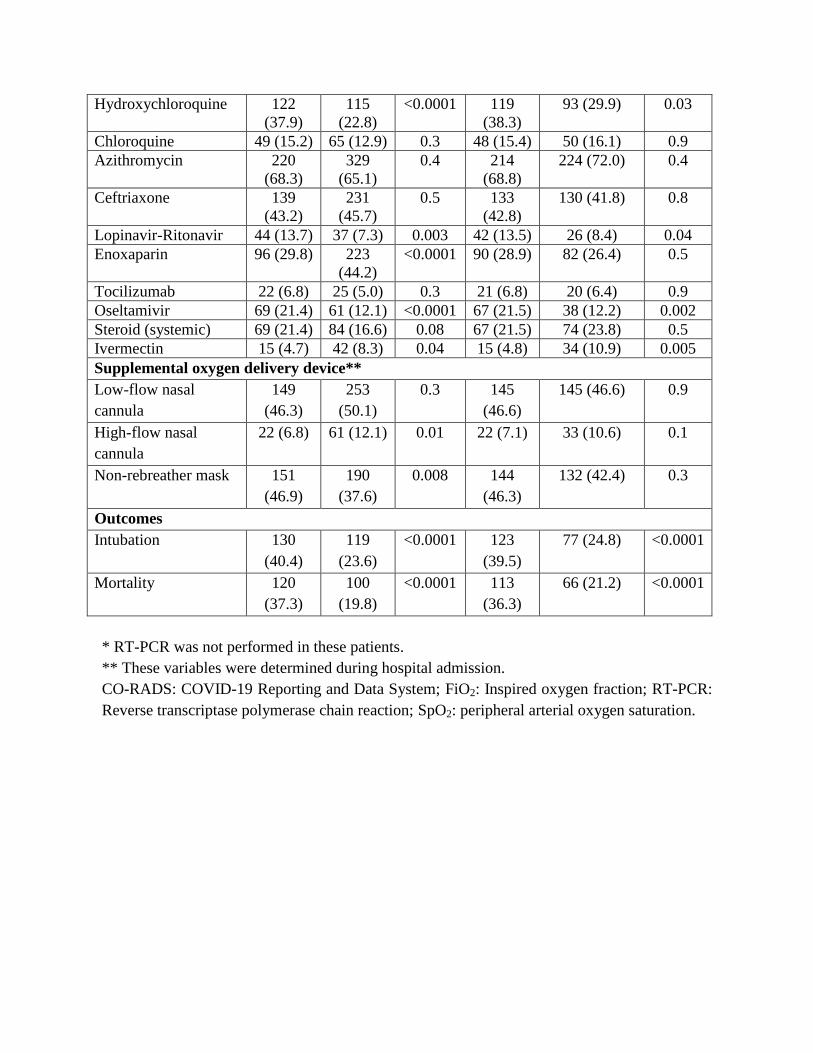
The characteristics of patients in the AP and supine groups, in both the unmatched and matched
cohorts, are provided in Table 2. Patients managed in AP had a median time-to-initiation of
prone positioning of 15.5 (IQR:8-48) hours. The median time spent in the prone position during
the hospital stay (total time in prone) was 12 (IQR:8-24) hours. A lesser proportion of patients
in the AP group required endotracheal intubation (23.6% vs 40.4%) or had a lethal outcome
(19.8% vs 37.3%). After propensity score matching, these differences prevailed. The SpO2/FiO2
ratio in the AP group was statistically significantly higher after prone (217.42, SD: 81.9)
compared with baseline values (182.39, SD: 81.91), with a mean difference of 35.03 (95%CI:
29.99-40.06, p<0.0001) units.

10
The results of univariable logistic regression models for orotracheal intubation risk are provided
in Table 3, for both the unmatched and matched cohorts. The main risk factors identified were
age, diabetes, arterial hypertension, obesity, heart disease, cancer, a baseline SpO2/FiO2 <100 or
between 100 and 199, and management with a non-rebreather mask. AP was a protective factor
for orotracheal intubation even after multivariable adjustment (Table 4) for confounding
variables (Adjusted OR=0.35, 95%CI:0.24-0.52, p<0.0001, E-value=2.12), which prevailed
after propensity score analysis (Adjusted OR=0.41, 95%CI:0.27-0.62, p<0.0001, E-value=1.86).
Similarly, AP was a protective factor for mortality (Adjusted OR=0.38, 95%CI:0.26-0.55,
p<0.0001, E-value=2.03, Goodness of fit: Hosmer-Lemeshow X2=10.2, p=0.3 AUC=0.78,
95%CI:0.74-0.81, p<0.0001) even after multivariable adjustment in propensity score analyses
(Adjusted OR=0.40, 95%CI:0.27-0.61, p<0.0001, E-value=1.88, Goodness of fit: Hosmer-
Lemeshow X2=7.81, p=0.4 AUC=0.78, 95%CI:0.74-0.82, p<0.0001). Lower intubation and
mortality risks for AP prevailed after sub-analyses of patients with a confirmatory SARS-CoV-2
RT-PCR (excluding those in whom molecular testing was not performed) (Appendix 8).
After adjusting for centre through GEE, 9 centres had an effect over the risk of intubation.
Despite this, AP continued to be associated with lower intubation risk (OR: 0.22, 95%CI: 0.15-
0.34, p<0.0001); the interaction between centre and AP was non-significant for all the centres.
The main variables associated with intubation amongst AP patients were increasing age
(OR=1.02, 95%CI: 1.01-1.04, p=0.005), SpO2/FiO2 <100 (OR=2.78, 95%CI: 1.35-5.72,
p=0.005), SpO2/FiO2 100-199 (OR=2.18, 95%CI: 1.31-3.64, p=0.003), and management with a
non-rebreather mask (OR=2.17, 95%CI: 1.34-3.49, p=0.002), Goodness of fit: Hosmer-
Lemeshow X2=10.52, p=0.2; AUC=0.70, 95%CI:0.64-0.74, p<0.0001. The distribution of risk
for increases in age and baseline SpO2/FiO2 are shown in Figure 2.
After the search of the literature, 99 records were retrieved, of which only 9 studies [10–12, 19–
24] were observational comparison-group studies including both AP and supine patients, with
sufficient information to calculate the overall risk of intubation, which are summarised
alongside the APRONOX study in Figure 3; the funnel plot is provided as Appendix 9.

DISCUSSION
In this multicentre observational study, we aimed to evaluate the association between awake
prone positioning and orotracheal intubation, as well as predictors of intubation amongst AP
patients, and mortality in hospitalised patients with COVID-19. Even after multivariable
adjustment and propensity score analyses, prone positioning in non-intubated patients was
associated with lower intubation and mortality risk.
Patients in our cohort were younger (mean age 53.4 years) than those in other studies (56.0-
65.8) [10–12]; hospitalised patients with COVID-19 in Mexico have been reported to be young
[25]. The prevalence of comorbidities in our study is similar to that reported in a population-
based sample of Mexican patients hospitalised with COVID-19, although diabetes was more
common in our study (38.1% vs 29.2%), whereas obesity (14.4% vs 22.5%) and heart disease
(2.1% vs 4.4%) were less frequent [25].
The total time spent in the prone position during in-hospital stay in our study was 12 (IQR:8-24)
hours, which is considerable compared to a recent pilot randomised study which reported that
self-proning patients spent only 1.6 (95%CI: 0.2-3.1) hours in the prone position in a 72-hour
evaluation period [26]. Daily time spent in the prone position has been reported to be highly
variable, with only 43% of patients achieving a daily dose of >6 hours in AP [27].
The overall intubation rate in the APRONOX cohort was higher (30.1%) than that reported for
hospitalised patients with COVID-19 in Mexico City (20.2%) [25]; however, limited access to
beds with ventilators in Mexico has been reported [28]. Intubation rates for patients in the
unmatched AP (23.6%) and supine (40.4%) cohorts fall within those reported in previous
studies (10–58% and 27.7–49%, respectively) [10–12]. AP in our study was associated with
decreased intubation risk even after multivariable adjustment in both the unmatched and
propensity-score matched cohorts, with an E-value of 2.01 and 2.21, respectively, which reflects
that in order to drive this association to be non-significant, an unmeasured risk factor should
have a lower-limit confidence interval that at least doubles the risk of the outcome between both
groups. Out of all comorbidities, only diabetes and heart disease were associated with increased
12
intubation risk after multivariable adjustment, however, diabetes was no longer a risk factor
after propensity score analysis. A higher baseline SpO2/FiO2 was associated with reduced
intubation risk. The mortality rate reported in our study was 19.8%, comparable to 23.4% [12]
and 27% [10] in other studies.
Regarding variables associated to intubation amongst AP patients, age, low SpO2/FiO2, and the
use of a non-rebreather mask were the main variables associated. The distribution of risk for
quantitative values of age show that the risk of intubation after AP is higher with increasing
ages, whereas higher baseline SpO2/FiO2 have the lowest risks.
AP has been presented as one the most cost-effective strategies to treat patients with COVID-19.
In countries with limited oxygen delivery devices, and a shortage of ventilators, AP could be
used to avoid intubating patients with COVID-19 [29]. Nonetheless, conflicting evidence from
observational studies for AP exists.
The supine position alters pulmonary function in patients with respiratory insufficiency due to
the gravitational differences between dependent and non-dependent regions, resulting in a more
negative pleural pressure (Ppl), increasing transpulmonary pressure (TPP) in non-dependent
areas (more distension), and producing the opposite effect in dependent areas where Ppl is less
negative and TPP is lower (less distension). Ventilation in the prone position causes even
distribution of TPP, favouring uniform ventilation [30]. Approximately 45 years ago, prone
positioning was shown to increase oxygenation in patients with respiratory insufficiency,
primarily by improving the ventilation-perfusion ratio (V/Q) [31].
Prone positioning has been evaluated in hospitalised patients with respiratory failure due to
COVID-19, having observed improvements in SpO2 and PaO2, decreased respiratory rate (RR),
decreased need for intubation and possible reductions in mortality, in addition to being cost-free
[8, 32–35]. As summarised in Figure 4, only three other studies to date have evaluated
intubation risk among AP compared with AS. While Ferando et al. and Padrão et al. found no
differences in intubation risk, Jagan et al found reduced intubation risk in AP patients [10–12].
The APRONOX study is the largest study to date evaluating the effect of AP on intubation risk.

Regarding oxygenation modality, the use of a non-rebreather mask was associated with greater
risk of intubation amongst all patients and within AP patients, whereas other oxygenation
devices were not. There is documented evidence of the correlation between the oxygen
saturation/fraction of inspired oxygen (SpO2/FiO2) ratio and the partial pressure of
oxygen/fraction of inspired oxygen (PaO2/FiO2) ratio, with the advantage that the SpO2/FiO2
ratio only relies on a pulse oximeter, with no need to perform a blood gas test, thereby
highlighting the value of validated cost-effective strategies [14].
Our study has the following limitations: 1) O2 delivery devices were not standardised to a
unique device, 2) the number of hours of AP varied between hospitals and patients, 3) no
standardised criteria were established to consider intubation in patients requiring mechanical
ventilation, 4) we were unable to asses which patients had do-not-intubate orders or other
reasons for not performing intubation, 5) availability of laboratory studies was limited across
centres and were thus not collected and analysed, 6) not all patients with a CO-RADS score >3
ultimately have a positive RT-PCR test [13]; this limitation was partially addressed by sub-
analysing patients with a positive SARS-CoV-2 RT-PCR, 7) a measure of oxygenation
comparable to post-prone SpO2/FiO2 in AP patients was not collected for patients in the supine
group, and 8) the length of stay of patients was not collected.
The strengths of our research include: 1) this is the largest study evaluating AP to date; 2) the
large number of hospitals included; and 3) the fact that various O2 delivery devices were
employed may reflect that the benefits of AP are not necessarily unique to NIV or HFNC
devices, which are costlier and not always available.
AP in spontaneously breathing patients with acute hypoxemic respiratory insufficiency may be a
justifiable treatment modality, given the improvements in oxygenation and its physiological
benefits, but the decision to intubate is based on the clinician’s best judgement and intubation
should not be delayed if under consideration. Close clinical evaluation of patients is key to avoid
poor outcomes. Studies of AP are challenging and randomised controlled trials are warranted to

14
fully elucidate its usefulness since this is an easy to administer, safe, and reproducible
intervention [36].
CONCLUSION
Prone positioning in awake hospitalised patients with COVID-19 is associated with a lower risk
of intubation and mortality.
Conflicts of Interest: The authors declare no conflicts of interest.
Funding: None.
Data availability: All data that support the findings of this study will be available from the
corresponding author upon reasonable request.
Acknowledgements:
Healthcare workers treating COVID-19 patients: Edgard Díaz Soto, Jaziel López Pérez, José
Antonio Meade Aguilar, Rubén Rodríguez Blanco, José Luis Patiño Pérez, Janisia Rodríguez
Solís, Maribel Santosbeña Lagunes, Alberto Calvo Zúñiga, Manuel de Jesús Santaella Sibaja,
Luis Iván Contreras Ley, María Alejandra Sicsik Aragón, Yessica Bernal Luna, Carlos Baez
Ambriz, Yanira Jiménez Blancas, Alejandro Ayala Mata, Tania Gabriela Ramírez Lira, Iván
Avalos Flores, Edwing Díaz Rodríguez, Roberto Robles Godínez, Eduardo Espino López, Hugo
Francisco Díaz Ramírez, Concepción Mendoza Fragoso, Oliver Garaz Trujillo, and Jesús Elías
Paredes Flores.
We thank the anonymous reviewers for their recommendations which allowed us to make
significant improvements to our manuscript.
REFERENCES
1. Valter C, Christensen AM, Tollund C, SchØnemann NK. Response to the prone position
in spontaneously breathing patients with hypoxemic respiratory failure. Acta Anaesthesiol
Scand 2003; 47: 416–418.
2. Scaravilli V, Grasselli G, Castagna L, Zanella A, Isgrò S, Lucchini A, Patroniti N, Bellani
G, Pesenti A. Prone positioning improves oxygenation in spontaneously breathing
nonintubated patients with hypoxemic acute respiratory failure: A retrospective study. J
Crit Care 2015; 30: 1390–1394.
3. Bower G, He H. Protocol for awake prone positioning in COVID-19 patients: to do it
earlier, easier, and longer. Crit Care 2020; 24: 371.
4. Ding L, Wang L, Ma W, He H. Efficacy and safety of early prone positioning combined
with HFNC or NIV in moderate to severe ARDS: a multi-center prospective cohort study.
Crit Care 2020; 24: 28.
5. Pérez-Nieto OR, Guerrero-Gutiérrez MA, Deloya-Tomas E, Ñamendys-Silva SA. Prone
positioning combined with high-flow nasal cannula in severe noninfectious ARDS. Crit
Care 2020; 24: 114.
6. Sun Q, Qiu H, Huang M, Yang Y. Lower mortality of COVID-19 by early recognition
and intervention: experience from Jiangsu Province. Ann Intensive Care 2020; 10: 33.
7. Caputo ND, Strayer RJ, Levitan R. Early Self-Proning in Awake, Non-intubated Patients
in the Emergency Department: A Single ED’s Experience During the COVID-19
Pandemic. Acad Emerg Med 2020; 27: 375–378.
8. Thompson AE, Ranard BL, Wei Y, Jelic S. Prone Positioning in Awake, Nonintubated
Patients With COVID-19 Hypoxemic Respiratory Failure. JAMA Intern Med 2020; 180:
1537.
9. Slessarev M, Cheng J, Ondrejicka M, Arntfield R. Patient self-proning with high-flow
nasal cannula improves oxygenation in COVID-19 pneumonia. Can J Anesth 2020; 67:
1288–1290.
10. Ferrando C, Mellado-Artigas R, Gea A, Arruti E, Aldecoa C, Adalia R, Ramasco F,
Monedero P, Maseda E, Tamayo G, Hernández-Sanz ML, Mercadal J, Martín-Grande A,
Kacmarek RM, Villar J, Suárez-Sipmann F. Awake prone positioning does not reduce the
risk of intubation in COVID-19 treated with high-flow nasal oxygen therapy: A

16
multicenter, adjusted cohort study. Crit Care 2020; 24: 1–11.
11. Padrão EMH, Valente FS, Besen BAMP, Rahhal H, Mesquita PS, de Alencar JCG, da
Costa MGP, Wanderley APB, Emerenciano DL, Bortoleto FM, Fortes JCL, Marques B,
de Souza SFB, Marchini JFM, Neto RAB, de Souza HP. Awake Prone Positioning in
COVID-19 Hypoxemic Respiratory Failure: Exploratory Findings in a Single-center
Retrospective Cohort Study. Acad Emerg Med 2020; 27: 1249–1259.
12. Jagan N, Morrow LE, Walters RW, Klein LP, Wallen TJ, Chung J, Plambeck RW. The
POSITIONED Study: Prone Positioning in Nonventilated Coronavirus Disease 2019
Patients—A Retrospective Analysis. Crit Care Explor 2020; 2: e0229.
13. Prokop M, van Everdingen W, van Rees Vellinga T, Quarles van Ufford H, Stöger L,
Beenen L, Geurts B, Gietema H, Krdzalic J, Schaefer-Prokop C, van Ginneken B, Brink
M, COVID-19 Standardized Reporting Working Group of the Dutch Radiological
Society. CO-RADS: A Categorical CT Assessment Scheme for Patients Suspected of
Having COVID-19-Definition and Evaluation. Radiology 2020; 296: E97–E104.
14. Rice TW, Wheeler AP, Bernard GR, Hayden DL, Schoenfeld DA, Ware LB, National
Institutes of Health, National Heart, Lung and BIAN. Comparison of the SpO2/FIO2
ratio and the PaO2/FIO2 ratio in patients with acute lung injury or ARDS. Chest 2007;
132: 410–417.
15. Parke RL, Eastwood GM, McGuinness SP, George Institute for Global Health, Australian
and New Zealand Intensive Care Society Clinical Trials Group. Oxygen therapy in non-
intubated adult intensive care patients: a point prevalence study. Crit Care Resusc 2013;
15: 287–293.
16. Argenziano MG, Bruce SL, Slater CL, Tiao JR, Baldwin MR, Barr RG, Chang BP, Chau
KH, Choi JJ, Gavin N, Goyal P, Mills AM, Patel AA, Romney M-LS, Safford MM,
Schluger NW, Sengupta S, Sobieszczyk ME, Zucker JE, Asadourian PA, Bell FM, Boyd
R, Cohen MF, Colquhoun MI, Colville LA, de Jonge JH, Dershowitz LB, Dey SA,
Eiseman KA, Girvin ZP, et al. Characterization and clinical course of 1000 patients with
coronavirus disease 2019 in New York: retrospective case series. BMJ 2020; 369: m1996.
17. Mancilla-Galindo J, Vera-Zertuche JM, Navarro-Cruz AR, Segura-Badilla O, Reyes-
Velázquez G, Tepepa-López FJ, Aguilar-Alonso P, Vidal-Mayo J de J, Kammar-García
A. Development and validation of the patient history COVID-19 (PH-Covid19) scoring

system: a multivariable prediction model of death in Mexican patients with COVID-19.
Epidemiol Infect 2020; 148: e286.
18. Mathur MB, Ding P, Riddell CA, VanderWeele TJ. Web Site and R Package for
Computing E-values. Epidemiology 2018; 29: e45–e47.
19. Prud’homme E, Trigui Y, Elharrar X, Gaune M, Loundou A, Lehingue S, Boyer A,
Lefebvre L, Dols A, Chanez P, Papazian L, Forel J. Effect of Prone Positioning on the
respiratory support of non-intubated patients with COVID-19 and acute hypoxemic
respiratory failure: A retrospective matching cohort study. Chest American College of
Chest Physicians; 2021; .
20. Belkhouja K, Alzahrani S, Al-Shalhoub N, Negm T, Darwish M, Pathan MW, Lone MM,
Omer S, Al-Sharif H. Feasibility and efficacy of prone position combined with CPAP in
COVID-19 patients with acute hypoxemic respiratory failure. Intensive Care Med Exp
2020; 8: 73.
21. Fazzini B, Fowler AJ, Zolfaghari P. Effectiveness of prone position in spontaneously
breathing patients with COVID-19: A prospective cohort study. J Intensive Care Soc
2021; : 2–5.
22. Tonelli R, Pisani L, Tabbì L, Comellini V, Prediletto I, Fantini R, Marchioni A,
Andrisani D, Gozzi F, Bruzzi G, Manicardi L, Busani S, Mussini C, Castaniere I, Bassi I,
Carpano M, Tagariello F, Corsi G, d’Amico R, Girardis M, Nava S, Clini E. Early awake
proning in critical and severe COVID-19 patients undergoing noninvasive respiratory
support: A retrospective multicenter cohort study. Pulmonology Sociedade Portuguesa de
Pneumologia; 2021; .
23. Alsharif H, Belkhouja K. 267: Feasibility and Efficacy of Prone Position Combined With
CPAP in COVID-19 Patients With AHRF. Crit Care Med 2021; 49.
24. Barker J, Pan D, Koeckerling D, Baldwin AJ, West R. Effect of serial awake prone
positioning on oxygenation in patients admitted to intensive care with COVID-19.
Postgrad Med J 2021; : postgradmedj-2020-139631.
25. Mancilla-Galindo J, Kammar-García A, Martínez-Esteban A, Meza-Comparán A-K,
Mancilla-Ramírez J, Galindo-Sevilla N. COVID-19 patients with increasing age
experience differential time to initial medical care and severity of symptoms. Cambridge
Open Engag 2021; : 10.33774/coe-2021-sjbcf.
18
26. Johnson SA, Horton DJ, Fuller MJ, Yee J, Aliyev N, Boltax JP, Chambers JH, Lanspa
MJ. Patient-Directed Prone Positioning in Awake Patients with COVID-19 Requiring
Hospitalization (PAPR). Ann Am Thorac Soc 2021; 0: 2–11.
27. Jayakumar D, Ramachandran, DNB P, Rabindrarajan, DNB E, Vijayaraghavan, MD
BKT, Ramakrishnan, AB N, Venkataraman, AB R. Standard Care Versus Awake Prone
Position in Adult Nonintubated Patients With Acute Hypoxemic Respiratory Failure
Secondary to COVID-19 Infection—A Multicenter Feasibility Randomized Controlled
Trial. J Intensive Care Med 2021; .
28. Fowler Z, Moeller E, Roa L, Castañeda-Alcántara ID, Uribe-Leitz T, Meara JG,
Cervantes-Trejo A. Projected impact of COVID-19 mitigation strategies on hospital
services in the Mexico City Metropolitan Area. Lazzeri C, editor. PLoS One 2020; 15:
e0241954.
29. Touchon F, Trigui Y, Prud’homme E, Lefebvre L, Giraud A, Dols A, Martinez S,
Bernardi M, Begne C, Granier P, Chanez P, Forel J, Papazian L, Elharrar X. Awake
prone positioning for hypoxaemic respiratory failure: past, COVID-19 and perspectives.
Eur Respir Rev 2021; 30: 210022.
30. Guérin C. Prone Positioning. In: Chiumello D, editor. Acute Respir Distress Syndr Cham:
Springer International Publishing; 2017. p. 73–84.
31. Bryan AC. Comments of a Devil’s Advocate. Am Rev Respir Dis 1974; 110: 143–144.
32. Elharrar X, Trigui Y, Dols A-M, Touchon F, Martinez S, Prud’homme E, Papazian L.
Use of Prone Positioning in Nonintubated Patients With COVID-19 and Hypoxemic
Acute Respiratory Failure. JAMA 2020; 323: 2336.
33. Sartini C, Tresoldi M, Scarpellini P, Tettamanti A, Carcò F, Landoni G, Zangrillo A.
Respiratory Parameters in Patients With COVID-19 After Using Noninvasive Ventilation
in the Prone Position Outside the Intensive Care Unit. JAMA 2020; 323: 2338.
34. Telias I, Katira BH, Brochard L. Is the Prone Position Helpful During Spontaneous
Breathing in Patients With COVID-19? JAMA 2020; 323: 2265.
35. Franco C, Facciolongo N, Tonelli R, Dongilli R, Vianello A, Pisani L, Scala R, Malerba
M, Carlucci A, Negri EA, Spoladore G, Arcaro G, Tillio PA, Lastoria C, Schifino G,
Tabbì L, Guidelli L, Guaraldi G, Ranieri VM, Clini E, Nava S. Feasibility and clinical
impact of out-of-ICU noninvasive respiratory support in patients with COVID-19-related

pneumonia. Eur Respir J 2020; 56: 2002130.
36. Taylor SP, Bundy H, Smith WM, Skavroneck S, Taylor B, Kowalkowski MA. Awake-
Prone Positioning Strategy for Non-Intubated Hypoxic Patients with COVID-19: A Pilot
Trial with Embedded Implementation Evaluation. Ann Am Thorac Soc 2020; :
AnnalsATS.202009-1164OC.
37. Project COVID-19 Open Access. Living Evidence on COVID-19 [Internet].
2020.Available from: https://ispmbern.github.io/covid-19/living-review/.

20
Table 1 Demographic and clinical characteristics at hospital admission and outcomes of
patients in the APRONOX cohort
Demographic variables
Age, years 54.3 (14.2)
Age categories, n (%)
<20 1 (0.1)
20-29 29 (3.5)
30-39 101 (12.2)
40-49 194 (23.5)
50-59 209 (25.3)
60-69 162 (19.6)
≥70, n 131 (15.8)
Sex
Women, n (%) 227 (27.4)
Men, n (%) 600 (72.6)
Type of care
ICU 142 (17.2)
Non-ICU 685 (82.8)
Clinical variables
Diabetes, n (%) 315 (38.1)
Systemic arterial hypertension, n (%) 285 (34.5)
Obesity, n (%) 119 (14.4)
Heart disease, n (%) 17 (2.1)
Lung disease, n (%) 41 (5)
Cancer, n (%) 10 (1.2)
Liver disease, n (%) 5 (0.6)
Chronic kidney disease, n (%) 35 (4.2)
PH-Covid19 Mortality Risk Score** 8.7 (3.6)
Pharmacological treatments
Hydroxychloroquine 237 (28.7)
Chloroquine 114 (13.8)
Azithromycin 549 (66.4)
Ceftriaxone 370 (44.7)
Lopinavir-Ritonavir 81 (9.8)
Enoxaparin 319 (38.6)
Tocilizumab 47 (5.7)
Oseltamivir 130 (15.7)
Steroid (systemic) 153 (18.5)
Ivermectin 57 (6.9)
Baseline SpO2/FiO2 ratio** 189.5 (81.6)

Awake prone, n (%)*** 505 (61.1)
Awake supine, n (%) 322 (38.9)
Supplemental oxygen delivery device
Low-flow nasal cannula, n (%) 402 (48.6)
High-flow nasal cannula, n (%) 83 (10)
Non-rebreather mask, n (%) 342 (41.4)
Outcomes, n (%)
Intubation, n (%) 249 (30.1)
Mortality, n (%) 220 (26.6)
Failure to the prone****, n (%) 119 (23.6)*
*Percentage calculated out of all awake prone-positioned patients.
**These variables were determined at hospital admission.
***Median time-to-initiation of prone: 15.5 (IQR:8-48) hours
****Defined as patients who were successfully managed in the awake prone position but
required orotracheal intubation anytime during follow-up.
FiO2: Inspired oxygen fraction; SpO2: peripheral arterial oxygen saturation.

22
Table 2. Comparison of demographic and clinical characteristics at hospital admission and
outcomes of patients in the awake prone and supine groups in both the unmatched and
propensity score-matched cohorts.
Unmatched Matched
Awake
supine
(n =
322)
Awake
prone
(n =
505)
p-value Awake
supine
(n =
311)
Awake
prone
(n = 311)
p-value
Demographic variables
Age, years 55.8
(14.5)
53.4
(13.9)
0.02 55.6
(14.5)
54.9 (14.1) 0.5
Women 92 (28.6) 135
(26.7)
0.6 86 (27.7) 79 (25.4) 0.5
Men 230
(71.4)
370
(73.3)
225
(72.3)
232 (74.6)
Diagnostic criterion
RT-PCR positive 294
(91.3)
440
(87.1)
0.06 282
(90.7)
289 (92.9) 0.3
CO-RADS 3-5* 28 (8.7) 65 (12.9) 29 (9.3) 22 (7.1)
Type of care
ICU 75 (23.3) 67 (13.3) <0.0001 73 (23.5) 60 (19.3) 0.2
Non-ICU 247
(76.7)
438
(86.7)
238
(76.5)
251 (80.7)
Clinical variables
Diabetes 121
(37.6)
194
(38.4)
0.8 117
(37.6)
119 (38.3) 0.9
Systemic arterial
hypertension
119 (37) 166
(32.9)
0.2 114
(36.7)
102 (32.8) 0.4
Obesity 45 (14) 74 (14.7) 0.8 45 (14.5) 39 (12.5) 0.6
Heart disease 4 (1.2) 13 (2.6) 0.2 4 (1.3) 8 (2.6) 0.4
Lung disease 17 (5.3) 24 (4.8) 0.7 16 (5.1) 17 (5.5) 0.9
Cancer 8 (2.5) 2 (0.4) 0.02 7 (2.3) 1 (0.3) 0.07
Liver disease 3 (0.9) 2 (0.4) 0.4 3 (1.0) 1 (0.3) 0.6
Chronic kidney
disease
12 (3.7) 23 (4.6) 0.6 12 (3.9) 13 (4.2) 0.8
SpO2/FiO2 ratio** 201.1
(89.8)
182.4
(75.4)
0.002 201.1
(88.8)
195.9 (77.9) 0.4
PH-Covid19 Mortality
Risk Score**
8.9 (3.6) 8.6 (3.5) 0.1 8.9 (3.6) 8.9 (3.5) 0.8
Pharmacological treatments
Hydroxychloroquine 122
(37.9)
115
(22.8)
<0.0001 119
(38.3)
93 (29.9) 0.03
Chloroquine 49 (15.2) 65 (12.9) 0.3 48 (15.4) 50 (16.1) 0.9
Azithromycin 220
(68.3)
329
(65.1)
0.4 214
(68.8)
224 (72.0) 0.4
Ceftriaxone 139
(43.2)
231
(45.7)
0.5 133
(42.8)
130 (41.8) 0.8
Lopinavir-Ritonavir 44 (13.7) 37 (7.3) 0.003 42 (13.5) 26 (8.4) 0.04
Enoxaparin 96 (29.8) 223
(44.2)
<0.0001 90 (28.9) 82 (26.4) 0.5
Tocilizumab 22 (6.8) 25 (5.0) 0.3 21 (6.8) 20 (6.4) 0.9
Oseltamivir 69 (21.4) 61 (12.1) <0.0001 67 (21.5) 38 (12.2) 0.002
Steroid (systemic) 69 (21.4) 84 (16.6) 0.08 67 (21.5) 74 (23.8) 0.5
Ivermectin 15 (4.7) 42 (8.3) 0.04 15 (4.8) 34 (10.9) 0.005
Supplemental oxygen delivery device**
Low-flow nasal
cannula
149
(46.3)
253
(50.1)
0.3 145
(46.6)
145 (46.6) 0.9
High-flow nasal
cannula
22 (6.8) 61 (12.1) 0.01 22 (7.1) 33 (10.6) 0.1
Non-rebreather mask 151
(46.9)
190
(37.6)
0.008 144
(46.3)
132 (42.4) 0.3
Outcomes
Intubation 130
(40.4)
119
(23.6)
<0.0001 123
(39.5)
77 (24.8) <0.0001
Mortality 120
(37.3)
100
(19.8)
<0.0001 113
(36.3)
66 (21.2) <0.0001
* RT-PCR was not performed in these patients.
** These variables were determined during hospital admission.
CO-RADS: COVID-19 Reporting and Data System; FiO2: Inspired oxygen fraction; RT-PCR:
Reverse transcriptase polymerase chain reaction; SpO2: peripheral arterial oxygen saturation.

24
Table 3. Results of univariable logistic regression analyses of orotracheal intubation risk in
patients with awake prone positioning.
Unmatched Matched
OR (95% CI) p value OR (95% CI) p value
Awake prone 0.46 (0.34-0.62) <0.0001 0.50 (0.36-0.71) <0.0001
Demographic variables
Age, years 1.02 (1.004-1.03) 0.007 1.01 (1.002-1.03) 0.02
Sex (Men) 0.91 (0.70-1.37) 0.9 1.12 (0.77-1.65) 0.6
Type of care
ICU 0.63 (0.41-0.96) 0.03 0.61 (0.39-0.94) 0.03
Clinical variables
Diabetes 1.70 (1.26-2.30) 0.001 1.80 (1.28-2.54) 0.001
Systemic arterial
hypertension
1.61 (1.19-2.19) 0.002 1.40 (0.99-1.99) 0.06
Obesity 2.01 (1.35-2.99) 0.001 2.69 (1.69-4.29) <0.0001
Heart disease 3.41 (1.28-9.07) 0.01 4.35 (1.29-14.64) 0.02
Lung disease 1.36 (0.71-2.62) 0.4 1.39 (0.68-2.87) 0.4
Cancer 9.56 (2.02-45.35) 0.004 15.27 (1.87-
124.96)
0.01
Liver disease 3.51 (0.58-21.15) 0.2 2.12 (0.29-15.17) 0.5
Chronic kidney disease 1.39 (0.69-2.81) 0.4 1.43 (0.63-3.24) 0.4
Baseline SpO2/FiO2 ratio
<100 5.69 (3.48-9.31) <0.0001 7.44 (4.18-13.24) <0.0001
100-199 3.69 (2.57-5.29) <0.0001 4.26 (2.86-6.33) <0.0001
≥200 Reference Reference
Pharmacological
treatments
Hydroxychloroquine 1.08 (0.78-1.49) 0.7 1.13 (0.79-1.61) 0.5
Chloroquine 0.81 (0.52-1.26) 0.3 0.77 (0.48-1.25) 0.3
Azithromycin 1.05 (0.76-1.43) 0.8 0.94 (0.65-1.35) 0.7
Ceftriaxone 0.82 (0.61-1.11) 0.2 0.72 (0.51-1.02) 0.07
Lopinavir-Ritonavir 0.45 (0.25-0.83) 0.01 0.51 (0.28-0.95) 0.03
Enoxaparin 0.84 (0.62-1.15) 0.3 0.88 (0.61-1.29) 0.5
Tocilizumab 0.53 (0.25-1.12) 0.09 0.58 (0.27-1.23) 0.2
Oseltamivir 0.79 (0.52-1.21) 0.3 0.82 (0.52-1.29) 0.4
Steroid (systemic) 0.53 (0.35-0.81) 0.004 0.47 (0.30-0.74) 0.001
Ivermectin 0.89 (0.49-1.64) 0.7 1.03 (0.55-1.91) 0.9
Supplemental oxygen
delivery device
Low-flow nasal cannula 0.27 (0.19-0.38) <0.0001 0.28 (0.19-0.41) <0.0001
High-flow nasal cannula 0.77 (0.46-1.29) 0.3 0.77 (0.42-1.44) 0.4

Non-rebreather mask 3.94 (2.88-5.39) <0.0001 3.75 (2.63-5.35) <0.0001
95%CI: 95% confidence interval; FiO2: Inspired oxygen fraction; ICU: Intensive Care Unit;
OR: odds ratio; SpO2: peripheral arterial oxygen saturation.

26
Table 4. Results of multivariable logistic regression analyses of orotracheal intubation risk in
patients with awake prone positioning, adjusted by confounding variables.
Unmatched* Matched**
OR (95% CI) P value OR (95% CI) P value
Awake prone 0.35 (0.24-0.52) <0.0001 0.41 (0.27-0.62) <0.0001
Age, years 1.01 (0.99-1.02) 0.4 1.01 (0.99-1.02) 0.6
Sex (Men) 1.15 (0.77-1.72) 0.5 1.26 (0.79-2.02) 0.3
ICU 0.52 (0.31-0.89) 0.01 0.50 (0.29-0.86) 0.01
Diabetes 1.50 (1.03-2.19) 0.03 1.66 (1.08-2.55) 0.02
Systemic arterial
hypertension
1.23 (0.84-1.81) 0.3 0.95 (0.61-1.48) 0.8
Obesity 1.39 (0.86-2.28) 0.18 1.47 (0.81-2.65) 0.2
Heart disease 6.82 (2.13-
21.78)
0.001 13.79 (3.31-
57.61)
<0.0001
Cancer 7.41 (0.96-
57.39)
0.06 12.58 (0.81-
196.11)
0.07
Chronic kidney
disease
1.11 (0.46-2.69) 0.8 1.29 (0.43-3.92) 0.7
Ceftriaxone 0.91 (0.63-
1.31)
0.6 0.82 (0.53-1.25) 0.4
Enoxaparin 0.79 (0.54-1.16) 0.2 0.85 (0.53-1.36) 0.5
Tocilizumab 0.56 (0.22-1.38) 0.2 0.58 (0.22-1.53) 0.3
Oseltamivir 0.59 (0.35-1.02) 0.06 0.68 (0.37-1.24) 0.2
Steroid (systemic) 0.62 (0.38-1.03) 0.06 0.57 (0.34-0.97) 0.04
Baseline
SpO2/FiO2 ratio
0.99 (0.98-0.99) <0.0001 0.99 (0.98-0.99) <0.0001
Low-flow nasal
cannula
- - - -
High-flow nasal
cannula
0.99 (0.53-1.88) 0.9 1.19 (0.51-2.45) 0.8
Non-rebreather
mask
2.70 (1.82-4.01) <0.0001 2.49 (1.56-3.99) <0.0001
*Goodness of fit: Hosmer-Lemeshow X2=2.79, p=0.9; AUC=0.79, 95%CI:0.77-0.83,
p<0.0001.
**Goodness of fit: Hosmer-Lemeshow X2=10.95, p=0.2; AUC=0.82, 95%CI:0.79-0.85,
p<0.0001.
95%CI: 95% confidence interval; AUC: area under de curve; FiO2: Inspired oxygen fraction;
OR: odds ratio; SpO2: peripheral arterial oxygen saturation.

Figure 1. Flow diagram of participants included in the APRONOX cohort.
Figure 2. Risk of intubation amongst patients in the awake prone positioning group, according
to age (A) and baseline SpO2/FiO2 (B)*.
*For this analysis, baseline SpO2/FiO2 was studied as continuous variable, therefore, the range
of odds ratios are different from others in the manuscript which consider baseline SpO2/FiO2 as
a categorical variable and use a category of reference to compare other categories.
95%CI: 95% confidence intervals; FiO2: Inspired oxygen fraction; SpO2: peripheral arterial
oxygen saturation
Figure 3. Forest plot of overall risk of orotracheal intubation in studies retrieved by the search
strategy and the APRONOX cohort.
*Only patients in the propensity score-matched cohorts were included for the APRONOX
study.
95%CI: 95% confidence intervals; M-H: Mantel-Haenszel
[37]

322 patients in the awake
supine group
- 296 Positive RT-PCR for
SARS-CoV-2
- 26 CO-RADS 3 to 5, RT-
PCR not performed
505 patients in the awake
prone group
- 438 Positive RT-PCR for
SARS-CoV-2
- 67 CO-RADS 3 to 5, RT-
PCR not performed
100 incomplete medical
records
- 34: Unable to calculate
SpO2/FiO2 ratio
- 66: Unable to ascertain
if patient remained in the
prone or supine position
827 patients included for
analysis
932 medical records
assessed for eligibility
27 hospitals
832 medical records
-2 voluntary discharge
from hospital
-3 transferred to another
hospital


Appendix 1. Full list of authors with place of affiliation.
Writing Committee
Orlando Ruben Perez Nieto MD; Hospital General San Juan del Río, Querétaro. Intensive Care
Unit. Manuel Alberto Guerrero Gutierrez MD; Instituto Nacional de Cancerología, Mexico
City. Intensive Care Unit. Eder Ivan Zamarron Lopez MD; Hospital CEMAIN Tampico,
Tamaulipas. Intensive Care Unit. Ernesto Deloya Tomas MD; Hospital General San Juan del
Río, Querétaro. Head of Intensive Care Unit. Javier Mancilla-Galindo MBBS; Instituto
Nacional de Enfermedades Respiratorias, México City. Respiratory Medicine Fellow and
Instituto Nacional de Cardiología, Mexico City. Unidad de Investigación UNAM-INC. Ashuin
Kammar-García, PhD; Instituto Nacional de Nutrición y Ciencias Médicas “Salvador Zubiran”,
Mexico City. Emergency Department. Raúl Soriano Orozco; Hospital de Alta Especialidad T1
IMSS, León, Guanajuato. Intensive Care Unit. Gabriel Cruz Chavez MD; Clínica Hospital
Mérida ISSSTE. Head of Intensive Care Unit. Jose David Salmeron Gonzalez MD; Hospital
General “Miguel Silva”, Morelia, Michoacán. Intensive Care Unit. Marco Antonio Toledo
Rivera MD; Hospital SEDNA, Mexico City. Head of Intensive Care Unit. Luis Antonio
Morgado Villaseñor MD; Hospital General de Zona IMSS No.15 Reynosa, Tamaulipas.
Intensive Care Unit.
Jenner Jose Martínez Mazariegos MD; Hospital Vida Mejor ISSSTECH Tuxtla Gutiérrez,
Chiapas. Intensive Care Unit. Silvio Antonio Ñamendys Sylva MD, MSc, FCCP, FCCM;
Instituto Nacional de Cancerología and Instituto Nacional de Ciencias Médicas y Nutrición
“Salvador Zubirán”, Mexico City. Head of Intensive Care Unit.
General Research Committee
Diego Escarraman Martinez MD, MSc; Centro Médico Nacional IMSS “La Raza”, Mexico City.
Department of Anaesthesiology. Miguel Angel Martinez Camacho, PT, MSc; Hospital General
de Mexico, Mexico City. Intensive Care Unit. Ivette Mata Maqueda MD, MSc. DSc; Secretaría
de Salud del Estado de Querétaro, Ethics and Research Committee. Jesús Salvador Sánchez
Díaz MD, MSc; Hospital de Alta Especialidad IMSS “Adolfo Ruiz Cortines” Veracruz,
Veracruz. Intensive Care Unit. Luis Alberto Macias Garcia, MD, MSc; Hospital Regional
ISSSTE “Fernando Quiroz Gutiérrez”, Mexico City. Intensive Care Unit. Josué Luis Medina
Estrada MD; Hospital Regional No. 1 IMSS “Vicente Guerrero”, Acapulco, Guerrero. Intensive
Care Unit.
Local researchers
Hospital SEDNA, Mexico City: Ivette Zapata Centeno MD, Intensive Care Unit; Cecilia
Hernández Fernández MD. Hospital General de Zona No. 33 IMSS Bahía de Banderas,
Nayarit: Francisco Agustín Martínez Ayuso MD, Intensive Care Unit, Hospital General “Dr.
Miguel Silva”, Morelia, Michoacán: José David Salmerón González MD, Intensive Care Unit.
Juan Manuel Angeles Uribe MD, Emergency Department. Centro Médico Lic. Adolfo López
Mateos, ISEM, Toluca, State of Mexico: Aaron Alacio Ávila MD, Intensive Care Unit. Abad

Quetzalcoatl Ortega Pérez MD, Head of Intensive Care Unit. Centro Medico Nacional 20 de
Noviembre, ISSSTE, Mexico City: Jessica Selene Cancino Cuevas MD, Intensive Care Unit.
Alberto Hilarion de la Vega Bravo MD, Head of Intensive Care Unit. Clínica Hospital Mérida
ISSSTE, Mérida, Yucatán: Gabriel Cruz Sanchez MD. Clínica Hospital Mérida ISSSTE. Head
of Intensive Care Unit. Hospital General Dr. Enrique Cabrera, Mexico City: Ivan Ilescas
Martinez MD, Emergency Department. Lilian Saraí Ramirez Serrano MD, Emergency
Department. Hospital Regional de Alta Especialidad de Zumpango, State of Mexico: Areli
Patricia Ortíz Jimenez MD, Emergency Department. María José Pecero Hidalgo MD,
Department of Pneumology. Hospital Estatal de Atención de Pacientes COVID-19, Leon,
Guanajuato: Jorge Adalid Díaz Rodriguez MD, Respiratory Care Unit. Hospital Juárez de
México, Mexico City: Juan Carlos Betancourt Aldana Villarruel MD, Department of
Cardiology. José Carlos Gasca Aldama MD, Respiratory Care Unit. Ruben Nicolas Mendoza
MD, Department of Cardiology. Luis Fausto García Mayen MD, Head of Cardiovascular
Medicine Department. Hospital General Tuxtepec, Oaxaca: Jesús Ariben Servando Álvarez
Ramirez MD, Respiratory Care Unit. Enrique Fleuvier Morales López MD. Respiratory Care
Unit. Hospital General de San Juan del Río, San Juan del Río, Querétaro. Jorge López
Fermin MD, Intensive Care Unit, Tania Mondragon Labelle MD, Intensive Care Unit, Gabriela
Castillo Gutierrez MD, Intensive Care Unit, Jorge Daniel Carrión Moya MD, Intensive Care
Unit, María Guadalupe Olvera Ramos MD. Intensive Care Unit, Manuel Alfredo Díaz
Martínez, MD, Department of Anaesthesiology. Hospital Santo Tomás Querétaro, Querétaro.
Cristobal Meneses Olguín MD, Head of Respiratory Care Unit, Andrea Guadalupe de la Torre
Rittscher MD, Respiratory Care Unit. Lizbeth Franco Morales MD, Respiratory Care Unit.
Martin de Jesus Reyna Ramirez MD, Respiratory Care Unit. Angélica Del Carmen Chimal
Ayohua MD, Respiratory Care Unit. Hospital General de Zona No. 48 “San Pedro Xalpa”,
Mexico City. César Daniel Alonso Bello MD, Internal Medicine Department. Edgar Pérez
Barragán MD. Department of Infectiology. Hospital General de Zona No. 71 Veracruz,
Veracruz. Oscar Rodrigo Jimenez Flores, Intensive Care Unit. Ulises Espinosa Hernandez
MD, Emergency Department. Hospital Comunitario de Ocuituco, Morelos: Iván Hernández
Bernabé MD. Internal Medicine Department. Yuliana Young Peralta MD, Emergency
Department. José Ramón Arteaga Solis MD, Medical Director. Hospital General Regional No
200 Tecámac, State of Mexico: Josafat Jesús Gutierrez de la Cruz MD. Emergency
Department. Unidad Médica de Alta Especialidad IMSS No. 189 “Adolfo Ruiz Cortines”:
Jesús Salvador Sánchez Díaz MD, Intensive Care Unit. Xiomara García Montes MD,
Emergency Department. Hospital General Regional IMSS No. 251 Metepec, State of
Mexico: Carlos Mendiola Villalobos MD, Emergency Department. Alejandro Esquivel Loza
MD, Internal Medicine Department. Hospital General Regional ISSSTE “Fernando Quiroz
Gutiérrez”, Mexico City: María Concepción Gonzalez Belmont MD, Hospital General de
Querétaro, Querétaro: Raul Arturo Gonzalez Toribio MD, Intensive Care Unit. Alicia
Alejandra Rico Pérez MD, Emergency Department. ArjunaAliel Sotomayor Zavala MD,
Emergency Departmen. Hospital IESS “Manuel Ygnacio Monteros”, Loja, Ecuador:
Tatiana Maribel Merino Mijas MD, Intensive Care Unit. Maria Eugenia Abad Guarnizo MD.
Intensive Care Unit. Hospital Materno de Celaya, Guanajuato:
Karen Pamela Pozos Cortes MD, Head of Intensive Care Unit. Hospital CEMAIN, Tampico,
Tamaulipas: María Angelica Sánchez Cepeda, MD, Head of Intensive Care Unit. Hospital
Regional No. 1 IMSS “Vicente Guerrero”, Acapulco, Guerrero: Josué Luis Medina Estrada
MD. Intensive Care Unit. Hospital de la Beneficencia Española San Luis Potosí, San Luis
Potosí: Luis Arturo López Reveles MD, Emergency Department. Elsa Berenice Arriaga Rivera
MD, Emergency Department. Hospital General de Zona No. 1, Mexico City: Ramses Dorado
García MD, Emergency Department. Angela Janeth Bustos Valdez MD, Emergency
Department. Nizaguiee Cecilia Zaragoza Ortiz MD, Emergency Department. José Daniel
Castañeda Casiano MD, Emergency Department. Maritssa Mabel Martínez Ramírez MD,
Emergency Department. Iván Rayón Andrade MD, Emergency Department.
Collaborators - monitors
Silvia Elena Uribe Moya MD – Centro Medico Nacional IMSS “La raza”, Hospital de
Infectologia “Dr. Daniel Mendez Hernandez”, Respiratory Care Unit. Rodrigo Fernando
Centeno Asencio MD – Hospital General Regional Mérida “Ignacio García Tellez”,
Emergency Department. Alfredo García Tellez MD, Raymundo Montiel Latorre MD - Hospital
General de Pachuca; Emergency Department. Luis Fernando Flores Zamora NUS - Hospital
Regional de la Universidad de Colima, Intensive Care Unit. Dulce María Bernal Martínez MD,
Victor Hugo García López MD - Hospital General de Tláhuac, Mexico City. Diego González
Barbosa RT - Centro Médico Nacional de Occidente, Guadalajara, Jalisco. Department of
Respiratory Physiology. Hospital Star Medica Luna Parc, Cuautitlán Izcalli, State of Mexico:
Marco Antonio Villagrana Rodríguez. Intensive Care Unit.
ACKNOWLEDGEMENTS
The APRONOX Group wishes to thank Edgard Díaz Soto, Jaziel López Pérez, José Antonio
Meade Aguilar, Rubén Rodríguez Blanco, José Luis Patiño Pérez, Janisia Rodríguez Solís,
Maribel Santosbeña Lagunes, Alberto Calvo Zúñiga, Manuel de Jesús Santaella Sibaja, Luis
Iván Contreras Ley, María Alejandra Sicsik Aragón, Yessica Bernal Luna, Carlos Baez Ambriz,
Yanira Jiménez Blancas, Alejandro Ayala Mata, Tania Gabriela Ramírez Lira, Iván Avalos
Flores, Edwing Díaz Rodríguez, Roberto Robles Godínez, Eduardo Espino López, Hugo
Francisco Díaz Ramírez, Concepción Mendoza Fragoso, Oliver Garaz Trujillo, and Jesús Elías
Paredes Flores. for their help in providing care to patients with COVID-19.

Appendix 2. List of hospitals participating in the study and physicians in charge
Name of hospital Institution State Country
1 Hospital de Beneficencia Española Private San Luis Potosi Mexico
2 Centro Medico Luis Adolfo López Mateos ISSSTE Mexico City Mexico
3 Centro Médico Nacional 20 de Noviembre ISSSTE Mexico City Mexico
4 Hospital General de Zona No. 33 Bahía de Banderas IMSS Nayarit Mexico
5 Centro CEMAIN Private Tamaulipas Mexico
6 Hospital General Miguel Silva SSA Michoacán Mexico
7 Clínica Hospital ISSSTE Mérida Mexico
8 Hospital General Dr. Enrique Cabrera SSA Mexico City Mexico
9 Hospital Estatal de Atención COVID 19 SSA Guanajuato Mexico
10 Hospital Materno de Celaya SSA Guanajuato Mexico
11 Hospital Juárez de México SSA Mexico City Mexico
12 Hospital Santo Tomas Private Querétaro Mexico
13 Hospital General Tuxtepec SSA Oaxaca Mexico
14 Hospital SEDNA Private Mexico City Mexico
15 Hospital General San Juan del Rio SSA Querétaro Mexico
16 Hospital General de Zona No. 48 San Pedro Xalpa IMSS Mexico City Mexico
17 Hospital General Fernando Quiroz Gutiérrez ISSSTE Mexico City Mexico
18 Hospital General Tláhuac SSA Mexico City Mexico
19 Hospital General SESEQ SSA Querétaro Mexico
20 Hospital General Regional No. 1 Vicente Guerrero IMSS Guerrero Mexico
21 Hospital General de Zona No. 1 IMSS Mexico City Mexico
22 Hospital General de Zona No. 71 IMSS Veracruz Mexico
23 Hospital General Regional No. 251 IMSS Mexico City Mexico
24 Hospital Manuel Ygnacio Monteros IESS Loja Ecuador
25 Unidad Médica de Alta Especialidad “Adolfo Ruiz
Cortines”
IMSS Veracruz Mexico
26 Hospital Comunitario de Ocuituco SSA Morelos Mexico
27 Hospital Rural No. 1 San Felipe Ecatepec IMSS Chiapas Mexico
* IMMS: Mexican Social Security Institute
* ISSSTE: Government Workers’ Social Security and Services Institute
* SSA: Secretariat of Health (Secretaría de Salud)
* IESS: Ecuadorian Social Security Institute

Appendix 3. Chest CT assessment using the CO-RADS* categorical assessments scheme to
evaluate suspicion of COVID-19
Category Level of COVID-19
suspicion
Chest CT findings
CO-RADS 1 Very low Normal or non-infectious abnormalities
CO-RADS 2 Low Abnormalities consistent with infections
other than COVID-19
CO-RADS 3 Indeterminate Unclear whether COVID-19 is present
CO-RADS 4 High Abnormalities suspicious for COVID-19
CO-RADS 5 Very high Typical COVID-19
CO-RADS 6 Proven RT-PCR + for SARS-CoV-2
*CO-RADS: COVID-19 Reporting and Data System.

Appendix 4. Calculation of FiO2 based on type of supplemental oxygen delivery device used.
Oxygen therapy Flow (L/min) *FiO2 (%)
Nasal cannula 1
2
3
4
5
6
24 %
28 %
32 %
36 %
40 %
44 %
Non-rebreather
mask
10-15 80-95 %
High-flow nasal
cannula
Flows up to 60 *Up to 100%
* FiO2: Fraction of inspired oxygen.

Appendix 5. Sample size calculation.
Sample size was calculated to determine the difference between two independent proportions
with the formula:
√ ( )] √ ( ) ( )]]
( )
Where at 95% (two-sided) was 1.96; at 90%, was 1.282; was 0.23 for the number of
patients with oxygen therapy who were intubated during in-hospital stay, according to
Argenziano MG, et al. 2020. Considering a clinically significant reduction of 1’% in the
incidence of orotracheal intubation, was estimated to be 0.13 for the number of patients in
prone position intubated during in-hospital stay. P was the pondered measure of the two
proportions, being equal to 0.18. Hence, the calculated sample size was 309 subjects per group.
Calculations were performed with the G*Power v.3.1.9.7 software.

Appendix 6. Density functions before and after propensity score matching of patients in the
awake prone (treated) and awake supine (control) cohorts.

Appendix 7. Search strategy
We searched MEDLINE and EMBASE through OVID, PubMed, BioRxiv and MedRxiv for
research on COVID-19 published until 8 June 2021. We used the publicly available COVID-19
Living Evidence on COVID-19 dataset [32]. Search terms for the search strategy were:
(‘severe acute respiratory syndrome coronavirus 2’ [supplementary concept] OR ‘COVID-19’
[supplementary concept] OR ‘coronavirus’ OR ‘HCoV’ OR ‘nCoV’ OR ‘2019 nCoV’ OR
‘covid’ OR ‘covid19’ OR ‘severe acute respiratory syndrome coronavirus 2’ OR ‘SARS-CoV-
2’ OR ‘SARS-CoV 2’ OR ‘SARS coronavirus 2’) AND (prone) AND (awake). The following
filters were applied for study design: case series, case-control study, cohort study, trial, other,
or unclassified. Studies were chosen regardless of language, provided an abstract in English
was available, and if the study included and clearly differentiated patients undergoing awake
prone positioning from those in awake supine position, as well as intubation rates for both
groups.

Appendix 8. Orotracheal intubation risk and mortality risk in patients with a positive SARS-
CoV-2 test (excluding patients in whom diagnostic testing was not performed) managed with
awake prone positioning, adjusted for confounding variables, in both the unmatched and the
propensity-score matched cohort.
Unmatched Matched
OR
(95% CI)
p value E-
Value
OR
(95% CI)
p value E-Value
Model for intubationa
Awake prone 0.18
(0.12-0.28)
<0.0001 3.19 0.20
(0.12-0.32)
<0.0001 2.93
Model for mortalityb
Awake prone 0.23
(0.15-0.35)
<0.0001 2.77 0.23
(0.14-0.37)
<0.0001 2.67
a: Model adjusted for age, sex [men], ICU attention, diabetes, systemic arterial
hypertension, obesity, heart disease, cancer, chronic kidney disease, pre-prone SpO2/FiO2
ratio, supplemental oxygen delivery device, ceftriaxone, enoxaparin, tocilizumab,
oseltamivir, and systemic steroids. Goodness of fit: Hosmer-Lemeshow X2=11.6, p=0.2
AUC=0.84, 95%CI:0.81-0.88, p<0.0001
b: Model adjusted for age, sex [men], ICU attention, diabetes, systemic arterial
hypertension, obesity, heart disease, pre-prone SpO2/FiO2 ratio, supplemental oxygen
delivery device, ceftriaxone, enoxaparin, tocilizumab, oseltamivir, and systemic steroids.
Goodness of fit: Hosmer-Lemeshow X2=12.7, p=0.1 AUC=0.80, 95%CI:0.77-0.84,
p<0.0001

Appendix 9. Funnel Plot

Abbreviations
PaO2: partial arterial pressure of oxygen, SpO2: peripheral arterial oxygen saturation,
PaO2/FiO2: arterial partial pressure of oxygen /fraction of inspired oxygen, PaCO2: arterial
partial pressure of carbon dioxide, RR: respiratory rate, NIV: non-invasive ventilation, HFNC:
high-flow nasal cannula, ARDS: Acute respiratory distress syndrome, COVID-19: coronavirus
disease, STROBE: Strengthening the Reporting of Observational studies in Epidemiology, AP:
awake prone, CO-RADS: COVID-19 Reporting and Data System, IQR: interquartile range,
SD: standard deviation, OR: odds ratio, CI: confidence interval, Ppl: pleural pressure, TPP:
Transpulmonary pressure, V/Q: ventilation-perfusion.
Top Related
Copyright © 2022 FDOKUMEN

