


Bahasa
Halaman
Hukum

Aggregation of Lens Crystallins in an In VivoHyperbaric Oxygen Guinea Pig Model of NuclearCataract: Dynamic Light-Scattering and HPLC Analysis
M. Francis Simpanya,1 Rafat R. Ansari,2 Kwang I. Suh,2 Victor R. Leverenz,1 andFrank J. Giblin1
PURPOSE. The role of oxygen in the formation of lens high-molecular-weight (HMW) protein aggregates during the devel-opment of human nuclear cataract is not well understood. Thepurpose of this study was to investigate lens crystallin aggre-gate formation in hyperbaric oxygen (HBO)–treated guineapigs by using in vivo and in vitro methods.
METHODS. Guinea pigs were treated three times weekly for 7months with HBO, and lens crystallin aggregation was investi-gated in vivo with the use of dynamic light-scattering (DLS) andin vitro by HPLC analysis of water-insoluble (WI) proteins. DLSmeasurements were made every 0.1 mm across the 4.5- to5.0-mm optical axis of the guinea pig lens.
RESULTS. The average apparent diameter of proteins in thenucleus (the central region) of lenses of HBO-treated animalswas nearly twice that of the control animals (P � 0.001). Sizedistribution analysis conducted at one selected point in thenucleus and cortex (the outer periphery of the lens) afterdividing the proteins into small-diameter and large-diametergroups, showed in the O2-treated nucleus a threefold increasein intensity (P � 0.001) and a doubling in apparent size (P �0.03) of large-diameter aggregate proteins, compared with thesame control group. No significant changes in apparent proteindiameter were detected in the O2-treated cortex, comparedwith the control. The average diameter of protein aggregates atthe single selected location in the O2-treated nucleus wasestimated to be 150 nm, a size capable of scattering light andsimilar to the size of aggregates found in human nuclear cata-racts. HPLC analysis indicated that one half of the experimentalnuclear WI protein fraction (that had been dissolved in guani-dine) consisted of disulfide cross-linked 150- to 1000-kDa ag-gregates, not present in the control. HPLC-isolated aggregatescontained �A-, �-, �-, and �-crystallins, but not �B-crystallin,which is devoid of �SH groups and thus does not participatein disulfide cross-linking. All �-crystallin present in the nuclearWI fraction appeared to be there as a result of disulfide cross-linking.
CONCLUSIONS. The results indicate that molecular oxygen in vivocan induce the cross-linking of guinea pig lens nuclear crystal-lins into large disulfide-bonded aggregates capable of scattering
light. A similar process may be involved in the formation ofhuman nuclear cataract. (Invest Ophthalmol Vis Sci. 2005;46:4641–4651) DOI:10.1167/iovs.05-0843
Cataract affects more than 20 million adults 40 years of ageand older in the United States1 and is the leading cause of
human blindness worldwide.2 Nuclear cataract, an opacity thatforms in the center of the aging ocular lens, is a major type ofthis disorder3,4 and is the type of cataract most likely to neces-sitate surgery.5 It has also been reported to be a significantpredictor of mortality.6 Although the specific mechanism offormation of nuclear cataract is unclear, it is known to belinked with substantial oxidation of the major proteins of thelens, the crystallins. Crystallins remain in the center of the lensfor many years because there is no protein renewal in thisregion. Oxidation of cysteine residues is a particularly signifi-cant feature of nuclear opacity.7–9 More than 90% of the nearly50-mM level of normally reduced �SH groups in the humanlens become oxidized in mature nuclear cataracts,10,11 produc-ing disulfide cross-linking of crystallins, aggregate formation,and protein insolubilization.12 To remain transparent, the cen-ter of the lens must continuously maintain �SH groups in anearly 100% reduced state.13–15 Why the mammalian lens, withits high concentration of the longest-lived proteins in the body,would require such a substantial level of potentially oxidizablecysteine residues is an important, unanswered question in lensand cataract research.16
The role that molecular oxygen may play in the formation ofnuclear cataract is of current interest. The partial pressure ofO2 in the center of the lens is normally low, approximately 10mm Hg or 1%,17 due in part to metal-catalyzed reactions of themolecule with ascorbate and glutathione (GSH).13,18 Much ofthe O2 that enters the anterior side of the lens from theaqueous humor is consumed by mitochondria located in thelens epithelium and superficial cortex.19 With age, the level ofGSH in the lens nucleus falls steadily,20,21 making this region ofthe organ more susceptible to O2-induced damage. Age-relatedliquefaction of human vitreous humor may permit increasedmigration of O2 from the retinal circulation through the vitre-ous space into the posterior side of the lens, possibly acceler-ating the formation of nuclear cataract.22,23 This type of mech-anism may also be associated with the lens nuclear opacity thatfrequently follows the surgical procedure of vitrectomy inpatients.17,23 Additional evidence to link O2 with the formationof nuclear cataract comes from the therapeutic treatment ofpatients with hyperbaric oxygen (HBO), exposures that whenconducted over extended periods of time can cause lens nu-clear opacity.24 Experimental treatment of guinea pigs withHBO has been shown to induce an increased level of light-scattering in the center of the lens,25 a known precursor tonuclear cataract.26 HBO treatment also accelerates various age-related effects in the guinea pig lens nucleus, including theinsolubilization of cytoplasmic protein, formation of proteindisulfide, oxidation of membrane lipids and degradation of
From the 1Eye Research Institute, Oakland University, Rochester,Michigan; and the 2National Aeronautics and Space Administration(NASA) Glenn Research Center, Cleveland, Ohio.
Supported in part by NASA Award NAG3-2892 and National EyeInstitute Grants EY02027 and EY014803.
Submitted for publication June 30, 2005; revised August 22, 2005;accepted October 18, 2005.
Disclosure: M.F. Simpanya, None; R.R. Ansari, P; K.I. Suh, P;V.R. Leverenz, None; F.J. Giblin, None
The publication costs of this article were defrayed in part by pagecharge payment. This article must therefore be marked “advertise-ment” in accordance with 18 U.S.C. §1734 solely to indicate this fact.
Corresponding author: Frank J. Giblin, Eye Research Institute,Oakland University, Rochester, MI 48309-4480; [email protected].
Investigative Ophthalmology & Visual Science, December 2005, Vol. 46, No. 12Copyright © Association for Research in Vision and Ophthalmology 4641

cytoskeletal proteins and the membrane protein aquaporin-0(formerly known as MIP26).25,27,28
Electron microscopic analysis of human nuclear cataracts,obtained after surgical extraction, has revealed no extensivecellular damage occurring in the lens center,29,30 suggestingthat aggregation of crystallins, rather than damage to the nu-clear fiber cell membrane, is the major contributor to increasedlight-scattering in this type of opacity. Theoretical consider-ations have predicted that aggregates with diameters of 20 to200 nm or greater and molecular masses �50 � 106 Da wouldbe needed to produce significant light-scattering in thelens.31–33 (The largest oligomer in the normal, clear lens is�-crystallin, with a size of 10 to 15 nm and an average molec-ular mass of 8 � 105 Da.34–36) Fourier analysis of electronmicrographs have indicated the presence of aggregates �100nm in diameter in the central region of both human nuclearcataracts and lenses of guinea pigs treated extensively withHBO.37,38 Dynamic light-scattering (DLS) has been used fornearly 30 years to investigate the aggregation of lens proteins,both in vitro in intact lenses39–42 and in vivo.43–48 DLS49
employs a weak incident laser beam to measure the Brownianmovement of lens proteins. The technique is emerging as avaluable diagnostic tool for early detection of ocular diseasessuch as cataract.50,51 In vivo DLS analyses conducted on humansubjects have shown increased aggregation of lens proteinswith age,47 and detected 100-nm aggregates in the lens nucleusof patients with early cataracts.46 Experimentally, in vivo DLShas been used to demonstrate an increase in protein size in thelenses of x-rayed rabbits, well before the formation of maturecataract.44,48 In the present study, we combined in vivo DLSwith HPLC analysis to investigate the formation of crystallinaggregates in lenses of guinea pigs treated with HBO. TheHPLC work focused on the analysis of water-insoluble (WI)proteins of the guinea pig lens. In a recent HPLC/mass spec-trometry study, it was found that disulfide is a major modifierof proteins present in the WI fraction of old, normal humanlenses.52
MATERIALS AND METHODS
Animals
Male retired breeder Hartley guinea pigs, initially 17 to 18 months old,were obtained from Kuiper Rabbit Ranch (Indianapolis, IN). The ani-mals were held for 1 to 2 weeks before HBO treatment, to allowrecovery from the stress of shipment and to identify the healthiestanimals for the study. During this time, the lenses of the guinea pigswere examined carefully by slit lamp biomicroscopy, and animals withcortical or nuclear opacities were excluded. Guinea pigs treated withHBO have been shown to grow at a normal rate.25 All animal care andwork performed in this study conformed to U.S. Department of Agri-culture standards and the ARVO Statement for the Use of Animals inOphthalmic and Vision Research.
HBO Treatment
Guinea pigs were treated in a pressure vessel 114 cm long and 46 cmin diameter (Amron, Escondido, CA). The vessel had a fully openinghinged door at one end and, at the opposite end, a 15-cm viewport forobserving the animals during the experiments. Light from a 50-Wtungsten halogen projection lamp, located outside the chamber, wasled inside through an acrylic light pipe and was kept on during eachtreatment. Fourteen animals were treated at a time—seven in each oftwo Lucite boxes with screened tops. Plastic trays containing waterwere placed inside the chamber to add humidity. Soda lime (Sodasorb;WR Grace, Lexington, MA) was added to absorb CO2, and ice wasadded to maintain the temperature below 23°C. After the chamber wassealed, it was flushed for 5 to 10 minutes with approximately 1 volumeof 100% O2 (USP Grade Medical Gas; Praxair, Danbury, CT) which was
vented outside the building. The pressure was then raised during 15minutes to 2.5 atm absolute (ATA; 22.3 psig [pounds per square inchgauge] or 51 ft of sea water) of O2. After 1.25 hours, the chamber wasflushed with approximately 1 volume of fresh 100% O2, while thepressure was maintained at 2.5 atm. At the end of a 2.5-hour holdingperiod, the pressure was released over a 15-minute period to 1 ATA (0psig), and the animals were removed. The guinea pigs were treatedthree times per week, on alternate days, at approximately the sametime each day, for a total of 84 treatments over a 7-month period.Age-matched control animals were included with each group of O2-treated animals. The transparency of lenses of control and HBO-treatedguinea pigs was assessed with a slit lamp microscope (Carl ZeissMeditec, Thornwood, NY) after induction of full mydriasis with tropi-camide (1%) and phenylephrine (10%). The results were documentedby photography. After animals were killed by CO2 asphyxiation, theeyes were enucleated, and the lenses removed in a posterior approach.
DLS Analysis
Static light-scattering (SLS) and DLS measurements were conducted oneyes of control and experimental guinea pigs in vivo by using acompact fiber optic laser probe. The experimental setup, as well as theprobe design and its application for three-dimensional scanning of theeyes of animals have been published.48,53,54 Before the measurements,the eyes of the guinea pigs were dilated with a 1% solution of tropic-amide (Mydriacyl; Alcon Pharmaceutical Co., Fort Worth, TX), and theanimals were anesthetized with intramuscular injections of xylazine (8mg/kg) and ketamine (40 mg/kg), administered 10 minutes apart. Alaser power of 100 �W with a wavelength of 640 nm was used for themeasurements, which were conducted in a dark room. Each animalwas positioned so that the laser beam passed through the center of theeye along the optical axis of the lens. The scattered light was collectedat 154°. The corneal surface of each eye under analysis was kept moistby topical application of 0.9% saline.
For SLS measurements, the cornea of the eye was detected bymoving the laser light probe toward the eye until a sharp peak wasobserved in collected light intensity (measured in K counts/s or 1000photon counts per second) as a result of reflection of the light by thecornea. At a threshold of �10,000 K counts/s, the movement of themotorized actuator was automatically stopped, and the surface of thecornea was used as the point of origin for the SLS scan. The laser lightprobe was moved at a velocity of 0.01 mm/s from the cornea to theposterior capsule of the lens, a distance of approximately 6 mm. Acomplete SLS scan of the guinea pig eye took approximately 13 min-utes.
DLS measurements, conducted to determine the relative sizes ofproteins in experimental lenses compared with controls, were per-formed immediately at the end of each SLS scan, without changing theposition of the laser light beam within the eye. The anterior capsule ofthe lens, which gave a high intensity of counts relative to the aqueoushumor, was used as the zero position for each DLS analysis. Measure-ments were made every 0.1 mm (5 seconds each) across the 4.5- to5.0-mm optical axis of the guinea pig lens, for a total of �50 measure-ments per lens. The analysis volume of focused laser light at eachmeasurement point was 8 � 10�6 mm3 (20 � 20 � 20 �m). Theinstrument performance was checked by measuring the DLS of stan-dard suspensions of polystyrene beads having an 80-nm diameter. Eachset of DLS measurements took approximately 10 minutes per lens tocomplete. At each of the 50 locations in the lens, the instrument made10 to 15 measurements of protein diameter in a 5-second period andprovided an average value. In this study, DLS data are not expressed asabsolute sizes of the lens proteins, but instead as arbitrary units ofdiameter. In this way, relative sizes of the lens proteins can be com-pared in different locations in the lens, between control and HBO-treated. The DLS technique can be used to measure absolute proteinsize only when the proteins are noninteracting and in dilute solution.Because of the very high concentration of crystallins present in theintact lens (�300 mg/mL), many Coulombic and Van der Waals inter-
4642 Simpanya et al. IOVS, December 2005, Vol. 46, No. 12

actions are produced, which significantly slow the diffusion of pro-teins, increasing their apparent size. Bettelheim55 has discussed thespuriously large measurements of proteins that can result from at-tempting to measure absolute protein size in intact lenses with DLS.Some earlier DLS studies have assumed that the lens protein–watersystem behaves as a dilute solution,40,41 but such an assumption wasnot made in the present work.
HPLC Analysis
For HPLC analysis, control and experimental guinea pig lenses werefrozen rapidly in crushed dry ice and separated into equatorial cortex(the periphery of the lens) and nucleus (the center of the lens) with theuse of a cork borer to produce a cylinder and a doughnut-shapedsegment. The cylinder was trimmed by 10% of the total length on eachend (the trimmed portions were discarded) to produce the nucleus,amounting to 25% of the total lens weight. The equatorial cortex (thedoughnut) amounted to 55% of the total weight. The tissues werehomogenized (100 mg wet weight of lens per milliliter buffer) at 0 ° to4°C in 5 mM Tris/HCl buffer (pH 7.5) containing 50 mM NaCl and 2mM EDTA. Care was taken not to produce any artifactual disulfide byinclusion of EDTA in the homogenizing buffer and conducting thehomogenization in an N2 atmosphere. In the initial experiments, noagents were added to the mixture to reduce endogenous disulfide.After centrifugation of the homogenate for 25 minutes at 14,000 rpm,the WI pellets were isolated and washed twice with cold 50 mM2-(N-morpholino)ethane sulfonic acid (MES) buffer (pH 6.2), contain-ing 1 mM EDTA. The pellets were dissolved in 50 mM MES buffer (pH6.2), containing 6 M guanidine hydrochloride (GndHCl) and 1 mMEDTA, by placing the sample on a gyrotatory shaker for 16 to 24 hoursat 4°C. The guanidine-dissolved sample was then centrifuged at 14,000rpm for 25 minutes, to remove any undissolved proteins. The remain-ing undissolved protein constituted a very small pellet at the tip of thecentrifuge tube (Eppendorf, Fremont, CA). Concentrations of water-soluble (WS) proteins (dissolved in Tris/HCl buffer) and WI proteins(dissolved in guanidine) were determined with a bicinchoninic assay(BCA) protein assay (Pierce Biotechnology, Rockford, IL), using bovineserum albumin as the standard. Standard curves run either in Tris/HClor guanidine were identical. In the BCA assay, one of the four aminoacids that produces color is cystine.56 The WI proteins are enriched incystine compared with the WS proteins, because of the presence of ahigh level of disulfide-cross-linked crystallins. Thus, it is not advisableto compare amounts of protein in the two fractions (as contained inTable 1), because the level of WI protein may be artificially highcompared with that of WS protein.
GndHCl-soluble proteins were fractionated by size-exclusion chro-matography on an HPLC system (Shimadzu Scientific Co., Kyoto, Ja-pan). The column was calibrated with the following protein standards
(all from Sigma-Aldrich Chemical Co., St. Louis, MO): thyroglobulin(669 kDa), apoferritin (443 kDa), �-amylase (200 kDa), alcohol dehy-drogenase (150 kDa), bovine serum albumin (66 kDa), and carbonicanhydrase (29 kDa). In initial experiments, the column was equili-brated with 50 mM MES buffer, 6M GndHCl, 1 mM EDTA (pH 6.2),with five times the column volume or until the OD280 reading attaineda zero baseline. GndHCl-soluble protein was filtered with a disposablesyringe filter (0.45 �m) before injection onto the column. Approxi-mately 1 mg of dissolved WI protein per run was injected from thenucleus and approximately 0.5 mg from the cortex. Separation ofproteins was performed on a 300 � 7.8-mm column (Biosep-SEC-S4000; Phenomenex, Torrance, CA), having an exclusion range of 15to 2000 kDa. Fractionation was conducted at a flow rate of 240�L/min.
In separate experiments, lens nuclear WI proteins were first treatedwith dithiothreitol (DTT) before HPLC analysis. Control and experi-mental proteins (1 mg) were incubated for 4 hours at room tempera-ture in a solution of 0.27 M Tris-HCl buffer (pH 8), containing 6 MGndHCl, 1 mM EDTA, and 10 mM DTT (0.1 mL total volume). At theend of the incubation period, the proteins were analyzed by HPLC,under the conditions just described, except that the elution buffercontained 10 mM DTT.
Western Blot Analysis
HPLC fractions from pooled peaks were dialyzed overnight at 0 °C to4°C against two changes of 3 L of 60 mM Tris/HCl buffer (pH 6.8),containing 0.1% SDS, and concentrated using ultrafiltration (10,000MW cutoff; Amicon, Beverly, MA). Proteins were analyzed by SDS-PAGE using a 7.5% gel after reduction of �SS� in a 4� SDS gel samplebuffer (0.5 M Tris/HCl [pH 6.8] containing 20% glycerol [vol/vol], 20%SDS, 1% bromophenol blue, and 50 mM DTT). Samples were boiled at95 °C to 97°C for 5 minutes and loaded into the wells. The gel was runat a constant current of 20 mA until the bromophenol blue dye was afew millimeters from the end. Fractionated proteins were electroblot-ted onto polyvinylidene fluoride (PVDF) membrane overnight (Immo-bilon-P; Millipore Corp., Bedford, MA) using a wet transfer techniqueat a constant voltage (30 V) and current (90 mA). Immunodetectionwas conducted using a modification of the method of Padgaonkar etal.57 Blotted samples were sequentially reacted with antibodies at thefollowing dilutions: �-, �-, and �B-crystallins, 1:2000; �-crystallin,1:4000; and �A- and �-crystallins, 1:8000, followed by anti-rabbit IgGhorseradish peroxidase. Lens crystallin antibodies produced in rabbitsagainst calf �-, �-, and �-crystallins and guinea pig �-crystallin, weregifts from J. Samuel Zigler, Jr. (Laboratory of Vision Research, NationalEye Institute). Antibodies produced in rabbits against recombinant �A-and �B-crystallins were gifts from Joseph Horwitz (Jules Stein EyeInstitute, UCLA School of Medicine, Los Angeles, CA). Protein bandswere visualized by chemiluminescence (ECL Western Blot DetectionSystem; GE Healthcare, Arlington Heights, IL).
RESULTS
The effect of HBO treatment of guinea pigs on lens transpar-ency was assessed using slit lamp biomicroscopy. After 84O2-treatments (7 months), lenses of experimental animals werefound to develop an increased level of nuclear light-scattering(NLS), particularly in the very center of the lens nucleus (Fig.1B), compared with age-matched control animals (Fig. 1A). Inaddition, the region of backscatter appeared to be larger in theO2-treated lens, than in the control. Treatment of animals withHBO produced a 10.5% decrease in the concentration of WSprotein and a 73.8% increase in the level of WI protein in thelens nucleus (defined as the inner 25% of the lens by weight),compared with control animals (Table 1). Minimal effects (a3.4% loss of WS protein and a 7.9% increase in WI protein) ofthe HBO treatment were observed for the lens equatorial cor-tex (the outer periphery).
TABLE 1. Effect of HBO Treatment In Vivo on the Concentration ofWS and WI Proteins in the Guinea Pig Lens
Conditions
Lens Cortex Lens Nucleus
WS WI WS WI
Control 358.0 17.8 308.0 126.7HBO-treated 346.0 19.2 275.7 220.2Change from control 3.4%2 7.9%1 10.5%2 73.8%1
Data are expressed as milligrams of protein per gram of wetweight. Guinea pigs were treated 84 times with HBO over a 7-monthperiod. Control and experimental animals were 25 months old at thetime of analysis. Lens cortical and nuclear tissues were pooled from thetwo lenses each of two control and two experimental animals. Thenucleus was defined as the inner 25% of the total lens weight. Thecortex was the equatorial region of the lens, amounting to 55% of thetotal lens weight. As explained in the text, it is not advisable tocompare amounts of WS and WI proteins; however, amounts of WIprotein in the nucleus and cortex can be compared.
IOVS, December 2005, Vol. 46, No. 12 Aggregation of Crystallins in an O2-Induced Cataract Model 4643

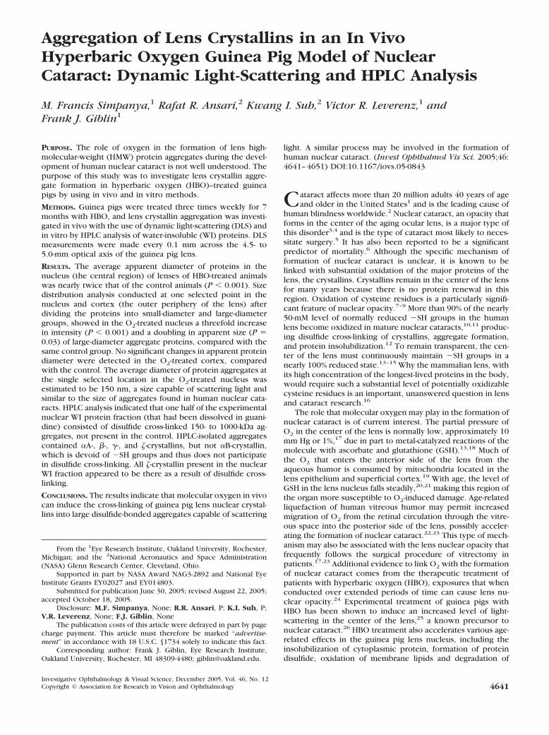
SLS analysis of a control guinea pig eye in vivo clearlyshowed the anatomic structure of the eye: the cornea, aqueoushumor, the three regions of the lens (anterior cortex, nucleus,and posterior cortex), and the vitreous humor (Fig. 2A). Thehigh level of scatter observed for the cornea was caused byreflection of laser light from the cornea back to the detector. Alow level of SLS was observed throughout the 4.5- to 5.0-mmaxial diameter of the lens of the 25 month-old control animal(Fig. 2A). Lenses of guinea pigs treated 84 times with HBOshowed an increased level of SLS, particularly in the nuclearregion (Fig. 2B).
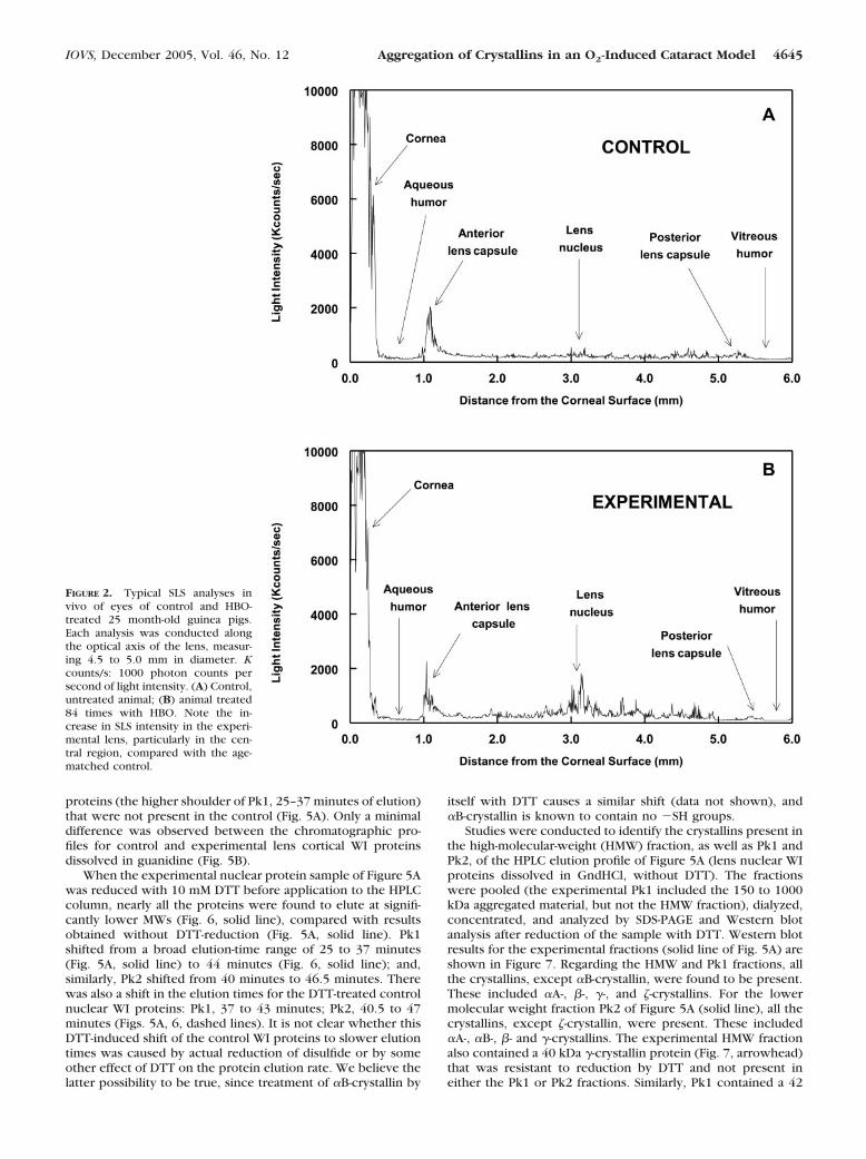
To evaluate changes in apparent lens protein size in vivo asa result of O2-treatment, we conducted DLS analyses. Measure-ments were made every 0.1 mm across the �5-mm optical axisof the guinea pig lens. Figure 3 shows a representative profileof average protein diameters across the lens of a control andHBO-treated animal (see Materials and Methods and the legendof Fig. 3 for a definition of average protein diameter and anexplanation of why the data are expressed as arbitrary units ofdiameter). An increase in average protein size was obvious inthe central region of the lens (the inner 2 mm) of the O2-treated animal compared with the control. Larger apparentprotein sizes were consistently noted in the central regions of
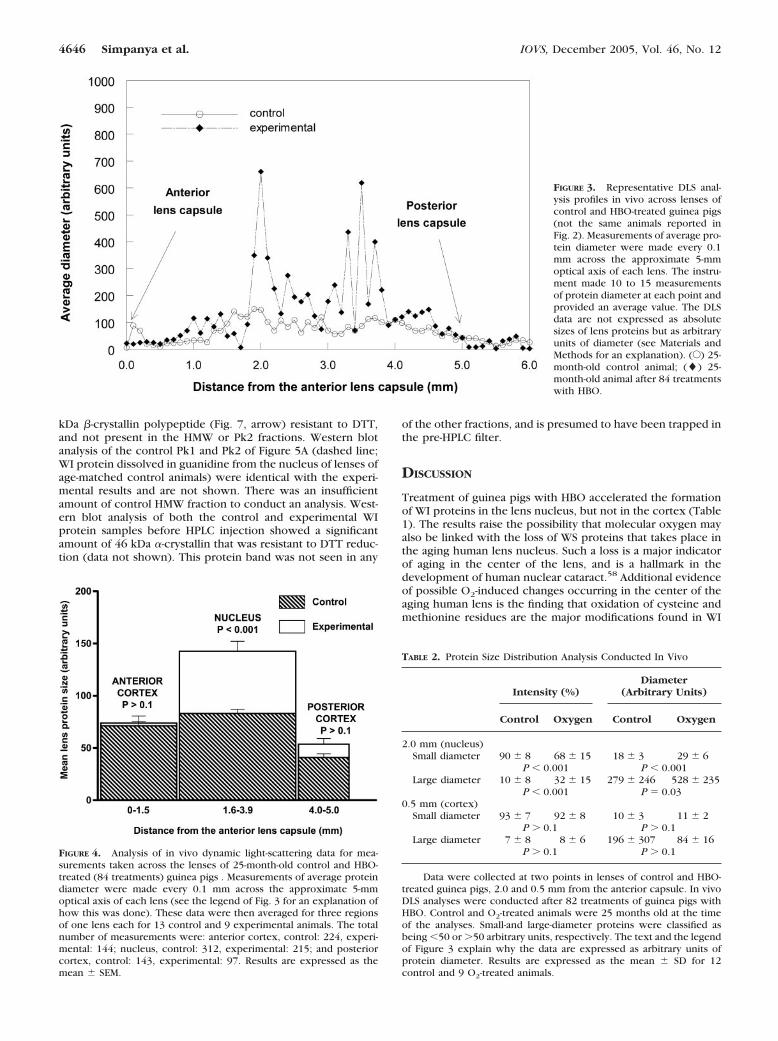
lenses of experimental animals, but not always in the sameregion of the nucleus. Whereas in Figure 3, the highest averagediameters were in the anterior and posterior regions of theexperimental nucleus, certain other lenses of HBO-treated an-imals showed large diameters in the very center of the lens. Toinvestigate the protein sizes in more detail, the DLS data wereanalyzed more thoroughly for one lens each of 13 control and9 experimental animals (Fig. 4), again making measurementsevery 0.1 mm in the lens. For these determinations, apparentprotein diameters (obtained as shown in Fig. 3) were averagedin three separate regions, as measured from the anterior lenscapsule, including the anterior cortex (0–1.5 mm), nucleus(l.6–3.9 mm), and posterior cortex (4.0–5.0 mm). The totalnumber of measurements made in each region in the 13 con-trol and 9 experimental animals are shown in the legend toFigure 4. The average protein size in the nucleus of lenses ofHBO-treated guinea pigs was found to be nearly twice that forthe same region of lenses of age-matched control animals (P �0.001; Fig. 4). No significant increases in average protein sizewere observed for either the anterior or posterior cortex as aresult of HBO-treatment of the animals (P � 0.1).
It was also of interest to investigate the size distribution ofproteins at certain locations in lenses of control and O2-treatedanimals. A size distribution analysis was conducted in one lenseach of 12 control and 9 experimental animals at 2 of the 50 invivo lens measurement locations, in the nucleus at a point 2.0mm from the anterior capsule and in the anterior cortex 0.5mm from the capsule. Protein size data were averaged forcontrol and O2-treated lenses after dividing the sizes into small-diameter (�50 arbitrary units) and large-diameter (�50 arbi-trary units) proteins (Table 2). In the nucleus, at the 2.0-mmlocation, lenses of control animals showed 90% of the totalprotein intensity clustered into the group of small-diameterpolypeptides (mean diameter, 18 arbitrary units). A second,less prominent control group, making up 10% of the totalintensity, had a size approximately 16 times that of the majorgroup (mean diameter, 279 arbitrary units). Results for theO2-treated lenses at the 2.0-mm location (Table 2) showed twoprominent groups of proteins. The O2-treated small-diameterproteins, making up 68% of the total intensity, had a size (meandiameter, 29 arbitrary units) 1.6 times that of the major controlgroup (P � 0.001), and the O2-treated large-diameter proteins,accounting for 32% of the total intensity, had a size (meandiameter, 528 arbitrary units) 30 times the control small pro-teins and nearly twice the control large proteins (P � 0.03).Thus, at this one location in the lens nucleus, the oxygentreatment produced a threefold increase in intensity and atwofold increase in size of the large, aggregated proteins. In theanterior cortex at the 0.5-mm location (Table 2), there were nosignificant differences in the intensities and apparent sizes ofthe two groups of proteins, control versus O2-treated.
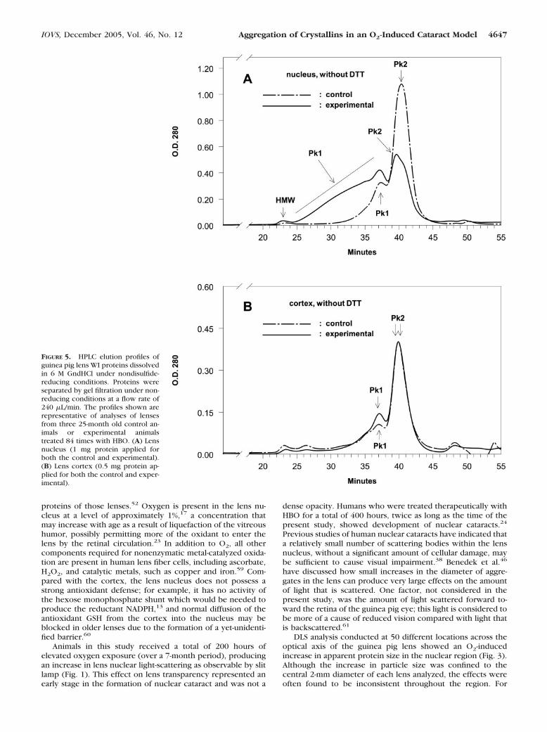
Aggregation of proteins in the lenses of O2-treated guineapigs was also investigated by HPLC analysis of lens WI proteins.The proteins were dissolved in 6 M GndHCl, taking care not toproduce any artifactual protein disulfide (see Materials andMethods), but initially without the use of disulfide-reducingagents. In addition, proteins were eluted initially under nonre-ducing conditions. The profile for the nuclear region of thecontrol lens (Fig. 5A) showed two well-defined peaks, a minorhigher molecular weight Pk1 (37.0 minutes of elution) and amore prominent lower Pk2 (40.5 minutes of elution). In con-trast, the experimental nuclear profile showed a 50% decreasein the amount of the lower molecular weight fraction Pk2, plusa shift of this peak to a 10% higher molecular weight (from 40.5minutes of elution to 39.5 minutes), as well as a substantialincrease and shift to a higher molecular weight for Pk1. Nearlyone half of the experimental nuclear WI protein fraction thathad been dissolved in guanidine consisted of 150 to 1000 kDa
FIGURE 1. Typical slit lamp biomicroscopy photographs of guinea pigeyes. (A) A 25-month-old control animal. (B) A 25-month-old animalafter 84 treatments with HBO over a 7-month period. Note the in-creased lens nuclear light-scattering in the O2-treated lens, comparedwith the control, particularly in the very center of the nucleus (arrow).In addition, note the larger region of backscatter in the O2-treated lens,compared with the control.
4644 Simpanya et al. IOVS, December 2005, Vol. 46, No. 12
proteins (the higher shoulder of Pk1, 25–37 minutes of elution)that were not present in the control (Fig. 5A). Only a minimaldifference was observed between the chromatographic pro-files for control and experimental lens cortical WI proteinsdissolved in guanidine (Fig. 5B).
When the experimental nuclear protein sample of Figure 5Awas reduced with 10 mM DTT before application to the HPLCcolumn, nearly all the proteins were found to elute at signifi-cantly lower MWs (Fig. 6, solid line), compared with resultsobtained without DTT-reduction (Fig. 5A, solid line). Pk1shifted from a broad elution-time range of 25 to 37 minutes(Fig. 5A, solid line) to 44 minutes (Fig. 6, solid line); and,similarly, Pk2 shifted from 40 minutes to 46.5 minutes. Therewas also a shift in the elution times for the DTT-treated controlnuclear WI proteins: Pk1, 37 to 43 minutes; Pk2, 40.5 to 47minutes (Figs. 5A, 6, dashed lines). It is not clear whether thisDTT-induced shift of the control WI proteins to slower elutiontimes was caused by actual reduction of disulfide or by someother effect of DTT on the protein elution rate. We believe thelatter possibility to be true, since treatment of �B-crystallin by
itself with DTT causes a similar shift (data not shown), and�B-crystallin is known to contain no �SH groups.
Studies were conducted to identify the crystallins present inthe high-molecular-weight (HMW) fraction, as well as Pk1 andPk2, of the HPLC elution profile of Figure 5A (lens nuclear WIproteins dissolved in GndHCl, without DTT). The fractionswere pooled (the experimental Pk1 included the 150 to 1000kDa aggregated material, but not the HMW fraction), dialyzed,concentrated, and analyzed by SDS-PAGE and Western blotanalysis after reduction of the sample with DTT. Western blotresults for the experimental fractions (solid line of Fig. 5A) areshown in Figure 7. Regarding the HMW and Pk1 fractions, allthe crystallins, except �B-crystallin, were found to be present.These included �A-, �-, �-, and �-crystallins. For the lowermolecular weight fraction Pk2 of Figure 5A (solid line), all thecrystallins, except �-crystallin, were present. These included�A-, �B-, �- and �-crystallins. The experimental HMW fractionalso contained a 40 kDa �-crystallin protein (Fig. 7, arrowhead)that was resistant to reduction by DTT and not present ineither the Pk1 or Pk2 fractions. Similarly, Pk1 contained a 42
FIGURE 2. Typical SLS analyses invivo of eyes of control and HBO-treated 25 month-old guinea pigs.Each analysis was conducted alongthe optical axis of the lens, measur-ing 4.5 to 5.0 mm in diameter. Kcounts/s: 1000 photon counts persecond of light intensity. (A) Control,untreated animal; (B) animal treated84 times with HBO. Note the in-crease in SLS intensity in the experi-mental lens, particularly in the cen-tral region, compared with the age-matched control.
IOVS, December 2005, Vol. 46, No. 12 Aggregation of Crystallins in an O2-Induced Cataract Model 4645
kDa �-crystallin polypeptide (Fig. 7, arrow) resistant to DTT,and not present in the HMW or Pk2 fractions. Western blotanalysis of the control Pk1 and Pk2 of Figure 5A (dashed line;WI protein dissolved in guanidine from the nucleus of lenses ofage-matched control animals) were identical with the experi-mental results and are not shown. There was an insufficientamount of control HMW fraction to conduct an analysis. West-ern blot analysis of both the control and experimental WIprotein samples before HPLC injection showed a significantamount of 46 kDa �-crystallin that was resistant to DTT reduc-tion (data not shown). This protein band was not seen in any
of the other fractions, and is presumed to have been trapped inthe pre-HPLC filter.
DISCUSSION
Treatment of guinea pigs with HBO accelerated the formationof WI proteins in the lens nucleus, but not in the cortex (Table1). The results raise the possibility that molecular oxygen mayalso be linked with the loss of WS proteins that takes place inthe aging human lens nucleus. Such a loss is a major indicatorof aging in the center of the lens, and is a hallmark in thedevelopment of human nuclear cataract.58 Additional evidenceof possible O2-induced changes occurring in the center of theaging human lens is the finding that oxidation of cysteine andmethionine residues are the major modifications found in WI
FIGURE 3. Representative DLS anal-ysis profiles in vivo across lenses ofcontrol and HBO-treated guinea pigs(not the same animals reported inFig. 2). Measurements of average pro-tein diameter were made every 0.1mm across the approximate 5-mmoptical axis of each lens. The instru-ment made 10 to 15 measurementsof protein diameter at each point andprovided an average value. The DLSdata are not expressed as absolutesizes of lens proteins but as arbitraryunits of diameter (see Materials andMethods for an explanation). (E) 25-month-old control animal; (�) 25-month-old animal after 84 treatmentswith HBO.
FIGURE 4. Analysis of in vivo dynamic light-scattering data for mea-surements taken across the lenses of 25-month-old control and HBO-treated (84 treatments) guinea pigs . Measurements of average proteindiameter were made every 0.1 mm across the approximate 5-mmoptical axis of each lens (see the legend of Fig. 3 for an explanation ofhow this was done). These data were then averaged for three regionsof one lens each for 13 control and 9 experimental animals. The totalnumber of measurements were: anterior cortex, control: 224, experi-mental: 144; nucleus, control: 312, experimental: 215; and posteriorcortex, control: 143, experimental: 97. Results are expressed as themean � SEM.
TABLE 2. Protein Size Distribution Analysis Conducted In Vivo
Intensity (%)Diameter
(Arbitrary Units)
Control Oxygen Control Oxygen
2.0 mm (nucleus)Small diameter 90 � 8 68 � 15 18 � 3 29 � 6
P � 0.001 P � 0.001Large diameter 10 � 8 32 � 15 279 � 246 528 � 235
P � 0.001 P � 0.030.5 mm (cortex)
Small diameter 93 � 7 92 � 8 10 � 3 11 � 2P � 0.1 P � 0.1
Large diameter 7 � 8 8 � 6 196 � 307 84 � 16P � 0.1 P � 0.1
Data were collected at two points in lenses of control and HBO-treated guinea pigs, 2.0 and 0.5 mm from the anterior capsule. In vivoDLS analyses were conducted after 82 treatments of guinea pigs withHBO. Control and O2-treated animals were 25 months old at the timeof the analyses. Small-and large-diameter proteins were classified asbeing �50 or �50 arbitrary units, respectively. The text and the legendof Figure 3 explain why the data are expressed as arbitrary units ofprotein diameter. Results are expressed as the mean � SD for 12control and 9 O2-treated animals.
4646 Simpanya et al. IOVS, December 2005, Vol. 46, No. 12
proteins of those lenses.52 Oxygen is present in the lens nu-cleus at a level of approximately 1%,17 a concentration thatmay increase with age as a result of liquefaction of the vitreoushumor, possibly permitting more of the oxidant to enter thelens by the retinal circulation.23 In addition to O2, all othercomponents required for nonenzymatic metal-catalyzed oxida-tion are present in human lens fiber cells, including ascorbate,H2O2, and catalytic metals, such as copper and iron.59 Com-pared with the cortex, the lens nucleus does not possess astrong antioxidant defense; for example, it has no activity ofthe hexose monophosphate shunt which would be needed toproduce the reductant NADPH,13 and normal diffusion of theantioxidant GSH from the cortex into the nucleus may beblocked in older lenses due to the formation of a yet-unidenti-fied barrier.60
Animals in this study received a total of 200 hours ofelevated oxygen exposure (over a 7-month period), producingan increase in lens nuclear light-scattering as observable by slitlamp (Fig. 1). This effect on lens transparency represented anearly stage in the formation of nuclear cataract and was not a
dense opacity. Humans who were treated therapeutically withHBO for a total of 400 hours, twice as long as the time of thepresent study, showed development of nuclear cataracts.24
Previous studies of human nuclear cataracts have indicated thata relatively small number of scattering bodies within the lensnucleus, without a significant amount of cellular damage, maybe sufficient to cause visual impairment.38 Benedek et al.46
have discussed how small increases in the diameter of aggre-gates in the lens can produce very large effects on the amountof light that is scattered. One factor, not considered in thepresent study, was the amount of light scattered forward to-ward the retina of the guinea pig eye; this light is considered tobe more of a cause of reduced vision compared with light thatis backscattered.61
DLS analysis conducted at 50 different locations across theoptical axis of the guinea pig lens showed an O2-inducedincrease in apparent protein size in the nuclear region (Fig. 3).Although the increase in particle size was confined to thecentral 2-mm diameter of each lens analyzed, the effects wereoften found to be inconsistent throughout the region. For
FIGURE 5. HPLC elution profiles ofguinea pig lens WI proteins dissolvedin 6 M GndHCl under nondisulfide-reducing conditions. Proteins wereseparated by gel filtration under non-reducing conditions at a flow rate of240 �L/min. The profiles shown arerepresentative of analyses of lensesfrom three 25-month old control an-imals or experimental animalstreated 84 times with HBO. (A) Lensnucleus (1 mg protein applied forboth the control and experimental).(B) Lens cortex (0.5 mg protein ap-plied for both the control and exper-imental).
IOVS, December 2005, Vol. 46, No. 12 Aggregation of Crystallins in an O2-Induced Cataract Model 4647

example, in Figure 3, particle sizes in the very center of theexperimental lens are shown to be two to three times lowerthan those in the anterior and posterior region of the nucleus.For the other eight experimental lenses analyzed, larger pro-tein sizes were frequently seen at certain locations in thenucleus, including the anterior, central, or posterior areas, butsometimes not in other parts of the nucleus. The reason for thisis not clear, but the data suggest that early O2-induced aggre-gation of proteins may not progress homogeneously through-out the nucleus, thus resulting in the formation of larger ag-gregates in certain regions of the lens nucleus, and smalleraggregates in other regions. It is also possible that if O2-inducedaggregates had become too large, multiple light-scattering (asingle photon scattering many times in the lens) would haveoccurred, giving rise to absorption of light, and causing diffu-sion broadening, which could produce apparently lower par-ticle sizes. This might explain the relatively lower protein sizesseen in the very center of the experimental lens in Figure 3.Considering the equatorial diameter of the lens (rather than theaxial diameter), previous in vitro laser scanning of lenses ofHBO-treated guinea pigs showed increased scatter along the
central 2 mm of equatorial length.62 Because the length of thefocused laser light beam used in the present study was 20 �m,and measurements were made only along the optical axis, only1% of the total 2-mm region of scatter along the equatoriallength of approximately 20 lens fiber cells would have beenanalyzed at each of the measurement points.
DLS analysis showed an average doubling in apparent pro-tein size in the central 2-mm region of lenses of O2-treatedguinea pigs (24 measurements per each lens nucleus along theoptical axis), compared with age-matched control animals (Fig.4). It is not clear whether this degree of aggregation could haveproduced the level of scatter observed by slit lamp examina-tion. The slit lamp–detected scatter may have been producedby a relatively small number of much larger aggregates. Size-distribution analysis conducted at one of the 24 measurementpoints in the lens nucleus (2.0 mm in from the anterior cap-sule) in nine experimental animals showed that the group oflarge-diameter proteins (�50 arbitrary units) increased three-fold in intensity and doubled in size, compared with the age-matched control animals (Table 2). The average diameter (528arbitrary units) of the O2-induced aggregates at this one loca-tion was 30 times greater than that of the control small-diam-eter proteins (18 arbitrary units). If a 5-nm average size isassumed for the control small-diameter proteins (approxi-mately 40% of this fraction should have consisted of 10- to12-nm �-crystallin), an approximate diameter of 150 nm can beestimated for the O2-induced aggregates (Table 2). Aggregatesof this size would have most certainly produced significantscattered light in the guinea pig lens. Other investigators, usingtechniques of electron microscopy or light-scattering, haveobserved aggregates with 100- to 500-nm diameters in humannuclear cataracts.29,37,38,46,63,64 Electron microscopic studiesof aggregates in experimental animal cataractous lensesshowed sizes of 100 nm in the HBO-treated guinea pig,37,38 andeven up to 1000 nm in the X-irradiated rabbit.65
Table 2 shows that small-diameter proteins present in theO2-treated nucleus (average diameter, 29 arbitrary units) were1.6 times the size of the same group of proteins in the controlnucleus (average diameter, 18 arbitrary units). It is conceivablethat some of this experimental fraction may have representedchaperone-like activity of �-crystallin,66 involving the bindingof oxidatively damaged crystallins to the �-crystallin complexby hydrophobic interaction.67 �-Crystallin has been shown todecrease the amount of light-scattering and thiol oxidation of
FIGURE 6. HPLC elution profiles forthe control and experimental nuclearsamples of Figure 5A (WI nuclearproteins dissolved in 6 M GndHCl)after reduction of the sample withDTT. Proteins (1 mg) were separatedby gel filtration under reducing con-ditions at a flow rate of 240 �L/min.The profiles shown are representa-tive of analyses of lenses from threeanimals 25-month-old control ani-mals or experimental animals treated84 times with HBO.
FIGURE 7. Western blot analysis of the HMW, Pk1 and Pk2 fractionscontained in the experimental HPLC elution profile of Figure 5A (WInuclear proteins dissolved in guanidine, without DTT, from the lensesof guinea pigs treated 84 times with HBO). Fractions were pooled(HMW: 22–24 minutes elution; Pk1: 25–37 minutes elution; and Pk2:39–42 minutes elution), dialyzed, concentrated, and analyzed afterreduction of disulfide with DTT. Immunostaining was conducted usingantibodies to �A-, �B-, �-, �-, and �-crystallins. Arrowhead and arrow:indicate polypeptides resistant to DTT reduction. Note the absence of�B-crystallin in the HMW and Pk1 fractions and the absence of �-crys-tallin in the Pk2 fraction.
4648 Simpanya et al. IOVS, December 2005, Vol. 46, No. 12

other crystallins induced by various conditions of oxidativestress in vitro,68 and rabbit lenses treated with HBO in vitrohave exhibited an apparent chaperone-like function for �-crys-tallin in the lens nucleus.69 In the aging bovine lens, �-crystal-lin present in the HMW fraction has been shown to bind �- and�-crystallins in the central region of the supramolecular com-plex.70
Nearly one half of the experimental nuclear WI proteindissolved in guanidine was found to consist of 150- to 1000-kDaproteins that were not present in the control (Fig. 5A). Almostall this aggregated material appeared to be held together bydisulfide bonds (based on the DTT reduction experiments ofFigs. 6, 7). For aggregates of this size to have existed inguanidine, 7 to 50 individual 20-kDa MW crystallin subunitswould have had to be cross-linked by disulfide bonds. It is notknown what the sizes of these aggregates might have been invivo (before denaturation with guanidine), but the materialmay have corresponded to the very large aggregates that weredetected in the experimental nucleus in vivo with the use ofDLS (Table 2). As mentioned earlier, those aggregates may havebeen up to 150 nm in diameter. Bettelheim et al.64 havecalculated that a 300-nm diameter aggregate would have amolecular mass of 500 � 106 Da. Aggregates up to 300 � 106
Da have been found to exist in the water-soluble fraction ofold, normal human lenses.40 As mentioned previously, thediameters of aggregates found in cataracts have ranged from100 to 1000 nm.
It appeared that when nuclear WI proteins present in con-trol and O2-treated lenses were dissolved in guanidine, theyconsisted of both disulfide-cross-linked aggregates (Fig. 5A,Pk1) and monomers (Fig. 5A, Pk2). The monomers apparentlyhad become WI for some reason other than direct oxidation of�SH groups (for example, they could have been initially boundhydrophobically to a cross-linked aggregate and then releasedas monomers during treatment with guanidine). Supportingevidence for this is that �B-crystallin, which does not containany �SH groups, was found to be present only in the Pk2fraction and not at all in the disulfide cross-linked material ofPk1. In addition, �-crystallin, which contains five �SH groupsper subunit, was present only in Pk1 and not at all in Pk2 (Fig.7). In a previous study, disulfide cross-linking was the majormodification present in the WI protein fraction of old, normalhuman lenses52 and, in x-ray cataracts, 50% of �SH groupspresent in the soluble HMW aggregate fraction were oxi-dized.71 Although in the present study, treatment with DTTreduced all the very HWM material (possessing molecularmasses in guanidine of �150 kDa; the shoulder of Pk1, Fig.5A), 50% of the reduced material was found to remain in Pk1,presumably mostly as smaller disulfide-cross-linked aggregates(Fig. 6). This may have been caused by DTT not being able toreach all the disulfide cross-links during the 4-hour incubationat room temperature. For Western blot studies (Fig. 7), inwhich samples were reduced with DTT at 95 °C to 97°C,nearly all the aggregated material was reduced to monomers.
The results of this study support the long-standing beliefthat disulfide bonding may be the main cause of HMW aggre-gate formation in human nuclear cataracts.7,8 Disulfide has alsobeen linked with HMW aggregate formation in certain exper-imental cataracts such as those induced by x-ray71 and diabe-tes.72 The cysteine residue is known to be highly sensitive tooxidation by almost all kinds of reactive oxygen species.73 Thefragile nature of the �SH to �SS� relationship in the humanlens is demonstrated by an Arg-14 to Cys mutation in the lensesof human infants, causing hereditary cataract as a result ofdisulfide cross-linked aggregate formation.74 Why the mamma-lian lens would require such a high �SH content (nearly 50mM), when cysteines are so susceptible to oxidation and cross-linking, is not known. It has been proposed that cysteine may
be needed to interact with aromatic-side chains in the lens,quenching excited states as crystallins absorb ultraviolet light,protecting the retina from damage.75
In addition to disulfide-cross-linking, Western blot analysisindicated a relatively lesser amount of nondisulfide covalentcross-linking in the control and experimental WI protein frac-tions (Fig. 7). A 46 kDa �A-crystallin polypeptide was detectedat a substantial level in the guanidine-dissolved protein samplebefore HPLC-injection (data not shown), but was not found atall in any of the HPLC fractions. This polypeptide may haverepresented a nondisulfide cross-linked �A-crystallin dimerpresent in aggregates that, even in 6 M guanidine, were toolarge to enter the HPLC column. The nondisulfide cross-linked40-kDa �-crystallin present in the guanidine-dissolved HMWfraction (Fig. 7) apparently existed in very large disulfide cross-linked aggregates in the experimental WI protein. A nondisul-fide cross-linked 42-kDa �-crystallin was present only in Pk1(Fig. 7). In a previous study, a 43-kDa polypeptide with immu-noreactivity to �-crystallin antibody was isolated from humannuclear cataracts and proposed to be a possible nucleation sitefor the generation of disulfide cross-linked HMW membrane-bound aggregates.76 The nature of the nondisulfide covalentcross-links mentioned earlier is not known. It is possible thatsome of the cross-links are related to transglutaminase activity,which has been reported to be present in the lenses of both theguinea pig and human.77 Transglutaminase-mediated cross-link-ing of both �- and �-crystallins has been shown to take placeunder certain conditions, including elevated oxidativestress.78,79
All the guinea pig lens crystallins, including �-, �-, �-, and �-,were found to be present in the WI protein fraction of theexperimental lens nucleus (Fig. 7). Both WI proteins and water-soluble protein aggregates in human lenses have been found tocontain �-, �-, and �-crystallins.40,52,80 As discussed earlier,�B-crystallin with no �SH content may have been present inthe WI fraction indirectly as a result of hydrophobic bonding todisulfide cross-linked �A-crystallin. The �- and �-crystallins pos-sess substantial �SH content and are known to be prone toinsolubilization as a result of exposure to oxidativestress.7,69,81,82 �-Crystallin, an NADPH-binding protein makingup 10% of the total soluble protein in the guinea pig lens83 andcontaining five �SH groups per subunit, was found in thepresent study to be particularly vulnerable to O2-induced dis-ulfide-cross-linking. �-Crystallin was the only crystallin in theWI fraction of the experimental nucleus to be present entirelyin the disulfide cross-linked aggregate fraction of Pk1, with noprotein existing in the monomer fraction Pk2 (Fig. 7). Thesame result was found for the WI fraction of the controlnucleus, implying that the only mechanism by which �-crystal-lin becomes insoluble in the guinea pig lens is through disul-fide cross-linking.
The crystallin composition of WI protein fractions in con-trol and O2-treated guinea pig lenses did not differ significantlyafter 84 treatments of the animals with HBO. The dramaticdifference between the two fractions was the substantiallygreater amount and size of disulfide cross-linked aggregatescontained in the experimental WI protein, compared with thecontrol. This suggests that an elevated level of oxygen in thelens may act to accelerate normal oxidative changes in crystal-lins in the center of the lens as lens nuclear proteins age in anonregenerating environment.
In summary, we have shown by in vivo and in vitro analysisthat HBO-treatment of guinea pigs induces HMW aggregateformation in the center of the lens, leading to increased nuclearlight-scattering. A similar process of O2-induced disulfide cross-linking of crystallins may be involved in the formation ofhuman nuclear cataract.
IOVS, December 2005, Vol. 46, No. 12 Aggregation of Crystallins in an O2-Induced Cataract Model 4649
Acknowledgments
The authors thank James King of NASA for help with in vivo analysis ofthe guinea pig lenses by dynamic light-scattering; Li-Ren Lin, MD, forslit lamp examination of the eyes of the animals; Oakland Universitystudents Aparna Bhat, Melodie Denstadt, Michael Lupe, Brijesh Patel,Pavan Vempaty, Nathanial Whitcomb, and Jasper Yung for treating theguinea pigs with hyperbaric oxygen; and Cliff Snitgen, Janet Schofding,and Mitun Chablani for providing the professional care of the animals.
References
1. The Eye Diseases Prevalence Research Group. Causes and preva-lence of visual impairment among adults in the United States. ArchOphthalmol. 2004;122:477–485.
2. Brian G, Taylor H. Cataract blindness: challenges for the 21stcentury. Bull World Health Organ. 2001;79:249–256.
3. Adamsons I, Munoz B, Enger C, Taylor HR. Prevalence of lensopacities in surgical and general populations. Arch Ophthalmol.1991;109:993–997.
4. Congdon NC, Taylor HR. Age-related cataract. In: Weale RA, WestSK, Johnson GJ, Minassian DC, eds. The Epidemiology of EyeDisease. London: Arnold; 2003:106–107.
5. Lewis A, Congdon N, Munoz B, et al. Cataract surgery and subtypein a defined, older population: the SEECAT Project. Br J Ophthal-mol. 2004;88:1512–1517.
6. West SK, Munoz B, Istre J, et al. Mixed lens opacities and subse-quent mortality. Arch Ophthalmol. 2000;118:393–397.
7. Dische Z, Zil H. Studies on the oxidation of cysteine to cystine inlens proteins during cataract formation. Am J Ophthalmol. 1951;34:104–113.
8. Harding JJ. Disulphide cross-linked protein of high molecularweight in human cataractous lens. Exp Eye Res. 1973;17:377–383.
9. Spector A. The search for a solution to senile cataracts. Proctorlecture. Invest Ophthalmol Vis Sci. 1984;25:130–146.
10. Truscott RJ, Augusteyn RC. Oxidative changes in human lensproteins during senile nuclear cataract formation. Biochim Bio-phys Acta. 1977;492:43–52.
11. Garner MH, Spector A. Selective oxidation of cysteine and methi-onine in normal and senile cataractous lenses. Proc Natl Acad SciUSA. 1980;77:1274–1277.
12. Spector A, Roy D. Disulfide-linked high molecular weight proteinassociated with human cataract. Proc Natl Acad Sci USA. 1978;75:3244–3248.
13. Giblin FJ. Glutathione: a vital lens antioxidant. J Ocul PharmacolTher. 2000;16:121–135.
14. Lou MF. Redox regulation in the lens. Prog Retin Eye Res. 2003;22:657–682.
15. Truscott RJ. Age-related nuclear cataract-oxidation is the key. ExpEye Res. 2005;80:709–725.
16. Srikanthan D, Bateman OA, Purkiss AG, Slingsby C. Sulfur inhuman crystallins. Exp Eye Res. 2004;79:823–831.
17. Barbazetto IA, Liang J, Chang S, et al. Oxygen tension in the rabbitlens and vitreous before and after vitrectomy. Exp Eye Res. 2004;78:917–924.
18. Eaton JW. Is the lens canned? Free Radic Biol Med. 1991;11:207–213.
19. McNulty R, Wang H, Mathias RT, et al. Regulation of tissue oxygenlevels in the mammalian lens. J Physiol. 2004;559:883–898.
20. Truscott RJ, Augusteyn RC. The state of sulphydryl groups innormal and cataractous human lenses. Exp Eye Res. 1977;25:139–148.
21. Lou MF, Dickerson JE Jr. Protein-thiol mixed disulfides in humanlens. Exp Eye Res. 1992;55:889–896.
22. Harocopos GJ, Shui YB, McKinnon M, et al. Importance of vitreousliquefaction in age-related cataract. Invest Ophthalmol Vis Sci.2004;45:77–85.
23. Holekamp NM, Shui YB, Beebe DC. Vitrectomy surgery increasesoxygen exposure to the lens: a possible mechanism for nuclearcataract formation. Am J Ophthalmol. 2005;139:302–310.
24. Palmquist BM, Philipson B, Barr PO. Nuclear cataract and myopiaduring hyperbaric oxygen therapy. Br J Ophthalmol. 1984;68:113–117.
25. Giblin FJ, Padgaonkar VA, Leverenz VR, et al. Nuclear light-scatter-ing, disulfide formation and membrane damage in lenses of olderguinea pigs treated with hyperbaric oxygen. Exp Eye Res. 1995;60:219–235.
26. Sigelman J, Trokel SL, Spector A. Quantitative biomicroscopy oflens light back scatter: changes in aging and opacification. ArchOphthalmol. 1974;92:437–442.
27. Padgaonkar VA, Lin LR, Leverenz VR, et al. Hyperbaric oxygen invivo accelerates the loss of cytoskeletal proteins and MIP26 inguinea pig lens nucleus. Exp Eye Res. 1999;68:493–504.
28. Borchman D, Giblin FJ, Leverenz VR, et al. Impact of aging andhyperbaric oxygen in vivo on guinea pig lens lipids and nuclearlight scatter. Invest Ophthalmol Vis Sci. 2000;41:3061–3073.
29. Philipson B. Changes in the lens related to the reduction of trans-parency. Exp Eye Res. 1973;16:29–39.
30. al Ghoul KJ, Costello MJ. Fiber cell morphology and cytoplasmictexture in cataractous and normal human lens nuclei. Curr EyeRes. 1996;15:533–542.
31. Benedek GB. Theory of transparency of the eye. Appl Opt. 1971;10:459–473.
32. Bettelheim FA. Physical basis of lens transparency. In: Maisel H, ed.The Ocular Lens. New York: Marcel Dekker; 1985:265–300.
33. Spector A, Li LK, Augusteyn RC, Schneider A, Freund T.�-Crystallin: the isolation and characterization of distinct macro-molecular fractions. Biochem J. 1971;124:337–343.
34. Horwitz J. Alpha-crystallin. Exp Eye Res. 2003;76:145–153.35. Bettelheim FA, Ansari R, Cheng QF, Zigler JS Jr. The mode of
chaperoning of dithiothreitol-denatured alpha-lactalbumin by al-pha-crystallin. Biochem Biophys Res Commun. 1999;261:292–297.
36. Siezen RJ, Bindels JG, Hoenders HJ. The interrelationship betweenmonomeric, oligomeric and polymeric alpha-crystallin in the calflens nucleus. Exp Eye Res. 1979;28:551–567.
37. Freel CD, Gilliland KO, Wesley LC, Giblin FJ, Joseph CM. Fourieranalysis of cytoplasmic texture in nuclear fiber cells from trans-parent and cataractous human and animal lenses. Exp Eye Res.2002;74:689–702.
38. Freel CD, Gilliland KO, Mekeel HE, Giblin FJ, Costello MJ. Ultra-structural characterization and Fourier analysis of fiber cell cyto-plasm in the hyperbaric oxygen treated guinea pig lens opacifica-tion model. Exp Eye Res. 2003;76:405–415.
39. Tanaka T, Benedek GB. Observation of protein diffusivity in intacthuman and bovine lenses with application to cataract. InvestOphthalmol. 1975;14:449–456.
40. Jedziniak JA, Nicoli DF, Baram H, Benedek GB. Quantitative veri-fication of the existence of high molecular weight protein aggre-gates in the intact normal human lens by light-scattering spectros-copy. Invest Ophthalmol Vis Sci. 1978;17:51–57.
41. Delaye M, Clark JI, Benedek GB. Identification of the scatteringelements responsible for lens opacification in cold cataracts. Bio-phys J. 1982;37:647–656.
42. Latina M, Chylack LT Jr, Fagerholm P, et al. Dynamic light-scatter-ing in the intact rabbit lens: its relation to protein concentration.Invest Ophthalmol Vis Sci. 1987;28:175–183.
43. Tanaka T, Ishimoto C. In vivo observation of protein diffusivity inrabbit lenses. Invest Ophthalmol Vis Sci. 1977;16:135–140.
44. Nishio I, Weiss JN, Tanaka T, et al. In vivo observation of lensprotein diffusivity in normal and X-irradiated rabbit lenses. Exp EyeRes. 1984;39:61–68.
45. Libondi T, Magnante P, Chylack LT Jr, Benedek GB. In vivo mea-surement of the aging rabbit lens using quasielastic light-scattering.Curr Eye Res. 1986;5:411–419.
46. Benedek GB, Chylack LT Jr, Libondi T, Magnante P, Pennett M.Quantitative detection of the molecular changes associated withearly cataractogenesis in the living human lens using quasielasticlight-scattering. Curr Eye Res. 1987;6:1421–1432.
47. Thurston GM, Hayden DL, Burrows P, et al. Quasielastic light-scattering study of the living human lens as a function of age. CurrEye Res. 1997;16:197–207.
4650 Simpanya et al. IOVS, December 2005, Vol. 46, No. 12

48. Ansari RR, Giblin FJ, King JF. Space Technology and ApplicationsInternational Forum, AIP. American Institute of Physics Confer-ence Proceedings. Albuquerque, NM, 2001;1224–1229.
49. Brown W. Dynamic Light-scattering: The Method and Some Ap-plications. Oxford: Clarendon Press; 1993.
50. Datiles MB III, Ansari RR, Reed GF. A clinical study of the humanlens with a dynamic light-scattering device. Exp Eye Res. 2002;74:93–102.
51. Ansari RR. Ocular static and dynamic light-scattering: a noninva-sive diagnostic tool for eye research and clinical practice.J Biomed Opt. 2004;9:22–37.
52. Hanson SR, Hasan A, Smith DL, Smith JB. The major in vivomodifications of the human water-insoluble lens crystallins aredisulfide bonds, deamidation, methionine oxidation and backbonecleavage. Exp Eye Res. 2000;71:195–207.
53. Ansari RR, Suh KI, Tumminia SJ, Russell P, Zigler JS. In vivocataractograms using a compact backscatter dynamic light-scatter-ing probe. Proc Med Appl Lasers Dermatol Ophthalmol DentistryEndosc. 1997;3192:202–210.
54. Ansari RR, Datiles MB III. Use of dynamic light-scattering andScheimpflug imaging for the early detection of cataracts. DiabetesTechnol Ther. 1999;1:159–168.
55. Bettelheim FA. Light-scattering in lens research: an essay on ac-complishments and promises. Exp Eye Res. 2004;79:747–752.
56. Wiechelman KJ, Braun RD, Fitzpatrick JD. Investigation of thebicinchoninic acid protein assay: identification of the groups re-sponsible for color formation. Anal Biochem. 1988;175:231–237.
57. Padgaonkar V, Giblin FJ, Reddy VN. Disulfide cross-linking ofurea-insoluble proteins in rabbit lenses treated with hyperbaricoxygen. Exp Eye Res. 1989;49:887–899.
58. Young RW. Age-Related Cataract. New York: Oxford UniversityPress; 1991.
59. Garland D. Role of site-specific, metal-catalyzed oxidation in lensaging and cataract: a hypothesis. Exp Eye Res. 1990;50:677–682.
60. Sweeney MH, Truscott RJ. An impediment to glutathione diffusionin older normal human lenses: a possible precondition for nuclearcataract. Exp Eye Res. 1998;67:587–595.
61. Pesudovs K, Elliott D. Cataract assessment. Part I. Optician. 2001;222:28–31.
62. Bantseev V, Oriowo OM, Giblin FJ, et al. Effect of hyperbaricoxygen on guinea pig lens optical quality and on the refractivestate of the eye. Exp Eye Res. 2004;78:925–931.
63. Ringens PJ, Liem-The KN, Hoenders HJ, Wollensak J. Normal andcataractous human eye lens crystallins. Interdiscipl Top Gerontol.1978;13:193–211.
64. Bettelheim FA, Siew EL, Chylack LT Jr. Studies on human cataracts.III. Structural elements in nuclear cataracts and their contributionto the turbidity. Invest Ophthalmol Vis Sci. 1981;20:348–354.
65. Liem-The KN, Stols AL, Jap PH, Hoenders HJ. X-ray induced cata-ract in rabbit lens. Exp Eye Res. 1975;20:317–328.
66. Horwitz J. Alpha-crystallin can function as a molecular chaperone.Proc Natl Acad Sci USA. 1992;89:10449–10453.
67. Das KP, Surewicz WK. Temperature-induced exposure of hydro-phobic surfaces and its effect on the chaperone activity of alpha-crystallin. FEBS Lett. 1995;369:321–325.
68. Wang K, Spector A. Alpha-crystallin can act as a chaperone underconditions of oxidative stress. Invest Ophthalmol Vis Sci. 1995;36:311–321.
69. Padgaonkar VA, Leverenz VR, Fowler KE, Reddy VN, Giblin FJ. Theeffects of hyperbaric oxygen on the crystallins of cultured rabbitlenses: a possible catalytic role for copper. Exp Eye Res. 2000;71:371–383.
70. Takemoto L, Boyle D. Molecular chaperone properties of the highmolecular weight aggregate from aged lens. Curr Eye Res. 1994;13:35–44.
71. Giblin FJ, Chakrapani B, Reddy VN. High molecular weight proteinaggregates in x-ray-induced cataract. Exp Eye Res. 1978;26:507–519.
72. Swamy MS, Abraham EC. Lens protein composition, glycation andhigh molecular weight aggregation in aging rats. Invest Ophthal-mol Vis Sci. 1987;28:1693–1701.
73. Stadtman ER. Protein oxidation in aging and age-related diseases.Ann NY Acad Sci. 2001;928:22–38.
74. Pande A, Pande J, Asherie N, et al. Molecular basis of a progressivejuvenile-onset hereditary cataract. Proc Natl Acad Sci USA. 2000;97:1993–1998.
75. Wistow G, Turnell B, Summers L, et al. X-ray analysis of the eyelens protein gamma-II crystallin at 1.9 A resolution. J Mol Biol.1983;170:175–202.
76. Garner WH, Garner MH, Spector A. Comparison of the 10 000 and43 000 dalton polypeptide populations isolated from the watersoluble and insoluble fractions of human cataractous lenses. ExpEye Res. 1979;29:257–276.
77. Lorand L, Hsu LK, Siefring GE Jr, Rafferty NS. Lens transglutami-nase and cataract formation. Proc Natl Acad Sci USA. 1981;78:1356–1360.
78. Shridas P, Sharma Y, Balasubramanian D. Transglutaminase-medi-ated cross-linking of alpha-crystallin: structural and functional con-sequences. FEBS Lett. 2001;499:245–250.
79. Groenen PJ, Seccia M, Smulders RH, et al. Exposure of beta H-crystallin to hydroxyl radicals enhances the transglutaminase-sus-ceptibility of its existing amine-donor and amine-acceptor sites.Biochem J. 1993;295:399–404.
80. Srivastava OP, Srivastava K, Silney C. Levels of crystallin fragmentsand identification of their origin in water soluble high molecularweight (HMW) proteins of human lenses. Curr Eye Res. 1996;15:511–520.
81. McDermott MJ, Gawinowicz-Kolks MA, Chiesa R, Spector A. Thedisulfide content of calf gamma-crystallin. Arch Biochem Biophys.1988;262:609–619.
82. Siezen RJ, Coppin CM, Kaplan ED, Dwyer D, Thomson JA. Oxida-tive modifications to crystallins induced in calf lenses in vitro byhydrogen peroxide. Exp Eye Res. 1989;48:225–235.
83. Garland D, Rao PV, Del Corso A, Mura U, Zigler JS Jr. Zeta-Crystallin is a major protein in the lens of Camelus dromedarius.Arch Biochem Biophys. 1991;285:134–136.
IOVS, December 2005, Vol. 46, No. 12 Aggregation of Crystallins in an O2-Induced Cataract Model 4651
Top Related
Copyright © 2022 FDOKUMEN

