


Bahasa
Halaman
Hukum

© 2
010
Nat
ure
Am
eric
a, In
c. A
ll ri
gh
ts r
eser
ved
.
nature neurOSCIenCe VOLUME 13 | NUMBER 8 | AUGUST 2010 1011
a r t I C l e S
Studies of prefrontal cortex (PFC) have provided considerable evidence for it being involved in high-level executive functions, including stimulus categorization1–3, planning and decision-making4, and working memory5. A fundamental component of these func-tions is the cognitive control of the flow of sensory inputs through cortex via top-down feedback from PFC6,7. The top-down signals are thought to facilitate the processing of task-relevant information and their effects are manifested by attention-driven changes in spatial or feature stimulus selectivity that occur in sensory areas when an animal engages in goal-directed behavior8–11. Premotor cortex shares some common response properties with PFC, such as similar attentional modulation and representation in the same task conditions12,13.
If frontal cortex is a source of top-down command signals that modulate sensory representations for optimal processing of task- relevant information, one would predict that this modulation would be contingent on behavioral state and task-dependent stimulus meaning. One would also predict a strong correspondence between modulation of evoked sensory responses in frontal cortex and in task-relevant sensory areas. To explore and test this hypothesis, we used the ferret to study frontal cortex control of auditory behavior. Recent anatomical studies have shown that the ferret frontal cortex includes areas in the orbital gyrus (OBG) and anterior sigmoid gyrus (ASG) that share common features of neuroanatomical structure and connectivity with PFC in primates, other carnivores and rodents14 (S.R.-S., unpublished observations) and are therefore likely to have a similar role in brain function as PFC in other species.
We recorded the activity of single neurons and the local field potential (LFP) in two cortical areas of ferret frontal cortex (ASG and the dorsal aspect of OBG) during auditory discrimination behaviors (tone detection and two-tone discrimination) that we have previously
shown8,9 to drive rapid receptive field plasticity in A1. For example, in A1, tasks that require the identification of a pure-tone target in a sequence of broadband noise distractors cause an enhancement of responses to stimuli at the frequency of the target tone. We predicted activity in frontal cortex that would be consistent with an output sig-nal that could control the frequency-specific enhancement observed in A1 for attended tonal targets. To assess changes in LFP coherence between these two regions during behavior, we recorded activity simultaneously in A1 and frontal cortex. Finally, to dissociate sensory effects from motor and motivational factors, we compared activity in frontal cortex during auditory and visual discrimination tasks with the same operant structure.
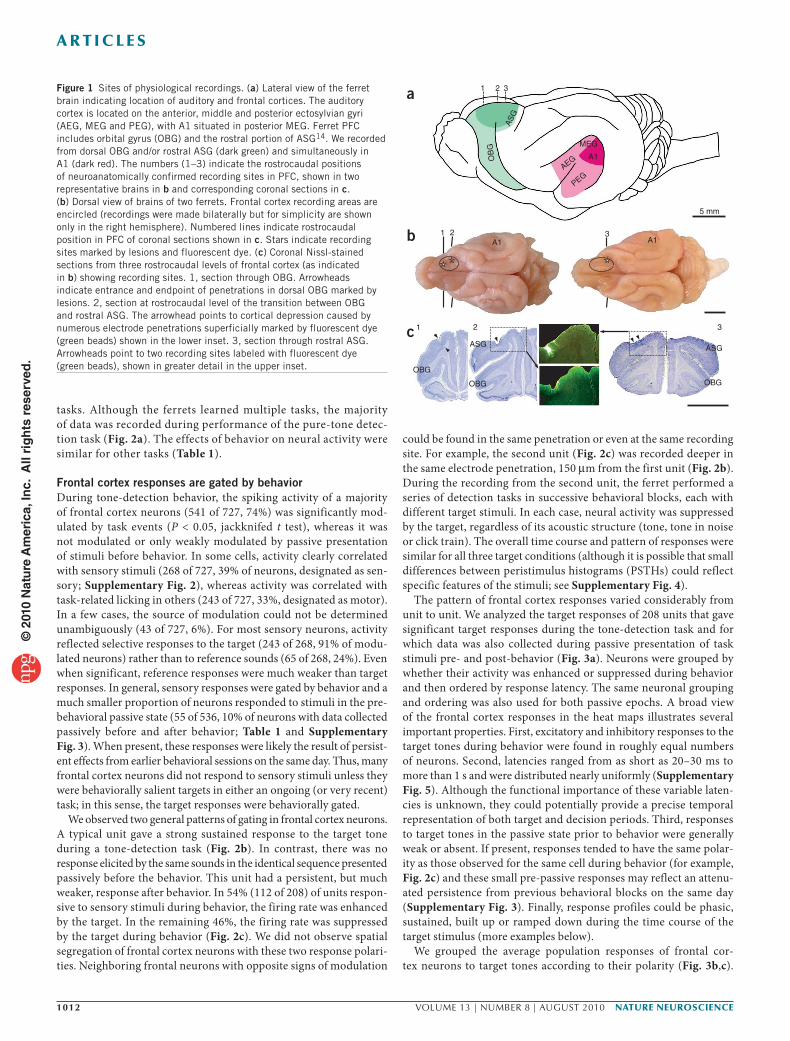
RESULTSTo study the representation of auditory stimuli during behavior, we recorded the activity of 766 single units in frontal cortex of five fer-rets that were trained on a variety of auditory and visual discrimina-tion tasks. Neural activity was recorded during behavior and during passive presentation of an identical sequence of task stimuli before and after behavior. These recordings (Fig. 1) were made in the dor-sal aspect of OBG, which is homologous to the primate dorsolateral prefrontal cortex (dlPFC), and the rostral ASG14 (S.R.-S. and J.B.F., unpublished observations). Responses were similar in both frontal areas and were therefore grouped together for analysis.
All tasks shared the same basic structure, in which ferrets learned by conditioned avoidance8 to lick water from a spout dur-ing the presentation of a class of reference stimuli and to cease licking after the presentation of the class of target stimuli to avoid a mild shock (Fig. 2a and Supplementary Fig. 1). The spectro- temporal features of reference and target classes varied between
1Institute for Systems Research, University of Maryland, College Park, Maryland, USA. 2Division of Neurobiology, Biocenter of Ludwig Maximilians University, Munich, Germany. Correspondence should be addressed to J.B.F. ([email protected]).
Received 5 April; accepted 16 June; published online 11 July 2010; doi:10.1038/nn.2598
Adaptive, behaviorally gated, persistent encoding of task-relevant auditory information in ferret frontal cortexJonathan B Fritz1, Stephen V David1, Susanne Radtke-Schuller2, Pingbo Yin1 & Shihab A Shamma1
Top-down signals from frontal cortex are thought to be important in cognitive control of sensory processing. To explore this interaction, we compared activity in ferret frontal cortex and primary auditory cortex (A1) during auditory and visual tasks requiring discrimination between classes of reference and target stimuli. Frontal cortex responses were behaviorally gated, selectively encoded the timing and invariant behavioral meaning of target stimuli, could be rapid in onset, and sometimes persisted for hours following behavior. These results are consistent with earlier findings in A1 that attention triggered rapid, selective, persistent, task-related changes in spectrotemporal receptive fields. Simultaneously recorded local field potentials revealed behaviorally gated changes in inter-areal coherence that were selectively modulated between frontal cortex and focal regions of A1 that were responsive to target sounds. These results suggest that A1 and frontal cortex dynamically establish a functional connection during auditory behavior that shapes the flow of sensory information and maintains a persistent trace of recent task-relevant stimulus features.
© 2
010
Nat
ure
Am
eric
a, In
c. A
ll ri
gh
ts r
eser
ved
.
1012 VOLUME 13 | NUMBER 8 | AUGUST 2010 nature neurOSCIenCe
a r t I C l e S
tasks. Although the ferrets learned multiple tasks, the majority of data was recorded during performance of the pure-tone detec-tion task (Fig. 2a). The effects of behavior on neural activity were similar for other tasks (Table 1).
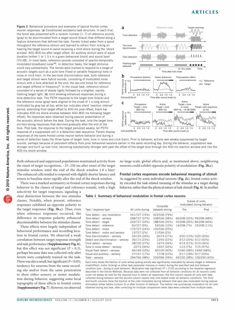
Frontal cortex responses are gated by behaviorDuring tone-detection behavior, the spiking activity of a majority of frontal cortex neurons (541 of 727, 74%) was significantly mod-ulated by task events (P < 0.05, jackknifed t test), whereas it was not modulated or only weakly modulated by passive presentation of stimuli before behavior. In some cells, activity clearly correlated with sensory stimuli (268 of 727, 39% of neurons, designated as sen-sory; Supplementary Fig. 2), whereas activity was correlated with task-related licking in others (243 of 727, 33%, designated as motor). In a few cases, the source of modulation could not be determined unambiguously (43 of 727, 6%). For most sensory neurons, activity reflected selective responses to the target (243 of 268, 91% of modu-lated neurons) rather than to reference sounds (65 of 268, 24%). Even when significant, reference responses were much weaker than target responses. In general, sensory responses were gated by behavior and a much smaller proportion of neurons responded to stimuli in the pre-behavioral passive state (55 of 536, 10% of neurons with data collected passively before and after behavior; Table 1 and Supplementary Fig. 3). When present, these responses were likely the result of persist-ent effects from earlier behavioral sessions on the same day. Thus, many frontal cortex neurons did not respond to sensory stimuli unless they were behaviorally salient targets in either an ongoing (or very recent) task; in this sense, the target responses were behaviorally gated.
We observed two general patterns of gating in frontal cortex neurons. A typical unit gave a strong sustained response to the target tone during a tone-detection task (Fig. 2b). In contrast, there was no response elicited by the same sounds in the identical sequence presented passively before the behavior. This unit had a persistent, but much weaker, response after behavior. In 54% (112 of 208) of units respon-sive to sensory stimuli during behavior, the firing rate was enhanced by the target. In the remaining 46%, the firing rate was suppressed by the target during behavior (Fig. 2c). We did not observe spatial segregation of frontal cortex neurons with these two response polari-ties. Neighboring frontal neurons with opposite signs of modulation
could be found in the same penetration or even at the same recording site. For example, the second unit (Fig. 2c) was recorded deeper in the same electrode penetration, 150 μm from the first unit (Fig. 2b). During the recording from the second unit, the ferret performed a series of detection tasks in successive behavioral blocks, each with different target stimuli. In each case, neural activity was suppressed by the target, regardless of its acoustic structure (tone, tone in noise or click train). The overall time course and pattern of responses were similar for all three target conditions (although it is possible that small differences between peristimulus histograms (PSTHs) could reflect specific features of the stimuli; see Supplementary Fig. 4).
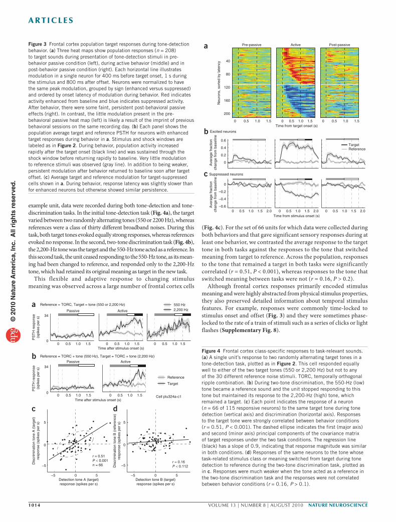
The pattern of frontal cortex responses varied considerably from unit to unit. We analyzed the target responses of 208 units that gave significant target responses during the tone-detection task and for which data was also collected during passive presentation of task stimuli pre- and post-behavior (Fig. 3a). Neurons were grouped by whether their activity was enhanced or suppressed during behavior and then ordered by response latency. The same neuronal grouping and ordering was also used for both passive epochs. A broad view of the frontal cortex responses in the heat maps illustrates several important properties. First, excitatory and inhibitory responses to the target tones during behavior were found in roughly equal numbers of neurons. Second, latencies ranged from as short as 20–30 ms to more than 1 s and were distributed nearly uniformly (Supplementary Fig. 5). Although the functional importance of these variable laten-cies is unknown, they could potentially provide a precise temporal representation of both target and decision periods. Third, responses to target tones in the passive state prior to behavior were generally weak or absent. If present, responses tended to have the same polar-ity as those observed for the same cell during behavior (for example, Fig. 2c) and these small pre-passive responses may reflect an attenu-ated persistence from previous behavioral blocks on the same day (Supplementary Fig. 3). Finally, response profiles could be phasic, sustained, built up or ramped down during the time course of the target stimulus (more examples below).
We grouped the average population responses of frontal cor-tex neurons to target tones according to their polarity (Fig. 3b,c).
3
ASGASG
OBG
OBG
OBG
1 2
1 3
5 mm
3A1
1 2A1
2
A1
a
b
c
MEG
AS
G
AEG
PEG
OB
G
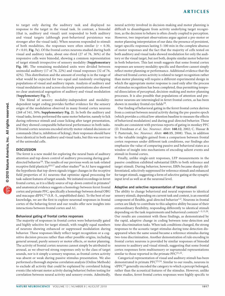
Figure 1 Sites of physiological recordings. (a) Lateral view of the ferret brain indicating location of auditory and frontal cortices. The auditory cortex is located on the anterior, middle and posterior ectosylvian gyri (AEG, MEG and PEG), with A1 situated in posterior MEG. Ferret PFC includes orbital gyrus (OBG) and the rostral portion of ASG14. We recorded from dorsal OBG and/or rostral ASG (dark green) and simultaneously in A1 (dark red). The numbers (1–3) indicate the rostrocaudal positions of neuroanatomically confirmed recording sites in PFC, shown in two representative brains in b and corresponding coronal sections in c. (b) Dorsal view of brains of two ferrets. Frontal cortex recording areas are encircled (recordings were made bilaterally but for simplicity are shown only in the right hemisphere). Numbered lines indicate rostrocaudal position in PFC of coronal sections shown in c. Stars indicate recording sites marked by lesions and fluorescent dye. (c) Coronal Nissl-stained sections from three rostrocaudal levels of frontal cortex (as indicated in b) showing recording sites. 1, section through OBG. Arrowheads indicate entrance and endpoint of penetrations in dorsal OBG marked by lesions. 2, section at rostrocaudal level of the transition between OBG and rostral ASG. The arrowhead points to cortical depression caused by numerous electrode penetrations superficially marked by fluorescent dye (green beads) shown in the lower inset. 3, section through rostral ASG. Arrowheads point to two recording sites labeled with fluorescent dye (green beads), shown in greater detail in the upper inset.
© 2
010
Nat
ure
Am
eric
a, In
c. A
ll ri
gh
ts r
eser
ved
.
nature neurOSCIenCe VOLUME 13 | NUMBER 8 | AUGUST 2010 1013
a r t I C l e S
Both enhanced and suppressed populations maintained activity from the onset of target recognition, ~25–250 ms after onset of the target stimulus window, until the end of the shock window 1.8 s later. The enhanced cells tended to respond with slightly shorter latency and return to baseline more rapidly after the end of the shock window.
There was a marked asymmetry in frontal cortex responses during behavior to the classes of target and reference sounds, with a high selectivity for target responses, signaling a clear distinction between the two stimulus classes. Notably, when present, reference responses exhibited an opposite polarity to the target responses (Fig. 3b,c). Thus, even when reference responses occurred, the difference in response polarity enhanced discriminability between the stimulus classes.
These effects were largely independent of behavioral performance and recording loca-tion in frontal cortex. We observed a weak correlation between target response strength and task performance (Supplementary Fig. 6), but this effect was not significant (P > 0.3), perhaps because data was collected only after ferrets were completely trained on the task. There was also a small, but significant (P < 0.05), tendency for neurons from the same record-ing site and/or from the same penetration to show either sensory or motor modula-tion during behavior, suggesting some local topography of these effects in frontal cortex (Supplementary Fig. 7). However, we observed
no large-scale, global effects and, as mentioned above, neighboring neurons could exhibit opposite polarity of modulation (Fig. 2b,c).
Frontal cortex responses encode behavioral meaning of stimuliAs suggested by some individual neurons (Fig. 2c), frontal cortex activ-ity encoded the task-related meaning of the stimulus as a target during behavior, rather than the physical nature of task stimuli (Fig. 4). In another
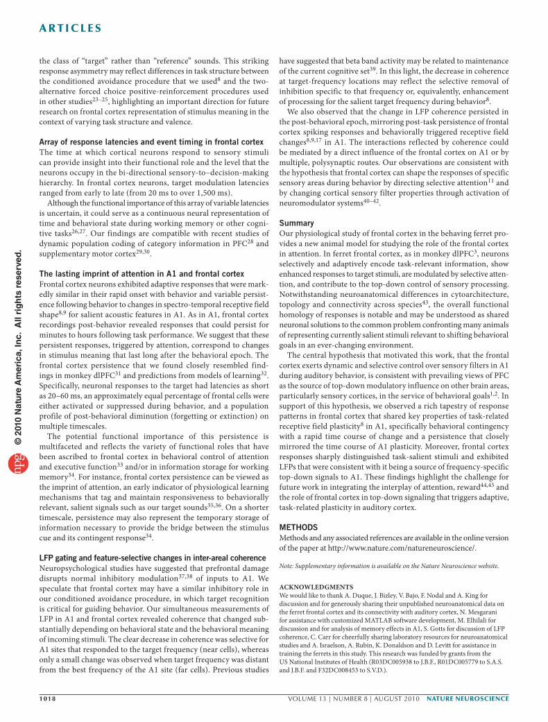
Figure 2 Behavioral procedure and examples of typical frontal cortex neuron responses. (a) Conditioned avoidance task structure. In each trial, the ferret was presented with a random number (1–7) of reference sounds (gray) to be discriminated from a target sound (black) that differed along a feature dimension that defined the task. Ferrets licked water from a spout throughout the reference stimuli and learned to refrain from licking on hearing the target sound to avoid receiving a mild shock during the ‘shock window’ 400–800 ms after target offset. All auditory stimuli were of equal duration (either 1 or 1.5 s in a given behavioral block) and sound level (70 dB). In most tasks, reference sounds consisted of spectro-temporally modulated broadband noise48. In detection tasks, the target stimulus could vary substantially. The ferrets were trained to respond to a range of acoustic targets such as a pure tone (fixed or variable frequency) tone in noise or click train. In the two-tone discrimination task, both reference and target stimuli were hybrid sounds, consisting of modulated noise stimuli with a tone attached at the end; the two end tones for reference and target differed in frequency9. In the visual task, reference stimuli consisted of a series of steady lights followed by a brighter, rapidly flashing target light. (b) Unit showing enhanced responses during a tone-detection task. The PSTH response to the target tone (black) and the reference noise (gray) were aligned to the onset of 1-s-long stimuli (indicated by gray bar at top; white bar indicates silent ‘reaction interval’ period, extending from target offset to 400-ms post-offset; black bar indicates 400-ms shock window between 400–800 ms following target offset). No responses were observed during passive presentation of the acoustic stimuli before the task. During the task, only the target tone elicited strong responses that declined gradually after the end of the tone. Post-task, the response to the target persisted weakly. (c) PSTH response of a suppressed cell in a detection task sequence. Panels display responses of the same frontal cortex neuron before behavior and during a series of detection tasks (for three types of target: tone, tone in noise and click train). Prior to behavior, activity was weakly suppressed by target sounds, perhaps because of persistent effects from prior behavioral sessions earlier in the same recording day. During the behavior, suppression was stronger and built up over time, becoming substantially stronger well past the offset of the target tone through the 400-ms reaction window and into the 400-ms shock window.
Two-tonediscrimination
. . .
or. . .
Reference (safe) Target (warning)
Tone detection
Flashing-lightdetection
Click detection . . .
. . .
0 21 . . . (3–18 s total) Time
a
19
0
Cell plu223b-d2
PS
TH
res
pons
e(s
pike
s pe
r s)
c
13
0
Cell plu223a-d1
PS
TH
res
pons
e(s
pike
s pe
r s)
Pre-passive (before) Active (behavior) Post-passive (after)b
0 1.0 1.50.5 0 1.0 1.50.5Time after sound onset (s)
0 1.0 1.50.5
Target
Reference
Time after trial onset
Time after sound onset (s)
Pre-passiveTarget = tone
(800 Hz)
ActiveTarget = tone
(800 Hz)
ActiveTarget = tone
in noise(800 Hz, –16 dB SNR)
ActiveTarget: click sequence
(5 Hz)
0 1.0 1.50.5 0 1.0 1.50.5 0 1.0 1.50.5 0 1.0 1.50.5
Table 1 Summary of behavioral modulation in frontal cortex neurons
Task / response type All units duringComplete
datasets during
Subset of units modulated during behavior
Pre Post
Tone detect – any modulation 541/727 (74%) 423/536 (79%) – –Tone detect – sensory 268/727 (37%) 208/536 (38%) 66/208 (32%) 99/208 (48%)Tone detect – target 243/727 (33%) 188/536 (35%) 55/208 (26%) 84/208 (40%)Tone detect – reference 65/727 (9%) 53/536 (10%) 14/208 (7%) 23/208 (11%)Tone detect – motor 175/727 (24%) 132/536 (25%) – –Tone detect – motor and sensory 13/727 (2%) 11/536 (2%) – –Tone discrimination – sensory 33/125 (26%) 20/74 (27%) 3/20 (15%) 5/20 (20%)Detect and discrimination – sensory 26/115 (23%) 12/55 (22%) 3/12 (25%) 5/12 (42%)Click detect – sensory 28/105 (27%) 19/74 (26%) 4/19 (21%) 5/19 (26%)Tone in noise detect – sensory 22/73 (30%) 15/47 (32%) 1/15 (7%) 7/15 (47%)Visual flash detect – sensory 56/169 (33%) 40/100 (40%) 15/40 (38%) 19/40 (48%)Visual and auditory – sensory 27/157 (17%) 17/78 (22%) 3/17 (18%) 9/17 (53%)Total – sensory 294/766 (38%) 230/596 (39%) 65/230 (28%) 100/230 (43%)
Each entry shows the fraction of units whose spiking activity was significantly modulated by sensory (target or reference onset/offset), motor (licking) or either task parameter (sensory or motor) during the specified task and behavior condition (pre-, during or post-behavior). Modulation was significant (P < 0.05) according to the stepwise regression described in the Online Methods. Because data were not collected from all behavior conditions for all neurons (cells could not always be held for the required hour to obtain all responses), the first column reports all cells with data collected during behavior and the second column reports only cells tested under all behavior conditions. The third and fourth columns show the fraction of the cells modulated during behavior that were also modulated during passive stimulation either before (column 3) or after (column 4) behavior. The bottom row summarizes modulation for all cells observed during any task, after correcting for multiple comparisons when data were collected from multiple tasks.
© 2
010
Nat
ure
Am
eric
a, In
c. A
ll ri
gh
ts r
eser
ved
.
1014 VOLUME 13 | NUMBER 8 | AUGUST 2010 nature neurOSCIenCe
a r t I C l e S
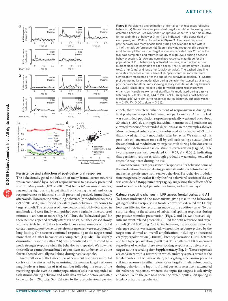
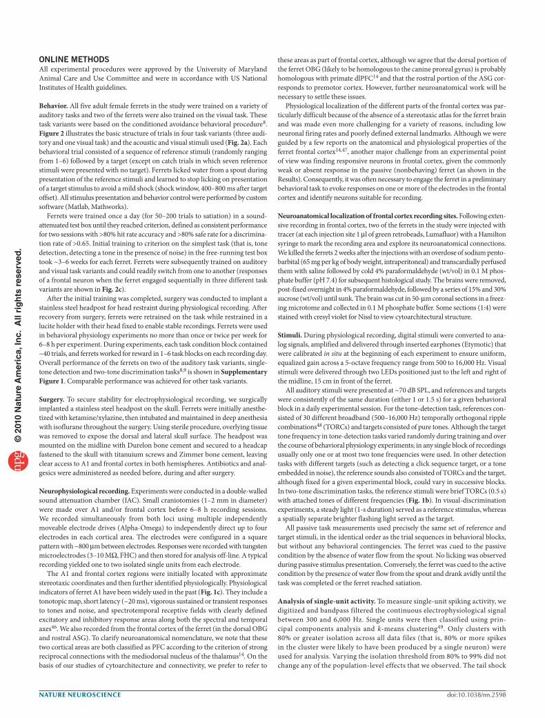
example unit, data were recorded during both tone-detection and tone-discrimination tasks. In the initial tone-detection task (Fig. 4a), the target varied between two randomly alternating tones (550 or 2200 Hz), whereas references were a class of thirty different broadband noises. During this task, both target tones evoked equally strong responses, whereas references evoked no response. In the second, two-tone discrimination task (Fig. 4b), the 2,200-Hz tone was the target and the 550-Hz tone acted as a reference. In this second task, the unit ceased responding to the 550-Hz tone, as its mean-ing had been changed to reference, and responded only to the 2,200-Hz tone, which had retained its original meaning as target in the new task.
This flexible and adaptive response to changing stimulus meaning was observed across a large number of frontal cortex cells
(Fig. 4c). For the set of 66 units for which data were collected during both behaviors and that gave significant sensory responses during at least one behavior, we contrasted the average response to the target tone in both tasks against the responses to the tone that switched meaning from target to reference. Across the population, responses to the tone that remained a target in both tasks were significantly correlated (r = 0.51, P < 0.001), whereas responses to the tone that switched meaning between tasks were not (r = 0.16, P > 0.2).
Although frontal cortex responses primarily encoded stimulus meaning and were highly abstracted from physical stimulus properties, they also preserved detailed information about temporal stimulus features. For example, responses were commonly time-locked to stimulus onset and offset (Fig. 3) and they were sometimes phase-locked to the rate of a train of stimuli such as a series of clicks or light flashes (Supplementary Fig. 8).
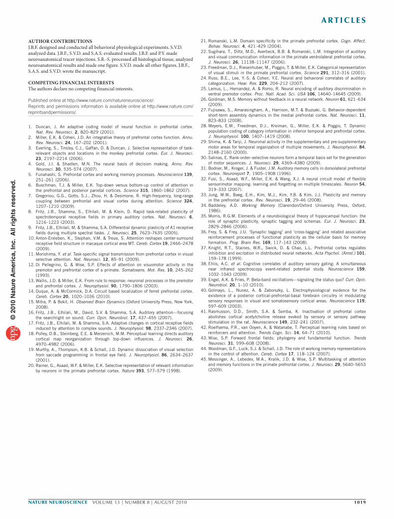
Figure 3 Frontal cortex population target responses during tone-detection behavior. (a) Three heat maps show population responses (n = 208) to target sounds during presentation of tone-detection stimuli in pre-behavior passive condition (left), during active behavior (middle) and in post-behavior passive condition (right). Each horizontal line illustrates modulation in a single neuron for 400 ms before target onset, 1 s during the stimulus and 800 ms after offset. Neurons were normalized to have the same peak modulation, grouped by sign (enhanced versus suppressed) and ordered by onset latency of modulation during behavior. Red indicates activity enhanced from baseline and blue indicates suppressed activity. After behavior, there were some faint, persistent post-behavioral passive effects (right). In contrast, the little modulation present in the pre-behavioral passive heat map (left) is likely a result of the imprint of previous behavioral sessions on the same recording day. (b) Each panel shows the population average target and reference PSTH for neurons with enhanced target responses during behavior in a. Stimulus and shock windows are labeled as in Figure 2. During behavior, population activity increased rapidly after the target onset (black line) and was sustained through the shock window before returning rapidly to baseline. Very little modulation to reference stimuli was observed (gray line). In addition to being weaker, persistent modulation after behavior returned to baseline soon after target offset. (c) Average target and reference modulation for target-suppressed cells shown in a. During behavior, response latency was slightly slower than for enhanced neurons but otherwise showed similar persistence.
a
b
0
0.2
0.4
0.6
–0.6
–0.4
–0.2
0
Ave
rage
frac
tion
chan
ge fr
om b
asel
ine
Time from stimulus onset (s)0 1.0 1.50.5 2.0
Ave
rage
frac
tion
chan
ge fr
om b
asel
ine
0 1.0 1.50.5 2.0 0 1.0 1.50.5 2.0
TargetReference
c
Excited neurons
Suppressed neurons
Pre-passive Active Post-passive
0 1.0 1.50.5
Neu
rons
, sor
ted
by la
tenc
y 40
80
120
160
200
0 1.0 1.50.5Time from target onset (s)
0 1.0 1.50.5
34
0
Cell plu324a-c1
PS
TH
res
pons
e(s
pike
s pe
r s)
34
00 1.0 1.50.5
Time after stimulus onset (s)
0 1.0 1.50.5Time after stimulus onset (s)
Passive Active
Reference = TORC, Target = tone (550 or 2,200 Hz)
Reference = TORC + tone (550 Hz), Target = TORC + tone (2,200 Hz)
Target
Reference
550 Hz2,200 Hz
a
b
PS
TH
res
pons
e(s
pike
s pe
r s)
Passive Active
Detection tone A (target)response (spikes per s)
Dis
crim
inat
ion
tone
A (
targ
et)
resp
onse
(sp
ikes
per
s)
Detection tone B (target)response (spikes per s)
Dis
crim
inat
ion
tone
B (
refe
renc
e)re
spon
se (
spik
es p
er s
)
c d
−5 0 5
r = 0.51P < 0.001n = 66
−5 0 5
r = 0.16P < 0.112−5
5
0
−5
5
0
0 1.0 1.50.5 0 1.0 1.50.5 0 1.0 1.50.5
Figure 4 Frontal cortex class-specific responses to task-relevant sounds. (a) A single unit’s response to two randomly alternating target tones in a tone-detection task, plotted as in Figure 2. This cell responded equally well to either of the two target tones (550 or 2,200 Hz) but not to any of the 30 different reference noise stimuli. TORC, temporally orthogonal ripple combination. (b) During two-tone discrimination, the 550-Hz (low) tone became a reference sound and the unit stopped responding to this tone but maintained its response to the 2,200-Hz (high) tone, which remained a target. (c) Each point indicates the response of a neuron (n = 66 of 115 responsive neurons) to the same target tone during tone detection (vertical axis) and discrimination (horizontal axis). Responses to the target tone were strongly correlated between behavior conditions (r = 0.51, P < 0.001). The dashed ellipse indicates the first (major axis) and second (minor axis) principal components of the covariance matrix of target responses under the two task conditions. The regression line (black) has a slope of 0.9, indicating that response magnitude was similar in both conditions. (d) Responses of the same neurons to the tone whose task-related stimulus class or meaning switched from target during tone detection to reference during the two-tone discrimination task, plotted as in c. Responses were much weaker when the tone acted as a reference in the two-tone discrimination task and the responses were not correlated between behavior conditions (r = 0.16, P > 0.1).
© 2
010
Nat
ure
Am
eric
a, In
c. A
ll ri
gh
ts r
eser
ved
.
nature neurOSCIenCe VOLUME 13 | NUMBER 8 | AUGUST 2010 1015
a r t I C l e S
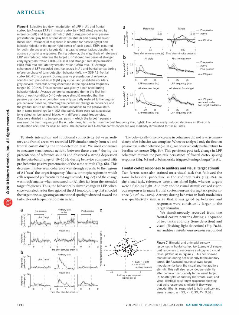
Persistence and extinction of post-behavioral responsesThe behaviorally gated modulation of many frontal cortex neurons was accompanied by a lack of responsiveness to passively presented stimuli. Many units (109 of 208, 52%) had a tabula rasa character, responding vigorously to target stimuli only during the task and losing responsiveness to identical stimuli presented passively immediately afterwards. However, the remaining behaviorally modulated neurons (99 of 208, 48%) manifested persistent post-behavioral responses to target stimuli. The responses of these neurons smoothly decreased in amplitude and were finally extinguished over a variable time course of minutes to an hour or more (Fig. 5a). Thus, the ‘behavioral gate’ for these neurons opened rapidly after task onset, but then closed slowly with a variable half-life after task offset. For a small number of frontal cortex neurons, post-behavior persistent responses were exceptionally long-lasting. One neuron continued responding to the target sound more than 2 h after behavior was completed (Fig. 5b). The slightly diminished response (after 2 h) was potentiated and restored to a much stronger response when the behavior was repeated. We note that these effects cannot be attributed to persistent motor behavior, as the ferrets showed virtually no licking during passive epochs.
An overall view of the time course of persistent responses in frontal cortex can be discerned by examining the average target response magnitude as a function of trial number following the onset of the recording epochs over the entire population of cells that responded to task stimuli during behavior and with data available before and after behavior (n = 208; Fig. 5c). Relative to the pre-behavioral passive
epoch, there was clear enhancement of responsiveness during the first post-passive epoch following task performance. After the task was concluded, population responses gradually weakened over about 20 trials (~200 s), although individual neurons could maintain an elevated response for extended durations (as in the examples above). More prolonged enhancement was observed in the subset of 99 units that showed significant modulation after behavior. We examined this post-task enhancement on a cell-by-cell basis using a scatter plot of the amplitude of modulation by target stimuli during behavior versus during post-behavioral passive stimulus presentation (Fig. 5d). The two measures are well correlated (r = 0.55, P < 0.001), indicating that persistent responses, although gradually weakening, tended to resemble responses during the task.
Given the long-term persistence of responses after behavior, some of the modulation observed during passive stimulation before behavior may reflect persistence from earlier behaviors. Pre-behavior modula-tion was generally weaker if only the first behavioral session of the day was considered (Supplementary Fig. 5), suggesting that effects of the most recent task target persisted for hours, rather than days.
Category-specific changes in LFP across frontal cortex and A1To better understand the mechanisms giving rise to the behavioral gating of spiking responses in frontal cortex, we extracted the LFP by low-pass filtering the recordings made during auditory tasks. To our surprise, despite the absence of substantial spiking responses during pre-passive stimulus presentation (Figs. 2 and 3), we observed sig-nificant event-related potentials (ERPs) for both reference and target stimuli (P < 0.0001; Fig. 6). During behavior, the response evoked by reference sounds was attenuated, whereas the response evoked by the target tone showed an overall amplification, including an increased early hyperpolarization (~100 ms), later depolarization (~450–650 ms) and late hyperpolarization (>700 ms). This pattern of ERPs occurred regardless of whether there were spiking responses to references or targets at the recording site (Supplementary Fig. 9). These responses are consistent with a network in which auditory signals arrive at the frontal cortex in the passive state, but a gating mechanism prevents spiking responses to either reference or target stimuli. Subsequently, during behavior, the input to frontal cortex is selectively attenuated for reference responses, whereas the input for targets is selectively enhanced. With the gate now open, the target inputs elicit spiking in frontal cortex during behavior.
d
a
−10 −5 0 5 10
n = 208r = 0.47 P < 0.001
Mean target response duringbehavior (spikes per s)
32
0
32
0
b
−10
−5
10
Mea
n ta
rget
res
pons
epo
st-b
ehav
ior
(spi
kes
per
s)
0
5
tel013b-a1
plu211a-a1
PS
TH
res
pons
e(s
pike
s pe
r s)
PS
TH
res
pons
e(s
pike
s pe
r s)
PS
TH
res
pons
e(s
pike
s pe
r s)
1:00Passive
0 1.0 2.0Time (s)
0:30Passive
0 1.0 2.0Time (s)
0:15Passive
0 1.0 2.0Time (s)
–0:15Passive
0 1.0 2.0Time (s)
0 1.0 2.0Time (s)
0:00Active
–0:10Passive
75
00 1.0 2.0
Time (s)
PS
TH
res
pons
e(s
pike
s pe
r s) 2:20
Passive
75
00 1.0 2.0
Time (s)
0:00Active
0 1.0 2.0Time (s)
2:40Active
0 1.0 2.0Time (s)
0:20Passive
0 1.0 2.0Time (s)
2:50Passive
0 1.0 2.0Time (s)
Behavior > passive (P < 0.05)Behavior ≤ passive
c
0 5 10 15 20Trial
n = 208
0
0.2
0.4
0.6
0.8
1.0
Nor
mal
ized
targ
et r
espo
nse
PreActivePost
2nd postPost**
Figure 5 Persistence and extinction of frontal cortex responses following behavior. (a) Neuron showing persistent target modulation following tone-detection behavior. Behavior condition (passive or active) and time relative to the beginning of behavior (h:min) are indicated in the upper right of each panel, with PSTHs plotted as in Figure 2. The target response post-behavior was more phasic than during behavior and faded within 1 h of the task performance. (b) Neuron showing exceptionally persistent modulation, plotted as in a. Target responses persisted over 2 h after the task was completed and returned rapidly to high levels during a second behavior session. (c) Average normalized response magnitude for the population of 208 behaviorally activated neurons, as a function of trial number since the beginning of each epoch (that is, before (green), during (red), after (blue) and long after (black) behavior). The dashed blue line indicates responses of the subset of 99 ‘persistent’ neurons that were significantly modulated after the end of the behavioral session. (d) Scatter plot comparing target modulation during behavior (horizontal axis) versus post-behavior for all neurons showing sensory modulation during behavior (n = 208). Black dots indicate units for which target responses were either significantly weaker or not significantly modulated during passive listening (P < 0.05, t test, 144 of 208, 69%). Responses post-behavior (vertical axis) were similar to responses during behavior, although weaker (r = 0.55, P < 0.001, slope = 0.31).
© 2
010
Nat
ure
Am
eric
a, In
c. A
ll ri
gh
ts r
eser
ved
.
1016 VOLUME 13 | NUMBER 8 | AUGUST 2010 nature neurOSCIenCe
a r t I C l e S
To study interaction and functional connectivity between audi-tory and frontal areas, we recorded LFP simultaneously from A1 and frontal cortex during the tone-detection task. We used coherence to measure synchronous activity between these areas15 during the presentation of reference sounds and observed a strong depression in the beta-band range of 10–20 Hz during behavior compared with pre-behavior passive presentation of the same stimuli (Fig. 6b). This decrease in inter-areal coherence was strongly specific to the regions of A1 ‘near’ the target frequency (that is, tonotopic regions in which cells responded preferentially to target sounds; Fig. 6c) and the change was much smaller when measured for A1 sites far from the attended target frequency. Thus, the behaviorally driven change in LFP coher-ence was selective for the region of the A1 tonotopic map that encoded the target tone, suggesting an attentional spotlight directed toward the task-relevant frequency domain in A1.
The behaviorally driven decrease in coherence did not reverse imme-diately after behavior was complete. When we analyzed only the first ten passive trials after behavior (~100 s), we observed only partial return to baseline coherence (Fig. 6b). This persistent post-task change in LFP coherence mirrors the post-task persistence of frontal cortex spiking responses (Fig. 5c) and of behaviorally triggered tuning changes8 in A1.
Frontal cortex responses to auditory and visual target stimuliTwo ferrets were also trained on a visual task that followed the same behavioral procedure as the auditory tasks (Fig. 2a). In the visual task, references were a sustained light, whereas targets were a flashing light. Auditory and/or visual stimuli evoked vigor-ous responses in many frontal cortex neurons during task perform-ance (76 of 157, 48%). Activity during behavior in both modalities was qualitatively similar in that it was gated by behavior and
responses were consistently larger to the target stimulus.
We simultaneously recorded from two frontal cortex neurons during a sequence of two tasks: auditory (tone detection) and visual (flashing-light detection) (Fig. 7a,b). An auditory tabula rasa neuron responded
Figure 6 Selective top-down modulation of LFP in A1 and frontal cortex. (a) Average ERPs in frontal cortex (n = 362 sites) evoked by reference (left) and target stimuli (right) during pre-behavior passive presentation (gray line) of tone detection stimuli and during behavior (black line). Variance of responses is reported for passive (gray) and behavior (black) in the upper right corner of each panel. ERPs occurred for both references and targets during passive presentation, despite the absence of spiking responses. During behavior, the magnitude of reference ERP was reduced, whereas the target ERP showed two peaks of stronger, early hyperpolarization (100–200 ms) and stronger, late depolarization (400–600 ms) and later hyperpolarization (>600 ms). (b) Average coherence of LFP recorded simultaneously in A1 and frontal cortex during reference phase of tone-detection behavior (left, n = 339 A1–frontal cortex (A1-FC) site pairs). During passive presentation of reference sounds (both pre-behavior (light gray curve) and post-behavior (dark gray curve)), there was strong coherence in the alpha-beta frequency range (10–20 Hz). This coherence was greatly diminished during behavior (black). Average coherence measured during the first ten trials of each condition (~40 reference stimuli) revealed that the passive post-behavior condition was only partially restored to the original pre-behavior baseline, reflecting the persistent change in coherence and the gradual return of intra-areal communications to the passive state. (c) In some recordings (n = 102 site pairs), there were two successive tone-detection behavioral blocks with different target frequencies. Data were divided into two groups, pairs in which the target frequency was near the best frequency of the A1 site (near, left) or far from the best frequency (far, right). The behaviorally induced decrease in 10–20-Hz modulation occurred for near A1 sites. The decrease in A1–frontal cortex coherence was markedly diminished for far A1 sites.
n = 362 sites
Pre-passiveActive
n = 339 pairs
Pre-passiveActivePost-passive
Target
0 0.5 1
Reference
0 0.5 1
5 20 35
0.1
0.2
5 20 35
5 20 35
0.1
0.2
5 20 35
Time after stimulus onset (s)
LFP frequency (Hz)
LFP frequency (Hz)
Ave
rage
A1-
FC
cohe
renc
eA
vera
ge A
1-F
Cco
here
nce
A1 sites far from targetA1 sites near target
First 10 trials only
a
c
b
n = 102 pairsrecorded undertwo target conditions
Time after stimulus onset (s)
LFP frequency (Hz)
LFP frequency (Hz)
All trials
−1
0
1
Ave
rage
norm
aliz
ed E
RP 0.03
0.010.100.23
plu417a-g1
36
0
plu417a-h1
20
0
Res
pons
e(s
pike
s pe
r s)
Res
pons
e(s
pike
s pe
r s)
Auditory taskactive
a
Auditory target response(spikes per s)
−5 0 5
0
r = 0.30, P < 0.01n = 44 of 157 auditory neurons
c
Vis
ual t
arge
t res
pons
e (s
pike
s pe
r s)
−5
5
Pre-passive Post-passive
0 1 2 1 2 1 20 0Time after stimulus onset (s)
Visual task
36
0
Res
pons
e(s
pike
s pe
r s)
20
0
Res
pons
e(s
pike
s pe
r s)
Auditory task
Visual task
b0 1 2 1 2 1 20 0
Time after stimulus onset (s)
Figure 7 Bimodal and unimodal sensory responses in frontal cortex. (a) Example of single-unit responses to successive auditory and visual tasks, plotted as in Figure 2. This cell showed modulation during behavior only to the auditory target. (b) A second neuron showed target modulation by both the visual and the auditory stimuli. This cell also responded persistently after behavior, particularly to the visual target. (c) Scatter plot of auditory (horizontal axis) and visual (vertical axis) target responses showing that cells responded similarly if they were bimodal (that is, responded to both auditory and visual stimuli, n = 93, r = 0.30, P < 0.01).
© 2
010
Nat
ure
Am
eric
a, In
c. A
ll ri
gh
ts r
eser
ved
.
nature neurOSCIenCe VOLUME 13 | NUMBER 8 | AUGUST 2010 1017
a r t I C l e S
to target only during the auditory task and displayed no response to the target in the visual task. In contrast, a bimodal (that is, auditory and visual) unit responded to both auditory and visual targets (although post-behavioral persistence was stronger after the visual task). When neurons responded to stimuli of both modalities, the responses were often similar (r = 0.30, P < 0.01; Fig. 7c). Of the frontal cortex neurons studied during both visual and auditory tasks, about one-third (27 of 76, 36%) of the responsive cells were bimodal, showing a common representation of target stimuli irrespective of sensory modality (Supplementary Fig. 10). The remaining modulated units were divided between unimodal auditory (17 of 76, 22%) and visual responses (32 of 76, 42%). This distribution and the amount of overlap is in the range of what would be expected for two equal and randomly overlapping populations of visual and auditory inputs. Analysis of auditory and visual modulation in and across electrode penetrations also showed no clear anatomical segregation of auditory and visual modulation (Supplementary Fig. 7).
The blend of sensory modality-independent and modality- dependent target coding provides further evidence for the sensory origin of the modulation observed in many frontal cortex neurons (268 of 541, 50%; Supplementary Fig. 2). In both the auditory and visual tasks, ferrets performed the same motor behavior, namely to lick during reference stimuli and cease licking after target presentation, and they showed comparable behavioral performance in both tasks. If frontal cortex neurons encoded strictly motor-related decisions or commands (that is, inhibition of licking), their responses should have been identical in both auditory and visual tasks, but this was not true of the unimodal cells.
DISCUSSIONThe ferret is a new model for exploring the neural basis of auditory attention and top-down control of auditory processing during goal-directed behavior16. The results of our previous work on task-related receptive field plasticity8,9,17 and other studies18 in A1 have suggested the hypothesis that top-down signals trigger changes in the receptive field properties of A1 neurons that optimize signal processing for task-salient features of target sounds. We initiated recordings in ferret frontal cortex, as it is a likely source of top-down control in primates2 and anatomical evidence suggests a homology between ferret frontal cortex and primate PFC, specifically a homology between dorsal OBG and macaque dlPFC14 (S.R.-S., unpublished data). To the best of our knowledge, we are the first to explore neuronal responses in frontal cortex of the behaving ferret and our results offer new insights into interactions between frontal cortex and A1.
Behavioral gating of frontal cortex responsesThe majority of responses in frontal cortex were behaviorally gated and highly selective for target stimuli, with roughly equal numbers of neurons showing enhanced or suppressed modulation during behavior. These responses likely reflect target recognition or a cog-nitive decision process rather than other possible origins, including general arousal, purely sensory or motor effects, or motor planning. The activity of frontal cortex neurons cannot simply be attributed to arousal, as we observed strong responses only to the class of target sounds, nor is it simply a sensory response, as frontal cortex activity was absent or weak during passive stimulus presentation. We also performed a thorough stepwise correlation analysis (Online Methods) to exclude all activity that could be explained by correlated licking events (the relevant motor activity during behavior) before testing for correlation between neural activity and sensory events. Admittedly,
neural activity involved in decision-making and motor planning is difficult to disambiguate from activity underlying target recogni-tion, as the decision to behave is often closely coupled to perception. However, two important observations argue against a pre-motor or motor planning interpretation of our data: the presence of persistent, target-specific responses lasting 5–100 min in the complete absence of motor responses and the fact that the majority of cells tested on both auditory and visual tasks showed modulation for only the audi-tory or the visual target, but not both, despite similar motor behavior in both behaviors. This last result suggests that some frontal cortex responses are sensory modality specific and therefore cannot simply reflect motor planning or performance. Additional evidence that the observed frontal cortex activity is related to target recognition rather than motor planning will require a different experimental design in which the appropriate motor response is cued only after the process of stimulus recognition has been completed, thus permitting tempo-ral dissociation of perceptual, decision-making and motor-planning processes. It is also possible that populations of attention and pre-motor neurons are intermingled in ferret frontal cortex, as has been shown in monkey frontal eye fields19.
Our finding of behavioral gating in the ferret frontal cortex derives from a contrast between neural activity during the pre-passive epoch (which provides a critical low-attention baseline to measure the effects of behavioral modulation) and during goal-directed behavior. These results are consistent with previous reports of gating in monkey PFC (D. Freedman et al. Soc. Neurosci. Abstr. 160.12, 2002; C. Hussar & T. Pasternak, Soc. Neurosci. Abstr. 460.15, 2008). Thus, in addition to the valuable insights gained from a comparison between frontal cortex responses under different task conditions, our findings also emphasize the value of comparing passive and behavioral states as a window of insight into mechanisms of encoding salient events and stimuli in frontal cortex.
Finally, unlike single-unit responses, LFP measurements in the passive condition exhibited substantial ERPs to both reference and target stimuli. During behavior, however, ERPs became sharply dif-ferentiated, selectively suppressed for reference stimuli and enhanced for target stimuli, suggesting a form of selective gating at the synaptic input level as well as at the neuronal level.
Adaptive and selective representation of target stimuliThe ability to change behavioral and neural responses to identical sensory stimuli, depending on current task and context, is an essential component of flexible, goal-directed behavior1,2. Neurons in frontal cortex are likely to contribute to this adaptive ability because of their extraordinary flexibility, responding differently to identical stimuli depending on the task requirements and behavioral contexts1–3,13,20. Our results are consistent with these findings, as demonstrated by the rapid, adaptive change in coding between tone-detection and tone-discrimination tasks. When task conditions changed, the strong responses to the acoustic target stimulus during tone detection dis-appeared when the same sound became a reference stimulus during two-tone discrimination. Another demonstration of rule encoding in frontal cortex neurons is provided by similar responses of bimodal neurons to auditory and visual stimuli, suggesting that some frontal cortex responses form multisensory or supramodal representations similar to those reported in the primate PFC21,22.
Categorical representation of visual and auditory stimuli has been demonstrated in primate PFC23,24. Similar to our results, neurons in vlPFC24 generally encoded the category to which a percept belonged, rather than the acoustical features of the stimulus. However, unlike these studies, ferret frontal cortex responses were highly specific to
© 2
010
Nat
ure
Am
eric
a, In
c. A
ll ri
gh
ts r
eser
ved
.
1018 VOLUME 13 | NUMBER 8 | AUGUST 2010 nature neurOSCIenCe
a r t I C l e S
the class of “target” rather than “reference” sounds. This striking response asymmetry may reflect differences in task structure between the conditioned avoidance procedure that we used8 and the two- alternative forced choice positive-reinforcement procedures used in other studies23–25, highlighting an important direction for future research on frontal cortex representation of stimulus meaning in the context of varying task structure and valence.
Array of response latencies and event timing in frontal cortexThe time at which cortical neurons respond to sensory stimuli can provide insight into their functional role and the level that the neurons occupy in the bi-directional sensory-to–decision-making hierarchy. In frontal cortex neurons, target modulation latencies ranged from early to late (from 20 ms to over 1,500 ms).
Although the functional importance of this array of variable latencies is uncertain, it could serve as a continuous neural representation of time and behavioral state during working memory or other cogni-tive tasks26,27. Our findings are compatible with recent studies of dynamic population coding of category information in PFC28 and supplementary motor cortex29,30.
The lasting imprint of attention in A1 and frontal cortexFrontal cortex neurons exhibited adaptive responses that were mark-edly similar in their rapid onset with behavior and variable persist-ence following behavior to changes in spectro-temporal receptive field shape8,9 for salient acoustic features in A1. As in A1, frontal cortex recordings post-behavior revealed responses that could persist for minutes to hours following task performance. We suggest that these persistent responses, triggered by attention, correspond to changes in stimulus meaning that last long after the behavioral epoch. The frontal cortex persistence that we found closely resembled find-ings in monkey dlPFC31 and predictions from models of learning32. Specifically, neuronal responses to the target had latencies as short as 20–60 ms, an approximately equal percentage of frontal cells were either activated or suppressed during behavior, and a population profile of post-behavioral diminution (forgetting or extinction) on multiple timescales.
The potential functional importance of this persistence is multifaceted and reflects the variety of functional roles that have been ascribed to frontal cortex in behavioral control of attention and executive function33 and/or in information storage for working memory34. For instance, frontal cortex persistence can be viewed as the imprint of attention, an early indicator of physiological learning mechanisms that tag and maintain responsiveness to behaviorally relevant, salient signals such as our target sounds35,36. On a shorter timescale, persistence may also represent the temporary storage of information necessary to provide the bridge between the stimulus cue and its contingent response34.
LFP gating and feature-selective changes in inter-areal coherenceNeuropsychological studies have suggested that prefrontal damage disrupts normal inhibitory modulation37,38 of inputs to A1. We speculate that frontal cortex may have a similar inhibitory role in our conditioned avoidance procedure, in which target recognition is critical for guiding behavior. Our simultaneous measurements of LFP in A1 and frontal cortex revealed coherence that changed sub-stantially depending on behavioral state and the behavioral meaning of incoming stimuli. The clear decrease in coherence was selective for A1 sites that responded to the target frequency (near cells), whereas only a small change was observed when target frequency was distant from the best frequency of the A1 site (far cells). Previous studies
have suggested that beta band activity may be related to maintenance of the current cognitive set39. In this light, the decrease in coherence at target-frequency locations may reflect the selective removal of inhibition specific to that frequency or, equivalently, enhancement of processing for the salient target frequency during behavior8.
We also observed that the change in LFP coherence persisted in the post-behavioral epoch, mirroring post-task persistence of frontal cortex spiking responses and behaviorally triggered receptive field changes8,9,17 in A1. The interactions reflected by coherence could be mediated by a direct influence of the frontal cortex on A1 or by multiple, polysynaptic routes. Our observations are consistent with the hypothesis that frontal cortex can shape the responses of specific sensory areas during behavior by directing selective attention11 and by changing cortical sensory filter properties through activation of neuromodulator systems40–42.
SummaryOur physiological study of frontal cortex in the behaving ferret pro-vides a new animal model for studying the role of the frontal cortex in attention. In ferret frontal cortex, as in monkey dlPFC3, neurons selectively and adaptively encode task-relevant information, show enhanced responses to target stimuli, are modulated by selective atten-tion, and contribute to the top-down control of sensory processing. Notwithstanding neuroanatomical differences in cytoarchitecture, topology and connectivity across species43, the overall functional homology of responses is notable and may be understood as shared neuronal solutions to the common problem confronting many animals of representing currently salient stimuli relevant to shifting behavioral goals in an ever-changing environment.
The central hypothesis that motivated this work, that the frontal cortex exerts dynamic and selective control over sensory filters in A1 during auditory behavior, is consistent with prevailing views of PFC as the source of top-down modulatory influence on other brain areas, particularly sensory cortices, in the service of behavioral goals1,2. In support of this hypothesis, we observed a rich tapestry of response patterns in frontal cortex that shared key properties of task-related receptive field plasticity8 in A1, specifically behavioral contingency with a rapid time course of change and a persistence that closely mirrored the time course of A1 plasticity. Moreover, frontal cortex responses sharply distinguished task-salient stimuli and exhibited LFPs that were consistent with it being a source of frequency-specific top-down signals to A1. These findings highlight the challenge for future work in integrating the interplay of attention, reward44,45 and the role of frontal cortex in top-down signaling that triggers adaptive, task-related plasticity in auditory cortex.
METhODSMethods and any associated references are available in the online version of the paper at http://www.nature.com/natureneuroscience/.
Note: Supplementary information is available on the Nature Neuroscience website.
AcknowledgmenTSWe would like to thank A. Duque, J. Bizley, V. Bajo, F. Nodal and A. King for discussion and for generously sharing their unpublished neuroanatomical data on the ferret frontal cortex and its connectivity with auditory cortex, N. Mesgarani for assistance with customized MATLAB software development, M. Elhilali for discussion and for analysis of memory effects in A1, S. Gotts for discussion of LFP coherence, C. Carr for cheerfully sharing laboratory resources for neuroanatomical studies and A. Israelson, A. Rubin, K. Donaldson and D. Levitt for assistance in training the ferrets in this study. This research was funded by grants from the US National Institutes of Health (R03DC005938 to J.B.F., R01DC005779 to S.A.S. and J.B.F. and F32DC008453 to S.V.D.).
© 2
010
Nat
ure
Am
eric
a, In
c. A
ll ri
gh
ts r
eser
ved
.
nature neurOSCIenCe VOLUME 13 | NUMBER 8 | AUGUST 2010 1019
a r t I C l e S
AUTHoR conTRIBUTIonSJ.B.F. designed and conducted all behavioral physiological experiments. S.V.D. analyzed data. J.B.F., S.V.D. and S.A.S. evaluated results. J.B.F. and P.Y. made neuroanatomical tracer injections. S.R.-S. processed all histological tissue, analyzed neuroanatomical results and made one figure. S.V.D. made all other figures. J.B.F., S.A.S. and S.V.D. wrote the manuscript.
comPeTIng FInAncIAl InTeReSTSThe authors declare no competing financial interests.
Published online at http://www.nature.com/natureneuroscience/. Reprints and permissions information is available online at http://www.nature.com/reprintsandpermissions/.
1. Duncan, J. An adaptive coding model of neural function in prefrontal cortex. Nat. Rev. Neurosci. 2, 820–829 (2001).
2. Miller, E.K. & Cohen, J.D. An integrative theory of prefrontal cortex function. Annu. Rev. Neurosci. 24, 167–202 (2001).
3. Everling, S., Tinsley, C.J., Gaffan, D. & Duncan, J. Selective representation of task-relevant objects and locations in the monkey prefrontal cortex. Eur. J. Neurosci. 23, 2197–2214 (2006).
4. Gold, J.I. & Shadlen, M.N. The neural basis of decision making. Annu. Rev. Neurosci. 30, 535–574 (2007).
5. Funahashi, S. Prefrontal cortex and working memory processes. Neuroscience 139, 251–261 (2006).
6. Buschman, T.J. & Miller, E.K. Top-down versus bottom-up control of attention in the prefrontal and posterior parietal cortices. Science 315, 1860–1862 (2007).
7. Gregoriou, G.G., Gotts, S.J., Zhou, H. & Desimone, R. High-frequency, long-range coupling between prefrontal and visual cortex during attention. Science 324, 1207–1210 (2009).
8. Fritz, J.B., Shamma, S., Elhilali, M. & Klein, D. Rapid task-related plasticity of spectrotemporal receptive fields in primary auditory cortex. Nat. Neurosci. 6, 1216–1223 (2003).
9. Fritz, J.B., Elhilali, M. & Shamma, S.A. Differential dynamic plasticity of A1 receptive fields during multiple spectral tasks. J. Neurosci. 25, 7623–7635 (2005).
10. Anton-Erxleben, K., Stephan, V.M. & Treue, S. Attention reshapes center-surround receptive field structure in macaque cortical area MT. Cereb. Cortex 19, 2466–2478 (2009).
11. Morishima, Y. et al. Task-specific signal transmission from prefrontal cortex in visual selective attention. Nat. Neurosci. 12, 85–91 (2009).
12. Di Pellegrino, G. & Wise, S.P. Effects of attention on visuomotor activity in the premotor and prefrontal cortex of a primate. Somatosens. Mot. Res. 10, 245–262 (1993).
13. Wallis, J.D. & Miller, E.K. From rule to response: neuronal processes in the premotor and prefrontal cortex. J. Neurophysiol. 90, 1790–1806 (2003).
14. Duque, A. & McCormick, D.A. Circuit based localization of ferret prefrontal cortex. Cereb. Cortex 20, 1020–1036 (2010).
15. Mitra, P. & Bokil, H. Observed Brain Dynamics (Oxford University Press, New York, 2008).
16. Fritz, J.B., Elhilali, M., David, S.V. & Shamma, S.A. Auditory attention—focusing the searchlight on sound. Curr. Opin. Neurobiol. 17, 437–455 (2007).
17. Fritz, J.B., Elhilali, M. & Shamma, S.A. Adaptive changes in cortical receptive fields induced by attention to complex sounds. J. Neurophysiol. 98, 2337–2346 (2007).
18. Polley, D.B., Steinberg, E.E. & Merzenich, M.M. Perceptual learning directs auditory cortical map reorganization through top-down influences. J. Neurosci. 26, 4970–4982 (2006).
19. Murthy, A., Thompson, K.B. & Schall, J.D. Dynamic dissociation of visual selection from saccade programming in frontal eye field. J. Neurophysiol. 86, 2634–2637 (2001).
20. Rainer, G., Asaad, W.F. & Miller, E.K. Selective representation of relevant information by neurons in the primate prefrontal cortex. Nature 393, 577–579 (1998).
21. Romanski, L.M. Domain specificity in the primate prefrontal cortex. Cogn. Affect. Behav. Neurosci. 4, 421–429 (2004).
22. Sugihara, T., Diltz, M.D., Averbeck, B.B. & Romanski, L.M. Integration of auditory and visual communication information in the primate ventrolateral prefrontal cortex. J. Neurosci. 26, 11138–11147 (2006).
23. Freedman, D.J., Riesenhuber, M., Poggio, T. & Miller, E.K. Categorical representation of visual stimuli in the primate prefrontal cortex. Science 291, 312–316 (2001).
24. Russ, B.E., Lee, Y.-S. & Cohen, Y.E. Neural and behavioral correlates of auditory categorization. Hear. Res. 229, 204–212 (2007).
25. Lemus, L., Hernandez, A. & Romo, R. Neural encoding of auditory discrimination in ventral premotor cortex. Proc. Natl. Acad. Sci. USA 106, 14640–14645 (2009).
26. Goldman, M.S. Memory without feedback in a neural network. Neuron 61, 621–634 (2009).
27. Fujisawa, S., Amarasingham, A., Harrison, M.T. & Buzsaki, G. Behavior-dependent short-term assembly dynamics in the medial prefrontal cortex. Nat. Neurosci. 11, 823–833 (2008).
28. Meyers, E.M., Freedman, D.J., Kreiman, G., Miller, E.K. & Poggio, T. Dynamic population coding of category information in inferior temporal and prefrontal cortex. J. Neurophysiol. 100, 1407–1419 (2008).
29. Shima, K. & Tanji, J. Neuronal activity in the supplementary and pre-supplementary motor areas for temporal organization of multiple movements. J. Neurophysiol. 84, 2148–2160 (2000).
30. Salinas, E. Rank-order–selective neurons form a temporal basis set for the generation of motor sequences. J. Neurosci. 29, 4369–4380 (2009).
31. Bodner, M., Kroger, J. & Fuster, J.M. Auditory memory cells in dorsolateral prefrontal cortex. Neuroreport 7, 1905–1908 (1996).
32. Fusi, S., Asaad, W.F., Miller, E.K. & Wang, X.J. A neural circuit model of flexible sensorimotor mapping: learning and forgetting on multiple timescales. Neuron 54, 319–333 (2007).
33. Jung, M.W., Baeg, E.H., Kim, M.J., Kim, Y.B. & Kim, J.J. Plasticity and memory in the prefrontal cortex. Rev. Neurosci. 19, 29–46 (2008).
34. Baddeley, A.D. Working Memory (Clarendon/Oxford University Press, Oxford, 1986).
35. Morris, R.G.M. Elements of a neurobiological theory of hippocampal function: the role of synaptic plasticity, synaptic tagging and schemas. Eur. J. Neurosci. 23, 2829–2846 (2006).
36. Frey, S. & Frey, J.U. ‘Synaptic tagging’ and ‘cross-tagging’ and related associative reinforcement processes of functional plasticity as the cellular basis for memory formation. Prog. Brain Res. 169, 117–143 (2008).
37. Knight, R.T., Staines, W.R., Swick, D. & Chao, L.L. Prefrontal cortex regulates inhibition and excitation in distributed neural networks. Acta Psychol. (Amst.) 101, 159–178 (1999).
38. Ehlis, A.C. et al. Cognitive correlates of auditory sensory gating: A simultaneous near infrared spectroscopy event-related potential study. Neuroscience 159, 1032–1043 (2009).
39. Engel, A.K. & Fries, P. Beta-band oscillations—signaling the status quo? Curr. Opin. Neurobiol. 20, 1–10 (2010).
40. Golmayo, L., Nunez, A. & Zaborszky, L. Electrophysiological evidence for the existence of a posterior cortical-prefrontal-basal forebrain circuitry in modulating sensory responses in visual and somatosensory cortical areas. Neuroscience 119, 597–609 (2003).
41. Rasmusson, D.D., Smith, S.A. & Semba, K. Inactivation of prefrontal cortex abolishes cortical acetylcholine release evoked by sensory or sensory pathway stimulation in the rat. Neuroscience 149, 232–241 (2007).
42. Roelfsema, P.R., van Ooyen, A. & Watanabe, T. Perceptual learning rules based on reinforcers and attention. Trends Cogn. Sci. 14, 64–71 (2010).
43. Wise, S.P. Forward frontal fields: phylogeny and fundamental function. Trends Neurosci. 31, 599–608 (2008).
44. Woodman, G.F., Luck, S.J. & Schall, J.D. The role of working memory representations in the control of attention. Cereb. Cortex 17, 118–124 (2007).
45. Messinger, A., Lebedev, M.A., Kralik, J.D. & Wise, S.P. Multitasking of attention and memory functions in the primate prefrontal cortex. J. Neurosci. 29, 5640–5653 (2009).
© 2
010
Nat
ure
Am
eric
a, In
c. A
ll ri
gh
ts r
eser
ved
.
nature neurOSCIenCe doi:10.1038/nn.2598
ONLINE METhODSAll experimental procedures were approved by the University of Maryland Animal Care and Use Committee and were in accordance with US National Institutes of Health guidelines.
Behavior. All five adult female ferrets in the study were trained on a variety of auditory tasks and two of the ferrets were also trained on the visual task. These task variants were based on the conditioned avoidance behavioral procedure8. Figure 2 illustrates the basic structure of trials in four task variants (three audi-tory and one visual task) and the acoustic and visual stimuli used (Fig. 2a). Each behavioral trial consisted of a sequence of reference stimuli (randomly ranging from 1–6) followed by a target (except on catch trials in which seven reference stimuli were presented with no target). Ferrets licked water from a spout during presentation of the reference stimuli and learned to stop licking on presentation of a target stimulus to avoid a mild shock (shock window, 400–800 ms after target offset). All stimulus presentation and behavior control were performed by custom software (Matlab, Mathworks).
Ferrets were trained once a day (for 50–200 trials to satiation) in a sound- attenuated test box until they reached criterion, defined as consistent performance for two sessions with >80% hit rate accuracy and >80% safe rate for a discrimina-tion rate of >0.65. Initial training to criterion on the simplest task (that is, tone detection, detecting a tone in the presence of noise) in the free-running test box took ~3–6 weeks for each ferret. Ferrets were subsequently trained on auditory and visual task variants and could readily switch from one to another (responses of a frontal neuron when the ferret engaged sequentially in three different task variants are shown in Fig. 2c).
After the initial training was completed, surgery was conducted to implant a stainless steel headpost for head restraint during physiological recording. After recovery from surgery, ferrets were retrained on the task while restrained in a lucite holder with their head fixed to enable stable recordings. Ferrets were used in behavioral physiology experiments no more than once or twice per week for 6–8 h per experiment. During experiments, each task condition block contained ~40 trials, and ferrets worked for reward in 1–6 task blocks on each recording day. Overall performance of the ferrets on two of the auditory task variants, single-tone detection and two-tone discrimination tasks8,9 is shown in Supplementary Figure 1. Comparable performance was achieved for other task variants.
Surgery. To secure stability for electrophysiological recording, we surgically implanted a stainless steel headpost on the skull. Ferrets were initially anesthe-tized with ketamine/xylazine, then intubated and maintained in deep anesthesia with isoflurane throughout the surgery. Using sterile procedure, overlying tissue was removed to expose the dorsal and lateral skull surface. The headpost was mounted on the midline with Durelon bone cement and secured to a headcap fastened to the skull with titanuium screws and Zimmer bone cement, leaving clear access to A1 and frontal cortex in both hemispheres. Antibiotics and anal-gesics were administered as needed before, during and after surgery.
neurophysiological recording. Experiments were conducted in a double-walled sound attenuation chamber (IAC). Small craniotomies (1–2 mm in diameter) were made over A1 and/or frontal cortex before 6–8 h recording sessions. We recorded simultaneously from both loci using multiple independently moveable electrode drives (Alpha-Omega) to independently direct up to four electrodes in each cortical area. The electrodes were configured in a square pattern with ~800 μm between electrodes. Responses were recorded with tungsten microelectrodes (3–10 MΩ, FHC) and then stored for analysis off-line. A typical recording yielded one to two isolated single units from each electrode.
The A1 and frontal cortex regions were initially located with approximate stereotaxic coordinates and then further identified physiologically. Physiological indicators of ferret A1 have been widely used in the past (Fig. 1c). They include a tonotopic map, short latency (~20 ms), vigorous sustained or transient responses to tones and noise, and spectrotemporal receptive fields with clearly defined excitatory and inhibitory response areas along both the spectral and temporal axes46. We also recorded from the frontal cortex of the ferret (in the dorsal OBG and rostral ASG). To clarify neuroanatomical nomenclature, we note that these two cortical areas are both classified as PFC according to the criterion of strong reciprocal connections with the mediodorsal nucleus of the thalamus14. On the basis of our studies of cytoarchitecture and connectivity, we prefer to refer to
these areas as part of frontal cortex, although we agree that the dorsal portion of the ferret OBG (likely to be homologous to the canine proreal gyrus) is probably homologous with primate dlPFC14 and that the rostral portion of the ASG cor-responds to premotor cortex. However, further neuroanatomical work will be necessary to settle these issues.
Physiological localization of the different parts of the frontal cortex was par-ticularly difficult because of the absence of a stereotaxic atlas for the ferret brain and was made even more challenging for a variety of reasons, including low neuronal firing rates and poorly defined external landmarks. Although we were guided by a few reports on the anatomical and physiological properties of the ferret frontal cortex14,47, another major challenge from an experimental point of view was finding responsive neurons in frontal cortex, given the commonly weak or absent response in the passive (nonbehaving) ferret (as shown in the Results). Consequently, it was often necessary to engage the ferret in a preliminary behavioral task to evoke responses on one or more of the electrodes in the frontal cortex and identify neurons suitable for recording.
neuroanatomical localization of frontal cortex recording sites. Following exten-sive recording in frontal cortex, two of the ferrets in the study were injected with tracer (at each injection site 1 μl of green retrobeads, Lumafluor) with a Hamilton syringe to mark the recording area and explore its neuroanatomical connections. We killed the ferrets 2 weeks after the injections with an overdose of sodium pento-barbital (65 mg per kg of body weight, intraperitoneal) and transcardially perfused them with saline followed by cold 4% paraformaldehyde (wt/vol) in 0.1 M phos-phate buffer (pH 7.4) for subsequent histological study. The brains were removed, post-fixed overnight in 4% paraformaldehyde, followed by a series of 15% and 30% sucrose (wt/vol) until sunk. The brain was cut in 50-μm coronal sections in a freez-ing microtome and collected in 0.1 M phosphate buffer. Some sections (1:4) were stained with cresyl violet for Nissl to view cytoarchitectural structure.
Stimuli. During physiological recording, digital stimuli were converted to ana-log signals, amplified and delivered through inserted earphones (Etymotic) that were calibrated in situ at the beginning of each experiment to ensure uniform, equalized gain across a 5-octave frequency range from 500 to 16,000 Hz. Visual stimuli were delivered through two LEDs positioned just to the left and right of the midline, 15 cm in front of the ferret.
All auditory stimuli were presented at ~70 dB SPL, and references and targets were consistently of the same duration (either 1 or 1.5 s) for a given behavioral block in a daily experimental session. For the tone-detection task, references con-sisted of 30 different broadband (500–16,000 Hz) temporally orthogonal ripple combinations48 (TORCs) and targets consisted of pure tones. Although the target tone frequency in tone-detection tasks varied randomly during training and over the course of behavioral physiology experiments; in any single block of recordings usually only one or at most two tone frequencies were used. In other detection tasks with different targets (such as detecting a click sequence target, or a tone embedded in noise), the reference sounds also consisted of TORCs and the target, although fixed for a given experimental block, could vary in successive blocks. In two-tone discrimination tasks, the reference stimuli were brief TORCs (0.5 s) with attached tones of different frequencies (Fig. 1b). In visual-discrimination experiments, a steady light (1-s duration) served as a reference stimulus, whereas a spatially separate brighter flashing light served as the target.
All passive task measurements used precisely the same set of reference and target stimuli, in the identical order as the trial sequences in behavioral blocks, but without any behavioral contingencies. The ferret was cued to the passive condition by the absence of water flow from the spout. No licking was observed during passive stimulus presentation. Conversely, the ferret was cued to the active condition by the presence of water flow from the spout and drank avidly until the task was completed or the ferret reached satiation.
Analysis of single-unit activity. To measure single-unit spiking activity, we digitized and bandpass filtered the continuous electrophysiological signal between 300 and 6,000 Hz. Single units were then classified using prin-cipal components analysis and k-means clustering49. Only clusters with 80% or greater isolation across all data files (that is, 80% or more spikes in the cluster were likely to have been produced by a single neuron) were used for analysis. Varying the isolation threshold from 80% to 99% did not change any of the population-level effects that we observed. The tail shock
© 2
010
Nat
ure
Am
eric
a, In
c. A
ll ri
gh
ts r
eser
ved
.
nature neurOSCIenCedoi:10.1038/nn.2598
for incorrect responses introduced a strong electrical artifact and signals recorded during this period were discarded before processing.
Significant neural modulation by auditory and visual stimuli was determined by stepwise linear regression of time-varying spike activity (binned at 50 ms) against stimulus (target and reference) and motor (licking) events. The complete regression modeled spiking activity as a function of reference, target and lick events.
r t h s t h s t h m tr r t t mT
T( ) ( ) ( ) ( ) ( ) ( ) ( ).= − + − + −
= −∑ t t t t t t
t
The stimulus functions, sr(t) and st(t), are 0, except at times, t, of reference or target onset, respectively, when they have a value of 1. Similarly, the motor func-tion, m(t), has a value of 0 except at times when lick events occur. The regression functions, hr(τ), ht(τ) and hm(τ), then indicate the average firing rate before and after each corresponding event. The regression functions were fit by normalized reverse correlation50, which discounted spurious effects that might arise as a result of correlations between stimulus events and changes in motor activity.
Neurons were classified as being significantly modulated by sensory inputs if the occurrence of a stimulus predicted a change in firing rate that could not be explained by a simpler model on the basis of motor activity alone.
r t h m tmT
T( ) ( ) ( )= −
= −∑ t t
t
Thus, a neuron was considered to be modulated by sensory inputs only if the full model predicted spiking activity significantly better than the model based only on licking activity (P < 0.05, jackknifed t test). Examples of neurons with activity significantly correlated with sensory or motor events appear in Supplementary Figure 2.
To compare responses across the neural population under different behavior conditions (passive versus active, tone detection versus discrimination, etc.),
(1)(1)
(2)(2)
we measured the average firing rate during the 1.0–1.5-s duration of stimulus and the subsequent 800-ms silent period/shock window. Similarity of popu-lation responses was then measured by the correlation coefficient between the average responses under the different conditions. Significance was deter-mined by a randomized paired t test (a bootstrapping procedure by which the probability of the measured correlation coefficient was computed directly from a distribution of correlation coefficients measured for randomly shuffled behavior conditions).
Analysis of local field potential. The LFP was extracted by low-pass filtering the same continuous recordings below 200 Hz. A hardware notch filter removed artifacts at 60 Hz and activity between 55 and 65 Hz was discarded to avoid artifacts.
Coherence of simultaneously recorded LFP was computed using a multi-taper method15. Because coherence has a fixed lower bound of 0, noise biases coher-ence toward positive values. Thus, when comparing coherence between behavior conditions, the number of trials included in each condition was fixed so as to keep the noise bias the same in each condition. Significant differences in coherence between behavior conditions were determined by a jackknifed t test measured across different recording sites.
46. Bizley, J.K., Nodal, F.R., Nelken, I. & King, A.J. Functional organization of ferret auditory cortex. Cereb. Cortex 15, 1637–1653 (2005).
47. Haider, B., Duque, A., Hasenstaub, A.R. & McCormick, D.A. Neocortical network activity in vivo is generated though a dynamic balance of excitation and inhibition. J. Neurosci. 26, 4535–4545 (2006).
48. Klein, D.J., Depireux, D.A., Simon, J.Z. & Shamma, S.A. Robust spectrotemporal reverse correlation for the auditory system: optimizing stimulus design. J. Comput. Neurosci. 9, 85–111 (2000).
49. David, S.V., Mesgarani, N., Fritz, J.B. & Shamma, S.A. Rapid synaptic depression explains nonlinear modulation of spectro-temporal tuning in primary auditory cortex by natural stimuli. J. Neurosci. 29, 3374–3386 (2009).
50. Theunissen, F.E., David, S.V., Singh, N.C., Hsu, A., Vinje, W.E. & Gallant, J.L. Estimating spatial temporal receptive fields of auditory and visual neurons from their responses to natural stimuli. Network: Comput. Neural Syst. 12, 289–316 (2001).
Top Related
Copyright © 2022 FDOKUMEN

