


Bahasa
Halaman
Hukum

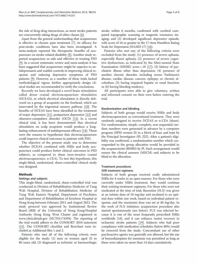
Man et al. BMC Complementary and Alternative Medicine 2014, 14:255http://www.biomedcentral.com/1472-6882/14/255
RESEARCH ARTICLE Open Access
A pilot controlled trial of a combination of densecranial electroacupuncture stimulation and bodyacupuncture for post-stroke depressionSui-Cheung Man1†, Ben H B Hung1†, Roger M K Ng2, Xiao-Chun Yu3, Hobby Cheung4, Mandy P M Fung4,Leonard S W Li5, Kwok-Pui Leung5, Kei-Pui Leung6, Kevin W Y Tsang6, Eric Ziea7, Vivian T Wong7
and Zhang-Jin Zhang1*
Abstract
Background: Our previous studies have demonstrated the treatment benefits of dense cranial electroacupuncturestimulation (DCEAS), a novel brain stimulation therapy in patients with major depression, postpartum depressionand obsessive-compulsive disorder. The purpose of the present study was to further evaluate the effectiveness ofDCEAS combined with body acupuncture and selective serotonin reuptake inhibitors (SSRIs) in patients withpost-stroke depression (PSD).
Methods: In a single-blind, randomized controlled trial, 43 patients with PSD were randomly assigned to 12 sessions ofDCEAS plus SSRI plus body electroacupuncture (n = 23), or sham (non-invasive cranial electroacupuncture, n-CEA) plusSSRI plus body electroacupuncture (n = 20) for 3 sessions per week over 4 weeks. Treatment outcomes were measuredusing the 17-item Hamilton Depression Rating Scale (HAMD-17), the Clinical Global Impression - Severity scale (CGI-S)and Barthel Index (BI), a measure used to evaluate movement ability associated with daily self-caring activity.
Results: DCEAS produced a significantly greater reduction of both HAMD-17 and CGI-S as early as week 1 and CGI-S atendpoint compared to n-CEA, but subjects of n-CEA group exhibited a significantly greater improvement on BI at week4 than DCEAS. Incidence of adverse events was not different in the two groups.
Conclusions: These results indicate that DCEAS could be effective in reducing stroke patients’ depressive symptoms.Superficial electrical stimulation in n-CEA group may be beneficial in improving movement disability of stroke patients.A combination of DCEAS and body acupuncture can be considered a treatment option for neuropsychiatric sequelaeof stroke.
Trial registration: www.clinicaltrials.gov, NCT01174394.
Keywords: Acupuncture, Post-stroke depression, Rehabilitation, Dense cranial electroacupuncture stimulation, DCEAS,Clinical trial
BackgroundMood depression is a common and serious consequenceof stroke. About 30% of stroke patients would developpost-stroke depression (PSD), either in the early or latestage after stroke onset [1]. Although PSD is stronglyassociated with poor prognosis and increased disability,
* Correspondence: [email protected]†Equal contributors1The School of Chinese Medicine, The University of Hong Kong, 10 SassoonRoad, Pokfulam, Hong Kong, ChinaFull list of author information is available at the end of the article
© 2014 Man et al.; licensee BioMed Central LtCommons Attribution License (http://creativecreproduction in any medium, provided the orDedication waiver (http://creativecommons.orunless otherwise stated.
it is often overlooked by clinicians. Only a small portionof patients are properly diagnosed and receive treatment[2,3]. Antidepressants, typically represented by selectiveserotonin reuptake inhibitors (SSRIs) and heterocyclics,are the mainstays of treatment for PSD [4,5]. Severalcontrolled studies have demonstrated that while somepatients significantly improved on depression scores,others discontinued due to side effects [4,5]. These sideeffects may also aggravate patients’ morbidity [1]. On theother hand, the use of antidepressants might also increase
d. This is an Open Access article distributed under the terms of the Creativeommons.org/licenses/by/2.0), which permits unrestricted use, distribution, andiginal work is properly credited. The Creative Commons Public Domaing/publicdomain/zero/1.0/) applies to the data made available in this article,
Man et al. BMC Complementary and Alternative Medicine 2014, 14:255 Page 2 of 8http://www.biomedcentral.com/1472-6882/14/255
the risk of drug-drug interactions, as most stroke patientsare concurrently taking drugs of other classes [6].Apart from the general understanding that acupuncture
is effective in chronic pain treatment [7], its efficacy forpost-stroke conditions have also been investigated. Ameta-analysis reported the therapeutic benefits of acu-puncture on stroke related disability [8]. Another study re-ported acupuncture as safe and effective in treating PSD[9]. In a recent systematic review and meta-analysis it hasbeen suggested that acupuncture might be superior to an-tidepressants and waitlist controls in improving clinical re-sponse and reducing depressive symptoms of PSDpatients [9]. However, as a number of these trials lackedmethodological vigour, further appropriately design cli-nical studies are recommended to verify the conclusions.Recently we have developed a novel brain stimulation
called dense cranial electroacupuncture stimulation(DCEAS), in which electrical stimulation is directly deli-vered on a group of acupoints on the forehead, which areinnervated by the trigeminal sensory pathway [10]. Thebenefits of DCEAS have been identified in the treatmentof major depression [11], postpartum depression [12] andobsessive-compulsive disorder (OCD) [13]. In a recentclinical trial, it has been demonstrated that electricalstimulation on scalp acupoints would produce a long-lasting enhancement of antidepressant efficacy [14]. Thesewere the reasons to hypothesize that electroacupuncturecould improve clinical outcomes in patients with PSD.The objective of the present study was to determine
whether DCEAS combined with SSRIs and body acu-puncture could produce better clinical outcomes in PSDsubjects, as compared to sham (non-invasive cranialelectroacupuncture, n-CEA). To test this hypothesis, thissingle-blind, randomized, sham-controlled clinical studywas designed.
MethodsSettings and subjectsThis single-blind, randomized, sham-controlled trial wasconducted in Division of Rehabilitation Medicine of TungWah Hospital, Division of Rehabilitation Medicine ofTung Wah Eastern Hospital, Department of Psychiatryand Department of Rehabilitation of Kowloon Hospital atHong Kong between February 2011 and August 2012. Thestudy protocol was approved by Institutional ReviewBoard (IRB) of the University of Hong Kong/HospitalAuthority Hong Kong West Cluster and registered inwww.clinicaltrials.gov (NCT01174394). The reporting ofthe trial would adhere to the CONSORT 2010 statement[15]. The CONSORT checklist and flowchart were in-cluded as Additional files 1 and 2.Patients who met all of the following criteria were
eligible for the study: (1) men or women aged 35 to80 years old; (2) diagnosed as ischemic or haemorrhagic
stroke within 6 months, confirmed with cerebral com-puted topographic scanning or magnetic resonance im-aging; and (3) developed significant depressive episode,with score of 16 or greater in the 17-item Hamilton RatingScale for Depression (HAMD-17) [16].Patients who met any of the following criteria were
excluded from the study: (1) presence of severe aphasia,especially fluent aphasia; (2) presence of severe cogni-tive dysfunction, as indicated by the Mini-mental StateExamination (MMSE) score < 18 [17]; (3) history of psy-chiatric illness other than depression; (4) presence ofanother chronic disorder, including severe Parkinson’sdisease, cardiac disease, cancers, epilepsy, or chronic al-coholism; (5) having impaired hepatic or renal function;or (6) having bleeding tendency.All participants were able to give voluntary, written
and informed consent on their own before entering thetrial.
Randomization and blindingSubjects of both groups would receive SSRIs and bodyelectroacupuncture as conventional treatment. They wererandomly assigned to receive DCEAS or n-CEA (sham).For randomization, simple, complete, non-sequential ran-dom numbers were generated in advance by a computerprogram (SPSS version II) in a block of four, and kept bythe Principal Investigator (PI, ZJZ). After a patient’s eligi-bility was confirmed, a randomization number which cor-responded to the group allocation would be provided tothe acupuncturist (BHBH) by PI. Such arrangement wouldensure the clinical assessor (MSCM) and subjects to beblind to the allocation.
Treatment proceduresSSRI treatment regimensSubjects of both groups received orally administeredSSRIs for 4 weeks in an open manner. For those who werecurrently under SSRIs treatment, they would continuetheir existing treatment regimens. For those who were notmedicated at the time of trial, fluoxetine (FLX) was givenat an initiate dose of 10 mg/day and escalated to an opti-mal dose within one week, based on individual patient re-sponse, and the maximum dose was set at 40 mg/day. Inthe week of FLX initiation, acupuncture procedure alsostarted spontaneously (see below). FLX was selected be-cause it is one of the most frequently prescribed SSRIsworldwide [18], and it can enhance motor recovery inischaemic stroke patients [19]. Subjects who had poorcompliance with medication schedules (below 80%) wouldbe removed from the study. Concomitant use of otherpsychoactive agents was generally not allowed, but the useof benzodiazepines for insomnia was permitted as long asthese were taken no more than 14 days cumulatively.
Man et al. BMC Complementary and Alternative Medicine 2014, 14:255 Page 3 of 8http://www.biomedcentral.com/1472-6882/14/255
Acupuncture interventionAcupuncture intervention was conducted for 3 sessionsper week over 4 consecutive weeks while subjects werebeing medicated with SSRIs. The 4-week treatment du-ration was designated because robust effects of acupunc-ture in stroke patients were generally observed within thisperiod [9]. Both groups of subjects received body electroa-cupuncture by electrically stimulating between ipsilateralHegu (LI4) and Quchi (LI11) and between ipsilateralZusanli (ST36) and Taichong (LR3) on both sides. Thesebody acupoints were commonly used in the acupunc-ture for stoke treatment. Meanwhile, subjects assigned toDCEAS received electrical stimulation on the following 6pairs of cranial acupoints: Baihui (GV20) and Yintang (EX-HN3), left Sishencong (EX-HN1) and Toulinqi (GB15),right Sishencong (EX-HN1) and Toulinqi (GB15), bilateralShuaigu (GB8), bilateral Taiyang (EX-HN5), and bilateralTouwei (ST8). For electrical stimulation on limbic and cra-nial acupoints, disposable acupuncture needles (0.30 mm indiameter and 25–40 mm in length) were inserted at a depthof 10–30 mm obliquely into limbic and cranial acupoints,on which electrical stimulation with continuous waves with2 Hz at 9 volts were delivered through an electrical acu-puncture stimulation instrument (Hwarto, SMY-10A). Theuse of the low frequency rather high frequency was adoptedbecause it could induce biochemical changes in the brain inmore favourable manner for alleviating depressive symp-toms [20,21]. The intensities of stimulation were adjustedto a level at which subjects felt most comfortable. Thestimulation lasted 30 min.For subjects assigned to n-CEA, Streitberger’s non-
invasive acupuncture needles were applied to serve assham control at the same cranial acupoints with thesame stimulation modality, except that the needles wereonly adhered to the skin instead of insertion [22]. Its va-lidity and credibility has been well demonstrated [22-24].Briefly, needles without sharp tips were quickly put ontothe acupoints as used in DCEAS. The needles did notactually pierce through the skin, though subjects wouldfeel like being treated. The needles were then affixed onthe skin with adhesive tapes. Since all the cranial acu-points were beyond subjects’ vision, they could notvisualize the acupuncture procedure. This sham acu-puncture modality has also been adopted in our previousstudies [11-13], without any problems encountered.
Rehabilitation therapyIn view of the fact that a large proportion of stroke pa-tients had physical rehabilitation treatment, subjects par-ticipating in the study would be allowed to continue theirdesignated physical rehabilitation therapy. Whether sub-jects had had rehabilitation therapy would be included instatistical analysis as a covariate (see below).
Clinical assessmentSubjects’ cognitive function was measured using MMSEduring screening. Those with MMSE score less than 18were excluded from the study as mentioned in the inclu-sion criteria. The primary efficacy was measured using theHAMD-17, CGI-S, and Barthel Index (BI). BI is a conciseindex of independence to score movement ability of a pa-tient in caring himself/herself [25]. Adverse events wereassessed using the Treatment Emergent Symptom Scale(TESS) [26]. Assessments were conducted at baseline, week1, 2 and 4. To ensure consistency and reliability of assess-ments across the study, assessors received a training ses-sion. A reliability coefficient of ≥ 0.80 was required to beachieved after the training session on HAMD-17 and BI.Assessments for a patient were generally completed by thesame assessor.To check the credibility of the n-CEA and DCEAS proce-
dures, based on the method by Flink et al. [24], each subjectwas asked the same question at the end of the last clinicalassessment: “As we informed you that you had an equalchance of receiving sham or active acupuncture treatment,which do you think you had received?”.
Data analysisPrevious studies have shown that acupuncture interventioncould produce at least a 4-point reduction in HAMD-17 inPSD patients [9]. Therefore, a sample size of 40 subjects(n = 20 per group) should provide an approximately 80%power at a statistical level of 0.05, with an estimated stan-dard deviation of 4.0 and an estimated dropout rate of 15%at the endpoint of treatment.Efficacy analyses were performed on the intention-to-
treat population, defined as participants who completedbaseline and at least one evaluation after treatment. A lin-ear mixed-effect model was applied to compare treatmentoutcomes (HAMD-17, CGI, and BI) over time between thetwo groups. The model was established using time andgroup for categorical fixed factors and random interceptswith scaled identity covariance matrix. Subject’s age, gender,duration of the illness, baseline HAMD-17 and BI weretreated as covariates. Between-group differences at eachmeasure time point were further examined using Studentt test. Student t test was also applied to detect between-group differences in continuous baseline variables. Catego-rical variables, including categorical baseline variables andincidence of adverse events were analyzed using Chi-square(χ2) or Fisher Exact test. Statistical significance was definedas a two-tailed P< 0.05. The analyses were performed withSPSS version 16 software (Chicago, IL, USA).
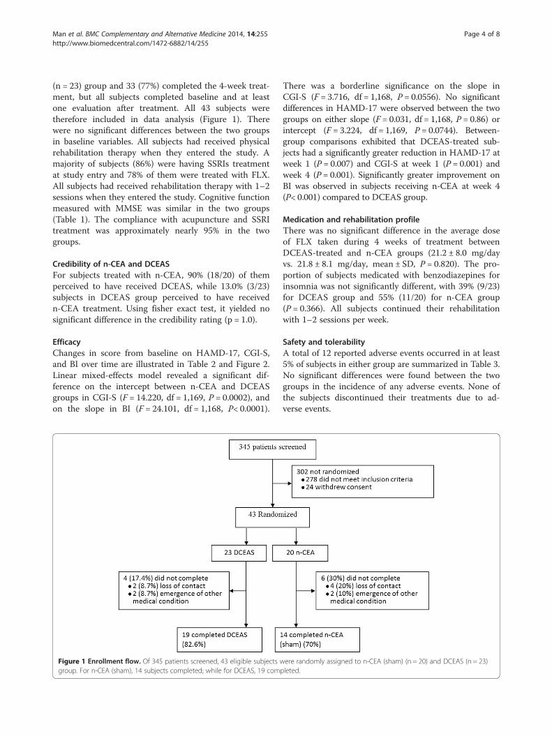
ResultsBaseline characteristics of subjectsOf 345 patients screened, 43 eligible subjects were ran-domly assigned to n-CEA (sham) (n = 20) and DCEAS
Man et al. BMC Complementary and Alternative Medicine 2014, 14:255 Page 4 of 8http://www.biomedcentral.com/1472-6882/14/255
(n = 23) group and 33 (77%) completed the 4-week treat-ment, but all subjects completed baseline and at leastone evaluation after treatment. All 43 subjects weretherefore included in data analysis (Figure 1). Therewere no significant differences between the two groupsin baseline variables. All subjects had received physicalrehabilitation therapy when they entered the study. Amajority of subjects (86%) were having SSRIs treatmentat study entry and 78% of them were treated with FLX.All subjects had received rehabilitation therapy with 1–2sessions when they entered the study. Cognitive functionmeasured with MMSE was similar in the two groups(Table 1). The compliance with acupuncture and SSRItreatment was approximately nearly 95% in the twogroups.
Credibility of n-CEA and DCEASFor subjects treated with n-CEA, 90% (18/20) of themperceived to have received DCEAS, while 13.0% (3/23)subjects in DCEAS group perceived to have receivedn-CEA treatment. Using fisher exact test, it yielded nosignificant difference in the credibility rating (p = 1.0).
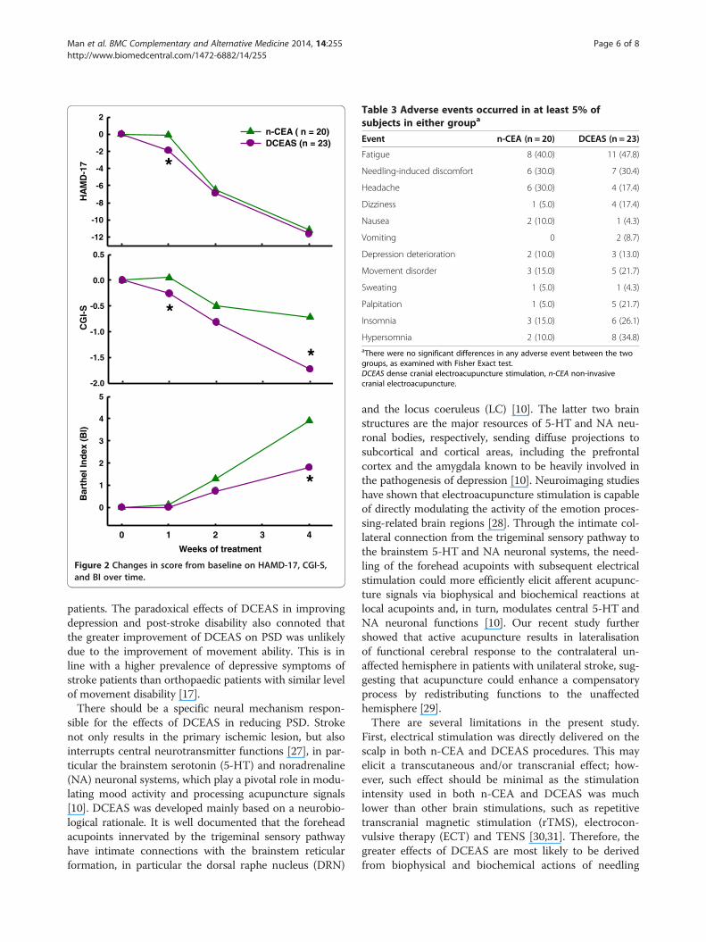
EfficacyChanges in score from baseline on HAMD-17, CGI-S,and BI over time are illustrated in Table 2 and Figure 2.Linear mixed-effects model revealed a significant dif-ference on the intercept between n-CEA and DCEASgroups in CGI-S (F = 14.220, df = 1,169, P = 0.0002), andon the slope in BI (F = 24.101, df = 1,168, P< 0.0001).
Figure 1 Enrollment flow. Of 345 patients screened, 43 eligible subjects wgroup. For n-CEA (sham), 14 subjects completed; while for DCEAS, 19 comp
There was a borderline significance on the slope inCGI-S (F = 3.716, df = 1,168, P = 0.0556). No significantdifferences in HAMD-17 were observed between the twogroups on either slope (F = 0.031, df = 1,168, P = 0.86) orintercept (F = 3.224, df = 1,169, P = 0.0744). Between-group comparisons exhibited that DCEAS-treated sub-jects had a significantly greater reduction in HAMD-17 atweek 1 (P = 0.007) and CGI-S at week 1 (P = 0.001) andweek 4 (P = 0.001). Significantly greater improvement onBI was observed in subjects receiving n-CEA at week 4(P< 0.001) compared to DCEAS group.
Medication and rehabilitation profileThere was no significant difference in the average doseof FLX taken during 4 weeks of treatment betweenDCEAS-treated and n-CEA groups (21.2 ± 8.0 mg/dayvs. 21.8 ± 8.1 mg/day, mean ± SD, P = 0.820). The pro-portion of subjects medicated with benzodiazepines forinsomnia was not significantly different, with 39% (9/23)for DCEAS group and 55% (11/20) for n-CEA group(P = 0.366). All subjects continued their rehabilitationwith 1–2 sessions per week.
Safety and tolerabilityA total of 12 reported adverse events occurred in at least5% of subjects in either group are summarized in Table 3.No significant differences were found between the twogroups in the incidence of any adverse events. None ofthe subjects discontinued their treatments due to ad-verse events.
ere randomly assigned to n-CEA (sham) (n = 20) and DCEAS (n = 23)leted.
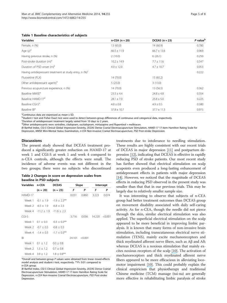
Table 1 Baseline characteristics of subjects
Variables n-CEA (n = 20) DCEAS (n = 23) P valueb
Female, n (%) 13 (65.0) 14 (60.9) 0.780
Age (y)a 66.5 ± 11.9 66.7 ± 13.6 0.969
Having previous stroke, n (%) 2 (10.0) 6 (26.1) 0.250
Post-stroke duration (m)a 10.2 ± 14.9 7.7 ± 11.6 0.547
Duration of PSD onset (m)a 4.9 ± 12.0 4.7 ± 10.7 0.953
Having antidepressant treatment at study entry, n (%)c 0.222
Fluoxetine (FLX) 14 (70.0) 15 (65.2)
Other antidepressant agentsd 5 (25.0) 3 (13.0)
Previous acupuncture experience, n (%) 14 (70.0) 13 (56.5) 0.362
Baseline MMSEa 23.5 ± 4.4 24.8 ± 4.8 0.354
Baseline HAMD-17a 28.1 ± 7.0 25.8 ± 5.0 0.225
Baseline CGI-Sa 4.8 ± 0.8 4.9 ± 0.5 0.580
Baseline BIa 37.8 ± 10.7 37.7 ± 11.3 0.975aContinuous data are expressed as mean ± SD.bStudent t test and Fisher Exact test were used to detect between-group differences of continuous and categorical data, respectively.cDuration of antidepressant treatment largely varied from 10 days to 2 years.dOther antidepressants were sertraline, citalopram, escitalopram, mirtazapine and flupentixol + melitracen.BI Barthel Index, CGI-S Clinical Global Impression-Severity, DCEAS Dense Cranial Electroacupuncture Stimulation, HAMD-17 17-item Hamilton Rating Scale forDepression, MMSE Mini-Mental Status Examination, n-CEA Non-invasive Cranial Electroacupuncture, PSD Post-stroke Depression.
Man et al. BMC Complementary and Alternative Medicine 2014, 14:255 Page 5 of 8http://www.biomedcentral.com/1472-6882/14/255
DiscussionsThe present study showed that DCEAS treatment pro-duced a significantly greater reduction on HAMD-17 atweek 1 and CGI-S at week 1 and week 4 compared ton-CEA controls, although the effects were small. Theincidence of adverse events was not different in thetwo groups; there were no subjects who discontinued
Table 2 Changes in score on depression scales frombaseline in PSD subjects
Variables n-CEA DCEAS Slope Intercept
(n = 20) (n = 23) F P F P
HAMD-17 0.031 0.860 3.223 0.074
Week 1 -0.1 ± 1.9 -1.9 ± 2.3**
Week 2 -6.5 ± 1.9 -6.8 ± 2.3
Week 4 -11.2 ± 1.9 -11.6 ± 2.3
CGI-S 3.716 0.056 14.220 <0.001
Week 1 0.1 ± 0.3 -0.3 ± 0.3**
Week 2 -0.7 ± 0.3 -0.8 ± 0.3
Week 4 -1.4 ± 0.3 -1.7 ± 0.3**
BI 24.101 <0.001
Week 1 0.1 ± 1.2 0.0 ± 0.8
Week 2 1.3 ± 1.2 0.7 ± 0.8
Week 4 3.9 ± 1.2 1.8 ± 0.8**aOverall and between-group P values were obtained from linear mixed-effectsmodel analysis and student t-test, respectively. **P< 0.01 compared ton-CEA group.BI Barthel Index, CGI-S Clinical Global Impression-Severity, DCEAS Dense CranialElectroacupuncture Stimulation, HAMD-17 17-item Hamilton Rating Scale forDepression, n-CEA Non-invasive Cranial Electroacupuncture, PSD Post-strokeDepression.
treatments due to intolerance to needling stimulation.These results are highly consistent with our recent trialsof DCEAS in major depression [11] and postpartum de-pression [12], indicating that DCEAS is effective in rapidlyreducing PSD of stroke patients. Our most recent studyhas further showed that electrical stimulation on scalpacupoints even produced a long-lasting enhancement ofantidepressant effects in patients with major depression[14]. However, we noticed that the magnitude of DCEASeffects in reducing PSD observed in the present study wassmaller than that that in our previous trials. This may belargely due to relatively smaller sample size.It was interesting to observe that subjects of n-CEA
group had better treatment outcomes than DCEAS groupon movement disability associated with daily self-caringactivity. As for n-CEA, though the needle did not piercethrough the skin, similar electrical stimulation was alsoapplied. The superficial electrical stimulation on the scalpappeared to be more beneficial in improving limbic par-alysis. It is known that many forms of non-invasive brainstimulation, including transcutaneous electrical nerve sti-mulation (TENS), mainly excite mechanoreceptors andthick myelinated afferent nerve fibers, such as Aβ and Aδ;whereas DCEAS is a noxious stimulation that mainly ex-cites noxious receptors of the scalp [10]. The activation ofmechanoreceptors and thick myelinated afferent nervefibers appeared to be more efficacious in alleviating loco-motor impairment [10]. This could probably explain theclinical empiricism that physiotherapy and traditionalChinese medicine (TCM) massage (tui-na) are generallymore effective in rehabilitating limbic paralysis of stroke
HA
MD
-17
-12
-10
-8
-6
-4
-2
0
2
n-CEA ( n = 20)DCEAS (n = 23)
CG
I-S
-2.0
-1.5
-1.0
-0.5
0.0
0.5
Weeks of treatment
0 1 2 3 4
Bar
thel
Ind
ex (
BI)
0
1
2
3
4
5
*
*
*
*
Figure 2 Changes in score from baseline on HAMD-17, CGI-S,and BI over time.
Table 3 Adverse events occurred in at least 5% ofsubjects in either groupa
Event n-CEA (n = 20) DCEAS (n = 23)
Fatigue 8 (40.0) 11 (47.8)
Needling-induced discomfort 6 (30.0) 7 (30.4)
Headache 6 (30.0) 4 (17.4)
Dizziness 1 (5.0) 4 (17.4)
Nausea 2 (10.0) 1 (4.3)
Vomiting 0 2 (8.7)
Depression deterioration 2 (10.0) 3 (13.0)
Movement disorder 3 (15.0) 5 (21.7)
Sweating 1 (5.0) 1 (4.3)
Palpitation 1 (5.0) 5 (21.7)
Insomnia 3 (15.0) 6 (26.1)
Hypersomnia 2 (10.0) 8 (34.8)aThere were no significant differences in any adverse event between the twogroups, as examined with Fisher Exact test.DCEAS dense cranial electroacupuncture stimulation, n-CEA non-invasivecranial electroacupuncture.
Man et al. BMC Complementary and Alternative Medicine 2014, 14:255 Page 6 of 8http://www.biomedcentral.com/1472-6882/14/255
patients. The paradoxical effects of DCEAS in improvingdepression and post-stroke disability also connoted thatthe greater improvement of DCEAS on PSD was unlikelydue to the improvement of movement ability. This is inline with a higher prevalence of depressive symptoms ofstroke patients than orthopaedic patients with similar levelof movement disability [17].There should be a specific neural mechanism respon-
sible for the effects of DCEAS in reducing PSD. Strokenot only results in the primary ischemic lesion, but alsointerrupts central neurotransmitter functions [27], in par-ticular the brainstem serotonin (5-HT) and noradrenaline(NA) neuronal systems, which play a pivotal role in modu-lating mood activity and processing acupuncture signals[10]. DCEAS was developed mainly based on a neurobio-logical rationale. It is well documented that the foreheadacupoints innervated by the trigeminal sensory pathwayhave intimate connections with the brainstem reticularformation, in particular the dorsal raphe nucleus (DRN)
and the locus coeruleus (LC) [10]. The latter two brainstructures are the major resources of 5-HT and NA neu-ronal bodies, respectively, sending diffuse projections tosubcortical and cortical areas, including the prefrontalcortex and the amygdala known to be heavily involved inthe pathogenesis of depression [10]. Neuroimaging studieshave shown that electroacupuncture stimulation is capableof directly modulating the activity of the emotion proces-sing-related brain regions [28]. Through the intimate col-lateral connection from the trigeminal sensory pathway tothe brainstem 5-HT and NA neuronal systems, the need-ling of the forehead acupoints with subsequent electricalstimulation could more efficiently elicit afferent acupunc-ture signals via biophysical and biochemical reactions atlocal acupoints and, in turn, modulates central 5-HT andNA neuronal functions [10]. Our recent study furthershowed that active acupuncture results in lateralisationof functional cerebral response to the contralateral un-affected hemisphere in patients with unilateral stroke, sug-gesting that acupuncture could enhance a compensatoryprocess by redistributing functions to the unaffectedhemisphere [29].There are several limitations in the present study.
First, electrical stimulation was directly delivered on thescalp in both n-CEA and DCEAS procedures. This mayelicit a transcutaneous and/or transcranial effect; how-ever, such effect should be minimal as the stimulationintensity used in both n-CEA and DCEAS was muchlower than other brain stimulations, such as repetitivetranscranial magnetic stimulation (rTMS), electrocon-vulsive therapy (ECT) and TENS [30,31]. Therefore, thegreater effects of DCEAS are most likely to be derivedfrom biophysical and biochemical actions of needling
Man et al. BMC Complementary and Alternative Medicine 2014, 14:255 Page 7 of 8http://www.biomedcentral.com/1472-6882/14/255
with subsequent electrical stimulation [10]. Second, si-milar to most previous studies [9], the determination ofbody acupoints used in the present study was basicallybased on empirical evidence. Empirical treatment regi-mens have resulted in a large variation in acupunctureprotocols and difficulties in comparing treatment out-comes among trials. Finally, although the present studydemonstrated the antidepressant efficacy of acupuncturetherapy, the underlying mechanisms are not yet well de-lineated. Previous studies have suggested that the anti-depressant effects of acupuncture may be associatedwith the restoration of decreased neuroimaging activityof brain regions involved in processing emotional signals[10]. The determination of neuroimaging correlates ofthe clinical improvement in depressed patients wouldhelp gain some insights into antidepressant mechanismsof acupuncture.
ConclusionsThese results indicate that DCEAS could be effective inreducing stroke patients’ depressive symptoms. Superficialelectrical stimulation in n-CEA group may be beneficial inimproving movement disability of stroke patients. A com-bination of DCEAS and body acupuncture can be consi-dered a treatment option for neuropsychiatric sequelae ofstroke.
Additional files
Additional file 1: CONSORT 2010 checklist of information to includewhen reporting a randomised trial.
Additional file 2: CONSORT 2010 Flow Diagram.
Abbreviations5-HT: Serotonin; BI: Barthel Index; CGI-S: Clinical Global Impression - Severityscale; CONSORT: Consolidated Standards of Reporting Trials; DCEAS: Densecranial electroacupuncture stimulation; DRN: Dorsal raphe nucleus;ECT: Electroconvulsive therapy; FLX: Fluoxetine; HAMD-17: 17-item HamiltonDepression Rating Scale; IRB: Institutional Review Board; LC: Locus coeruleus;MMSE: Mini-mental State Examination; NA: Noradrenaline; n-CEA: Non-invasive cranial electroacupuncture; OCD: Obsessive-compulsive disorder;PI: Principal Investigator; PSD: Post-stroke depression; rTMS: Repetitivetranscranial magnetic stimulation; SSRIs: Selective serotonin reuptakeinhibitors; TCM: Traditional Chinese medicine; TENS: Transcutaneous electricalnerve stimulation.
Competing interestsThe authors declare that they have no competing interests in the study.
Authors’ contributionsConceived and designed the experiments: ZJZ, HC, RN, LSWL, KPL, KPL, VTW,EZ. Performed the experiments: SCM, BHBH, MPMF, HC, RN, KPL, KWYT.Analyzed the data: BHBH, SCM, ZJZ, XCY. Wrote the paper: BHBH, SCM, ZJZ.All authors read and approved the final manuscript.
AcknowledgementsThis study was supported by fund from Division of Chinese Medicine ofHong Kong Hospital Authority (HA) and General Research Fund (GRF) ofHong Kong Research Grant Council (RGC) (786611).
Author details1The School of Chinese Medicine, The University of Hong Kong, 10 SassoonRoad, Pokfulam, Hong Kong, China. 2Department of Psychiatry, KowloonHospital, Hong Kong, China. 3Institute of Acupuncture, China Academy ofChinese Medical Sciences, Dongchen District, Beijing 100700, China.4Department of Rehabilitation, Kowloon Hospital, Hong Kong, China.5Rehabilitation Unit, Tung Wah Hospital, Hong Kong, China. 6Department ofMedicine and Rehabilitation, Tung Wah Eastern Hospital, Hong Kong, China.7Chinese Medicine Department, Hospital Authority, Hong Kong, China.
Received: 10 January 2014 Accepted: 14 July 2014Published: 19 July 2014
References1. Paolucci S: Epidemiology and treatment of post-stroke depression.
Neuropsychiatr Dis Treat 2008, 4(1):145–154.2. Gustafson Y, Nilsson I, Mattsson M, Astrom M, Bucht G: Epidemiology and
treatment of post-stroke depression. Drugs Aging 1995, 7(4):298–309.3. Williams LS, Ghose SS, Swindle RW: Depression and other mental health
diagnoses increase mortality risk after ischemic stroke. Am J Psychiatry2004, 161(6):1090–1095.
4. Bhogal SK, Teasell R, Foley N, Speechley M: Heterocyclics and selectiveserotonin reuptake inhibitors in the treatment and prevention ofpoststroke depression. J Am Geriatr Soc 2005, 53(6):1051–1057.
5. Starkstein SE, Mizrahi R, Power BD: Antidepressant therapy in post-strokedepression. Expert Opin Pharmacother 2008, 9(8):1291–1298.
6. Hemeryck A, Belpaire FM: Selective serotonin reuptake inhibitors andcytochrome P-450 mediated drug-drug interactions: an update.Curr Drug Metab 2002, 3(1):13–37.
7. Zhao ZQ: Neural mechanism underlying acupuncture analgesia.Prog Neurobiol 2008, 85(4):355–375.
8. Sze FK, Wong E, Or KK, Lau J, Woo J: Does acupuncture improve motorrecovery after stroke? A meta-analysis of randomized controlled trials.Stroke 2002, 33(11):2604–2619.
9. Zhang ZJ, Chen HY, Yip KC, Ng R, Wong VT: The effectiveness and safetyof acupuncture therapy in depressive disorders: systematic review andmeta-analysis. J Affect Disord 2010, 124(1–2):9–21.
10. Zhang ZJ, Wang XM, McAlonan GM: Neural acupuncture unit: a newconcept for interpreting effects and mechanisms of acupuncture.Evid Based Complement Alternat Med 2012, 2012:429412.
11. Zhang ZJ, Ng R, Man SC, Li TY, Wong W, Tan QR, Wong HK, Chung KF,Wong MT, Tsang WK, Yip KC, Ziea E, Wong VT: Dense cranialelectroacupuncture stimulation for major depressive disorder–a single-blind, randomized, controlled study. PLoS One 2012, 7(1):e29651.
12. Chung KF, Yeung WF, Zhang ZJ, Yung KP, Man SC, Lee CP, Lam SK,Leung TW, Leung KY, Ziea ET, Wong VT: Randomized non-invasivesham-controlled pilot trial of electroacupuncture for postpartumdepression. J Affect Disord 2012, 142(1–3):115–121.
13. Zhang ZJ, Wang XY, Tan QR, Jin GX, Yao SM: Electroacupuncture forrefractory obsessive-compulsive disorder: a pilot waitlist-controlled trial.J Nerv Ment Dis 2009, 197(8):619–622.
14. Qu SS, Huang Y, Zhang ZJ, Chen JQ, Lin RY, Wang CQ, Li GL, Wong HK,Zhao CH, Pan JY, Guo SC, Zhang YC: A 6-week randomized controlled trialwith 4-week follow-up of acupuncture combined with paroxetine inpatients with major depressive disorder. J Psychiatr Res 2013, 47(6):726–732.
15. Schulz KF, Altman DG, Moher D, Group C: CONSORT 2010 statement:updated guidelines for reporting parallel group randomised trials.PLoS Med 2010, 7(3):e1000251.
16. Hamilton M: A rating scale for depression. J Neurol Neurosurg Psychiatry1960, 23:56–62.
17. Folstein MF, Folstein SE, McHugh PR: “Mini-mental state”. A practicalmethod for grading the cognitive state of patients for the clinician.J Psychiatr Res 1975, 12(3):189–198.
18. Stark P, Hardison CD: A review of multicenter controlled studies offluoxetine vs. imipramine and placebo in outpatients with majordepressive disorder. J Clin Psychiatry 1985, 46(3 Pt 2):53–58.
19. Chollet F, Tardy J, Albucher JF, Thalamas C, Berard E, Lamy C, Bejot Y,Deltour S, Jaillard A, Niclot P, Guillon B, Moulin T, Marque P, Pariente J,Arnaud C, Loubinoux I: Fluoxetine for motor recovery after acuteischaemic stroke (FLAME): a randomised placebo-controlled trial. LancetNeurol 2011, 10(2):123–130.
Man et al. BMC Complementary and Alternative Medicine 2014, 14:255 Page 8 of 8http://www.biomedcentral.com/1472-6882/14/255
20. Han JS: Acupuncture: neuropeptide release produced by electricalstimulation of different frequencies. Trends Neurosci 2003, 26(1):17–22.
21. Ulett GA, Han S, Han JS: Electroacupuncture: mechanisms and clinicalapplication. Biol Psychiatry 1998, 44(2):129–138.
22. Streitberger K, Kleinhenz J: Introducing a placebo needle intoacupuncture research. Lancet 1998, 352(9125):364–365.
23. Enck P, Klosterhalfen S, Zipfel S: Acupuncture, psyche and the placeboresponse. Auton Neurosci 2010, 157(1–2):68–73.
24. Fink M, Gutenbrunner C, Rollnik J, Karst M: Credibility of a newly designedplacebo needle for clinical trials in acupuncture research. ForschKomplementarmed Klass Naturheilkd 2001, 8(6):368–372.
25. Mahoney FI, Barthel DW: Functional evaluation: the Barthel index.Md State Med J 1965, 14:61–65.
26. Guy W: ECDEU Assessment Manual for Psychopharmacology. Revised.Bethesda, MD: US Department of Health, Education, and Welfare; 1976.
27. Loubinoux I, Kronenberg G, Endres M, Schumann-Bard P, Freret T,Filipkowski RK, Kaczmarek L, Popa-Wagner A: Post-stroke depression:mechanisms, translation and therapy. J Cell Mol Med 2012, 16(9):1961–1969.
28. Dhond RP, Kettner N, Napadow V: Neuroimaging acupuncture effects inthe human brain. J Altern Complement Med 2007, 13(6):603–616.
29. Huang Y, Chen JQ, Lai XS, Tang CZ, Yang JJ, Chen H, Wu JX, Xiao HL, Qu SS,Zhang YD, Zhang ZJ: Lateralisation of cerebral response to activeacupuncture in patients with unilateral ischaemic stroke: an fMRI study.Acupunct Med 2013, 31(3):290–296.
30. Kirkcaldie M, Pridmore S, Reid P: Bridging the skull: electroconvulsivetherapy (ECT) and repetitive transcranial magnetic stimulation (rTMS) inpsychiatry. Convuls Ther 1997, 13(2):83–91.
31. van Dijk KR, Scherder EJ, Scheltens P, Sergeant JA: Effects oftranscutaneous electrical nerve stimulation (TENS) on non-pain relatedcognitive and behavioural functioning. Rev Neurosci 2002, 13(3):257–270.
doi:10.1186/1472-6882-14-255Cite this article as: Man et al.: A pilot controlled trial of a combinationof dense cranial electroacupuncture stimulation and body acupuncturefor post-stroke depression. BMC Complementary and Alternative Medicine2014 14:255.
Submit your next manuscript to BioMed Centraland take full advantage of:
• Convenient online submission
• Thorough peer review
• No space constraints or color figure charges
• Immediate publication on acceptance
• Inclusion in PubMed, CAS, Scopus and Google Scholar
• Research which is freely available for redistribution
Submit your manuscript at www.biomedcentral.com/submit
Top Related
Copyright © 2022 FDOKUMEN

