Zoomed Functional Imaging in the Human Brain at 7 Tesla with Simultaneous High Spatial and High...
15
Zoomed Functional Imaging in the Human Brain at 7 Tesla with Simultaneous High Spatial and High Temporal Resolution Josef Pfeuffer, 1,2 Pierre-Francois van de Moortele, Essa Yacoub, Amir Shmuel, Gregor Adriany, Peter Andersen, Hellmut Merkle, Michael Garwood, Kamil Ugurbil, and Xiaoping Hu Center for Magnetic Resonance Research, University of Minnesota Medical School, 2021 6th Street S.E., Minneapolis, Minnesota 55455 Received August 15, 2001 Functional neuroimaging in the human brain using noninvasive magnetic resonance methods has the po- tential of providing highly resolved maps of neuronal activation. Decreasing the voxel size and obtaining simultaneously high temporal resolution is a major challenge and is mainly limited by sensitivity. Here, signal-to-noise gains at high magnetic fields (7 Tesla) and an optimized surface coil setup are combined with a novel approach for zoomed functional imaging in the visual cortex. For echoplanar imaging, the acquisition time and segmentation was shortened fourfold by us- ing a reduced field-of-view. An adiabatic outer-volume suppression method, BISTRO, was used to obliterate signal outside the area-of-interest achieving effective suppression even for inhomogeneous B 1 -fields. A sin- gle-shot acquisition was performed at submillimeter resolution in the human brain, while simultaneously maintaining a high temporal resolution of 125 ms. Functional studies with and without field-of-view re- duction were performed. Activation and percent change maps were compared with respect to spatial extent, t values and percentage changes of the BOLD contrast. The detection of functional activation was found to be equal within the inter-series variability for the two acquisition schemes. Thus, single-trial BOLD responses were detected for the first time robustly at a 500 500 m 2 in plane and 250 ms temporal resolution, significantly expanding the possibilities of event-re- lated functional imaging in the human brain. The mag- netization transfer effect induced by the outer-volume suppression pulses was investigated and found to be in- creased during neuronal activity. © 2002 Elsevier Science (USA) INTRODUCTION In the last decade, neuroimaging methods based on magnetic resonance (fMRI) have evolved rapidly to become a frequently used approach in neuroscience. However, the utility of this method can still be signif- icantly enhanced with increased spatial and temporal resolution. The potential and significance of high res- olution fMRI has been recently demonstrated in ani- mal models (e. g., Kim et al., 2000; Duong et al., 2001; Logothetis et al., 2001; Logothetis et al., 2002) and in human studies (e.g., Cheng et al., 2001; Goodyear et al., 2001; Kim et al., 2001). Ideally, it is desirable to obtain such high resolution images also rapidly and with high time resolution. While fMRI methods do not access evolution of neuronal activity in the neuronal time scale, temporal resolution in the 0.3 to 1 s domain is still of importance. Such a capability can be used, for example, for investigating mechanisms operative in the metabolic and hemodynamic response to elevated neuronal activity (Lee et al., 1995; Hu et al., 1997; Aguirre et al., 1998; Duong et al., 2000), to utilize a particular time-segment of the hemodynamic response for increased specificity (Kim et al., 2000; Goodyear et al., 2001; Duong et al., 2001), for executing “true” event-related study in multislice where the response to each execution of the task generates a fMRI time series which is then correlated with variations in the perfor- mance (Richter et al., 1997a,b, 2000) or even conven- tional event-related studies in multislice (e.g., Rosen et al., 1998). Our goal in this work was to achieve submillimeter voxel resolution simultaneously with rapid image ac- quisition in human brain fMRI. FMRI studies predom- inantly rely on ultrafast image acquisition schemes such as EPI. In such schemes, for a given spatial res- olution, the acquisition time is proportional to the field- of-view (FOV) in the phase encode direction and thus, to the number of k space lines. For high spatial reso- lution, the acquisition is often segmented using several excitations to cover the total k space (e.g., Lee et al., 1995; Kim et al., 1996; Thulborn et al., 1997; Hoogen- raad et al., 1999; Menon et al., 1999; Chen et al., 1999). At a given minimum repetition time between applica- tions of excitation RF pulses, the time for acquiring a 1 To whom correspondence should be addressed. E-mail: josef. [email protected]. 2 Present address: Max-Planck Institute of Biological Cybernetics, Spemannstrasse 38, 72076 Tuebingen, Germany. NeuroImage 17, 272–286 (2002) doi:10.1006/nimg.2002.1103 272 1053-8119/02 $35.00 © 2002 Elsevier Science (USA) All rights reserved.
Transcript of Zoomed Functional Imaging in the Human Brain at 7 Tesla with Simultaneous High Spatial and High...

NeuroImage 17, 272–286 (2002)doi:10.1006/nimg.2002.1103
Zoomed Functional Imaging in the Human Brain at 7 Tesla withSimultaneous High Spatial and High Temporal Resolution
Josef Pfeuffer,1,2 Pierre-Francois van de Moortele, Essa Yacoub, Amir Shmuel, Gregor Adriany,Peter Andersen, Hellmut Merkle, Michael Garwood, Kamil Ugurbil, and Xiaoping Hu
Center for Magnetic Resonance Research, University of Minnesota Medical School, 2021 6th Street S.E., Minneapolis, Minnesota 55455
Received August 15, 2001
Functional neuroimaging in the human brain usingnoninvasive magnetic resonance methods has the po-tential of providing highly resolved maps of neuronalactivation. Decreasing the voxel size and obtainingsimultaneously high temporal resolution is a majorchallenge and is mainly limited by sensitivity. Here,signal-to-noise gains at high magnetic fields (7 Tesla)and an optimized surface coil setup are combined witha novel approach for zoomed functional imaging in thevisual cortex. For echoplanar imaging, the acquisitiontime and segmentation was shortened fourfold by us-ing a reduced field-of-view. An adiabatic outer-volumesuppression method, BISTRO, was used to obliteratesignal outside the area-of-interest achieving effectivesuppression even for inhomogeneous B1-fields. A sin-gle-shot acquisition was performed at submillimeterresolution in the human brain, while simultaneouslymaintaining a high temporal resolution of 125 ms.Functional studies with and without field-of-view re-duction were performed. Activation and percentchange maps were compared with respect to spatialextent, t values and percentage changes of the BOLDcontrast. The detection of functional activation wasfound to be equal within the inter-series variability forthe two acquisition schemes. Thus, single-trial BOLDresponses were detected for the first time robustly at a500 � 500 �m2 in plane and 250 ms temporal resolution,significantly expanding the possibilities of event-re-lated functional imaging in the human brain. The mag-netization transfer effect induced by the outer-volumesuppression pulses was investigated and found to be in-creased during neuronal activity. © 2002 Elsevier Science (USA)
INTRODUCTION
In the last decade, neuroimaging methods based onmagnetic resonance (fMRI) have evolved rapidly to
1 To whom correspondence should be addressed. E-mail: [email protected].
2 Present address: Max-Planck Institute of Biological Cybernetics,Spemannstrasse 38, 72076 Tuebingen, Germany.
2721053-8119/02 $35.00© 2002 Elsevier Science (USA)All rights reserved.
become a frequently used approach in neuroscience.However, the utility of this method can still be signif-icantly enhanced with increased spatial and temporalresolution. The potential and significance of high res-olution fMRI has been recently demonstrated in ani-mal models (e. g., Kim et al., 2000; Duong et al., 2001;Logothetis et al., 2001; Logothetis et al., 2002) and inhuman studies (e.g., Cheng et al., 2001; Goodyear et al.,2001; Kim et al., 2001). Ideally, it is desirable to obtainsuch high resolution images also rapidly and with hightime resolution. While fMRI methods do not accessevolution of neuronal activity in the neuronal timescale, temporal resolution in the 0.3 to 1 s domain isstill of importance. Such a capability can be used, forexample, for investigating mechanisms operative inthe metabolic and hemodynamic response to elevatedneuronal activity (Lee et al., 1995; Hu et al., 1997;Aguirre et al., 1998; Duong et al., 2000), to utilize aparticular time-segment of the hemodynamic responsefor increased specificity (Kim et al., 2000; Goodyear etal., 2001; Duong et al., 2001), for executing “true”event-related study in multislice where the response toeach execution of the task generates a fMRI time serieswhich is then correlated with variations in the perfor-mance (Richter et al., 1997a,b, 2000) or even conven-tional event-related studies in multislice (e.g., Rosen etal., 1998).
Our goal in this work was to achieve submillimetervoxel resolution simultaneously with rapid image ac-quisition in human brain fMRI. FMRI studies predom-inantly rely on ultrafast image acquisition schemessuch as EPI. In such schemes, for a given spatial res-olution, the acquisition time is proportional to the field-of-view (FOV) in the phase encode direction and thus,to the number of k space lines. For high spatial reso-lution, the acquisition is often segmented using severalexcitations to cover the total k space (e.g., Lee et al.,1995; Kim et al., 1996; Thulborn et al., 1997; Hoogen-raad et al., 1999; Menon et al., 1999; Chen et al., 1999).At a given minimum repetition time between applica-tions of excitation RF pulses, the time for acquiring a
single image is then directly related to the number ofsegments, leading to lower temporal resolution. Toachieve simultaneously high spatial and rapid imageacquisition, the FOV can be reduced, which truncatesthe number of readout lines in the phase encode direc-tion. However, at a reduced FOV, signal outside canfold back into the image in the phase encode directionand has to be either suppressed or not excited, e.g., byusing volume selective excitation.
We present a method for zoomed imaging with areduced FOV in combination with an outer-volumesuppression method. To attain optimum signal-to-noise ratios (SNR), a surface coil was used. Therefore,a special outer-volume suppression technique was nec-essary to achieve B1 insensitive suppression in theinhomogeneous RF field of the surface coil. The use ofa high magnetic field (7 Tesla) increased SNR(Vaughan et al., 2001) and contrast-to-noise ratio(CNR) (Yacoub et al., 2001b). The combination of thesefactors together made it feasible to perform fMRI stud-ies in the human brain at submillimeter resolution andwith rapid single-shot acquisition. Single-trial BOLDresponses were detected at a 0.5 � 0.5 � 3 mm3 spatialand 250 ms temporal resolution, significantly broaden-ing the possibilities of event-related fMRI in the hu-man brain. Preliminary results of this work were pub-lished in abstract form (Pfeuffer et al., 2001a,b).
MATERIALS AND METHODS
Instrumentation and Data Acquisition
MR imaging of the human visual cortex was per-formed on a 7 Tesla/90 cm horizontal bore magnet(Magnex Scientific, Abingdon, UK) interfaced to a Var-ian INOVA console (Varian Inc, Palo Alto, CA). Thesystem is equipped with a Magnex head gradient set,which is torque-balanced, self-shielded, water-cooled(38 cm ID) driven by a Siemens Harmony/Symphony800V/300A gradient amplifier (Siemens, Erlangen,Germany) and capable of generating a maximum gra-dient strength of 40 mT/m with a rise rate of 200mT/m/ms. For increased signal-to-noise ratio in theoccipital cortex, curved quadrature surface coils withtwo partially overlapping 7- or 14-cm diameter loopswere used for radiofrequency transmission and recep-tion at 300 MHz (Adriany et al., 2001). Localized shim-ming was done using FAST(EST)MAP (Gruetter et al.,2000), typically providing a linewidth of 15 Hz in a6-cm sphere, which covered most of the visual cortex.
A blipped gradient-echo echoplanar EPI sequence(EPI) was used for image acquisition. The sequencewas implemented with a readout gradient waveformconsisting of alternating trapezoidal lobes. Nonlinearsampling in time was used on the ramps of each gra-dient, providing equidistant sampling in the k spacealong the readout direction. To minimize the Nyquist
ghost, a reference scan was collected and used to cor-rect for discrepancy between the odd and even echoes.A reconstruction method that utilized the referencescans (Bruder et al., 1992) also reduced to a largeextent the distortions caused by off-resonance, provid-ing less distorted images and thereby a better registra-tion between the EPI data and the anatomical scans.
Sagittal EPI images were acquired at four in-planeresolutions, 3.1 � 3.1 mm2, 1.5 � 1.5 mm2, 0.9 � 0.9mm2, and 0.5 � 0.5 mm2. Slice thickness was 3 mm.The corresponding field FOV in the phase-encode di-rection of EPI was 20 cm (64 lines), 4.8 cm (32 lines),2.88 cm (32 lines), and 3.2 cm (64 lines, two segments)with a readout duration per line of 0.42 ms, 0.67 ms,1.02 ms, and 1.68 ms, respectively. Image repetitiontime was 0.125 and 0.5 s, while the Ernst flip angle wasused for excitation. According to a previous high-reso-lution fMRI study (Yacoub et al., 2001b), T*2 at 7 Teslawas (25.1 � 3.5) ms for gray matter. Therefore, an echotime of 20 to 30 ms, close to the T*2 of gray matter, waschosen to provide optimum T*2-BOLD contrast at 7 Tesla.
Physiological data were acquired using the softwareAcqKnowledge (BIOPAC Systems, Inc, CA). The respi-ratory signal was recorded using a pneumatic beltplaced around the subject’s upper abdomen. The car-diac signal was gauged with a pulse oximeter placed onthe subject’s finger, which provides a delayed systolicsignal.
Subjects and Paradigm
A total of 14 normal subjects were enrolled in thisstudy approved by the institutional review board at theUniversity of Minnesota. All subjects provided writtenconsent.
Visual stimuli were presented via a rear projectionsystem consisting of a high luminance video projectorand an optical setup with mirrors and specialty lenses.Moving sinusoidal gratings with 100% contrast werepresented as the visual stimulus. Subjects were askedto fixate during the entire scan on a fixation point atthe center of the screen. For functional activation, aparadigm with blocks of 20-s stimulus-on and 24-sstimulus-off (six epochs, 5 min per series) was used.Also, an event-related paradigm with 2-s stimulationand 12-s intertrial interval was used (16 epochs, 5 minper series).
Fast Imaging with Adiabatic Field-of-View Reduction(BISTRO)
To minimize segmentation and achieve high tempo-ral resolution, the FOV was reduced by the use of asmall surface coil, decreasing the FOV from over 20 cmto about 12 cm, and furthermore by the use of outer-volume suppression, which is common in single-voxelMR spectroscopy. Because of the inhomogeneous B1
fields of surface coils, a B1-insensitive technique, BIS-
273ZOOMED FUNCTIONAL IMAGING AT HIGH RESOLUTION

TRO (‘B1-InSensitive TRain to Obliterate signal’), wasused (Luo et al., 2001).
As seen in Fig. 1, a sagittal image exhibited most ofthe visual cortex demonstrating high sensitivity in theposterior part of the brain with fast decreasing inten-sity in the anterior direction. The target area-of-inter-est, 2.9-cm wide, is indicated in Fig. 1a. With the BIS-TRO technique, outer-volume signal is suppressed onboth anterior and posterior sides (Fig. 1c), whereby thesmall amount of signal remaining arose from theNyquist ghost. Power adjustment and parameter opti-mization of BISTRO was performed using a projectionprofile of the imaged slice in the phase-encode direc-tion, i.e., anterior-posterior. As demonstrated in Fig.1d, signal suppression outside the area-of-interest im-proved with increasing RF power. In addition, even atlower RF power, a sharp edge is discernible.
BISTRO is an outer-volume suppression methodthat uses frequency-swept RF pulses to achieve insen-sitivity to RF field inhomogeneities. Briefly, a train ofRF pulsed with variable power and dephasing gradi-
ents are applied to suppress signal in a defined slab.BISTRO is different from commonly used outer-volumesuppression methods (see Luo et al., 2001, and refer-ences therein), since it is designed to work in inhomo-geneous B1 fields of surface coils where methods failthat rely on the ability to adjust a 90° flip angle over alarge volume.
For rapid imaging, two variants of the BISTRO tech-nique were implemented and inserted directly beforethe excitation pulse of the EPI sequence. (1) BIS-TRO-16 uses a train of 16 RF pulses at multiple powerlevels with 300 ms duration for two outer-volume slabs.It is designed to work with a single-shot applicationand can be used for imaging with long repetition times(�0.4 s). (2) BISTRO-1 uses a single RF pulse for eachouter-volume slab (24-ms duration for two slabs). Withrepetition times below 200 ms, BISTRO completelysuppresses the signal once a steady state has beenreached following a few “dummy scans.” We typicallyused repetition times of 125 ms. BISTRO-1 was foundespecially useful when high temporal resolution was
FIG. 1. Full field-of-view sagittal images of the human visual cortex showing areas where outer-volume-suppression is applied. (a)Anatomical image (TurboFLASH). (b) Gradient echo EPI with 3.1 � 3.1 mm2 in-plane resolution. (c) Same as in (b) with two BISTRO-16pulses applied, which suppressed signal outside the area-of-interest with a 2.9-cm width. (d) Suppression efficiency demonstrated with aprojection profile (anterior-posterior) with two BISTRO-16 pulses applied at different RF power settings (1-cm and 4-cm outer-volume slab,see black rectangles). The outer-volume suppression worked nicely despite the inhomogeneous B1-distribution (apparent by the signal drop)of the surface coil. Even at low RF power, a sharp edge at the most negative off-center position is discernible.
274 PFEUFFER ET AL.
required or multi-slice experiments were performed.The choice of parameters depended on the B1-charac-teristic of the RF coil, the imaging sequence used andthe position of the slabs to suppress.
To accomplish single-shot acquisition at 0.9 � 0.9mm2 in-plane resolution with gradient-echo EPI, theFOV was reduced in the phase-encode direction to 2.9cm, which covered most of the functionally activatedareas in the early visual cortex. Similarly, for 1.5 � 1.5mm2 and 0.5 � 0.5 mm2 in-plane resolution, the FOVwas reduced to 4.8 and 3.2 cm, respectively.
RF power deposition was calculated online in the pulsesequence before each experiment not to exceed SAR re-quirements of FDA regulations. In addition, a built-inhardware safety monitor in the console ensured thatSAR requirements were not exceeded. For both settings,BISTRO-16 with a 0.5 s TR and BISTRO-1 with a 0.125s TR, the average power deposition was below 1 W.
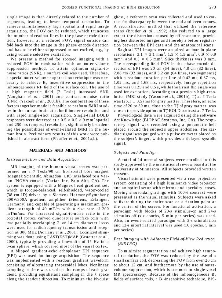
For the suppression of the signal in the outer-vol-ume, it is desirable to use RF pulses that have sharpprofiles to ensure a sharp fall-off of the signal at theedge of the targeted FOV. To shorten the pulse dura-tion, an asymmetric pulse with a sharp profile only onone side is advantageous, because for one outer-volumesuppression slab a sharp transition is only needed atthe edge neighboring the targeted inner-volume. Theasymmetric, adiabatic full-passage “skewed” RF pulse[HS1
2, R � 0.9, 0.9 Tp; tanh/tan, R � 100, 0.1 Tp](Hwang et al., 1999) featured offset-independent adia-baticity on one side (Fig. 2a). A Bloch equation simu-lation showed a sharp edge on one side even at small B1
(Fig. 2b). The sharp edge was preserved with increas-ing B1, whereas the magnetization at the other sidereveals a much gradual transition. Compared to thehyperbolic secant pulse with equal duration, the“skewed” pulse provided a 2.5 times narrower transi-tion band (Fig. 2b).
Data Analysis
After correcting for respiration-induced dynamic off-resonance using DORK (Pfeuffer et al., 2002b) and aphase correction using the reference scan, a fast Fou-rier transformation was applied to reconstruct the im-ages. The image series were analyzed with STIMU-LATE (Strupp, 1996) and with routines written in PV-Wave (Visual Numerics, Inc., Boulder, CO) andMATLAB (Mathworks Inc., Natick, MA). The time se-ries were screened for motion of the volunteer using“center of mass” detection.
Statistical maps were generated from the fMRI timeseries comparing image intensities during the taskversus the resting condition, which resulted in themean and standard deviation of both levels, the per-cent change, the t value and the confidence level, andthresholded using a Student’s t test. Before testing, theimage series were averaged over the six epochs. Also,
cross correlation maps and onset time maps were gen-erated using a boxcar reference function convolutedwith a standard hemodynamic response model. For testsof statistical significance, one and two-tailed Student’s ttests were used (Microcal Origin, Northampton, MA).
RESULTS
Zoomed Functional Imaging Using FOV Reduction
High-resolution rapid imaging of the human visualcortex with non-segmented acquisition is demon-
FIG. 2. Asymmetric “skewed” RF pulse with offset-independentadiabaticity used for outer-volume suppression (adiabatic full-pas-sage [HS1
2, R � 0.9, 0.9 Tp; tanh/tan, R � 100, 0.1 Tp]) (Hwang et al.,1999; Luo et al., 2001). (a) Amplitude, phase, and real part. (b)Designed with one steep edge at one side (offset-independent adia-baticity) and smoother tails at the other side. Compared to thecommonly used hyperbolic secant pulse HS8 (Silver et al., 1984), seedotted line in Fig. 2b, the skewed pulse has a 2.5 times narrowertransition band on one side. Skewed is especially useful for outer-volume suppression, where the sharp edge of the frequency profile isused for a sharp transition at the edge of the reduced FOV and theouter-volume. At different B1 always a sharp edge is discernible(arrow indicates increasing B1). The MT effects for the skewed pulseand a comparable nonskewed pulse are similar.
275ZOOMED FUNCTIONAL IMAGING AT HIGH RESOLUTION

strated in Fig. 3 using the FOV reduction technique.Single-shot EPI images, acquired with BISTRO-16,were compared to images acquired with a full FOV of20 cm with the same spatial resolution (1.5 � 1.5 � 3mm3 and 128 � 128 matrix size). The full FOV imagesrequired four segments to attain this in plane spatialresolution. With BISTRO-16, the same resolution wasachieved in a single-shot, using a reduced FOV of 4.8cm in the phase encoding direction and a 32 � 128matrix (Fig. 3a). Similarly, a 0.9 � 0.9 � 3-mm3 voxelsize was achieved with no segmentation using BIS-TRO-16 with a FOV of 2.9 cm and matrix size of 32 �256 (Fig. 3a). At this spatial resolution, eight segments
with a 256 � 256-matrix size were needed for a fullFOV image, corresponding to an eight-fold decrease inimage acquisition time.
In the reduced FOV (single-shot) images, a decreasein signal intensity and change in contrast was discern-ible relative to the full FOV images. This was due to amagnetization transfer (MT) effect induced by the RFpulses used for outer-volume suppression. MT is neg-ligible for CSF and blood, and was about 20 to 50% forgray/white matter tissues. Therefore, voxels contain-ing CSF appeared enhanced in Fig. 3.
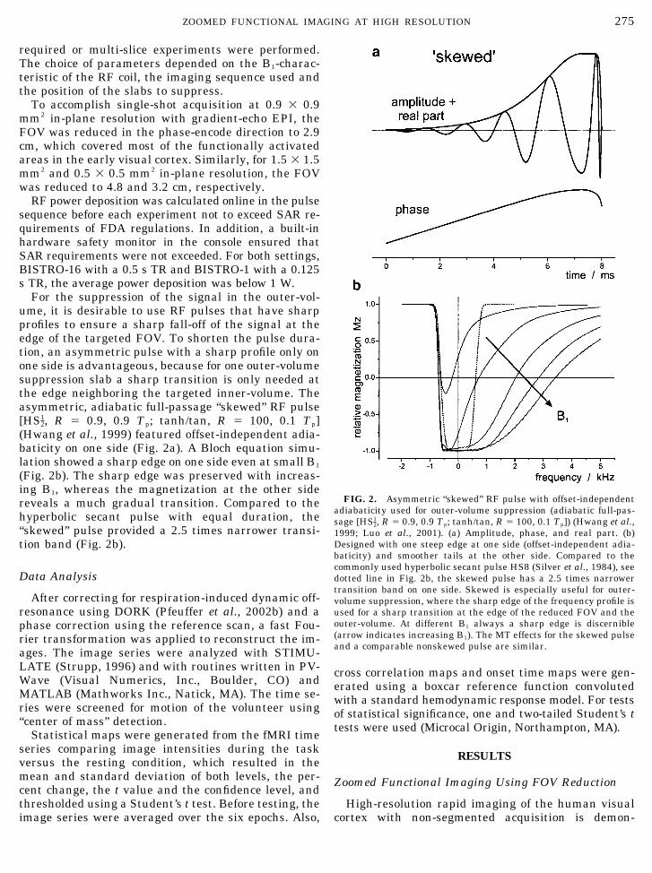
The effect of the BISTRO outer-volume suppressionmethod on the detection of activated voxels was sys-tematically examined. High-resolution EPI imageswere acquired with 0.9 � 0.9 � 3 mm2 voxel size usingthree types of imaging setups: (A) a full FOV (foursegments), which was the control, (B) a full FOV (foursegments) and BISTRO outer-volume suppression ap-plied, and (C) a reduced FOV of 2.9 cm (single-shot)and BISTRO outer-volume suppression applied. Func-tional experiments were performed with a block design(20 s task, 24 s rest). The t value maps of the differenttype of experiments (A, B, C) are shown in Figs. 4a–4cwith the corresponding percent change maps in Figs.4d–4f. Significantly activated voxels were those withP � 0.05. Upon visual inspection, the results of exper-iments A, B, C were found to be very similar in termsof the t values, percentage signal changes, and thespatial distribution of the functional activation (Fig. 4).The time courses averaged across the six epochs andover all voxels with P � 0.05 in Figs. 4g–4i demon-strate that the signal changes and contrast-to-noiseratio between the different experiments were compa-rable. Thus, the zoomed imaging approach providedthe four-fold increase in time resolution with single-shot acquisition without compromising detection of ac-tivation.
The results of statistical analysis of 12 series areshown in Table 1. The t values and percent signalchanges maps were calculated for experiments A, B,and C. First, the median of the 300 highest activatedvoxels was compared to assess the most significantvoxels. Second, all time courses for voxels with P �0.05 were averaged and a subsequent t test on theaveraged time course was performed to assess the per-formance on a more global level taking into accountlower and higher activated voxels. Statistical signifi-cance was established for a comparison of (B) versus(A) and (C) versus (A) on an intersubject comparisonusing a t test analysis between the groups. As indicatedby the intersubject mean with standard error (Table 1,last line), in most cases no significant difference wasfound. Using the first analysis (median), t values andpercentage changes tended to be lower when usingBISTRO (experiment C). Using the second analysis(averaged time series), t values and percent changestended to be higher. Also inspection of the single stud-
FIG. 3. Single-shot echoplanar imaging acquired with a reducedFOV. (a and c) Sagittal images of the human visual cortex whereouter-volume suppression (BISTRO) has been applied outside of theselected 2.9-cm slab. Using this technique, single-shot echoplanarimaging is possible with a voxel size of 1.5 � 1.5 � 3 mm3 (a) and0.9 � 0.9 � 3 mm3 (c). To achieve the same spatial resolution with afull FOV, four- and eight-segmented acquisitions (b and d) have to beused, which considerably decreases the temporal resolution by afactor of four and eight. Due to magnetization transfer during theBISTRO, signal intensity is reduced for gray and white matter, butnegligible for CSF. Hence, in the images of a and c, CSF appears muchbrighter than other tissues. For comparison, see the correspondinganatomical image in Fig. 1a. Parameters: High-resolution EPI im-ages at 7 Tesla with an in-plane resolution of 1.5 � 1.5 mm2 (a, b) and0.9 � 0.9 mm2 (c, d) at 20 ms echo time, 0.5-s repetition time persegment, and matrix sizes of 32 � 128/128 � 128 and 32 � 256/256 �256. Outer-volume suppression in a and c was achieved with a300-ms BISTRO-16 module, applied before the imaging sequence.
276 PFEUFFER ET AL.
ies in Table 1 reveals that the variance of the controlstudies is larger than the effect of using BISTRO.These data confirmed that using a single-shot ap-
proach with BISTRO for outer-volume suppression didnot change the contrast-to-noise ratio of the functionalactivation and did not alter the percent changes. This
FIG. 4. fMRI images of the human visual cortex comparing high-resolution activation maps of three experiments: Type-A, full FOVacquisition (four segments, control); type-B, full FOV acquisition with BISTRO outer volume suppression (four segments); and type-C,single-shot acquisition with a reduced FOV in the phase encode direction, i. e., anterior–posterior (one segment). The top row showsfunctional t value maps of activated pixels with EPI images, calculated with a Student’s t test (t � 2 (threshold), 2–10 (red-yellow)). Themiddle row shows relative signal changes (�S) in percent of the BOLD signal at a significance level of P � 0.05 (�S � 3–10% (red-yellow)).In the bottom row, the corresponding time courses are plotted using the average of all voxels with t � 2. Overall, t value maps and percentchange maps are very similar for (type A), (type B), and (type C). With zoomed imaging using a reduced FOV (type C), a fourfold increasetemporal resolution was achieved. Despite signal losses due to magnetization transfer in (type B) and (type C), outer-volume suppression didnot alter the statistical significance and percent changes of the BOLD signal (see also Table 1 for data from all studies). Parameters:High-resolution EPI images at 7 Tesla with an in-plane resolution of 0.9 � 0.9 mm2 (22 ms echo time, 0.5-s repetition time per segment, and matrixsizes of 128 � 128 and 32 � 128). Outer-volume suppression for type B and C experiments was achieved with a 300-ms BISTRO-16 module.
277ZOOMED FUNCTIONAL IMAGING AT HIGH RESOLUTION

TABLE 2
Statistical Data of the Magnetization Transfer Ratio (MTR) Comparing a Type-A andType-C Series of Functional T*2 BOLD Studies Corresponding to Fig. 4
Study No.
Intercept (%) Slope (�10�2 )
Nonactive Active � Nonactive Active �
1-1 0.3 2.5 �2.2 �1.4 �5.0 �3.61-2 0.6 3.7 �3.1 �2.7 �6.3 �3.62-1 0.4 2.9 �2.5 �1.1 �4.7 �3.62-2 0.3 2.8 �2.5 �0.3 �4.2 �3.93-1 0.4 3.3 �2.9 �1.3 �6.1 �4.83-2 0.4 2.4 �2.0 �2.0 �5.7 �3.73-3 0.5 2.7 �2.2 �3.2 �5.5 �2.34-1 �0.7 2.7 �3.4 1.5 �4.6 �6.14-2 0.7 2.7 �2.0 �1.8 �6.5 �4.74-3 0.0 3.7 �3.7 0.0 �6.3 �6.35-1 �0.4 3.5 �3.9 0.5 �4.9 �5.45-2 0.2 0.9 �0.7 �0.5 �2.0 �1.5
Mean � SE 0.2 � 0.1 2.8 � 0.2*** �2.6 � 0.3*** �1.0 � 0.4 �5.2 � 0.4*** �4.1 � 0.4***
Note. MTR was calculated voxelwise for nonactivated pixels with t � 2 (“nonactive”) and activated pixels t � 2 (“active”). The MTR wasdetermined separately for the resting state (MTRREST) and the task state (MTRTASK) and statistically tested for deviation from a linearcorrelation. A linear regression was for performed for the difference (MTRTASK � MTRREST) versus MTRREST providing intercept and slope (seealso Fig. 5). Statistical significance was established for the values “nonactive,” “active,” and its difference �.
*** P � 10�6; t test against mean 0.0.
TABLE 1
Statistical Data of a t Test Analysis of a Series of Functional T*2 BOLD Studies Corresponding to Fig. 4
Study No.
Median analysis Analysis of averaged time series
t value Percentage change t value Percentage change
Aa B C A B C A B C A B C
1-1 9.5 7.6 8.4 7.0 6.7 6.6 24 33 24 4.3 4.4 4.41-2 6.1 5.2 7.3 6.1 6.0 6.4 14 24 28 4.4 5.0 4.72-1 6.0 3.8 4.0 4.0 3.9 4.3 11 20 12 2.6 3.3 3.12-2 5.8 2.8 5.1 3.6 2.7 5.7 24 11 20 2.6 2.9 3.43-1 8.6 4.6 6.0 5.8 3.7 4.4 23 8 17 3.6 3.2 3.23-2 6.8 6.5 7.1 4.7 7.6 6.0 15 11 26 3.1 4.6 3.73-3 4.2 5.2 5.5 5.4 4.9 5.2 11 24 20 3.7 3.6 3.44-1 8.4 4.9 4.8 8.0 4.0 6.0 16 15 18 3.5 3.3 4.24-2 5.0 5.2 8.7 5.5 5.3 10.2 9 15 19 3.6 3.8 5.14-3 7.5 6.7 6.7 11.1 8.6 8.3 9 12 11 4.6 4.8 4.65-1 6.0 5.0 4.3 5.6 4.5 4.5 17 19 22 3.0 3.2 3.25-2 6.2 4.4 3.9 3.9 3.8 4.0 21 18 14 2.6 2.9 3.1
B/A C/A B/A C/A B/A C/A B/A C/A
Mean � SEb 0.80 � 0.06*** 0.94 � 0.10 0.92 � 0.08 1.07 � 0.10 1.22 � 0.17 1.31 � 0.15 1.10 � 0.05 1.13 � 0.05**
Note. Type-A (control), type-B (full FOV with BISTRO), and type-C (reduced FOV with BISTRO). All series were acquired with six blockstimulation periods using the same paradigm (20 s on, 24 s off). After averaging across the epochs, activated voxels were selected based ona threshold of t � 2 (p � 0.05). The t-values and percent change were calculated by 1) taking the median of the 300 highest activated voxels,and 2) by averaging all times series of voxels with t � 2 and subsequently performing a t-test on the voxel-averaged time courses (as shownin Figs. 4g, 4h, and 4i).
a For definition of the experiment types A, B, and C: see Fig. 4.b Interstudy mean and standard error (SE) were determined after normalization of B and C with A.
** P � 0.05; ***P � 0.01; t test against mean 1.0.
278 PFEUFFER ET AL.
finding is especially relevant when considering that,with the BISTRO method applied, signal attenuationdue to magnetization transfer was induced in the im-age.
Magnetization Transfer Effects during FunctionalActivation
The amount of magnetization transfer was calcu-lated by comparing voxelwise the signal intensity ofthe control image M0 (type A, no BISTRO) to the signalintensity MMT of the image with reduced FOV (type C,with BISTRO). The magnetization transfer ratio(MTR) was defined as (Wolff et al., 1989)
MTR �M0 � MMT
M0. (1)
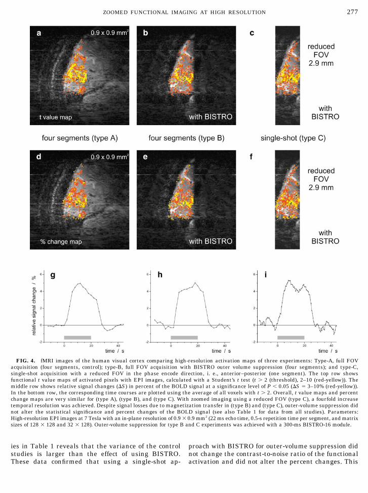
The voxels were classified based on the t value in theM0 image into “nonactive” voxels (t � 2) and “active”voxels (t � 2), the latter of which were further dividedinto “low-active” (2 � t � 5) and “high-active” (t � 5)voxels. The distribution of the MTR was fitted by a
Gaussian function: the MTR mean was (34 � 2)% for“nonactive” voxels and (37 � 2)% for “active” voxels(mean � SE, n � 12), the MTR width � was (20 � 1)%for “nonactive” voxels and (16 � 1)% for “active” voxels.Since the MTR is calculated from the difference inimage intensity comparing the control image (type A,no BISTRO) vs the image with reduced FOV (type C,with BISTRO), an increase in MTR corresponds to adecrease in signal-to-noise ratio. Assuming the noisebeing approximately similar, a decrease in the signal-to-noise ratio was found between 15 to 55%. Neverthe-less, as shown in Table 1, no alteration in the func-tional contrast-to-noise ratio was found.
MTR during a functional experiment was previouslyreported (Zhang et al., 1997) to be different betweenthe resting (MTRREST) and the task (MTRTASK) states.We also determined for our data the MTR separatelyfor the resting and the task states and found a highlinear correlation between MTRTASK versus MTRREST.Further, we found that MTRTASK during activation wasdifferent from MTRREST. A regression analysis of thedifferent (MTRTASK � MTRREST) versus MTRREST isshown in Fig. 5 and the corresponding results from all
FIG. 5. Assessment of the magnetization transfer effects in fMRI experiments comparing the MTR of the resting state (MTRREST) withthe MTR of the task state (MTRTASK). MTRTASK and MTRREST were highly linear correlated. To analyze alterations from linearity, a regressionanalysis was performed for the difference (MTRTASK � MTRREST) versus MTRREST (full analysis shown in Table 2). For voxels with low t value(nonactivated), no significant increase of MTRTASK compared to MTRREST was found. However, for voxels exhibiting activation (t � 2), asignificant increase of MTR up to 3% during the task period was found, which is consistent with previous findings (Zhang et al., 1997). Both,intercept and slope increased significantly with higher t values. In the plots of Fig. 5 the 95% confidence intervals are shown in addition tothe linear fit.
279ZOOMED FUNCTIONAL IMAGING AT HIGH RESOLUTION
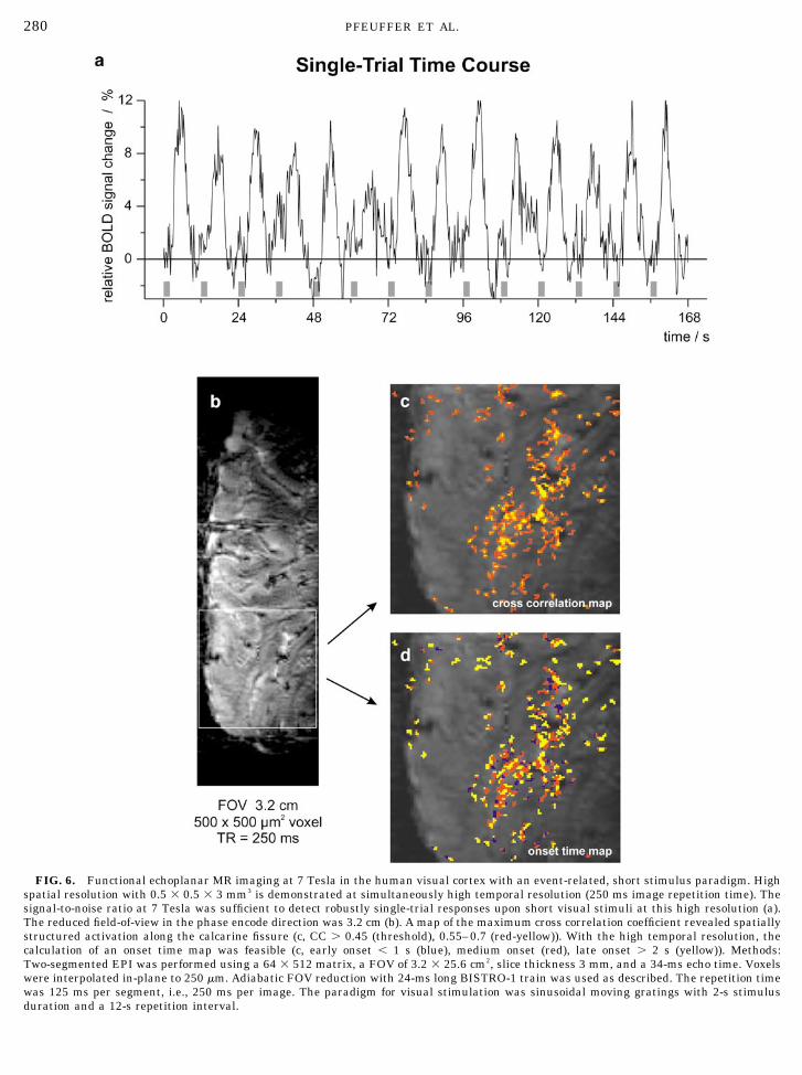
FIG. 6. Functional echoplanar MR imaging at 7 Tesla in the human visual cortex with an event-related, short stimulus paradigm. Highspatial resolution with 0.5 � 0.5 � 3 mm3 is demonstrated at simultaneously high temporal resolution (250 ms image repetition time). Thesignal-to-noise ratio at 7 Tesla was sufficient to detect robustly single-trial responses upon short visual stimuli at this high resolution (a).The reduced field-of-view in the phase encode direction was 3.2 cm (b). A map of the maximum cross correlation coefficient revealed spatiallystructured activation along the calcarine fissure (c, CC � 0.45 (threshold), 0.55–0.7 (red-yellow)). With the high temporal resolution, thecalculation of an onset time map was feasible (c, early onset � 1 s (blue), medium onset (red), late onset � 2 s (yellow)). Methods:Two-segmented EPI was performed using a 64 � 512 matrix, a FOV of 3.2 � 25.6 cm2, slice thickness 3 mm, and a 34-ms echo time. Voxelswere interpolated in-plane to 250 �m. Adiabatic FOV reduction with 24-ms long BISTRO-1 train was used as described. The repetition timewas 125 ms per segment, i.e., 250 ms per image. The paradigm for visual stimulation was sinusoidal moving gratings with 2-s stimulusduration and a 12-s repetition interval.
280 PFEUFFER ET AL.

series are summarized in Table 2. For “nonactive” vox-els, the intercept and slope were not significantly dif-ferent from zero, which means that no change of MTRoccurs in the task state for these “nonactivated” voxels.For “active” voxels, a highly significant increase inintercept (2.6 � 0.3)% and negative slope (�0.041 �0.004) was found (mean � SE, n � 12), see Table 2. Inaddition, the significance is made more apparent by theestimated errors of these parameters in the regressionanalysis. The error of the intercept was as low as 0.2%and the error of the slope was 0.007 for the singleregressions. These results gave evidence that the MTRis larger in the task state than in the resting state inactivated voxels. Also, voxels exhibiting a lowerMTRREST show a larger increase of MTRTASK duringactivation.
In addition, we tested our data for a difference ofMTR between “low-active” and “high-active” voxels.For “low-active” voxels the intercept of the linear re-gression was (2.4 � 0.2)%, the slope was (�0.043 �0.004). For “high-active” voxels the intercept was(3.6 � 0.3)% and the slope was (�0.067 � 0.006)(mean � SE, n � 12). For illustration, these results areshown in Fig. 5 for one series, which clearly indicatethat the intercept and slope changes are larger withhigher activation.
Event-Related fMRI at High Spatial and TemporalResolution
Since we demonstrated that zoomed fMRI using FOVreduction provides activation detection comparable toa full-FOV approach, we further utilized its advantageof increased temporal resolution. An event-relatedfMRI experiment with short 2-s visual stimuli wasperformed at simultaneously high spatial and tempo-ral resolution (two subjects, eight series). At a voxelsize of 0.5 � 0.5 � 3 mm3, a temporal resolution of 250ms was achieved using a reduced FOV of 3.2 cm and atwo-segment acquisition (Fig. 6b). The signal-to-noiseand contrast-to-noise ratio at 7 Tesla was sufficient todetect robustly single-trial responses upon short visualstimuli at this high resolution (Figs. 6a and 6c). Withthe high temporal resolution, the calculation of an on-set time map was also feasible, which distinguishedearly (blue), medium (red), and late responses (yellow)based on different lags in the cross-correlation analysis(Fig. 6d).
Intrascan reproducibility was tested by averagingthe first eight (test) and last eight trials (retest) of theexperiment shown in Fig. 6 comparing the cross corre-lation values. For a simple measure for reproducibility,the number of commonly activated voxels relative tothe total number of activated voxels was determined ata given threshold (CC � 0.45) and found to be 52% forthe test trials and 57% for the retest trials. Figure 7shows the reproducibility map with commonly acti-
vated voxels in yellow and voxels activated only in oneof the test series in either red or blue.
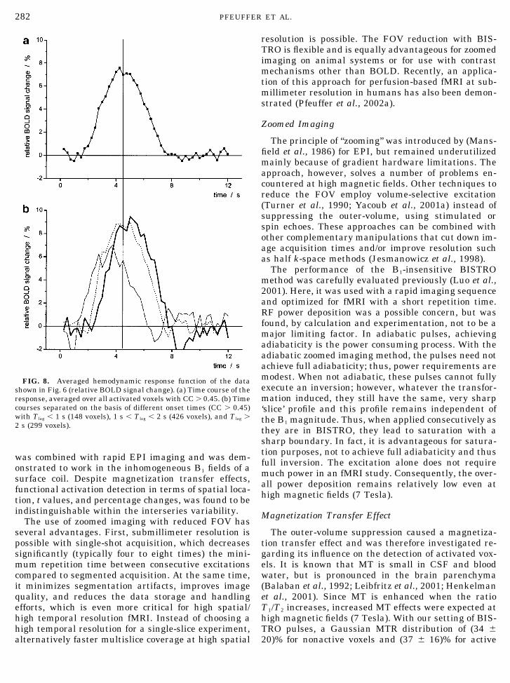
The time course of the event-related experiment inFig. 8a was averaged over all epochs and voxels withCC � 0.45 (see Fig. 6c) showing the hemodynamicresponse from data with 500 �m in-plane resolution.The three time courses in Fig. 8b were separated basedon different onset lags, which were smaller than 1 s,between 1 and 2 s, and larger than 2 s. For a paramet-ric characterization of the average event-related signaltime courses, we used a Gaussian fit to determine thewidth � and the time-to-peak (TP), and a linear regres-sion of the rising response (10 to 90%) to determine theonset time (TO). The parameters for the averaged re-sponse (Fig. 8a) were � � 2.8 � 0.1 s, TP � 4.5 � 0.1 s,and TO � 1.7 � 0.1 s. For the three different responsesin Fig. 8b, � � 2.4 � 0.2 s, 2.7 � 0.1 s, and 2.6 � 0.1 sfor the width, TP � 3.7 � 0.1 s, 4.6 � 0.1 s, 5.1 � 0.1 sfor the time-to-peak, and TO � 0.7 � 0.1 s, 1.8 � 0.2 s,2.4 � 0.1 s for the onset time. These findings indicatethat averaging across voxels leads to a broadening ofthe hemodynamic response function, which is causedby spatial heterogeneity even at a high resolution of500 �m in the human brain.
DISCUSSION
In the present work, we describe an approach forfunctional neuroimaging, which provides simultaneoushigh temporal and spatial resolution. For the firsttime, single-trial fMRI images are presented from thehuman brain with 0.5 � 0.5 � 3-mm3 spatial and250-ms temporal resolution. The method is based on azoomed imaging approach, which minimizes imagingtime by reducing the FOV, and utilizes the SNR andCNR gains provided by high magnetic fields, in thiscase 7 Tesla. Outer-volume suppression with BISTRO
FIG. 7. A reproducibility test was performed comparing activa-tion maps from the first eight and last eight trials of the datadescribed in the legend of Fig. 6. The number of commonly activatedvoxels for CC � 0.45 was 55% (shown in yellow). Voxels activatedonly in one of the test trials are shown in either red or blue.
281ZOOMED FUNCTIONAL IMAGING AT HIGH RESOLUTION
was combined with rapid EPI imaging and was dem-onstrated to work in the inhomogeneous B1 fields of asurface coil. Despite magnetization transfer effects,functional activation detection in terms of spatial loca-tion, t values, and percentage changes, was found to beindistinguishable within the interseries variability.
The use of zoomed imaging with reduced FOV hasseveral advantages. First, submillimeter resolution ispossible with single-shot acquisition, which decreasessignificantly (typically four to eight times) the mini-mum repetition time between consecutive excitationscompared to segmented acquisition. At the same time,it minimizes segmentation artifacts, improves imagequality, and reduces the data storage and handlingefforts, which is even more critical for high spatial/high temporal resolution fMRI. Instead of choosing ahigh temporal resolution for a single-slice experiment,alternatively faster multislice coverage at high spatial
resolution is possible. The FOV reduction with BIS-TRO is flexible and is equally advantageous for zoomedimaging on animal systems or for use with contrastmechanisms other than BOLD. Recently, an applica-tion of this approach for perfusion-based fMRI at sub-millimeter resolution in humans has also been demon-strated (Pfeuffer et al., 2002a).
Zoomed Imaging
The principle of “zooming” was introduced by (Mans-field et al., 1986) for EPI, but remained underutilizedmainly because of gradient hardware limitations. Theapproach, however, solves a number of problems en-countered at high magnetic fields. Other techniques toreduce the FOV employ volume-selective excitation(Turner et al., 1990; Yacoub et al., 2001a) instead ofsuppressing the outer-volume, using stimulated orspin echoes. These approaches can be combined withother complementary manipulations that cut down im-age acquisition times and/or improve resolution suchas half k-space methods (Jesmanowicz et al., 1998).
The performance of the B1-insensitive BISTROmethod was carefully evaluated previously (Luo et al.,2001). Here, it was used with a rapid imaging sequenceand optimized for fMRI with a short repetition time.RF power deposition was a possible concern, but wasfound, by calculation and experimentation, not to be amajor limiting factor. In adiabatic pulses, achievingadiabaticity is the power consuming process. With theadiabatic zoomed imaging method, the pulses need notachieve full adiabaticity; thus, power requirements aremodest. When not adiabatic, these pulses cannot fullyexecute an inversion; however, whatever the transfor-mation induced, they still have the same, very sharp‘slice’ profile and this profile remains independent ofthe B1 magnitude. Thus, when applied consecutively asthey are in BISTRO, they lead to saturation with asharp boundary. In fact, it is advantageous for satura-tion purposes, not to achieve full adiabaticity and thusfull inversion. The excitation alone does not requiremuch power in an fMRI study. Consequently, the over-all power deposition remains relatively low even athigh magnetic fields (7 Tesla).
Magnetization Transfer Effect
The outer-volume suppression caused a magnetiza-tion transfer effect and was therefore investigated re-garding its influence on the detection of activated vox-els. It is known that MT is small in CSF and bloodwater, but is pronounced in the brain parenchyma(Balaban et al., 1992; Leibfritz et al., 2001; Henkelmanet al., 2001). Since MT is enhanced when the ratioT1/T2 increases, increased MT effects were expected athigh magnetic fields (7 Tesla). With our setting of BIS-TRO pulses, a Gaussian MTR distribution of (34 �20)% for nonactive voxels and (37 � 16)% for active
FIG. 8. Averaged hemodynamic response function of the datashown in Fig. 6 (relative BOLD signal change). (a) Time course of theresponse, averaged over all activated voxels with CC � 0.45. (b) Timecourses separated on the basis of different onset times (CC � 0.45)with Tlag � 1 s (148 voxels), 1 s � Tlag � 2 s (426 voxels), and Tlag �2 s (299 voxels).
282 PFEUFFER ET AL.
voxels (mean � width) was observed in the humanbrain. Its consequences for fMRI were a decrease inimage signal and change in EPI image contrast.
Increased MTR was observed during the task periodcompared to the resting period, although it was rela-tively small (about 2% MTR difference). For voxelsexhibiting higher significant activation, the differencein MTR increased (Fig. 5), likely due to less partialvolume contributions to the MT effect. The MT effect isthought to originate from the coupling between highlymobile water protons and relatively immobile protonsthat are associated with macromolecules (Henkelmanet al., 2001). Assuming a blood volume increase duringincreased neuronal activity, one would expect that theMTR has to decrease, because blood has low MTR.However, very short T2 values of venous blood at 7Tesla means venous blood contribution is virtually ab-sent in T*2-weighted images at 7 Tesla at the echo timesused. Even arterial blood contribution is expected to besignificantly suppressed since its T2 also decreasedsignificantly at very high fields (Lee et al., 1999). Wewould predict that at 1.5 Tesla, blood volume increasesmay indeed lead to a decrease in MT but at 7 Tesla, MTis not expected to decrease much or at all due to bloodvolume increase. On the other hand, it is possible thatexchange of water protons across the capillaries (be-tween blood and tissue), across cell membranes (i.e.,between extracellular and intracellular space) or evenbetween cellular and mitochondrial space may in-crease leading to a small increase in MT. Neurons arethought to swell due to altered ion content duringneurotransmission (this is thought to be the source ofthe light scattering signals seen in optical imaging)(Cohen et al., 1970s). Such a swelling, though it is verytiny (less than 0.1%), can lead to higher permeability ofthe cellular membranes, leading to a faster exchangebetween the intracellular space rich in macromoleculesand the extracellular space, resulting in an increase inMT. Similarly, it is possible that the permeability ofthe capillary walls also increases during activation. Anincrease in the permeability of capillaries to oxygenhas previously been proposed (e.g., Hyder et al., 1998).The same can be also true for water, in which case MTwould increase. The activation dependent alterationsin MTR at high fields can even be used for generationof functional images (Song et al., 1997), that potentiallyreport on processes associated with neuronal activitythat are not simply blood volume and/or flow changesand having a somewhat different specificity than theBOLD effect.
Functional Imaging at High Resolution
Despite the reduction in SNR, the MT effect did notalter functional contrast under our experimental con-ditions. Comparing functional studies with full vs re-duced FOV (12 series at 0.9 � 0.9 mm2 resolution), we
did not find a statistically significant alteration in thefunctional activation detection (Fig. 4, Table 1). A re-cent fMRI investigation at high spatial resolution pro-posed a new model predicting that the contrast-to-noise ratio tends to be independent of the voxel volumein the absence of partial volume effects for T*2-weightedimages, provided the contrast-to-noise is not limited byimage SNR but by physiological processes. (Hyde et al.,2001). Our findings are consistent with this model.However, this conclusion does not necessarily meanthat the same study can be performed with equal CNReven if SNR was significantly lower, for example 2.3-fold at 3 Tesla. At some point, the image SNR willdominate and limit the CNR and the ability to accu-rately detect activation will be determined by SNR. Wedo not know how far away this limit is in our studies.It is possible that additional improvements in resolu-tion (thus decrease in SNR) can still be attained in ourstudies and retain a similar CNR. In this context, it isalso important to consider that the BOLD contrast-to-noise ratio depends on the voxel size effects (Yoo et al.,2001; Hyde et al., 2001). An optimum voxel size is to beexpected for functional detection, when the resolutionmatches the size of activation to minimize partial vol-uming and the distribution of the blood vessels thatdominate the observed BOLD effect. For example,Hyde et al. interpret their optimal resolution in thecontext of veins that are separated by about 1 mm inthe cortex. However, the size and consequently thedistribution of the blood vessels that contribute to theBOLD effect will strongly depend on the field magni-tude (Ogawa et al., 2000; Yacoub et al., 2001b).
The advantages of single-shot imaging at high spa-tial resolution have been previously described (Gloveret al., 1998; Jesmanowicz et al., 1998; Hoogenraad etal., 2000) and are more important at high magneticfields considering that physiological fluctuations in-crease (van de Moortele et al., 2002; Pfeuffer et al.,2002b). The reduction in t values (Table 1) comparingaverages of the four-segment and the single-shot datais consistent with single-shot imaging being more ad-vantageous. In the setup of the event-related experi-ment at 500 � 500-�m2 in-plane resolution (Fig. 6), wecompared activation maps acquired with two segments(250 ms TR) and four segments (500 ms TR) and alsofound a reduced activation detection with higher seg-mentation, which indicated that experiments at veryhigh spatial resolution are susceptible to intersegmentfluctuations (data not shown).
Realignment of image time series is commonly re-garded as an essential preprocessing step in fMRI dataanalysis. It was shown recently for fMRI studies on thewhole brain (Freire et al., 2001) that BOLD activationscan produce artificial motion detected by registrationalgorithms, thus yielding spurious activations in thecorrected image time series. The use of a zoomed im-aging technique may amplify this effect and poses spe-
283ZOOMED FUNCTIONAL IMAGING AT HIGH RESOLUTION

cific problems for realignment, since a much largerfraction of the total image is activated compared to thewhole brain images in the quoted paper. Adaptation ofrealignment algorithms is to be considered, for exam-ple, using a simple movement detection in binary form(brain/nonbrain regions).
The effective spatial resolution in the phase-encod-ing direction can be somewhat reduced as a result ofthe T*2 dephasing over the total readout period (Farza-neh et al., 1990). Taking the measured T*2 of 25 ms at 7Tesla (Yacoub et al., 2001b), there was no significant T*2broadening for the single-shot experiment at 0.9 �0.9 � 3 mm3 resolution (readout time 21 ms). At thehigh spatial 0.5 � 0.5 � 3 mm3 resolution (readouttime 54 ms), the effective resolution decreased by 20%.In addition, one expects that the T*2 at this high reso-lution can be locally increased, lessening the effect ofT*2 blurring. Therefore, our current set of parameterswith two segments for 0.5 mm spatial and 0.25 s tem-poral resolution is a valid trade-off between segmenta-tion artifacts and T*2 blurring.
Given the slowness of the hemodynamic responseand assuming full trial-to-trial reproducibility, one canuse k-space segmentation for high spatial resolutionand stroboscopic time-shifted acquisition for high tem-poral resolution. But both increase linearly the time ittakes to acquire an fMRI series of a given number ofimages. Although in single trial fMRI as commonlypracticed, such time resolution is not utilized, it isrecognized that the response of the subject is not nec-essarily the same due to numerous confounding prob-lems such as effects of learning, alterations in strategy,and errors made in performing the task. Ideally, withsufficient CNR, each trial should be sampled sepa-rately and correlated with performance. This approachwas introduced by our laboratory several years ago. Itwas demonstrated that the width and onset time of theactivities in the parietal and motor cortex (Richter etal., 1997b, 2000) changed from trial to trial and thechange in the different brain regions correlated withdifferent parameters that measured aspects of perfor-mance. Despite its power, this method has not beenused except in a few 4 Tesla studies due to the de-mands of CNR (see for example the review of (Ugurbilet al., 1999)). However, the current work introduces thepossibility of performing these studies with high spa-tial resolution.
The methodology described in the paper is not re-stricted to the TR of 0.5 s and 0.125 s we have employedin these studies. It can easily be used with a longer TR(e.g., 1 s) and would still have significant advantagesover a segmented image acquired with the same reso-lution. With no FOV reduction applied, the minimumnumber of segments using the same coil setup wouldhave been eight to achieve the 0.5-mm resolution (FOV12.8 cm), corresponding to an image repetition time of1 s using a reasonable intersegment time of 125 ms.
However, now only a single slice can be monitored withthis 1 s time resolution. A single slice study is accept-able for methodological development and demonstra-tions but not for most applications of fMRI to studybrain function. With a high-resolution image of oneslice acquired in a single or two shots with FOV reduc-tion, multiple slices can be collected for the same dataacquisition duration.
If the rapid image acquisition with reduced FOV isused to attain a short TR and high temporal resolution,then image-to-image signal fluctuations due to physi-ological processes can be sampled. This in turn permitsthe use a variety of methods to suppress them includ-ing those that utilize the information in the spectraldomain (Biswal et al., 1996; Mitra et al., 1997). ShortTR, however, can in principle lead to deleterious “ma-crovascular” inflow artifacts (Menon et al., 1993; Seg-ebarth et al., 1994). However, this inflow effect can beminimized by reducing the flip angle as was done inour studies (see Methods). The signal-to-noise is de-creased because of the low flip angle, but at high mag-netic fields sufficient signal persists to make functionaldetection feasible. For high resolution imaging, usingmultiple segments rather than FOV reduction wouldsimply aggravate this problem. Whatever the time res-olution desired, it is better to go with fewer segmentsso that relaxation is allowed without having to go tovery low tip angles. In addition, ‘macrovascular’ infloweffects are minimized at high fields due to short trans-verse relaxation rates. At 7 Tesla, blood has a T2 under10 ms (Yacoub et al., 2001b). Consequently, veins donot contribute to macrovascular inflow artifacts in gra-dient- or spin-echo EPI images where typical TE’s ex-ceed the venous blood T2 several-fold. Even the arterialblood has a short T2 at high fields (40 ms at 9.4 Tesla(Lee et al., 1999)). Thus, arterial blood signals aresignificantly suppressed during a T*2-weighted acquisi-tion and venous blood signals are virtually eliminated.
Somewhat surprisingly, we found a local variabilityof the hemodynamic onset at the 0.5 mm scale (Figs. 6and 8). Our recent results from high-resolution perfu-sion-based fMRI studies at 0.9 mm voxel size (Pfeufferet al., 2002a) showed that t scores and percent changeswere also spatially varying, potentially accounting forthe varying onset of the BOLD response observed here.Further investigations by combining different contrastmechanisms like cerebral blood flow and BOLD in thesame experiment are needed to understand better thebrain physiology and the underlying basis of functionalneuroimaging.
CONCLUSION
Zoomed imaging, using reduction of the FOV withBISTRO outer-volume suppression, is most advanta-geous for rapid functional imaging at high magneticfields when high spatial and temporal information is
284 PFEUFFER ET AL.
needed simultaneously. This adiabatic approach isstraightforward to implement as an addition to exist-ing rapid imaging sequences and is easy to use, be-cause it needs only a threshold RF power level towork. Application on lower field systems are readilyachieved, since RF power demands are reduced. Thisimaging approach minimizes segmentation-related ar-tifacts and furthers novel functional paradigm designs.It makes fast imaging faster at high spatial resolution.
ACKNOWLEDGMENTS
This work was supported by National Institutes of Health GrantsRR08079 and RO1MH55346, the W. M. Keck Foundation, and theMIND institute (formerly the National Foundation of FunctionalBrain Imaging (NFFBI)).
REFERENCES
Adriany, G., Pfeuffer, J., Yacoub, E., van de Moortele, P.-F., Shmuel,A., Andersen, P., Hu, X., Vaughan, J. T., and Ugurbil, K. 2001. AHalf-Volume Transmit/ Receive Coil Combination for 7 Tesla Ap-plications. Proc. ISMRM 9th Scientific Meeting, Glasgow, p. 1097.
Aguirre, G. K., Zarahn, E., and D’esposito, M. 1998. The variabilityof human, BOLD hemodynamic responses. Neuroimage 8: 360–369.
Balaban, R. S., and Ceckler, T. L. 1992. Magnetization transfercontrast in magnetic resonance imaging. Magn. Reson. Quart. 8:116–137.
Biswal, B., De Yoe, A. E., and Hyde, J. S. 1996. Reduction of physi-ological fluctuations in fMRI using digital filters. Magn. Reson.Med. 35: 107–113.
Bruder, H., Fischer, H., Reinfelder, H. E., and Schmitt, F. 1992.Image reconstruction for echo planar imaging with nonequidistantk-space sampling. Magn. Reson. Med. 23: 311–323.
Chen, W., and Ugurbil, K. 1999. High spatial resolution functionalmagnetic resonance imaging at very-high-magnetic field. Top.Magn. Reson. Imag. 10: 63–78.
Cheng, K., Waggoner, R. A., and Tanaka, K. 2001. Human oculardominance columns as revealed by high-field functional magneticresonance imaging. Neuron 32: 359–374.
Duong, T. Q., Kim, D. S., Ugurbil, K., and Kim, S. G. 2000. Spatio-temporal dynamics of the BOLD fMRI signals: Toward mappingsubmillimeter cortical columns using the early negative response.Magn. Reson. Med. 44: 231–242.
Duong, T. Q., Kim, D. S., Ugurbil, K., and Kim, S. G. 2001. Localizedcerebral blood flow response at submillimeter columnar resolution.Proc. Natl. Acad. Sci. USA 98: 10904–10909.
Farzaneh, F., Riederer, S. J., and Pelc, N. J. 1990. Analysis of T2limitations and off-resonance effects on spatial resolution andartifacts in echo-planar imaging. Magn. Reson. Med. 14: 123–139.
Freire, L., and Mangin, J. F. 2001. Motion correction algorithms maycreate spurious brain activations in the absence of subject motion.Neuroimage 14: 709–722.
Glover, G. H., and Lai, S. 1998. Self-navigated spiral fMRI: Inter-leaved versus single-shot. Magn. Reson. Med. 39: 361–368.
Goodyear, B. G., and Menon, R. S. 2001. Brief visual stimulationallows mapping of ocular dominance in visual cortex using fMRI.Hum. Brain Mapp. 14: 210–217.
Gruetter, R., and Tkac, I. 2000. Field mapping without referencescan using asymmetric echo-planar techniques. Magn. Reson.Med. 43: 319–323.
Henkelman, R. M., Stanisz, G. J., and Graham, S. J. 2001. Magne-tization transfer in MRI: A review. NMR Biomed. 14: 57–64.
Hoogenraad, F. G., Hofman, M. B., Pouwels, P. J., Reichenbach,J. R., Rombouts, S. A., and Haacke, E. M. 1999. Sub-millimeterfMRI at 1.5 Tesla: Correlation of high resolution with low resolu-tion measurements. J. Magn. Reson. Imag. 9: 475–482.
Hoogenraad, F. G., Pouwels, P. J., Hofman, M. B., Rombouts, S. A.,Lavini, C., Leach, M. O., and Haacke, E. M. 2000. High-resolutionsegmented EPI in a motor task fMRI study. Magn. Reson. Imag.18: 405–409.
Hu, X., Le, T. H., and Ugurbil, K. 1997. Evaluation of the earlyresponse in fMRI in individual subjects using short stimulus du-ration. Magn. Reson. Med. 37: 877–884.
Hwang, T. L., van Zijl, P. C., and Garwood, M. 1999. Asymmetricadiabatic pulses for NH selection. J. Magn. Reson. 138: 173–177.
Hyde, J. S., Biswal, B. B., and Jesmanowicz, A. 2001. High-resolu-tion fMRI using multislice partial k-space GR-EPI with cubicvoxels. Magn. Reson. Med. 46: 114–125.
Hyder, F., Shulman, R. G., and Rothman, D. L. 1998. A model for theregulation of cerebral oxygen delivery. J Appl. Physiol. 85: 554–564.
Jesmanowicz, A., Bandettini, P. A., and Hyde, J. S. 1998. Single-shothalf k-space high-resolution gradient-recalled EPI for fMRI at 3Tesla. Magn. Reson. Med. 40: 754–762.
Kim, D. S., Duong, T. Q., and Kim, S. G. 2000. High-resolutionmapping of iso-orientation columns by fMRI. Nat. Neurosci. 3:164–169.
Kim, D. S., Formisano, E., Van De Moortele, P. F., Ugurbil, K., andGoebel, R. 2001. Ultra-high field (7T) mapping of the human ven-tral visual area for “head-from-motion.” Neuroimage 13: S901.
Kim, S. G., Hu, X., Adriany, G., and Ugurbil, K. 1996. Fast inter-leaved echo-planar imaging with navigator: High resolution ana-tomic and functional images at 4 Tesla. Magn. Reson. Med. 35:895–902.
Lee, A. T., Glover, G. H., and Meyer, C. H. 1995. Discrimination oflarge venous vessels in time-course spiral blood-oxygen-level-de-pendent magnetic-resonance functional neuroimaging. Magn. Re-son. Med. 33: 745–754.
Lee, S. P., Silva, A. C., Ugurbil, K., and Kim, S. G. 1999. Diffusion-weighted spin-echo fMRI at 9.4 T: Microvascular/tissue contribu-tion to BOLD signal changes. Magn. Reson. Med. 42: 919–928.
Leibfritz, D., and Dreher, W. 2001. Magnetization transfer MRS.NMR Biomed. 14: 65–76.
Logothetis, N. K., Merkle, H., Augath, M., Trinath, T., and Ugurbil,K. 2002. Ultra-High Resolution fMRI in Monkeys with ImplantedRF Coils. J. Neurosci. Methods, in press.
Logothetis, N. K., Pauls, J., Augath, M., Trinath, T., and Oelter-mann, A. 2001. Neurophysiological investigation of the basis of thefMRI signal. Nature 412: 150–157.
Luo, Y., de Graaf, R. A., DelaBarre, L., Tannus, A., and Garwood, M.2001. BISTRO: An outer-volume suppression method that toler-ates RF field inhomogeneity. Magn. Reson. Med. 45: 1095–1102.
Menon, R. S., and Goodyear, B. G. 1999. Submillimeter functionallocalization in human striate cortex using BOLD contrast at 4Tesla: Implications for the vascular point-spread function. Magn.Reson. Med. 41: 230–235.
Menon, R. S., Ogawa, S., Tank, D. W., and Ugurbil, K. 1993. 4 Teslagradient recalled echo characteristics of photic stimulation-in-duced signal changes in the human primary visual cortex. Magn.Reson. Med. 30: 380–386.
Mitra, P. P., Ogawa, S., Hu, X., and Ugurbil, K. 1997. The nature ofspatiotemporal changes in cerebral hemodynamics as manifestedin functional magnetic resonance imaging. Magn. Reson. Med. 37:511–518.
285ZOOMED FUNCTIONAL IMAGING AT HIGH RESOLUTION

Ogawa, S., Lee, T. M., Stepnoski, R., Chen, W., Zhu, X. H., andUgurbil, K. 2000. An approach to probe some neural systemsinteraction by functional MRI at neural time scale down to milli-seconds. Proc. Natl. Acad. Sci. USA 97: 11026–11031.
Pfeuffer, J., Adriany, G., Shmuel, A., Yacoub, E., van de Moortele,P.-F., Hu, X., and Ugurbil, K. 2002a. Perfusion-based high-resolu-tion functional imaging in the human brain at 7 Tesla. Magn.Reson. Med. 47: 903–911.
Pfeuffer, J., van de Moortele, P.-F., Adriany, G., Hu, X., and Ugurbil,K. 2001a. Sub-Millimeter Event-Related fMRI at High TemporalResolution. Proc. ISMRM 9th Scientific Meeting, Glasgow, p. 1257.
Pfeuffer, J., van de Moortele, P.-F., Yacoub, E., Adriany, G., Shmuel,A., Andersen, P., Merkle, H., Ugurbil, K., and Hu, X. 2001b.Single-Shot Imaging at Sub-Millimeter Resolution Using Adia-batic Outer-Volume-Suppression. Proc. ISMRM 9th ScientificMeeting, Glasgow, p. 1811.
Pfeuffer, J., Van De Moortele, P. F., Ugurbil, K., Hu, X., and Glover,G. H. 2002b. Correction of physiologically induced global off-reso-nance effects in dynamic echo-planar and spiral functional imag-ing. Magn. Reson. Med. 47: 344–353.
Richter, W., Andersen, P. M., Georgopoulos, A. P., and Kim, S. G.1997a. Sequential activity in human motor areas during a delayedcued finger movement task studied by time-resolved fMRI. Neuro-report 8: 1257–1261.
Richter, W., Somorjai, R., Summers, R., Jarmasz, M., Menon, R. S.,Gati, J. S., Georgopoulos, A. P., Tegeler, C., Ugurbil, K., and Kim,S. G. 2000. Motor area activity during mental rotation studied bytime-resolved single-trial fMRI. J. Cogn. Neurosci. 12: 310–320.
Richter, W., Ugurbil, K., Georgopoulos, A., and Kim, S. G. 1997b.Time-resolved fMRI of mental rotation. Neuroreport 8: 3697–3702.
Rosen, B. R., Buckner, R. L., and Dale, A. M. 1998. Event-relatedfunctional MRI: Past, present, and future. Proc Natl. Acad. Sci.USA 95: 773–780.
Segebarth, C., Belle, V., Delon, C., Massarelli, R., Decety, J., Le Bas,J. F., Decorps, M., and Benabid, A. L. 1994. Functional MRI of thehuman brain: Predominance of signals from extracerebral veins.Neuroreport 5: 813–816.
Silver, M. S., Joseph, R. J., and Hoult, D. I. 1984. Highly selective �/2and � pulse generation. J. Magn. Reson. 59: 347–351.
Song, A. W., Wolff, S. D., Balaban, R. S., and Jezzard, P. 1997. Theeffect of off-resonance radiofrequency pulse saturation on fMRIcontrast. NMR Biomed. 10: 208–215.
Strupp, J. P. 1996. Stimulate: A GUI based fMRI Analysis SoftwarePackage. Neuroimage 3: S607.
Thulborn, K. R., Chang, S. Y., Shen, G. X., and Voyvodic, J. T. 1997.High-resolution echo-planar fMRI of human visual cortex at 3.0Tesla. NMR Biomed. 10: 183–190.
Turner, R., von Kienlin, M., Moonen, C. T., and van Zijl, P. C. 1990.Single-shot localized echo-planar imaging (STEAM-EPI) at 4.7tesla. Magn. Reson. Med. 14: 401–408.
Ugurbil, K., Hu, X., Chen, W., Zhu, X. H., Kim, S. G., and Georgo-poulos, A. 1999. Functional mapping in the human brain usinghigh magnetic fields. Philos. Trans. R. Soc. Lond B Biol. Sci. 354:1195–1213.
van de Moortele, P.-F., Pfeuffer, J., Glover, G. H., Ugurbil, K., andHu, X. 2002. Respiration-induced B0 fluctuations in the humanbrain at 7 Tesla and its spatial distribution. Magn. Reson. Med. 47:888–895.
Vaughan, J. T., Garwood, M., Collins, C. M., Liu, W., DelaBarre, L.,Adriany, G., Andersen, P., Merkle, H., Goebel, R., Smith, M. B.,and Ugurbil, K. 2001. 7T vs. 4T: RF power, homogeneity, andsignal-to-noise comparison in head images. Magn. Reson. Med. 46:24–30.
Wolff, S. D., and Balaban, R. S. 1989. Magnetization transfer con-trast (MTC) and tissue water proton relaxation in vivo. Magn.Reson. Med. 10: 135–144.
Yacoub, E., Duong, T. Q., Adriany, G., Kim, S. G., Ugurbil, K., andHu, X. 2001a. T2 weighted fMRI in humans at high magneticfields. Proc. ISMRM 9th Scientific Meeting, Glasgow, p. 1183.
Yacoub, E., Shmuel, A., Pfeuffer, J., Van De Moortele, P. F., Adriany,G., Andersen, P., Vaughan, J. T., Merkle, H., Ugurbil, K., and Hu,X. 2001b. Imaging brain function in humans at 7 Tesla. Magn.Reson. Med. 45: 588–594.
Yoo, S. S., Guttmann, C. R., and Panych, L. P. 2001. Multiresolutiondata acquisition and detection in functional MRI. Neuroimage 14:1476–1485.
Zhang, R., Cox, R. W., and Hyde, J. S. 1997. The effect of magneti-zation transfer on functional MRI signals. Magn. Reson. Med. 38:187–192.
286 PFEUFFER ET AL.