Zinc alleviates mercury-induced oxidative stress in Pfaffia glomerata (Spreng.) Pedersen
13
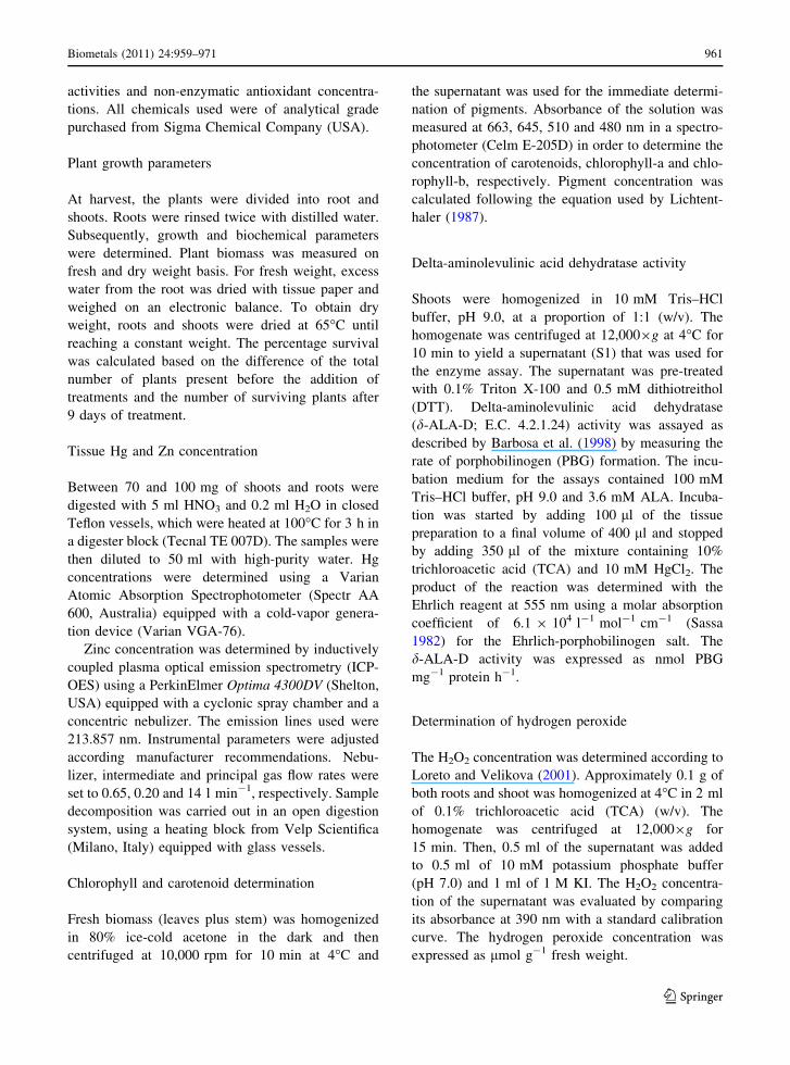
Zinc alleviates mercury-induced oxidative stress in Pfaffia glomerata (Spreng.) Pedersen Nice ´ia Spanholi Calgaroto • Denise Cargnelutti • Liana Veronica Rossato • Ju ´lia Gomes Farias • Sibila Trojahn Nunes • Luciane Almeri Tabaldi • Fabiane Goldschmidt Antes • Erico Marlon Moraes Flores • Maria Rosa Chitolina Schetinger • Fernando Teixeira Nicoloso Received: 7 January 2011 / Accepted: 19 April 2011 / Published online: 8 May 2011 Ó Springer Science+Business Media, LLC. 2011 Abstract The possible role of zinc (Zn) to reverse the oxidative stress caused by mercury (Hg) was investigated in Pfaffia glomerata plantlets. Thirty- day-old acclimatized plantlets of P. glomerata were exposed to four treatments: control, 50 lM Zn, 50 lM Hg and 50 lM Zn ? 50 lM Hg for 9 days. In Zn ? Hg treatment, shoot and root Hg concentra- tions were 59 and 24% smaller than that plants exposed to 50 lM Hg added alone. An increase in the Zn concentration in the shoot of plants exposed to Zn ? Hg occurred, although in the roots Zn concen- tration was not altered, when compared to the control. Fresh and dry weights, as well as the activity of d-aminolevulinic acid dehydratase (d-ALA-D) in Hg-treated plants were significantly reduced. Per- centage survival, fresh and dry weights and d-ALA-D activity of plants treated by 50 lM Zn ? 50 lM Hg were greater than of that treated by Hg alone. Moreover, Zn treatment reduced the lipid peroxida- tion caused by Hg, being this effect related to increased root superoxide dismutase activity, and shoot catalase and ascorbate peroxidase activities. In conclusion, the presence of Zn in the substrate caused a significant reduction in the oxidative stress induced by Hg. Keywords Antioxidant system Brazilian ginseng Mercury Oxidative stress Pfaffia glomerata Zinc Introduction Over the past 200 years emissions of toxic heavy metals have risen tremendously and significantly exceed those from natural sources for practically all metals (Clemens 2006). Mercury (Hg) pollution is a ubiquitous problem resulting both from natural events and anthropogenic activities and it has caused deep concern because of its toxicity, mobility, bioaccumu- lation, methylation and dispersion in the biosphere (Rodrı ´guez et al. 2003). Mercury is extremely toxic to plants and animals (Patra and Sharma 2000). To combat Hg toxicity, plant cells activates the antiox- idant system that include non-enzymatic scavengers N.S. Calgaroto and D. Cargnelutti contributed equally to this work. N. S. Calgaroto J. G. Farias S. T. Nunes F. T. Nicoloso (&) Departamento de Biologia, Centro de Cie ˆncias Naturais e Exatas, Universidade Federal de Santa Maria, Santa Maria, RS 97105-900, Brazil e-mail: [email protected] D. Cargnelutti L. V. Rossato L. A. Tabaldi F. G. Antes E. M. M. Flores M. R. C. Schetinger (&) Departamento de Quı ´mica, Centro de Cie ˆncias Naturais e Exatas, Universidade Federal de Santa Maria, Santa Maria, RS 97105-900, Brazil e-mail: [email protected] 123 Biometals (2011) 24:959–971 DOI 10.1007/s10534-011-9457-y
-
Upload
independent -
Category
Documents
-
view
0 -
download
0
Transcript of Zinc alleviates mercury-induced oxidative stress in Pfaffia glomerata (Spreng.) Pedersen

Zinc alleviates mercury-induced oxidative stress in Pfaffiaglomerata (Spreng.) Pedersen
Niceia Spanholi Calgaroto • Denise Cargnelutti • Liana Veronica Rossato •
Julia Gomes Farias • Sibila Trojahn Nunes • Luciane Almeri Tabaldi •
Fabiane Goldschmidt Antes • Erico Marlon Moraes Flores •
Maria Rosa Chitolina Schetinger • Fernando Teixeira Nicoloso
Received: 7 January 2011 / Accepted: 19 April 2011 / Published online: 8 May 2011
� Springer Science+Business Media, LLC. 2011
Abstract The possible role of zinc (Zn) to reverse
the oxidative stress caused by mercury (Hg) was
investigated in Pfaffia glomerata plantlets. Thirty-
day-old acclimatized plantlets of P. glomerata were
exposed to four treatments: control, 50 lM Zn,
50 lM Hg and 50 lM Zn ? 50 lM Hg for 9 days.
In Zn ? Hg treatment, shoot and root Hg concentra-
tions were 59 and 24% smaller than that plants
exposed to 50 lM Hg added alone. An increase in the
Zn concentration in the shoot of plants exposed to
Zn ? Hg occurred, although in the roots Zn concen-
tration was not altered, when compared to the control.
Fresh and dry weights, as well as the activity of
d-aminolevulinic acid dehydratase (d-ALA-D) in
Hg-treated plants were significantly reduced. Per-
centage survival, fresh and dry weights and d-ALA-D
activity of plants treated by 50 lM Zn ? 50 lM Hg
were greater than of that treated by Hg alone.
Moreover, Zn treatment reduced the lipid peroxida-
tion caused by Hg, being this effect related to
increased root superoxide dismutase activity, and
shoot catalase and ascorbate peroxidase activities. In
conclusion, the presence of Zn in the substrate caused
a significant reduction in the oxidative stress induced
by Hg.
Keywords Antioxidant system � Brazilian ginseng �Mercury � Oxidative stress � Pfaffia glomerata � Zinc
Introduction
Over the past 200 years emissions of toxic heavy
metals have risen tremendously and significantly
exceed those from natural sources for practically all
metals (Clemens 2006). Mercury (Hg) pollution is a
ubiquitous problem resulting both from natural events
and anthropogenic activities and it has caused deep
concern because of its toxicity, mobility, bioaccumu-
lation, methylation and dispersion in the biosphere
(Rodrıguez et al. 2003). Mercury is extremely toxic
to plants and animals (Patra and Sharma 2000). To
combat Hg toxicity, plant cells activates the antiox-
idant system that include non-enzymatic scavengers
N.S. Calgaroto and D. Cargnelutti contributed equally to this
work.
N. S. Calgaroto � J. G. Farias � S. T. Nunes �F. T. Nicoloso (&)
Departamento de Biologia, Centro de Ciencias Naturais
e Exatas, Universidade Federal de Santa Maria, Santa
Maria, RS 97105-900, Brazil
e-mail: [email protected]
D. Cargnelutti � L. V. Rossato � L. A. Tabaldi �F. G. Antes � E. M. M. Flores �M. R. C. Schetinger (&)
Departamento de Quımica, Centro de Ciencias Naturais
e Exatas, Universidade Federal de Santa Maria, Santa
Maria, RS 97105-900, Brazil
e-mail: [email protected]
123
Biometals (2011) 24:959–971
DOI 10.1007/s10534-011-9457-y

(glutathione, ascorbate, carotenoids, proline, and
a-tocopherol) and enzymatic scavengers (superoxide
dismutase, ascorbate peroxidase, catalase, and thio-
redoxin peroxidase) that participate in scavenging
reactive oxygen species (ROS) comprising both free
radical (O2_�, OH�, HO2
� ) and non-radical (molecular)
forms (H2O2) (Edreva 2005; Cargnelutti et al. 2006;
Zhou et al. 2008; Calgaroto et al. 2010).
Zinc (Zn) is an essential component of thousands
of proteins in plants (Broadley et al. 2007) and is
involved in cell membrane stabilization, metallothi-
onein synthesis, protein synthesis, carbohydrate
metabolism, and tryptophan and indoleacetic acid syn-
thesis (Marschner 2002). Studies with animals showed
that Zn is an important antioxidant, decreasing ROS
production (Zago and Oteiza 2001; Fernandez et al.
2003). Some studies have reported the ability of Zn to
interact with essential elements such as Cu and Fe,
decreasing their content in tissues and hence retard-
ing the oxidative processes when these two micro-
nutrients are present in excess (Zago and Oteiza
2001; Santon et al. 2003; Broadley et al. 2007).
Generally, Zn applications decrease Cd uptake and
accumulation in plants (McLaughlin et al. 1994;
Oliver et al. 1997). Hart et al. (2002) attributed
the competitive interaction between Cd and Zn to the
existence of a common transport system in the
plasma membrane. Wu and Zhang (2002) found that
increasing Zn application could alleviate Cd toxicity
stress in barley plants by improving growth and
reducing membrane damage.
The genus of Pfaffia belongs to the Amaranthaceae
family and has about 90 species distributed through-
out Central and South America. In Brazil, 27 species
have been described (Taniguchi et al. 1997). Carneiro
et al. (2002) showed that an undetermined species of
the genus Pfaffia exhibited high tolerance to soil
contamination, growing quite abundantly in soil
mixtures with 90 and 1,450 mg kg-1 of Cd and Zn,
respectively. Skrebsky et al. (2008) showed that
Pfaffia glomerata (Spreng.) Pedersen plantlets grown
hydroponically seemed to have reasonable degree of
Cd tolerance. In a recent study, we also observed
that the growth and the antioxidant mechanisms of
P. glomerata plants were significantly affected by
high levels of Hg (25 and 50 lM) in the substrate
(Calgaroto et al. 2010). However, no information has
been found in plants on whether Zn is capable of
ameliorating Hg toxicity. Under this context, the
present work was designed to analyze the effect of Zn
on the biochemical and physiological alterations
caused by Hg in P. glomerata plants.
Materials and methods
Plant material and growth conditions
Pfaffia glomerata (Spreng.) Pedersen plantlets for
tissue culture were obtained from the Brazilian
Ginseng Germplasm Program, Universidade Federal
de Santa Maria, RS, Brazil. Nodal segments (1.0 cm
long) without leaves were micropropagated in MS
medium (Murashige and Skoog 1962), supplemented
with 30 g l-1 of sucrose, 0.1 g l-1 of myo-inositol
and 6 g l-1 of agar according to Nicoloso et al.
(2001). One thirty-day-old in vitro-grown plantlet
was transferred per pot (1.2 l) which contains 1.0 l of
acid-washed sand. Each experimental unit consisted
of 10 plantlets, totalizing five replicates per treat-
ment. Throughout cultivation, sand was maintained at
80% of field capacity, determined with a sample
altered on a tension table. Irrigation was performed
daily by replacement of transpired water, calculated
by weighing the pots. The plantlets were supple-
mented daily with nutrient solution containing the
following composition: 65.1 mg l-1 NH4Cl, 76.2
mg l-1 MgSO4�7H2O, 135.2 mg l-1 MgCl2�6H2O,
33.1 mg l-1 KH2PO4, 181.5 mg l-1 KCl, 575.3
mg l-1 Ca(NO3)2�4H2O, 0.11 mg l-1 CuSO4�5H2O,
0.39 mg l-1 MnCl2�4H2O, 0.57 mg l-1 ZnSO4�7H2O, 0.04 mg l-1 NiSO4, 1.54 mg l-1 H3BO3,
0.09 mg l-1 H2MoO4�H2O and 13.34 mg l-1 FeSO4�7H2O. After 1 month of plantlet acclimatization, four
treatments (control; 50 lM Zn; 50 lM Hg; and
50 lM Zn ? 50 lM Hg) were added to the nutrient
solution. Hg and Zn were added as HgCl2 and ZnCl2,
respectively. Both in vitro and ex vitro cultured
plantlets were grown in a growth chamber at
25 ± 1�C on a 16/8 h light/dark cycle with
35 lmol m-2 s-1 of irradiance by cold fluorescent
lamps. After 9 days of Hg exposure, three plantlets
per replicate were harvested randomly either for
growth or biochemical analyses. Three independent
and representative tissue samples were used for Hg
and Zn determination. Fresh samples were used for
measurements of H2O2, MDA, chlorophyll and
carotenoids concentrations, antioxidant enzyme
960 Biometals (2011) 24:959–971
123
activities and non-enzymatic antioxidant concentra-
tions. All chemicals used were of analytical grade
purchased from Sigma Chemical Company (USA).
Plant growth parameters
At harvest, the plants were divided into root and
shoots. Roots were rinsed twice with distilled water.
Subsequently, growth and biochemical parameters
were determined. Plant biomass was measured on
fresh and dry weight basis. For fresh weight, excess
water from the root was dried with tissue paper and
weighed on an electronic balance. To obtain dry
weight, roots and shoots were dried at 65�C until
reaching a constant weight. The percentage survival
was calculated based on the difference of the total
number of plants present before the addition of
treatments and the number of surviving plants after
9 days of treatment.
Tissue Hg and Zn concentration
Between 70 and 100 mg of shoots and roots were
digested with 5 ml HNO3 and 0.2 ml H2O in closed
Teflon vessels, which were heated at 100�C for 3 h in
a digester block (Tecnal TE 007D). The samples were
then diluted to 50 ml with high-purity water. Hg
concentrations were determined using a Varian
Atomic Absorption Spectrophotometer (Spectr AA
600, Australia) equipped with a cold-vapor genera-
tion device (Varian VGA-76).
Zinc concentration was determined by inductively
coupled plasma optical emission spectrometry (ICP-
OES) using a PerkinElmer Optima 4300DV (Shelton,
USA) equipped with a cyclonic spray chamber and a
concentric nebulizer. The emission lines used were
213.857 nm. Instrumental parameters were adjusted
according manufacturer recommendations. Nebu-
lizer, intermediate and principal gas flow rates were
set to 0.65, 0.20 and 14 l min-1, respectively. Sample
decomposition was carried out in an open digestion
system, using a heating block from Velp Scientifica
(Milano, Italy) equipped with glass vessels.
Chlorophyll and carotenoid determination
Fresh biomass (leaves plus stem) was homogenized
in 80% ice-cold acetone in the dark and then
centrifuged at 10,000 rpm for 10 min at 4�C and
the supernatant was used for the immediate determi-
nation of pigments. Absorbance of the solution was
measured at 663, 645, 510 and 480 nm in a spectro-
photometer (Celm E-205D) in order to determine the
concentration of carotenoids, chlorophyll-a and chlo-
rophyll-b, respectively. Pigment concentration was
calculated following the equation used by Lichtent-
haler (1987).
Delta-aminolevulinic acid dehydratase activity
Shoots were homogenized in 10 mM Tris–HCl
buffer, pH 9.0, at a proportion of 1:1 (w/v). The
homogenate was centrifuged at 12,0009g at 4�C for
10 min to yield a supernatant (S1) that was used for
the enzyme assay. The supernatant was pre-treated
with 0.1% Triton X-100 and 0.5 mM dithiotreithol
(DTT). Delta-aminolevulinic acid dehydratase
(d-ALA-D; E.C. 4.2.1.24) activity was assayed as
described by Barbosa et al. (1998) by measuring the
rate of porphobilinogen (PBG) formation. The incu-
bation medium for the assays contained 100 mM
Tris–HCl buffer, pH 9.0 and 3.6 mM ALA. Incuba-
tion was started by adding 100 ll of the tissue
preparation to a final volume of 400 ll and stopped
by adding 350 ll of the mixture containing 10%
trichloroacetic acid (TCA) and 10 mM HgCl2. The
product of the reaction was determined with the
Ehrlich reagent at 555 nm using a molar absorption
coefficient of 6.1 9 104 l-1 mol-1 cm-1 (Sassa
1982) for the Ehrlich-porphobilinogen salt. The
d-ALA-D activity was expressed as nmol PBG
mg-1 protein h-1.
Determination of hydrogen peroxide
The H2O2 concentration was determined according to
Loreto and Velikova (2001). Approximately 0.1 g of
both roots and shoot was homogenized at 4�C in 2 ml
of 0.1% trichloroacetic acid (TCA) (w/v). The
homogenate was centrifuged at 12,0009g for
15 min. Then, 0.5 ml of the supernatant was added
to 0.5 ml of 10 mM potassium phosphate buffer
(pH 7.0) and 1 ml of 1 M KI. The H2O2 concentra-
tion of the supernatant was evaluated by comparing
its absorbance at 390 nm with a standard calibration
curve. The hydrogen peroxide concentration was
expressed as lmol g-1 fresh weight.
Biometals (2011) 24:959–971 961
123

Estimation of lipid peroxidation
The level of lipid peroxidation products was esti-
mated following the method of El-Moshaty et al.
(1993) by measuring the concentration of malondi-
aldehyde (MDA) as an end product of lipid perox-
idation by reaction with thiobarbituric acid (TBA).
Fresh tissue samples (0.1 g fresh weight) were
ground in 2 ml of 0.2 M citrate–phosphate buffer
(pH 6.5) containing 0.5% Triton X-100, using mortar
and pestle. The homogenate was filtered through two
layers of paper and centrifuged for 15 min at
20,0009g. One milliliter of the supernatant fraction
was added to an equal volume of 20% (w/v) TCA
containing 0.5% (w/v) TBA. The mixture was heated
at 95�C for 40 min and then quickly cooled in an ice
bath for 15 min. After centrifugation at 10,000 g for
15 min, the absorbance of the supernatant was
measured at 532 nm. A correction for non-specific
turbidity was made by subtracting the absorbance
value taken at 600 nm. The lipid peroxides were
expressed as nmol MDA (mg protein)-1, by using an
extinction coefficient of 155 l mol-1 cm-1.
Catalase assay
Catalase (CAT, E.C. 1.11.1.6) activity was assayed
according to the method of Aebi (1984) with some
modifications. Fresh samples (1 g) were homogenized
in 5 ml of 50 mM KH2PO4/K2HPO4 (pH 7.0), 10 g l-1
PVP, 0.2 mM EDTA and 10 ml l-1 Triton X-100. The
homogenate was centrifuged at 12,0009g at 4�C for
20 min and the supernatant was used for the enzyme
assay. CAT activity was determined by monitoring
the disappearance of H2O2 by measuring the decrease
in absorbance at 240 nm from a reaction mixture
containing 2 ml of 15 mM H2O2 in KPO4 buffer (pH
7.0) and 30 ll extract. Activity was expressed as
DE min-1 mg-1 protein.
Ascorbate peroxidase assay
Ascorbate peroxidase (APX, E.C. 1.11.1.11) activity
was measured according to Zhu et al. (2004). The
reaction mixture, at a total volume of 2 ml, contained
25 mM (pH 7.0) sodium phosphate buffer, 0.1 mM
EDTA, 0.25 mM ascorbate, 1.0 mM H2O2 and 100 ll
enzyme extract. H2O2-dependent oxidation of ascor-
bate was monitored by a decrease in absorbance at
290 nm (E = 2.8 l mol-1 cm-1) and activity was
expressed as lmol ascorbate oxidated min-1 mg-1
protein.
Superoxide dismutase assay
Superoxide dismutase (SOD, E.C. 1.15.1.1) activity
was assayed according to Misra and Fridovich
(1972). About 200 mg of roots and shoots were
homogenized in 5 ml of 100 mmol l-1 K-phosphate
buffer (pH 7.8) containing 0.1 mmol l-1 EDTA,
0.1% (v/v) Triton X-100 and 2% PVP (w/v). The
homogenate was centrifuged at 22,0009g at 4�C for
10 min. The assay mixture consisted of a total
volume of 1 ml, containing glycine buffer (pH
10.5), 1 mmol l-1 epinephrine and enzyme material.
Epinephrine was the last component added. Adeno-
chrome formation over the next 4 min was spectro-
photometrically recorded at 480 nm. One unit of
SOD activity is expressed as the amount of enzyme
required to cause 50% inhibition of epinephrine
oxidation under the experimental conditions used.
This method is based on the ability of SOD to inhibit
the autoxidation of epinephrine at an alkaline pH.
Since the oxidation of epinephrine leads to the
production of a pink adrenochrome, the rate of
increase of absorbance at 480 nm, which represents
the rate of autoxidation of epinephrine, can be
conveniently followed. The enzyme has been found
to inhibit this radical-mediated process.
Ascorbic acid concentration
Ascorbic acid (AsA) determination was performed as
described by Jacques-Silva et al. (2001). Both roots
and shoots were homogenized in a solution contain-
ing 50 mmol l-1 Tris–HCl and 10 ml l-1 Triton
X-100 (pH 7.5), centrifuged at 6,8009g for 10 min.
To the supernatant, 10% TCA was added at a
proportion of 1:1 (v/v) followed by centrifugation
(6,800 g for 10 min) to remove protein. An aliquot of
the sample (300 ll) was incubated at 37�C in a
medium containing 100 ll 13.3% TCA, 100 ll
deionized water and 75 ll 2,4-dinitrophenylhydra-
zine (DNPH). The DNPH solution contained 2%
DNPH, 0.23% thiourea, 0.27% CuSO4 diluted in 49%
H2SO4. After 3 h, 500 ll of 65% H2SO4 was added
and samples were read at 520 nm. A standard curve
was constructed using L(?) ascorbic acid.
962 Biometals (2011) 24:959–971
123

Non-protein thiols group concentration
Non-protein thiol (NPSH) concentration was mea-
sured spectrophotometrically with Ellman’s reagent
(Ellman 1959). Root and shoot samples were homog-
enized in a solution containing 50 mmol l-1 Tris–
HCl and 10 ml l-1 Triton X-100 (pH 7.5), centri-
fuged at 6,8009g for 10 min, and NPSH was
determined in a fraction obtained after mixing 1
volume of supernatant with 1 volume of 10% TCA
followed by centrifugation (6,800 g for 10 min) and
neutralization (to pH 7.4) with 1 M Tris–HCl as
described by Jacques-Silva et al. (2001). The reaction
was read at 412 nm after the addition of 0.05 ml of
10 mM 5,5-dithio-bis (2-nitrobenzoic acid) (DTNB).
A standard curve using cysteine was used to calculate
the concentration of NPSH in the samples and was
expressed as lmol SH g-1 fresh weight.
Protein extraction
In all the enzyme preparations, protein concentration
was determined by the method of Bradford (1976)
using bovine serum albumin as standard and was
expressed in mg l-1.
Statistical analysis
The analyses of variance were computed on statisti-
cally significant differences determined based on the
appropriate F-tests. The results are the means ± SD
of at least three independent replicates. The mean
differences were compared utilizing Tukey test with
P \ 0.05.
Results
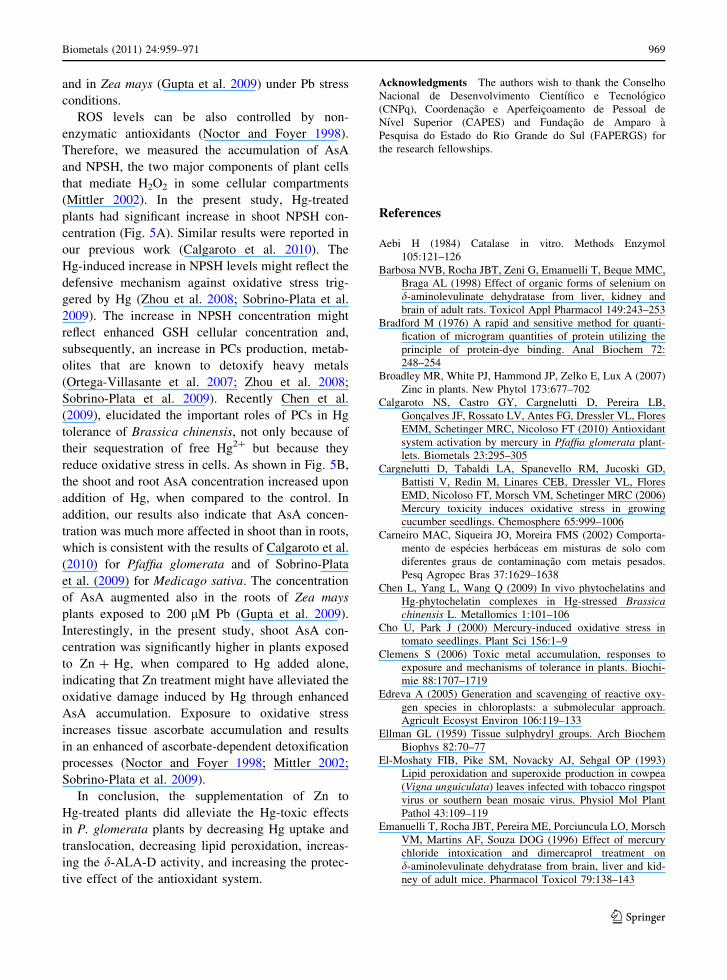
Tissue metals concentration
Mercury (Hg) concentration was greater in roots than
in shoots upon addition of Hg in the substrate
(Table 1). Conversely, Hg concentrations were,
approximately, 15 and 6.8% greater in shoots than
in roots, respectively, in control and Zn treatments. In
Zn ? Hg treatment, shoot and root Hg levels were 59
and 24% smaller than that plants exposed to 50 lM
Hg added alone. In addition, an increase in the Zn
levels in the shoot of plants exposed to Zn ? Hg
occurred, although in the roots, Zn concentration was
not altered, when compared to the control.
Percentage survival and fresh and dry weight
of plantlets
In comparison to the control, fresh and dry weight, as
well as percentage survival in the Hg-exposed
P. glomerata plants decreased significantly, while
the addition of Zn together with Hg reversed the
negative effect caused by Hg (Fig. 1). The percentage
survival was about 47 and 20% lower that of control in
plants exposed to Hg alone and Zn ? Hg treatments,
respectively. In 50 lM Hg treatment, shoot fresh
weight decreased about 71.5% when compared to the
control, whereas root fresh weight decreased by 59%.
On the other hand, in plant treated with Zn ? Hg,
shoot and roots fresh weight decreased about 51.4 and
24.4% in relation to the control, respectively.
Shoot dry weight in Hg-exposed plants decreased
69% in relation to the control. In plants exposed to
Table 1 Hg and Zn concentrations in shoot and roots of Pfaffia glomerata plantlets exposed to the treatments for 9 days
Treatments Hg concentration (lg g-1 dry weight) Zn concentration (lg g-1 dry weight)
Shoot Root Shoot Root
Control 16.9 ± 1.5 cA* 2.53 ± 0.25 bB 79.7 ± 4.0 bB 161.7 ± 10.0 aA
50 lM Zn 15.5 ± 0.7 cA 1.06 ± 0.52 bB 116 ± 4.6 bB 176 ± 37.9 aA
50 lM Hg 351.5 ± 39.5 aB 797 ± 149 aA 73.7 ± 4.7 bB 143 ± 34.0 aA
50 lM Zn ? 50 lM Hg 208 ± 94.0 bB 605.3 ± 159.9 aA 216.7 ± 65.6 aA 150.7 ± 23.4 aB
Data represent mean values ± SD based on independent determination
* Mean values followed by the same small letters in the column, and capital letters in the line did not differ significantly by Tukey test
of P \ 0.05
Biometals (2011) 24:959–971 963
123

Zn ? Hg treatment, the dry weight also decreased,
but this value was only about 26% to the control.
Interestingly, root dry weight in Hg-treated plants
was slightly but not significantly, decreased upon
presence of Hg, when compared to the control
(Fig. 1C), whereas, Zn-treated plants showed greater
root dry weight than that of Hg-treated.
Concentration of chlorophyll and carotenoids
and delta-aminolevulinic acid dehydratase
activity
In comparison to control, the concentrations of
chlorophyll-a and chlorophyll-b were not altered
upon addition of Hg, Zn and Zn ? Hg treatments
(Fig. 2A) On the other hand, Hg-treated plants
showed higher chlorophyll-a concentration than those
treated with Zn alone. Carotenoids concentration
increased in plants grown in the presence of
Zn ? Hg, when compared with control and Zn
treatments. d-ALA-D activity decreased in plant
treated with Zn or Hg alone compared with the
control, whereas there were no changes in activity
when both metal were added together (Fig. 2B).
Fig. 1 Percentage survival (A), fresh weight (B) and dry
weight (C) of P. glomerata plantlets exposed to different
treatments. Control: without Zn and Hg; Zn: 50 lM Zn as
ZnCl2; Hg: 50 lM Hg as HgCl2; Zn ? Hg: 50 lM
Zn ? 50 lM Hg. Data represent the mean ± SD of three
replicates. Identical letters indicate no significant differences
among the treatments according to Tukey0s multiple range test
(P \ 0.05)
Control Zn Hg Zn + Hg
Treatments
0
1
2
3
4
5P
igm
ents
con
cent
ratio
n (m
g g
-1 F
W)
chlorophyll a
aba a
a
abb
chlorophyll b carotenoids
bc c
ab
a
a
a
Control Zn Hg Zn + Hg
Treatments
0
2
4
6
8
10
ALA
-D a
ctiv
ity (
nmol
PB
G m
in-1 m
g pr
otei
n-1
)
Shoot
a
b
a
b
A
B
Fig. 2 Pigments concentration (A) and d-aminolevulinic acid
(d-ALA-D) activity (B) of shoots of P. glomerata plantlets
exposed to different treatments. Control: without Zn and Hg;
Zn: 50 lM Zn as ZnCl2; Hg: 50 lM Hg as HgCl2; Zn ? Hg:
50 lM Zn ? 50 lM Hg. Data represent the mean ± SD of
three replicates. Identical letters indicate no significant
differences among the treatments according to Tukey0smultiple range test (P \ 0.05)
964 Biometals (2011) 24:959–971
123

Hydrogen peroxide and lipid peroxidation
The concentration of H2O2 augmented in shoots of
plants treated with Hg and Zn ? Hg (Fig. 3). Con-
versely, root H2O2 concentration was only changed
upon addition of Zn ? Hg treatment, where it
decreased when compared to other treatments. Shoot
lipid peroxidation was significantly increased upon
addition of Hg, while it decreased significantly in the
treatment with Zn ? Hg added together, when com-
pared to the control (Fig. 3B). On the other hand, lipid
peroxidation in roots did not alter upon all treatments.
Antioxidant enzymes activity
SOD activity was higher in shoots than in roots
(Fig. 4A). Mercury-treated plants showed 2.3-fold
higher shoot SOD activity than the control. Treat-
ments either with Zn applied alone or together with
Hg showed greater shoot SOD activity that the
control, but these increase was lower than that
observed in plants treated exclusively with Hg. In
the roots, however, the highest SOD activity occurred
Control Zn Hg Zn + Hg
Treatments
0.00
0.30
0.60
0.90
1.20
1.50
H2O
2 (µ
mol
g-1
fres
h w
eigh
t)
Shoot
a
b
a
b
Root
aa b
a
Control Zn Hg Zn + Hg
Treatments
0.00
0.06
0.12
0.18
0.24
0.30
TB
AR
S (
nmol
MD
A m
g-1
pro
tein
)
Shoot
a
b
abc
Root
aa a
c
B
A
Fig. 3 Hydrogen peroxide concentration (A) and lipid
peroxidation (B) of P. glomerata plantlets exposed to different
treatments. Control: without Zn and Hg; Zn: 50 lM Zn as
ZnCl2; Hg: 50 lM Hg as HgCl2; Zn ? Hg: 50 lM
Zn ? 50 lM Hg. Data represent the mean ± SD of three
replicates. Identical letters indicate no significant differences
among the treatments according to Tukey0s multiple range test
(P \ 0.05)
Control Zn Hg Zn + Hg
Treatments
0
160
320
480
640
800
SO
D a
ctiv
ity (
units
SO
D m
g-1
pro
tein
)
Shoot
a
d
b
b
Root
d
a
c
c
Control Zn Hg Zn + Hg
Treatments
0
14
28
42
56
70
CA
T a
ctiv
ity (
109
E m
in-1 g
-1) Shoot
a
abb
b
Root
ba
bab
Control Zn Hg Zn + Hg
Treatments
0.00
0.16
0.32
0.48
0.64
0.80
AP
X a
ctiv
ity (
units
mg
-1 p
rote
in)
Shoot
cb
a
b
Root
abb
ca
A
B
C
Fig. 4 Superoxide dismutase (SOD) activity (A), catalase
(CAT) activity (B) and ascorbate peroxidase (APX) activity
(C) of P. glomerata plantlets exposed to different treatments.
Control: without Zn and Hg; Zn: 50 lM Zn as ZnCl2; Hg:
50 lM Hg as HgCl2; Zn ? Hg: 50 lM Zn ? 50 lM Hg. Data
represent the mean ± SD of three replicates. Identical letters
indicate no significant differences among the treatments
according to Tukey0s multiple range test (P \ 0.05)
Biometals (2011) 24:959–971 965
123

at the control, whereas, the minimum SOD activity
occurred in the Hg alone treatment, being 60% lower
than to control. Plants exposed to Zn alone and
Zn ? Hg concomitantly, showed approximately 37
and 73% of decrease in the SOD activity compared to
control plants.
Shoot CAT activity decreased upon addition of
Hg, whereas there was no significant differences in
plants supplied with Zn or Zn ? Hg. Root CAT
activity had a different response, it significantly
increased (60%) upon addition of Hg, when com-
pared to the other treatments.
The treatment with Zn applied alone showed
greater root APX activity than control, whereas
treatments containing Hg, either added alone or
together with Zn, showed a significant decrease,
when compared to the control. On the other hand,
shoot APX activity only increased upon addition of
Zn ? Hg.
Antioxidant non-enzymatic concentration
The NPSH concentration was much higher in the
shoot than in roots (Fig. 5A). Shoot NPSH concen-
tration increased only upon addition of Zn ? Hg,
when compared to the other treatments. Treatments
with Hg added alone or together with Zn showed
greater root NPSH concentration than the control.
Conversely, root NPSH concentration did not change
upon addition of Zn treatment, when compared to the
control.
In general, AsA concentration in shoots and roots
followed a similar pattern as that found for NPSH
concentration, with the exception of Hg-treated
plants, which shoot AsA increased above control
levels. Moreover, plants exposed to Zn ? Hg showed
the highest shoot AsA concentration (Fig. 5B).
Discussion
This study was performed to analyze the mechanism
by which Zn antagonizes Hg toxicity in P. glomerata,
an important medicinal plant. This interaction
between a nutrient and a toxic element may be
important for understanding, analyzing and improv-
ing the defense strategies through various parameters.
In the present study, plants of P. glomerata exposed
to Hg added alone in the nutrient solution showed
significant oxidative stress symptoms, observed as an
increase in shoot H2O2 concentration (Fig. 3A) and
lipid peroxidation (Fig. 3B). Similar results were
reported in our previous work (Calgaroto et al. 2010).
These alterations were followed by significant
changes in the antioxidant system (Fig. 4, 5). How-
ever, the antioxidant system was not sufficient to
avoid damage, resulting in 52.8% of mortality in Hg-
treated plants (Fig. 1A). Percentage survival was
significantly higher in plants exposed to Zn ? Hg,
when compared to Hg added alone, indicating that Zn
treatment alleviated the oxidative damage induced
by Hg.
In the present study, shoot Hg concentration
decreased concomitantly with a significant increase
of Zn concentration in plants exposed to Zn ? Hg
(Table 1), indicating a possible negative interaction
between these metals. Zinc, Cd and Hg are divalent
metals that constitute group II B of the periodic table
Control Zn Hg Zn + Hg
Treatments
0.00
0.30
0.60
0.90
1.20
1.50
NP
SH
(µ
mol
SH
g-1 fr
esh
wei
ght) Shoot
bb
b
b
Root
aba
a
Control Zn Hg Zn + Hg
Treatments
0
30
60
90
120
150
Asc
orbi
c ac
id (
µg A
sA g
-1 fr
esh
wei
ght)
Shoot
b
c
b
c
Root
ab
a
a
B
A
Fig. 5 Non-protein thiols (NPSH) concentration of
P. glomerata plantlets exposed to different treatments. Control:
without Zn and Hg; Zn: 50 lM Zn as ZnCl2; Hg: 50 lM Hg as
HgCl2 and; Zn ? Hg: 50 lM Zn ? 50 lM Hg. Data represent
the mean ± SD of three replicates. Identical letters indicate no
significant differences among the treatments according to
Tukey0s multiple range test (P \ 0.05)
966 Biometals (2011) 24:959–971
123

of elements. Although Zn is an essential trace metal
while the remaining two are toxic metals, they share
biological responses such as affinity towards thiol
groups, antagonism to biological cations, participa-
tion in redox reaction and modulation of nutrients
homeostasis (Peixoto et al. 2003). Hart et al. (2002)
observed that the concentration-dependent uptake of
both Cd2? and Zn2? in both Triticum aestivum L. and
Triticum turgidum L. var. durum consisted of a
combination of linear binding by cell walls and
saturable, Michaelis–Menten influx across the plasma
membrane. Several other data suggest the existence
of antagonist effect of Zn on other essential transition
metals like Cu and Fe (Zago and Oteiza 2001;
Marschner 2002). In the present study, the concen-
tration of Zn in shoots of plants exposed to Zn ? Hg
might be had some positive influence on the antiox-
idant system, which resulted in significant decrease of
shoot lipid peroxidation (Fig. 3B) associated with a
slight increase of CAT and APX activities (Fig. 4B
and C). Therefore, Zn might interact negatively with
Hg and it also suggests that Zn is a pivotal component
of the antioxidant defense network that protects
membranes from oxidation.
Shoot and root fresh weight from plants exposed to
Hg added alone showed reduction of about 70% and
60%, respectively, when compared to the control
(Fig 1B). Such result might be related to the effect of
Hg on aquaporins. Mercury induces conformational
changes in aquaporins because it is thought to bind to
–SH groups of the proteins, then blocking the
channels and reducing their hydraulic conductivity
(Tyerman et al. 1999). The addition of HgCl2(0.1 mM) reduced pressure-induced water flux and
root hydraulic conductivity in the roots of 1-year-old
aspen (Populus tremuloides Michx.) seedlings by
about 50% (Wan and Zwiazek 1999). In the present
work, the symptoms of wilting showed by plants
treated with Hg can explain the decrease in fresh
weight and this data corroborates with other reports
(Zang and Tyerman 1999; Cargnelutti et al. 2006).
Interestingly, in P. glomerata plants treated with
Zn ? Hg, shoot and roots fresh weight showed a
significant recover when compared to the Hg treat-
ment (Fig. 1B).
Zinc is an essential component of many vital
enzymes, a structural stabilizer for proteins, mem-
brane and DNA-binding proteins (Epstein and Bloom
2005). On the other hand, Zn excess in substrate
might causes nutritional disorders in plants (Marschner
2002; Reichman 2002). In the present study, signif-
icant decrease of shoot fresh and dry weight in plants
exposed to Zn alone might be due to Fe deficiency
once that shoot Fe concentration was about 53%
lower than that control plants (data not shown).
Tewari et al. (2008) also found that excess Zn led to a
decrease in Fe level in tissues of Morus alba L.
Moreover, both H2O2 and MDA accumulated in
leaves of Zn–stressed plants. Iron is an essential
element for plants and plays an important role in
several plant biological processes including the
catalytic groups for many redox enzymes (Epstein
and Bloom 2005). The antagonist interaction between
Zn and Fe can lead to nutrient deficiencies and crop
yield reduction (Marschner 2002; Reichman 2002).
The toxicity of heavy metals, such as non-essen-
cial ones, Hg and Cd, is generally accepted to occur
by inactivation of enzymes and/or functional proteins
by directly binding to them. However, several studies
also showed that the toxicity may be due, in part at
least, to oxidative damage by generation of ROS due
to the presence of heavy metals (Cargnelutti et al.
2006; Zhou et al. 2008; Sobrino-Plata et al. 2009).
Mercury ions can also substitute metal ions in
photosynthetic pigments, causing a decrease in pho-
tosynthesis rates (Xylander et al. 1996). Decreased
pigments content due to high levels of Hg in substrate
has been reported in Licopersicum esculentum L.
(Cho and Park 2000), Cucumis sativus L. (Cargnelutti
et al. 2006) and Medicago sativa cv Aragon (Sobrino-
Plata et al. 2009). In the present study, no effect was
observed in pigments concentration from P. glomer-
ata plants exposed to Hg (Fig. 2A). This result might
be related to the reduction in fresh weight (Fig. 1B)
and diminution of leaf expansion (data not shown),
which would lead to an increase in the concentration
of cellular components. Very similar results were
reported in our previous work (Calgaroto et al. 2010).
The enzyme d-aminolevulinic acid dehydratase
(d-ALA-D) is an important enzyme for the synthesis of
porphyrins, hemes and chlorophylls, essential factors
of an adequate aerobic metabolism and photosynthe-
sis (Pereira et al. 2006). In the present study, in
response to the imposed Hg toxicity, a significant
decrease in the d-ALA-D activity, by about 53%
when compared to the control, was observed
(Fig. 2B). Due to its sulphydryl nature, d-ALA-D is
highly sensitive to the presence of heavy metals such
Biometals (2011) 24:959–971 967
123

as Hg (Emanuelli et al. 1996). Therefore, the
significant decrease in d-ALA-D activity observed
in this study could be the result of Hg-binding with
–SH groups of d-ALA-D enzyme. Similar results
were reported in our previous work (Calgaroto et al.
2010). Plants exposed to Zn ? Hg, in contrast,
showed a sharp increase in d-ALA-D activity, so
that was significantly equal to the control. The
present study indicated that decrease in d-ALA-D
activity induced by Hg was prevented by increase of
Zn concentration in shoot (Table 1). Probably, Zn
avoided Hg-binding with –SH groups of this enzyme,
also avoiding its inactivation.
Oxidative stress occur either due to excess
production of free radicals, or inadequate availability
of antioxidants or a combination of both (Zhou et al.
2008; Wang et al. 2009). Lipid peroxidation is a basic
deteriorating change in unsaturated fatty acids of the
cell membranes induced by excess free radicals
(Haliwell and Gutteridge 1999). Estimation of mal-
onyldialdehyde (MDA) continues to be a reliable
method to assess the degree of peroxidative damage
to cell membrane, as it is the most abundant aldehyde
formed as a by-product during this process (Zhou
et al. 2008; Wang et al. 2009). In the present study,
roots MDA concentration was not altered in presence
of Zn or Hg (Fig. 3B), although other stress symp-
toms had occurred in Hg-stressed plants, such as
growth reduction (Fig. 1B and C) and root darkening
(data not shown). In shoot, however, the MDA
reached higher concentration in plants exposed to Hg
added alone, when compared to the control. This
result is in agreement with the report of Cho and Park
(2000), which showed that tomato plants exposed to
50 lM HgCl2 had a significant increase on MDA
concentration. Using alfalfa plants grown on an inert
solid substrate in a hydroponic culture, Sobrino-Plata
et al. (2009) also observed that Hg leaded to a higher
lipid peroxidation in shoots, with much more mod-
erate response in roots. These results are in agreement
with previous work by Vazquez and Carpena-Ruiz
(2005), which compared the toxic effects of Cd in
Lupinus albus grown in a liquid-hydroponic medium
or a semi-hydroponic system. They found that the
inert substrate perlite limited the rate of Cd absorp-
tion, and the toxic effects on roots were moderate.
Goncalves et al. (2009) also reported that the
influence of Cd on nutrient concentration in Solanum
tuberosum was related to many factors such as the
level of Cd in the substrate, potato cultivar, plant
organ, essential element, growth medium and expo-
sure time. In the present study, the higher MDA
concentration in the shoot indicates that above
ground tissues are more sensitive to the metals than
roots, and Zn addition had an important role in the
ROS detoxification induced by Hg.
H2O2 concentration is, in part, a result of SOD
activity, the first antioxidant enzyme involved with
ROS detoxification. Although H2O2 is a ROS not
reactive as the superoxide radical, accumulation of
H2O2 may lead to OH� due to the reaction with
transition metals like Fe and Cu, causing extensive
cellular damage (Zago and Oteiza 2001). To avoid a
potentially hazardous situation, APX and CAT anti-
oxidant enzymes act degrading these molecules to
H2O (Mittler 2002). H2O2 concentration (Fig. 3A)
and SOD activity (Fig. 4A) were higher in the shoot
of plants exposed to Hg alone, indicating that
occurred an over-production of superoxide radicals
resulting then, in increase by 2.3-fold in the SOD
activity when compared to the control. Perhaps Zn
addition in nutrient solution had a major role in
detoxification of superoxide radicals, because its
presence allowed the increase of SOD activity
(Fig. 4A). The higher SOD activity observed in shoot
of plants exposed to Zn and Zn ? Hg can be due
presence of Zn, which has a structural role in the
Cu-ZnSOD enzyme (Wang et al. 2009). As a result of
SOD action in plants exposed to Hg, shoot H2O2 was
produced, inducing slightly the scavenging-activity
of APX enzyme in this tissue (Fig. 4C). Conversely,
SOD activity was lower in roots exposed to Hg alone,
whereas CAT activity was greater. It seems that
excess of ROS production in presence of Hg in roots
would have inhibited antioxidant enzymes such as
SOD and APX. Similar results were reported in our
previous work (Calgaroto et al. 2010). However,
Zhou et al. (2008) found that high Hg level (40 lM)
increased the activities of SOD, CAT and APX in
Medicago sativa plants. Therefore, the varied
responses showed in the present paper and in others
might be due to differences related to many factors as
reported by Vazquez and Carpena-Ruiz (2005) and
Goncalves et al. (2009). In addition, the experiments
of the present study were carried out twice and gave
reproducible results. Besides that, the values mea-
sured of SOD and CAT activities are in the range
already reported in P. glomerata (Gupta et al. 2011)
968 Biometals (2011) 24:959–971
123
and in Zea mays (Gupta et al. 2009) under Pb stress
conditions.
ROS levels can be also controlled by non-
enzymatic antioxidants (Noctor and Foyer 1998).
Therefore, we measured the accumulation of AsA
and NPSH, the two major components of plant cells
that mediate H2O2 in some cellular compartments
(Mittler 2002). In the present study, Hg-treated
plants had significant increase in shoot NPSH con-
centration (Fig. 5A). Similar results were reported in
our previous work (Calgaroto et al. 2010). The
Hg-induced increase in NPSH levels might reflect the
defensive mechanism against oxidative stress trig-
gered by Hg (Zhou et al. 2008; Sobrino-Plata et al.
2009). The increase in NPSH concentration might
reflect enhanced GSH cellular concentration and,
subsequently, an increase in PCs production, metab-
olites that are known to detoxify heavy metals
(Ortega-Villasante et al. 2007; Zhou et al. 2008;
Sobrino-Plata et al. 2009). Recently Chen et al.
(2009), elucidated the important roles of PCs in Hg
tolerance of Brassica chinensis, not only because of
their sequestration of free Hg2? but because they
reduce oxidative stress in cells. As shown in Fig. 5B,
the shoot and root AsA concentration increased upon
addition of Hg, when compared to the control. In
addition, our results also indicate that AsA concen-
tration was much more affected in shoot than in roots,
which is consistent with the results of Calgaroto et al.
(2010) for Pfaffia glomerata and of Sobrino-Plata
et al. (2009) for Medicago sativa. The concentration
of AsA augmented also in the roots of Zea mays
plants exposed to 200 lM Pb (Gupta et al. 2009).
Interestingly, in the present study, shoot AsA con-
centration was significantly higher in plants exposed
to Zn ? Hg, when compared to Hg added alone,
indicating that Zn treatment might have alleviated the
oxidative damage induced by Hg through enhanced
AsA accumulation. Exposure to oxidative stress
increases tissue ascorbate accumulation and results
in an enhanced of ascorbate-dependent detoxification
processes (Noctor and Foyer 1998; Mittler 2002;
Sobrino-Plata et al. 2009).
In conclusion, the supplementation of Zn to
Hg-treated plants did alleviate the Hg-toxic effects
in P. glomerata plants by decreasing Hg uptake and
translocation, decreasing lipid peroxidation, increas-
ing the d-ALA-D activity, and increasing the protec-
tive effect of the antioxidant system.
Acknowledgments The authors wish to thank the Conselho
Nacional de Desenvolvimento Cientıfico e Tecnologico
(CNPq), Coordenacao e Aperfeicoamento de Pessoal de
Nıvel Superior (CAPES) and Fundacao de Amparo a
Pesquisa do Estado do Rio Grande do Sul (FAPERGS) for
the research fellowships.
References
Aebi H (1984) Catalase in vitro. Methods Enzymol
105:121–126
Barbosa NVB, Rocha JBT, Zeni G, Emanuelli T, Beque MMC,
Braga AL (1998) Effect of organic forms of selenium on
d-aminolevulinate dehydratase from liver, kidney and
brain of adult rats. Toxicol Appl Pharmacol 149:243–253
Bradford M (1976) A rapid and sensitive method for quanti-
fication of microgram quantities of protein utilizing the
principle of protein-dye binding. Anal Biochem 72:
248–254
Broadley MR, White PJ, Hammond JP, Zelko E, Lux A (2007)
Zinc in plants. New Phytol 173:677–702
Calgaroto NS, Castro GY, Cargnelutti D, Pereira LB,
Goncalves JF, Rossato LV, Antes FG, Dressler VL, Flores
EMM, Schetinger MRC, Nicoloso FT (2010) Antioxidant
system activation by mercury in Pfaffia glomerata plant-
lets. Biometals 23:295–305
Cargnelutti D, Tabaldi LA, Spanevello RM, Jucoski GD,
Battisti V, Redin M, Linares CEB, Dressler VL, Flores
EMD, Nicoloso FT, Morsch VM, Schetinger MRC (2006)
Mercury toxicity induces oxidative stress in growing
cucumber seedlings. Chemosphere 65:999–1006
Carneiro MAC, Siqueira JO, Moreira FMS (2002) Comporta-
mento de especies herbaceas em misturas de solo com
diferentes graus de contaminacao com metais pesados.
Pesq Agropec Bras 37:1629–1638
Chen L, Yang L, Wang Q (2009) In vivo phytochelatins and
Hg-phytochelatin complexes in Hg-stressed Brassicachinensis L. Metallomics 1:101–106
Cho U, Park J (2000) Mercury-induced oxidative stress in
tomato seedlings. Plant Sci 156:1–9
Clemens S (2006) Toxic metal accumulation, responses to
exposure and mechanisms of tolerance in plants. Biochi-
mie 88:1707–1719
Edreva A (2005) Generation and scavenging of reactive oxy-
gen species in chloroplasts: a submolecular approach.
Agricult Ecosyst Environ 106:119–133
Ellman GL (1959) Tissue sulphydryl groups. Arch Biochem
Biophys 82:70–77
El-Moshaty FIB, Pike SM, Novacky AJ, Sehgal OP (1993)
Lipid peroxidation and superoxide production in cowpea
(Vigna unguiculata) leaves infected with tobacco ringspot
virus or southern bean mosaic virus. Physiol Mol Plant
Pathol 43:109–119
Emanuelli T, Rocha JBT, Pereira ME, Porciuncula LO, Morsch
VM, Martins AF, Souza DOG (1996) Effect of mercury
chloride intoxication and dimercaprol treatment on
d-aminolevulinate dehydratase from brain, liver and kid-
ney of adult mice. Pharmacol Toxicol 79:138–143
Biometals (2011) 24:959–971 969
123

Epstein E, Bloom AJ (2005) Mineral nutrition of plants: prin-
ciples and perspectives. Sinauer Associates, Sunderland
Fernandez E, Gustafson A, Anderson M, Hellman B, Dencker
L (2003) Cadmium-induced changes in apoptic gene
expression blocked by zinc supplementation. Toxicol Sci
10:85–99
Goncalves JF, Antes FG, Maldaner J, Pereira LB, Tabaldi LA,
Rauber R, Rossato LV, Bisognin DA, Dressler VL, Flores
EMM, Nicoloso FT (2009) Cadmium and mineral nutrient
accumulation in potato plantlets grown under cadmium
stress in two different experimental culture conditions.
Plant Physiol Biochem 47:814–821
Gupta DK, Nicoloso FT, Schetinger MRC, Rossato LV, Pereira
LB, Castro GY, Srivastava S, Tripathi RD (2009) Anti-
oxidant defense mechanism in hydroponically grown Zeamays seedlings under moderate lead stress. J Hazard Mat
172:479–484
Gupta DK, Nicoloso FT, Schetinger MRC, Rossato LV, Huang
HC, Srivastava S, Yang XE (2011) Lead induced
responses of Pfaffia glomerata, an economically impor-
tant Brazilian medicinal plant, under in vitro culture
conditions. Bull Environ Contam Toxicol. doi: 10.1007/
s00128-011-0226-y
Haliwell B, Gutteridge JMC (1999) Free radicals in biology
and medicine. Oxford University Press, Oxford
Hart JJ, Welch RM, Norvell WA, Kochian LV (2002) Transport
interactions between cadmium and zinc in roots of bread
and durum wheat seedlings. Physiol Plant 116:73–78
Jacques-Silva MC, Nogueira CW, Broch LC, Flores EMM,
Rocha JBT (2001) Diphenyl diselenide and ascorbic acid
changes deposition of selenium and ascorbic acid in liver
and brain of mice. Pharmacol Toxicol 88:119–125
Lichtenthaler HK (1987) Chlorophylls and carotenoids–pig-
ments of photosynthetic biomembranes. Methods Enzy-
mol 148:350–382
Loreto F, Velikova V (2001) Isoprene produced by leaves
protects the photosynthetic apparatus against ozone
damage, quenches ozone products, and reduces lipid
peroxidation of cellular membranes. Plant Physiol 127:
1781–1787
Marschner H (2002) Mineral nutrition of higher plants. Aca-
demic Press, Amsterdam
McLaughlin MJ, Palmer LT, Tiller KG, Beech TA, Smart MK
(1994) Increased soil-salinity causes elevated cadmium
concentrations in field-grown potato tubers. J Environ
Qual 23:1013–1018
Misra HP, Fridovich I (1972) The role of superoxide anion in
the autoxidation of epinephrine and simple assay for
superoxide dismutase. J Biol Chem 244:6049–6055
Mittler R (2002) Oxidative stress, antioxidants and stress tol-
erance. Trends Plant Sci 7:405–410
Murashige T, Skoog F (1962) A revised medium for rapid
growth and bioassays with tobacco tissue cultures. Physiol
Plant 15:473–497
Nicoloso FT, Erig AC, Martins CF, Russowski D (2001) Mi-
cropropagacao de ginseng brasileiro (Pfaffia glomerata(Spreng.) Pedersen). Braz J Med Plants 3:11–18
Noctor G, Foyer CH (1998) Ascorbate and glutathione: keep-
ing active oxygen under control. Annu Rev Plant Physiol
Plant Biol 49:249–279
Oliver DP, Wilhelm NS, McFarlane JD, Tiller KG, Cozens GD
(1997) Effect of soil and foliar applications of zinc on
cadmium concentration in wheat grain. Aust J Exp Agri-
cult 37:677–681
Ortega-Villasante C, Hernandez LE, Rellan-Alvarez R, Del
Campo FF, Carpena-Ruiz RO (2007) Rapid alteration of
cellular redox homeostasis upon exposure to cadmium and
mercury in alfalfa seedlings. New Phytol 176:96–107
Patra M, Sharma A (2000) Mercury toxicity in plants. Bot Rev
66:379–422
Peixoto NC, Roza T, Flores EMM, Pereira ME (2003) Effects
of zinc and cadmium on HgCl2-d-ALA-D inhibition and
Hg levels in tissues of suckling rats. Toxicol Lett
146:17–25
Pereira LB, Tabaldi LA, Goncalves JF, Jucoski GO, Pauletto
MM, Weis SN, Nicoloso FT, Borher D, Rocha JBR,
Schetinger MRC (2006) Effect of aluminum on d-am-
inolevulinic acid dehydratase (ALA-D) and the develop-
ment of cucumber (Cucumis sativus). Environ Exp Bot
57:106–115
Reichman SM (2002) The responses of plants to metal toxicity:
a review focusing on copper, manganese and zinc. Aus-
tralian Mineral and Energy Environment Foundation,
Melbourne
Rodrıguez L, Lopez-Bellido F, Carnicer A, Alcalde V (2003)
Phytoremediation of mercury-polluted soils using crop
plants. Fresenius Environ Bull 12:967–971
Santon A, Irato P, Medici V, D’Inca R, Albergoni V, Sturniolo
GC (2003) Effect and possible role of Zn treatment in
LEC rats, an animal model of Wilsons disease. Biochim
Biophys Acta 1637:91–97
Sassa S (1982) Delta-aminolevulinic acid dehydratase assay.
Enzyme 28:133–145
Skrebsky EC, Tabaldi LA, Pereira LB, Rauber R, Maldaner J,
Cargnelutti D, Goncalves JF, Castro GY, Schetinger
MRC, Nicoloso FT (2008) Effect of cadmium on growth,
micronutrient concentration, and d-aminolevulinic acid
dehydratase and acid phosphatase activities in plants of
Pfaffia glomerata. Braz J Plant Physiol 20:285–294
Sobrino-Plata J, Ortega-Villasante C, Flores-Caceres ML,
Escobar C, Del Campo FF, Hernandez LE (2009) Dif-
ferential alterations of antioxidant defences as bioindica-
tors of mercury and cadmium toxicity in alfalfa.
Chemosphere 77:946–954
Taniguchi SF, Bersani-Amado CA, Sudo LS, Assef SMC, Oga
S (1997) Effect of Pfaffia iresinoides on the experimental
inflammatory process in rats. Phytother Res 11:568–571
Tewari RK, Kumar P, Sharma PN (2008) Morphology and
physiology of zinc-stressed mulberry plants. J Plant Nutr
Soil Sci 171:286–294
Tyerman SD, Bohnert HJ, Maurel C, Steudle E, Smith JAC
(1999) Plant aquaporins:their molecular biology, bio-
physics and significance for plant water relations. J Exp
Bot 50:1055–1071
Vazquez S, Carpena-Ruiz R (2005) Use of perlite in cadmium
plant studies: an approach to polluted soil conditions.
J Environ Monitor 7:1355–1358
Wan X, Zwiazek JJ (1999) Mercuric cloride effects on root
water transport in aspen seedlings. Plant Physiol 121:
939–946
970 Biometals (2011) 24:959–971
123

Wang C, Zhang SH, Wang PF, Hou JQJ, Zhang WJ, Lu J
(2009) Excess Zn alters the nutrient uptake and
induces the antioxidative responses in submerged plant
Hydrilla verticillata (L.f.) Royle. Chemosphere 76:938–
945
Wu FB, Zhang GP (2002) Alleviation of cadmium-toxicity by
application of zinc and ascorbic acid in barley. J Plant
Nutr 25:2745–2761
Xylander M, Hagen C, Braune W (1996) Mercury increases
light susceptibility in the green alga Haematococcus la-custris. Bot Acta 109:222–228
Zago P, Oteiza PL (2001) The antioxidant properties of zinc:
interactions with iron and antioxidants. Free Radic Biol
Med 31:266–274
Zang WH, Tyerman SD (1999) Inhibition of water channels by
HgCl2 in intact wheat root cells. Plant Physiol 120:849–857
Zhou ZS, Wang SJ, Yang ZM (2008) Biological detection and
analysis of mercury toxicity to alfalfa (Medicago sativa)
plants. Chemosphere 70:1500–1509
Zhu Z et al (2004) Silicon alleviates salt stress and increases
antioxidant enzymes activity in leaves of salt-stressed
cucumber (Cucumis sativus L.). Plant Sci 167:527–533
Biometals (2011) 24:959–971 971
123