WRAP_Theses_Senghore_2016.pdf - warwick.ac.uk/lib ...
206
warwick.ac.uk/lib-publications A Thesis Submitted for the Degree of PhD at the University of Warwick Permanent WRAP URL: http://wrap.warwick.ac.uk/98451 Copyright and reuse: This thesis is made available online and is protected by original copyright. Please scroll down to view the document itself. Please refer to the repository record for this item for information to help you to cite it. Our policy information is available from the repository home page. For more information, please contact the WRAP Team at: [email protected]
-
Upload
khangminh22 -
Category
Documents
-
view
0 -
download
0
Transcript of WRAP_Theses_Senghore_2016.pdf - warwick.ac.uk/lib ...

warwick.ac.uk/lib-publications
A Thesis Submitted for the Degree of PhD at the University of Warwick
Permanent WRAP URL:
http://wrap.warwick.ac.uk/98451
Copyright and reuse:
This thesis is made available online and is protected by original copyright.
Please scroll down to view the document itself.
Please refer to the repository record for this item for information to help you to cite it.
Our policy information is available from the repository home page.
For more information, please contact the WRAP Team at: [email protected]

Towards understanding
the genomic epidemiology
of bacterial infections in
West Africa
Madikay Senghore, BSc, MSc
University of Warwick
This thesis is submitted for the degree of Doctor of Philosophy
December 2016

i
DECLARATION This thesis is submitted to the University of Warwick in support of my
application for the degree of Doctor of Philosophy. It has been composed by
myself and has not been submitted in any previous application for any
degree.
The work presented (including data generated and data analysis) was carried
out by the author except in the cases outlined below:
x The bacterial culture, identification and characterisation of isolates by
MLST and/or antimicrobial susceptibility testing was carried out by
colleagues at the MRC Unit The Gambia:
o Jacob Otu performed the viability testing and antimicrobial
susceptibility testing for all Mycobacterium tuberculosis isolates
o Ebenezer Foster-Nyarko performed the genotyping and
antimicrobial susceptibility testing of the invasive disease
Staphylococcus aureus strains
o Chinelo Ebruke performed the genotyping and antimicrobial
susceptibility testing of the human carriage S. aureus strains
o Jainaba Manneh performed the genotyping and antimicrobial
susceptibility testing of the monkey S. aureus strains
o Ebenezer Foster-Nyarko and Jacob Otu performed the culture
and antimicrobial susceptibility testing on the outbreak
Streptococcus pneumoniae strains
o Catherine Okoi performed the qPCR for detecting pathogens
from cerebrospinal fluid specimens from the meningitis
outbreak and performed serotyping on S. pneumoniae strains
Working with collaborators provided a learning experience for me. I spent ten
days at the University of Bath in July 2015 analysing the S. aureus dataset
with Professor Edward Feil, Dr Sion Bayliss and Harry Thorpe. I also spent
ii
three days at St George’s University of London in July 2015 analysing the
Nigerian MTBC dataset with Dr Adam Witney.
x Collaborators at the University of Bath performed the following
aspects of the S. aureus genome sequence analysis:
o The phylogenetic analysis on the S. aureus strains using a
custom pipeline developed by Dr Sion Bayliss and Harry
Thorpe
o The proportion of shared accessory genome analysis was
performed with Dr Sion Bayliss
o The heatmap for Figure3.4 and the BLAST ring for Figure3.5
were plotted by Dr Sion Bayliss and annotated by the author
o Professor Edward Feil proposed the mutation rate used for
phylogenetic dating based on published mutation rates
o Professor Edward Feil identified genes that were conserved in
monkeys but absent in humans that were likely to be
associated with host adaptation
x The phylogenetic analysis of the Mycobacterium tuberculosis complex
(MTBC) strains isolated from Nigeria was performed using a custom
pipeline developed by Dr Adam Witney at St George’s University of
London
x The clonal diversity indexes computed for the serotype 1
Streptococcus pneumoniae strains was performed by a colleague at
the MRC Unit The Gambia, Archibald Worwui
iii
PUBLICATIONS Parts of this thesis have been published by the author:
1. Senghore M, Bayliss SC, Kwambana-Adams BA, Foster-Nyarko E, Manneh J, Dione M, Badji H, Ebruke C, Doughty EL, Thorpe HA, Jasinska AJ, Schmitt CA, Cramer JD, Turner TR, Weinstock G, Freimer NB, Pallen MJ, Feil EJ, Antonio M. Transmission of Staphylococcus aureus from Humans to Green Monkeys in The Gambia as Revealed by Whole-Genome Sequencing. Appl Environ Microbiol. 2016 Sep 16;82(19):5910-7. doi: 10.1128/AEM.01496-16. PubMed PMID: 27474712; PubMed Central PMCID: PMC5038045.
2. Kwambana-Adams BA, Asiedu-Bekoe F, Sarkodie B, Afreh OK, Kuma GK, Owusu-Okyere G, Foster-Nyarko E, Ohene SA, Okot C, Worwui AK, Okoi C, Senghore M, Otu JK, Ebruke C, Bannerman R, Amponsa-Achiano K, Opare D, Kay G, Letsa T, Kaluwa O, Appiah-Denkyira E, Bampoe V, Zaman SM, Pallen MJ, D'Alessandro U, Mwenda JM, Antonio M. An outbreak of pneumococcal meningitis among older children (≥5 years)and adults after the implementation of an infant vaccination programme with the13-valent pneumococcal conjugate vaccine in Ghana. BMC Infect Dis. 2016 Oct18;16(1):575. PubMed PMID: 27756235; PubMed Central PMCID: PMC5070171.
3. Gehre F, Otu J, Kendall L, Forson A, Kwara A, Kudzawu S, Kehinde AO, Adebiyi O, Salako K, Baldeh I, Jallow A, Jallow M, Dagnra A, Dissé K, Kadanga EA, Idigbe EO, Onubogu C, Onyejepu N, Gaye-Diallo A, Ba-Diallo A, Rabna P, Mane M, Sanogo M, Diarra B, Dezemon Z, Sanou A, Senghore M, Kwambana-Adams BA, Demba E, Faal-Jawara T, Kumar S, Tientcheu LD, Jallow A, Ceesay S, Adetifa I, Jaye A, Pallen MJ, D'Alessandro U, Kampmann B, Adegbola RA, Mboup S, Corrah T, de Jong BC, Antonio M. The emerging threat of pre-extensively drug-resistant tuberculosis in West Africa: preparing for large-scale tuberculosis research and drug resistance surveillance. BMC Med. 2016 Nov 3;14(1):160. PubMed PMID: 27806714; PubMed Central PMCID: PMC5094099.
Submitted manuscripts:
1. Senghore, M., Otu, J., Witney, A., Gehre, F., Doughty, E.L., Kay, G.L., Butcher, P., Salako, K., Kehinde, A., Onyejepu, N., Idigbe, E., Corrah, T., de Jong, B., Pallen, M.J., and Antonio, M. (Submitted Dec 2016). Whole-genome sequencing illuminates the evolution and spread of multidrug-resistant tuberculosis in Southwest Nigeria. Microbial Genomics
iv
Other Publications:
1. Cornick JE, Chaguza C, Harris SR, Yalcin F, Senghore M, Kiran AM, Govindpershad S, Ousmane S, Du Plessis M, Pluschke M, Ebruke C, McGee L, Sigaùque B, Collard JM, Antonio M, von Gottberg A, French N, Klugman KP, Heyderman RS, Bentley SD, Everett DB, for the PAGe Consortium (2015). Region-specific diversification of the highly virulent serotype 1 Streptococcus pneumoniae. Microbial Genomics, 1(2). http://doi.org/10.1099/mgen.0.000027
2. Chaguza C, Cornick JE, Harris SR, Andam CP, Bricio-Moreno L, Yang M, Yalcin F, Ousmane S, Govindpersad S, Senghore M, Ebruke C, Du Plessis M, Kiran AM, Pluschke G, Sigauque B, McGee L, Klugman KP, Turner P, Corander J, Parkhill J, Collard JM, Antonio M, von Gottberg A, Heyderman RS, French N, Kadioglu A, Hanage WP, Everett DB, Bentley SD; PAGe Consortium.. Understanding pneumococcal serotype 1 biology through population genomic analysis. BMC Infect Dis. 2016 Nov 8;16(1):649. PubMed PMID: 27821148; PubMed Central PMCID: PMC5100261.

v
ACKNOWLEDGEMENTS In the name of Allah I begin, for all praise is due to Allah and I ask Him to
send peace and blessings on His messenger.
I owe a huge debt of gratitude to my supervisors, Professor Mark Pallen and
Professor Martin Antonio. Their kind support, continued guidance and
consistent supervision were instrumental in completing this thesis. Thank you
so much for investing time, effort and resources in helping me complete this
thesis. I would like to extend my gratitude to all members of the Microbiology
and Infection Unit at the University of Warwick. In particular, Emma Doughty
and Gemma Kay; they have contributed considerably to the sequencing of
strains that are presented in this thesis.
This September marked ten years since I joined the MRC Unit The Gambia.
Words cannot express how much I appreciate my family at the MRC. I wish
to thank; my mentors Professor Tumani Corrah and Dr Assan Jaye for
encouraging and motivating me to pursue a career in medical research; Dr
Brenda Kwambana-Adams for being a role model and a source of inspiration;
Dr Bouke De Jong, Dr Florian Gehre and Jacob Otu for sharing their
expertise in Mycobacteriology; Dr Thushan Da Silva and Chinelo Ebruke for
reviewing parts of this thesis; and Archibald for his assistance in
bioinformatics. To my colleagues from the Vaccines and Immunity theme,
Molecular Microbiology team, Molecular Microbiology Focus Group and
Tuberculosis Diagnostic Laboratory, thank you for your unrelenting support.
This work has benefitted immensely from external collaborators. I would like
to thank Professor Edward Feil, Dr Sion Bayliss and Harry Thorpe for hosting
me at the University of Bath and teaching me a lot about Staphylococcus
aureus genomics. Equally I would like to thank Professor Phillip Butcher and
Dr Adam Witney for welcoming me at St George’s University of London and
sharing their expertise in tuberculosis genomics.
A special thanks goes to our long-term collaborators, the members of the
Pneumococcal African Genomics consortium who gave me my first training
in pathogen genomics and contributed to the analysis of the pneumococcal
meningitis outbreak. I want to particularly thank Dr Jennifer Cornick for her

vi
advice on my analysis of the pneumococcal meningitis outbreak dataset. We
appreciate the collaborators on the West African Nodes of Excellence for
Tuberculosis AIDS and Malaria for their contributions to the tuberculosis drug
resistance survey in West Africa. We thank the WHO African regional office
and the Ghanaian Ministry of Health for their collaboration during the
meningitis outbreak in the Brong-Ahafo region of Ghana in 2016. We also
thank the International Vervet consortium for collecting nasopharyngeal
swabs from African green monkeys in The Gambia.
Finally, I would like to thank my family for their support. My mother and father
raised me upon intellectual values and always pushed me to bring the best
out of me. I consider myself very fortunate to have such amazing parents. I
wish to thank my wife for putting up with my constant late nights and working
odd hours. My brothers Momodou and Abdoukarim, thank you for being there
for me when I needed you the most. My dear sister Sukai, thank you for
being a good listener and pen pal.
I wish to dedicate this thesis to my late grandfathers Alhagi Momodou
Babucarr Njie and Alhagi Abdoukarim Senghore. I know they would have
been proud of this achievement.
vii
ABSTRACT Bacterial infection is a major cause of morbidity and mortality in sub-Saharan
Africa especially among young children. Despite the high burden of disease
caused by bacterial infection in Africa, there remains a significant paucity of
data on the molecular epidemiology of most pathogens in the sub-region.
Healthcare facilities are generally underfunded in West Africa and most
facilities lack the basic capacity to perform standard microbiological
identification of bacterial pathogens.
Understanding the biology and epidemiology of pathogens is fundamental to
a successful intervention strategy. Genomics offers unprecedented insights
into the epidemiology and biology of infectious diseases, which dominate the
public health agenda in West Africa. Here, I introduce a case study of three
important pathogens in West Africa. I describe a unique scenario associated
with each pathogen and present WGS as a solution to the problem.
Firstly, whole genome sequencing has provided insights into the evolutionary
origin of Staphylococcus aureus in monkeys from The Gambia and
established that monkeys in The Gambia do not pose a threat of serving as
reservoirs of highly virulent S. aureus that can infect humans. Secondly,
genomics has unravelled the evolutionary mechanisms that led to the
emergence of a novel clone of serotype 1 Streptococcus pneumoniae, which
caused an outbreak of meningitis in Ghana following the introduction of the
13-valent pneumococcal vaccine, PCV-13. Thirdly, through genomics we are
beginning to build a deeper understanding of the epidemiology of
Mycobacterium tuberculosis complex in West Africa. Genomics is unravelling
the evolutionary mechanisms that are driving the emergence of multidrug
resistant tuberculosis. Importantly, genomics has shown that lineages of
MTBC that are endemic to West Africa are the principal proponents of
multidrug resistance in this sub-region.
The time has come for West Africa to embrace the genomics era and exploit
the full potential of microbial genomics. I hope that my work will inspire West
African scientists to embrace whole genome sequencing in the fight against
infectious bacterial diseases.
viii
TABLE OF CONTENTS DECLARATION ................................................................................................................................... I
PUBLICATIONS ............................................................................................................................... III
ACKNOWLEDGEMENTS .................................................................................................................V
ABSTRACT ...................................................................................................................................... VII
TABLE OF CONTENTS ................................................................................................................ VIII
LIST OF TABLES ........................................................................................................................... XIII
LIST OF FIGURES ......................................................................................................................... XIV
LIST OF ABBREVIATIONS ......................................................................................................... XVI
1 CHAPTER ONE: INTRODUCTION ......................................................................................... 1 1.1 BACTERIAL INFECTION .......................................................................................................................... 1
1.1.1 The nature and discovery of bacteria ...................................................................................... 1 1.1.2 The global burden of bacterial infection ................................................................................ 2
1.1.2.1 Infection is a disease of poverty .......................................................................................................................... 3 1.1.3 Sources of bacterial infections .................................................................................................... 4 1.1.4 Zoonosis and emerging disease .................................................................................................. 5
1.1.4.1 Ecological factors ........................................................................................................................................................ 7 1.2 MANAGEMENT OF BACTERIAL INFECTION .......................................................................................... 8
1.2.1 Prevention of bacterial infection: vaccination .................................................................... 8 1.2.2 Diagnosis of bacterial infection .................................................................................................. 9
1.2.2.1 Molecular diagnostic approaches ....................................................................................................................... 9 1.2.2.2 Rise of genomics and metagenomics ............................................................................................................. 10
1.2.3 Treatment of bacterial infection: antibiotics .....................................................................10 1.2.4 Epidemiological typing to monitor spread of bacterial infection ............................12
1.2.4.1 Phenotypic typing approaches .......................................................................................................................... 12 1.2.4.2 Molecular typing: MLST........................................................................................................................................ 12 1.2.4.3 Molecular typing for Mycobacterium tuberculosis ................................................................................. 14 1.2.4.4 Genomic epidemiology ......................................................................................................................................... 14
1.3 DNA SEQUENCING ................................................................................................................................ 15 1.3.1 Sanger sequencing ..........................................................................................................................15 1.3.2 Whole-genome sequencing .........................................................................................................16 1.3.3 Bacterial genome analysis ..........................................................................................................17
1.3.3.1 Bacterial genome assembly and annotation............................................................................................... 18 1.3.3.2 Insights into pathogen biology .......................................................................................................................... 19
ix
1.3.3.3 Discovery of novel targets ................................................................................................................................... 20 1.4 BACTERIAL GENOME EVOLUTION ....................................................................................................... 21
1.4.1 Processes shaping bacterial evolution...................................................................................21 1.4.1.1 Mutation, insertion, deletion .............................................................................................................................. 21 1.4.1.2 Rearrangement and homologous recombination .................................................................................... 22 1.4.1.3 Recombination and horizontal gene transfer ............................................................................................ 23 1.4.1.4 Mobile Genetic Elements...................................................................................................................................... 23 1.4.1.5 Genome degradation and deletion bias ........................................................................................................ 24
1.4.2 Pan-genomes .....................................................................................................................................25 1.4.3 Bacterial evolution in monomorphic lineages ...................................................................26 1.4.4 Phylogenetic dating........................................................................................................................28 1.4.5 Population genomics and evolution .......................................................................................28
1.4.5.1 Within-host evolution and transmission history ..................................................................................... 28 1.4.5.2 Selection for antibiotic resistance ................................................................................................................... 29 1.4.5.3 Vaccine-induced selective pressure ............................................................................................................... 30
1.5 BACTERIAL INFECTION IN WEST AFRICA: THREE CASE STUDIES.................................................. 30 1.5.1 West Africa: geography and demographics ........................................................................30 1.5.2 Staphylococcus aureus ..................................................................................................................32
1.5.2.1 Clinical features and pathogenesis.................................................................................................................. 32 1.5.2.2 The burden of staphylococcal infection in West Africa ......................................................................... 33 1.5.2.3 Potential for animal reservoirs in West Africa .......................................................................................... 34
1.5.3 Streptococcus pneumoniae .........................................................................................................35 1.5.3.1 Clinical features and pathogenesis.................................................................................................................. 35 1.5.3.2 The burden of pneumococcal infection in sub-Saharan Africa.......................................................... 36 1.5.3.3 Vaccination and genomic outbreak surveillance ..................................................................................... 36
1.5.4 Mycobacterium tuberculosis ......................................................................................................37 1.5.4.1 Clinical features and pathogenesis.................................................................................................................. 37 1.5.4.2 The burden of mycobacterial infection in West Africa .......................................................................... 38 1.5.4.3 Knowledge gaps for tuberculosis in West Africa ..................................................................................... 39
1.6 AIMS AND OBJECTIVES ......................................................................................................................... 40
2 CHAPTER TWO: METHODS ................................................................................................ 41 2.1 BACTERIAL CULTURES ......................................................................................................................... 41
2.1.1 Staphylococcal culture and identification ...........................................................................41 2.1.2 Pneumococcal culture and identification ............................................................................42 2.1.3 Mycobacterial culture and identification ............................................................................42 2.1.4 Antimicrobial susceptibility testing ........................................................................................43
2.2 SEQUENCING .......................................................................................................................................... 44 2.2.1 Attribution of effort ........................................................................................................................44 2.2.2 DNA extraction .................................................................................................................................45
x
2.2.3 Library preparation and sequencing .....................................................................................46 2.3 SEQUENCE ANALYSIS ............................................................................................................................ 46
2.3.1 Genome assembly and annotation ..........................................................................................47 2.3.2 MLST and antibiotic resistance prediction from whole genome for S. aureus ...47 2.3.3 In silico serotyping for S. pneumoniae ...................................................................................48 2.3.4 Analysis using Nullarbor ..............................................................................................................48 2.3.5 Lineage assignment and drug resistance prediction for MTBC .................................49
2.4 PHYLOGENETIC ANALYSES .................................................................................................................. 49 2.4.1 Staphylococcus aureus ..................................................................................................................49 2.4.2 Streptococcus pneumoniae .........................................................................................................50 2.4.3 Mycobacterium tuberculosis complex ...................................................................................52 2.4.4 Tree visualisation and annotation ..........................................................................................53
2.5 ACCESSORY GENOME ANALYSIS .......................................................................................................... 53
3 CHAPTER THREE: HUMAN TO MONKEY TRANSMISSION OF STAPHYLOCOCCUS
AUREUS IN THE GAMBIA ............................................................................................................ 55 3.1 INTRODUCTION ..................................................................................................................................... 55
3.1.1 Genome evolution in S. aureus ..................................................................................................55 3.1.2 Genomic Islands ...............................................................................................................................56 3.1.3 Evolution of drug resistance ......................................................................................................56 3.1.4 Population structure of human S. aureus ............................................................................57 3.1.5 Genomic epidemiology of S. aureus infections ...................................................................57 3.1.6 Evidence for host switching ........................................................................................................59 3.1.7 S. aureus among non-human primates .................................................................................59 3.1.8 Objectives and study rationale ..................................................................................................60
3.2 MATERIALS AND METHODS ................................................................................................................ 61 3.2.1 Bacterial isolates .............................................................................................................................61 3.2.2 Geography and demographics of study area ......................................................................64 3.2.3 Workflow for bioinformatics and sequence analysis ......................................................65
3.3 RESULTS ................................................................................................................................................. 67 3.3.1 Genome sequences ..........................................................................................................................67 3.3.2 Population structure of S. aureus in The Gambia ............................................................67 3.3.3 Anthroponotic transmission of S. aureus .............................................................................70 3.3.4 Proportion of shared accessory genome ..............................................................................71 3.3.5 Gene loss in clade 2 and host adaptation .............................................................................72 3.3.6 Antibiotic resistance ......................................................................................................................74
3.4 DISCUSSION ............................................................................................................................................ 75
xi
3.4.1 Anthroponotic transmission .......................................................................................................75 3.4.2 Gene loss as a contributor to host adaptation ...................................................................76 3.4.3 Risk of monkeys serving as reservoirs for highly virulent S. aureus ........................77 3.4.4 Limitations .........................................................................................................................................77
4 GENOMIC EPIDEMIOLOGY OF A PNEUMOCOCCAL MENINGITIS OUTBREAK IN
GHANA, 2016 .................................................................................................................................. 78 4.1 INTRODUCTION ................................................................................................................................. 78
4.1.1 The African meningitis belt ........................................................................................................78 4.1.2 Molecular epidemiology of meningitis in West Africa ...................................................79 4.1.3 Vaccine intervention in the African meningitis belt........................................................80 4.1.4 Evolution of the pneumococcal genome ...............................................................................80 4.1.5 Evolution of drug resistance ......................................................................................................81 4.1.6 The Brong-Ahafo meningitis outbreak ..................................................................................82 4.1.7 Objectives and study rationale ..................................................................................................83
4.2 METHODS ............................................................................................................................................... 84 4.2.1 Geography and demographics of the study area ..............................................................84 4.2.2 Bacterial isolates .............................................................................................................................85 4.2.3 Outbreak summary and workflow for sequence analysis .............................................87
4.3 RESULTS ................................................................................................................................................. 88 4.3.1 Genome analysis...............................................................................................................................88 4.3.2 Characterisation of strain genotypes .....................................................................................89 4.3.3 Phylogeography of outbreak strains ......................................................................................90 4.3.4 Comparison with West African ST303 and ST217 ...........................................................93 4.3.5 Recombination events that define the novel clade ..........................................................95
4.4 DISCUSSION ............................................................................................................................................ 99 4.4.1 Novel clade drove the outbreak ................................................................................................99 4.4.2 Evolution of the novel clade .......................................................................................................99 4.4.3 Vaccine-induced selective pressure ...................................................................................... 100 4.4.4 Potential reasons for outbreak .............................................................................................. 101 4.4.5 Need for carriage surveillance ............................................................................................... 101 4.4.6 Limitations ...................................................................................................................................... 102
5 CHAPTER FIVE: GENOMIC EPIDEMIOLOGY OF TUBERCULOSIS IN WEST
AFRICA............................................................................................................................................103 5.1 INTRODUCTION .................................................................................................................................. 103
5.1.1 Global population structure of M. tuberculosis .............................................................. 103
xii
5.1.2 Population structure of TB in West Africa........................................................................ 105 5.1.3 Multidrug-resistant tuberculosis (MDR-TB) ................................................................... 107 5.1.4 Rationale for the WATENAM study...................................................................................... 108 5.1.5 Objectives and study rationale ............................................................................................... 109
5.2 METHODS ............................................................................................................................................ 110 5.2.1 Study sites ........................................................................................................................................ 110 5.2.2 Study isolates and workflow ................................................................................................... 113 5.2.3 Workflow and bioinformatics analysis .............................................................................. 119
5.3 RESULTS .............................................................................................................................................. 120 5.3.1 Phylogenetic analysis of Nigerian isolates ....................................................................... 120 5.3.2 Microevolution of drug resistance in Nigeria ................................................................. 123 5.3.3 Molecular basis of resistance in Nigeria ............................................................................ 125 5.3.4 Phylogenetic analysis of Malian isolates ........................................................................... 127 5.3.5 Transmission clusters in Mali ................................................................................................. 128 5.3.6 Evolution of MDR in the Ghana clade ................................................................................. 130 5.3.7 MDR risk factor analysis ........................................................................................................... 132
5.4 DISCUSSION ......................................................................................................................................... 133 5.4.1 Diversity of TB: Nigeria versus Mali .................................................................................... 133 5.4.2 Impact of MDR transmission on TB control ..................................................................... 135 5.4.3 Usefulness of rapid molecular testing for drug resistance in high burden
settings ......................................................................................................................................................... 135 5.4.4 Need for genomic surveillance of TB across West Africa........................................... 136 5.4.5 Limitations ...................................................................................................................................... 137
6 CHAPTER SIX: CONCLUSIONS ..........................................................................................138 6.1 THE VARIETIES OF BACTERIAL EVOLUTION ................................................................................... 138 6.2 NOVEL INSIGHTS ................................................................................................................................ 140 6.3 ADVANCES IN MICROBIAL GENOMICS ............................................................................................. 141 6.4 FUTURE PROSPECTS .......................................................................................................................... 142
APPENDIX......................................................................................................................................144
BIBLIOGRAPHY ...........................................................................................................................145
xiii
LIST OF TABLES Table 1.1 Important zoonotic bacterial pathogens that are causes of emerging infection in
humans. The list was adapted from 8 citing 36-43. 6 Table 3.1 Use of genomic epidemiology to track the evolution of S. aureus infection.
Important citations are listed and a brief summary is provided for each study. 58 Table 3.2 Metadata for 90 S. aureus isolates analysed by WGS. 62 Table 3.3 A summary of the three studies that formed the basis of the S. aureus analysis.
For each study the distance from the MRC camp to the study sampling site and the period
of sampling was listed. The number of samples analysed by MLST and the number of
samples analysed by WGS are indicated. 65 Table 3.4 The estimated time-scales for the anthroponotic transmission events. 71 Table 4.1 The antibiotic susceptibility profiles for the outbreak S. pneumoniae strains to
commonly used antibiotics in the sub region. 86 Table 4.2 Summary of statistics for whole genome sequence mapping to spn1041
reference genome and genome assembly. 88 Table 4.3 The genomic characterisation of the S. pneumoniae strains by MLST and
serotyping. 89 Table 4.4 A distance matrix showing the number of core SNV differences between
pairwise comparisons of the meningitis outbreak S. pneumoniae strains. Values are
colour coded based on a scale of magnitude: dark blue = 0, white = medium and red =
highest value. 92 Table 4.5 The clonal diversity indexes of West African and outbreak serotype 1 strains. 93 Table 4.6 Table showing the genes that overlap with the recombination regions that
characterise the novel clade (blocks I - iii). The start and stop positions show where the
recombination starts and stops on the reference genome. The sizes of the recombination
blocks are listed. 97 Table 4.7 A continuation of table 4.6 showing the genes that overlap with recombination
regions (blocks iv – vii). 98 Table 5.1 Summary of patient information for isolates that were collected as part of the
WANETAM drug resistance survey in Mali, Ibadan and Lagos. 114 Table 5.2 Prevalence of MDR-TB in Mali and Nigeria (Ibadan and Lagos) from the
WANETAM survey stratified by patient treatment history. 114 Table 5.3 Patient data for all genome-sequenced isolates from Mali, Ibadan and Lagos.115 Table 5.4 Metadata for MTBC isolates from Nigeria that were genome-sequenced. 115 Table 5.5 Metadata for MTBC Isolates from Mali that were genome-sequenced. 117 Table 5.6 The prevalence of MTBC lineages and genotypes among MDR and non-MDR
cases in Nigeria. 121
xiv
Table 5.7 The prevalence of MTBC lineages and genotypes among MDR and non-MDR
cases in Mali. 127 Table 5.8 Risk factor analysis for MDR-TB in Mali. Regression analysis showing the
relationship between MDR and Ghana genotype strains or retreatment cases by the Chi
squared test. P value <0.05 = significant. 132
LIST OF FIGURES Figure 1.1 A map of West Africa highlighting the 15 countries on the mainland. The
geographic position of West Africa is highlighted in the thumbnail map of Africa. 31 Figure 3.1 A Google map screen shot showing the spatial distribution of the study sites
where the S. aureus isolates were sampled. Red stars indicate monkeys sampling sites
and blue star indicates the site for the carriage study. 65 Figure 3.2 A flowchart summarizing the workflow for the bioinformatics analysis of the S.
aureus dataset. 66 Figure 3.3 Maximum likelihood phylogenetic tree of all S. aureus study isolates and
reference genomes. Tips coloured by epidemiology and branches coloured based on
arbitrary nomenclature (clade 1-5). Branches are also annotated with the major human
clonal complexes (CCs) that are described in the literature and CCs for clade 2 strains
assigned by eBURST. 69 Figure 3.4 A heatmap showing the proportion of accessory genes between S. aureus
linked to the phylogenetic tree. The nodes on the phylogenetic tree are coloured to
represent their respective CCs. The list of tips in the key corresponds to the order of CCs in
the vertical tree on the left from top to bottom. 72 Figure 3.5 Genome-wide BLAST comparison of isolates representing the CCs of clade 2
against the USA300 reference genome showing regions of genome erosion. (A) The
inner ring represents the reference genome and each of the outer rings represents a
unique strain. Annotations represent mobile genetic elements and Genomic Islands. (B) A
schematic of clade 2 wherein each CC is coloured uniquely. 73 Figure 4.1 A map showing of part of Africa highlighting the African meningitis belt (red
line) and the Brong-Ahafo region (red highlight) adapted from 283. 78 Figure 4.2 A district map of the Brong-Ahafo region of Ghana (source:
http://www.maphill.com). 84 Figure 4.3 A summary of the pneumococcal meningitis outbreak and the workflow for the
bioinformatics analysis. 87 Figure 4.4 A phylogeographic analysis of S. pneumoniae strains from the meningitis
outbreak. (A) The phylogeny of the outbreak strains was coloured based genotype: novel
xv
clade, ST303 and non-serotype 1. (B) The map of Brong-Ahafo shaded to represent the
number of confirmed cases per 100,000 in each district during the outbreak. Pie chart
shows the prevalence of each genotype in each district. 91 Figure 4.5 The phylogeny of the outbreak serotype 1 strains and the West African
serotype 1 genotypes ST303 and ST217. Tips without labels are strains with new STs.
94 Figure 4.6 An Artemis window snapshot showing the largest recombination region (i)
(misc_feature) overlapping with a region flanking the fucose metabolism operon. 95 Figure 4.7 A heatmap showing the presence/absence of recombination blocks next to the
phylogeny of serotype 1 reconstructed by Gubbins. Red blocks are present in multiple
strains and blue blocks are present in one strain. The three genotypic clusters are
highlighted. 96 Figure 5.1 A schematic representation summarizing the phylogenetic relationships of the
major the MTBC lineages that cause human tuberculosis. Tips are labelled by lineage.
Tree topology and nomenclature adapted from 407 and 413 respectively. 104 Figure 5.2 Map of West Africa highlighting the countries (in red) that participated in the
WANETAM tuberculosis drug resistance survey. 111 Figure 5.3 A schematic summary of the bioinformatics analysis for the two MTBC datasets
from Nigeria and Mali 119 Figure 5.4 Phylogeny of 73 MTBC strains from Nigeria showing patient metadata and
inter-patient transmission events. Branches are coloured based by lineage and tips are
coloured based on the clade/family. Heatmap shows MDR/pre-XDR status, presence of
fluoroquinolone resistance mutations (FQ), study case/treatment status and HIV status.
122 Figure 5.5 Microevolution of MTBC strains from Nigeria within patients during treatment
and between patients during putative transmission. Phylogeny is shown in the context
of antimicrobial susceptibility profiles. 124 Figure 5.6 The molecular mechanisms of isoniazid and rifampicin resistance among
Nigerian MTBC strains in the context of phylogeny. Phylogenetic tree linked to a
heatmap showing the phenotypic resistance profiles and the presence/absence of known
resistance mutations. 126 Figure 5.7 Phylogenetic tree for the Malian the MTBC dataset showing patient metadata
and highlighting transmission clusters. Tree is linked to a heatmap showing MDR
status, study case/treatment status and HIV status. Transmission clusters are highlighted
in a grey oval. Branches are coloured by lineage and tips were coloured by clade. 129 Figure 5.8 The evolution of MDR-TB among Ghana genotype strains in Mail. The phylogeny
of the Ghana genotype transmission cluster is shown in the context of antibiotic resistance
profiles. 131

xvi
LIST OF ABBREVIATIONS
Abbreviation Meaning AIDS Acquired Immune deficiency virus
BRIG BLAST Ring Image generator
CC Clonal complex
CLIMB Cloud infrastructure for microbial bioinformatics
CPS Capsular polysaccharide
CTAB Cetyl trimethylammonium bromide
DOTS Directly Observed Treatment Short-Course
ECOWAS Economic community of West African states
GBS Group B Streptococcus
GI Genomic Island
HGT Horizontal gene transfer
HIV Human Immunodeficiency virus
IA Index of association
IEC Immune evasion cluster
kB Kilobyte
MAF1 Mycobacterium africanum West Africa 1
MAF2 Mycobacterium africanum West Africa 2
MB Megabyte
MDR-TB Multidrug resistant tuberculosis
MGE Mobile genetic element
MIRU-VNTR Mycobacterial interspersed repetitive unit variable number tandem repeat
MLST Multilocus sequence typing
MRCA Most recent common ancestor
MRCG Medical research council unit The Gambia
MRSA Methicillin resistant Staphylococcus aureus
MTBC Mycobacterium tuberculosis complex
PAGe Pneumococcal African Genomics consortium
PBP Penicillin binding protein
PCR Polymerase chain reaction
PCV Pneumococcal conjugate vaccine
PCV-13 13-valent pneumococcal conjugate vaccine
PCV-7 7-valent pneumococcal conjugate vaccine
PreXDR-TB Pre-extensively drug resistant tuberculosis

xvii
r¯ d Standardized index of association
SaPI Staphylococcus aureus pathogenicity Island
SCCmec Staphylococcal chromosomal cassette mec
SNV Single nucleotide variant
ST Sequence type
TB Tuberculosis
vSa Staphylococcus aureus genomic Island
WANETAM West African nodes of excellence for tuberculosis AIDS and malaria
WGS Whole genome sequencing
WHO World Health Organisation
XDR-TB Extensively drug resistant tuberculosis

1
1 CHAPTER ONE: INTRODUCTION
1.1 Bacterial Infection
1.1.1 The nature and discovery of bacteria
The discovery of bacteria by Antonie van Leeuwenhoek is one of the most
important discoveries in the field of microbiology. Armed with a simple
microscope, Leeuwenhoek observed microscopic living organisms in
specimens of water 1. In his 1667 letter to the Royal Society, Leeuwenhoek
describes these microorganisms as "animalcules" 2.
Bacteria are unicellular microorganisms that are characterized by the
absence of a nucleus and membrane-bound cell organelles 3. Bacteria form a
diverse group of organisms that are highly adaptable and occupy niches in all
environments. Most bacteria can be considered harmless to humans or
beneficial components of the human microbiome 4.
In 1850, Ignaz Semmelweis delivered a lecture to the Vienna Medical Society
detailing a link between puerperal fever (also known as childbed fever) and
the re-absorption of noxious agents in decaying animal-organic matter 5.
Semmelweis' observation that medical interns who performed autopsies were
transmitting the disease to patients in the maternity ward contradicted the
accepted scientific opinions of his time. Nonetheless, his observation led him
to impose chlorine hand washing on medical personnel, which led to a
decrease in puerperal fever mortality 6.
The experiments of Louis Pasteur and Robert Koch were instrumental in
establishing the proof that microorganisms can cause disease in healthy
hosts. This proof led to the widespread acceptance of the germ theory that
infectious diseases are caused by transmission of an infective organism from
one host to another 7. Koch formulated what are now known as Koch's
postulates, which were fundamental to the germ theory and establishing the
2
causative relationship between germs and diseases 7. Koch’s postulates
state that in order to define a microorganism as the causal agent of infection
it must:
1. Be present in all diseased hosts but not be present in healthy hosts
2. Be isolated from the diseased host and grown in a pure culture
3. Be able to cause disease when the cultured microorganism is
introduced into a healthy host
4. Be re-isolated from the diseased experimental host and be identified
as identical to the original specific causal agent
It is important to note that Koch’s postulates are limited when it comes to
defining a causal relation between disease and opportunistic bacteria,
uncultivable bacteria or bacteria that elicit toxin-dependent pathogenesis 8.
Bacterial infection involves a complex host-pathogen interaction, whose
outcome is not solely dependent on the microbe. A number of factors
contribute to disease outcome including but not restricted to 9:
x A conducive environment for microbial replication
x The microbe’s ability to cause disease
x The immune status of the host
x The ability of the microbe to penetrate the host's protective barriers
and evade its innate defence mechanisms
1.1.2 The global burden of bacterial infection
Despite advances in sanitation, hygiene, medicine, healthcare and
biotechnology, infectious diseases continue to be a major public health
burden in all regions of the world 10. In 2012, the WHO ranked lower
respiratory tract infections and diarrhoeal disease as the fourth and seventh
most common causes of death globally respectively 11. Although the 2015
global disease burden estimate reported a decline in the mortality due to
communicable disease, diarrhoeal disease, lower respiratory tract infections
and other infectious diseases continue to be a major cause of mortality 12.
3
Diarrhoea and pneumonia are the leading causes of death due to infection
among children 13. The vast majority of these deaths occur in the first two
years of life 13. Africa and South-East Asia bear the highest burden of severe
pneumonia and severe diarrhoea globally 13. The Global Enteric Multisite
Study reported that four species caused the majority of diarrhoeal infection in
Africa and South-East Asia: Escherichia coli, Shigella, Cryptosporidium and
rotavirus 14. Prior to the introduction of pneumococcal conjugate vaccines the
bacterial species Streptococcus pneumoniae and Haemophilus influenzae
were the leading causes of pneumonia, but the influenza virus and
respiratory syncytial virus were also important causes of pulmonary disease 11.
The global burden of bacterial meningitis has remained relatively steady over
the past decade 12 despite vaccine interventions in the African meningitis belt 15. Healthcare-associated infections are a major public health concern
globally and the highest burden exists in developing countries 16. It is
worrying that healthcare-associated multidrug-resistant bacteria continue to
emerge in different regions and spread across the globe 17.
The WHO declared tuberculosis a global emergency in 1993 18. Mortality due
to tuberculosis has been halved since 1990, but it remains one of the leading
causes of death globally 19.Tuberculosis is the leading cause of death due to
infection globally after HIV, despite the availability of effective treatment for
both 12. HIV-infected individuals are at risk of developing bacterial sepsis,
particularly from S. pneumoniae and non-typhoidal Salmonella 20.
1.1.2.1 Infection is a disease of poverty
In low-income countries, infectious diseases remain the most common cause
of death: twenty species — principally viruses and bacteria — caused two-
thirds of the deaths from infectious diseases in low-income countries in 2010 10. According to the 2010 global disease-burden study, lower-respiratory-tract
infection was the leading cause of death in low- to middle-income countries 11. The symbiotic relationship between poverty and infection breeds a vicious
4
cycle; the WHO estimates that infection forces 100 million people below the
poverty line annually 21.
Under-nutrition is a direct consequence of poverty 22. Children suffering from
protein-calorie malnutrition are more susceptible to infection because they
have a weakened innate immune response 23. Worryingly, the causal links
run in both directions: malnutrition is a risk factor for diarrhoea and lower
respiratory tract infection, but these infections also perpetuate malnutrition
through decreased nutrient intake and energy loss 23,24. In adults, under-
nutrition leads to a weakened immune system, which increases the risk of
progression from latent tuberculosis to active disease 25.
Poor housing and suboptimal hygiene conditions have a negative impact on
health. Poor housing leads to overcrowding and inadequate ventilation,
which increases the risk of airborne disease such as tuberculosis 23. Lack of
a clean water supply and poor sanitation are known risk factors for diarrhoea 26. People living in the poorest regions of the world have limited access to
healthcare. Those who are fortunate to make it hospital often cannot afford to
pay for treatment and suffer from lost income 27.
Infectious diseases disproportionately infect the most impoverished regions
of the world, particularly sub-Saharan Africa 28,29. In the last decade mortality
due to some neglected tropical diseases has risen: examples include Chagas
disease, leishmaniasis, dengue and Ebola virus infection (mainly through the
West African Ebola outbreak) 12. These infections (excluding Ebola) are
largely treatable with existing drugs, but delivery is not always forthcoming 28.
Intervention policies need to tackle the socio-economic causes and impacts
of infectious diseases.
1.1.3 Sources of bacterial infections
The mechanisms for transmission of bacterial infection are well understood.
Infectious disease are transmitted between individuals mainly though
inoculation (coming into contact with the body fluids of an infected individual),
airborne or waterborne transmission 30. Non-human vectors also play an
important role in the life cycle and transmission of some pathogenic bacteria

5
31. Effective strategies exist for managing the spread of infectious diseases.
These include clinical interventions such as vaccination and antibiotic
therapy, as well as general measures like sanitation, chemical disinfection,
hand washing and vector control 30. Despite these advances outbreaks of
bacterial pathogens occur recurrently in the community and within healthcare
facilities.
Outbreaks of infectious disease are marked by a sudden increase in the
incidence of a certain disease in a given locality. As little as two
epidemiologically linked cases of a rare infectious disease may be
considered as an outbreak 32. During an outbreak, individuals may have
acquired the infection from a common source (e.g. a contaminated water
source) or the outbreak may be propagated by inter-person transmission.
When outbreaks spread to a large proportion of the population, they are
referred to as epidemics. For example, the epidemic threshold for meningitis
outbreaks is when more than 15 cases of meningitis per hundred thousand
population are recorded averaged over two weeks 33.
1.1.4 Zoonosis and emerging disease
Zoonotic infections are infections that humans acquire from animals.
Emerging infectious diseases can be described as the onset of a novel
infection within a population or a rapid increase in the prevalence and/or
geographical range of an existing infection 34. Emerging infectious diseases
represent a major threat to public health globally 8. Unlike viruses that
emerge mainly through rapid evolution, emerging bacterial infections are
rarely due to novel pathogenic species/strains 8.
The rise of virulent and antibiotic-resistant forms of known human-adapted
bacterial pathogens may be considered as a source of emerging infectious
diseases. Two of the best-known examples are methicillin-resistant
Staphylococcus aureus (MRSA) and multidrug-resistant tuberculosis (MDR-
TB). Rapid dissemination of these strains within the community and among
hospitalised patients justifies their consideration as emerging infectious
6
disease 8. Other notable examples are the vancomycin-resistant
Enterococcus and extended-beta-lactamase-producing E. coli 8.
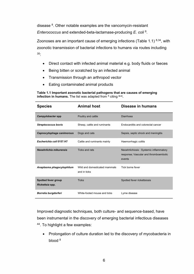
Zoonoses are an important cause of emerging infections (Table 1.1) 8,34, with
zoonotic transmission of bacterial infections to humans via routes including 35:
x Direct contact with infected animal material e.g. body fluids or faeces
x Being bitten or scratched by an infected animal
x Transmission through an arthropod vector
x Eating contaminated animal products
Table 1.1 Important zoonotic bacterial pathogens that are causes of emerging infection in humans. The list was adapted from 8 citing 36-43.
Species Animal host Disease in humans
Campylobacter spp Poultry and cattle Diarrhoea
Streptococcus bovis Sheep, cattle and ruminants Endocarditis and colorectal cancer
Capnocytophaga canimorsus Dogs and cats Sepsis, septic shock and meningitis
Escherichia coli 0157:H7 Cattle and ruminants mainly Haemorrhagic colitis
Neoehrlichia mikurensis Ticks and rats Neoehrlichiosis : Systemic inflammatory
response; Vascular and thromboembolic
events
Anaplasma phagocytophilum Wild and domesticated mammals
and in ticks
Tick borne fever
Spotted fever group Rickettsia spp.
Ticks Spotted fever rickettsiosis
Borrelia burgdorferi White-footed mouse and ticks Lyme disease
Improved diagnostic techniques, both culture- and sequence-based, have
been instrumental in the discovery of emerging bacterial infectious diseases 44. To highlight a few examples:
x Prolongation of culture duration led to the discovery of mycobacteria in
blood 8

7
x Performing culture on media inoculated with antibiotics made it
possible to grow Campylobacter and Helicobacter species 45,46
x Culture with non-mammalian cell lines is a promising approach for
culturing otherwise uncultivable bacteria 8
x PCR is a useful tool for detecting emerging infections from sterile sites
x 16S RNA sequencing has been useful in taxonomic classification of
emerging infections
1.1.4.1 Ecological factors
The steady growth in human population and the consequent increase in
population density aid the spread of bacterial infections among humans.
Travelling has become easier over the years and international trade and
tourism mean more and more people are travelling around the globe, which
further aids the dissemination of infectious agents. Expanding communities
continue to encroach deeper into natural habitats increasing the potential for
zoonotic transmission of emerging infections. In North America, the re-
emergence of Lyme disease has been associated with deforestation and loss
of habitat of its animal host 43.
The deadliest emerging infection of our time is HIV/AIDS, with consequent
bacterial infection as an emergent threat among HIV positive individuals 47.
Ironically, advances in medicine have contributed to the emergence of new
bacterial infections in vulnerable patients. When immunosuppression is
employed due to organ transplants or autoimmune disease it renders
patients vulnerable to infection. Patients being treated for chronic infections
such as cancer, diabetes and renal insufficiencies are also prone to impaired
immune systems and are vulnerable to infection. Urinary catheters have
contributed to the emergence of bacterial urinary tract infections like
Actinobaculum schaalii and Alloscardovia omnicolens 48.

8
1.2 Management of bacterial infection
1.2.1 Prevention of bacterial infection: vaccination
Vaccination is an important output from microbiology that embraces
synergies between molecular biology, immunology, public health and
biotechnology 49. The aim of vaccination is to induce long-term protection by
priming the human immune system with a component of a pathogen 50. The
general principle is that introduction of the vaccine leads to the production of
an immune response against the antigenic component of the vaccine, which
is protective when the body encounters the pathogenic microorganism.
Classical vaccines are mostly comprised of attenuated organisms,
inactivated whole-cell preparations or purified cellular components.
The discovery of the smallpox vaccine by the physician Edward Jenner in the
18th century paved the way for the eradication of smallpox 51. In the 19th
century Pasteur discovered that the causative agent of chicken cholera
Pasteurella multocida could be attenuated into less virulent forms 50.
Although the true rationale behind attenuation would not become apparent
until the latter part of the century, it formed the basis for Pasteur’s work on
rabies and anthrax vaccines 50. The start of the 20th Century was marked by
the discovery of the attenuated Mycobacterium bovis Bacille Calmette-Guérin
vaccine, which is still the only licensed vaccine for tuberculosis 52. During this
period, inactivated whole-cell vaccines were also produced against typhoid,
cholera and the plague 50.
Some bacteria secrete toxins that serve as important virulence factors 53. The
discovery that sub-lethal doses of inactivated bacterial toxins (toxoids) could
induce production of antitoxins in human serum, and thus confer protection,
led to the development of the tetanus, pertussis and diphtheria vaccines 50.
More recently, vaccines against Haemophilus influenzae type B, Neisseria
meningitidis and Streptococcus pneumoniae have been developed by
conjugating their capsular polysaccharide to protein molecules such as the
diphtheria toxoid 54-56.
9
1.2.2 Diagnosis of bacterial infection
Identifying the aetiological agent of infection facilitates the appropriate
delivery of antibiotic therapy, which increases the chances of treatment
success 57. Phenotypic methods for species identification, including
microscopy, culture and biochemical assays, are still widely used as
diagnostic tools. Classical taxonomy defined bacterial species as clusters of
organisms sharing phenotypic traits (e.g. cell morphology, growth conditions
and metabolic characteristics). This approach relies on being able to grow
the bacteria in the lab, usually a pure culture, and therefore precludes the
classification of uncultivable bacteria 58. Technological advances have made
DNA-based molecular techniques for pathogen detection more accessible
and affordable 57,59.
1.2.2.1 Molecular diagnostic approaches
Over time, species classification based on phenotypic traits was
complemented with molecular indicators such as GC content and whole-
genome DNA-DNA hybridisation, where a cut-off of 70% DNA-DNA
relatedness was recommended for classifying organisms into the same
species (60 cited in 61).
DNA-DNA hybridisation has now been replaced in most labs by the less
arduous 16S ribosomal RNA sequencing, leading to an updated species
definition which requires ≥97% sequence identity in the 16S rRNA gene 62.
16S rRNA sequencing is a suitable marker for phylogenetic analysis and was
instrumental in distinguishing Archaea as an independent domain of life 63.
However, despite its usefulness, 16S rRNA fails to discriminate between
some closely related species 63, even at a more stringent cut-off of 99%
sequence similarity (e.g. species within the genus Acinetobacter) 61.
PCR amplification of single gene loci is now the most widely used molecular
technique for pathogen detection in research and diagnostics laboratories 57.
The advantages of PCR-based detection include avoiding the onerous need
for culture and the ability to detect specific pathogens at low concentrations
10
and even from non-viable cells 64,65. In addition, multiplex PCR assays can
detect multiple pathogens in a single reaction 66,67. However, the chief
disadvantage of PCR-based detection is that only known or expected
species can be targeted.
1.2.2.2 Rise of genomics and metagenomics
Whole-genome sequencing (WGS) offers an opportunity to study the entire
DNA of an organism. This has immense value beyond simply identifying the
species—in addition, genomic data can be used for phylogenetic analysis
and genotypic characterisation of strains 68.
In the genome era, pairwise genomic comparisons have provided new
insights into bacterial diversity, prompting a review of the boundaries of
taxonomic classifications 69. In particular, measuring average nucleotide
identity through pairwise comparison of shared sequences in the genome
offers improved resolution at the species and sub-species level 70. An
average nucleotide identity of 95% or more corresponds to 70% or more
DNA-DNA re-association and strains with >94% average nucleotide identity
show consistent phenotypes in the context of species designation 69. A
similar metric based on amino acid identity is a useful measure for comparing
distantly related genomes 70.
Culture-free techniques like unbiased metagenomic sequencing are opening
new frontiers in clinical microbiology 71, offering potential for detecting and
characterising bacterial pathogens directly from clinical samples 72-74.
Metagenomics also identifies uncultivable potentially pathogenic bacteria and
offers a holistic snapshot of the microbiome 71.
1.2.3 Treatment of bacterial infection: antibiotics
The antibiotics era was ushered in by the discovery of naturally occurring
compounds – penicillin, streptomycin, chloramphenicol and tetracycline –
that have bactericidal properties 75. Antibiotics elicit bactericidal activity by
inhibiting important cellular functions such as synthesis of the cell wall, of
proteins, of DNA and of RNA 76. Despite the success of antibiotics as

11
therapeutic agents the antibiotics era has been marred by the widespread
emergence of antibiotic resistance 77.
Most antibiotics operate by binding to their target molecule with a high
affinity. Antibiotic resistance can be conferred by a number of mechanisms
such as 78:
1. Preventing the antibiotic from reaching its target through:
a. Decreased bacterial cell permeability by down-regulation of
porin channels 79
b. Increased removal of antibiotics from the bacterial cell through
efflux pumps
2. Modifying the drug target through:
a. Point mutations in the gene encoding the drug target
b. Homologous recombination with genes encoding insensitive
drug targets e.g. penicillin binding proteins
c. Modification of the target protein by methylation or binding of
proteins
3. Inactivating or modifying the antibiotic through:
a. Enzyme-catalysed hydrolysis of the antibiotic e.g. β-
lactamases
b. Chemical modification of the antibiotic e.g. aminoglycoside-
modifying-enzymes 80
Although a crisis looms, the development of new antibiotics is no longer
deemed economically viable 81. Instead pharmaceutical companies have
turned their attention the development of more lucrative therapeutic agents
for chronic non-communicable disorders 82. Antibiotic resistance is
recognised as a global emergency 83. Scientists are exploring novel targets
such as bacterial cell-division proteins as potential targets for future
antibiotics 84. Bacteriophage therapy is also re-emerging as a promising
alternative to antibiotics 85,86. Regulation, however, is a double-edged sword,
because although it may be beneficial in controlling the rise of existing
resistance, it can be an obstacle in pursuing the discovery of novel antibiotics 82,87.
12
1.2.4 Epidemiological typing to monitor spread of
bacterial infection
Microbial typing allows us to track the emergence and spread of bacterial
pathogens and in some cases can be used to retrace evolutionary history 88.
Typing methods that discriminate between closely related strains are
essential in identifying person-to-person transmission in an outbreak 89.
1.2.4.1 Phenotypic typing approaches
Phenotypic typing methods discriminate between strains on the basis of
phenotypic traits. Serotyping is perhaps the most widespread phenotypic
typing method. Serotyping exploits intra-species strain-specific differences in
the expression of cell-surface antigens. For example, variation in the
capsular polysaccharide of S. pneumoniae is used to classify strains into
over 90 serotypes. The gold standard for this is the Quellung reaction, which
detects a serotype-specific interaction between an antibody and the capsular
antigen. However, molecular techniques are now available for serotyping S.
pneumoniae based on detection of capsular genes 90,91.
Salmonella isolates are serotyped according to the immunological profiles of
their somatic (O) and flagellar (H) antigens. Serotyping has been the
cornerstone of epidemiological studies of Salmonella. However, keeping
stock of the antisera for all the antigenic variants is demanding. Molecular
approaches for serotyping Salmonella are gradually replacing conventional
techniques 92,93.
1.2.4.2 Molecular typing: MLST
The shortfall of the earliest typing methods was that they lacked a
standardized nomenclature for clonal lineages and they showed poor
reproducibility in unrelated datasets 94. Multilocus sequence typing (MLST)
succeeded in providing a standardised format for documenting gene diversity
to facilitate ease of inter-laboratory data comparison 95. MLST assigns a
13
sequence type (ST) based on the allelic combination of 7 housekeeping
genes that are conserved in a given species 95.
The nucleotide sequence for each of the seven housekeeping genes is
compared to known allele sequences that are stored in a curated database.
Each time a new sequence for a given gene is reported, it is designated a
new allele number. Each unique combination of alleles is designated as a
unique ST. STs are clustered into clonal complexes based on the sharing of
at least 5 allelic loci with at least one other ST in the clonal complex 96. By
convention the most abundant ST is designated as the founder of the clonal
complexes.
MLST is a robust tool for assessing population-based microbial diversity 57,
as shown by a few key examples:
x MLST has been instrumental in understanding the global population
structure of Staphylococcus aureus 97,98
x Through MLST, the impact of vaccine introduction on the genetic
diversity of Streptococcus pneumoniae has been studied 99
x MLST has been implemented in tracking the emergence and spread
of antibiotic-resistant strains of Neisseria meningitidis 100
x Associations between bacterial species and animal reservoirs have
been revealed through MLST 101,102
The ease of use and reproducibility of MLST make it the gold standard for
typing most common bacterial pathogens 103,104. However, MLST does not
offer enough discriminatory power to make it applicable for:
x Detailed outbreak investigation in most species 89
x Typing of genetically monomorphic bacteria like Mycobacterium
tuberculosis (Mtb) 88
14
1.2.4.3 Molecular typing for Mycobacterium tuberculosis
A wide range of molecular typing methods have been applied to Mtb,
including:
x IS6110-RFLP typing, which involves cleaving genomic DNA at
specific sites and using electrophoresis to separate the IS6110
fragments to yield a specific banding pattern
x Spoligotyping, which characterizes strains based on the presence or
absence of spacer units in the direct repeat locus
x Mycobacterial interspersed repetitive unit variable number tandem
repeat (MIRU-VNTR) analysis, which relies on the rapid rate of
change in repetitive DNA elements, to distinguish between isolates
with high-discriminatory power
Although spoligotyping can distinguish between strains at the sub-species
level it is limited by the fact that it uses less than 0.1% of the M. tuberculosis
genome. MIRU-VNTR offers higher discriminatory power than spoligo typing
but it lacks standardised laboratory protocols. IS6110-RFLP also offers high
discriminatory power between strains, but it requires an expert technician
and sophisticated hardware 105.
1.2.4.4 Genomic epidemiology
WGS using next-generation sequencing (see following section) has emerged
as a useful tool for the detection and characterisation of outbreaks, as well as
informing outbreak management 106,107. In fact, next-generation sequencing
is now ready to contribute routine pathogen surveillance 108-111. This kind of
digital surveillance twinned with effective data sharing will facilitate real time
analysis of outbreaks in the context of the global phylogeny 108. However,
whole-genome sequencing needs to be made accessible in low resource
settings, where the burden of infection is highest.
The problem with most existing genotyping methods is that they probe only a
very small proportion of the genome 112. WGS has emerged as a superior
technique for typing bacteria, since it can detect single nucleotide changes at
15
all genomic loci 113. WGS offers high-resolution inter-strain comparison,
which has improved our understanding of geographic distribution of species
and their evolutionary origins 114,115. Evolution within a species can be
investigated by reconstructing a phylogeny from variant sites in the core
genome 114,116.
To establish WGS as the gold standard for microbial typing, standardised
schemes for nomenclature and lineage assignment need to be devised. For
some species like Mtb, a robust typing scheme exists for characterising
strains based on a barcode of genome-wide single nucleotide variants
(SNVs) 117. In other bacteria, like Staphylococcus aureus, nomenclature of
clonal lineages inferred from MLST are still used to annotate whole-genome
phylogenies 118. A ribosomal MLST scheme that probes 53 ribosomal
proteins present in almost all bacteria has been proposed to ease the
transition from working with MLST to working with whole genomes 119.
1.3 DNA Sequencing
1.3.1 Sanger sequencing
The development of the DNA sequencing technique, now commonly known
as Sanger sequencing, by Frederick Sanger and his colleagues in 1977
remains a cornerstone of molecular biology 120. Sanger sequencing is based
on a DNA synthesis polymerase reaction in the presence of
dideoxynucleotide chain terminators 121. Initially, the reaction was run in
quadruplicate with different base terminators (ddA, ddC, ddT or ddG)
followed by gel separation in separate lanes. The user would then examine
the gel to reconstruct the nucleotide sequence at each position based on the
terminator base and fragment length 121.
The advent of fluorescently labelled dideoxynucleotide chain terminators
increased the throughput of Sanger sequencing and enabled automation.
This meant that the sequencing could be performed in a single reaction and
the strands could be separated through capillary electrophoresis 122. A
16
fluorescent detector connected to a computer was able to detect the
nucleotides and reconstruct the sequence in real time 123. Sanger
sequencing can generate reads up to a thousand base pairs long 124.
1.3.2 Whole-genome sequencing
The first bacterial genome sequences were published just over two decades
ago 125,126. Since then, there has been an exponential increase in the number
of bacterial genomes sequenced and our understanding of the bacterial
genome has improved significantly. This development stems from
technological advances in high-throughput sequencing, the decreased cost
of sequencing and improved means of data sharing 127.
Bacterial genome sequencing has evolved through three phases 128:
1. The whole-genome shotgun sequencing era
2. The next-generation sequencing era
3. The single-molecule sequencing era
The advent of shotgun cloning in WGS was the first breakthrough 128. In this
three-step process, genomic DNA is randomly sheared, the fragments are
cloned into E. coli and sequenced en masse by Sanger sequencing 128.
Bioinformatics tools were developed to assemble the short reads into contigs
(contiguous strings of bases). Shotgun cloning and subsequent whole-
genome sequencing provided novel insights into the biology of the deadly
human pathogens Mycobacterium tuberculosis 129 and Yersinia pestis 130 and
identified a myriad of novel genes in the model organism E. coli 131. Genome
sequencing that relied on shotgun cloning, however, had its drawbacks: most
notably that only clonable regions of the genome could be sequenced 128.
The introduction of next-generation sequencing to bacterial genomics
marked the second breakthrough. Next-generation sequencing involves
preparing templates by tagging genomic fragments with chemical adapters,
which are then amplified by PCR and undergo sequencing-by-synthesis on a
solid surface 132. This technology ushered in the era of bench top sequencers
like the Illumina MiSeq and the Roche 454 Jr 128. Bench top sequencers are
17
now commonplace in most research groups and for now Illumina dominate
the field with short-read sequencing 109.
Next-generation sequencing brought a massive improvement in throughput
and volume of data but at the cost of significantly shorter read length 132. This
meant that only draft assemblies could be generated, rather than complete
genome sequences. Inadvertently, this influenced bacterial genomics by
changing the emphasis from in depth analysis of single genomes to
comparative genomics of large dataset of closely related strains 128.
Single-molecule sequencing marks the third breakthrough in bacterial
genome sequencing, while also reviving the study of complete genome
sequences through long read lengths. Pacific Biosciences launched the first
widely used single-molecule sequencing technology, the PacBio. The PacBio
can generate high-quality finished genomes in combination with Illumina
short reads, or even on its own 133,134. This makes PacBio ideal for structural
genomics and plasmid analysis e.g. in multidrug resistant bacteria 135.
Unfortunately the PacBio is mostly restricted to major sequencing centres
due to the size of the instruments and its hefty price tag 128.
Oxford Nanopore sequencing remains a work-in-progress, despite proof-of-
principle that it can generate reasonably good draft genomes 128. Palmtop
sequencing with Oxford Nanopore’s Minion instrument has made on-site
sequencing of samples possible in outbreak surveillance even in remote
regions 136,137.
1.3.3 Bacterial genome analysis
Although most bacterial genomes consist of a single circular chromosome, a
few species have evolved to harbour multiple chromosomes 138,139. Bacterial
genomes are usually smaller than eukaryotic genomes and vary in size from
112 kB to 14 MB 140,141. The nucleotide content of bacterial genomes varies
markedly, e.g. the GC content of finished assemblies ranges from 15% to
85%. Bacteria from complex environmental niches tend to have larger
genomes and higher GC contents than host-associated bacteria 127.

18
1.3.3.1 Bacterial genome assembly and annotation
Over the years, technological advances in sequencing have been paralleled
by computational advances geared towards analysing sequence data.
Genome assembly involves stitching together millions of short sequence
fragments in an attempt to reconstruct the original genome. Reference-based
assembly methods map short reads to a reference genome and overlapping
fragments are assembled into contigs. However, reference-based mapping is
limited by the fact the regions that do not map to the reference are excluded.
De novo assemblers attempt to reconstruct the genome from scratch without
the aid of external data. This eliminates the bias towards the reference, but
the assemblies are generally more fragmented 124. De novo assembly is
recommended for new pathogens or novel strains of well-studied pathogens 142. Hybrid approaches that combine de novo assembly of long reads from
single-molecule sequencing and error correction with high accuracy short
reads have produced almost-complete genome assemblies 133,143. Contigs
generated from de novo assembly with short reads can be improved by
joining them into scaffolds using a reference guided approach 144,145.
Bacterial genomes show a good correlation between genome size and
number of genes and have low proportions of non-coding DNA 146. Automatic
annotation pipelines are often employed for annotating new sequences of
bacteria. The generic workflow of automated pipelines for annotating
bacterial genomes includes the following steps 147:
1. Predicting open reading frames from a FASTA sequence
a. GLIMMER is the most common tool for this 148
2. Comparing the predicted sequence to a reference genome or online
databases e.g. Uniprot
3. Search for a homologue
a. If there is a match, assign annotation from the reference
b. If there is no match, then annotate the coding sequence (CDS)
as a hypothetical protein
4. Predict functional domains for the protein and update annotation
5. Predict other non-coding features e.g. non-coding RNAs
19
The exponential increase in the number of prokaryotic genomes available
provides an amazing resource for comparative genomics. However, one
must proceed with caution when transferring annotation from one genome to
the next, as there are inconsistencies in the annotation protocols and
pipelines implemented by different research groups 147. In addition, programs
like Microsoft Excel have been shown to corrupt gene names 149, while
spelling mistakes and use of inconsistent product names for the same gene
compounds this problem 147. For all these reasons, to achieve high-quality
annotation, the predicted annotation must be manually curated.
1.3.3.2 Insights into pathogen biology
Bacterial genomics has been instrumental in shaping our understanding of
host-pathogen interactions. Knowing the sequence for all genes in the
genome has given crucial insights into the biology of bacterial pathogens.
Through the genome, we are able to study the full profile of virulence genes
that mediate an organism’s pathogenesis 150. WGS has been instrumental in
the detection and characterisation of novel variants of important virulence-
associated genes e.g. WGS has characterised diversity in the cagA gene,
the most important virulence factor in Helicobacter pylori infection 151.
Genome-wide association studies can be used to dissect the genetic basis
for phenotypic traits in bacteria 152. A genome-wide association study of
Campylobacter identified a seven-gene vitamin B5 biosynthesis region as a
host-specificity factor 153. In the Campylobacter jejuni genome, the presence
of imported genes has been linked with evolutionary relationships with
specific host species 154,155. In Mycobacterium tuberculosis genome-wide
association studies have been employed to detect antibiotic resistance
mutations based on convergent evolution 156. Genome-wide associations in
bacteria are confounded by the strong forces of natural selection that are
exerted on bacterial populations 152.
Advances in next generation sequencing have made it possible to sequence
the entire bacterial transcriptome of a strain 157. The transcriptome is key to
understanding how bacteria respond to changes in their environment.
20
Genomics has also provided insights into the role of regulatory non-coding
RNA. Regulatory non-coding RNA’s can affect the physiology of bacteria by
binding to messenger RNA in order to reduce translation efficiency 158.
1.3.3.3 Discovery of novel targets
Advances in genomics have led to the discovery of novel targets for
diagnostics, antibiotic and vaccines 159. Comparative genomics can identify
diagnostic targets that are conserved within a species, but are not found in
other species. These targets can then be incorporated into real-time PCR
diagnostic assays 160.
In the genomics era, scientists have looked increasingly towards genomics
solutions to the rising antibiotic resistance problem 161,162. To take just one
example: in 2014, Mobegi et al published a genomic approach for
systematically discovering novel antibiotic targets in three leading respiratory
pathogens; Streptococcus pneumoniae, Haemophilus influenzae and
Moraxella catarrhalis 163. Their approach exploited high-density transposon
mutagenesis, high-throughput sequencing, and integrative genomics to
identify essential genes that were conserved, but unlikely to interfere with
host function or the microbiome. As proof of concept they identified four
novel targets that were experimentally validated 163. Their approach
overcame the shortfalls in specificity and throughput that held back previous
attempts to discover novel targets from genomic data 164,165. As genomics
screening for antibiotic targets circumvents the need for traditional laboratory
screening, this can fast-track the discovery of novel antibiotics 163.
The genomics era has also revolutionised vaccine development through
“reverse vaccinology”, which mines bacterial genomes for proteins that can
potentially elicit protective immunity as candidates for subunit vaccines 166-
168. Comparative genomics of multiple strains can predict the suitability of a
candidate protein by determining how conserved it is across diverse strains 169. Reverse vaccinology has the potential to lead to vaccines for bacteria
where the conventional approaches have failed 167,169.
21
1.4 Bacterial genome evolution
Bacterial evolution is driven by the accumulation of genetic variation in the
bacterial genome. WGS has enhanced our understanding of the evolutionary
mechanisms that shape major human pathogens, documenting
unprecedented genomic diversity between and within bacterial species 127,170.
Understanding microbial evolutionary mechanisms through the bacterial
genome is central to refining intervention measures to counteract pathogenic
bacteria.
1.4.1 Processes shaping bacterial evolution
1.4.1.1 Mutation, insertion, deletion
Point mutations are the smallest units of evolution 171. Point mutations result
from the substitution of a single nucleotide (referred to as SNV) or from the
insertion or deletion of a single nucleotide. The convergence of point
mutations in multiple independent lineages is an indication that the mutation
is under positive natural selection 171. Point mutations may confer a selective
advantage that leads them to become fixed in the population.
SNVs that occur in protein-coding genes can yield synonymous mutations
that code for the same amino acid or non-synonymous mutations that result
in a change in the amino acid, potentially altering protein function. However,
point mutations in non-coding regions can also affect gene function,
especially if they occur in promoter regions 171. Deletions and insertions can
introduce a frameshift mutation that leads to downstream sequences being
translated in a different reading frame, with major effects on protein
sequence, structure and function. Similarly, nonsense mutations result in
premature stop codons that truncate proteins with severe consequences for
function.

22
1.4.1.2 Rearrangement and homologous recombination
Genome rearrangements are structural variations involving large chunks of
DNA that can result from cellular process such as replication, DNA repair and
DNA recombination 172. The impact of genome rearrangements on the
evolution of eukaryotes has been widely studied and there is growing
evidence that genome rearrangements are important evolutionary
mechanisms in bacteria 172. Genome rearrangements can alter the order of
genes as well as their orientation and copy number. These changes can
affect the phenotype of an organism. Evidence shows the rearrangements
can also influence gene expression 173,174.
The bacterial genome is highly flexible and can undergo large sequence
variation through insertions, deletions, inversions, translocations and
duplications 171.
1. Deletions can occur in intergenic regulatory regions and lead to gene
inactivation. Deletions can also lead to loss of functional domains of
proteins and altered gene function 172
2. Duplications can have functional implications depending on the
variation in the duplicated segment and how the segment is inserted 172
3. Insertions can introduce novel segments into the genome. The most
well studied mechanism of insertion is horizontal gene transfer (HGT) 175 . Mobile genetic elements (MGEs) can mediate the insertion of
genomic segments from other loci within the genome. The functional
implications of insertions are also governed by the genetic nature of
the insert and context of the insertion loci 172
4. Translocations involve the exchange of DNA segments within the
same genome. They are important contributors to genome plasticity.
Translocations can affect chromosome position, gene orientation and
the distance between a target gene and its transcription factors 176
5. Inversions flip the orientation of genomic segments

23
1.4.1.3 Recombination and horizontal gene transfer
Plasticity within the bacterial genome allows bacteria to adapt to selective
pressures from the environment and their hosts 177. Understanding the
evolutionary mechanisms that confer genome plasticity in bacteria can yield
valuable insights into their pathogenesis and adaptive mechanisms. Bacteria
are able to acquire foreign DNA through HGT 175. HGT is the largest source
of variation in the genome. Uptake of foreign DNA by bacteria is mediated
mainly through three processes:
1. Transformation involves the incorporation of exogenous DNA from
the surroundings into the host 2. Conjugation results from direct cell-to-cell contact between cells and
transfer of DNA via a sex pilus 3. Transduction is mediated by bacteriophages – viruses that infect
bacterial cells
Recombination is a DNA repair process in bacteria that facilitates the
incorporation of foreign DNA into the bacterial genome 178. Recombination
maintains genetic diversity in bacterial populations while preventing the build-
up of unfavourable changes 171. Homologous recombination classically
involves the interaction of two strands of DNA with high sequence similarity.
However, recombination can also account for HGT wherein highly divergent
sequences are incorporated into the genome 171. Moreover, recombination
can mediate the incorporation of DNA acquired through phage mediated
transduction as well as conjugation 171.
1.4.1.4 Mobile Genetic Elements
MGEs shape the evolution of bacterial pathogens by mediating movement of
DNA within the genome and between species 179. MGEs introduce diversity
in the accessory genome mainly through plasmids, bacteriophages and
genomic islands 180. Plasmids are large extra chromosomal strands of DNA,
linear or circular, that replicate independently and carry a wide range of
genes 180. Plasmids can carry a large number of genes that introduce
complex phenotypic traits and lead to quantum leaps in evolution 181,182.
24
Plasmids can be transferred, mainly by conjugation, between distantly
related species and even between bacteria and some eukaryotes 183.
Bacteriophages are viruses that infect bacteria. Bacteriophages can replicate
within a bacterial cell and kill the cell within minutes of contact when the lytic
cycle is activated 184. However, in the lysogenic cycle the lytic cycle is not
activated and the phage genome can be integrated into the host
chromosome 184 to become a prophage 171. Prophages can remain dormant
in the chromosome until they are reactivated. In the pseudolysogenic cycle,
the phage genome remains in the cytoplasm as an episomal plasmid 184.
Bacteriophage genomes may encode genes that can greatly enhance
virulence and transmissibility such as exotoxins 185. In some cases phages
are involved in the transfer of toxins as well as their activation 186. Phages
can alter the microbiome by killing off competitors 187.
Genomic islands are regions of the genomes that are believed to have
evolved through the interchange of plasmids and bacteriophages 180.
Genomic islands are present in some strains but are not conserved across a
species. Genomic islands can carry genes that mediate genetic mobility but
also frequently carry virulence-associated genes 180. Pathogenicity islands
are genomic islands that carry one or more virulence genes and are present
in the genomes of pathogenic strains but absent in non-pathogenic strains
from the same or closely related species 188. Pathogenicity islands play an
important role in mediating the virulence of bacterial pathogens in a range of
host organisms 188.
1.4.1.5 Genome degradation and deletion bias
Genome degradation is believed to be an important evolutionary mechanism
that sometimes contributes to the evolution of bacterial pathogenicity 189,190.
Comparing the genomes of some ancient human pathogens (Yersinia pestis,
Salmonella enterica serovar Typhi CT18 and Mycobacterium leprae) with
genomes from closely related species reveals extensive genome degradation
and gene loss 191-193. The observed genome degradation in these species is
believed to mark a “change in lifestyle” for these organisms towards
25
occupying a simpler host niche 190. Gene loss may also contribute to
diminished virulence: the Salmonella species S. bongori has multiple deletion
regions compared to its more virulent close relative S. enterica 194.
Conversely the loss of the antivirulence cadA gene in Shigella flexeneri
increases the invasiveness of the species 195. cadA deactivates the enzyme
lysine decarboxylase, whose product inhibits the organisms plasma-encoded
virulence factors 195.
Comparative genomics has revealed that some species show an
evolutionary bias towards gene deletion 196. Studying the dynamics of
genome degradation can provide insights into the mechanisms bacterial
pathogens employ in host specialisation. The smallest bacterial genomes
occur in intracellular pathogens and symbionts 197. The constant supply of
nutrients from the host allows these bacteria to survive with a shrunken
genome and reduced functionality 197.
1.4.2 Pan-genomes
All of the genes within a bacterial species make up its pan-genome, which is
comprised of a conserved core genome and a flexible accessory genome 198,199. The bacterial core genome largely consists of housekeeping genes
that are conserved across the entire species 177. Accessory genome
elements mediate important evolutionary mechanisms such as host
adaptation, virulence and antibiotic resistance 200. Bacterial species that live
in highly specialised niches tend to have closed pan genomes that are finite 201. More generalist species possess open pan-genomes that increase with
the addition of more strains, and are characterised by high rates of HGT 201.
The unprecedentedly high rates of HGT in bacterial genomes have led
scientists to question the existence of bacterial species 62, particularly as one
might suppose that it would not be possible for a bacterial species to
maintain its identity in such a genetically promiscuous environment.
However, the existence of phenotypically stable clusters despite the
observed genome fluidity is explained by the core genome hypothesis. The
core genome hypothesis attributes the stability of phenotypic clusters to a
26
repository of core genes (the core genome) that maintain the identity of a
species, despite rampant changes in the accessory/flexible genome 62.
The core genome hypothesis predicts that core genes are primarily under
purifying selection as a mechanism to maintain existing function 62. Core
genomes are believed, on average, to express a neutral rate of evolution
while a variety of selective pressures are levied on accessory genes 62. The
core genome hypothesis is, however, challenged by the existence of open
pan genomes in some species and the high rates of homologous
recombination between closely related bacteria 202.
1.4.3 Bacterial evolution in monomorphic lineages
Genetically monomorphic lineages are bacterial taxa that are characterised
by very low sequence diversity to such an extent that sequencing
housekeeping genes from divergent strains yields few if any SNVs 88. This
trait precludes the use of standard genotyping techniques such as MLST to
discriminate between strains from monomorphic lineages, and, thus, the
evolutionary history of monomorphic lineages remained unclear until the
widespread application of whole-genome sequencing 88.
Some of the deadliest human pathogens form genetically monomorphic
lineages that have emerged from species with greater diversity. But why do
monomorphic lineages occur? One explanation is that this kind of evolution
intensifies disease transmission 203. Often the population dynamics of
genetically monomorphic lineages are distinct from those of the supra-
species 88 e.g.:
x Y. pestis (causes plague, including the Black Death), is a recently
emerged clone of the species Yersinia pseudotuberculosis 204
x Mycobacterium tuberculosis (causes tuberculosis, the white plague) is
part of a much more diverse Mycobacterium tuberculosis complex 205
x Bacillus anthracis (causes anthrax) is part of the Bacillus cereus
group of soil-dwelling saprophytes 206
27
Similarly, highly virulent monomorphic clades can exist within genetically
diverse species e.g.:
x The O157:H7 clade of E. coli, which infects cattle and can sometimes
infect humans inducing haemorrhagic colitis and the haemolytic–
uremic syndrome 207
x The serovar Typhi of Salmonella enterica the cause of typhoid fever 208
x The O1 or O139 clade of Vibrio cholerae, the cause of pandemic
cholera 209
There is evidence to suggest that Darwinian selection is a leading contributor
to the evolution of bacterial pathogens 210. Darwinian selection has been
demonstrated in laboratory experiments 211 as well as in nature. For
example, the widespread use of fluoroquinolones over more than twenty
years has resulted in decreased susceptibility in all lineages of serovar Typhi
due to accumulation of mutations in the gyrA gene 208. However, Darwinian
selection for a single trait sometimes leads to only transient selection,
because the selected variants may prove less fit than their progenitors in the
absence of the selective pressure 212. This might explain why susceptible
clones of serovar Typhi have co-evolved with the resistant clones.
Comparative genomics suggests that genetically monomorphic lineages are
under neutral selection, with mild purifying selection 203. Comparative
genomics shows that synonymous and non-synonymous mutations are
accumulated at similar rates in these lineages 115,205,213. Moreover there is
limited evidence of convergent evolution in these lineages since they exhibit
almost negligible levels of homoplasy, with the exception of antibiotic-
resistant genes 203.
Although monomorphic lineages are devoid of traces of recent homologous
recombination, genomic evidence suggests the occurrence of recombination
events that predated the emergence of some lineages 214,215. The absence of
HGT makes genomic deletions useful phylogenetic markers in monomorphic
lineages like M. tuberculosis 216.
28
1.4.4 Phylogenetic dating
In 1962, Linus Pauling and Emile Zuckerkandl noted a linear correlation
between the number of amino acid differences in haemoglobin and time, as
determined by fossil evidence 217. This formed the basis of the molecular
clock theory, which states that the rate of evolutionary change in a given
protein is constant over time and across different lineages 218.
The assumption that mutation rates are constant in lineages has been known
to obscure phylogenetic inference. To overcome this barrier statistical
methods like maximum likelihood and Bayesian inference have been
employed. These methods use a “relaxed molecular clock” that assumes an
independent mutation rate in each lineage 219.
In order to infer dates using a molecular clock, the mutation rate must be
calibrated with independent dating evidence from sources such as fossils or
ancient DNA 220. Once a mutation rate is established for a species, it can be
employed to estimate the time since divergence of closely related or distant
strains.
1.4.5 Population genomics and evolution
1.4.5.1 Within-host evolution and transmission history
Classical genotyping techniques can identify the existence of mixed infection
due to different lineages 221,222. However, they lack the resolution to trace the
minute evolutionary changes that occur within a single lineage during
colonisation. Advances in WGS continue to shed light on the evolution of
single bacterial strains within a host 223. Microevolution is driven mainly by
point mutations and in a number of species the short-term evolution rates are
much higher than the long-term evolution rates 224-226. Clouds of diversity can
result from microevolution; within these clouds, variants with different
phenotypic traits can potentially co-exist 227. Changes in the host immune
system or antibiotic treatment introduce selection bias that selects for the
most favourable trait 227.
29
Most transmission studies rely on single genomes isolated from patients and
do not take into consideration the effect of microevolution, which is a
problem, as cloud diversity potentially hinders the ability to accurately infer
transmission networks 228. To understand cloud diversity within a host,
multiple genomes need to be sampled—at different time points and/or at a
single time point but from multiple colonies 229. Moreover, heterogeneous
transmission from one host to another may occur whereby multiple
genotypes are transmitted 228,229. Co-colonisation of multiple species that are
part of the normal flora can lead to recombination which further complicates
efforts to detect transmission chains 229.
1.4.5.2 Selection for antibiotic resistance
As noted previously, the world is going through a crisis in antibiotic
resistance driven largely by overuse and inappropriate prescription of
antibiotics 82. The natural environment is replete with antibiotic resistance
genes, which when transferred to pathogenic bacteria, render them resistant 230. On top of that, bacteria can spontaneously accumulate mutations that
confer antibiotic resistance. Resistant variants typically form minority
populations because they are often less fit than their susceptible
counterparts. However, in the presence of antibiotic treatment, a selection
sweep occurs within the host-associated bacterial population so that
antibiotic-resistant variants become dominant 229. WGS has documented
microevolution within a single host leading to the emergence of drug-
resistant bacteria 231-233.
The large-scale use of antibiotics has rendered antibiotic resistance a
selective advantage leading to the emergence of almost impossible to treat
superbugs 234. Relaxed stewardship of antibiotics in hospitals means that
vulnerable patients are at an increased risk of nosocomial antibiotic-resistant
infection 81. Widespread use of antibiotics in agriculture and animal
husbandry, particularly in developed countries, has also contributed to the
rise in antibiotic resistance 81,235.
30
1.4.5.3 Vaccine-induced selective pressure
Vaccines have proven to be an effective means of eradicating or
substantially reducing the prevalence of infectious diseases. However,
occasionally some infections persist despite the availability of a vaccine and
some vaccinated individuals remain susceptible. This may arise due to the
selection of adaptive features that counteract the impact of the vaccine 221.
Adaptive features include evasion and virulence evolution 221. Evasion arises
through the emergence of novel mutants that can escape the immune
response induced by the vaccine 221. An example of this is the serotype
replacement phenomenon that occurs in S. pneumoniae in response to
conjugate vaccines 236,237.
Bacteria can alter their virulence mechanisms as an evolutionary mechanism
to escape the effect of a vaccine. A classical example is the diphtheria
antitoxin vaccine, which primes the immune system against the diphtheria
toxin, the main virulence agent of Corynebacterium diphtheriae 238. However,
despite vaccination, a resurgence of diphtheria has been observed in Russia
and surrounding territories. Genomic analysis showed that this resurgence
was driven by the persistent circulation of non-toxinogenic strains 239.
Worryingly there is evidence that these strains are evolving into toxinogenic
forms and driving a re-emergence 239. In the UK, vaccine pressure led to an
increase in the prevalence of diphtheria due to the closely related species C.
ulcerans 240.
1.5 Bacterial infection in West Africa: three
case studies
1.5.1 West Africa: geography and demographics
Africa is comprised of five regions: Northern Africa, Eastern Africa, Southern
Africa, Central Africa and Western Africa. Despite being replete with natural
resources, Africa is home to some of the poorest communities in the world
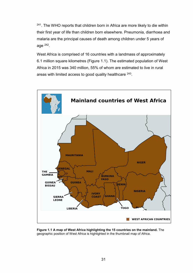
31
241. The WHO reports that children born in Africa are more likely to die within
their first year of life than children born elsewhere. Pneumonia, diarrhoea and
malaria are the principal causes of death among children under 5 years of
age 242.
West Africa is comprised of 16 countries with a landmass of approximately
6.1 million square kilometres (Figure 1.1). The estimated population of West
Africa in 2015 was 340 million, 55% of whom are estimated to live in rural
areas with limited access to good quality healthcare 243.
Figure 1.1 A map of West Africa highlighting the 15 countries on the mainland. The geographic position of West Africa is highlighted in the thumbnail map of Africa.
32
West African countries, with the exception of Mauritania, form the Economic
Community of West African States (ECOWAS), an economic partnership that
mandates free movement of people and goods between member states 244.
This union has obvious economic benefits for the region, but it presents a
unique public health challenge: the mass movement of people and goods
can facilitate the spread of human infectious diseases. Cross-border cattle
trading also presents an opportunity for the spread of zoonotic pathogens
across the region 245.
Traditionally, government spending on healthcare has been low in West
Africa. The recent Ebola outbreak in West Africa highlighted the fragility of
healthcare infrastructure in the region 246. Neighbouring countries could do
little more than close their borders and watch as this deadly disease ravaged
through parts of Sierra Leone, Liberia and Guinea.
Understanding the biology and epidemiology of pathogens is fundamental to
a successful intervention strategy. Here, I introduce a case study of three
important pathogens in West Africa. I describe a unique scenario associated
with each pathogen and present WGS as a solution to the problem.
1.5.2 Staphylococcus aureus
1.5.2.1 Clinical features and pathogenesis
S. aureus is a Gram-positive commensal that can cause infection in humans.
It is estimated that 50-60% of the human population are intermittently or
persistently colonized with S. aureus 247. The most common site of
colonisation is the anterior nares, but other sites such as the skin, perineum
and pharynx are also commonly colonised by S. aureus 248. Colonisation with
S. aureus places humans at risk of subsequent infection with the colonizing
strain 249,250. S. aureus can cause superficial skin and soft tissue infections,
as well as more invasive life threatening infections such as sepsis,
endocarditis, meningitis and pneumonia 251,252. S. aureus is a leading cause
of bacteraemia and skin and soft tissues infection globally 252-255.
33
Neutrophils are the host’s first line of defence against S. aureus infection 247.
Once engulfed in neutrophils, S. aureus can avert apoptosis through the
release of leukotoxins such as Panton-Valentine leukocidin, which promote
the direct lysis of neutrophils by permeating the plasma membrane 256. S.
aureus employs other mechanisms to evade the host immune system and
invade host tissue, which include 251:
x Surface proteins, which mediate the adhesion to and invasion of host
cells as well as biofilm formation. Variation of the surface proteins
allows S. aureus to evade the host immune system
x α-Hemolysin, which causes host cell cytolysis and induces neutrophil
recruitment. α-Hemolysin plays an important role in the pathogenesis
of S. aureus in skin and soft tissue infection
x Coagulases, which interfere with the host defences by promoting
coagulation
Worryingly, drug resistance has emerged for virtually all antibiotics used to
treat S. aureus including the “last-line” methicillin and vancomycin 257.
Hospital-associated MRSA levies a significant economic burden on health
care services and can lead to loss of life 258. The rise in morbidity and
mortality due to community-associated MRSA is a growing public health
problem 259.
1.5.2.2 The burden of staphylococcal infection in West Africa
Population-based studies, mainly in developed countries, estimate the
burden of S. aureus bacteraemia to be approximately 15–40 per 100,000
population per year 260. S. aureus is the fourth commonest cause of invasive
disease in sub-Saharan Africa and, in The Gambia, it is the second
commonest pathogen isolated from patients with bacteraemia 261-263. Despite
the importance of this pathogen, serious knowledge gaps still exist regarding
the epidemiology of S. aureus in sub-Saharan Africa. Studies in Ghana,
Senegal and Nigeria show that a higher proportion of urinary tract infections
are due to S. aureus compared to Europe 264,265. It is likely that the burden of
S. aureus in West Africa is underestimated since the majority of cases are
34
likely to go undetected or treated empirically without laboratory confirmation.
Moreover in West Africa research efforts are more concentrated on other
infectious agents such as HIV, tuberculosis and malaria that are greater
threats to public health.
1.5.2.3 Potential for animal reservoirs in West Africa
S. aureus colonises a wide range of hosts including domestic animals 266 and
wild animals 267. The epidemiology of S. aureus in non-human primates has
been studied in remote regions of West Africa using MLST 268. Although
these studies describe the population structure of S. aureus in non-human
primates, they do not investigate the molecular mechanisms that govern host
adaptation. Moreover, these genotyping techniques cannot ascertain whether
the non-human primates can serve as a reservoir for highly virulent S. aureus
that can infect humans.
Humans come into frequent contact with Green Vervet Monkeys in The
Gambia. This interaction provides opportunities for the exchange of
pathogenic bacteria between humans and monkeys. In 2011 The Vervet
Consortium partnered with the MRC Unit The Gambia to investigate the
epidemiology of Simian Immunodeficiency Virus in Green Vervet monkeys in
The Gambia. Speculatively, nasopharyngeal swabs were collected from the
monkeys and ~30% of the monkeys were asymptomatic carriers of S.
aureus.
My task was to employ WGS to infer the evolutionary origin of S. aureus in
monkeys in The Gambia. This involved investigating the existence of inter-
species transmission of S. aureus and attempting to elucidate the
mechanisms that govern host adaptation. Through this analysis, I sought to
establish whether monkeys posed a threat as a reservoir of highly virulent S.
aureus in The Gambia.

35
1.5.3 Streptococcus pneumoniae
1.5.3.1 Clinical features and pathogenesis
S. pneumoniae has a propensity to colonise the young, the elderly and the
immunocompromised 269. Successful colonisation by S. pneumoniae is a
prerequisite to infection, which usually occurs within a month of colonisation 270. The carriage rates of S. pneumoniae are higher in children under 5
compared to older children and adolescents 270. Carriage of S. pneumoniae
can lead to transmission through respiratory droplets and children are the
main source of transmission 270.
From the upper respiratory tract, S. pneumoniae can invade the lungs
through aspiration to cause pneumonia (by invading the alveoli) or empyema
(by invading the pleura or the pericardium) 269. S. pneumoniae can also enter
the bloodstream and cause bacteraemia/sepsis 271. Bloodstream invasion by
S. pneumoniae can lead to this pathogen crossing the blood-brain barrier to
cause meningitis; pneumococcal meningitis is associated with a high case-
fatality rate and long-term neurological damage 271. S. pneumoniae can also
spread locally from the upper respiratory tract to cause otitis media and
sinusitis 269.
The pneumococcal capsule, which is the main virulence factor of S.
pneumoniae, is important for nasal colonisation 272. Adhesion to epithelial
cells is a pre-requisite for colonisation. Adhesion is mediated through the
binding of pneumococcal surface proteins such as pneumococcal surface
adhesin A and choline-binding protein A to cell surface carbohydrates on
epithelial cells 273. The enzyme neuraminidase reduces the viscosity of the
mucosal layer making the epithelial surface more accessible for adhesion 274.
36
1.5.3.2 The burden of pneumococcal infection in sub-Saharan
Africa
In sub-Saharan Africa, rates of pneumococcal carriage are generally high,
but there is a paucity of data from most countries 275. Rates of invasive
pneumococcal disease are higher among younger children and
immunocompromised individuals, due to higher rates of carriage 276. Other
risk factors for invasive disease include respiratory viral co-infection,
overcrowding and lack of vaccination 270. S. pneumoniae is the most
common cause of pneumoniae among children and is one of the major killers
among HIV infected individuals in sub-Saharan Africa 277. In West Africa,
lethal outbreaks of pneumococcal meningitis have been reported across the
so-called African meningitis belt 278,279.
1.5.3.3 Vaccination and genomic outbreak surveillance
Pneumococcal conjugate vaccines are efficient intervention tools for
preventing pneumococcal infection. They reduce nasopharyngeal carriage in
both vaccinated and unvaccinated individuals and reduce the burden of
invasive disease due to vaccine serotypes 280-282. However, throughout most
of West Africa the impact of vaccination on the molecular epidemiology of S.
pneumoniae is unknown due to the lack of carriage surveillance or extensive
disease surveillance.
The pneumococcal meningitis outbreak that broke out in the Brong-Ahafo
region of Ghana in December 2015 was a reminder that vaccination is not
always the end of the story. This outbreak occurred two years after the
introduction of the 13-valent pneumococcal conjugate vaccine, PCV-13.
During the outbreak sample integrity was a major shortfall and the majority of
samples were not viable for culture. The cause of the meningitis outbreak in
Ghana would thus have remained unknown without the use of the
quantitative PCR assay for detection of bacterial pathogens from human
cerebrospinal fluid 283. Quantitative PCR identified serotype 1 S.
pneumoniae, a vaccine serotype, as the main etiological agent from the
37
outbreak 283. But how could this happen in a country where PCV-13 had
been administered to over 80% of the children under five years of age?
Fortunately, a handful of viable isolates of S. pneumoniae were salvaged
from the outbreak and as described in a subsequent chapter, were analysed
using WGS. Previously sequenced serotype 1 genomes from West Africa
served as a baseline for comparing the outbreak strains. This made it
possible to identify evolutionary changes that distinguished the outbreak
strains. This analysis demonstrated the power of genomics in outbreak
surveillance and reiterated the fact that to characterise an outbreak properly,
it must be analysed in the context of the global phylogeny 108.
1.5.4 Mycobacterium tuberculosis
1.5.4.1 Clinical features and pathogenesis
Tuberculosis is caused by a group of closely related ecotypes that form the
Mycobacterium tuberculosis complex 284. Mycobacteria are slow growing,
intracellular, non-motile, non-sporulating, acid-fast bacilli. Mycobacteria have
a very thick cell wall, uniquely multi-layered, with varied hydrophobicity. The
inner layer consists of hydrophilic peptidoglycan sacculus and
arabinogalactan layers, covalently linked to a hydrophobic outer layer of
mycolic acids that form a waxy mesh of long chain fatty acids 285,286.
A hallmark of the cell-mediated immune response to tuberculosis infection is
the formation of granulomas 287. Granulomas can suppress the spread of
bacteria within tissues by trapping the bacilli within macrophages in a mesh
of immune cells 288. However, mycobacteria can survive in an encapsulated
form in lung granulomas for several years in a state of latency 288. In a tenth
of cases, the tubercle bacilli will escape from the granuloma and spread
through the body to cause active disease.
The most common site of tuberculous infection is the lungs, but other extra-
pulmonary sites including the bones, brain and kidneys can also become
infected 289. Tuberculosis is transmitted by aerosol from patients with active
disease through coughing, sneezing, laughing or even talking 289. Symptoms
38
of tuberculosis may include persistent cough, productive cough, malaise,
night sweats, weight loss, loss of appetite and fever 289.
Treatment of tuberculosis is complicated by the emergence of resistance to
anti-TB drugs. Efforts to eradicate tuberculosis have been set back by the
emergence of MDR-TB and extensively drug resistant (XDR-TB) forms of
tuberculosis. MDR-TB is defined as resistance to at least isoniazid and
rifampicin, the two most powerful first-line anti-TB drugs 290. XDR-TB are
MDR-TB that are also resistant to a fluoroquinolone and one of the second-
line injectable drugs (amikacin, kanamycin or capreomycin) 291.
1.5.4.2 The burden of mycobacterial infection in West Africa
Tuberculosis is a major cause of death globally and the highest-burden
regions are Africa and Southeast Asia. In 2015, over 1.3 million cases of
tuberculosis were notified in Africa 292. However, among the notified cases of
pulmonary tuberculosis, only 64% were bacteriologically confirmed 292. Most
health facilities in the continent lack the facilities for bacteriological
confirmation of tuberculosis and rely on expert opinion based on clinical
symptoms. This highlights an urgent need to strengthen diagnostic capacities
in Africa.
In 2015, there were an estimated 480,000 new cases of MDR-TB 292.
Estimating the burden of MDR-TB in Africa is challenging due to lack of
accurate surveillance data. In 2015, the reported treatment success rates
among MDR-TB and XDR-TB patients were 54% and 24% respectively 292.
Poor adherence due to lack of awareness among patients and shortages of
medication are a major factor is treatment failure. However it is worth
considering the impact of misdiagnosis at primary health care facilities on the
treatment outcomes of MDR-TB and XDR-TB. Most primary health facilities
will not be able to perform drug susceptibility testing and are likely to place
patients with MDR-TB on an inadequate treatment regimen before they are
referred to the tertiary referral centres for retreatment.
Between 2009 and 2013, the West African Nodes of Excellence for
Tuberculosis AIDS and Malaria (WANETAM) carried out surveys in eight
39
West African countries to estimate the true burden of drug-resistant
tuberculosis 293. At the time, only Senegal, Gambia and Nigeria had
performed drug-resistance surveys in the sub-region. The estimates of drug
resistance from WANETAM were higher than WHO estimates, suggesting
that the burden of MDR-TB in West Africa could be underestimated 293. In
particular, the prevalence of MDR-TB among retreatment cases in Mali
(59%) and Nigeria, Ibadan and Lagos (39 % and 66 %), was significantly
higher than WHO estimate for prevalence of MDR-TB among retreatment
cases in the African region (20%) 293.
1.5.4.3 Knowledge gaps for tuberculosis in West Africa
Molecular epidemiology studies that employ classical genotyping techniques
have been carried out across West Africa. These studies involve a range
sampling strategies and isolated communities, making it challenging to
compare the epidemiology across countries 294. MTBC in West Africa is
highly diverse, with six of the seven global lineages present in the region 294.
Despite this rich diversity very little is known about the evolutionary history of
MTBC in West Africa, especially of the lineages and genotypes that are
exclusively endemic to this region. Moreover, classical genotyping
techniques do not have the resolution to distinguish between closely related
strains and to infer on going transmission.
The WANETAM dataset of over 900 archived MTBC isolates presents an
ideal platform through which we can begin to understand the evolution of
MTBC in this region. WGS holds the potential to offer novel insights into the
phylogeography of MTBC in West Africa. WGS has the power to unearth on
going chains of transmission and even detect microevolution of MTBC within
the host over the duration of treatment. With the phenotypic drug-resistance
profiles already known, the genome can be studied to understand the
molecular mechanisms that are driving drug resistance in this setting.

40
1.6 Aims and objectives
The time has come for West Africa to embrace the genomics era and exploit
the full potential of microbial genomics. Genomics offers unprecedented
insights into the epidemiology and biology of infectious diseases, which
dominate the public health agenda in West Africa. In this thesis, I aim to
exploit the sequencing and analysis of whole bacterial genomes to
understand the evolution and spread of bacterial infection and antimicrobial
resistance in West Africa. My work focuses on three important human
pathogens in different contexts.
1. In Chapter 3, I will evaluate the use of whole genomic sequencing to
study the molecular epidemiology of S. aureus from humans and
monkeys in The Gambia. I aim to determine whether transmission has
occurred between hosts and, if it has, I aim to identify the underlying
evolutionary mechanisms that accompany host adaptation. Through
this work, I hope to demonstrate the applicability of genomics to
investigate animal reservoirs for deadly human pathogens in West
Africa
2. In Chapter 4, I will assess the value of WGS in unravelling the
genomic epidemiology of a pneumococcal meningitis outbreak that
occurred in Ghana after the introduction of the pneumococcal
conjugate vaccine. Here, I aim to identify the factors that led to the
outbreak and to demonstrate the benefits of genomics in outbreak
surveillance
3. In Chapter 5, I will explore the genomic epidemiology of tuberculosis
in West Africa. I employ pilot datasets from two West African countries,
Mali and Nigeria. Through genomics, I investigate the phylogenetic
relationships of strains causing tuberculosis in West Africa and the
molecular mechanisms that are driving drug resistance in this setting.
Through this work I hope to showcase the applicability of genome-
scale monitoring to the evolution and the spread of antibiotic
resistance in a major pathogen in West Africa.
41
2 CHAPTER TWO: METHODS
2.1 Bacterial cultures
Colleagues at the MRC Unit The Gambia (MRCG) performed identification,
characterisation and antimicrobial susceptibility testing for S. aureus, S.
pneumoniae and MTBC.
2.1.1 Staphylococcal culture and identification
S. aureus strains were isolated from archived clinical specimens (venous
blood pleural fluid, cerebrospinal fluid, joint aspirate, lung aspirate, ascitic
fluid, and lymph node aspirates) and from nasopharyngeal and
oropharyngeal swabs collected from healthy humans and monkeys using
sterile calcium alginate swabs. Swabs were inoculated into chilled skim-milk
tryptone glucose glycerol transport medium immediately after collection. All
swabs were vortexed for fifteen seconds and stored at -70ºC within eight
hours of collection. Specimens were plated on Mannitol Salt Agar and 5%
sheep blood agar for 48 hours and 24 hours respectively under aerobic
conditions (Oxoid, Basingstoke, UK). Suspected S. aureus colonies were
sub-cultured on blood agar and confirmed using the Slidex Staph kit
(Biomerieux, Basingstoke, Hampshire, UK).
S. aureus isolates were characterised by MLST, targeting seven
housekeeping genes, aroE, ptA, glp, arcC, gmK, tpi, and yqiL as described
previously 97. Sequence types (STs) were grouped into clonal complexes
(CCs) by performing an eBURST analysis 295 of all STs in the MLST
database (http://saureus.mlst.net) (Appendix 2.1). Occasionally eBURST
grouped two or more previously described CCs into one eBURST group. In
such cases, the known CC designations were retained in order to maintain
consistency with the existing literature.

42
2.1.2 Pneumococcal culture and identification
Cerebrospinal fluid specimens were collected from suspected meningitis
cases in the Brong-Ahafo region by performing a lumbar puncture.
Specimens were plated on Columbia blood agar and chocolate agar for
overnight incubation. Suspected pneumococcal colonies were confirmed
based on growth characteristics, Gram stain and the optochin test (5µg
optochin disks; Oxoid Basingstoke, UK). Serotyping using latex agglutination
was performed on all confirmed pneumococcus isolates as previously
described 296. Nucleic acid extraction was performed using the Qiagen DNA
Mini kit (Qiagen, UK) following manufacturer’s instructions. Serotyping was
confirmed by qPCR at the World Health Organization Regional Reference
Laboratory for Invasive Bacterial Diseases hosted at the MRCG.
2.1.3 Mycobacterial culture and identification
Mycobacterial isolates on Löwenstein–Jensen media were sent to the MRCG
from WANETAM collaboration sites 293. Isolates were sub-cultured in the
MGIT 960 system (Becton Dickinson, Oxford Science Park, Oxford, UK) to
maintain viability. Positive cultures were tested for purity by inoculation on
blood agar, and followed up only if there was no growth after 24 hours
incubation. The presence of MTBC was confirmed by Ziehl–Neelsen staining.
In order to extract whole-genome DNA I sub-cultured isolates from the MGIT
tubes into Middlebrooks 7H9 liquid medium (Sigma Aldrich, Gillingham,
Dorset, UK). Middlebrook 7H9 medium was prepared according to
manufacturer’s instructions:
1. 4.9g of broth stock dried medium was dissolved in 900ml of distilled
water with stirring at room temperature
2. The media was autoclaved
3. 2ml of glycerol was added to the hot media before cooling to 5ºC
4. 100ml of Oleic Albumin Dextrose Catalase enrichment medium
(Sigma Aldrich, Gilling, Dorset, UK) was added in a safety cabinet and
mixed by pipetting up and down with a 25 ml pipette
43
5. 10-15ml of the enriched 7H9 medium was aliquoted into 25ml tissue
culture flasks (ThermoFisher Scientific, MA, USA)
6. The top of the flasks were sealed with tape to prevent the media from
drying
7. Flasks were incubated at 37ºC for 48 hours to check for purity
(contamination would lead to turbidity)
8. Flasks were stored at 5ºC
Isolates were grown on 7H9 for 3-8 weeks and the presence of cells at the
bottom of the culture flask confirmed growth. MTBC was confirmed by the
rapid test CapiliaTM TB-Neo (Sigma Aldrich, Gillingham, Dorset, UK) and BD
MGIT™ TBc (Becton, Dickinson and Company, Oxford Science Park,
Oxford, UK). To test for purity the culture medium was streaked onto blood
agar and incubated at 37ºC for 48 hours. Blood agar plates were checked for
growth and referred to the senior laboratory microbiologists for confirmation.
Contaminated samples were decontaminated by:
1. Transferring the sample to a 50 ml falcon tube and adding an equal
amount N-Acetyl-L-Cysteine Sodium Hydroxide solution and vortexing
2. Incubating at room temperature for 15 minutes
3. Adding phosphate buffer up to the 50 ml mark
4. Centrifuging at 3000 x g for 15 minutes
5. Decanting the supernatant in 5% Hycolin
6. Re-suspending pellet in 1ml phosphate buffer
7. Inoculating decontaminated isolates into a fresh 7H9 culture flask
2.1.4 Antimicrobial susceptibility testing
S. aureus isolates were screened for antimicrobial resistance by disc
diffusion with penicillin, cotrimoxazole, tetracycline, chloramphenicol,
gentamicin, cloxacillin, erythromycin and cefoxitin (Oxoid, Basingstoke, UK)
according to Clinical and Laboratory Standards Institute guidelines 297. For
pneumococcal isolates, antimicrobial susceptibility testing was done for
clindamycin, vancomycin, trimethoprim-sulfamethoxazole, erythromycin,
44
chloramphenicol and rifampicin using disc and E-test diffusion testing
following CSLI guidelines.
For MTBC, susceptibility testing for the first-line drugs streptomycin (STR, 1
µg/mL), isoniazid (INH, 0.1 µg/mL), rifampicin (RIF, 1µg/mL), and ethambutol
(EMB, 4.5 µg/mL) was performed on the MGIT 960 system (Becton
Dickinson, Oxford Science Park, Oxford, UK) according to manufacturer’s
instructions 298. Multidrug-resistant strains (resistant to isoniazid and
rifampicin) were further tested for susceptibility to the second-line drugs
capreomycin (CAP, 2.5 µg/mL), ofloxacin (OFX, 2 µg/mL), and ethionamide
(ETH, 5 µg/mL) (Sigma-Aldrich, St. Louis, Mo, USA) 293.
2.2 Sequencing
2.2.1 Attribution of effort
Colleagues at the MRCG performed DNA extractions for S. aureus and S.
pneumoniae strains. I performed the DNA extractions for all MTBC strains.
All the S. aureus and MTBC strains as well as eight of the outbreak S.
pneumoniae strains were sequenced at the University of Warwick on the
Illumina MiSeq following Nextera XT library preparation. Seven outbreak
strains were sent to the Crick Institute, UK for sequencing after failing to
sequence in Warwick. At the Crick the libraries were prepared with the
TruSeq library preparation kit and sequenced on the Illumina MiSeq.
Contributions to library preparation and sequencing were as follows:
x S. aureus: Madikay Senghore (50%) and Emma Doughty (50%)
x S. pneumoniae: Madikay Senghore (30%), Gemma Kay (25%) and
Crick Institute (45%)
x MTBC (Nigeria): Madikay Senghore (70%) and Emma Doughty (30%)
x MTBC (Mali): Madikay Senghore (80%) and Gemma Kay (20%)

45
2.2.2 DNA extraction
For S. pneumoniae and S. aureus strains, genomic DNA was extracted from
fresh overnight cultures using the Qiagen DNA Mini kit and Qiagen Genomic
DNA extraction Kit (Qiagen, UK) respectively, following manufacturer’s
instructions. Briefly, following lysis, the lysate suspension was aliquoted into
the Qiagen separation column. The lysate was washed with manufacturer’s
wash buffers before being eluted as pure genomic DNA.
For the MTBC, I performed genomic DNA extraction from 3-8 weeks cultures
using the cetyl trimethylammonium bromide (CTAB) method 299. Briefly:
1. The culture flasks were slanted against the safety cabinet to
concentrate cells at the bottom of the flask
2. 1.5 ml of cell suspension was transferred into a 2mL Safe-Lock
Eppendorf tube
3. 1.5 ml of cell suspension was mixed with 1.5 ml Tryptone soya broth
and aliquoted into two barcode labelled 2 ml cryovials (ThermoFisher)
for storage at -70ºC
4. The cells were heat killed at 80ºC for at least 20 minutes
5. The suspension was spun for 5 min at 9000rpm and the supernatant
was decanted in a 5% Hycolin waste bucket
6. Cells were suspended in 450μL Tris/EDTA (TE) buffer (Sigma Aldrich,
Gillingham, Dorset, UK)
7. Cells were lysed by adding 50μL of 10mg/ml lysozyme and shaking
overnight at 37ºC
8. Proteins were denatured by adding 100μL of 20% Sodium dodecyl
sulphate and 10μL proteinase K then incubating at 60ºC for 40
minutes
9. The DNA was released by adding 100μL of 5M NaCl (Sigma Aldrich,
Gillingham, Dorset, UK) and then adding 100μL of NaCl/CTAB
solution and vortexing
10. The DNA was separated by adding chloroform/Isoamyl alcohol (24:1)
(Sigma Aldrich, Gillingham, Dorset, UK) and spinning for 8 minutes at
11000 rpm
46
11. The aqueous layer containing the DNA was carefully transferred to a
fresh 1.5 mL Eppendorf tube using a P200 pipette
12. Finally, the DNA was stained with Glycoblue (ThermoFisher Scientific,
MA, USA) and resuspended by adding cold Isopropanol stored at -
20ºC (Sigma Aldrich, Gillingham, Dorset, UK)
13. The DNA was washed with cold 70% ethanol and resuspended in
200μL TE buffer for storage at -70ºC
2.2.3 Library preparation and sequencing
Genomic DNA libraries were prepared for whole genome sequencing using
the Nextera XT library prep kit according to manufacturer’s instructions
(Illumina, Little Chesterford, UK). Briefly, input DNA was fragmented using a
tagmentation step at 55qC. The fragmented DNA was tagged with adapters
and indexed for multiplex sequencing. The tagged DNA fragments were then
amplified by PCR and the PCR products were purified using Ampure beads.
Purified PCR products were quantified using the Qubit® fluorometer
(ThermoFisher, MA, USA). Libraries were typically diluted to 4ng/μl. To
ensure an equal representation of all samples, a volume representing 40ng
of each PCR product (typically 10μl of a 4ng/μl dilution) was added to the
pool. Samples were sequenced on an Illumina MiSeq following the
manufacturer’s instructions (Illumina, Little Chesterford, UK).
2.3 Sequence analysis
The bioinformatics and sequence analysis was performed on a LINUX server
at the University of Warwick and on an Ubuntu instance running the
Genomics Virtual Lab on the Cloud Infrastructure for Microbial Genomics
(CLIMB) 300. Andrew Millard administers the Pallen group server. The CLIMB
instance came preloaded with numerous bioinformatics tools including the
Nullarbor pipeline for clinical microbiologists. My collaborators at the
University of Bath and at St George’s University of London performed
analyses on their own servers.
47
2.3.1 Genome assembly and annotation
For each strain de novo contigs were generated from the paired-end
sequencing reads using SPAdes (kmers: 21, 33, 55, 77, 99 and 127) 301.
Draft assemblies were annotated with prokka 302. S. aureus genomes were
filtered to remove contigs that were likely to be contaminants. This was
achieved by removing short contigs (< 300 bp) that had a low coverage
(kmer coverage < 2). This information was retrieved from the headers of
each contig generated by SPAdes.
2.3.2 MLST and antibiotic resistance prediction from
whole genome for S. aureus
The MLST profile of each S. aureus strain was inferred from the genome
sequence. Initially, I used an assembly approach whereby each draft
assembly was compared to all alleles in the MLST database
(http://saureus.mlst.net) using BLAST with default parameters. Alleles were
called based on 100% sequence coverage and 100% nucleotide identity.
Some strains had unresolved profiles either due to partial coverage of an
allele or less than 100% nucleotide identity with a given allele (Appendix 2.2).
My collaborators at the University of Bath repeated this analysis using the
SRST2 tool to map raw sequence reads from each strain to all MLST alleles
and call alleles based on the best match 303 (Appendix 2.3). The results from
the mapping approach were compared to the results from the BLAST
approach and they agreed in most instances. The alleles that could not be
resolved by the BLAST comparison were assigned based on the results of
the mapping approach. Genomic MLST profiles were compared to MLST
profiles reported from the laboratory as a quality control. Eleven isolates that
showed discordance between genomic and laboratory MLST profiles were
excluded from further analysis.
The antibiotic resistance profiles for S. aureus strains were predicted by
uploading sequencing reads onto the Mykrobe predictor tool 304. Mykrobe
48
predicted antibiotic resistance to the main antibiotics: Beta-lactams
(methicillin, penicillin), quinolones (ciprofloxacin), macrolides/lincosamides
(erythromycin, clindamycin), tetracycline, aminoglycosides (gentamicin),
glycopeptides (vancomycin), rifampicin, mupirocin, fusidic acid and
trimethoprim based on the presence of whole genes as well as point
mutations.
2.3.3 In silico serotyping for S. pneumoniae
Sequencing reads from each isolate were mapped to a multiFASTA file
containing the sequences of the capsular region for 95 pneumococcal
serotypes 305 using SMALT. Each capsular type was concatenated into a
single contig in the multiFASTA file. Samtools version 0.1.18 was used to
convert the SAM file to a BAM file and to sort the BAM file. The depth utility
on samtools calculated the depth coverage for each site from the sorted bam
file. I wrote a Perl script to calculate the average coverage depth and the
proportion of the sequence covered by at least one read for each capsular
type (averagecoverage_cps.pl). The average coverage was calculated as the
sum of depth coverage at all sites divided by the number of sites. Serotype
was assigned based on the capsular type with the highest proportion of its
sequence covered by at least one read.
2.3.4 Analysis using Nullarbor
S. pneumoniae strains were analysed using the Nullarbor pipeline 306 , which
performed a number of steps:
x Sequencing reads were trimmed and filtered for low quality bases and
reads by trimmomatic 307
x The species of each isolate was determined by a kmer analysis of a
database of known genomes using kraken version 0.10.5-beta 308
x De novo assemblies were generated using MEGA-HIT 309 and
annotated with prokka 302. Although Nullarbor has an option to
49
generate assemblies using SPAdes, this option proved too memory-
intensive and caused the program to crash
x MLST profiles 310 were inferred from the assemblies using Seeman’s
MLST program 311
x Resistance genes were detected from the assembled genomes using
abricate, which BLASTs contigs against a database of known
antimicrobial resistance genes 312
2.3.5 Lineage assignment and drug resistance
prediction for MTBC
Three tools were used to infer phylogenetic lineage and antibiotic resistance
profiles from MTBC genomes:
x Kvarq run locally, which predicted drug resistance based on canonical
mutations 313. However, kvarq predicted resistance in only 66% of the
phenotypically isoniazid resistant isolates. Kvarq placed strains into
the seven global lineages, but did not offer phylogenetic placement of
strains into the sub-lineages
x PhyResSe and TBProfiler were web-based applications that also
predicted drug resistance and placed strains into phylogenetic
lineages 314,315. PhyResSe and TBProfiler provided improved
resistance prediction for isoniazid resistance, as they scanned through
a wider repository of resistance mutations. Both tools placed strains
into the sub-lineages of the Euro-American super-lineage
2.4 Phylogenetic analyses
2.4.1 Staphylococcus aureus
SNVs in the core genome of S. aureus were identified using a customized
pipeline built by our collaborators at the University of Bath 316. Briefly, paired-
end reads from each genome were mapped against the well-annotated
50
EMRSA15 reference genome (Accession number: HE681097) using SMALT 317. Known repeat regions, insertion sequences and mobile genetic elements
(identified by our collaborators at Bath) were excluded and SNVs were called
using Samtools (version 0.1.18), the GATK toolbox and custom Perl scripts 318,319. A maximum-likelihood phylogeny was reconstructed using FastTree
on default parameters 320.
In line with published estimates, the intra-clonal mutation rate for S. aureus
was assumed to be ~ 2 x 10⁻⁶ per site per year, with a core genome size of
~2.5 Mb, and was assumed to be constant across lineages and over time 321.
This equates to a short-term mutation rate of 5 SNVs per year, therefore
dividing the number of SNVs between two strains by ten gives an estimate of
the number of years since divergence from a most recent common ancestor
(MRCA).
𝑇𝑖𝑚𝑒 𝑠𝑖𝑛𝑐𝑒 𝑀𝑅𝐶𝐴 =𝑁𝑢𝑚𝑏𝑒𝑟 𝑜𝑓 𝑆𝑁𝑉𝑠
𝑀𝑢𝑡𝑎𝑡𝑖𝑜𝑛 𝑟𝑎𝑡𝑒 × 𝑛𝑢𝑚𝑏𝑒𝑟 𝑜𝑓 𝑠𝑖𝑡𝑒𝑠 ×2
2.4.2 Streptococcus pneumoniae
Sequencing reads of S. pneumoniae strains from the meningitis outbreak in
Ghana were mapped onto the spn1041 reference genome (Accession
number: PRJNA50807) using Snippy and Snippy-Core within the Nullarbor
suite to create a multiple sequence alignment of SNVs in the core genome 322. Nullarbor then created a phylogenetic tree using FastTree. This
phylogenetic tree was visualised in the context of the geographic origin of the
strains. The Nullarbor suite also calculated a distance matrix based on the
pairwise SNV difference between strains.
In a subsequent analysis, draft assemblies were generated from S.
pneumoniae serotype 1 isolates using MEGA-HIT. A quality check was
performed by self-mapping sequencing reads from each strain to its draft
assembly to ensure that each base in the assembly was supported by at
least four reads. These assemblies, together with 108 archived draft
genomes from West African serotype 1 isolates (68 from ST303 and 44 from
ST217), were mapped against the spn1041 reference genome and SNVs in
51
the core genome were called using core.ope, an assembly-based SNV-
calling pipeline created by Dr Zhemin Zhou (University of Warwick) 323.
The core.ope method initially aligned each assembly to the spn1041
reference genome using the nucmer function in MUMmer 324 and placed all
aligned query genomes in a multiple sequence alignment with the reference
genome. Custom Perl scripts drawing on a user-generated table with the
start and stop positions of all insertion sequences in the reference genome
were used to remove insertions. The mobile genetic elements in the spn1041
reference genome were listed in the annotation provided by the Sanger
institute (Appendix 2.4). I searched the CRISPR database for CRISPRs in
the spn1041 reference genome but none were listed. The core genome was
assigned as the regions that were conserved in all genomes that were not
insertion sequences. (A folder with the core.ope source code is available in
the Appendix folder)
Variants were called from the core genome and recorded in a SNV table
listing variant calls in each strain at every polymorphic site in the core
genome. The SNV table was converted into a multiple sequence alignment,
which was then loaded into RAxML: Randomized Axelerated Maximum
Likelihood to reconstruct a maximum likelihood phylogeny using a general
time reversible model for substitution 325.
The multiple sequence alignment of the outbreak serotype 1 strains and 108
West African serotype 1 strains was inputted into Gubbins and recombination
events were inferred with default parameters 326. The drawing tools in
Gubbins were used to present recombination events as a heatmap adjacent
to a phylogenetic tree reconstructed by Gubbins.
Genotypic diversity indices for serotype 1 strains were computed using R
(3.3.1) statistics tools and R packages (poppr 2.2, adegenet 2.1). The SNV
table inferred from the core genome of the outbreak serotype 1 strains and
108 West African serotype 1 strains was inputted into R. Strains were
clustered into three groups:
x West African ST303 clade
x Novel ST303 clade
52
x West African ST217 clade.
Allele frequencies and the Simpson diversity index were calculated for each
variable site. As a quality control, uninformative sites that did not distinguish
between the three clusters and sites that had a Simpson’s index < 0.1 were
not considered. The Shannon diversity index for each cluster was calculated
to determine the level of diversity within the cluster 327. The index of
association (IA) and the standardized index of association, r¯ d were
computed for each cluster to determine the level of clonality within each
cluster 328.
2.4.3 Mycobacterium tuberculosis complex
The phylogeny of MTBC isolates from Nigeria was reconstructed using a
custom pipeline built by Dr Adam Witney (St George’s University of London) 329. Sequencing reads were mapped on to the H37Rv reference genome and
variants were called using Samtools and custom Perl scripts. Variants were
called from sites that were covered by at least two reads in either direction,
were supported by at least 75% of reads and had a quality score of at least
30. An alignment of variants in the core genome from all strains was
generated and the maximum likelihood phylogeny was reconstructed using
RAxML with a general time reversible model for substitution.
The core.ope pipeline was employed to reconstruct the phylogeny of MTBC
isolates from Mali. Draft assemblies generated with SPAdes were mapped to
the H37Rv reference genome and SNVs were called from the core genome
as described above. The repeat regions in the H37Rv genome, which were
excluded from the core genome were identified by Dr Martin Sergeant
(University of Warwick) as part of his work on tuberculosis in ancient
mummies 330. A distance matrix was generated by uploading the multiFASTA
alignment of core genome SNVs onto MEGA7: Molecular Evolutionary
Genetics Analysis version 7.0 331, which computed the number of differences
between strains. The maximum likelihood phylogeny with RAxML using a
general time-reversible model of substitution was reconstructed.
53
2.4.4 Tree visualisation and annotation
Phylogenetic trees were visualised with their associated metadata using
FigTree 1.4.2 and were annotated on inkscape. The following context-
specific colouring approaches were used:
x For S. aureus, tips were coloured based on the source of the isolate
(i.e. invasive disease, carriage, reference or monkey) and the
branches were coloured to represent the major clades in the
phylogeny
x Outbreak S. pneumoniae strains were placed into three groups: Novel
ST303, West African ST303 and non-serotype 1. Unique tip colours
were assigned to each group
x For the broader serotype 1 phylogeny, branches were coloured to
show three major clades (1) West African ST303 clade, (2) Novel
ST303 clade and (3) West African ST217 clade and tips were
coloured based on the country of origin of the strain. The novel clade
was highlighted on the phylogenetic tree
x For MTBC, the branches were coloured according to major lineage
(lineage 1-7) and the tips were coloured according to sub-lineage
2.5 Accessory genome analysis
An accessory genome analysis for the S. aureus dataset was performed
using SPINE and AGEnt 332. To infer a core genome from our dataset, a
representative genome was selected from each clade or clonal complex in
our phylogeny. The best quality assemblies were identified according to
assembly size, number of contigs and n50. Where possible, a published
closed genome was used to represent major clonal complexes. The
representative dataset included 35 genomes. SPINE used Mummer to align
the genomes and then extracted the core genome by identifying genes that
54
were conserved in all 35 genomes. AGEnt inferred the accessory genome of
each query genome by removing the representative set of core genes.
A Bio Perl script authored by Dr Sion Bayliss (University of Bath) was used
iteratively to identify orthologous groups of coding sequences (CDSs)
between the accessory genomes (FindSharedGeneContent.pl). The scripts
iteratively took each CDS in the accessory genome of each strain and
searched the accessory genome of all other strains for a homolog using
BLAST. To prevent redundancy, CDSs that already had a match were not
compared in subsequent queries. For each CDS, presence or absence of an
orthologue was scored based on 85% sequence identity at the protein level,
while CDSs generating multiple hits in any genome were ignored. A pairwise
matrix was generated showing the proportion of shared accessory genome
(PercentID.pl). An R script authored by Dr Sion Bayliss plotted the proportion
of shared accessory genome as a heatmap linked to the phylogenetic tree
(Plot_Gambia.R).
55
3 CHAPTER THREE: HUMAN TO MONKEY TRANSMISSION OF STAPHYLOCOCCUS AUREUS IN THE GAMBIA
3.1 Introduction
3.1.1 Genome evolution in S. aureus
The Staphylococcus aureus genome consists of a circular chromosome of
approximately 2.8 Mb 333 together with, in some strains, one or more extra-
chromosomal plasmids that can carry virulence and antibiotic resistance
genes 334. S. aureus is characterised by high rates of horizontal gene transfer
(HGT). This leads to marked strain-to-strain diversity in the repertoire of
mobile genetic elements (MGEs) and, consequently, in repertoires of
virulence and resistance genes 335,336. MGE content can vary remarkably
even between strains within the same clonal lineage 337. MGEs make up 15-
20% of the S. aureus genome 337,338 and include:
x Transposons
x Bacteriophages
x Plasmids
x S. aureus pathogenicity islands (SaPIs)
x The staphylococcal cassette chromosome mec (SCCmec), which
encodes methicillin-resistance
The S. aureus core genome makes up ~75% of the genome content in any
particular strain 335,339. There is marked diversity at the core genome level
between the independently evolving clonal lineages of S. aureus 340.
Diversity within the core genome is driven mainly by accumulation of point
mutations 341. Core genome transfer is also a source of diversity in the core
genome 340. However, although rates of core genome transfer between
closely related strains are low 97, for basal branches of the species tree, there
56
is strong evidence for high rates of recombination between clonal complexes 340.
3.1.2 Genomic Islands
Genomic islands (GIs) are regions of the genome, 10–200 kb in length, that
have an atypical nucleotide composition and contain clusters of genes
acquired through HGT 342. GIs can carry a wide variety of genes that often
confer mechanisms for microbial adaptation to the host and environment 342.
Most S. aureus strains carry a diverse array of GIs, including three highly
polymorphic genomic islands (νSaα, νSaβ and νSaγ) that carry genes for
superantigens, lipoproteins and proteases 339,343. Diversity of vSas is driven
by gene loss, high recombination rates and transferability by transducing
phage particles 339,344.
3.1.3 Evolution of drug resistance
Shortly after the introduction of penicillin in the early 1940s, penicillin-
resistant S. aureus emerged 345. Early penicillin resistance was conferred
through acquisition of the blaZ gene. blaZ encodes for the production of β-
lactamase, which hydrolyses the beta-lactam ring 346. Since then, drug
resistance has emerged for virtually all antibiotics used to treat S. aureus,
even including drugs such as methicillin and vancomycin 257.
Methicillin resistance is conferred by the mecA gene, which is carried by the
Staphylococcal chromosome cassette mec (SCCmec). mecA encodes an
altered penicillin binding protein that has a low affinity for all beta-lactam
antimicrobials and their derivatives 347,348. Methicillin-resistant S. aureus
(MRSA) spread mainly through clonal inheritance, but the SCCmec can also
be transferred between strains 349. MRSA was originally associated with
hospital infection, but over the past three decades a remarkable increase has
been observed in the prevalence of community-associated MRSA 345.
The unprecedented rise of antibiotic resistance in S. aureus has led to
increased use of vancomycin for treatment 350. The selective pressure due to
57
widespread vancomycin usage has led to the emergence of vancomycin
resistant strains of S. aureus 351.
3.1.4 Population structure of human S. aureus
Staphylococcus aureus is a largely clonal organism, which means that
strains cluster into several widespread clonal complexes (CCs) 97,352. Aside
from similar sequence alleles among core genes, each clonal complex is
defined by possession of a common set of lineage-specific variable genes 335,337. However, some variable genes are shared between lineages through
vertical transfer and subsequent sequence divergence 335.
Globally ten dominant lineages (CC1, CC5, CC8, CC12, CC15, CC22, CC25,
CC30, CC45 and CC51) are believed to cause the majority of human
infection 97. Occasionally localised lineages of S. aureus emerge that are
predominant in particular regions. For example, CC152 is uncommon in
Europe, but is dominant in Mali, in Ghana and probably throughout much of
sub-Saharan Africa 118,353,354. In Japan, the predominant community-
associated methicillin resistant lineage CC89 is believed to have emerged
locally and evolved under strong selective pressure from antibiotics 355.
3.1.5 Genomic epidemiology of S. aureus infections
When the first S. aureus genome was sequenced 15 years ago it revealed a
vast repository of virulence genes and the contribution of HGT to genome
diversity 356. Whole genome sequencing (WGS) offered a new perspective on
the population structure and evolution of S. aureus, which built on knowledge
from phenotypic methods and genotyping techniques 357 (Table 3.1). WGS
has enhanced our understanding of the transmission and spread of MRSA
both in the community and in hospital settings 266,321,358. During an outbreak
of S. aureus chains of transmission can be identified through WGS 359,360.
Longitudinal studies have revealed fluctuations in gene diversity that occur
within the host during asymptomatic carriage and subsequent invasive
disease 236,361. Routine WGS of S. aureus in Europe has provided insights
into the transmission dynamics of S .aureus and served as an early warning
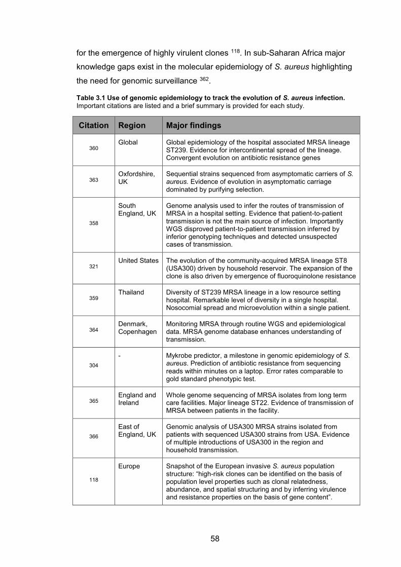
58
for the emergence of highly virulent clones 118. In sub-Saharan Africa major
knowledge gaps exist in the molecular epidemiology of S. aureus highlighting
the need for genomic surveillance 362.
Table 3.1 Use of genomic epidemiology to track the evolution of S. aureus infection. Important citations are listed and a brief summary is provided for each study.
Citation Region Major findings
360 Global Global epidemiology of the hospital associated MRSA lineage
ST239. Evidence for intercontinental spread of the lineage. Convergent evolution on antibiotic resistance genes
363 Oxfordshire, UK
Sequential strains sequenced from asymptomatic carriers of S. aureus. Evidence of evolution in asymptomatic carriage dominated by purifying selection.
358
South England, UK
Genome analysis used to infer the routes of transmission of MRSA in a hospital setting. Evidence that patient-to-patient transmission is not the main source of infection. Importantly WGS disproved patient-to-patient transmission inferred by inferior genotyping techniques and detected unsuspected cases of transmission.
321 United States The evolution of the community-acquired MRSA lineage ST8
(USA300) driven by household reservoir. The expansion of the clone is also driven by emergence of fluoroquinolone resistance
359 Thailand Diversity of ST239 MRSA lineage in a low resource setting
hospital. Remarkable level of diversity in a single hospital. Nosocomial spread and microevolution within a single patient.
364 Denmark, Copenhagen
Monitoring MRSA through routine WGS and epidemiological data. MRSA genome database enhances understanding of transmission.
304 - Mykrobe predictor, a milestone in genomic epidemiology of S.
aureus. Prediction of antibiotic resistance from sequencing reads within minutes on a laptop. Error rates comparable to gold standard phenotypic test.
365 England and Ireland
Whole genome sequencing of MRSA isolates from long term care facilities. Major lineage ST22. Evidence of transmission of MRSA between patients in the facility.
366 East of England, UK
Genomic analysis of USA300 MRSA strains isolated from patients with sequenced USA300 strains from USA. Evidence of multiple introductions of USA300 in the region and household transmission.
118
Europe Snapshot of the European invasive S. aureus population structure: “high-risk clones can be identified on the basis of population level properties such as clonal relatedness, abundance, and spatial structuring and by inferring virulence and resistance properties on the basis of gene content”.
59
3.1.6 Evidence for host switching
S. aureus colonises a wide range of non-human hosts including livestock,
companion animals 266 and wild animals 267. Some clonal lineages of S.
aureus have a strong association with specific hosts 367, but occasionally
bacteria will adapt to a new host and spread through a new host population 352. For example, some livestock-associated lineages of MRSA lineages,
most notably CC97 and CC398, have overcome the species barrier to infect
humans 368,369. Similarly MRSA clones from human-associated lineages,
such as CC5, CC9 and ST88, have been reported in livestock 370. A study
based on MLST across multiple host species showed that anthroponotic
(human-to-animal) transmission of S. aureus occurs more frequently than
zoonosis (animal-to-human transmission) 101. Whole-genome sequencing
has been used to detect and analyse transmission of S. aureus within and
between various host species, including anthroponotic and zoonotic events 368,371,372.
3.1.7 S. aureus among non-human primates
Recent data shows that human-associated S. aureus lineages can infect
great apes that come into contact with humans in captivity and in the wild 373,374. These infections can be fatal and this poses a threat to some
endangered great apes species 373,374. African monkeys in remote regions
are colonized by S. aureus from uncharacterized clonal complexes that rarely
colonize or infect humans 268. One highly divergent clade of coagulase-
positive staphylococci is known to infect monkeys in sub-Saharan Africa, but
has never been recovered from humans so far; this clade has recently been
classified as a new species, S. schweitzeri 268,375. However, such monkeys
are also often colonized by human-associated genotypes of S. aureus, which
they are likely to have acquired from humans 268. Previously S. aureus in
monkeys was only studied through MLST, which does not have the
resolution to infer inter-species transmission and cannot elucidate the
evolutionary mechanisms that drive host adaptation. Our study was the first
60
comparative genomics study on S. aureus from humans and monkeys in
sub-Saharan Africa 316.
3.1.8 Objectives and study rationale
Here, I describe the use whole-genome sequencing of S. aureus isolates
from humans and monkeys in The Gambia with the following goals:
x To determine whether transmission of S. aureus has occurred between humans and non-human primates in The Gambia through
a phylogenetic analysis of S. aureus isolates from monkeys alongside
human S. aureus from carriage and invasive disease
x To estimate the timescale of any inter-host transmission events using a published mutation rate to estimate the time since the most
recent common ancestor based on the number of mutations
accumulated in the core genome
x To investigate the genetic changes accompanying host adaptation by studying the patterns of gene presence and absence in
the accessory genome
61
3.2 Materials and Methods
3.2.1 Bacterial isolates
Prior to this study, staff at the MRC Unit in The Gambia had compiled an
initial dataset of MLST profiles from 298 S. aureus isolates from The Gambia.
This set of isolates included:
1. Monkey isolates cultured from nasopharyngeal and oropharyngeal
swabs taken from African Green Monkeys (Chlorocebus sabaeus)
in 2011 by the International Vervet Research Consortium during a
study on Simian Immunodeficiency Virus in The Gambia 376.
2. Human invasive disease isolates (unpublished data), recovered
from archived clinical specimens including venous blood, pleural
fluid, cerebrospinal fluid, joint aspirate, lung aspirate, ascetic fluid,
and lymph node aspirates
3. Human carriage isolates were recovered from nasopharyngeal and
oropharyngeal swabs from healthy humans
For this study, a subset of 90 human and monkey isolates of S. aureus was
selected. These strains had been previously characterised by MLST and
antimicrobial susceptibility testing by staff at the MRC Unit in The Gambia
(Table 3.2). This subset included 46 human invasive disease, 13 human
carriage and 31 monkey pharyngeal carriage isolates (Table 3.2) This subset
included at least one strain from each clonal complex present in the full
MLST dataset and 20 reference genomes were included to represent as
many of the major clonal complexes as possible.

62
Table 3.2 Metadata for 90 S. aureus isolates analysed by WGS.
ID
Epid
emio
logy
ST
CC
Pen
Sxt
Tet C Cn
Ob E
Met
h
Date
A2 Invasive 1 CC1 R S S S S S S S 25/05/02
C11 Invasive 852 CC1 R S S S S S S S 11/08/07
C12 Invasive 852 CC1 R S S S S S S S 28/10/09
SA9 Carriage 1* CC1 S S S S S S S S 29/03/04
B12 Invasive 101 CC101 S S I S S S S S 19/09/02
C1 Invasive 101 CC101 R S S S S S S S 24/11/05
C3 Invasive 121 CC121 R S S S S S S S 04/04/03
SA32 Invasive 121 CC121 R S R S S S S S 27/04/06
SA33 Invasive 121 CC121 R S R S S S S S 24/12/02
D11 Invasive 2433 CC121 R S S S S S S S 01/03/03
SA2 Carriage 15 CC15 S S S S R S S S 13/01/04
A8 Invasive 15 CC15 R S S S S S S S 11/01/08
A9 Invasive 15 CC15 R S R S S S S S 18/11/05
A10 Invasive 15 CC15 R S R S S S S S 02/09/04
A11 Invasive 15 CC15 R S S S S S S S 23/02/09
B1 Invasive 15 CC15 R S S S S S S S 14/07/09
B3 Invasive 15 CC15 R S R S S S S S 18/07/02
B4 Invasive 15 CC15 R S R S S S S S 30/08/06
SA23 Invasive 15 CC15 R S R S S S S S 04/08/10
SA24 Invasive 15 CC15 R S R S S S S S 19/05/09
SA26 Invasive 15 CC15 R S S S S S S S 11/10/05
SA31 Invasive 15 CC15 R S R S S S S S 07/07/05
SA29 Monkey 15 CC15 S S S S S S S S 01/04/11
D12 Invasive 2434 CC15 R S S S S S R S 26/08/08
C4 Invasive 152 CC152 R S S S S S S S 10/02/09
C6 Invasive 152 CC152 R S S S S S S S 24/04/08
C7 Invasive 152 CC152 R S R S S S S S 24/08/09
C8 Invasive 152 CC152 R S S S S S S S 12/09/05
SA34 Invasive 152 CC152 R S R S S S S S 25/09/07
F5 Monkey CC152 S S S S S S I S 01/04/11
D10 Invasive 2432 CC2432 S S S S S S S S 20/02/07
B5 Invasive 25 CC25 R R R S S S S S 31/07/09
B6 Invasive 25 CC25 R R S S S S S S 04/11/08
F7 Monkey Novel CC2531 S S S S S S S S 01/04/11
F8 Monkey Novel CC2531 S S S S S S S S 01/04/11
SA22 Monkey Novel CC2531 S S S S S S S S 01/04/11
F9 Monkey 2532 CC2532 S S S S S S S S 01/04/11
G2 Monkey 2532 CC2532 S S S S S S S S 01/04/11
F11 Monkey 2533 CC2533 S S S S S S S S 01/04/11
F12 Monkey 2533 CC2533 S S S S S S S S 01/04/11
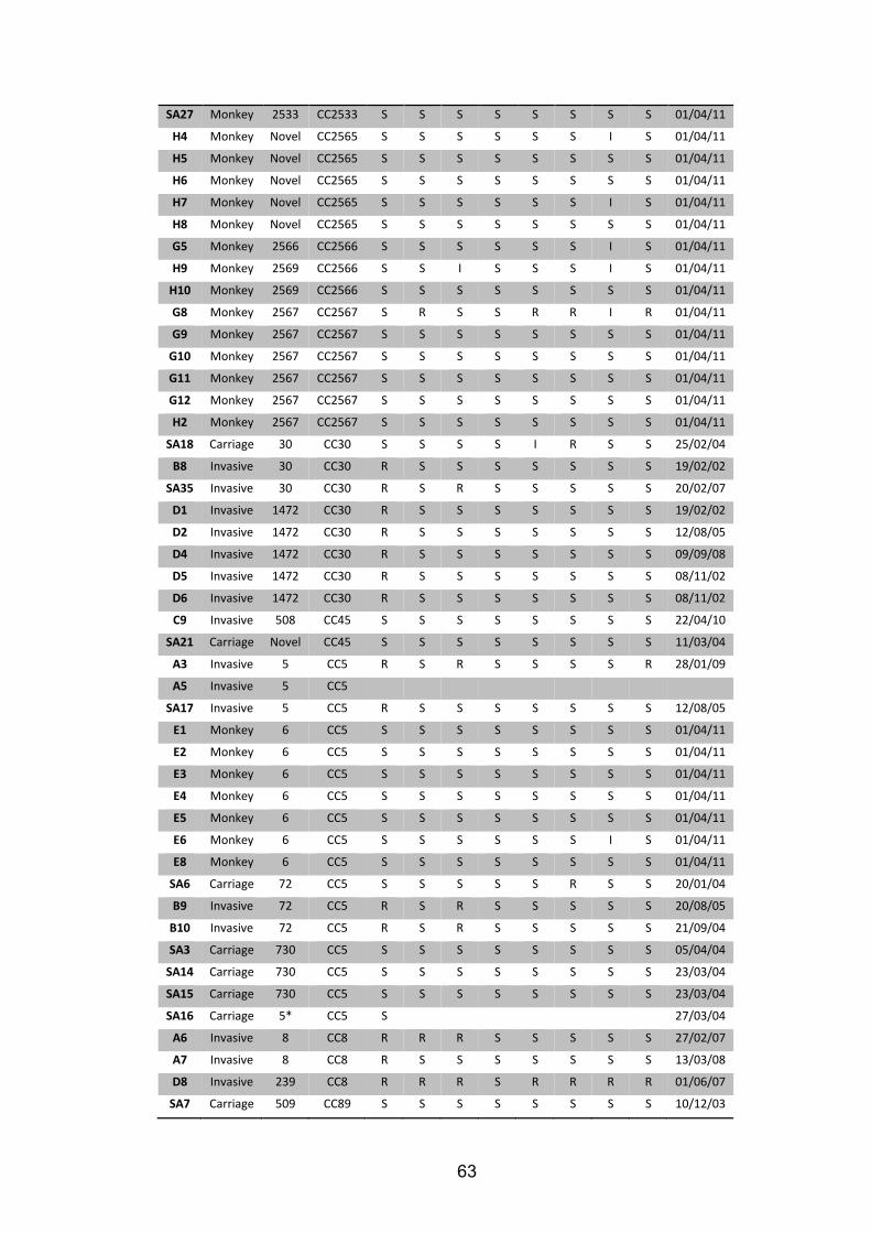
63
SA27 Monkey 2533 CC2533 S S S S S S S S 01/04/11
H4 Monkey Novel CC2565 S S S S S S I S 01/04/11
H5 Monkey Novel CC2565 S S S S S S S S 01/04/11
H6 Monkey Novel CC2565 S S S S S S S S 01/04/11
H7 Monkey Novel CC2565 S S S S S S I S 01/04/11
H8 Monkey Novel CC2565 S S S S S S S S 01/04/11
G5 Monkey 2566 CC2566 S S S S S S I S 01/04/11
H9 Monkey 2569 CC2566 S S I S S S I S 01/04/11
H10 Monkey 2569 CC2566 S S S S S S S S 01/04/11
G8 Monkey 2567 CC2567 S R S S R R I R 01/04/11
G9 Monkey 2567 CC2567 S S S S S S S S 01/04/11
G10 Monkey 2567 CC2567 S S S S S S S S 01/04/11
G11 Monkey 2567 CC2567 S S S S S S S S 01/04/11
G12 Monkey 2567 CC2567 S S S S S S S S 01/04/11
H2 Monkey 2567 CC2567 S S S S S S S S 01/04/11
SA18 Carriage 30 CC30 S S S S I R S S 25/02/04
B8 Invasive 30 CC30 R S S S S S S S 19/02/02
SA35 Invasive 30 CC30 R S R S S S S S 20/02/07
D1 Invasive 1472 CC30 R S S S S S S S 19/02/02
D2 Invasive 1472 CC30 R S S S S S S S 12/08/05
D4 Invasive 1472 CC30 R S S S S S S S 09/09/08
D5 Invasive 1472 CC30 R S S S S S S S 08/11/02
D6 Invasive 1472 CC30 R S S S S S S S 08/11/02
C9 Invasive 508 CC45 S S S S S S S S 22/04/10
SA21 Carriage Novel CC45 S S S S S S S S 11/03/04
A3 Invasive 5 CC5 R S R S S S S R 28/01/09
A5 Invasive 5 CC5
SA17 Invasive 5 CC5 R S S S S S S S 12/08/05
E1 Monkey 6 CC5 S S S S S S S S 01/04/11
E2 Monkey 6 CC5 S S S S S S S S 01/04/11
E3 Monkey 6 CC5 S S S S S S S S 01/04/11
E4 Monkey 6 CC5 S S S S S S S S 01/04/11
E5 Monkey 6 CC5 S S S S S S S S 01/04/11
E6 Monkey 6 CC5 S S S S S S I S 01/04/11
E8 Monkey 6 CC5 S S S S S S S S 01/04/11
SA6 Carriage 72 CC5 S S S S S R S S 20/01/04
B9 Invasive 72 CC5 R S R S S S S S 20/08/05
B10 Invasive 72 CC5 R S R S S S S S 21/09/04
SA3 Carriage 730 CC5 S S S S S S S S 05/04/04
SA14 Carriage 730 CC5 S S S S S S S S 23/03/04
SA15 Carriage 730 CC5 S S S S S S S S 23/03/04
SA16 Carriage 5* CC5 S 27/03/04
A6 Invasive 8 CC8 R R R S S S S S 27/02/07
A7 Invasive 8 CC8 R S S S S S S S 13/03/08
D8 Invasive 239 CC8 R R R S R R R R 01/06/07
SA7 Carriage 509 CC89 S S S S S S S S 10/12/03
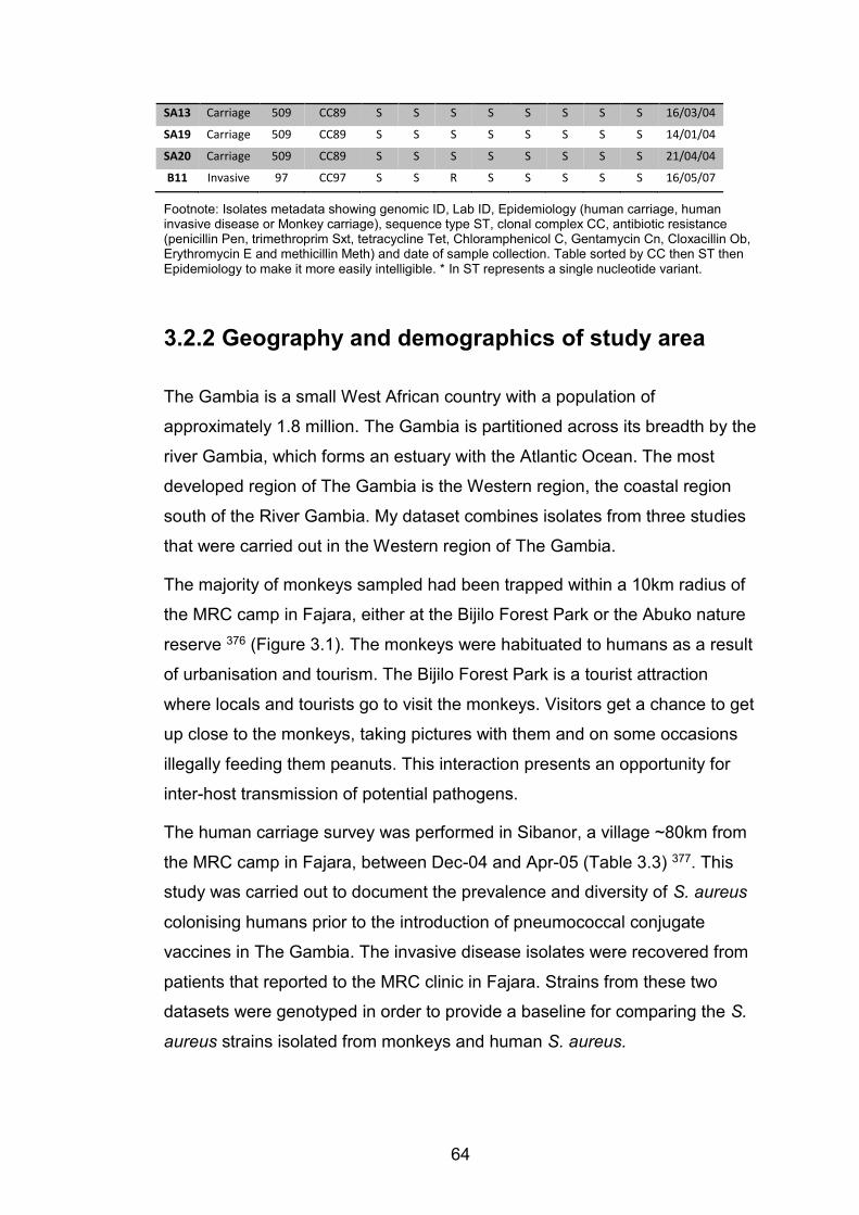
64
SA13 Carriage 509 CC89 S S S S S S S S 16/03/04
SA19 Carriage 509 CC89 S S S S S S S S 14/01/04
SA20 Carriage 509 CC89 S S S S S S S S 21/04/04
B11 Invasive 97 CC97 S S R S S S S S 16/05/07
Footnote: Isolates metadata showing genomic ID, Lab ID, Epidemiology (human carriage, human invasive disease or Monkey carriage), sequence type ST, clonal complex CC, antibiotic resistance (penicillin Pen, trimethroprim Sxt, tetracycline Tet, Chloramphenicol C, Gentamycin Cn, Cloxacillin Ob, Erythromycin E and methicillin Meth) and date of sample collection. Table sorted by CC then ST then Epidemiology to make it more easily intelligible. * In ST represents a single nucleotide variant.
3.2.2 Geography and demographics of study area
The Gambia is a small West African country with a population of
approximately 1.8 million. The Gambia is partitioned across its breadth by the
river Gambia, which forms an estuary with the Atlantic Ocean. The most
developed region of The Gambia is the Western region, the coastal region
south of the River Gambia. My dataset combines isolates from three studies
that were carried out in the Western region of The Gambia.
The majority of monkeys sampled had been trapped within a 10km radius of
the MRC camp in Fajara, either at the Bijilo Forest Park or the Abuko nature
reserve 376 (Figure 3.1). The monkeys were habituated to humans as a result
of urbanisation and tourism. The Bijilo Forest Park is a tourist attraction
where locals and tourists go to visit the monkeys. Visitors get a chance to get
up close to the monkeys, taking pictures with them and on some occasions
illegally feeding them peanuts. This interaction presents an opportunity for
inter-host transmission of potential pathogens.
The human carriage survey was performed in Sibanor, a village ~80km from
the MRC camp in Fajara, between Dec-04 and Apr-05 (Table 3.3) 377. This
study was carried out to document the prevalence and diversity of S. aureus
colonising humans prior to the introduction of pneumococcal conjugate
vaccines in The Gambia. The invasive disease isolates were recovered from
patients that reported to the MRC clinic in Fajara. Strains from these two
datasets were genotyped in order to provide a baseline for comparing the S.
aureus strains isolated from monkeys and human S. aureus.
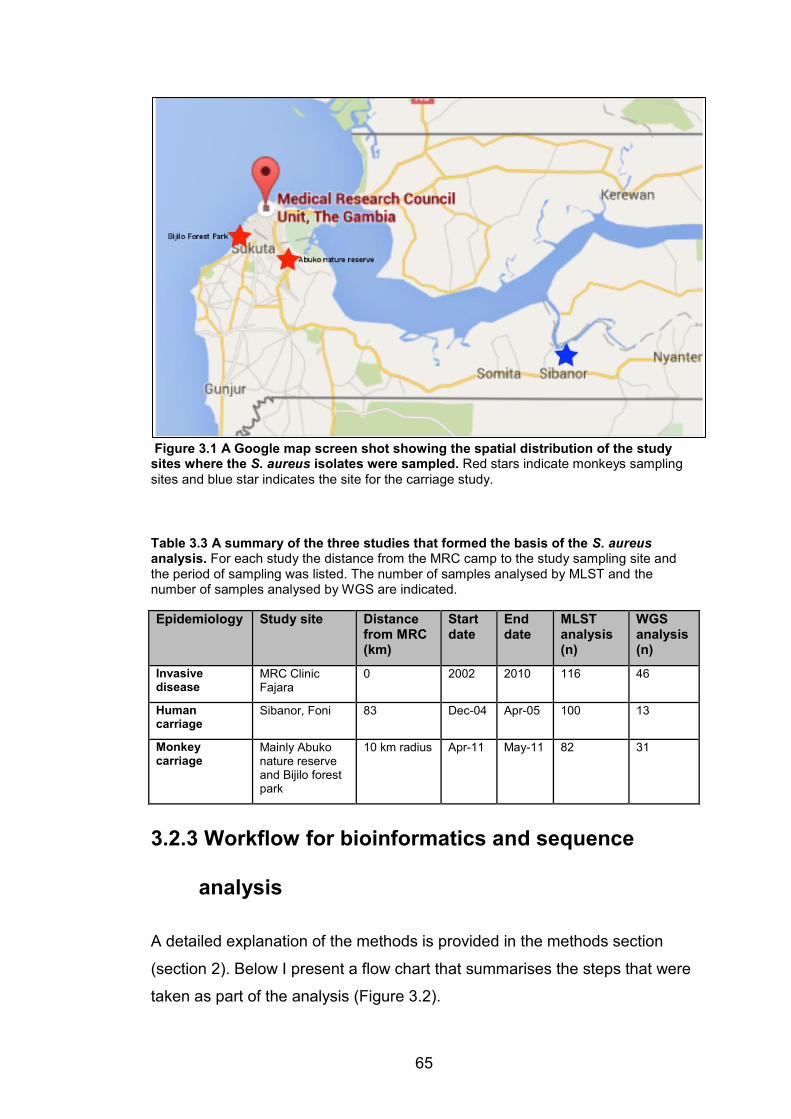
65
Figure 3.1 A Google map screen shot showing the spatial distribution of the study sites where the S. aureus isolates were sampled. Red stars indicate monkeys sampling sites and blue star indicates the site for the carriage study.
Table 3.3 A summary of the three studies that formed the basis of the S. aureus analysis. For each study the distance from the MRC camp to the study sampling site and the period of sampling was listed. The number of samples analysed by MLST and the number of samples analysed by WGS are indicated.
Epidemiology Study site Distance from MRC (km)
Start date
End date
MLST analysis (n)
WGS analysis (n)
Invasive disease
MRC Clinic Fajara
0 2002 2010 116 46
Human carriage
Sibanor, Foni 83 Dec-04 Apr-05 100 13
Monkey carriage
Mainly Abuko nature reserve and Bijilo forest park
10 km radius Apr-11 May-11 82 31
3.2.3 Workflow for bioinformatics and sequence
analysis
A detailed explanation of the methods is provided in the methods section
(section 2). Below I present a flow chart that summarises the steps that were
taken as part of the analysis (Figure 3.2).
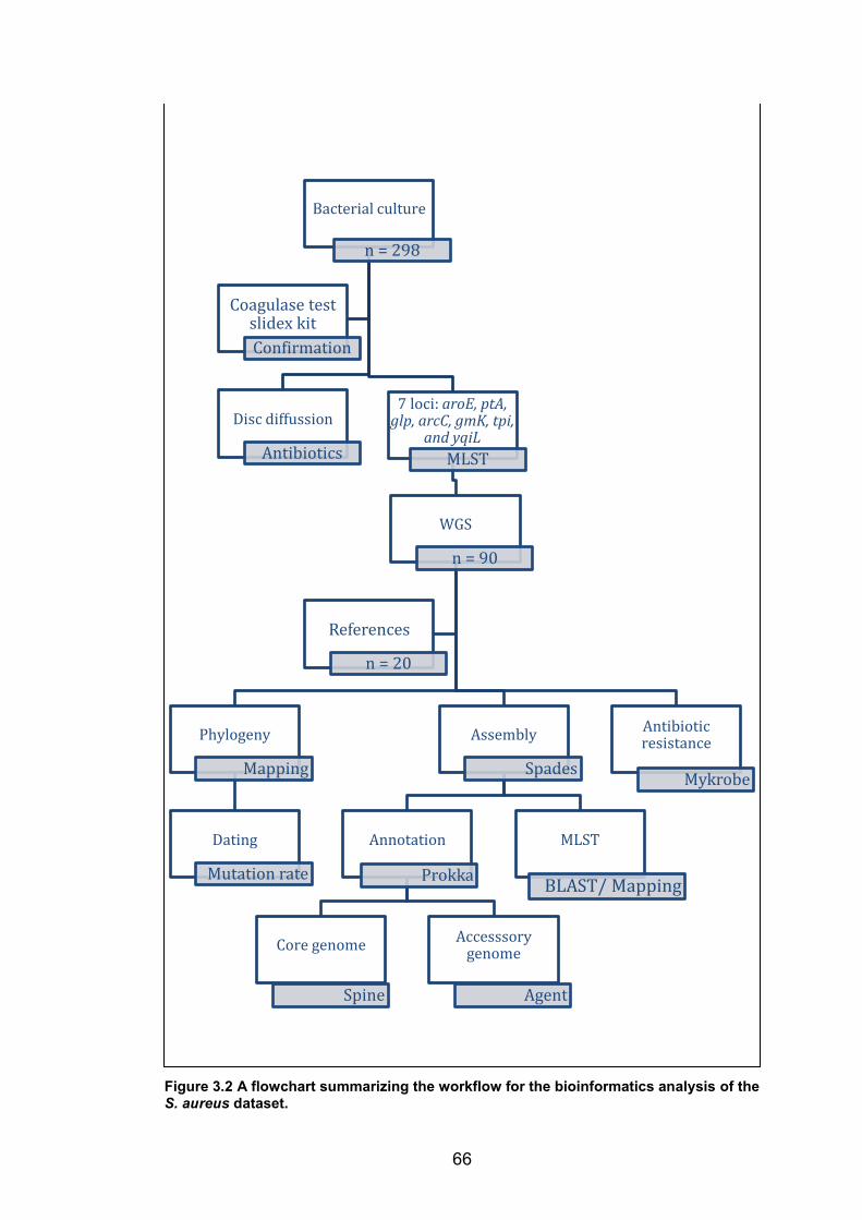
66
Figure 3.2 A flowchart summarizing the workflow for the bioinformatics analysis of the S. aureus dataset.
Bacterial culture
n = 298
Disc diffussion
Antibiotics
7 loci: aroE, ptA, glp, arcC, gmK, tpi,
and yqiLMLST
WGS
n = 90
Phylogeny
Mapping
Dating
Mutation rate
Assembly
Spades
Annotation
Prokka
Core genome
Spine
Accesssory genome
Agent
MLST
BLAST/ Mapping
Antibiotic resistance
Mykrobe
References
n = 20
Coagulase test slidex kitConfirmation

67
3.3 Results
3.3.1 Genome sequences
Draft assemblies ranged from 2,701,991 to 3,048,160 nucleotides in length
(average 2,793,864.9, median 2,786,149), with an average GC content of
32.7 % (range: 32.5 to 32.9%). The genomes were annotated with an
average of 2593 coding sequences (CDSs) (range 2462 to 2921). When
mapped to the EMRSA15 reference genome, the mean genome-wide
coverage was 61.5 fold (range 12.5 to 126.2).
3.3.2 Population structure of S. aureus in The Gambia
Single nucleotide variants (SNVs) were called from 232,276 variable sites in
the core genome after excluding all known repeat regions and transposable
mobile elements. These SNVs were used to create a maximum-likelihood
phylogenetic tree linking isolates with reference strains (See the multiple
sequence alignment of core genome SNVs and the core SNV distance matrix
in Appendices 3.1 & 3.2 respectively). This phylogenetic tree revealed the
clonal population structure of S. aureus. Strains belonging to the same clonal
complex clustered on monophyletic branches on the phylogenetic tree. The
tree resolved five major S. aureus clades that were consistent with previous
species-wide phylogenetic analyses (Figure 3.3):
x Clade 1 encompassed pandemic clonal lineages CCs 1, 5, 6, 8, 15
and 25, and closely related genotypes. Clade 1 encompassed both
human carriage and human invasive disease strains including two
MRSA invasive disease strains from the ST5 and ST339. Within clade
1 there was also a cluster of ST6 isolates from monkeys and a ST15
isolate from a monkey that clustered with the human ST15 isolates
x Clade 2 represented a monkey-specific clade that contained most of
the S. aureus isolates recovered from monkeys. This clade was
comprised of lineages bearing (at the time) novel STs that were only
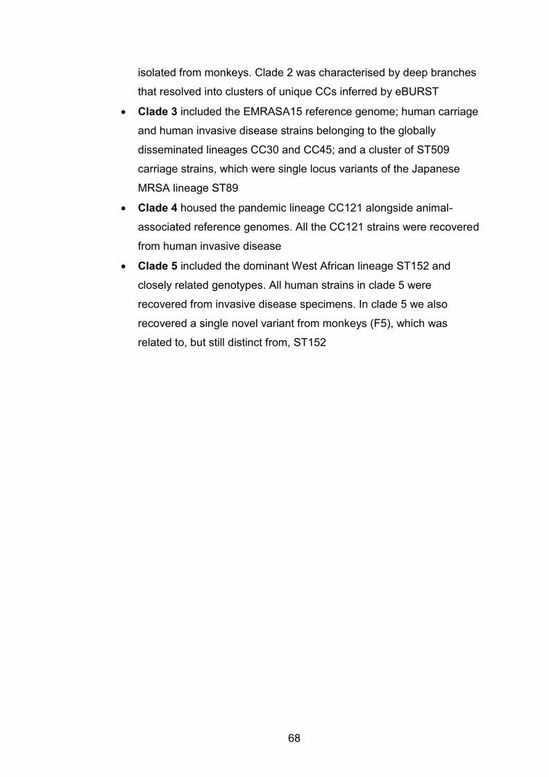
68
isolated from monkeys. Clade 2 was characterised by deep branches
that resolved into clusters of unique CCs inferred by eBURST
x Clade 3 included the EMRASA15 reference genome; human carriage
and human invasive disease strains belonging to the globally
disseminated lineages CC30 and CC45; and a cluster of ST509
carriage strains, which were single locus variants of the Japanese
MRSA lineage ST89
x Clade 4 housed the pandemic lineage CC121 alongside animal-
associated reference genomes. All the CC121 strains were recovered
from human invasive disease
x Clade 5 included the dominant West African lineage ST152 and
closely related genotypes. All human strains in clade 5 were
recovered from invasive disease specimens. In clade 5 we also
recovered a single novel variant from monkeys (F5), which was
related to, but still distinct from, ST152
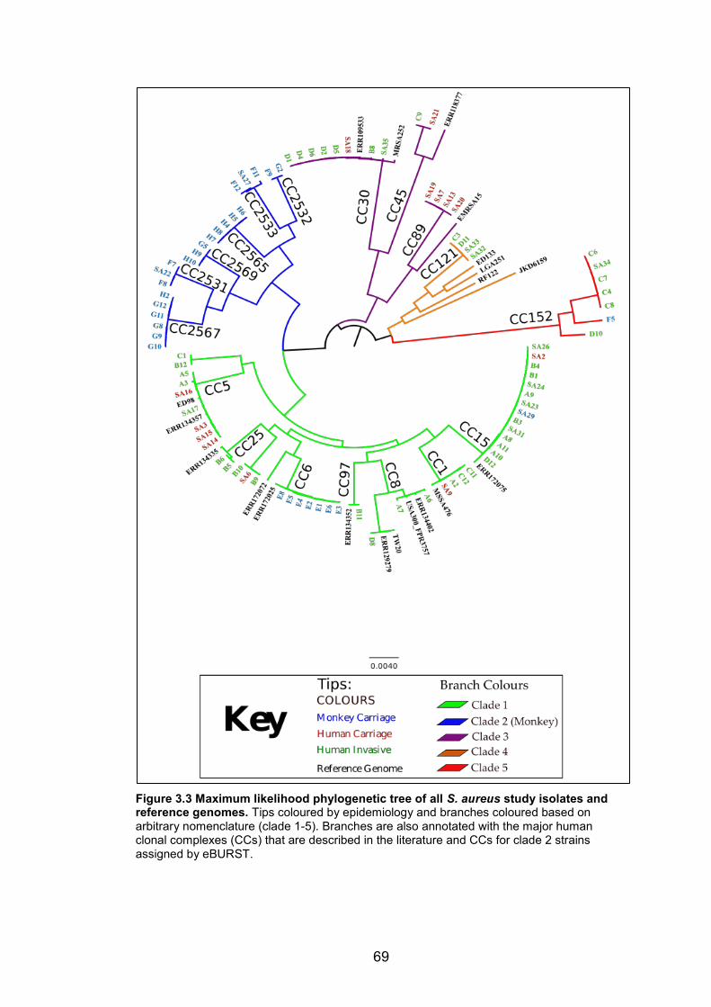
69
Figure 3.3 Maximum likelihood phylogenetic tree of all S. aureus study isolates and reference genomes. Tips coloured by epidemiology and branches coloured based on arbitrary nomenclature (clade 1-5). Branches are also annotated with the major human clonal complexes (CCs) that are described in the literature and CCs for clade 2 strains assigned by eBURST.

70
3.3.3 Anthroponotic transmission of S. aureus
This dataset provided a rare insight into the contemporaneous population
structure of S. aureus in humans and monkeys. Assuming a constant
mutation rate, the timescales for inter-host transmission of S. aureus
between humans and monkeys in The Gambia were estimated (Table 3.4).
Current estimates of intra-clonal mutation rates are consistent in S. aureus 321. Based on the intra-clonal mutation rate for S. aureus of ~ 2 x 10⁻⁶ per site
per year 321, and a core genome size of ~2.5 Mb, it was possible to estimate
an upper bound date for the transmission events. The short-term mutation
rate equates to about 5 SNPs per year. Thus simply dividing the total number
of SNPs between any pair of contemporary S. aureus isolates by ten gives
the approximate number of years since they shared a common ancestor.
The phylogenetic placement of clade 2 – nested between human-associated
clades – suggests this clade has human ancestry. The most divergent pair of
clade 2 isolates differed by 26,464 SNVs. Similarly, the closest pairwise
distance to an isolate outside clade 2 (The MSSA476 reference genome)
was 26,968 SNVs. Thus, clade 2 was likely to have emerged from a host
jump that occurred ~2700 years ago. There were no clear examples of
zoonotic transmission (from monkeys to humans), as no human-derived
isolates fell within the novel monkey-associated clade 2.
The ST6 monkey isolates in clade 1 formed a tight cluster, within which the
furthest distance between two ST6 monkey isolates was 270 SNVs. The
lowest divergence between a ST6 isolate from a monkey and the reference
ST6 isolate from a human in Europe was just 311 SNVs. These observations
suggest that ST6 was transmitted from humans to monkeys a little under
three decades ago.
The single ST15 isolated from a monkey in clade 1 differed from an invasive
disease isolate by 71 SNVs. This most likely represents an anthroponotic
host switch that occurred approximately less than 7 years ago. This estimate
was not attempted for the monkey isolate in clade 5 due to lack of a closely
related human isolate.
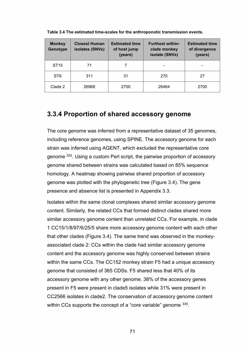
71
Table 3.4 The estimated time-scales for the anthroponotic transmission events.
Monkey Genotype
Closest Human isolates (SNVs)
Estimated time of host jump
(years)
Furthest within-clade monkey isolate (SNVs)
Estimated time of divergence
(years)
ST15 71 7 - -
ST6 311 31 270 27
Clade 2 26968 2700 26464 2700
3.3.4 Proportion of shared accessory genome
The core genome was inferred from a representative dataset of 35 genomes,
including reference genomes, using SPINE. The accessory genome for each
strain was inferred using AGENT, which excluded the representative core
genome 332. Using a custom Perl script, the pairwise proportion of accessory
genome shared between strains was calculated based on 85% sequence
homology. A heatmap showing pairwise shared proportion of accessory
genome was plotted with the phylogenetic tree (Figure 3.4). The gene
presence and absence list is presented in Appendix 3.3.
Isolates within the same clonal complexes shared similar accessory genome
content. Similarly, the related CCs that formed distinct clades shared more
similar accessory genome content than unrelated CCs. For example, in clade
1 CC15/1/8/97/6/25/5 share more accessory genome content with each other
that other clades (Figure 3.4). The same trend was observed in the monkey-
associated clade 2: CCs within the clade had similar accessory genome
content and the accessory genome was highly conserved between strains
within the same CCs. The CC152 monkey strain F5 had a unique accessory
genome that consisted of 365 CDSs. F5 shared less that 40% of its
accessory genome with any other genome. 38% of the accessory genes
present in F5 were present in clade5 isolates while 31% were present in
CC2566 isolates in clade2. The conservation of accessory genome content
within CCs supports the concept of a “core variable” genome 335.
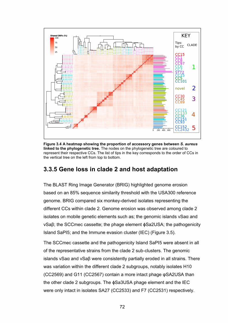
72
Figure 3.4 A heatmap showing the proportion of accessory genes between S. aureus linked to the phylogenetic tree. The nodes on the phylogenetic tree are coloured to represent their respective CCs. The list of tips in the key corresponds to the order of CCs in the vertical tree on the left from top to bottom.
3.3.5 Gene loss in clade 2 and host adaptation
The BLAST Ring Image Generator (BRIG) highlighted genome erosion
based on an 85% sequence similarity threshold with the USA300 reference
genome. BRIG compared six monkey-derived isolates representing the
different CCs within clade 2. Genome erosion was observed among clade 2
isolates on mobile genetic elements such as; the genomic islands νSaα and
νSaβ; the SCCmec cassette; the phage element ϕSa2USA; the pathogenicity
Island SaPI5; and the Immune evasion cluster (IEC) (Figure 3.5).
The SCCmec cassette and the pathogenicity Island SaPI5 were absent in all
of the representative strains from the clade 2 sub-clusters. The genomic
islands νSaα and νSaβ were consistently partially eroded in all strains. There
was variation within the different clade 2 subgroups, notably isolates H10
(CC2569) and G11 (CC2567) contain a more intact phage φSA2USA than
the other clade 2 subgroups. The ϕSa3USA phage element and the IEC
were only intact in isolates SA27 (CC2533) and F7 (CC2531) respectively.
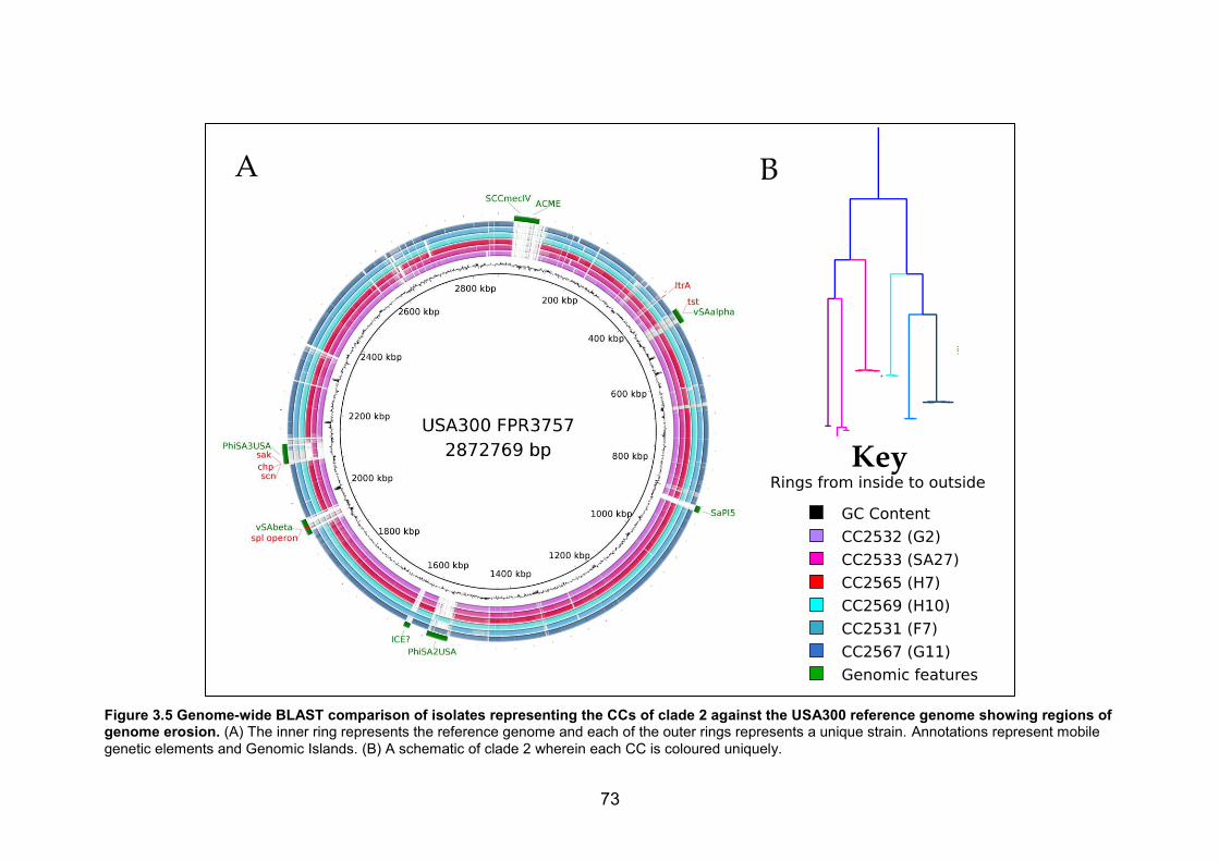
73
Figure 3.5 Genome-wide BLAST comparison of isolates representing the CCs of clade 2 against the USA300 reference genome showing regions of genome erosion. (A) The inner ring represents the reference genome and each of the outer rings represents a unique strain. Annotations represent mobile genetic elements and Genomic Islands. (B) A schematic of clade 2 wherein each CC is coloured uniquely.
74
3.3.6 Antibiotic resistance
The Mykrobe predictor was employed to predict antimicrobial resistance from
the genome sequence data 304. Mykrobe predicted antibiotic resistance
based on the presence of whole genes as well as point mutations. The
Mykrobe predictor also determined the presence of the Panton-Valentine
leukocidin toxin genes in the strains.
Based on phenotypic antimicrobial susceptibility testing high levels of
penicillin resistance were observed among invasive disease isolates (89%).
Penicillin resistance was conferred by presence of the blaZ gene in most
cases. Among invasive disease isolates 19 were resistant to tetracycline.
The tetL, tetM and/or tetK genes mediated tetracycline resistance.
Two invasive disease isolates were confirmed to be methicillin resistant and
both isolates bore the mecA gene. The MRSA invasive disease strains
belonged to the globally disseminated genotypes ST239 and ST5. These
represented the first published reports of MRSA causing invasive disease in
The Gambia 316.
None of the carriage strains were classified as penicillin or tetracycline
resistant based on disc diffusion antimicrobial susceptibility testing. However,
based on the genome Mykrobe predicted that 12/13 and 4/13 carriage strains
were resistant to penicillin and tetracycline respectively. This indicates that
the burden of antimicrobial resistance among S. aureus carriage strains in
The Gambia is likely to be underestimated by disc diffusion.
This dataset does not confirm drug resistance among S. aureus isolates from
monkeys in The Gambia. Initially one isolate from a monkey had been
characterised as methicillin resistant based on cefotoxin disc diffusion.
However, when the antibiotic resistance test was repeated with eTest the
isolate was reclassified as methicillin sensitive. Furthermore there was no
genetic evidence of MRSA in this isolate based on a Mykrobe prediction 304.

75
3.4 Discussion
3.4.1 Anthroponotic transmission
We posit that clade 2 represents an ancient human-to-monkey transmission
event that occurred a minimum of 2700 years ago. Clade 2 has undergone
clonal expansion and diversified into at least six sub-clades within the
monkey population. Whilst this can be largely explained in terms of common
clonal inheritance (descent with modification), it is also possible that some of
the differences in gene content between clade 2 and the other clades in the
tree underlie adaptation to the monkey host. The presence of the monkey-
associated isolates in both Clade 2 and ST6 provides an opportunity to
detect any genes that may have been acquired independently in two
unrelated monkey-adapted lineages.
Human-associated genotypes have been previously isolated from monkeys
in West Africa: ST1, ST6 and ST15 268. We report the presence of ST6 and
ST15 in monkeys raising the possibility that these genotypes may be prone
to human-to-monkey transmission. The fact that ST15 was only recovered
once from monkeys is also consistent with a recent transmission event. The
cluster of monkey-derived ST6 isolates is consistent with onward
transmission within the monkey population. However, the upper-bound
estimate for time since transmission of ST6 to monkeys may be spuriously
high since we were unable to sequence the closest human ST6 isolate.
Furthermore, the ST6 cluster may have emerged as a result of multiple host
switching events. In this case, the within-clade diversity would also represent
a high upper-bound estimate for the time since the host switch occurred.
The evolutionary origin of the CC152 monkey isolate F5 is unclear given its
unique accessory genome composition and the similarity to ST152 strains at
the core genome level. A likely scenario is that a ST152 isolate was
transmitted to monkeys then subsequently lost some of its accessory
genomes and acquired some accessory content from monkey specific
lineages through recombination. ST152 strains are able to adapt to multiple
76
host niches and have been detected among domestic animals in Africa 268.
Moreover, an overhaul of the accessory genome is not uncommon in S.
aureus. It has previously been described that ST239 is a product of
recombination of a large block of DNA from a CC30 donor into a CC8
background 378.
3.4.2 Gene loss as a contributor to host adaptation
The loss of genes harboured on the genomic islands νSaα and νSaβ in clade
2 isolates may have enhanced adaptation to the monkey host. The spl
operon (spl A-F) is harboured on νSaβ and it encodes serine proteases as
well as lytN, which is essential for bacterial growth and cell wall synthesis 379,380. All clade 2 isolates harboured novel variants of spl genes that were
not present in human isolates or reference genomes highlighting a potential
role in host adaptation.
The immune evasion cluster IEC1 harbours the immune evasion genes (chp,
sak and scn) and enterotoxin genes such as sea, sep, sek, and seq 381,382.
These genes elicit human-specific immune modulation using different
mechanisms including the blockage of neutrophil chemotaxis 383. Thus it is
not surprising that the IEC1 was lost in the vast majority of isolates recovered
from monkeys. Similarly the Panton-Valentine leukocidin genes (lukF-PV and
lukS-PV) were absent from all isolates recovered from monkeys. Panton-
Valentine leukocidin is a potent human neutrophil cytotoxic factor that does
not work on monkeys 384.
Genes carried on MGEs play an important role in host adaptation 343.
However, our data suggests that in addition to gene acquisition, gene loss
may play an important role in adaptation to non-human hosts. If gene loss is
assumed to occur more readily than gene acquisition, then this relationship
might explain why anthroponotic transmission of S. aureus is more common
than zoonotic transmission. Protein-truncating mutations in S. aureus have
been previously implicated in the progression from carriage to invasive
disease 361.

77
3.4.3 Risk of monkeys serving as reservoirs for highly
virulent S. aureus
Our data supports previous suggestions that anthroponotic transmission
events may be relatively frequent, while zoonotic transmission is much less
common 101. Although human-to-monkey transmission has been documented
in West Africa there is no evidence of zoonotic transmission from monkeys 268. From a public health perspective, this is reassuring as it suggests that
monkeys do not constitute a serious risk of S. aureus infection in humans.
Related to this, we also note that none of the monkey isolates were MRSA.
Furthermore, although there were high levels of resistance to penicillin
among invasive disease isolates, the isolates from monkeys were all
susceptible to penicillin.
3.4.4 Limitations
The main limitation of this study is that the samples from the three
epidemiological classes were collected over different timescales and from
different regions. De novo sample collection over a standard harmonized
timeframe would have enabled us to make more accurate inferences on the
direction of transmission. A longitudinal study would also be required to
confidently determine the dynamic of the inferred host switching events.
Finally, our study lacked precise demographic information that could link
each isolates to the location where the monkey bearing it was trapped and
sampled. This information would have facilitated a more in depth
understanding of the dynamic of S. aureus in monkeys in The Gambia.
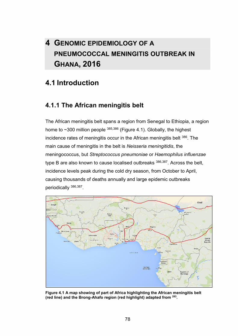
78
4 GENOMIC EPIDEMIOLOGY OF A PNEUMOCOCCAL MENINGITIS OUTBREAK IN GHANA, 2016
4.1 Introduction
4.1.1 The African meningitis belt
The African meningitis belt spans a region from Senegal to Ethiopia, a region
home to ~300 million people 385,386 (Figure 4.1). Globally, the highest
incidence rates of meningitis occur in the African meningitis belt 386. The
main cause of meningitis in the belt is Neisseria meningitidis, the
meningococcus, but Streptococcus pneumoniae or Haemophilus influenzae
type B are also known to cause localised outbreaks 386,387. Across the belt,
incidence levels peak during the cold dry season, from October to April,
causing thousands of deaths annually and large epidemic outbreaks
periodically 386,387.
Figure 4.1 A map showing of part of Africa highlighting the African meningitis belt (red line) and the Brong-Ahafo region (red highlight) adapted from 283.
79
The dynamics behind the seasonality of meningitis outbreaks are unclear,
but climate is believed to be the strongest driving force 388,389. During the dry
season, the Harmattan winds blow from the Northeast, carrying high levels of
atmospheric mineral dust 390. The persistent dust and low humidity are
believed to damage the mucosal epithelium leading to an increased risk of
progression from asymptomatic carriage to invasive disease 386,391. Similarly,
viral respiratory tract infections such as the flu weaken the immune system
enhancing transmission and invasion of bacteria 392.
4.1.2 Molecular epidemiology of meningitis in West
Africa
Since the 1990s, meningococcal ST5 complex strains belonging to
serogroup A have been the most common causes of meningitis outbreaks 393. In 2000, the serogroup W NmW135 strain emerged as the cause of a
major meningitis outbreak during the annual Hajj pilgrimage in Mecca, Saudi
Arabia 387. Shortly afterwards, in 2002, NmW135 was responsible for a major
meningitis outbreak in Burkina Faso 394. NmW135 reached epidemic status
due to high incidence rates among children but for unknown reasons the
prevalence of NmW135 decreased significantly within a short space of time 395. The year 2003 saw the emergence of a new serogroup A strain from the
ST2859 complex, which demonstrated the potential to cause epidemics in
the future 387.
Since the 2000s, lethal community-based pneumococcal meningitis
outbreaks have been reported in northern Ghana, Burkina Faso and Togo,
affecting all age groups 278,279,396. S. pneumoniae serotype 1 is the leading
cause of pneumococcal meningitis in the meningitis belt 397. Serotype 1 can
cause lethal outbreaks of meningitis in high-burden settings 396,398. Serotype
1 is highly virulent and has a high invasiveness index, since it is rarely
detected in asymptomatic carriage 397. The sequence type (ST) 217 clonal
complex (CC) and closely related genotypes ST303 and ST618 have been
80
implicated in previous outbreaks of pneumococcal meningitis in West Africa 396,398,399.
4.1.3 Vaccine intervention in the African meningitis
belt
The MenAfriVac® campaign was launched in 2010 in a bid to eradicate
serogroup A. MenAfriVac® has been a success story, since it led to
elimination of serogroup A carriage and to herd immunity that protected even
unvaccinated individuals 15. MenAfriVac vaccination in Burkina Faso led to
the eradication of serogroup A strains among carriage and diseased
individuals. However, following vaccination the serogroup X strains have
become the dominant genotype isolated from carriage and invasive disease 400. Worryingly the serogroup W strains, which have demonstrated the ability
to cause lethal outbreaks in the past 394, have re-emerged in Burkina Faso
post-vaccination 15,400.
The need for mass vaccination campaigns against S. pneumoniae, especially
serotype 1, in the meningitis belt cannot be stressed enough. In The Gambia
the introduction of the pneumococcal conjugate vaccine PCV-13, which
includes serotype 1, has led to a marked decrease in the prevalence of
vaccine serotypes causing invasive pneumococcal disease 280. A study
showed that across Burkina Faso, Ghana, Nigeria, Mali, Senegal, and The
Gambia 65-87% of pneumococcal isolates had serotypes covered by PCV-
13 401. Evidence shows that pneumococcal vaccination campaigns should
also be extended to cover older children and adults in order to boost efficacy 278.
4.1.4 Evolution of the pneumococcal genome
Variation is introduced into the pneumococcal core genome primarily through
homologous recombination (uptake and incorporation of exogenous DNA)
and point mutations 178. Pneumococcal genomes undergo very high rates of
recombination; rates are influenced by capsular type and carriage rates
81
across lineages 402. Clinical interventions such as vaccination and antibiotic
therapy rapidly select for novel pneumococcal genotypes that arise through
homologous recombination 116,403. In addition, S. pneumoniae can acquire
novel genes that mediate virulence and antimicrobial resistance from other
species by horizontal gene transfer (HGT) 403,404.
4.1.5 Evolution of drug resistance
The emergence of drug resistance in S. pneumoniae is a major public health
concern driven by the emergence of multidrug-resistant strains 405.
Antimicrobial resistance can be spread by clonal dissemination of resistant
strains or emergence of resistance in susceptible strains 405. Pneumococcal
strains can acquire antibiotic resistance through target modification, efflux
pump and/or enzyme inactivation of the drug 406.
β-lactam resistance is acquired through modification of the penicillin binding
protein (PBP). Modifications of PBP occur primarily through point mutations
but the mosaic structure of PBP suggests that the PBP may have been
modified in other Streptococci before being transformed into S. pneumoniae
through recombination 178,405. Fluoroquinolone resistance is driven primarily
by spontaneous mutations in the quinolone resistance determinant region 405.
Genes carried on transposable mobile genetic elements confer resistance to
a broad range of antibiotics 407. For example, the Tn916 family transposons
carry:
x Macrolide resistance determinants ermB and mef,
o ermB encodes a protein product that induces resistance by
methylating the 23S ribosomal binding site and the
o mef genes, which encode an active efflux pump 408
x Accessory genes catQ and tetM that confer chloramphenicol and
tetracycline resistance through enzymatic inactivation and target
alteration respectively 405,409
A new antibiotic called teixobactin, which inhibits cell wall biosynthesis has
recently been discovered 410. Teixobactin has shown efficacy in mice infected
82
with S. pneumoniae and a wide range of gram-positive bacteria 410.
Encouragingly it is expected that this compound will avoid the development
of resistance that is common to most antibiotics 410. Perhaps teixobactin and
similar compounds that will emerge from this discovery may influence the
future of therapeutic intervention for S. pneumoniae.
4.1.6 The Brong-Ahafo meningitis outbreak
A meningitis outbreak broke out in the Brong-Ahafo region of Ghana during
the peak of the 2015-2016 dry season. The WHO Regional Reference
Laboratory hosted at the MRC Unit The Gambia (MRCG) played a crucial
role in providing laboratory and data management support to the local health
authorities in Brong-Ahafo region in response to the outbreak. A team of
scientists and a database manager were dispatched from MRCG to work
with the WHO and Ghana Ministry of Health in the Brong-Ahafo region.
There were 886 suspected cases of meningitis in the Brong-Ahafo region
between 2nd December 2015 and 26th February 2016. The mortality rate
among suspected cases whose outcome was known was 10%. Over 95% of
the confirmed cases occurred in children over 5 years and adults. All
available cerebrospinal fluid specimens from suspected cases were tested by
rapid test, qPCR and/or culture. 135 cases of meningitis were confirmed by
identification of a pathogen:
x Streptococcus pneumoniae (77%, 104/135)
x Neisseria meningitidis (22%, 30/135)
x Group B streptococcus (GBS) (1%, 1/135).
49 pneumococcal strains were successfully serotyped by PCR of which, 40
(78%) were serotype 1 and five (12%) were serotype 12F. Other serotypes
detected included 35B, 6A/6B, 7F and 3.
It was surprising that serotype 1 S. pneumoniae was the main pathogen
identified during the outbreak, as PCV-13 was included in the Ghanaian
expanded program of immunization in 2012. Although nationwide coverage
of PCV-13 was below 50% in 2012, it rose steadily and by 2014 had reached
83
81%. PCVs usually induce herd immunity leading to a decrease in the
prevalence of vaccine serotypes even in non-vaccinated individuals 281,282,411.
However recent estimates suggests that conjugate vaccines may take up to
a decade to induce herd immunity among the unvaccinated population 412.
4.1.7 Objectives and study rationale
To understand the molecular epidemiology of the outbreak we set out to
perform whole genome sequencing (WGS) on stored pneumococcal isolates
from outbreak patients with the following objectives:
x To confirm the serotype and determine the MLST profile of the outbreak S. pneumoniae strains by comparing their genome
sequence to the sequences of the known pneumococcal serotypes
and MLST alleles respectively
x To reconstruct the phylogeny of the outbreak strains based on
single nucleotide variants in the core genome
x To study the evolutionary events that led to the emergence of serotype 1 outbreak following the introduction of PCV-13 by
gleaning the genome for evidence of capsular switching and changes
in the core and accessory genome that could have led to enhanced
fitness or virulence
x To compare the serotype 1 strains from the outbreak with previously sequenced serotype 1 strains from West Africa. These
strains were sequenced as part of a previous collaboration through
the Pneumococcal African Genomics Consortium (PAGe)

84
4.2 Methods
4.2.1 Geography and demographics of the study area
The Brong-Ahafo region lies at heart of Ghana, midway up the North-South
axis but below the African meningitis belt (Figure 4.2). The region consists of
27 districts and has a population of 2.4 million. Like much of West Africa, the
Brong-Ahafo region has a tropical climate, with a dry season that lasts from
November to March and two rainy seasons in between. Prior to the outbreak
the WHO did not have standard guidelines for containing a pneumococcal
meningitis outbreak. Moreover, such outbreaks were not expected to occur in
the Brong-Ahafo region because it lies outside the meningitis belt. As a
result, regional health authorities were caught off guard and initially struggled
to contain the outbreak. After helping to contain the outbreak the MRCG
team made important recommendations to the WHO for dealing with
pneumococcal meningitis outbreaks.
Figure 4.2 A district map of the Brong-Ahafo region of Ghana (source: http://www.maphill.com).
85
4.2.2 Bacterial isolates
During the outbreak meningitis, the primary method for confirmation of
meningitis cases was the Pastorex meningitis kit (Biorad, Hemel Hempstead,
Hertfordshire, UK). When the WHO RRL was called in, qPCR assays were
performed at the MRCG to identify the aetiological pathogen from
cerebrospinal fluid. S. pneumoniae serotyping was performed using latex
agglutination 296. Culture facilities were not available at most health centres,
so samples had to be sent to the regional hospital in Sunyani for culture.
However, sub-optimal storage and transportation conditions meant that most
samples were not viable for culture. Consequently out of 104 laboratory-
confirmed cases of pneumococcal meningitis, we recovered just sixteen
isolates. These isolates were characterised by antibiotic susceptibility testing
to Clindamycin, Optochin, Vancomycin, Trimetoprim-sulfamethoxazole,
Erythromycin, Chloramphenicol, Rifampin, Ceftriaxone, Penicillin and
Tetracycline. WGS was performed on 15 isolates of S. pneumoniae that were
stored from the outbreak (Figure 4.3). These included 12 serotype 1 and one
each of serotype 35B, serotype 7A and serotype 3.
The majority of the strains analysed by WGS were isolated from
cerebrospinal fluid specimens that were sampled from patients during the
outbreak (14/15). We analysed one strain that was recovered from a throat
swab of a healthy carrier in Wenchi district. Unfortunately we were unable to
retrieve the patient information for the throat swab and the exact time of
sample collection. The majority of isolates were recovered between the 6th
and 9th week of 2016 inclusive. Patients’ age ranged from 9 to 45 years
(mean 21, median 19) and the ratio of male to female was 3:4 (Table 4.1).
The antibiotic susceptibility profiles to commonly used antibiotics are
presented in table 4.1 but the antibiogram for the throat swab isolate
(BAR_19) was not reported. Staff at the MRCG reported high rates of
resistance to tetracycline and trimethoprim-sulfamethoxazole (commonly
known as co-trimoxazole) among these isolates. One serotype 1 isolate was
also resistant to penicillin in addition to tetracycline and co-trimoxazole
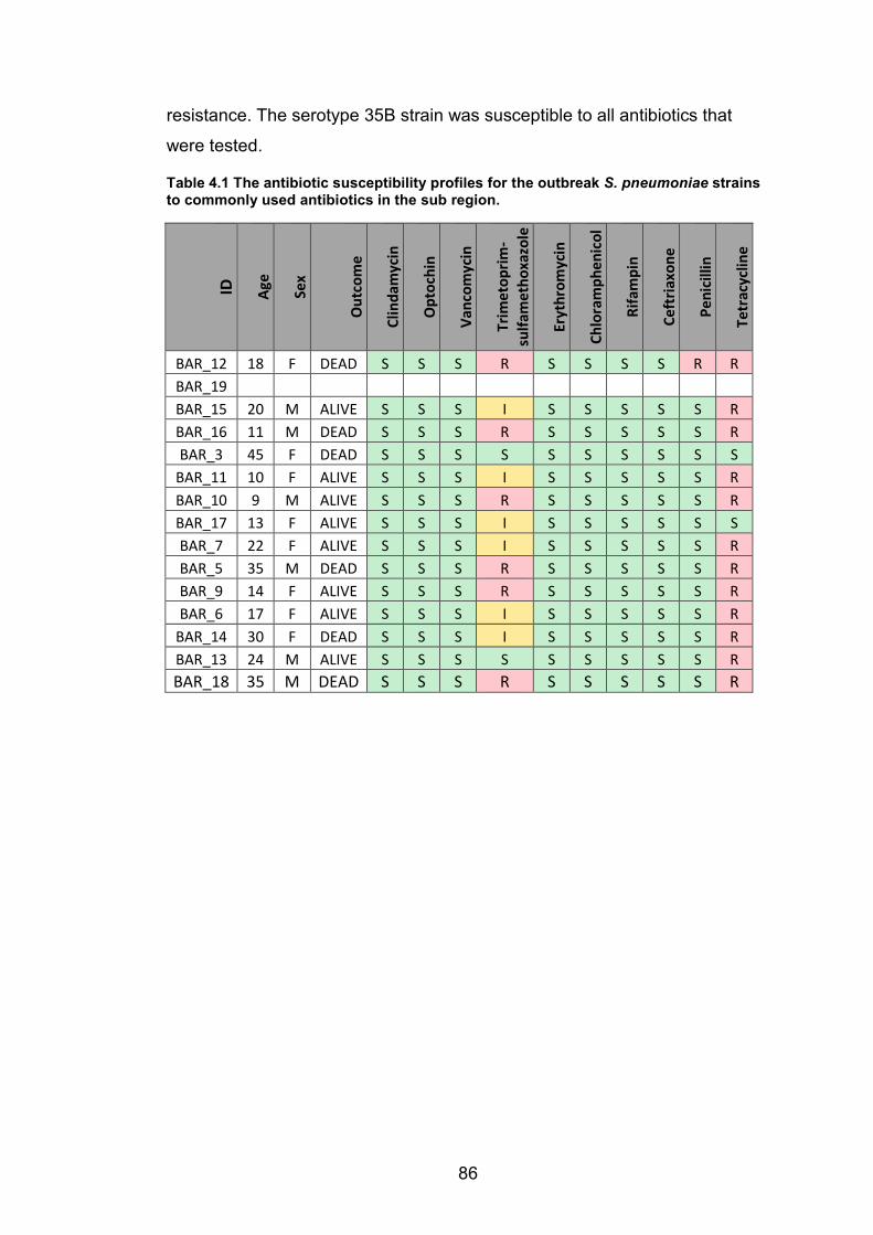
86
resistance. The serotype 35B strain was susceptible to all antibiotics that
were tested.
Table 4.1 The antibiotic susceptibility profiles for the outbreak S. pneumoniae strains to commonly used antibiotics in the sub region.
ID
Age
Sex
Out
com
e
Clin
dam
ycin
Opt
ochi
n
Vanc
omyc
in
Trim
etop
rim-
sulfa
met
hoxa
zole
Eryt
hrom
ycin
Chlo
ram
phen
icol
Rifa
mpi
n
Ceft
riaxo
ne
Peni
cilli
n
Tetr
acyc
line
BAR_12 18 F DEAD S S S R S S S S R R BAR_19
BAR_15 20 M ALIVE S S S I S S S S S R BAR_16 11 M DEAD S S S R S S S S S R BAR_3 45 F DEAD S S S S S S S S S S
BAR_11 10 F ALIVE S S S I S S S S S R BAR_10 9 M ALIVE S S S R S S S S S R BAR_17 13 F ALIVE S S S I S S S S S S BAR_7 22 F ALIVE S S S I S S S S S R BAR_5 35 M DEAD S S S R S S S S S R BAR_9 14 F ALIVE S S S R S S S S S R BAR_6 17 F ALIVE S S S I S S S S S R
BAR_14 30 F DEAD S S S I S S S S S R BAR_13 24 M ALIVE S S S S S S S S S R BAR_18 35 M DEAD S S S R S S S S S R

87
4.2.3 Outbreak summary and workflow for sequence
analysis
Figure 4.3 A summary of the pneumococcal meningitis outbreak and the workflow for the bioinformatics analysis.
Suspected cases
n = 886
CSF collected
n = 795
Pathogen detected
n = 135
N. meningitidis
n= 30
S. pneumoniae
n = 104
WGS
n = 14 + 1
Speciation
KRAKEN
Assembly
MEGA-HIT
MLST
BLAST
Antibiogram
Abricate
Serotyping
Mapping
Serotype 1
n= 12
West Africa serotype 1
n = 108
Phylogeny
core.ope
Clonality
R packages
Recombination
GUBBINS
Phylogeny
Snippy
GBS
n = 1
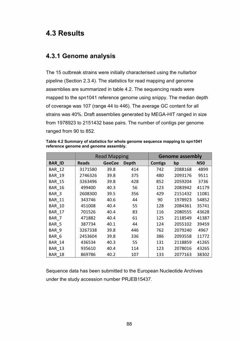
88
4.3 Results
4.3.1 Genome analysis
The 15 outbreak strains were initially characterised using the nullarbor
pipeline (Section 2.3.4). The statistics for read mapping and genome
assemblies are summarized in table 4.2. The sequencing reads were
mapped to the spn1041 reference genome using snippy. The median depth
of coverage was 107 (range 44 to 446). The average GC content for all
strains was 40%. Draft assemblies generated by MEGA-HIT ranged in size
from 1978923 to 2151432 base pairs. The number of contigs per genome
ranged from 90 to 852.
Table 4.2 Summary of statistics for whole genome sequence mapping to spn1041 reference genome and genome assembly.
Read Mapping Genome assembly
BAR_ID Reads GeeCee Depth Contigs bp N50 BAR_12 3171580 39.8 414 742 2088168 4899 BAR_19 2746326 39.8 375 480 2093176 9511 BAR_15 3263496 39.8 428 852 2059204 3736 BAR_16 499400 40.3 56 123 2083942 41179 BAR_3 2608300 39.5 356 429 2151432 11081 BAR_11 343746 40.6 44 90 1978923 54852 BAR_10 451008 40.4 55 128 2084361 35741 BAR_17 701526 40.4 83 116 2080555 43628 BAR_7 471882 40.4 61 125 2118549 41387 BAR_5 387734 40.1 44 124 2055102 39459 BAR_9 3267338 39.8 446 762 2079240 4967 BAR_6 2453604 39.8 336 386 2093558 11772 BAR_14 436534 40.3 55 131 2118859 41265 BAR_13 935610 40.4 114 123 2078016 43265 BAR_18 869786 40.2 107 133 2077163 38302
Sequence data has been submitted to the European Nucleotide Archives
under the study accession number PRJEB15437.
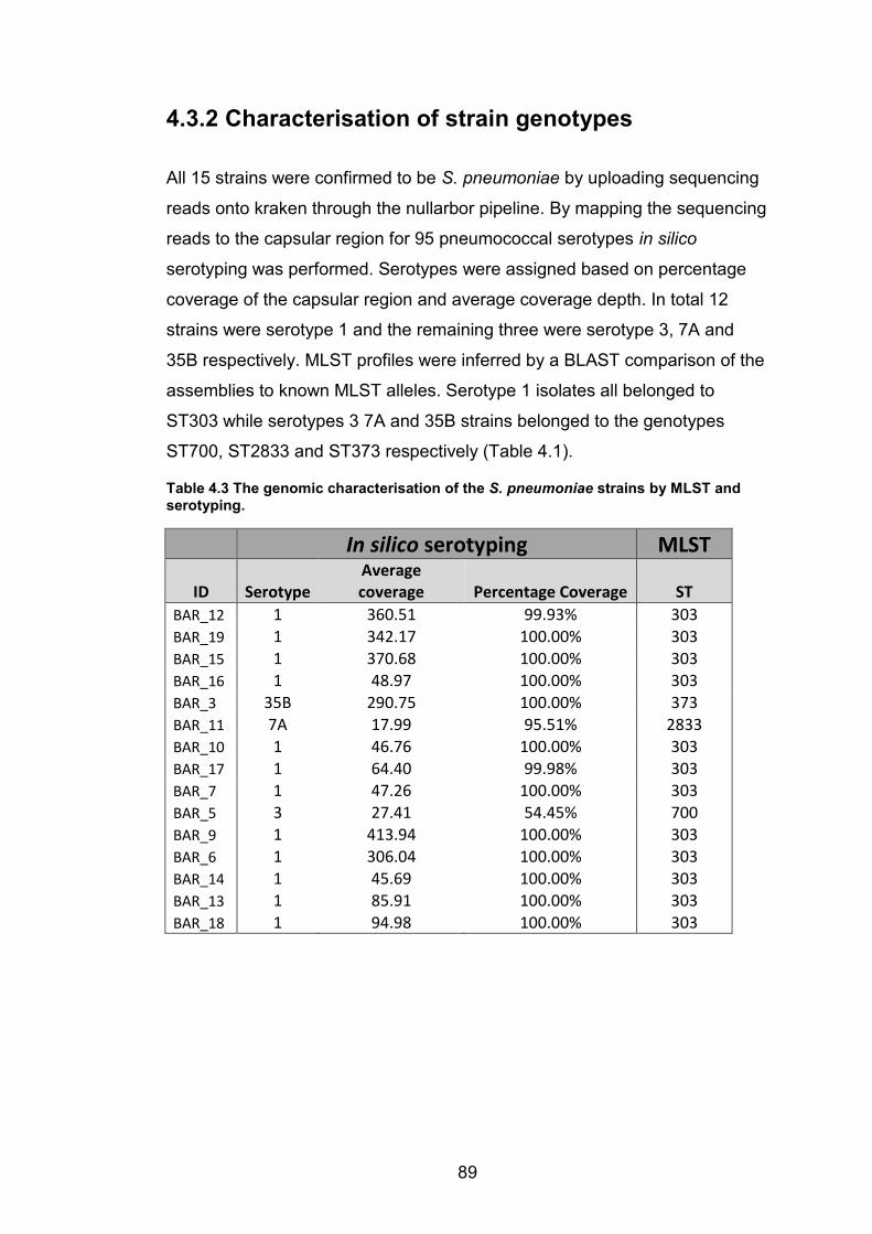
89
4.3.2 Characterisation of strain genotypes
All 15 strains were confirmed to be S. pneumoniae by uploading sequencing
reads onto kraken through the nullarbor pipeline. By mapping the sequencing
reads to the capsular region for 95 pneumococcal serotypes in silico
serotyping was performed. Serotypes were assigned based on percentage
coverage of the capsular region and average coverage depth. In total 12
strains were serotype 1 and the remaining three were serotype 3, 7A and
35B respectively. MLST profiles were inferred by a BLAST comparison of the
assemblies to known MLST alleles. Serotype 1 isolates all belonged to
ST303 while serotypes 3 7A and 35B strains belonged to the genotypes
ST700, ST2833 and ST373 respectively (Table 4.1).
Table 4.3 The genomic characterisation of the S. pneumoniae strains by MLST and serotyping.
In silico serotyping MLST
ID Serotype Average coverage Percentage Coverage ST
BAR_12 1 360.51 99.93% 303 BAR_19 1 342.17 100.00% 303 BAR_15 1 370.68 100.00% 303 BAR_16 1 48.97 100.00% 303 BAR_3 35B 290.75 100.00% 373 BAR_11 7A 17.99 95.51% 2833 BAR_10 1 46.76 100.00% 303 BAR_17 1 64.40 99.98% 303 BAR_7 1 47.26 100.00% 303 BAR_5 3 27.41 54.45% 700 BAR_9 1 413.94 100.00% 303 BAR_6 1 306.04 100.00% 303 BAR_14 1 45.69 100.00% 303 BAR_13 1 85.91 100.00% 303 BAR_18 1 94.98 100.00% 303
90
4.3.3 Phylogeography of outbreak strains
The sequencing reads from all outbreak strains were mapped to the spn1041
reference genome with snippy and variants were called from the core
genome using snippy-core (Alignment in Appendix 4.1). The maximum
likelihood was reconstructed using FastTree.
The phylogenetic tree resolved the serotype 1 strains into two clades (Figure
4.4A). Eight serotype 1 strains formed a novel clade that was distinct from
the spn1041 reference genome. Isolates within the novel clade were closely
related with a within-clade average pairwise distance of 16 core genome
single nucleotide variants (SNVs) (Table 4.4). The most similar strains within
the novel clade differed at four core genome loci and the most divergent pair
differed by 27 SNVs. Four other outbreak serotype 1 strains clustered in a
clade with the ST303 reference genome spn1041 (henceforth referred to as
reference-type ST303). Strains within the reference-type ST303 clade
differed by an average of 91 core genome SNVs (Table 4.4). The strains
belonging to other serotypes differed from serotype strains by at least 16,000
SNVs and formed outliers on individual long branches of the phylogenetic
tree.
A phylogeography analysis was attempted by linking the annotated
phylogenetic tree to a district map showing the incidence rates of confirmed
cases in the respective districts of the Brong-Ahafo region. Seven strains in
the novel clade were isolated from patients in Techiman, Wenchi or Tain;
districts within the epicentre of the outbreak (Figure 4.4B). During the
outbreak the incidents rates in the epicentre rose above the epidemic
threshold for meningitis outbreaks 283. The eighth strain from the novel clade
was isolated from a patient in Tano South, south of the epicentre. Two
reference type ST303 strains were isolated from patients in Nkoranza South
and one from Techiman while the origin of the fourth was unknown (on
Figure 4.4 this strain was assigned to Sunyani where the regional hospital is
located). The non-serotype 1 strains were isolated from the fringe districts,
Jaman South, Dorma and Tano South, where the number of confirmed cases
was relatively low.
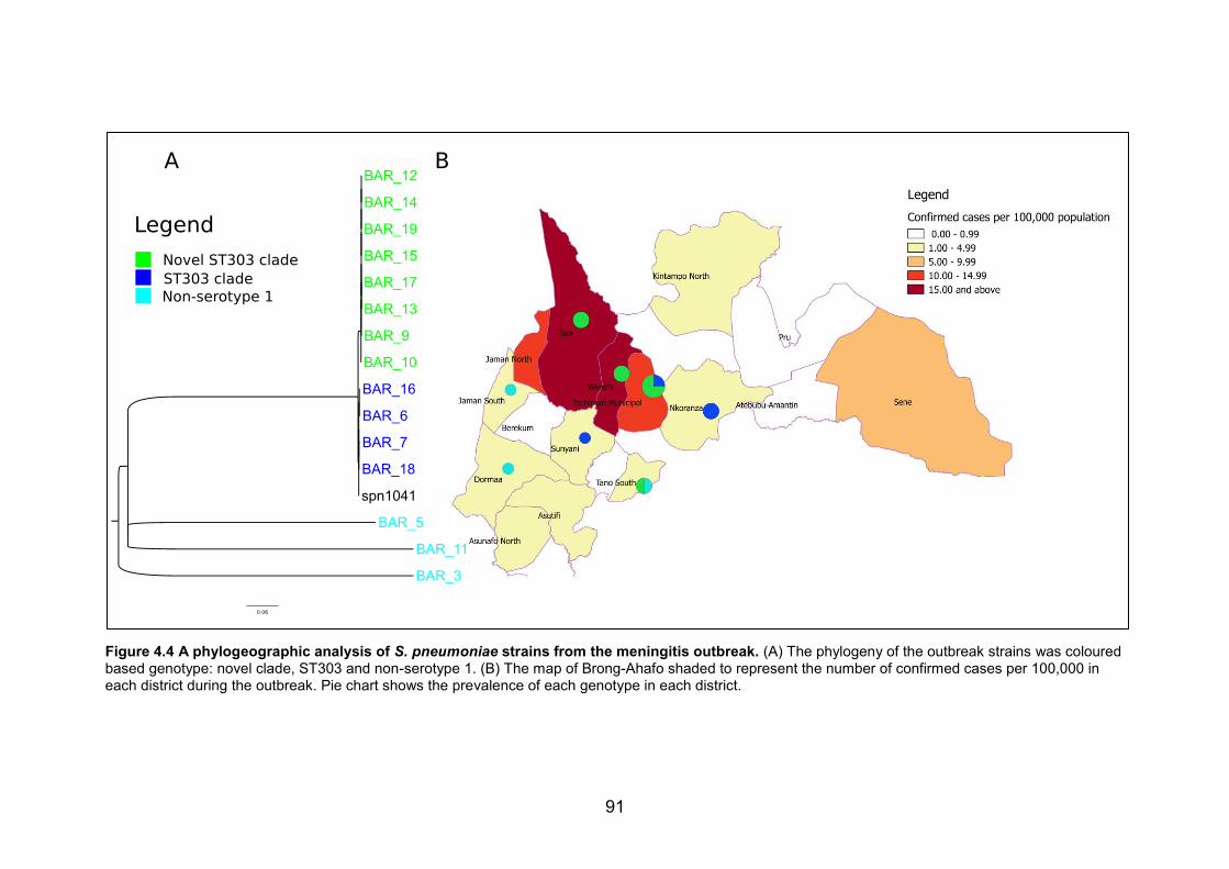
91
Figure 4.4 A phylogeographic analysis of S. pneumoniae strains from the meningitis outbreak. (A) The phylogeny of the outbreak strains was coloured based genotype: novel clade, ST303 and non-serotype 1. (B) The map of Brong-Ahafo shaded to represent the number of confirmed cases per 100,000 in each district during the outbreak. Pie chart shows the prevalence of each genotype in each district.
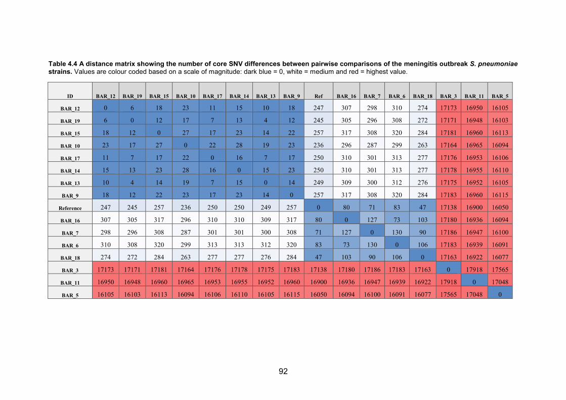
92
Table 4.4 A distance matrix showing the number of core SNV differences between pairwise comparisons of the meningitis outbreak S. pneumoniae strains. Values are colour coded based on a scale of magnitude: dark blue = 0, white = medium and red = highest value.
ID BAR_12 BAR_19 BAR_15 BAR_10 BAR_17 BAR_14 BAR_13 BAR_9 Ref BAR_16 BAR_7 BAR_6 BAR_18 BAR_3 BAR_11 BAR_5
BAR_12 0 6 18 23 11 15 10 18 247 307 298 310 274 17173 16950 16105
BAR_19 6 0 12 17 7 13 4 12 245 305 296 308 272 17171 16948 16103
BAR_15 18 12 0 27 17 23 14 22 257 317 308 320 284 17181 16960 16113
BAR_10 23 17 27 0 22 28 19 23 236 296 287 299 263 17164 16965 16094
BAR_17 11 7 17 22 0 16 7 17 250 310 301 313 277 17176 16953 16106
BAR_14 15 13 23 28 16 0 15 23 250 310 301 313 277 17178 16955 16110
BAR_13 10 4 14 19 7 15 0 14 249 309 300 312 276 17175 16952 16105
BAR_9 18 12 22 23 17 23 14 0 257 317 308 320 284 17183 16960 16115
Reference 247 245 257 236 250 250 249 257 0 80 71 83 47 17138 16900 16050
BAR_16 307 305 317 296 310 310 309 317 80 0 127 73 103 17180 16936 16094
BAR_7 298 296 308 287 301 301 300 308 71 127 0 130 90 17186 16947 16100
BAR_6 310 308 320 299 313 313 312 320 83 73 130 0 106 17183 16939 16091
BAR_18 274 272 284 263 277 277 276 284 47 103 90 106 0 17163 16922 16077
BAR_3 17173 17171 17181 17164 17176 17178 17175 17183 17138 17180 17186 17183 17163 0 17918 17565
BAR_11 16950 16948 16960 16965 16953 16955 16952 16960 16900 16936 16947 16939 16922 17918 0 17048
BAR_5 16105 16103 16113 16094 16106 16110 16105 16115 16050 16094 16100 16091 16077 17565 17048 0
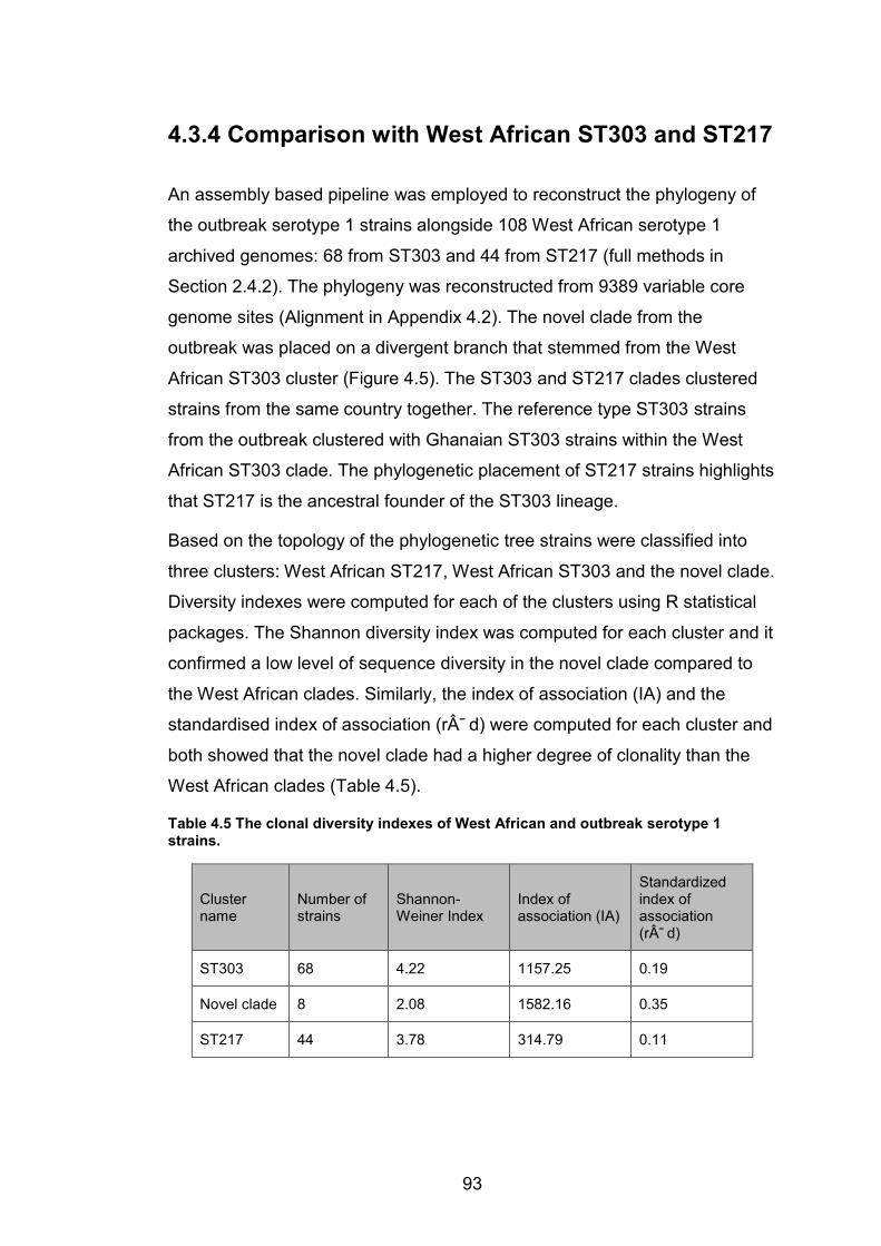
93
4.3.4 Comparison with West African ST303 and ST217
An assembly based pipeline was employed to reconstruct the phylogeny of
the outbreak serotype 1 strains alongside 108 West African serotype 1
archived genomes: 68 from ST303 and 44 from ST217 (full methods in
Section 2.4.2). The phylogeny was reconstructed from 9389 variable core
genome sites (Alignment in Appendix 4.2). The novel clade from the
outbreak was placed on a divergent branch that stemmed from the West
African ST303 cluster (Figure 4.5). The ST303 and ST217 clades clustered
strains from the same country together. The reference type ST303 strains
from the outbreak clustered with Ghanaian ST303 strains within the West
African ST303 clade. The phylogenetic placement of ST217 strains highlights
that ST217 is the ancestral founder of the ST303 lineage.
Based on the topology of the phylogenetic tree strains were classified into
three clusters: West African ST217, West African ST303 and the novel clade.
Diversity indexes were computed for each of the clusters using R statistical
packages. The Shannon diversity index was computed for each cluster and it
confirmed a low level of sequence diversity in the novel clade compared to
the West African clades. Similarly, the index of association (IA) and the
standardised index of association (r¯ d) were computed for each cluster and
both showed that the novel clade had a higher degree of clonality than the
West African clades (Table 4.5).
Table 4.5 The clonal diversity indexes of West African and outbreak serotype 1 strains.
Cluster name
Number of strains
Shannon-Weiner Index
Index of association (IA)
Standardized index of association (r¯ d)
ST303 68 4.22 1157.25 0.19
Novel clade 8 2.08 1582.16 0.35
ST217 44 3.78 314.79 0.11

94
Figure 4.5 The phylogeny of the outbreak serotype 1 strains and the West African serotype 1 genotypes ST303 and ST217. Tips without labels are strains with new STs.
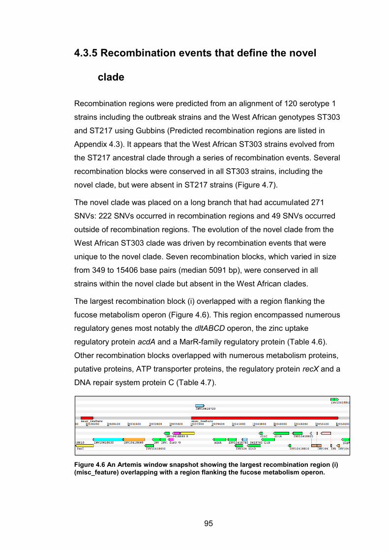
95
4.3.5 Recombination events that define the novel
clade
Recombination regions were predicted from an alignment of 120 serotype 1
strains including the outbreak strains and the West African genotypes ST303
and ST217 using Gubbins (Predicted recombination regions are listed in
Appendix 4.3). It appears that the West African ST303 strains evolved from
the ST217 ancestral clade through a series of recombination events. Several
recombination blocks were conserved in all ST303 strains, including the
novel clade, but were absent in ST217 strains (Figure 4.7).
The novel clade was placed on a long branch that had accumulated 271
SNVs: 222 SNVs occurred in recombination regions and 49 SNVs occurred
outside of recombination regions. The evolution of the novel clade from the
West African ST303 clade was driven by recombination events that were
unique to the novel clade. Seven recombination blocks, which varied in size
from 349 to 15406 base pairs (median 5091 bp), were conserved in all
strains within the novel clade but absent in the West African clades.
The largest recombination block (i) overlapped with a region flanking the
fucose metabolism operon (Figure 4.6). This region encompassed numerous
regulatory genes most notably the dltABCD operon, the zinc uptake
regulatory protein acdA and a MarR-family regulatory protein (Table 4.6).
Other recombination blocks overlapped with numerous metabolism proteins,
putative proteins, ATP transporter proteins, the regulatory protein recX and a
DNA repair system protein C (Table 4.7).
Figure 4.6 An Artemis window snapshot showing the largest recombination region (i) (misc_feature) overlapping with a region flanking the fucose metabolism operon.
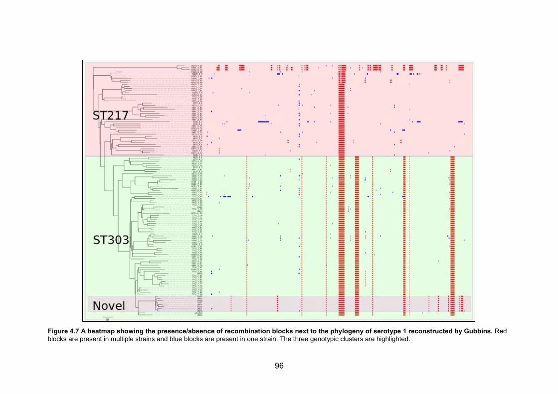
96
Figure 4.7 A heatmap showing the presence/absence of recombination blocks next to the phylogeny of serotype 1 reconstructed by Gubbins. Red blocks are present in multiple strains and blue blocks are present in one strain. The three genotypic clusters are highlighted.

97
Table 4.6 Table showing the genes that overlap with the recombination regions that characterise the novel clade (blocks I - iii). The start and stop positions show where the recombination starts and stops on the reference genome. The sizes of the recombination blocks are listed.
Region Start Stop Size (b.p.) Gene name /locus tag Description
i 2037275 2052681 15406
fucK putative fuculokinase INV10418720 putative fucose phosphotransferase system repressor adcA Zinc-binding protein AdcA precursor INV10418740 ABC transporter permease protein INV10418750 ABC transporter ATP-binding protein INV10418760 MarR-family regulatory protein dltD putative D-alanyl-lipoteichoic acid biosynthesis protein dltC D-alanyl carrier protein dltB putative activated D-alanine transport protein dltA D-alanine--poly(phosphoribitol) ligase subunit 1 INV10418820 major facilitator family transport protein INV10418830 putative reverse transcriptase - Group II intron (pseudogene) INV10402910 Group II intron maturase
ii 2025618 2026848 1230 fucI putative L-fucose isomerase
iii 1933663 1938754 5091
gltX glutamyl-tRNA synthetase pgi glucose-6-phosphate isomerase INV10417860 putative peptidase INV10417870 ABC transporter ATP-binding membrane protein
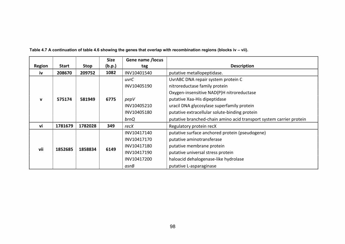
98
Table 4.7 A continuation of table 4.6 showing the genes that overlap with recombination regions (blocks iv – vii).
Region Start Stop Size
(b.p.) Gene name /locus
tag Description iv 208670 209752 1082 INV10401540 putative metallopeptidase.
v 575174 581949 6775
uvrC UvrABC DNA repair system protein C INV10405190 nitroreductase family protein
Oxygen-insensitive NAD(P)H nitroreductase
pepV putative Xaa-His dipeptidase INV10405210 uracil DNA glycosylase superfamily protein INV10405180 putative extracellular solute-binding protein brnQ putative branched-chain amino acid transport system carrier protein
vi 1781679 1782028 349 recX Regulatory protein recX
vii 1852685 1858834 6149
INV10417140 putative surface anchored protein (pseudogene) INV10417170 putative aminotransferase INV10417180 putative membrane protein INV10417190 putative universal stress protein INV10417200 haloacid dehalogenase-like hydrolase asnB putative L-asparaginase

99
4.4 Discussion
4.4.1 Novel clade drove the outbreak
Novel lineages of serotype 1 can emerge through clonal expansion and in
some cases they can progress to become the dominant clone 413. It is highly
likely that the novel clade was the dominant clone in the meningitis outbreak
since it was the most prevalent genotype among the genome-sequenced
isolates and it was predominantly isolated from the epicentre of the outbreak.
Additionally, the close genetic relationship between the strains suggests that
the strains in the novel clade were undergoing clonal expansion and
spreading through the community. The other strains isolated from patients
during the outbreak such as the reference-type ST303 strains were
genetically more diverse, which suggested they were not being transmitted or
undergoing clonal expansion. These strains are likely to represent sporadic
cases of meningitis that occurred during the time of the outbreak.
4.4.2 Evolution of the novel clade
The evolution of novel clones due to selective pressure from the host is an
on going phenomenon for S. pneumoniae 414. However, due to homologous
recombination, most novel clonal clusters are transient and get reabsorbed
into the main clonal lineages 415. There is evidence that the novel clade
underwent substantial sequence diversity, primarily through homologous
recombination, to form a distinct cluster. Recombination blocks overlapped
with genes responsible for important cellular functions that could impact on
fitness and virulence:
x The dltABCD operon mediates D-alanylation of lipoteichoic acids, a
mechanism for evasion of host innate immune response through
repulsion of cationic antimicrobial peptides 416. Thus, modification of
the dlt operon through recombination could have enhanced host
immune evasion in the novel clade
100
x MarR-family proteins regulate expression of virulence factors and
responses to antibiotic and oxidative stress 417. Mutations in MarR-
family genes have been associated with increased virulence and
invasiveness in a number of bacterial species 418. In the novel clade,
modification of the MarR family protein could have led to increased
virulence and invasiveness
x ATP transporter proteins play an important role in bacterial virulence
through uptake of nutrients and metal ions as well as in host cell
attachment 419. ATP transporters are highly immunogenic and have
been considered as potential vaccine targets for S. pneumoniae 419
x The zinc-uptake regulatory protein adcA is highly immunogenic and
plays an important role in S. pneumoniae infection 417. Modification of
the adcA gene by recombination could enhance immune evasion
4.4.3 Vaccine-induced selective pressure
The introduction of vaccines can induce selective pressures that favour
serotype replacement through capsular switching or the rise of low
prevalence non-vaccine serotypes 116,420. The very small sample size that we
had showed 5 cases due to 12F. This could have represented a substantial
transmission network supported by the same conditions that drove the
overall outbreak. Alternatively they might all have been sporadic, and just at
the usual background level. There was no evidence of capsular switching
through the alteration of cps genes in the novel clade. However, it cannot be
ruled out that vaccine-induced selective pressure may have contributed to
the emergence of this novel clone.
Conjugate vaccines alter the nasopharyngeal microbiome by eliminating the
most prevalent serotypes 421. This provides new opportunities for S.
pneumoniae to acquire novel DNA by recombination within the modified
microbial ecology of the nasopharnyx 414. It is known that vaccine
intervention selects for S. pneumoniae lineages that have undergone
recombination 116,403. It can be hypothesized that the ancestral clone of the
novel clade underwent rapid evolution through recombination and then was
101
selected for by the introduction of PCV-13. The recombination events are
likely to have conferred a selective advantage through enhanced virulence
and potential host immune evasion mechanisms.
4.4.4 Potential reasons for outbreak
A likely scenario is that at the time of the outbreak, the strains from the novel
clade were already disseminated in the population. The most divergent
strains within the novel clade differed by as many as 28 core genome SNVs
(without excluding recombination). This is an indication that the novel clade
has been undergoing clonal expansion for some time, perhaps a few years.
The dust and low humidity that characterises the dry season favoured a
switch from carriage to invasiveness, leading to disease. This hypothesis is
supported by the emergence of sporadic cases of meningitis due to other
serotypes and other genotypes of ST303 serotype 1.
Once the new clade emerged with potentially superior fitness, it could have
easily spread among the older unvaccinated population in the community.
This is supported by the fact that overall over 95% of confirmed cases
occurred among children over 5 and adults. Similarly, the novel clade strains
were isolated from patients aged 9 years and above. A strain isolated from a
healthy individual was also part of the novel clade, which suggests that strain
from the novel clade may have been disseminated in the community prior to
the outbreak. Unfortunately this cannot be confirmed since there is no
information on when this isolate was collected.
4.4.5 Need for carriage surveillance
A carriage study needs to be carried out in Ghana in order to provide
baseline data on the molecular epidemiology of S. pneumoniae after the
introduction of PCV-13. This is important since the altered dynamics of
competition and collaboration between pneumococcal strains and with
bacterial species can drive the evolution of novel clones of S. pneumoniae 414. A carriage study would identify the emergence of novel clones and the
dissemination of genotypes that have the potential to cause lethal outbreaks.
102
4.4.6 Limitations
The main limitation of this paper is the small sample size. This highlights the
vital need to strengthen laboratory services across West Africa to a level
where they can perform bacterial culture and identification. The lack of
longitudinal carriage data makes it difficult to predict when the novel clade
emerged. A Bayesian estimate phylogenetic dating approach could have
shed light on the potential timeline for the emergence of the novel clade.
Unfortunately I was unable to attempt phylogenetic dating on the novel clade
because the samples were collected within a short time frame. The paucity of
patient metadata and small sample size ruled out the feasibility of a risk
factor analysis using regression models. The use of wider reference datasets
would support more solid inference on the origin of the clade.
103
5 CHAPTER FIVE: GENOMIC EPIDEMIOLOGY OF TUBERCULOSIS IN WEST AFRICA
5.1 Introduction
5.1.1 Global population structure of M. tuberculosis
The Mycobacterium tuberculosis complex (MTBC) consists of several
ecotypes that are genetically distinct yet closely related. The complex is
characterised by identical 16S ribosomal RNA sequences and 99.9%
nucleotide identity at the whole genome level 284. Ecotypes within MTBC are
adapted to infect a wide range of hosts. Strains belonging to the same
ecotype are differentiated by means of genotyping techniques into lineages
and families 422.
The main etiological agents of tuberculosis (TB) in humans are
Mycobacterium tuberculosis sensu stricto and Mycobacterium africanum 284.
M. tuberculosis and M. africanum form seven distinct phylogenetic lineages
that are believed to have co-evolved with humans over millennia 423-425
(Figure 5.1). The global diversity of the MTBC may be explained by recent
patterns of migration, trade and conquest 426. It has been proposed that
MTBC originated in East Africa 426 before spreading out of Africa with human
migration. Certain lineages (e.g. the Euro-American lineage) were later re-
introduced into Africa by travellers, traders and through conquest 423. The
Out-of-Africa scenario estimates on the age of the MTBC is 40,000 – 70,000
years 423. However studies that relied on MTBC genomes recovered from
ancient DNA estimate that the complex is approximately 6000 years old 424,427.
Comparative genomics of MTBC has revealed that significant genetic
variation exists between different lineages of MTBC, which can impact the
pathogenicity of and transmissibility strains 428.
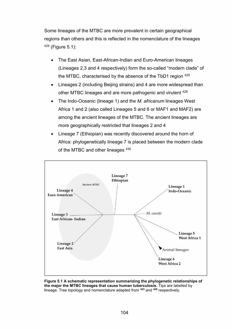
104
Some lineages of the MTBC are more prevalent in certain geographical
regions than others and this is reflected in the nomenclature of the lineages 429 (Figure 5.1):
x The East Asian, East-African-Indian and Euro-American lineages
(Lineages 2,3 and 4 respectively) form the so-called “modern clade” of
the MTBC, characterised by the absence of the TbD1 region 429
x Lineages 2 (including Beijing strains) and 4 are more widespread than
other MTBC lineages and are more pathogenic and virulent 428
x The Indo-Oceanic (lineage 1) and the M. africanum lineages West
Africa 1 and 2 (also called Lineages 5 and 6 or MAF1 and MAF2) are
among the ancient lineages of the MTBC. The ancient lineages are
more geographically restricted that lineages 2 and 4
x Lineage 7 (Ethiopian) was recently discovered around the horn of
Africa: phylogenetically lineage 7 is placed between the modern clade
of the MTBC and other lineages 430
Figure 5.1 A schematic representation summarizing the phylogenetic relationships of the major the MTBC lineages that cause human tuberculosis. Tips are labelled by lineage. Tree topology and nomenclature adapted from 423 and 429 respectively.
105
5.1.2 Population structure of TB in West Africa
A unique feature of TB in West Africa is the presence of all six major lineages
of the MTBC (Lineages 1-6). However, the Euro-American and the two M.
africanum lineages cause most human pulmonary TB in West Africa 294.
M. africanum is almost exclusive to West Africa and sporadic cases in other
regions are generally linked to West African migrants 431. Geographic
clustering is observed within the two M. africanum lineages:
x MAF1 is more prevalent around the Gulf of Guinea
x MAF2 is more prevalent in the western regions of West Africa 431
The restriction of M. africanum to West Africa may be driven by an unknown
animal reservoir. This hypothesis is supported by the close phylogenetic
relationship that exists between M. africanum and other animal lineages 428,432. An alternative hypothesis is that due to its inferior fitness, M.
africanum cannot compete with other M. tuberculosis lineages in other
regions 433. However, this does not explain why M. africanum remains an
important cause of disease in West Africa, despite the presence of “fitter”
lineages in the region 294.
A plausible scenario is that M. africanum is adapted to infect the human
genotypes that are predominant in West Africa. A recent study has shown an
association between lineage 5 (MAF1) and the Ewe ethnic group in Ghana 434. Similarly, a retrospective study in the US showed reported a high
prevalence of M. africanum among US-born African Americans 435. Co-
evolution of the MTBC with the human host is supported by striking
topological resemblances between the phylogeny of the MTBC and the
phylogeny of human mitochondrial genomes 423.
The Cameroon clade has emerged as the dominant genotype in much of
West Africa, particularly the eastern parts 436-440. The reasons for the
selection and dissemination of this genotype in West Africa remain unclear,
highlighting the need to investigate its evolutionary origins through WGS. In
Ghana, the Cameroon clade was most likely introduced through the south of
106
the country, where a higher proportion of infection is associated with this
clade than in the north 441.
The Ghana genotype (or T1 lineage) is disseminated across almost all West
African countries 294. The evolutionary origins of this genotype remain a
mystery and its epidemiology is not well described. However, it is the most
prevalent genotype in Guinea 442 and evidence from Ghana and Mali
suggests that the Ghana genotype is associated with drug resistance 441,443.
The Beijing clade is a globally disseminated lineage that is highly
transmissible and is associated with drug resistance 428,444-446. Although the
prevalence of the Beijing genotype in West Africa is generally low, its
prevalence is increasing in some countries. For example, Beijing is the most
common genotype causing human pulmonary in Benin 447. Similarly a high
prevalence of Beijing strains has been recorded in Senegal 448: both
countries host large international ports.
The animal lineages of the MTBC are associated with a wide range of host
species 284:
x Mycobacterium bovis (cattle)
x Mycobacterium caprae (sheep and goats)
x Mycobacterium microti (voles)
x Mycobacterium mungi (mongoose)
x Mycobacterium pinnipedii (seals and sealions)
x Mycobacterium orygis (antelope)
M. bovis has been found to cause human pulmonary TB in West Africa 449,450.
Patients infected with M. bovis need special treatment, because M. bovis
isolates are intrinsically resistant to pyrazinamide 451, which is part of the
recommended treatment regimen for drug-sensitive TB.
107
5.1.3 Multidrug-resistant tuberculosis (MDR-TB)
Mycobacteria are intrinsically resistant to most commonly prescribed
antibiotics due to a thick cell envelope that serves as a barrier and to
intracellular mechanisms that evade or deactivate antibiotics 452. This means
that TB is usually treated with drugs specific to management of this infection.
However, mycobacteria are able to acquire resistance to anti-TB drugs,
through chromosomal mutations in the drug target gene, the drug target
promoter region and/or drug activation enzymes 452.
Multidrug resistant tuberculosis (MDR-TB) emerges through the acquisition
of resistance in the individual patient during treatment or through
transmission of MDR-TB from patients with active pulmonary disease. Drug
resistance emerges through spontaneous resistance mutations that are
selected for during intermittent exposure to sub-lethal levels of drugs. Such
sub-optimal levels arise due to poor adherence, poor drug quality or patient-
specific pharmacodynamics 290. However, in high-burden settings, inter-
patient transmission is the main source of MDR-TB infection 453. WGS can
reliably trace the emergence MDR-TB during treatment and can identify on
going transmission chains 454,455.
Early diagnosis of MDR-TB is instrumental in ensuring successful treatment.
MDR-TB is confirmed by antimicrobial susceptibility testing. Culture-based
phenotypic testing is the gold standard, but it is slow and laborious. The
WHO now recommends rapid testing, at least for rifampicin resistance, at the
time of diagnosis 292. Molecular based techniques such as Xpert® MTB/RIF
(Cepheid Inc.) and the Hain-MTBDR-plus kit provide rapid early diagnosis of
MDR-TB. Whole-genome sequencing is emerging as a useful tool for
diagnosing and guiding treatment of MDR-TB by offering early detection of
resistance to both first- and second-line drugs 456-458.
It is recommended that MDR-TB cases be treated with individualized
treatment regimens guided by drug susceptibility testing 459. Unfortunately
the global success rate for treating MDR-TB is only about 50% 460. Moreover,
a large number of cases go undetected or unreported: e.g. in 2012 only 2%
of the estimated cases of MDR-TB in Nigeria were reported 460.
108
5.1.4 Rationale for the WATENAM study
In the 2016 global TB report, the WHO highlights a major shortfall in the
annual funding on TB research and development of TB treatment 292. This
shortfall is profound in West Africa where 12 of the 15 countries are among
the 42 least developed countries worldwide 461. Since the year 2000, only
The Gambia, Ghana and Nigeria have reported surveillance on the
prevalence of TB 292. There is a paucity of data on the prevalence of drug-
resistant TB in West Africa. The vast majority of health facilities in the region
lack the facilities for handling Mycobacterium tuberculosis, a category 3
pathogen.
The West African Nodes of Excellence for Tuberculosis, AIDS and Malaria
(WANETAM) project was set up in 2008, with the aim of tackling these three
major public health problems through sub-regional collaboration. The TB
nodes of excellence were set up in eight countries (The Gambia, Senegal,
Guinea Bissau, Togo, Ghana, Burkina Faso, Mali and Nigeria) with two aims:
x To provide capacity-building through training in mycobacterial culture
and identification to prepare the sites for clinical trials
x To estimate the burden of drug resistance through recruitment of
patients at tertiary health facilities and antimicrobial susceptibility
testing
The WANETAM coordinators have provided numerous training opportunities,
including courses in:
x Good clinical and laboratory practice
x Identification, culture, storage and transport of mycobacterial isolates
x Molecular typing and rapid detection of mycobacteria
x Data analysis and publishing
x Introductory bioinformatics, taught by the author
109
5.1.5 Objectives and study rationale
I wished to exploit whole-genome sequencing to study the epidemiology of
TB and genetics of multi-drug resistance among patients with active
pulmonary disease in West Africa. Due to limitations in time and money, it
was not possible to sequence all the isolates from the WANETAM sites. I
therefore undertook a pilot study of isolates from two West African countries:
x Nigeria, where the high burden of drug resistance makes it an ideal
setting to study the molecular mechanisms of MDR-TB in West Africa
x Mali, which has a well-structured hierarchy of TB reference health
care facilities in the capital Bamako. The WANETAM dataset
presented the opportunity to study the epidemiology of the TB in this
city
I used whole genomes sequencing data to:
x Understand the genomic epidemiology of MTBC in each country
by reconstructing the phylogeny of MTBC and classifying strains into
the global phylogenetic lineages and families
x Investigate the existence of inter-patient transmission by
searching for identical or closely related strains in different patients
x Understand the molecular mechanisms that are driving the emergence of dug resistance by scanning the genomes for known
resistance inducing mutations
x Understand the evolution of drug resistance by studying the
patterns of resistance mutations in the context of the phylogeny

110
5.2 Methods
5.2.1 Study sites
The WANETAM network has coordinated TB drug resistance surveys in nine
sites across eight West African countries (Figure 5.2):
1. Senegal, Laboratoire Bactériologie Virologie, Le Dantec, Dakar
2. The Gambia, National Public Health Laboratory Services, Banjul
3. Guinea-Bissau, National Institute for Public Health, Bissau
4. Mali, SEREFO (HIV/TB Research and Training Center) Faculté de
Médecine, de Pharmacie et d’Odento-Stamologie, University of
Bamako, Bamako
5. Burkina Faso, Centre Muraz and the National TB Program,
Ouagadougou
6. Ghana, Korle Bu Teaching Hospital, Accra
7. Togo, Laboratoire National de Référence des Mycobactéries, Lome
8. Nigeria, Nigerian Institute for Medical Research, Lagos
9. Nigeria, College of Medicine, University of Ibadan, Ibadan
Patients with confirmed active pulmonary TB based on WHO guidelines 462
were recruited at the study sites. Mycobacteria were cultured on site and the
pure isolates were sent to the MRCG for viability testing and antimicrobial
susceptibility testing to first and second line anti-TB drugs. The Joint Gambia
Government/MRC Ethics Committee and the relevant ethical bodies in other
participating countries approved the study protocols.
As noted, two countries were chosen for investigation in this study: Nigeria
and Mali. Nigeria borders Cameroon, Chad, Niger and Benin and has a
coastal region on the Gulf of Guinea. Nigeria has implemented WHO
recommended DOTS (Directly Observed Treatment Short-Course) since
1996. In recent years, Nigeria has increased use of Xpert® for early detection
of rifampicin resistance 463. Despite these measures, Nigeria still has one of
the highest burdens of TB globally and is among the six countries that
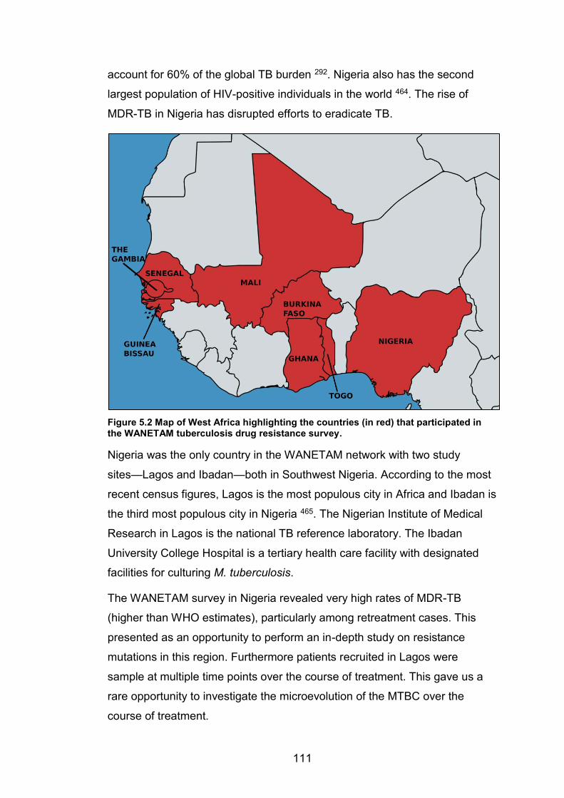
111
account for 60% of the global TB burden 292. Nigeria also has the second
largest population of HIV-positive individuals in the world 464. The rise of
MDR-TB in Nigeria has disrupted efforts to eradicate TB.
Figure 5.2 Map of West Africa highlighting the countries (in red) that participated in the WANETAM tuberculosis drug resistance survey.
Nigeria was the only country in the WANETAM network with two study
sites—Lagos and Ibadan—both in Southwest Nigeria. According to the most
recent census figures, Lagos is the most populous city in Africa and Ibadan is
the third most populous city in Nigeria 465. The Nigerian Institute of Medical
Research in Lagos is the national TB reference laboratory. The Ibadan
University College Hospital is a tertiary health care facility with designated
facilities for culturing M. tuberculosis.
The WANETAM survey in Nigeria revealed very high rates of MDR-TB
(higher than WHO estimates), particularly among retreatment cases. This
presented as an opportunity to perform an in-depth study on resistance
mutations in this region. Furthermore patients recruited in Lagos were
sample at multiple time points over the course of treatment. This gave us a
rare opportunity to investigate the microevolution of the MTBC over the
course of treatment.
112
Mali is a large landlocked country that borders seven countries including
Senegal, Algeria and Niger. Mali has a population of 15.3 million that
consists of people from diverse ethnic backgrounds 466. The study
participants from Mali were recruited in the capital city Bamako. Bamako is
highly cosmopolitan and is one of the fastest growing cities in Africa. Bamako
has a population of about 2 million people and is divided into six districts.
Each district has a TB referral health centre, equipped with diagnosis and
treatment facilities. Our isolates were collected at the University Teaching
Hospital, which acts as the principal TB referral centre. Retreatment cases
from across Mali are referred to the University Teaching Hospital for further
investigation and treatment. The Malian Ministry of health reported that in
2014 more than a third of all TB cases in Mali were managed in Bamako 467.
The Mali dataset offered an opportunity to study the molecular epidemiology
of the MTBC in Bamako since all the referral health centres report to the
central teaching hospital. From the WANETAM survey the prevalence of
MDR in Mali was low among new cases, but very high among retreatment
cases. Through WGS we set out to study the on going transmission of TB
between patients in Bamako and to establish whether MDR-TB among
retreatment cases was associated with a particular genotype.

113
5.2.2 Study isolates and workflow
Mycobacterial isolates were sent to the MRCG laboratories from Mali, Ibadan
and Lagos (Table 5.1). Isolates were tested for viability and antimicrobial
susceptibility testing to the first-line drugs isoniazid, rifampicin, ethambutol
and streptomycin. MDR-TB strains were further tested for susceptibility to the
second-line agents fluoroquinolones, kanamycin, capreomycin and
ethionamide (Table 5.2). For whole genome sequencing isolates were sub-
cultured on Middlebrooks 7H9 liquid medium for 3-8 weeks and genomic
DNA was extracted using the cetyl-trimethylammonium-bromide method
(Section 2.2.2). Whole-genome sequencing was performed on the Illumina
MiSeq using Nextera XT library kits.
The datasets from Mali and Nigeria were analysed separately due to
differences in sampling strategy. The datasets comprised of:
x Seventy-five isolates (43 MDR and then 32 non-MDR) that were
selected at random from 177 archived Nigerian isolates for whole-
genome sequencing. Sequencing failed for two isolates from Nigeria
so only 73 isolates were followed up. These isolates had been
recovered from 63 patients (21 and 42 from Ibadan and Lagos
respectively), with some patients supplying multiple isolates (Tables
5.3 and 5.4).
x Seventy-eight isolates that were selected at random from 208
archived Malian isolates for whole-genome sequencing (Tables 5.3
5.5). All originated from distinct patients. Two sequenced isolates from
Mali had no patient metadata or antimicrobial susceptibility profiles.
Detailed methods are provided in Chapter 2 but the workflow for
bioinformatics analyses is shown in Figure 5.3. This workflow included the
characterisation of strains based on their phylogenetic lineage and
genotypically predicted antimicrobial susceptibility profiles. For each dataset
a detailed phylogenetic analysis was performed that probed for on going
transmission and gave insights into the evolution of drug resistance in
tuberculosis infection.
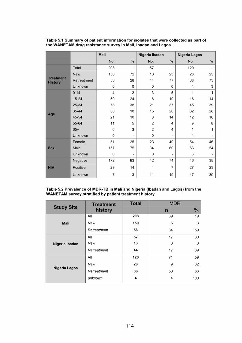
114
Table 5.1 Summary of patient information for isolates that were collected as part of the WANETAM drug resistance survey in Mali, Ibadan and Lagos.
Mali Nigeria Ibadan Nigeria Lagos
No. % No. % No. %
Total 208 - 57 - 120 -
Treatment History
New 150 72 13 23 28 23
Retreatment 58 28 44 77 88 73
Unknown 0 0 0 0 4 3
Age
0-14 4 2 3 5 1 1
15-24 50 24 6 10 16 14
25-34 78 38 21 37 45 39
35-44 38 18 15 26 32 28
45-54 21 10 8 14 12 10
55-64 11 5 2 4 9 8
65+ 6 3 2 4 1 1
Unknown 0 - 0 - 4 -
Sex Female 51 25 23 40 54 46
Male 157 75 34 60 63 54
Unknown 0 - 0 - 3 -
HIV
Negative 172 83 42 74 46 38
Positive 29 14 4 7 27 23
Unknown 7 3 11 19 47 39
Table 5.2 Prevalence of MDR-TB in Mali and Nigeria (Ibadan and Lagos) from the WANETAM survey stratified by patient treatment history.
Study Site Treatment history
Total MDR n %
Mali
All 208 39 19
New 150 5 3
Retreatment 58 34 59
Nigeria Ibadan
All 57 17 30 New 13 0 0
Retreatment 44 17 39
Nigeria Lagos
All 120 71 59
New 28 9 32
Retreatment 88 58 66
unknown 4 4 100

115
Table 5.3 Patient data for all genome-sequenced isolates from Mali, Ibadan and Lagos.
Mali Ibadan Lagos
MDR Non-MDR MDR Non-MDR MDR Non-MDR
Total
12 64 6 15 30 12
HIV Status Negative 11 51 6 15 21 8
Positive 1 6 0 0 7 4
Not Done 0 7 0 0 2 0
Gender Female 2 19 4 3 11 6
Male 10 45 2 12 18 6
Treatment History
New 3 58 0 8 4 4
Retreatment 9 6 6 7 21 1
Failure 0 0 0 0 2 3
Relapse 0 0 0 0 2 4
Age (years)
0-15 1 2 0 1 1 1
16-30 5 30 1 6 10 1
Above 30 6 32 5 8 18 10
Table 5.4 Metadata for MTBC isolates from Nigeria that were genome-sequenced.
ID Site MDR preXDR XDR StudyCase HIV Age Sex Date
NG46 IBADAN R S S Retreatment -ve 35 F 1/8/2012
NG64 IBADAN R R S Retreatment -ve 23 F 3/8/2012
NG68 IBADAN R S S Retreatment -ve 42 M 7/8/2012
NG70 IBADAN R S S Retreatment -ve 47 M 1/8/2012
NG44 IBADAN R S S Retreatment -ve 34 F 7/8/2012
NG45 IBADAN R S S Retreatment -ve 49 F 1/8/2012
NG88 IBADAN S New -ve 40 M 7/8/2012
NG47 IBADAN S New -ve 38 F 7/8/2012
NG62 IBADAN S New -ve 44 M 7/8/2012
NG81 IBADAN S New -ve 25 F 7/8/2012
NG82 IBADAN S New -ve 7 M 7/8/2012
NG89 IBADAN S New -ve 33 M 7/8/2012
NG84 IBADAN S New -ve 41 M 7/8/2012
NG69 IBADAN S New -ve 77 M 7/8/2012
NG61 IBADAN S Retreatment -ve 51 M 1/8/2012
NG65 IBADAN S Retreatment -ve 29 F 1/8/2012
NG80 IBADAN S Retreatment -ve 47 M 3/8/2012
NG83 IBADAN S Retreatment -ve 17 M 1/8/2012
NG85 IBADAN S Retreatment -ve 27 M 1/8/2012
NG86 IBADAN S Retreatment -ve 28 M 3/8/2012
NG67 IBADAN S Retreatment -ve 30 M 1/8/2012
NG36 LAGOS R S S NA 3/11/2012
NG55 LAGOS R S S NA 12/27/2011
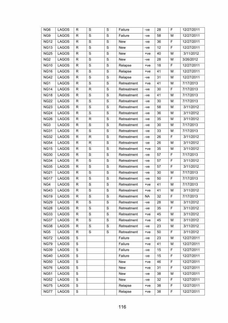
116
NG6 LAGOS R S S Failure -ve 28 F 12/27/2011
NG9 LAGOS R S S Failure -ve 58 M 12/27/2011
NG12 LAGOS R S S New -ve 36 F 12/27/2011
NG13 LAGOS R S S New -ve 12 F 12/27/2011
NG25 LAGOS R S S New +ve 40 M 3/11/2012
NG2 LAGOS R S S New -ve 28 M 3/26/2012
NG10 LAGOS R S S Relapse +ve 18 F 12/27/2011
NG16 LAGOS R S S Relapse +ve 41 M 12/27/2011
NG42 LAGOS R S S Relapse -ve 31 M 12/27/2011
NG1 LAGOS R S S Retreatment +ve 41 M 7/17/2013
NG14 LAGOS R R S Retreatment -ve 30 F 7/17/2013
NG18 LAGOS R S S Retreatment -ve 41 M 7/17/2013
NG22 LAGOS R S S Retreatment -ve 30 M 7/17/2013
NG23 LAGOS R S S Retreatment -ve 58 M 3/11/2012
NG24 LAGOS R S S Retreatment -ve 36 M 3/11/2012
NG26 LAGOS R R S Retreatment -ve 35 M 3/11/2012
NG3 LAGOS R S S Retreatment -ve 30 M 7/17/2013
NG31 LAGOS R S S Retreatment -ve 33 M 7/17/2013
NG32 LAGOS R R S Retreatment -ve 26 F 3/11/2012
NG54 LAGOS R R S Retreatment -ve 26 M 3/11/2012
NG15 LAGOS R S S Retreatment +ve 35 M 3/11/2012
NG30 LAGOS R S S Retreatment -ve 57 F 7/17/2013
NG34 LAGOS R S S Retreatment -ve 57 F 3/11/2012
NG35 LAGOS R S S Retreatment -ve 57 F 3/11/2012
NG21 LAGOS R S S Retreatment -ve 30 M 7/17/2013
NG17 LAGOS R S S Retreatment -ve 50 F 7/17/2013
NG4 LAGOS R S S Retreatment +ve 41 M 7/17/2013
NG43 LAGOS R S S Retreatment +ve 41 M 3/11/2012
NG19 LAGOS R S S Retreatment NA 32 F 7/17/2013
NG29 LAGOS R S S Retreatment -ve 28 M 3/11/2012
NG28 LAGOS R S S Retreatment -ve 35 F 3/11/2012
NG33 LAGOS R S S Retreatment +ve 45 M 3/11/2012
NG37 LAGOS R S S Retreatment +ve 45 M 3/11/2012
NG38 LAGOS R S S Retreatment -ve 23 M 3/11/2012
NG5 LAGOS R S S Retreatment +ve 50 F 3/11/2012
NG72 LAGOS S Failure -ve 23 M 12/27/2011
NG79 LAGOS S Failure +ve 41 M 12/27/2011
NG39 LAGOS S Failure -ve 15 F 12/27/2011
NG40 LAGOS S Failure -ve 15 F 12/27/2011
NG50 LAGOS S New +ve 46 F 12/27/2011
NG76 LAGOS S New +ve 31 F 12/27/2011
NG51 LAGOS S New -ve 38 M 12/27/2011
NG52 LAGOS S New -ve 32 F 12/27/2011
NG75 LAGOS S Relapse +ve 38 F 12/27/2011
NG77 LAGOS S Relapse +ve 38 F 12/27/2011

117
NG78 LAGOS S Relapse -ve 36 M 12/27/2011
NG53 LAGOS S Relapse -ve 45 M 12/27/2011
NG71 LAGOS S Relapse -ve 42 F 12/27/2011
NG74 LAGOS S S S Relapse -ve 42 F 12/27/2011
NG90 LAGOS S Retreatment -ve 36 M 3/11/2012
Footnote: Table sorted by Site, then MDR status (R = resistant, S = non-MDR) and then Study Case to make the table more intelligible. NA is not applicable or no result available. Date listed is the date that patients were enrolled into the study.
Table 5.5 Metadata for MTBC Isolates from Mali that were genome-sequenced.
ID MDR preXDR XDR StudyCase HIV Age Sex Date MA154 MA272 MA209 R S S New -ve 42 M 02/07/14 MA240 R New -ve 72 M 02/07/14 MA026 R S S New +ve 40 M 05/07/14 MA066 R S S Retreatment -ve 21 F 05/07/14 MA249 R S S Retreatment -ve 20 F 02/07/14 MA059 R S S Retreatment -ve 19 M 05/07/14 MA078 R S S Retreatment -ve 27 M 05/07/14 MA080 R R S Retreatment -ve 33 M 05/07/14 MA086 R S S Retreatment -ve 57 M 05/07/14 MA089 R S S Retreatment -ve 3 M 05/07/14 MA100 R S S Retreatment -ve 35 M 05/07/14 MA170 R Retreatment -ve 21 M 16/07/14 MA008 S New -ve 43 F 05/07/14 MA028 S New -ve 24 F 05/07/14 MA057 S New -ve 35 F 05/07/14 MA204 S New -ve 35 F 02/07/14 MA207 S New -ve 45 F 02/07/14 MA210 S New -ve 26 F 02/07/14 MA215 S New -ve 14 F 02/07/14 MA224 S New -ve 23 F 02/07/14 MA225 S New -ve 27 F 02/07/14 MA241 S New -ve 27 F 02/07/14 MA242 S New -ve 22 F 02/07/14 MA245 S New -ve 45 F 02/07/14 MA246 S New -ve 40 F 02/07/14 MA250 S New -ve 4 F 02/07/14 MA003 S New -ve 37 M 05/07/14 MA006 S New -ve 43 M 05/07/14 MA009 S New -ve 34 M 05/07/14 MA010 S New -ve 57 M 05/07/14 MA016 S New -ve 20 M 05/07/14 MA017 S New -ve 26 M 05/07/14

118
MA018 S New -ve 25 M 05/07/14 MA019 S New -ve 47 M 05/07/14 MA020 S New -ve 25 M 05/07/14 MA036 S New -ve 25 M 03/07/14 MA050 S New -ve 30 M 05/07/14 MA056 S New -ve 28 M 05/07/14 MA129 S New -ve 23 M 15/07/14 MA148 S New -ve 39 M 15/07/14 MA153 S New -ve 34 M 16/07/14 MA202 S New -ve 55 M 02/07/14 MA205 S New -ve 34 M 02/07/14 MA206 S New -ve 20 M 02/07/14 MA208 S New -ve 25 M 02/07/14 MA213 S New -ve 27 M 02/07/14 MA214 S New -ve 19 M 02/07/14 MA218 S New -ve 35 M 02/07/14 MA219 S New -ve 36 M 02/07/14 MA220 S New -ve 35 M 02/07/14 MA221 S New -ve 34 M 02/07/14 MA222 S New -ve 29 M 02/07/14 MA227 S New -ve 52 M 02/07/14 MA228 S New -ve 21 M 02/07/14 MA239 S New -ve 24 M 02/07/14 MA247 S New -ve 25 M 02/07/14 MA248 S New -ve 18 M 02/07/14 MA230 S New +ve 42 F 02/07/14 MA231 S New +ve 26 F 02/07/14 MA243 S New +ve 26 F 02/07/14 MA027 S New +ve 45 M 05/07/14 MA034 S New +ve 36 M 05/07/14 MA048 S New +ve 46 M 03/07/14 MA237 S New NA 20 F 02/07/14 MA229 S New NA 37 M 02/07/14 MA233 S New NA 32 M 02/07/14 MA234 S New NA 29 M 02/07/14 MA235 S New NA 58 M 02/07/14 MA236 S New NA 23 M 02/07/14 MA238 S New NA 25 M 02/07/14 MA058 S Retreatment -ve 34 F 05/07/14 MA067 S Retreatment -ve 38 M 05/07/14 MA069 S Retreatment -ve 78 M 05/07/14 MA070 S Retreatment -ve 31 M 03/07/14 MA087 S Retreatment -ve 20 M 05/07/14 MA096 S Retreatment -ve 60 M 03/07/14 Footnote: Columns were sorted by MDR status, then Study Case, then HIV status.
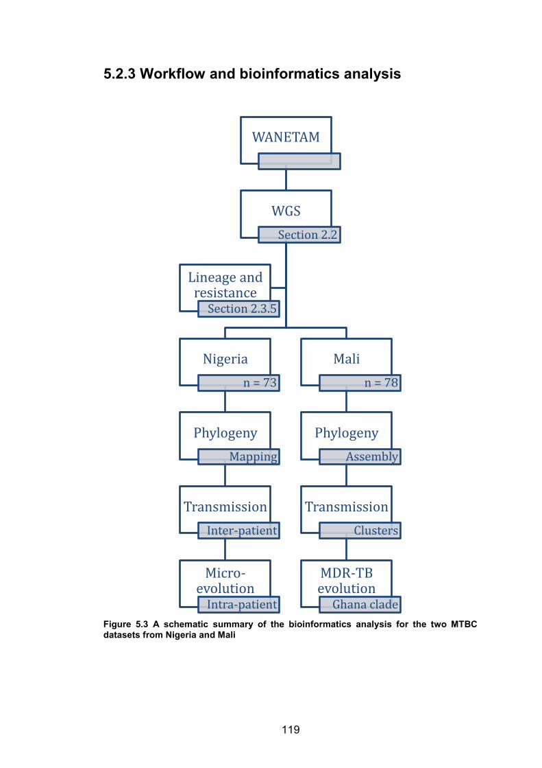
119
5.2.3 Workflow and bioinformatics analysis
Figure 5.3 A schematic summary of the bioinformatics analysis for the two MTBC datasets from Nigeria and Mali
WANETAM
WGSSection 2.2
Nigerian = 73
PhylogenyMapping
TransmissionInter-patient
Micro-evolution
Intra-patient
Malin = 78
PhylogenyAssembly
TransmissionClusters
MDR-TB evolution
Ghana clade
Lineage and resistance
Section 2.3.5
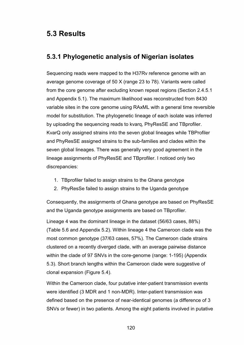
120
5.3 Results
5.3.1 Phylogenetic analysis of Nigerian isolates
Sequencing reads were mapped to the H37Rv reference genome with an
average genome coverage of 50 X (range 23 to 78). Variants were called
from the core genome after excluding known repeat regions (Section 2.4.5.1
and Appendix 5.1). The maximum likelihood was reconstructed from 8430
variable sites in the core genome using RAxML with a general time reversible
model for substitution. The phylogenetic lineage of each isolate was inferred
by uploading the sequencing reads to kvarq, PhyResSE and TBprofiler.
KvarQ only assigned strains into the seven global lineages while TBProfiler
and PhyResSE assigned strains to the sub-families and clades within the
seven global lineages. There was generally very good agreement in the
lineage assignments of PhyResSE and TBprofiler. I noticed only two
discrepancies:
1. TBprofiler failed to assign strains to the Ghana genotype
2. PhyResSe failed to assign strains to the Uganda genotype
Consequently, the assignments of Ghana genotype are based on PhyResSE
and the Uganda genotype assignments are based on TBprofiler.
Lineage 4 was the dominant lineage in the dataset (56/63 cases, 88%)
(Table 5.6 and Appendix 5.2). Within lineage 4 the Cameroon clade was the
most common genotype (37/63 cases, 57%). The Cameroon clade strains
clustered on a recently diverged clade, with an average pairwise distance
within the clade of 97 SNVs in the core-genome (range: 1-195) (Appendix
5.3). Short branch lengths within the Cameroon clade were suggestive of
clonal expansion (Figure 5.4).
Within the Cameroon clade, four putative inter-patient transmission events
were identified (3 MDR and 1 non-MDR). Inter-patient transmission was
defined based on the presence of near-identical genomes (a difference of 3
SNVs or fewer) in two patients. Among the eight patients involved in putative

121
transmission events, six were retreatment cases, one had succumbed to
treatment failure and only one was a new case.
The Haarlem, X-type, Ghana, TUR, LAM, Uganda and H37Rv-like genotypes
of lineage 4 were collectively associated with 20 cases of TB (32%). There
was no indication that these genotypes were involved in on going
transmission. The closest indication of transmission was in two TUR
genotype MDR strains from different patients that differed by 18 SNVs.
The M. africanum sub-lineage 1 (MAF1) was isolated from six patients: five
were MDR and one was non-MDR. Two closely related MAF1 strains that
differed by 11 SNVs were isolated from two retreatment cases from Lagos
and Ibadan. Although this is not definitive evidence of direct transmission
between the two patients, it suggests that MAF1 is involved in transmission
of MDR in this setting. The high prevalence of MDR MAF1 is probably
because we sampled patients at referral centres where severe cases are
managed. One patient was infected with the animal lineage M. bovis.
Table 5.6 The prevalence of MTBC lineages and genotypes among MDR and non-MDR cases in Nigeria.
Lineage Clade Total MDR Non-MDR M. bovis Bovis 1 1
Lineage 4
Cameroon 37 19 18
Uganda 6 2 4
Ghana 2 2
X-type 1 1
H37Rv 1 1
Haarlem 3 2 1
LAM 4 2 2
TUR 2 2
Lineage 5 West Africa 1 6 5 1
Grand Total 63 36 27
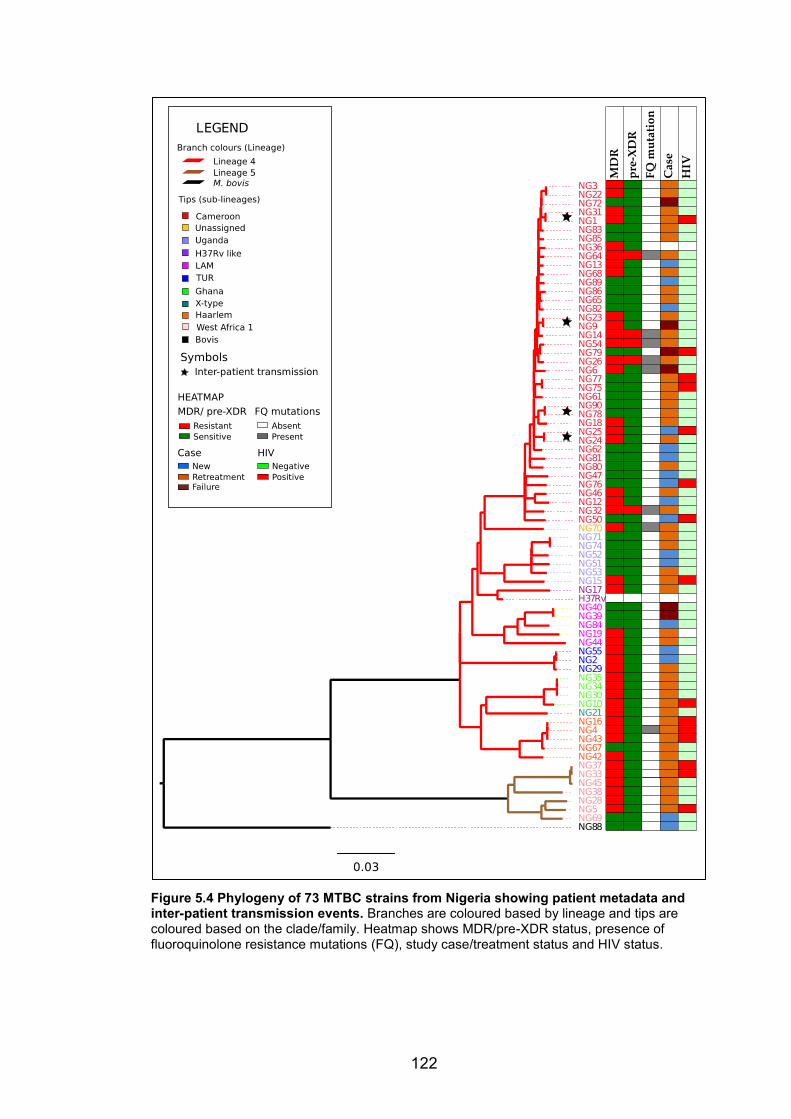
122
Figure 5.4 Phylogeny of 73 MTBC strains from Nigeria showing patient metadata and inter-patient transmission events. Branches are coloured based by lineage and tips are coloured based on the clade/family. Heatmap shows MDR/pre-XDR status, presence of fluoroquinolone resistance mutations (FQ), study case/treatment status and HIV status.
123
5.3.2 Microevolution of drug resistance in Nigeria
To study the microevolution of drug resistance we compared resistance
profiles of identical/near-identical strains, which formed flat branches in the
phylogenetic tree (Figure 5.5). These included genomes recovered from the
same patient over the course of treatment as well as those isolated from
different patients, which I have cited as putative transmission events. Six
patients had isolates from two time points during treatment genome-
sequenced and two patients had isolates from three time points genome-
sequenced. Three instances were observed where microevolution influenced
the susceptibility profiles of strains:
x NG9 and NG23; two patients enrolled in Lagos in 2011 and 2012.
Both were MDR strains, but NG23 isolate acquired katG mutation
L587P
x NG1 and NG31; two patients from Lagos. Both MDR, with canonical
mutations in embB. NG1 acquired a mutation in the embA promoter,
so became ethambutol resistant, while NG31 was ethambutol
susceptible
x NG16, NG43, NG4 were from the same patient on treatment. All were
MDR, but fluoroquinolone-susceptible on phenotypic testing. However
the most recent isolate, NG4, acquired a low-level fluoroquinolone-
resistance mutation in the gyrB gene
To confirm whether the samples were from the same patient or different
patients the lab numbers of these strains were sent to our collaborators at
the National Institute for Medical Research in Lagos for confirmation. Due to
patient confidentiality the patient names are not presented as part of this
thesis. NG39 and NG40 were isolated from the same patient at different time
points. Both strains were susceptible to all first-line drugs by phenotypic
testing. However, both strains bore katG and rpoB mutations that confer
isoniazid and rifampicin respectively. It is unclear why this discrepancy was
observed but a sample mix-up cannot be ruled out.

124
Figure 5.5 Microevolution of MTBC strains from Nigeria within patients during treatment and between patients during putative transmission. Phylogeny is shown in the context of antimicrobial susceptibility profiles.
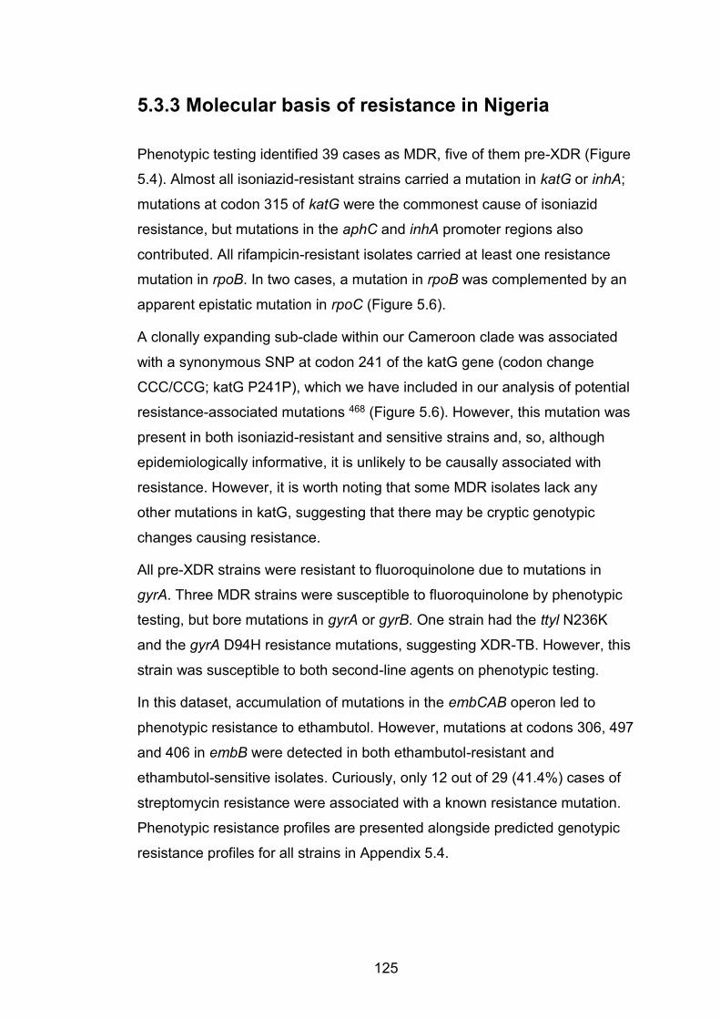
125
5.3.3 Molecular basis of resistance in Nigeria
Phenotypic testing identified 39 cases as MDR, five of them pre-XDR (Figure
5.4). Almost all isoniazid-resistant strains carried a mutation in katG or inhA;
mutations at codon 315 of katG were the commonest cause of isoniazid
resistance, but mutations in the aphC and inhA promoter regions also
contributed. All rifampicin-resistant isolates carried at least one resistance
mutation in rpoB. In two cases, a mutation in rpoB was complemented by an
apparent epistatic mutation in rpoC (Figure 5.6).
A clonally expanding sub-clade within our Cameroon clade was associated
with a synonymous SNP at codon 241 of the katG gene (codon change
CCC/CCG; katG P241P), which we have included in our analysis of potential
resistance-associated mutations 468 (Figure 5.6). However, this mutation was
present in both isoniazid-resistant and sensitive strains and, so, although
epidemiologically informative, it is unlikely to be causally associated with
resistance. However, it is worth noting that some MDR isolates lack any
other mutations in katG, suggesting that there may be cryptic genotypic
changes causing resistance.
All pre-XDR strains were resistant to fluoroquinolone due to mutations in
gyrA. Three MDR strains were susceptible to fluoroquinolone by phenotypic
testing, but bore mutations in gyrA or gyrB. One strain had the ttyl N236K
and the gyrA D94H resistance mutations, suggesting XDR-TB. However, this
strain was susceptible to both second-line agents on phenotypic testing.
In this dataset, accumulation of mutations in the embCAB operon led to
phenotypic resistance to ethambutol. However, mutations at codons 306, 497
and 406 in embB were detected in both ethambutol-resistant and
ethambutol-sensitive isolates. Curiously, only 12 out of 29 (41.4%) cases of
streptomycin resistance were associated with a known resistance mutation.
Phenotypic resistance profiles are presented alongside predicted genotypic
resistance profiles for all strains in Appendix 5.4.
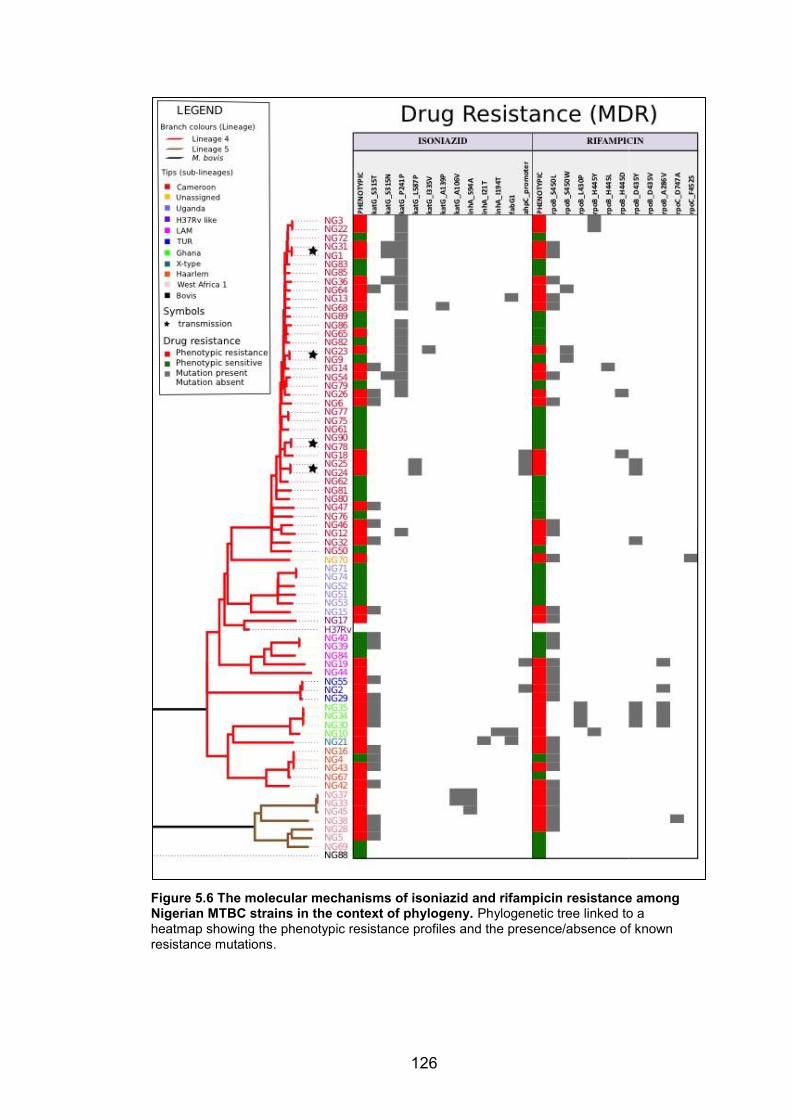
126
Figure 5.6 The molecular mechanisms of isoniazid and rifampicin resistance among Nigerian MTBC strains in the context of phylogeny. Phylogenetic tree linked to a heatmap showing the phenotypic resistance profiles and the presence/absence of known resistance mutations.
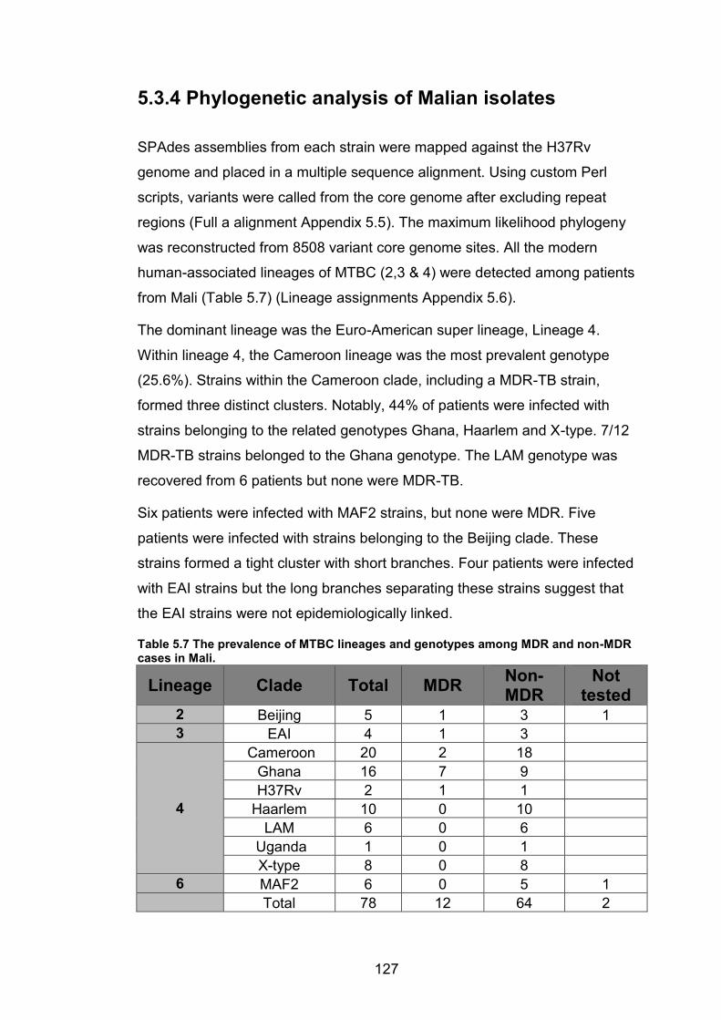
127
5.3.4 Phylogenetic analysis of Malian isolates
SPAdes assemblies from each strain were mapped against the H37Rv
genome and placed in a multiple sequence alignment. Using custom Perl
scripts, variants were called from the core genome after excluding repeat
regions (Full a alignment Appendix 5.5). The maximum likelihood phylogeny
was reconstructed from 8508 variant core genome sites. All the modern
human-associated lineages of MTBC (2,3 & 4) were detected among patients
from Mali (Table 5.7) (Lineage assignments Appendix 5.6).
The dominant lineage was the Euro-American super lineage, Lineage 4.
Within lineage 4, the Cameroon lineage was the most prevalent genotype
(25.6%). Strains within the Cameroon clade, including a MDR-TB strain,
formed three distinct clusters. Notably, 44% of patients were infected with
strains belonging to the related genotypes Ghana, Haarlem and X-type. 7/12
MDR-TB strains belonged to the Ghana genotype. The LAM genotype was
recovered from 6 patients but none were MDR-TB.
Six patients were infected with MAF2 strains, but none were MDR. Five
patients were infected with strains belonging to the Beijing clade. These
strains formed a tight cluster with short branches. Four patients were infected
with EAI strains but the long branches separating these strains suggest that
the EAI strains were not epidemiologically linked.
Table 5.7 The prevalence of MTBC lineages and genotypes among MDR and non-MDR cases in Mali.
Lineage Clade Total MDR Non-MDR
Not tested
2 Beijing 5 1 3 1 3 EAI 4 1 3
4
Cameroon 20 2 18 Ghana 16 7 9 H37Rv 2 1 1 Haarlem 10 0 10 LAM 6 0 6 Uganda 1 0 1 X-type 8 0 8 6 MAF2 6 0 5 1
Total 78 12 64 2

128
5.3.5 Transmission clusters in Mali
In the absence of patient epidemiological data it was not possible to
reconstruct chains of transmission. However, it was possible to detect
clusters of strains that were likely to be part of a transmission network based
on genomic similarity. Transmission clusters were defined as five or more
strains that clustered in a clade characterised by short branches and a highly
conserved core genome (on average <25 SNVs difference between strains)
(Appendix 5.7). The phylogenetic tree suggested that there were three
transmission clusters within the dataset (Figure 5.7):
x A sub-clade of 13 Ghana clade strains clustered on a recently
divergent branch. On average, strains within this cluster differed by 12
core-genome SNVs. The most divergent pair of strains within this
cluster differed by 24 core genome SNVs. Within this transmission
cluster there was an MDR sub-cluster and a non-MDR sub-cluster
x Eight non-MDR Cameroon clade strains formed a cluster on a recently
divergent branch. The average pairwise distance for strains within this
cluster was 7 core genome SNVs and the most divergent strains
differed by 13 core genome SNVs. This cluster represented the most
conserved transmission cluster in this dataset
x The five Beijing strains formed a monophyletic clade with a within-
clade pairwise distance of 21 core-genome SNVs. The pairwise
distance between strains within this cluster ranged from 11 to 30 core
genome SNVs. The Beijing cluster was less conserved than the
Ghana and Cameroon genotype clusters
Two sporadic cases of inter-patient transmission involving non-MDR strains
were noted (Figure 5.7):
x Identical LAM genotype strains were isolated from two new cases
(MA245 & MA246)
x Identical Cameroon clade strains were recovered from two new cases
(MA242 & MA243). They differed from a third strain MA224 by six
core genome SNVs
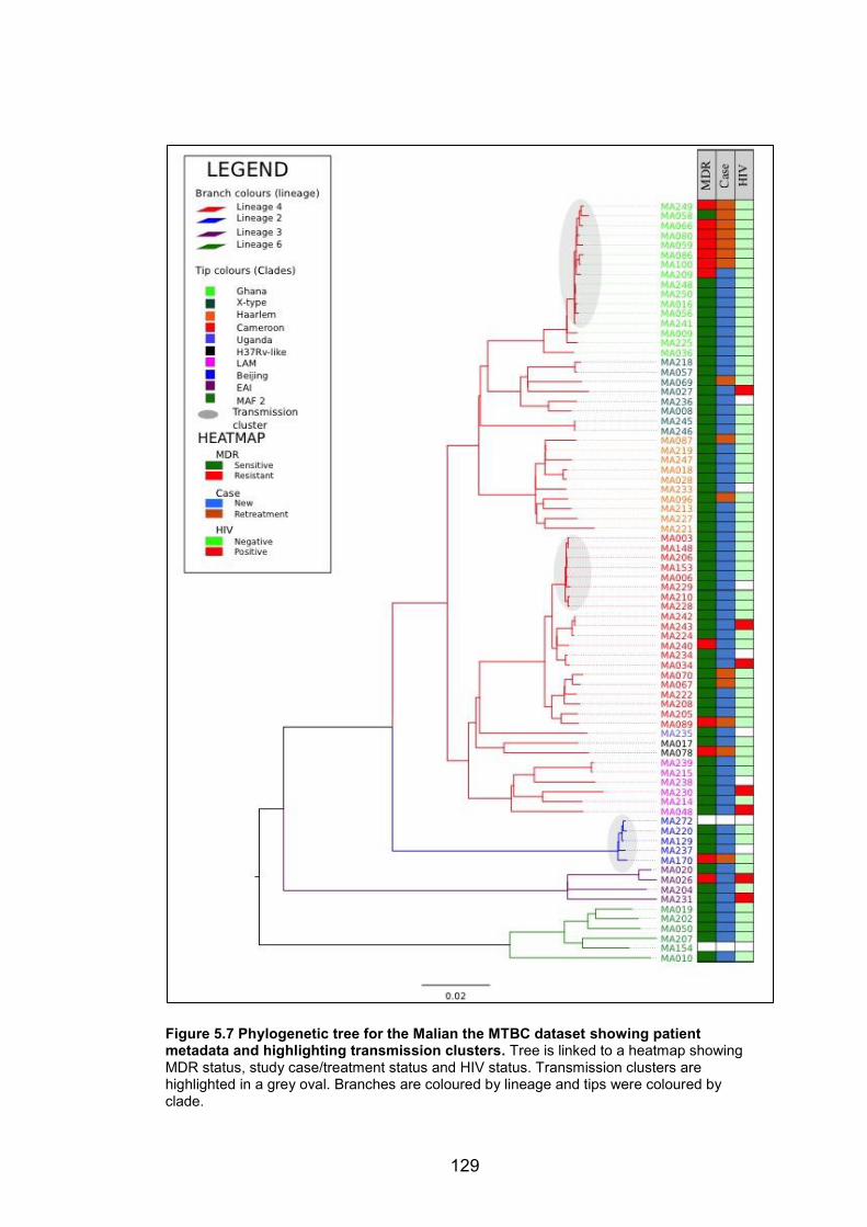
129
Figure 5.7 Phylogenetic tree for the Malian the MTBC dataset showing patient metadata and highlighting transmission clusters. Tree is linked to a heatmap showing MDR status, study case/treatment status and HIV status. Transmission clusters are highlighted in a grey oval. Branches are coloured by lineage and tips were coloured by clade.
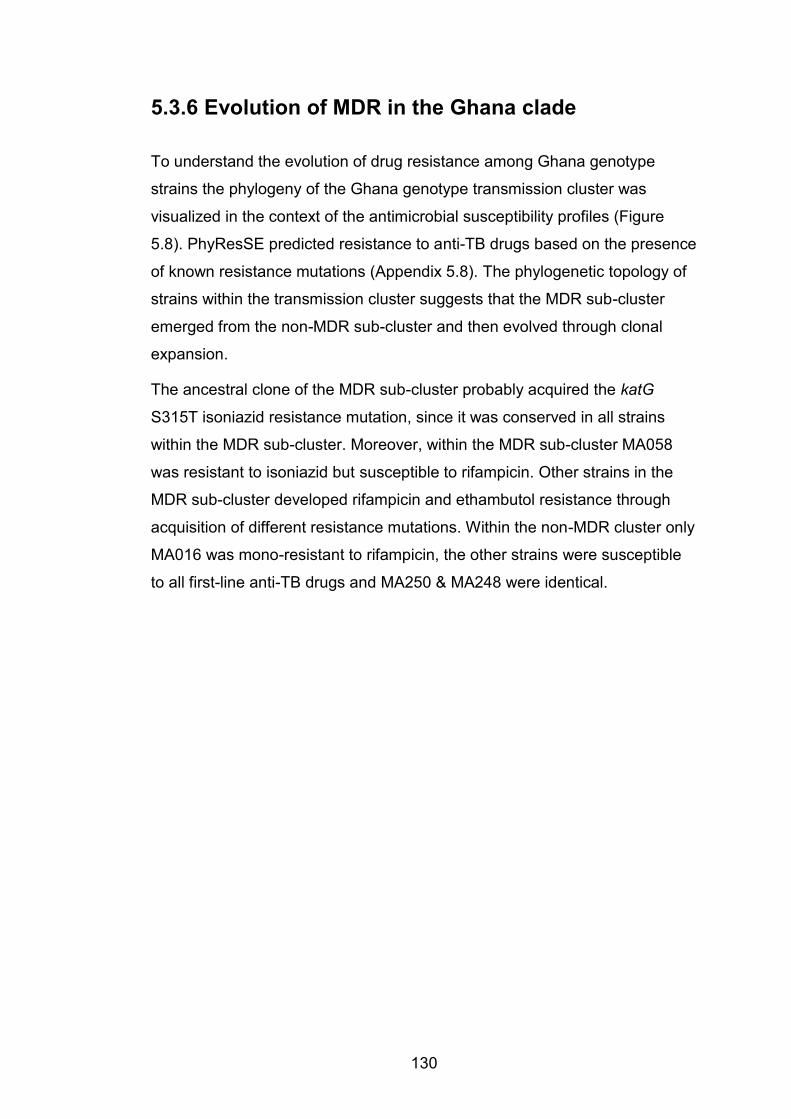
130
5.3.6 Evolution of MDR in the Ghana clade
To understand the evolution of drug resistance among Ghana genotype
strains the phylogeny of the Ghana genotype transmission cluster was
visualized in the context of the antimicrobial susceptibility profiles (Figure
5.8). PhyResSE predicted resistance to anti-TB drugs based on the presence
of known resistance mutations (Appendix 5.8). The phylogenetic topology of
strains within the transmission cluster suggests that the MDR sub-cluster
emerged from the non-MDR sub-cluster and then evolved through clonal
expansion.
The ancestral clone of the MDR sub-cluster probably acquired the katG
S315T isoniazid resistance mutation, since it was conserved in all strains
within the MDR sub-cluster. Moreover, within the MDR sub-cluster MA058
was resistant to isoniazid but susceptible to rifampicin. Other strains in the
MDR sub-cluster developed rifampicin and ethambutol resistance through
acquisition of different resistance mutations. Within the non-MDR cluster only
MA016 was mono-resistant to rifampicin, the other strains were susceptible
to all first-line anti-TB drugs and MA250 & MA248 were identical.
131
Figure 5.8 The evolution of MDR-TB among Ghana genotype strains in Mail. The phylogeny of the Ghana genotype transmission cluster is shown in the context of antibiotic resistance profiles.
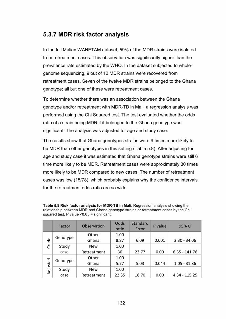
132
5.3.7 MDR risk factor analysis
In the full Malian WANETAM dataset, 59% of the MDR strains were isolated
from retreatment cases. This observation was significantly higher than the
prevalence rate estimated by the WHO. In the dataset subjected to whole-
genome sequencing, 9 out of 12 MDR strains were recovered from
retreatment cases. Seven of the twelve MDR strains belonged to the Ghana
genotype; all but one of these were retreatment cases.
To determine whether there was an association between the Ghana
genotype and/or retreatment with MDR-TB in Mali, a regression analysis was
performed using the Chi Squared test. The test evaluated whether the odds
ratio of a strain being MDR if it belonged to the Ghana genotype was
significant. The analysis was adjusted for age and study case.
The results show that Ghana genotypes strains were 9 times more likely to
be MDR than other genotypes in this setting (Table 5.8). After adjusting for
age and study case it was estimated that Ghana genotype strains were still 6
time more likely to be MDR. Retreatment cases were approximately 30 times
more likely to be MDR compared to new cases. The number of retreatment
cases was low (15/78), which probably explains why the confidence intervals
for the retreatment odds ratio are so wide.
Table 5.8 Risk factor analysis for MDR-TB in Mali. Regression analysis showing the relationship between MDR and Ghana genotype strains or retreatment cases by the Chi squared test. P value <0.05 = significant.
Factor Observation Odds ratio
Standard Error P value 95% CI
Crud
e Genotype Other 1.00 Ghana 8.87 6.09 0.001 2.30 - 34.06 Study case
New 1.00 Retreatment 30 23.77 0.00 6.35 - 141.76
Adju
sted
Genotype Other 1.00 Ghana 5.77 5.03 0.044 1.05 - 31.86 Study case
New 1.00 Retreatment 22.35 18.70 0.00 4.34 - 115.25

133
5.4 Discussion
5.4.1 Diversity of TB: Nigeria versus Mali
The Euro-American super-lineage was dominant in all our study sites. In
Bamako, two other “modern” lineages, Beijing and EAI, were also detected.
The M. africanum lineages MAF1 and MAF2 were detected in Southwest
Nigeria and Bamako respectively. The geographic restriction of these two
lineages to West Africa has been documented 431. The animal lineage M.
bovis has been previously reported in both Mali and Nigeria 449,450. We report
one only case of M bovis infection that occurred in Ibadan. Prevalence of M.
bovis is higher in regions where cattle are more widespread. All our study
sites were in urban cities where contact with cattle is minimal.
Our data shows that the diversity of MTBC isolates in Bamako, towards the
northwest of the West African region, is greater that the diversity in Ibadan
and Lagos, towards the Gulf of Guinea. This is in line with the most recent
analysis on the phylogeography of the MTBC in West Africa by Gehre et. al. 294. Furthermore, in Southwest Nigeria evidence of on going transmission
and clonal expansion was found only in the Cameroon clade. However, in
Bamako there was evidence of on going spread not just of Cameroon-clade
strains but also of the Ghana genotype and Beijing strains.
As its name suggests, the Cameroon clade is believed to have emerged in
Cameroon and has now spread throughout much of West Africa 439: it has
been reported as the most dominant genotype in Nigeria, Ghana, Burkina
Faso, Chad and Cameroon 436,438,440,469,470. Nigeria is probably the largest
reservoir of the Cameroon clade genotype 439. However, our data suggests
that the prevalence of the Cameroon clade in Bamako is increasing rapidly. A
recent survey based on strains collected between 2006-2010 estimated the
prevalence of Cameroon strains to be 10% 443. This figure has more than
doubled based on our dataset. The reasons for the selection and
dissemination of the Cameroon clade in West Africa remain unclear, but
perhaps will become clearer with additional WGS studies.
134
Although the Ghana genotype, also known as the T1 clade, has been
replaced by the Cameroon clade as the most abundant genotype in Bamako 443, it remains comparable in prevalence. Our data confirms previous reports
that the Ghana genotype is associated with MDR-TB 440,443. Among our
isolates, this association appears to have been driven by emergence of a
sub-clade of the Ghana genotype, carrying the katG S315T isoniazid-
resistance mutation. In Lagos, an MDR Ghana-genotype isolate acquired a
fluoroquinolone-resistance mutation over the course of treatment. As more
strains belonging to this genotype are genome-sequenced, the evolutionary
mechanisms driving its association with drug resistance will be unravelled.
There is a paucity of data on the prevalence of Beijing strains in sub-Saharan
Africa. Reviews published from the 2000s suggest that the Beijing strains
were rarely found in sub-Saharan Africa 471. In 2008, two cases of TB in
Bamako were due to unrelated Beijing strains 472. Our data shows that
Beijing strains are on the rise in Bamako through clonal expansion of drug
susceptible strains. Introduction of Beijing strains in Cape Town was followed
by a rapid increase in prevalence 473,474. Similarly, recent evidence shows
that the Beijing strain was introduced in Afghanistan during the Soviet
invasion in the 1980’s and has continued to evolve and spread with human
migration since 475. It is imperative to closely monitor the rise in prevalence of
Beijing strains in Bamako and other parts of West Africa, given this clade’s
propensity for increased virulence, high transmissibility and resistance.
Despite reports of a diminished presence in Cameroon 476 M. africanum is
still an important cause of TB in West Africa 294,439,440,477. Although we have
not found any recent inter-patient transmission events involving MAF1 in
Nigeria, our findings do suggest that multidrug resistant MAF1 isolates have
been transmitted in this setting during the recent past. There was no
evidence of direct transmission of MAF2 strains isolated from patients in
Bamako and none of the strains were MDR. The high prevalence of MDR
among MAF1 strains from Nigeria (5/6) is probably because we sampled
patients from retreatment centres in a high MDR-TB burden setting.
135
5.4.2 Impact of MDR transmission on TB control
Globally the burden of MDR-TB is increasing, especially in low-resource
settings 292. This increase is driven largely by the transmission of MDR-TB
especially in high-TB-burden regions 453. Effective TB control strategies
deliver prompt identification of cases and adequate treatment 478. Correct
administration of treatment renders the patient non-contagious and
completion of treatment results in a high cure rate 478. It is encouraging to
see a decrease in the prevalence of MDR-TB in countries that have
implemented effective TB control programs 479
In Bamako, retreatment cases are referred to the central teaching hospital for
antimicrobial susceptibility testing and adequate treatment. However, it is
unclear whether the peripheral health facilities are equipped to perform
antimicrobial susceptibility testing. Failing to diagnose MDR would lead to
treatment failure on the standard regimen for drug-susceptible TB.
In Nigeria, culture facilities are also scarce, in part because of the stringent
biosafety requirements. Funding and resources need to be directed to
provide adequate diagnostic facilities in low-resource settings where the
burden of B is highest. Measures need to be taken to stem the spread of
MDR-TB in West Africa. Policy makers are encouraged to consider social
intervention such as sensitisation campaigns, improved affordable housing
and improved sanitation.
5.4.3 Usefulness of rapid molecular testing for drug
resistance in high burden settings
Early diagnosis of MDR-TB can be instrumental in ensuring treatment
success. The WHO recommends rapid testing, at least for rifampicin
resistance, at the time of diagnosis 292. The implementation of Xpert®
MTB/RIF (Cepheid Inc.) for rapid detection of Mycobacterium tuberculosis
complex (the MTBC) and rifampicin resistance has been intensified in Nigeria
136
to good effect 463,469. However, Xpert® probably needs to be complemented
with additional resistance testing 480.
The Hain MTBDR plus kit is a rapid tool for early detection of drug resistance
in the MTBC using molecular probes. In Bamako, the Hain kit would be
adequate for detecting the MDR Ghana genotype cluster since it has probes
for detecting the katG S315T isoniazid-resistance mutations. In Nigeria, the
Hain kit is unlikely to be sufficient to rule out isoniazid resistance due to the
presence of rare isoniazid resistance mutation in the katG gene and in gene
promoter region. In Nigeria, there appears to be an emerging pre-XDR TB
problem driven by fluoroquinolone resistance. The Hain kit also offers early
detection of fluoroquinolone.
5.4.4 Need for genomic surveillance of TB across
West Africa
West African health authorities may wish to consider a move towards routine
in situ use of bacterial WGS for characterisation of TB isolates. WGS offers
early detection of resistance to both first- and second-line drugs, making it
useful for diagnosing and guiding treatment of MDR-TB 457,481,482. User-
friendly web tools such as PhyResSe and TBProfiler are available for
inferring the phylogenetic lineages and drug-resistance profiles of the MTBC
isolates directly from WGS data 314,315. WGS boasts superior discriminatory
power in delineating human-to-human transmission and it differentiates
strains that appear identical by conventional genotyping techniques (e.g.
spoligo typing, MIRU VNTR and IS6110 typing) 455,483. Moreover WGS offers
insights into the evolutionary mechanisms that drive important process like
transmission and drug resistance.

137
5.4.5 Limitations
The samples in the WANETAM dataset were collected from patients
recruited at tertiary referral centres. This is likely to present a sampling bias
since strains belonging to hypervirulent lineages that are prone to resistance
are more likely to cause chronic cases of tuberculosis disease that warrant
referral. Consequently these samples are likely not to be an accurate
representation of the true genomic epidemiology of MTBC in Mali and
Nigeria. Due to the lack of patient epidemiology and social network data it
was not possible to conclusively infer the routes of inter-patient transmission.
138
6 CHAPTER SIX: CONCLUSIONS
6.1 The varieties of bacterial evolution
My work has involved three bacterial species that evolve in quite different
ways. Through genomics, I have gained valuable insights into the
epidemiology and antimicrobial resistance patterns of each pathogen. Some
insights have a clear and present translational impact e.g. unravelling the
cause of a meningitis outbreak and illuminating the mechanisms of antibiotic
resistance in MDR-TB. Others, like the evolutionary history of S. aureus in
monkeys in The Gambia, satisfy the curiosity, but have a less direct impact
on public health.
The mode and tempo of evolution are known to vary between different
bacterial species 190 and this applies to the three species studied in this
thesis:
x Staphylococcus aureus readily undergoes horizontal gene transfer,
resulting in changes in the accessory genome that contribute
significantly to its evolution 337,338. However, this data demonstrates
the distinction between the core genome and accessory genome is
somewhat arbitrary, as shown by the conservation of accessory
genome components within clonal lineages. Evolution within the core
genome of S. aureus is driven mainly by point mutations, although
there is evidence for recombination between distantly related strains.
Recombination events can lead to the emergence of epidemic clones
of S. aureus e.g. a recombination event between ST8 and ST30 is
believed to be the origin of the ST239 “hybrid lineage”, which evolved
into a methicillin-resistant lineage 378,484. However, the full functional
implications of recombination in S. aureus remain unclear 485. Point
mutations in the core genome were key to reconstructing the
phylogenetic tree and in inferring the approximate dates for host
139
species jumps. These inferences hinged on the assumption of a
neutral evolutionary rate and a uniform mutation rate. This approach
can be criticised for not considering the effect of variable mutation
rates in different lineages. Nonetheless it provides a reasonable
estimate for the timescales of the jumps from one host species to
another.
x Streptococcus pneumoniae is also known for its promiscuity in
terms of acquiring foreign DNA, but also experiences very high rates
of homologous recombination that shape its evolutionary landscape 178. Homologous recombination proved to be the main driving force
behind the emergence of the novel clade that drove the meningitis
outbreak described here—over two hundred mutations were
introduced into this lineage through recombination. These
recombination blocks overlapped with genes that are associated with
fitness and virulence. In S. pneumoniae, antibiotic resistance is
mediated through point mutations as well the acquisition of genes via
horizontal gene transfer 406. Although antibiotic resistance genes were
detected in the novel clade, no accessory genes are conserved in this
clade yet absent in the West African ST303 and ST217 strains.
x Mycobacterium tuberculosis is a genetically monomorphic lineage
devoid of horizontal gene transfer and homologous recombination 203.
Instead, evolution within this species is driven by point mutations and
large genomic deletions. Novel insights have emerged into the
phylogeny of the M. tuberculosis complex in West Africa, highlighting
inter-patient transmission and microevolution of strains within and
between patients. Point mutations are the underlying mechanism of
resistance to anti-TB drugs. A vast repository of known resistance
mutations exists and resistance mutations are confirmed by
convergent evolution in different lineages. I characterised antibiotic
resistance in my datasets based on the presence/absence of known
resistance mutations. This gave valuable insights into the mechanisms
behind the emerging MDR-TB and pre-XDR-TB threat in West Africa 293.
140
6.2 Novel insights
Whole-genome sequencing has identified a novel clade of S. aureus, which
colonizes Green Vervet monkeys in The Gambia. Analysis of gene
repertoires points to gene loss as a major contributor to adaptation of the
bacterium to a new host, the monkeys. Genomic epidemiology points
towards to humans as the source of S. aureus in monkeys in The Gambia
with recent anthroponotic transmission of human-associated genotypes to
monkeys. The novel clade is believed to well before the advent of recent
industrialisation and urbanisation.
Quantitative PCR (qPCR) was instrumental in revealing the aetiological
agent of the meningitis outbreak in Ghana. However, WGS went beyond
qPCR and standard genotyping to unveil a novel serotype 1/ST303 genotype
lineage that showed features of an outbreak strain: high clonality, low
sequence divergence and geographic clustering in the epicentre. Comparing
the outbreak serotype strains to previously sequenced serotype 1 strains
from West Africa confirmed the novelty of the novel clade. This novel clade
has probably emerged because of rapid evolution through homologous
recombination. Genomic evidence confirmed that strains in the novel clade
have undergone capsular switching. A likely scenario is that the novel clade
had acquired selective traits that enhanced its dissemination among the
unvaccinated older population.
WGS has highlighted the gravity of the MDR-TB burden in Nigeria. MDR
strains belonging to almost all the major genotypes of the Euro-America
super lineage were detected in Nigeria. This widespread emergence of MDR-
TB should be treated as a public health emergency and measures must be
taken to prevent Nigeria from becoming a reservoir for MDR-TB. This
situation could ominously deteriorate into an XDR-TB problem.
In Mali, the emerging MDR-TB problem appears to be driven by the Ghana
genotype. Authorities should work towards enhancing diagnostic capacities
across the Bamako area, so that patients are treated appropriately. Currently
only retreatment cases are referred to the tertiary tuberculosis referral

141
hospital in Bamako. It is important that new cases are also referred for
antimicrobial susceptibility testing to place patients on the right treatment
regimen from the start. Mali also appears to face a rising threat from Beijing
strains. This is a concern, given the highly virulent nature of this genotype
and its predilection for drug resistance 486.
My results show that MDR transmission is an on going phenomenon in our
setting. The confirmation of microevolution of MTBC during treatment should
be a wake up call to health-care officials, showing the devastating
consequences of sub-optimal treatment: worsening, now-resistant infection
that poses an imminent threat to the public.
6.3 Advances in microbial genomics
It is worth pausing to consider the breathless pace of progress in this field
that has taken place while the work described in this thesis was on going.
Since enrolling for my PhD in October 2013, microbial genomics has become
increasingly accessible. The prediction of anti-TB drug resistance from the
genome has become increasingly easier. For example, at the time of my
upgrade in 2014, I reviewed two major studies that identified drug resistance
mutations in MTBC through custom pipelines that detected homoplastic
mutations in multiple lineages 156,487. I put on a brave face at the time, as the
prospect of me reproducing a similar feat from The Gambia was farfetched.
But today there are several publicly available user-friendly tools that can
predict drug resistance from the tuberculosis genome, including kvarq,
PhyResSE, TBProfiler and Mykrobe 304,313-315 and such approaches have
now become part of routine clinical practice in Public Health England.
The nullarbor pipeline from Torsten Seeman exemplifies the ideal approach
for clinical and research microbiologists with basic computational skills. It can
characterise a number of bacterial species from the sequencing reads. This
data includes speciation, genotyping, resistance predictions, phylogenetic
analysis and pan genome analysis. This data can then be fed into
downstream analysis pipelines.
142
Similar considerations apply to the analysis of pan-genomes. I recall building
my first pan-genome in 2012, which included 50 genomes of S. pneumoniae
serotype 1. This was a painstaking experience, as I manually curated
comparisons of each strain with the growing pan genome to build the
accessory genome. But now ROARY has made pan genome analysis very
accessible. All you need to do is feed it annotated genomes and it has tools
for visualising output and for comparing sets of sequences within your
dataset.
Access to computational facilities has been facilitated by the advent of cloud
computing, particularly the environment made available by the Cloud
Infrastructure for Microbial Bioinformaticians (CLIMB) project 300. With
CLIMB, you can now fire up a genome virtual lab that is preloaded with the
basic bioinformatics tools you need. This circumvents the onerous task of
setting up a Linux or Ubuntu workstation from scratch and installing a myriad
of programs and dependencies. All you need is a stable Internet connection
to log in from anywhere in the world and perform your analysis.
6.4 Future prospects
Our aim at the MRC Unit in The Gambia is to develop our unit into a regional
centre of excellence for genomics and bioinformatics. We recently
established a bioinformatics core platform at the unit to serve the growing
need for big data analysis. Our genomics focus group has successfully
lobbied MRCG leadership to invest in an Illumina Miseq and a high
performance computer server. By 2017, we aim to perform whole genome
sequencing and bioinformatics analysis on site in The Gambia.
The insights gained from analysing 15 outbreak strains of S. pneumoniae
have opened up new avenues of investigation. We are now collaborating with
the WHO to carry out a carriage surveillance of S. pneumoniae in Ghana to
investigate the effect of PCV-13 on the diversity of S. pneumoniae. I will be
part of a team that analyses 3000 S. pneumoniae genomes from The
Gambia and West Africa through the Gates pneumococcal sequencing
143
project. Through these collaborative efforts we hope to gain insights into the
genomic epidemiology of S. pneumoniae to guide intervention strategies
towards the elimination of this deadly pathogen as a public health problem.
The analysis reported here of MTBC in Mali and Nigeria has paved the way
for a more ambitious programme of research. During this PhD, I have
created a biobank: I have barcoded and stored 400 MTBC isolates from
across West Africa together with associated genomic DNA samples. We aim
to create a comprehensive database for these isolates that combines
phenotypic drug susceptibility profiles with genomic data. This database will
be a useful tool in testing novel diagnostic tools to assess how suited they
are for use in West Africa. Moving forward, I am keen to devote effort
towards understanding the evolutionary origins of genotypes and lineages
that are endemic to West Africa. The Cameroon and Ghana genotypes are
on the rise and urgent intervention is needed to curb their rise.
Colleagues at the MRCG continue to work on S. aureus, incorporating
detailed studies on this dynamic pathogen into their PhD projects, using
WGS to assess the impact of azithromycin treatment during labour on
antibiotic resistance or to reconstruct perinatal transmission chains for S.
aureus.
Finally, I hope that I have convinced the reader that West Africa is now
poised to embrace microbial genomics as a key weapon in the arms race
between man and microbe. We Africans stand ready to turn the tide on
infection, harnessing the force of reason and the progress of science against
our most fearsome—but far from invincible—microbial adversaries. The
struggle continues! A luta continua!

144
APPENDIX The appendix is presented on a DVD that is attached to this thesis due to the
size of the files. If the DVD is not attached or you cannot access the
appendix please contact me on [email protected] or

145
BIBLIOGRAPHY
1 Bardell, D (1983) ‘The roles of the sense of taste and clean teeth in the discovery of bacteria by Antoni van Leeuwenhoek’. Microbiol Rev, 47(1), pp. 121–126.
2 Porter, J R (1976) ‘Antony van Leeuwenhoek: tercentenary of his discovery of bacteria’. Bacteriol Rev, 40(2), pp. 260–269.
3 Baron, S (1996) ‘Introduction to Bacteriology’, in S, B. (ed.), Medical Microbiology.
4 Consortium, Microbiome (2012) ‘Structure, function and diversity of the healthy human microbiome’. Nature, 486(7402), pp. 207–214.
5 Carter, K C (1985) ‘Ignaz Semmelweis, Carl Mayrhofer, and the rise of germ theory’. Med Hist, 29(1), pp. 33–53.
6 Semmelweis, Ignaz Philipp (1981) ‘Childbed Fever’. Clinical Infectious Diseases, 3.
7 Stewart, G T (1968) ‘Limitations of the germ theory’. The Lancet, 291(7551), pp. 1077–1081.
8 Vouga, M and Greub, G (2016) ‘Emerging bacterial pathogens: the past and beyond’. Clinical Microbiology and Infection, 22(1), pp. 12–21.
9 Casadevall, A and Pirofski, L A (1999) ‘Host-pathogen interactions: redefining the basic concepts of virulence and pathogenicity’. Infect Immun, 67(8), pp. 3703–3713.
10 Dye, C (2014) ‘After 2015: infectious diseases in a new era of health and development’. Philos Trans R Soc Lond B Biol Sci, 369(1645).
11 Lozano, Rafael, Naghavi, Mohsen, Foreman, Kyle, Lim, Stephen, et al. (2012) ‘Global and regional mortality from 235 causes of death for 20 age groups in 1990 and 2010: a systematic analysis for the Global Burden of Disease Study 2010’. The Lancet, 380(9859), pp. 2095–2128.
12 Anon (2016) ‘Global, regional, and national life expectancy, all-cause mortality, and cause-specific mortality for 249 causes of death, 1980–2015: a systematic analysis for the Global Burden of Disease Study 2015’. The Lancet, 388(10053), pp. 1459–1544.
13 Walker, Christa L Fischer, Rudan, Igor, Liu, Li, Nair, Harish, et al. (2013) ‘Global burden of childhood pneumonia and diarrhoea’. The
146
Lancet, 381(9875), pp. 1405–1416.
14 Kotloff, Karen L, Nataro, James P, Blackwelder, William C, Nasrin, Dilruba, et al. (2013) ‘Burden and aetiology of diarrhoeal disease in infants and young children in developing countries (the Global Enteric Multicenter Study, GEMS): a prospective, case-control study.’. Lancet, 382(9888), pp. 209–222.
15 Kristiansen, Paul A, Diomandé, Fabien, Ba, Absatou Ky, Sanou, Idrissa, et al. (2012) ‘Impact of the Serogroup A Meningococcal Conjugate Vaccine, MenAfriVac, on Carriage and Herd Immunity’. Clinical Infectious Diseases, 56(3), pp. cis892–363.
16 Allegranzi, Benedetta, Bagheri Nejad, Sepideh, Combescure, Christophe, Graafmans, Wilco, et al. (2011) ‘Burden of endemic health-care-associated infection in developing countries: systematic review and meta-analysis.’. Lancet, 377(9761), pp. 228–241.
17 Molton, James S, Tambyah, Paul A, Ang, Brenda S P, Ling, Moi Lin and Fisher, Dale A (2013) ‘The global spread of healthcare-associated multidrug-resistant bacteria: a perspective from Asia.’ Weinstein, R. A. (ed.). Clin Infect Dis, 56(9), pp. 1310–1318.
18 Anon (1993) ‘WHO declares tuberculosis a global emergency.’. Sozial- und Praventivmedizin, 38(4), pp. 251–252.
19 Eurosurveillance editorial team (2013) ‘WHO publishes Global tuberculosis report 2013.’. Euro Surveill, 18(43)24 October.
20 Huson, M A M, Stolp, S M, van der Poll, T and Grobusch, M P (2013) ‘Community-Acquired Bacterial Bloodstream Infections in HIV-Infected Patients: A Systematic Review’. Clinical Infectious Diseases, 58(1), pp. 79–92.
21 World Health Organisation (2008) World health statistics 2008, World Health Organization, Geneva Switzerland.
22 Nelson, Michael (2000) ‘Childhood nutrition and poverty’. Proceedings of the Nutrition Society, 59(2), pp. 307–315.
23 Rodríguez, Leonor, Cervantes, Elsa and Ortiz, Rocío (2011) ‘Malnutrition and Gastrointestinal and Respiratory Infections in Children: A Public Health Problem’. International Journal of Environmental Research and Public Health, 8(4), pp. 1174–1205.
24 Schaible, Ulrich E and Kaufmann, Stefan H E (2007) ‘Malnutrition and Infection: Complex Mechanisms and Global Impacts’. PLoS medicine, 4(5), p. e115.
25 Badmus, O and Camorlinga, S (2016) ‘Poverty and the emergence of tuberculosis: An agent-based modelling approach’. 2016 IEEE-EMBS ….
147
26 Cairncross, Sandy, Hunt, Caroline, Boisson, Sophie, Bostoen, Kristof, et al. (2010) ‘Water, sanitation and hygiene for the prevention of diarrhoea.’. International Journal of Epidemiology, 39 Suppl 1(suppl 1), pp. i193–205.
27 Tanimura, Tadayuki, Jaramillo, Ernesto, Weil, Diana, Raviglione, Mario and Lönnroth, Knut (2014) ‘Financial burden for tuberculosis patients in low- and middle-income countries: a systematic review’. European Respiratory Journal, 43(6), pp. 1763–1775.
28 Bhutta, Zulfiqar A, Sommerfeld, Johannes, Lassi, Zohra S, Salam, Rehana A and Das, Jai K (2014) ‘Global burden, distribution, and interventions for infectious diseases of poverty’. Infectious Diseases of Poverty, 3(1), p. 21.
29 Hotez, Peter J (2010) ‘Neglected Infections of Poverty among the Indigenous Peoples of the Arctic’. PLoS Negl Trop Dis, 4(1), p. e606.
30 Fauci, Anthony S and Morens, David M (2012) ‘The Perpetual Challenge of Infectious Diseases’. The New England journal of medicine, 366(5), pp. 454–461.
31 Rosenberg, Ronald and Ben Beard, C (2011) ‘Vector-borne Infections’. Emerg Infect Dis, 17(5), pp. 769–770.
32 Centers for Disease Control and Prevention (2012) Principles of epidemiology in public health practice, … . Self-study course SS1000. 3e éd.
33 Green, Manfred S, Swartz, Tiberio, Mayshar, Elana, Lev, Boaz, et al. (2002) ‘When is an epidemic an epidemic?’. The Israel Medical Association journal : IMAJ, 4(1), pp. 3–6.
34 Morens, David M, Folkers, Gregory K and Fauci, Anthony S (2004) ‘The challenge of emerging and re-emerging infectious diseases’. Nature, 430(6996), pp. 242–249.
35 Chikeka, I and Dumler, J S (2015) ‘Neglected bacterial zoonoses’. Clinical Microbiology and Infection, 21(5), pp. 404–415.
36 Park, Simon F (2002) ‘The physiology of Campylobacter species and its relevance to their role as foodborne pathogens’. Memorial Issue for Gordon Stewart, 74(3), pp. 177–188.
37 Abdulamir, Ahmed S, Hafidh, Rand R and AbuBakar, Fatimah (2011) ‘The association of Streptococcus bovis/gallolyticus with colorectal tumors: The nature and the underlying mechanisms of its etiological role’. Journal of Experimental & Clinical Cancer Research : CR, 30(1), pp. 11–24.
38 Butler, T (2015) ‘Capnocytophaga canimorsus: an emerging cause of sepsis, meningitis, and post-splenectomy infection after dog bites’. Eur
148
J Clin Microbiol Infect Dis, 34(7), pp. 1271–1280.
39 Ferens, Witold A and Hovde, Carolyn J (2011) ‘Escherichia coli O157:H7: Animal Reservoir and Sources of Human Infection’. Foodborne Pathog Dis, 8(4), pp. 465–487.
40 Fehr, Jan S, Bloemberg, Guido V, Ritter, Claudia, Hombach, Michael, et al. (2010) ‘Septicemia Caused by Tick-borne Bacterial Pathogen <em>Candidatus</em> Neoehrlichia mikurensis’. Emerging Infectious Disease journal, 16(7), p. 1127.
41 Stuen, Snorre, Granquist, Erik G and Silaghi, Cornelia (2013) ‘Anaplasma phagocytophilum—a widespread multi-host pathogen with highly adaptive strategies’. Frontiers in Cellular and Infection Microbiology, 3, p. 31.
42 Socolovschi, Cristina, Mediannikov, Oleg, Raoult, Didier and Parola, Philippe (2009) ‘The relationship between spotted fever group Rickettsiae and Ixodid ticks’. Veterinary Research, 40(2), p. 34.
43 LoGiudice, Kathleen, Ostfeld, Richard S, Schmidt, Kenneth A and Keesing, Felicia (2003) ‘The ecology of infectious disease: effects of host diversity and community composition on Lyme disease risk.’. Proceedings of the National Academy of Sciences, 100(2), pp. 567–571.
44 Houpikian, Pierre and Raoult, Didier (2002) ‘Traditional and Molecular Techniques for the Study of Emerging Bacterial Diseases: One Laboratory’s Perspective’. Emerg Infect Dis, 8(2), pp. 122–131.
45 Warren, J Robin and Marshall, Barry (1983) ‘Unidentified curved baccili on gastric epithelium in active chronic gastritis’. The Lancet, 321(8336), pp. 1273–1275.
46 Skirrow, M B (1977) ‘Campylobacter enteritis: a “new” disease.’. British Medical Journal, 2(6078), pp. 9–11.
47 Berger, B J, Hussain, F and Roistacher, K (1994) ‘Bacterial infections in HIV-infected patients.’. Infectious disease clinics of North America, 8(2), pp. 449–465.
48 Cattoir, Vincent (2012) ‘Actinobaculum schaalii: Review of an emerging uropathogen’. Journal of Infection, 64(3), pp. 260–267.
49 Plotkin, Stanley (2014) ‘History of vaccination.’. Proc Natl Acad Sci U S A, 111(34), pp. 12283–12287.
50 Plotkin, Stanley A and Plotkin, Susan L (2011) ‘The development of vaccines: how the past led to the future’. Nat Rev Microbiol, 9(12), pp. 889–893.
51 DiMaio, Daniel (2016) ‘Thank You, Edward. Merci, Louis.’ Spindler, K.
149
R. (ed.). PLoS Pathog, 12(1), p. e1005320.
52 Calmette, A, Guérin, C, Boquet, A and Nègre, L (1927) La vaccination préventive contre la tuberculose par le‘ BCG,’,
53 Ivarsson, Mattias E, Leroux, Jean Christophe and Castagner, Bastien (2012) ‘Targeting Bacterial Toxins’. Angewandte Chemie International Edition, 51(17), pp. 4024–4045.
54 Kelly, Dominic F, Moxon, E Richard and Pollard, Andrew J (2004) ‘Haemophilus influenzae type b conjugate vaccines’. Immunology, 113(2), pp. 163–174.
55 ESKOLA, JUHANI and ANTTILA, MERJA (1999) ‘Pneumococcal conjugate vaccines’. Pediatr Infect Dis J, 18 N2 -(6).
56 English, M (2000) ‘A randomised, double-blind, controlled trial of the immunogenicity and tolerability of a meningococcal group C conjugate vaccine in young British infants’. Vaccine, 19(9-10), pp. 1232–1238.
57 Sibley, C D, Peirano, G and Church, D L (2012) ‘Molecular methods for pathogen and microbial community detection and characterization: current and potential application in diagnostic microbiology’. Infect Genet Evol, 12(3), pp. 505–521.
58 Stewart, E J (2012) ‘Growing unculturable bacteria’. J Bacteriol, 194(16), pp. 4151–4160.
59 Foxman, B and Riley, L (2001) ‘Molecular epidemiology: focus on infection’. Am J Epidemiol, 153(12), pp. 1135–1141.
60 Wayne, L G, Brenner, D J, Colwell, R R, Grimont, P A D, et al. (1987) ‘Report of the Ad Hoc Committee on Reconciliation of Approaches to Bacterial Systematics’. Int J Syst Evol Microbiol, 37(4), pp. 463–464.
61 Chan, J Z, Halachev, M R, Loman, N J, Constantinidou, C and Pallen, M J (2012) ‘Defining bacterial species in the genomic era: insights from the genus Acinetobacter’. BMC Microbiol, 12, p. 302.
62 Riley, M A and Lizotte-Waniewski, M (2009) ‘Population genomics and the bacterial species concept’. Methods Mol Biol, 532, pp. 367–377.
63 Rossello-Mora, R and Amann, R (2001) ‘The species concept for prokaryotes’. FEMS Microbiol Rev, 25(1), pp. 39–67.
64 Mothershed, E A and Whitney, A M (2006) ‘Nucleic acid-based methods for the detection of bacterial pathogens: present and future considerations for the clinical laboratory’. Clin Chim Acta, 363(1-2), pp. 206–220.
65 Josephson, K L, Gerba, C P and Pepper, I L (1993) ‘Polymerase chain reaction detection of nonviable bacterial pathogens.’. Appl Environ
150
Microbiol, 59(10), pp. 3513–3515.
66 Chang, S S, Hsieh, W H, Liu, T S, Lee, S H, et al. (2013) ‘Multiplex PCR system for rapid detection of pathogens in patients with presumed sepsis - a systemic review and meta-analysis’. PLoS One, 8(5), p. e62323.
67 Mengelle, C, Mansuy, J M, Prere, M F, Grouteau, E, et al. (2013) ‘Simultaneous detection of gastrointestinal pathogens with a multiplex Luminex-based molecular assay in stool samples from diarrhoeic patients’. Clin Microbiol Infect, 19(10), pp. E458–65.
68 Didelot, X, Bowden, R, Wilson, D J, Peto, T E and Crook, D W (2012) ‘Transforming clinical microbiology with bacterial genome sequencing’. Nat Rev Genet, 13(9), pp. 601–612.
69 Konstantinidis, K T and Tiedje, J M (2005) ‘Genomic insights that advance the species definition for prokaryotes’. Proc Natl Acad Sci U S A, 102(7), pp. 2567–2572.
70 Rossello-Mora, R (2005) ‘Updating prokaryotic taxonomy’. J Bacteriol, 187(18), pp. 6255–6257.
71 Pallen, M J (2014) ‘Diagnostic metagenomics: potential applications to bacterial, viral and parasitic infections’. Parasitology, 141(14), pp. 1856–1862.
72 Nakamura, S, Maeda, N, Miron, I M, Yoh, M, et al. (2008) ‘Metagenomic diagnosis of bacterial infections’. Emerg Infect Dis, 14(11), pp. 1784–1786.
73 Loman, N J, Constantinidou, C, Christner, M, Rohde, H, et al. (2013) ‘A culture-independent sequence-based metagenomics approach to the investigation of an outbreak of Shiga-toxigenic Escherichia coli O104:H4’. JAMA, 309(14), pp. 1502–1510.
74 Doughty, E L, Sergeant, M J, Adetifa, I, Antonio, M and Pallen, M J (2014) ‘Culture-independent detection and characterisation of Mycobacterium tuberculosis and M. africanum in sputum samples using shotgun metagenomics on a benchtop sequencer’. PeerJ, 2, p. e585.
75 Clardy, Jon, Fischbach, Michael A and Currie, Cameron R (2009) ‘The natural history of antibiotics’. Current biology, 19(11), pp. R437–R441.
76 Kohanski, Michael A, Dwyer, Daniel J and Collins, James J (2010) ‘How antibiotics kill bacteria: from targets to networks’. Nat Rev Microbiol, 8(6), pp. 423–435.
77 Aminov, Rustam I (2010) ‘A Brief History of the Antibiotic Era: Lessons Learned and Challenges for the Future’. Frontiers in microbiology, 1.
151
78 Blair, Jessica M A, Webber, Mark A, Baylay, Alison J, Ogbolu, David O and Piddock, Laura J V (2015) ‘Molecular mechanisms of antibiotic resistance’. Nat Rev Microbiol, 13(1), pp. 42–51.
79 Kojima, Seiji and Nikaido, Hiroshi (2013) ‘Permeation rates of penicillins indicate that Escherichia coli porins function principally as nonspecific channels.’. Proc Natl Acad Sci U S A, 110(28), pp. E2629–34.
80 Qin, Shangshang, Wang, Yang, Zhang, Qijing, Chen, Xia, et al. (2012) ‘Identification of a novel genomic island conferring resistance to multiple aminoglycoside antibiotics in Campylobacter coli.’. Antimicrob Agents Chemother, 56(10), pp. 5332–5339.
81 Bartlett, John G, Gilbert, David N and Spellberg, Brad (2013) ‘Seven ways to preserve the miracle of antibiotics.’. Clin Infect Dis, 56(10), pp. 1445–1450.
82 Ventola, C Lee (2015) ‘The Antibiotic Resistance Crisis: Part 1: Causes and Threats’. Pharmacy and Therapeutics, 40(4), pp. 277–283.
83 Hoffman, Steven J, Outterson, Kevin, Røttingen, John-Arne, Cars, Otto, et al. (2015) ‘An international legal framework to address antimicrobial resistance’. Bulletin of the World Health Organization, 93(2), pp. 66–66.
84 Sass, Peter and Brötz-Oesterhelt, Heike (2013) ‘Bacterial cell division as a target for new antibiotics’. Curr Opin Microbiol, 16(5), pp. 522–530.
85 Golkar, Zhabiz, Bagasra, Omar and Pace, Donald Gene (2014) ‘Bacteriophage therapy: a potential solution for the antibiotic resistance crisis’. The Journal of Infection in Developing Countries, 8(02), pp. 129–136.
86 Wittebole, Xavier, De Roock, Sophie and Opal, Steven M (2013) ‘A historical overview of bacteriophage therapy as an alternative to antibiotics for the treatment of bacterial pathogens’. Virulence, 5(1), pp. 226–235.
87 Piddock, Laura JV (2012) ‘The crisis of no new antibiotics—what is the way forward?’. Lancet Infect Dis, 12(3), pp. 249–253.
88 Achtman, M (2008) ‘Evolution, population structure, and phylogeography of genetically monomorphic bacterial pathogens’. Annu Rev Microbiol, 62, pp. 53–70.
89 Sabat, A J, Budimir, A, Nashev, D, Sa-Leao, R, et al. (2013) ‘Overview of molecular typing methods for outbreak detection and epidemiological surveillance’. Euro Surveill, 18(4), p. 20380.
90 Habib, Maha, Porter, Barbara D and Satzke, Catherine (2014)
152
‘Capsular Serotyping of Streptococcus pneumoniae Using the Quellung Reaction’. J Vis Exp, (84), pp. e51208–e51208.
91 Raymond, Frédéric, Boucher, Nancy, Allary, Robin, Robitaille, Lynda, et al. (2013) ‘Serotyping of Streptococcus pneumoniae Based on Capsular Genes Polymorphisms’ de Lencastre, H. (ed.). PLoS One, 8(9), p. e76197.
92 McQuiston, John R, Waters, R Jordan, Dinsmore, Blake A, Mikoleit, Matthew L and Fields, Patricia I (2011) ‘Molecular determination of H antigens of Salmonella by use of a microsphere-based liquid array.’. J Clin Microbiol, 49(2), pp. 565–573.
93 Herrera-León, Silvia, McQuiston, John R, Usera, Miguel A, Fields, Patricia I, et al. (2004) ‘Multiplex PCR for distinguishing the most common phase-1 flagellar antigens of Salmonella spp.’. J Clin Microbiol, 42(6), pp. 2581–2586.
94 Achtman, M (1996) ‘A surfeit of YATMs?’. J Clin Microbiol, 34(7), p. 1870.
95 Maiden, M C (2006) ‘Multilocus sequence typing of bacteria’. Annu Rev Microbiol, 60, pp. 561–588.
96 Francisco, A P, Bugalho, M, Ramirez, M and Carrico, J A (2009) ‘Global optimal eBURST analysis of multilocus typing data using a graphic matroid approach’. BMC Bioinformatics, 10, p. 152.
97 Feil, E J, Cooper, J E, Grundmann, H, Robinson, D A, et al. (2003) ‘How clonal is Staphylococcus aureus?’. J Bacteriol, 185(11), pp. 3307–3316.
98 Oliveira, Duarte C, Tomasz, Alexander and de Lencastre, Herminia (2002) ‘Secrets of success of a human pathogen: molecular evolution of pandemic clones of meticillin-resistant Staphylococcus aureus.’. Lancet Infect Dis, 2(3), pp. 180–189.
99 Adetifa, Ifedayo M O, Antonio, Martin, Okoromah, Christy A N, Ebruke, Chinelo, et al. (2012) ‘Pre-Vaccination Nasopharyngeal Pneumococcal Carriage in a Nigerian Population: Epidemiology and Population Biology’ Wright, L. (ed.). PLoS One, 7(1), p. e30548.
100 Ibarz-Pavón, Ana Belén, Morais, Luis, Sigaúque, Betuel, Mandomando, Inacio, et al. (2011) ‘Epidemiology, Molecular Characterization and Antibiotic Resistance of Neisseria meningitidis from Patients ≤15 Years in Manhiça, Rural Mozambique’ Gagneux, S. (ed.). PLoS One, 6(6), p. e19717.
101 Shepheard, M A, Fleming, V M, Connor, T R, Corander, J, et al. (2013) ‘Historical zoonoses and other changes in host tropism of Staphylococcus aureus, identified by phylogenetic analysis of a population dataset’. PLoS One, 8(5), p. e62369.
153
102 Sheppard, Samuel K, Colles, Frances, Richardson, Judith, Cody, Alison J, et al. (2010) ‘Host association of Campylobacter genotypes transcends geographic variation.’. Appl Environ Microbiol, 76(15), pp. 5269–5277.
103 Pérez-Losada, Marcos, Cabezas, Patricia, Castro-Nallar, Eduardo and Crandall, Keith A (2013) ‘Pathogen typing in the genomics era: MLST and the future of molecular epidemiology’. Infection, Genetics and Evolution, 16, pp. 38–53.
104 Larsen, Mette V, Cosentino, Salvatore, Rasmussen, Simon, Friis, Carsten, et al. (2012) ‘Multilocus sequence typing of total-genome-sequenced bacteria.’. J Clin Microbiol, 50(4), pp. 1355–1361.
105 Jagielski, T, van Ingen, J, Rastogi, N, Dziadek, J, et al. (2014) ‘Current methods in the molecular typing of Mycobacterium tuberculosis and other mycobacteria’. Biomed Res Int, 2014, p. 645802.
106 Halachev, Mihail R, Chan, Jacqueline Z-M, Constantinidou, Chrystala I, Cumley, Nicola, et al. (2014) ‘Genomic epidemiology of a protracted hospital outbreak caused by multidrug-resistant Acinetobacter baumannii in Birmingham, England’. Genome medicine, 6(11), p. 70.
107 Tang, Patrick and Gardy, Jennifer L (2014) ‘Stopping outbreaks with real-time genomic epidemiology’. Genome medicine, 6(11), p. 104.
108 Gardy, Jennifer, Loman, Nicholas J and Rambaut, Andrew (2015) ‘Real-time digital pathogen surveillance — the time is now’. Genome biology, 16(1), p. 155.
109 Pallen, Mark J (2016) ‘Microbial bioinformatics 2020.’. Microb Biotechnol, 9(5), pp. 681–686.
110 Luheshi, Leila M, Raza, Sobia and Peacock, Sharon J (2015) ‘Moving pathogen genomics out of the lab and into the clinic: what will it take?’. Genome medicine, 7(1), p. 132.
111 Köser, Claudio U, Ellington, Matthew J, Cartwright, Edward J P, Gillespie, Stephen H, et al. (2012) ‘Routine Use of Microbial Whole Genome Sequencing in Diagnostic and Public Health Microbiology’ Rall, G. F. (ed.). PLoS Pathog, 8(8), p. e1002824.
112 Fournier, Pierre-Edouard, Drancourt, Michel and Raoult, Didier (2007) ‘Bacterial genome sequencing and its use in infectious diseases’. Lancet Infect Dis, 7(11), pp. 711–723.
113 Croucher, Nicholas J, Harris, Simon R, Grad, Yonatan H and Hanage, William P (2013) ‘Bacterial genomes in epidemiology—present and future’. Philosophical Transactions of the Royal Society B: Biological Sciences, 368(1614).
114 Parkhill, Julian and Wren, Brendan W (2011) ‘Bacterial epidemiology
154
and biology - lessons from genome sequencing’. Genome biology, 12(10), p. 230.
115 Morelli, Giovanna, Song, Yajun, Mazzoni, Camila J, Eppinger, Mark, et al. (2010) ‘Yersinia pestis genome sequencing identifies patterns of global phylogenetic diversity’. Nat Genet, 42(12), pp. 1140–1143.
116 Croucher, Nicholas J, Harris, Simon R, Fraser, Christophe, Quail, Michael A, et al. (2011) ‘Rapid pneumococcal evolution in response to clinical interventions.’. Science, 331(6016), pp. 430–434.
117 Coll, F, McNerney, R, Guerra-Assuncao, J A, Glynn, J R, et al. (2014) ‘A robust SNP barcode for typing Mycobacterium tuberculosis complex strains’. Nat Commun, 5, p. 4812.
118 Aanensen, D M, Feil, E J, Holden, M T, Dordel, J, et al. (2016) ‘Whole-Genome Sequencing for Routine Pathogen Surveillance in Public Health: a Population Snapshot of Invasive Staphylococcus aureus in Europe’. MBio, 7(3).
119 Jolley, K A, Bliss, C M, Bennett, J S, Bratcher, H B, et al. (2012) ‘Ribosomal multilocus sequence typing: universal characterization of bacteria from domain to strain’. Microbiology, 158(Pt 4), pp. 1005–1015.
120 Sanger, F (1988) ‘Sequences, sequences, and sequences’. Annual review of biochemistry.
121 Sanger, F (1981) ‘Determination of nucleotide sequences in DNA’. Bioscience reports.
122 LM, Smith, JZ, Sanders, RJ, Kaiser, P, Hughes, et al. (1986) ‘Fluorescence detection in automated DNA sequence analysis.’. Nature, 321(6071), pp. 674–679.
123 Luckey, John A, Drossman, Howard, Kostichka, Anthony J, Mead, David A, et al. (1990) ‘High speed DNA sequencing by capillary electrophoresis’. Nucleic Acids Res, 18(15), pp. 4417–4421.
124 Baker, M (2012) ‘De novo genome assembly: what every biologist should know’. Nature Methods, 9, pp. 333–337.
125 Fleischmann, R D, Adams, M D, White, O, Clayton, R A, et al. (1995) ‘Whole-genome random sequencing and assembly of Haemophilus influenzae Rd’. Science, 269(5223), pp. 496–512.
126 Fraser, C M, Gocayne, J D, White, O, Adams, M D, et al. (1995) ‘The minimal gene complement of Mycoplasma genitalium’. Science, 270(5235), pp. 397–403.
127 Land, M, Hauser, L, Jun, S R, Nookaew, I, et al. (2015) ‘Insights from 20 years of bacterial genome sequencing’. Funct Integr Genomics,
155
15(2), pp. 141–161.
128 Loman, N J and Pallen, M J (2015) ‘Twenty years of bacterial genome sequencing’. Nat Rev Microbiol.
129 Cole, S T, Brosch, R, Parkhill, J, Garnier, T, et al. (1998) ‘Deciphering the biology of Mycobacterium tuberculosis from the complete genome sequence’. Nature, 393(6685), pp. 537–544.
130 Parkhill, J, Wren, B W, Thomson, N R, Titball, R W, et al. (2001) ‘Genome sequence of Yersinia pestis, the causative agent of plague’. Nature, 413(6855), pp. 523–527.
131 Blattner, Frederick R, Plunkett, Guy, Bloch, Craig A, Perna, Nicole T, et al. (1997) ‘The Complete Genome Sequence of Escherichia coli K-12’. Science, 277(5331), pp. 1453–1462.
132 Metzker, Michael L (2010) ‘Sequencing technologies |[mdash]| the next generation’. Nat Rev Genet, 11(1), pp. 31–46.
133 Bashir, Ali, Klammer, Aaron A, Robins, William P, Chin, Chen-Shan, et al. (2012) ‘A hybrid approach for the automated finishing of bacterial genomes’. Nature Biotechnology, 30(7), pp. 701–707.
134 Chin, Chen-Shan, Alexander, David H, Marks, Patrick, Klammer, Aaron A, et al. (2013) ‘Nonhybrid, finished microbial genome assemblies from long-read SMRT sequencing data’. Nature Methods, 10(6), pp. 563–569.
135 Stoesser, N, Sheppard, A E, Shakya, M, Sthapit, B, et al. (2015) ‘Dynamics of MDR Enterobacter cloacae outbreaks in a neonatal unit in Nepal: insights using wider sampling frames and next-generation sequencing.’. Journal of Antimicrobial Chemotherapy, 70(4), pp. 1008–1015.
136 Quick, Joshua, Ashton, Philip, Calus, Szymon, Chatt, Carole, et al. (2015) ‘Rapid draft sequencing and real-time nanopore sequencing in a hospital outbreak of Salmonella’. Genome biology, 16(1), p. 114.
137 Quick, Joshua, Loman, Nicholas J, Duraffour, Sophie, Simpson, Jared T, et al. (2016) ‘Real-time, portable genome sequencing for Ebola surveillance’. Nature, 530(7589), pp. 228–232.
138 White, O, Eisen, J A, Heidelberg, J F, Hickey, E K, et al. (1999) ‘Genome sequence of the radioresistant bacterium Deinococcus radiodurans R1’. Science, 286(5444), pp. 1571–1577.
139 Mendell, J E, Clements, K D, Choat, J H and Angert, E R (2008) ‘Extreme polyploidy in a large bacterium’. 105(18), pp. 6730–6734.
140 McCutcheon, J P and Dohlen, von, C D (2011) ‘An interdependent metabolic patchwork in the nested symbiosis of mealybugs’. Curr Biol,
156
21(16), pp. 1366–1372.
141 Bennett, G M and Moran, N A (2013) ‘Small, smaller, smallest: the origins and evolution of ancient dual symbioses in a Phloem-feeding insect’. Genome Biol Evol, 5(9), pp. 1675–1688.
142 Loman, Nicholas J, Constantinidou, Chrystala, Chan, Jacqueline Z-M, Halachev, Mihail, et al. (2012) ‘High-throughput bacterial genome sequencing: an embarrassment of choice, a world of opportunity’. Nat Rev Microbiol, 10(9), pp. 599–606.
143 Koren, Sergey, Schatz, Michael C, Walenz, Brian P, Martin, Jeffrey, et al. (2012) ‘Hybrid error correction and de novo assembly of single-molecule sequencing reads’. Nature Biotechnology, 30(7), pp. 693–700.
144 Page, Andrew J, De Silva, Nishadi, Hunt, Martin, Quail, Michael A, et al. (2016) ‘Robust high-throughput prokaryote de novo assembly and improvement pipeline for Illumina data’. Microbial Genomics, 2(8).
145 Silva, Genivaldo GZ, Dutilh, Bas E, Matthews, T David, Elkins, Keri, et al. (2013) ‘Combining de novo and reference-guided assembly with scaffold_builder’. Source Code for Biology and Medicine, 8(1), p. 23.
146 Mira, A, Ochman, H and Moran, N A (2001) ‘Deletional bias and the evolution of bacterial genomes’. Trends Genet, 17(10), pp. 589–596.
147 Richardson, Emily J and Watson, Mick (2013) ‘The automatic annotation of bacterial genomes.’. Brief Bioinform, 14(1), pp. 1–12.
148 Delcher, A L, Harmon, D, Kasif, S, White, O and Salzberg, S L (1999) ‘Improved microbial gene identification with GLIMMER.’. Nucleic Acids Res, 27(23), pp. 4636–4641.
149 Ziemann, Mark, Eren, Yotam and El-Osta, Assam (2016) ‘Gene name errors are widespread in the scientific literature’. Genome biology, 17(1), p. 177.
150 Suez, Jotham, Porwollik, Steffen, Dagan, Amir, Marzel, Alex, et al. (2013) ‘Virulence Gene Profiling and Pathogenicity Characterization of Non-Typhoidal Salmonella Accounted for Invasive Disease in Humans’ Chabalgoity, J. A. (ed.). PLoS One, 8(3), p. e58449.
151 Iwamoto, Akira, Tanahashi, Toshihito, Okada, Rina, Ogawa, Hirofumi, et al. (2015) ‘696 Whole-Genome Sequencing Detects Novel Virulence Variants of Helicobacter pylori cagA Gene and Its Relationship With Oncogenic cagA’. Gastroenterology, 148(4), pp. S–137–147.e9.
152 Chen, Peter E and Shapiro, B Jesse (2015) ‘The advent of genome-wide association studies for bacteria’. Curr Opin Microbiol, 25, pp. 17–24.

157
153 Sheppard, Samuel K, Didelot, Xavier, Meric, Guillaume, Torralbo, Alicia, et al. (2013) ‘Genome-wide association study identifies vitamin B5 biosynthesis as a host specificity factor in Campylobacter.’. Proc Natl Acad Sci U S A, 110(29), pp. 11923–11927.
154 McCarthy, Noel (2013) ‘An epidemiological view of microbial genomic data.’. Lancet Infect Dis, 13(2), pp. 104–105.
155 McCarthy, Noel D, Colles, Frances M, Dingle, Kate E, Bagnall, Mary C, et al. (2007) ‘Population genetic approaches to assigning the source of human pathogens: host associated genetic import in Campylobacter jejuni.’. Emerg Infect Dis, 13(2), pp. 267–272.
156 Farhat, Maha R, Shapiro, B Jesse, Kieser, Karen J, Sultana, Razvan, et al. (2013) ‘Genomic analysis identifies targets of convergent positive selection in drug-resistant Mycobacterium tuberculosis’. Nat Genet, 45(10), pp. 1183–1189.
157 Croucher, Nicholas J and Thomson, Nicholas R (2010) ‘Studying bacterial transcriptomes using RNA-seq’. Curr Opin Microbiol, 13(5), pp. 619–624.
158 Repoila, Francis and Darfeuille, Fabien (2009) ‘Small regulatory non‐coding RNAs in bacteria: physiology and mechanistic aspects’. Biology of the Cell, 101(2), pp. 117–131.
159 Medini, Duccio, Serruto, Davide, Parkhill, Julian, Relman, David A, et al. (2008) ‘Microbiology in the post-genomic era’. Nat Rev Microbiol, 6(6), pp. 419–430.
160 Behr, Jürgen, Geissler, Andreas J, Schmid, Jonas, Zehe, Anja and Vogel, Rudi F (2016) ‘The Identification of Novel Diagnostic Marker Genes for the Detection of Beer Spoiling Pediococcus damnosus Strains Using the BlAst Diagnostic Gene findEr’ Green, S. J. (ed.). PLoS One, 11(3), p. e0152747.
161 Dougherty, T J, Barrett, J F and Pucci, M J (2002) ‘Microbial Genomics and Novel Antibiotic Discovery: New Technology to Search for New Drugs’. Current Pharmaceutical Design, 8(13), pp. 1119–1135.
162 Moir, Donald T, Shaw, Karen J, Hare, Roberta S and Vovis, Gerald F (1999) ‘Genomics and Antimicrobial Drug Discovery’. Antimicrob Agents Chemother, 43(3), pp. 439–446.
163 Mobegi, Fredrick M, van Hijum, Sacha AFT, Burghout, Peter, Bootsma, Hester J, et al. (2014) ‘From microbial gene essentiality to novel antimicrobial drug targets’. BMC Genomics, 15(1), p. 958.
164 Molzen, T E, Burghout, P, Bootsma, H J, Brandt, C T, et al. (2011) ‘Genome-wide identification of Streptococcus pneumoniae genes essential for bacterial replication during experimental meningitis.’.
158
Infect Immun, 79(1), pp. 288–297.
165 Sassetti, C M, Boyd, D H and Rubin, E J (2001) ‘Comprehensive identification of conditionally essential genes in mycobacteria.’. Proceedings of the National Academy of Sciences, 98(22), pp. 12712–12717.
166 Sette, Alessandro and Rappuoli, Rino (2010) ‘Reverse Vaccinology: Developing Vaccines in the Era of Genomics’. Immunity, 33(4), pp. 530–541.
167 Heinson, Ashley I, Woelk, Christopher H and Newell, Marie-Louise (2015) ‘The promise of reverse vaccinology.’. International health, 7(2), pp. 85–89.
168 Rinaudo, C Daniela, Telford, John L, Rappuoli, Rino and Seib, Kate L (2009) ‘Vaccinology in the genome era’. J Clin Invest, 119(9), pp. 2515–2525.
169 Maione, D (2005) ‘Identification of a Universal Group B Streptococcus Vaccine by Multiple Genome Screen’. Science, 309(5731), pp. 148–150.
170 Binnewies, T T, Motro, Y, Hallin, P F, Lund, O, et al. (2006) ‘Ten years of bacterial genome sequencing: comparative-genomics-based discoveries’. Funct Integr Genomics, 6(3), pp. 165–185.
171 Bryant, J, Chewapreecha, C and Bentley, S D (2012) ‘Developing insights into the mechanisms of evolution of bacterial pathogens from whole-genome sequences’. Future Microbiol, 7(11), pp. 1283–1296.
172 Periwal, Vinita and Scaria, Vinod (2014) ‘Insights into structural variations and genome re-arrangements in prokaryotic genomes’. Bioinformatics, 31(1), pp. btu600–9.
173 Sousa, Carolina, de Lorenzo, Victor and Cebolla, Angel (1997) ‘Modulation of gene expression through chromosomal positioning in Escherichia coli’. Microbiology, 143(6), pp. 2071–2078.
174 Couturier, EtienneRocha, Eduardo P. C. (2006) ‘Replication‐associated gene dosage effects shape the genomes of fast‐growing bacteria but only for transcription and translation genes’. Mol Microbiol, 59(5), pp. 1506–1518.
175 Thomas, C M and Nielsen, K M (2005) ‘Mechanisms of, and barriers to, horizontal gene transfer between bacteria’. Nat Rev Microbiol, 3(9), pp. 711–721.
176 Block, Dena H S, Hussein, Razika, Liang, Lusha W and Lim, Han N (2012) ‘Regulatory consequences of gene translocation in bacteria.’. Nucleic Acids Res, 40(18), pp. 8979–8992.
159
177 Dobrindt, U, Zdziarski, J, Salvador, E and Hacker, J (2010) ‘Bacterial genome plasticity and its impact on adaptation during persistent infection’. Int J Med Microbiol, 300(6), pp. 363–366.
178 Chaguza, C, Cornick, J E and Everett, D B (2015) ‘Mechanisms and impact of genetic recombination in the evolution of Streptococcus pneumoniae’. Comput Struct Biotechnol J, 13, pp. 241–247.
179 Frost, Laura S, Leplae, Raphael, Summers, Anne O and Toussaint, Ariane (2005) ‘Mobile genetic elements: the agents of open source evolution’. Nat Rev Microbiol, 3(9), pp. 722–732.
180 Jackson, R W, Vinatzer, B, Arnold, D L, Dorus, S and Murillo, J (2011) The influence of the accessory genome on bacterial pathogen evolution. Mob. Genet. Elements 1: 55–65,
181 Kumwenda, Benjamin, Litthauer, Derek and Reva, Oleg (2014) ‘Analysis of genomic rearrangements, horizontal gene transfer and role of plasmids in the evolution of industrial important Thermus species’. BMC Genomics, 15(1), p. 813.
182 Paauw, Armand, Hall, Maurine A Leverstein-van, Verhoef, Jan and Fluit, Ad C (2010) ‘Evolution in Quantum Leaps: Multiple Combinatorial Transfers of HPI and Other Genetic Modules in Enterobacteriaceae’ Dobrindt, U. (ed.). PLoS One, 5(1), p. e8662.
183 Zupan, John, Ward, Doyle and Zambryski, Patricia (2002) ‘Inter-kingdom DNA transfer decoded’. Nature Biotechnology, 20(2), pp. 129–131.
184 Penadés, José R, Chen, John, Quiles-Puchalt, Nuria, Carpena, Nuria and Novick, Richard P (2015) ‘Bacteriophage-mediated spread of bacterial virulence genes’. Curr Opin Microbiol, 23, pp. 171–178.
185 Wagner, Patrick L and Waldor, Matthew K (2002) ‘Bacteriophage control of bacterial virulence.’. Infect Immun, 70(8), pp. 3985–3993.
186 Sumby, Paul and Waldor, Matthew K (2003) ‘Transcription of the toxin genes present within the Staphylococcal phage phiSa3ms is intimately linked with the phage's life cycle.’. J Bacteriol, 185(23), pp. 6841–6851.
187 Selva, Laura, Viana, David, Regev-Yochay, Gili, Trzcinski, Krzysztof, et al. (2009) ‘Killing niche competitors by remote-control bacteriophage induction.’. Proc Natl Acad Sci U S A, 106(4), pp. 1234–1238.
188 Schmidt, H and Hensel, M (2004) ‘Pathogenicity islands in bacterial pathogenesis’. Clin Microbiol Rev, 17(1), pp. 14–56.
189 Moran, N A (2002) ‘Microbial minimalism: genome reduction in bacterial pathogens’. Cell, 108(5), pp. 583–586.
190 Pallen, M J and Wren, B W (2007) ‘Bacterial pathogenomics’. Nature,
160
449(7164), pp. 835–842.
191 Cole, S T, Eiglmeier, K, Parkhill, J, James, K D, et al. (2001) ‘Massive gene decay in the leprosy bacillus’. Nature, 409(6823), pp. 1007–1011.
192 Parkhill, J, Dougan, G, James, K D, Thomson, N R, et al. (2001) ‘Complete genome sequence of a multiple drug resistant Salmonella enterica serovar Typhi CT18’. Nature, 413(6858), pp. 848–852.
193 Chain, P S, Carniel, E, Larimer, F W, Lamerdin, J, et al. (2004) ‘Insights into the evolution of Yersinia pestis through whole-genome comparison with Yersinia pseudotuberculosis’. Proc Natl Acad Sci U S A, 101(38), pp. 13826–13831.
194 Fookes, Maria, Schroeder, Gunnar N, Langridge, Gemma C, Blondel, Carlos J, et al. (2011) ‘ Salmonella bongori Provides Insights into the Evolution of the Salmonellae’ Ochman, H. (ed.). PLoS Pathog, 7(8), p. e1002191.
195 Maurelli, A T (2007) ‘Black holes, antivirulence genes, and gene inactivation in the evolution of bacterial pathogens’. FEMS Microbiol Lett, 267(1), pp. 1–8.
196 Moran, Nancy A (2002) ‘Microbial Minimalism’. Cell, 108(5), pp. 583–586.
197 Ochman, H (2005) ‘Genomes on the shrink’. Proc Natl Acad Sci U S A, 102(34), pp. 11959–11960.
198 Mira, A, Martin-Cuadrado, A B, D'Auria, G and Rodriguez-Valera, F (2010) ‘The bacterial pan-genome:a new paradigm in microbiology’. Int Microbiol, 13(2), pp. 45–57.
199 van Tonder, A J, Mistry, S, Bray, J E, Hill, D M, et al. (2014) ‘Defining the estimated core genome of bacterial populations using a Bayesian decision model’. PLoS computational biology, 10(8), p. e1003788.
200 Croll, Daniel and McDonald, Bruce A (2012) ‘The Accessory Genome as a Cradle for Adaptive Evolution in Pathogens’ Heitman, J. (ed.). PLoS Pathog, 8(4), p. e1002608.
201 Rouli, L, Merhej, V, Fournier, P E and Raoult, D (2015) ‘The bacterial pangenome as a new tool for analysing pathogenic bacteria’. New Microbes and New Infections, 7, pp. 72–85.
202 Doolittle, W F and Papke, R T (2006) ‘Genomics and the bacterial species problem’. Genome biology, 7(9), p. 116.
203 Achtman, Mark (2012) ‘Insights from genomic comparisons of genetically monomorphic bacterial pathogens’. Philosophical Transactions of the Royal Society B: Biological Sciences, 367(1590), pp. 860–867.
161
204 Achtman, M, Zurth, K, Morelli, G, Torrea, G, et al. (1999) ‘Yersinia pestis, the cause of plague, is a recently emerged clone of Yersinia pseudotuberculosis.’. Proceedings of the National Academy of Sciences, 96(24), pp. 14043–14048.
205 Comas, Iñaki, Chakravartti, Jaidip, Small, Peter M, Galagan, James, et al. (2010) ‘Human T cell epitopes of Mycobacterium tuberculosis are evolutionarily hyperconserved’. Nat Genet, 42(6), pp. 498–503.
206 Didelot, Xavier, Barker, Margaret, Falush, Daniel and Priest, Fergus G (2009) ‘Evolution of pathogenicity in the Bacillus cereus group’. Systematic and Applied Microbiology, 32(2), pp. 81–90.
207 Leopold, Shana R, Magrini, Vincent, Holt, Nicholas J, Shaikh, Nurmohammad, et al. (2009) ‘A precise reconstruction of the emergence and constrained radiations of Escherichia coli O157 portrayed by backbone concatenomic analysis.’. Proc Natl Acad Sci U S A, 106(21), pp. 8713–8718.
208 Roumagnac, Philippe, Weill, François-Xavier, Dolecek, Christiane, Baker, Stephen, et al. (2006) ‘Evolutionary History of Salmonella Typhi’. Science, 314(5803), pp. 1301–1304.
209 Chun, Jongsik, Grim, Christopher J, Hasan, Nur A, Lee, Je Hee, et al. (2009) ‘Comparative genomics reveals mechanism for short-term and long-term clonal transitions in pandemic Vibrio cholerae.’. Proc Natl Acad Sci U S A, 106(36), pp. 15442–15447.
210 Torres-Morquecho, Araceli, Giono-Cerezo, Silvia, Camorlinga-Ponce, Margarita, Vargas-Mendoza, Carlos F and Torres, Javier (2010) ‘Evolution of bacterial genes: Evidences of positive Darwinian selection and fixation of base substitutions in virulence genes of Helicobacter pylori’. Infection, Genetics and Evolution, 10(6), pp. 764–776.
211 Barrick, Jeffrey E, Yu, Dong Su, Yoon, Sung Ho, Jeong, Haeyoung, et al. (2009) ‘Genome evolution and adaptation in a long-term experiment with Escherichia coli’. Nature, 461(7268), pp. 1243–1247.
212 Zhou, Zhemin, McCann, Angela, Weill, François-Xavier, Blin, Camille, et al. (2014) ‘Transient Darwinian selection in Salmonella enterica serovar Paratyphi A during 450 years of global spread of enteric fever.’. Proc Natl Acad Sci U S A, 111(33), pp. 12199–12204.
213 Holt, Kathryn E, Parkhill, Julian, Mazzoni, Camila J, Roumagnac, Philippe, et al. (2008) ‘High-throughput sequencing provides insights into genome variation and evolution in Salmonella Typhi’. Nat Genet, 40(8), pp. 987–993.
214 Gutierrez, M Cristina, Brisse, Sylvain, Brosch, Roland, Fabre, Michel, et al. (2005) ‘Ancient Origin and Gene Mosaicism of the Progenitor of Mycobacterium tuberculosis’. PLoS Pathog, 1(1), p. e5.

162
215 Didelot, X, Achtman, M and Parkhill, J (2007) ‘A bimodal pattern of relatedness between the Salmonella Paratyphi A and Typhi genomes: convergence or divergence by homologous recombination?’. 17(1), pp. 61–68.
216 Hershberg, Ruth, Lipatov, Mikhail, Small, Peter M, Sheffer, Hadar, et al. (2008) ‘High Functional Diversity in Mycobacterium tuberculosis Driven by Genetic Drift and Human Demography’ Blaser, M. J. (ed.). PLoS Biol, 6(12), p. e311.
217 Zuckerkandl, E and Pauling L B (1962) ‘Molecular disease, evolution, and genetic heterogeneity’. Horizons in Biochemistry, Aca-demic Press, New York, (189), p. 36.
218 Bromham, L and Penny, D (2003) ‘The modern molecular clock’. Nat Rev Genet, 4(3), pp. 216–224.
219 Drummond, A J, Ho, S Y, Phillips, M J and Rambaut, A (2006) ‘Relaxed phylogenetics and dating with confidence’. PLoS Biol, 4(5), p. e88.
220 Benton, M JDonoghue, P. C. (2007) ‘Paleontological evidence to date the tree of life’. Mol Biol Evol, 24(1), pp. 26–53.
221 Mongkolrattanothai, Kanokporn, Gray, Barry M, Mankin, Peggy, Stanfill, Amy B, et al. (2011) ‘Simultaneous carriage of multiple genotypes of Staphylococcus aureus in children’. Journal of Medical Microbiology, 60(3), pp. 317–322.
222 Votintseva, A A, Miller, R R, Fung, R, Knox, K, et al. (2014) ‘Multiple-strain colonization in nasal carriers of Staphylococcus aureus.’. J Clin Microbiol, 52(4), pp. 1192–1200.
223 Wilson, Daniel J (2012) ‘Insights from Genomics into Bacterial Pathogen Populations’ Chitnis, C. E. (ed.). PLoS Pathog, 8(9), p. e1002874.
224 Didelot, Xavier, Eyre, David W, Cule, Madeleine, Ip, Camilla LC, et al. (2012) ‘Microevolutionary analysis of Clostridium difficile genomes to investigate transmission’. Genome biology, 13(12), p. R118.
225 Wilson, Daniel J, Gabriel, Edith, Leatherbarrow, Andrew J H, Cheesbrough, John, et al. (2009) ‘Rapid evolution and the importance of recombination to the gastroenteric pathogen Campylobacter jejuni.’. Mol Biol Evol, 26(2), pp. 385–397.
226 Morelli, Giovanna, Didelot, Xavier, Kusecek, Barica, Schwarz, Sandra, et al. (2010) ‘Microevolution of Helicobacter pylori during Prolonged Infection of Single Hosts and within Families’ Malik, H. S. (ed.). PLoS Genet, 6(7), p. e1001036.
227 Paterson, Gavin K, Harrison, Ewan M, Murray, Gemma G R, Welch,
163
John J, et al. (2015) ‘Capturing the cloud of diversity reveals complexity and heterogeneity of MRSA carriage, infection and transmission’. Nat Commun, 6, p. 6560.
228 Worby, Colin J, Lipsitch, Marc and Hanage, William P (2014) ‘Within-Host Bacterial Diversity Hinders Accurate Reconstruction of Transmission Networks from Genomic Distance Data’ Koelle, K. (ed.). PLoS computational biology, 10(3), p. e1003549.
229 Didelot, Xavier, Walker, A Sarah, Peto, Tim E, Crook, Derrick W and Wilson, Daniel J (2016) ‘Within-host evolution of bacterial pathogens’. Nat Rev Microbiol, 14(3), pp. 150–162.
230 Wright, Gerard D (2010) ‘Antibiotic resistance in the environment: a link to the clinic?’. Curr Opin Microbiol, 13(5), pp. 589–594.
231 Mwangi, Michael M, Wu, Shang Wei, Zhou, Yanjiao, Sieradzki, Krzysztof, et al. (2007) ‘Tracking the in vivo evolution of multidrug resistance in Staphylococcus aureus by whole-genome sequencing.’. Proceedings of the National Academy of Sciences, 104(22), pp. 9451–9456.
232 Eldholm, Vegard, Norheim, Gunnstein, Lippe, von der, Bent, Kinander, Wibeke, et al. (2014) ‘Evolution of extensively drug-resistant Mycobacterium tuberculosis from a susceptible ancestor in a single patient’. Genome biology, 15(11), p. 490.
233 Koch, Anastasia and Wilkinson, Robert John (2014) ‘The road to drug resistance in Mycobacterium tuberculosis’. Genome biology, 15(11), p. 520.
234 Davies, J and Davies, D (2010) ‘Origins and evolution of antibiotic resistance’. Microbiology and Molecular Biology Reviews.
235 Marshall, Bonnie M and Levy, Stuart B (2011) ‘Food animals and antimicrobials: impacts on human health.’. Clin Microbiol Rev, 24(4), pp. 718–733.
236 Golubchik, T, Brueggemann, A B, Street, T and Gertz, R E, Jr (2012) ‘Pneumococcal genome sequencing tracks a vaccine escape variant formed through a multi-fragment recombination event’. Nature.
237 Croucher, Nicholas J, Finkelstein, Jonathan A, Pelton, Stephen I, Mitchell, Patrick K, et al. (2013) ‘Population genomics of post-vaccine changes in pneumococcal epidemiology’. Nat Genet, 45(6), pp. 656–663.
238 Robbins, John B, Schneerson, Rachel, Trollfors, Birger, Sato, Hiroko, et al. (2005) ‘The diphtheria and pertussis components of diphtheria-tetanus toxoids-pertussis vaccine should be genetically inactivated mutant toxins.’. J Infect Dis, 191(1), pp. 81–88.
164
239 Mokrousov, I (2009) ‘Corynebacterium diphtheriae: Genome diversity, population structure and genotyping perspectives’. Infection, Genetics and Evolution, 9(1), pp. 1–15.
240 Wagner, K S, White, J M, Crowcroft, N S, MARTIN, S DE, et al. (2010) ‘Diphtheria in the United Kingdom, 1986–2008: the increasing role of <span class="italic">Corynebacterium ulcerans</span>’. Epidemiology & Infection, 138(11), pp. 1519–1530.
241 (null), The World Bank (2016) ‘Poverty rising in Africa report’. Poverty in Africa. [online] Available from: ttpwww.worldbank.orgenregionafrpublicationpoverty-rising-africa-poverty-report (Accessed 13 November 2016)
242 Liu, Li, Johnson, Hope L, Cousens, Simon, Perin, Jamie, et al. (2012) ‘Global, regional, and national causes of child mortality: an updated systematic analysis for 2010 with time trends since 2000’. The Lancet, 379(9832), pp. 2151–2161.
243 United NationsUN Social Affairs, Population Division (2014) ‘Urban /rural division of countries for the years 2015 and 2025’. Urban/ Rural population. [online] Available from: http://www.geohive.com/earth/pop_urban.aspx (Accessed 12 November 2016)
244 Bolarinwa, Joshua Olusegun (2015) ‘The ECOWAS Free Movement Protocol: Obstacle or Driver of Regional Integration?’. Insight on Africa, 7(2), pp. 154–168.
245 Dean, Anna S, Fournié, Guillaume, Kulo, Abalo E, Boukaya, G Aboudou, et al. (2013) ‘Potential Risk of Regional Disease Spread in West Africa through Cross-Border Cattle Trade’ Kaltenboeck, B. (ed.). PLoS One, 8(10), p. e75570.
246 Cenciarelli, Orlando, Pietropaoli, Stefano, Malizia, Andrea, Carestia, Mariachiara, et al. (2015) ‘Ebola Virus Disease 2013-2014 Outbreak in West Africa: An Analysis of the Epidemic Spread and Response’. International Journal of Microbiology, 2015(2), pp. 1–12.
247 Kobayashi, Scott D, Malachowa, Natalia and DeLeo, Frank R (2015) ‘Pathogenesis of Staphylococcus aureus Abscesses’. The American Journal of Pathology, 185(6), pp. 1518–1527.
248 Wertheim, Heiman FL, Melles, Damian C, Vos, Margreet C, van Leeuwen, Willem, et al. (2005) ‘The role of nasal carriage in Staphylococcus aureus infections’. Lancet Infect Dis, 5(12), pp. 751–762.
249 Kooistra-Smid, Mirjam, Nieuwenhuis, Marianne, van Belkum, Alex and Verbrugh, Henri (2009) ‘The role of nasal carriage in Staphylococcus
165
aureus burn wound colonization’. FEMS Immunol Med Microbiol, 57(1), pp. 1–13.
250 Clarke, Simon R, Brummell, Kirsten J, Horsburgh, Malcolm J, McDowell, Philip W, et al. (2006) ‘Identification of in vivo-expressed antigens of Staphylococcus aureus and their use in vaccinations for protection against nasal carriage.’. J Infect Dis, 193(8), pp. 1098–1108.
251 Foster, Timothy J, Geoghegan, Joan A, Ganesh, Vannakambadi K and Höök, Magnus (2014) ‘Adhesion, invasion and evasion: the many functions of the surface proteins of Staphylococcus aureus’. Nat Rev Microbiol, 12(1), pp. 49–62.
252 Tong, S Y, Davis, J S, Eichenberger, E, Holland, T L and Fowler, V G Jr (2015) ‘Staphylococcus aureus infections: epidemiology, pathophysiology, clinical manifestations, and management’. Clin Microbiol Rev, 28(3), pp. 603–661.
253 Naber, Christoph K (2009) ‘Staphylococcus aureus bacteremia: epidemiology, pathophysiology, and management strategies.’. Clin Infect Dis, 48 Suppl 4(Supplement 4), pp. S231–7.
254 Malachowa, Natalia, Kobayashi, Scott D, Sturdevant, Daniel E, Scott, Dana P and DeLeo, Frank R (2015) ‘Insights into the Staphylococcus aureus -Host Interface: Global Changes in Host and Pathogen Gene Expression in a Rabbit Skin Infection Model’ Kaufmann, G. F. (ed.). PLoS One, 10(2), p. e0117713.
255 Ray, G Thomas, Suaya, Jose A and Baxter, Roger (2013) ‘Microbiology of skin and soft tissue infections in the age of community-acquired methicillin-resistant Staphylococcus aureus’. Diagn Microbiol Infect Dis, 76(1), pp. 24–30.
256 Spaan, András N, Henry, Thomas, van Rooijen, Willemien J M, Perret, Magali, et al. (2013) ‘The Staphylococcal Toxin Panton-Valentine Leukocidin Targets Human C5a Receptors’. Cell Host & Microbe, 13(5), pp. 584–594.
257 Stryjewski, Martin E and Corey, G Ralph (2014) ‘Methicillin-resistant Staphylococcus aureus: an evolving pathogen.’. Clin Infect Dis, 58 Suppl 1(suppl 1), pp. S10–9.
258 Kim, Chung-Jong, Kim, Hong-Bin, Oh, Myoung-don, Kim, Yunhee, et al. (2014) ‘The burden of nosocomial staphylococcus aureus bloodstream infection in South Korea: a prospective hospital-based nationwide study’. BMC Infect Dis, 14(1), p. 1.
259 Zetola, Nicola, Francis, John S, Nuermberger, Eric L and Bishai, William R (2005) ‘Community-acquired meticillin-resistant Staphylococcus aureus: an emerging threat’. Lancet Infect Dis, 5(5), pp. 275–286.

166
260 Laupland, K B, Lyytikäinen, O, Søgaard, M, Kennedy, K J, et al. (2013) ‘The changing epidemiology of Staphylococcus aureus bloodstream infection: a multinational population‐based surveillance study’. Clin Microbiol Infect, 19(5), pp. 465–471.
261 Musiime, V, Cook, A, Bakeera-Kitaka, S, Vhembo, T, et al. (2013) ‘Bacteremia, causative agents and antimicrobial susceptibility among HIV-1-infected children on antiretroviral therapy in Uganda and Zimbabwe’. Pediatr Infect Dis J, 32(8), pp. 856–862.
262 Hill, P. C., Onyeama, C O, Ikumapayi, U N, Secka, O, et al. (2007) ‘Bacteraemia in patients admitted to an urban hospital in West Africa’. BMC Infect Dis, 7, p. 2.
263 Mulholland, E K, Ogunlesi, O O, Adegbola, R A, Weber, M, et al. (1999) ‘Etiology of serious infections in young Gambian infants’. Pediatr Infect Dis J, 18(10 Suppl), pp. S35–41.
264 Dromigny, J A, Nabeth, P and Perrier Gros Claude, J D (2002) ‘Distribution and susceptibility of bacterial urinary tract infections in Dakar, Senegal’. International Journal of Antimicrobial Agents, 20(5), pp. 339–347.
265 Adjei, Ohene and Opoku, Charles (2004) ‘Urinary tract infections in African infants’. International Journal of Antimicrobial Agents, 24, pp. 32–34.
266 Harrison, E M, Weinert, L A, Holden, M T, Welch, J J, et al. (2014) ‘A shared population of epidemic methicillin-resistant Staphylococcus aureus 15 circulates in humans and companion animals’. MBio, 5(3), pp. e00985–13.
267 Porrero, M Concepción, Mentaberre, Gregorio, Sánchez, Sergio, Fernández-Llario, Pedro, et al. (2014) ‘Carriage of Staphylococcus aureus by free-living wild animals in Spain.’. Appl Environ Microbiol, 80(16), pp. 4865–4870.
268 Schaumburg, F, Pauly, M, Anoh, E, Mossoun, A, et al. (2015) ‘Staphylococcus aureus complex from animals and humans in three remote African regions’. Clin Microbiol Infect, 21(4), pp. 345 e1–8.
269 Bogaert, D, de Groot, R and Hermans, PWM (2004) ‘Streptococcus pneumoniae colonisation: the key to pneumococcal disease’. Lancet Infect Dis, 4(3), pp. 144–154.
270 Mehr, Sam and Wood, Nicholas (2012) ‘Streptococcus pneumoniae – a review of carriage, infection, serotype replacement and vaccination’. Paediatric respiratory reviews, 13(4), pp. 258–264.
271 Mook-Kanamori, Barry B, Geldhoff, Madelijn, van der Poll, Tom and van de Beek, Diederik (2011) ‘Pathogenesis and pathophysiology of
167
pneumococcal meningitis.’. Clin Microbiol Rev, 24(3), pp. 557–591.
272 Magee, A D and Yother, J (2001) ‘Requirement for capsule in colonization by Streptococcus pneumoniae.’. Infect Immun, 69(6), pp. 3755–3761.
273 Swiatlo, Edwin, Champlin, Franklin R, Holman, Steven C, Wilson, W William and Watt, James M (2002) ‘Contribution of choline-binding proteins to cell surface properties of Streptococcus pneumoniae.’. Infect Immun, 70(1), pp. 412–415.
274 Tong, H H, Blue, L E, James, M A and DeMaria, T F (2000) ‘Evaluation of the virulence of a Streptococcus pneumoniae neuraminidase-deficient mutant in nasopharyngeal colonization and development of otitis media in the chinchilla model.’. Infect Immun, 68(2), pp. 921–924.
275 Usuf, E, Bottomley, C, Adegbola, R A and Hall, A (2014) ‘Pneumococcal carriage in sub-Saharan Africa—a systematic review’. PLoS One.
276 Centers for Disease Control and Prevention (CDC) (2008) ‘Invasive pneumococcal disease in children 5 years after conjugate vaccine introduction--eight states, 1998-2005.’. MMWR. Morbidity and mortality weekly report, 57(6), pp. 144–148.
277 Lawn, Stephen D, Harries, Anthony D, Anglaret, Xavier, Myer, Landon and Wood, Robin (2008) ‘Early mortality among adults accessing antiretroviral treatment programmes in sub-Saharan Africa’. AIDS (London, England), 22(15), pp. 1897–1908.
278 Gessner, Bradford D, Mueller, Judith E and Yaro, Seydou (2010) ‘African meningitis belt pneumococcal disease epidemiology indicates a need for an effective serotype 1 containing vaccine, including for older children and adults’. BMC Infect Dis, 10(1), p. 1.
279 Traore, Yves, Tameklo, Tsidi Agbeko, Njanpop-Lafourcade, Berthe-Marie, Lourd, Mathilde, et al. (2009) ‘Incidence, seasonality, age distribution, and mortality of pneumococcal meningitis in Burkina Faso and Togo.’. Clin Infect Dis, 48 Suppl 2(s2), pp. S181–9.
280 Mackenzie, Grant A, Hill, Philip C, Jeffries, David J, Hossain, Ilias, et al. (2016) ‘Effect of the introduction of pneumococcal conjugate vaccination on invasive pneumococcal disease in The Gambia: a population-based surveillance study’. Lancet Infect Dis, 16(6), pp. 703–711.
281 Egere, Uzochukwu, Townend, John, Roca, Anna, Akinsanya, Abiodun, et al. (2012) ‘Indirect Effect of 7-Valent Pneumococcal Conjugate Vaccine on Pneumococcal Carriage in Newborns in Rural Gambia: A Randomised Controlled Trial’ Ellis, R. D. (ed.). PLoS One, 7(11), p. e49143.

168
282 Millar, Eugene V, Watt, James P, Bronsdon, Melinda A, Dallas, Jean, et al. (2008) ‘Indirect effect of 7-valent pneumococcal conjugate vaccine on pneumococcal colonization among unvaccinated household members.’. Clin Infect Dis, 47(8), pp. 989–996.
283 Kwambana-Adams, Brenda Anna, Asiedu-Bekoe, Franklin, Sarkodie, Badu, Afreh, Osei Kuffour, et al. (2016) ‘An outbreak of pneumococcal meningitis among older children (≥5 years) and adults after the implementation of an infant vaccination programme with the 13-valent pneumococcal conjugate vaccine in Ghana’. BMC Infect Dis, 16(1), p. 17.
284 Gagneux, Sebastien (2013) ‘Genetic Diversity in Mycobacterium tuberculosis’, in Pathogenesis of Mycobacterium tuberculosis and its Interaction with the Host Organism, Current Topics in Microbiology and Immunology, Berlin, Heidelberg, Springer Berlin Heidelberg, pp. 1–25.
285 Zuber, Benoît, Chami, Mohamed, Houssin, Christine, Dubochet, Jacques, et al. (2008) ‘Direct visualization of the outer membrane of mycobacteria and corynebacteria in their native state.’. J Bacteriol, 190(16), pp. 5672–5680.
286 Brennan, Patrick J and Nikaido, Hiroshi (2003) ‘The Envelope of Mycobacteria’. Annual Reviews, 64(1), pp. 29–63.
287 Kahnert, Antje, Höpken, Uta E, Stein, Maik, Bandermann, Silke, et al. (2007) ‘Mycobacterium tuberculosis triggers formation of lymphoid structure in murine lungs.’. J Infect Dis, 195(1), pp. 46–54.
288 Miranda, M Silva, Breiman, A and Allain, S (2012) ‘The tuberculous granuloma: an unsuccessful host defence mechanism providing a safety shelter for the bacteria?’. Clinical and and Developmental Immunology.
289 Lawn, Stephen D and Zumla, Alimuddin I (2011) ‘Tuberculosis’. The Lancet, 378(9785), pp. 57–72.
290 Müller, Borna, Borrell, Sonia, Rose, Graham and Gagneux, Sebastien (2013) ‘The heterogeneous evolution of multidrug-resistant Mycobacterium tuberculosis’. Trends in Genetics, 29(3), pp. 160–169.
291 Jassal, Mandeep and Bishai, William R (2009) ‘Extensively drug-resistant tuberculosis’. Lancet Infect Dis, 9(1), pp. 19–30.
292 World Health Organization (2016) ‘WHO | Global tuberculosis report 2016’. WHO. [online] Available from: http://www.who.int/tb/publications/global_report/en/
293 Gehre, Florian, Otu, Jacob, Kendall, Lindsay, Forson, Audrey, et al. (2016) ‘The emerging threat of pre-extensively drug-resistant tuberculosis in West Africa: preparing for large-scale tuberculosis research and drug resistance surveillance’. BMC Medicine, 14(1), p.
169
160.
294 Gehre, Florian, Kumar, Samrat, Kendall, Lindsay, Ejo, Mebrat, et al. (2016) ‘A Mycobacterial Perspective on Tuberculosis in West Africa: Significant Geographical Variation of M. africanum and Other M. tuberculosis Complex Lineages.’ Burke, M. (ed.). PLoS Negl Trop Dis, 10(3), p. e0004408.
295 Feil, E J, Li, B C, Aanensen, D M, Hanage, W P and Spratt, B G (2004) ‘eBURST: inferring patterns of evolutionary descent among clusters of related bacterial genotypes from multilocus sequence typing data’. J Bacteriol, 186(5), pp. 1518–1530.
296 Hill, Philip C, Cheung, Yin Bun, Akisanya, Abiodun, Sankareh, Kawsu, et al. (2008) ‘Nasopharyngeal carriage of Streptococcus pneumoniae in Gambian infants: a longitudinal study.’. Clin Infect Dis, 46(6), pp. 807–814.
297 Clinical Laboartory Standards institute (2014) ‘Performance Standards for Antimicrobial Susceptibility Testing. Twenty-Fourth Information Supplement (M100-S24)’. CLSI.
298 Ardito, F, Posteraro, B, Sanguinetti, M, Zanetti, S and Fadda, G (2001) ‘Evaluation of BACTEC Mycobacteria Growth Indicator Tube (MGIT 960) automated system for drug susceptibility testing of Mycobacterium tuberculosis’. J Clin Microbiol, 39(12), pp. 4440–4444.
299 Hosek, J, Svastova, P, Moravkova, M and Pavlik, I (2006) ‘Methods of mycobacterial DNA isolation from different biological material: a review’. Veterinarni medicina.
300 Connor, Thomas R, Loman, Nicholas J, Thompson, Simon, Smith, Andy, et al. (2016) ‘CLIMB (the Cloud Infrastructure for Microbial Bioinformatics): an online resource for the medical microbiology community’. Microbial Genomics, 2(9).
301 Bankevich, Anton, Nurk, Sergey, Antipov, Dmitry, Gurevich, Alexey A, et al. (2012) ‘SPAdes: A New Genome Assembly Algorithm and Its Applications to Single-Cell Sequencing’. dx.doi.org, 19(5), pp. 455–477.
302 Seemann, Torsten (2014) ‘Prokka: rapid prokaryotic genome annotation’. Bioinformatics, 30(14), pp. btu153–2069.
303 Inouye, Michael, Dashnow, Harriet, Raven, Lesley-Ann, Schultz, Mark B, et al. (2014) ‘SRST2: Rapid genomic surveillance for public health and hospital microbiology labs.’. Genome medicine, 6(11), p. 90.
304 Bradley, P, Gordon, N C, Walker, T M, Dunn, L, et al. (2015) ‘Rapid antibiotic-resistance predictions from genome sequence data for Staphylococcus aureus and Mycobacterium tuberculosis’. Nat Commun, 6, p. 10063.
170
305 Bentley, Stephen D, Aanensen, David M, Mavroidi, Angeliki, Saunders, David, et al. (2006) ‘Genetic analysis of the capsular biosynthetic locus from all 90 pneumococcal serotypes.’. PLoS Genet, 2(3), p. e31.
306 Seemann, Torsten (n.d.) ‘Nullarbor’. Nullarbor. [online] Available from: httpsgithub.comtseemannnullarbor (Accessed 2016)
307 Bolger, Anthony M, Lohse, Marc and Usadel, Bjoern (2014) ‘Trimmomatic: a flexible trimmer for Illumina sequence data’. Bioinformatics, 30(15), pp. btu170–2120.
308 Wood, Derrick E and Salzberg, Steven L (2014) ‘Kraken: ultrafast metagenomic sequence classification using exact alignments’. Genome biology, 15(3), p. R46.
309 Li, Dinghua, Liu, Chi-Man, Luo, Ruibang, Sadakane, Kunihiko and Lam, Tak-Wah (2015) ‘MEGAHIT: an ultra-fast single-node solution for large and complex metagenomics assembly via succinct de Bruijn graph.’. Bioinformatics, 31(10), pp. 1674–1676.
310 Enright, M C and Spratt, B G (1998) ‘A multilocus sequence typing scheme for Streptococcus pneumoniae: identification of clones associated with serious invasive disease.’. Microbiology, 144 ( Pt 11)(11), pp. 3049–3060.
311 Seemann, Torsten (2016) ‘MLST’. [online] Available from: https://github.com/tseemann/mlst
312 Seemann, Torsten (n.d.) ‘Abricate’. Abricate. [online] Available from: https://github.com/tseemann/abricate (Accessed May 2016)
313 Steiner, A, Stucki, D, Coscolla, M, Borrell, S and Gagneux, S (2014) ‘KvarQ: targeted and direct variant calling from fastq reads of bacterial genomes’. BMC Genomics, 15, p. 881.
314 Feuerriegel, Silke, Schleusener, Viola, Beckert, Patrick, Kohl, Thomas A, et al. (2015) ‘PhyResSE: a Web Tool Delineating Mycobacterium tuberculosis Antibiotic Resistance and Lineage from Whole-Genome Sequencing Data’ Carroll, K. C. (ed.). J Clin Microbiol, 53(6), pp. 1908–1914.
315 Coll, Francesc, McNerney, Ruth, Preston, Mark D, Guerra-Assunção, José Afonso, et al. (2015) ‘Rapid determination of anti-tuberculosis drug resistance from whole-genome sequences’. Genome medicine, 7(1), p. 51.
316 Senghore, Madikay, Bayliss, Sion C, Kwambana-Adams, Brenda A, Foster-Nyarko, Ebenezer, et al. (2016) ‘Transmission of Staphylococcus aureus from Humans to Green Monkeys in The Gambia as Revealed by Whole-Genome Sequencing’ Björkroth, J. (ed.). Appl Environ Microbiol, 82(19), pp. 5910–5917.
171
317 Ponstingl, Hannes (n.d.) ‘SMALT’. SMALT. [online] Available from: http://www.sanger.ac.uksciencetoolssmalt- (Accessed 18 November 2016)
318 Li, Heng (2011) ‘A statistical framework for SNP calling, mutation discovery, association mapping and population genetical parameter estimation from sequencing data.’. Bioinformatics, 27(21), pp. 2987–2993.
319 McKenna, Aaron, Hanna, Matthew, Banks, Eric, Sivachenko, Andrey, et al. (2010) ‘The Genome Analysis Toolkit: a MapReduce framework for analyzing next-generation DNA sequencing data.’. Genome Res, 20(9), pp. 1297–1303.
320 Price, Morgan N, Dehal, Paramvir S and Arkin, Adam P (2010) ‘FastTree 2--approximately maximum-likelihood trees for large alignments.’ Poon, A. F. Y. (ed.). PLoS One, 5(3), p. e9490.
321 Uhlemann, A C, Dordel, J, Knox, J R, Raven, K E, et al. (2014) ‘Molecular tracing of the emergence, diversification, and transmission of S. aureus sequence type 8 in a New York community’. Proceedings of the National Academy of Science, 111(18), pp. 6738–6743.
322 Seemann, Torsten (n.d.) ‘Snippy’. SNIPPY. [online] Available from: httpsgithub.comtseemannsnippy (Accessed 18 November 2016)
323 Zhou, Zhemin, McCann, Angela, Litrup, Eva, Murphy, Ronan, et al. (2013) ‘Neutral genomic microevolution of a recently emerged pathogen, Salmonella enterica serovar Agona.’ Casadesús, J. (ed.). PLoS Genet, 9(4), p. e1003471.
324 Delcher, Arthur L, Salzberg, Steven L and Phillippy, Adam M (2003) Using MUMmer to Identify Similar Regions in Large Sequence Sets, Hoboken, NJ, USA, John Wiley & Sons, Inc.
325 Stamatakis, Alexandros (2014) ‘RAxML version 8: a tool for phylogenetic analysis and post-analysis of large phylogenies.’. Bioinformatics, 30(9), pp. 1312–1313.
326 Croucher, Nicholas J, Page, Andrew J, Connor, Thomas R, Delaney, Aidan J, et al. (2014) ‘Rapid phylogenetic analysis of large samples of recombinant bacterial whole genome sequences using Gubbins’. Nucleic Acids Res, 43(3), pp. gku1196–e15.
327 Shannon, C E (2001) ‘A mathematical theory of communication’. ACM SIGMOBILE Mobile Computing and Communications Review, 5(1), pp. 3–55.
328 Agapow, Paul-Michael and Burt, Austin (2001) ‘Indices of multilocus linkage disequilibrium’. Molecular Ecology Notes, 1(1-2), pp. 101–102.
329 Witney, Adam A, Gould, Katherine A, Arnold, Amber, Coleman, David,
172
et al. (2015) ‘Clinical Application of Whole-Genome Sequencing To Inform Treatment for Multidrug-Resistant Tuberculosis Cases’ Forbes, B. A. (ed.). J Clin Microbiol, 53(5), pp. 1473–1483.
330 Chan, Jacqueline Z-M, Sergeant, Martin J, Lee, Oona Y C, Minnikin, David E, et al. (2013) ‘Metagenomic Analysis of Tuberculosis in a Mummy’. N Engl J Med, 369(3), pp. 289–290.
331 Kumar, Sudhir, Stecher, Glen and Tamura, Koichiro (2016) ‘MEGA7: Molecular Evolutionary Genetics Analysis Version 7.0 for Bigger Datasets.’. Mol Biol Evol, 33(7), pp. 1870–1874.
332 Ozer, Egon A, Allen, Jonathan P and Hauser, Alan R (2014) ‘Characterization of the core and accessory genomes of Pseudomonas aeruginosa using bioinformatic tools Spine and AGEnt.’. BMC Genomics, 15(1), p. 737.
333 Lowy, Franklin D (2009) ‘ Staphylococcus aureus Infections’. dx.doi.org, 339(8), pp. 520–532.
334 McCarthy, Alex J and Lindsay, Jodi A (2012) ‘The distribution of plasmids that carry virulence and resistance genes in Staphylococcus aureus is lineage associated’. BMC Microbiol, 12(1), p. 1.
335 Lindsay, J A, Moore, C E, Day, N P, Peacock, S J, et al. (2006) ‘Microarrays reveal that each of the ten dominant lineages of Staphylococcus aureus has a unique combination of surface-associated and regulatory genes’. J Bacteriol, 188(2), pp. 669–676.
336 Goerke, Christiane, Pantucek, Roman, Holtfreter, Silva, Schulte, Berit, et al. (2009) ‘Diversity of prophages in dominant Staphylococcus aureus clonal lineages.’. J Bacteriol, 191(11), pp. 3462–3468.
337 Lindsay, Jodi A (2014) ‘Staphylococcus aureus genomics and the impact of horizontal gene transfer’. International Journal of Medical Microbiology, 304(2), pp. 103–109.
338 McCarthy, Alex J, Loeffler, Anette, Witney, Adam A, Gould, Katherine A, et al. (2014) ‘Extensive horizontal gene transfer during Staphylococcus aureus co-colonization in vivo.’. Genome Biol Evol, 6(10), pp. 2697–2708.
339 Lindsay, J A and Holden, M T (2004) ‘Staphylococcus aureus: superbug, super genome?’. Trends Microbiol, 12(8), pp. 378–385.
340 Everitt, R G, Didelot, X, Batty, E M, Miller, R R, et al. (2014) ‘Mobile elements drive recombination hotspots in the core genome of Staphylococcus aureus’. Nat Commun, 5, p. 3956.
341 Uhlemann, A C, Porcella, S F, Trivedi, S, Sullivan, S B, et al. (2012) ‘Identification of a highly transmissible animal-independent Staphylococcus aureus ST398 clone with distinct genomic and cell
173
adhesion properties’. MBio, 3(2).
342 Langille, M G, Hsiao, W W and Brinkman, F S (2010) ‘Detecting genomic islands using bioinformatics approaches’. Nat Rev Microbiol, 8(5), pp. 373–382.
343 Malachowa, Natalia and DeLeo, Frank R (2010) ‘Mobile genetic elements of Staphylococcus aureus’. Cellular and molecular life sciences, 67(18), pp. 3057–3071.
344 Moon, B Y, Park, J Y, Hwang, S Y, Robinson, D A, et al. (2015) ‘Phage-mediated horizontal transfer of a Staphylococcus aureus virulence-associated genomic island’. Sci Rep, 5, p. 9784.
345 Chambers, Henry F and DeLeo, Frank R (2009) ‘Waves of resistance: Staphylococcus aureus in the antibiotic era’. Nat Rev Microbiol, 7(9), pp. 629–641.
346 Olsen, John Elmerdahl, Christensen, Henrik and Aarestrup, Frank Møller (2006) ‘Diversity and evolution of blaZ from Staphylococcus aureus and coagulase-negative staphylococci.’. Journal of Antimicrobial Chemotherapy, 57(3), pp. 450–460.
347 Lee, John Hwa (2003) ‘Methicillin (Oxacillin)-resistant Staphylococcus aureus strains isolated from major food animals and their potential transmission to humans.’. Appl Environ Microbiol, 69(11), pp. 6489–6494.
348 Brumfitt, William and Hamilton-Miller, Jeremy (2010) ‘Methicillin-Resistant Staphylococcus aureus’. dx.doi.org, 320(18), pp. 1188–1196.
349 Berger-Bächi, Brigitte and Rohrer, Susanne (2002) ‘Factors influencing methicillin resistance in staphylococci’. Archives of Microbiology, 178(3), pp. 165–171.
350 Appelbaum, Peter C (2007) ‘Microbiology of antibiotic resistance in Staphylococcus aureus.’. Clin Infect Dis, 45 Suppl 3(Supplement 3), pp. S165–70.
351 Hiramatsu, Keiichi, Aritaka, Nanae, Hanaki, Hideaki, Kawasaki, Shiori, et al. (1997) ‘Dissemination in Japanese hospitals of strains of Staphylococcus aureus heterogeneously resistant to vancomycin’. The Lancet, 350(9092), pp. 1670–1673.
352 Shambat, S, Nadig, S, Prabhakara, S, Bes, M, et al. (2012) ‘Clonal complexes and virulence factors of Staphylococcus aureus from several cities in India’. BMC Microbiol, 12, p. 64.
353 Ruimy, Raymond, Maiga, Aminata, Armand-Lefevre, Laurence, Maiga, Ibrahim, et al. (2008) ‘The carriage population of Staphylococcus aureus from Mali is composed of a combination of pandemic clones and the divergent Panton-Valentine leukocidin-positive genotype
174
ST152.’. J Bacteriol, 190(11), pp. 3962–3968.
354 Kraef, Christian, Alabi, Abraham S, Peters, Georg, Becker, Karsten, et al. (2015) ‘Co-detection of Panton-Valentine leukocidin encoding genes and cotrimoxazole resistance in Staphylococcus aureus in Gabon: implications for HIV-patients' care.’. Frontiers in microbiology, 6(455), p. 60.
355 Kikuta, H, Shibata, M, Nakata, S, Yamanaka, T, et al. (2011) ‘Predominant Dissemination of PVL-Negative CC89 MRSA with SCCmec Type II in Children with Impetigo in Japan.’. International journal of pediatrics, 2011(5), pp. 143872–8.
356 Kuroda, M, Ohta, T, Uchiyama, I, Baba, T, et al. (2001) ‘Whole genome sequencing of meticillin-resistant Staphylococcus aureus.’. Lancet, 357(9264), pp. 1225–1240.
357 Fitzgerald, J Ross and Holden, Matthew T G (2016) ‘Genomics of Natural Populations of Staphylococcus aureus.’. Annu Rev Microbiol, 70(1), pp. 459–478.
358 Price, James R, Golubchik, Tanya, Cole, Kevin, Wilson, Daniel J, et al. (2014) ‘Whole-genome sequencing shows that patient-to-patient transmission rarely accounts for acquisition of Staphylococcus aureus in an intensive care unit.’. Clin Infect Dis, 58(5), pp. 609–618.
359 Tong, SYC, Holden, MTG and Nickerson, E K (2015) ‘Genome sequencing defines phylogeny and spread of methicillin-resistant Staphylococcus aureus in a high transmission setting’. Genome Res, 25(3), pp. 111–118.
360 Harris, S R, Feil, E J, Holden, M T G, Quail, M A, et al. (2010) ‘Evolution of MRSA During Hospital Transmission and Intercontinental Spread’. Science, 327(5964), pp. 469–474.
361 Young, Bernadette C, Golubchik, Tanya, Batty, Elizabeth M, Fung, Rowena, et al. (2012) ‘Evolutionary dynamics of Staphylococcus aureus during progression from carriage to disease.’. Proc Natl Acad Sci U S A, 109(12), pp. 4550–4555.
362 Schaumburg, F, Alabi, A S, Peters, G and Becker, K (2014) ‘New epidemiology of Staphylococcus aureus infection in Africa’. Clin Microbiol Infect, 20(7), pp. 589–596.
363 Golubchik, Tanya, Batty, Elizabeth M, Miller, Ruth R, Farr, Helen, et al. (2013) ‘Within-host evolution of Staphylococcus aureus during asymptomatic carriage.’ Aziz, R. K. (ed.). PLoS One, 8(5), p. e61319.
364 Bartels, M D, Larner-Svensson, H and Meiniche, H (2015) ‘Monitoring meticillin resistant Staphylococcus aureus and its spread in Copenhagen, Denmark, 2013, through routine whole genome sequencing’. Eurosurveillance.
175
365 Harrison, Ewan M, Ludden, Catherine, Brodrick, Hayley J, Blane, Beth, et al. (2016) ‘Transmission of methicillin-resistant Staphylococcus aureus in long-term care facilities and their related healthcare networks’. Genome medicine, 8(1), p. 102.
366 Toleman, M S, Reuter, S, Coll, F, Harrison, E M and Blane, B (2016) ‘Systematic surveillance detects multiple silent introductions and household transmission of’. J Infect Dis.
367 Smyth, Davida S, Feil, Edward J, Meaney, William J, Hartigan, Patrick J, et al. (2009) ‘Molecular genetic typing reveals further insights into the diversity of animal-associated Staphylococcus aureus’. Journal of Medical Microbiology, 58(10), pp. 1343–1353.
368 Spoor, L E, McAdam, P R, Weinert, L A, Rambaut, A, et al. (2013) ‘Livestock origin for a human pandemic clone of community-associated methicillin-resistant Staphylococcus aureus’. MBio, 4(4).
369 Lewis, H C, Molbak, K, Reese, C, Aarestrup, F M, et al. (2008) ‘Pigs as source of methicillin-resistant Staphylococcus aureus CC398 infections in humans, Denmark’. Emerg Infect Dis, 14(9), pp. 1383–1389.
370 Kock, R, Schaumburg, F, Mellmann, A, Koksal, M, et al. (2013) ‘Livestock-associated methicillin-resistant Staphylococcus aureus (MRSA) as causes of human infection and colonization in Germany’. PLoS One, 8(2), p. e55040.
371 McCarthy, A J, van Wamel, W, Vandendriessche, S, Larsen, J, et al. (2012) ‘Staphylococcus aureus CC398 clade associated with human-to-human transmission’. Appl Environ Microbiol, 78(24), pp. 8845–8848.
372 McCarthy, A J and Lindsay, J A (2013) ‘Staphylococcus aureus innate immune evasion is lineage-specific: a bioinfomatics study’. Infect Genet Evol, 19, pp. 7–14.
373 Schaumburg, F, Mugisha, L, Peck, B, Becker, K, et al. (2012) ‘Drug-resistant human Staphylococcus aureus in sanctuary apes pose a threat to endangered wild ape populations’. Am J Primatol, 74(12), pp. 1071–1075.
374 Nagel, M, Dischinger, J, Turck, M, Verrier, D, et al. (2013) ‘Human-associated Staphylococcus aureus strains within great ape populations in Central Africa (Gabon)’. Clin Microbiol Infect, 19(11), pp. 1072–1077.
375 Tong, S Y, Schaumburg, F, Ellington, M J, Corander, J, et al. (2015) ‘Novel staphylococcal species that form part of a Staphylococcus aureus-related complex: the non-pigmented Staphylococcus argenteus sp. nov. and the non-human primate-associated Staphylococcus schweitzeri sp. nov’. Int J Syst Evol Microbiol, 65(Pt 1), pp. 15–22.
376 Ma, Dongzhu, Jasinska, Anna J, Feyertag, Felix, Wijewardana,
176
Viskam, et al. (2014) ‘Factors associated with siman immunodeficiency virus transmission in a natural African nonhuman primate host in the wild.’. J Virol, 88(10), pp. 5687–5705.
377 Ebruke, C, Dione, M M, Walter, B, Worwui, A, et al. (2016) ‘High genetic diversity of Staphylococcus aureus strains colonising the nasopharynx of Gambian villagers before widespread use of pneumococcal conjugate vaccines’. BMC Microbiol, 16, p. 38.
378 Robinson, D Ashley and Enright, Mark C (2004) ‘Evolution of Staphylococcus aureus by large chromosomal replacements.’. J Bacteriol, 186(4), pp. 1060–1064.
379 Reed, S B, Wesson, C A, Liou, L E, Trumble, W R, et al. (2001) ‘Molecular characterization of a novel Staphylococcus aureus serine protease operon.’. Infect Immun, 69(3), pp. 1521–1527.
380 Frankel, Matthew B, Hendrickx, Antoni P A, Missiakas, Dominique M and Schneewind, Olaf (2011) ‘LytN, a murein hydrolase in the cross-wall compartment of Staphylococcus aureus, is involved in proper bacterial growth and envelope assembly.’. J Biol Chem, 286(37), pp. 32593–32605.
381 Cuny, Christiane, Abdelbary, Mohamed, Layer, Franziska, Werner, Guido and Witte, Wolfgang (2015) ‘Prevalence of the immune evasion gene cluster in Staphylococcus aureus CC398.’. Vet Microbiol, 177(1-2), pp. 219–223.
382 Verkaik, N J, Benard, M, Boelens, H A, de Vogel, C P, et al. (2011) ‘Immune evasion cluster‐positive bacteriophages are highly prevalent among human Staphylococcus aureus strains, but they are not essential in the first stages of nasal colonization’. Clin Microbiol Infect, 17(3), pp. 343–348.
383 van Wamel, W J, Rooijakkers, S H, Ruyken, M, van Kessel, K P and van Strijp, J A (2006) ‘The innate immune modulators staphylococcal complement inhibitor and chemotaxis inhibitory protein of Staphylococcus aureus are located on beta-hemolysin-converting bacteriophages’. J Bacteriol, 188(4), pp. 1310–1315.
384 Loffler, B, Hussain, M, Grundmeier, M, Bruck, M, et al. (2010) ‘Staphylococcus aureus panton-valentine leukocidin is a very potent cytotoxic factor for human neutrophils’. PLoS Pathog, 6(1), p. e1000715.
385 Molesworth, Anna M, Thomson, Madeleine C, Connor, Stephen J, Cresswell, Mark P, et al. (2002) ‘Where is the meningitis belt? Defining an area at risk of epidemic meningitis in Africa’. Transactions of The Royal Society of Tropical Medicine and Hygiene, 96(3), pp. 242–249.
386 Agier, L, Deroubaix, A, Martiny, N, Yaka, P, et al. (2013) ‘Seasonality

177
of meningitis in Africa and climate forcing: aerosols stand out’. Journal of The Royal Society Interface, 10(79), pp. 20120814–20120814.
387 Teyssou, Rémy and Muros-Le Rouzic, Erwan (2007) ‘Meningitis epidemics in Africa: A brief overview’. Vaccine, 25, pp. A3–A7.
388 Sultan, Benjamin, Labadi, Karima, Guégan, Jean-François and Janicot, Serge (2005) ‘Climate Drives the Meningitis Epidemics Onset in West Africa’ Hales, S. (ed.). PLoS medicine, 2(1), p. e6.
389 Yaka, Pascal, Sultan, Benjamin, Broutin, Hélène, Janicot, Serge, et al. (2008) ‘Relationships between climate and year-to-year variability in meningitis outbreaks: A case study in Burkina Faso and Niger’. International Journal of Health Geographics, 7(1), p. 34.
390 Marticorena, B, Chatenet, B, Rajot, J L, Traoré, S, et al. (2010) ‘Temporal variability of mineral dust concentrations over West Africa: analyses of a pluriannual monitoring from the AMMA Sahelian Dust Transect’. Atmospheric Chemistry and Physics Discussions, 10(3), pp. 8051–8101.
391 Mueller, Judith E and Gessner, Bradford D (2010) ‘A hypothetical explanatory model for meningococcal meningitis in the African meningitis belt’. International Journal of Infectious Diseases, 14(7), pp. e553–e559.
392 Mueller, Judith E, Yaro, Seydou, Madec, Yoann, Somda, Paulin K, et al. (2008) ‘Association of respiratory tract infection symptoms and air humidity with meningococcal carriage in Burkina Faso’. Tropical Medicine & International Health, 13(12), pp. 1543–1552.
393 Caugant, Dominique A (1998) ‘Population genetics and molecular epidemiology of Neisseria meningitidis’. Apmis, 106(1‐6), pp. 505–525.
394 Nathan, Nicolas, Rose, Angela M C, Legros, Dominique, Tiendrebeogo, Sylvestre R M, et al. (2007) ‘Meningitis serogroup W135 outbreak, Burkina Faso, 2002.’. Emerg Infect Dis, 13(6), pp. 920–923.
395 Traore, Yves, Njanpop-Lafourcade, Berthe-Marie, Adjogble, Kokou-Louis-Sewonou, Lourd, Mathilde, et al. (2006) ‘The rise and fall of epidemic Neisseria meningitidis serogroup W135 meningitis in Burkina Faso, 2002-2005.’. Clin Infect Dis, 43(7), pp. 817–822.
396 Leimkugel, Julia, Adams Forgor, Abudulai, Gagneux, Sebastien, Pflüger, Valentin, et al. (2005) ‘An outbreak of serotype 1 Streptococcus pneumoniae meningitis in northern Ghana with features that are characteristic of Neisseria meningitidis meningitis epidemics.’. J Infect Dis, 192(2), pp. 192–199.
397 Mueller, Judith E, Yaro, Seydou, Ouedraogo, Macaire S, Levina, Natalia, et al. (2012) ‘Pneumococci in the African Meningitis Belt:
178
Meningitis Incidence and Carriage Prevalence in Children and Adults’ Beall, B. (ed.). PLoS One, 7(12), p. e52464.
398 Yaro, Seydou, Lourd, Mathilde, Traore, Yves, Njanpop-Lafourcade, Berthe-Marie, et al. (2006) ‘Epidemiological and molecular characteristics of a highly lethal pneumococcal meningitis epidemic in Burkina Faso.’. Clin Infect Dis, 43(6), pp. 693–700.
399 Antonio, Martin, Hakeem, Ishrat, Awine, Timothy, Secka, Ousman, et al. (2008) ‘Seasonality and outbreak of a predominant Streptococcus pneumoniae serotype 1 clone from The Gambia: expansion of ST217 hypervirulent clonal complex in West Africa.’. BMC Microbiol, 8(1), p. 198.
400 Kristiansen, Paul A, Ba, Absatou Ky, Sanou, Idrissa, Ouédraogo, Abdoul Salam, et al. (2013) ‘Phenotypic and genotypic characterization of meningococcal carriage and disease isolates in Burkina Faso after mass vaccination with a serogroup a conjugate vaccine.’. BMC Infect Dis, 13(1), p. 363.
401 Donkor, Eric S, Dayie, Nicholas TKD and Badoe, Ebenezer V (2013) ‘Vaccination against pneumococcus in West Africa: perspectives and prospects’. International Journal of General Medicine, 6, pp. 757–764.
402 Chaguza, Chrispin, Andam, Cheryl P, Harris, Simon R, Cornick, Jennifer E, et al. (2016) ‘Recombination in Streptococcus pneumoniae Lineages Increase with Carriage Duration and Size of the Polysaccharide Capsule.’. MBio, 7(5), pp. e01053–16.
403 Andam, Cheryl P and Hanage, William P (2015) ‘Mechanisms of genome evolution of Streptococcus’. Infection, Genetics and Evolution, 33, pp. 334–342.
404 Donati, Claudio, Hiller, N Luisa, Tettelin, Hervé, Muzzi, Alessandro, et al. (2010) ‘Structure and dynamics of the pan-genome of Streptococcus pneumoniae and closely related species’. Genome biology, 11(10), p. R107.
405 Cornick, J E and Bentley, S D (2012) ‘Streptococcus pneumoniae: the evolution of antimicrobial resistance to beta-lactams, fluoroquinolones and macrolides’. Microbes and Infection, 14(7-8), pp. 573–583.
406 Charpentier, Emmanuelle and Tuomanen, Elaine (2000) ‘Mechanisms of antibiotic resistance and tolerance in Streptococcus pneumoniae’. Microbes and Infection, 2(15), pp. 1855–1864.
407 Rice, Louis B (1998) ‘Tn916 Family Conjugative Transposons and Dissemination of Antimicrobial Resistance Determinants’. Antimicrob Agents Chemother, 42(8), pp. 1871–1877.
408 Okitsu, Naohiro, Kaieda, Satoru, Yano, Hisakazu, Nakano, Ryuichi, et al. (2005) ‘Characterization of ermB gene transposition by Tn1545 and
179
Tn917 in macrolide-resistant Streptococcus pneumoniae isolates.’. J Clin Microbiol, 43(1), pp. 168–173.
409 Ayoubi, P, Kilic, A O and Vijayakumar, M N (1991) ‘Tn5253, the pneumococcal omega (cat tet) BM6001 element, is a composite structure of two conjugative transposons, Tn5251 and Tn5252.’. J Bacteriol, 173(5), pp. 1617–1622.
410 Ling, Losee L, Schneider, Tanja, Peoples, Aaron J, Spoering, Amy L, et al. (2015) ‘A new antibiotic kills pathogens without detectable resistance.’. Nature, 517(7535), pp. 455–459.
411 O'Brien, K L and Dagan, R (2003) ‘The potential indirect effect of conjugate pneumococcal vaccines’. Vaccine, 21(17-18), pp. 1815–1825.
412 Shiri, Tinevimbo, Datta, Samik, Madan, Jason, Tsertsvadze, Alexander, et al. (2017) ‘Indirect effects of childhood pneumococcal conjugate vaccination on invasive pneumococcal disease: a systematic review and meta-analysis’. Lancet Glob Health, 5(1), pp. e51–e59.
413 Cornick, Jennifer E, Chaguza, Chrispin, Harris, Simon R, Yalcin, Feyruz, et al. (2015) ‘Region-specific diversification of the highly virulent serotype 1 Streptococcus pneumoniae’. Microbial Genomics, 1(2).
414 Henriques-Normark, Birgitta, Blomberg, Christel, Dagerhamn, Jessica, Bättig, Patrick and Normark, Staffan (2008) ‘The rise and fall of bacterial clones: Streptococcus pneumoniae’. Nat Rev Microbiol, 6(11), pp. 827–837.
415 Hanage, William P, Spratt, Brian G, Turner, Katherine M E and Fraser, Christophe (2006) ‘Modelling bacterial speciation’. Philosophical Transactions of the Royal Society B: Biological Sciences, 361(1475), pp. 2039–2044.
416 Saar-Dover, Ron, Bitler, Arkadi, Nezer, Ravit, Shmuel-Galia, Liraz, et al. (2012) ‘D-alanylation of lipoteichoic acids confers resistance to cationic peptides in group B streptococcus by increasing the cell wall density.’ Peschel, A. (ed.). PLoS Pathog, 8(9), p. e1002891.
417 Reyes-Caballero, Hermes, Guerra, Alfredo J, Jacobsen, Faith E, Kazmierczak, Krystyna M, et al. (2010) ‘The metalloregulatory zinc site in Streptococcus pneumoniae AdcR, a zinc-activated MarR family repressor.’. J Mol Biol, 403(2), pp. 197–216.
418 Ellison, Damon W and Miller, Virginia L (2006) ‘Regulation of virulence by members of the MarR/SlyA family.’. Curr Opin Microbiol, 9(2), pp. 153–159.
419 Garmory, Helen S and Titball, Richard W (2004) ‘ATP-binding cassette transporters are targets for the development of antibacterial vaccines
180
and therapies.’. Infect Immun, 72(12), pp. 6757–6763.
420 Wyres, Kelly L, Lambertsen, Lotte M, Croucher, Nicholas J, McGee, Lesley, et al. (2013) ‘Pneumococcal capsular switching: a historical perspective.’. J Infect Dis, 207(3), pp. 439–449.
421 McDaniel, Larry S and Swiatlo, Edwin (2016) ‘Should Pneumococcal Vaccines Eliminate Nasopharyngeal Colonization?’. MBio, 7(3), pp. e00545–16.
422 Smith, Noel H, Kremer, Kristin, Inwald, Jacqueline, Dale, James, et al. (2006) ‘Ecotypes of the Mycobacterium tuberculosis complex’. Journal of Theoretical Biology, 239(2), pp. 220–225.
423 Comas, Iñaki, Coscolla, Mireia, Luo, Tao, Borrell, Sonia, et al. (2013) ‘Out-of-Africa migration and Neolithic coexpansion of Mycobacterium tuberculosis with modern humans.’. Nat Genet, 45(10), pp. 1176–1182.
424 Bos, Kirsten I, Harkins, Kelly M, Herbig, Alexander, Coscolla, Mireia, et al. (2014) ‘Pre-Columbian mycobacterial genomes reveal seals as a source of New World human tuberculosis’. Nature, 514(7523), pp. 494–497.
425 Gagneux, S and Small, P M (2007) ‘Global phylogeography of Mycobacterium tuberculosis and implications for tuberculosis product development’. Lancet Infect Dis, 7(5), pp. 328–337.
426 Niemann, Stefan and Supply, Philip (2014) ‘Diversity and Evolution of Mycobacterium tuberculosis: Moving to Whole-Genome-Based Approaches’. Cold Spring Harbor Perspectives in Medicine, 4(12), pp. a021188–a021188.
427 Kay, Gemma L, Sergeant, Martin J, Zhou, Zhemin, Chan, Jacqueline Z-M, et al. (2015) ‘Eighteenth-century genomes show that mixed infections were common at time of peak tuberculosis in Europe.’. Nat Commun, 6, p. 6717.
428 Coscolla, Mireia and Gagneux, Sebastien (2014) ‘Consequences of genomic diversity in Mycobacterium tuberculosis.’. Semin Immunol, 26(6), pp. 431–444.
429 Gagneux, Sebastien, DeRiemer, Kathryn, Van, Tran, Kato-Maeda, Midori, et al. (2006) ‘Variable host-pathogen compatibility in Mycobacterium tuberculosis.’. Proc Natl Acad Sci U S A, 103(8), pp. 2869–2873.
430 Firdessa, Rebuma, Berg, Stefan, Hailu, Elena, Schelling, Esther, et al. (2013) ‘Mycobacterial Lineages Causing Pulmonary and Extrapulmonary Tuberculosis, Ethiopia’. Emerg Infect Dis, 19(3), pp. 460–463.
431 de Jong, Bouke C, Antonio, Martin and Gagneux, Sebastien (2010)
181
‘Mycobacterium africanum—Review of an Important Cause of Human Tuberculosis in West Africa’ Picardeau, M. (ed.). PLoS Negl Trop Dis, 4(9), p. e744.
432 Winglee, Kathryn, McGuire, Abigail Manson, Maiga, Mamoudou, Abeel, Thomas, et al. (2016) ‘Whole Genome Sequencing of Mycobacterium africanum Strains from Mali Provides Insights into the Mechanisms of Geographic Restriction’ Small, P. L. C. (ed.). PLoS Negl Trop Dis, 10(1), p. e0004332.
433 Bold, Tyler D, Davis, Daphne C, Penberthy, Kristen K, Cox, Laura M, et al. (2012) ‘Impaired fitness of Mycobacterium africanum despite secretion of ESAT-6.’. J Infect Dis, 205(6), pp. 984–990.
434 Asante-Poku, A, Yeboah-Manu, D, Otchere, I D, Aboagye, S Y, et al. (2015) ‘Mycobacterium africanum is associated with patient ethnicity in Ghana’. PLoS Negl Trop Dis, 9(1), p. e3370.
435 Sharma, Aditya, Bloss, Emily, Heilig, Charles M and Click, Eleanor S (2016) ‘Tuberculosis Caused by Mycobacterium africanum, United States, 2004–2013’. Emerg Infect Dis, 22(3), pp. 396–403.
436 Niobe-Eyangoh, S N, Kuaban, C, Sorlin, P, Cunin, P, et al. (2003) ‘Genetic biodiversity of Mycobacterium tuberculosis complex strains from patients with pulmonary tuberculosis in Cameroon’. J Clin Microbiol, 41(6), pp. 2547–2553.
437 Thumamo, B P, Asuquo, A E, Abia-Bassey, L N, Lawson, L, et al. (2012) ‘Molecular epidemiology and genetic diversity of Mycobacterium tuberculosis complex in the Cross River State, Nigeria’. Infect Genet Evol, 12(4), pp. 671–677.
438 Godreuil, S, Torrea, G, Terru, D, Chevenet, F, et al. (2007) ‘First molecular epidemiology study of Mycobacterium tuberculosis in Burkina Faso’. J Clin Microbiol, 45(3), pp. 921–927.
439 Lawson, L, Zhang, J, Gomgnimbou, M K, Abdurrahman, S T, et al. (2012) ‘A molecular epidemiological and genetic diversity study of tuberculosis in Ibadan, Nnewi and Abuja, Nigeria’. PLoS One, 7(6), p. e38409.
440 Yeboah-Manu, D, Asante-Poku, A, Bodmer, T, Stucki, D, et al. (2011) ‘Genotypic diversity and drug susceptibility patterns among M. tuberculosis complex isolates from South-Western Ghana’. PLoS One, 6(7), p. e21906.
441 Yeboah-Manu, Dorothy, Asare, P, Asante-Poku, A, Otchere, I D, et al. (2016) ‘Spatio-Temporal Distribution of Mycobacterium tuberculosis Complex Strains in Ghana.’ Sreevatsan, S. (ed.). PLoS One, 11(8), p. e0161892.
442 Ejo, Mebrat, Gehre, Florian, Barry, Mamadou Dian, Sow, Oumou, et al.
182
(2015) ‘First insights into circulating Mycobacterium tuberculosis complex lineages and drug resistance in Guinea.’. Infect Genet Evol, 33, pp. 314–319.
443 Traore, B, Diarra, B, Dembele, B P P, Somboro, A M, et al. (2012) ‘Molecular strain typing of <I>Mycobacterium tuberculosis</I> complex in Bamako, Mali’. The international journal of tuberculosis and lung disease : the official journal of the International Union against Tuberculosis and Lung Disease, 16(7), pp. 911–916.
444 Park, Young Kil, Shin, Sonya, Ryu, Sungweon, Cho, Sang Nae, et al. (2005) ‘Comparison of drug resistance genotypes between Beijing and non-Beijing family strains of Mycobacterium tuberculosis in Korea.’. Journal of microbiological methods, 63(2), pp. 165–172.
445 Glynn, Judith R, Whiteley, Jennifer, Bifani, Pablo J, Kremer, Kristin and van Soolingen, Dick (2002) ‘Worldwide occurrence of Beijing/W strains of Mycobacterium tuberculosis: a systematic review.’. Emerg Infect Dis, 8(8), pp. 843–849.
446 Ioerger, Thomas R, Feng, Yicheng, Chen, Xiaohua, Dobos, Karen M, et al. (2010) ‘The non-clonality of drug resistance in Beijing-genotype isolates of Mycobacterium tuberculosis from the Western Cape of South Africa.’. BMC Genomics, 11(1), p. 670.
447 Affolabi, D, Anyo, G, Faïhun, F, Sanoussi, N, et al. (2009) ‘First molecular epidemiological study of tuberculosis in Benin.’. The international journal of tuberculosis and lung disease : the official journal of the International Union against Tuberculosis and Lung Disease, 13(3), pp. 317–322.
448 Diallo, A B, Walbang, O G and Camara, M (2016) ‘Molecular genotypes of Mycobacterium tuberculosis strains circulating in Dakar, Senegal’. African Journal of ….
449 Adesokan, H K, Jenkins, A O, van Soolingen, D and Cadmus, S I B (2012) ‘ Mycobacterium bovis infection in livestock workers in Ibadan, Nigeria: evidence of occupational exposure’. The international journal of tuberculosis and lung disease : the official journal of the International Union against Tuberculosis and Lung Disease, 16(10), pp. 1388–1392.
450 Diallo, Mamadou, Diarra, Bassirou, Sanogo, Moumine, Togo, Antieme C G, et al. (2016) ‘Molecular identification of Mycobacterium bovis from cattle and human host in Mali: expanded genetic diversity’. BMC Veterinary Research, 12(1), p. 145.
451 Michel, A L, Muller, B and van Helden, P D (2010) ‘Mycobacterium bovis at the animal-human interface: a problem, or not?’. Vet Microbiol, 140(3-4), pp. 371–381.
452 Smith, Tasha, Wolff, Kerstin A and Nguyen, Liem (2012) ‘Molecular
183
Biology of Drug Resistance in Mycobacterium tuberculosis’, in Pathogenesis of Mycobacterium tuberculosis and its Interaction with the Host Organism, Current Topics in Microbiology and Immunology, Berlin, Heidelberg, Springer Berlin Heidelberg, pp. 53–80.
453 Kendall, Emily A, Fofana, Mariam O and Dowdy, David W (2015) ‘Burden of transmitted multidrug resistance in epidemics of tuberculosis: a transmission modelling analysis’. The Lancet Respiratory Medicine, 3(12), pp. 963–972.
454 Clark, Taane G, Mallard, Kim, Coll, Francesc, Preston, Mark, et al. (2013) ‘Elucidating Emergence and Transmission of Multidrug-Resistant Tuberculosis in Treatment Experienced Patients by Whole Genome Sequencing’ Metcalfe, J. Z. (ed.). PLoS One, 8(12), p. e83012.
455 Gardy, Jennifer L, Johnston, James C, Sui, Shannan J Ho, Cook, Victoria J, et al. (2011) ‘Whole-Genome Sequencing and Social-Network Analysis of a Tuberculosis Outbreak’. dx.doi.org, 364(8), pp. 730–739.
456 Walker, Timothy M, Kohl, Thomas A, Omar, Shaheed V, Hedge, Jessica, et al. (2015) ‘Whole-genome sequencing for prediction of Mycobacterium tuberculosis drug susceptibility and resistance: a retrospective cohort study’. Lancet Infect Dis, 15(10), pp. 1193–1202.
457 Köser, Claudio U, Bryant, Josephine M, Becq, Jennifer, Török, M Estee, et al. (2013) ‘Whole-Genome Sequencing for Rapid Susceptibility Testing of M. tuberculosis’. dx.doi.org, 369(3), pp. 290–292.
458 Gardy, J L (2015) ‘Towards genomic prediction of drug resistance in tuberculosis’. Lancet Infect Dis, 15(10), pp. 1124–1125.
459 Ahuja, Shama D, Ashkin, David, Avendano, Monika, Banerjee, Rita, et al. (2012) ‘Multidrug Resistant Pulmonary Tuberculosis Treatment Regimens and Patient Outcomes: An Individual Patient Data Meta-analysis of 9,153 Patients’ Evans, C. (ed.). PLoS medicine, 9(8), p. e1001300.
460 Falzon, Dennis, Mirzayev, Fuad, Wares, Fraser, Baena, Inés Garcia, et al. (2015) ‘Multidrug-resistant tuberculosis around the world: what progress has been made?’. European Respiratory Journal, 45(1), pp. 150–160.
461 UNDP (2014) The 2014 Human Development Report—Sustaining Human Progress: Reducing Vulnerabilities and Building Resilience UNDP (ed.),
462 World Health Organization (2015) Global tuberculosis report 2015, World Health Organization.
184
463 Abdurrahman, Saddiq Tsimiri, Emenyonu, Nnamdi, Obasanya, Olusegun Joshua, Lawson, Lovett, et al. (2014) ‘The hidden costs of installing xpert machines in a tuberculosis high-burden country: experiences from Nigeria’. Pan Afr Med J, 18.
464 Musa, B M, Musa, B, Muhammed, H, Ibrahim, N and Musa, A G (2015) ‘Incidence of tuberculosis and immunological profile of TB/HIV co-infected patients in Nigeria’. Ann Thorac Med, 10(3), pp. 185–192.
465 NIGERIA, FEDERAL REPUBLIC OF (2009) 2006 Population Census,
466 CIA (n.d.) ‘The World Factbook AFRICA::MALI’.
467 la santé et de lHygiene Publique, de, Ministère (2014) Direction nationale de la sante- Rapport d'Activités du Programme National de Lutte contre la Tuberculose, 2013, Mali 2014, Bamako.
468 Baker, L V, Brown, T J, Maxwell, O, Gibson, A L, et al. (2005) ‘Molecular analysis of isoniazid-resistant Mycobacterium tuberculosis isolates from England and Wales reveals the phylogenetic significance of the ahpC -46A polymorphism.’. Antimicrob Agents Chemother, 49(4), pp. 1455–1464.
469 Agatha, Ani, Yetunde, Isah, Rosemary, Pwol, Chindak, Lekuk, et al. (2015) ‘Detection of Mycobacterium tuberculosis by rapid molecular methods augments acid fast bacilli (AFB) smear microscopy in a non-culture tuberculosis laboratory’. African Journal of Microbiology Research, 9(13), pp. 960–964.
470 Diguimbaye, C, Hilty, M, Ngandolo, R, Mahamat, H H, et al. (2006) ‘Molecular Characterization and Drug Resistance Testing of Mycobacterium tuberculosis Isolates from Chad’. J Clin Microbiol, 44(4), pp. 1575–1577.
471 Glynn, Judith R, Whiteley, Jennifer, Bifani, Pablo J, Kremer, Kristin and van Soolingen, Dick (2002) ‘Worldwide Occurrence of Beijing/W Strains of Mycobacterium tuberculosis: A Systematic Review’. Emerg Infect Dis, 8(8), pp. 843–849.
472 Diarra, Bassirou, Siddiqui, Sophia, Sogoba, Dramane, Traoré, Bréhima, et al. (2010) ‘Mycobacterium tuberculosisBeijing Strain, Bamako, Mali’. Emerg Infect Dis, 16(2), pp. 361–363.
473 van der Spuy, G D, Kremer, K, Ndabambi, S L, Beyers, N, et al. (2009) ‘Changing Mycobacterium tuberculosis population highlights clade-specific pathogenic characteristics.’. Tuberculosis (Edinb), 89(2), pp. 120–125.
474 Cowley, Donna, Govender, Dhirendra, February, Bradley, Wolfe, Mary, et al. (2008) ‘Recent and Rapid Emergence of W‐Beijing Strains of Mycobacterium tuberculosisin Cape Town, South Africa’. Clinical
185
Infectious Diseases, 47(10), pp. 1252–1259.
475 Eldholm, Vegard, Pettersson, John H-O, Brynildsrud, Ola B, Kitchen, Andrew, et al. (2016) ‘Armed conflict and population displacement as drivers of the evolution and dispersal of Mycobacterium tuberculosis.’. Proc Natl Acad Sci U S A, p. 201611283.
476 Koro Koro, Francioli, Kamdem Simo, Yannick, Piam, Félix Fotso, Noeske, Jurgen, et al. (2013) ‘Population dynamics of tuberculous Bacilli in Cameroon as assessed by spoligotyping.’. J Clin Microbiol, 51(1), pp. 299–302.
477 Asante-Poku, A, Yeboah-Manu, D, Otchere, I D, Aboagye, S Y, et al. (2015) ‘Mycobacterium africanum is associated with patient ethnicity in Ghana’. PLoS Negl Trop Dis, 9(1), p. e3370.
478 Campbell, P J, Morlock, G P, Sikes, R D, Dalton, T L, et al. (2011) ‘Molecular detection of mutations associated with first- and second-line drug resistance compared with conventional drug susceptibility testing of Mycobacterium tuberculosis’. Antimicrob Agents Chemother, 55(5), pp. 2032–2041.
479 Cohen, Ted, Jenkins, Helen E, Lu, Chunling, McLaughlin, Megan, et al. (2014) ‘On the spread and control of MDR-TB epidemics: An examination of trends in anti-tuberculosis drug resistance surveillance data’. Drug Resistance Updates, 17(4), pp. 105–123.
480 Drobniewski, Francis, Nikolayevskyy, Vladyslav, Maxeiner, Horst, Balabanova, Yanina, et al. (2013) ‘Rapid diagnostics of tuberculosis and drug resistance in the industrialized world: clinical and public health benefits and barriers to implementation’. BMC Medicine, 11(1), p. S15.
481 Peacock, Sharon J and Weinstock, George M (2014) ‘Microbial sequencing to improve individual and population health.’. Genome medicine, 6(11), p. 103.
482 Walker, Timothy M, Kohl, Thomas A, Omar, Shaheed V, Hedge, Jessica, et al. (2015) ‘Whole-genome sequencing for prediction of Mycobacterium tuberculosis drug susceptibility and resistance: a retrospective cohort study’. Lancet Infect Dis, 15(10), pp. 1193–1202.
483 Roetzer, Andreas, Diel, Roland, Kohl, Thomas A, Rückert, Christian, et al. (2013) ‘Whole Genome Sequencing versus Traditional Genotyping for Investigation of a Mycobacterium tuberculosis Outbreak: A Longitudinal Molecular Epidemiological Study’ Neyrolles, O. (ed.). PLoS medicine, 10(2), p. e1001387.
484 Smyth, Davida S, McDougal, Linda K, Gran, Frode W, Manoharan, Anand, et al. (2010) ‘Population Structure of a Hybrid Clonal Group of Methicillin-Resistant Staphylococcus aureus, ST239-MRSA-III’ DeLeo,

186
F. R. (ed.). PLoS One, 5(1), p. e8582.
485 Spoor, Laura E, Richardson, Emily, Richards, Amy C, Wilson, Gillian J, et al. (2015) ‘Recombination-mediated remodelling of host–pathogen interactions during Staphylococcus aureus niche adaptation’. Microbial Genomics, 1(4).
486 European Concerted Action on New Generation Genetic Markers and Techniques for the Epidemiology and Control of Tuberculosis (2006) ‘Beijing/W Genotype Mycobacterium tuberculosisand Drug Resistance’. Emerg Infect Dis, 12(5), pp. 736–743.
487 Casali, Nicola, Nikolayevskyy, Vladyslav, Balabanova, Yanina, Harris, Simon R, et al. (2014) ‘Evolution and transmission of drug-resistant tuberculosis in a Russian population.’. Nat Genet, 46(3), pp. 279–286.