Whole-body Retention, Tissue Distribution and Excretion of Selenium75 After Oral and Intravenous...
10
Whole-body Retention, Tissue Distribution and Excretion of Selenium-75 After Oral and Intravenous Administration in Lambs Fed Varying Selenium Intakes 1/2 PERLA L. LOPEZ, R. L. PRESTON AND W. H. PFANDER Department of Animal Husbandry, University of Missouri, Columbia, Missouri ABSTRACT Selenium metabolism was studied by measuring whole-body retention, urine and fecal losses and retention in selected tissues following administration of "Se to lambs fed varying levels of dietary selenium. Whole-body loss of 75Se48 to 336 hours after administration of the isotope could be described by a first-order rate constant which was inversely proportional to the dietary level of selenium. Intravenous injection of "Se resulted in a higher retention than oral administration, especially on low dietary intakes of selenium. The concentration of "Se in various tissues was inversely related to the dietary selenium level. Kidney and liver tissue were con sistently higher in "Se concentration than any other internal organ tissue. Of the total "Se dose administered, the carcass retained the highest amount, being indirectly proportional to the dietary selenium level. The major pathways of ™Seexcretion are the feces and urine. The kidney exhibited an unlimited ability to excrete "Se. Since a significant amount of "Se excretion was not accounted for as urine and fecal loss, respiratory loss of volatile selenium products formed in the rumen is offered as a possible explanation. Relative importance of these three excretory pathways depended upon dietary selenium intake level and route of "Se administration. In recent years, a number of investiga tors have studied the distribution and elimination of selenium in several species. Studies with rats are numerous (1—5); however, ruminant whole-body turnover studies using "Se have not been reported, although this isotope has been used to study certain aspects of selenium metabo lism in sheep (6—11). In vivo turnover of "Se in rats was studied by Blincoe (12). The present studies were carried out to determine whole-body retention, tissue dis tribution and excretion of "Se in lambs maintained for at least 6 weeks on vary ing dietary levels of selenium. A spectrometer designed for whole-body counting was used in this work. This tech nique has the advantage of measuring in vivo or in situ without destroying the ani mal to obtain a sample for assay. Due to the limited number of "whole-body count ers," studies on mineral metabolism us ing this technique are limited. EXPERIMENTAL Experimental animals and rations used in these studies were described in a pre- J. NUTRITION, 97: 123-132. vious report (13). The basal diet was com posed of: (in percent) Torula yeast (6.5), Solka Floe (BW-40),3 (30), corn starch (30), glucose (23.5), urea (3), lard (3) and minerals and vitamins. Selenium as NaaSeO3 was added to this basal diet to achieve the desired levels. Experiment 1. After receiving diets containing 0.016, 0.516, 1.016, 1.516 and 5.016 ppm Se as well as a natural ration, for 97 days, the lambs were transferred to metabolism cages for separate quantita tive collection of feces and urine. Each lamb was given an oral dose of "Se (1.5 uCi) as Na2SeO3 in a gelatin capsule. Each capsule was counted in a gamma spec trometer 4 with a 12.7 cm by 12.7 cm so dium iodide (Tl) detector. Received for publication May 31, 1968. 1Contribution from the Missouri Agricultural Ex periment Station. Journal Series no. 5410; approved by the Director. Supported jointly by funds provided by the Missouri Agricultural Experiment Station and a Rockefeller Foundation Fellowship. 2Part of doctoral dissertation of the senior author. Department of Animal Husbandry, University of Missouri, Columbia, Missouri 65201. 3Solka Floe (BW-40), Brown Company, Berlin, N. H. 4 Model 34-12, Radiation Instrument Development Laboratory, Inc., Melrose Park, 111. 123 by guest on February 16, 2014 jn.nutrition.org Downloaded from
-
Upload
independent -
Category
Documents
-
view
1 -
download
0
Transcript of Whole-body Retention, Tissue Distribution and Excretion of Selenium75 After Oral and Intravenous...

Whole-body Retention, Tissue Distribution andExcretion of Selenium-75 After Oral andIntravenous Administration in LambsFed Varying Selenium Intakes 1/2
PERLA L. LOPEZ, R. L. PRESTON AND W. H. PFANDERDepartment of Animal Husbandry, University of Missouri,Columbia, Missouri
ABSTRACT Selenium metabolism was studied by measuring whole-body retention,urine and fecal losses and retention in selected tissues following administration of"Se to lambs fed varying levels of dietary selenium. Whole-body loss of 75Se48 to 336hours after administration of the isotope could be described by a first-order rateconstant which was inversely proportional to the dietary level of selenium. Intravenousinjection of "Se resulted in a higher retention than oral administration, especiallyon low dietary intakes of selenium. The concentration of "Se in various tissues wasinversely related to the dietary selenium level. Kidney and liver tissue were consistently higher in "Se concentration than any other internal organ tissue. Of thetotal "Se dose administered, the carcass retained the highest amount, being indirectlyproportional to the dietary selenium level. The major pathways of â„¢Seexcretion arethe feces and urine. The kidney exhibited an unlimited ability to excrete "Se. Sincea significant amount of "Se excretion was not accounted for as urine and fecal loss,respiratory loss of volatile selenium products formed in the rumen is offered as apossible explanation. Relative importance of these three excretory pathways dependedupon dietary selenium intake level and route of "Se administration.
In recent years, a number of investigators have studied the distribution andelimination of selenium in several species.Studies with rats are numerous (1—5);however, ruminant whole-body turnoverstudies using "Se have not been reported,although this isotope has been used tostudy certain aspects of selenium metabolism in sheep (6—11). In vivo turnover of"Se in rats was studied by Blincoe (12).
The present studies were carried out todetermine whole-body retention, tissue distribution and excretion of "Se in lambsmaintained for at least 6 weeks on varying dietary levels of selenium.
A spectrometer designed for whole-bodycounting was used in this work. This technique has the advantage of measuring invivo or in situ without destroying the animal to obtain a sample for assay. Due tothe limited number of "whole-body counters," studies on mineral metabolism using this technique are limited.
EXPERIMENTAL
Experimental animals and rations usedin these studies were described in a pre-
J. NUTRITION,97: 123-132.
vious report (13). The basal diet was composed of: (in percent) Torula yeast (6.5),Solka Floe (BW-40),3 (30), corn starch(30), glucose (23.5), urea (3), lard (3)and minerals and vitamins. Selenium asNaaSeO3 was added to this basal diet toachieve the desired levels.
Experiment 1. After receiving dietscontaining 0.016, 0.516, 1.016, 1.516 and5.016 ppm Se as well as a natural ration,for 97 days, the lambs were transferred tometabolism cages for separate quantitative collection of feces and urine. Eachlamb was given an oral dose of "Se (1.5uCi) as Na2SeO3 in a gelatin capsule. Eachcapsule was counted in a gamma spectrometer 4 with a 12.7 cm by 12.7 cm sodium iodide (Tl) detector.
Received for publication May 31, 1968.1Contribution from the Missouri Agricultural Ex
periment Station. Journal Series no. 5410; approvedby the Director. Supported jointly by funds providedby the Missouri Agricultural Experiment Station anda Rockefeller Foundation Fellowship.
2Part of doctoral dissertation of the senior author.Department of Animal Husbandry, University ofMissouri, Columbia, Missouri 65201.
3Solka Floe (BW-40), Brown Company, Berlin, N. H.4Model 34-12, Radiation Instrument Development
Laboratory, Inc., Melrose Park, 111.
123
by guest on February 16, 2014
jn.nutrition.orgD
ownloaded from
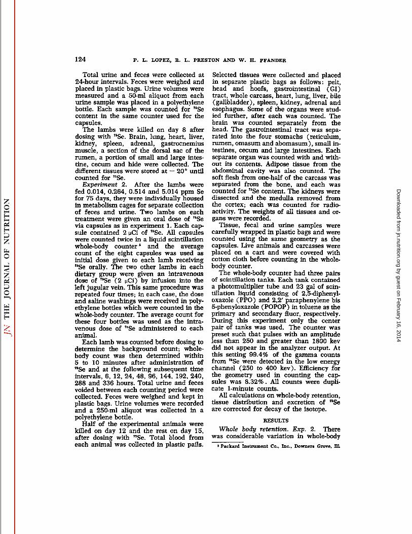
124 P. L. LOPEZ, R. L. PRESTON AND W. H. PFANDER
Total urine and feces were collected at24-hour intervals. Feces were weighed andplaced in plastic bags. Urine volumes weremeasured and a 50-ml aliquot from eachurine sample was placed in a polyethylenebottle. Each sample was counted for "Secontent in the same counter used for thecapsules.
The lambs were killed on day 8 afterdosing with "Se. Brain, lung, heart, liver,kidney, spleen, adrenal, gastrocnemiusmuscle, a section of the dorsal sac of therumen, a portion of small and large intestine, cecum and hide were collected. Thedifferent tissues were stored at —20°untilcounted for "Se.
Experiment 2. After the lambs werefed 0.014, 0.264, 0.514 and 5.014 ppm Sefor 75 days, they were individually housedin metabolism cages for separate collectionof feces and urine. Two lambs on eachtreatment were given an oral dose of "Sevia capsules as in experiment 1. Each capsule contained 2 uCi of "Se. All capsuleswere counted twice in a liquid scintillationwhole-body counters and the averagecount of the eight capsules was used asinitial dose given to each lamb receiving"Se orally. The two other lambs in eachdietary group were given an intravenousdose of "Se (2 uCi) by infusion into theleft jugular vein. This same procedure wasrepeated four times; in each case, the doseand saline washings were received in polyethylene bottles which were counted in thewhole-body counter. The average count forthese four bottles was used as the intravenous dose of "Se administered to eachanimal.
Each lamb was counted before dosing todetermine the background count; whole-body count was then determined within5 to 10 minutes after administration of"Se and at the following subsequent timeintervals, 6, 12, 24, 48, 96, 144, 192, 240,288 and 336 hours. Total urine and fecesvoided between each counting period werecollected. Feces were weighed and kept inplastic bags. Urine volumes were recordedand a 250-ml aliquot was collected in apolyethylene bottle.
Half of the experimental animals werekilled on day 12 and the rest on day 15,after dosing with "Se. Total blood fromeach animal was collected in plastic pails.
Selected tissues were collected and placedin separate plastic bags as follows: pelt,head and hoofs, gastrointestinal (GI)tract, whole carcass, heart, lung, liver, bile(gallbladder), spleen, kidney, adrenal andesophagus. Some of the organs were studied further, after each was counted. Thebrain was counted separately from thehead. The gastrointestinal tract was separated into the four stomachs (reticulum,rumen, omasum and abomasum), small intestines, cecum and large intestines. Eachseparate organ was counted with and without its contents. Adipose tissue from theabdominal cavity was also counted. Thesoft flesh from one-half of the carcass wasseparated from the bone, and each wascounted for "Se content. The kidneys weredissected and the medulla removed fromthe cortex; each was counted for radioactivity. The weights of all tissues and organs were recorded.
Tissue, fecal and urine samples werecarefully wrapped in plastic bags and werecounted using the same geometry as thecapsules. Live animals and carcasses wereplaced on a cart and were covered withcotton cloth before counting in the whole-body counter.
The whole-body counter had three pairsof scintillation tanks. Each tank containeda photomultiplier tube and 23 gal of scintillation liquid consisting of 2,5-diphenyl-oxazole (PPO) and 2,2' paraphenylene bis5-phenyloxazole (POPOP) in toluene as theprimary and secondary fluor, respectively.During this experiment only the centerpair of tanks was used. The counter waspreset such that pulses with an amplitudeless than 250 and greater than 1800 kevdid not appear in the analyzer output. Atthis setting 99.4% of the gamma countsfrom "Se were detected in the low energychannel (250 to 400 kev). Efficiency forthe geometry used in counting the capsules was 8.32%. All counts were duplicate 1-minute counts.
All calculations on whole-body retention,tissue distribution and excretion of "Seare corrected for decay of the isotope.
RESULTS
Whole body retention. Exp. 2. Therewas considerable variation in whole-body
5 Packard Instrument Co., Inc., Downers Grove, 111
by guest on February 16, 2014
jn.nutrition.orgD
ownloaded from

SELENIUM METABOLISM IN LAMBS 125
count determined 5 to 10 minutes through24 hours after "Se administration, regard-less of selenium level in the diet and modeof isotope administration. Whole-bodycounts decreased constantly in all lambsbeyond 24 hours. Therefore, data from 48hours through the end of the retentionstudy were fitted to a linear regression,
using a log transformation of the whole-body counting rates. A high linear corre-lation (r = 0.90 to 1.00) resulted (table1); however, extrapolation of each linearequation to zero time gave significantlydifferent (P < 0.01) a values (gammacounts at zero time) between lambs. Awide variation in a values seemed improb-
TABLE 1Values for intercept, regression coefficient and correlation coefficient of the linear regression
equations (log y = a + bx) on whole-body retention of 75Se by individual lambs 1
Dietaryselenium
levelppm0.0140.2640.5145.014Routeof
administrationOralIntravenousOralIntravenousOralIntravenousOralIntravenousReplicate12112121212121212a4.8160—5.02385.05254.79854.85604.87044.84784.69554.92924.88114.76744.59884.80384.48484.5654bx
10-»-0.5891—-0.2896-0.2855-1.5050-1.6670-0.7767-0.7487-1.7800-2.2250-1.3890-
1.4390-4.0260-3.6900-2.2890-1.9170T0.98—0.900.900.980.981.000.990.990.990.990.990.980.980.990.98
1Data from 48 to 336 hours were used for regression analysesadministration).
i Denotes lamb was lost.
= cpm; x-=.hours after 75Se
TABLE 2The a, b and c values of the second-degree equations (log y ~ a + bx + cx*~) on whole-body
retention of 7sSe by individual lambs *
Dietaryselenium
levelppm0.0140.2640.5145.014Routeof
administrationOralIntravenousOralIntravenousOralIntravenousOralIntravenousReplicate12212121212121212a4.9455—5.02425.00994.95664.94444.91434.90844.87134.98264.96664.89414.97605.12834.84704.8852bx
10-'-2.1306—-0.1845-
0.2595—3.4740—3.1450—1.2214-1.6375-3.8435-3.2200-2.3120-3.1281-8.9535-8.6395-6.7123-6.3240cx
IO-«3.67070.39531.48554.84554.48460.98732.55104.94863.53802.10214.921012.477014.520010.813012.2930
1Data from zero to 336 hours were used for regression analyses (y = cpm; x = hours after «Seadministration ).
- Denotes lamb was lost.
by guest on February 16, 2014
jn.nutrition.orgD
ownloaded from

126 P. L. LOPEZ, R. L. PRESTON AND W. H. PFANDER
able since extrapolated values were not related to either size of the animal or routeof "Se administration. Because a curvilinear relation may exist between the log ofthe whole-body count and time after "Seadministration, data from zero time werefitted to a second-degree equation (table2). The a values of each quadratic equation were not significantly different between individual lambs, dietary seleniumlevels or mode of "Se administration.
Retention curves, represented by a quadratic equation which is the average of thetwo equations for lambs that received thesame treatment, show that whole-body retention of "Se is a function of dietaryselenium intake and route of selenium administration. The b values obtained, eitherby quadratic or linear regression analyses,showed the marked effect of dietary selenium level and mode of "Se administration(P< 0.001).
The biological half-life (T1/2B) of selenium based on whole-body loss 48 to 336hours after oral administration of "Se was636, 202, 162 and 82 hours for the 0.01,0.26, 0.51 and 5.01 ppm Se levels, respectively. Similarly, the half-life of intravenously administered "Se was 1660, 469,229 and 149 hours, respectively. The effective half-life (T, ,£),which is the combined effect of biological half-life and radiological half-life (T,,2P) of "Se, wascalculated with the formula:
T1/2E =TI/J.P + Ti/2B
Ti/2P Ti/2B
The calculated T,/2E was 521, 189, 154and 80 hours for the oral dose and 1048,402, 212 and 142 hours for the infuseddose of "Se for lambs on the 0.01, 0.26,0.51 and 5.01 ppm Se, respectively.
Fecal, urinary and respiratory excretionsof "Se. Exp. 1. Fecal excretion of "Se ofall lambs was maximum 48 hours afterisotope administration, decreased rapidlyduring the next 48 hours, and decreasedslowly through the remainder of the excretion study. Urinary excretion exhibited asimilar trend; a rapid drop in "Se excretion was observed for all lambs at 72 hours,followed by an almost constant decreasethroughout the 8-day observation period.
Dietary selenium levels were reflected inurinary excretion but not in fecal excre-
CU FecesI UrineI VolatileSe
0.014 0.264 0,514 5.014
DietarySeleniumLevels(ppm)Fig. 1 Excretion of "Se by lambs fed varying
selenium intakes after administration of the isotope during a 12-day period.
tion of "Se; excretion in the urine duringthe 8-day period increased with increasingdietary selenium level. These differences inurinary excretion of lambs on different dietary treatments were observed within 48hours after "Se administration.
Experiment 2. The elimination of "Sein the feces, urine and in the calculatedvolatile form is presented in figure 1. Volatile selenium was calculated as the difference of 100 minus the whole-body retention of "Se and total fecal and urinaryexcretions on a percentage basis. Whole-body retention was calculated from bodycount of the animal on day 12 after selenium administration, divided by the estimated a value (counts at zero time) ofthe second-degree equation. Fecal and urinary excretions were computed as percentage of the activity of the capsule or of theinfused "Se. Excretion values were notcorrected for fecal and urinary loss whenthe lambs were counted and in transit (approximately 100 m distance). A conservative estimate of this loss would be 10%
by guest on February 16, 2014
jn.nutrition.orgD
ownloaded from

SELENIUM METABOLISM IN LAMBS 127
of the total fecal and urinary excretion.Thus, the computed value for volatile selenium compounds is an overestimate andalso includes all possible analytical errors.
The excretion pattern of â„¢Sewas different between intravenous and oral administration of the isotope. Volatile eliminationby lambs on 0.01 ppm Se which receivedthe isotope orally was slightly lower thanthe fecal loss but higher than the urinaryexcretion; this pattern was reversed inlambs on 5.01 ppm Se. Volatile loss wasslightly lower than fecal and urinary excretion of lambs on 0.26 and 0.51 ppmlevels. Fecal excretion was higher than urinary loss in lambs on 0.01 and 0.26 ppmlevels. The reverse was observed in thehigher dietary groups (0.51 and 5.01 ppmSe). Respiratory excretion was higher thanfecal in lambs infused with "Se but lowerthan urinary excretions in all dietarygroups, except the 0.01 ppm level. Radioactive selenium in the feces and urine wasabout the same and was slightly higherthan volatile losses in lambs fed die basaldiet. Urinary and volatile excretion increased with increasing dietary seleniumlevel, especially when the isotope was intravenously administered.
Respiratory and urinary "Se excretionwas dependent (P < 0.05) both on dietaryselenium levels and route of "Se administration; however, route of isotope administration is the major factor affecting fecalloss of "Se. The effect of dietary seleniumintake on urinary excretion of "Se wasobserved within 48 hours after isotope administration; the difference in fecal excretion relative to route of administration wasapparent within 24 hours.
Fecal and urinary "Se excretions weremaximum at 96 hours regardless of dietary selenium level and route of administration with few exceptions. Lambs onthe 5.01 ppm level that received "Se intravenously had maximum activity of theisotope in the urine within 12 hours afteradministration. In contrast, lambs on 0.01and 0.26 ppm levels that received oral "Sehad maximum concentration of the isotope in the urine between 96 and 144hours after "Se administration. Lambs onthis same dietary selenium level which received an intravenous dose of "Se exhibited a maximum fecal excretion between
144 and 192 hours after isotope administration. A constant decrease followed aftermaximum excretion in all dietary groupswith either mode of administration.
Distribution of retained "Se in tissues.Exp. 1. Selenium-75 retained per gramof tissue decreased with increasing dietarylevel of selenium. The greatest amount of"Se in the internal organs was found inthe liver, kidney and lungs when resultswere based on the amount of "Se in thewhole organ; the liver retained the highestamount in lambs fed the diets with addedselenium, whereas the kidney retained thegreatest amount in the lamb fed the basaldiet.
When results were based on "Se concentration, retention in descending orderwas as follows: kidney, adrenal, liver,spleen, thyroid, lung, bile (gallbladder),brain and heart. Accumulations of "Se inthe pelt, gastrocnemius muscle and femurbone were about the same and were higherthan in adipose tissue. Concentration of"Se in the small intestines was higher thanin either the rumen or large intestines.
Experiment 2. The distribution of "Sein the tissues of animals fed varying levelsof selenium and killed on day 12 andday 15 after oral and intravenous administration of radioactive selenium are shownin figures 2 through 4.
«menons" Si «Mwtntm
212019IB17-
1615-14-
1312
en 0014 ppm Se
l l O264 ppn Se» 0514 pi» S»• 5014 pf»Se
Adipose Cjfciss Muscle I
Fig. 2 Average distribution of 75Se in carcass and constituent parts of lambs fed varyingselenium Intakes.
by guest on February 16, 2014
jn.nutrition.orgD
ownloaded from

128 P. L. LOPEZ, R. L. PRESTON AND W. H. PFANDER
Intravenous SeAdministration
1=10.014ppmSeEH 0.264ppmSe•0.514ppmSe•5.014ppmSe
Ik,Oral75& Administration
Liver Spleen Bile KidneyAdrenal Pelt Whole Brain Heart LungBlood
Fig. 3 Average distribution of retained 75Sein tissues of lambs fed varying seleniumintakes.
0.014ppmSemm 0.264ppmSe
0.514ppmSe5.014ppmSe
SeAdministration
G.I. Four Smajl Cecum LargeTract StomachsIntestine Intestine
Fig. 4 Average distribution of retained â„¢Seingastrointestinal tract including contents of lambsfed varying selenium intakes.
Retention of "Se in the tissues was calculated as a percentage of the activity ofthe capsule or of the infused dose. Retained "Se in the carcass was determinedas a percentage of the whole-body count at
zero time (a value, second-degree equation).
Selenium levels in the diet showed amarked influence on the "Se concentrationretained by the tissues. Tissue retention of"Se decreased with increasing dietary selenium level (P < 0.01). A higher "Se retention in the tissues was observed inlambs that received the intravenous dose(P < 0.01 to 0.05) with some exceptions.The "Se retention in tissues of lambs killedon days 12 and 15 were generally not statistically different.
The carcass retained the greatestamount of "Se. Activity in the carcass wasdistributed between the muscles and bonesin approximately equal amounts. A negligible amount of the isotope was presentin adipose tissue. Carcasses of lambs killedon day 12 after isotope administration contained a greater amount of "Se (P < 0.05)than those killed on day 15 regardless ofmode of administration; however, the difference was not significant when either themuscle, bone or adipose tissues were examined separately.
The internal organs retained a higherconcentration of activity than other tissueswhen the results were expressed per gramtissue. Lambs fed the basal ration retainedhigher "Se concentration in the kidneythan in the liver, irrespective of mode ofadministration. Retention by these tissueswas reversed in lambs that received added
by guest on February 16, 2014
jn.nutrition.orgD
ownloaded from

SELENIUM METABOLISM IN LAMBS 129
selenium in the ration. More than 99%of the activity in the kidney was presentin the cortex; a negligible amount waspresent in the medulla.
The order of "Se distribution in the various parts of the gastrointestinal tract oflambs receiving the basal ration was thesmall intestines > four stomachs > largeintestines > cecum regardless of mode of"Se administration. The same trend wastrue for lambs that received added selenium in the diet except that the four stomachs were higher in "Se concentration thanin the small intestines. The relative distribution of retained "Se in these tissueswith respect to dietary selenium intake didnot change when the contents were washedfrom the organs. The four stomachs free ofcontents, from lambs that received an intravenous dose of "Se, were higher (P <0.05) in activity than those given the oral"Se. The activity of the washed small intestine was not affected by mode of administration. In contrast, the large intestinesand cecum when washed retained more"Se after an intravenous dose than afteroral administration (P < 0.01).
When the results were compared pergram tissue, the adrenal, kidney, bile,spleen, liver, brain, lungs, heart, wholeblood, pelt, gastrointestinal tract and carcass in decreasing order retained the highest concentration of "Se. There were somevariations in the amount of retained "Sein the different tissues as influenced bydietary selenium but the maximum concentration of "Se occurred in the tissuesof lambs that received the basal diet regardless of mode of "Se administration.
DISCUSSION
It appears from the quadratic curvesthat whole-body turnover of "Se in lambson varying selenium intake may be described by two first-order processes withdifferent rate constants, the initial slopebeing greater than the final although thefinal slope increased with increasing selenium intake irrespective of mode of "Se administration. Whether the initial slope isactually another rate constant or merelyrepresents the total unequilibrated selenium is difficult to evaluate. The relativelyshort period (48 hours) covered by the initial slope which corresponds to maximum
selenium excretion rates would indicatethat this part of the quadratic curve represents the total unequilibrated selenium,consisting of that not absorbed from thegastrointestinal tract and the absorbed selenium not equilibrated in the tissues. Theunabsorbed selenium is eliminated in thefeces whereas the latter is excreted in theurine, the expired air or reabsorbed intothe gastrointestinal tract following biliaryexcretion.
Results of the present study show conclusive evidence that selenium undergoesactive metabolism in the body of the animal. The metabolic rate is governed by afirst-order process where the rate constantvaried according to dietary intake andmode of administration over a period of12 to 15 days. This may indicate that therate limiting process is the release of selenium from the various tissues in whichit is bound. Based on the b values of thelinear regressions (table 1), selenium supplementation in lambs with low seleniumintake would be more beneficial if administered intravenously than orally, sincethe lambs would retain twice as muchselenium by this route of administration.Route of selenium administration, however, showed no marked differences inbody retention at dietary levels of 0.51ppm Se or higher.
The effective half-life of "Se in lambsdecreased with increasing selenium levelin the diet. The rate of decrease was notlinear suggesting that at higher dietarylevels (> 5 ppm) selenium turnover maynot be further increased. This same trendwas observed in the study of in vitro uptake of "Se by ovine blood cells of lambsin the same dietary groups (13). Blincoe(12) obtained a retention curve for ratssimilar to the quadratic curves observedin this study.
Several authors (7-10, 14, 15) have compared selenium excretion in the feces andurine of sheep. Several studies on respiratory elimination of selenium in rats havealso been reported (1-4, 16). It is evidentfrom this study that selenium excretionby any one pathway is dependent on thedietary selenium intake and route of "Seadministration.
Generally, respiratory excretion increased with increasing selenium intake
by guest on February 16, 2014
jn.nutrition.orgD
ownloaded from

130 P. L. LOPEZ, R. L. PRESTON AND W. H. PFANDER
irrespective of route of "Se administration.This is in agreement with the results obtained on rats by Ganther et al. (4). Theyreported that volatilization was increasedgreatly from a purified diet containing 0.03ppm Se to a crude commercial diet with0.5 ppm of the element.
Considerable evidence indicates that di-methylselenide is the metabolic product ofinorganic selenium metabolism in severalbiological processes (4, 17). The volatileproduct is normally synthesized in theliver, and transported by the blood to thelungs. In our study, formation of volatileselenium compounds was higher followingthe oral dose than the intravenous dose.The difference between the two routes wasgreater on the lower dietary levels of selenium (0.01 and 0.26 ppm); on the higher levels (0.51 and 5.01 ppm), volatilization of selenium from the oral dose wasonly slightly higher than the intravenousdose. Since lambs on lower selenium intake which were dosed orally with "Sevolatilized a greater amount of seleniumthan lambs dosed intravenously, formationof volatile or gaseous selenium productsin the gastrointestinal tract, possibly in therumen, is suggested.
Selenium as sodium selenite (Na2SeO3)is in the + 4 oxidation state. Reduction ofNa2SeO3 could readily occur in the rumendue to its anaerobic, highly reducing atmosphere. Rosenfeld and Beath (18) reported that bacteria are capable of reducing selenium salts to elemental seleniumwhich in turn may form metal selenides.Metal selenides on hydrolysis form hydrogen selenide (H2Se), a colorless, very toxicgas closely resembling H2S in generalproperties. There is evidence that smallamounts of H2S are formed in the rumen.The analogy of selenium to sulfur in chemical behavior makes it reasonable to postulate the formation of H2Se in the rumen.
Dimethylselenide could be formed in therumen from metal selenide and methylcompounds. The difference in volatilization between oral and intravenous administration on the lower dietary seleniumgroups suggests that a greater proportionof metal selenides are formed into di-methylselenide than into H2Se due to thehighly toxic nature of the latter. Dimethylselenide may be absorbed into the blood
and eliminated through the respiratorytract together with that formed in the liver.Hydrogen selenide may be eliminated together with some of the gases from therumen via eructation.
Respiratory excretion in lambs given"Se intravenously was the same for the0.51 and 5.01 ppm Se levels. This suggests that there is a limit in the synthesisof dimethylselenide by the liver, resultingin an increase in urinary excretion of theisotope. Lambs that received "Se by capsule, however, showed a further increasein respiratory loss above the 0.51 ppmlevel. This difference in respiratory elimination between intravenous and oral administration of the isotope further supports the hypothesis that volatile seleniumproducts are formed in the rumen.
Urinary excretion of 75Se increasedmarkedly with increasing selenium level inthe diet, suggesting that at these dietarylevels there is no limit to the amount of"Se that can be eliminated by this pathway.
Selenium-75 in the feces was essentiallythe same in all dietary groups receivingthe isotope orally. This suggests that totalabsorption of ingested selenium from thegastrointestinal tract is a direct functionof selenium intake of the animal. On theother hand, equal fecal excretions in alldietary groups that received "Se intravenously suggest that "Se secretion fromthe body into the lumen of the gastrointestinal tract is constant and not related todietary selenium level.
Wright (10) obtained a higher concentration of "Se in the urine than in thefeces of ewes fed on hay after intravenousinjection of the isotope. Peterson and Sped-ding (8) and Butler and Peterson (6) reported that selenium is excreted predominantly in the feces of sheep fed on redclover grown in "Se solution. Rosenfeldand Eppson (9) observed in sheep withchronic selenosis that urinary excretionwas slightly higher than fecal elimination.These results are in general agreementwith those presented in this study and indicate that the major route of seleniumexcretion is a function of the present selenium status of the animal. Other factorssuch as nature of the diet and mode ofselenium administration also may affect
by guest on February 16, 2014
jn.nutrition.orgD
ownloaded from

SELENIUM METABOLISM IN LAMBS 131
the route of excretion. It is difficult, however, to accept the statements made byButler and Peterson (6) and Cousins andCairney (7) that the major excretorypathway in sheep is via the feces.
Of the "Se retained by the lamb, thecarcass contained the greatest amount ofall tissues examined. The sum of retained"Se in the muscle, bone and adipose tissues was higher than that of the carcass.This discrepancy is probably explained bythe difference in geometry of counting.Selenium-75 in the carcass was distributedbetween the muscles and bones in approximately equal amounts, with an almost negligible amount in the adipose tissue. Thissuggests that selenium is associated withtissue proteins and these tissues are adeptin binding selenium with the proteins.These results support those of Rosenfeld(3) and Hopkins et al. (5) that the carcass of rats retained the highest concentration of selenium when results werebased on the whole body, irrespective ofdietary selenium levels and route of administration.
The liver, kidney and lung retained thehighest concentration of "Se of the internal organs and tissue fluids, irrespectiveof dietary selenium levels or mode of administration. This conforms with the results of Rosenfeld and Eppson (9) andWright (11). Studies on tissue distribution of "Se in other species such as mice,rats, dogs and swine are similar with thefindings in these experiments (3, 5, 19).This suggests that in the liver, kidney andlung, selenium exchange with "Se in theplasma is more rapid and complete, whereas in the brain, heart, spleen, bile andadrenal, selenium is exchanged moreslowly.
Lambs fed the basal ration retained agreater amount of selenium in the kidneywith both modes of administration. Thiswas also observed in experiment 1 inlambs fed similar levels of dietary selenium. This was not expected since the urinewas not the main excretory pathway inlambs on low selenium intake. This maysuggest some specific function of the kidney in selenium metabolism other than excretion.
Considerable concentration of "Se wasfound in the pelt (fig. 3). It is possible
that part of this radioactivity is due to "Seexcreted via the skin. Previous reportsshow that selenium is deposited and retained in the hide, wool and hair of animals (14, 20-22).
ACKNOWLEDGMENTS
The authors thank Dr. E. R. Grahamfor helpful suggestions relative to whole-body counting of the animals, and W.Coffman for technical assistance.
LITERATURE CITED
1. McConnell, K. P., and O. W. Portman 1952Excretion of dimethylselenide by the rat. J.Biol. Chem., 195: 277.
2. Olson, O. E., B. M. Schulte, E. T. Whiteheadand A. W. Halverson 1963 Effects of arsenic on selenium metabolism in rats. J.Agr. Food Chem., 11: 531.
3. Rosenfeld, I. 1964 Metabolic effects andmetabolism of selenium in animals. IV. Excretion and retention of "Se in relation tomodes of administration, toxicity and pregnancy in rats. Wyoming Agr. Exp. Sta. Bull,no. 414, p. 35.
4. Ganther, H. E., O. A. Levander and C. A.Baumann 1966 Dietary control of selenium volatilization in the rat. J. Nutr., 88: 55.
5. Hopkins, L. L., Jr., A. L. Pope and C. A.Baumann 1966 Distribution of microgramquantities of selenium in the tissues of therats and effects of previous selenium intake. J. Nutr., 88: 61.
6. Butler, G. W., and P. J. Peterson 1961Aspects of the fecal excretion of selenium bysheep. New Zeal. J. Agr. Res., 4: 484.
7. Cousins, F. B., and I. M. Cairney 1961Some aspects of selenium metabolism insheep. Australian J. Agr. Res., 12: 927.
8. Peterson, P. J., and D. J. Spedding 1963The excretion by sheep of "Se incorporatedinto red clover (Trifolium pratense L.): Thechemical nature of selenium and its uptakeby three plant species. New Zeal. J. Agr.Res., 6: 13.
9. Rosenfeld, I., and H. F. Eppson 1964 Metabolic effects and metabolism of seleniumin animals. V. Metabolism of selenium insheep. Wyoming Agr. Exp. Sta. Bull. no. 414,p. 53.
10. Wright, E. 1965 The distribution and excretion of radioselenium in sheep. New Zeal.J. Agr. Res., 8 (2): 284.
11. Wright, E. 1965 The concentration of radioselenium by the tissues of selenium-responsive sheep in relation to their growthrates. New Zeal. J. Agr. Res., 8 (2): 292.
12. Blincoe, C. 1960 Whole-body turnover ofselenium in rats. Nature, 186: 398.
13. Lopez, P. L., R. L. Preston and W. H. Pfander 1968 In vitro uptake of selenium-75by red blood cells from the immature ovineduring varying selenium intakes. J. Nutr.,94: 219.
by guest on February 16, 2014
jn.nutrition.orgD
ownloaded from

132 P. L. LOPEZ, R. L. PRESTON AND W. H. PFANDER
14. Rosenfeld, I., and O. A. Beath 1945 Theelimination and distribution of selenium inthe tissues in experimental selenium poisoning. J. Nutr., 30; 443.
15. Butler, G. W., and P. J. Peterson 1963Availability of selenium in forage to ruminants. Proc. New Zeal. Soc. Anim. Prod.,¡23:13.
16. Schultz, J., and H. B. Lewis 1940 The excretion of volatile selenium compounds afteradministration of sodium selenite to whiterats. J. Biol. Chem., 133: 199.
17. McConnell, K. P., and B. J. Cooper 1950Distribution of selenium in serum proteinsand red blood cells after subcutaneous injection of sodium selenate containing radio-selenium. J. Biol. Chem., 383:459.
18. Rosenfeld, I., and O. A. Beath 1964 Selenium: Geobotany, Biochemistry, Toxicity andNutrition. Academic Press, New York.
19. Lindberg, P., and M. Siren 1963 Seleniumconcentration in kidneys of normal pigs andpigs affected with nutritional muscular dystrophy and liver dystrophy. Life Sci., 5: 326.
20. Demiruren, A. S., and B. S. Alen 1963 Effects of selenium on certain mechanical properties of the wool fiber. Can. J. Anim. Sci.,42 (2): 233.
21. McConnell, K. P., and A. E. Kreamer 1960Incorporation of â„¢Sewith dog's hair. Proc.
Soc. Exp. Biol. Med., 105: 170.22. Underwood, E. J. 1962 Trace Elements in
Human and Animal Nutrition, ed. 2. Academic Press, New York.
by guest on February 16, 2014
jn.nutrition.orgD
ownloaded from