Acquired Facial, Maxillofacial, and Oral Asymmetries ... - MDPI
Upload
hms-harvardCategory
view
1download
0
www.elsevier.com/locate/ynimg
NeuroImage 23 (2004) 213–223
White matter hemisphere asymmetries in healthy subjects and
in schizophrenia: a diffusion tensor MRI study
Hae-Jeong Park,a,b,c,d Carl-Fredrik Westin,b,c Marek Kubicki,a,c Stephan E. Maier,e
Margaret Niznikiewicz,a Aaron Baer,a Melissa Frumin,a Ron Kikinis,c,e Ferenc A. Jolesz,c,e
Robert W. McCarley,a and Martha E. Shentona,c,*
aClinical Neuroscience Division, Laboratory of Neuroscience, Boston VA Health Care System-Brockton Division, Department of Psychiatry,
Harvard Medical School, Boston, MA 02301, USAbLaboratory of Mathematics in Imaging, Department of Radiology, Brigham and Women’s Hospital, Harvard Medical School, Boston, MA 02301, USAcSurgical Planning Laboratory, Department of Radiology, Brigham and Women’s Hospital, Harvard Medical School, Boston, MA 02301, USAdDivision of Nuclear Medicine, Department of Diagnostic Radiology, Yonsei University, College of Medicine, Seoul 120-752, South KoreaeMRI Division, Department of Radiology, Brigham and Women’s Hospital, Harvard Medical School, Boston, MA 02301, USA
Received 27 January 2004; revised 3 April 2004; accepted 28 April 2004
Available online 8 July 2004
Hemisphere asymmetry was explored in normal healthy subjects and in
patients with schizophrenia using a novel voxel-based tensor analysis
applied to fractional anisotropy (FA) of the diffusion tensor. Our voxel-
based approach, which requires precise spatial normalization to
remove the misalignment of fiber tracts, includes generating a
symmetrical group average template of the diffusion tensor by applying
nonlinear elastic warping of the demons algorithm. We then
normalized all 32 diffusion tensor MRIs from healthy subjects and
23 from schizophrenic subjects to the symmetrical average template.
For each brain, six channels of tensor component images and one T2-
weighted image were used for registration to match tensor orientation
and shape between images. A statistical evaluation of white matter
asymmetry was then conducted on the normalized FA images and their
flipped images. In controls, we found left-higher-than-right anisotropic
asymmetry in the anterior part of the corpus callosum, cingulum
bundle, the optic radiation, and the superior cerebellar peduncle, and
right-higher-than-left anisotropic asymmetry in the anterior limb of the
internal capsule and the anterior limb’s prefrontal regions, in the
uncinate fasciculus, and in the superior longitudinal fasciculus. In
patients, the asymmetry was lower, although still present, in the
cingulum bundle and the anterior corpus callosum, and not found in
the anterior limb of the internal capsule, the uncinate fasciculus, and
the superior cerebellar peduncle compared to healthy subjects. These
findings of anisotropic asymmetry pattern differences between healthy
controls and patients with schizophrenia are likely related to neuro-
developmental abnormalities in schizophrenia.
Published by Elsevier Inc.
Keywords: Hemisphere asymmetry; Diffusion tensor MRI; Fractional
anisotropy
1053-8119/$ - see front matter. Published by Elsevier Inc.
doi:10.1016/j.neuroimage.2004.04.036
* Corresponding author. Department of Psychiatry-116A, VA Boston
Healthcare System-Brockton Division, Harvard Medical School, 940
Belmont Street, Brockton, MA 02301. Fax: +1-508-586-0894.
E-mail address: [email protected] (M.E. Shenton).
Available online on ScienceDirect (www.sciencedirect.com.)
Introduction
Hemisphere asymmetry in the brain has long been reported in
healthy subjects in both neuroanatomy and function (Galaburda
et al., 1978a; Geschwind, 1972; Geschwind and Galaburda,
1985a,b,c; Holinger et al., 2000; Kimura, 1973; Springer and
Deutsch, 1998; Toga and Thompson, 2003). More particularly,
neuroanatomical asymmetry, which has been presumed to be
associated with functional lateralization (Amunts et al., 2000;
Beaton, 1997; Kennedy et al., 1999; Moffat et al., 1998; Springer
and Deutsch, 1998), has been found at the cytoarchitectronic level
(Galaburda et al., 1978b; Rosen et al., 1993), in macrostructural
volumes (Geschwind, 1972), and in morphometry (Sowell et al.,
2002; Lancaster et al., 2003). Most typically, areas of macrostruc-
tural asymmetry include right frontal and left occipital petalias
(Kennedy et al., 1999), the Sylvian fissures (Galaburda et al.,
1978a; Sowell et al., 2002; Westbury et al., 1999), and the superior
temporal plane, especially involving the planum temporale and
Heschl gyrus (Anderson et al., 1999; Beaton, 1997; Galaburda
et al., 1978b; Good et al., 2001; Moffat et al., 1998; Pujol et al.,
2002; Watkins et al., 2001).
Neuroanatomical asymmetry in healthy subjects has been
revealed not only in cortical gray matter, but also in white
matter that interconnects cortical brain regions. White matter
asymmetry has been explored primarily in studies of white
matter volume of structures or in voxel-based concentration of
white matter among subjects using structural MRI (Good et al.,
2001; Pujol et al., 2002; Zhou et al., 2003). The recent advent
of diffusion tensor magnetic imaging (DT-MRI) techniques
(Basser et al., 1994), however, makes it possible to measure
the quality of the neuronal fiber bundles within white matter
regions of interest (ROI). For example, anisotropy of the
diffusion tensor (Basser, 1995) provides information about the
underlying structure of a given region, such as the myelination,
density, and coherence of fibers. Accordingly, white matter

H.-J. Park et al. / NeuroImage 23 (2004) 213–223214
asymmetry has been investigated using diffusion tensor images
to explore anisotropic asymmetry in the uncinate fasciculi
(Kubicki et al., 2002), the cingulate fasciculi (Kubicki et al.,
2003), the anterior limb of internal capsule (Peled et al., 1998),
and subinsular white matter (Cao et al., 2003). With the
exception of the anterior limb of the internal capsule, the above
regions of interest, that is, uncinate fasciculi, cingulate fasciculi,
and subinsular white matter, all show a pattern of left-greater-
than-right anisotropy in healthy controls.
Of note, an alteration that deviates from normal asymmetry
has been explored in schizophrenia (Holinger et al., 2000) in
neuroanatomy, including both postmortem studies of parahippo-
campal, fusiform gyri, and temporal lobe (Highley et al.,
1998a,b, 1999; McDonald et al., 2000), and in MRI studies
of temporal lobe, anterior cingulate gyrus, and frontal lobe
(Takahashi et al., 2002; Turetsky et al., 1995), as well as in
functions, including auditory processing such as mismatch
negativity (Mohr et al., 2001; Rockstroh et al., 2001; Sommer
et al., 2001; Youn et al., 2003). Alterations in white matter
asymmetry in schizophrenia have also been exploited using DT-
MRI, in studies looking at the cingulum bundle and uncinate
fasciculus (Kubicki et al., 2002, 2003; Zhou et al., 2003).
These latter studies follow closely in time the first DT-MRI
studies of schizophrenia (Buchsbaum et al., 1998; Lim et al.,
1999).
Methods used to explore white matter asymmetry, however,
have been mostly based on measuring volume or anisotropy of a
given region, that is, region of interest (ROI) in white matter
structures. For example, early DT-MRI studies of white matter
hemispheric asymmetry have included an evaluation of the
internal capsule (Peled et al., 1998), the uncinate fasiculi
(Kubicki et al., 2002), and the cingulate fasciculi (Kubicki
et al., 2003) of healthy subjects and/or schizophrenia, and are
based on an ROI method. Cao et al. (2003) also studied asymmetry
of anisotropy of the subinsular white matter. The ROI-based
approach requires a priori hypothesis to predefine the expected
region of interest.
As an explorative method, the voxel-based strategy can be more
helpful in identifying unanticipated or unpredicted/unhypothesized
areas of neuroanatomical asymmetry. The voxel-based analysis of
gray/white matter asymmetry has been conducted using voxel-
based morphometry (VBM) (Good et al., 2001; Watkins et al.,
2001) and deformation field analysis (Lancaster et al., 2003). The
voxel-based analysis of white matter asymmetry has been con-
ducted with structural MRI, where white matter throughout whole
brain is regarded as homogeneous (Good et al., 2001). Such
studies, using voxel-based morphometry to evaluate gray/white
matter, have focused mainly on detecting the anatomical shape as
well as regional volumetric difference. However, no study has been
reported, as far as we know, on the asymmetry of white matter fiber
bundles using voxel-based analysis of diffusion tensor images,
where more sophisticated spatial normalization might be required
to remove anatomical confounds, such as misalignment of narrow
fiber bundles.
In this paper, we propose a new method for the voxel-based
exploration of asymmetry of diffusion tensor using a higher
order spatial normalization technique for tensor data (Guimond
et al., 2002; Park et al., 2003a). This technique matches
homologous locations in fiber bundles and minimizes potential
misinterpretation of results that might otherwise be caused by
misregistration. Using this method, we explore anisotropic
asymmetry in diffusion tensor images of healthy subjects and
patients with schizophrenia.
Methods
Subjects
Thirty-two normal male healthy subjects, with a mean age of 44
(30–55, SD: 6.2), were recruited from the general community.
Twenty-three male patients with schizophrenia, with a mean age
of 43 (28–53, SD: 7.2) were recruited from inpatient, day treatment,
outpatient, and foster care programs at the VA Boston Healthcare
System, Brockton, MA. The inclusion criteria for all subjects were
right-handedness, no history of electroconvulsive shock treatment,
no history of neurological illness or significant head trauma, no
alcohol or drug dependence in the past 5 years, and no abuse within
the past year, no medication with known effects on MR (such as
steroids), verbal IQ above 75, English as a first language, and an
ability and desire to cooperate with the procedures. In addition,
healthy subjects were screened to exclude individuals who had a
first-degree relative with an Axis I disorder, based on DSM-IV
criteria. As part of a comprehensive neuropsychological battery, all
subjects were evaluated using the verbal paired associate learning
subtest of the Wechsler Memory Scale-3rd ed. (WMS) (Wechsler,
1997b), the Wisconsin Card Sorting Test (Nestor et al., 1998), the
Trail Making Test, and the similarities subtest of the WAIS-III
(Wechsler, 1997a). This research was approved by the local insti-
tutional review board of the VA Boston Healthcare System, and all
subjects signed written informed consent before participation.
Acquisition of diffusion tensor images
Subjects were scanned using Line Scan Diffusion Imaging
(LSDI) (Gudbjartsson et al., 1996; Maier et al., 1998; Mamata et
al., 2002), which is comprised of a series of parallel columns lying
in the image plane. The sequential collection of these line data in
independent acquisitions makes the sequence largely insensitive to
bulk motion artifact since no phase encoding is used and shot-to-
shot phase variations are fully removed by calculating the magni-
tude of the signal.
A quadrature head coil was used on a 1.5 T GE Echospeed
system (General Electric Medical Systems, Milwaukee, WI), which
permits maximum gradient amplitudes of 40 mT/m. For each slice,
six images with high diffusion-weighting (1000 s/mm2) along six
noncolinear and noncoplanar directions were collected. Two base
line images with low diffusion-weighting (5 s/mm2) were also
collected and averaged. Scan parameters were as follows: rectan-
gular FOV (field of view) 220 � 165 mm; 128 � 128 scan matrix
(256 � 256 interpolated image matrix); slice thickness 4-mm;
interslice distance 1-mm; receiver band width F4 kHz; TE (echo
time) 64 ms; effective TR (repetition time) 2592 ms; scan time 60 s
per slice/section. A total of 31–35 coronal slices covering the
entire brain were acquired, depending upon brain size.
Voxel-based analysis: asymmetry of diffusion tensor
The previous voxel-based analysis of hemisphere asymmetry
has been performed by normalizing gray/white matter to a sym-
metric group average template and by comparing raw and flipped
images of these normalized images (Good et al., 2001; Watkins et

H.-J. Park et al. / NeuroImage 23 (2004) 213–223 215
al., 2001). Neuroanatomical correspondences between left and
right hemisphere across subjects were determined by spatial
normalization to a symmetric template.
The essential requirement for a voxel-based analysis of the
diffusion tensor, including a study of white matter asymmetry, is to
minimize the misinterpretation of statistical results due to mis-
alignment. Most previous voxel-based DTI reports (Barnea-Goraly
et al., 2003; Eriksson et al., 2001; Foong et al., 2002; Rugg-Gunn
et al., 2001) have used a method similar to the one used for the
voxel-based analysis of the structural scans called ‘‘voxel based
morphometry’’.
In this study, we propose a method for voxel-based analysis of
diffusion tensor image by reducing misalignment using a group
template and higher order spatial normalization using multiple
channel information.
Spatial normalization using multiple channel information of tensor
field
Since diffusion tensor image contains underlying information
(i.e., orientation, magnitude, and anisotropy of the tensor) of white
matter, the registration needs to match these properties of the tensor
to find correspondence between two images. We used a multiple
channel demons algorithm (Guimond et al., 2001) to estimate
deformation fields in the spatial normalization. This algorithm
finds the displacement v(x) for each voxel x of a target image T
to match the corresponding location in a source image S. One
channel of T2-weighted image and six channels of tensor compo-
nents (Dxx, Dxy, Dxz, Dyy, Dyz, and Dzz) make it possible to
match the shape, as well as the magnitude and orientation of local
tensors between images. During the registration, the local tensor
orientations were adjusted based on the deformation field as
described by Alexander et al. (2001). Registration using the whole
tensor information shows better registration performance than just
using a channel of fractional anisotropy (FA) image or a channel of
T2-weighted image (Park et al., 2003a).
Group atlas representing the mean of tensor field and mean of
brain morphology
We created a group diffusion tensor atlas from 32 normal subjects
by combining the average tensor field of all the normalized diffusion
images and the average deformation field (Guimond et al., 2000;
Park et al., 2003a). A diffusion image from healthy subjects was
chosen as a temporary atlas, and all other images were registered to
the temporary atlas with the adjustment of tensor orientation. The
average of the registered diffusion images was resampled with the
inverse of the average deformation field to achieve a morphological
(shape) mean as well as an intensity (tensor) mean of the group. The
average map was again used for the target atlas of the next iteration
to reduce the effect of the first template chosen from a subject. Four
iterations were used to create an average diffusion tensor map.
Symmetric atlas as a template for hemisphere correspondence
To create a symmetric template, both Good and Watkins groups
(Good et al., 2001; Watkins et al., 2001) utilized the averaging of
intensities from the original and flipped images of a scalar volume
which may cause blurring of the template image due to hemispher-
ic asymmetry. To address the issue of the blurring, we developed a
novel method using a registration scheme with a multiple-channel
demons algorithm (Guimond et al., 2000) to make a tensor image
symmetric along the interhemispheric fissure, that is, midsagittal
plane with reduced blurring effects. To make a diffusion tensor
image I to be symmetric, the algorithm searches homologous
locations between hemispheres by registering the tensor image I
to its flipped image F, that is, F(x1, x2, x3) = I(�x1, x2, x3), flipped
along the midsagittal plane. As a result of the registration, the
displacement v(x) and the transformation h(x), that is, h(x)=x+v(x),
at each voxel x(x1, x2, x3) of the image I can be found to match the
corresponding anatomical location in its flipped image F. Using the
transformation, h(x), the registered image R was created by
resampling the original diffusion tensor image I with the adjust-
ment of the tensor orientation using the ‘‘Preservation of Principal
Direction’’ algorithm described by Alexander et al. (2001). We
averaged the flipped image, F, and the registered tensor image, R,
which can be In this study, we propose a method for voxel-based
analysis of diffusion tensor image by reducing misalignment using
a group template and higher order spatial normalization using
multiple channel information. Eq. (1).
RðxÞ ¼ JRðhðxÞÞ � IðhðxÞÞ ð1Þ
S̄ðxÞ ¼ 1=2 FðxÞ þ RðxÞ½ �
where JR(h(x))� indicates the adjustment of tensor orientation.
Eq. (1) renders the average of tensor component images between
corresponding positions of both hemispheres. The final step was to
resample the average diffusion tensor image with the inverse of the
mean displacement field as Eq. (2).
v̄ðxÞ ¼ vðxÞ=2; h̄ðxÞ ¼ xþ v̄ðxÞ ð2Þ
SðxÞ ¼ S̄ðh̄�1ðxÞÞ
This step renders the corresponding tensors to be located at the
symmetric positions along the midsagittal section of the brain and
thus renders the average of anatomical shape between hemispheres.
Since the transformation from the left hemisphere to its
corresponding right hemisphere and the transformation from the
right hemisphere to its corresponding left hemisphere are not
necessarily coincident, that is, h(h(x))cx, at the final stage, we
averaged both S(x) and its flipped tensor image along the midsagittal
section. Since S(x) already is fairly close to symmetric, this final step
did not introduce much blurring. This method combined both the
average tensor field, that is, average magnitude and average direc-
tion of the tensor, and the average morphology, that is, shape, size,
and position of the white matter, between left and right hemispheres.
Fig. 1 shows a coronal slice from a single diffusion tensor
image (upper row) and a coronal slice from the symmetric average
tensor template (lower row) derived from the 32 healthy subjects.
The major eigenvector of each voxel is displayed with a line (in-
plane component of the eigenvector) and color (from green to red,
out-of-plane component of the eigenvector) on the background
image of fractional anisotropy (FA), which is defined below:
FA ¼
ffiffiffiffiffiffiffiffiffiffiffiffiffiffiffiffiffiffiffiffiffiffiffiffiffiffiffiffiffiffiffiffiffiffiffiffiffiffiffiffiffiffiffiffiffiffiffiffiffiffiffiffiffiffiffiffiffiffiffiffiffiffiffiffiffiffiðl1 � l2Þ2 þ ðl2 � l3Þ2 þ ðl1 � l3Þ2
qffiffiffi2
p ffiffiffiffiffiffiffiffiffiffiffiffiffiffiffiffiffiffiffiffiffiffil21 þ l22 þ l23
p ð3Þ
where l1, l2, l3 are eigenvalues of the tensor from larger to lower
values, respectively.
Spatial smoothing of fractional anisotropy
To reduce the effect of misregistration in spatial normalization
and to increase signal to noise ratio, smoothing with Gaussian filter
is usually used before applying statistics. We considered two

Fig. 1. Coronal slices from a single diffusion tensor image (upper panel, A and B) and coronal slices from the symmetric average tensor template derived from
32 healthy subjects (lower panel, C and D). Major eigenvectors for each voxel are displayed with line (in-plane component of the eigenvector) and color (from
green to red, out-of-plane component of the eigenvector) on the background image of fractional anisotropy.
H.-J. Park et al. / NeuroImage 23 (2004) 213–223216
approaches to smooth the fractional anisotropy: (1) the fractional
anisotropy from the Gaussian filtered tensor field and (2) the
Gaussian filtered fractional anisotropy derived from the raw tensor
field. The former, which is a local averaging of the tensor field with a
Gaussian mask, is a powerful way to measure the local coherence of
the fibers and results in stable estimates of the tensor directionality in
the area where there is a clear bias in one direction (Westin et al.,
2002). However, it may not be optimal for reducing the effect of
misalignment for statistical analysis of fractional anisotropy. For
example, the application of Gaussian filter on tensor fields where
neighboring fiber bundles are perpendicular to each other, such as in
the cingulum bundle and corpus callosum, will result in mixing
tensors of different orientation and thus reduced fractional anisot-
ropy in that region. The two smoothing operations are thus very
different. The first case, smoothing the tensor data, should be viewed
as a data relaxation operation, where tensor data information is
spread spatially. The second case of smoothing FA should be viewed
as a feature relaxation operation, where the derived feature here is
FA, and the smoothing spreads this information spatially. It is not
surprising that the results are different since the calculation of FA is a
nonlinear operation. However, sometimes the two operations give
similar results. In regions of coherent tensors, anisotropic or not, the
two methods tend to perform similarly, but in regions of mixed
tensors, they give very different results. Smoothing tensor data
reduce the FA in such regions. This is especially noticeable along
the boundary of white matter and in regions of fiber crossings or
differently oriented fiber bundles at close proximity. However,
smoothing the fractional anisotropy spreads the information spatial-
ly and thus reduces the effect of registration errors in the statistical
evaluation of fiber bundle anisotropy. Therefore, we have chosen to
use the latter method, that is, smoothing the fractional anisotropy of
normalized tensor images, which was registered to a symmetric
template.
The kernel size of Gaussian smoothing can be determined by
weighting the need for reduction of anatomical misalignment and
increase of the signal to noise ratio versus the loss of spatial
resolution. Previous studies have used different size of Gaussian
kernels, for example, full width at half maximums (FWHM) of 4 �4 � 4 mm (Barnea-Goraly et al., 2003), 8 � 8 � 8 mm (Eriksson
et al., 2001), and 12 � 12 � 12 mm (Burns et al., 2003). Fig. 2
shows example of the two filtering approaches defined above with
different kernel sizes of FWHMs 3 � 3 � 3 mm, 6 � 6 � 6 mm,
and 9 � 9 � 9 mm. In this study, we applied Gaussian smoothing
with FWHM of 6 � 6 � 6 mm to all normalized fractional images.
Note how the FA from the smoothed tensor field is darker in the
interfaces between bundles having different fiber directions.
Statistical analysis of diffusion anisotropy between hemispheres
The statistical analysis of hemispheric asymmetry can be
summarized as follows:
(1) We normalized all tensor images to a symmetric DT-MRI
template.
(2) We smoothed the fractional anisotropy images with a Gaussian
filter of FWHM 6 � 6 � 6 mm.
(3) We created left/right flipped smoothed FA images along the
interhemispheric fissure, that is, midline of the symmetric atlas.
(4) We calculated statistics and created a statistic map of
anisotropic difference at each voxel between the original FA
images and their flipped images.
(5) We defined the threshold and the cluster extent of the statistic
maps. To evaluate the effect of smoothing kernel size on
statistical results, we also applied the same statistical procedures
to the Gaussian filtered images with both FWHM 3� 3� 3 mm
and 9 � 9 � 9 mm.

Fig. 2. Smoothing methods and kernel size for fractional anisotropy. The original raw fractional anisotropy map and its 2D glyphs display are shown in panels
A and D. Coronal slices of Gaussian filtered fractional anisotropy (B, C, and D) and coronal slices of fractional anisotropy of Gaussian filtered tensor field
(F, G, and H) have been displayed with full-width half-maximum FWHMs of 3 � 3 � 3 mm, 6 � 6 � 6 mm, and 9 � 9 � 9 mm.
H.-J. Park et al. / NeuroImage 23 (2004) 213–223 217
We conducted statistical analysis for each voxel within the
white matter of the average image of normalized images using a
pairwise t test, since the sample sizes were sufficiently large (32 for
NC and 23 for SZ) and since a test of normality, using the Lin-
Fig. 3. The statistical map of the statistically significant asymmetric regions in fra
(neurological convention) indicate voxels with higher fractional anisotropy in the
voxels with higher fractional anisotropy in the right hemisphere than the left. The
was conducted.
Mudholkar normality test (Lin and Mudholkar, 1980) at each
voxel, showed no significant evidence for non-Gaussian distribu-
tion of fractional anisotropy in the white matter. At a threshold P <
0.005 without a correction for multiple comparisons, clusters
ctional anisotropy of healthy subjects. Colored dots in the left hemisphere
left hemisphere than right, and colored dots in the right hemisphere indicate
white thin lines in the images show boundary voxels where a pairwise t test

H.-J. Park et al. / NeuroImage 23 (2004) 213–223218
consisting a minimum of 60 contiguous voxels were considered to
show asymmetry statistically.
Results
Fig. 3 shows the statistical map of the significantly asym-
metric regions in fractional anisotropy of healthy subjects thresh-
olded by P < 0.005, P < 0.001, P < 0.0005, P < 0.00005, and
P < 0.00001 with a cluster size of >60 voxels at P < 0.005.
Colored dots in the left hemisphere (neurological convention, left
hemisphere on the left side of the picture) indicate voxels with
higher fractional anisotropy in the left hemisphere than right, and
colored dots in the right hemisphere indicate voxels with higher
fractional anisotropy in the right hemisphere than the left. The
white thin lines in the images, which were extracted from the
template, show boundary voxels where a pairwise t test was
conducted.
Table 1 summarizes hemispheric asymmetry from fractional
anisotropy in healthy subjects. Left-greater-than-right asymmetry
was found in genu, rostrum, and splenium of the corpus
callosum, cingulum bundle, optic radiation, and superior cere-
bellar peduncles, while right-greater-than-left asymmetry was
found at the prefrontal regions of anterior limb of the internal
capsule fibers, the uncinate fasciculus, internal capsule, and the
superior and inferior aspect of the arcuate fasciculus (superior
longitudinal fasciculus and inferior longitudinal fasciculus).
Right-greater-than-left asymmetry of the uncinate fasciculus
appears in the stem and the inferior aspect of the fiber bundle.
Table 1
Hemispherical asymmetry of fractional anisotropy in healthy subjects
Description of
extent of cluster
Talairach coordinates
of most significant
voxel (x, y, z)
Cluster
sizeaz-Score
Cingulum bundle
and CC
L > R �7.8, 35.5, 10.4 946 5.81
Optic radiation L > R �36.0, �44.1, 8.0 614 5.81
Superior cerebellar
peduncle
L > R �7.8, �51.8, �32.9 161 5.00
Splenium (CC) L > R �11.4, �49.9, 11.2 173 4.60
Posterior limb IC �SLF
L > R �23.7, �5.3, 24.1 140 4.36
Medial prefrontal
white matter
L > R �21.9, 23.8, 1.0 78 4.70
Anterior prefrontal
white matter
L < R 2.7, 27.7, �4.1 406 6.14
Uncinate fasciculus L < R 25.6, �1.4, �17.9 305 5.80
Splenium (CC) �posterior SLF
L < R 32.6, �30.5, 3.4 262 5.29
Inferior longitudinal
fasciculus
L < R 32.6, �22.7, �15.5 206 4.54
SLF L < R 32.6, �16.9, 27.0 200 3.97
Anterior limb IC L < R 6.2, 6.4, 3.6 129 4.65
Posterior limb IC L < R 6.2, �11.1, �9.6 80 3.72
Arcuate fasciculus
and SLF complex
L < R 27.4, �1.4, 17.3 69 4.05
Superior prefrontal
white matter
L < R 18.6, 31.6, 26.0 62 3.77
CC, corpus callosum; SLF, superior longitudinal fasciculus; IC, internal
capsule; �, crossing area of fiber bundles.a Cluster size is in the unit of voxel.
However, we did not find significant left-greater-than-right asym-
metry at the uncinate fasciculus. Having in mind the previous
report (Kubicki et al., 2002), we explored the effect of smoothing
kernel size on the results. Fig. 4 demonstrates three consecutive
coronal slices of the statistical asymmetry maps in healthy subjects
derived from three different Gaussian kernel sizes, that is, FWHM
of 3 � 3 � 3 mm, 6 � 6 � 6 mm, and 9 � 9 � 9 mm. For this
particular structure, statistical maps with smaller smoothing filter
with FWHM 3 � 3 � 3 mm (Fig. 4C) demonstrate the left-greater-
than-right asymmetry in controls in superior portion of uncinate
fasciculus, which is consistent with the previous manual result
(Kubicki et al., 2002) measured at the similar location. Since the
uncinate fasciculus is coherent only for a very short distance, the
smoothing kernel of 6 � 6 � 6 mm FWHM might enhance the
partial volume effect by including low anisotropy voxels located
outside the stem of this fiber bundle, reducing the statistical power
in this region. In the map of larger smoothing kernel size with
FWHM 9 � 9 � 9 mm, the anisotropy of the cingulum bundle was
smeared into neighboring low anisotropy structures, and the
asymmetry was not detected significantly in the cingulum bundle
(Figs. 4G–I).
Note that significant asymmetry was detected at fiber crossing
areas, such as left-higher-than-right in the medial regions where the
superior longitudinal fasciculus and fibers from posterior limb of
the internal capsule are crossing, and right-higher-than-left in the
posterior regions where corpus callosum fibers (splenium) cross
the superior longitudinal fasciculus.
The statistical asymmetry map of fractional anisotropy in
schizophrenics is shown in Fig. 5 and is summarized at Table 2.
Here, it is clear that patients with schizophrenia show reduced
hemispheric asymmetry in fractional anisotropy compared with
healthy subjects.
We found anisotropy asymmetry in patients with schizophrenia
at cingulum bundle, callosal fibers, optic radiation, and middle and
posterior portions of superior longitudinal fibers. Normal asym-
metry in controls was not found at anterior limb of the internal
capsule and at the uncinate fasciculus.
Fig. 6 shows the results of our study superimposed on the
three-dimensional model of white matter fiber tracts, obtained
with the method described elsewhere (Park et al., 2003b). The
colors in the fiber bundles are derived from the voxel’s z-values
and show left–right asymmetry. The portion of the figure above
the yellow line illustrates left-greater-than-right FA asymmetry
similar to Figs. 3 and 5, whereas the portion of the figure below
the line shows FA asymmetry in the opposite direction (i.e.,
right-greater-than-left). These z-values for the cingulum bundle
and the anterior portion of the CC clearly demonstrate a left-
greater-than-right asymmetry in controls that is present, albeit,
reduced in schizophrenia.
Discussion
Voxel-based analysis of asymmetry of diffusion tensor in white
matter
For voxel-based analysis, the basic requirement is to correctly
find anatomical correspondence between images. In the analysis of
gray matter, each gyrus and sulcus can be mapped to the
corresponding template structure based only on the signal intensity
obtained from T1- or T2-weighted images. However, structural
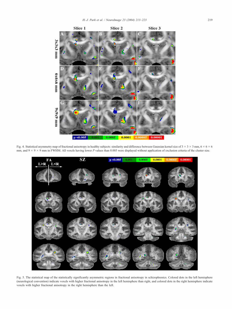
Fig. 5. The statistical map of the statistically significantly asymmetric regions in fractional anisotropy in schizophrenics. Colored dots in the left hemisphere
(neurological convention) indicate voxels with higher fractional anisotropy in the left hemisphere than right, and colored dots in the right hemisphere indicate
voxels with higher fractional anisotropy in the right hemisphere than the left.
Fig. 4. Statistical asymmetry map of fractional anisotropy in healthy subjects: similarity and difference between Gaussian kernel size of 3� 3� 3 mm, 6� 6� 6
mm, and 9 � 9 � 9 mm in FWHM. All voxels having lower P values than 0.005 were displayed without application of exclusion criteria of the cluster size.
H.-J. Park et al. / NeuroImage 23 (2004) 213–223 219

Table 2
Hemispherical asymmetry of fractional anisotropy in schizophrenic subjects
Description of
extent of cluster
Talairach
coordinates of
most significant
voxel (x, y, z)
Cluster
sizeaz-Score
Cingulum bundle
and CC
L > R �7.8, 29.7, 13.6 426 6.05
Optic radiation L > R �37.8, �30.5, �1.8 455 4.49
Posterior limb
IC � SLF
L > R �23.7, 0.6, 24.4 177 5.15
Splenium (CC) L > R �18.4, �53.8, 16.3 175 3.61
Splenium (CC) �posterior SLF
L < R 20.3, �32.4, 24.5 348 4.46
Anterior prefrontal
white matter
L < R 13.3, 49.1, �3.0 93 3.50
Superior prefrontal
white matter
L < R 18.6, 35.5, 17.4 83 3.76
Arcuate fasciculus
and SLF complex
L < R 27.4, �5.3, 22.3 78 3.83
CC, corpus callosum; SLF, superior longitudinal fasciculus; IC, internal
capsule; �, crossing area of fiber bundles.a Cluster size is in the unit of voxel.
H.-J. Park et al. / NeuroImage 23 (2004) 213–223220
MR images, which have been generally used for previous voxel-
based studies of fractional anisotropy (Barnea-Goraly et al., 2003;
Burns et al., 2003; Eriksson et al., 2001; Foong et al., 2002; Rugg-
Gunn et al., 2001), do not include information essential for finding
anatomical correspondence of the white matter structures (namely,
fiber tract’s orientation and organization) and thus might not be
appropriate for finding precise anatomical correspondence in DT-
MRI. This is why we used a more complex registration approach
that utilizes multiple channel tensor information to match anatom-
ical correspondence between subjects and between hemispheres
instead of using fractional anisotropy or T2-weighted images, even
for the analysis of fractional anisotropy.
In addition, as a result of the better coregistration of the
corresponding anatomical structures between subjects and be-
tween hemispheres, and as a result of the combination of the
averaged deformation field, our mean image (symmetric tem-
plate further used for normalization) was both less blurred and
more representative of the morphology of the group than is the
case for more traditional voxel-based studies. In spite of our
strategy for better registration, by looking at the overlap of
normalized white matter segmentations thresholded with FA >
Fig. 6. Asymmetry of fractional anisotropy (FA) in normal controls (NC) and
tractography. The colors in the fiber bundles are derived from the left – right asymm
yellow line illustrates left-greater-than-right FA asymmetry, while the portion be
indicates anterior; P, posterior.
0.3, we found some registration errors in the boundary of
narrow fiber bundles. These errors were most likely due to
anisocubic voxels (0.8594 � 5 � 0.8594 mm, an interpolated
image voxel unit of the original scan unit 1.7 � 5 � 1.7 mm),
relatively low resolution tensor images that we used for the
analysis, as well as due to presumable interindividual variations
of topology.
To reduce the effect of normalization errors on the statistical
analysis, Gaussian smoothing was applied to the normalized
fractional anisotropy images. As was demonstrated in Fig. 4, a
smaller smoothing kernel can be used to detect asymmetry even in
narrow fiber bundles or in the fibers in close proximity to
neighboring fibers, although using such a smaller smoothing kernel
increases the probability of a type I error. On the other hand, a
larger smoothing kernel increases robustness to errors resulting
from either registration or artifacts, or both, as it sacrifices spatial
resolution. Thus, the smoothing kernel size should be small enough
to detect narrow structures but large enough to be robust to errors.
Finally, although we determined smoothing kernel size by exper-
iment in our case, a more generalized method using an appropriate
model needs to be further researched.
Since diffusion tensors characterize diffusion strength and
orientation much better than the FA indices, tensor asymmetry
comparison, instead of FA asymmetry comparison, would be the
appealing and logical choice. From a clinical perspective, however,
asymmetry in the tensor of one voxel or of neighboring voxels is
not easy to interpret. On the other hand, FA has a relatively clearer
meaning; that is, it reflects the fiber bundle quantity underlying the
voxel, such as myelination, coherence, and/or density.
Asymmetry of fractional anisotropy in white matter of normal
controls and patients diagnosed with schizophrenia
We found anisotropic asymmetry differences for the cingulum
bundle, which showed left-higher-than-right anisotropy in healthy
subjects. This finding is consistent with a previous finding from
our group based on an ROI method (Kubicki et al., 2003). We also
noted an anisotropic asymmetry in healthy controls in the anterior
limb of the internal capsule and extended fibers that connect orbital
frontal regions, which showed higher anisotropy in the right than
left and which is also consistent with a previous finding in the
literature (Peled et al., 1998).
The subinsular regions, reported to be left-greater-than-right
asymmetry (Cao et al., 2003) using manual ROI methods, did not
patients with schizophrenia (SZ) superimposed on the three-dimensional
etry z-values displayed in Figs. 3 and 5. The portion of the figure above the
low shows FA asymmetry in opposite direction (right-greater-than-left). A

H.-J. Park et al. / NeuroImage 23 (2004) 213–223 221
show a significant asymmetry (P < 0.001) but did show a tendency
toward asymmetry (P < 0.005), as depicted in Fig. 4. This slight
discrepancy might be due to the narrow structure of the subinsular
region and thus due to low spatial alignment after normalization of
this region.
In our results, in healthy controls, uncinate fasciculus was
compromised of fibers showing two different patterns of asymme-
try (right-greater-than-left in the middle and inferior portion, and
left-greater-than-right in the superior portion at the smoothed FA
images with FWHM 3 � 3 � 3 mm). Previous DT-MRI report of
Kubicki et al. (2002), which investigated uncinate fasciculus by
measuring fractional anisotropy at a center point of each tract at
most perpendicular to the coronal plane, was able to demonstrate
only the second pattern of asymmetry, that is, left-greater-than-
right fractional anisotropy in control subjects, and no asymmetries
in schizophrenia. In postmortem study of Highley et al. (2002),
both schizophrenia and controls showed right-greater-than-left
asymmetry of cross-sectional area and fiber number in the uncinate
fascicle. Therefore, the asymmetry of the uncinate fasciculus is still
controversial, and further study is necessary for both control group
and schizophrenia group.
In schizophrenia, the overall pattern of asymmetry findings was
similar to that of healthy controls but showed reduced asymmetry.
More specifically, in the uncinate fasciculus and in the anterior
limb of the internal capsule, the asymmetry pattern observed in
controls was not found in schizophrenia subjects, while reduced
asymmetry was observed in the anterior part of the corpus
callosum in schizophrenics compared to control subjects. Aniso-
tropic asymmetry was also observed in the cingulum bundles of
schizophrenia, which corresponds to a previous report by our
group (Kubicki et al., 2003).
The cingulum bundle is the most prominent connection be-
tween limbic structures and is known to consolidate information by
interconnecting thalamus, prefrontal, parietal, temporal lobes (in-
cluding amygdala, hippocampus, and parahippocampal gyrus) with
cingulate gyrus. The anisotropic asymmetry of the cingulum
bundle indicates the lateralization of brain function, which appears
to be similar in both normal controls and schizophrenics, that is,
left greater than right.
The anterior limb of the internal capsule connecting prefrontal
association cortex and subcortical nuclei comprise frontal-subcor-
tical feedback loops. The asymmetry of right>left in healthy
subject is not found in schizophrenia. Absence of anisotropic
asymmetry in schizophrenia might be related to functional discon-
nection as well as a disturbance in thalamic filtering in this group.
Zhou et al. (2003) reported right-greater-than-left asymmetry in the
volumes of anterior limb in both controls and patients with
schizophrenia, although the schizophrenia group showed signifi-
cantly increased asymmetry compared with controls. However, our
voxel-based approach showed reduced, right-greater-than-left
asymmetry of fractional anisotropy in schizophrenia.
Of further note, the uncinate fasciculus, the major fiber tract
connecting the inferior frontal and anterior temporal lobes (Ebeling
and von Cramon, 1992), is known to play a role in decision making
as well as in the retrieval of semantic and episodic memory. The
absence of asymmetry of fractional anisotropy in the uncinate
fasciculus in schizophrenia thus suggests an abnormality in the
integrity of the fibers connecting the inferior frontal and anterior
temporal regions (Akbarian et al., 1996;Deakin and Simpson, 1997).
In our study, one of the most significant asymmetry regions for
controls was observed for the anterior portion of the corpus
callosum. This asymmetry was also found but in a reduced way
in patients with schizophrenia. Of interest here, we note that the
anterior corpus callosum interconnects the prefrontal lobes of both
hemispheres (Makris et al., 1999) and is reported to be involved in
the higher order transfer of semantic information (Gazzaniga,
2000). Furthermore, thin fibers that are lightly myelinated, of
which a large majority are believed to interconnect association
cortex, are most dense in the anterior corpus callosum (genu),
relative to total corpus callosum (Aboitiz et al., 1992). Moreover,
heterotopic connections between nonequivalent cortical regions in
each hemisphere are known to be numerous and widespread (Di
Virgilio and Clarke, 1997; Toga and Thompson, 2001). Thus, these
neuroanatomical and functional differences in the anterior part of
the corpus callosum may play a critical role in our finding of
fractional anisotropic asymmetry of the anterior corpus callosum.
Note that we did not directly compare asymmetry difference
between healthy subjects and schizophrenic subjects in this study.
While we considered using an asymmetric index, as for example,
(L � R) / (L + R) / 2, such asymmetry indices assume that the
normalization error is not significantly different between groups
when matching homologous points. For the schizophrenic group,
this assumption may not be true. In fact, when we checked the
within-group overlap of normalized white matter segmentations,
derived from individual subjects of each group using a threshold of
0.3 for FA, the overlap of schizophrenics was less than that of
healthy subjects especially in the narrow fiber structures. This
might be associated with the high interindividual anatomical
variability known to exist in schizophrenia, a very heterogeneous
group. For this reason, we decided to use a within-group compar-
ison of hemisphere asymmetry.
Interpretation of fractional anisotropy asymmetry between
hemispheres
Although we found hemispheric asymmetry of fractional an-
isotropy in healthy subjects and, to a much lesser extent, in
schizophrenia, the interpretation of these results requires some
caution. For example, since fractional anisotropy represents a
combination of fiber size, density, myelination, and fiber coher-
ence, any of these factors can influence the results. Moreover,
fractional anisotropy can be influenced by fiber crossings, espe-
cially in areas where two or more major bundles cross. One such
area lies lateral to the genu of the corpus callosum, where fibers
coming out of the corpus cross with internal capsule radiation
fibers and fibers of the superior longitudinal fasciculus. Therefore,
fractional anisotropy differences do not necessarily indicate differ-
ences in indices of the anatomical connectivity, such as the number
of fibers or myelination. Likewise, the lack of FA differences does
not necessarily indicate unambiguous connectivity. The interpreta-
tion should be further researched.
One potential approach to increase the specificity of DT-MRI
asymmetry studies is to use fiber tractography. We have applied
such an approach to explore asymmetry of neural connectivity in
both controls and schizophrenia by measuring mean fractional
anisotropy along the fibers (Park et al., 2003b). However, explo-
ration of asymmetry using fiber tractography can only be used in
an exploratory manner since there are many caveats, not the least
of which is the problem of how to evaluate fibers that cross. Other
problems include partial volume effects, signal to noise ratio, and
validation of fiber tracking. If we can strive to achieve reliable
tractography, this would be an important step toward evaluating the

H.-J. Park et al. / NeuroImage 23 (2004) 213–223222
asymmetry of fibers, which would lead to a further understanding
of ‘‘neural connectivity.’’
Conclusion
In this study, we proposed a novel method for exploring voxel-
based white matter asymmetry. Findings demonstrate anatomical
asymmetry of white matter anisotropy in healthy subjects in
cingulum bundles, uncinate fasciculus, anterior portion of corpus
callosum, the internal capsule, and optic radiation, with similar,
albeit more attenuated, findings in schizophrenia.
Acknowledgments
Information on grants. We gratefully acknowledge the support
of the Post-doctoral Fellowship Program of Korea Science and
Engineering Foundation (KOSEF) (HJP), National Alliance for
Research on Schizophrenia and Depression (MK), the National
Institutes of Health (K02 MH 01110 and R01 MH 50747 to MES,
R01 MH 40799 to RWM, R01 NS 39335 to SEM, and RO3
MH068464-01 to MK), the Department of Veterans Affairs Merit
Awards (MES, RWM), and the National Center for Research
Resources (11747 to RK and P41-RR13218 to FAJ and CFW).
References
Aboitiz, F., Scheibel, A.B., Fisher, R.S., Zaidel, E., 1992. Fiber composi-
tion of the human corpus callosum. Brain Res. 598 (1–2), 143–153.
Akbarian, S., Kim, J.J., Potkin, S.G., Hetrick, W.P. , Bunney Jr., W.E.,
Jones, E.G. 1996. Maldistribution of interstitial neurons in prefrontal
white matter of the brains of schizophrenic patients. Arch. Gen. Psy-
chiatry 53 (5), 425–436.
Alexander, D.C., Pierpaoli, C., Basser, P.J., Gee, J.C., 2001. Spatial trans-
formations of diffusion tensor magnetic resonance images. IEEE Trans.
Med. Imag. 20 (11), 1131–1139.
Amunts, K., Jancke, L., Mohlberg, H., Steinmetz, H., Zilles, K., 2000.
Interhemispheric asymmetry of the human motor cortex related to hand-
edness and gender. Neuropsychologia 38 (3), 304–312.
Anderson, B., Southern, B.D., Powers, R.E., 1999. Anatomic asymmetries
of the posterior superior temporal lobes: a postmortem study. Neuro-
psychiatry Neuropsychol. Behav. Neurol. 12 (4), 247–254.
Barnea-Goraly, N., Eliez, S., Hedeus, M., Menon, V., White, C.D., Mose-
ley, M., Reiss, A.L., 2003. White matter tract alterations in fragile X
syndrome: preliminary evidence from diffusion tensor imaging. Am. J.
Med. Genet. 118B (1), 81–88.
Basser, P.J., 1995. Inferring microstructural features and the physiolog-
ical state of tissues from diffusion-weighted images. NMR Biomed.
8 (7–8), 333–344.
Basser, P.J., Mattiello, J., LeBihan, D., 1994. MR diffusion tensor spec-
troscopy and imaging. Biophys. J. 66 (1), 259–267.
Beaton, A.A., 1997. The relation of planum temporale asymmetry and
morphology of the corpus callosum to handedness, gender, and dyslex-
ia: a review of the evidence. Brain Lang. 60 (2), 255–322.
Buchsbaum, M.S., Tang, C.Y., Peled, S., Gudbjartsson, H., Lu, D., Hazlett,
E.A., Downhill, J., Haznedar, M., Fallon, J.H., Atlas, S.W., 1998. MRI
white matter diffusion anisotropy and PET metabolic rate in schizophre-
nia. NeuroReport 9 (3), 425–430.
Burns, J., Job, D., Bastin, M.E., Whalley, H., Macgillivray, T., Johnstone,
E.C., Lawrie, S.M., 2003. Structural disconnectivity in schizophrenia: a
diffusion tensor magnetic resonance imaging study. Br. J. Psychiatry
182, 439–443.
Cao, Y., Whalen, S., Huang, J., Berger, K.L., DeLano, M.C., 2003. Asym-
metry of subinsular anisotropy by in vivo diffusion tensor imaging.
Hum. Brain Mapp. 20, 82–90.
Deakin, J.F., Simpson, M.D., 1997. A two-process theory of schizophrenia:
evidence from studies in post-mortem brain. J. Psychiatr. Res. 31 (2),
277–295.
Di Virgilio, G., Clarke, S., 1997. Direct interhemispheric visual input to
human speech areas. Hum. Brain Mapp. 5 (5), 347–354.
Ebeling, U., von Cramon, D., 1992. Topography of the uncinate fascicle
and adjacent temporal fiber tracts. Acta Neurochir. (Wien.) 115 (3–4),
143–148.
Eriksson, S.H., Rugg-Gunn, F.J., Symms, M.R., Barker, G.J., Duncan, J.S.,
2001. Diffusion tensor imaging in patients with epilepsy and malforma-
tions of cortical development. Brain 124 (Pt. 3), 617–626.
Foong, J., Symms, M.R., Barker, G.J., Maier, M., Miller, D.H., Ron, M.A.,
2002. Investigating regional white matter in schizophrenia using diffu-
sion tensor imaging. NeuroReport 13 (3), 333–336.
Galaburda, A.M., LeMay, M., Kemper, T.L., Geschwind, N., 1978a.
Right– left asymmetrics in the brain. Science 199 (4331), 852–856.
Galaburda, A.M., Sanides, F., Geschwind, N., 1978b. Human brain.
Cytoarchitectonic left-right asymmetries in the temporal speech region.
Arch. Neurol. 35 (12), 812–817.
Gazzaniga, M.S., 2000. Cerebral specialization and interhemispheric com-
munication: does the corpus callosum enable the human condition?
Brain 123 (Pt. 7), 1293–1326.
Geschwind, N., 1972. Cerebral dominance and anatomic asymmetry. N.
Engl. J. Med. 287 (4), 194–195.
Geschwind, N., Galaburda, A.M., 1985a. Cerebral lateralization. Biological
mechanisms, associations, and pathology: I. A hypothesis and a pro-
gram for research. Arch. Neurol. 42 (5), 428–459.
Geschwind, N., Galaburda, A.M., 1985b. Cerebral lateralization. Biological
mechanisms, associations, and pathology: II. A hypothesis and a pro-
gram for research. Arch. Neurol. 42 (6), 521–552.
Geschwind, N., Galaburda, A.M., 1985c. Cerebral lateralization. Biological
mechanisms, associations, and pathology: III. A hypothesis and a pro-
gram for research. Arch. Neurol. 42 (7), 634–654.
Good, C.D., Johnsrude, I., Ashburner, J., Henson, R.N., Friston, K.J.,
Frackowiak, R.S., 2001. Cerebral asymmetry and the effects of sex
and handedness on brain structure: a voxel-based morphometric analy-
sis of 465 normal adult human brains. NeuroImage 14 (3), 685–700.
Gudbjartsson, H., Maier, S.E., Mulkern, R.V., Morocz, I.A., Patz, S., Jolesz,
F.A., 1996. Line scan diffusion imaging. Magn. Reson. Med. 36 (4),
509–519.
Guimond, A., Meunier, J., Thirion, J.-P., 2000. Average brain models: a
convergence study. Comput. Vis. Image Underst. 77, 192–210.
Guimond, A., Roche, A., Ayache, N., Meunier, J., 2001. Three-dimensional
multimodal brain warping using the demons algorithm and adaptive
intensity corrections. IEEE Trans. Med. Imag. 20 (1), 58–69.
Guimond, A., Guttmann, C.R.G., Warfield, S.K., Westin, C.-F., 2002.
Deformable registration of DT-MRI data based on transformation in-
variant tensor characteristics. Proceedings of the IEEE International
Symposium on Biomedical Imaging (ISBI’02), Washington (DC),
USA.
Highley, J.R., Esiri, M.M., McDonald, B., Cooper, S.J., Crow, T.J., 1998a.
Temporal-lobe length is reduced, and gyral folding is increased in
schizophrenia: a post-mortem study. Schizophr. Res. 34 (1–2), 1–12.
Highley, J.R., Esiri, M.M., McDonald, B., Cortina-Borja, M., Cooper, S.J.,
Herron, B.M., Crow, T.J., 1998b. Anomalies of cerebral asymmetry in
schizophrenia interact with gender and age of onset: a post-mortem
study. Schizophr. Res. 34 (1–2), 13–25.
Highley, J.R., McDonald, B., Walker, M.A., Esiri, M.M., Crow, T.J., 1999.
Schizophrenia and temporal lobe asymmetry. A post-mortem stereolog-
ical study of tissue volume. Br. J. Psychiatry 175, 127–134.
Highley, J.R., Walker, M.A., Esiri, M.M., Crow, T.J., Harrison, P.J., 2002.
Asymmetry of the uncinate fasciculus: a post-mortem study of normal
subjects and patients with schizophrenia. Cereb. Cortex 12 (11),
1218–1224.

H.-J. Park et al. / NeuroImage 23 (2004) 213–223 223
Holinger, D.P., Galaburda, A.M., Harrison, P.J., 2000. Cerebral asym-
metry. In: Harrison, P.J., Roberts, G.W. (Eds.), The Neuropathology
of Schizophrenia. Progress and Interpretation. Oxford Univ. Press,
Oxford, pp. 151–172.
Kennedy, D.N., O’Craven, K.M., Ticho, B.S., Goldstein, A.M., Makris, N.,
Henson, J.W., 1999. Structural and functional brain asymmetries in
human situs inversus totalis. Neurology 53 (6), 1260–1265.
Kimura, D., 1973. The asymmetry of the human brain. Sci. Am. 228 (3),
70–78.
Kubicki, M., Westin, C.F., Maier, S.E., Frumin, M., Nestor, P.G., Salisbury,
D.F., Kikinis, R., Jolesz, F.A., McCarley, R.W., Shenton, M.E., 2002.
Uncinate fasciculus findings in schizophrenia: a magnetic resonance
diffusion tensor imaging study. Am. J. Psychiatry 159 (5), 813–820.
Kubicki, M., Westin, C.F., Nestor, P.G., Wible, C.G., Frumin, M., Maier,
S.E., Kikinis, R., Jolesz, F.A., McCarley, R.W., Shenton, M.E., 2003.
Cingulate fasciculus integrity disruption in schizophrenia: a magnetic
resonance diffusion tensor imaging study. Biol. Psychiatry 54 (11),
1171–1180.
Lancaster, J.L., Kochunov, P.V., Thompson, P.M., Toga, A.W., Fox, P.T.,
2003. Asymmetry of the brain surface from deformation field analysis.
Hum. Brain Mapp. 19 (2), 79–89.
Lim, K.O., Hedehus, M., Moseley, M., de Crespigny, A., Sullivan, E.V.,
Pfefferbaum, A., 1999. Compromised white matter tract integrity in
schizophrenia inferred from diffusion tensor imaging. Arch. Gen. Psy-
chiatry 56 (4), 367–374.
Lin, C.C., Mudholkar, G.S., 1980. A simple test for normality against
asymmetric alternatives. Biometrika 67, 455–461.
Maier, S.E., Gudbjartsson, H., Patz, S., Hsu, L., Lovblad, K.O., Edelman,
R.R., Warach, S., Jolesz, F.A., 1998. Line scan diffusion imaging:
characterization in healthy subjects and stroke patients. AJR Am. J.
Roentgenol. 171 (1), 85–93.
Makris, N., Meyer, J.W., Bates, J.F., Yeterian, E.H., Kennedy, D.N., Cavi-
ness, V.S., 1999. MRI-Based topographic parcellation of human cere-
bral white matter and nuclei II. Rationale and applications with
systematics of cerebral connectivity. NeuroImage 9 (1), 18–45.
Mamata, H., Mamata, Y., Westin, C.F., Shenton, M.E., Kikinis, R., Jolesz,
F.A., Maier, S.E., 2002. High-resolution line scan diffusion tensor MR
imaging of white matter fiber tract anatomy. AJNR Am. J. Neuroradiol.
23 (1), 67–75.
McDonald, B., Highley, J.R., Walker, M.A., Herron, B.M., Cooper, S.J.,
Esiri, M.M., Crow, T.J., 2000. Anomalous asymmetry of fusiform and
parahippocampal gyrus gray matter in schizophrenia: a postmortem
study. Am. J. Psychiatry 157 (1), 40–47.
Moffat, S.D., Hampson, E., Lee, D.H., 1998. Morphology of the planum
temporale and corpus callosum in left handers with evidence of left
and right hemisphere speech representation. Brain 121 (Pt. 12),
2369–2379.
Mohr, B., Heim, S., Pulvermuller, F., Rockstroh, B., 2001. Functional
asymmetry in schizophrenic patients during auditory speech processing.
Schizophr. Res. 52 (1–2), 69–78.
Nestor, P.G., Shenton, M.E., Wible, C., Hokama, H., O’Donnell, B.F., Law,
S., McCarley, R.W., 1998. A neuropsychological analysis of schizo-
phrenic thought disorder. Schizophr. Res. 29 (3), 217–225.
Park, H.J., Kubicki, M., Shenton, M.E., Guimond, A., McCarley, R.W.,
Maier, M., Kikinis, R., Jolesz, F.A., Westin, C.-F., 2003a. Spatial nor-
malization of diffusion tensor MRI using multiple channels. Neuro-
Image 20 (4), 1995–2009.
Park, H.J., Westin, C.-F., Brun, A., Kubicki, M., McCarley, R.W., Shenton,
M.E., 2003b. A method for hemispheric asymmetry of white matters
using diffusion tensor MRI. Proceedings of Human Brain Mapping
2003, New York, USA, Neuroimage S(918).
Peled, S., Gudbjartsson, H., Westin, C.F., Kikinis, R., Jolesz, F.A., 1998.
Magnetic resonance imaging shows orientation and asymmetry of white
matter fiber tracts. Brain Res. 780 (1), 27–33.
Pujol, J., Lopez-Sala, A., Deus, J., Cardoner, N., Sebastian-Galles, N.,
Conesa, G., Capdevila, A., 2002. The lateral asymmetry of the human
brain studied by volumetric magnetic resonance imaging. NeuroImage
17 (2), 670–679.
Rockstroh, B., Kissler, J., Mohr, B., Eulitz, C., Lommen, U., Wienbruch,
C., Cohen, R., Elbert, T., 2001. Altered hemispheric asymmetry of
auditory magnetic fields to tones and syllables in schizophrenia. Biol.
Psychiatry 49 (8), 694–703.
Rosen, G.D., Sherman, G.F., Galaburda, A.M., 1993. Neuronal subtypes
and anatomic asymmetry: changes in neuronal number and cell-packing
density. Neuroscience 56 (4), 833–839.
Rugg-Gunn, F.J., Eriksson, S.H., Symms, M.R., Barker, G.J., Duncan, J.S.,
2001. Diffusion tensor imaging of cryptogenic and acquired partial
epilepsies. Brain 124 (Pt. 3), 627–636.
Sommer, I.E., Ramsey, N.F., Kahn, R.S., 2001. Language lateraliza-
tion in schizophrenia, an fMRI study. Schizophr. Res. 52 (1–2),
57–67.
Sowell, E.R., Thompson, P.M., Rex, D., Kornsand, D., Tessner, K.D.,
Jernigan, T.L., Toga, A.W., 2002. Mapping sulcal pattern asymmetry
and local cortical surface gray matter distribution in vivo: maturation in
perisylvian cortices. Cereb. Cortex 12 (1), 17–26.
Springer, S.P., Deutsch, G., 1998. Left Brain, Right Brain: Perspectives
From Cognitive Neuroscience Freeman and Co. Worth Publishers,
USA.
Takahashi, T., Kawasaki, Y., Kurokawa, K., Hagino, H., Nohara, S.,
Yamashita, I., Nakamura, K., Murata, M., Matsui, M., Suzuki, M.,
Seto, H., Kurachi, M., 2002. Lack of normal structural asymmetry of
the anterior cingulate gyrus in female patients with schizophrenia: a
volumetric magnetic resonance imaging study. Schizophr. Res. 55
(1–2), 69–81.
Toga, A.W., Thompson, P.M., 2001. Maps of the brain. Anat. Rec. 265 (2),
37–53.
Toga, A.W., Thompson, P.M., 2003. Mapping brain asymmetry. Nat. Rev.
Neurosci. 4 (1), 37–48.
Turetsky, B., Cowell, P.E., Gur, R.C., Grossman, R.I., Shtasel, D.L., Gur,
R.E., 1995. Frontal and temporal lobe brain volumes in schizophrenia.
Relationship to symptoms and clinical subtype. Arch. Gen. Psychiatry
52 (12), 1061–1070.
Watkins, K.E., Paus, T., Lerch, J.P., Zijdenbos, A., Collins, D.L., Neelin, P.,
Taylor, J., Worsley, K.J., Evans, A.C., 2001. Structural asymmetries in
the human brain: a voxel-based statistical analysis of 142 MRI scans.
Cereb. Cortex 11 (9), 868–877.
Wechsler, D., 1997a. Wechsler Adult Intelligence Scale Psychological
Corp., San Antonio, TX (Harcourt).
Wechsler, D., 1997b. The Wechsler Memory Scale Psychological Corp.,
San Antonio, TX (Harcourt).
Westbury, C.F., Zatorre, R.J., Evans, A.C., 1999. Quantifying variability
in the planum temporale: a probability map. Cereb. Cortex 9 (4),
392–405.
Westin, C.F., Maier, S.E., Mamata, H., Nabavi, A., Jolesz, F.A., Kikinis, R.,
2002. Processing and visualization for diffusion tensor MRI. Med. Im-
age Anal. 6 (2), 93–108.
Youn, T., Park, H.J., Kim, J.J., Kim, M.S., Kwon, J.S., 2003. Altered
hemispheric asymmetry and positive symptoms in schizophrenia:
equivalent current dipole of auditory mismatch negativity. Schizophr.
Res. 59 (2–3), 253–260.
Zhou, S.Y., Suzuki, M., Hagino, H., Takahashi, T., Kawasaki, Y., Nohara,
I., Yamashita, I., Seto, H., Kurachi, M., 2003. Decreased volume and
increased asymmetry of the anterior limb of the internal capsule in
patients with schizophrenia. Biol. Psychiatry 54 (4), 427–436.
Copyright © 2022 FDOKUMEN