Bivalent domains enforce transcriptional memory of DNA methylated genes in cancer cells
Upload
independentCategory
view
0download
0
Cell, Vol. 121, 859–872, June 17, 2005, Copyright ©2005 by Elsevier Inc. DOI 10.1016/j.cell.2005.03.036
WDR5 Associates with Histone H3Methylated at K4 and Is Essential forH3 K4 Methylation and Vertebrate Development
Joanna Wysocka,1,5 Tomek Swigut,2,5
Thomas A. Milne,1 Yali Dou,3 Xin Zhang,4
Alma L. Burlingame,4 Robert G. Roeder,3
Ali H. Brivanlou,2 and C. David Allis1,*1Laboratory of Chromatin Biology2Laboratory of Molecular Vertebrate Embryology3Laboratory of Biochemistry and Molecular BiologyThe Rockefeller UniversityNew York, New York 100214Mass Spectrometry FacilityUniversity of California, San FranciscoSan Francisco, California 94143
Summary
Histone H3 lysine 4 (K4) methylation has been linkedto the transcriptional activation in a variety of eukary-otic species. Here we show that a common compo-nent of MLL1, MLL2, and hSet1 H3 K4 methyltransfer-ase complexes, the WD40-repeat protein WDR5,directly associates with histone H3 di- and trimethy-lated at K4 and with H3-K4-dimethylated nucleo-somes. WDR5 is required for binding of the methyl-transferase complex to the K4-dimethylated H3 tail aswell as for global H3 K4 trimethylation and HOX geneactivation in human cells. WDR5 is essential for verte-brate development, in that WDR5-depleted X. laevistadpoles exhibit a variety of developmental defectsand abnormal spatial Hox gene expression. Our re-sults are the first demonstration that a WD40-repeatprotein acts as a module for recognition of a specifichistone modification and suggest a mechanism forreading and writing an epigenetic mark for gene acti-vation.
Introduction
Lysine methylation of histones is recognized as an im-portant epigenetic indexing system demarcating tran-scriptionally active and inactive chromatin domains ineukaryotic genomes. For example, methylation of ly-sine 4 (K4) of histone H3 has been linked to transcrip-tional activation in a variety of eukaryotes (for review,see Sims et al. [2003]). To add to the complexity of epi-genetic regulation, lysine residues can be mono-, di-,or trimethylated in vivo, and distinct methylation statesare associated with different transcriptional outcomes.In particular, trimethylation of H3 K4 is strongly andpreferentially associated with promoter and transcribedregions of active genes (Santos-Rosa et al., 2002;Schneider et al., 2004).
The first H3 K4 methyltransferase to be identified wasthe S. cerevisiae Set1 complex, whose catalytic sub-unit, the SET domain protein Set1, is homologous tothe Drosophila trithorax protein and to a family of re-
*Correspondence: [email protected]
5 These authors contributed equally to this work.lated human proteins, hSet1, MLL1, and MLL2, all ofwhich are capable of methylating H3 at K4 (Roguev etal., 2001; Miller et al., 2001; Nagy et al., 2002; Milne etal., 2002; Nakamura et al., 2002; Wysocka et al., 2003;Hughes et al., 2004; Yokoyama et al., 2004). HumanSet1-like enzymes are present in multiprotein com-plexes that appear to be structurally and functionallysimilar to the yeast Set1 complex (Wysocka et al., 2003;Goo et al., 2003; Hughes et al., 2004; Yokoyama et al.,2004). In humans, the MLL1 complex is recruited to pro-moters of homeobox (Hox) genes to maintain their acti-vation state (Milne et al., 2002). In keeping with a tran-scriptional maintenance function(s), the activation stateof Hox genes is epigenetically inherited through numer-ous cell divisions (reviewed in Francis and Kingston[2001]). These data suggest that H3 K4 methylation, fa-cilitated by trithorax-related proteins, has a role in es-tablishing “cellular memory” of transcription.
While progress has been made in identifying en-zymes responsible for adding methyl groups to H3 K4,little progress has been made in characterizing the mo-lecular effectors responsible for specifically recogniz-ing this modification and translating it into a biologicalreadout. A paradigm, however, has already been estab-lished for histone H3 K9 and K27 methylation. Here,specific effectors, chromodomain-containing proteinsHP1 and Polycomb, associate with histone H3 methyl-ated at K9 and K27, respectively, leading to changes inDNA template accessibility and higher-order changesin chromatin structure (Sims et al., 2003). Proteins thatdirectly associate with H3 K4 methyl remain elusive, al-though a chromodomain protein Chd1 has been re-ported to bind to H3 methylated at K4 in yeast (Pray-Grant et al., 2005).
Using a biochemical approach to identify proteinsthat specifically recognize histone H3 methylated at K4,we identified WDR5, a highly conserved WD40-repeatprotein, as a major methyl K4-associated protein in hu-man cells and investigated its function in gene expres-sion regulation and vertebrate development.
Results
WDR5 Is a Major H3-K4-Methyl-Associated Proteinand a Common Component of Mammalian H3 K4Methyltransferase ComplexesTo identify proteins that bind to methylated H3 K4, weturned to an unbiased pull-down assay using biotiny-lated H3 peptide immobilized on avidin beads. Nuclearextracts from 293 cells were incubated with H3 pep-tides (aa 1–20 peptides; either unmodified or dimethy-lated at K4). Bound polypeptides were then eluted fromthe resin, resolved by SDS PAGE, and visualized by sil-ver staining (Figure 1A). This analysis consistently re-vealed the presence of a single w40 kDa band that wasspecifically enriched in the H3-K4-dimethylated pep-tide purification (see arrow in Figure 1A). The identity ofthis polypeptide was determined by mass spectrometry

Cell860
Figure 1. WDR5 Is a Major H3-K4-Methyl-Associated Protein
(A) Peptide pull-down assays were performed using 293 cell nuclear extracts and H3 peptides, either unmodified or dimethylated at K4.Polypeptides were resolved by SDS PAGE and visualized by silver staining. A polypeptide specifically enriched in the H3 K4(Me)2 pull-downwas analyzed by mass spectrometry and identified as WD40-repeat protein WDR5 (see arrow). Molecular weight markers are indicated onthe left.(B) Peptide pull-down assays using 293 nuclear extracts and a series of unmodified or appropriately modified H3 peptides (indicated on top)were performed and analyzed by immunobloting with α-WDR5 (Ba), α-HP1β (Bb), α-RbBP5 (Bc), and α-Ash2 (Bd) antibodies. After elution,beads were boiled in Laemmli buffer and peptide load analyzed by the silver staining (Be).(C) Peptide pull-down assay was performed using 293 cells nuclear extract and H3 K4(Me)2 peptide. Beads were divided into four equalportions and bound proteins eluted by competition with indicated peptides. Eluates were analyzed by immunoblotting with α-WDR5.(D) 20-fold excess of BSA was incubated with equimolar amounts of purified recombinant HP1β and GST-WDR5 (each at 1 �g), and thismixture was used as input for the pull-down assay with indicated peptides. Bound proteins were analyzed by SDS PAGE and Coomassieblue staining. The position of each polypeptide is indicated on the left.(E) Recombinant purified GST-WDR5 was used as input for pulldown assay, with various unmodified and modified histone-tail peptidesindicated on top. Results were analyzed by SDS PAGE and Coomassie blue staining.(F) Peptide pull-down assay with recombinant GST-WDR5 and H3 peptides either unmodified or mono-, di-, or trimethylated at K4.
and found to be the 333 amino acid long WD40-repeat Wwprotein WDR5, a common component of multiple H3
K4 methyltransferase complexes. Thus, similar to HP1, Twwhich associates with methylated H3 K9 and binds to
the H3 K9 methyltransferase SUV39H1 (Bannister et al., ps2001; Lachner et al., 2001; Aagaard et al., 1999), WDR5
associates with methylated H3 K4 and is present in var- tnious H3 K4 methyltransferase complexes.
DR5 but Not RbBP5 Associatesith Methylated H3 K4
o confirm the specificity of the association of WDR5ith H3 tail methylated at K4, we performed peptideull-down assays using 293 nuclear extracts and aeries of unmodified or appropriately modified H3 pep-ides (Figure 1B). Results were then analyzed by immu-obloting with α-WDR5 antibody, or as a positive con-

WDR5 Associates with H3 Methylated at K4861
trol, with α-HP1β antibody. As expected, HP1β associatesspecifically with the K9(Me)2 H3 peptide (Figure 1Bb).In contrast, WDR5 preferentially associates with H3 thatis either di- or trimethylated at K4 (Figure 1Ba). Al-though a weak association is also observed with H3K9(Me)2 peptide, significance of this binding is unclear,as we were unable to detect association of WDR5 withnucleosomes methylated at H3 K9 (see Figure 2A).
Figure 2. WDR5 Associates with Nucleosomes Methylated on H3K4 and Activates Transcription
(A) Mononucleosomal fraction was purified from mock or Flag-WDR5 HeLa cell line and used for immunoprecipitation with α-Flagantibody followed by Flag peptide elution. Immunoprecipitateswere analyzed by immunobloting with α-tagged WDR5 (α-FLAG;Aa), α-Ash2 (Ab), α-H3 K4(Me)2 (Ac), and α-H3 K9(Me)2 (Ad) anti-bodies. Five percent of input mononucleosomal fraction was usedas a control.(B) GAL4-WDR5 fusion activates transcription of a reporter gene.293 cells stably expressing luciferase reporter under the control ofGAL4 UAS were transfected in duplicates with indicated amountsof GAL4 DBD cDNA or GAL4-WDR5 cDNA, or mock transfected,and cell extracts were prepared. Relative luciferase activities (RLU)were measured for each sample and are shown as the average ofthree measurements of two independent transfections. Error barsrepresent standard deviation (SD).
WDR5 did not associate with H3 S10P peptide. Peptidecompetition experiments showed that H3 K4(Me)2 pep-tide is more efficient in eluting the WDR5 protein pre-bound to K4-methylated H3 tail than either K9(Me)2 orunmodified peptide, suggesting that K4-methylated H3tail is the preferred binding substrate for WDR5 (seeFigure 1C).
Despite the existence of multiple enzymatic subunitscapable of methylating H3 K4, human H3 K4 methyl-transferase complexes share three common compo-nents: two WD40-repeat proteins, WDR5 and RbBP5,and the trithorax group protein Ash2 (Wysocka et al.,2003; Goo et al., 2003; Hughes et al., 2004; Yokoyamaet al., 2004). Thus, we sought to determine whetherAsh2 and RbBP5 can bind to H3 methylated at K4. Asshown in Figures 1Bc and 1Bd, neither Ash2 nor RbBP5associated with any of the H3 peptides. These data,together with the fact that WDR5 is a major polypeptideenriched in the H3 K4(Me)2 pull-down (Figure 1A), sug-gest that WDR5 specifically recognizes the H3 tailmethylated at K4.
Association of WDR5 with H3 Methylatedat K4 Is Direct and SpecificTo further address whether interaction of WDR5 withK4-methylated H3 tail is direct, GST-WDR5 fusion pro-tein was expressed in E. coli and purified to near homo-geneity. To ensure specificity, the pull-down assay wasperformed with and excess of BSA (20 �g) and a 1:1mixture of purified HP1β and GST-WDR5 (each at 1 �g).This mixture was then used as input for the pull-downassay with unmodified K9(Me)2 and K4(Me)2 peptides.As expected, HP1β was enriched in the K9(Me)2 pull-down (Figure 1D). In contrast, WDR5 was enriched inthe K4(Me)2 pull-down, while BSA failed to bind toeither peptide. Next, we tested binding of the purifiedGST-WDR5 to H2A, H2B, H4 (aa 1–20), and various un-modified and modified H3 peptides. As shown in Figure1E, of the entire set of peptides tested, GST-WDR5binding was specifically enriched in the H3 K4(Me)2peptide pull-down, indicating that association of WDR5with H3 methylated at K4 is both direct and, within thecontext of the peptides used here, specific.
In order to investigate which K4 methylation state ofH3 associates with WDR5, we tested binding of GST-WDR5 with a series of H3 peptides unmodified ormono-, di-, or trimethylated at K4. WDR5 preferentiallyassociates with all forms of the H3-K4-methylated pep-tide as compared to unmodified peptide. Reproducibly,association with H3 K4(Me)2 peptide is the most robust,while H3 K4(Me)1 is the weakest (see Figure 1F).
WDR5 Associates with H3-K4-MethylatedMononucleosomesTo confirm the functional significance of WDR5 associ-ation with H3 peptides methylated at K4, we sought toaddress whether an in vivo association of WDR5 withH3-K4-methylated nucleosomes exists. Since the avail-able WDR5 antibody fails to immunoprecipitate nativeprotein, we took advantage of a HeLa cell line stablyexpressing Flag/HA epitope-tagged full-length WDR5.Total chromatin was isolated and solubilized with

Cell862
microccocal nuclease; fractions containing mono- oonucleosomal DNA were then pooled and used for im-
munoprecipitation experiments with α-Flag antibody, t(followed by Flag peptide elution. To monitor specificity,
chromatin from HeLa cells lacking ectopic WDR5 were feused in parallel mock purifications. Immunoprecipitates
were subsequently analyzed with antibodies against: s(1) WDR5 (Figure 2Aa), to monitor immunoprecipitationefficiency; (2) Ash2 (Figure 2Ab), a known WDR5-asso- m
cciated protein, as a positive control; and (3) H3 methyl-ated at K4 (Figure 2Ac) or K9 (Figure 2Ad). Results m
tshown in Figures 2Ac and 2Ad demonstrate that WDR5specifically coimmunoprecipitates with the H3-K4- a
amethylated but not K9-methylated mononucleosomes(compare lanes 3 and 4 in Figures 2Ac and 2Ad). These s
(data suggest that WDR5 binds to H3 tail methylated atK4 in vivo in the context of native chromatin. A
pcWDR5 Is a Strong Transcriptional ActivatordDi- and trimethylation of H3 K4 has been correlatedawith transcriptional activation in several organisms.tTherefore, we hypothesized that WDR5, which prefer-bentially recognizes di- and trimethylated H3 K4, may
have transcriptional activation properties. To test thisthypothesis, full-length human WDR5 cDNA, fused tomthe GAL4 DNA binding domain cDNA (GAL4-WDR5),fwas transfected into 293 cells stably expressing a lucif-Werase reporter under the control of five tandem GAL4sUAS sites. Use of a stably integrated reporter in thisnassay ensured a dependency on the state of the chro-αmatin template. Transfection of 50 ng of GAL4-WDR5tfusion cDNA resulted in 20-fold activation of the repor-ater, as compared to signals obtained from cells trans-lfected with 50 ng of GAL4 DBD cDNA alone (Figure 2B).kComparison of GAL4-WDR5 with one of the strongestsactivators GAL4-VP16 revealed that activation bywGAL4-WDR5 is w10-fold weaker than GAL4-VP16-
mediated activation (data not shown). Thus, when teth-ered to chromatin, WDR5 acts as a strong transcrip- Wtional activator. o
wTLoss of WDR5 Affects Global H3 K4 Methylation
Levels in Human Cells without Affecting foGlobal Association of MLL1, MLL2,
and Ash2 with Chromatin apOne potential mechanism by which WDR5 activates
transcription may be by facilitating methylation of H3 pcK4. To address whether WDR5 knockdown may have a
global effect on the steady-state level of H3 K4 methyl- flation, WDR5 protein was specifically eliminated by
transfecting 293 cells with a pool of four WDR5 siRNAs. Nl293 cells transfected with the corresponding amounts
of noncoding siRNAs served as a control. Whole-cell Waextracts were prepared 3 days after transfection and
analyzed by immunoblotting with various antibodies. cwWDR5 siRNA transfection resulted in the efficient
knockdown of WDR5 protein (Figure 3Aa) but did not lPaffect expression levels of other components of H3 K4
methyltransferases, Ash2, MLL1, and MLL2 (Figures pl3Ab, 3Ac, and 3Ad). However, global levels of the K4
methylation were significantly reduced in WDR5 siRNA- Fltransfected cells with a strong decrease of H3 K4
mono- (Figure 3Ae) and trimethylation (Figure 3Ag) and a
nly a modest effect on dimethylation (Figure 3Af). Littler no effect was observed on H3 K9 and K27 trimethyla-ion (Figures 3Ai and 3Aj) and unmodified histone H3Figure 3Ah); global histone levels were also not af-ected (Figure 3Ak). Similar results were obtained withxtracts from cells transfected with each individualiRNA from the pool of four siRNAs (data not shown).The global effect of WDR5 knockdown on H3 K4ethylation may be due to its effect on chromatin asso-
iation of other proteins directly involved in H3 K4ethylation. To ask whether global chromatin associa-
ion of components of human H3 K4 methyltransfer-ses is affected by the loss of WDR5 protein, we tookdvantage of a small-scale biochemical fractionationcheme originally developed by Mendez and StillmanMendez and Stillman, 2000) and depicted in Figure 3B.lthough each fraction has a distinct polypeptide com-osition, as expected, histones are recovered in thehromatin fraction P3 (Figure 3Cf, lanes 3 and 6). Theistribution of WDR5, different H3 K4 methyltransfer-ses, and several of their complex components werehen examined by immunoblotting with specific anti-odies (Figures 3Ca–3Ce).As shown in Figure 3Ca, in cells transfected with con-
rol siRNA, a portion of WDR5 is associated with chro-atin (lane 3), and a portion is recovered in S2 soluble
raction (lane 1). In contrast, transfection of cells withDR5 siRNA pool resulted in loss of WDR5 from both
oluble and chromatin fractions (lanes 4 and 6). Immu-oblot analyses with α-Ash2, α-RbBP5, α-MLL1, and-MLL2 antibodies (Figures 3Cb–3Ce) revealed that
here is no effect of WDR5 knockdown on the globalssociation of these proteins with chromatin (compare
anes 3 and 6 in all panels). Thus, the effect of WDR5nockdown on H3 K4 methylation cannot be explainedimply by the loss of methyltransferase associationith chromatin.
DR5 Is Required for Associationf the Methyltransferase Complexith H3-K4-Dimethylated Substrate
o address the mechanism by which loss of WDR5 af-ects H3 K4 methylation without affecting associationf H3 K4 methyltransferases with chromatin, we char-cterized binding of the H3 K4 methyltransferase com-lexes to either unmodified, K4 di-, or trimethylated H3eptides. To this end, we took advantage of two HeLaell lines stably expressing (1) Flag/HA epitope-taggedull-length WDR5 or (2) Flag/HA epitope-tagged full-ength RbBP5 (Dou et al. [2005], this issue of Cell).uclear extracts were fractionated on P11 phosphocel-
ulose. Fractions containing (1) both free WDR5 andDR5 associated with hSet1 and MLL2 methyltransfer-
se complexes or (2) both free RbBP5 and RbBP5-asso-iated hSet1 and MLL2 methyltransferase complexesere subjected to anti-Flag immunoprecipitation fol-
owed by Flag peptide elution (depicted in Figure 4A).urified material was then used as input for the peptideull-down assays with unmodified or K4 di- or trimethy-
ated H3 peptides. Immunoblot analysis reveals thatlag-WDR5 preferentially associates with di- and, to a
esser extent, trimethylated peptide, with only a minorssociation with the unmodified peptide (Figure 4Ba).

WDR5 Associates with H3 Methylated at K4863
Figure 3. Loss of WDR5 Affects Global H3 K4 Methylation Levels but Not MLL1, MLL2, or Ash2 Chromatin Association
(A) Analysis of effects of WDR5 knockdown on histone H3 K4 methylation. 293 cells were transfected with control siRNAs or WDR5 siRNApool and counted, and whole-cell extracts were prepared 3 days after transfection and probed by immunoblotting with α-WDR5 (Aa), α-Ash2(Ab), α-MLL1 (Ac), α-MLL2 (Ad), H3 K4(Me)1−3 (Ae–Ag), α-H3 1–20 unmodified (Ah), α-H3 K9(Me)3 (Ai), α-H3 K27(Me)3 (Aj) antibodies or stainedwith Coomassie blue for total histones to ensure equal loading (Ak). Two-fold dilution of each extract was used in order to control for thedynamics of the antibody response.(B) Biochemical fractionation scheme. Final fractions used for analysis—soluble “cytosolic” (S2), soluble nuclear (S3), and chromatin-enriched(P3)—are boxed. The cytosolic fraction S2 also contains soluble nuclear components owing to permeabilization of the nuclear membraneduring cell lysis with nonionic detergent.(C) Loss of WDR5 has no effect on global chromatin association of MLL1, MLL2, Ash2, and RbBP5. 293 cells were transfected with controlsiRNAs or WDR5 siRNA pool and subjected to biochemical fractionation. Fractions S2, S3, and P3 were probed by immunoblotting withα-WDR5 (Ca), α-Ash2 (Cb), α-RbBP5 (Cc), α-MLL1 (Cd), and α-MLL2 (Ce) antibodies and stained with Coomassie blue for analysis of totalhistones distribution (Cf). All fractions were resuspended in equal final volumes and thus represent the cellular distribution of the analyzed pro-teins.
These data are consistent with the binding specificityof recombinant WDR5 (Figure 1F). In contrast, othercomponents of the H3 K4 methyltransferase complex—RbBP5, Ash2, and MLL2—bind equally well to the un-modified and dimethylated peptides and poorly to thetrimethylated peptide (Figures 4Bb–4Bd). As all of theproteins present in the input are associated with Flag-WDR5, these results suggest that free Flag-WDR5, pre-sent in excess, preferentially associates with K4-dimethylated H3, whereas H3 K4 methyltransferasecomplexes associate both with unmodified and K4-dimethylated H3. Consistent with these observations,immunoblot analysis of the pull-down with Flag-RbBP5
demonstrates that all of the tested complex compo-nents, including WDR5, bind both to unmodified anddimethylated peptides (Figure 4C). Furthermore, only asmall fraction of Flag-RbBP5 is recovered in the pull-down, as compared to Flag-WDR5 (compare Figures4Ca and 4Ba), suggesting that RbBP5 is unlikely to bindhistone H3 tail on its own and that observed associa-tion reflects specificity of the methyltransferase com-plex. Interestingly, in the experiments presented in Fig-ure 1B, we failed to detect binding of RbBP5 and Ash2to any of the H3 peptides or binding of WDR5 to theunmodified peptide, which is likely due to the fact thatproteins bound in the complex were below sensitivity

Cell864
Figure 4. WDR5 Is Important for Binding of the H3 K4 Methyltransferase Complex to Dimethylated H3 K4
(A) Schematic of the experimental design.(B) Flag-WDR5 and its associated proteins were purified from stable Flag-WDR5 HeLa cell line as depicted in (A) and used as input for thepeptide pull-down assay with indicated peptides. Results were analyzed by immunoblotting with α-WDR5 (Ba), α-RbBP5 (Bb), α-Ash2 (Bc),and α-MLL2 (Bd) antibodies.(C) Flag-RbBP5 and its associated proteins were purified from stable Flag-RbBP5 HeLa cell line as depicted in (A) and used as input for thepeptide pull-down assay with indicated peptides. Results were analyzed by immunoblotting with α-RbBP5 (Ca), α-WDR5 (Cb), α-Ash2 (Cc),and α-MLL2 (Cd) antibodies.(D) Schematic of the experimental design for data presented in (E).(E) Flag-RbBP5 cells were transfected with control siRNAs or WDR5 siRNA pool, and nuclear extracts were prepared 3 days after transfection.Flag-RbBP5 and its associated proteins were purified and used for peptide pull-down assay as depicted in (D). Results were analyzed byimmunobloting with α-RbBP5 (Ea), α-WDR5 (Eb), and α-Ash2 (Ec) antibodies.
of our assay. This also indicates that majority of the upendogenous WDR5 is present in the nuclear extract in
its free form, either because the majority of complexes FWfall apart during the nuclear extract preparation or due
to excess of WDR5 naturally present in the nucleus. AlResults presented in Figure 4B and C suggest that
WDR5-containing H3 K4 methyltransferase complexes nwhave dual binding specificity toward the H3 tail, i.e.,
they bind to both unmodified and K4-dimethylated H3 cctail. We hypothesized that WDR5 provides specificity
toward the dimethylated tail, whereas another compo- tbnent is responsible for binding of the complex to the
nmodified histone H3 tail. To test this hypothesis, weurified Flag-RbBP5 and associated proteins from thelag/HA-RbBP5 cells treated with control siRNA orDR5 siRNA, as schematically depicted in Figure 4D.s shown in Figure 4E, WDR5 knockdown resulted in
oss of binding of RbBP5 and Ash2 to dimethylated butot unmodified peptide. These findings are consistentith our observations that loss of WDR5 does not affecthromatin association of methyltransferase complexomponents and strongly suggests that WDR5 endowshe MLL methyltransferase complex with an ability toind to dimethylated substrates. We also noted de-

WDR5 Associates with H3 Methylated at K4865
Figure 5. Loss of WDR5 Protein Results in a Decrease of HistoneH3 Lysine 4 Trimethylation but Not Dimethylation at HOX Genes
(A) Treatment of 293 cells with WDR5 siRNA (+) results in a de-crease of WDR5 protein levels compared to control (−) cells trans-fected with noncoding siRNA.(B) Schematic of the HOXA9 and HOXC8 loci. Gray arrows markthe positions of Taqman primer/probe sets (P1–P4) used to quantifyChIP experiments. Gray boxes represent exons, black arrows aretranscription start sites, and black boxes represent CpG rich re-gions. A putative TATA box at the HOXA9 locus identified by Naka-mura et al. (2002) as a site of MLL binding is also shown. Distancesare in kilobases.(C) Binding of MLL to the promoter and coding regions of HOXA9and HOXC8 is not altered by a reduction of WDR5 protein. Primer/probe sets are as shown in (B). Signal was quantified relative to
get loci.
input chromatin as described in Experimental Procedures. Back-ground signal was determined in Mll−/− cells to be lower than 0.05%(data not shown). Binding at the GAPDH locus is shown as a con-trol and is below background levels. Error bars represent SD basedon three independent experiments.(D) Histone H3 K4(Me)3 (light gray bars) but not K4(Me)2 (dark graybars) is reduced by WDR5 siRNA treatment (+) compared to cellstreated with a control siRNA (−). ChIP experiments were quantifiedas in (C), but signals from WDR5 siRNA-treated cells (+) were stan-dardized relative to signal from control cells (−), which were set to1.0. Primer/probe sets are as shown in (B). Error bars represent SDbased on three independent experiments.(E) Treatment of 293 cells with WDR5 siRNA (+) reduces HOXA9and HOXC8 gene expression compared to cells treated with a con-trol siRNA (−). Gene expression was quantified using bACTIN as aninternal standard. Expression of HOX genes in WDR5 siRNA treatedcells (+) is standardized relative to expression in control cells (−).Error bars represent SD based on three independent experiments.
creased association of Ash2 with RbBP5 in WDR5-depleted cells, suggesting that WDR5 stabilizes Ash2association with the complex (Figure 4Ec).
Taken together, our data suggest a model in whichWDR5 does not play a role in the recruitment of its as-sociated methyltranferase complex to localized regionof chromatin and in the initiation of methylation but in-stead is important for di- to trimethyl lysine convertion.We sought to test this hypothesis at the localized re-gion of chromatin, known to be regulated by the WDR5-associated methyltransferase.
WDR5 Knockdown Does Not Affect MLL1Recruitment to the HOXA9 and HOXC8 LociTo determine whether loss of WDR5 affects the recruit-ment of its associated methyltransferase to a localizedregion of chromatin, we examined if WDR5 knockdownhas any effect on the recruitment of one of its associ-ated methyltransferases, MLL1, to two of its known tar-gets, the HOXA9 and HOXC8 genes. To this end, 293cells were transfected with a pool of WDR5 siRNAs orwith control noncoding siRNA. Samples were normal-ized by cell counting and used for whole-cell extractand total RNA preparations; knockdown efficiency wasconfirmed by immunoblotting (Figure 5A). Remainingcells were used for a chromatin immunoprecipitationassay (ChIP) with α-MLL1 antibody. To determinewhether association of MLL1 with either the HOXA9 orHOXC8 locus is affected by the WDR5 knockdown, theimmunoprecipitated DNA was quantified by real-timePCR using probes to the HOXA9 promoter and genecoding region and HOXC8 promoter and 5# leader se-quence (see Figure 5B). As a negative control, a probeto the GAPDH control locus was used. As shown inFigure 5C, reduction of WDR5 levels did not have anymeasurable effect on the association of MLL with theHOXA9 and HOXC8 loci, suggesting that WDR5 doesnot act through the recruitment of this K4 methyl-transferase to its target loci. As expected, no MLL bind-ing was observed at the GAPDH locus. Consistent withour previous results, this suggests that WDR5 does notdirectly recruit methyltransferases such as MLL1 to tar-

Cell866
Figure 6. WDR5 Knockdown in X. laevis Embryos Results in Loss of Mono- and Trimethylation of H3 K4
(A) Schematic of experimental design.(B) Two-cell stage X. laevis embryos were coinjected with xWDR5 morpholino and Alexa-647-dextran fluorescent lineage tracer (Ba–Bi) orwith xWDR5 morpholino, Alexa-647-dextran tracer and synthetic mRNA encoding human GST-WDR5 (Bm–Bo), or as negative control, withAlexa-647-dextran alone (Bj–Bl). Embryos were allowed to develop to stage 11, fixed, immunostained with H3 K4 methyl antibodies (green),followed by Alexa-555-conjugated anti-rabbit secondary antibodies (red), and analyzed by confocal microscopy. Panels show a combinationof 20 focal planes corresponding to approximately 20 �m section stacks of the embryo.
WDR5 Knockdown Leads to the Decrease in the H3 krK4 Trimethylation but Not H3 K4 Dimethylation
over the HOXA9 and HOXC8 Loci jsNext, we asked whether WDR5 knockdown affects the
status of K4 methylation at the HOXA9 and HOXC8 dagenes, known targets of MLL-mediated activation.
ChIP assays were performed with antibodies againstH3 either di- or trimethylated at K4. Immunoprecipi- W
otated DNA was then subjected to quantitative real-timePCR using probes to the HOXA9 and HOXC8 genes T
e(Figure 5B). As shown in Figure 5D, WDR5 knockdowndid not affect H3 K4 dimethylation over the promoter t
pregions of HOXA9 and HOXC8 and 5# leader sequenceof HOXC8. Interestingly, we reproducibly observed an q
Fincrease of K4 dimethylation over the coding region ofHOXA9 upon loss of WDR5. In contrast, WDR5 knock- 5
mdown resulted in a significant and reproducible de-crease in H3 K4 trimethylation over HOXA9 and HOXC8 m
cgenes (Figure 5D). Similar results were obtained usingtwo α-H3 K4 trimethyl antibodies and four independent s
nockdown/ChIP experiments (data not shown). Ouresults demonstrate that WDR5 contributes to the ma-ority of K4 trimethylation at the HOX loci and are con-istent with a model wherein WDR5 is important for thei- to trimethyl conversion by the H3 K4 methyltransfer-se complex.
DR5 Is Important for Maintenancef HOX Gene Expressiono test whether WDR5 knockdown has an effect on thexpression of HOXA9 and HOXC8 genes, a portion ofhe same set of samples used for the above ChIP ex-eriments was used for the total RNA preparation anduantitative real-time RT-PCR analysis. As shown inigure 5E, knockdown of WDR5 expression resulted in-fold downregulation of HOXC8 mRNA levels and ainor but reproducible downregulation of HOXA9RNA levels, as compared to cells transfected with
ontrol siRNAs. These results also correlate withtronger effect of WDR5 loss on K4 trimethylation on

WDR5 Associates with H3 Methylated at K4867
the HOXC8 locus, compared to the HOXA9 locus,which may reflect differences in the dynamic transcrip-tional regulation of the two genes. Whichever the case,these results demonstrate that WDR5 function is impor-tant for HOX gene regulation in human cells.
WDR5 Is Essential for H3 K4 Methylation in X. laevisOur data suggest that WDR5 affects global H3 K4methylation levels and expression of HOX genes in hu-man cells. It was of interest to know if these observa-tions could be reproduced on an organismal level and,if so, what the effect of perturbations in K4 methylationon the development of a vertebrate organism would be.Evolutionary conservation of WDR5 is quite remark-able. With the exception of the N-terminal “tail,” whoselength and sequence is variable among different spe-cies, the part of the protein that encompasses theseven WD40 repeats has a very high degree of se-quence identity among the metazoan species (see Fig-ure S1 in the Supplemental Data available with this arti-cle online). Given evolutionary conservation of WDR5,we sought to address the above questions using X.laevis as a developmental system and morpholino anti-sense technology to block translation of X. laevisxWDR5 mRNAs.
A morpholino oligonucleotide overlapping with theATG codon of X. laevis WDR5 (xWDR5 morpholino) wasdesigned and injected into the marginal zone of oneblastomere of a two-cell stage X. laevis embryo. Totrace the progeny of injected blastomeres, injectionsalso included a fluorescent lineage tracer (red-stainedcells). As depicted in Figure 6A, embryos were then al-lowed to develop to midgastrula stage (stage 11), afterwhich in situ immunostaining was performed with theα-H3 K4 methyl antibodies followed by confocal micro-scopy. The noninjected side of the embryo served asan internal control. Results of these experiments areshown in Figure 6B. A striking inverse correlation wasobserved between intensity of staining with the α-H3K4 mono- (Figures 6Ba–6Bc) and trimethyl (Figures6Bg–6Bi) antibodies (green) and the presence of the flu-orescent tracer (red), suggesting that xWDR5 knock-down results in a reduction of K4 mono- and trimethyla-tion in Xenopus embryos. Consistent with the resultsobtained in mammalian cells, xWDR5 morpholino injec-tion did not have a significant effect on α-K4(Me)2 stain-ing (Figures 6Bd–6Bf); no reduction in H3 K4 methylstaining was observed in embryos injected with thefluorescent tracer alone (example shown in Figures6Bj–6Bl). To control for the specificity of the observedeffects, WDR5 morpholino was coinjected with the invitro-transcribed GST-hWDR5 RNA. As shown in Fig-ures 6Bm–6Bo, GST-hWDR5 RNA reverted the effect ofthe xWDR5 morpholino on K4 trimethylation, as cellscontaining fluorescent tracer stained for trimethylationwith an intensity similar to that of cells that are tracernegative. Furthermore, xWDR5 morpholino injectioninto one-cell embryos results in a global loss of WDR5protein and H3 K4 trimethylation (Figure 7B).
Knockdown of xWDR5 during Xenopus DevelopmentLeads to Somitic, Gut, and Hematopoetic DefectsTo date, no WDR5 loss-of-function analysis has beendone in any vertebrate organism, and the general role
of K4 methylation in vertebrate development is poorlyunderstood. Thus, the X. laevis model provides aunique opportunity to look at the effects of loss ofWDR5 and, in consequence, a reduction of global K4methylation levels on the development of a vertebrate.To this end, one-cell stage X. laevis embryos were in-jected with WDR5 morpholino, and the injected em-bryos were allowed to develop to the feeding tadpolestages. To eliminate synthetic phenotypes resultingfrom different genetic backgrounds, the analysis wasperformed on embryos derived from eggs of multiplefrogs (n = 6).
In general, tadpoles derived from xWDR5 morpho-lino-injected embryos were delayed in development byas much as four stages compared to the tadpoles de-rived from control-injected embryos. These embryosexhibited a variety of developmental defects: mesoder-mal axial defects resulting in shortened and twistedtadpoles as well as gut patterning defects, resulting inlack of gut coiling and ventral lesions (see Figures 7Aa–7Ac). These phenotypes were highly penetrant andwere effectively rescued by the coinjection of GST-hWDR5 mRNA along with xWDR5 morpholino (Figures7Ad and 7C). Injection of the GST-hWDR5 mRNA alonehad no significant effect on development (Figures 7Aeand 7C), and phenotype penetrance and severity weredependent on xWDR5 morpholino dose (data notshown). Taken together, these observation indicate thatobserved phenotypes are specific to the WDR5 proteinlevel knockdown and cannot be attributed to some un-specific effect of the morpholino oligonucleotide injec-tion. Immunoblot analysis of extracts prepared fromsingle embryos that were control injected, xWDR5 mor-pholino injected, or coinjected with xWDR5 morpholinoand GST-hWDR5 mRNA revealed that morpholino injec-tion results in global loss of xWDR5 protein and H3 K4trimethylation (compare Figures 7Ba–7Bc, lanes 1 and2). Coinjection of GST-hWDR5 mRNA resulted in ex-pression of GST-hWDR5 fusion protein and in the res-cue of H3 K4 trimethylation (Figure 7B, see lane 3 inall panels).
Phenotypic analyses were also performed with two-to four-cell stage embryos injected with xWDR5 mor-pholino into one blastomere (either dorsal or ventral).Tadpoles derived from single blastomere-injected em-bryos exhibited somewhat milder phenotypes. In par-ticular, ventral lesions where absent and axial defectslimited to a lateral bend. As shown in Figure 7D, xWDR5morpholino-injected tadpoles exhibited somitic defectson one side of the body, resulting in a distinctive bend(different from more random shortening and twistingthat was observed for one-cell-stage-injected embryos)as well as delayed or absent gut (see Figure 7E for sta-tistical analysis of the penetrance of phenotypes pre-sented in Figure 7D). Many of the tadpoles also exhib-ited hematopoetic abnormalities consistent with ventralmesoderm defects: no blood, or blood missing from theheart, while forming extensive internal hemorrhages.These results indicate that WDR5 function, and pre-sumably transcriptional profiles determined in part byK4 methylation, is essential for vertebrate devel-opment.
Cell868
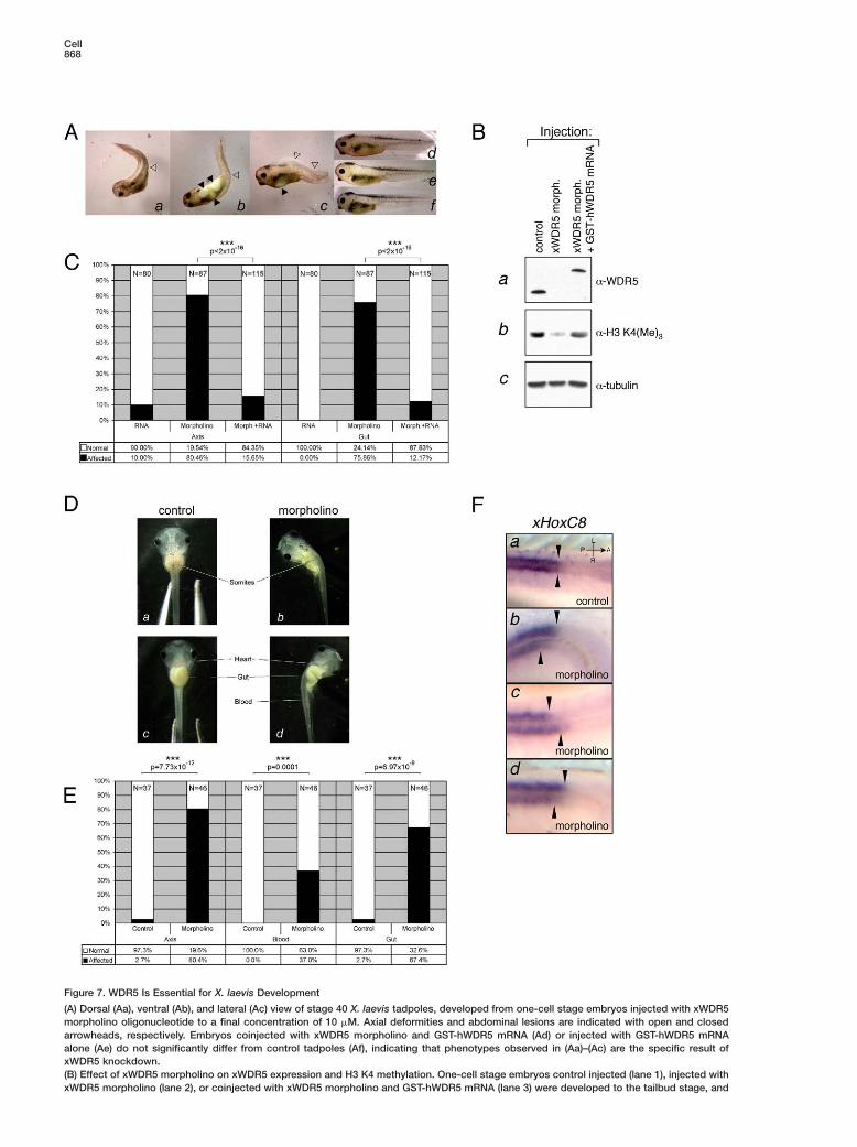
Figure 7. WDR5 Is Essential for X. laevis Development
(A) Dorsal (Aa), ventral (Ab), and lateral (Ac) view of stage 40 X. laevis tadpoles, developed from one-cell stage embryos injected with xWDR5morpholino oligonucleotide to a final concentration of 10 �M. Axial deformities and abdominal lesions are indicated with open and closedarrowheads, respectively. Embryos coinjected with xWDR5 morpholino and GST-hWDR5 mRNA (Ad) or injected with GST-hWDR5 mRNAalone (Ae) do not significantly differ from control tadpoles (Af), indicating that phenotypes observed in (Aa)–(Ac) are the specific result ofxWDR5 knockdown.(B) Effect of xWDR5 morpholino on xWDR5 expression and H3 K4 methylation. One-cell stage embryos control injected (lane 1), injected withxWDR5 morpholino (lane 2), or coinjected with xWDR5 morpholino and GST-hWDR5 mRNA (lane 3) were developed to the tailbud stage, and
WDR5 Associates with H3 Methylated at K4869
that WDR5 endows the methyltransferase complex withmethyltransferase complexes, participates both in
endoderm was excised to eliminate egg yolk. Embryos lacking endoderm were subsequently used for whole-cell extract preparation. Extractswere analyzed by immunoblotting with α-WDR5 (Ba), α-H3 K4(Me)3 (Bb), and α-tubulin (Bc) antibodies.(C) Quantitative analysis of axial deformities (“Axis”) and abdominal lesions (“Gut”) present in morpholino-injected embryos (“Morpholino”),embryos coinjected with morpholino and GST-hWDR5 mRNA (“Morph.+RNA”), or injected with GST-hWDR5 mRNA alone (“RNA”). Totalnumber of counted embryos is indicated at the top of each column. Differences between morpholino-injected and RNA + morpholino-coinjected populations are highly statistically significant (p < 2 × 10⁻¹⁶) as determined by test for equal proportions and indicated at the topof the plot. Coinjection of GST-hWDR5 mRNA with morpholino rescues the phenotypes essentially to the penetrance observed in controlsample (p = 0.35 for axial and p = 0.003 for the gut defects).(D) Feeding stage tadpoles derived from two-cell stage embryos were control injected (Da and Dc) or injected in one of the two blastomereswith 10 nl of 0.5 mM xWDR5 morpholino (Db and Dd). Morpholino-injected tadpoles exhibit axial deformation on the injected side, abnormaldistribution of blood cells (blood is absent from heart while forming a hemorrhage in the tail), and failure of gut patterning.(E) Quantitative analysis of the penetrance of phenotypes presented in (D). Number of occurrences of axial defects (“Axis”), blood distributionabnormalities (“Blood”), and gut coiling defects (“Gut”) were noted in mock injected (“Control”) and morpholino injected (“Morpholino”) andplotted as proportions in total population counted. Total number of counted embryos is indicated at the top of each column. Observeddifferences are highly statistically significant (p % 0.0001, test for equal proportions), and the p values are indicated at the top of the plot.(F) Whole-mount in situ detection of xHoxC8 transcript. Dorsal view of midtrunk region of stage 30 tadpoles derived from embryos controlinjected (Fa) or injected at two-cell stage in one of the blastomeres with xWDR5 morpholino (Fb–Fd) is shown. Purple stain indicates xHoxC8expression, which is most prominent in neural tube. Arrows indicate misalignment of the xHoxC8 expression domains between left and rightside of neural tube in morpholino-injected but not control embryos. All images are shown in the same orientation (see [Fa], upper right corner:A–P, anterior-posterior axis; L–R, left-right axis).
xWDR5 Knockdown Results in AbnormalExpression Pattern of xHoxC8Specific somitic, hematopoetic, and gut defects ob-served in xWDR5-depleted tadpoles point to the abnor-mal regulation of the Hox genes. For example, HoxC8depletion in X. laevis results in severe defects of abdo-minal structure that are reminiscent of the phenotypeobserved with the WDR5 knockdown (Ko and Chung,2003). Furthermore, loss of WDR5 in human cells leadsto decrease of HOXC8 expression.
To address whether xWDR5 knockdown leads to thederegulation of xHoxC8 expression, we used in situ hy-bridization to visualize xHoxC8 spatial expression pat-tern in tadpoles derived from two-cell stage embryosinjected with xWDR5 morpholino into one blastomere.As shown in Figure 7F, xWDR5 knockdown resulted inthe misalignment of xHoxC8 expression domain by atleast one somite length (compare control panel Figure7Fa with Figures 7Fb–7Fd). In addition, the injectedside frequently exhibited diminished levels of xHoxC8-specific staining. Together, these results demonstratethat WDR5 is important for proper maintenance andspatial control of Hox gene expression during devel-opment.
Discussion
There is growing recognition of the importance of his-tone methylation as an epigenetic regulatory mark thatmediates activation or repression of specific genomicloci or chromosomal domains. In particular, establish-ment and maintenance of heterochromatin have beenshown to involve methylation of H3 K9, which is read bythe effector protein HP1 (reviewed in Elgin and Grewal[2003]). HP1 itself, however, associates with H3 K9methyltransferase SUV39H1 (Aagaard et al., 1999) andis a critical determinant for H3 K9 methylation spread-ing (Hall et al., 2002). We propose that a similar para-digm may be extended for positive regulation of geneexpression, in that WDR5, a component of H3 K4
reading and writing the H3 K4 methylation mark (seeFigure 8).
WD40 Propeller: A Module for Associationwith the Histone TailWD40-repeat proteins are involved in very diverse cel-lular functions in which the WD40 propeller usually me-diates protein-protein interactions. They are also wellrepresented among chromatin-associated proteins. Forexample, EED, a WD40-repeat protein, is an essentialcomponent of the PRC2 complex. Two other WD40-repeat proteins, RbAp46 and RbAp48, are found to-gether in a variety of chromatin-associated complexes(for reviews, see Cao and Zhang [2004], Loyola and Al-mouzni [2004]). Molecular modeling of the WDR5 struc-ture reveals that it is predicted to fold into a seven-bladed propeller containing a centrally located cavity(see Figure S2). Determining where the H3 K4 methylpeptide binds (i.e., to the side, top, or internal cavity)will require future structural studies. Despite these un-certainties, our results point to a potentially novel modeof methyl-lysine histone peptide recognition that ap-pears to be distinct from chromodomain or chromodo-main-like modules.
WDR5 as an Effector ProteinWe demonstrated that WDR5 is important for bindingof the methyltransferase complex to the K4-dimethy-lated H3 tail and for global and gene-specific K4 tri-methylation. In contrast, WDR5 is not required for theassociation of methyltranferase complexes with chro-matin or for H3 K4 dimethylation. Thus, our data sup-port the model presented in Figure 8. We envision thatH3 K4 methyltransferase complex is recruited to chro-matin independently of WDR5 and that this event ismost likely mediated by site-specific transcriptional ac-tivators at target loci (step 1). Our data suggest that theK4 methylation pathway can be initiated independentlyof WDR5, as methyltransferase complex subunit(s) dis-tinct from WDR5 associates with unmodified H3 tailsubstrates. Once methylation is initiated, we propose

Cell870
Figure 8. Model for WDR5’s Role in Activation of Gene Expression
(1) H3 K4 methyltransferase complex is recruited to chromatin in a reaction dependent upon binding to unmodified H3 tails, resulting ininitiation of methylation. (2) Once methylation is initiated, WDR5 endows the H3 K4 methyltransferase complex with an ability to bind todimethylated substrates and is important for di- to trimethyl conversion at K4. (3) As the methyltransferase complex does not bind efficientlyto the trimethylated tail, we propose that it is then released from its substrate, resulting in the binding to the nearby nucleosome andpropagation of the methylation signal. Trimethylation may then lead to the recruitment of downstream effector proteins and activation oftranscription. WDR5’s role in H3 K4 monomethylation is not yet clear.
an ability to bind to dimethylated substrate and is im- mKportant for di- to trimethyl conversion (step 2). Such atmodel is consistent with the fact that dimethylated H3sK4 peptide can be further methylated by the WDR5-icontaining hSet1 and MLL1 complexes (Wysocka et al.,d2003; Dou et al., 2005) but not by recombinant MLL1tSET domain (Milne et al., 2002; Nakamura et al., 2002).gAs methyltransferase complex does not appear to bindvefficiently to the trimethylated tail, we suspect that theWcomplex is then released from its substrate, which canpresult in the binding to the nearby nucleosome andFpropagation of the methylation signal. Trimethylationdmay then lead to the recruitment of downstream effec-ptor proteins and activation of transcription by pathwaystthat are still largely unknown (step 3). This proposed
model is also consistent with the transactivation prop-ierties of the WDR5 protein, as trimethylation of H3 K4“is associated with promoter and 5# transcribed regionswof active genes (Santos-Rosa et al., 2002; Schneider eteal., 2004).asRole of WDR5 and H3 K4 Methylationmin Development and Differentiation
A WDR5 homolog, Swd3, exists in yeast S. cerevisiae,E
where it has been shown to be a component of theyeast Set1 complex (Roguev et al., 2001; Miller et al., P2001; Nagy et al., 2002). Swd3 is nonessential for viabil- N
city, but is essential for the H3 K4 methylation in yeastb(Miller et al., 2001; Nagy et al., 2002). In contrast, we5report here that WDR5 is essential both for viability andw
for H3 K4 methylation in vertebrates. Furthermore, D. 3melanogaster WDR5 homolog, called “will die slowly” c
4(wds) is encoded by an essential gene (Hollmann et al.,i2002). These observations suggest that H3 K4 methyla-rtion is essential in multicellular as opposed to unicellu-b
lar organisms. Steady-state levels of H3 K4 methylation lare considerably higher in bulk histone preparations b
cisolated from unicellular eukaryotes as compared to
ulticellular organisms, a result anticorrelated with H39 methylation (Briggs et al., 2001). These data hint at
he intriguing possibility that the epigenetic “defaulttate” in multicellular organims is set more to a silenc-ng mode. If correct, regulated gene activation, in partictated by activating marks such as H3 K4 methyla-
ion, may play a more important role in muticellular or-anisms, where complex developmental processes, in-olving cellular differentiation, are critical. In support,DR5 is important for maintenance of Hox gene ex-
ression in human cells and during Xenopus development.urthermore, mesodermal and endodermal patterningefects observed in the WDR5-depleted X. laevis tad-oles are consistent with differentiation defects arisinghrough abnormalities in homeotic gene expression.
In sum, our data provide evidence that WDR5 is anmportant regulator of gene expression by acting as asensor” protein that participates in both reading andriting the H3 K4 methyl mark. Perturbations in genexpression patterns caused by loss of WDR5 result inbnormal execution of developmental programs, under-coring crucial roles of the epigenetic signal of H3 K4ethylation in differentiation and development.
xperimental Procedures
ull-Down Assaysuclear extracts were prepared from 293 cells using Dignam proto-ol (Dignam et al., 1983), precleared with avidin beads, and incu-ated with peptide prebound to avidin beads for 3 hr at 4°C. About�g of peptide and 108 cells were used per pull-down. Beads wereashed eight times with buffer containing 20 mM HEPES (pH 7.9),00 mM KCl, 0.2% Triton X-100, 1 mM PMSF, and protease inhibitorocktail (Roche). A final wash was performed with buffer containingmM HEPES (pH 7.9), 10 mM NaCl, 1 mM PMSF, and protease
nhibitor cocktail (Roche). Bound proteins were eluted from theesin two times with 100 mM glycine (pH 2.8). Eluates were com-ined, neutralized using 1/10 volume of 1 M Tris (pH 8), and ana-
yzed by SDS PAGE. For peptide competition experiments, proteinsound to the resin were incubated with 20-fold molar excess of theompeting peptide for 20 min at RT.

WDR5 Associates with H3 Methylated at K4871
Chromatin Fractionation and Isolationof Mononucleosomal FractionThe biochemical fractionation procedure and microccocal nucleasetreatment were performed as described (Wysocka et al., 2001;Mendez and Stillman, 2000). Samples that were determined to con-tain only mononuclesomal DNA were pooled and used for thecoimmunoprecipitation assays.
CoimmunoprecipitationMononucleosomal fractions were prepared from a HeLa cell linestably expressing Flag/HA-tagged full-length WDR5 (Dou et al.,2005) and as a control from HeLa cells lacking ectopic WDR5. Mo-nonucleosomal fractions were then incubated with FLAG M2 agar-ose beads (Sigma). Immunoprecipitates were washed and elutedfrom beads with Flag peptide (Sigma) at concentration of 0.5mg/ml.
Antibodiesα-WDR5 and α-Ash2 affinity-purified rabbit polyclonal antibodieswere kind gifts of W. Herr; α-MLL1 rabbit polyclonal antibody wasdescribed in Dou et al. (2005); other antibodies were obtained com-mercially: α-RbBP5 and α-MLL2 (Bethyl Laboratories); α-H3 K4mono-, di-, and trimethyl (Upstate Biotechnology and Abcam); α-H3K9 trimethyl, α-H3 K27 trimethyl, α-H3 1-20 unmodified, and α-HP1(Upstate Biotechnology); and α-tubulin (Sigma).
Luciferase AssaysA 293T cell line stably expressing luciferase reporter under the con-trol of tandem of five GAL4 UAS sites was transfected using Lipo-fectamine 2000 (Invitrogen) with indicated amounts of pCMX-GAL4DNA binding domain (DBD) or pCMX-GAL4 full-length WDR5 ex-pression vectors or mock transfected. In each case, pUC119 DNAwas additionally added to make up for a total of 4 �g of transfectedDNA. Cells were normalized by counting and then lysed, and lucif-erase assay kit (Promega) was used to determine relative levels ofthe luciferase gene product.
WDR5 Knockdown in Human CellsHuman WDR5 siRNA SMART pool consisting of four siRNA du-plexes was purchased from Dharmacon. 293 cells were seeded on6-well plates and transfected with siRNA SMART pool or each offour individual WDR5 SMART duplexes by using Lipofectamine2000 (Invitrogen) according to the manufacturer’s instructions. Acorresponding amount of noncoding siRNA duplexes (Dharmacon)was used for control transfections. One additional round of trans-fection was performed using identical conditions 24 hr after theinitial transfection. Cells were harvested 72 hr after second trans-fection and were either lysed in SDS Laemmli buffer, subjected toRNA preparation, or fixed with formaldehyde for ChIP analysis.
Chromatin Immunoprecipitation and Q-PCR DetectionChromatin Immunoprecipitations (ChIPs) were performed using theChromatin Immunoprecipitation Assay Kit (Upstate Biotechnology)and the following antibodies: histone H3 K4(Me)2 (ab7766-50) andH3 K4(Me)3 (ab8580-50) both from Abcam, and H3 K4(Me)3 (07-473)from Upstate Biotechnology. Anti-MLLC is described in Dou et al.(2005). Real-time PCR quantification of ChIP was performed in trip-licate using Taqman probes and a Stratagene Mx3000p. ChIP wasquantified relative to inputs using the method described in Franket al. (2001).
HOX Gene ExpressionTotal RNA was isolated using RNeasy columns (Qiagen), treatedwith DNase, and reverse transcribed (SuperScript, Invitrogen).Real-time PCR quantification of Hox gene expression was per-formed in triplicate using Taqman probes and a StratageneMx3000p instrument using a standard curve and the relative quan-titation method as described in ABI User Bulletin 2. βACTIN andGAPDH probes and primers used were from Applied BiosystemsInc.
In Vitro TranscriptionSynthetic open reading frame encoding human GST-WDR5 fusionprotein was cloned into pCS2+ vector, and 5#-capped mRNAs weretranscribed in vitro from the SP6 promoter of the HpaI-linearizedtemplate using the mMessageMachine kit (Ambion).
Morpholino Design and xWDR5 KnockdownBLAST analysis of database EST sequences provides evidencethat X. laevis WDR5 is encoded by two distinct yet closely relatedgenes. Morpholino oligonucleotide of the sequence 5#-CATGGTGTCAGCACTAGAATGGTGC-3# targeting ATG region of both putativeX. laevis WDR5 mRNAs was synthesized by Gene Tools, LLC, andhandled according to manufacturer’s specifications. One or twocell stage embryos were injected with 20 and 10 nl, respectively, of1 mM morpholino solution or 1 mM morpholino with 50 ng/�l syn-thetic human GST-WDR5 mRNA and grown at 18°C. Embryos weretransferred into 0.1 × MMR + 50 �g/ml gentamycin prior to gastru-lation. Number of clear phenotypical abnormalities was noted inobserved population of injected embryos. Statistical significanceof the differences in phenotype frequency between experimentalsamples and control was determined by test for equal proportions(function prop.test in R statistical suite [R Development CoreTeam, 2004]).
Immunostaining of Xenopus EmbryosStage 11 Xenopus embryos were fixed, and immunostaining wasperformed according to Brivanlou and Harland (1989) protocol.Prior to observation, embryos were brought to RT and cleared with2:1 benzyl benzoate:benzyl alcohol solution, and in situ fluores-cence was visualized with Zeiss Pascal LSM confocal microscope.
In Situ HybridizationcDNA fragment corresponding to the xHoxC8 open reading framewas amplified by PCR from X. laevis stage 30 cDNA and clonedinto pCS2+ vector. Digitoxin-labeled riboprobe was synthesized byin vitro transcription with T7 RNA polymerase using DIG labelingmix from Roche. Staged embryos were fixed, and in situ hybridiza-tion was performed as described in Sive et al. (2000). Probe wasdetected with AP-conjugated anti-DIG antibodies followed by colorreaction with BM Purple reagent (Roche).
Supplemental DataSupplemental Data include two figures and can be found with thisarticle online at http://www.cell.com/cgi/content/full/121/6/859/DC1/.
Acknowledgments
We thank W.K. Wang and D. Patel for molecular modeling of WDR5;W. Herr for α-WDR5 and α-Ash2 antibodies; M. Guenther for adviceon pull-down assays; F. Spagnoli and A. Vonica for advice on Xeno-pus techniques; W. Fischle for recombinant HP1β protein; and R.Cook, C. McDonald, and RU Proteomics Resource Center for pep-tide synthesis. We are grateful to members of the Allis and Brivan-lou labs for helpful discussions and to W. Herr, A. Vonica, S. Hake,E. Bernstein, E. Duncan, and S. Taverna for critical readings of themanuscript. J.W. is a Damon Runyon CRF fellow, T.S. is a CharlesH. Revson fellow, and Y.D. is an Irvington Institute for Immunologyfellow. This work was supported by NIH grants: MERIT Award (GM53512) to C.D.A., R01 GM066977 to A.H.B., and NCRR RR015804and RR001614 to A.L.B.
Received: January 26, 2005Revised: March 14, 2005Accepted: March 31, 2005Published: June 16, 2005
References
Aagaard, L., Laible, G., Selenko, P., Schmid, M., Dorn, R., Schotta,G., Kuhfittig, S., Wolf, A., Lebersorger, A., Singh, P.B., et al. (1999).Functional mammalian homologues of the Drosophila PEV-modifier
Cell872
Su(var)3–9 encode centromere-associated proteins which complex NAwith the heterochromatin component M31. EMBO J. 18, 1923–
1938. i9Bannister, A.J., Zegerman, P., Partridge, J.F., Miska, E.A., Thomas,NJ.O., Allshire, R.C., and Kouzarides, T. (2001). Selective recognitionsof methylated lysine 9 on histone H3 by the HP1 chromo domain.ANature 410, 120–124.pBriggs, S.D., Bryk, M., Strahl, B.D., Cheung, W.L., Davie, J.K., Dent,1S.Y., Winston, F., and Allis, C.D. (2001). Histone H3 lysine 4 methyla-Ption is mediated by Set1 and required for cell growth and rDNAPsilencing in Saccharomyces cerevisiae. Genes Dev. 15, 3286–3295.SBrivanlou, A.H., and Harland, R.M. (1989). Expression of an en-Rgrailed-related protein is induced in the anterior neural ectodermfof early Xenopus embryos. Development 106, 611–617.(Cao, R., and Zhang, Y. (2004). The functions of E(Z)/EZH2-mediatedRmethylation of lysine 27 in histone H3. Curr. Opin. Genet. Dev. 14,A155–164.iDignam, J.D., Lebovitz, R.M., and Roeder, R.G. (1983). Accuratettranscription initiation by RNA polymerase II in a soluble extractSfrom isolated mammalian nuclei. Nucleic Acids Res. 11, 1475–1489.sDou, Y., Milne, T.A., Tackett, A.J., Smith, E.R., Fukuda, A., Wysocka,TJ., Allis, C.D., Chait, B.T., Hess, J.L., and Roeder, R.G. (2005). Physi-tcal association and coordinate function of the H3 K4 methyl-Stransferase MLL1 and the H4 K16 acetyltransferase MOF. Cell 121,Rthis issue, 873–885.yElgin, S.C., and Grewal, S.I. (2003). Heterochromatin: silence isSgolden. Curr. Biol. 13, R895–R898.mFrancis, N.J., and Kingston, R.E. (2001). Mechanisms of transcrip-6tional memory. Nat. Rev. Mol. Cell Biol. 2, 409–421.SFrank, S.R., Schroeder, M., Fernandez, P., Taubert, S., and Amati, B.m(2001). Binding of c-Myc to chromatin mediates mitogen-inducedNacetylation of histone H4 and gene activation. Genes Dev. 15,W2069–2082.mGoo, Y.H., Sohn, Y.C., Kim, D.H., Kim, S.W., Kang, M.J., Jung, D.J.,tKwak, E., Barlev, N.A., Berger, S.L., Chow, V.T., et al. (2003). Activa-Wting signal cointegrator 2 belongs to a novel steady-state complexWthat contains a subset of trithorax group proteins. Mol. Cell. Biol.h23, 140–149.b
Hall, I.M., Shankaranarayana, G.D., Noma, K., Ayoub, N., Cohen,YA., and Grewal, S.I. (2002). Establishment and maintenance of atheterochromatin domain. Science 297, 2232–2237.o
Hollmann, M., Simmerl, E., Schafer, U., and Schafer, M.A. (2002).c
The essential Drosophila melanogaster gene wds (will die slowly)B
codes for a WD-repeat protein with seven repeats. Mol. Genet. Ge-nomics 268, 425–433.
Hughes, C.M., Rozenblatt-Rosen, O., Milne, T.A., Copeland, T.D.,Levine, S.S., Lee, J.C., Hayes, D.N., Shanmugam, K.S., Bhat-tacharjee, A., Biondi, C.A., et al. (2004). Menin associates with atrithorax family histone methyltransferase complex and with thehoxc8 locus. Mol. Cell 13, 587–597.
Ko, C., and Chung, H.M. (2003). Xenopus hoxc8 during early devel-opment. Biochem. Biophys. Res. Commun. 300, 9–15.
Lachner, M., O’Carroll, D., Rea, S., Mechtler, K., and Jenuwein, T.(2001). Methylation of histone H3 lysine 9 creates a binding site forHP1 proteins. Nature 410, 116–120.
Loyola, A., and Almouzni, G. (2004). Histone chaperones, a sup-porting role in the limelight. Biochim. Biophys. Acta 1677, 3–11.
Mendez, J., and Stillman, B. (2000). Chromatin association of hu-man origin recognition complex, cdc6, and minichromosome main-tenance proteins during the cell cycle: assembly of prereplicationcomplexes in late mitosis. Mol. Cell. Biol. 20, 8602–8612.
Miller, T., Krogan, N.J., Dover, J., Erdjument-Bromage, H., Tempst,P., Johnston, M., Greenblatt, J.F., and Shilatifard, A. (2001). COM-PASS: a complex of proteins associated with a trithorax-relatedSET domain protein. Proc. Natl. Acad. Sci. USA 98, 12902–12907.
Milne, T.A., Briggs, S.D., Brock, H.W., Martin, M.E., Gibbs, D., Allis,C.D., and Hess, J.L. (2002). MLL targets SET domain methyl-transferase activity to Hox gene promoters. Mol. Cell 10, 1107–1117.
agy, P.L., Griesenbeck, J., Kornberg, R.D., and Cleary, M.L. (2002).trithorax-group complex purified from Saccharomyces cerevisiae
s required for methylation of histone H3. Proc. Natl. Acad. Sci. USA9, 90–94.
akamura, T., Mori, T., Tada, S., Krajewski, W., Rozovskaia, T., Was-ell, R., Dubois, G., Mazo, A., Croce, C.M., and Canaani, E. (2002).LL-1 is a histone methyltransferase that assembles a supercom-lex of proteins involved in transcriptional regulation. Mol. Cell 10,119–1128.
ray-Grant, M.G., Daniel, J.A., Schieltz, D., Yates, J.R., and Grant,.A. (2005). Chd1 chromodomain links histone H3 methylation withAGA- and SLIK-dependent acetylation. Nature 433, 434–438.
Development Core Team (2004). R: A Language and Environmentor Statistical Computing. R Foundation for Statistical Computinghttp://www.R-project.org).
oguev, A., Schaft, D., Shevchenko, A., Pijnappel, W.W., Wilm, M.,asland, R., and Stewart, A.F. (2001). The Saccharomyces cerevis-
ae Set1 complex includes an Ash2 homologue and methylates his-one 3 lysine 4. EMBO J. 20, 7137–7148.
antos-Rosa, H., Schneider, R., Bannister, A.J., Sherriff, J., Bern-tein, B.E., Emre, N.C.T., Schreiber, S.L., Mellor, J., and Kouzarides,. (2002). Active genes are tri-methylated at K4 of histone H3. Na-ure 419, 407–411.
chneider, R., Bannister, A.J., Myers, F.A., Thorne, A.W., Crane-obinson, C., and Kouzarides, T. (2004). Histone H3 lysine 4 meth-lation patterns in higher eukaryotic genes. Nat. Cell Biol. 6, 73–77.
ims, R.J., 3rd, Nishioka, K., and Reinberg, D. (2003). Histone lysineethylation: a signature for chromatin function. Trends Genet. 19,
29–639.
ive, H.L., Grainger, R.M., and Harland, R.M. (2000). Early Develop-ent of Xenopus Laevis: A Laboratory Manual (Cold Spring Harbor,Y: Cold Spring Harbor Laboratory Press).
ysocka, J., Reilly, P.T., and Herr, W. (2001). Loss of HCF-1 chro-atin association precedes temperature-induced growth arrest of
sBN67 cells. Mol. Cell. Biol. 21, 3820–3829.
ysocka, J., Myers, M.P., Laherty, C.D., Eisenman, R.N., and Herr,. (2003). Human Sin3 deacetylase and trithorax-related Set1/Ash2
istone H3–K4 methyltransferase are tethered together selectivelyy the cell-proliferation factor HCF-1. Genes Dev. 17, 896–911.
okoyama, A., Wang, Z., Wysocka, J., Sanyal, M., Aufiero, D.J., Ki-abayashi, I., Herr, W., and Cleary, M.L. (2004). Leukemia proto-ncoprotein MLL forms a SET1-like histone methyltransferaseomplex with menin to regulate Hox gene expression. Mol. Cell.iol. 24, 5639–5649.
Copyright © 2022 FDOKUMEN