The Neuroanatomy of Verbal Working Memory in Schizophrenia: A Voxel-Based Morphometry Study
Upload
independentCategory
view
3download
0
NeuroImage: Clinical 5 (2014) 408–419
Contents lists available at ScienceDirect
NeuroImage: Clinical
j ourna l homepage: www.e lsev ie r .com/ locate /yn ic l
Voxel-wise resting-state MEG source magnitude imaging study revealsneurocircuitry abnormality in active-duty service members and veteranswith PTSD
Ming-Xiong Huanga,b,*, Kate A. Yurgila,c, Ashley Robba, Annemarie Angelesa, Mithun Diwakarb,Victoria B. Risbrougha,c,d, Sharon L. Nicholse, Robert McLayf, Rebecca J. Theilmannb, Tao Songb,Charles W. Huangg, Roland R. Leea,b, Dewleen G. Bakera,c,d
aRadiology, Research, and Psychiatry Services, VA San Diego Healthcare System, San Diego, CA, USAbDepartment of Radiology, University of California San Diego, San Diego, CA, USAcVA Center of Excellence for Stress and Mental Health, San Diego, CA, USAdDepartment of Psychiatry, University of California San Diego, San Diego, CA, USAeDepartment of Neurosciences, University of California San Diego, San Diego, CA, USAfNaval Medical Center San Diego, San Diego, CA, USAgDepartment of Bioengineering, University of California San Diego, San Diego, CA, USA
* Corresponding author at: Radiology Imaging LaboraSan Diego, 3510 Dunhill Street, San Diego, CA 92121, USA
E-mail address: [email protected] (M.-X. Huang).
http://dx.doi.org/10.1016/j.nicl.2014.08.0042213-1582/Published by Elsevier Inc. This is an open acce
a b s t r a c t
a r t i c l e i n f oArticle history:Received 28 May 2014Received in revised form 25 July 2014Accepted 2 August 2014Available online 7 August 2014
Keywords:MEGPost-traumatic stress disorderAmygdalaVentromedial prefrontal cortexOrbitofrontal cortexPrecuneous
Post-traumatic stress disorder (PTSD) is a leading cause of sustained impairment, distress, and poor quality of lifein military personnel, veterans, and civilians. Indirect functional neuroimaging studies using PET or fMRI withfear-related stimuli support a PTSD neurocircuitry model that includes amygdala, hippocampus, and ventrome-dial prefrontal cortex (vmPFC). However, it is not clear if this model can fully account for PTSD abnormalities de-tected directly by electromagnetic-based source imaging techniques in resting-state. The present studyexamined resting-state magnetoencephalography (MEG) signals in 25 active-duty service members and vet-erans with PTSD and 30 healthy volunteers. In contrast to the healthy volunteers, individuals with PTSD showed:1) hyperactivity from amygdala, hippocampus, posterolateral orbitofrontal cortex (OFC), dorsomedial prefrontalcortex (dmPFC), and insular cortex in high-frequency (i.e., beta, gamma, and high-gamma) bands; 2)hypoactivity from vmPFC, Frontal Pole (FP), and dorsolateral prefrontal cortex (dlPFC) in high-frequencybands; 3) extensive hypoactivity from dlPFC, FP, anterior temporal lobes, precuneous cortex, and sensorimotorcortex in alpha and low-frequency bands; and 4) in individuals with PTSD, MEG activity in the left amygdalaand posterolateral OFC correlated positively with PTSD symptom scores, whereas MEG activity in vmPFC andprecuneous correlated negativelywith symptom score. The present study showed thatMEG source imaging tech-nique revealed new abnormalities in the resting-state electromagnetic signals from the PTSD neurocircuitry. Par-ticularly, posterolateral OFC and precuneous may play important roles in the PTSD neurocircuitry model.
Published by Elsevier Inc. This is an open access article under the CC BY-NC-ND license(http://creativecommons.org/licenses/by-nc-nd/3.0/).
1. Introduction
Individuals exposed to a traumatic event may develop post-traumaticstress disorder (PTSD) with debilitating post-traumatic stress symptoms,including intrusive memories, avoidance behavior, emotional numbing,and hyperarousal (American Psychiatric Association, 2004). PTSD is amajor health concern that affects approximately 7.7% of Americans(Kessler et al., 1995, 2005) and is particularly prevalent among mil-itary service members who have served in combat (Dohrenwend
tory, University of California at.
ss article under the CC BY-NC-ND lic
et al., 2006; Magruder and Yeager, 2009). The recent conflicts inIraq and Afghanistan have been no exception, with combat veteransreturning with elevated rates of PTSD (Hoge et al., 2004; Smith et al.,2008; Tanielian and Jaycox, 2008).
In light of these findings, much effort has been focused on determin-ing symptom etiology and the associated neural mechanisms of PTSD.The development of neurocircuitry models of PTSD has relied stronglyon findings from pre-clinical studies of fear conditioning. Evidencefrom lesion studies, pharmacological manipulations, and electrophysi-ology in animals and humans suggest that interactions between theamygdala, ventromedial prefrontal cortex (vmPFC), and hippocampuscontrol different aspects of fear processing (Hartley and Phelps, 2010;Rosen and Lilienfeld, 2008). The amygdala is involved in acquisition of
ense (http://creativecommons.org/licenses/by-nc-nd/3.0/).

409M.-X. Huang et al. / NeuroImage: Clinical 5 (2014) 408–419
fear conditioning and extinction learning, whereas the vmPFC is thoughttomediatememory storage and retrieval during extinction learning. Hip-pocampal connections to the amygdala and vmPFCmay support process-ing contextual information of threat-related stimuli.
Amygdala, vmPFC, and hippocampal regions implicated in pre-clinical fear processing are thought to be dysfunctional in PTSD (Rauchet al., 1998, 2006). Functional neuroimaging findings using positronemission topography (PET) and functional magnetic resonance imaging(fMRI) suggest that individuals with PTSD exhibit hyperresponsiveamygdala activity to trauma or fear-related stimuli (Shin and Liberzon,2010), during emotionally neutral tasks (Bryant et al., 2005; Shinet al., 2004b), and even at rest (Chung et al., 2006; Semple et al.,2000). A hyperresponsive amygdala contributes to the exaggeratedfear response characteristic of PTSD (Anderson et al., 2003). Conversely,PTSD has been associated with a hyporesponsive vmPFC (Hughes andShin, 2011). A hyporesponsive PFC, as well as reduced connectivity tothe amygdala (Jin et al., 2013; Shin et al., 2004a) may indicate insuffi-cient inhibitory control over exaggerated fear responses. Lastly, abnor-mal hippocampal function (Corcoran and Maren, 2001) and reducedconnectivity to the amygdala (Dolcos et al., 2004; McGaugh, 2004)may be associated impairments in contextual memory processing andthe ability to inhibit intrusive memories (Shin et al., 2004a), althoughfindings have been mixed (Hughes and Shin, 2011). A recent resting-state fMRI study showed increased activity in amygdala and reducedspontaneous neural activity in the dlPFC, but no abnormal decrease ofresting-state fMRI activity in the vmPFC (Yan et al., 2013).
Neuroimaging studies using PET and fMRI have contributed greatlyto understanding PTSD neurocircuitry in humans; however, these tech-niques measure metabolic and hemodynamic changes which reflectneuronal activity indirectly (Logothetis, 2003). In addition, PET andfMRI techniques have limited temporal resolution (minutes to seconds)and consequently limited coverage and resolution in the frequency do-main. Since neurons communicate to each other via exchanging electriccurrent signals, direct electrophysiological measures are required tostudy neurophysiological processes that are associated with these he-modynamic signals (Scholvinck et al., 2013). PET and fMRI studies alsohave implicated different neural pathways that may be hyporesponsivein PTSD; thus, there is some remaining discrepancy whether PTSD is as-sociatedwith reduced activity in the vmPFCor dlPFC pathways. Further-more, although the orbitofrontal cortex (OFC) is usually considered tobe part of the extended limbic system, the contribution of OFC to PTSDhas not been fully elucidated.
Electromagneticmeasures such asmagnetoencephalography (MEG)provide direct measurements of neuronal activity with millisecondtemporal resolution. Using a single dipolemodel, Kolassa and colleaguesreported elevated production of focally generated slowwaves (1–4 Hz)in PTSD, particularly in left temporal brain regions, with peak activitiesin the region of the insula. Using aMEG sensor-space synchronous neu-ral interactions analysis, Georgopoulos, Engdahl, and their colleaguescorrectly classified individuals with PTSD and healthy control subjectswith N90% overall accuracy of classification (Georgopoulos et al., 2010).They also found differences in MEG communication measures betweentemporal and parietal and/or parieto-occipital right hemispheric areaswith other brain areas in PTSD (Engdahl et al., 2010). However in sensorspace, it is difficult to determine whether the structures identified by PETand fMRI in PTSD neurocircuitry generate abnormal electromagnetic ac-tivity. Namely, whether electromagnetic-based source imaging tech-niques will lead to similar or different findings from those obtained inPET and fMRI in PTSD neurocircuitry has largely been unexplored.
In the current study, we examined neural activity associated withPTSD using resting-state MEG.MEG is a non-invasive functional imagingtechnique that directly measures magnetic signals generated by neuro-nal current in gray matter with high temporal resolution (b1 ms) andspatial localization accuracy (2–3 mm at cortical level) (Leahy et al.,1998). MEG3s high temporal resolution directly translates into a widerange of coverage for the neuronal magnetic signals in the frequency
domain, which is usually divided into different frequency bands. MEG3sinsensitivity to the electric conductivity profile of the head tissuemakes it a better technique than electroencephalography (EEG) in lo-calizing neuronal sources. Our newly developed high-resolution MEGsource imaging method called Fast-VESTAL allowed us to performvoxel-wise whole-brain source imaging of human brain rhythms inhealthy volunteers (Huang et al., 2014a), and makes MEG source imag-ing a good candidate for localizing abnormal electromagnetic signals indisorders such as PTSD. The primary goal for this study was to examineif the existing PTSD neurocircuitry model including the amygdala,vmPFC, and hippocampus can account for abnormalities detected di-rectly by electromagnetic-based source imaging techniques in resting-state. To achieve this goal, weused high-resolutionMEG source imagingtechnique for direct examination of neuronal activity in PTSD, especiallyin the areas that we think to be abnormal, i.e. amygdala, vmPFC, OFC,hippocampus, dlPFC, dmPFC including dorsal anterior cingulate cortex(dACC), insular cortex, and precuneous. In addition, using MEG, weare able to explore potential MEG abnormalities in different frequencybands which are associated with different neuronal mechanisms (seeDiscussion), and compare MEG findings with previously published re-sults from other functional imaging techniques that have been used tostudy PTSD.
2. Materials and methods
2.1. Research participants
Twenty-five participants (24 males, 1 female; mean [SD] age: 31.0[5.5]) with PTSD took part in this study. Among these participants, 10were active-duty Marines and Sailors from Camp Pendleton and NavalMedical Center in San Diego, and 15were adult outpatient OEF/OIF Vet-erans recruited from VA San Diego Healthcare System. Mean [SD] yearsof education for the participants with PTSD were 13.2 [1.4]. All partici-pants gave written informed consent for study procedures, whichwere reviewed and approved by institutional review boards of the VASan Diego Healthcare System and Naval Health Research Center at SanDiego. The informed consent followed the ethical guidelines of the Dec-larations of Helsinki (1975) and additional research requirements foractive-duty military personnel and veterans.
Symptoms of PTSD were assessed using the Clinician AdministeredPTSD Scale (CAPS) (Blake et al., 1995) or the Post-traumatic Stress Dis-order Checklist (PCL) (Weathers et al., 1999) in accordance with thecriteria from the Diagnostic and Statistical Manual of Mental DisordersIV-TR (American Psychiatric Association, 2000). A total of 18 partici-pants met the criteria for PTSD and 7 met the criteria for partial PTSD.Participants who completed the CAPS met the criteria for PTSD (n =14) if they reported at least 1 re-experiencing symptom, 3 avoidancesymptoms, and 2 hyperarousal symptoms; patients met the criteriafor partial PTSD (n = 5) if they reported at least 1 re-experiencingsymptom and either 3 avoidance symptoms or 2 hyperarousal symp-toms (Blanchard et al., 1995). Symptoms must have occurred at leastonce within the past month (frequency≥ 1) and have caused a moder-ate amount of distress (intensity≥ 2) (Weathers et al., 1999, 2001). Par-ticipants who completed the PCL questionnaire and had a minimumtotal score of 50met the criteria for PTSD (n=3), and thosewith scoresfrom39 to 49met the criteria for partial PTSD (n=2) (Hoge et al., 2008;Iversen et al., 2008; Renshaw, 2011; Weathers et al., 1993). Study par-ticipants with partial PTSD and PTSD were analyzed together (n = 25)to maintain statistical power and to examine broad group differencesin PTSD neurocircuitry. The PTSD patients were not on medications atthe time of theMEG exam. All had discontinued any psychotropic med-ications prior to the scan, and at least at 5-day wash-out.
We recruited thirty healthy volunteers (29 male, 1 female; mean[SD] age: 29.8 [6.4]) with no history of neurological or psychiatric disor-ders assessed by Structured Clinical Interview for DSM-IV. Among thesehealthy volunteers, 12were active-dutymilitary personnel and 18were
410 M.-X. Huang et al. / NeuroImage: Clinical 5 (2014) 408–419
civilians. Mean [SD] years of education were 13.4 [1.7]. There were nostatistically significant differences in age or education between thehealthy volunteer and PTSD groups.
Exclusion criteria for study participation were as follows: 1) otherneurological, developmental or psychiatric disorders (e.g., braintumor, stroke, epilepsy, Alzheimer3s disease, or schizophrenia, bipolardisorder, history of learning disability, or lesions visible in structuralMRI); 2) substance or alcohol abuse according to DSM-IV criteria withinthe 6months prior to the study; 3) history ofmetabolic or other diseasesknown to affect the central nervous system (Dikmen et al., 1995); and 4)extensive metal dental hardware (e.g., braces and large metal dentures;fillings are permitted) or other metal objects in the head, neck, or faceareas that cause non-removable MEG artifacts.
2.2. MEG data acquisition and signal pre-processing to remove artifacts
Resting-state MEG data (spontaneous recording for detecting MEGslow-wave signals) were collected at the UCSD MEG Center using theVectorView™ whole-head MEG system (Elekta-Neuromag, Helsinki,Finland) with 306 MEG channels. Participants sat inside a multi-layermagnetically-shielded room (IMEDCO-AG) (Cohen et al., 2002). Precau-tions were taken to ensure head stability; foam wedges were insertedbetween the participant3s head and the inside of the unit, and a Velcrostrap was placed under the participant3s chin and anchored in superiorand posterior axes. Headmovement across different sessionswas about2–3 mm. MEG recording was divided into two 5-minute blocks witheyes closed, alternating with two 5-minute blocks with eyes open. Inthe eyes-closed condition, the participant was instructed to keep his/her eyes closed and empty his/her mind. In the eyes-open condition,the participant was instructed to fix his/her eyes on a fixation pointand empty his/her mind. The order of blocks was counter-balancedbetween participants. Data were sampled at 1000 Hz and were runthrough a high-pass filter with a 0.1 Hz cut-off, and a low-pass filterwith a 300 Hz cut-off. The filter associated with MEG data acquisitionis a first-order time-domain filter with 3 dB around the cut-off points.Eye blinks, eye movements, and heart signals were monitored. Sincethe MEG eyes-open data were contaminated with eye-blinks in manyparticipants, we focused on analyzing the eyes-closed data in the presentstudy.
Substantial efforts were taken to help ensure that participants werealert during the MEG recordings. Participants were scheduled early inthe day to avoid fatigue from performing daily activities. Prior to all ofthe study sessions, participants completed a questionnaire about thenumber of hours they slept the previous night, how rested they felt,and if there was any reason that they might not be attentive and per-form to the best of their abilities (due to headache, pain, etc.). Sessionsalternated between eyes-closed and eyes-open conditions, and eyeblinking andmovementweremonitored. DuringMEG recording, partic-ipants were viewed on camera while technicians also monitored alphaband oscillations, which are consistently associated with tonic alertness(Oken et al., 2006).
Eyes-closed MEG data were first run through MaxFilter, also knownas signal space separation (Song et al., 2008; Taulu et al., 2004a,b) to re-move external sources of interference (e.g., magnetic artifacts due tometal objects, strong cardiac signals, environment noises), and to co-register the MEG data by removing the small head movements acrossthe two 5-minute eyes-closed sessions. Next, residual artifacts due toeye movements and residual cardiac signals were removed using Inde-pendent Component Analysis using our customized version of ICALABsoftware (www.bsp.brain.riken.jp/ICALAB/).
2.3. Structural MRI, MEG-MRI registration, BEM forward calculation
Structural MRI of the participant3s head was collected using aGeneral Electric 1.5 T Excite MRI scanner. The acquisition containsa standard high-resolution anatomical volume with a resolution of
0.94 × 0.94 × 1.2 mm3 using a T1-weighted 3D-IR-FSPGR pulse se-quence. To co-register the MEG with MRI coordinate systems, threeanatomical landmarks (i.e., left and right pre-auricular points, andnasion) were measured for each participant using the Probe PositionIdentification system (Polhemus, USA). By using MRILAB (Elekta/Neuromag) to identify the same three points on the participant3sMR images, a transformation matrix involving both rotation andtranslation between theMEG andMR coordinate systemswas generated.To increase the reliability of the MEG–MR co-registration, approximately80 points on the scalp were digitized with the Polhemus system, in addi-tion to the three landmarks, and those points were co-registered onto thescalp surface of the MR images. The T1-weighted images were also usedto extract the brain volumeand innermost skull surface (SEGLAB softwaredeveloped by Elekta/Neuromag). Realistic Boundary Element Method(BEM) head model was used for MEG forward calculation (Huang et al.,2007;Mosher et al., 1999). The BEMmeshwas constructed by tessellatingthe inner skull surface from the T1-weighted MRI into ~6000 triangularelements with ~5 mm size. A cubic source grid with 5 mm size wasused for calculating the MEG gain (i.e., lead-field) matrix, which leads toa grid with ~10,000 nodes covering the whole brain. Other conventionalMRI sequences typical for identifying structural lesions were alsoperformed: 1) Axial T2*-weighted; 2) Axial fast spin-echo T2-weighted;and 3) Axial FLAIR. These conventional MRIs were carefully reviewed bya Board-certified neuroradiologist (R.R. Lee) to determine if the partici-pant had visible lesions on MRI. Subjects with lesions visible in MRIwere excluded from the study (see exclusion criteria).
2.4. MEG slow-wave source magnitude imaging using fast-VESTAL
The voxel-wise MEG source magnitude images were obtained usingour recent high-resolution Fast-VESTAL MEG source imaging method(Huang et al., 2014a). This approach requires the sensor waveform co-variance matrix. Here, the second 5-minute resting-state MEG sensor-waveform dataset was registered to the first 5-minute resting-statedataset using MaxFilter. The artifact-free, eyes-closed, resting-stateMEG sensor-waveform datasets were divided into 2.5 s epochs. Thedata in each epoch were first DC-corrected and then run throughband-pass filters for the following frequency bands: alpha band(8–12 Hz), beta band (15–30 Hz), gamma band (30–80 Hz), high-gamma band (80–150 Hz), and low-frequency band (1–7 Hz) thatcombined delta (1–4 Hz) and theta bands (4–7 Hz). Notch filters at60 Hz and 120 Hz were applied to remove the power line signals andtheir second harmonics. Frequency-domain band-pass filter with zerophase-shift via discrete Fourier transform was used. At each end of theband-pass filter, the transition of the Hanning window in the filterwas selected to be at 10% of the associated cut-off frequency.
Waveforms from all 306 sensors including 204 planar-gradiometersand 102 magnetometers were used in the analysis. For each frequencyband, sensor-waveform covariancematrices were calculated for individ-ual epochs after the band-pass filtering, then, the final sensor-waveformcovariance matrix was obtained by averaging the covariance matricesacross individual epochs for the 10-minute resting-state data. Usingsuch a covariance matrix, MEG slow-wave source magnitude imagesthat cover the whole brain were obtained for each participant followingthe Fast-VESTAL procedure (Huang et al., 2014a) for a given frequencyband.
The brain volume is pre-divided into a grid of dipoles with P nodes.Let R be the M × M sensor-waveform MEG covariance matrix whereM is the number of MEG sensors for a given frequency band (e.g., betaband) and time-window (e.g., length of an epoch); and G be theM × 2P gain (lead-field) matrix calculated fromMEG forward modelingfor the pre-defined source grid with P dipole locations, with each dipolelocationhaving twoorthogonal orientations (i.e., θ andϕ). In the sphericalMEG forward head model, θ and ϕ represent the two tangential orienta-tions for each dipole location, whereas in a realistic MEG forward modelusing the Boundary Element Method (BEM), the θ and ϕ-orientations

411M.-X. Huang et al. / NeuroImage: Clinical 5 (2014) 408–419
are obtained as the two dominant orientations from the singular-valuedecomposition (SVD) of the M × 3 lead-field matrix for each dipole, aspreviously documented (Huang et al., 2006).
Eigen-value decomposition is performed for the sensor-waveformcovariance matrix:
R ¼ UBΣBUBT ¼ UBSBSB
TUBT ð1Þ
where the diagonal elements in SB are simply the square root (SQRT)of the corresponding eigenvalues of Rwhich are the diagonal elementsin ΣB. Next, SVD is performed for the gain matrix G:
G ¼ UGSGVTG ð2Þ
The dimensions forUG, SG, and V
GareM×M,M× 2P, and 2P× 2P, re-
spectively. Following the procedure in (Huang et al., 2014a), a distribut-ed source solution for Eq. 2 can be expressed as:
UBSB ¼ UGSGVTGH ð3Þ
The 2P × M matrix H is called the distributed source spatial mapmatrix. The goal of MEG inverse source imaging is to obtain H forgiven R in Eq. 3. However, Eq. 3 is under-determined, with the numberof unknown variables in each column of H = [h1,h2,…,hk,…,hM](i.e., 2P) much larger than the number of measurements in each col-umn of UBSB = [s1u1,s2u2,…,skuk
,…,sMu
M,] (i.e., M), so additional
constraint(s) are needed to obtain a unique solution for Eq. 3. Here,the number of signal (dominant) spatial modes k is usually muchsmaller than the number of MEG sensor measurementsM. After mul-tiplying from the left side with UGT, for individual dominant spatialmodes of Eq. 3, Eq. 3 can be written as:
UTGuisi ¼ SGV
TGhi; i ¼ 1;2; :::; k ð4Þ
where i = 1,2,…,k are the indices of spatial modes in sensor space.By introducing additional minimum L1-norm constraints (Huang
et al., 2014a) to Eq. 4, one can obtain the Fast-VESTAL solution for hi:
minðwT jhijÞ; subject to constraints SGVGThi≅U
TGuisi; i ¼ 1;2;…; k ð5Þ
where the2P×1 vectorhi is the source imagingmap associatedwiththe dominant spatial mode vector u
i(dimension M × 1) of the sensor-
domain. In Eq. 5,w ¼ffiffiffiffiffiffiffiffiffiffiffiffiffiffiffiffiffiffiffiffiffiffiffiffiffidiagðVGV
TGÞ
qis a 2P × 1weighting vector chosen
to remove potential bias towards grid nodes at the superficial layer(Huang et al., 2014a). After solving for h
iand hence H using Eq. 5, the
Fast-VESTAL source imaging result can be obtained on the source gridas:
A ¼ffiffiffiffiffiffiffiffiffiffiffiffiffiffiffiffiffiffiffiffiffiffiffidiagðHHT Þ
qð6Þ
which is the 2P × 1 source magnitude value across grid nodes. Themain feature of A, the Fast-VESTAL-based distributed source solution,is that it is highly sparse, with many of its elements being either zeroor close to zero, as a direct consequence of L1-norm minimization. AnObjective Pre-whitening Method was applied to remove correlated en-vironmental noise and objectively select the dominant eigen-modes(i.e., k) of sensor-waveform covariance matrix (Huang et al., 2014a).
2.5. Statistical analysis of MEG source magnitude images
Statistical analysis was performed separately for each frequencyband. MEG source magnitude imaging volumes obtained from Fast-VESTAL that cover the whole brain from all healthy control and PTSDparticipants were first spatially smoothed using a Gaussian kernelwith 3 mm full width half maximum (FWHM), and then co-registered
to anMNI-152 brain-atlas template using FLIRT program in FSL softwarepackage (http://www.fmrib.ox.ac.uk/fsl/). For each voxel in the MNIspace, the MEG source magnitude data were run through a logarithmtransformation. A two-tailed t-test was performed to assess the groupdifference for each voxel of the brain volume in theMNI space. False dis-covery rate (FDR) was used to control the family-wise error (Benjaminiand Hochberg, 1995) with q b .05. The above procedure was performedfor each of the frequency bands separately.
2.6. Correlation with symptom scores in PTSD
For brain areas that showed group differences within a specific fre-quency band, regions of interest (ROIs) were obtained by grouping thevoxels together.Wewere specifically interested in theROIs that coveredamygdala, vmPFC, OFC, precuneous, and dlPFC. Within each ROI, weperformed a correlational analysis between MEG source magnitudeand PTSD symptom score. The analyses were performed in the 20 par-ticipants with PTSD or partial PTSD as measured by CAPS Total score.The remaining 5 participants with PTSD or partial PTSD as measuredby PCL were not included in this correlational analysis.
3. Results
3.1. Beta-band MEG source magnitude imaging results
Fig. 1 shows group differences between participants with PTSD andhealthy volunteers in resting-state MEG source magnitude for thebeta-band (15–30 Hz). Increased beta-band activity in PTSD (hyperac-tivity, PTSD N controls) was generated from bilateral amygdala andleft anterior hippocampus (white arrows), left and right posterolateralOFC (magenta arrows), several regions within the right insular cortex,bilateral middle temporal gyri, right posterior cingulate cortex (PCC,brown arrow), bilateral junctions of PCC and lingual gyri, and leftoccipito-temporal–parietal junction. In addition, Fig. 1 shows decreasedbeta-band activity in PTSD (hypoactivity, PTSD b controls) from vmPFC(green arrows) including rostral anterior cingulate cortex (rACC) andmedial OFC, bilateral FPs (more R than L), bilateral caudate, bilateraldlPFC (more R than L), right superior frontal gyrus, mid-line supple-mentary motor areas (SMA), right anterior aspect of superior temporalgyrus, bilateral precuneous cortices, and bilateral sensorimotor cortices(more R than L). For a region, an asymmetry is reported when onehemisphere has twice or more voxels being significant than the equiva-lent region in the opposite hemisphere.
3.2. Gamma and high-gamma-bands MEG source magnitude imagingresults
The upper panel of Fig. 2 shows increased gamma-band (30–80 Hz)activity in PTSD compared to the healthy control group that was gener-ated from left and right posterolateral OFC (magenta arrows, more Lthan R), bilateral dmPFC including the dorsal paracingulate corticesand dorsal anterior cingulate cortex (dACC) (more L than R), several re-gions within the bilateral insular cortices, bilateral occipito-temporal–parietal junctions (more L than R), bilateral temporal–occipital fusiformcortices (more R than L), left occipital fusiform gyrus and right lingualgyrus, and right dorsomedial occipital cortex. The upper panel of Fig. 2also shows decreased gamma-band activity in PTSD compared to thecontrol group from vmPFC (green arrows) including rACC and medialOFC, bilateral FPs (more R than L), right dlPFC, mid-line SMA, andright sensorimotor cortices.
The lower panel of Fig. 2 shows increased high-gamma-band(80–150 Hz) activity in PTSD from left amygdala and hippocampus(white arrows), left posterolateral OFC (magenta arrows), right dACC,left FP, several regions within the bilateral insular cortices, bilateraloccipito-temporal–parietal junctions (more L than R), and rightdorsomedial occipital cortex. The lower panel of Fig. 2 also shows

R L
t = -3.5 -2.9 2.9 3.5
Fig. 1. Abnormal beta band (15–30 Hz) MEG activity in PTSD. Red-yellow color scale indicates increased (hyper-) activity in PTSD over health controls, whereas blue-cyan color scale in-dicates decreased (hypo-) activity in PTSD over health controls. White arrows: amygdala and hippocampus activity. Green arrows: vmPFC activity. Magenta arrows: posterolateral OFCactivity. Brown arrow: PCC activity. The t-threshold of 2.9 is associated with FDR corrected p b .05.
R L
t = -3.5 -2.9 2.9 3.5
Fig. 2.Top panel: abnormal gammaband (30–80Hz)MEGactivity in PTSD; bottompanel: abnormal high-gammaband (80–150Hz)MEGactivity in PTSD. Red-yellow color scale indicatesincreased (hyper-) activity in PTSD over health controls, whereas blue-cyan color scale indicates decreased (hypo-) activity in PTSD over health controls. White arrows: amygdala andhippocampus activity. Green arrows: vmPFC activity. Magenta arrows: posterolateral OFC activity. The t-threshold of 2.9 is associated with FDR corrected p b .05.
412 M.-X. Huang et al. / NeuroImage: Clinical 5 (2014) 408–419
413M.-X. Huang et al. / NeuroImage: Clinical 5 (2014) 408–419
decreased high-gamma-band activity in PTSD from mid-line vmPFC(green arrows) including rACC and medial OFC, right dlPFC, and rightsensorimotor cortices.
3.3. Alpha and low-frequency bands MEG source magnitude imagingresults
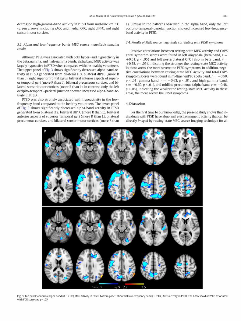
Although PTSDwas associated with both hyper- and hypoactivity inthe beta, gamma, and high-gamma bands, alpha bandMEG activity waslargely hypoactive in PTSDwhen comparedwith the healthy volunteers.The upper panel of Fig. 3 shows significantly decreased alpha-band ac-tivity in PTSD generated from bilateral FPs, bilateral dlPFC (more Rthan L), right superior frontal gyrus, bilateral anterior aspects of superi-or temporal gyri (more R than L), bilateral precuneous cortices, and bi-lateral sensorimotor cortices (more R than L). In contrast, only the leftoccipito-temporal–parietal junction showed increased alpha-band ac-tivity in PTSD.
PTSD was also strongly associated with hypoactivity in the low-frequency band compared to the healthy volunteers. The lower panelof Fig. 3 shows significantly decreased alpha-band activity in PTSDgenerated from bilateral FPs, bilateral dlPFC (more R than L), bilateralanterior aspects of superior temporal gyri (more R than L), bilateralprecuneous cortices, and bilateral sensorimotor cortices (more R than
R
t = -3.5 -2.9 2
Fig. 3. Top panel: abnormal alpha band (8–12 Hz) MEG activity in PTSD; bottom panel: abnormwith FDR corrected p b .05.
L). Similar to the patterns observed in the alpha band, only the leftoccipito-temporal–parietal junction showed increased low-frequency-band activity in PTSD.
3.4. Results of MEG source magnitude correlating with PTSD symptoms
Positive correlations between resting-state MEG activity and CAPSTotal symptom scores were found in left amygdala (beta band, r =+0.51, p b .05) and left posterolateral OFC (also in beta band, r =+0.55, p b .05), indicating the stronger the resting-state MEG activityin these areas, the more severe the PTSD symptoms. In addition, nega-tive correlations between resting-state MEG activity and total CAPSsymptom scores were found in midline vmPFC (beta band, r = −0.58,p b .01; gamma band, r = −0.63, p b .01; and high-gamma band,r = −0.60, p b .01), and midline precuneous (alpha band, r = −0.48,p b .05), indicating the weaker the resting-state MEG activity in theseareas, the more severe the PTSD symptoms.
4. Discussion
For the first time to our knowledge, the present study shows that in-dividuals with PTSD have abnormal electromagnetic activity that can bedirectly imaged by resting-state MEG source imaging technique for all
L
.9 3.5
al low-frequency band (1–7 Hz)MEG activity in PTSD. The t-threshold of 2.9 is associated

414 M.-X. Huang et al. / NeuroImage: Clinical 5 (2014) 408–419
frequency bands. PTSDwas associated with: 1)MEG hyperactivity fromamygdala, hippocampus, posterolateral OFC, dmPFC, insular cortex, andPCC in high frequency bands (i.e., beta, gamma, and high gammabands); 2) MEG hypoactivity from vmPFC, FP, and dlPFC in the highfrequency bands; 3) extensive MEG hypoactivity from dlPFC, FP, an-terior temporal lobes, precuneous cortex, and sensorimotor cortex inalpha and low-frequency bands, with dlPFC and sensorimotor cortexhypoactivity more prominent in right versus left hemispheres; and4) resting-state MEG activity in left amygdala and posterolateralOFC positively correlated with PTSD symptom scores, whereas MEGactivity in vmPFC and precuneous correlated negatively with thePTSD symptoms.
Neuronal activity fromdifferent frequency bands is considered to re-flect different neuronal mechanisms. Thalamo-cortical interactions areessential for alpha rhythms, and normal alpha-band activity is associat-ed with functional inhibition; specifically, increased alpha-band powerin a brain area was linked to reduced functional connectivity withother brain areas (De Munck et al., 2009; Hindriks and van Putten,2013; Scheeringa et al., 2012). Activity in the beta band is associatedwith communication between remote brain structures, whereas gammasynchrony promotes local computations (Kopell et al., 2000; Singer,1999). Although the gamma band electromagnetic signals are generatedlocally, non-local brain areas can still show significant functional connec-tivity asmeasured by coherence related to the gammaband signals. Usingcombined electrophysiological and fMRI measurements, studies in bothhuman and animals showed that gamma-band power exhibits spatial co-herence over long timescaleswith the strongest coherence between func-tionally related areas that are not necessarily local (He et al., 2008; Niret al., 2008; Scholvinck et al., 2010, 2013; Shmuel and Leopold, 2008). Un-like alpha-band activity, beta and gamma-band activity does not neces-sarily have to involve thalamus. Theta-band signals have been reportedin previous EEG studies, although these signals were predominantlytask-activated (e.g., problem solving) (Mizuki et al., 1980, 1984, 1992;Niedermeyer and Lopes da Silva, 2005; Takahashi et al., 1997). Increasedlow-frequency brain rhythms in delta bandwere often seen in individualswith neurological disorders, e.g. epilepsy and traumatic brain injury(Baayen et al., 2003; de Jongh et al., 2003; Decker and Knott, 1972;Huang et al., 2009, 2012, 2014b; Lewine et al., 1999; Lewine andOrrison, 1995; Nagata et al., 1985; Vieth et al., 1996). When examin-ing the mechanism of abnormal delta rhythms, electrophysiologicalstudies in animals show that abnormal delta activity is from graymatter neurons that have experienced deafferentation due to neuro-logical injuries in underlying white matter, resulting from axonal in-jury or blockage/limitation in the cholinergic pathways (Ball et al.,1977; Gloor et al., 1977; Schaul et al., 1978; Schaul, 1998).
4.1. MEG findings in amygdala and hippocampus
Individuals with PTSD showed amygdala hyperactivity. Our MEGfindings are consistent with previous PET and fMRI findings of hy-perresponsive amygdala activity in PTSD (Rauch et al., 1998, 2006),which is one of the most robust functional neuroimaging findingsin PTSD (Hughes and Shin, 2011). The amygdala is involved in process-ing threat-related stimuli (Davis andWhalen, 2001; Morris et al., 1998;Whalen et al., 1998, 2001) and is necessary for fear conditioning (Davisand Whalen, 2001; LeDoux, 2000; Shin et al., 2006). Moreover, theamygdala is a key component in the neurocircuitry model of PTSD(Rauch et al., 2006). The present MEG study shows that the amygdalahyperactivity in PTSD can also be detected using electromagnetic sourceimaging measures, which increases the confidence in our MEG tech-nique for detecting new abnormalities in PTSD. Furthermore, we dem-onstrate that amygdala hyperactivity was only observable in the highfrequency bands (i.e., beta and high-gamma bands). In addition, theMEG hyperactivity in PTSD from left hippocampus in beta and high-gamma bands is also consistent with the current PTSD neurocircuitry
model (Rauch et al., 2006), although the findings from previous PETand fMRI in this region have been mixed (Hughes and Shin, 2011).
4.2. MEG findings in dmPFC and insula
The MEG gamma-band hyperactivity from dmPFC, including thedACC, in PTSD was also consistent with prior PET and fMRI findings(Bremner et al., 1999; Felmingham et al., 2009; Pannu et al., 2009;Shin et al., 2001, 2007, 2011). The dmPFC, including thedACC, is thoughtto play an important role in a variety of cognitive processes such as per-formance monitoring, response selection, error detection, and decisionmaking (Shin et al., 2011). In addition, PTSD was associated with in-creased MEG insular activity. Our findings are consistent with studiesthat used trauma-event-script-driven imagery with SPECT (Lindaueret al., 2008) and fMRI (Lanius et al., 2007), as well as with studies thatused emotional and trauma-unrelated stimuli with PET and fMRI(Hughes and Shin, 2011). Painful stimuli have also been shown to in-crease insular activity in PTSD (Geuze et al., 2007; Strigo et al., 2010).The insular cortex processes information about the body3s internal stateand contributes to the autonomic component of the overall pain re-sponse. It has been suggested that the insular cortex integrates thesensory, affective, and cognitive components necessary for normal re-sponses to pain (Kandel et al., 2000). Abnormal insular activity inPTSD may reflect a deficit in integrating these components, therebycontributing to an abnormal pain response (Nagai et al., 2007).
4.3. MEG findings in vmPFC
MEG hypoactivity from vmPFC in PTSDwas consistent with findingsfrom PET and fMRI studies (Hughes and Shin, 2011; Rauch et al., 1998,2006). Hyporesponsive vmPFC is another key component in the currentneurocircuitry model of PTSD (Rauch et al., 2006), which suggests thathyporesponsive vmPFC fails to suppress the amygdala (Rauch et al.,2006; Shin et al., 2006). The vmPFC is connected to and receives inputfrom the ventral tegmental area, amygdala, temporal lobe, olfactory sys-tem, and dorsomedial thalamus. In turn, vmPFC sends signals to amyg-dala, temporal lobe, lateral hypothalamus, hippocampal formation,cingulate cortex, and certain other regions of the prefrontal cortex(Carlson, 2013). In the present study, hypoactivity in vmPFC associatedwith PTSDwas evident in beta, gamma, and high-gammabands, but notthe lower frequency bands. These findings suggest that the beta- andgamma-band interactions between vmPFC and amygdala may not in-volve thalamus, as evidenced by the lack of group differences in vmPFCin thalamus-dependent alpha band activity (Hindriks and van Putten,2013).
4.4. Resting-state MEG versus resting-state fMRI
We used a resting-state protocol that is insensitive to stimulus fea-tures and participant performance. Furthermore, we focused on examin-ing MEG source-magnitude images rather than functional connectivity(Jin et al., 2013). Our protocol was similar to a recent resting-state fMRIstudy of combat-related PTSD that used a magnitude imaging approach(Yan et al., 2013). Our findings are consistent with Yan and colleagues,who also found that individuals with PTSD showed increased activity inamygdala, insular cortex, and OFC, as well as decreased activity in dlPFC,superior frontal gyrus, and precuneous cortex. Despite these similarities,participantswith PTSD in the present study showed decreasedMEGactiv-ity in vmPFC (beta, gamma, andhigh-gammabands, see Figs. 1 and2) andbilateral FP areas (Figs. 1–3), whereas Yan and colleagues, in their fMRIstudy showed increased activity in a similar region (Yan et al., 2013).Although it is known that the fMRI measurements in ventral frontallobe areas are challenging to obtain due to signal loss, imaging distortion,and susceptibility artifacts (Czervionke et al., 1988; Domsch et al., 2013),the exact reason of the decreased MEG versus increased fMRI resting-state activity in vmPFC is unknown.

415M.-X. Huang et al. / NeuroImage: Clinical 5 (2014) 408–419
Overall, the beta-band MEG source imaging maps are similar to thefMRI maps of Yan and colleagues, except for the activity in vmPFC.Such a degree of similarity across two different imaging modalities(i.e., electromagnetic measures fromMEG and hemodynamic measuresfrom fMRI) is likely due to beta-band synchronization over long conduc-tion delays,which corresponds to signals traveling a significant distanceacross brain regions. Electrophysiological studies of the rat hippocam-pus have shown that the beta rhythm allows neuronal synchrony atlarge time delays, while the gamma band allows such synchrony atshort delays. Thus, beta synchrony promotes communication betweenremote structures, whereas gamma synchrony promotes local compu-tations (Kopell et al., 2000; Singer, 1999). Interestingly, more recentwork in identifying MEG correlates of fMRI resting-state networks hasfound that power fluctuations in the beta band produce spatial net-works very similar to fMRI resting-state networks (Brookes et al.,2011b). Our findings suggest that abnormal beta-band neuronal activityin PTSD is likely a candidate for the abnormal fMRI signal observed byYan and colleagues (Yan et al., 2013).
The consistent finding of decreases of resting-state activity inprecuneous and dlPFC associated with PTSD in the present MEG study(in beta, alpha, and low-frequency bands) and in the fMRI study byYan et al. (Yan et al., 2013) highlight the contribution of these regionsin PTSD neurocircuitry. The precuneous is a key region of the “default-mode network (DMN)” in resting brain which has been detected byfMRI (Fransson and Marrelec, 2008) and MEG (Brookes et al., 2011b).Furthermore, the precuneous plays a pivotal role in how intrinsic activ-ity is mediated throughout the DMN, and helps sustain a sense of self-consciousness in self-referential mental thoughts during rest (Cavannaand Trimble, 2006; Fransson and Marrelec, 2008). Non-trauma relatedwords elicit decreased precuneous fMRI activity in PTSD, and the de-crease in precuneous activity is correlated with CAPS scores (Geuzeet al., 2008). Dissociative symptoms of patients with PTSD may play arole in the decreased activation of precuneous (Geuze et al., 2008).The dlPFC is a key region for a variety of executive brain functionssuch asworkingmemory, attention, and other executive functions. It fa-cilitates goal-directed behavior through indirect modulation of theamygdala response to threat, possibly through connections with thetemporal cortex (Bishop, 2008; Gold et al., 2014; Mitchell, 2011). Inthe present study, MEG hyperactivity in both right dlPFC and anteriortemporal lobe in alpha, beta, gamma, and low-frequency (Figs. 1–3) isconsistent with the modulation deficit of the dlPFC–anterior temporal-amygdala pathway in PTSD. Using a task involving cognitive regulationof negative affect via reappraisal, Rabinak and colleagues found thatPTSD patients engaged the dlPFC during cognitive reappraisal, albeit toa lesser extent than the control participants (Rabinak et al., 2014). In alongitudinal cortical thickness study, individuals with PTSD showedgreater dlPFC thickness in a follow-up exam about 1 year after the trau-ma than in the acute exam, and greater dlPFC thickness was associatedwith greater PTSD symptom reductions and better recovery (Lyoo et al.,2011). On the other hand, healthy volunteers showed greater dlPFC ac-tivation and increased amygdala connectivity to threats compared tonon-threat condition (Gold et al., 2014). Elevated activity in dlPFC wasalso observed in PTSD during amaintenance period ofworkingmemoryin an fMRI test (Moores et al., 2008). Future functional imaging studiesof PTSD are needed to examine the association between resting-statedlPFC activity and dlPFC responses to different types of workingmemo-ry and/or attention stimuli.
In an event related potential (ERP) study of combat veterans withPTSD after mild TBI by Shu and colleagues, PCC and precuneous areasexhibit greater ERPs evoked by emotional facial stimuli (Shu et al.,2014). In the present study, PCC also showed hyperactivity in thebeta-band resting-state MEG source image (brown arrow in Fig. 1), afinding consistent with the above ERP study. However, the hypoactiveprecuneous is seen in our resting-state MEG source image acrossmany frequency bands, also observed in resting-state fMRI by Yan andcolleagues (Yan et al., 2013), seems to be different from the greater
ERPs in precuneous found by Shu and colleagues using emotional stim-uli. Additional studies are needed to directly examine the associationbetween resting-state electromagnetic signal and evoked responses,as well as the impact of mild TBI on PTSD.
4.5. MEG findings in OFC
Another interesting finding from the present study is the increasedactivity from the posterolateral OFC areas in beta, gamma, and highgamma bands. Our finding is consistent with fMRI findings of increasedresting-state activity in PTSD (Yan et al., 2013). The OFC is closely con-nected to the limbic system, especially the amygdala, and is sometimesregarded as part of the expanded limbic system (Nauta, 1979). Whileregions known to be part of the existing neurocircuitry model of PTSD(i.e., amygdala, vmPFC, and insular cortex) have been studied extensive-ly (Rauch et al., 1998, 2006; Shin et al., 2006), the role of the posterolat-eral OFC in PTSD is unclear and should be examined further. Based onour MEG findings, posterolateral OFC activity increased with PTSDsymptom severity, thus OFC and its interactions with the amygdalamay be added to the existing neurocircuitry model of PTSD. This ideais supported by studies that show thatOFC has direct anatomical projec-tions to the amygdala and hippocampus via the uncinate fasciculus inhumans (Bach et al., 2011; Talairach and Tournoux, 1988) as well asin non-human primates (Carmichael and Price, 1995). It was alsoshown that such projections were abnormal in some psychiatric disor-ders such as conduct disorder (Passamonti et al., 2012), bipolar disorder(Benedetti et al., 2011), and schizophrenia (Jackowski et al., 2012). Fur-ther studies are needed to confirm whether disrupted interactions be-tween OFC-amygdala may be implicated in PTSD.
4.6. Decreased MEG alpha-band activity in PTSD
Individuals with PTSD showed extensive MEG alpha-bandhypoactivity from dlPFC, FP, anterior temporal lobes, precuneous cor-tex, and sensorimotor cortex. Neuronal modeling studies showed thatthalamo-cortical interactions are essential to the generation of alpharhythms (Freyer et al., 2011; Hindriks and van Putten, 2013; Lopes daSilva et al., 1997). Combined EEG and fMRI studies have also shownthat increased alpha-band power in a brain area is associated with re-duced functional connectivity with other brain areas, suggesting thatalpha-band activity is associated with functional inhibition (De Muncket al., 2009; Scheeringa et al., 2012). The observed MEG alpha-bandhypoactivity may suggest a deficit in thalamo-cortical interactions,which possibly leads to reduced functional inhibition in the above cor-tical areas in PTSD. In general, a normal amount of alpha activity is pre-ferred in the resting-state, and reduced alpha-band power has also beenobserved in individuals with Alzheimer3s disease (Babiloni et al., 2013;Tartaglione et al., 2012), and schizophrenia (Hinkley et al., 2011;Sponheim et al., 2000).
4.7. MEG source imaging with fast-VESTAL
Our method plays an essential role in obtaining the source magni-tude images for the neurocircuitry in PTSD (Figs. 1–3). It was shownthat Fast-VESTAL can effectively create resting-stateMEG source imagesthat are highly consistent with known neurophysiology findings (Huanget al., 2014a). We have shown that for resting-state MEG signal, thesource magnitude images obtained using a beamformer technique(a popular MEG source analysis method) are less consistent withneurophysiology findings (Huang et al., 2014a). This is likely due tobeamformer3s intrinsic limitation which assumes the neuronal sourcesare uncorrelated (Robinson and Vrba, 1999; Sekihara et al., 2001; VanVeen et al., 1997), a questionable assumption when dealing withresting-state MEG signals.
In the present study, we focus on MEG source magnitude images inPTSD. No results were presented regarding theMEG-based connectivity

416 M.-X. Huang et al. / NeuroImage: Clinical 5 (2014) 408–419
analyses. This is because we are in the process of finalizing theFast-VESTAL based voxel-wise MEG connectivity method (Huanget al., in preparation). Although MEG-based connectivity study isa hot topic in literature, with most published approaches usedBeamformer or minimum L2-norm based techniques to obtain thesource time-courses (Brookes et al., 2011a,b, 2012; Ghuman et al.,2011; Gramfort et al., 2014). It was known that source time-courses ob-tain by Beamformer are distorted when multiple correlated neuronalsources contribute to the sensor-waveforms even at noiseless cases(Huang et al., 2014a), and across-talk between source time-coursesfrom minimum L2-norm approaches can also be a serious issue.However, even though many researchers were aware of the issuesassociated with distorted source time-courses, the impact on a vari-ety of connectivity measures using the distorted source time-courseshas not been examined thoroughly in resting-state data, at least toour knowledge. Before we publish our method for Fast-VESTALbased connectivity analysis, we believe that it would be beyond thescope of the present study to include MEG connectivity results forthe PTSD population.
There are several limitations of the present study that warrant con-sideration. First, the spatial resolution and localization accuracy of MEGare expected to be limited for amygdala, hippocampus, and vmPFC,which may explain some minor location discrepancies between ourfindings and those of previous fMRI or PET studies. Second, althoughwe acquired resting-state MEG signal in the eyes-closed condition,eye-movements and micro-eye-blinks may be confounding factors. Al-though we pre-processed the MEG data through both MaxFilter andICA to remove the eye-movement and micro-eye-blinks, the impact ofresidual eye-activity-related artifacts may not be totally negligible.Third, despite substantial efforts to ensure andmonitor alertness duringthe eyes-closed condition (seeMaterials andmethods), drowsinessmaystill have had an impact on the MEG recording. Fourth, since the active-duty and veteran PTSD patients are mostly males, the present study isdominated by male subjects, with just one woman in each of the twogroups.
Despite these limitations, the present study showed that our MEGsource imaging technique revealed new abnormalities in the resting-state electromagnetic signals from PTSD neurocircuitry. Abnormalresting-state electromagnetic signals in PTSD neurocircuitry can be ef-fectively imaged by MEG source imaging technique for different fre-quency bands. In high frequency bands (i.e., beta, gamma, and highgamma bands), PTSD was associated with 1) MEG hyperactivity fromamygdala, hippocampus, posterolateral OFC, dmPFC, and insular cortex;and 2) MEG hypoactivity from vmPFC, FP, and dlPFC. In alpha and low-frequency bands, PTSDwas associatedwith extensiveMEG hypoactivityfrom dlPFC, FP, anterior temporal lobes, precuneous cortex, and sensori-motor cortex. Lastly, PTSD symptom scores correlated positively withresting-state MEG activity in left amygdala and posterolateral OFC andnegatively with MEG activity in vmPFC and precuneous. Particularly,posterolateral OFC and precuneous may play important roles in thePTSD neurocircuitry model.
Acknowledgments
This work was supported in part by Headquarters Marine Corps andNavy BUMED to D.G. Baker, M.A. Geyer, M.X. Huang and V.B. Risbrough(MRS-II; Navy BUMED contract # N62645-11-C-4037) and Merit Re-view Grants from the Department of Veterans Affairs to M.X. Huang(I01-CX000499, NURC-022-10F, NEUC-044-06S). We acknowledge theMRS-II administrative core, Anjana Patel, AndrewDe La Rosa, andmem-bers of the MRS-II Team, including logistic coordinators, clinician-interviewers, and data collection staff. We thank staff at the VeteransMedical Research Foundation (VRMF). We also thank the Marine,Navy and veteran volunteers for their military service and participationin this study.
References
American Psychiatric Association, 2000. Diagnostic and Statistical Manual of Mental Dis-orders IV, Text RevisionAmerican Psychiatric Association.
American Psychiatric Association, 2004. Practice Guideline for the Treatment of PatientsWith Acute Stress Disorder and PTSDAmerican Psychiatric Association, Arlington, VA.
Anderson, A.K.,Christoff, K.,Panitz, D.,De Rosa, E.,Gabrieli, J.D., 2003. Neural correlates ofthe automatic processing of threat facial signals. Journal of Neuroscience: the OfficialJournal of the Society for Neuroscience 23, 5627–5633 12843265.
Baayen, J.C.,de Jongh, A., Stam, C.J.,De Munck, J.C., Jonkman, J.J., Trenité, D.G.,Berendse, H.W.,van Walsum, A.M.,Heimans, J.J., Puligheddu, M.,Castelijns, J.A.,Vandertop, W.P.,2003. Localization of slow wave activity in patients with tumor-associated epilepsy.Brain Topography 16, 85–93 14977201.
Babiloni, C.,Del Percio, C.,Bordet, R.,Bourriez, J.L.,Bentivoglio, M.,Payoux, P.,Derambure, P.,Dix, S., Infarinato, F., Lizio, R.,Triggiani, A.I.,Richardson, J.C.,Rossini, P.M., 2013. Effectsof acetylcholinesterase inhibitors and memantine on resting-state electroencephalo-graphic rhythms in Alzheimer3s disease patients. Clinical Neurophysiology: OfficialJournal of the International Federation of Clinical Neurophysiology 124, 837–850.http://dx.doi.org/10.1016/j.clinph.2012.09.01723098644.
Bach, D.R., Behrens, T.E.,Garrido, L.,Weiskopf, N.,Dolan, R.J., 2011. Deep and superficialamygdala nuclei projections revealed in vivo by probabilistic tractography. Journalof Neuroscience: the Official Journal of the Society for Neuroscience 31, 618–623.http://dx.doi.org/10.1523/JNEUROSCI.2744-10.201121228170.
Ball, G.J., Gloor, P., Schaul, N., 1977. The cortical electromicrophysiology of pathologicaldelta waves in the electroencephalogram of cats. Electroencephalography and Clini-cal Neurophysiology 43, 346–361 70336.
Benedetti, F.,Absinta, M.,Rocca, M.A.,Radaelli, D., Poletti, S., Bernasconi, A.,Dallaspezia, S.,Pagani, E., Falini, A., Copetti, M., Colombo, C., Comi, G., Smeraldi, E., Filippi, M., 2011.Tract-specific white matter structural disruption in patients with bipolar disorder. Bi-polar Disorders 13, 414–424. http://dx.doi.org/10.1111/j.1399-5618.2011.00938.x21843281.
Benjamini, Y.,Hochberg, Y., 1995. Controlling the false positive rate: a practical and pow-erful approach to multiple testing. Journal of the Royal Statistical Society, Series B 57,289–300.
Bishop, S.J., 2008. Neural mechanisms underlying selective attention to threat. Annals ofthe New York Academy of Sciences 1129, 141–152. http://dx.doi.org/10.1196/annals.1417.01618591476.
Blake, D.D.,Weathers, F.W.,Nagy, L.M.,Kaloupek, D.G.,Gusman, F.D.,Charney, D.S.,Keane, T.M., 1995. The development of a Clinician-Administered PTSD Scale. Journal of Trau-matic Stress 8, 75–90 7712061.
Blanchard, E.B.,Hickling, E.J.,Taylor, A.E.,Forneris, C.A.,Loos, W.,Jaccard, J., 1995. Effects ofvarying scoring rules of the Clinician-Administered PTSD Scale (CAPS) for the diagno-sis of post-traumatic stress disorder in motor vehicle accident victims. Behaviour Re-search and Therapy 33, 471–475 7755537.
Bremner, J.D.,Narayan,M.,Staib, L.H.,Southwick, S.M.,McGlashan, T.,Charney, D.S., 1999. Neu-ral correlates of memories of childhood sexual abuse in women with and without post-traumatic stress disorder. American Journal of Psychiatry 156, 1787–1795 10553744.
Brookes, M.J.,Hale, J.R., Zumer, J.M., Stevenson, C.M., Francis, S.T., Barnes, G.R.,Owen, J.P.,Morris, P.G.,Nagarajan, S.S., 2011a. Measuring functional connectivity using MEG:methodology and comparison with fcMRI. NeuroImage 56, 1082–1104. http://dx.doi.org/10.1016/j.neuroimage.2011.02.05421352925.
Brookes, M.J.,Woolrich, M., Luckhoo, H.,Price, D.,Hale, J.R., Stephenson, M.C., Barnes, G.R.,Smith, S.M.,Morris, P.G., 2011b. Investigating the electrophysiological basis of restingstate networks using magnetoencephalography. Proceedings of the National Acade-my of Sciences of the United States of America 108, 16783–16788. http://dx.doi.org/10.1073/pnas.111268510821930901.
Brookes, M.J.,Woolrich, M.W., Barnes, G.R., 2012. Measuring functional connectivity inMEG: a multivariate approach insensitive to linear source leakage. NeuroImage 63,910–920. http://dx.doi.org/10.1016/j.neuroimage.2012.03.04822484306.
Bryant, R.A., Felmingham, K.L.,Kemp, A.H., Barton, M., Peduto, A.S., Rennie, C.,Gordon, E.,Williams, L.M., 2005. Neural networks of information processing in posttraumaticstress disorder: a functional magnetic resonance imaging study. Biological Psychiatry58, 111–118. http://dx.doi.org/10.1016/j.biopsych.2005.03.02116038681.
Carlson, N.R., 2013. Physiology of BehavIOreleventh edition. Pearson, Boston.Carmichael, S.T., Price, J.L., 1995. Limbic connections of the orbital and medial prefrontal
cortex in macaque monkeys. Journal of Comparative Neurology 363, 615–641.http://dx.doi.org/10.1002/cne.9036304088847421.
Cavanna, A.E.,Trimble, M.R., 2006. The precuneus: a review of its functional anatomy andbehavioural correlates. Brain: A Journal of Neurology 129, 564–583. http://dx.doi.org/10.1093/brain/awl00416399806.
Chung, Y.A.,Kim, S.H.,Chung, S.K., Chae, J.H.,Yang, D.W.,Sohn, H.S., Jeong, J., 2006. Alter-ations in cerebral perfusion in posttraumatic stress disorder patients without re-exposure to accident-related stimuli. Clinical Neurophysiology: Official Journal ofthe International Federation of Clinical Neurophysiology 117, 637–642. http://dx.doi.org/10.1016/j.clinph.2005.10.02016426890.
Cohen, D., Schläpfer, U., Ahlfors, S., Hämäläinen, M., Halgren, E., 2002. New six-layermagnetically-shielded room forMEG. In: Nowak, H.H.J., Gießler, F. (Eds.), Proceedingsof the 13th International Conference on Biomagnetism. Vde Verlag, Jena, Germany.
Corcoran, K.A.,Maren, S., 2001. Hippocampal inactivation disrupts contextual retrieval offear memory after extinction. Journal of Neuroscience: the Official Journal of the So-ciety for Neuroscience 21, 1720–1726 11222661.
Czervionke, L.F.,Daniels, D.L.,Wehrli, F.W.,Mark, L.P.,Hendrix, L.E.,Strandt, J.A.,Williams, A.L.,Haughton, V.M., 1988. Magnetic susceptibility artifacts in gradient-recalled echoMR imaging. American Journal of Neuroradiology 9, 1149–1155 3143237.
Davis, M.,Whalen, P.J., 2001. The amygdala: vigilance and emotion. Molecular Psychiatry6, 13–34 11244481.
417M.-X. Huang et al. / NeuroImage: Clinical 5 (2014) 408–419
de Jongh, A., Baayen, J.C.,De Munck, J.C.,Heethaar, R.M.,Vandertop, W.P.,Stam, C.J., 2003.The influence of brain tumor treatment on pathological delta activity in MEG.NeuroImage 20, 2291–2301 14683730.
De Munck, J.C.,Gonçalves, S.I.,Mammoliti, R.,Heethaar, R.M.,Lopes da Silva, F.H., 2009. In-teractions between different EEG frequency bands and their effect on alpha-fMRI cor-relations. NeuroImage 47, 69–76. http://dx.doi.org/10.1016/j.neuroimage.2009.04.02919376236.
Decker Jr., D.A.,Knott, J.R., 1972. The EEG in intrinsic supratentorial brain tumors: a com-parative evaluation. Electroencephalography and Clinical Neurophysiology 33,303–310 4114914.
Dikmen, S.S.,Ross, B.L.,Machamer, J.E.,Temkin, N.R., 1995. One year psychosocial outcomein head injury. Journal of the International Neuropsychological Society: JINS 1, 67–779375211.
Dohrenwend, B.P., Turner, J.B., Turse, N.A., Adams, B.G., Koenen, K.C., Marshall, R.,2006. The psychological risks of Vietnam for U.S. veterans: a revisit with newdata and methods. Science (New York, N.Y.) 313, 979–982. http://dx.doi.org/10.1126/science.112894416917066.
Dolcos, F.,LaBar, K.S.,Cabeza, R., 2004. Interaction between the amygdala and the medialtemporal lobe memory system predicts better memory for emotional events. Neuron42, 855–863 15182723.
Domsch, S., Linke, J.,Heiler, P.M.,Kroll, A., Flor, H.,Wessa, M.,Schad, L.R., 2013. IncreasedBOLD sensitivity in the orbitofrontal cortex using slice-dependent echo times at 3 T.Magnetic Resonance Imaging 31, 201–211. http://dx.doi.org/10.1016/j.mri.2012.06.02022925606.
Engdahl, B., Leuthold, A.C., Tan, H.-R.M., Lewis, S.M., Winskowski, A.M., Dikel, T.N.,Georgopoulos, A.P., 2010. Post-traumatic stress disorder: a right temporal lobe syn-drome? Journal of Neural Engineering 7, 066005. http://dx.doi.org/10.1088/1741-2560/7/6/06600520980718.
Felmingham, K.L.,Williams, L.M.,Kemp, A.H.,Rennie, C.,Gordon, E.,Bryant, R.A., 2009. An-terior cingulate activity to salient stimuli is modulated by autonomic arousal in post-traumatic stress disorder. Psychiatry Research 173, 59–62. http://dx.doi.org/10.1016/j.pscychresns.2008.12.00519446442.
Fransson, P.,Marrelec, G., 2008. The precuneus/posterior cingulate cortex plays a pivotalrole in the default mode network: evidence from a partial correlation network anal-ysis. NeuroImage 42, 1178–1184. http://dx.doi.org/10.1016/j.neuroimage.2008.05.05918598773.
Freyer, F.,Roberts, J.A.,Becker, R.,Robinson, P.A.,Ritter, P.,Breakspear, M., 2011. Biophysicalmechanisms of multistability in resting-state cortical rhythms. Journal of Neurosci-ence: the Official Journal of the Society for Neuroscience 31, 6353–6361. http://dx.doi.org/10.1523/JNEUROSCI.6693-10.201121525275.
Georgopoulos, A.P., Tan, H.-R.M., Lewis, S.M., Leuthold, A.C.,Winskowski, A.M., Lynch,J.K., Engdahl, B., 2010. The synchronous neural interactions test as a functionalneuromarker for post-traumatic stress disorder (PTSD): a robust classificationmethod based on the bootstrap. Journal of Neural Engineering 7, 16011. http://dx.doi.org/10.1088/1741-2560/7/1/01601120086271.
Geuze, E.,Vermetten, E., de Kloet, C.S.,Westenberg, H.G., 2008. Precuneal activity duringencoding in veterans with posttraumatic stress disorder. Progress in Brain Research167, 293–297. http://dx.doi.org/10.1016/S0079-6123(07)67026-518037028.
Geuze, E.,Westenberg, H.G.,Jochims, A.,de Kloet, C.S.,Bohus, M.,Vermetten, E.,Schmahl, C.,2007. Altered pain processing in veterans with posttraumatic stress disorder. Ar-chives of General Psychiatry 64, 76–85. http://dx.doi.org/10.1001/archpsyc.64.1.7617199057.
Ghuman, A.S.,McDaniel, J.R.,Martin, A., 2011. A wavelet-based method for measuring theoscillatory dynamics of resting-state functional connectivity in MEG. NeuroImage 56,69–77. http://dx.doi.org/10.1016/j.neuroimage.2011.01.04621256967.
Gloor, P.,Ball, G.,Schaul, N., 1977. Brain lesions that produce delta waves in the EEG. Neu-rology 27, 326–333557774.
Gold, A.L.,Morey, R.A.,McCarthy, G., 2014. Amygdala-prefrontal cortex functional connectiv-ity during threat-induced anxiety and goal-distractionBiological Psychiatry (in press).
Gramfort, A., Luessi, M., Larson, E., Engemann, D.A., Strohmeier, D., Brodbeck, C.,Parkkonen, L., Hämäläinen, M.S., 2014. MNE software for processing MEG andEEG data. NeuroImage 86, 446–460. http://dx.doi.org/10.1016/j.neuroimage.2013.10.02724161808.
Hartley, C.A.,Phelps, E.A., 2010. Changing fear: the neurocircuitry of emotion regula-tion. Neuropsychopharmacology: Official Publication of the American College ofNeuropsychopharmacology 35, 136–146. http://dx.doi.org/10.1038/npp.2009.12119710632.
He, B.J., Snyder, A.Z., Zempel, J.M., Smyth, M.D., Raichle, M.E., 2008. Electrophysiolog-ical correlates of the brain3s intrinsic large-scale functional architecture. Pro-ceedings of the National Academy of Sciences of the United States of America105, 16039–16044. http://dx.doi.org/10.1073/pnas.080701010518843113.
Hindriks, R., van Putten, M.J., 2013. Thalamo-cortical mechanisms underlying changes inamplitude and frequency of human alpha oscillations. NeuroImage 70, 150–163.http://dx.doi.org/10.1016/j.neuroimage.2012.12.01823266701.
Hinkley, L.B., Vinogradov, S., Guggisberg, A.G., Fisher, M., Findlay, A.M., Nagarajan, S.S.,2011. Clinical symptoms and alpha band resting-state functional connectivity imag-ing in patients with schizophrenia: implications for novel approaches to treatment.Biological Psychiatry 70, 1134–1142. http://dx.doi.org/10.1016/j.biopsych.2011.06.02921861988.
Hoge, C.W., Castro, C.A., Messer, S.C., McGurk, D., Cotting, D.I., Koffman, R.L., 2004.Combat duty in Iraq and Afghanistan, mental health problems, and barriers tocare. New England Journal of Medicine 351, 13–22. http://dx.doi.org/10.1056/NEJMoa04060315229303.
Hoge, C.W.,McGurk, D.,Thomas, J.L.,Cox, A.L.,Engel, C.C.,Castro, C.A., 2008. Mild traumaticbrain injury in U.S. soldiers returning from Iraq. New England Journal of Medicine358, 453–463. http://dx.doi.org/10.1056/NEJMoa07297218234750.
Huang, M.X., Dale, A.M., Song, T., Halgren, E., Harrington, D.L., Podgorny, I., Canive, J.M.,Lewis, S., Lee, R.R., 2006. Vector-based spatial-temporal minimum L1-norm solutionfor MEG. NeuroImage 31, 1025–1037. http://dx.doi.org/10.1016/j.neuroimage.2006.01.02916542857.
Huang, M.X.,Huang, C.W.,Robb, A.,Angeles, A.,Nichols, S.L.,Baker, D.G.,Song, T.,Harrington,D.L.,Theilmann, R.J.,Srinivasan, R.,Heister, D.,Diwakar, M.,Canive, J.M.,Edgar, J.C.,Chen,Y.H., Ji, Z., Shen, M., El-Gabalawy, F., Levy, M.,McLay, R.,Webb-Murphy, J., Liu, T.T.,Drake, A., Lee, R.R., 2014a. MEG source imaging method using fast L1 minimum-norm and its applications to signals with brain noise and human resting-state sourceamplitude images. NeuroImage 84, 585–604. http://dx.doi.org/10.1016/j.neuroimage.2013.09.02224055704.
Huang, M.X., Nichols, S., Baker, D.G., Robb, A., Angeles, A., Yurgil, K.A., Drake, A., Levy,M., Song, T.,McLay, R., Theilmann, R.J., Diwakar, M., Risbrough, V.B., Ji, Z., Huang,C.W., Chang, D.G.,Harrington, D.L.,Muzzatti, L., Canive, J.M., Christopher Edgar, J.,Chen, Y.H., Lee, R.R., 2014b. Single-subject-based whole-brain MEG slow-waveimaging approach for detecting abnormality in patients with mild traumaticbrain injury. NeuroImage. Clinical 5, 109–119. http://dx.doi.org/10.1016/j.nicl.2014.06.00425009772.
Huang, M.X.,Nichols, S.,Robb, A.,Angeles, A.,Drake, A.,Holland, M.,Asmussen, S.,D’Andrea,J.,Chun, W.,Levy, M.,Cui, L.,Song, T.,Baker, D.G.,Hammer, P.,McLay, R.,Theilmann, R.J.,Coimbra, R., Diwakar, M., Boyd, C., Neff, J., Liu, T.T.,Webb-Murphy, J., Farinpour, R.,Cheung, C., Harrington, D.L., Heister, D., Lee, R.R., 2012. An automatic MEG low-frequency source imaging approach for detecting injuries in mild and moderate TBIpatients with blast and non-blast causes. NeuroImage 61, 1067–1082. http://dx.doi.org/10.1016/j.neuroimage.2012.04.02922542638.
Huang, M.X.,Song, T.,Hagler Jr., D.J.,Podgorny, I.,Jousmaki, V.,Cui, L.,Gaa, K.,Harrington, D.L.,Dale, A.M., Lee, R.R., Elman, J.,Halgren, E., 2007. A novel integrated MEG and EEGanalysis method for dipolar sources. NeuroImage 37, 731–748. http://dx.doi.org/10.1016/j.neuroimage.2007.06.00217658272.
Huang, M.X.,Theilmann, R.J.,Robb, A.,Angeles, A.,Nichols, S.,Drake, A.,D’Andrea, J.,Levy, M.,Holland, M.,Song, T.,Ge, S.,Hwang, E.,Yoo, K.,Cui, L.,Baker, D.G.,Trauner, D.,Coimbra, R.,Lee, R.R., 2009. Integrated imaging approach with MEG and DTI to detect mild trau-matic brain injury in military and civilian patients. Journal of Neurotrauma 26,1213–1226. http://dx.doi.org/10.1089/neu.2008.067219385722.
Hughes, K.C., Shin, L.M., 2011. Functional neuroimaging studies of post-traumatic stressdisorder. Expert Review of Neurotherapeutics 11, 275–285. http://dx.doi.org/10.1586/ern.10.19821306214.
Iversen, A.C., Fear, N.T., Ehlers, A.,Hacker Hughes, J.,Hull, L., Earnshaw, M.,Greenberg, N.,Rona, R.,Wessely, S.,Hotopf, M., 2008. Risk factors for post-traumatic stress disorderamong UK armed forces personnel. Psychological Medicine 38, 511–522. http://dx.doi.org/10.1017/S003329170800277818226287.
Jackowski, A.P.,Araújo Filho, G.M.,Almeida, A.G.,Araújo, C.M.,Reis, M.,Nery, F.,Batista, I.R.,Silva, I.,Lacerda, A.L., 2012. The involvement of the orbitofrontal cortex in psychiatricdisorders: an update of neuroimaging findings. Revista Brasileira de Psiquiatria (SãoPaulo, Brazil: 1999) 34, 207–212 22729418.
Jin, C.,Qi, R.,Yin, Y.,Hu, X.,Duan, L.,Xu, Q.,Zhang, Z.,Zhong, Y.,Feng, B.,Xiang, H.,Gong, Q.,Liu, Y.,Lu, G., Li, L., 2013. Abnormalities in whole-brain functional connectivity observed intreatment-naive post-traumatic stress disorder patients following an earthquake. Psy-chological Medicine 1–10 http://dx.doi.org/10.1017/S003329171300250X24168716.
Kandel, E.R., Schwartz, J.H., Jessell, T.M., 2000. Principles of Neural ScienceFourth edition.McGraw-Hill, New York.
Kessler, R.C.,Berglund, P.,Demler, O., Jin, R.,Merikangas, K.R.,Walters, E.E., 2005. Lifetimeprevalence and age-of-onset distributions of DSM-IV disorders in the National Co-morbidity Survey Replication. Archives of General Psychiatry 62, 593–602. http://dx.doi.org/10.1001/archpsyc.62.6.59315939837.
Kessler, R.C., Sonnega, A., Bromet, E.,Hughes, M.,Nelson, C.B., 1995. Posttraumatic stressdisorder in the National Comorbidity Survey. Archives of General Psychiatry 52,1048–1060 7492257.
Kopell, N., Ermentrout, G.B.,Whittington, M.A., Traub, R.D., 2000. Gamma rhythms andbeta rhythms have different synchronization properties. Proceedings of the NationalAcademy of Sciences of the United States of America 97, 1867–1872 10677548.
Lanius, R.A.,Frewen, P.A.,Girotti, M.,Neufeld, R.W.,Stevens, T.K.,Densmore, M., 2007. Neu-ral correlates of trauma script-imagery in posttraumatic stress disorder with andwithout comorbid major depression: a functional MRI investigation. Psychiatry Re-search 155, 45–56. http://dx.doi.org/10.1016/j.pscychresns.2006.11.00617412567.
Leahy, R.M.,Mosher, J.C., Spencer, M.E.,Huang, M.X., Lewine, J.D., 1998. A study of dipolelocalization accuracy for MEG and EEG using a human skull phantom. Electroenceph-alography and Clinical Neurophysiology 107, 159–173 9751287.
LeDoux, J.E., 2000. Emotion circuits in the brain. Annual Review of Neuroscience 23,155–184. http://dx.doi.org/10.1146/annurev.neuro.23.1.15510845062.
Lewine, J.D.,Davis, J.T.,Sloan, J.H.,Kodituwakku, P.W.,Orrison Jr., W.W., 1999. Neuromagneticassessment of pathophysiologic brain activity induced by minor head trauma. AJNR.American Journal of Neuroradiology 20, 857–866 10369357.
Lewine, J.D.,Orrison Jr., W.W., 1995. Spike and slowwave localization bymagnetoenceph-alography. Neuroimaging Clinics of North America 5, 575–596 8564285.
Lindauer, R.J.,Booij, J.,Habraken, J.B.,van Meijel, E.P.,Uylings, H.B.,Olff, M.,Carlier, I.V.,denHeeten, G.J.,van Eck-Smit, B.L.,Gersons, B.P., 2008. Effects of psychotherapy on region-al cerebral blood flow during trauma imagery in patients with post-traumatic stressdisorder: a randomized clinical trial. Psychological Medicine 38, 543–554. http://dx.doi.org/10.1017/S003329170700143217803835.
Logothetis, N.K., 2003. The underpinnings of the BOLD functional magnetic resonance im-aging signal. Journal of Neuroscience: the Official Journal of the Society for Neurosci-ence 23, 3963–3971 12764080.
Lopes da Silva, F.H.,Pijn, J.P.,Velis, D.,Nijssen, P.C., 1997. Alpha rhythms: noise, dynamicsandmodels. International Journal of Psychophysiology: Official Journal of the Interna-tional Organization of Psychophysiology 26, 237–249 9203006.

418 M.-X. Huang et al. / NeuroImage: Clinical 5 (2014) 408–419
Lyoo, I.K.,Kim, J.E.,Yoon, S.J.,Hwang, J.,Bae, S.,Kim, D.J., 2011. The neurobiological roleof the dorsolateral prefrontal cortex in recovery from trauma. Longitudinal brainimaging study among survivors of the south Korean subway disaster. Archives ofGeneral Psychiatry 68, 701–713. http://dx.doi.org/10.1001/archgenpsychiatry.2011.7021727254.
Magruder, K.M.,Yeager, D.E., 2009. The prevalence of PTSD across War eras and the effectof deployment on PTSD: a systematic review and meta-analysis. Psychiatric Annals39, 778–788.
McGaugh, J.L., 2004. The amygdala modulates the consolidation of memories of emotion-ally arousing experiences. Annual Review of Neuroscience 27, 1–28. http://dx.doi.org/10.1146/annurev.neuro.27.070203.14415715217324.
Mitchell, D.G., 2011. The nexus between decision making and emotion regulation: a re-view of convergent neurocognitive substrates. Behavioural Brain Research 217,215–231. http://dx.doi.org/10.1016/j.bbr.2010.10.03021055420.
Mizuki, Y.,Kajimura, N.,Kai, S.,Suetsugi, M.,Ushijima, I.,Yamada, M., 1992. Differential re-sponses to mental stress in high and low anxious normal humans assessed by frontalmidline theta activity. International Journal of Psychophysiology: Official Journal ofthe International Organization of Psychophysiology 12, 169–178 1592670.
Mizuki, Y.,Kajimura, N.,Nishikori, S.,Imaizumi, J.,Yamada, M., 1984. Appearance of frontalmidline theta rhythm and personality traits. Folia Psychiatrica et Neurologica Japon-ica 38, 451–458 6535746.
Mizuki, Y., Tanaka, M., Isozaki, H.,Nishijima, H., Inanaga, K., 1980. Periodic appearance oftheta rhythm in the frontal midline area during performance of a mental task. Elec-troencephalography and Clinical Neurophysiology 49, 345–351 6158411.
Moores, K.A.,Clark, C.R.,McFarlane, A.C.,Brown, G.C.,Puce, A.,Taylor, D.J., 2008. Abnormalrecruitment of working memory updating networks during maintenance of trauma-neutral information in post-traumatic stress disorder. Psychiatry Research 163,156–170. http://dx.doi.org/10.1016/j.pscychresns.2007.08.01118455372.
Morris, J.S.,Ohman, A.,Dolan, R.J., 1998. Conscious and unconscious emotional learn-ing in the human amygdala. Nature 393, 467–470. http://dx.doi.org/10.1038/309769624001.
Mosher, J.C., Leahy, R.M., Lewis, P.S., 1999. EEG and MEG: forward solutions for inversemethods. IEEE Transactions on Bio-Medical Engineering 46, 245–259 10097460.
Nagai, M.,Kishi, K.,Kato, S., 2007. Insular cortex and neuropsychiatric disorders: a reviewof recent literature. European Psychiatry: the Journal of the Association of EuropeanPsychiatrists 22, 387–394. http://dx.doi.org/10.1016/j.eurpsy.2007.02.00617416488.
Nagata, K.,Gross, C.E.,Kindt, G.W.,Geier, J.M.,Adey, G.R., 1985. Topographic electroenceph-alographic study with power ratio index mapping in patients with malignant braintumors. Neurosurgery 17, 613–619 4058699.
Nauta, W.J.H., 1979. Expanding borders of the limbic system concept. In: Rasmussen, I.,Marino, R. (Eds.), Functional Neurosurgery. Raven, New York, pp. 7–23.
Niedermeyer, E.,Lopes da Silva, F.H., 2005. Electroencephalography: Basic Principles, Clin-ical Applications, and Related FieldsFifth edition. Lippincott Williams & Wilkins,Philadelphia.
Nir, Y., Mukamel, R., Dinstein, I., Privman, E., Harel, M., Fisch, L., Gelbard-Sagiv, H.,Kipervasser, S., Andelman, F.,Neufeld, M.Y.,Kramer, U.,Arieli, A., Fried, I.,Malach, R.,2008. Interhemispheric correlations of slow spontaneous neuronal fluctuations re-vealed in human sensory cortex. Nature Neuroscience 11, 1100–1108 19160509.
Oken, B.S., Salinsky, M.C., Elsas, S.M., 2006. Vigilance, alertness, or sustained attention:physiological basis and measurement. Clinical Neurophysiology: Official Journal ofthe International Federation of Clinical Neurophysiology 117, 1885–1901. http://dx.doi.org/10.1016/j.clinph.2006.01.01716581292.
Pannu Hayes, J., LaBar, K.S., Petty, C.M.,McCarthy, G.,Morey, R.A., 2009. Alterations in theneural circuitry for emotion and attention associatedwith posttraumatic stress symp-tomatology. Psychiatry Research 172, 7–15. http://dx.doi.org/10.1016/j.pscychresns.2008.05.00519237269.
Passamonti, L., Fairchild, G., Fornito, A., Goodyer, I.M., Nimmo-Smith, I., Hagan, C.C.,Calder, A.J., 2012. Abnormal anatomical connectivity between the amygdalaand orbitofrontal cortex in conduct disorder. PloS One 7, e48789. http://dx.doi.org/10.1371/journal.pone.004878923144970.
Rabinak, C.A., Macnamara, A., Kennedy, A.E., Angstadt, M., Stein, M.B., Liberzon, I.,Phan, K.L., 2014. Focal and aberrant prefrontal engagement during emotion reg-ulation in veterans with posttraumatic stress disorderDepression and Anxiety(in press) 24677490.
Rauch, S.L.,Shin, L.M.,Phelps, E.A., 2006. Neurocircuitrymodels of posttraumatic stress dis-order and extinction: Human neuroimaging research — past, present, and future. Bi-ological Psychiatry 60, 376–382. http://dx.doi.org/10.1016/j.biopsych.2006.06.00416919525.
Rauch, S.L.,Shin, L.M.,Whalen, P.J.,Pitman, R.K., 1998. Neuroimaging and the neuroanato-my of PTSD. C.N.S. Spectrums 3 (Suppl. 2), 30–41.
Renshaw, K.D., 2011. An integrated model of risk and protective factors for post-deployment PTSD symptoms in OEF/OIF era combat veterans. Journal of Affective Dis-orders 128, 321–326. http://dx.doi.org/10.1016/j.jad.2010.07.02220692705.
Robinson, S.E.,Vrba, J., 1999. Functional neuroimaging by synthetic aperture magnetom-etry (SAM). In: Yoshimoto, T., Kotani, M., Kuriki, S., Karibe, H., Nakasato, N. (Eds.), Re-cent Advances in Biomagnetism. Tohoku University Press, Sendai, Japan, pp. 302–305.
Rosen, G.M.,Lilienfeld, S.O., 2008. Posttraumatic stress disorder: an empirical evaluation ofcore assumptions. Clinical Psychology Review 28, 837–868. http://dx.doi.org/10.1016/j.cpr.2007.12.00218329146.
Schaul, N., 1998. The fundamental neural mechanisms of electroencephalography. Elec-troencephalography and Clinical Neurophysiology 106, 101–107 9741769.
Schaul, N.,Gloor, P., Ball, G.,Gotman, J., 1978. The electromicrophysiology of delta wavesinduced by systemic atropine. Brain Research 143, 475–486 647373.
Scheeringa, R., Petersson, K.M.,Kleinschmidt, A., Jensen, O.,Bastiaansen, M.C., 2012. EEGalpha power modulation of fMRI resting-state connectivity. Brain Connectivity 2,254–264. http://dx.doi.org/10.1089/brain.2012.008822938826.
Schölvinck, M.L., Leopold, D.A., Brookes, M.J.,Khader, P.H., 2013. The contribution of elec-trophysiology to functional connectivity mapping. NeuroImage 80, 297–306. http://dx.doi.org/10.1016/j.neuroimage.2013.04.01023587686.
Schölvinck, M.L., Maier, A., Ye, F.Q., Duyn, J.H., Leopold, D.A., 2010. Neural basis ofglobal resting-state fMRI activity. Proceedings of the National Academy of Sci-ences of the United States of America 107, 10238–10243. http://dx.doi.org/10.1073/pnas.091311010720439733.
Sekihara, K.,Nagarajan, S.S., Poeppel, D.,Marantz, A.,Miyashita, Y., 2001. Reconstructingspatio-temporal activities of neural sources using an MEG vector beamformer tech-nique. IEEE Transactions on Bio-Medical Engineering 48, 760–771. http://dx.doi.org/10.1109/10.93090111442288.
Semple, W.E.,Goyer, P.F.,McCormick, R.,Donovan, B.,Muzic Jr., R.F.,Rugle, L.,McCutcheon,K.,Lewis, C.,Liebling, D.,Kowaliw, S.,Vapenik, K.,Semple, M.A.,Flener, C.R.,Schulz, S.C.,2000. Higher brain blood flow at amygdala and lower frontal cortex blood flow inPTSD patients with comorbid cocaine and alcohol abuse compared with normals.Psychiatry 63, 65–74 10855761.
Shin, L.M.,Bush, G.,Milad, M.R.,Lasko, N.B.,Brohawn, K.H.,Hughes, K.C.,Macklin, M.L.,Gold,A.L.,Karpf, R.D.,Orr, S.P.,Rauch, S.L.,Pitman, R.K., 2011. Exaggerated activation of dor-sal anterior cingulate cortex during cognitive interference: a monozygotic twin studyof posttraumatic stress disorder. American Journal of Psychiatry 168, 979–985. http://dx.doi.org/10.1176/appi.ajp.2011.0912181221724666.
Shin, L.M.,Bush, G.,Whalen, P.J.,Handwerger, K.,Cannistraro, P.A.,Wright, C.I.,Martis, B.,Macklin, M.L., Lasko, N.B.,Orr, S.P.,Pitman, R.K.,Rauch, S.L., 2007. Dorsal anterior cin-gulate function in posttraumatic stress disorder. Journal of Traumatic Stress 20,701–712. http://dx.doi.org/10.1002/jts.2023117955522.
Shin, L.M.,Liberzon, I., 2010. The neurocircuitry of fear, stress, and anxiety disorders.Neuropsychopharmacology: Official Publication of the American College ofNeuropsychopharmacology 35, 169–191. http://dx.doi.org/10.1038/npp.2009.8319625997.
Shin, L.M., Orr, S.P., Carson, M.A., Rauch, S.L., Macklin, M.L., Lasko, N.B., Peters, P.M.,Metzger, L.J., Dougherty, D.D., Cannistraro, P.A., Alpert, N.M., Fischman, A.J.,Pitman, R.K., 2004a. Regional cerebral blood flow in the amygdala and medialprefrontal cortex during traumatic imagery in male and female Vietnam vet-erans with PTSD. Archives of General Psychiatry 61, 168–176. http://dx.doi.org/10.1001/archpsyc.61.2.16814757593.
Shin, L.M.,Rauch, S.L.,Pitman, R.K., 2006. Amygdala, medial prefrontal cortex, and hippo-campal function in PTSD. Annals of the New York Academy of Sciences 1071, 67–79.http://dx.doi.org/10.1196/annals.1364.00716891563.
Shin, L.M., Shin, P.S., Heckers, S., Krangel, T.S.,Macklin, M.L., Orr, S.P., Lasko, N., Segal, E.,Makris, N., Richert, K., Levering, J., Schacter, D.L., Alpert, N.M., Fischman, A.J., Pitman,R.K.,Rauch, S.L., 2004b. Hippocampal function in posttraumatic stress disorder. Hip-pocampus 14, 292–300.
Shin, L.M.,Whalen, P.J.,Pitman, R.K.,Bush, G.,Macklin, M.L.,Lasko, N.B.,Orr, S.P.,McInerney,S.C., Rauch, S.L., 2001. An fMRI study of anterior cingulate function in posttraumaticstress disorder. Biological Psychiatry 50, 932–942 11750889.
Shmuel, A., Leopold, D.A., 2008. Neuronal correlates of spontaneous fluctuations in fMRIsignals in monkey visual cortex: implications for functional connectivity at rest.Human Brain Mapping 29, 751–761. http://dx.doi.org/10.1002/hbm.2058018465799.
Shu, I.W.,Onton, J.A.,Prabhakar, N.,O’Connell, R.M., Simmons, A.N.,Matthews, S.C., 2014.Combat veterans with PTSD after mild TBI exhibit greater ERPs from posterior-medial cortical areas while appraising facial features. Journal of Affective Disorders155, 234–240. http://dx.doi.org/10.1016/j.jad.2013.06.05724342149.
Singer, W., 1999. Neuronal synchrony: a versatile code for the definition of relations?Neuron 24, 49–65 10677026.
Smith, T.C.,Ryan, M.A.,Wingard, D.L.,Slymen, D.J.,Sallis, J.F.,Kritz-Silverstein, D., 2008. Newonset and persistent symptoms of post-traumatic stress disorder self reported afterdeployment and combat exposures: prospective population based US military cohortstudy. BMJ (Clinical Research Ed.) 336, 366–371. http://dx.doi.org/10.1136/bmj.39430.638241.AE18198395.
Song, T.,Gaa, K.,Cui, L.,Feffer, L.,Lee, R.R.,Huang, M., 2008. Evaluation of signal space sep-aration via simulation. Medical & Biological Engineering & Computing 46, 923–932.http://dx.doi.org/10.1007/s11517-007-0290-y18196307.
Sponheim, S.R.,Clementz, B.A., Iacono, W.G.,Beiser, M., 2000. Clinical and biological con-comitants of resting state EEG power abnormalities in schizophrenia. Biological Psy-chiatry 48, 1088–1097 11094142.
Strigo, I.A., Simmons, A.N.,Matthews, S.C.,Grimes, E.M.,Allard, C.B.,Reinhardt, L.E.,Paulus,M.P., Stein, M.B., 2010. Neural correlates of altered pain response in women withposttraumatic stress disorder from intimate partner violence. Biological Psychiatry68, 442–450. http://dx.doi.org/10.1016/j.biopsych.2010.03.03420553750.
Takahashi, N.,Shinomiya, S.,Mori, D.,Tachibana, S., 1997. Frontal midline theta rhythm inyoung healthy adults. Clinical EEG (Electroencephalography) 28, 49–54, 9013051.
Talairach, J., Tournoux, P., 1988. Co-planar Stereotactic Atlas of the Human BrainThiemeMedical Publishers, Stuttgart and New York.
Tanielian, T., Jaycox, L.H., 2008. Invisible Wounds of War: Psychological and Cognitive In-juries, Their Consequences, and Services to Assist RecoveryRAND Corporation, SantaMonica, CA.
Tartaglione, A., Spadavecchia, L., Maculotti, M., Bandini, F., 2012. Resting state inAlzheimer’s disease: a concurrent analysis of flash-visual evoked potentials andquantitative EEG. BMC Neurology 12, 145. http://dx.doi.org/10.1186/1471-2377-12-14523190493.
Taulu, S.,Kajola, M.,Simola, J., 2004a. Suppression of interference and artifacts by the sig-nal space separation method. Brain Topography 16, 269–275 15379226.
Taulu, S., Simola, J., Kajola, M., 2004b. MEG recordings of DC fields using the signal spaceseparation method (SSS). Neurology and Clinical Neurophysiology 35.
Van Veen, B.D., van Drongelen, W.,Yuchtman, M., Suzuki, A., 1997. Localization of brainelectrical activity via linearly constrained minimum variance spatial filtering. IEEE

419M.-X. Huang et al. / NeuroImage: Clinical 5 (2014) 408–419
Transactions on Bio-Medical Engineering 44, 867–880. http://dx.doi.org/10.1109/10.6230569282479.
Vieth, J.B.,Kober, H.,Grummich, P., 1996. Sources of spontaneous slowwaves associated withbrain lesions, localized by using the MEG. Brain Topography 8, 215–221 8728406.
Weathers, F.W.,Keane, T.M.,Davidson, J.R., 2001. Clinician-administered PTSD scale: a re-viewof thefirst ten years of research. Depression andAnxiety 13, 132–156, 11387733.
Weathers, F.W.,Litz, B.T.,Herman, D.S.,Huska, J.A.,Keane, T.M., 1993. The PTSD checklist:reliability, validity, & diagnostic utility. Paper Presented at the Annual Meeting ofthe International Society for Traumatic Stress Studies.
Weathers, F.W.,Ruscio, A.M.,Keane, T.M., 1999. Psychometric properties of nine scoringrules for the clinician-administered posttraumatic stress disorder scale. PsychologicalAssessment 11, 124–133.
Whalen, P.J., Rauch, S.L., Etcoff, N.L.,McInerney, S.C., Lee, M.B., Jenike, M.A., 1998. Maskedpresentations of emotional facial expressions modulate amygdala activity withoutexplicit knowledge. Journal of Neuroscience: The Official Journal of the Society forNeuroscience 18, 411–418 9412517.
Whalen, P.J., Shin, L.M.,McInerney, S.C., Fischer, H.,Wright, C.I.,Rauch, S.L., 2001. A func-tional MRI study of human amygdala responses to facial expressions of fear versusanger. Emotion (Washington, D.C.) 1, 70–83 12894812.
Yan, X., Brown, A.D., Lazar, M., Cressman, V.L., Henn-Haase, C., Neylan, T.C., Shalev,A.,Wolkowitz, O.M., Hamilton, S.P., Yehuda, R., Sodickson, D.K.,Weiner, M.W.,Marmar, C.R., 2013. Spontaneous brain activity in combat related PTSD.Neuroscience Letters 547, 1–5. http://dx.doi.org/10.1016/j.neulet.2013.04.03223643995.
Copyright © 2022 FDOKUMEN