Visualizing the dynamics of tuberculosis pathology using ...
11
The Journal of Clinical Investigation REVIEW 1 Introduction Mycobacterium tuberculosis, the causative agent of tuberculosis (TB), remains a global public health emergency, as it is one of the top ten causes of death worldwide and a leading cause of death from a single infectious agent (1). New biomarkers and shorter treatments are therefore urgently needed to curb the TB epidemic (2). The alarming rise of multidrug-resistant (MDR) and exten- sively drug-resistant (XDR) TB (3, 4) poses additional challenges to TB treatment and signifies a need to not only expand the TB drug pipeline but also optimize the use of current and new TB drugs. Despite successes in the development of better diagnostic tools (5) and therapeutics (6), a lack of fundamental knowledge of TB pathogenesis and host-pathogen interaction dynamics hinders the development of innovative new tools, effective vaccines, and strategies for TB elimination (7). M. tuberculosis is notable for complex interactions with the host, leading to diseased states that range from subclinical infec- tion to active disease and varied pathological lesions, such as necrotic lesions, cavitation, bronchiectasis, fibrosis, and pneu- monia. These pathologies often coexist simultaneously in the same patient, each with distinct local milieu (bacterial burden, antimicrobial exposure, host response; refs. 8–13). Studies in ani- mal models and TB patients have demonstrated that individual TB lesions within the same host are independent and asynchro- nous (9, 10, 12, 13). There is also spatial heterogeneity even with- in the TB lesion, with intralesional differences in bacterial bur- den and immune response (14–16). Unfortunately, conventional methods, which are primarily based on assays on clinical samples (e.g., sputum, blood, cerebrospinal fluid) or resected tissues, can- not optimally capture the heterogeneity or the temporal changes associated with disease progression or treatment (Figure 1). For instance, clinical samples may not correlate well with the lesion Nearly 140 years after Robert Koch discovered Mycobacterium tuberculosis, tuberculosis (TB) remains a global threat and a deadly human pathogen. M. tuberculosis is notable for complex host-pathogen interactions that lead to poorly understood disease states ranging from latent infection to active disease. Additionally, multiple pathologies with a distinct local milieu (bacterial burden, antibiotic exposure, and host response) can coexist simultaneously within the same subject and change independently over time. Current tools cannot optimally measure these distinct pathologies or the spatiotemporal changes. Next-generation molecular imaging affords unparalleled opportunities to visualize infection by providing holistic, 3D spatial characterization and noninvasive, temporal monitoring within the same subject. This rapidly evolving technology could powerfully augment TB research by advancing fundamental knowledge and accelerating the development of novel diagnostics, biomarkers, and therapeutics. Visualizing the dynamics of tuberculosis pathology using molecular imaging Alvaro A. Ordonez, 1,2,3 Elizabeth W. Tucker, 1,2,4 Carolyn J. Anderson, 5 Claire L. Carter, 6 Shashank Ganatra, 7 Deepak Kaushal, 7 Igor Kramnik, 8,9 Philana L. Lin, 10 Cressida A. Madigan, 11 Susana Mendez, 12 Jianghong Rao, 13 Rada M. Savic, 14 David M. Tobin, 15 Gerhard Walzl, 16 Robert J. Wilkinson, 17,18,19 Karen A. Lacourciere, 12 Laura E. Via, 20 and Sanjay K. Jain 1,2,3 1 Center for Infection and Inflammation Imaging Research, 2 Center for Tuberculosis Research, 3 Department of Pediatrics, and 4 Department of Anesthesiology and Critical Care Medicine, Johns Hopkins University School of Medicine, Baltimore, Maryland, USA. 5 Department of Chemistry, University of Missouri, Columbia, Missouri, USA. 6 Hackensack Meridian Health Center for Discovery and Innovation, Nutley, New Jersey, USA. 7 Southwest National Primate Research Center, Texas Biomedical Research Institute, San Antonio, Texas, USA. 8 Pulmonary Center, Department of Medicine, Boston University School of Medicine, Boston, Massachusets, USA. 9 National Emerging Infectious Diseases Laboratories, Boston University, Boston, Massachusetts, USA. 10 Children’s Hospital of Pittsburgh, University of Pittsburgh School of Medicine, Pittsburgh, Pennsylvania, USA. 11 Department of Biological Sciences, UCSD, San Diego, La Jolla, California, USA. 12 National Institute of Allergy and Infectious Diseases (NIAID), NIH, Rockville, Maryland, USA. 13 Molecular Imaging Program at Stanford, Department of Radiology and Chemistry, Stanford University, Stanford, California, USA. 14 Department of Bioengineering and Therapeutic Sciences, School of Pharmacy and Medicine, UCSF, San Francisco, California, USA. 15 Department of Molecular Genetics and Microbiology, Duke University, Durham, North Carolina, USA. 16 SAMRC Centre for Tuberculosis Research, DST/NRF Centre of Excellence for Biomedical Tuberculosis Research, Division of Molecular Biology and Human Genetics, Faculty of Medicine and Health Sciences, Department of Biomedical Sciences, Stellenbosch University, Cape Town, South Africa. 17 Department of Infectious Diseases, Imperial College London, London, United Kingdom. 18 Wellcome Centre for Infectious Diseases Research in Africa and Institute of Infectious Disease and Molecular Medicine, University of Cape Town, Cape Town, South Africa. 19 The Francis Crick Institute, London, United Kingdom. 20 Tuberculosis Research Section, Laboratory of Clinical Immunology and Microbiology, and Tuberculosis Imaging Program, Division of Intramural Research, NIAID, NIH, Bethesda, Maryland, USA. Authorship note: AAO and EWT are co–first authors. Conflict of interest: AAO and SKJ are investigators on research grants to Johns Hopkins University from T3 Pharmaceuticals AG, Switzerland; Fujirebio Diagnostics, Malvern, Pennsylvania, USA; and NovoBiotic Pharmaceuticals, Cambridge, Massachu- setts, USA. AAO and SKJ are coinventors on pending patents US20150250906A1 and USPA 63/071,755, filed by Johns Hopkins University. Copyright: © 2021, Ordonez et al. This is an open access article published under the terms of the Creative Commons Attribution 4.0 International License. Reference information: J Clin Invest. 2021;131(5):e145107. https://doi.org/10.1172/JCI145107.
-
Upload
khangminh22 -
Category
Documents
-
view
3 -
download
0
Transcript of Visualizing the dynamics of tuberculosis pathology using ...

The Journal of Clinical Investigation R E V I E W
1
IntroductionMycobacterium tuberculosis, the causative agent of tuberculosis (TB), remains a global public health emergency, as it is one of the top ten causes of death worldwide and a leading cause of death from a single infectious agent (1). New biomarkers and shorter treatments are therefore urgently needed to curb the TB epidemic (2). The alarming rise of multidrug-resistant (MDR) and exten-sively drug-resistant (XDR) TB (3, 4) poses additional challenges to TB treatment and signifies a need to not only expand the TB drug pipeline but also optimize the use of current and new TB drugs. Despite successes in the development of better diagnostic
tools (5) and therapeutics (6), a lack of fundamental knowledge of TB pathogenesis and host-pathogen interaction dynamics hinders the development of innovative new tools, effective vaccines, and strategies for TB elimination (7).
M. tuberculosis is notable for complex interactions with the host, leading to diseased states that range from subclinical infec-tion to active disease and varied pathological lesions, such as necrotic lesions, cavitation, bronchiectasis, fibrosis, and pneu-monia. These pathologies often coexist simultaneously in the same patient, each with distinct local milieu (bacterial burden, antimicrobial exposure, host response; refs. 8–13). Studies in ani-mal models and TB patients have demonstrated that individual TB lesions within the same host are independent and asynchro-nous (9, 10, 12, 13). There is also spatial heterogeneity even with-in the TB lesion, with intralesional differences in bacterial bur-den and immune response (14–16). Unfortunately, conventional methods, which are primarily based on assays on clinical samples (e.g., sputum, blood, cerebrospinal fluid) or resected tissues, can-not optimally capture the heterogeneity or the temporal changes associated with disease progression or treatment (Figure 1). For instance, clinical samples may not correlate well with the lesion
Nearly 140 years after Robert Koch discovered Mycobacterium tuberculosis, tuberculosis (TB) remains a global threat and a deadly human pathogen. M. tuberculosis is notable for complex host-pathogen interactions that lead to poorly understood disease states ranging from latent infection to active disease. Additionally, multiple pathologies with a distinct local milieu (bacterial burden, antibiotic exposure, and host response) can coexist simultaneously within the same subject and change independently over time. Current tools cannot optimally measure these distinct pathologies or the spatiotemporal changes. Next-generation molecular imaging affords unparalleled opportunities to visualize infection by providing holistic, 3D spatial characterization and noninvasive, temporal monitoring within the same subject. This rapidly evolving technology could powerfully augment TB research by advancing fundamental knowledge and accelerating the development of novel diagnostics, biomarkers, and therapeutics.
Visualizing the dynamics of tuberculosis pathology using molecular imagingAlvaro A. Ordonez,1,2,3 Elizabeth W. Tucker,1,2,4 Carolyn J. Anderson,5 Claire L. Carter,6 Shashank Ganatra,7 Deepak Kaushal,7 Igor Kramnik,8,9 Philana L. Lin,10 Cressida A. Madigan,11 Susana Mendez,12 Jianghong Rao,13 Rada M. Savic,14 David M. Tobin,15 Gerhard Walzl,16 Robert J. Wilkinson,17,18,19 Karen A. Lacourciere,12 Laura E. Via,20 and Sanjay K. Jain1,2,3
1Center for Infection and Inflammation Imaging Research, 2Center for Tuberculosis Research, 3Department of Pediatrics, and 4Department of Anesthesiology and Critical Care Medicine, Johns Hopkins
University School of Medicine, Baltimore, Maryland, USA. 5Department of Chemistry, University of Missouri, Columbia, Missouri, USA. 6Hackensack Meridian Health Center for Discovery and Innovation,
Nutley, New Jersey, USA. 7Southwest National Primate Research Center, Texas Biomedical Research Institute, San Antonio, Texas, USA. 8Pulmonary Center, Department of Medicine, Boston University School
of Medicine, Boston, Massachusets, USA. 9National Emerging Infectious Diseases Laboratories, Boston University, Boston, Massachusetts, USA. 10Children’s Hospital of Pittsburgh, University of Pittsburgh
School of Medicine, Pittsburgh, Pennsylvania, USA. 11Department of Biological Sciences, UCSD, San Diego, La Jolla, California, USA. 12National Institute of Allergy and Infectious Diseases (NIAID), NIH,
Rockville, Maryland, USA. 13Molecular Imaging Program at Stanford, Department of Radiology and Chemistry, Stanford University, Stanford, California, USA. 14Department of Bioengineering and Therapeutic
Sciences, School of Pharmacy and Medicine, UCSF, San Francisco, California, USA. 15Department of Molecular Genetics and Microbiology, Duke University, Durham, North Carolina, USA. 16SAMRC Centre for
Tuberculosis Research, DST/NRF Centre of Excellence for Biomedical Tuberculosis Research, Division of Molecular Biology and Human Genetics, Faculty of Medicine and Health Sciences, Department of
Biomedical Sciences, Stellenbosch University, Cape Town, South Africa. 17Department of Infectious Diseases, Imperial College London, London, United Kingdom. 18Wellcome Centre for Infectious Diseases
Research in Africa and Institute of Infectious Disease and Molecular Medicine, University of Cape Town, Cape Town, South Africa. 19The Francis Crick Institute, London, United Kingdom. 20Tuberculosis
Research Section, Laboratory of Clinical Immunology and Microbiology, and Tuberculosis Imaging Program, Division of Intramural Research, NIAID, NIH, Bethesda, Maryland, USA.
Authorship note: AAO and EWT are co–first authors.Conflict of interest: AAO and SKJ are investigators on research grants to Johns Hopkins University from T3 Pharmaceuticals AG, Switzerland; Fujirebio Diagnostics, Malvern, Pennsylvania, USA; and NovoBiotic Pharmaceuticals, Cambridge, Massachu-setts, USA. AAO and SKJ are coinventors on pending patents US20150250906A1 and USPA 63/071,755, filed by Johns Hopkins University.Copyright: © 2021, Ordonez et al. This is an open access article published under the terms of the Creative Commons Attribution 4.0 International License.Reference information: J Clin Invest. 2021;131(5):e145107. https://doi.org/10.1172/JCI145107.

The Journal of Clinical Investigation R E V I E W
2 J Clin Invest. 2021;131(5):e145107 https://doi.org/10.1172/JCI145107
Figure 1. Spatial and temporal heterogeneity in TB lesions. (A) Coronal CT section from a TB patient with newly diagnosed cavitary TB demon-strating pathologically distinct TB lesions — granulomas (blue), pneumonia-like disease (blue), or cavities (red) — compared with unaffected lung (yellow). These different lesions demonstrate distinct pathological characteristics. (B) Radiolabeled 11C-rifampin PET/CT demonstrates spatially compartmentalized rifampin exposures in the pathologically distinct TB lesions within the same patient, with low cavity wall rifampin exposures. The 11C-rifampin AUC is shown as a heatmap overlay in the selected transverse section. A and B were adapted with permission from Nature Medicine (13). (C) MRI (T2 flair) demonstrates heterogeneous brain inflammation (arrow) in a patient with TB meningitis with the corresponding spatially heterogeneous 11C-rifampin AUC exposures. Adapted with permission from Science Translational Medicine (78). (D) Longitudinal 18F-FDG PET/CT in a cavitary TB patient over 6 months of standard treatment. Increased 18F-FDG uptake (compared with month 1) is noted at 6 months into treatment, coincident with treatment failure. D was adapted with permission from EJNMMI Research (108). (E) Fluorescence microscopy allows longitudinal imaging of the brain (dashed line) and eye (solid line) of a zebrafish larva infected i.v. with approximately 100 CFU of fluorescent Mycobacterium marinum:tdTomato. Infection began in the hindbrain ventricle (arrowheads). (F) Ex vivo H&E staining of a large rabbit necrotic TB granuloma (left), matrix-assisted laser desorption/ionization mass spectrometry imaging (MALDI-MSI) ion map of moxifloxacin in the same section (middle), and coregistration and overlay of moxifloxacin ion image with H&E staining (in grayscale) demonstrating accumulation in macrophage-rich regions (right). Scale bar: 2 mm. Images in F are courtesy of Drs. Landry Blanc and Véronique Dartois.

The Journal of Clinical Investigation R E V I E W
3J Clin Invest. 2021;131(5):e145107 https://doi.org/10.1172/JCI145107
with TB, especially in children (17). Longitudinal CT has been used to monitor the dynamics of cavitary formation in animal models (18, 19) as well as TB treatment response in animals and humans (4, 8, 20–23). MRI provides high-resolution and -contrast anatomic imag-ing and can detect tissue necrosis (an important pathological feature of TB) with high sensitivity (24). Additionally, MRI has advanced capabilities, such as dynamic contrast-enhanced imaging, chemical exchange saturation transfer (CEST) contrast, and MR spectroscopy (MRS), that can detect physiological or metabolic changes without the need of exogenous agents. In animal models, these novel MRI capabilities were able to differentiate sterile inflammation or onco-logical processes from bacterial infections (25, 26).
Clinically available whole-body molecular imaging modalities also include nuclear medicine tools such as PET or single-photon emission computed tomography (SPECT), which are based on the detection of the energy produced by radioactive compounds admin-istered in micromolar quantities to a patient or subject. These tools can provide unparalleled opportunities for visualizing infections, especially as molecular and metabolic alterations occur earlier than structural changes. The possi bility of radiolabeling a very large num-ber of compounds that can then be detected with PET/SPECT leads to a wide range of applica tions in clinical and preclinical research.
biology at infection sites where the pathogen resides. Moreover, because of the difficulties of obtaining direct tissue and the risk of sampling bias due to lesion heterogeneity, data on human lesion biology remain limited. Essentially, TB is a 21st century global health threat; however, the field continues to be reliant on several diagnostic and research tools developed more than 100 years ago. In contrast to conventional methods that use preserved tissue samples (e.g., histology) to obtain molecular information, molecular imaging approaches focus on imaging molecules of interest within living subjects. Therefore, in this Review, we will focus primarily on imaging of intact living subjects (Table 1), while also highlighting the complementary contributions that other ex vivo imaging technologies, such as matrix-assisted laser desorption/ionization mass spectrometry imaging (MALDI-MSI) and autoradiography, have made to the TB field.
Tomographic imaging can evaluate disease processes deep within the body, noninvasively and relatively rapidly, by providing 3D information about the disease and local biology throughout the body, and is less prone to sampling errors (Table 2). CT provides ana-tomic information and is used extensively in the clinical manage-ment of several diseases, including TB. Chest CT is generally consid-ered superior to chest radiography for identifying features consistent
Table 1. Description of imaging technologies
Technology DescriptionComputed tomography (CT) Relies on differential levels of x-ray attenuation by tissues within the body to produce a 3D anatomic image.
Magnetic resonance imaging (MRI) Using a powerful magnet and radiofrequency energy, MRI generates 3D anatomic images with high contrast for soft tissue without ionizing radiation.
Low-field MRI Low-field MRI (0.5 T vs. 1.5 or 3 T for most clinical scanners) can be used for high-quality imaging of the heart and lungs. This technology is currently under development and is not available for clinical use.
MR spectroscopy (MRS) Imaging of biochemical processes using endogenous molecules (e.g., choline, creatine, lactate) with 1H spectroscopy or substances labeled with exogenous nuclei such as 13C and 19F. This technique can be performed with most clinical MRI scanners.
Chemical exchange saturation transfer (CEST) contrast MRI
MR contrast technique to image exogenous or endogenous compounds containing either exchangeable protons or exchangeable molecules. It enables imaging of certain compounds at concentrations that are too low to visualize using standard MR imaging.
Positron emission tomography (PET) Highly sensitive technology to detect molecules labeled with radioactive isotopes that decay via positron emission (e.g., 18F, 11C). The detectors transform the gamma rays into electrical signals that are reconstructed as 3D tomographic images. Dynamic imaging allows temporal (minutes-seconds-hours) characterization of the pharmacokinetics/metabolism of the labeled molecules.
EXPLORER total-body PET A PET scanner with geometric coverage to encompass the entire body at once. This provides increased sensitivity (×40) that can be used to perform PET scans at extremely low radiation doses, improve the scan speed (potentially in less than a minute), and track labeled molecules for longer periods of time after injection.
Single-photon emission computed tomography (SPECT)
A rotating gamma camera captures the energies from labeled molecules, which decay via the emission of single gamma rays. Most cameras produce 2D images, although some can perform tomographic 3D reconstructions.
Optical imaging Macroscopic imaging of live animals and tissues with fluorescent or bioluminescent agents. Highly sensitive; but the use of low-energy photons means that the depth of penetration is limited to only a few centimeters.
Multiphoton intravital microscopy (MP-IVM)
Based on the simultaneous absorption of two or more photons with wavelengths in the near-infrared or infrared range. MP-IVM allows visualization at very high resolution with a depth of a few millimeters.
Ex vivo techniquesAutoradiography Radioactive molecules detected and quantified at high resolution by exposure of excised tissues to a detection system (e.g., x-ray film,
phosphorimaging screen).
Fluorescence microscopy Genetically encoded fluorescent reporters or fluorescently tagged molecules to visualize cells and tissues with high resolution.
Fluorescence-lifetime imaging microscopy (FLIM)
Measures how long a fluorophore remains in its excited state before returning to the ground state by emitting a fluorescence photon, which are dependent on variations in the molecular microenvironment. FLIM has been used to provide detailed information on ion concentrations, pH, cellular signaling, and other molecular interactions.
Matrix-assisted laser desorption/ionization mass spectrometry imaging (MALDI-MSI)
Visualization of molecules based on mass detection. MALDI-MSI can simultaneously detect multiple compounds and provides high spatial resolution.

The Journal of Clinical Investigation R E V I E W
4 J Clin Invest. 2021;131(5):e145107 https://doi.org/10.1172/JCI145107
proteins) and overlay them onto histologically stained sections to enable spatial distribution of each ion of interest with cellular and subcellular resolution (31, 32). MALDI-MSI can also be applied to archived tissue blocks dating back decades (33).
Pathogen-specific molecular imaging18F-FDG PET has been extensively used in TB patients and ani-mal models to monitor and characterize disease (8, 20, 34, 35). Immune cells increase the use of glucose as an energy source during metabolic bursts associated with inflammatory responses due to infection, and this change in glucose utilization can be visu-alized with 18F-FDG PET with high sensitivity (36). 18F-FDG PET/CT has been successfully used to assess TB pathogenesis, bacteri-al dissemination, and disease progression in animal models that mimic different stages of pulmonary TB disease (37, 38). However, as an analog of glucose, 18F-FDG is unable to differentiate among oncological, inflammatory, and infectious processes. Therefore, pathogen-specific imaging agents are being developed to specifi-cally detect M. tuberculosis complex bacteria. Radio-analogs (e.g., para-aminobenzoic acid [ref. 39] and trehalose [ref. 40]) that tar-get specific metabolic pathways present in bacteria but absent in mammalian cells provide opportunities for whole-body imaging to detect M. tuberculosis (ref. 30; see Table 3 for a summary of molecular imaging agents that have been used for TB). Similar-ly, enzyme-activated substrate probes, such as those activated by mycobacterial hydrolases and proteases, can also serve as targets to develop pathogen-specific imaging agents (41). The ability to detect and quantify the bacterial burden using M. tuberculosis–spe-cific imaging agents with high sensitivity has potential not only to improve diagnostic accuracy but also to allow the development of tools to noninvasively monitor treatment response and prognosti-cate disease. It should be noted that all whole-body pathogen-spe-
These compounds can be synthesized to target specific biochemi-cal processes that can then be detected and quantified (e.g., glucose metabolism with 2-[18F]fluoro-2-deoxy-d-glucose [18F-FDG] PET). PET- and SPECT-based technologies have augmented early diag-nosis, monitoring, and investigation of various diseases (27). For example, 68Ga-DOTATATE PET can detect early neuroendocrine tumors with higher accuracy than conventional imaging modalities (28). Similarly, the implementation of prostate-specific membrane antigen–targeted (PSMA-targeted) PET agents has significantly improved the management of prostate cancer (29). While these tools are integral in the management of patients with cancer, molec-ular imaging is not widely used for infectious disease but has similar potential (30). PET and SPECT, combined with anatomic imaging (CT, MRI), allow noninvasive detection of dynamic biochemical changes in TB disease without altering the system. This allows the possibility of repeated studies in the same subject, providing lon-gitudinal measurements in the same patient, representing a fun-damental advantage over traditional tools. These data can then be used to inform mathematical models of disease progression, which represents a major advance for the field that has primarily relied on snapshots to understand TB. Spatial information enables therapeutic monitoring in patients with deep-seated infections for whom clinical samples (e.g., blood) may be noncorrelative to dis-ease severity and biopsy may be risky or impractical.
Optical imaging provides high-resolution (e.g., single-cell resolution) live imaging in small animal models and has provid-ed valuable insights into biological processes (e.g., TB granuloma formation). Ex vivo techniques can also provide detailed visualiza-tion of tissues, and MALDI-MSI is being increasingly utilized in TB research to spatially localize endogenous and exogenous mol-ecules within tissue sections (31). MALDI-MSI is label-free and can localize ionized molecules (e.g., drugs, metabolites, lipids,
Table 2. Characteristics of imaging tools
Imaging modality Relative cost Temporal resolution/Acquisition times Spatial resolution Sensitivity CommentsIn vivo toolsCT Moderate Minutes Preclinical: 5–100 μm - High resolution (≤1 μm) with dedicated scanners
for postmortem tissuesClinical: 0.5–2 mm
MRI High Minutes to hours Preclinical: 0.1–100 μm μM High tissue contrast without ionizing radiation; can also detect molecular events often without the
need for exogenous contrast agentsClinical: 0.5–1.5 mm
PET High Seconds to minutes Preclinical: 1–2 mm pM Detection of molecular events with high sensitivity and ability to target a wide range of biological processesClinical: 4–10 mm
SPECT Moderate Minutes Preclinical: 0.5–2 mm nM to pM Similar to PET with the advantage that multiple radiolabeled agents can be simultaneously detectedClinical: 7–15 mm
Optical imaging Low Seconds to minutes 0.1–100 μm fM Limited depth penetration, and therefore only applicable to small animals (e.g., zebrafish, mice)
Ex vivo toolsMALDI-MSI Moderate – μm to cmA – Can simultaneously detect multiple molecules —
drugs, metabolites, lipids, proteins
Autoradiography Moderate – μm to cmA – Nondestructive detection of radiolabeled compounds in tissue or whole-body sections
ACellular resolution can be achieved with dedicated techniques.

The Journal of Clinical Investigation R E V I E W
5J Clin Invest. 2021;131(5):e145107 https://doi.org/10.1172/JCI145107
since the 1970s and 1980s, it has been known that 80%–85% of patients with uncomplicated, drug-susceptible pulmonary TB may be successfully cured after 4 months of therapy (50). However, the current standard of care still requires a 6-month treatment regi-men to avoid relapse, which occurs in a small subset of patients whose characteristics are largely unknown and who are therefore not readily identifiable. Although the 2-month sputum culture status is widely used to assess treatment efficacy in clinical trials and for patient care (51), its correlation with the risk of relapse after treatment completion has been disappointing (52), presum-ably because sputum bacterial burden only represents lesions in free communication with the airway and thus is not reflective of the total pulmonary disease burden. More recently, promising whole-blood transcriptomic signatures have been identified that correlate with the radiological extent of disease (53) and severi-ty of lung inflammation (54), distinguish patients with active TB from latent infection (55), and potentially identify patients at risk for treatment failure (56). In addition to these advances, imaging techniques could serve as important complementary approaches to noninvasively characterize the diseased states (57).
In a small study in adults with MDR-TB utilizing 18F-FDG PET as a metabolic marker and CT to assess the radiological extent of disease, quantitative changes in computed abnormal volumes on CT or 18F-FDG uptake at 2 months into treatment were predictive of long-term outcomes in these patients (20).
cific imaging approaches for M. tuberculosis are in early develop-ment. Therefore, detailed preclinical validation and subsequent human studies are required before clinical application.
Optical imaging is a complementary approach to PET/SPECT but is limited by the absorption of light within deep tissues (42), and therefore can be applied primarily to small animal research. For example, specific hydrolysis of a novel fluorescence report-er enzyme substrate by β-lactamase (BlaC), which is naturally expressed by M. tuberculosis, was used to image M. tuberculosis in situ in a mouse model with high sensitivity (43). Membrane-lo-calized mycobacterial enzymes like BlaC, mycolyltransesterase, decaprenyl-phosphoryl-ribose 2′-epimerase (DprE1), and treha-lose have been targeted to develop fluorogenic probes that can rap-idly detect M. tuberculosis in sputum samples (44–46) and within macrophages (47). These enzyme-dependent probes can be used for time-lapse and fixed-cell imaging of mycobacteria in microflu-idic devices to visualize single-cell biology (48). Finally, fluores-cent reporters have been combined with bronchoscopy for specific detection of bacteria in distal human airways and alveoli (49).
Biomarkers for disease stratification and outcomesDisease heterogeneity has been a major barrier to clinical stud-ies and patient care, as TB patients with different disease states are grouped together to receive the same treatment. For instance,
Table 3. Molecular imaging agents evaluated for TB
Agent Imaging method Target Stage ReferencesPathogen-specific11C-PABA PET Folate synthesis Human studies 39, 1202-18F-PABA PET Folate synthesis Preclinical 39, 1212-18F-Trehalose PET Cell wall Preclinical 40CDG-3 Optical BlaC PreclinicalA 122CNIR800/CNIR5 Optical BlaC Preclinical 43, 123CDG-DNB3 Optical BlaC and DprE1 Preclinical 44FLASH Optical Hip1 Preclinical 46DMN-, CDG-, and FITC-labeled trehalose Optical Cell wall PreclinicalA 45, 47, 124
Host response18F-FDG PET Glucose metabolism Human studies 10, 57, 10864Cu-ATSM/18F-FMISO PET Hypoxia Human studies 91, 9218F-NaF PET Chronic TB lesions/calcification Preclinical 125124/125I-iodo-DPA-713 PET/SPECT TSPO Human studies 60, 61, 83, 8464Cu-LLP2A PET VLA-4 Preclinical 8664Cu-cFLFLF PET FPR1 Preclinical 8518F-ICMT-11 PET Caspase-3/7 Human studies 90125I-FIAU SPECT Genetically modified M. tuberculosis Preclinical 126125I-Anti-C3d mAb SPECT iC3b and C3d fragments Preclinical 127
Drug biodistribution18F-Isonizaid PET – Preclinical 7611C-Rifampin PET – Human studies 13, 75, 7818F-Pyrazinamide PET – Preclinical 12818F-Linezolid PET – Preclinical 7776Br-Bedaquiline PET – Preclinical 7499mTc-Ethambutol SPECT – Human studies 129AHuman sputum samples have been evaluated with these agents.
The Journal of Clinical Investigation R E V I E W
6 J Clin Invest. 2021;131(5):e145107 https://doi.org/10.1172/JCI145107
18F-FDG PET/CT has also been used to accurately identify TB reactivation risk in animal models and human subjects (8, 23, 58). A prospective, multicenter, randomized phase IIb clinical trial is under way to evaluate whether baseline stratification of disease burden quantified by 18F-FDG PET/CT and changes in 18F-FDG PET/CT parameters at 1 month after treatment initia-tion can identify pulmonary TB patients who can be cured with 4 months of standard treatment (59). Although 18F-FDG PET is currently under investigation, more specific imaging biomark-ers, such as 124I-DPA-713 PET (60, 61), and pathogen-specific molecular imaging approaches (39) could also be used to aid patient stratification, monitor response to treatment, and accel-erate the development of novel therapeutics.
Antimicrobial biodistributionEffective treatment of infections depends on achieving ade-quate antimicrobial concentrations at infection sites, where the pathogen resides (62). However, because of the difficul-ties of direct tissue sampling, drug pharmacokinetic (PK) and pharmacodynamic (PD) studies have relied on sequential sampling of blood and rarely other fluids (i.e., cerebrospinal fluid or bronchoalveolar lavage) to measure drug levels. TB creates heterogeneous pathology with multiple disease states occurring simultaneously, with potentially different coexisting microenvironments. Inadequate antimicrobial levels at the site of infection are one important contributor to treatment failure and emergence of MDR and XDR strains (15). However, antimi-crobial concentrations account for the majority of variance in TB treatment outcomes (failure, relapse, death), which can be abrogated by higher drug levels (63).
Ex vivo techniques such as MALDI-MSI can provide high-res-olution 2D ion maps of molecules (e.g., lipids, proteins, etc.) and their metabolites (64). This technology has been used for nearly a decade to assess drug biodistribution in animal models of TB providing valuable data (65). For instance, Prideaux et al. used MALDI-MSI to demonstrate limited penetration of moxifloxacin into necrotic caseum versus higher levels in the surrounding cel-lular regions (66). Similarly, MALDI-MSI has confirmed that iso-niazid rapidly and homogenously penetrates TB lesions with rapid clearance (67) and identified multiple patterns of drug distribution in lesions (68–71). A major advantage of MALDI-MSI is that mul-tiple molecules, such as drugs and lipids, can be simultaneously detected from the same tissue (72). While extensive MALDI-MSI data are available from animal models, MALDI-MSI relies on tis-sue resection, which is only performed in patients when it is clin-ically indicated for disease management. Thus far, intralesional drug levels have been measured only in patients with refractory disease undergoing surgical resection for clinical reasons (66) and thus may not be representative of the vast majority of TB patients (73). Similarly, the acquisition of longitudinal measurements in the same subject is also challenging.
Whereas MALDI-MSI provides high-resolution drug distri-bution of resected tissue, PET imaging enables dynamic, longi-tudinal assessment of drug PK, albeit at a lower resolution. Sev-eral key and newer TB drugs, including rifampin, pyrazinamide, isoniazid, linezolid, and bedaquiline, have been radiolabeled to provide noninvasive PK data with PET imaging (Table 3 and
refs. 13 and 74–77). PET imaging can directly visualize multiple compartments simultaneously and monitor changes over time, thereby reducing sampling bias. Coregistration of the PET signal with CT or MRI provides anatomic information and localization of the PET signal with disease pathology. By noninvasive repeat acquisitions, PET can also provide AUC measurements (the criti-cal PD parameter for several TB drugs) or detect changes in anti-microbial distribution during treatment or disease progression (13). For instance, longitudinal PET imaging in a rabbit model of TB meningitis demonstrated a significant decrease in 11C-ri-fampin brain penetration as early as 2 weeks into treatment (78). Ordonez et al. demonstrated lower penetration of 11C-rifampin in cavitary walls, which are associated with a high bacterial burden, compared with other types of TB lesions as well as independent temporal evolution of different TB lesions in the same patient during treatment (13). Ex vivo autoradiography can also be used with other staining techniques, such as immunofluorescence, in animals after PET to provide high-resolution intralesional biodis-tribution similar to MALDI-MSI (74). Multimodality imaging can also provide valuable information on the interaction of the host response and drug penetration. However, PET imaging is limit-ed by the radiological half-life, which can range from minutes to days, of the isotope used to radiolabel the drug and is unable to distinguish between the radiolabeled drug and its metabolites.
CT characteristics, such as presence or absence of cavitation, have also been used to stratify patients into easy-to-treat and hard-to-treat phenotypes in patient-level pooled analysis of shortened treatment regimens, and incorporation of additional molecular imaging PK data could provide imaging biomarker information (79–81). Together, the integration of data from molecular imaging and ex vivo techniques could build more accurate and sophisticat-ed PK/PD models to optimize antibiotic treatments and improve outcomes in TB patients (82).
Understanding host-pathogen interactionsImaging-specific host responses could provide important insights into TB pathogenesis and have potential to serve as biomarkers to predict treatment response and accelerate therapeutic devel-opment. Importantly, disease outcomes in certain forms of TB, such as TB meningitis, may be more strongly associated with changes in intracerebral inflammation than with bacterial killing, as immunoinflammatory damage is a critical pathological process in this disease (83). Molecular imaging tools could allow noninva-sive readouts of neuroinflammation in animal models as well as in human studies (60, 83).
Radiolabeled iodo-DPA-713, a synthetic ligand for the trans-locator protein (TSPO), is a specific imaging biomarker for microglia/macrophage-associated inflammation and was pre-dictive of treatment efficacy and relapse in an M. tuberculosis murine pulmonary infection model (61, 84). In this model, iodo-DPA-713 imaging was found to be superior to 18F-FDG PET as a marker for treatment response, and an early increase in iodo-DPA-713 activity, but not 18F-FDG, correlated significantly with the bacterial burden at relapse (61). Additionally, 124I-DPA-713 PET imaging in a rabbit model of TB meningitis showed trac-er colocalization with tuberculomas, and postmortem immu-nohistochemical staining confirmed microglial activation

The Journal of Clinical Investigation R E V I E W
7J Clin Invest. 2021;131(5):e145107 https://doi.org/10.1172/JCI145107
(83). Biodistribution and dosimetry studies demonstrated that 124I-DPA-713 PET was safe and well tolerated in humans with low pulmonary background signal (60). Therefore, 124I-DPA-713 PET is an example of a molecular imaging approach that could be translated to the clinic to monitor TB-associated microglia/macrophage inflammation.
Other PET ligands targeting various immune cells and bio-logical processes (e.g., apoptosis and hypoxia) are also in devel-opment. cFLFLF, which targets formyl peptide receptor 1 (FPR1) expressed in neutrophils and some monocytes in peripheral blood, has been evaluated in animal models of TB (85). Similar-ly, 64Cu-LLP2A, a high-affinity peptidomimetic ligand for very late antigen-4 (VLA-4; also known as α4β1 integrin), has also been evaluated in M. tuberculosis–infected macaques, where VLA-4 was found to be expressed primarily by macrophages and T cells, and to a lesser extent by neutrophils and B cells (86). Molecular imaging can also be used to understand TB pathogenesis and host-directed pharmacological interventions. For example, M. tuberculosis pro-liferates in macrophages during the early phase of infection and induces antiapoptotic proteins, leading to necrosis of the infected cells and subsequent tissue destruction (87, 88). In addition to tis-sue destruction, necrosis also reduces antibiotic penetration and access to immune cells in the infected sites. Therefore, proapop-totic drugs could be used as host-directed therapies that could be noninvasively monitored using caspase-3/7–specific PET agents like 18F-ICMT-11 and 18F-C-SNAT (89, 90). Similarly, hypoxia-tar-geting imaging agents have been used in TB to evaluate tissue damage and response to treatment (91, 92).
Although the use of molecular imaging in vaccine develop-ment is still in its infancy, it holds promise to elucidate mecha-nistic information, visualize spatiotemporal dynamics of immune responses, and expedite vaccine development. 18F-FDG PET/CT can be used to noninvasively monitor disease progression in animal models after immunization (93). Similarly, molecular imaging with probes that target specific immune cells can provide valuable information to elucidate the spatiotemporal kinetics of immune responses elicited by a vaccine or subsequent chal-lenge by the pathogen (94, 95). Immune cell–specific imaging could provide information on when and where optimal immune responses develop. Molecular imaging could also be used to mon-itor “vaccine-take,” a successful immune response to the vaccine, in deep-seated sites, such as pulmonary lymph nodes, in animal models as well as human subjects (96). Moreover, this approach could be used to assess vaccine effect on key immune types and inform optimum vaccine dose and administration route. Finally, molecular imaging can also provide data to identify biomarkers of disease characteristics that could be used to select participants for clinical trials or stratify individuals at enrollment of therapeu-tic or vaccine trials to control for disease heterogeneity.
Optical imaging can provide complementary data when combined with other methods. For example, while CD4+ T cells must have direct contact with M. tuberculosis–infected cells to provide immunological protection, the mechanisms that restrict this process remain poorly understood (97, 98). However, flu-orescent microscopy of engineered mycobacterial strains and cells, including fluorescent reporter T cells specific for activation by antigen, in animal models can overcome these limitations to
better evaluate these processes. For instance, the use of bacterial reporter strains allows imaging of the bacteria within thick lung tissue via fluorescence confocal microscopy, and thus allows evaluation of host-pathogen interactions at a single-bacterium level while retaining the intact lung architecture (99). Techniques such as fluorescence-lifetime imaging microscopy (FLIM) allow the simultaneous use of multiple fluorophores in the same tissue to evaluate different pathological processes. Similarly, Mycobac-terium marinum and Mycobacterium leprae can be used in the zebrafish model (100, 101), which, owing to the optical transpar-ency of the larvae, allows direct visualization of granuloma for-mation. Recently, mature, fully organized zebrafish granulomas have been microdissected and maintained in 3D in vitro cultures, allowing direct visualization of the granuloma and screening of chemical libraries for potential treatments (102, 103). Multipho-ton intravital microscopy (MP-IVM) can also provide quantitative live cell imaging with high contrast, specificity, and resolution. Recent developments have allowed imaging of 1–2 mm of lung tissue surface of live animals using an intercostal window (104) that could allow longitudinal tracking of granulomas located on the lung surface in live M. tuberculosis–infected mice (105).
Finally, ex vivo techniques such as MALDI-MSI have been used to interrogate inflammatory signaling lipids within TB lesions, which have been shown to be spatially organized within the developing granuloma (12). Elucidation of this information is critical to understanding host-pathogen interactions during infection, and spatial organization of TB lesions is currently being investigated. Similarly, CT and MRI performed on ex vivo tissues have provided insights into TB pathogenesis, including the char-acterization of excised human TB tissues (106). Multimodality imaging, such as PET/SPECT and CT/MRI, could be combined with other readouts, such as optical imaging and MALDI-MSI, in preclinical models and select clinical scenarios to provide new insights into host-pathogen interactions.
Implementing molecular imaging: the not-too-distant futureMany molecular imaging techniques can be implemented across multiple animal species as well as for human research (Figure 2). A major advantage of imaging is the ability to follow subjects longitudinally, which can decrease variability and sampling bias, and, for animal studies, substantially decrease the number need-ed. By decreasing animal numbers, imaging may also improve research costs, which could allow for more efficient experiment. Imaging tools could also be used to stratify disease in preclini-cal models such that animals assigned to each treatment group have comparable disease. The characteristics of commonly used models for TB and the available imaging tool are summarized in Figure 3. Integration of molecular imaging approaches can com-plement ex vivo technologies to elucidate mechanisms and to develop novel therapeutics.
Molecular imaging can also benefit clinical research. Although not specific for TB, 18F-FDG PET/CT could be used to screen eligible subjects in clinical trials, determine disease heterogeneity to correctly randomize patients between intervention groups, monitor treatment effects, and help predict outcomes (20, 107, 108). However, a stan-dardized methodology to analyze and represent imaging findings
The Journal of Clinical Investigation R E V I E W
8 J Clin Invest. 2021;131(5):e145107 https://doi.org/10.1172/JCI145107
tle longitudinal changes in imaging biomarkers. Pathogen-specific PET imaging agents, which are currently in development, could serve as specific diagnostic tools with the potential to provide more accu-rate data on bacterial burden, and thus provide longitudinal informa-tion on infection dynamics and treatment responses (39, 40).
Although advanced molecular imaging tools are not uniformly available in all countries, many developing countries are installing and using advanced imaging tools with increased frequency with imaging studies often performed at a substantially lower cost than
should be implemented to allow accurate comparisons between different studies. Artificial intelligence and machine learning may improve data acquisition, reduce scan times, and improve the speed and accuracy of image interpretations (109, 110). Deep learning algo-rithms have been used to detect features consistent with pulmonary TB in chest radiographs or CT scans, and it is anticipated that com-puter-aided tools could streamline image analysis as well as improve reproducibility (17, 111). Furthermore, deep learning algorithms could be used to monitor treatment response by detecting obvious and sub-
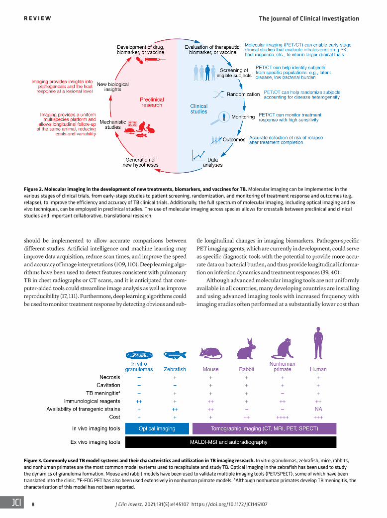
Figure 2. Molecular imaging in the development of new treatments, biomarkers, and vaccines for TB. Molecular imaging can be implemented in the various stages of clinical trials, from early-stage studies to patient screening, randomization, and monitoring of treatment response and outcomes (e.g., relapse), to improve the efficiency and accuracy of TB clinical trials. Additionally, the full spectrum of molecular imaging, including optical imaging and ex vivo techniques, can be employed in preclinical studies. The use of molecular imaging across species allows for crosstalk between preclinical and clinical studies and important collaborative, translational research.
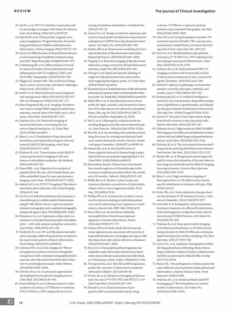
Figure 3. Commonly used TB model systems and their characteristics and utilization in TB imaging research. In vitro granulomas, zebrafish, mice, rabbits, and nonhuman primates are the most common model systems used to recapitulate and study TB. Optical imaging in the zebrafish has been used to study the dynamics of granuloma formation. Mouse and rabbit models have been used to validate multiple imaging tools (PET/SPECT), some of which have been translated into the clinic. 18F-FDG PET has also been used extensively in nonhuman primate models. AAlthough nonhuman primates develop TB meningitis, the characterization of this model has not been reported.

The Journal of Clinical Investigation R E V I E W
9J Clin Invest. 2021;131(5):e145107 https://doi.org/10.1172/JCI145107
Next-generation molecular imaging is an emerging technology that affords unparalleled opportunities for visualizing infec-tions, as molecular alterations occur earlier than structural changes. The ability for holistic, 3D spatial characterization and noninvasive, longitudinal monitoring in the same subject is a fundamental advantage over current tools and allows detailed insights into the dynamics and spatiotemporal disease hetero-geneity noted with TB. Since many molecular imaging tools are readily available for humans, they could advance fundamental knowledge by enabling basic biology studies in TB patients and accelerate the development of new therapeutics, as well as aid clinical management by serving as precision tools for diagnosis, monitoring, and prognostication.
Author contributionsAAO, EWT, and SKJ wrote the initial draft and designed the figures. KAL and SM planned and organized the workshop. All authors par-ticipated in the workshop as well as writing and editing the report. The order of co–first authors was determined alphabetically.
AcknowledgmentsThis Review was inspired by a workshop sponsored by the US National Institute of Allergy and Infectious Diseases on August 26–27, 2019, supported by the Division of Microbiology and Infectious Diseases. We thank all the organizers, speakers, and participants. Logistical support from Mediso USA is also acknowledged. The views expressed in this article are solely those of the authors and do not necessarily represent the official views of the NIH.
Address correspondence to: Sanjay K. Jain, Department of Pediat-rics, Johns Hopkins University School of Medicine, 1550 Orleans Street, CRB-II, Room 1.09, Baltimore, MD 21287, USA. Phone: 410.502.8241; Email: [email protected].
many developed countries (112). Chest CT can be performed rap-idly (seconds) with focused PET scans (3–5 minutes) without the need for sedation, even in young children, and with much lower radiation doses owing to improved CT technologies (4, 113). Addi-tionally, many molecular imaging tracers (PET, SPECT) currently under development for TB are rapidly excreted, which substantial-ly decreases radiation exposure.
It should be noted that although patients with MDR-TB have mortality risks similar to or higher than those of patients with many common cancers (112), the use of radiopharmaceutical imaging is accepted for the management of many cancers (even for children) but is avoided for infectious diseases. The advent of higher-resolution, high-sensitivity PET scanners, such as the EXPLORER total-body PET scanner, could increase the use of PET in both pediatric and adult patients with infectious diseases (114–116). MRI does not expose subjects to ionizing radiation but usually requires longer acquisition times. However, more recently, short-sequence lung MRI has been used for pulmonary imaging in TB patients (117). Additionally, low-field-strength (0.5 T) MRI equipped with state-of-the-art hardware can enable the develop-ment of lower-cost MRI machines with good image quality in the lung regions (118). Finally, although specialized equipment (cyclo-tron) is required to synthesize many PET agents, some radionu-clides, such as 68Ga, can be synthesized without radionuclide gen-erators and could be produced in remote areas (119). PET agents using 18F (e.g., 18F-FDG) can also be transported locally, usually within a 2- to 3-hour travel radius. By taking these logistics into consideration, a pragmatic approach could allow for more wide-spread adoption of imaging technologies for TB.
ConclusionsIn summary, TB is a 21st-century global health threat; howev-er, the field continues to be reliant on several diagnostic and research tools that were developed more than 100 years ago.
1. WHO. Global Tuberculosis Report 2020. World Health Organization; 2020.
2. Dye C, Williams BG. Eliminating human tubercu-losis in the twenty-first century. J R Soc Interface. 2008;5(23):653–662.
3. CDC. Emergence of Mycobacterium tuberculosis with extensive resistance to second-line drugs—worldwide, 2000–2004. MMWR Morb Mortal Wkly Rep. 2006;55(11):301–305.
4. Salazar-Austin N, et al. Extensively drug-resistant tuberculosis in a young child after travel to India. Lancet Infect Dis. 2015;15(12):1485–1491.
5. Boehme CC, et al. Rapid molecular detection of tuberculosis and rifampin resistance. N Engl J Med. 2010;363(11):1005–1015.
6. Conradie F, et al. Treatment of highly drug- resistant pulmonary tuberculosis. N Engl J Med. 2020;382(10):893–902.
7. The NIAID Tuberculosis Research Strategic Plan Working Group. NIAID Strategic Plan for Tuber-culosis Research. National Institute of Allergy and Infectious Diseases; 2018.
8. Davis SL, et al. Noninvasive pulmonary [18F]-2-fluoro-deoxy-d-glucose positron emission tomography correlates with bactericidal activity of tuberculosis drug treatment. Antimicrob Agents
Chemother. 2009;53(11):4879–4884. 9. Lin PL, et al. Sterilization of granulomas is com-
mon in active and latent tuberculosis despite within-host variability in bacterial killing. Nat Med. 2014;20(1):75–79.
10. Murawski AM, et al. Imaging the evolution of reactivation pulmonary tuberculosis in mice using 18F-FDG PET. J Nucl Med. 2014;55(10):1726–1729.
11. Gideon HP, et al. Variability in tuberculosis granuloma T cell responses exists, but a bal-ance of pro- and anti-inflammatory cytokines is associated with sterilization. PLoS Pathog. 2015;11(1):e1004603.
12. Marakalala MJ, et al. Inflammatory signaling in human tuberculosis granulomas is spatially orga-nized. Nat Med. 2016;22(5):531–538.
13. Ordonez AA, et al. Dynamic imaging in patients with tuberculosis reveals heterogeneous drug exposures in pulmonary lesions. Nat Med. 2020;26(4):529–534.
14. Dheda K, et al. Spatial network mapping of pul-monary multidrug-resistant tuberculosis cavities using RNA sequencing. Am J Respir Crit Care Med. 2019;200(3):370–380.
15. Dheda K, et al. Drug-penetration gradients asso-ciated with acquired drug resistance in patients
with tuberculosis. Am J Respir Crit Care Med. 2018;198(9):1208–1219.
16. Urbanowski ME, et al. Cavitary tuberculosis: the gateway of disease transmission. Lancet Infect Dis. 2020;20(6):e117–e128.
17. Jain SK, et al. Advanced imaging tools for childhood tuberculosis: potential applications and research needs. Lancet Infect Dis. 2020;20(11):e289–e297.
18. Ordonez AA, et al. Matrix metalloproteinase inhi-bition in a murine model of cavitary tuberculosis paradoxically worsens pathology. J Infect Dis. 2019;219(4):633–636.
19. Urbanowski ME, et al. Repetitive aerosol expo-sure promotes cavitary tuberculosis and enables screening for targeted inhibitors of extensive lung destruction. J Infect Dis. 2018;218(1):53–63.
20. Chen RY, et al. PET/CT imaging correlates with treatment outcome in patients with mul-tidrug-resistant tuberculosis. Sci Transl Med. 2014;6(265):265ra166.
21. Tangg SJ, et al. Efficacy and safety of linezolid in the treatment of extensively drug-resistant tuber-culosis. Jpn J Infect Dis. 2011;64(6):509–512.
22. Yanardag H, et al. Computed tomography and bronchoscopy in endobronchial tuberculosis. Can Respir J. 2003;10(8):445–448.
The Journal of Clinical Investigation R E V I E W
1 0 J Clin Invest. 2021;131(5):e145107 https://doi.org/10.1172/JCI145107
23. Lin PL, et al. PET CT identifies reactivation risk in cynomolgus macaques with latent M. tubercu-losis. PLoS Pathog. 2016;12(7):e1005739.
24. Peprah KO, et al. Characteristic magnetic reso-nance imaging low T2 signal intensity of necrotic lung parenchyma in children with pulmonary tuberculosis. J Thorac Imaging. 2012;27(3):171–174.
25. Liu J, et al. MRI detection of bacterial brain abscess-es and monitoring of antibiotic treatment using bacCEST. Magn Reson Med. 2018;80(2):662–671.
26. Goldenberg JM, et al. Differentiation of myosi-tis-induced models of bacterial infection and inflammation with T2-weighted, CEST, and DCE-MRI. Tomography. 2019;5(3):283–291.
27. Higgins LJ, Pomper MG. The evolution of imag-ing in cancer: current state and future challenges. Semin Oncol. 2011;38(1):3–15.
28. Sanli Y, et al. Neuroendocrine tumor diagnosis and management: 68Ga-DOTATATE PET/CT. AJR Am J Roentgenol. 2018;211(2):267–277.
29. Salas Fragomeni RA, et al. Imaging of nonpros-tate cancers using PSMA-targeted radiotracers: rationale, current state of the field, and a call to Arms. J Nucl Med. 2018;59(6):871–877.
30. Ordonez AA, et al. Molecular imaging of bacterial infections: overcoming the bar-riers to clinical translation. Sci Transl Med. 2019;11(508):eaax8251.
31. Blanc L, et al. Visualization of mycobacterial biomarkers and tuberculosis drugs in infected tissue by MALDI-MS imaging. Anal Chem. 2018;90(10):6275–6282.
32. Niehaus M, et al. Transmission-mode MALDI-2 mass spectrometry imaging of cells and tissues at subcellular resolution. Nat Methods. 2019;16(9):925–931.
33. Paine MRL, et al. Digestion-free analysis of peptides from 30-year-old formalin-fixed, par-affin-embedded tissue by mass spectrometry imaging. Anal Chem. 2018;90(15):9272–9280.
34. Ankrah AO, et al. PET/CT imaging of Mycobacte-rium tuberculosis infection. Clin Transl Imaging. 2016;4:131–144.
35. Via LE, et al. Infection dynamics and response to chemotherapy in a rabbit model of tuberculosis using [F-18]2-fluoro-deoxy-d-glucose positron emission tomography and computed tomography. Antimicrob Agents Chem. 2012;56(8):4391–4402.
36. Marjanovic S, et al. Expression of glycolytic iso-enzymes in activated human peripheral lympho-cytes — cell-cycle analysis using flow-cytometry. Exp Cell Res. 1991;193(2):425–431.
37. Diedrich CR, et al. SIV and Mycobacterium tuber-culosis synergy within the granuloma accelerates the reactivation pattern of latent tuberculosis. PLoS Pathog. 2020;16(7):e1008413.
38. Coleman MT, et al. Early changes by 18fluoro-deoxyglucose positron emission tomography coregistered with computed tomography predict outcome after Mycobacterium tuberculosis infec-tion in cynomolgus macaques. Infect Immun. 2014;82(6):2400–2404.
39. Ordonez AA, et al. A systematic approach for developing bacteria-specific imaging tracers. J Nucl Med. 2017;58(1):144–150.
40. Pena-Zalbidea S, et al. Chemoenzymatic radio-synthesis of 2-deoxy-2-[18F]fluoro-d-trehalose ([18F]-2-FDTre): a PET radioprobe for in vivo
tracing of trehalose metabolism. Carbohydr Res. 2019;472:16–22.
41. Lentz CS, et al. Design of selective substrates and activity-based probes for hydrolase important for pathogenesis 1 (HIP1) from Mycobacterium tuber-culosis. ACS Infect Dis. 2016;2(11):807–815.
42. Durkee MS, et al. Fluorescence modeling of in vivo optical detection of Mycobacterium tuberculosis. Biomed Opt Express. 2019;10(10):5445–5460.
43. Yang HJ, et al. Real-time imaging of Mycobacterium tuberculosis, using a novel near-infrared fluorescent substrate. J Infect Dis. 2017;215(3):405–414.
44. Cheng Y, et al. Rapid and specific labeling of single live Mycobacterium tuberculosis with a dual-targeting fluorogenic probe. Sci Transl Med. 2018;10(454):eaar4470.
45. Kamariza M, et al. Rapid detection of Mycobacterium tuberculosis in sputum with a solvatochromic treha-lose probe. Sci Transl Med. 2018;10(430):eaam6310.
46. Babin BM, et al. A chemiluminescent protease probe for rapid, sensitive, and inexpensive detec-tion of live Mycobacterium tuberculosis [preprint]. https://doi.org/10.1101/2020.09.14.296772. Posted on bioRxiv September 14, 2020.
47. Dai T, et al. A fluorogenic trehalose probe for tracking phagocytosed Mycobacterium tuberculo-sis. J Am Chem Soc. 2020;142(36):15259–15264.
48. Boot M, et al. Accelerating early antituberculosis drug discovery by creating mycobacterial indi-cator strains that predict mode of action. Antimi-crob Agents Chemother. 2018;62(7):e00083-18.
49. Akram AR, et al. In situ identification of Gram-negative bacteria in human lungs using a topical fluorescent peptide targeting lipid A. Sci Transl Med. 2018;10(464):eaal0033.
50. [No authors listed]. Clinical trial of six-month four-month regimens of chemotherapy in the treatment of pulmonary tuberculosis: the results up to 30 months. Tubercle. 1981;62(2):95–102.
51. Wallis RS, et al. Month 2 culture status and treatment duration as predictors of tuberculosis relapse risk in a meta-regression model. PLoS One. 2013;8(8):e71116.
52. Phillips PP, et al. Limited role of culture conver-sion for decision-making in individual patient care and for advancing novel regimens to confir-matory clinical trials. BMC Med. 2016;14:19.
53. Berry MP, et al. An interferon-inducible neutrophil-driven blood transcriptional signature in human tuberculosis. Nature. 2010;466(7309):973–977.
54. Gideon HP, et al. Early whole blood transcrip-tional signatures are associated with severity of lung inflammation in cynomolgus macaques with Mycobacterium tuberculosis infection. J Immunol. 2016;197(12):4817–4828.
55. Ho J, et al. A transcriptional blood signature dis-tinguishes early tuberculosis disease from latent tuberculosis infection and uninfected individuals in a Vietnamese cohort. J Infect. 2020;81(1):72–80.
56. Thompson EG, et al. Host blood RNA signatures predict the outcome of tuberculosis treatment. Tuberculosis (Edinb). 2017;107:48–58.
57. Vorster M, et al. Advances in imaging of tubercu-losis: the role of 18F-FDG PET and PET/CT. Curr Opin Pulm Med. 2014;20(3):287–293.
58. Esmail H, et al. Characterization of pro-gressive HIV-associated tuberculosis using
2-deoxy-2-[18F]fluoro-d-glucose positron emission and computed tomography. Nat Med. 2016;22(10):1090–1093.
59. Chen RY, et al. Using biomarkers to predict TB treatment duration (Predict TB): a prospective, randomized, noninferiority, treatment shorten-ing clinical trial. Gates Open Res. 2017;1:9.
60. Foss CA, et al. Biodistribution and radiation dosimetry of 124I-DPA-713, a PET radiotracer for macrophage-associated inflammation. J Nucl Med. 2018;59(11):1751–1756.
61. Ordonez AA, et al. Radioiodinated DPA-713 imaging correlates with bactericidal activity of tuberculosis treatments in mice. Antimicrob Agents Chemother. 2015;59(1):642–649.
62. Ehrlich P. Address in pathology on chemother-apeutics: scientific principles, methods, and results. Lancet. 1913;2(4694):445–451.
63. Pasipanodya JG, et al. Artificial intelligence- derived 3-way concentration-dependent antago-nism of gatifloxacin, pyrazinamide, and rifampi-cin during treatment of pulmonary tuberculosis. Clin Infect Dis. 2018;67(suppl 3):S284–S292.
64. Dartois V. The path of anti-tuberculosis drugs: from blood to lesions to mycobacterial cells. Nat Rev Microbiol. 2014;12(3):159–167.
65. Prideaux B, et al. High-sensitivity MALDI-MRM-MS imaging of moxifloxacin distribution in tuber-culosis-infected rabbit lungs and granulomatous lesions. Anal Chem. 2011;83(6):2112–2118.
66. Prideaux B, et al. The association between steril-izing activity and drug distribution into tubercu-losis lesions. Nat Med. 2015;21(10):1223–1227.
67. Manier ML, et al. Reagent precoated targets for rapid in-tissue derivatization of the anti-tubercu-losis drug isoniazid followed by MALDI imaging mass spectrometry. J Am Soc Mass Spectrom. 2011;22(8):1409–1419.
68. Blanc L, et al. High-resolution mapping of fluoroquinolones in TB rabbit lesions reveals specific distribution in immune cell types. Elife. 2018;7:e41115.
69. Dutta NK, et al. Statin adjunctive therapy short-ens the duration of TB treatment in mice. J Anti-microb Chemother. 2016;71(6):1570–1577.
70. Irwin SM, et al. Bedaquiline and pyrazinamide treatment responses are affected by pulmonary lesion heterogeneity in Mycobacterium tubercu-losis infected C3HeB/FeJ mice. ACS Infect Dis. 2016;2(4):251–267.
71. Prideaux B, et al. Mass spectrometry imaging of levofloxacin distribution in TB-infected pul-monary lesions by MALDI-MSI and continuous liquid microjunction surface sampling. Int J Mass Spectrom. 2015;377:699–708.
72. Carter CL, et al. Lipidomic dysregulation within the lung parenchyma following whole-thorax lung irradiation: markers of injury, inflammation and fibrosis detected by MALDI-MSI. Sci Rep. 2017;7(1):10343.
73. Hunter RL. The pathogenesis of tuberculosis: the early infiltrate of post-primary (adult pulmonary) tuberculosis: a distinct disease entity. Front Immunol. 2018;9:2108.
74. Ordonez AA, et al. Radiosynthesis and PET bioimaging of 76Br-bedaquiline in a murine model of tuberculosis. ACS Infect Dis. 2019;5(12):1996–2002.
The Journal of Clinical Investigation R E V I E W
1 1J Clin Invest. 2021;131(5):e145107 https://doi.org/10.1172/JCI145107
75. DeMarco VP, et al. Determination of [11C] rifamp-in pharmacokinetics within Mycobacterium tuber-culosis-infected mice by using dynamic positron emission tomography bioimaging. Antimicrob Agents Chemother. 2015;59(9):5768–5774.
76. Weinstein EA, et al. Noninvasive determination of 2-[18F]-fluoroisonicotinic acid hydrazide phar-macokinetics by positron emission tomography in Mycobacterium tuberculosis-infected mice. Antimi-crob Agents Chemother. 2012;56(12):6284–6290.
77. Mota F, et al. Radiosynthesis and biodistribution of 18F-linezolid in Mycobacterium tuberculosis- infected mice using positron emission tomogra-phy. ACS Infect Dis. 2020;6(5):916–921.
78. Tucker EW, et al. Noninvasive 11C-rifampin posi-tron emission tomography reveals drug biodistri-bution in tuberculous meningitis. Sci Transl Med. 2018;10(470):eaau0965.
79. Bartelink IH, et al. New paradigm for transla-tional modeling to predict long-term tuber-culosis treatment response. Clin Transl Sci. 2017;10(5):366–379.
80. Radtke KK, et al. Alternative dosing guidelines to improve outcomes in childhood tuberculosis: a mathematical modelling study. Lancet Child Adolesc Health. 2019;3(9):636–645.
81. Imperial MZ, et al. A patient-level pooled analysis of treatment-shortening regimens for drug-sus-ceptible pulmonary tuberculosis. Nat Med. 2018;24(11):1708–1715.
82. Strydom N, et al. Tuberculosis drugs’ distribu-tion and emergence of resistance in patient’s lung lesions: a mechanistic model and tool for regimen and dose optimization. PLoS Med. 2019;16(4):e1002773.
83. Tucker EW, et al. Microglia activation in a pedi-atric rabbit model of tuberculous meningitis. Dis Model Mech. 2016;9(12):1497–1506.
84. Foss CA, et al. Noninvasive molecular imag-ing of tuberculosis-associated inflammation with radioiodinated DPA-713. J Infect Dis. 2013;208(12):2067–2074.
85. Locke LW, et al. Use of a leukocyte-targeted peptide probe as a potential tracer for imaging the tuberculosis granuloma. Tuberculosis (Edinb). 2018;108:201–210.
86. Mattila JT, et al. Positron emission tomography imaging of macaques with tuberculosis identifies temporal changes in granuloma glucose metabo-lism and integrin α4β1-expressing immune cells. J Immunol. 2017;199(2):806–815.
87. Sly LM, et al. Survival of Mycobacterium tubercu-losis in host macrophages involves resistance to apoptosis dependent upon induction of antia-poptotic Bcl-2 family member Mcl-1. J Immunol. 2003;170(1):430–437.
88. Gan H, et al. Mycobacterium tuberculosis blocks crosslinking of annexin-1 and apoptotic envelope formation on infected macrophages to maintain virulence. Nat Immunol. 2008;9(10):1189–1197.
89. Palner M, et al. Preclinical kinetic analysis of the caspase-3/7 PET tracer 18F-C-SNAT: quantifying the changes in blood flow and tumor retention after chemotherapy. J Nucl Med. 2015;56(9):1415–1421.
90. Ordonez AA, et al. Caspase-based PET for evaluating pro-apoptotic treatments in a tuberculosis mouse model. Mol Imaging Biol. 2020;22(6):1489–1494.
91. Harper J, et al. Mouse model of necrotic tubercu-losis granulomas develops hypoxic lesions. J Infect Dis. 2012;205(4):595–602.
92. Belton M, et al. Hypoxia and tissue destruction in pulmonary TB. Thorax. 2016;71(12):1145–1153.
93. Darrah PA, et al. Prevention of tuberculosis in macaques after intravenous BCG immunization. Nature. 2020;577(7788):95–102.
94. Malherbe ST, et al. The potential of imaging tools as correlates of infection and disease for new TB vac-cine development. Semin Immunol. 2018;39:73–80.
95. Pai M, et al. Tuberculosis. Nat Rev Dis Primers. 2016;2:16076.
96. Blazevic A, et al. Pilot studies of a human BCG challenge model. Tuberculosis (Edinb). 2017;105:108–112.
97. Srivastava S, Ernst JD. Cutting edge: direct recog-nition of infected cells by CD4 T cells is required for control of intracellular Mycobacterium tubercu-losis in vivo. J Immunol. 2013;191(3):1016–1020.
98. Ernst JD, et al. Limited antimycobacterial effi-cacy of epitope peptide administration despite enhanced antigen-specific CD4 T-cell activation. J Infect Dis. 2018;218(10):1653–1662.
99. MacGilvary NJ, Tan S. Fluorescent Mycobacterium tuberculosis reporters: illuminating host-patho-gen interactions. Pathog Dis. 2018;76(3):fty017.
100. Tobin DM, et al. The lta4h locus modulates suscep-tibility to mycobacterial infection in zebrafish and humans. Cell. 2010;140(5):717–730.
101. Madigan CA, et al. A macrophage response to mycobacterium leprae phenolic glycolip-id initiates nerve damage in leprosy. Cell. 2017;170(5):973–985.
102. Cronan MR, et al. An explant technique for high-resolution imaging and manipulation of mycobacterial granulomas. Nat Methods. 2018;15(12):1098–1107.
103. Matty MA, et al. Potentiation of P2RX7 as a host-directed strategy for control of mycobacteri-al infection. Elife. 2019;8:e39123.
104. Barlerin D, et al. Biosafety level 3 setup for multi-photon microscopy in vivo. Sci Rep. 2017;7(1):571.
105. Entenberg D, et al. A permanent window for the murine lung enables high-resolution imaging of cancer metastasis. Nat Methods. 2018;15(1):73–80.
106. Wells G, et al. 3D microarchitecture of the human tuberculous granuloma [preprint]. https://doi.org/10.1101/2020.06.14.149898. Posted on bioRxiv June 15, 2020.
107. Coleman MT, et al. PET/CT imaging reveals a therapeutic response to oxazolidinones in macaques and humans with tuberculosis. Sci Transl Med. 2014;6(265):265ra167.
108. Malherbe ST, et al. Quantitative 18F-FDG PET-CT scan characteristics correlate with tuberculosis treatment response. EJNMMI Res. 2020;10(1):8.
109. Wahl RL, et al. Mars shot for nuclear medicine, molecular imaging, and molecularly target-ed radiopharmaceutical therapy. J Nucl Med. 2021;62(1):6–14.
110. LaLonde R, et al. Capsules for biomedical image segmentation. Med Image Anal. 2020;68:101889.
111. Khan FA, et al. Chest x-ray analysis with deep learning-based software as a triage test for pul-monary tuberculosis: a prospective study of diag-nostic accuracy for culture-confirmed disease. Lancet Digit Health. 2020;2(11):e573–e581.
112. Jain SK. The promise of molecular imaging in the study and treatment of infectious diseases. Mol Imaging Biol. 2017;19(3):341–347.
113. Andronikou S, et al. Technique, pitfalls, qual-ity, radiation dose and findings of dynamic 4-dimensional computed tomography for airway imaging in infants and children. Pediatr Radiol. 2019;49(5):678–686.
114. Cherry SR, et al. Total-body imaging: transform-ing the role of positron emission tomography. Sci Transl Med. 2017;9(381):eaaf6169.
115. Badawi RD, et al. First human imaging studies with the EXPLORER total-body PET scanner. J Nucl Med. 2019;60(3):299–303.
116. Lv Y, et al. Mini EXPLORER II: a prototype high-sensitivity PET/CT scanner for companion animal whole body and human brain scanning. Phys Med Biol. 2019;64(7):075004.
117. Sodhi KS, et al. Rapid lung MRI in children with pulmonary infections: time to change our diagnostic algorithms. J Magn Reson Imaging. 2016;43(5):1196–1206.
118. Campbell-Washburn AE, et al. Opportunities in interventional and diagnostic imaging by using high-performance low-field-strength MRI. Radiology. 2019;293(2):384–393.
119. Ebenhan T, et al. Preclinical evaluation of 68Ga- labeled 1,4,7-triazacyclononane-1,4,7-triacetic acid-ubiquicidin as a radioligand for PET infection imaging. J Nucl Med. 2014;55(2):308–314.
120. Mutch CA, et al. [11C]para-aminobenzoic acid: a positron emission tomography tracer targeting bacteria-specific metabolism. ACS Infect Dis. 2018;4(7):1067–1072.
121. Zhang Z, et al. Positron emission tomogra-phy imaging with 2-[18F]F-p-aminobenzoic acid detects Staphylococcus aureus infections and monitors drug response. ACS Infect Dis. 2018;4(11):1635–1644.
122. Sule P, et al. Rapid tuberculosis diagnosis using reporter enzyme fluorescence. J Clin Microbiol. 2019;57(12):e01462-19.
123. Kong Y, et al. Imaging tuberculosis with endog-enous beta-lactamase reporter enzyme fluo-rescence in live mice. Proc Natl Acad Sci U S A. 2010;107(27):12239–12244.
124. Backus KM, et al. Uptake of unnatural trehalose analogs as a reporter for Mycobacterium tubercu-losis. Nat Chem Biol. 2011;7(4):228–235.
125. Ordonez AA, et al. Imaging chronic tuberculous lesions using sodium [(18)F]fluoride positron emission tomography in mice. Mol Imaging Biol. 2015;17(5):609–614.
126. Davis SL, et al. Bacterial thymidine kinase as a non-invasive imaging reporter for Mycobac-terium tuberculosis in live animals. PLoS One. 2009;4(7):e6297.
127. Foss CA, et al. SPECT/CT imaging of Mycobac-terium tuberculosis infection with [125I]anti-C3d mAb. Mol Imaging Biol. 2019;21(3):473–481.
128. Zhang Z, et al. The biodistribution of 5-[18F]fluoropyrazinamide in Mycobacterium tuberculosis-infected mice determined by positron emission tomography. PLoS One. 2017;12(2):e0170871.
129. Singh N, Bhatnagar A. Clinical evaluation of effi-cacy of 99mTC -ethambutol in tubercular lesion imaging. Tuberc Res Treat. 2010;2010:618051.