VISUAL LITERACY IN ANATOMY J. Bradley Barger Submitted ...
237
VISUAL LITERACY IN ANATOMY J. Bradley Barger Submitted to the faculty of the University Graduate School in partial fulfillment of the requirements for the degree Doctor of Philosophy in the Department of Anatomy & Cell Biology, Indiana University July 2016
-
Upload
khangminh22 -
Category
Documents
-
view
0 -
download
0
Transcript of VISUAL LITERACY IN ANATOMY J. Bradley Barger Submitted ...
VISUAL LITERACY IN ANATOMY
J. Bradley Barger
Submitted to the faculty of the University Graduate School
in partial fulfillment of the requirements
for the degree
Doctor of Philosophy
in the Department of Anatomy & Cell Biology,
Indiana University
July 2016

ii
Accepted by the Graduate Faculty, Indiana University, in partial
fulfillment of the requirements for the degree of Doctor of Philosophy.
Doctoral Committee
April 29, 2016
___________________________________________
Valerie Dean O’Loughlin, Ph.D., Chair
___________________________________________
James Brokaw, Ph.D.
____________________________________________
David Estell, Ph.D.
____________________________________________
Anthony Mescher, Ph.D.

iii
Dedication
This dissertation is dedicated to Laura and Simon. The two of you have been the
most supportive and loving family I could ever want.
iv
Acknowledgements
I would like to thank all of the people who helped me see this project to completion:
Valerie O’Loughlin for guidance, mentorship, and being a fantastic committee
chair.
The other members of my research committee James Brokaw, David Estell,
and Anthony Mescher for valuable input on the research and writing process
Jackie Cullison for helping to distribute and de-identify surveys.
The faculty of the anatomy department for feedback on survey and research
design.
The students in all of my classes who were always supportive of trying new
teaching techniques, and (usually) happy to complete surveys.
The other graduate students in the department who were a constant source
of good ideas, encouragement, and fun times.
v
J. Bradley Barger
VISUAL LITERACY IN ANATOMY
All branches of anatomy (gross anatomy, histology, neuroanatomy, and embryology)
involve significant amounts of visual identification. Understanding the spatial
relationship and visual representations of anatomical structures forms the basis for
much of anatomy education, particularly in laboratory courses. Students in these
courses frequently struggle with the visual aspects of identification, and many lack
the metacognitive awareness to identify this problem. The research presented here
details a series of experiments designed to elucidate the factors involved in
students’ difficulties with studying the visual aspects of anatomy. All of the research
projects discussed involved surveying students about their specific study habits.
Student populations surveyed include first-year medical students and
undergraduates in anatomy, physiology. These populations were surveyed about
their study habits in each course, and their level of familiarity with visual learning.
Additionally some populations were given a mental rotation test to assess their
spatial abilities. These survey data were then correlated with course grades in an
effort to determine the most successful study strategies. Active learning approaches
(including student-produced drawings) were most strongly correlated with high
course grades. However, efforts to teach lower-performing students active learning
skills did not produce significant results, possibly due to the lack of a metacognitive
component in this instruction. The results of each project indicate a lack of good
study skills among students at all levels of anatomy instruction, and highlight the
vi
need for more instruction in how to study for anatomy, including metacognitive
awareness, especially focused on the visual aspects of the course.
Valerie Dean O’Loughlin, Ph.D., Chair
vii
Table of Contents
CHAPTER 1: Introduction ...................................................................................................................... 1
Definitions ................................................................................................................................................ 5
Dissertation Research Outline ......................................................................................................... 8
General arrangement of the dissertation ................................................................................. 12
CHAPTER 2: Visual Literacy and Spatial Ability in Educational Research ..................... 14
Visual Literacy ..................................................................................................................................... 15
Visual literacy as a concept, construct, cultural resource or measurable
phenomenon? .................................................................................................................................. 20
The ACRL List of visual literacy competency standards ............................................... 22
Visual literacy in Fine Arts ......................................................................................................... 30
Visual literacy in biochemistry ................................................................................................ 34
Visual literacy in astronomy ..................................................................................................... 38
Visual literacy in anatomy .......................................................................................................... 38
What does the visual literacy research in the other fields mean for anatomy? .. 39
Spatial Ability ....................................................................................................................................... 42
Does Gender play a role in spatial ability skills? .............................................................. 43
SA in fields other than anatomy .............................................................................................. 45
Spatial ability in geology ............................................................................................................. 46
Spatial ability in chemistry ........................................................................................................ 48
Spatial ability in engineering .................................................................................................... 51
Spatial ability in physics ............................................................................................................. 52
Non-discipline specific spatial ability ................................................................................... 53
Spatial ability in anatomy ........................................................................................................... 55

viii
Summary of Visual Literacy and Spatial Ability ............................................................... 60
CHAPTER 3: Materials, Methods, and Research Questions .................................................. 62
Student populations and courses examined ........................................................................... 63
Research Questions and Methodology of Each Project ...................................................... 66
Methodology ......................................................................................................................................... 72
CHAPTER 4: Assessing Student Study Habits in Anatomy and Physiology ................... 87
Methodology ......................................................................................................................................... 90
Results ..................................................................................................................................................... 92
Discussion .............................................................................................................................................. 99
CHAPTER 5: Visual Study Skills in Anatomy ............................................................................. 103
Methodology ....................................................................................................................................... 112
Results ................................................................................................................................................... 114
Discussion ............................................................................................................................................ 118
CHAPTER 6: Mental Rotation .......................................................................................................... 122
Methodology ....................................................................................................................................... 126
Results ................................................................................................................................................... 127
Discussion ............................................................................................................................................ 132
CHAPTER 7: In-class Drawings ....................................................................................................... 134
Methodology ....................................................................................................................................... 137
Results ................................................................................................................................................... 140
Discussion ............................................................................................................................................ 144
CHAPTER 8: Online Drawing Instruction ................................................................................... 147
Methodology ....................................................................................................................................... 148
Results ................................................................................................................................................... 151
Discussion ............................................................................................................................................ 153
ix
CHAPTER 9: Conclusions ................................................................................................................... 156
Discussion ............................................................................................................................................ 158
Limitations .......................................................................................................................................... 174
Future Directions ............................................................................................................................. 177
Final Summary ................................................................................................................................... 178
APPENDIX A: Survey Design, Validation, and Sample Surveys ......................................... 180
APPENDIX B: Mental Rotation Test .............................................................................................. 185
APPENDIX C: Course Syllabus Excerpts ...................................................................................... 198
APPENDIX D: IRB Documents ......................................................................................................... 201
APPENDIX E: SPSS outputs ............................................................................................................... 205
References ................................................................................................................................................ 207
Curriculum Vitae
x
List of tables
1.1 The relationship between research questions and the individual facets .................. 9
2.1 Visual literacy skills and competencies ................................................................................. 41
3.1 The relationship between research questions and the individual facets ............... 73
3.2 Populations and participants ..................................................................................................... 77
3.3 Populations and participants MRT .......................................................................................... 83
4.1 The relationship between research questions and the individual facets ............... 91
4.2 Populations and participants study habits .......................................................................... 91
4.3 Correlations between study habits and grade by course ............................................... 97
5.1 Visual literacy skills and competencies ............................................................................... 108
5.2 The relationship between research questions and the individual facets ............. 111
6.1 The relationship between research questions and the individual facets ............. 125
7.1 The relationship between research questions and the individual facets ............. 137
9.1 The relationship between research questions and the individual facets ............. 157
xi
List of Figures
3.1 Example drawing from visual lecture .................................................................................... 82
3.2 Draw It to Know It software example ..................................................................................... 85
4.1 Correlations of active learning and course grade ............................................................. 95
4.2 Correlations of pasive learning and course grade ............................................................ 96
5.1 Comparison of early and late semester visual learning................................................ 115
5.2 Correlation of visual learning and final grade .................................................................. 116
5.3 Correlation of visual preference and final grade ............................................................. 117
5.4 Correlation of visual preference and visual learning .................................................... 118
6.1 Mental rotation scores ................................................................................................................ 128
6.2 Correlation of mental rotation scores and final grade .................................................. 129
6.3 Correlation of visual study skills and mental rotation score ...................................... 130
6.4 Correlation of visual preference with mental rotation score ..................................... 131
7.1 Mental rotation experimental and control ......................................................................... 141
7.2 Visual preference experimental and control ..................................................................... 142
7.3 Visual studying experiemntal and control ......................................................................... 143
7.4 Correlation of drawing and final grade ................................................................................ 144
8.1 Draw It to Know It software example ................................................................................... 149
8.2 Active learning and software use ........................................................................................... 152
8.3 Visual learning and software use .......................................................................................... 153

1
CHAPTER 1: Introduction
Success in anatomy courses requires a variety of study skills, including the ability to
understand visual representations of anatomical structures. Students in anatomy
courses are often provided with instruction in how to study, but this instruction
rarely includes a visual element. That is, if visual learning is recommended to
students, the recommendation may not include all of the information a student
needs to confidently engage in visual learning. Consequently, students are often left
to interpret images using their existing (often inadequate) visual skills. It is the goal
of this multi-faceted dissertation project to examine the visual skills students bring
to anatomy courses, to examine if certain prior visual skills are necessary (or
sufficient) to succeed in anatomy, to determine if students leave anatomy classes
with a greater development of visual skills, and to explore methods of teaching
visual skills to anatomy students.
The current literature in anatomy education has many examples of teaching spatial
ability (with few devoted to visual literacy) to students (Keehner et al. 2006, Provo
et al. 2002, Lufler et al. 2012, Luursema et al. 2008, Boudreau et al. 2008, Hegarty et
al. 2009, Wanzel et al. 2002, Fuks et al. 2009, Garg et al. 2001, Lawrence et al. 2014,
Roach et al. 2012, Roach et al. 2014, Roach et al. 2016, Backhouse et al. 2016,
Pickering 2015, Mione, et al. 2013), but these are examples of specific experimental
implementations, or research on new types of visual representations. There is very
little published literature regarding the basic visual instruction given to students
(Bardes et al. 2001). That is, outside of specific visual learning interventions, it is
2
unclear how students are instructed to understand images in anatomy courses.
While these specific interventions (described in the above citations) provide
valuable information about how students use images in learning, the information
about student use of images outside of experimental designs is lacking. Data
collected from course syllabuses and instructor comments (only from five courses at
Indiana University, see course descriptions in chapter 3) indicates that visual
approaches to learning are encouraged, but no class time is spent developing the
skills needed to engage in visual learning (see appendices for course syllabuses).
The published literature in visual learning indicates that acquiring the skills needed
to understand images is not a passive process, and specific instruction is required
for students to access the informational content of images (Titus and Horsman 2009
is a notable exception, this paper demonstrated significant passive acquisition of
spatial ability in geology students). For a detailed summary of the published
literature concerning visual literacy and spatial ability in anatomy students, see
chapter 2.
The research presented in this dissertation is divided into three main components-
examining students study skills, comparing visual literacy and spatial ability skills to
course performance, and improving visual literacy and spatial ability in students.
These three main components are further discussed in the following paragraphs,
with detailed methodologies and results discussed in chapters 4-8.
Having reviewed the existing literature, and finding a lack of information about the
visual literacy skills naïve students bring to their anatomy courses, the first step of
3
this dissertation project was to determine what our students are doing when
studying for anatomy. By examining their study habits, their familiarity and comfort
with visual literacy and spatial ability can be inferred. Knowing what the students
do to study, when not provided with any explicit instruction in study methods, helps
to fill the gap defined above. That is, what kind of visual literacy skills do students
bring to class? The first phase of dissertation research used early-semester surveys
to determine what kind of study techniques students are using, without any specific
training in how to study. Chapters 4-6 explore this question in greater detail, with
detailed methodology and results included.
The second step in this dissertation was to determine if visual literacy (and spatial
ability) skills change during an anatomy course. Acquisition of spatial ability (but
not visual literacy) has been documented (Keehner et al. 2006, Provo et al. 2002,
Lufler et al. 2012, Luursema et al. 2008, Boudreau et al. 2008, Hegarty et al. 2009,
Wanzel et al. 2002, Fuks et al. 2009, Garg et al. 2001, Lawrence et al. 2014, Roach et
al. 2012, Backhouse et al. 2016, Pickering 2015, Mione, et al. 2013) in a variety of
science courses, including anatomy, but often only through a specific pedagogical
intervention. Some studies (Titus and Horsman 2009) have shown a passive
acquisition of visual skills simply by taking a course, but this has not been frequently
repeated. This part of the dissertation work used early- and late-semester surveys
and mental rotation tests (MRT) to examine passive acquisition of visual skills.
Chapters 5-6 deal with this question in greater detail, including detailed
methodology and results.
4
The final major facet of this dissertation work explored ways of instructing students
in using visual literacy and spatial ability skills in their studying. There are many
published studies describing the benefits and drawbacks of specific pedagogical
interventions in teaching visual literacy or spatial ability (Keehner et al. 2006, Provo
et al. 2002, Lufler et al. 2012, Luursema et al. 2008, Boudreau et al. 2008, Hegarty et
al. 2009, Wanzel et al. 2002, Fuks et al. 2009, Garg et al. 2001, Lawrence et al. 2014,
Roach et al. 2012, Backhouse et al. 2016, Pickering 2015, Mione, et al. 2013 are just
a sample, for a full accounting of the various debates in the visual learning literature,
see chapter 2). A number of contradictions are apparent in the literature regarding
the teaching of visual skills. For example, the literature examining two-dimensional
vs. three-dimensional images, static vs. animated images, or interactive vs. passive
representations; contains support for any of these visual representations as the
‘best’ for teaching different facets of anatomy. It may be that all types of visual
representations are useful in different contexts, or with different students
populations, or maybe the instruction in visual learning is the important feature,
and the type of image is less important than the underlying visual skills being
conveyed in these different instructional contexts. Sadly, this question is still
unanswerable. The existing literature has yet to converge on a consensus, and this
dissertation project does not argue for one variety of image or instructional
technique over another, but rather makes the case for spending time providing
instruction in visual literacy and mental rotation as part of an anatomy course. To
make that case, two different methods of teaching visual literacy were used. The
first method included in-person drawing instruction combined with anatomy
5
content. The second method used a series of web-based drawing tutorials which
allowed students to draw along with a narrator explaining neuroanatomy content.
Both of these methods showed some increase in student use of visual study skills,
again indicating that the teaching of visual literacy, at all, may be more important
than the specific type of images being used. This result is seen many times in the
literature review in chapter 2 (under subheading “Non-discipline specific spatial
ability”). Full details of the pedagogical interventions used to teach spatial ability
and visual literacy can be found in chapters 7-8 with methodology and results.
Definitions
Before continuing, it is worthwhile to carefully define some of the terms that are
used frequently throughout the following projects. Study skills, study habits, visual
skills, visual learning, and visual study skills are some terms with variable definitions,
and they may be used by different authors in different ways. The following
paragraphs define these terms as they are used in the context of this research.
Study skills, study habits, and study strategies are sometimes defined differently
(Credé and Kuncel 2008, Morehead et al. 2016), but in this research they are used
interchangeably. Study skills (a.k.a. study habits or study strategies) are patterns of
effort and techniques used by students outside of class to learn course material and
prepare for exams (Schutte 2013, Husmann et al. 2016, McGuire 2015).
Visual skills are the entire set of mental processes related to understanding the
visual world, and include spatial ability and visual literacy. This definition is unique
to this dissertation, as the term ‘visual skills’ has been used by different fields to
6
refer to any and all types of vision. (For example, researchers in human vision use
‘visual skills’ to refer to normal operation of the visual apparatus and brain, see
Achtman et al. 2008). Visual skills (as used in this dissertation) will be discussed at
length in the literature review, including the historical arguments about definitions
and utility of visual skills in education.
Visual learning includes all of the activities a student may use to employ visual
skills in studying. This is closely related to visual study skills, and these two terms
will often be used interchangeably. Visual learning has a well-established definition
in the educational literature (Azer 2013, Estevez et al. 2010, Fernandez et al. 2011,
Lufler et al. 2012, Nelson 2004).
Some additional terms are used frequently in this research, and may need a
definition for consistency. These terms have widely recognized definitions, but
other authors may use them slightly differently, so I will define each of the following
as they are used in this research.
Active learning is any study approach in which the student physically creates
something (Drapkin et al. 2015, Pickering 2014, Sweeney et al. 2014, for examples
of active learning in anatomy). Active learning can include writing, making a chart,
drawing, or modeling. Discussions with other students or instructors can also be
considered active learning. Some sources (LCME 2015) require students to create
their own learning objectives for an activity to be considered ‘active,’ but that
definition is not used here.
7
Passive learning is any study approach that does not produce a physical artifact.
Passive learning primarily consists of reading or listening to lectures. Rote copying
may also be considered passive learning because it does not engage a student’s
higher cognitive faculties. These definitions of active and passive learning focus on
the physical product made by a student for the sake of simplicity. In reality, the main
difference between these approaches the level of mental engagement a student has
during the learning activity; however, measuring mental engagement is difficult,
while measuring products made by students is easy. Therefore, production of a
physical product is used a proxy for the level of student engagement (Prince 2004
for a meta-analysis of active learning literature, Griffith 2015 for a primary school
example, Meyers and Jones 1993 and Silberman 1993 for some early examples of
active learning research).
Superficial learning is focused on reproducing results, and is often used by novice
students who are grade oriented (Marton and Saljo 1976, Biggs et al. 2001, Young
2005, Billett 2001, Pandey and Zimitat 2007, Ramberg and Karlgren 1998). This
approach leads students to focus on memorizing content in anticipation of
examinations.
Deep Learning is a learning approach focused on understanding content and
making connections to existing knowledge. This approach is often used by students
with more experience studying, and tends to lead to greater long-term retention
(Marton and Saljo 1976, Biggs et al. 2001, Young 2005, Billett 2001, Karjcik and
Blumenfeld 2006).
8
Metacognition is a conscious awareness of one’s own understanding of a topic
(Biggs 1988, Bransford et al. 2000, Bjork et al. 2013, Flavell 1979, Hacker et al. 1998,
Kamp et al. 2015, Kornell and Bjork 2007, Metcalfe 2008, Vadhan and Stander 1994,
Zohar and Barzilai 2013). Students new to college often lack the study skills and
self-reflective skills to regulate their own learning, they “don’t know what they don’t
know;” a definitive lack of metacognition (Bransford et al. 2000). Further, these
novice students (as defined by Bransford et al.) are often unaware of even the
concept of metacognition, and are therefore, not only unable to regulate their own
learning, but are lacking the tools necessary to understand self-regulation
(Bransford et al. 2000). Many students are able to ‘trick’ themselves into thinking
they know more than they actually do. A lack of metacognitive skills makes self-
regulation of learning nearly impossible, and students may study inefficiently and
approach exams with undue confidence (Kruger and Dunning 1999).
Dissertation Research Outline
Unless one had abundant resources and an endless pool of volunteers, no single
study could effectively address the three main components listed above (guided by
the eight research questions described in chapter 3). Multiple different studies
(entitled “facets”, below) were needed to examine each of the following research
questions (for a detailed breakdown of the research questions see chapter 3,
subheading “Research questions”). Sometimes, one study or facet could address
multiple research questions. Thus, depending on the research question, one or
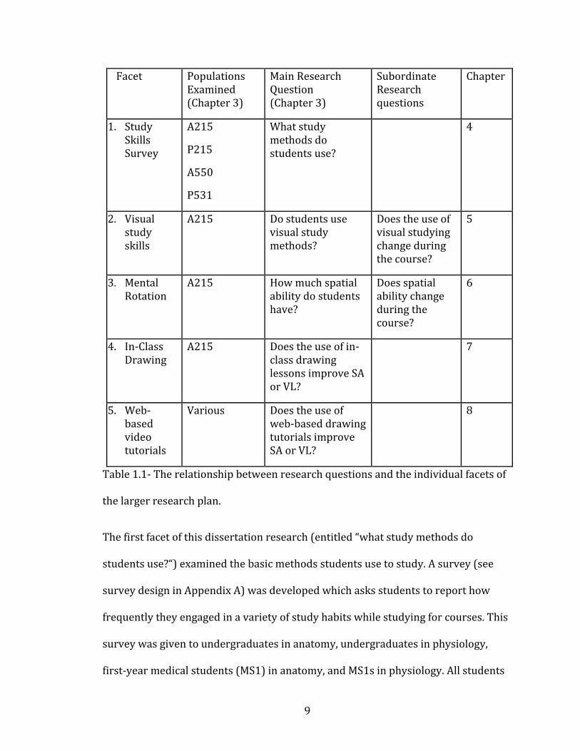
more studies (facets) may provide evidence for the question. Table 1.1 provides a
visual for how the facets of this dissertation research map to the research questions:
9
Facet Populations Examined (Chapter 3)
Main Research Question (Chapter 3)
Subordinate Research questions
Chapter
1. Study Skills Survey
A215
P215
A550
P531
What study methods do students use?
4
2. Visual study skills
A215 Do students use visual study methods?
Does the use of visual studying change during the course?
5
3. Mental Rotation
A215
How much spatial ability do students have?
Does spatial ability change during the course?
6
4. In-Class Drawing
A215 Does the use of in-class drawing lessons improve SA or VL?
7
5. Web-based video tutorials
Various Does the use of web-based drawing tutorials improve SA or VL?
8
Table 1.1- The relationship between research questions and the individual facets of
the larger research plan.
The first facet of this dissertation research (entitled “what study methods do
students use?“) examined the basic methods students use to study. A survey (see
survey design in Appendix A) was developed which asks students to report how
frequently they engaged in a variety of study habits while studying for courses. This
survey was given to undergraduates in anatomy, undergraduates in physiology,
first-year medical students (MS1) in anatomy, and MS1s in physiology. All students
10
surveyed were on the campus of Indiana University, Bloomington. A four-way
comparison was then conducted to look for differences in study approaches in the
two different classes at two different levels. Some of these data were previously
reported in Husmann et al. 2016. Husmann et al. examined the changes in study
skills exhibited by individual students between their different classes. The analysis
of changing study skills is not part of this dissertation. The correlations of study
skills with grades reported in this dissertation have also been presented as a poster
at the Experimental Biology national meeting 2011 (Husmann and Barger 2011).
The detailed methodology, results and discussion of this facet may be found in
chapter 4 of this dissertation.
The next facet of this dissertation research (entitled: “visual study skills”) involved a
more refined survey (Survey Appendix A), which included more specific questions
dealing with visual approaches to studying. This survey was given only to
undergraduates, due to the relatively small number of medical students available for
research (n = 36). Additionally, a mental rotation test (MRT) was developed by
modifying the work of Bodner and Guay (1997). The MRT functions as a measure of
students’ ability to mentally manipulate two-dimensional representations of three-
dimensional objects. This ability is necessary in anatomy, and a student’s MRT
scores have been shown to correlate with success in visually oriented science
courses (Keehner et al. 2006, among many others detailed in the literature review).
Preliminary versions of the results found in chapter 5 were also presented at the
Experimental Biology national meeting 2012 (Barger 2012). The detailed

11
methodology, results and discussion of this facet may be found in chapters 5-6 of
this dissertation
Having established a baseline of student performance and visual skills, the third
part of this dissertation research introduced a pedagogical intervention designed to
improve student MRT scores, visual approaches to studying, and course success
metrics. This intervention consisted of teaching students how to draw specific
anatomical structures, while learning the normal anatomy course content, during
regular lectures. The instructor led the students through guided drawings of
anatomical structures, while explaining their functions and relationships. Students
involved in this intervention showed small improvements in the above measures,
but none of the changes reached statistical significance. Preliminary versions of the
results found in chapter 7 were also presented at the Experimental Biology national
meeting 2013 (Barger and Husmann 2013). The detailed methodology, results and
discussion of this facet may be found in chapter 7 of this dissertation.
In an effort to increase student exposure to anatomical drawing, the next facet of
this research moved all of the experimental materials (surveys, MRT, and drawings)
to an online environment, with the goal of delivering more drawing practice to busy
students. The online drawings were part of a software tool called Draw It to Know
It. This software was developed by a neurologist (and former IU student) who was
unconnected to the research (Draw it to know it 2016). This phase of the research
showed that a proportion of students are resistant to visual learning, an expected,
but disappointing finding. However, even among students who reported a distaste
12
for visual learning in general (and drawing specifically) they reported
understanding the necessity of learning how to use images in their studying. The
detailed methodology, results and discussion of this facet may be found in chapter 8
of this dissertation.
All of these pieces, taken as a whole, show the need for increased instruction in
visual skills for anatomy students. Many students enter college (or medical school)
with inadequate visual learning skills, do not necessarily understand the utility of
visual communication, and have no way of improving these skills without specific
instruction. The following chapters will explain each of these facets in greater detail,
with a final summary detailing the pedagogical implications for anatomy instructors.
General arrangement of the dissertation
The following chapters in this dissertation are arranged around the research
questions and individual projects defined in Table 1.1 (above), starting with
chapter four. Preceding the individual projects chapters, chapter 2 is a summary
and discussion of the existing publications related to visual literacy and spatial
ability. Chapter 2 is subdivided in specific sections dealing with visual literacy
(with specific examples from many of the disciplines to have researched the
teaching of visual literacy) and spatial ability (again with a discipline specific
organization of the literature). Chapter 3 outlines the research questions guiding
the research conducted in chapter 4-8. Chapter 4 reports the detailed methodology,
survey, and student populations used to establish the baseline of student study
habits followed by results. Chapter 5 contains detailed methodology, survey, and
13
student populations used to research the visual literacy of anatomy
undergraduates, with results. Chapter 6 introduces a measurement of spatial
ability, and contains the detailed methodology of measuring spatial ability in
anatomy undergraduates, with results correlating spatial ability with a variety of
other measures of success in an anatomy course. Chapter 7 introduces a
pedagogical intervention (in-class drawings) designed to teach spatial ability and
visual literacy, and compares an experimental and control group in measures of
spatial ability, visual literacy, and course grades. Chapter 8 uses a similar
pedagogical intervention to that in chapter 7, but the drawings are delivered
through a web-based series of video tutorials. Chapter 8 contains detailed
methodology and results, comparing use of the web-based tutorials to mental
rotation score, visual literacy, and other study skills. Chapter 9 is a detailed
discussion, summarizing the results of the previous chapters, relating the research
of this dissertation to the exiting literature, and discussing limitations of the
current research. Following chapter 9 are a series of appendices including the
surveys used, the statistical validation of the surveys, the mental rotation test used,
IRB approval numbers, IRB informed consent documents (also called ‘study
information sheets’), IRB recruitment documents, and syllabuses from courses
surveyed.
14
CHAPTER 2: Visual Literacy and Spatial Ability in Educational Research
All four branches of the anatomical sciences (gross anatomy, neuroanatomy,
embryology and histology) share the properties of visual identification,
understanding of structure/function relationships, and understanding the
relationships and interconnections of all the parts of the body. To be successful in
anatomy courses, a student needs a strong set of visual-spatial skills, due the
importance of visual identification of structures in anatomy courses. To be
successful in visual identification, a student must be able to apply a variety of visual
skills. These skills include visual literacy, spatial ability, and mental rotation. Visual
literacy (VL) is the skill of interpreting visual images (Avgerinou and Ericson 1997,
Fransecky and Debes 1977). This skill includes interpreting drawings of anatomical
structures, three-dimensional models, and charts or graphs. While this short
definition of VL seems simple, it contains a long and complicated history which will
be discussed in the following section.
Another term occasionally seen in the literature is pattern recognition. This term is
most frequently associated with foreign language learners (Martinez-Trinidad and
Guzman-Arenas 2001), and computer-based neural networks designed to identify
patterns (Carpenter and Grossberg 1988). While this term accurately describes some
of the behaviors anatomy experts use in identifying structures and relationships
(Berliner 1988), it is used infrequently in publications related to visual literacy
pedagogy.
15
Spatial ability (SA) is the related, but distinct, ability a person has for mentally
manipulating three-dimensional mental objects (Linn and Peterson 1985, Linn
and Peterson 1986, Lohman 1979). Mental rotation (MR) is a specific sub‐category of
spatial ability, and will be used as a measurement of spatial ability (Bodner and
Guay 1997). These three skills (VL, MR, and SA), which I will collectively define as
visual skills (VS), are critical for success in all fields of anatomy, and in many other
science courses, but are often overlooked by students. Instructors can also
overlook the need for visual skills, and it is often assumed that students will begin
the course with the relevant background skills, or pick them up during the course,
without any specific instruction in improving visual skills (Faurie and Khadra,
2012). (As clarification of the term ‘visual skills,’ I am unaware of any educational
literature that uses this term. When the term ‘visual skills’ appears in published
literature, it is often used as a near synonym for ‘visual ability,’ that is, the actual
functions and processes related to normal human vision. I am using the term
‘visual skills’ to refer to the mental processes students use to make meaning from
images, and not to the functioning of their eyes). Spatial ability and mental
rotation will be discussed in the last section of this chapter.
Visual Literacy
Visual literacy (VL) is the ability to meaningfully interpret static and dynamic
images, and has a long and contentious history, described in the following pages
(Avgerinou and Ericson 1997). Despite the simple, one-line definition for visual
literacy given above, it is a complicated and wide‐ranging topic that has resisted a
16
firm definition for the last fifty years. Practitioners and researchers in a variety of
fields have each applied their own unique definition to this term, making it even
harder to reach a consensus definition (Avgerinou and Ericson 1997). Very little
has been written about visual literacy in anatomy or biology, so this review will
include literature from other fields, including biochemistry, astronomy, and fine
arts. Additionally, writings by philosophers and historians of science (Pauwels
2006, Marcaida 2016), sociologists (Grady 2006), and graphic designers (Tufte
1990) will be considered in order to help define the limits and goals of visual
literacy. The other gap in the visual literacy literature is in assessment. So much
time and effort has been spent in trying to define ‘visual literacy’ as a term, that
few writers have commented on the most effective ways of teaching the actual
skills involved in visual literacy. The lack of assessable goals for the visual literacy
movement is directly linked to the definition problem, and a major source of
concern for educational psychologists (Seels 1994).
Visual literacy was first defined in 1969 by John Debes as “…a group of vision-‐‐
competencies a human being can develop by seeing and at the same time having
and integrating other sensory experiences… when developed they enable a
visually literate person to discriminate and interpret visible actions, objects,
symbols… Through the creative use of these competencies he is able to
communicate with others,” (Fransecky and Debes 1972, Avgerinou and Ericson
1997, p. 281.) In the intervening years, most other authors on the subject of
visual literacy have modified this definition to best fit their field of study; these
17
will be discussed in greater detail in the discipline specific sections to follow.
However, all of the subsequent definitions seem to retain the basic kernel of
interpretation of symbols, and visual communication. A full summary of the
historical debate concerning VL is beyond the scope of this work, as I will be
focusing on the current definitions and applications in use across a variety of
fields. For a review of the history of VL see Avgerinou and Ericson (1997).
While I will not be exploring the historical squabbles over semantics in VL, there
have been a few interesting arguments made against VL as a term and as a
concept. Some authors (e.g., Cassidy and Knowlton 1983, Suhor and Little 1988)
have even argued for abandoning the term as a misleading and potentially
damaging concept. Suhor and Little (1988) argue that semiotics already
encompasses the majority of what is meant by visual literacy, and that education
in semiotics is more valuable than creating a new term of visual literacy. While I
agree that semiotics and VL have some overlap (discussed in the section on Fine
Arts, below) I do not think VL and semiotics are synonymous enough for one
concept to replace the other. Cassidy and Knowlton (1983) argue that the term
“visual literacy” is used as a scientific metaphor, and that it fails on the principles
used to judge other scientific metaphors. However, I find their argument a weak
one, because they have applied their own definition to the term “visual literacy”
which seems to overlook the goal of the original definition. The definition used by
Cassidy and Knowlton (1983) very literally interprets the various meaning of
‘visual’ and ‘literacy’ in such a way that “visual literacy” as a separate concept can
18
no longer exist. They also argue that no evidence has been shown that visual
literacy can be taught, and that the ‘definition problem’ (defined earlier) has
actually been counterproductive to inquiry in this field. I agree with both of these
arguments, but that alone is not enough to destroy the concept of VL. Cassidy and
Knowlton do concede the utility of images in communicating complex concepts,
but argue that all normal humans are able to communicate with images without
any specific training. This is my biggest problem with their argument against VL.
Understanding and communicating with non-arbitrary (representational) images
is just as complicated as learning to communicate with the arbitrary symbols
used to create words and sentences, especially when applying all of the
discipline-specific ways of using images (Seels 1994). Since the early 1980s, the
arguments against the utility of visual literacy seem to have subsided. The last
thirty years have seen tremendous growth in the number of visual images in daily
life, and most authors now seem to agree that educating students to critically
interpret images has value (Avgerinou and Ericson 1997, Alenn 1994, Glasgow
1994, for a discussion of visual literacy as a tool against pressures of advertising
and consumerism and Ainsworth et al. 2011, Dempsey and Betz 2001, Heuschele
1999, Lyon et al. 2013 for a representative sample of the small number of
publications to discuss the utility of VL in science). In fact visual literacy has
become so widely accepted as a field of inquiry, that a Journal of Visual Literacy
was founded in 1989, and still publishes two issues each year. The content of this
journal is more abstract and philosophical in nature, and rarely deals with
specific disciplines. However, the continued conversation in visual literacy is
19
valuable resource to instructors interested in improving visual literacy in
students. The remainder of this chapter will examine the current state of the VL
debate, how VL has been taught, and the necessity of teaching VL to novice
learners in a variety of fields, including anatomy.
As a direct counterpoint to the arguments against VL as a concept and a field, this
section will explore some of the literature focused on attempts to teach VL, and
the researchers who have defined the need for VL education.
In people with normal visual acuity, vision is the primary sensory modality
mediating the experiences of the world, so understanding the interpretation of
visuals is of obvious interest (Avgerinou and Ericson 1997). Additionally, the use
of images as communication has only increased in recent years, leading
Avgerinou and Ericson (1997) to coin the term “bain d’images,” literally a bath of
images, an apt description for the prevalence of visual communication in mass
media. This constant input of visual communication means that skills must be
developed to interpret and analyze these images. Cassidy and Knowlton (1983)
might argue at this point that understanding images does not require specific
skills, but it is the deeper meaning and fine details of an image that are so often
ignored without the use of visual literacy (Sless 1984, Seels 1994). Attempts at
introducing visual literacy into school curricula (of all grade levels) go back
decades, with varied goals and success rates (Avgerinou and Ericson 1997, Alenn
1994, Glasgow 1994). Many of the earliest attempts to teach VL focused on

20
primary and secondary school students, with the goal of critical analysis of
advertising. This skill is one way of teaching VL, and a valuable exercise, but not
specifically relevant to teaching anatomy to post-secondary students. Therefore,
a following section will summarize recent projects aimed at teaching VL to
college undergraduates and professional students in a variety of fields, with the
goal of distilling a working definition of VL, and supporting its value as a
continued realm of inquiry.
Visual literacy as a concept, construct, cultural resource or measurable
phenomenon?
If visual literacy has proven so hard to define, but so resilient as an idea, what
kinds of definitions have been attempted? This section is an attempt to
summarize some of the most interesting definitions and conceptual schemes that
have been applied to the idea of visual literacy. Sless (1983) argues that visual
literacy is “a cultural resource, not an experimental result” (p.228). He explains
this position by arguing that visual communication has existed for centuries, and
current psychological tools are still not able to understand the complex
interactions that make this possible. Sless continues to say that there are
undeveloped areas of education where visual literacy can be employed, but
empirical research should not necessarily be the goal.
Despite the decades of argument and lack of consensus, visual literacy has
persisted as an idea and as a term, only becoming more prominent in recent
21
years with the growth of visual communication technology. Even if the term
remains difficult to define, the idea of visual literacy has utility in science
education that will be examined in the following sections.
While a consensus definition of visual literacy amongst all interested parties may
never be possible, the following section will explore recent developments and
current plans for the visual literacy idea. A recent group to tackle this problem of
defining and assessing VL has been The Association of College and Research
Libraries (ACRL) who released a set of standards for VL in October 2011 (Hattwig
et al. 2013). With this new, highly versatile, and all‐encompassing definition,
perhaps future researchers can shift their focus from the “definition problem” to
assessing the effectiveness of teaching VL (as mentioned by Perkins 1994).
Assessment has still been given short shrift in the ACRL list. Even in the new
standard, assessment only merits a few sentences in the twelve-page document.
The ACRL opinion of assessing VL competencies is the same as previous authors,
and is still a work in progress. This is one goal of this dissertation; to find effective
ways of teaching and assessing visual literacy and spatial ability in anatomy
students. Despite the assessment weakness, the ACRL standard still has a lot of
strengths, in that it cleanly defines seven competencies of VL that have been
approached by the previous authors on the subject. The seven competencies are
summarized below, as well as how previous authors (in a variety of fields) fit into
this new scheme.
22
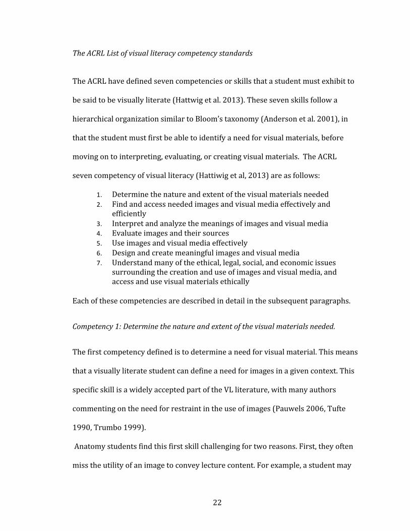
The ACRL List of visual literacy competency standards
The ACRL have defined seven competencies or skills that a student must exhibit to
be said to be visually literate (Hattwig et al. 2013). These seven skills follow a
hierarchical organization similar to Bloom’s taxonomy (Anderson et al. 2001), in
that the student must first be able to identify a need for visual materials, before
moving on to interpreting, evaluating, or creating visual materials. The ACRL
seven competency of visual literacy (Hattiwig et al, 2013) are as follows:
1. Determine the nature and extent of the visual materials needed 2. Find and access needed images and visual media effectively and
efficiently 3. Interpret and analyze the meanings of images and visual media 4. Evaluate images and their sources 5. Use images and visual media effectively 6. Design and create meaningful images and visual media 7. Understand many of the ethical, legal, social, and economic issues
surrounding the creation and use of images and visual media, and access and use visual materials ethically
Each of these competencies are described in detail in the subsequent paragraphs.
Competency 1: Determine the nature and extent of the visual materials needed.
The first competency defined is to determine a need for visual material. This means
that a visually literate student can define a need for images in a given context. This
specific skill is a widely accepted part of the VL literature, with many authors
commenting on the need for restraint in the use of images (Pauwels 2006, Tufte
1990, Trumbo 1999).
Anatomy students find this first skill challenging for two reasons. First, they often
miss the utility of an image to convey lecture content. For example, a student may
23
not realize the direct connection between the vocal folds and arytenoid cartilages
when this concept is described verbally, but upon seeing an image of this
connection, the concept becomes clear. A student with high visual literacy, and an
understanding of the need for appropriate images, can find or construct this kind of
image from the verbal description, while a student with low VL cannot. The second
challenge for anatomy students in using this skill is overuse or over‐reliance on
images. Over reliance on images shows up most frequently when a student will
simply glance at an image, without taking the time to understand the meaning
conveyed. This may also be considered as a misuse of the image, but it is related to
the above competency in that relying solely on images while ignoring text and
context is a common pitfall for the student with underdeveloped visual literacy
skills. One of the biggest risks in use of images is making an image too complicated,
a frequent problem for introductory anatomy students when using anatomical
atlases. The over-complication of images is one of the reasons students fail to fully
develop their visual literacy skills. When a novice learner is presented with expert-
level visual aids, they can easily feel overwhelmed, and without proper scaffolding
in visual literacy training, they may stop using images altogether.
Competency 2: Find and access needed images and visual media effectively and
efficiently.
The second competency is the ability to find appropriate images once a need for
an image has been defined. Finding images has a place in the larger conversation
24
about visual literacy, but does not play an important role in the use of images for
anatomy students. Anatomy students have access to a wide variety of anatomical
images in textbooks, lab guides, coloring books, and websites, so assessing and
evaluating those images for content and applicability (competencies 3 and 4)
becomes much more important.
Competency 3: Interpret and analyze the meanings of images and visual media
The third competency is, in many ways, the core of the original definition of VL. This
skill can essentially be rephrased into Debes original definition of “the ability to
make meaning from an image” (Fransecky and Debes, 1977). It is interesting that
the original definition of VL is now placed at the middle of the hierarchy, indicating
that there are even more basic skills needed before a student can begin to make
meaning from an image. The fact that there are also more complicated skills, which
build on the ability to interpret an image, tells us that reading an image is not the
only goal of VL, and many of the early definitions missed a key component of VL.
Much like verbal literacy, reading and interpreting a passage is not enough, a
student must be able to evaluate, use, and create text to truly have verbal literacy.
For most anatomy students, this skill is where they will spend most of their time.
Making meaning from an image is what students do when they study anatomy
atlases, and it is also how most student learning is assessed in anatomy laboratory
courses. To elaborate, most laboratory examinations are based on the visual
25
identification of an image (histological, photographic, or radiological), a model, or
a dissection (for a detailed examination of this topic, see Chapter 9 which explores
the role of ‘authentic assessment’ in student learning). If instructors are using
visual literacy skills as a major part of assessing student learning, then visual
literacy skills must be taught side-by-side with course content. Additionally,
making meaning from images is a critical diagnostic skill for physicians (again
histology and radiology, specifically), so competency #3 has obvious clinical
applications (Murphy et al. 2014).
Competency 4: Evaluate images and their sources.
The fourth competency of evaluating images incorporates several meanings in
the idea of evaluation. First, a student must be able to critique the effectiveness of
an image; does it convey the intended meaning? What features are missing from
this image because of the choices made by the artist? The second definition of
evaluation involves the reliability of image sources, an important consideration
with web-sourced images, but (hopefully) not one that students will encounter in
textbook or lab images.
Evaluating images may be too lofty a goal for most anatomy students, but if this
idea can be discussed (even briefly) in a laboratory context, it will lead to
students applying more critical thought to their observations. All images are an
attempt to communicate, and in any form of communication, choices must be
26
made about what is worth conveying, and how best to do that. If students can be
taught to consider the choices made in the construction of an image, they will
gain a better understanding of the subject matter.
Competency 5: Use images and visual media effectively.
The fifth competency defines the use of images, but more specifically relates to
the use of images to communicate ideas. To meet this competency, a student
must be able to do all of the preceding steps, but is now selecting and curating
their own images to communicate new ideas. In my personal experience, I have
seen this skill used most frequently in histology. Much of a student’s time in
histology labs is spent scanning through slides on a microscope, and checking
with an instructor to ensure the correct cells and tissues have been identified.
With new virtual microscope software, students can now view high-resolution
scans of traditional microscope slides on a computer, and capture images from
the slides (Husmann et al. 2009, Collier et al. 2012, Bruch et al. 2009, Braun and
Kearns 2008). Capturing and labeling microscope images is exactly the process
of “selecting and curating images to communicate ideas,” a clear fit with the fifth
competency as defined by the ACRL.
Using images, as defined by the fifth competency, can also relate to a student
explaining a concept with an image. For example, if students were given the
opportunity to present research findings in a visual way, while explaining the
27
image, that may demonstrate this competency. Additionally, a multiple choice
examination question could ask a student to match a concept with the image that
illustrates the concept. There is a place for this competency in anatomy
education, but the current structure of many anatomy courses (lecture-based
with purely verbal multiple-choice exams) may make this idea harder to
integrate in the classroom or laboratory. Of course, this competency is frequently
modeled by instructors who will often use and explain images in lectures. A
short description by the instructor to make explicit that the lecture is a model of
a type of visual literacy, could help students understand this competency. If
students are then encouraged to use this behavior during their own study time,
by writing explanations of images, or using images to describe concepts to peers,
this competency can easily be taught.
Competency 6: Design and create meaningful images and visual media.
The sixth competency has the students creating their own, new images. Much like
in (some revisions of) Bloom’s taxonomy, creation is the highest goal of the truly
successful student (Anderson et al. 2001). In the ACRL scheme, the creation of
appropriate, meaningful, and necessary images will best be understood once a
student has exposure to all of the components that unite to form a good image. In
anatomy, creation of images is often suggested by instructors, but without the
appropriate scaffolding of the lower levels of visual literacy. Producing images to
convey meaning is a challenging goal. For students to begin making meaningful
28
images, they must first be instructed in the lower levels of image understanding.
The lower levels are easy to overlook, especially in a content-based course where
visual instruction is an afterthought. Much of the content in following chapters will
deal with different instructional interventions designed to instruct anatomy
students in image creation.
Competency 7: Understand many of the ethical, legal, social, and economic issues
surrounding the creation and use of images and visual media, and access and use
visual materials ethically
The seventh competency lies somewhat outside the context of communicating
with images, and focuses on the ethical and cultural practices associated with
images. This is an important idea, but not one that has been discussed much in
previous literature. Publications dealing with the ethics of body donation are quite
common (Champney 2016, Fonseca 2016, Winkleman et al. 2016), including
several recently published articles detailing ‘best practices’ for maintaining
transparent and ethical body donation programs (Jones 2016, Riederer 2016,
Schmitt 2014). However, few of these papers deal explicitly with the ethical
questions of producing images from donated specimens (exceptions include
Cornwall 2016, Cornwall et al. 2016). Issues such as ethical production and
acquisition of images can be important for artists and publishers, but rarely
appears in the science literature. Many, maybe all, images used in human anatomy
are based on human specimens. This has obvious ethical implications when an
29
image is a photograph of a human cadaver (Jones et al. 2003, Barilan 2005, Barilan
2006, Cornwall et al. 2016). Human remains willed to schools as a part of a body
donation program are subject to guidelines and informed consent documents, but
no specific legal protections exist in all jurisdictions (body donation ethics being
an international concern) (Jones 2016). The guidelines and consent documents
provided to body donors frequently include a limited period of use (Jones 2016),
but a photograph of a body may violate the terms of use. Additionally, body
donors may be unaware of the possibility of their remains being subject to a
permanent photographic record, and may be uncomfortable with that possibility,
especially when images of body tissues are not considered in the original donation
documents. The wishes of the body donor must be considered to ensure ethical
production of images (Jones 2016). A new study also discusses the ethics of 3D
printing of anatomical models based on actual human remains (Cornwall 2016).
Three-dimensional printing is a new technology for producing 3D images
(models) that has only recently been explored for the making of anatomical
images, and the ethics of this practice will doubtless become a productive area of
discussion in the coming years (McMenamin et al. 2014). Less obvious are the
ethical implications of drawings based on human remains. Information is
produced in the creation of the image, but if that image is based on human
remains that have been obtained through morally or legally dubious means, what
are the responsibilities of the instructor or student who uses that image? Recent
works have seemingly rediscovered the anatomical contributions of Nazi
scientists working with human specimens from concentration camps (Atlas 2001,
30
Panush 1996). Is the use of these images ethical? I am not an ethicist, and cannot
answer that question, but see (Atlas 2001, Hildebrandt 2016, Jones 2016) for a
discussion of the various ethical arguments arising from this material.
Additionally, the use of microscope slides containing human tissue can be an
ethical briar patch. Often, the provenance of the tissue in microscope slides is
uncertain at best, and the user of the slide cannot be certain the donor gave
consent for his or her tissue to be used in perpetuity (Jones et al. 2003). These
ethical questions form a fascinating and critical part of image use in anatomy, but
are outside the scope of the current research.
Having defined visual literacy through a series of competencies, the following
sections will focus on the application of visual literacy research in a variety of
academic fields. Fine Arts research will be considered first, with design and
sciences following.
Visual literacy in Fine Arts
The field of Fine Arts has understandably spent a lot of time defining visual literacy.
Much of the arguing about specific definitions of the term comes from educators in
the Fine Arts. There is still no consensus amongst arts educators, but several good
discussions have been published in the arts literature in the last four decades
(Yenawine 1997, Raney 1999, Kemp 2006, Rourke and O’Connor 2010). Much of
the VL literature in Fine Arts has focused on using the rules of art and composition
to understand the process of making an image (Dondis 1973, Perkins 1994). While
31
this is visual literacy in that the art instructor is using images to create meaning,
the meanings are about the image itself, as opposed to what the image is about (see
the following paragraph on semiotics [p. 37] for a discussion of terms ‘sign’ and
‘signified’ which relate to the various types of meaning an image can have) .
In contrast, the VL literature from science is primarily concerned with conveying
meaning about a specific topic using an image, not understanding the construction
of the image (specific examples in the relevant sections). Even so, the definitions
and principles used in VL in the Fine Arts can form valuable background
information for the discussion of VL in science. The difference between the content
of the image and the construction of the image itself has been exhaustively
discussed in the literature of semiotics, using the terms ‘sign’ and ‘signified’ to
denote the image and its contents respectively. When discussing images, a variety
of different terms have been used with similar and overlapping meanings. Another
specific term that reoccurs is the ‘external representation.’ This terminology has
been used a cover term that includes all manner of visuals, images, charts, models,
and graphs. This term has specific utility when contrasting internal visualizations
(i.e., mental images) and external visualizations (e.g., a drawing on paper).
All external representations are made of the same fundamental elements as
defined in A Primer on Visual Literacy (Dondis, 1973). The author defines these
elements as analogous to an alphabet for verbal literacy, and then uses these
building blocks to construct an entire visual/verbal literacy metaphor, which goes
32
on to clearly teaches a student of the visual arts how to make meaning of an image.
The visual/verbal metaphor begins by defining literacy as “a group [sharing] the
assigned meaning of a common body of information” (p. x of preface), a valuable
starting point for the discussion of literacy. However, there is no discussion of
using images to convey content about abstract ideas, or how to assess the
acquisition of visual literacy skills in students. This gap in assessment may be due
to the time period in which the book was published. In the early 1970s workers in
visual literacy were still struggling to agree on the goals of a visual literacy
movement, assuming assessment and measurement questions would be answered
in the future (Perkins 1994 and Wiles 2016 also mention the need for assessment
of VL as a future goal). The greatest strength of this book in the explicit definition
of the six visual design elements that everyone subconsciously decodes when
looking at an image: line, color, shape, texture, space, and form. Dondis also warns
against ‘over defining’ visual literacy (p. 9), saying that the natural and
instantaneous ability for humans to recognize images is a strength of visual
literacy, and that forcing too many restrictions on the concept weakens it. Dondis
also makes the claim that vision, while natural, takes great practice to use
efficiently and effectively, a claim supported by many later authors (Seels 1994,
Allen 1994, Glasgow 1994, Mayer 2014), ignored but Cassidy and Knowlton
(1983), and renamed by Suhor and Little (1993).
The literature of visual arts (Fine Arts, graphic design, industrial design, and
others) is filled with discussion of the interplay between these six elements, and
33
how they can be used by an artist to build an image (Kemp 2006). Understanding
how to build an image is of obvious necessity to a visual artist, but it tells us little
about how these elements can be deconstructed from a scientific photograph or
illustration. For a discussion of the use of these principles in an identification‐
heavy field, the field of textile and interior design has some interesting things to
say. Rourke and O’Connor (2010), both visual artists and designers, have used
these elements of design to teach students how to identify the work of specific
designers. By identifying the typical use of color and pattern, a naïve student can
take a novel piece of industrial design and identify the designer. This is very
similar to the process by which a student will identify a specific tissue in a
histology course. So, the understanding of the elements or principles of visual arts
will help a student create a background to begin decoding the information within
an image.
Another valuable definition to come from the Fine Arts is the idea of “seeing as
cultural habit” (Raney 1999). The cultural background and experience of a person
directly effects what they see, so an expert and a novice in a field will make
completely different meanings from the same image. This idea will become crucial in
teaching novice learners in anatomy; a lot of the instruction in labs is a tacit process
of acculturation to be able to see like an expert.
Art historians have also used visual literacy principles to understand the ways
different cultures and different time periods have produced images. A quote from
34
Wolfflin (1950) will help to illustrate this idea; “not everything is possible at all
times” (p. 11). This reminds us that art history is a gradual process of different
cultures and time periods acquiring new conventions for the creation of images (as
reiterated by Francey et al. 1996). To understand why and how a specific culture or
time period uses images requires visual literacy. By ‘reading’ the alphabet of the
image - to use the metaphor of Dondis (1973) – art historians can understand the
cultural conventions that produced the image and place the image in context. Art
history research has also provided another piece of the definition of visual literacy;
by making clear the separation of the visual and the verbal (Gombrich 2000).
Gombrich defines the unique place of visuals by noting that visual representations
of reality have “fidelity” to reality that words never do. He goes on to state that
visuals may be more or less faithful to reality to convey different meanings, but a
word is always a word with a set meaning. Of course context plays a role in defining
a word, but image context is equally important as seen above.
Having defined some of the limits of visual literacy through the work of artists and
art historians, the next section will apply these principles to the sciences.
Biochemistry will be discussed first, followed by a section summarizing VL as it
applies to anatomy.
Visual literacy in biochemistry
This section discusses pure visual literacy (that is, the interpretation of an image)
and is not directly related to spatial ability (mental manipulation of 3D images).
Many scientific fields have examined spatial ability, but pure visual literacy research
in science education is less common.
35
Biochemistry educational researchers have done the most work with VL of any of
the sciences (Schonborn and Anderson 2006, Anderson 2007, Schonborn and
Anderson 2010, Wiles 2016, Serpente 2016, Linenberger and Bretz 2015). This
may be due to the necessity of creating meaningful mental representations of
biochemical entities in the study of biochemistry. Biochemical entities are mostly
molecules and proteins, impossible to actually visualize with a microscope, so
conceptual visual models are the only visual representations available to student
of biochemistry. The variety of representational schemes for biochemical entities
is also mentioned as a need for robust VL education in biochemistry (Wiles 2016).
That is, a protein, for example, can be represented by a linear arrangement of
letters corresponding to amino acids, a 3D ball-and-stick model, or a 3D ribbon
diagram. Each of these visual representations of the protein conveys different
information, and the biochemistry expert is able to extract the information of the
image, and even infer the appearance of one type of image from another type of
image (Schonborn and Anderson 2006). The publications in biochemistry have
been particularly valuable in defining the facets of VL that most directly relate to
anatomy. One group of biochemistry pedagogy researchers have defined a unique
set of factors that are directly related to how students interpret images and
visualize concepts (Schonborn and Anderson 2010). In this scheme, there are
three major factors influencing VL; 1) conceptual knowledge of the information in
the image, 2) reasoning ability with respect to images, and 3) representation mode
of the image itself. These three factors use different names, but are largely in line
with the skills defined by the ACRL, and VL definitions from the fine arts. Though
36
the terms used in biochemistry may have more use in VL of sciences, the
definitions are still not perfect, and introduce some ambiguity where other
authors have been more precise (Schonborn and Anderson 2006, Moore and
Dwyer 1994).
The biochemistry definitions add the conceptual knowledge component, which is
ignored in so much of the arts literature because of the focus on the image itself, as
opposed to its content. The conceptual knowledge element is an important
consideration when using an image to communicate information about a specific
topic, rather than the image itself.
The second component from Schonborn and Anderson (2010) is the reasoning
ability with respect to images. Reasoning ability encompasses the entire
“repertoire of skills” a student brings to the interpretation of an image. The
definition of “reasoning” used here is a bit ambiguous with respect to the ACRL
standards, because Schonborn and Anderson have grouped a huge variety of skills
with different cognitive bases together under one single umbrella term. The list of
skills involved in reasoning with images includes:
• Decode the symbolic language composing an image
• Evaluate the power, limitations, and quality of an image
• Interpret and use an image to solve a problem
• Spatially manipulate an image to interpret and explain a concept
• Construct an image to explain a concept or solve a problem
• Translate horizontally across multiple images of a concept
• Translate vertically between images that depict various levels

37
of organization and complexity
• Visualize orders of magnitude, relative size, and scale.
This list incorporates decoding, interpreting, evaluating, and constructing images,
grouped together as a single skill, while the ACRL defines each of these as
individual skills. Additionally, this list explicitly defines the need understand
scale, and the ability to use multiple images with different scales and
representation modes, crucial factors in scientific visual literacy. This list also
includes spatial manipulation, which will be treated separately in a following
chapter.
The final part of the Schonborn and Anderson VL scheme (2010) is the
representation mode of the image. An image must clearly convey the desired
information in a meaningful, and some modes are more appropriate for
specific applications. For example a protein can be visually represented by a ball-
and-stick chemical model or a space-filing model, depending on the ideas the
instructor wishes to present. Each mode of presentation has value, and each
highlights different aspects of the protein. The mode of presentation interacts
with the conceptual knowledge and reasoning ability of the viewer to
produce visual literacy.

38
Visual literacy in astronomy
Only one paper has been published on the visual literacy of astronomy students
(Crider 2016). The author is an instructor of undergraduate astronomy students,
and uses the ACRL scheme for teaching visual literacy as a way of understanding
astronomy. The author does not use any specific visual literacy assessments, but
rather uses the ‘Moon Landing Hoax’ as a case study for the utility of good visuals in
science, and the understanding of those images as a bulwark against bad
information from ‘edutainment products.’ (p. 17). This research points to the utility
of visuals in communication, especially for eliminating deep-seated misconceptions,
like the ‘Moon Landing Hoax.’ While there is no explicit assessment of students
learning of VL, the author does say that strong visual literacy skills, especially in
understanding how images are made, and in making one’s own images, can help
students understand misconceptions about science.
Visual literacy in anatomy
In the anatomical education literature, spatial ability research is common (see
below), but visual literacy discussions are rare. The few examples of VL research in
anatomy education (Backhouse et al. 2016, Bardes et al. 2001, Bell et al. 2014,
Pickering 2014) are explained in the next paragraph.
Backhouse et al. 2016 used an in-class drawing exercise with anatomy first-year
medical students to teach anatomy content and VL skills at the same time. They
found that using drawing (combined with reflection and editing) as an in-class
activity led to lower exam performance, despite positive perceptions of the drawing
39
exercises. When the drawing protocol was moved to an optional tutor-led
environment, exam scores improved significantly. Additionally, students who self-
reported liking to draw did not differ from students who disliked drawing in any of
the measured variables.
Pickering 2014 used a computer animation (with narration and a student drawing
component) to teach anatomy to medical students. The paper deals with visual
literacy as related to working memory and cognitive load, and shows that computer
based drawing lessons correlate well with evidence-based best practices in medical
education. Bell and Evans 2014 and Bardes et al. 2001 both used observation
training with of works of art to improve the clinical observation skills of medical
students. Both papers report a high degree of student satisfaction with the program,
and an increase in observational skills. Observational skills were measured by a
panel of experts, and the number and detail of clinical observation increased in the
study populations.
What does the visual literacy research in the other fields mean for anatomy?
Having defined VL across multiple fields, each with its strengths and weaknesses,
what can we say about the visual literacy skills needed by an anatomy student?
Which specific competencies are most important? I define here a list of visual
literacy skills that are most important to anatomy students. This list is based on the
definitions and studies reviewed in the previous pages. The following table uses the
ACRL list of visual literacy competencies as its base, but includes some of the
specific skills defined by the work of biochemistry educational researchers.
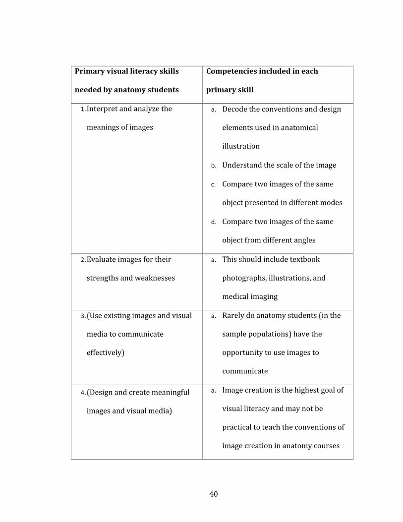
40
Primary visual literacy skills
needed by anatomy students
Competencies included in each
primary skill
1. Interpret and analyze the
meanings of images
a. Decode the conventions and design
elements used in anatomical
illustration
b. Understand the scale of the image
c. Compare two images of the same
object presented in different modes
d. Compare two images of the same
object from different angles
2. Evaluate images for their
strengths and weaknesses
a. This should include textbook
photographs, illustrations, and
medical imaging
3. (Use existing images and visual
media to communicate
effectively)
a. Rarely do anatomy students (in the
sample populations) have the
opportunity to use images to
communicate
4. (Design and create meaningful
images and visual media)
a. Image creation is the highest goal of
visual literacy and may not be
practical to teach the conventions of
image creation in anatomy courses

41
Table 2.1- A list of the visual literacy skills anatomy students need to learn from
images and effectively communicate with images, as prepared by the dissertation
author. Skills 3 and 4 are enclosed in parentheses because they are goals of visual
literacy education, but may be outside the scope of most anatomy courses.
Most of the time spent working with images in anatomy focuses on the first two
skills defined here. That is, students will need to interpret images, and evaluate their
quality. For example, a textbook image of the heart can be a photograph of a human
cadaver, a model organism (like a pig), a realistic drawing, or a diagrammatic
drawing. The student studying the heart will need to make several interpretations
about the image presented. First, the medium of presentation will give the student
some information. If it is a photograph, it is a realistic representation of one heart,
that is, it may exhibit abnormalities or idiosyncrasies not seen in all specimens. If
the image is a drawing, the student must be aware that it is a generalization, and
variations exist in the real world. Additionally, the student must consider the
choices the artist made in the rendering of the heart image. Why are veins blue? Are
the great vessels shown intact or cut? Is the relationship to the rest of the thorax
presented? All of these questions subconsciously form the learning that is taking
place when looking at an image. This is interpretation and evaluation.
Images outside of textbooks require an additional level of evaluation. The
source must be evaluated for accuracy, and relevance. Anatomical images found
through Google image search may contain errors, nomenclature variations, or
42
overwhelming details, all of which can hinder student learning. The student
must be aware of the mental process of image evaluation to successfully
navigate the sea of images.
Students are rarely given the opportunity to use or create their own images for
communication in the subject, so skills 3 and 4 are listed as potential goals of
visual literacy education, but goals which may be outside the scope of most
anatomy courses.
The research presented in chapter 7 details a plan where VL skills are taught to
anatomy students as a means of learning course content and VL at the same
time, the best way to teach VL according to college librarians who have
researched the problem (Nelson 2004).
Spatial Ability
Spatial ability (SA) is distinct from visual literacy in that involves the ability to
mentally manipulate, and gain information from, two- and three- dimensional
images (Bodner and Guay 1997, Meneghetti et al. 2011). As a specific set of skills, SA
has been well defined, and literature dealing with SA has shown more concrete
results than the VL literature (examples cited below). That is, spatial ability can be
trained and measured . This effect has been demonstrated many time, for example:
Lord 1985, Orion et al. 1997, Titus and Horsman 2009, Keehner et al. 2006, Provo et
al. 2002, Lufler et al. 2012, Luursema et al. 2008, Boudreau et al. 2008, Hegarty et al.
2009, Wanzel et al. 2002, Fuks et al. 2009, Garg et al. 2001, Nicholson et al. 2016,

43
Canty et al. 2015. Spatial ability can be assessed through a variety of tools, including
polyhedral net folding, aperture passing, and cube counting. Each of these spatial
ability tasks uses some amount of mental rotation, so mental rotation is frequently
used as a measure of spatial ability as a whole (Meneghetti et al. 2016). Spatial
ability uses many other mental processes besides mental rotation, but the spatial
ability literature seems to agree that mental rotation is a good proxy for measuring
all facets of spatial ability (Uttal et al. 2013 for a meta-analysis of the transferability
of mental rotation skills to other spatial tasks).
Spatial ability has been well studied in a variety of populations, including anatomy
students. In the following paragraphs, I examine the role of gender in spatial ability,
and then I summarize the spatial ability studies in both non-anatomical and
anatomic fields.
Does Gender play a role in spatial ability skills?
One of the oldest questions in the spatial ability literature is the role of sex (or
gender, the terms are not always used consistently) in performance of spatial ability
tasks (Hyde 2016 for a history of gender in cognitive science). Cognitive science has
shown men and women to be equal in nearly all cognitive tasks, with the exception
of spatial ability (Hyde 2016, Moe 2016, Voyer et al. 1995). Hypotheses to explain
these differences can be broadly divided into biological explanations (e.g., hormonal
or brain organization explanations) or social explanations (e.g., perceptions of
gender roles for men and women, spatially oriented hobbies, or attitude) (Quaiser-
Pohl et al. 2016, Williams and Meck 1991). In the mid-1990s, two meta-analyses of
44
gender in spatial ability were published, one indicating a decrease in gender
differences over time (Voyer 1995), and the other indicating that gender differences
have remained stable over time (Masters and Sanders 1993).
Since then, dozens more studies about the role of gender (or sex) in spatial ability
have been published (e.g, Barnett-Cowan et al. 2010, Dabbs et al. 1998, Feng et al.
2007, Hausmann et al. 2000, Kennedy and Raz 2005, Langlois et al. 2013, Lawton
1994, Moe 2016, Moffat et al. 1998, Quaiser-Pohl 2016, Xiong et al. 2016), and still
no consensus has been reached. Quaiser-Pohl et al. (2016) examined the role of
salivary gonadal hormone levels in the performance of mental rotation tasks in
children aged 9-14. They found boys performed the mental rotation tasks faster, but
no more accurately than the girls in the sample. Additionally there was no
correlation between hormone levels and mental rotation performance. Gradl-
Dietsch et al. (2016) examined the role of gender and learning style in the learning
of spatial ability in medical students in anatomy. They found no gender differences
in the spatial ability tasks, or anatomy learning tasks. While there is still an active
discussion about the role of gender (or sex) in training spatial ability skills, these
two recent examples, one with children in a pure mental rotation test (Quaiser-Pohl
et al. 2016), and one with adult learners in anatomy (Gradl-Dietsch et al. 2016),
indicate that men and women are equally good at spatial ability skills. Other recent
papers also support the idea that gender differences in spatial ability are minimal at
best, and women are equally amenable to the training effects spatial ability
instruction (Meneghetti et al. 2016). For this reason, men and women are treated as
a single group in this dissertation research.
45
SA in fields other than anatomy
The SA literature is full of experiments designed to test this ability in a variety of
populations. This section will summarize a number of these experiments in fields
other than anatomy. These experiments will then be compared those done in
anatomy to look for consistency, and draw conclusions about what SA means when
learning anatomy. This section also examines the SA literature where researchers
have simply compared a student’s baseline SA with performance on a variety of
tasks. These articles will also provide meaningful background in the ways that SA
influences learning.
One of the earliest studies to examine SA in students and use an intervention to
improve SA comes from Lord (1985). At the time of his writing, there was still an
active debate about the possibility of teaching SA. Some researchers argued that SA
was an innate ability which could not be improved (Carp and Silberman 1969,
Culver and Dunham 1969), while others claimed that environmental interactions
teach SA (McGee 1979). The research presented by Lord used weekly interventions
in which college undergraduates in biology courses mentally bisected 3D figures,
and drew the resulting 2D image on a paper. His results show that the students in
the weekly intervention performed better on an SA posttest than a control group.
This result lends support to the idea that teaching SA is possible.
Since 1985, teaching spatial ability has become the norm, with no publications since
arguing for a purely innate spatial ability skill. Many researchers have shown the
effectiveness of teaching spatial ability across multiple populations with a variety of

46
different pedagogical interventions. The following sections provide a detailed
summary of the SA teaching literature of the intervening thirty years. The following
sections do not use a historiographic approach to the SA literature, but are rather
divided into discipline specific sections, to better understand the unique needs of
each discipline, and explore how each of them may be relevant to the teaching of
anatomy.
Spatial ability in geology
The field of geology has had an active group of SA researchers for many years, all
emphasizing the importance of SA for all levels of geology students and
practitioners. Researchers in geology have used mental rotation as a measure of SA,
but they also add a unique SA assessment technique in internal visualization. This is
the skill required to visualize how the external features of a rock or mountain
influence the internal structures. So far, this appears to be a skill unique to the
geology literature, but it has obvious uses in anatomy, and is worth future
exploration.
In 1996, Kali and Orion published a paper detailing a new method for assessing SA
in secondary students of geology. In this test, students were asked to visualize the
internal structures of a rock that would be revealed by slicing the rock along a given
line. The students who at least attempted to visualize the internal parts of
structures, even when wrong, did much better in geology courses overall. They
concluded that geology students need to be given specific instruction in SA,
including working with models that allow the visualization of layers and internal
47
structures. The authors continued by saying that other sciences with a similar SA
need should be identified, and these students should also be offered SA training.
Anatomy would certainly fit in this category, but it appears that none of these
suggestions have yet been widely implemented, and this article seems infrequently
read (or at least cited) outside the geology literature.
Several studies have demonstrated that studying a visually challenging science such
as geology will lead to improvements in SA, even without specific instruction or
testing in SA (Orion et al. 1997, Titus and Horsman 2009). Orion et al. (1997)
showed a reciprocal relationship between the acquisition of SA and geology skills.
Learning a visual science or specific SA skills will lead to improvements in the other,
and learning both at once will maximize the gains students make (Orion et al. 1997).
Titus and Horsman (2009) demonstrated a passive acquisition of SA just by taking a
geology course. They assessed SA in geology students at the beginning and end of an
introductory geology course, and found significant improvements in SA, even
without any specific lessons designed to teach SA to the students.
Piburn et al. (2005) used a computer based interactive animation of geological
processes to aid geology undergraduate learning. They found that the novel tool
improved acquisition of geology knowledge, and some measures of spatial ability.
Additionally, the authors demonstrated that training in SA was able to eliminate
differences in geology content acquisition and SA between genders. This shows that
explicit training in SA can reinforce content knowledge, while simultaneously
improving SA, and can help lower-performing students to meet their peers in
48
visually-oriented science courses. The animations used in this study illustrated
two- and three-dimensional views of geological processes, and included
visualizations of the interior feature of the earth’s crust. The control group did not
receive any explicit visualization or SA training. The results of this study indicate
that some training in SA is beneficial, but they do not make any claims about the
efficacy of the specific animations used. It may be that other types of SA training are
equally valuable.
Spatial ability in chemistry
Chemistry is another visually oriented science in which students must be able to
visualize the shape and orientation of the atoms in a molecule to predict their
behavior in reactions. Because there are no photographs that show a molecule as a
“ball-and-stick” model, chemistry instructors have to rely on illustrations and
metaphors, but the resulting ways of teaching SA are very similar to more
macroscopic sciences (Boz and Boz 2008, Ferk and Yrtacnik 2003).
Virtual 3D environments have been shown to help students learn chemistry and
improve SA (Trindade et al. 2002). The authors of this study taught chemistry
concepts to first-year undergraduates using a three-dimensional image rendered on
a flat computer screen, as well as stereoscopic 3D images with a virtual reality
rendering environment. The results indicate that 3D visualizations are good at
helping students learn, but flat-screen rendering is as good as stereoscopic
rendering. Additionally, these benefits in student learning seemed to be confined to
those students who were already good at SA. They found no improvement in
49
students’ SA scores which contradicts Piburn et al. (2005) results in geology
students’ use of 3D visualization software. Both studies (Trindade et al. 2002 and
Piburn et al. 2005) agree that 3D visualizations can help students learn content, but
they reach different conclusions about the utility of teaching students SA skills.
As early as 1983, Small and Morton demonstrated that training undergraduates in
spatial-visual skills (their term for spatial ability) improved scores in an organic
chemistry class. The students only showed improvement on exam items directly
related to 3D visualization of structures, indicating that SA training helps in some
specific parts of chemistry learning, but is not widely applicable to all types of
questions. Additionally, these students were given SA assessments at the beginning
of the course to control for their previous spatial ability experiences, but no post-
course SA assessments were conducted, perhaps due to the innate theory of SA
which was still common at that time (Lord 1985).
Carter et al. 1987 and Pribyl and Bodner 1987 used SA a predictor of success in
chemistry, also apparently working from the innate theory of SA, and finding a
correlation between SA and chemistry performance, but did not investigate the
interaction between these two variables, or examine possible causal relationships.
Neither of these 1987 studies explicitly attempted to teach SA, but only used it as a
predictor of success in undergraduate chemistry courses. The teaching of SA as a
specific skill did not really enter specific science disciplines until the 1990s as seen
in the following paragraphs.
50
Barke (1993) found that training middle school students in the spatial visualization
tasks related to chemistry improved both their knowledge of chemistry and SA
scores. This effect was seen in boys and girls, and was most pronounced when the
visualizations had a kinesthetic component, that is students were handling 3D
physical models of chemical entities.
Coleman and Gotch (1998) studied the effect of spatial ability on undergraduates in
introductory chemistry courses. They began with the assumption that spatial ability
was strongly correlated with achievement in science (a result which has been
repeatedly demonstrated for 50 years in cognitive science according to a meta-
analysis by Wai et al. 2009). The assumption that SA and science performance are
correlated was shown in the Coleman and Gotch research, but the training effect of
SA was also demonstrated, indicating that the causal relationship between SA and
science performance is in question. Will increasing a student’s SA make them better
at scientific reasoning, or is the correlation of these two factors based on some other
variable? This study also included a longitudinal component which looked at
approximately twenty years of student SA scores. They found that SA differences in
men and women were decreasing, but it was entirely due to men’s scores falling, as
opposed to women’s scores rising.
Yang et al. 2003 showed that teaching college undergraduates improved their SA
and general chemistry knowledge through the use of animations, as opposed to
static diagrams. The animations used included narration, and instructor lectures to
provide additional context to the animated images. The necessity of narration and
51
lecture in the use of animations was also demonstrated with undergraduates by
Tasker (2016) and in secondary students by Kelly (2016).
Other authors discussing animations in chemistry have found web-based
animations to be an effective supplement to undergraduate learning (Morozov et al.
2004, Lester et al. 1999, Frailich et al. 2009, Calik et al. 2010). All of the preceding
studies provided students with animated chemical reactions or processes and found
improved student grades with those using the animations as a study tool (as
compared to a non-animation using control group). None of the above papers
explicitly discuss the spatial abilities of the students in the study population, but the
use of visuals to effectively teach scientific concepts is well documented (see Harle
and Towns 2011 for a review of spatial ability literature in chemistry).
Spatial ability in engineering
Engineering, as a field, is highly dependent on spatial ability Consequently, several
studies about how to teach these skills have been published. The SA literature in
engineering reaches the same conclusions as seen in the sections on chemistry and
geology, above. That is, training students in spatial ability may improve their SA
scores (Alias et al. 2002, Martin-Gutierrez et al. 2010, Potter et al. 2009), but
improved SA does not always lead to greater content acquisition (Prieto and Velasco
2010). Each of the above studies used different methods for teaching SA to college
undergraduates, for example 2D paper-based drawings (Alias et al. 2002), technical
drawing ( Martin-Gutierrez et al. 2010), and augmented reality (Potter et al. 2009).
While all of these approaches showed improvements in student SA scores, the
52
methods used were greatly different, from low tech (pencil and paper drawing) to
high tech (augmented reality, using a mobile device to provide additional
information about a real-world object). This again indicates that the method of
teaching SA is less important than explicitly teaching these skills to novice learners
(as seen in Piburn 2005).
Spatial ability in physics
In physics, a few studies have examined explicitly the role of SA and physics content
acquisition (Kozhevnikov and Thornton 2006, Kozhevnikov et al. 2007, Pallrand and
Seeber 1984). These studies have shown that adding visual skills instructions to a
physics lab improved both undergraduate SA skills and content acquisition. The
most interesting finding in Kozhevnikov and Thorton (2006) is that the
undergraduates who began the introductory physics lab with the lowest
background SA skills showed the most benefit from working with the computer
models. This is in contrast to some other work (e.g., Cohen and Hegarty 2007 ) in SA
that shows the greatest benefit in students who already have good visual skills. (VS
is defined in this dissertation as the combination of visual literacy and spatial ability.
This is a new term, unique to this dissertation, but the Cohen and Hegarty paper
defines a suite of spatial ability and visual literacy skills closely in line with my use
us the term ‘visual skills’). Cohen and Hegarty (2007) found that use of external
representations as an aid for completing visualization tasks was much greater in
students with higher SA, and strongly correlated with performance on other types of
SA-based tasks.
53
Non-discipline specific spatial ability
Some general research on SA that does not fall into any specific discipline is outlined
below. These papers provide background on how students acquire SA skills, which
students benefit most from different types of SA teaching, and how SA is used by
students.
Kastens (2010) outlined a brain science perspective on SA by detailing some of the
key differences between SA and VL, by defining two separate visual systems in the
brain. These systems are broadly called “what” and “where.” The “what” system is
closely related to VL in that this is the part of the brain that makes sense of the
content of an image. The “where” system is closely related to SA in that this system
is the part of the brain that builds a model of a structure, and keeps the various
parts in order. The cognitive science behind how the brain constructs and image is
only tangentially related to the instruction of SA and VL discussed in this
dissertation (and this topic is still an active area of research), but a brief overview of
the cognitive science behind mental image construction follows. Wai et al. 2009,
Harle and Towns 2011 both examine the history of spatial ability as a cognitive
discipline, and explain that spatial ability has repeatedly been demonstrated as a
strong correlate with success in STEM (science, technology, engineering, and math)
fields. Both papers go on to recommend the nurturing of students with high spatial
abilities in STEM fields, but do not provide any definite guidelines for how to do so.
Pickering (2014) examined the cognitive load and working memory theories behind
the acquisition of visual literacy in anatomy students. People have a finite amount of
54
working memory that cannot be modified by instruction or training, so visual aids to
learning must help the working memory by reducing the cognitive load. Information
in working can be consolidated into long-term memory (learning), but only if
extrinsic factors (such as visuals) do not interfere.
Foo et al. (2013) describe the mental workload principles behind using two- and
three dimensional images to teach anatomy students. Traditionally students in
anatomy are given two-dimensional images to emphasize spatial relationships,
while three-dimensional images help with understanding the shape of an object. Foo
et al. (2013) described a method of using ony 3D images to reduce cognitive load by
reducing the number of extraneous images students view. It was found that mental
workload was decreased, and accuracy of identification increased in first-year
anatomy students when they viewed only 3D images.
Despite all of the benefit shown to learning visual skills, students still seem unlikely
to know how best to apply these skills in new situations (Stieff et al. 2010). Steiff
and colleagues indicate that acquisition of visual skills is unrelated to a student
using those skills on an exam. Steiff and colleagues interviewed undergraduate
students enrolled in an organic chemistry course using a thinking aloud protocol to
solve chemistry problems similar to those they would find on an exam. The author
found that students rarely used the visual thinking skills they had learned in the
course, and fell back on algorithmic thinking. This is in contrast to experts in
chemistry, who used image-based reasoning for the majority of their thinking.
55
Some authors (e.g., Darian 2001, Anderson and Dietrich 2012, Serpente 2016) have
examined the role of visuals in textbooks. Ideally these visuals would lead students
to greater visual thinking, and help them to acquire VL and SA skills, but this is
rarely the goal of the textbook authors and illustrators. The main problem identified
in the above papers is the different ways in which experts and novices interpret
images. Experts and novices will use images differently, and consequently are able
to generate different information from the same image. The research cited above
offers no definitive solution to this problem, but rather seeks to outline all of the
possible approaches to the problem. They advise care in the selection of images, and
specify several criteria that must be addressed when choosing images for a science
textbook.
Good spatial ability skills seems a given for computer graphics students, but this
assumption has been challenged (Mohler 2006). Mohler argued that computer
graphics instructors cannot assume their students have an equal SA background,
and specific classroom tasks should be used to improve SA. Several activities (e.g.,
computer-based drawing, use of physical models) are suggested, with hand
sketching on paper showing good results, with the added bonus of that it is
inexpensive and easy to deploy. Other authors (Czarnolewski and Eliot, 2012) have
also found that sketching by hand improves spatial ability and memory.
Spatial ability in anatomy
Spatial ability has been studied extensively in anatomy with many experiments
using similar designs to those in other fields. The consensus view of the collected
56
literature in anatomy is that SA is a crucial ability for students and future
practitioners (Langlois et al. 2014, Keehner et al. 2006, Pahuta et al. 2012, Provo et
al. 2002, Lufler 2012, Lufler et al. 2012, Luursema et al. 2008, Boudreau et al. 2008,
Hegarty et al. 2009, Wanzel et al. 2002, Fuks et al. 2009, Garg et al. 2001, Nicholson
et al. 2016, Canty et al. 2015, Beck et al. 1978, Hoyek et al. 2014, Nugent et al 2012a,
Nugent et al. 2012b, Sweeney et al. 2014), but there is a still an active debate about
the best ways to teach SA while delivering content (Allen et al. 2014, Azer and Azer
2016, Bareither et al. 2013, Faurie and Khadra 2012, Garg et al. 2001, Gradl-diestch
et al. 2016, Keehner 2011, Keehner et al. 2008, Khot et al. 2013, Kotze and Mole
2015, Knobe et al. 2013, Kooloos et al. 2014, Moscova et al. 2014, Murphy et al.
2014, Ngan et al. 2010, Ngan et al. 2012, Nguyen et al. 2014, Nguyen et al. 2011,
Nguyen et al. 2009, Noel 2013, Preece et al. 2013, Richardson-Hatcher et al. 2014,
Ruisoto et al. 2012, Ruiz et al. 2006, Ruiz et al., 2009, Saltarelli et al. 2014, Schwibbe
et al. 2016, Smith 2015, Smith et al. 2015, Sweetman et al. 2013, Tam 2010, Topping
2014, Trelease and Nieder 2013, Vorstenbosch et al. 2013, Weber et al. 2016, Willis
and Martin 2016, Yammine and Violato 2014, ). Many of these studies repeat the
same experimental designs or results with only minor changes, so several salient
examples are examined in detail in the following paragraphs.
The need for strong SA skills in anatomy students is well demonstrated, with each of
the following sources showing a correlation between SA in anatomy students and
various measures of success. Keehner et al. (2006) showed a strong correlation
between SA skills and laparoscopy skills in surgeons. The authors even show that SA
was a stronger correlate with surgical skill than general aptitude (also shown by
57
Schlickum et al. 2016 and Maan et al. 2012 for a meta-analysis of predicitive factors
in surgical training). An earlier study (Wanzel et al. 2002) demonstrates the same
results in a population of general surgeons, and also shows that surgeons with lower
SA can be helped by practice and feedback. In a similar vein Boudreau et al. (2008)
discuss the importance of clinical observation skills for clinicians of all specialties.
The authors go on to define the guiding principles of clinical observation, and
emphasize the importance of introducing these to medical students as soon as
possible. They have also developed a series of modules designed to teach
observation through practice, but no results on efficacy have yet been published.
Hegarty et al. (2009) also showed the need for SA skills, in this case for practicing
dentists. They found a correlation between dental reconstruction performance and
SA, but did not see a correlation with general anatomy performance and SA. This
may be due to the fact that SA is more strongly assessed in incoming dental students
than it is for medical students, or other anatomy students.
Lufler et al. (2012) demonstrated a strong predictive effect of medical student SA on
gross anatomy performance. This paper also suggests that intervention may be
necessary to improve SA skills in students with lower SA scores, but does not offer
any suggestions. This paper also re-confirms a small amount of passive acquisition
of SA for all anatomy students, similar to the effect seen in geology (Titus and
Horsman 2009).
The following sources all look for methods of improving SA in anatomy. Many have
been tried, and every teaching intervention that has been published shows some
58
effect on SA, but there is no consensus on the best practice for teaching SA in
anatomy. Methods tested include passive learning (Vorstenbosch et al. 2013), 3D
computer based models (Ruisoto et al. 2012, Ruiz et al. 2009, Garg et al. 2001,
Pickering 2014, Roach et al. 2016), Animations (Ruiz et al. 2009, Evans 2011),
Drawing (Ruiz et al. 2009), 3D images (Luursema et al. 2008), anatomical cross
sections (Provo et al. 2002), and surgery simulations (Keehner et al. 2006, Roach et
al. 2012).
Vorstenbosch et al (2013) showed that medical students passively acquire greater
spatial ability simply through studying anatomy, in the absence of any specific
spatial ability interventions. Passive acquisition of SA through the learning of course
content has been shown in other fields (Titus and Horsman 2009) and this effect has
also been seen in anatomy (Vorstenbosch et al. 2013). Vorstenbosch et al. (2013)
also demonstrated a reciprocal effect between SA and anatomy, that is learning one
leads to better learning of the other, but they do not offer any specific interventions
to aid this process.
The use of 3D computer models to teach and learn anatomy has been a big topic for
many years, but results are mixed. Garg et al. (1999a, 1999b) showed that computer
models were no worse than traditional images for student learning. More recently,
more favorable results have been obtained, perhaps due to the improvement in
computer-produced visuals in recent years. Ruiz et al. (2009) found that three-
dimensionality was the most important factor dictating the usefulness of an image.
Ruisoto et al. (2012) showed an increase in clinical reasoning for neuroanatomy
59
students who used 3D computer images to study, over those who used only flat
images. Animation of images only occasionally correlates with learning of content
(Ruiz et al. 2009). This review article found a mixed bag of results when examining
the effectiveness of animated images for student learning in anatomy. Keehner et al.
(2008) demonstrated an advantage to animated images over static images, but only
in learning SA, not course content.
Multiple views of any type have been shown to help student learning; this is the idea
behind animation and 3D models as a teaching tool, because each of these methods
can potentially offer multiple views of the same object (Ruiz et al. 2009). Another
way of increasing the number of images and number of different types of images a
student uses is the use of cross sectional views. Provo et al. (2002) used cross
sectional views of a dog’s head to help veterinary anatomy students learn head
anatomy. They found that the group using the cross sections had significantly higher
exam grades than the group who used dissection alone (Provo et al. 2002).
Stereopsis is another intervention that has been tried in anatomy as well as other
fields, with mixed results. Stereoscopic images are a type of 3D image that relies on
projecting two slightly different angles of the same image to each eye, much the way
modern 3D movies, or the old-fashioned stereopticon, create the illusion of 3D (also
called binocular parallax). In chemistry, stereoscopic views (produced with a
virtual reality headset viewer) did not help students any more than traditional 3D
renderings on a flat screen (Trindade et al. 2002). In a more recent anatomy study,
stereoscopic images were shown to benefit students with lower base SA scores, but
60
time spent with images was a bigger predictor of usefulness than the specific
rendering of the image (Luursema et al. 2008). In another study, neuroscience
undergrads using stereoscopic 3Dimages to learn brain anatomy showed improved
knowledge retention over traditionally taught students, but no measures of SA were
made (de Faria et al. 2106).
Summary of Visual Literacy and Spatial Ability
Spatial ability, as described above, has been well studied for decades. There is broad
agreement that spatial ability correlates with student performance in STEM fields,
but the causal relationship between these two factors has not been elucidated.
Additionally, improving SA skills in students through training is now widely
accepted, but improvements in science knowledge related to SA gains are not
always supported in the literature. It seems that improving SA in students with low
SA is possible through a wide variety of methods, but none of these methods have
led to consistent improvements in knowledge gains, or success in science fields.
Results in SA studies in anatomy parallel those seen in other sciences. One goal of
this dissertation research is to continue looking at improving SA in anatomy
students, and how gaining SA skills relates to learning anatomy content. The
projects designed around this question can be found in Chapters 5-8.
Visual Literacy was previously defined in chapter 1. Remember, visual literacy is
related to spatial ability, but it is presently considered a completely separate
cognitive skill. Visual literacy research, especially in anatomy, is greatly lacking. In a
search of the journal Anatomical Sciences Educator (a journal which publishes
61
research based on teaching anatomy) only 13 results dealing with VL were found
(search conducted on June 1, 2016). Of these 13 results, only four articles actually
dealt with the teaching and assessment of visual literacy (Backhouse et al. 2016,
Bardes et al. 2001, Bell et al. 2014, Pickering 2014). The other major goal of this
current dissertation project is to examine visual literacy in anatomy students. The
specific research questions dealing with VL in anatomy are detailed in the next
chapter. Results and detailed methodologies concerning the VL research projects
are in chapters 4-6.
62
CHAPTER 3: Materials, Methods, and Research Questions
This chapter outlines the specific research questions which guided the research
detailed in the chapters 4-8, followed by a description of the instruments and
methods used in each facet of the research. Before explaining the research questions
and methods, it is worth noting the populations and courses examined in the
research. Samples from each course syllabus can be found in Appendix C.
As a reminder of the current state of visual literacy and spatial ability research, see
Chapter 2, but a brief summary follows.
Spatial ability, as described in Chapter 2, has been well studied for decades (Wai et
al. 2009 for a meta-analysis of 50 years of spatial ability research). There is broad
agreement that spatial ability correlates with student performance in STEM fields,
but the causal relationship between these two factors has not been elucidated.
Additionally, improving SA skills in students through training is now widely
accepted, but improvements in science knowledge related to SA gains are not
always supported in the literature. It seems that improving SA in students with low
SA is possible through a wide variety of methods, but none of these methods have
led to consistent improvements in knowledge gains, or success in science fields.
Results in SA studies in anatomy parallel those seen in other sciences. One goal of
this dissertation research is to continue looking at improving SA in anatomy
students, and how gaining SA skills relates to learning anatomy content. The
projects designed around this question can be found in Chapters 5-8.
63
Visual Literacy is also defined in Chapter 2. Visual literacy is related to spatial
ability, but it is presently considered a completely separate cognitive skill. Visual
literacy research, especially in anatomy, is greatly lacking. In a search of the journal
Anatomical Sciences Educator (a journal which publishes research based on
teaching anatomy) only 13 results dealing with VL were found (search conducted on
June 1, 2016). Of these 13 results, only four articles actually dealt with the teaching
and assessment of visual literacy. The other major goal of this current dissertation
project is to examine visual literacy in anatomy students. The specific research
questions dealing with VL in anatomy are detailed below. Results and detailed
methodologies concerning the VL research projects are in chapters 4-6.
Student populations and courses examined
This section will provide background information detailing the students and courses
examined in the following research. All research (unless otherwise noted) was
conducted using students at the Bloomington, Indiana campus of Indiana University.
Six different undergraduate and medical courses were analyzed in this research:
ANAT-A215 Basic Human Anatomy (referred to as A215 throughout this
dissertation) is a one-semester, introductory course for undergraduates
consisting of three hours of lecture and four hours of laboratory time each week.
Lab time is used for students to study anatomical models, histological images,
and prosected cadavers. Four multiple-choice lecture exams are given during the
semester. Students also take four free-response lab practical exams that are
entirely based on identification. Lecture and lab exams cover the same topics.
64
The course is taught using an organ system (versus regional) approach, and all
body systems are covered in the semester. The course enrolls approximately 400
students in one or two lecture sections, and has twelve lab sections each with
36-40 students. Students are mostly freshmen or sophomores, and come from a
variety of academic programs, mostly pre-nursing. This course is offered every
fall, spring and summer semester. Further demographic details of the student
population may be found in the results section of the following chapters.
ANAT-A550/551 Medical Gross Anatomy (referred to as A550 throughout this
dissertation) is a two-semester (full-year) anatomy course for first-year medical
students (MS1) consisting of two hours of lecture and four hours of laboratory
time each week. Students in lab work in teams of four to complete a full
dissection of a cadaver. Because a full-body dissection is completed in this
course, course topics are organized around a regional approach to anatomy, with
specific clinical correlations included in all sections. Student assessment in this
course is varied and includes six written exams with multiple-choice,
identification, short-answer and multiple-mark questions to assess topics
learned in lecture. Students also take six lab practical exams which are mostly
identification, but approximately 10% of lab exam questions include higher-
order questions about functions and relationships of structures. Students also
receive grades for completing problem-based learning exercises during the
course. This course enrolls 36-40 MS1 students who all take lectures and labs at
the same time. (Two to six graduate students not in the medical school are also
typically enrolled in this course, but they have not been included in the results
65
due to their small number, and their differences from the rest of the population).
Further demographic details of the student population can be found in the
results section of the following chapters.
PHSL-P215 Basic Human Physiology (referred to as P215 throughout this
dissertation) is a one-semester introductory course for undergraduates. Each
week, P215 students attend lecture for five hours and laboratory for two hours.
Topics covered include the physiological basis of all organ systems in the human
body, with specific focus on topics of homeostasis, membrane potential, and
metabolism. Students are assessed with four multiple choice exams covering lab
and lecture material. Additional points are allotted for weekly quizzes and lab
write-ups. This course is offered every fall, spring and summer semester.
PHSL-P531/532 Medical Physiology (referred to as P531 throughout this
dissertation) is a two-semester (year-long) course for first-year medical
students (MS1s). Lectures include the physiological basis of all organ systems in
the human body, with specific focus on topics of homeostasis, membrane
potential, and metabolism. Students are assessed with four multiple choice
exams covering lab and lecture material. Additional points are allotted for
weekly quizzes and lab write-ups.
Other courses- Chapter 8 deals with a nation-wide survey of students enrolled
in a variety of neuroscience and neuroanatomy courses. Detailed syllabuses of
these courses are not available, but the student populations and course
descriptions will be discussed in chapter 9.

66
Research Questions and Methodology of Each Project
Larger Research Questions
The research presented in this document is comprised of a series of smaller projects
(also called ‘facets’) all designed to explore a number of larger research questions.
This chapter will detail each of those research questions and the assumptions
implied in each. This chapter will also provide a brief introduction to each individual
project (or facet), and serve as an outline for the remainder of the document.
Research Question 1: What methods do students use to study for anatomy?
The students in this question are considered broadly and come from populations in
A215 (anatomy undergraduates), A550(first-year medical anatomy),
P215(physiology undergraduates) and P531(first-year medical physiology) as
described in chapter 3. Instructors in all listed courses provide students with
numerous tips and tools for efficient study, but it is unclear which of those
techniques the students use. Study tips provided in all classes include active
learning techniques and student-produced drawings (as seen in the syllabus
excerpts, Appendix C). Educational research literature indicates that use of the
above study techniques is effective, but it is unclear whether there is a correlation
between use of the suggested study techniques and final grades in the courses
studied (Barger 2012, Husmann et al. 2016, Vasan et al. 2009, Zumwalt et al. 2010).
This first question was designed to look at the study techniques students report
67
actually using, and how student use of study techniques compares to the
recommendations provided by instructors.
Research Question 2: How frequently do students use each study technique for
the learning of anatomy?
More specifically, this research question may be subdivided into the following
questions:
a. Do medical students and undergraduates employ different study
strategies?
b. Do medical students and undergraduates spend different amounts of time
with each specific study habit?
c. Do students use different study habits in anatomy courses as compared to
physiology courses?
This second research question is closely related to the first, and serves to elucidate
some of the other ideas contained in the question of “what methods do students use
to study anatomy?” This second question was designed to look more closely at
differences in student populations, and if successful study strategies are
transferable to new contexts. The students considered are detailed above in the
populations section.
68
Research Question 3: Do different study habits lead to different course
outcomes, when controlled for student demographics?
Knowing what activities students are doing in their study time is only the first step
in helping them to use that time more effectively. The next obvious step is to
discover which strategies, if any, correlate with course outcomes. Again, the
educational research literature (e.g., Barger 2012, Husmann et al. 2016, Mitchell et
al. 2004, Vasan et al. 2009, Zumwalt et al. 2011) is full of details concerning effective
study strategies, but it was unclear how these broad principles apply to the
populations considered here. The survey used in this research contained some
questions about visual approaches to learning anatomy, and correlations between
these visual study methods and final course grades were examined.
Research Question 4: How often do anatomy students use visual study skills?
This question is the central idea guiding the overall research plan. The following
questions are all further elaborations of the visual study skills question. The
literature review and introduction chapters (see above) are full of justifications and
research explaining the utility of visual learning (especially mental rotation and
spatial ability) in a variety of fields, including other sciences, and anatomy, but the
use of visual study skills in novice students has not been fully explored in the
existing literature (to clarify, mental rotation and spatial ability are well explored in
the existing literature, but the use of visual study skills, as defined in this
dissertation (Chapter 1, Definitions section) is not widely reported).
69
Research Question 5: Are there differences in student self-reported visual
learning measures?
More specifically:
a. Do students who self-report a preference for visual learning use visual
study skills more often in their anatomy courses?
b. Do students who self-report a preference for visual learning do better in
anatomy course performance metrics?
This question was designed to look at differences in student perceptions and
uses of visual learning in anatomy. Students enter anatomy courses with a wide
variety of previous experiences, and it is unclear how those experiences shape a
student’s study habits. This question asks whether different students use different
study habits without any intervention from the instructors or researchers.
Research Question 6: Do student study habits for anatomy change during
the semester?
All of the previous questions rely on sampling students at a single time point in
the semester. This question allows for the exploration of study habit changes in
students during a course, including those habits related to visual learning and
spatial ability. It has been suggested in some visual literacy literature (Orion et al.
1997, Titus and Horsman 2009) that simply taking a science course can lead
students to changes their habits and increase their visual literacy abilities. As
students navigate the challenges of a course, and experiment with new ways of
70
learning, they may change their study habits, without any specific suggestions from
instructors.
Research Question 7: Do undergraduates enter an introductory anatomy
course with adequate mental rotation skills (MRT scores)? More specifically:
a. Do mental rotation (MRT) scores change during the semester?
b. Do mental rotation scores change, given specific instruction in anatomical
drawing?
c. Do mental rotation scores correlate with anatomy course performance
metrics?
Previously published literature suggests mental rotation abilities (summarized in
the spatial ability section of Chapter 2) of novice students can be highly variable
(Faurie and Khadra 2012, Garg et al. 2001, Keehner 2011, Keehner et al. 2008,
Ruisoto et al. 2012, Ruiz et al., 2009, Vorstenbosch et al. 2013). In general, students
rarely have enough experience with mental rotation (or similar spatial ability tasks)
to have good MRT scores, which can make learning anatomical relationships more
challenging (Boudreau et al. 2008, Codd and Choudhury 2011, Fuks et al. 2009, Garg
et al. 2001, Hegarty et al. 2009,Keehner et al. 2006, Lufler et al. 2012, Luursema et
al. 2008, Provo et al. 2002, Wanzel et al. 2002).
71
Research Question 8: Does the use of an online software tool for anatomy
drawing change student mental rotation scores, or their attitudes and use of
visual learning study skills?
The previous question (#7) investigates the use of in-class drawing as a pedagogical
intervention to teach visual literacy and mental rotation skills to students. This
question (#8) investigates whether drawing lessons can be effective if delivered
through web-based video tutorials.
Research Question 9: What are the best ways to study anatomy, and is visual
learning an important part of the student experience? What are the
pedagogical implications of the above questions? That is, what should
instructors do to ensure students are getting the most benefit from an
anatomy course?
This question is not a specific measurable question to be answered by the research
in this dissertation, but more a guiding principle and a reminder of the overarching
goals of all the above research questions put together. All of the preceding
questions were treated as largely separate to make each individual project as
specific as possible, but all of the questions work together to address to problem of
student learning in anatomy, which is the ultimate goal of the entire series of
research projects. Question 9 is almost impossible to answer in isolation, but it is the
ultimate goal of this dissertation to address this problem, and provide answers to
the question of “best way to study.” While evaluating or measuring ‘best way to
study’ may not be possible, the answers to the preceding questions will inform the
72
broad, theoretical question posed in Question 9. Additionally, the teaching methods
employed by instructors will influence how students study, so identifying successful
study strategies has obvious pedagogical implications; teaching methods used in the
classroom, and advice given to struggling students will be influenced by the answers
to these research questions.
Methodology
To answer the above questions, a series of individual facets (or projects) relating to
the overarching research goals were designed to examine the ways in which
students study anatomy. The next section will discuss the instruments used to
conduct the research including surveys and mental rotation tests. After that, a brief
overview of each individual facet will be included, with detailed methodology and
results to be found in the following chapters.
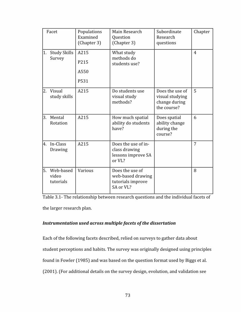
73
Facet Populations Examined (Chapter 3)
Main Research Question (Chapter 3)
Subordinate Research questions
Chapter
1. Study Skills Survey
A215
P215
A550
P531
What study methods do students use?
4
2. Visual study skills
A215 Do students use visual study methods?
Does the use of visual studying change during the course?
5
3. Mental Rotation
A215
How much spatial ability do students have?
Does spatial ability change during the course?
6
4. In-Class Drawing
A215 Does the use of in-class drawing lessons improve SA or VL?
7
5. Web-based video tutorials
Various Does the use of web-based drawing tutorials improve SA or VL?
8
Table 3.1- The relationship between research questions and the individual facets of
the larger research plan.
Instrumentation used across multiple facets of the dissertation
Each of the following facets described, relied on surveys to gather data about
student perceptions and habits. The survey was originally designed using principles
found in Fowler (1985) and was based on the question format used by Biggs et al.
(2001). (For additional details on the survey design, evolution, and validation see

74
the survey appendix A). The survey also was used in the recently published
Husmann et al. (2016), which contains further details about the evolution and
validity of the survey. The original survey was designed to look only at student
study habits and collect background demographic data (Project 1 “What study
methods do students use?” 2010-2011). The survey consisted of 26 questions,
including questions about study methods used to prepare for exams, and basic
demographic information. Each survey question asked the student to rate how
frequently they used each study method listed on a five-point, Likert-type scale. (For
example, a question may ask “I read the class notes to study” and the response
choices are A. always B. frequently C. about half of the time D. rarely E. never).
Further revisions of the survey added more specific questions about visual study
skills, and perceptions of visual learning (Project 2 “Visual Literacy” and Project 3
“Mental Rotation”, 2011-2014. As an additional note, even though project 3 is
entitled “Mental Rotation”, it also assessed visual literacy using the same survey as
the previously mentioned projects).
Another piece of the research introduced a mental rotation test (MRT) to assess
students’ spatial ability skills (Bodner and Guay 1997). Finally, pedagogical
interventions were employed in an effort to teach visual learning skills, and improve
mental rotation abilities (Project 4 “Drawing and visual literacy” and Project 5 “web-
based drawing”, 2013-2014). All data were collected from students at Indiana
University Bloomington, including students in the Indiana University Medical School
(see above; section “Student Populations” unless otherwise noted (viz. Project 5 “on-
line drawing tutorials”). IRB approval was obtained for each project; approval
75
numbers are listed in the relevant methodology sections, as well as in Appendix D.
The web-based software tool used was Draw It to Know It (Draw It to Know It
2016). This software was originally designed as a review tool for neurology
residents, but was substantially re-written to work with neuroanatomy and
neuroscience content at all levels. The aim of the software is to present
neuroanatomy concept in a series of short (3-8 minute) video tutorials, in which the
student draws anatomical structures along with a narrator demonstrating the
drawing. The tutorials also include information about the functional aspects of the
anatomy being described.
Project 1, “What methods do students use to study anatomy/physiology”
2010-2011
Project 1 was designed to answer research questions 1-3, namely “how do students
study anatomy/physiology, how frequently do students use each study strategy, and
does it change course outcomes.” This project also aimed to answer each of the
subcategories included in the above research questions. In this project, four
populations of students were sampled; medical students in anatomy (n=36),
medical students in physiology (n=36), undergraduates in anatomy (n=469), and
undergraduates in physiology (n=238). Each of the provided n-values represent the
total sample population, but the number of students completing all parts of the
survey research were often lower, which will be explained in the results. Medical
students in the study population were all first-year medical students (MS1),
76
undergraduates included in the sample population included students from all four
years of undergraduate coursework.
To examine the question of study skills used in the listed courses, a paper-based
survey was given to all enrolled students near the end of their course. For the
undergraduate course (A215) this was in the fall of 2010, for the medical course
(A550/551) this was spring of 2011. Students were not mandated or incentivized to
complete the surveys, but were encouraged to do so, using a recruitment script
(APPENDIX D) approved by the IU IRB (IRB#: 1010002079). Students were also
provided with an informed consent document (later re-named as a ‘Study
Information Sheet’) to explain potential risks and benefits of participating in the
research (APPENDIX D). Students who opted to complete the survey did so during
normal class time, and returned the completed surveys to a third-party investigator
so neither the researcher or classroom instructor would know who participated.
Surveys were then de-identified using a random number which was then tied to that
specific student’s anonymous course grade, to correlate survey responses with
course performance outcomes.
At the completion of the semester, survey data were compiled into a database
(Excel, Microsoft 2010) and matched with course grades. These paired data were
then analyzed using SPSS 21.0 (IBM 2012). A correlation matrix was created to
explore the survey responses which correlated most strongly with final grades.
Latent variables were pulled from the data using an exploratory factor analysis as a
way of finding which study skills and demographic traits naturally clumped together
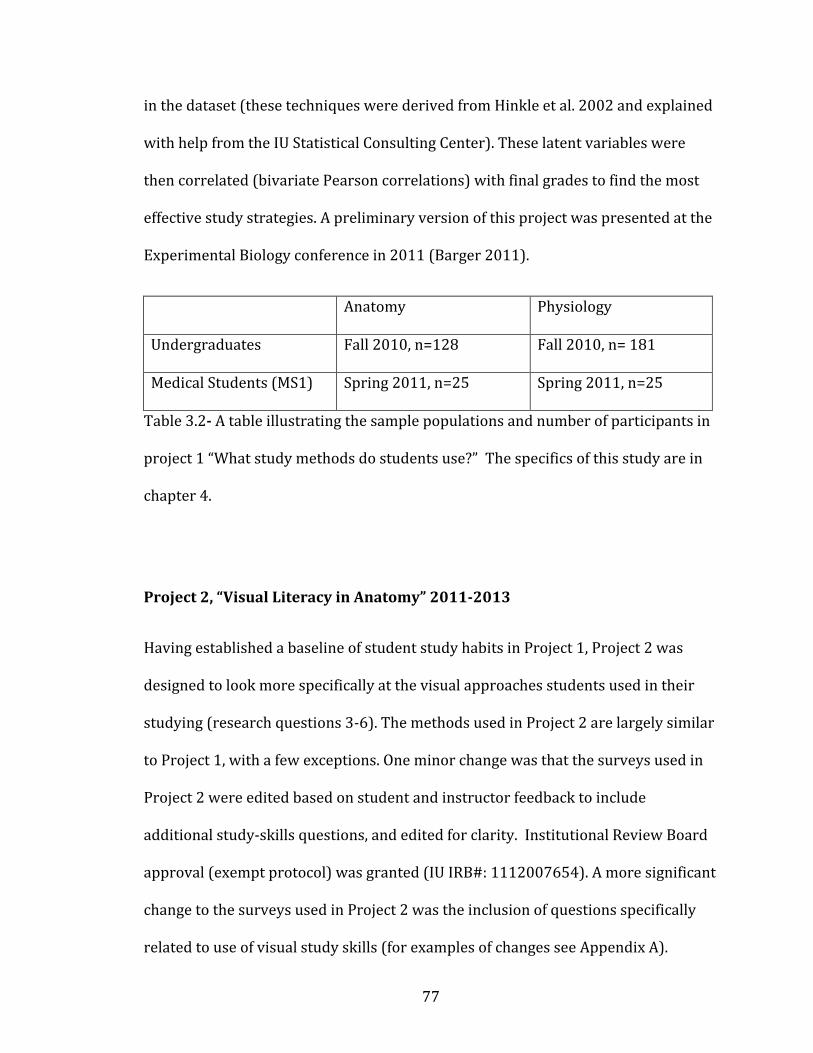
77
in the dataset (these techniques were derived from Hinkle et al. 2002 and explained
with help from the IU Statistical Consulting Center). These latent variables were
then correlated (bivariate Pearson correlations) with final grades to find the most
effective study strategies. A preliminary version of this project was presented at the
Experimental Biology conference in 2011 (Barger 2011).
Anatomy Physiology
Undergraduates Fall 2010, n=128 Fall 2010, n= 181
Medical Students (MS1) Spring 2011, n=25 Spring 2011, n=25
Table 3.2- A table illustrating the sample populations and number of participants in
project 1 “What study methods do students use?” The specifics of this study are in
chapter 4.
Project 2, “Visual Literacy in Anatomy” 2011-2013
Having established a baseline of student study habits in Project 1, Project 2 was
designed to look more specifically at the visual approaches students used in their
studying (research questions 3-6). The methods used in Project 2 are largely similar
to Project 1, with a few exceptions. One minor change was that the surveys used in
Project 2 were edited based on student and instructor feedback to include
additional study-skills questions, and edited for clarity. Institutional Review Board
approval (exempt protocol) was granted (IU IRB#: 1112007654). A more significant
change to the surveys used in Project 2 was the inclusion of questions specifically
related to use of visual study skills (for examples of changes see Appendix A).
78
Additionally, this project only sampled undergraduates in anatomy, ignoring
medical students (for small sample sizes), and physiology students. Physiology
students were excluded from this (and future) projects to focus the results
specifically on anatomy. It was also found in the earlier project that physiology
students tended to use more verbal (versus visual) approaches to studying, and it
was believed that their potential contribution to visual studying research would be
minimal.
The newly-designed, visually-focused surveys were then deployed to anatomy
undergraduates (n=469, the number enrolled in A215 “Basic Human Anatomy”) at
the beginning and end of the semester, using the same IRB protocol from the earlier
project. This project continued to survey anatomy undergraduates for three
semesters across three years. Demographic data indicated that student populations
were statistically the same in each of the courses surveyed (See individual
methodology chapters for demographic details and results, chapter 5). The collected
data were again entered into a database (Excel, Microsoft 2010)) and analyzed using
SPSS 21.0 (IBM 2012). A correlation matrix was created to explore the survey
responses which showed the greatest effect. Latent variables were pulled from the
data using an exploratory factor analysis as a way of finding which study skills and
demographic traits naturally clumped together in the dataset. These latent variables
were then correlated (bivariate correlations) with final grades to find the most
effective study strategies. This data analysis also used the added technique of
comparing visual studying strategies with other active learning approaches, as well
as correlating visual study strategies with final grades. Because this survey also
79
included questions about a student’s own perception of visual learning, the data
analysis allowed for comparisons of high-visual and low-visual students as an
independent variable compared with the other variables defined in the dataset.
Because student survey data were collected at two different time points in the
semester, this dataset was also used to analyze research question 6, “do study habits
change during the semester?” For this analysis, paired students responses (from the
early- and late-semester surveys) were compared using t-tests to see if the student
population (as a whole) showed any significant changes in study habits as a result of
their enrollment in an anatomy course. Preliminary results from this study were
presented at the Experimental Biology conference 2012 (Barger 2012).
Having examined student study habits and visual literacy measures, the next project
considers spatial ability, and its relationship to study habits and visual literacy in
anatomy undergraduates.
Project 3 “Mental rotation ability in anatomy students” 2012-2013
This project was designed to research questions 7a and 7c, namely “do students
have adequate mental rotation skills in anatomy, and do those skills translate to
course performance?” Project 3 used the same survey and IRB protocol as Project 2,
with no additional changes to the survey. An additional test of mental rotation
ability was also given to students in conjunction with the basic survey. The mental
rotation test (MRT) was originally designed by Bodner and Guay (1997) for use with
chemistry students, but use of mental rotation tests is well-established as a proxy
for spatial ability in the educational literature (Chatterjee and Walker 2011, Faurie

80
and Khadra 2012). The MRT (see appendix B for a full version of the test) contains
20 questions which ask the participant to mentally manipulate a 2-dimensional
drawing of a 3-dimensional, geometric object. Each question consists of an example
rotation of one object which the participant must then reproduce in a new object,
via analogy, and choose the correct answer from a multiple-choice list of four
options. The final score on the test is calculated out of twenty points, based on the
number of correct answers.
The responses from the whole class were then averaged, and individual students
were divided into a high-MRT and low-MRT score group, separated by the median
score. The high- and low-MRT variable was then introduced into a correlation
matrix with the other survey variables (as above) to examine which factors most
strongly correlated with MRT scores. MRT scores were also correlated with final
grade to answer research question 7c.
Because MRT scores were collected at two time points in the semester, changes in
whole-class performance were analyzed using a t-test to look for statistically
significant changes in MRT scores during the semester of anatomy.
Preliminary results from this study were presented at the Experimental Biology
conference 2013 (Barger and Husmann 2013).
The three projects outlined above all look at what students do without any
intervention from instructors, and how study habits, visual literacy, and spatial
ability all change during an anatomy course. The following projects each implement
81
pedagogical interventions designed to explicitly teach visual literacy and mental
rotation.
Project 4 “Anatomical drawing and study habits” 2013
The first three projects explained above only collected data from students without
suggesting or implementing any changes in course design or study habits. Project 4
was created around a pedagogical intervention designed to actively teach students
how to employ more visual skills in their studying. Project 4 looked at research
questions 1-8, re-assessing the baseline study habits of anatomy undergraduates,
testing their mental rotation ability, and teaching them ways to improve MRT scores
and course grades. Institutional Review Board approval (exempt protocol) was
granted (IU IRB#: 1301010307).
Project 4 used a quasi-experimental design in the anatomy undergraduate
classroom. In spring of 2013, the anatomy course was taught as two separate
sections (n=252 and n=263), with identical course goals, labs, and exam formats
(See APPENDIX C for course syllabi). Although there were minor differences in
syllabus and some specific exam questions, these differences were not considered
important enough to confound the results, because previous semester course
outcomes and grades were similar between the two sections.
One section of the lecture portion course was taught traditionally with only didactic
lectures, while the other lecture section incorporated a series (6 total) of visually-
focused lectures in which students drew anatomical objects along with the guest-
instructor (me, your humble author, dear reader). The visually-focused lecture
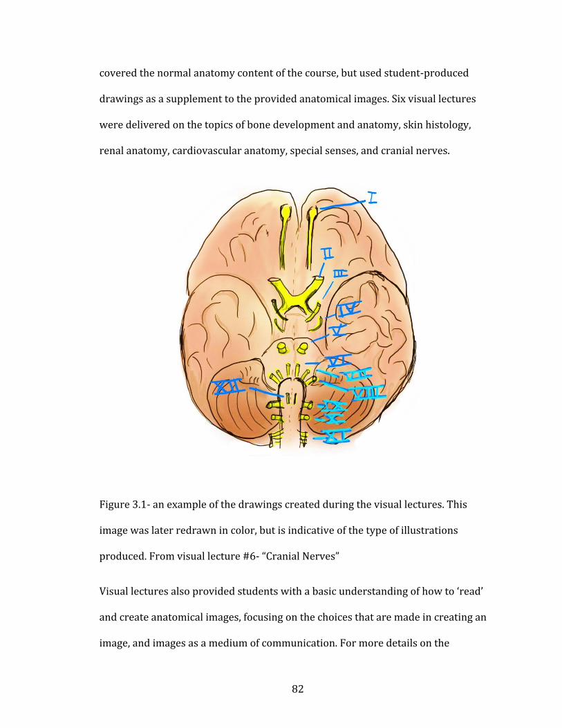
82
covered the normal anatomy content of the course, but used student-produced
drawings as a supplement to the provided anatomical images. Six visual lectures
were delivered on the topics of bone development and anatomy, skin histology,
renal anatomy, cardiovascular anatomy, special senses, and cranial nerves.
Figure 3-0-1
Figure 3.1- an example of the drawings created during the visual lectures. This
image was later redrawn in color, but is indicative of the type of illustrations
produced. From visual lecture #6- “Cranial Nerves”
Visual lectures also provided students with a basic understanding of how to ‘read’
and create anatomical images, focusing on the choices that are made in creating an
image, and images as a medium of communication. For more details on the
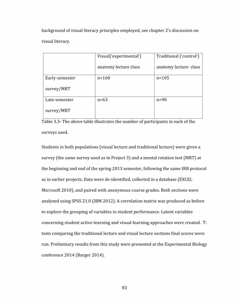
83
background of visual literacy principles employed, see chapter 2’s discussion on
visual literacy.
Visual(‘experimental’)
anatomy lecture class
Traditional (‘control’)
anatomy lecture class
Early-semester
survey/MRT
n=160 n=105
Late-semester
survey/MRT
n=63 n=90
Table 3.3- The above table illustrates the number of participants in each of the
surveys used.
Students in both populations (visual lecture and traditional lecture) were given a
survey (the same survey used as in Project 3) and a mental rotation test (MRT) at
the beginning and end of the spring 2013 semester, following the same IRB protocol
as in earlier projects. Data were de-identified, collected in a database (EXCEL
Microsoft 2010), and paired with anonymous course grades. Both sections were
analyzed using SPSS 21.0 (IBM 2012). A correlation matrix was produced as before
to explore the grouping of variables in student performance. Latent variables
concerning student active-learning and visual-learning approaches were created. T-
tests comparing the traditional lecture and visual lecture sections final scores were
run. Preliminary results from this study were presented at the Experimental Biology
conference 2014 (Barger 2014).
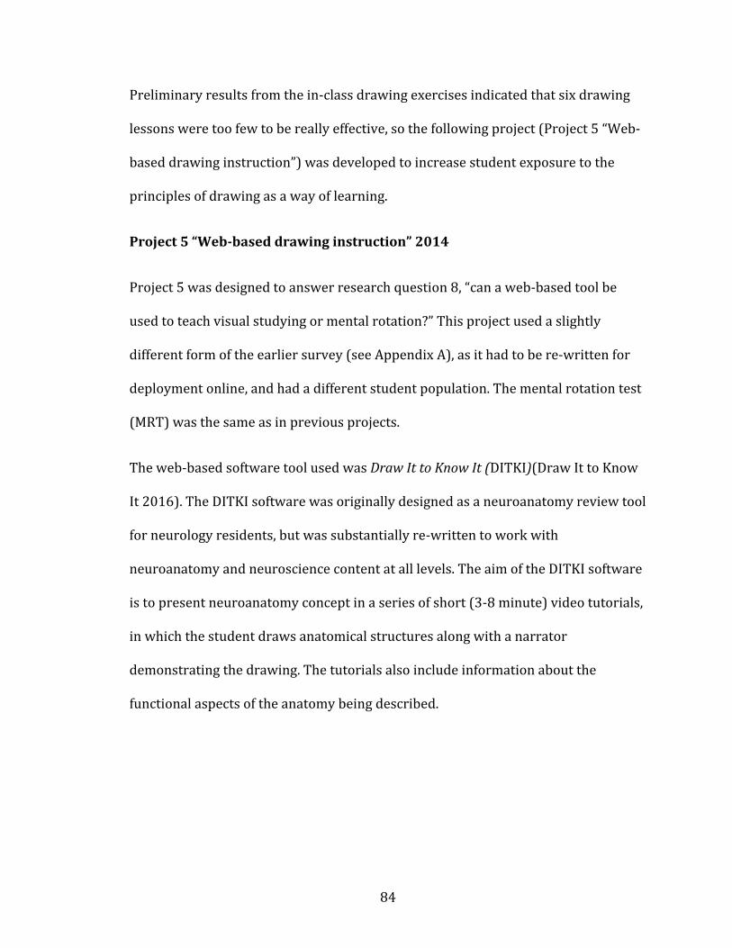
84
Preliminary results from the in-class drawing exercises indicated that six drawing
lessons were too few to be really effective, so the following project (Project 5 “Web-
based drawing instruction”) was developed to increase student exposure to the
principles of drawing as a way of learning.
Project 5 “Web-based drawing instruction” 2014
Project 5 was designed to answer research question 8, “can a web-based tool be
used to teach visual studying or mental rotation?” This project used a slightly
different form of the earlier survey (see Appendix A), as it had to be re-written for
deployment online, and had a different student population. The mental rotation test
(MRT) was the same as in previous projects.
The web-based software tool used was Draw It to Know It (DITKI)(Draw It to Know
It 2016). The DITKI software was originally designed as a neuroanatomy review tool
for neurology residents, but was substantially re-written to work with
neuroanatomy and neuroscience content at all levels. The aim of the DITKI software
is to present neuroanatomy concept in a series of short (3-8 minute) video tutorials,
in which the student draws anatomical structures along with a narrator
demonstrating the drawing. The tutorials also include information about the
functional aspects of the anatomy being described.
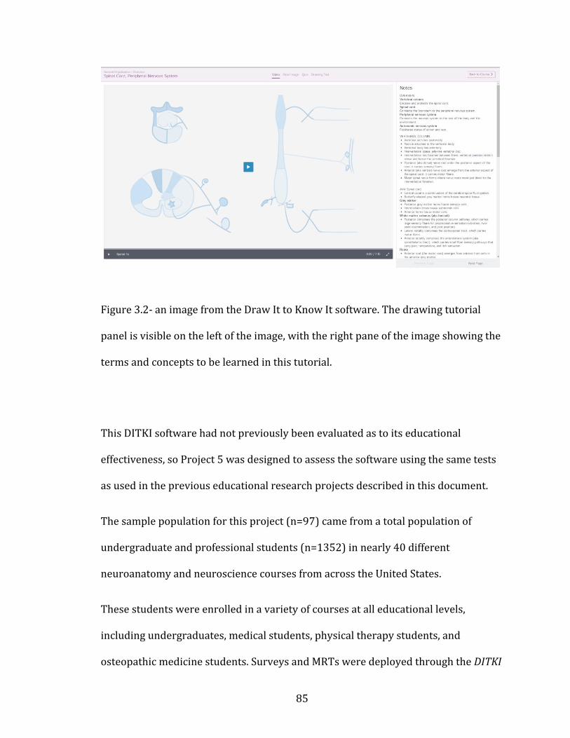
85
igure 3-0-2
Figure 3.2- an image from the Draw It to Know It software. The drawing tutorial
panel is visible on the left of the image, with the right pane of the image showing the
terms and concepts to be learned in this tutorial.
This DITKI software had not previously been evaluated as to its educational
effectiveness, so Project 5 was designed to assess the software using the same tests
as used in the previous educational research projects described in this document.
The sample population for this project (n=97) came from a total population of
undergraduate and professional students (n=1352) in nearly 40 different
neuroanatomy and neuroscience courses from across the United States.
These students were enrolled in a variety of courses at all educational levels,
including undergraduates, medical students, physical therapy students, and
osteopathic medicine students. Surveys and MRTs were deployed through the DITKI
86
website, and a Study Information Sheet (previously called Informed Consent
Statement) allowed students top opt-in to the data-collection portion of the project
(IU IRB#: 1310535940). Those students who opted in completed a survey at the
beginning of their neuroanatomy course, and another at the end. They were also
asked to complete the MRT at the beginning and end of their neuroanatomy course.
De-identified student data were collected in a database (Excel Microsoft 2010) as
before, and analyzed using SPSS 21.0 (IBM 2012). A notable difference in this data
analysis is the lack of student final grades. Due the constraints of ethical research
across multiple campuses, no course performance metrics were collected, and the
data analysis relied on student self-reported satisfaction with the software, and
their perceived quality of learning from it. Preliminary results from this study were
presented at the Experimental Biology conference 2015 (Barger and Fisch 2015).
Each subsequent chapter will discuss the research projects in greater detail,
including results and a discussion of the pedagogical implications of each question
about how students study.
87
CHAPTER 4: Assessing Student Study Habits in Anatomy and Physiology
Many factors have been proposed as predictors of success in college students.
Motivation (Phinney et al. 2005), peer support (Harackiewicz et al. 2002), financial
support (Karpicke et al. 2009, Phinney et al. 2005), and academic strategies (or
study skills) (Aleven et al. 2000, Hmelo-Silver 2004, Robbins et al. 2004), have all
been used as predictors of performance in post-secondary education. Broadly
considered, these variables can be divided into cognitive (study skills) and non-
cognitive (social, financial, and motivational) determinants of success (Robbins et al.
2004). Each of these predictive variables has been shown to have some predictive
power of college success when measured across large populations of students, but
course-specific predictors are rarely found. A meta-analysis of these various factors
indicated that motivation (non-cognitive) tends to predict student retention, while
study skills (cognitive) are one of the strongest predictors of GPA (Robbins et al.
2004). All of the above publications examined large groups of college students, with
no discipline specific analyses, or analyses of medical students.
The goal of the research facet presented here, is to further examine the cognitive
predictors of success. That is, do study strategies have any correlation with course
grade in anatomy (or physiology) undergraduates or medical students? It is
reasonable to assume that a discipline specific look at predictors of success will not
vary greatly from the institution-wide data present in the research literature,
however, knowing what anatomy (or physiology) students do outside of class has
pedagogical implications. Understanding what successful students do to study can
influence the advice given to struggling students, and help instructors ensure course
88
assessments are authentically promoting learning (Hart 1994, Gurung and McCann
2011, Schwartz and Gurung 2012, Wiggins 1990).
Knowing that cognitive factors play a strong predictive role in GPA, a research
project was initiated to measure the study skills of a variety of anatomy and
physiology students at Indiana University. In 2010 a project (one facet of the larger
dissertation) to survey students about their study habits began. . Populations
studied included first-year medical students and undergraduates enrolled in
anatomy courses and physiology courses (see table 4.1). The specific focus of this
project was to understand how students use their time in preparation for exams,
and to find differences between student populations and courses, and to see if any
specific study strategies correlated with course success as measured by course
percentages (as demonstrated in other course contexts by Gurung and McCann
2011, Schwartz and Gurung 2012) . In the previous chapter, this project was
designated as Project 1, and was designed to answer the following research
questions as seen in Table 4.1.
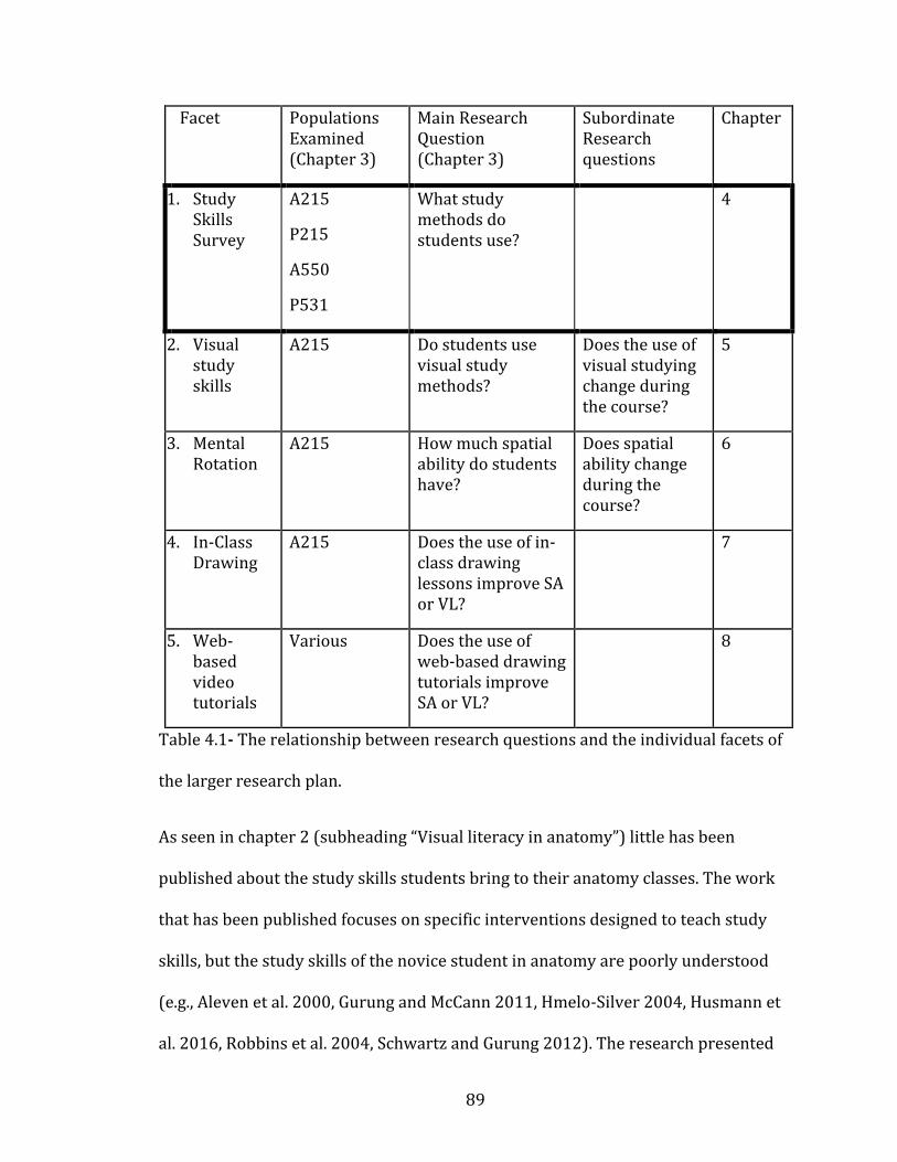
89
Facet Populations Examined (Chapter 3)
Main Research Question (Chapter 3)
Subordinate Research questions
Chapter
1. Study Skills Survey
A215
P215
A550
P531
What study methods do students use?
4
2. Visual study skills
A215 Do students use visual study methods?
Does the use of visual studying change during the course?
5
3. Mental Rotation
A215
How much spatial ability do students have?
Does spatial ability change during the course?
6
4. In-Class Drawing
A215 Does the use of in-class drawing lessons improve SA or VL?
7
5. Web-based video tutorials
Various Does the use of web-based drawing tutorials improve SA or VL?
8
Table 4.1- The relationship between research questions and the individual facets of
the larger research plan.
As seen in chapter 2 (subheading “Visual literacy in anatomy”) little has been
published about the study skills students bring to their anatomy classes. The work
that has been published focuses on specific interventions designed to teach study
skills, but the study skills of the novice student in anatomy are poorly understood
(e.g., Aleven et al. 2000, Gurung and McCann 2011, Hmelo-Silver 2004, Husmann et
al. 2016, Robbins et al. 2004, Schwartz and Gurung 2012). The research presented
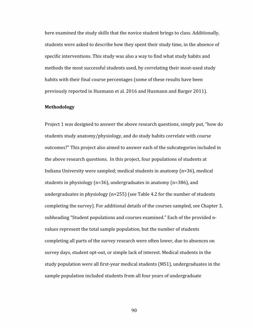
90
here examined the study skills that the novice student brings to class. Additionally,
students were asked to describe how they spent their study time, in the absence of
specific interventions. This study was also a way to find what study habits and
methods the most successful students used, by correlating their most-used study
habits with their final course percentages (some of these results have been
previously reported in Husmann et al. 2016 and Husmann and Barger 2011).
Methodology
Project 1 was designed to answer the above research questions, simply put, “how do
students study anatomy/physiology, and do study habits correlate with course
outcomes?” This project also aimed to answer each of the subcategories included in
the above research questions. In this project, four populations of students at
Indiana University were sampled; medical students in anatomy (n=36), medical
students in physiology (n=36), undergraduates in anatomy (n=386), and
undergraduates in physiology (n=255) (see Table 4.2 for the number of students
completing the survey). For additional details of the courses sampled, see Chapter 3,
subheading “Student populations and courses examined.” Each of the provided n-
values represent the total sample population, but the number of students
completing all parts of the survey research were often lower, due to absences on
survey days, student opt-out, or simple lack of interest. Medical students in the
study population were all first-year medical students (MS1), undergraduates in the
sample population included students from all four years of undergraduate
91
coursework. These populations were chosen to examine differences in exam
preparation between student educational level and course.
Anatomy Physiology
Undergraduates Fall 2010, n=217 Fall 2010 n=181
Medical Students (MS1) Spring 2011, n=25 Spring 2011, n=25
Table 4.2- The sample populations and number of participants completing all
surveys on the methods students use to study anatomy and physiology.
To examine the question of study skills used in the listed courses, a paper-based
survey (see Appendix A) was given to all enrolled students near the end of the
semester, approximately one week before the final exam. This time for deploying
the survey was chosen to collect retrospective data from the students about their
entire semester of studying for their course. The survey consisted of 26 questions,
including questions about study methods used to prepare for exams, and basic
demographic information. Each survey question asked the student to rate how
frequently they used each study method listed on a five-point, Likert-type scale. (For
example, a question may ask “I read the class notes to study” and the response
choices are A. always B. frequently C. about half of the time D. rarely E. never).
Students were not mandated or incentivized to complete the surveys, but were
encouraged to do so, using a recruitment script (Appendix D) approved by the IU
IRB (IRB#: 110002079). Students were also provided with an informed consent
document (later re-named as a ‘Study Information Sheet’) to explain potential risks
and benefits of participating in the research (Appendix D). Students who opted to
92
complete the survey did so during normal class time, and returned the completed
surveys to a third-party investigator so neither the researcher or classroom
instructor would know who participated. Surveys were then de-identified using a
random number which was then tied to that specific student’s anonymous course
percentage, to correlate survey responses with course performance outcomes. At
the completion of the semester, survey data were compiled into a database (Excel,
Microsoft 2010) and matched with course grades. These paired data were then
analyzed using SPSS 21.0 (IBM 2012). A correlation matrix was created to explore
the survey responses which showed the highest correlation with final course
percentage. Latent variables were pulled from the data using an exploratory factor
analysis as a way of finding which study skills and demographic traits naturally
clumped together in the dataset (Tabachnick et al. 2001). These latent variables
were then correlated (bivariate correlations) with final grades to find the most
effective study strategies.
Results
Initial results showed little differences between discipline (anatomy versus
physiology) or student population (undergraduate versus medical), based on single
study methods. That is, no single study method correlated strongly with final course
grade, in any of the sampled populations (Appendix E, figure E.3 Correlation table is
not included here for space considerations, but relevant data are shown in the
following figures). As seen in the correlation table provided, the strongest
correlation was a negative correlation (r=-.205, p=.003) between students who used
93
other websites (non-course specific anatomy resources) and final grade. The lack of
any positive correlations between study habits and grade indicates that there may
not be a best way to study, but there are some less productive methods students
should be aware of.
The next step of the analysis was to look for other trends in the data. An exploratory
factor analysis was run on the data (after method explained in Tabachnick et al.
2001 ), which showed strong grouping of certain sets of study skills. The tables
produced can be seen in appendix E, Table E.1. The factor analysis identified two
variables (components) which explained the largest portion of the variance in the
survey responses. After looking at which of the original survey questions had the
highest loading on the new variables, it was clear that active learning and passive
learning were the latent variables which explained the most difference in the survey
sample. Component 1 in the SPSS output table (appendix E, Table E.2) was termed
active learning because it had high loadings with ‘made my own flashcards’, ‘made
my own tables’ and ‘made my own drawings’ survey questions. Component 2 was
termed passive learning because it had the highest loadings with ‘reviewed lecture
notes’, ‘looked at textbook’, ‘looked at a website’ survey questions. A third
component (not a strong explanation for survey variance, but with high loading
values) was identified as self-efficacy measures. Self-efficacy included questions
related to a student’s expected performance on an exam.
Three groups were defined, with these new latent variables being labeled as ‘active
learning,’ ‘passive learning,’ and ‘self-efficacy measures.’ Active learning methods
94
included ‘creating my own flashcards,’ ‘making drawings,’ and ‘creating tables of
information.’ Passive learning methods included ‘reading the textbook,’ ‘reviewing
diagrams,’ and ‘using commercially available flashcards.’ Self-efficacy measures
included ‘expected grade’ and ‘I have studied enough.’ Each category was then
assigned a score based on how frequently the student reported using each method
included in the category. These new categories (latent variables) showed more
pronounced differences between populations and courses, as shown in the following
graphs.
Figure 4.1 illustrates the correlation between final grade (percentage) and the
active learning score the student reported on the survey (for all four groups of
students (anatomy undergraduates, physiology undergraduates, anatomy medical
students, physiology medical students). It can be seen in the series of scatterplots
that all courses had weak positive correlations between active learning and grade.
None of the correlations are strong or statistically significant, but the fact that all
courses showed the same trends is indicative of a real pattern in the data.
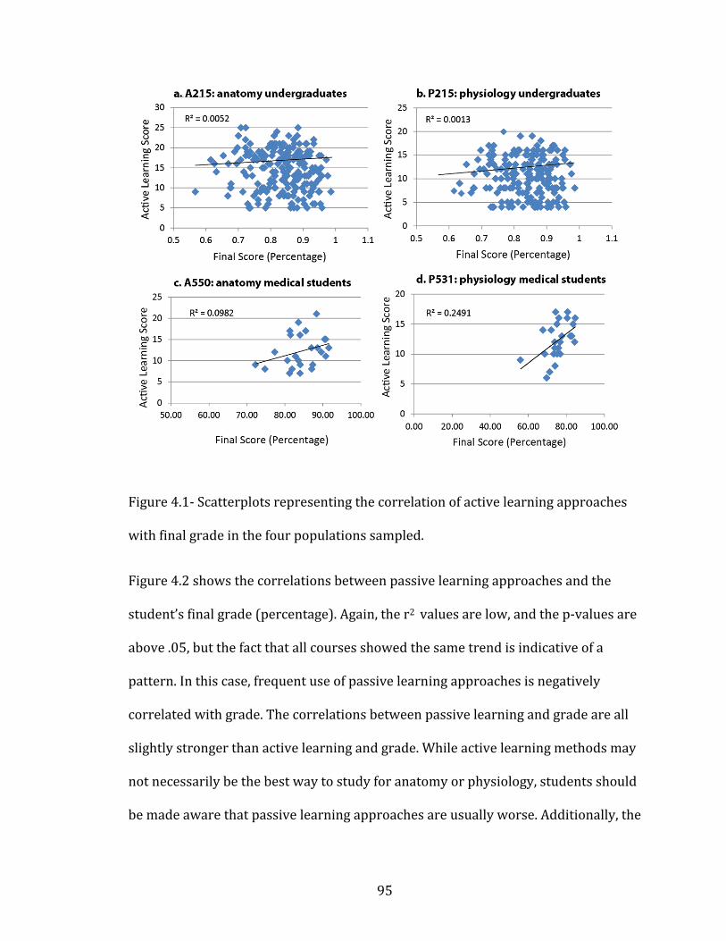
95
Figure 4.0-1
Figure 4.1- Scatterplots representing the correlation of active learning approaches
with final grade in the four populations sampled.
Figure 4.2 shows the correlations between passive learning approaches and the
student’s final grade (percentage). Again, the r2 values are low, and the p-values are
above .05, but the fact that all courses showed the same trend is indicative of a
pattern. In this case, frequent use of passive learning approaches is negatively
correlated with grade. The correlations between passive learning and grade are all
slightly stronger than active learning and grade. While active learning methods may
not necessarily be the best way to study for anatomy or physiology, students should
be made aware that passive learning approaches are usually worse. Additionally, the
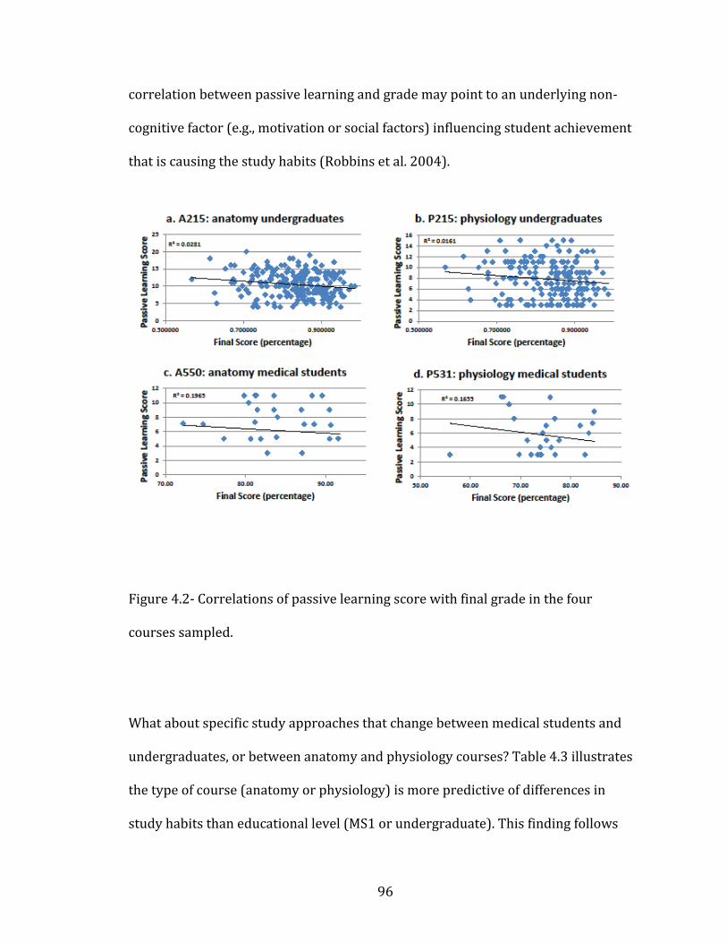
96
correlation between passive learning and grade may point to an underlying non-
cognitive factor (e.g., motivation or social factors) influencing student achievement
that is causing the study habits (Robbins et al. 2004).
Figure 4.0-2
Figure 4.2- Correlations of passive learning score with final grade in the four
courses sampled.
What about specific study approaches that change between medical students and
undergraduates, or between anatomy and physiology courses? Table 4.3 illustrates
the type of course (anatomy or physiology) is more predictive of differences in
study habits than educational level (MS1 or undergraduate). This finding follows

97
Gurung and Schwartz (2009) and Husmann et al. (2016) who suggested that course
content is a stronger predictor of student study methods than any intrinsic factor in
the student population. The lack of apparent difference between MS1 and
undergraduates may be explained by the relatively small difference in their
educational levels. Medical students are often treated as fundamentally different
from undergraduates, but they have four years of undergraduate study patterns to
draw from, and are unlikely to have learned to study differently after only one
semester of medical school coursework. Additionally, in all populations, the lowest
grade quartile of students reported trying the most different study techniques. The
top quartile of students used fewer study techniques more frequently, while those at
the bottom used a lot of different techniques but spent little time on each one.
Anatomy study
techniques
Physiology study
techniques
Undergraduates Looking at figures (.38)
Reading the text (.56)
Reading course notes(.48)
Practice questions(.42)
Medical Students (MS1) Looking at figures (.56)
Reading the text (.68)
Reading course notes(.52)
Practice questions(.76)
Table 4.3- The most popular study techniques in each course. The numbers in
parentheses indicate the percentage of students who reported ‘frequent’ or ‘very
frequent’ use of the indicated study technique.
The third factor identified in the exploratory factor analysis was self-efficacy. While
this factor only explained a small part of the sample variance, the correlations
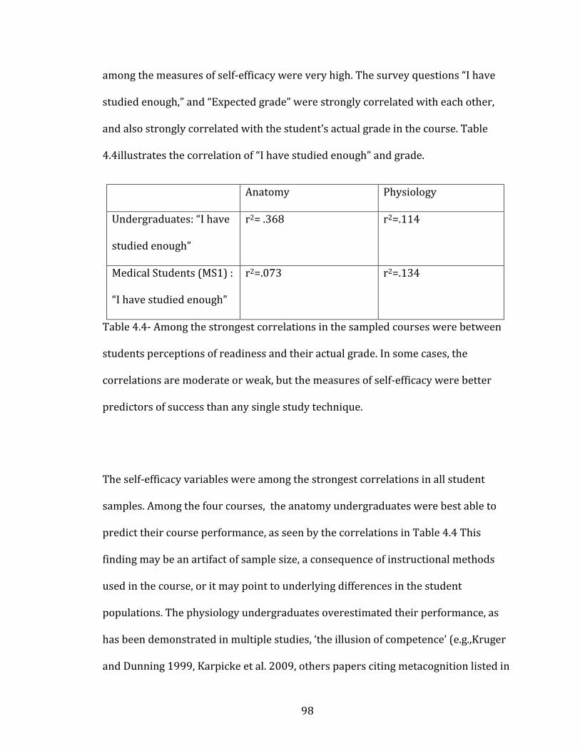
98
among the measures of self-efficacy were very high. The survey questions “I have
studied enough,” and “Expected grade” were strongly correlated with each other,
and also strongly correlated with the student’s actual grade in the course. Table
4.4illustrates the correlation of “I have studied enough” and grade.
Anatomy Physiology
Undergraduates: “I have
studied enough”
r2= .368 r2=.114
Medical Students (MS1) :
“I have studied enough”
r2=.073 r2=.134
Table 4.4- Among the strongest correlations in the sampled courses were between
students perceptions of readiness and their actual grade. In some cases, the
correlations are moderate or weak, but the measures of self-efficacy were better
predictors of success than any single study technique.
The self-efficacy variables were among the strongest correlations in all student
samples. Among the four courses, the anatomy undergraduates were best able to
predict their course performance, as seen by the correlations in Table 4.4 This
finding may be an artifact of sample size, a consequence of instructional methods
used in the course, or it may point to underlying differences in the student
populations. The physiology undergraduates overestimated their performance, as
has been demonstrated in multiple studies, ‘the illusion of competence’ (e.g.,Kruger
and Dunning 1999, Karpicke et al. 2009, others papers citing metacognition listed in
99
chapter 2). Interestingly, the medical students, in both courses, tended to
underestimate their readiness, and inversion of the frequently seen pattern in
undergraduates, but one that has been observed before (Blanch-Hartigan 2011).
Discussion
The stated goal of this project was to determine what students are actually doing
with their study time. Students in all of the sampled courses are sometimes advised
about ways to improve their studying (Appendix C), but there were no data
examining what the students were really doing. Typical advice given to students
suggest using active learning and self-quizzing to prepare for exams. This project
showed that students use a wide variety of study skills, with passive approaches
(including reading the textbook, reading course notes, and reviewing diagrams)
being among the most frequently used in all courses. Active learning approaches
have been shown to be more effective learning strategies, but passive study habits
tend to be used more frequently (Biggs 2001, Drapkin et al. 2015, Freeman et al.
2014, Pickering 2014, Prince 2004, Sweeney et al. 2014). Practice exam questions
were frequently used by both levels of physiology students, a study habit that has
been shown to be very effective, and improve metacognitive awareness (Karpicke et
al. 2009). Interestingly, the physiology students had lower self-efficacy scores than
the anatomy students, despite using a study technique (practice exam questions)
that is supposed to improve metacognition (and self-efficacy). This contradiction
may be due to differences in the method of examination in the two courses,
100
differences in the students, or inaccurate reporting on the surveys. The current
dataset is unable to answer this question.
While practice questions were used frequently in some of the courses, the most
popular techniques across all courses tended to be very passive. This is not
surprising, as passive learning techniques require the least effort, and can lead to
acceptable grades when deep learning is not required (Biggs et al. 2001). The use of
passive approaches contradicts the advice of instructors, and was shown to be
slightly correlated with poorer course performance, a trend evident in all four
courses sampled.
A subordinate goal of this project was to determine which, if any, study methods led
to higher scores on exams. In that goal, the results were inconclusive. There were no
statistically significant differences in study habits employed by the higher-scoring
students. However, some trends can be observed, with active learning approaches
being weakly positively correlated with course grade (Figure 4.1). Interestingly, the
students who used the widest variety of study skills tended to be the lower-scoring
students. This may represent a sampling bias, or may represent the idea of “grasping
at straws.” A student without a well-focused study plan may try a little bit of
everything, without ever finding a truly effective study approach.
Another surprising finding in this dataset was in the measures of self-efficacy.
Students in the sampled population could accurately predict their exam grades, and
were able to honestly report when they felt well prepared for an exam, but this
effect was strongest in only one of the four courses (anatomy undergraduates
101
A215). Medical students tended to underestimate their grades, perhaps due to the
stresses of medical school courses, and the surveys being conducted near the
beginning of their medical school careers. The MS1 students may simply have felt
unprepared for their exams based on their patterns of undergraduate studying
(Blanch-Hartigan 2011).
It was also found that anatomy and physiology students did not study in a
significantly different way. While whole-class averages indicated that the most
popular study techniques differed between the two courses, the overall study
patterns taken by individual students did not change much. That is, passive learning
patterns persisted in all courses, regardless of subject matter or educational level.
This pattern of passive learning may represent a strategic approach (Biggs et al.
2001) taken by students who understand that the exams in both courses are
multiple choice, and passive study approaches are often adequate for multiple-
choice exams. Active learning approaches may be viewed as a less efficient when the
student knows that a deep knowledge of the material is not required on an exam
that only assesses lower-level thinking.
The findings of this project have direct pedagogical implications, indicating that
instructor advice on student study skills should be changed. Rather than advocating
continued use of active learning only, instructors should encourage students to
choose a few specific study skills (Barger 2012, Barger and Husmann 2013,
Husmann et al. 2016). A reliance on a few study skills, used frequently, appears to
lead to higher course grades, than using a lot of study skills infrequently.

102
Having established how students study (Research questions 1-3), the following
chapter will examine research questions 4-6, visual approaches to studying.
103
CHAPTER 5: Visual Study Skills in Anatomy
Visual literacy and spatial ability are two different, but related, skills involved in
understanding the visual world (Avgerinou and Ericson 1997, Bodner and Guay
1997, Meneghetti et al. 2011, Meneghetti et al. 2016). Spatial ability has long been
understood as a correlate with success in STEM fields (discussed in Chapter 2 and
Chapter 6), but visual literacy in STEM has not been well studied (Backhouse et al.
2016, Bardes et al. 2001, Bell et al. 2014, Boudreau et al. 2008, Naghshineh et al.
2008). Efforts to teach visual literacy in STEM fields include (Backhouse et al. 2016,
Bardes et al. 2001, Bell et al. 2014, Pickering 2014), but measuring visual literacy
attainment is still a challenge. All of the above citations used works of fine art to
teach medical students clinically-relevant observational skills, but only Naghshineh
et al. (2008) developed a measurement of visual literacy. To measure visual literacy,
Naghshineh and colleagues introduced first-year medical students to the practice of
carefully observing works of art, then made a quantitative measure of the number of
specific observations those students made during clinical exams. It was found that
students who had participated in the art observation training made significantly
more clinical observations than their peers who did not receive any specific training
in observation skills.
This study (Naghshineh et al. 2008) is the only one with any kind of assessment of
student visual literacy in STEM. Other publications dealing with visual literacy in
STEM have shown students to derive benefit from visual literacy training, but these
benefits are all based in student attitudes and do not contain measurable
improvements (Backhouse et al. 2016, Bardes et al. 2001, Bell et al. 2014).
104
Assessment of visual literacy has been recognized as a problem since the term was
introduced in 1969 (Avgerinou and Ericson 1997, Perkins 1994, Wiles 2016).
Measuring the number of clinical observations studetns make is a good way of
assessing visual literacy skills, but it is time consuming and impractical in pre-
clinical students. Chapter 2 summarizes the history of visual literacy research, but a
brief recapitulation follows.
Visual literacy (VL) is the ability to meaningfully interpret static and dynamic
images, and has a long and contentious history (Avgerinou and Ericson 1997).
Despite the simple, one-line definition for visual literacy given above, it is a
complicated and wide‐ranging topic that has resisted a firm definition for the last
fifty years.
Very little has been written about visual literacy in anatomy or biology, but several
revealing publication in biochemistry discuss visual literacy (Anderson 2007,
Linenberger and Bretz 2015, Schonborn and Anderson 2006 , Schonborn and
Anderson 2010, Serpente 2016, Wiles 2016). The existence of visual literacy
discussion in biochemistry may be due to the necessity of creating meaningful
mental representations of biochemical entities in the study of biochemistry.
Biochemical entities are mostly molecules and proteins, impossible to actually
visualize with a microscope, so conceptual visual models are the only visual
representations available to student of biochemistry. The variety of
representational schemes for biochemical entities is also mentioned as a need for
robust VL education in biochemistry (Wiles 2016). That is, a protein, for example,
105
can be represented by a linear arrangement of letters corresponding to amino acids,
a 3D ball-and-stick model, or a 3D ribbon diagram. Each of these visual
representations of the protein conveys different information, and the biochemistry
expert is able to extract the information of the image, and even infer the appearance
of one type of image from another type of image (Schonborn and Anderson 2006).
In 2010 Schonborn and Anderson continued their work in VL and defined a list of
skills the visually literate biochemistry students should have, termed ‘reasoning
with images’ in the original. The list of skills involved in reasoning with images
includes:
• Decode the symbolic language composing an image
• Evaluate the power, limitations, and quality of an image
• Interpret and use an image to solve a problem
• Spatially manipulate an image to interpret and explain a concept
• Construct an image to explain a concept or solve a problem
• Translate horizontally across multiple images of a concept
• Translate vertically between images that depict various levels
of organization and complexity
• Visualize orders of magnitude, relative size, and scale.
Outside of discipline specific literature, The Association of College and Research
Libraries (ACRL) who released a set of standards for VL in October 2011 (Hattwig et
al. 2013). The ACRL standards defined a series of seven competencies designed to
106
show what a visually literate person can do. Assuming each of the competencies is
met, one can be called ‘visually literate’. The ACRL list still contains very little detail
on how to assess each competency, but it provides a theoretical foundation for
further inquiry in VL research. The ACRL list has some similarity with the
biochemistry list above, and both were incorporated to create a list of VL
competencies for anatomy students described below.
1. Determine the nature and extent of the visual materials needed
2. Find and access needed images and visual media effectively and
efficiently
3. Interpret and analyze the meanings of images and visual media
4. Evaluate images and their sources
5. Use images and visual media effectively
6. Design and create meaningful images and visual media
7. Understand many of the ethical, legal, social, and economic issues
surrounding the creation and use of images and visual media, and access
and use visual materials ethically
Elaboration of each competency can be found in chapter 2.
The following table prepared by the dissertation author (Table 5.1) uses the ACRL
list of visual literacy competencies as its base, but includes some of the specific skills
defined by the work of biochemistry educational researchers. While rigorously
assessing visual literacy is still a challenge, the following list of anatomy-specific
107
visual literacy competencies provides a theoretical framework for discussing what
the visually literate anatomy student can do.
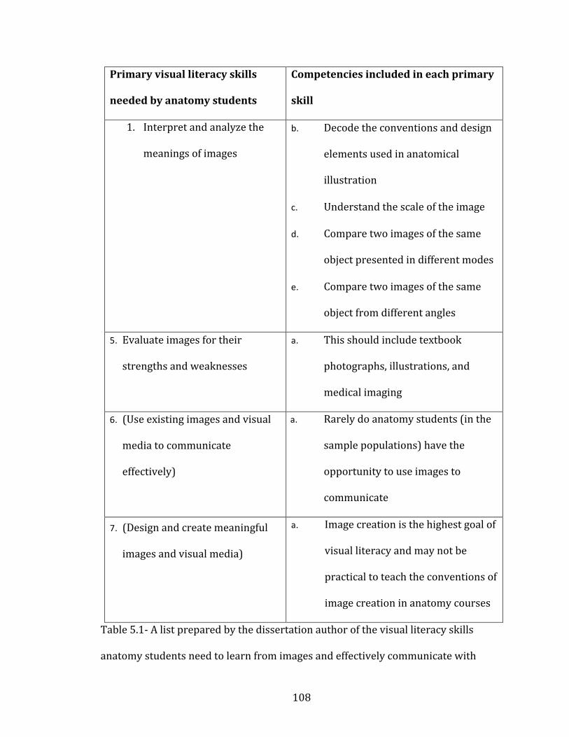
108
Primary visual literacy skills
needed by anatomy students
Competencies included in each primary
skill
1. Interpret and analyze the
meanings of images
b. Decode the conventions and design
elements used in anatomical
illustration
c. Understand the scale of the image
d. Compare two images of the same
object presented in different modes
e. Compare two images of the same
object from different angles
5. Evaluate images for their
strengths and weaknesses
a. This should include textbook
photographs, illustrations, and
medical imaging
6. (Use existing images and visual
media to communicate
effectively)
a. Rarely do anatomy students (in the
sample populations) have the
opportunity to use images to
communicate
7. (Design and create meaningful
images and visual media)
a. Image creation is the highest goal of
visual literacy and may not be
practical to teach the conventions of
image creation in anatomy courses
Table 5.1- A list prepared by the dissertation author of the visual literacy skills
anatomy students need to learn from images and effectively communicate with
109
images. Skills 3 and 4 are enclosed in parentheses because they are goals of visual
literacy education, but may be outside the scope of most anatomy courses.
Having created a theoretical basis for understanding visual literacy in anatomy, the
next step was to examine how visual literacy is actually employed by students.
The research project discussed here (facet 2: “Visual literacy in anatomy”, of the
original research outline in Chapter 1) was designed to answer the following
questions (see also Table 5.2):
Research Question 4: How often do anatomy students use visual study skills?
This question is the central idea guiding the overall research plan. The following
questions are all further elaborations of the visual study skills question. The
introduction and literature review chapters (Chapters 1 and 2) are full of
justifications and research explaining the utility of visual learning (especially mental
rotation and spatial ability) in a variety of fields, including other sciences, and
anatomy, but the use of visual study skills in novice students has not been fully
explored in the existing literature (to clarify, mental rotation and spatial ability are
well explored in the existing literature, but the use of visual study skills, as defined
in this dissertation, Chapter 1, subheading “Definitions” is not widely reported)
(Backhouse et al. 2016, Bardes et al. 2001, Bell et al. 2014, Boudreau et al. 2008,
Naghshineh et al. 2008).
110
Research Question 5: Are there differences in student self-reported visual
learning measures?
More specifically:
c. Do students who self-report a preference for visual learning use
visual study skills more often in their anatomy courses?
d. Do students who self-report a preference for visual learning do
better in anatomy course performance metrics?
This question was designed to look at differences in student perceptions and uses of
visual learning in anatomy. That is, do students use visual study strategies
frequently, if so which kind of students (those who self-report as visual learners, or
not), and do these strategies correlate with course grade? Students enter anatomy
courses with a wide variety of previous experiences, and it is unclear how those
experiences shape a student’s study habits. This question asks whether different
students use different study habits without any intervention from the instructors or
researchers.
Research Question 6: Do student study habits for anatomy change during
the semester?
All of the previous questions rely on sampling students at a single time point in the
semester. This question allows for the exploration of study habit changes in
students during a course, including those habits related to visual learning and
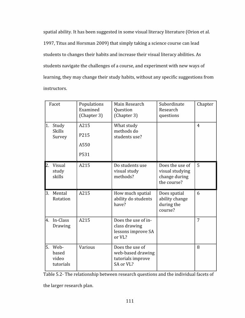
111
spatial ability. It has been suggested in some visual literacy literature (Orion et al.
1997, Titus and Horsman 2009) that simply taking a science course can lead
students to changes their habits and increase their visual literacy abilities. As
students navigate the challenges of a course, and experiment with new ways of
learning, they may change their study habits, without any specific suggestions from
instructors.
Facet Populations Examined (Chapter 3)
Main Research Question (Chapter 3)
Subordinate Research questions
Chapter
1. Study Skills Survey
A215
P215
A550
P531
What study methods do students use?
4
2. Visual study skills
A215 Do students use visual study methods?
Does the use of visual studying change during the course?
5
3. Mental Rotation
A215
How much spatial ability do students have?
Does spatial ability change during the course?
6
4. In-Class Drawing
A215 Does the use of in-class drawing lessons improve SA or VL?
7
5. Web-based video tutorials
Various Does the use of web-based drawing tutorials improve SA or VL?
8
Table 5.2- The relationship between research questions and the individual facets of
the larger research plan.
112
Methodology
To answer the research questions listed above, a survey (Appendix A) was
developed to assess students’ self-reporting of visual study strategies. This survey
tested only anatomy undergraduates (and specifically, students in Anatomy A215,
see chapter 3). Because there are no established assessments for measuring visual
literacy, it was assumed that use of visual study strategies was a good estimator of
visual literacy. This assumption was based on the anatomy-specific list of visual
literacy competencies (defined above). If students possess the competencies, it was
expected that their study habits will align with the competencies, and they will
employ more visual study strategies (e.g., looking at textbook images, drawing their
own images or tables,).
The survey used to examine visual study strategies included 16 questions about the
frequency of use of a variety of study skills, and 7 questions about the students’ self-
perception as a visual learner. The survey may be seen, in full, in Appendix A.
An additional feature of the survey used in this facet of the dissertation was the
inclusion of questions relating to learning style (the 7 questions about the students’
self-perception as a visual learner). Learning styles are a controversial topic with
numerous authors for and against their validity in educational research (e.g.,
Fleming et al. 2006, Leite et al. 2010, Lujan et al. 2006). Learning styles will be
discussed further in the discussion section. The learning style inventory used here
consisted of seven VARK-like questions related only to the visual axis of the VARK.
(e.g., “if I am planning a route to a new location, I like to look at a map,” If I am
113
planning a presentation, I create an outline first,” “I consider myself a visual
leaner.”). The inclusion of a learning style inventory was necessary for two reasons.
First, does a student’s self-perception of learning style have any impact on course
performance? Second, does self-perception play a role in how a student chooses to
study?
Surveys were deployed to anatomy (A215) undergraduates at the beginning and
end of the semester, using the same IRB protocol from the earlier project (see
Appendix A for survey details and Appendix D for IRB details). Students were
surveyed near the beginning of the course (before the first set of lecture and lab
exams, approximately three weeks into the semester) and near the end of the course
(before the final lecture exam, approximately one week before the end of the
semester). These two survey time points were chosen to assess the study habits of
novice students (before they have had any examination experience) and more
experienced students to see if their study habits changed after exposure to course
material and examinations.
The collected data were again entered into a database (Excel, Microsoft 2010) and
analyzed using SPSS 21.0 (IBM 2012). A correlation matrix was created to explore
the survey responses with the strongest correlations to final grade. Latent variables
were pulled from the data using an exploratory factor analysis as a way of finding
which study skills and demographic traits naturally clumped together in the dataset.
These latent variables were then correlated (bivariate or Pearson correlations) with
final grades to find the most effective study strategies. The VARK-style questions all
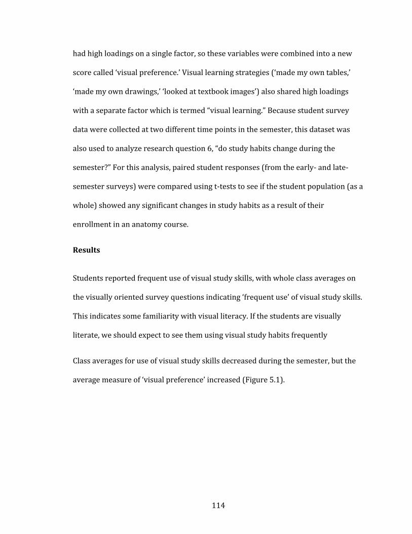
114
had high loadings on a single factor, so these variables were combined into a new
score called ‘visual preference.’ Visual learning strategies (‘made my own tables,’
‘made my own drawings,’ ‘looked at textbook images’) also shared high loadings
with a separate factor which is termed “visual learning.” Because student survey
data were collected at two different time points in the semester, this dataset was
also used to analyze research question 6, “do study habits change during the
semester?” For this analysis, paired student responses (from the early- and late-
semester surveys) were compared using t-tests to see if the student population (as a
whole) showed any significant changes in study habits as a result of their
enrollment in an anatomy course.
Results
Students reported frequent use of visual study skills, with whole class averages on
the visually oriented survey questions indicating ‘frequent use’ of visual study skills.
This indicates some familiarity with visual literacy. If the students are visually
literate, we should expect to see them using visual study habits frequently
Class averages for use of visual study skills decreased during the semester, but the
average measure of ‘visual preference’ increased (Figure 5.1).
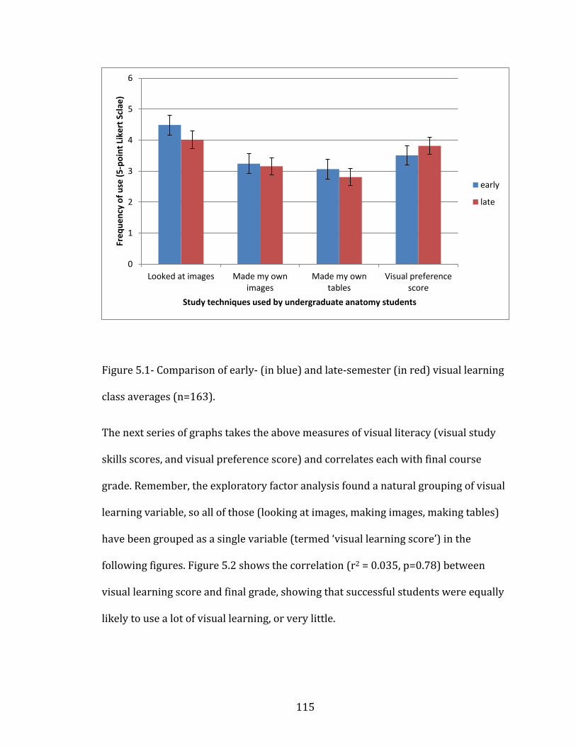
115
Figure 5.0-1
Figure 5.1- Comparison of early- (in blue) and late-semester (in red) visual learning
class averages (n=163).
The next series of graphs takes the above measures of visual literacy (visual study
skills scores, and visual preference score) and correlates each with final course
grade. Remember, the exploratory factor analysis found a natural grouping of visual
learning variable, so all of those (looking at images, making images, making tables)
have been grouped as a single variable (termed ‘visual learning score’) in the
following figures. Figure 5.2 shows the correlation (r2 = 0.035, p=0.78) between
visual learning score and final grade, showing that successful students were equally
likely to use a lot of visual learning, or very little.
0
1
2
3
4
5
6
Looked at images Made my ownimages
Made my owntables
Visual preferencescore
Fre
qu
en
cy o
f u
se (
5-p
oin
t Li
kert
Scl
ae)
Study techniques used by undergraduate anatomy students
early
late
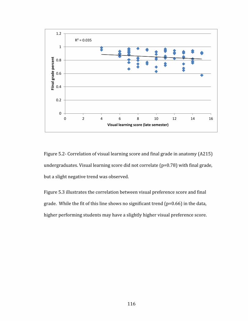
116
Figure 5.0-2
Figure 5.2- Correlation of visual learning score and final grade in anatomy (A215)
undergraduates. Visual learning score did not correlate (p=0.78) with final grade,
but a slight negative trend was observed.
Figure 5.3 illustrates the correlation between visual preference score and final
grade. While the fit of this line shows no significant trend (p=0.66) in the data,
higher performing students may have a slightly higher visual preference score.
R² = 0.035
0
0.2
0.4
0.6
0.8
1
1.2
0 2 4 6 8 10 12 14 16
Fiin
al g
rad
e p
erc
en
t
Visual learning score (late semester)
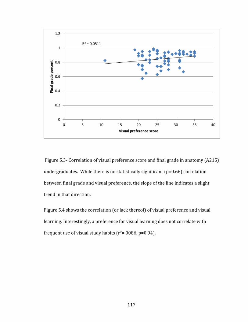
117
Figure 5.0-3
Figure 5.3- Correlation of visual preference score and final grade in anatomy (A215)
undergraduates. While there is no statistically significant (p=0.66) correlation
between final grade and visual preference, the slope of the line indicates a slight
trend in that direction.
Figure 5.4 shows the correlation (or lack thereof) of visual preference and visual
learning. Interestingly, a preference for visual learning does not correlate with
frequent use of visual study habits (r2=.0086, p=0.94).
R² = 0.0511
0
0.2
0.4
0.6
0.8
1
1.2
0 5 10 15 20 25 30 35 40
Fin
al g
rad
e p
erc
en
t
Visual preference score

118
Figure 5.0-4
Figure 5.4- There is no statistically significant correlation (p=0.94) between visual
preference score and use of visual learning skills in anatomy (A215)
undergraduates.
Discussion
This facet of the dissertation was designed to examine student use of visual learning
during an anatomy course, and answer the following questions:
Research Question 4: How often do anatomy students use visual study skills?
Research Question 5: Are there differences in student self-reported visual
learning measures?
a. Do students who self-report a preference for visual learning use
visual study skills more often in their anatomy courses?
R² = 0.0086
0
5
10
15
20
25
30
35
40
0 2 4 6 8 10 12 14 16
visu
al p
refe
ren
ce s
core
Visual learning score
119
b. Do students who self-report a preference for visual learning do
better in anatomy course performance metrics?
Research Question 6: Do student study habits for anatomy change during
the semester?
It was found that visual study skills were used frequently by the whole class, but
they tended to be lower-level visual literacy skills (as described in table 5.1) (e.g.,
looking at a book image rather than drawing a new image). Students report
understanding and appreciating the need for visual literacy (Bell et al. 2014), but the
students in this dissertation sample were only using the lowest levels of visual
literacy (looking at images). The use of higher-level visual literacy skills may be
inaccessible to students who have not had specific instruction in these skills
(Hattwig et al. 2014). Much like in verbal literacy where reading precedes writing,
creating images is a higher-level skill than ‘reading’ an image, and many students
will not have the necessary background to understand how to create meaningful
images from which to study anatomy content.
Learning style has received a lot of attention in the educational research literature
(for an attempt to create a guiding thematic principle for learning style inventories,
see Hawk and Shah 2007, for others attempting to validate learning style
inventories see Davidson and Richie 2016, Fleming and Baume 2006, Ganesh 2014,
Leite et al. 2010, Yazici 2016). The research in this dissertation facet indicates that a
self-reported preference for visual learning has no correlation with course
outcomes, or the student’s preferred study method. That self-reported visual
120
learners are equally likely to be high- or low-scoring anatomy students is not
surprising given Ganesh (2014) who reported no differences in students’ outcomes
on a series of university exams and learning styles.
What is surprising is that self-reported visual learners do not necessarily use visual
study skills more frequently than students with a low visual preference. This may be
related to the problems inherent in measuring learning style preferences (Farkas et
al. 2015, Leite et al. 2010, Riener and Willingham 2010), or it may be related to
assessment methods used in the sampled course. Student with a high visual
preference may prefer to study visually, but have found through their experiences in
anatomy that visual studying may not be beneficial, given the written nature of
lecture exams. Although lab exams are visual in nature, lab exam questions are
simple identification (the lowest level of visual literacy) and success on lab exams
may not require higher-level visual literacy or spatial ability, limiting the utility of
visual studying, even for students with a high visual preference.
The results of the visual study skills survey confirm that visual study habits do
change during the course (Question 6). Whole-class averages of visual learning
habits decreased during the semester, but so did reported use of all study skills.
These findings may indicate that students had a better idea of how to study and
therefore were simply using fewer different study strategies as the semester
progressed. The decrease in frequency of use of visual study skills may also be a
natural part of all courses as motivation decreases during the semester. While self-
reported hours of studying did not decrease, the reported frequency of each study
121
habit did decrease. This may indicate student use of techniques not listed on the
survey, or an inaccurate reporting of their use.
The visual study skill that decreased least was “made my own images.” This may
indicate that students who make drawings find them to be the most useful study
strategy. Other possible explanations are from non-cognitive variables, namely
motivation (Robbins et al. 2004). Motivation has been shown to be strongly
correlated with college success and retention, and typically students using the most
active learning (such as making drawings) are the most motivated (Aleven and
Koedinger 2000).
Individual student visual preference (the combined variable based on the VARK-
style questions in the survey) increased during the semester, based on comparisons
of early semester and late semester surveys. This finding may indicate that students
are gaining an appreciation for visual studying, even though their actual use of
visual study methods decreased (Figure 5.1). Use of all study methods (including
visual ones) decreased during the semester, but self-reported preference for using
those methods increased.
Fewer students completed the late-semester survey (n=63, as compared to the early
semester survey n=127) so the results may also be an artifact of sampling bias.
However, if it is a sampling bias that does indicate that the students with the
greatest preference for visual studying were more likely to be present during the
final days of the semester, a possible correlation with the results indicating visual
learners excel in STEM fields (McGrath and Brown 2005).
122
CHAPTER 6: Mental Rotation
Visual literacy and spatial ability are two different, but related, skills involved in
understanding the visual world (Avgerinou and Ericson 1997, Bodner and Guay
1997, Meneghetti et al. 2011). Spatial ability has long been understood as a correlate
with success in STEM fields (discussed in Chapter 2), but visual literacy in STEM has
not been well studied (Backhouse et al. 2016, Bardes et al. 2001, Bell et al. 2014,
Boudreau et al. 2008, Naghshineh et al. 2008). Chapter 6 deals with spatial ability in
anatomy students. For more details on visual literacy, see chapter 5.
Since cognitive science first explored the issue of spatial ability, it has been seen to
be strongly correlated with success in science fields (Carp and Silberman 1969,
Culver and Dunham 1969, McGee 1979). The exact causal relationship of this
correlation is still poorly understood, but there is an increasing body of literature
suggesting that teaching spatial ability can lead to improved success for students in
STEM fields (Beck et al. 1978, Boudreau et al. 2008, Canty et al. 2015, Fuks et al.
2009, Garg et al. 2001, Hegarty et al. 2009, Hoyek et al. 2014 , Keehner et al. 2006,
Langlois et al. 2014, Lufler 2012, Lufler et al. 2012, Luursema et al. 2008, Nicholson
et al. 2016, Nugent et al 2012a, Nugent et al. 2012b, Pahuta et al. 2012, Provo et al.
2002, Sweeney et al. 2014, Wanzel et al. 2002).
Spatial ability is often defined as the ability to mentally manipulate two- and three-
dimensional images (Bodner and Guay 1997, Meneghetti et al. 2011, Meneghetti et
al. 2016). A variety of cognitive science sources have further explored spatial ability
and have defined a number of different sub-categories within spatial ability

123
(Meneghetti et al. 2016). However, the new cognitive definitions have been slow to
permeate the discipline specific literature, and most STEM educational researchers
still treat spatial ability as single entity. Spatial ability can be tested in a variety of
ways including polyhedral net folding, aperture passing, and cube counting. There
appears to be a consensus that mental rotation plays a role in all of these facets of
spatial ability, so mental rotation is often treated as a close proxy for spatial ability
as a whole (Meneghetti et al. 2016).
Over the past 80 years, spatial ability has been frequently assessed in anatomy
students (both undergraduates and medical students) with a broad consensus that
spatial ability correlates strongly with course performance. Most recent
publications in the spatial ability of anatomy students focus on teaching spatial
ability through a variety of pedagogical interventions. These interventions include
passive learning (Vorstenbosch et al. 2013), 3D computer based models (Ruisoto et
al. 2012, Ruiz et al. 2009, Garg et al. 2001, Pickering 2014, Roach et al. 2016),
animations (Ruiz et al. 2009, Evans 2011), Drawing (Ruiz et al. 2009), 3D images
(Luursema et al. 2008), anatomical cross sections (Provo et al. 2002), and surgery
simulations (Keehner et al. 2006, Roach et al. 2012), and all have shown some
success in improving students’ spatial ability scores. However, improving anatomy
grades through improving spatial ability has been less frequently demonstrated.
Knowing that spatial ability correlates with student success in STEM fields
(including anatomy), the following research project was initiated to test the spatial
ability scores of anatomy undergraduates at Indiana University. Mental rotation was

124
used as an easily tested proxy for the entire suite of factors included in spatial
ability (Meneghetti et al. 2016).
The research in this chapter was organized to answer the following research
questions:
Research Question 7: Do undergraduates enter an introductory
anatomy course with adequate mental rotation skills (MRT scores)?
More specifically:
a. Do mental rotation (MRT) scores change during the semester?
b. Do mental rotation scores change, given specific instruction in
anatomical drawing?
c. Do mental rotation scores correlate with anatomy course
performance metrics?
Previously published literature suggests mental rotation abilities (summarized in
the spatial ability section of Chapter 2) of novice students can be highly variable
(Faurie and Khadra 2012, Garg et al. 2001, Keehner 2011, Keehner et al. 2008,
Ruisoto et al. 2012, Ruiz et al. 2009, Vorstenbosch et al. 2013). In general, students
rarely have enough experience with mental rotation (or similar spatial ability tasks)
to have good MRT scores, which can make learning anatomical relationships more
challenging (Boudreau et al. 2008, Codd and Choudhury 2011, Fuks et al. 2009, Garg
et al. 2001, Hegarty et al. 2009, Keehner et al. 2006, Lufler et al. 2012, Luursema et
al. 2008, Provo et al. 2002, Wanzel et al. 2002).
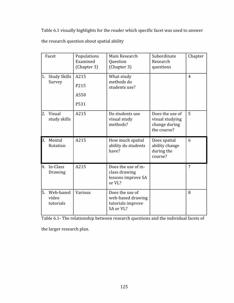
125
Table 6.1 visually highlights for the reader which specific facet was used to answer
the research question about spatial ability
Facet Populations Examined (Chapter 3)
Main Research Question (Chapter 3)
Subordinate Research questions
Chapter
1. Study Skills Survey
A215
P215
A550
P531
What study methods do students use?
4
2. Visual study skills
A215 Do students use visual study methods?
Does the use of visual studying change during the course?
5
3. Mental Rotation
A215
How much spatial ability do students have?
Does spatial ability change during the course?
6
4. In-Class Drawing
A215 Does the use of in-class drawing lessons improve SA or VL?
7
5. Web-based video tutorials
Various Does the use of web-based drawing tutorials improve SA or VL?
8
Table 6.1- The relationship between research questions and the individual facets of
the larger research plan.
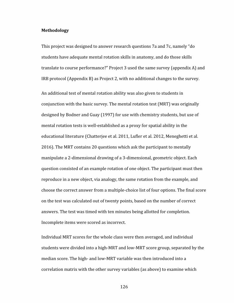
126
Methodology
This project was designed to answer research questions 7a and 7c, namely “do
students have adequate mental rotation skills in anatomy, and do those skills
translate to course performance?” Project 3 used the same survey (appendix A) and
IRB protocol (Appendix B) as Project 2, with no additional changes to the survey.
An additional test of mental rotation ability was also given to students in
conjunction with the basic survey. The mental rotation test (MRT) was originally
designed by Bodner and Guay (1997) for use with chemistry students, but use of
mental rotation tests is well-established as a proxy for spatial ability in the
educational literature (Chatterjee et al. 2011, Lufler et al. 2012, Meneghetti et al.
2016). The MRT contains 20 questions which ask the participant to mentally
manipulate a 2-dimensional drawing of a 3-dimensional, geometric object. Each
question consisted of an example rotation of one object. The participant must then
reproduce in a new object, via analogy, the same rotation from the example, and
choose the correct answer from a multiple-choice list of four options. The final score
on the test was calculated out of twenty points, based on the number of correct
answers. The test was timed with ten minutes being allotted for completion.
Incomplete items were scored as incorrect.
Individual MRT scores for the whole class were then averaged, and individual
students were divided into a high-MRT and low-MRT score group, separated by the
median score. The high- and low-MRT variable was then introduced into a
correlation matrix with the other survey variables (as above) to examine which
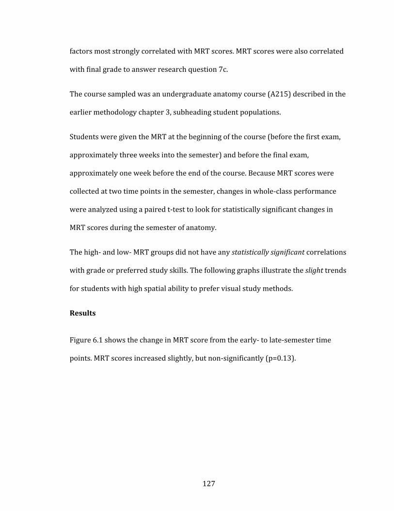
127
factors most strongly correlated with MRT scores. MRT scores were also correlated
with final grade to answer research question 7c.
The course sampled was an undergraduate anatomy course (A215) described in the
earlier methodology chapter 3, subheading student populations.
Students were given the MRT at the beginning of the course (before the first exam,
approximately three weeks into the semester) and before the final exam,
approximately one week before the end of the course. Because MRT scores were
collected at two time points in the semester, changes in whole-class performance
were analyzed using a paired t-test to look for statistically significant changes in
MRT scores during the semester of anatomy.
The high- and low- MRT groups did not have any statistically significant correlations
with grade or preferred study skills. The following graphs illustrate the slight trends
for students with high spatial ability to prefer visual study methods.
Results
Figure 6.1 shows the change in MRT score from the early- to late-semester time
points. MRT scores increased slightly, but non-significantly (p=0.13).
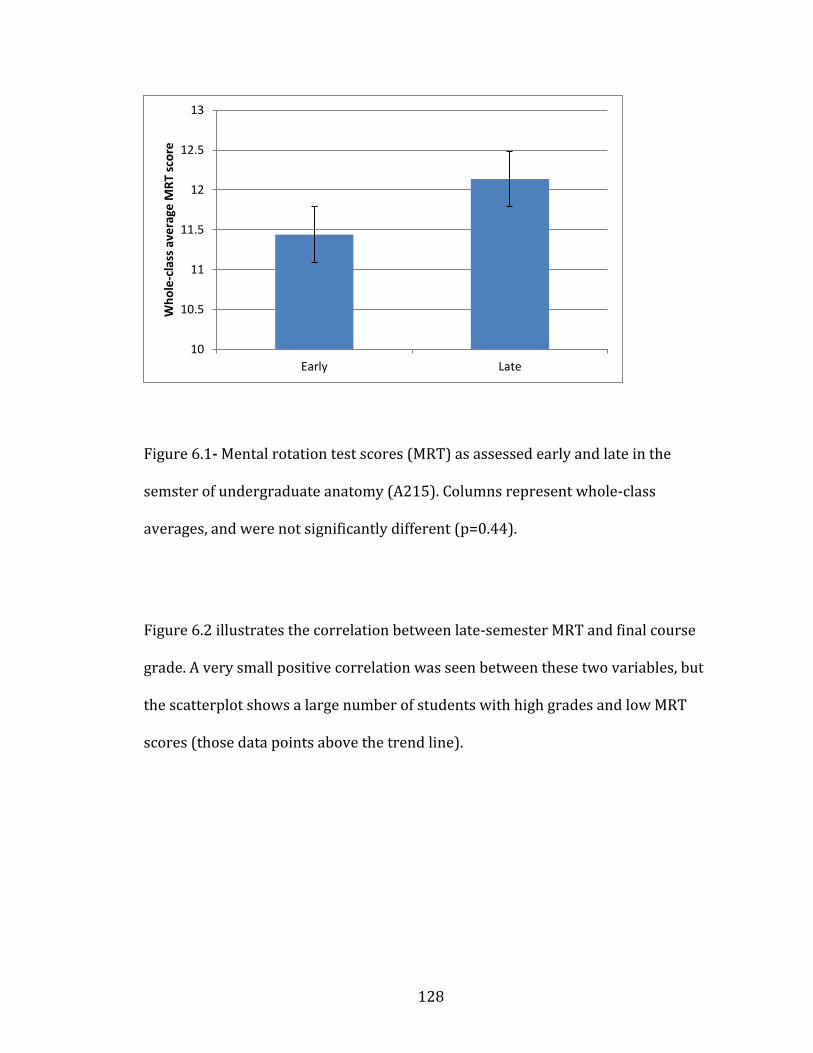
128
Figure 6.0-1
Figure 6.1- Mental rotation test scores (MRT) as assessed early and late in the
semster of undergraduate anatomy (A215). Columns represent whole-class
averages, and were not significantly different (p=0.44).
Figure 6.2 illustrates the correlation between late-semester MRT and final course
grade. A very small positive correlation was seen between these two variables, but
the scatterplot shows a large number of students with high grades and low MRT
scores (those data points above the trend line).
10
10.5
11
11.5
12
12.5
13
Early Late
Wh
ole
-cla
ss a
vera
ge M
RT
sco
re
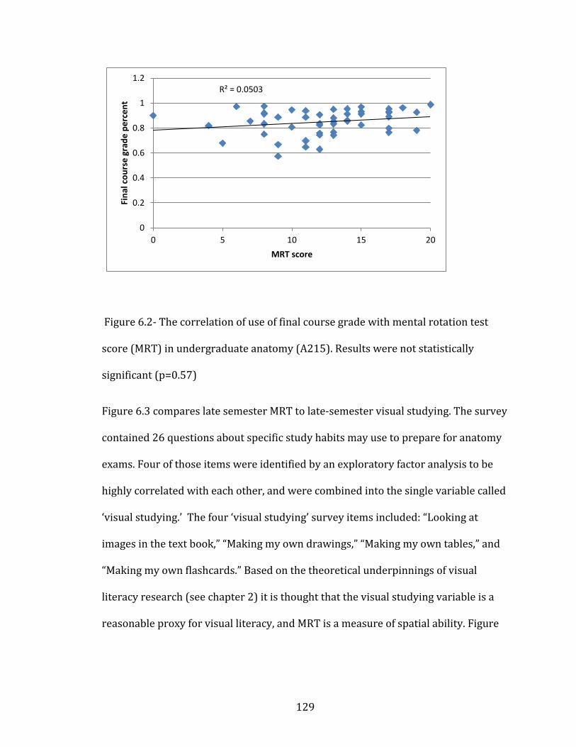
129
Figure 6.0-2
Figure 6.2- The correlation of use of final course grade with mental rotation test
score (MRT) in undergraduate anatomy (A215). Results were not statistically
significant (p=0.57)
Figure 6.3 compares late semester MRT to late-semester visual studying. The survey
contained 26 questions about specific study habits may use to prepare for anatomy
exams. Four of those items were identified by an exploratory factor analysis to be
highly correlated with each other, and were combined into the single variable called
‘visual studying.’ The four ‘visual studying’ survey items included: “Looking at
images in the text book,” “Making my own drawings,” “Making my own tables,” and
“Making my own flashcards.” Based on the theoretical underpinnings of visual
literacy research (see chapter 2) it is thought that the visual studying variable is a
reasonable proxy for visual literacy, and MRT is a measure of spatial ability. Figure
R² = 0.0503
0
0.2
0.4
0.6
0.8
1
1.2
0 5 10 15 20
Fin
al c
ou
rse
gra
de
pe
rce
nt
MRT score
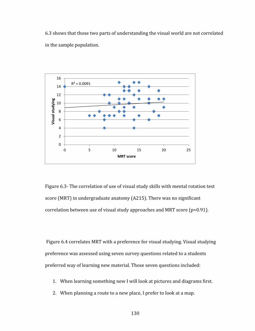
130
6.3 shows that those two parts of understanding the visual world are not correlated
in the sample population.
Figure 6.0-3
Figure 6.3- The correlation of use of visual study skills with mental rotation test
score (MRT) in undergraduate anatomy (A215). There was no significant
correlation between use of visual study approaches and MRT score (p=0.91).
Figure 6.4 correlates MRT with a preference for visual studying. Visual studying
preference was assessed using seven survey questions related to a students
preferred way of learning new material. Those seven questions included:
1. When learning something new I will look at pictures and diagrams first.
2. When planning a route to a new place, I prefer to look at a map.
R² = 0.0091
0
2
4
6
8
10
12
14
16
0 5 10 15 20 25
Vis
ual
stu
dyi
ng
MRT score
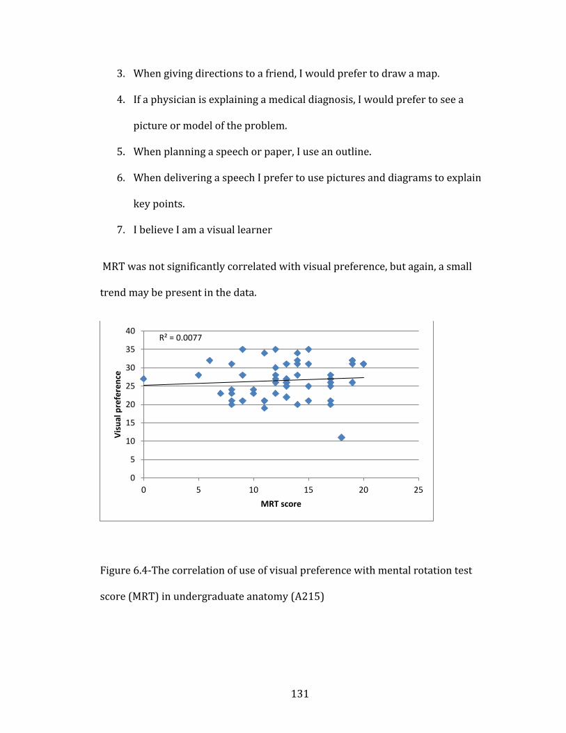
131
3. When giving directions to a friend, I would prefer to draw a map.
4. If a physician is explaining a medical diagnosis, I would prefer to see a
picture or model of the problem.
5. When planning a speech or paper, I use an outline.
6. When delivering a speech I prefer to use pictures and diagrams to explain
key points.
7. I believe I am a visual learner
MRT was not significantly correlated with visual preference, but again, a small
trend may be present in the data.
Figure 6.0-4
Figure 6.4-The correlation of use of visual preference with mental rotation test
score (MRT) in undergraduate anatomy (A215)
R² = 0.0077
0
5
10
15
20
25
30
35
40
0 5 10 15 20 25
Vis
ual
pre
fere
nce
MRT score
132
Discussion
Mental rotation score was found to increase slightly while taking an undergraduate
anatomy course (Anatomy A215), with no specific instruction in visual approaches
to learning or training in spatial ability. That is, passive acquisition of spatial ability
may occur simply through the act of taking an anatomy course. This result has been
seen with spatial ability in other fields, notably in geology (Titus and Horsman
2009) and anatomy (Vorstenbosch et al. 2013).
Gender differences in spatial ability are a contentious topic (see chapter 2) with
various sources supporting and denying the existence of gender differences in
spatial ability (Hyde 2016, Moe 2016, Quaiser-Pohl et al. 2016, Voyer et al. 1995,
Williams and Meck 1991). The participants in the sampled population were 80%
female (n=359), with comparable proportion of women completing all parts of the
survey and mental rotation test (n=48). Gender differences were not examined in
this research due to limited sample sizes and unequal distribution of men and
women in the sample. Future directions for this research would involve larger
sample sizes, so the populations could be subdivided by gender and any potential
gender differences could be explored
Mental rotation scores were not found to significantly correlate with any other
variable, including final grade, visual study skills, or preference for visual learning.
The lack of correlation between MRT and use of visual study skills may be related to
a variety of other factors including motivation (Robbins et al. 2004) , strategic
133
studying (Biggs et al. 2001) , or authentic assessment (Hart 1994, Gurung and
McCann 2011, Schwartz and Gurung 2012, Wiggins 1990), unconnected to a
student’s mental rotation ability. These factors are discussed in detail chapter 5.
The lack of correlation between MRT and visual preference is surprising. It was
expected that those students with the strongest preference for using visual study
skills would be naturally good at spatial ability. The lack of correlation here could
indicate that self-reported preferences for a specific study skill are not truly
indicative of a student’s abilities. Additionally, visual learning preference is
indicative of visual literacy and the MRT is indicative of spatial ability, two different
ways of understanding the visual world (chapter 2, Kastens 2010). It may be that
visual literacy and spatial ability are not strongly correlated, at least when measured
in this population.
134
CHAPTER 7: In-class Drawings
In the previous two chapters, spatial ability and visual literacy of anatomy
undergraduates were discussed. These two components of understanding the visual
world have been shown to be important in anatomy education (e.g., visual literacy
Backhouse et al. 2016, Bardes et al. 2001, Bell et al. 2014, Pickering 2014. Spatial
ability- Beck et al. 1978, Boudreau et al. 2008, Canty et al. 2015, Fuks et al. 2009,
Garg et al. 2001, Hegarty et al. 2009, Hoyek et al. 2014 , Keehner et al. 2006, Langlois
et al. 2014, Lufler 2012, Lufler et al. 2012, Luursema et al. 2008, Nicholson et al.
2016, Nugent et al 2012a, Nugent et al. 2012b, Pahuta et al. 2012, Provo et al. 2002,
Sweeney et al. 2014, Wanzel et al. 2002. For a full review of both concepts, see
Chapter 2). Chapter 7 describes a pedagogical intervention designed to teach
undergraduates spatial ability and visual literacy while they learn anatomy course
content. Students were instructed in drawing anatomical structures in a series of
lectures designed to emphasize the communication aspects of creating an image.
Drawing by hand was chosen as the pedagogical intervention in this project for the
reasons described in the following paragraphs.
A number of different methods to teach students spatial ability in anatomy have
been described in the literature including: passive learning (Vorstenbosch et al.
2013), 3D computer based models (Ruisoto et al. 2012, Ruiz et al. 2009, Garg et al.
2001, Pickering 2014, Roach et al. 2016), Animations (Ruiz et al. 2009, Evans 2011),
drawing (Ruiz et al. 2009), 3D images (Luursema et al. 2008), anatomical cross
sections (Provo et al. 2002), and surgery simulations (Keehner et al. 2006, Roach et
135
al. 2012) but often these rely on expensive software, complex computer models, or
animations. While effective, these other methods are expensive or time-consuming.
Only one published article has measured visual literacy gains in anatomy students
(Naghshineh et al. 2008), but this required numerous observational visits to an art
museum, and lengthy interviews with each participant afterward.
Other fields have demonstrated the teaching of visual literacy (examples from
biochemistry include: Schonborn and Anderson 2006, Anderson 2007, Schonborn
and Anderson 2010, Wiles 2016, Serpente 2016, Linenberger and Bretz 2015). One
specific method that has shown improvements in visual literacy is drawing by hand
(Ainsworth et al. 2011, Dempsey and Betz 2001, Heuschele 1999, Lyon et al. 2013).
Drawing by hand has been shown to improve spatial ability (Ruiz et al. 2009) and
visual literacy (Ainsworth et al. 2011). It is also a frequently suggested study aid to
struggling students (Gurung and McCann 2011, Van Meter and Garner 2005, course
syllabi Appendix C), and has shown some improvements in student metacognition
(Lyon et al. 2013, Van Meter and Garner 2005). Additionally, drawing is inexpensive,
easy to demonstrate, and easy for students to do outside of class time. The only
potential drawback to drawing is student resistance to the activity, often for fear of
being judged on artistic merit (Dempsey and Betz 2001).
Drawing by hand therefore was chosen as the pedagogical intervention to aid
students in learning spatial ability, visual literacy and anatomy content. To test the
effectiveness of this intervention, the following research questions were used to
organize this facet of the research:
136
Research Question 7: Do undergraduates enter an introductory anatomy
course with adequate mental rotation skills (MRT scores)? More specifically:
a. Do mental rotation (MRT) scores change during the semester?
b. Do mental rotation scores (or visual literacy metrics) change,
given specific instruction in anatomical drawing?
c. Do mental rotation scores correlate with anatomy course
performance metrics?
d. Does participation in drawing lessons correlate anatomy course
performance metrics?
Parts b. and d. of this larger question will be addressed by this chapter. Table 7.1
visually represents how the study facet (in-class drawing) relates to the overall
research questions.
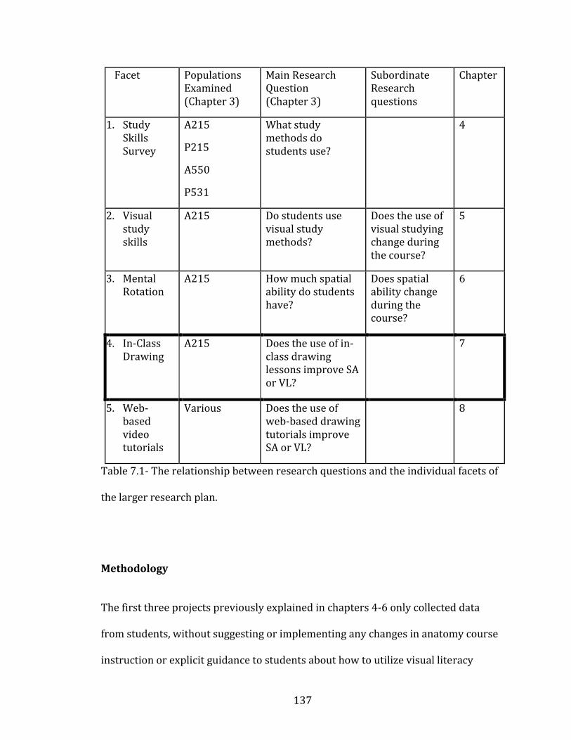
137
Facet Populations Examined (Chapter 3)
Main Research Question (Chapter 3)
Subordinate Research questions
Chapter
1. Study Skills Survey
A215
P215
A550
P531
What study methods do students use?
4
2. Visual study skills
A215 Do students use visual study methods?
Does the use of visual studying change during the course?
5
3. Mental Rotation
A215
How much spatial ability do students have?
Does spatial ability change during the course?
6
4. In-Class Drawing
A215 Does the use of in-class drawing lessons improve SA or VL?
7
5. Web-based video tutorials
Various Does the use of web-based drawing tutorials improve SA or VL?
8
Table 7.1- The relationship between research questions and the individual facets of
the larger research plan.
Methodology
The first three projects previously explained in chapters 4-6 only collected data
from students, without suggesting or implementing any changes in anatomy course
instruction or explicit guidance to students about how to utilize visual literacy

138
strategies when studying. While the researcher did not provide any explicit advice
relating to visual or spatial studying in the above projects, instructors frequently
advise students to use drawings as a study aid, and could not be controlled. For
additional details on study suggestions made in anatomy courses, see APPENDIX C
with sample syllabuses). In contrast, the research presented here deals with a
specific pedagogical intervention to teach visual and spatial ability.
This project was created around a pedagogical intervention designed to actively
teach students how to employ more visual skills in their studying. Project 4 looked
at research questions 1-8, re-assessing the baseline study habits of anatomy
undergraduates, testing their mental rotation ability, and teaching them ways to
improve MRT scores and course grades. Institutional Review Board approval
(exempt protocol) was granted (IU IRB#: 1301010307).
Project 4 used a quasi-experimental design in the anatomy undergraduate
classroom involving two sections of the Anatomy A215 class (class details in
Methodology Chapter 3). In spring of 2013, the anatomy course was taught as two
separate sections, with identical course goals, labs, and exam formats. Minor
differences in syllabus and specific exam questions were ignored because previous
versions of this course had shown student outcomes to be the same. One section of
the course was taught in the traditional way with only didactic lectures, while the
other section incorporated a series of visually-focused lectures in which students
drew anatomical objects along with the instructor. The visually-focused lecture
covered the normal anatomy content of the course, but used student-produced
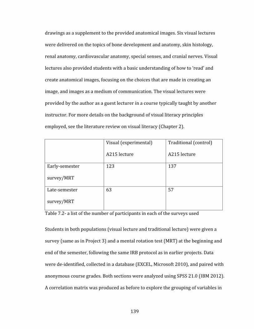
139
drawings as a supplement to the provided anatomical images. Six visual lectures
were delivered on the topics of bone development and anatomy, skin histology,
renal anatomy, cardiovascular anatomy, special senses, and cranial nerves. Visual
lectures also provided students with a basic understanding of how to ‘read’ and
create anatomical images, focusing on the choices that are made in creating an
image, and images as a medium of communication. The visual lectures were
provided by the author as a guest lecturer in a course typically taught by another
instructor. For more details on the background of visual literacy principles
employed, see the literature review on visual literacy (Chapter 2).
Visual (experimental)
A215 lecture
Traditional (control)
A215 lecture
Early-semester
survey/MRT
123 137
Late-semester
survey/MRT
63 57
Table 7.2- a list of the number of participants in each of the surveys used
Students in both populations (visual lecture and traditional lecture) were given a
survey (same as in Project 3) and a mental rotation test (MRT) at the beginning and
end of the semester, following the same IRB protocol as in earlier projects. Data
were de-identified, collected in a database (EXCEL, Microsoft 2010), and paired with
anonymous course grades. Both sections were analyzed using SPSS 21.0 (IBM 2012).
A correlation matrix was produced as before to explore the grouping of variables in
140
student performance. Latent variables concerning student active-learning and
visual-learning approaches were created. T-tests comparing the traditional lecture
and visual lecture sections final scores were run.
Having established a baseline of undergraduate study methods for anatomy in the
previous phases of this project, a pedagogical intervention was designed to teach
students how to use more effective study methods. Knowing that active learning
approaches (including visual learning and drawing) typically correlate with better
course performance (Drapkin et al. 2015, Freeman et al. 2014, Pickering 2014,
Prince 2004, Sweeney et al. 2014), a plan of teaching students to draw anatomical
structures was initiated. Additionally, both section of the course were provided with
a mental rotation test at the beginning and end of the semester, to examine changes
in mental rotation ability based on the method of instruction provided.
Results
Mental rotation scores increased in both the experimental and control groups. The
increase in MRT scores in the control groups reconfirms the passive acquisition of
spatial ability simply through taking an anatomy course, as seen in Chapter 6 and in
other sources (e.g., Vorstenbosch et al. 2013). The experimental group showed a
larger increase in MRT, but the change was not statistically significant (Figure 7.1).
The control group increased an average of 0.7 points on the MRT (paired t-test
p=.34), while the experimental group increased and average of 0.9 points (paired t-
test p=0.52). The experimental group started the course with an average of 0.6
points better MRT scores (unpaired t-test p=.36). At the end of the semester the
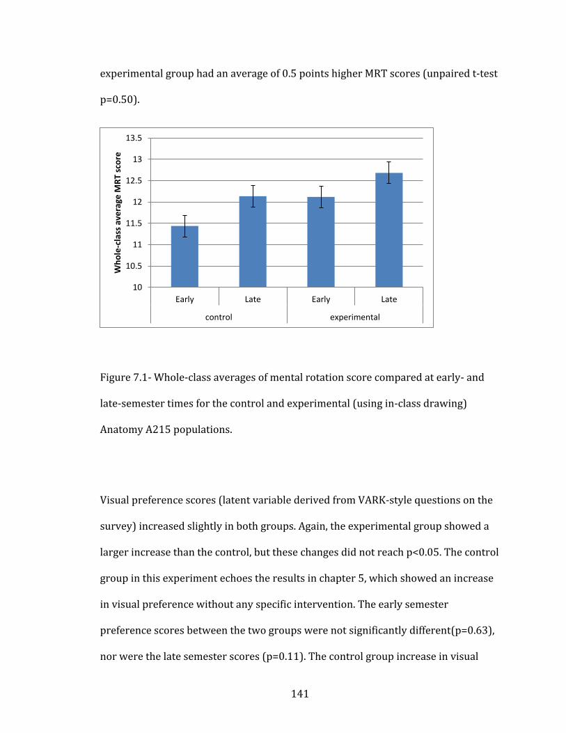
141
experimental group had an average of 0.5 points higher MRT scores (unpaired t-test
p=0.50).
Figure 7.0-1
Figure 7.1- Whole-class averages of mental rotation score compared at early- and
late-semester times for the control and experimental (using in-class drawing)
Anatomy A215 populations.
Visual preference scores (latent variable derived from VARK-style questions on the
survey) increased slightly in both groups. Again, the experimental group showed a
larger increase than the control, but these changes did not reach p<0.05. The control
group in this experiment echoes the results in chapter 5, which showed an increase
in visual preference without any specific intervention. The early semester
preference scores between the two groups were not significantly different(p=0.63),
nor were the late semester scores (p=0.11). The control group increase in visual
10
10.5
11
11.5
12
12.5
13
13.5
Early Late Early Late
control experimental
Wh
ole
-cla
ss a
vera
ge M
RT
sco
re

142
preference score was also not statistically significant (p=0.84). The experimental
group increase from early to late semester was also not significant (p=0.08), but it
was the largest difference observed.
Figure 7.0-2
Figure 7.2- Whole-class average of visual preference score at early- and late-
semester time points, compared between the experimental and control classrooms.
The experimental group demonstrated the greatest gains in visual preference score
(for studying).
Visual learning techniques used by students in both groups included re-viewing
images, making tables, and making drawings. As seen in chapter 5, the use of these
techniques tended to decrease through the semester and within the control group of
this experiment. However, usage of visual studying increased in the experimental
group (Figure 7.3).
23
23.5
24
24.5
25
25.5
26
26.5
27
27.5
Early Late Early Late
control experimental
Wh
ole
-cla
ss a
vera
ge v
isu
al p
refe
ren
ce
sco
re
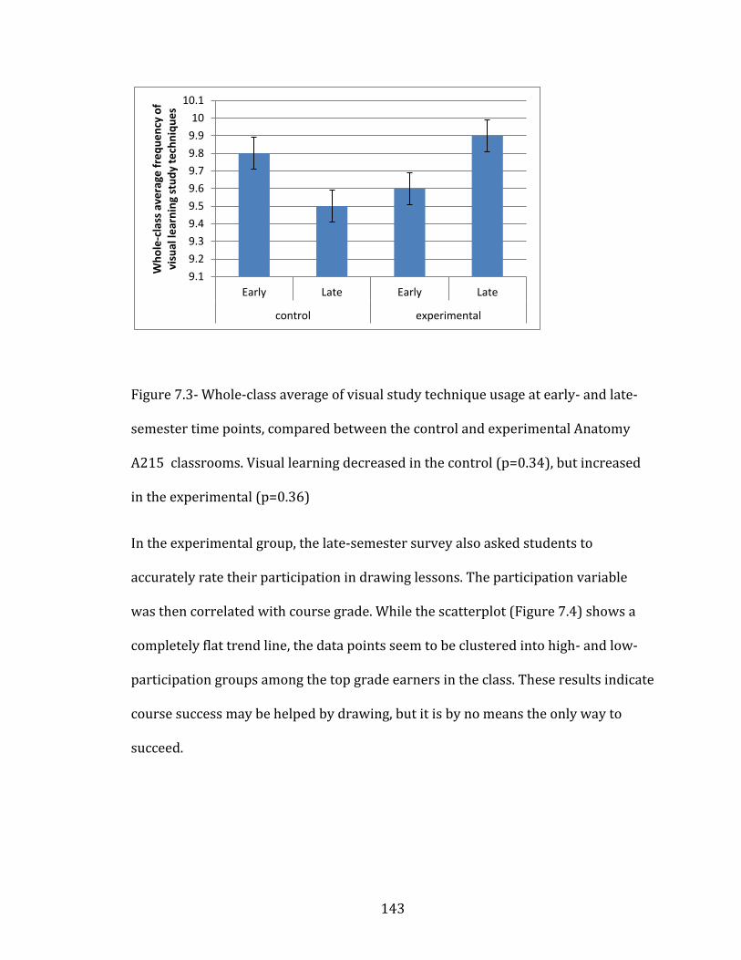
143
Figure 7.0-3
Figure 7.3- Whole-class average of visual study technique usage at early- and late-
semester time points, compared between the control and experimental Anatomy
A215 classrooms. Visual learning decreased in the control (p=0.34), but increased
in the experimental (p=0.36)
In the experimental group, the late-semester survey also asked students to
accurately rate their participation in drawing lessons. The participation variable
was then correlated with course grade. While the scatterplot (Figure 7.4) shows a
completely flat trend line, the data points seem to be clustered into high- and low-
participation groups among the top grade earners in the class. These results indicate
course success may be helped by drawing, but it is by no means the only way to
succeed.
9.1
9.2
9.3
9.4
9.5
9.6
9.7
9.8
9.9
10
10.1
Early Late Early Late
control experimental
Wh
ole
-cla
ss a
vera
ge f
req
ue
ncy
of
visu
al le
arn
ing
stu
dy
tech
niq
ue
s
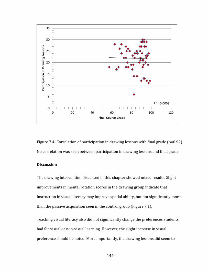
144
Figure 7.0-4
Figure 7.4- Correlation of participation in drawing lessons with final grade (p=0.92).
No correlation was seen between participation in drawing lessons and final grade.
Discussion
The drawing intervention discussed in this chapter showed mixed results. Slight
improvements in mental rotation scores in the drawing group indicate that
instruction in visual literacy may improve spatial ability, but not significantly more
than the passive acquisition seen in the control group (Figure 7.1).
Teaching visual literacy also did not significantly change the preferences students
had for visual or non-visual learning. However, the slight increase in visual
preference should be noted. More importantly, the drawing lessons did seem to
R² = 0.0008
0
5
10
15
20
25
30
35
0 20 40 60 80 100 120
Par
tici
pat
ion
in D
raw
ing
Less
on
s
Final Course Grade
145
change the way students studied. Figure 7.3 shows that use of visual studying
increased in the experimental group, but decreased in the control group (and this
decrease in visual studying was also seen in Chapter 5).The use of drawing lessons
may help to counteract the drop off of visual studying seen in the control group, and
help students maintain the motivation needed to continue using active learning as
the semester progresses (Robbins et al. 2004).
These results suggest that teaching visual literacy through drawing may help
students use more visual approaches in their studying, a factor that has previously
been correlated with higher grade in STEM (Ainsworth et al. 2011, Dempsey and
Betz 2001, Heuschele 1999, Lyon et al. 2013). Explanations for the drop-off of visual
approaches in the late semester group may also include non-cognitive variables,
namely motivation (Robbins et al. 2004). Motivation has been shown to be strongly
correlated with college success and retention, and typically students using the most
active learning (such as making drawings) are the most motivated (Aleven and
Koedinger 2000). It is possible that the drawing lessons helped to change some of
the non-cognitive influences in students studying, perhaps increasing motivation by
demonstrating a novel approach to studying. Active learning has been shown to be
strongly correlated with course grades, and is almost certainly better than
traditional lecture at helping students gain higher-order thinking skills, and mastery
of core concepts (Freeman et al. 2014). If teaching students how to draw does lead
to an increase in active learning, as suggested by the results in chapter 7, then it is a
technique that should be used more frequently in anatomy courses. Students can
use active learning approaches without being taught how to, but explicitly teaching
146
active learning strategies as part of the course should help all students improve
their content mastery (Freeman et al. 2014).
147
CHAPTER 8: Online Drawing Instruction
Student produced drawings have been demonstrated to be an effective tool for
learning spatial ability, visual literacy, and course content in science (Ainsworth et
al. 2011, Dempsey and Betz 2001, Heuschele 1999, Lyon et al. 2013). Web-based
animations have also shown gains visual skills and course content (Ruiz et al. 2009,
Evans 2011). “Draw along tutorials” are the combination of these two features, and
have only rarely been examined in the literature (Evans 2011, Pickering 2014).
The research facet detailed in this chapter examines the use of an on-line series of
video tutorials designed to teach students neuroanatomy content (Draw It to Know
It 2016). This research was guided by the following questions:
Research Question 8: Does the use of an online software tool for anatomy
drawing change student mental rotation scores, or their attitudes and use of
visual learning study skills?
The previous question (#7 found in Chapter 7) investigates the use of in-class
drawing as a pedagogical intervention to teach visual literacy and mental
rotation skills to students. This question (#8) investigates whether drawing
lessons can be effective if delivered through web-based video tutorials.
148
Methodology
Project 5 “Web-based drawing instruction” 2014
Project 5 was designed to answer research question 8, “can a web-based tool be
used to teach visual studying or mental rotation?” Web-based “draw along” videos
(also called screencasts) have shown some success in teaching anatomy content in
other publication (Pickering 2014, Evans 2011).
This project used a slightly different form of the earlier survey (see appendix A), as
it had to be re-written for deployment online, and had a different student
population. The survey consisted of 16 questions about the frequency of use of a
variety of study skills, 7 questions about the students’ self-perception as a visual
learner, and 5 questions about their opinions of the software.
The mental rotation test (MRT) was originally designed by Bodner and Guay (1997)
for use with chemistry students, but use of mental rotation tests is well-established
as a proxy for spatial ability in the educational literature (Chatterjee et al. 2011,
Lufler et al. 2012, Meneghetti 2016). The MRT contains 20 questions which ask the
participant to mentally manipulate a 2-dimensional drawing of a 3-dimensional,
geometric object. Each question consists of an example rotation of one object which
the participant must then reproduce in a new object, via analogy, and choose the
correct answer from a multiple-choice list of four options. The final score on the test
is calculated out of twenty points, based on the number of correct answers. The test
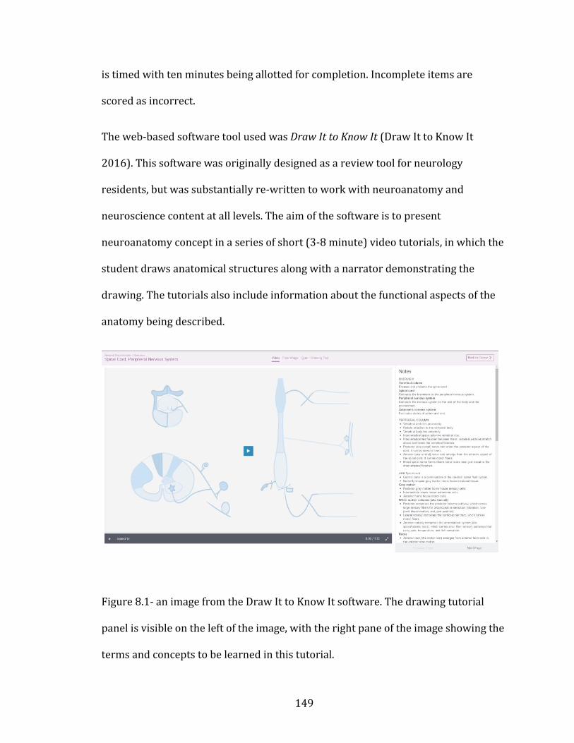
149
is timed with ten minutes being allotted for completion. Incomplete items are
scored as incorrect.
The web-based software tool used was Draw It to Know It (Draw It to Know It
2016). This software was originally designed as a review tool for neurology
residents, but was substantially re-written to work with neuroanatomy and
neuroscience content at all levels. The aim of the software is to present
neuroanatomy concept in a series of short (3-8 minute) video tutorials, in which the
student draws anatomical structures along with a narrator demonstrating the
drawing. The tutorials also include information about the functional aspects of the
anatomy being described.
Figure 8.0-1
Figure 8.1- an image from the Draw It to Know It software. The drawing tutorial
panel is visible on the left of the image, with the right pane of the image showing the
terms and concepts to be learned in this tutorial.
150
This software had not previously been evaluated as to its educational effectiveness,
so Project 5 was designed to assess the software using the surveys and mental
rotation tests (MRT) as used in the previous educational research projects described
in earlier chapter (chapter 4-7 above).
The sample population for this project (n=97) came from a total population of
undergraduate and professional students (n=1352) in nearly 40 different
neuroanatomy and neuroscience courses from across the United States.
These students were enrolled in a variety of courses at all educational levels,
including undergraduates, medical students, physical therapy students, and
osteopathic medicine students. Surveys and MRTs were deployed through the DITKI
website, and a Study Information Sheet (previously called Informed Consent
Statement) allowed students top opt-in to the data-collection portion of the project.
Those students who opted in completed a survey at the beginning of their
neuroanatomy course, and another at the end. They were also asked to complete the
MRT at the same two time points.
De-identified student data were collected in a database (Excel Microsoft 2010) as
before, and analyzed using SPSS 21.0 (IBM 2012). A notable difference in this data
analysis is the lack of student final grades. Due the constraints of ethical research
across multiple campuses, no course performance metrics were collected, and the
data analysis relies on student self-reported satisfaction with the software, and their
perceived quality of learning from it.
151
Only 97 students completed all surveys and tests. These 97 students are the only
ones assessed in the following results. Due to software limitations, partial data were
not available, and only those student completing all surveys and MRT tests were
included in the data analysis.
Results
All students in the survey reported high satisfaction with the software, finding it
both useful and effective. Because course performance metrics were not available,
student survey responses could not be compared on final grades. Students were
instead compared based on their measures of visual learning strategies, and use of
active learning strategies. Students who reported frequent use of active learning
strategies found the software to be significantly better (useful and efficient) than
their peers who used more passive learning strategies (Figure 8.2).
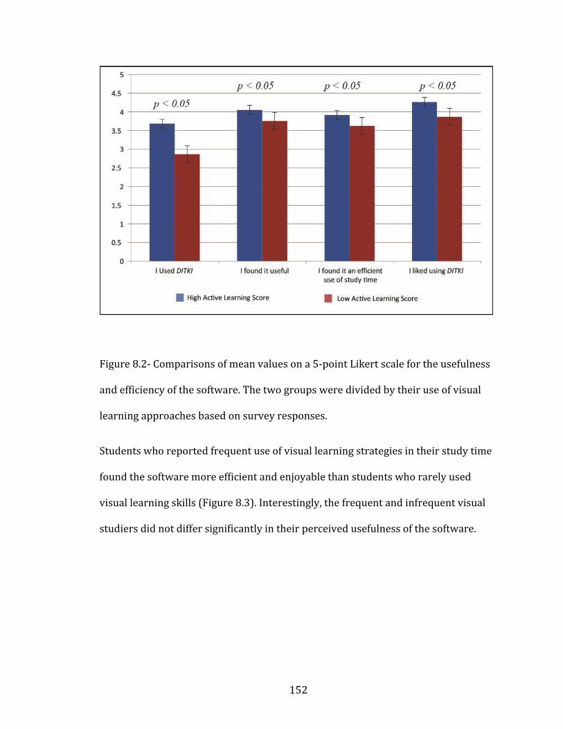
152
Figure 8.0-2
Figure 8.2- Comparisons of mean values on a 5-point Likert scale for the usefulness
and efficiency of the software. The two groups were divided by their use of visual
learning approaches based on survey responses.
Students who reported frequent use of visual learning strategies in their study time
found the software more efficient and enjoyable than students who rarely used
visual learning skills (Figure 8.3). Interestingly, the frequent and infrequent visual
studiers did not differ significantly in their perceived usefulness of the software.
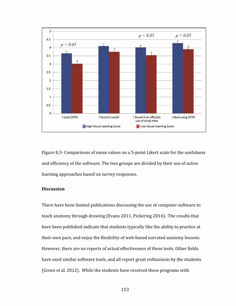
153
Figure 8.3
Figure 8.3- Comparisons of mean values on a 5-point Likert scale for the usefulness
and efficiency of the software. The two groups are divided by their use of active
learning approaches based on survey responses.
Discussion
There have been limited publications discussing the use of computer software to
teach anatomy through drawing (Evans 2011, Pickering 2014). The results that
have been published indicate that students typically like the ability to practice at
their own pace, and enjoy the flexibility of web-based narrated anatomy lessons.
However, there are no reports of actual effectiveness of these tools. Other fields
have used similar software tools, and all report great enthusiasm by the students
(Green et al. 2012). While the students have received these programs with
154
enthusiasm, no positive effects on learning have been demonstrated, but they have
also not been shown to be detrimental (Green et al. 2012). The research presented
here falls into the same category; enthusiastic support from students, but no
measurable outcomes on learning.
Despite the lack of measurable learning goals, the uniform enthusiasm from all
groups of students (high to low visual study skill users, and high to low active
learning users) indicates that learning style preference does not dictate the
perceived effectiveness of different instructional methods. Students who do not
necessarily like using visual approaches in their studying report that drawing was a
valuable way to study, even if they do not like it. This indicates that drawing and
visual literacy can be taught to all students, even if they are do not consider
themselves visual learners. Learning styles (or preferences) have long been debated
(Davidson and Richie 2016, Fleming and Baume 2006, Fleming et al. 2006, Ganesh
2014, Gradl-Dietsch et al. 2016, Hawk and Shah 2007, Leite et al. 2010, Lujan et al.
2006 Yazici 2016). These results indicate that students who claim to have a dislike
for visual learning still appreciated being taught in a visual way. It may be that
learning preferences are more related to the course content than a fundamental
preference in the individual (Farkas et al. 2015, Leite et al. 2010, Riener and
Willingham 2010). Even students who consider themselves to be non-visual
learners should be encouraged to try visual approaches to studying, as indicated by
the results in this dissertation. Students should also be warned about the potential
pitfalls of holding too strongly to a learning preference, given the controversy
present in the learning styles literature.

155
The following chapter will revisit all of the above research projects and recapitulate
the thematic connections between the various research facets. A global discussion
tying all of the results together is included. Limitations and future directions will be
discussed.
156
CHAPTER 9: Conclusions
Each previous chapter contained a brief discussion of the relevant results, and
explanation of how each ties into the overall project. This chapter will re-examine
the most pertinent results from previous chapters, and draw connections between
all of the disparate projects to further explain the thematic connections running
throughout this dissertation. The first section will restate the main research
questions that formed this dissertation. The second section will re-address each
individual study facet and how it connects to the research questions. The third
section will discuss the results of each research facet and how those results relate to
the original research questions. The fourth section of this chapter will discuss
limitations of each project, and outline future directions for research to address
each limitation.
The research presented in this document is comprised of a series of smaller projects
(also called ‘facets’) all designed to explore a number of larger research questions
(see Table 9.1).
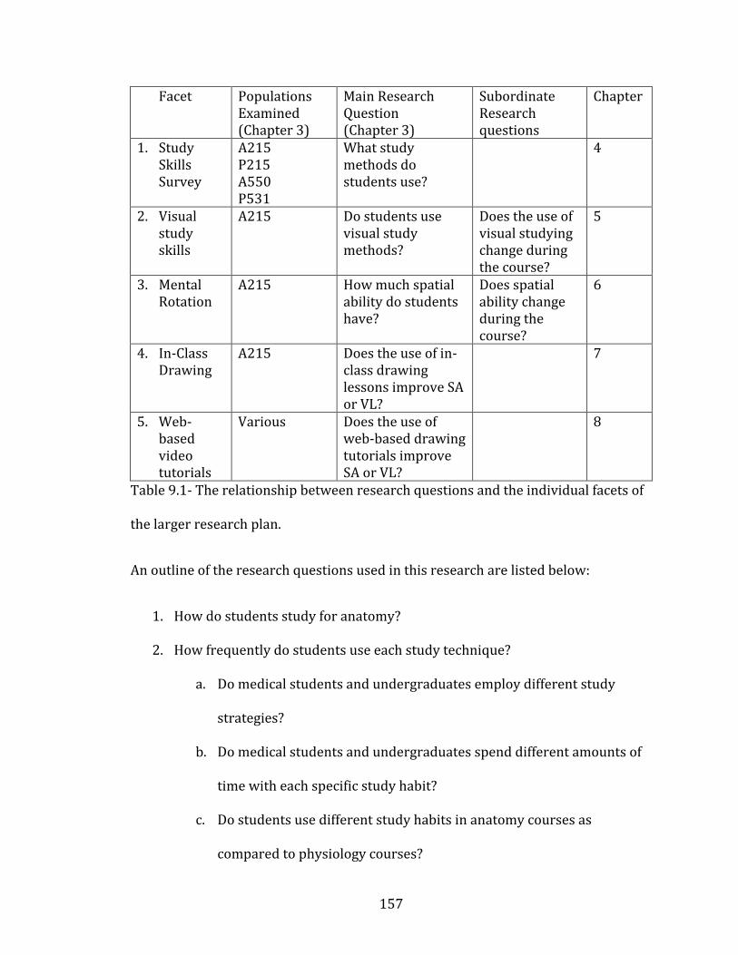
157
Facet Populations Examined (Chapter 3)
Main Research Question (Chapter 3)
Subordinate Research questions
Chapter
1. Study Skills Survey
A215 P215 A550 P531
What study methods do students use?
4
2. Visual study skills
A215 Do students use visual study methods?
Does the use of visual studying change during the course?
5
3. Mental Rotation
A215
How much spatial ability do students have?
Does spatial ability change during the course?
6
4. In-Class Drawing
A215 Does the use of in-class drawing lessons improve SA or VL?
7
5. Web-based video tutorials
Various Does the use of web-based drawing tutorials improve SA or VL?
8
Table 9.1- The relationship between research questions and the individual facets of
the larger research plan.
An outline of the research questions used in this research are listed below:
1. How do students study for anatomy?
2. How frequently do students use each study technique?
a. Do medical students and undergraduates employ different study
strategies?
b. Do medical students and undergraduates spend different amounts of
time with each specific study habit?
c. Do students use different study habits in anatomy courses as
compared to physiology courses?
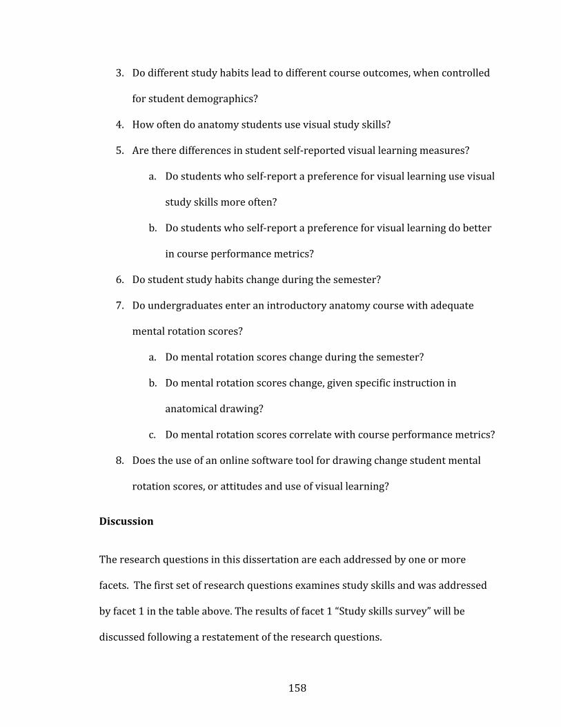
158
3. Do different study habits lead to different course outcomes, when controlled
for student demographics?
4. How often do anatomy students use visual study skills?
5. Are there differences in student self-reported visual learning measures?
a. Do students who self-report a preference for visual learning use visual
study skills more often?
b. Do students who self-report a preference for visual learning do better
in course performance metrics?
6. Do student study habits change during the semester?
7. Do undergraduates enter an introductory anatomy course with adequate
mental rotation scores?
a. Do mental rotation scores change during the semester?
b. Do mental rotation scores change, given specific instruction in
anatomical drawing?
c. Do mental rotation scores correlate with course performance metrics?
8. Does the use of an online software tool for drawing change student mental
rotation scores, or attitudes and use of visual learning?
Discussion
The research questions in this dissertation are each addressed by one or more
facets. The first set of research questions examines study skills and was addressed
by facet 1 in the table above. The results of facet 1 “Study skills survey” will be
discussed following a restatement of the research questions.

159
Research Question 1: What methods do students use to study for anatomy or
physiology?
The students in this question are considered broadly and come from populations in
A215 (anatomy undergraduates), A550 (first-year medical anatomy),
P215(physiology undergraduates) and P531(first-year medical physiology) as
described in chapter 3. Instructors in all listed courses provide students with
numerous tips and tools for efficient study, but it is unclear which of those
techniques the students use. Study tips provided in all classes include active
learning techniques and student-produced drawings (as seen in the syllabus
excerpts, Appendix C). Educational research literature indicates that use of the
above study techniques is effective, but it is unclear whether there is a correlation
between use of the suggested study techniques and final grades in the courses
studied (Barger 2012, Husmann et al. 2016, Vasan et al. 2009, Zumwalt et al. 2010).
This first question was designed to look at the study techniques students report
actually using, and how student use of study techniques compares to the
recommendations provided by instructors.
Research Question 2: How frequently do students use each study technique for
the learning of anatomy?
More specifically, this research question may be subdivided into the following
questions:
d. Do medical students and undergraduates employ different study
strategies?
160
e. Do medical students and undergraduates spend different amounts of time
with each specific study habit?
f. Do students use different study habits in anatomy courses as compared to
physiology courses?
This second research question is closely related to the first, and serves to elucidate
some of the other ideas contained in the question of “what methods do students use
to study anatomy?” This second question was designed to look more closely at
differences in student populations, and if successful study strategies are
transferable to new contexts. The students indicated in this question come from
four courses: A215 (anatomy undergraduates), A550 (first-year medical anatomy),
P215 (physiology undergraduates) and P531(first-year medical physiology).
Research Question 3: Do different study habits lead to different course
outcomes, when controlled for student demographics?
Knowing what activities students are doing in their study time is only the first step
in helping them to use that time more effectively. The next obvious step is to
discover which strategies, if any, correlate with course outcomes. Again, the
educational research literature (Barger 2012, Husmann et al. 2016, Mitchell et al.
2004, Vasan et al. 2009, Zumwalt et al. 2011) is full of details concerning effective
study strategies, but it was unclear how these broad principles apply to the
populations considered here.
161
Student study habits- Question 1-3, Facet 1, Chapter 4
Results of the research in this dissertation indicate that there is no ‘best’ way to
study. That is, there were no significant correlations between study habits and final
grade in any of the sampled populations. All of these populations demonstrated a
wide variety of study skills, with varying degrees of success. Among the top scoring
students in all courses, there were no single study techniques used most frequently.
However, a slight positive correlation with all active learning techniques and final
course grade was observed. A negative correlation between use of passive learning
techniques and grade was also observed. Regardless of course or student
educational level, there is weak support for active learning as an effective method of
studying anatomy and physiology. The most frequently used study techniques
tended to be passive, with reading the text and reviewing course notes as the most
frequently used. The lack of active learning may relate to motivation (Karpicke et al.
2009, Phinney et al. 2005), strategic learning (Biggs 2001), or assessment style
(Hart 1994, Gurung and McCann 2011, Schwartz and Gurung 2012, Wiggins 1990).
Medical students and undergraduates did not use dramatically different study
methods, but anatomy students and physiology students did show some differences
in the use frequency of some study techniques. Both of these findings (little
difference in studying based on educational level, but large differences between
courses) are supported by other published research (Gurung and Schwartz 2009,
Husmann et al. 2016).

162
The next set of research questions address the study facet 2 “visual learning in
anatomy.” The research questions are restated, followed by a discussion of the
results of facet 2.
Research Question 4: How often do anatomy students use visual study skills?
This question is the central idea guiding the overall research plan. The preceding
research questions (research questions 1-3) were investigated as background
information about basic study habits before visual study habits could be addressed.
It was worth investigating the basic study habits of students to create a framework
of potential study methods before focusing too closely on only one specific subset of
study skills. Having collected the data from the projects guided by research
questions 1-3, it became clear that students use a wide variety of study habits, and
there are some potential differences in course outcomes based on how a student
chooses to study. Knowing that study strategies matter, and having read some of the
publications concerned with the debates (e.g., what kind of images are ‘best’?, are
spatial ability and visual literacy really different?) in visual learning (see chapter 2
for references), it was decided that a focus on visual learning (visual study skills)
was a valuable next question.
The literature review and introduction chapters (see previous section this chapter)
are full of justifications and research explaining the utility of visual learning
(especially mental rotation and spatial ability) in a variety of fields, including other
sciences, as well as anatomy, but the use of visual study skills in novice students has
not been fully explored in the existing literature (to clarify, mental rotation and
163
spatial ability are well explored in the existing literature, but the use of visual study
skills, as defined in Chapter 1 of this dissertation, is not widely reported).
Research Question 5: Are there differences in student self-reported visual
learning measures?
More specifically:
a. Do students who self-report a preference for visual learning use visual
study skills more often in their anatomy courses?
b. Do students who self-report a preference for visual learning do better in
anatomy course performance metrics?
This question was designed to look at differences in student perceptions and uses of
visual learning in anatomy. Students enter anatomy courses with a wide variety of
previous experiences, and it is unclear how those experiences shape a student’s
study habits. This question asks whether different students use different study
habits without any intervention from the instructors or researchers.
Visual Literacy- Question 4-5, Facet 2, Chapter 5
Visual literacy has been notoriously hard to study and evaluate (Avgerinou and
Ericson 1997, Perkins 1994, Wiles 2014) but the research in this facet of the
dissertation attempted to measure visual literacy through survey questions about
the students’ use of visual study habits and their preferences for visual methods of
study. Students in this study were anatomy undergraduates in the A215 course.

164
The use of visual study techniques among these undergraduate students was highly
variable, and not correlated with course grades. Some undergraduates succeeded in
anatomy with frequent use of visual study techniques, while others rarely used
visual skills and achieved the same grades. Interestingly, a student’s self-reported
preferences for visual learning did not correlate with his/her preferred study
methods in anatomy. That is, students who claimed to be visual learners did not
necessarily use more visual learning skills in their anatomy study time (see also for
discussion of learning style and study skills Farkas et al. 2015, Leite et al. 2010,
Riener and Willingham 2010). This result may be related to the method of testing
used in the undergraduate anatomy course. The lecture exams in this course do not
emphasize visual understanding, so the lack of visual studying may represent a
strategic approach by the students (Biggs 2001). Additionally, the pervasive belief
that anatomy is ‘all memorization’ may lead students to use less effective strategies,
or to revert to old habits that were successful in memorizing content for earlier
courses. The lack of statistically significant correlations between preference and
actual study habits points back to the historic debates in this field, and indicate the
need for further study in the ways students learn how to use visual images in
science (see Chapter 2 references on this subject). It may be that an indicated
preference for visual learning is actually unrelated to visual literacy, and the surveys
attempting to measure visual literacy were based on faulty assumptions about
student preferences. The literature on learning styles and student preferences is an
active source of debate (Davidson and Richie 2016, Fleming and Baume 2006,
Fleming et al. 2006, Ganesh 2014, Gradl-Dietsch et al. 2016, Hawk and Shah 2007,
165
Leite et al. 2010, Lujan et al. 2006 Yazici 2016). Future research may be to elucidate
the connections between visual preferences and visual literacy.
The following section introduces a time component to the surveys used in an
attempt to capture the changes in student study habits and preferences during the
semester. The previous study facets only considered students at the end of their
anatomy (or physiology) courses, but understanding the changes in study habits
that occur during an anatomy course is poorly understood.
Research Question 6: Do student study habits for anatomy change during the
semester?
All of the previous questions rely on sampling students at a single time point in the
semester. Question 6 allows for the exploration of study habit changes in students
during a course, including those habits related to visual learning and spatial ability.
It has been suggested in some visual literacy literature (e.g., Orion et al. 1997, Titus
and Horsman 2009, Vorstenbosch et al. 2013) that simply taking a science course
can lead students to changes their habits and increase their visual literacy and
spatial ability. As students navigate the challenges of a course, and experiment with
new ways of learning, they may change their study habits, without any specific
suggestions from instructors. More frequently, the published literature in anatomy
education deals with specific pedagogical interventions designed to help students in
one specific aspect of the course. There have been many interventions related to
spatial ability and visual literacy published (e.g., Allen et al. 2014, Azer and Azer
2016, Bareither et al. 2013, Faurie and Khadra 2012, Garg et al. 2001, Gradl-diestch

166
et al. 2016, Keehner 2011, Keehner et al. 2008, Khot et al. 2013, Kotze and Mole
2015, Knobe et al. 2013, Kooloos et al. 2014, Moscova et al. 2014, Murphy et al.
2014, Ngan et al. 2010, Ngan et al. 2012, Nguyen et al. 2014, Nguyen et al. 2011,
Nguyen et al. 2009, Noel 2013, Preece et al. 2013, Richardson-Hatcher et al. 2014,
Ruisoto et al. 2012, Ruiz et al. 2006, Ruiz et al., 2009, Saltarelli et al. 2014, Schwibbe
et al. 2016, Smith 2015, Smith et al. 2015, Sweetman et al. 2013, Tam 2010, Topping
2014, Trelease and Nieder 2013, Vorstenbosch et al. 2013, Weber et al. 2016, Willis
and Martin 2016, Yammine and Violato 2014). For a full list, and details on the
various methods that have been tried, see chapter 2. The problem with the
intervention literature is that it rarely captures the changes that happen to students
simply from taking a course. This question was designed to capture the changes in
student study habits that happen without intervention, to establish a quasi-control
for the following questions, all of which deal with specific interventions.
Changing study habits- Facets 2-3, Chapter 5-6
The question of changing study habits was addressed by research facets 2 and 3,
and can be found in chapters 5 and 6. It was found that study habits do change
during the semester, often by becoming more passive and less visual. In the absence
of instructor intervention (by emphasizing active or visual learning techniques, as
seen in chapter 7) students will tend to study less, and study less actively as the
course progresses. These changes may be related to non-cognitive factors outside
the scope of the surveys used. However, focusing only on the cognitive determinants
of course performance (i.e. study skills) shows that active learning tends to be
167
correlated with course grade, but even the top scoring students tend to become
more passive during the course. Having found that study skills tend to drop off
during a course, the interventions found at the end of this chapter sought to improve
study habits, and improve course grades.
Research Question 7: Do undergraduates enter an introductory anatomy
course with adequate mental rotation skills (MRT scores)? More specifically:
a. Do mental rotation (MRT) scores change during the semester?
b. Do mental rotation scores change, given specific instruction in
anatomical drawing?
c. Do mental rotation scores correlate with anatomy course performance
metrics?
Previously published literature suggests mental rotation abilities (summarized in
the spatial ability section of Chapter 2) of novice students can be highly variable
(Vorstenbosch et al. 2013, Ruisoto et al. 2012, Ruiz et al., 2009, Faurie and Khadra
2012, Keehner 2011, Keehner et al. 2008, Garg et al. 2001). In general, students
rarely have enough experience with mental rotation (or similar spatial ability tasks)
to have good MRT scores, which can make learning anatomical relationships more
challenging (Keehner et al. 2006, Provo et al. 2002, Lufler et al. 2012, Luursema et
al. 2008, Boudreau et al. 2008, Hegarty et al. 2009, Wanzel et al. 2002, Fuks et al.
2009, Garg et al. 2001). While there is a long-standing misconception among
students that the study of anatomy is equivalent to memorizing a long list of names,
a large part of the study of anatomy is understanding the relationships of anatomical
168
entities in the human body. To understand structural relationships, one must know
the three-dimensional shapes and locations of anatomical structures. Understanding
three-dimensional shapes is not an easy task; it requires practice to learn how
complex 3D objects interact in the body. A high spatial ability (or mental rotation
ability) may make this task easier. It is the goal of research question 7 to see how
spatial ability relates to anatomy performance and study skills.
Mental Rotation: Question 7a, 7c., Facet 3, Chapter 6
Spatial ability was measured by a mental rotation test (MRT). The MRT was created
and validated by Bodner and Guay (1997), and mental rotation is an acceptable
proxy for spatial ability as a whole (Meneghetti et al. 2011, Meneghetti et al. 2016,
Uttal et al. 2013). In research facet 3, mental rotation scores did not correlate with
course performance, or with any of the study skills assessed by the survey.
Surveyed populations included undergraduates in anatomy (A215). However, the
MRT scores did increase slightly during the semester, even without specific
instruction in spatial ability, an indication of the reciprocal relationship of anatomy
and spatial ability as seen in Vorstenbosch et al. (2013). It has been proposed that
studying anatomy acts as a way of practicing spatial ability, even when it is not given
that name. Students (especially undergraduates who may not go on to use anatomy
professionally) should be reminded of the practical implications of studying
anatomy in terms of learning spatial ability. That is, understanding the relationships
of 3D structures in the body may have benefits beyond ‘memorizing terms.’
169
Research Question 8: Does the use of an online software tool for anatomy
drawing change student mental rotation scores, or their attitudes and use of
visual learning study skills?
The previous question (7b) investigates the use of in-class drawing as a pedagogical
intervention to teach visual literacy and mental rotation skills to students. This
question (8) investigates whether drawing lessons can be effective if delivered
through web-based video tutorials.
Pedagogical interventions: In-class drawing tutorials and web-based video
drawing tutorials. Questions 7b-8, Facets 4-5, Chapters 7-8
The two facets discussed in this section were reported separately in chapters 7-8,
but will be discussed together because they both deal with attempts at teaching
visual literacy and spatial ability to anatomy students. Populations sampled to
answer this question came from the undergraduate anatomy course A215.
Students are often encouraged to create their own drawings as a way of
understanding anatomy content (see appendix C for course syllabuses). However,
students are rarely given enough instruction in creating images to use this study
technique effectively. The surveys used in Chapters 4-5 asked students to rate how
frequently they produced their own drawings while studying. Students reported
frequent use of this technique in their studying, but it is unclear if this technique is
actually effective, as it did not correlate with course grades. In an effort to make
student-produced drawings more effective, facets 4-5 of this dissertation taught

170
students some of the basics of drawing for communicating science (Ainsworth
2011).
Facet 4 used in-class drawings to communicate anatomy content to students and
provide them with the tools needed to make their own drawings outside of class
time. Students who participated in these drawing lessons reported that drawing is a
useful way to learn content, but rarely transferred this study method to new
contexts. That is, even among students who participated in the drawings, enjoyed
drawing, and showed a high preference for visual studying, they rarely made their
own drawings for studying. The drawing participants also did not show any
significant improvement in course grades, or mental rotation scores, as compared to
students taught through traditional didactic lectures. Drawing as a tool for learning
is highly regarded in the educational literature (Ainsworth et al. 2011, Dempsey and
Betz 2001, Heuschele 1999, Lyon et al. 2013) but empirical evidence for its
usefulness in conveying course content is mixed (Van Meter and Garner 2005). The
evidence for drawing also indicates that drawing can lead to cognitive
improvements outside of a discipline-specific domain (Lyon et al. 2013). Visual
literacy is important to understanding science, and the drawing lessons provided in
facet 4 may have improved visual literacy, even if anatomy content delivery was
unaltered.
Facet 5 used a web-based drawing tutorial video series (Draw It to Know It) to teach
drawing outside of class time. One of the limitations of Facet 4 was the limited
number of drawing lessons that could be provided to students. By moving the
171
drawing content online, the number of drawing lessons a students could potentially
experience was greatly increased. The online format provided the additional benefit
of repeated practice, pausing and rewinding, and a lower stress environment than a
classroom. Students reported liking all of these features of the drawing tutorials,
and all students found the tutorials a useful and efficient way to study. Regardless of
self-reported preferences for different types of studying, all participants in the Draw
It to Know It study reported a high degree of satisfaction with it as a way of learning
anatomy content. Unfortunately, the logistical limitation of working with multiple
study sites prevented the collection of certain data. IRB restrictions prevented the
collection of course grades, and recruitment at different campuses was not
monitored, leading to the potential loss of some study participants.
The final research question is addressed below. It is not a specific, measurable
question, but a reminder of the underlying interests that began this dissertation
research.
Research Question 9: What are the best ways to study anatomy, and is visual
learning an important part of the student experience? What are the
pedagogical implications of the research questions 1-8? That is, what should
instructors do to ensure students are getting the most benefit from an
anatomy course?
Question 9 is less of a research question than it is a guiding principle, a reminder of
the overarching goals, and an opportunity to tie all the above research questions
together. All of the preceding questions were treated as largely separate to make
172
each individual project as specific as possible, but all of the questions work together
to address to problem of student learning in anatomy, which is the ultimate goal of
the entire series of research projects. Question 9 is almost impossible to answer in
isolation, but it is the ultimate goal of this dissertation to address this problem, and
provide answers to the question of “what is the best way to study?” As has been
seen in the research literature cited throughout this dissertation, there is no ‘best
way to study.’ In fact, defining ‘best’ in this context is impossible. Does “best” mean
getting the highest course grade? Does best mean having the most long-term
retention? Does best mean improving multiple cognitive facets while learning
discipline specific material? Does best have a non-cognitive component? In trying to
define useful approaches to studying, the shifting definitions of ‘best’ must be kept
in mind.
The hypotheses implicit in the research questions were that improving visual
learning and spatial ability skills would lead to better course performance in
anatomy students. While SA (and to a lesser extent VL) have demonstrated
correlations with anatomy performance (e.g., Boudreau et al. 2008, Hegarty et al.
2008, Lufler et al. 2012), improving anatomy scores through teaching VL and SA has
yet to be definitively demonstrated. While teaching VL and SA may not lead to
directly improved anatomy scores, it is worth noting how useful these skills are in
other areas. For example, visual literacy has been linked to ‘critical observation’ in
clinical reasoning (e.g., Bardes et al. 2001, Boudreau et al. 2008, Bardes et al. 2001).
Visual literacy (and visual thinking) have also been described as a method of flexible
or deep retrieval, and is a crucial part of becoming an expert in a field of study
173
(Bransford et al. 2000). Novice students tend to think very algorithmically, and lack
the flexible retrieval of experts; training in visual literacy has been shown to help
students break their algorithmic thinking patterns (Stieff et al. 2010). If anatomy
courses (especially for medical students) are part of a training program to get the
novice student to think like an expert, then visual literacy is a crucial part of this
goal. Outside of anatomy, basic visual literacy skills are important for any type of
visual communication, including advertising (Alenn 1994, Glasgow 1994),
understanding science (Ainsworth et al. 2011, Dempsey and Betz 2001, Heuschele
1999, Lyon et al. 2013), and even problem solving (Kozhevnikov et al. 2007).
Spatial ability has been shown to correlate not only with anatomy, but other
sciences (Orion et al. 1997), map reading (Lawton 1994), and mathematical
reasoning (Linn and Peterson 1986). Improving visual skills has a definite place in
anatomy education, even if it does not always lead to higher exam scores. Using
student-produced drawings to teach visual skills has demonstrated effectiveness
(Ainsworth et al. 2011), but is not without its detractors (Van Meter and Garner
2005 for a meta-analysis of drawing in science). Perhaps the strongest argument for
teaching drawing (as a means of visual communication) is the metacognitive
component (Lyon et al. 2013). To make an effective drawing, a student must have a
firm grasp of the content s/he wishes to draw. To produce an accurate drawing, a
student is required to honestly evaluate what s/he knows, thereby increasing
metacognitive awareness. Frank Netter, noted medical illustrator and physician put
it best when he said:
174
“The making of a picture is a stern discipline. One may ‘write around’ a subject where one is not quite sure of the details, but [in an image] one must be precise and realistic. The white paper before the artist demands the truth.” (Netter 1989, back cover)
Limitations
Some limitations exist in this research. Student participation in surveys may not
accurately represent the entire class. Because students were not mandated or
incentivized to complete surveys, there was a large number of students who did not
participate in the research. The non-uniform participation in surveys could lead to a
selection bias, in which the sample is not representative of the population (Berk
1983). While survey participant grade distributions and demographics were similar
to the class a whole, there may still be a selection bias present.
Additionally, surveys relied on students to accurately self-report their habits and
opinions. Self-reporting on surveys is subject to (at least) two different biasing
factors: social desirability bias (Furnham 1986) and acquiescence bias (Knowles
and Nathan 1997). Social desirability bias can cause participants to over-report
perceived good behaviors, and may have caused students to overestimate their
study habits. Acquiescence bias is the tendency for survey respondents to agree
with all questions on a survey, which could also have led to an overestimation of
study habits in this research.
The mental rotation test was given to students at two time points during the
semester, which means it is susceptible to the test-retest effect (Windle 1954). This
175
effect indicates that scores will typically improve on a retest due to lower testing
anxiety, and a familiarity with the questions, even if the subject has not actually
changed. This effect is most prominent in short-interval test-retest situations,
typically under two months. In my experimental design, the test-retest interval was
close to three months, but an effect may still exist.
Course assessments (e.g., lecture and lab exams) may not have authentically
assessed the variables being measured in the research (Wiggins 1990, Hart 1994).
Authentic assessment refers to directly testing students on the intellectual tasks
they should know. An assessment may be deemed authentic if it directly and
accurately measures what has been taught in a course. Simply put, does the exam
actually test what the instructor thinks it tests? The assessments used in the
anatomy courses surveyed in this dissertation were surely authentic in their
anatomy content, but may not have directly assessed VL or SA as authentically as
desired. That is, course grades may not have been the best way to measure
improvements in anatomy learning through VL and SA. In the courses described
here (see above for details in the Methodology Chapter 3), students were assessed
with multiple-choice exams, with most questions focusing on identification of
written descriptions, meaning visual literacy was not important for success in the
course. Laboratory exams were based on identifying static structures, which is a
visual skill, but a very low-level visual task, again meaning that visual literacy was
not being well assessed. Anatomy taught through didactic lecture and largely
passive laboratory sessions does not emphasize the visual aspects under
examination in this research. Related to the assessment problem, the teaching
176
methods used in the course did not emphasize the visual understanding of anatomy
being tested in the surveys.
Non-cognitive factors (e.g., motivation, peer support, and financial support) have
been shown to be important determinants of student success in higher education
(Robbins et al. 2004). It is possible there were non-cognitive factors, especially
motivational factors underlying some of the unexpected changes seen in student
study habits. The surveys did not assess any non-cognitive variables.
Gender differences in spatial ability (mental rotation) were not considered due to
sample size limitations and unequal gender distribution in the undergraduate
anatomy course. The undergraduate anatomy (A215) typically enrolls 80% female
students, creating an skewed environment for investigating gender differences in
study habits or mental rotation skills. There is still an active debate about the role of
gender in spatial ability (e.g., Hyde 2016, Masters and Sanders 1993, Moe 2016,
Quaiser-Pohl 2016, Voyer et al. 1995, Xiong et al. 2016, for a more complete
examination of gender in spatial ability, see Chapter 2). If a gender difference in
spatial ability does exist in the population as a whole, its causal factors are not yet
understood (Quaiser-Pohl 2016).
Sampling bias was of particular note in chapter 8 due to the low participation from
the available pool of participants. Over 1300 students were asked to complete the
surveys and MRTs for the research on Draw It to Know It, but only 97 did. While 97
is a large enough number for statistical reliability, and the participants were a good
representative of the total courses enrolled in the survey, there is no way to ensure
177
those 97 participants accurately represent the entire population. I was unable to
monitor recruitment on the various campuses where Draw It to Know It was
provided, so it is unclear how aware of the study the students were. Additionally,
ethical research standards prevented incentives from being provided to students
completing the surveys, possibly leading to more loss of participants.
Future Directions
The limitations presented above point the way to interesting future directions for
this research.
Authentic assessment, non-cognitive factors, and qualitative data are the three most
interesting areas for future research. These three factors seemed to be the biggest
confounders (or missing variables) in the data. Additionally these three issues may
be addressed without the need for larger samples or better student participation in
surveys (a logistical nightmare faced by all educational researchers).
Authentic assessment of visual literacy and spatial ability will require the
cooperation of anatomy classroom instructors. Working with classroom instructors
to ensure exam items are assessing visual skills authentically will provide much
more valuable variable in future studies. Authentic assessment of visual skills in
anatomy might consist of exam items which require mental rotation or making
inferences from images. Changing exams to require more visual thinking should
provide benefits to instructors as well as benefitting this research plan.
178
Non-cognitive factors (e.g., motivation, peer support, and financial support) have
been shown to be important determinants of student success in higher education
(Robbins et al. 2004). It is possible there were non-cognitive factors, especially
motivational factors underlying some of the unexpected changes seen in student
study habits. Adding survey questions to assess the non-cognitive components of a
student’s study strategies will provide a wealth of new information about anatomy
students.
Qualitative data collected from students may help elucidate some of the questions
raised by this research. For example, in Figure 7.4 the highest performing students
in the course final grades can be grouped into high and low-visual learning score
groups, but both groups performed equally well in the course. From the survey data,
it is unclear what other differences these two groups may have. Both groups
reported using the same amount of time studying and the same amount of previous
experience in anatomy courses. There may be other underlying factors explaining
this difference, but the survey did not ask those questions. An interview or focus
group would allow students the opportunity for more open-ended discussion that
may reveal some additional variables that were not in the survey.
Final Summary
This dissertation supports the hypothesis that visual skills (spatial ability and visual
literacy) are important in anatomy education. Students enjoyed learning how to
apply visual learning techniques to their anatomy courses, but many of them lack
the ability to do so without more instruction. Adding visual learning to anatomy
179
courses (and exams) is a goal that can lead to higher student satisfaction, improved
visual literacy, and improved metacognition. While instructors may struggle with
incorporating visual learning into an established course, online resources (such as
Draw It to Know It) can help. Additionally, adding course assessments that require
higher-level visual thinking will increase students’ awareness of their own visual
thinking, and increase student openness to new learning techniques. Visual literacy
is necessary to all fields of medicine (the goal of most anatomy students), but to
many instructors and practitioners, it has become second nature. However, the slow
accumulation of visual literacy by practice in a discipline is often inadequate, and
may leave behind many students who fail to grasp it. Visual literacy should not be
treated as skill only the most expert members of a field have, it should be taught to
all students from the beginning of their education in anatomy.
180
APPENDIX A: Survey Design, Validation, and Sample Surveys
Surveys were developed using methods previously established (Fowler, 1995) and
piloted in a smaller, undergraduate anatomy class before being used in the
dissertation research. After piloting the survey, students were asked for feedback on
their interpretation of the questions. This feedback was then applied to make
questions about study skills more clear and relevant to the students. Additionally,
the survey was discussed and edited with several anatomy faculty members to
ensure the questions being used aligned with the courses being taught. The same
survey has also been used in other studies of undergraduates over several
semesters, and responses in all categories are statistically consistent between years,
indicating a high level of survey reliability (Barger 2012, Barger and Husmann
2013). Cronbach’s alpha was also calculated on the combined undergraduate and
medical student population at 0.767 indicating a good level of reliability. While
undergraduate students and medical students are different groups, this study
focuses on students in their first year of medical school, many of which are entering
medical school immediately after completing their bachelor’s degree. It is unlikely
that medical students would interpret the questions differently than the
undergraduates on whom the survey was validated. All of the surveys included
Likert scale and categorical questions, as well as boxes for open-ended comments.
Questions covered three general themes: student study habits (use of text, notes,
learning exercises, etc.), attendance and attitudes (confidence, integration,
etc.), and basic demographics and background information (previous coursework,
experience, etc.). The demographic sections were omitted from later surveys to
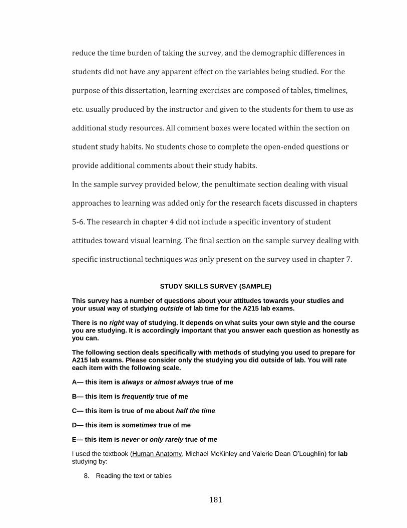
181
reduce the time burden of taking the survey, and the demographic differences in
students did not have any apparent effect on the variables being studied. For the
purpose of this dissertation, learning exercises are composed of tables, timelines,
etc. usually produced by the instructor and given to the students for them to use as
additional study resources. All comment boxes were located within the section on
student study habits. No students chose to complete the open-ended questions or
provide additional comments about their study habits.
In the sample survey provided below, the penultimate section dealing with visual
approaches to learning was added only for the research facets discussed in chapters
5-6. The research in chapter 4 did not include a specific inventory of student
attitudes toward visual learning. The final section on the sample survey dealing with
specific instructional techniques was only present on the survey used in chapter 7.
STUDY SKILLS SURVEY (SAMPLE)
This survey has a number of questions about your attitudes towards your studies and your usual way of studying outside of lab time for the A215 lab exams.
There is no right way of studying. It depends on what suits your own style and the course you are studying. It is accordingly important that you answer each question as honestly as you can.
The following section deals specifically with methods of studying you used to prepare for A215 lab exams. Please consider only the studying you did outside of lab. You will rate each item with the following scale.
A— this item is always or almost always true of me
B— this item is frequently true of me
C— this item is true of me about half the time
D— this item is sometimes true of me
E— this item is never or only rarely true of me
I used the textbook (Human Anatomy, Michael McKinley and Valerie Dean O’Loughlin) for lab studying by:
8. Reading the text or tables
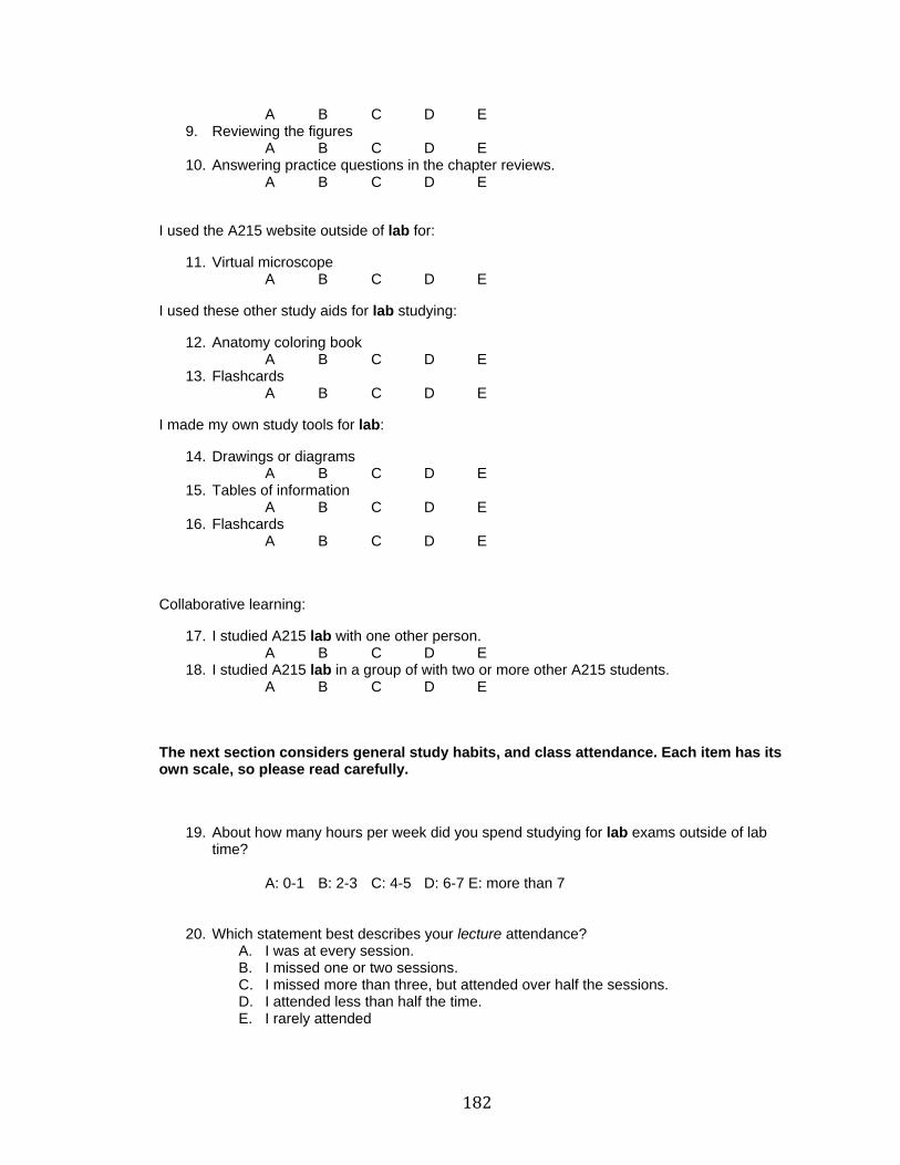
182
A B C D E 9. Reviewing the figures
A B C D E 10. Answering practice questions in the chapter reviews.
A B C D E
I used the A215 website outside of lab for:
11. Virtual microscope A B C D E
I used these other study aids for lab studying:
12. Anatomy coloring book A B C D E
13. Flashcards A B C D E
I made my own study tools for lab:
14. Drawings or diagrams A B C D E
15. Tables of information A B C D E
16. Flashcards A B C D E
Collaborative learning:
17. I studied A215 lab with one other person. A B C D E
18. I studied A215 lab in a group of with two or more other A215 students. A B C D E
The next section considers general study habits, and class attendance. Each item has its own scale, so please read carefully.
19. About how many hours per week did you spend studying for lab exams outside of lab time?
A: 0-1 B: 2-3 C: 4-5 D: 6-7 E: more than 7
20. Which statement best describes your lecture attendance? A. I was at every session. B. I missed one or two sessions. C. I missed more than three, but attended over half the sessions. D. I attended less than half the time. E. I rarely attended
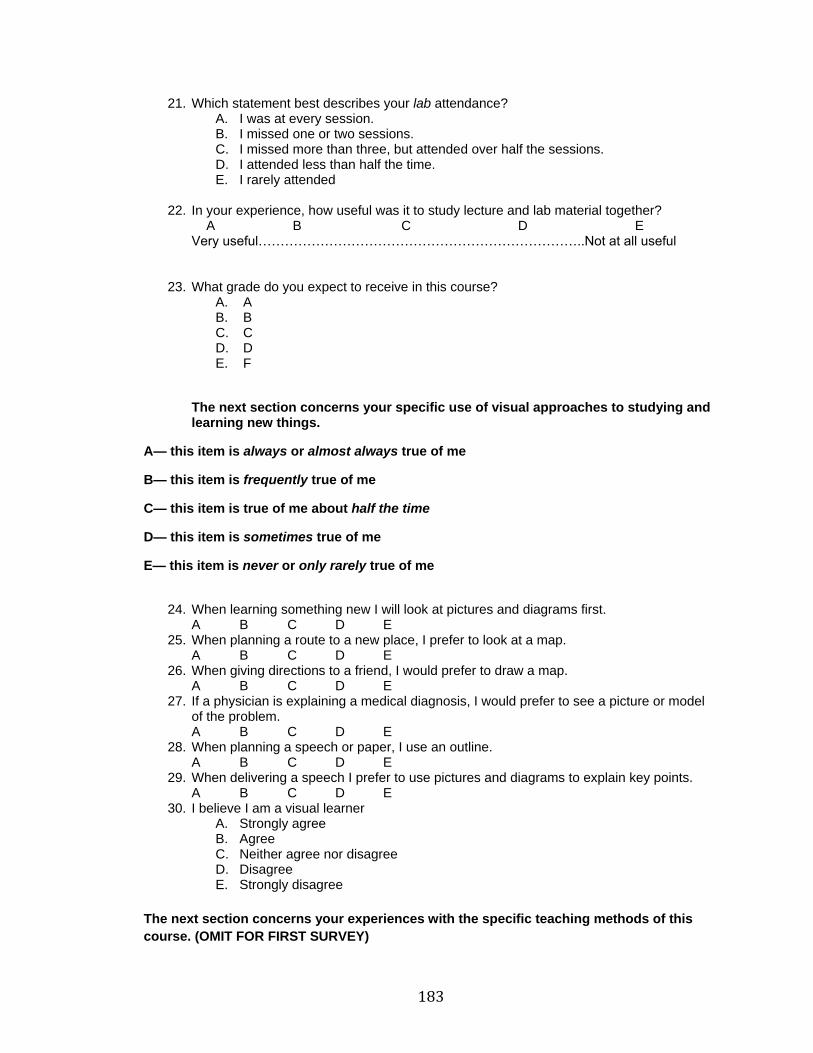
183
21. Which statement best describes your lab attendance? A. I was at every session. B. I missed one or two sessions. C. I missed more than three, but attended over half the sessions. D. I attended less than half the time. E. I rarely attended
22. In your experience, how useful was it to study lecture and lab material together?
A B C D E Very useful………………………………………………………………..Not at all useful
23. What grade do you expect to receive in this course? A. A B. B C. C D. D E. F
The next section concerns your specific use of visual approaches to studying and learning new things.
A— this item is always or almost always true of me
B— this item is frequently true of me
C— this item is true of me about half the time
D— this item is sometimes true of me
E— this item is never or only rarely true of me
24. When learning something new I will look at pictures and diagrams first.
A B C D E 25. When planning a route to a new place, I prefer to look at a map.
A B C D E 26. When giving directions to a friend, I would prefer to draw a map.
A B C D E 27. If a physician is explaining a medical diagnosis, I would prefer to see a picture or model
of the problem. A B C D E
28. When planning a speech or paper, I use an outline. A B C D E
29. When delivering a speech I prefer to use pictures and diagrams to explain key points. A B C D E
30. I believe I am a visual learner A. Strongly agree B. Agree C. Neither agree nor disagree D. Disagree E. Strongly disagree
The next section concerns your experiences with the specific teaching methods of this
course. (OMIT FOR FIRST SURVEY)
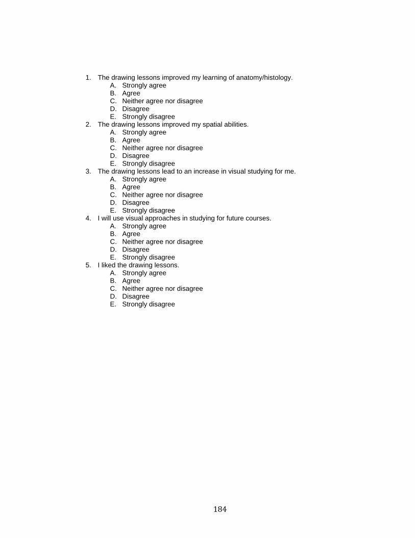
184
1. The drawing lessons improved my learning of anatomy/histology. A. Strongly agree B. Agree C. Neither agree nor disagree D. Disagree E. Strongly disagree
2. The drawing lessons improved my spatial abilities. A. Strongly agree B. Agree C. Neither agree nor disagree D. Disagree E. Strongly disagree
3. The drawing lessons lead to an increase in visual studying for me. A. Strongly agree B. Agree C. Neither agree nor disagree D. Disagree E. Strongly disagree
4. I will use visual approaches in studying for future courses. A. Strongly agree B. Agree C. Neither agree nor disagree D. Disagree E. Strongly disagree
5. I liked the drawing lessons. A. Strongly agree B. Agree C. Neither agree nor disagree D. Disagree E. Strongly disagree
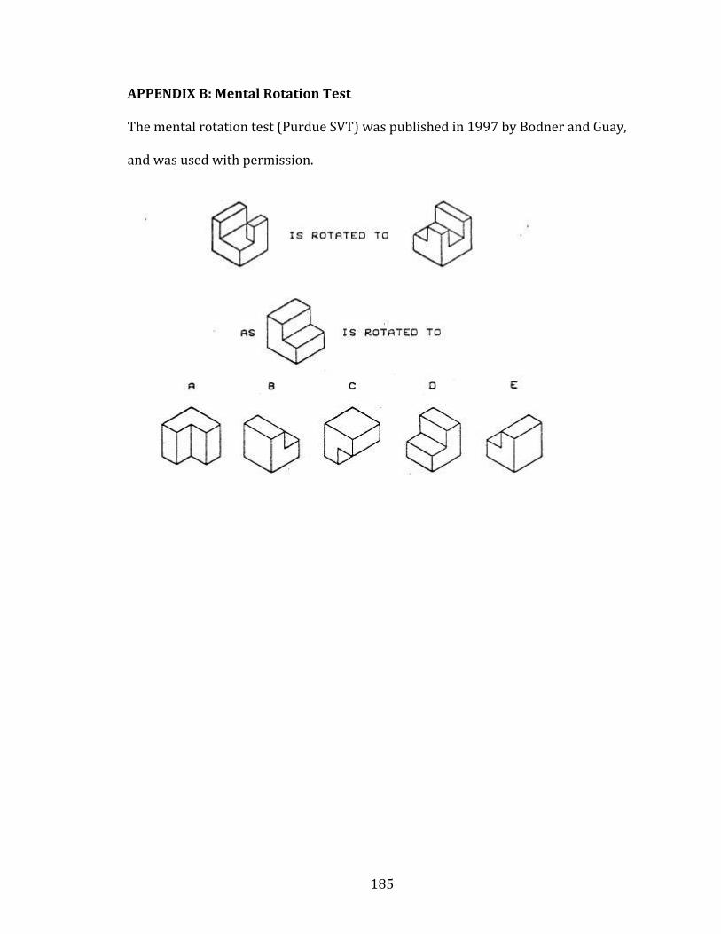
185
APPENDIX B: Mental Rotation Test
The mental rotation test (Purdue SVT) was published in 1997 by Bodner and Guay,
and was used with permission.
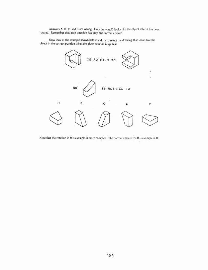
186
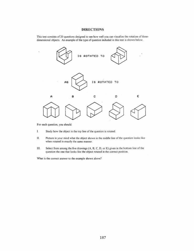
187
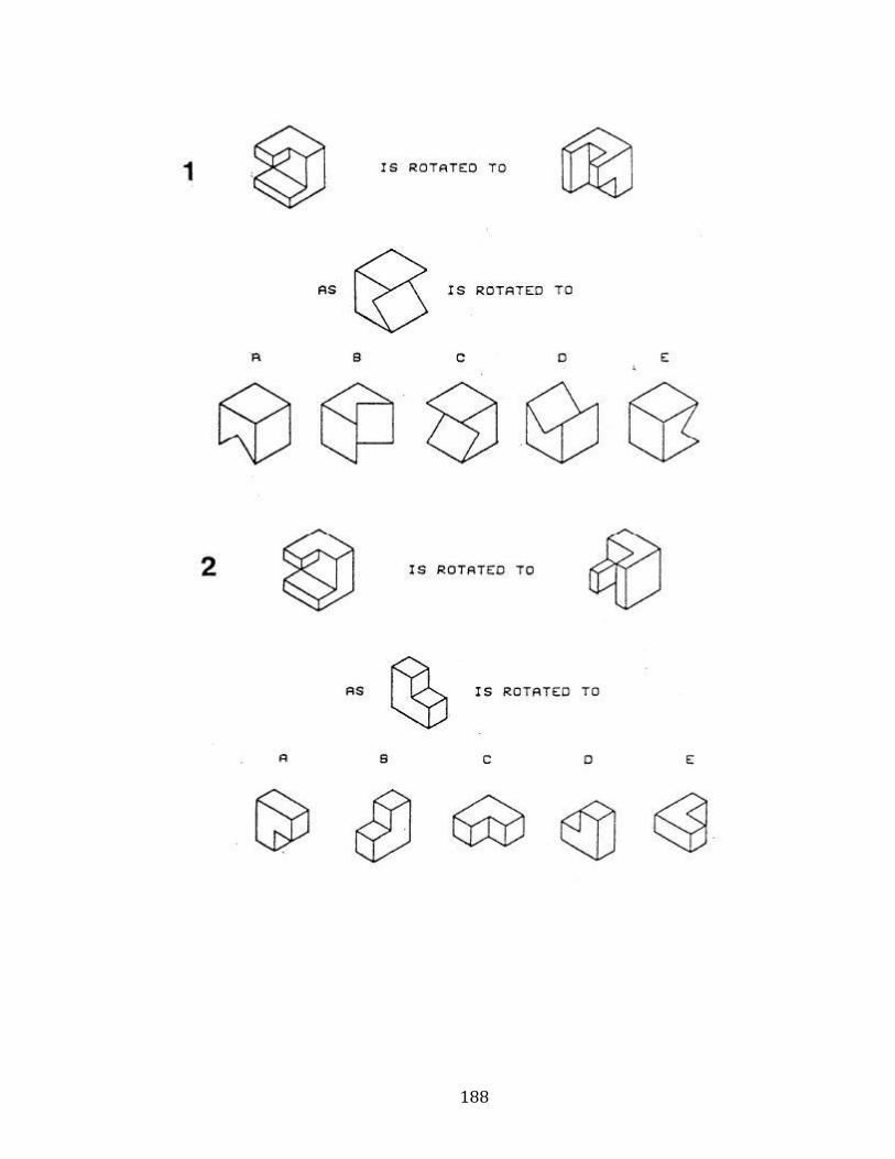
188
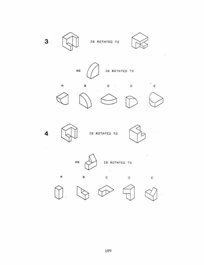
189
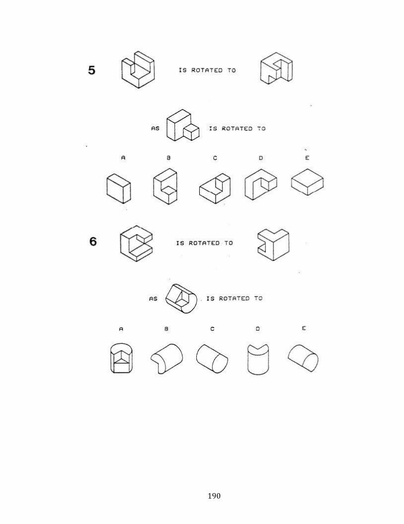
190
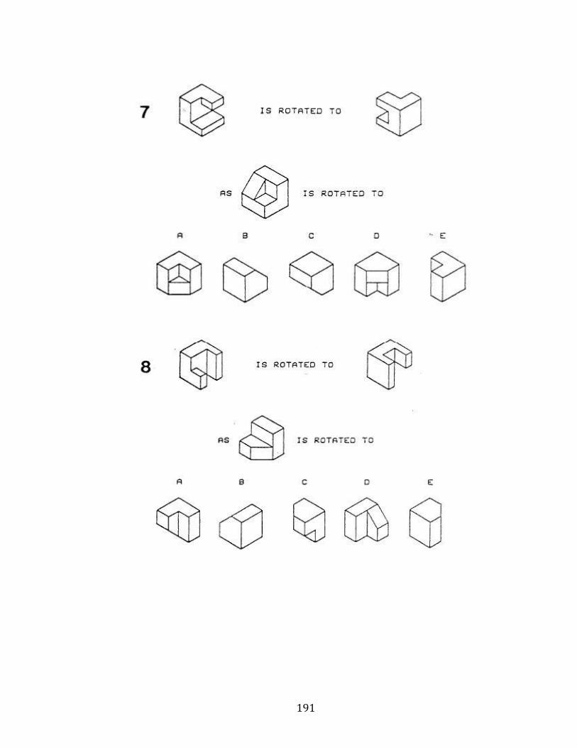
191
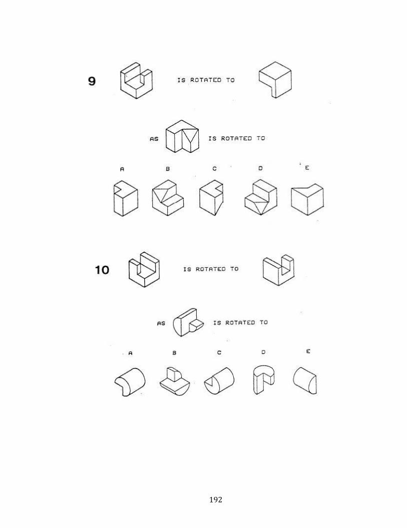
192
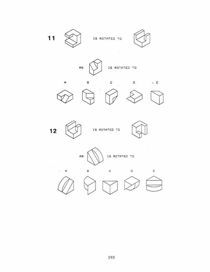
193
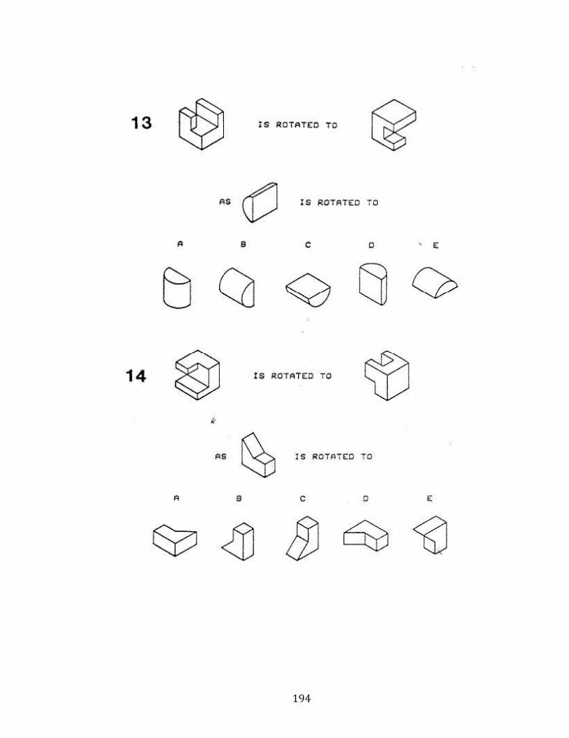
194

195
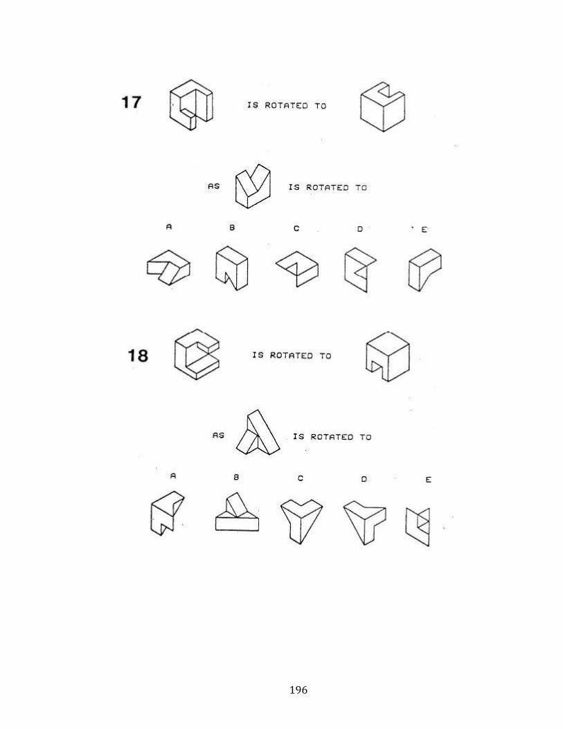
196
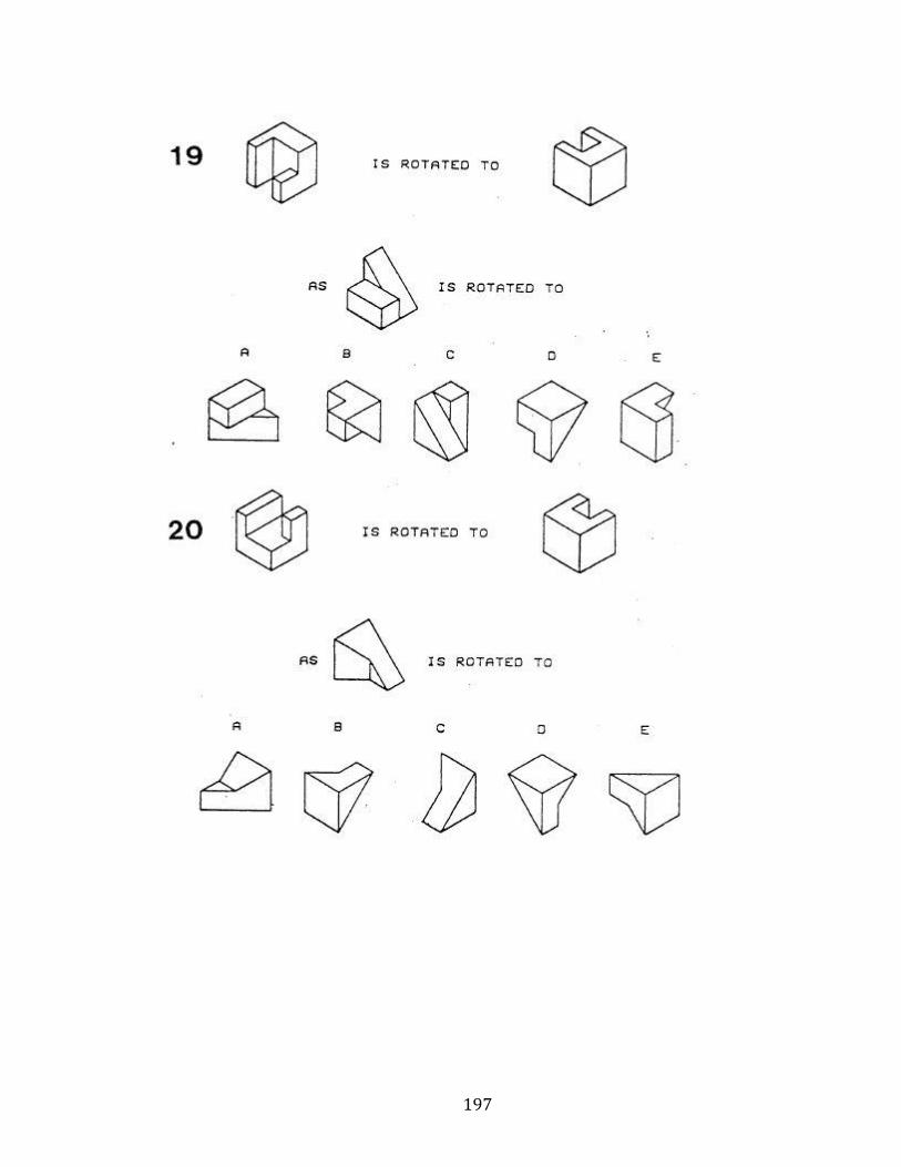
197
198
APPENDIX C: Course Syllabus Excerpts
The syllabus excerpts provided here cover the study skills and techniques students
frequently recommend to students. Usually, this brief syllabus paragraph is the only
instruction in study skills students receive.
A215-
ANATOMY A215 (BASIC HUMAN ANATOMY) SYLLABUS OF INFORMATION AND
POLICIES FALL 2015
Once you come to lecture, we will apply and reinforce the material you learned in
the podcast. We then will discuss the clinical applications of the anatomical region in
question and learn new material. Thus, your preparation ahead of time (by watching
the podcasts and doing your reading) will allow us to discuss the interesting
applications of anatomy! Note that all questions on the lecture exams will deal with
material covered in the podcasts, your lecture outlines, and in lecture. Thus, it is to
your advantage prepare ahead of time and to attend lectures. You will
understand the material much better during lecture if you read the textbook
pages indicated on your schedule. In particular, study the figures in the book;
they are generally very good and will help you understand the material
(emphasis added).
P215-
P215 Basic Human Physiology Spring 2016
199
Lectures may begin with conceptual questions that emphasize material important
for exams. It is important that you are able to answer these questions on your own.
If you cannot that means you should either ask questions in class, in office hours or
via email. If a student misses lecture for any reason, they are responsible for
obtaining copies of the lecture notes from another student. Questions about the
content of those notes are welcome, but realize that nothing can replace the
learning process of taking your own notes. To be successful in this class,
students will need to fully understand the concepts. Attempts to memorize the
material will generally not achieve good results (emphasis added).
A550-
SYLLABUS – Fall 2015 GROSS HUMAN ANATOMY 1 (ANAT-A550) –
STUDY TIPS FOR GROSS ANATOMY: 1. We strongly encourage you to study together
and work in groups when preparing for both lecture and lab. Discuss complex topics
and quiz each other. Working in a group will allow all involved to better understand
the material and help clarify any misconceptions. 2. Make sure you examine and
study ALL of the bodies in the gross lab. As you will learn, variation is considerable
and structures can look completely different in different bodies. 3. Do NOT try to
“cram” for the exams. Lecture exams ask you not only to identify material, but to
apply and synthesize your knowledge. Instead, try to study a little bit every day. 4. If
you have questions, or aren’t doing as well as you’d like, please see one of the
instructors right away! We want you to succeed and we are here to help. However,
we do not know if you need help unless you come and speak with us! 5. In lab, make
200
sure you come prepared to lab (read dissector ahead of time and review atlas
images) and expect to spend the full time in lab, and use the resources available to
you in lab (e.g., AIs, sample images and dissection quizzes, etc.).
P531-
Syllabus P531-P532: MEDICAL PHYSIOLOGY Fall 2014
The goal of this class is to facilitate your learning of medical physiology. To this end,
the course is designed to emphasize active student learning by integrating class
session, discussion, problem-based learning exercises, and laboratories. Class
sessions will involve periods of explanation interspersed with problem solving and
discussion. The time will be split roughly 50/50 between these two modes of
learning. We encourage questions at any time. Questions before and after class,
during office hours, and via e-mail, are encouraged as well. Your teachers do not
expect you to know everything. We certainly don’t! We encourage you to voice your
confusions to us; we learn as much from you as you do from us. Part of the respect
rule is to make the classroom learning environment as safe and open as possible, so
you can relax and feel free to be confused and to work on those items that are
confusing. We have all worked to develop teaching styles and methods that best
meet the needs of our students. There will be periodic opportunities to voice your
opinions (which are welcome at any time) about things that may or may not be
working for you in the classroom. We will try to make any change that we can that
will help facilitate your learning.
201
APPENDIX D: IRB Documents
These sample documents are provided for informational purposes only. Slight
wording changes may exist between various versions of the documents.
IRB approval numbers: Study skills- 1010002079
Illustration teaching- 1301010307
DITKI- 1310535940
INDIANA UNIVERSITY BLOOMINGTON
INFORMED CONSENT STATEMENT
Examining Study Skills in Anatomy & Physiology
You are invited to participate in a research study analyzing the effectiveness of a study skills course on anatomy students. You were selected as a possible subject because you are enrolled in A215, P215, A550, or P531. We ask that you read this form and ask any questions you may have before agreeing to be in the study.
The study is being conducted by Bradley Barger and Polly Husmann, both of whom are graduate students, in Anatomy and Anthropology, respectively. Since one of the researchers may be an instructor for your class neither of them will know if you have participated in this research until after all semester grades are final and have been reported to the Registrar.
STUDY PURPOSE
The purpose of this study is to determine what kind of study methods are employed by students, and which methods, if any, lead to higher final grades.
NUMBER OF PEOPLE TAKING PART IN THE STUDY:
All students enrolled in A215, P215, A550, or P531 will be asked to participate in the study.
PROCEDURES FOR THE STUDY:
If you agree to be in the study, you will complete a survey, expected to take no more than ten minutes and grant the investigators permission to compare the responses to this survey to your course grades. After the comparison between your survey and course grades has been completed all identifying data will be removed.
If you are a student in A215 you will complete this survey twice (once at the beginning of the semester and again at the end of the semester); students in all other courses will complete this survey only once at the end of the semester.
202
RISKS OF TAKING PART IN THE STUDY:
There are no foreseeable risks or discomforts for any of the procedures to be used in this study.
BENEFITS OF TAKING PART IN THE STUDY:
By participating in this study, you will help the principal investigators determine which study habits are the most useful for studying in anatomy and physiology courses. This information will help future students in the listed courses.
ALTERNATIVES TO TAKING PART IN THE STUDY:
Instead of being in the study, you have the option to not to participate. If you choose not to participate you can work on other course materials during the 10 minutes that the survey is being completed in the classroom. Your participation in this study is voluntary. There is no penalty if you decide not to participate.
CONFIDENTIALITY:
Efforts will be made to keep your personal information confidential. We cannot guarantee absolute confidentiality, although we will not report names or identification numbers. Your personal information may be disclosed if required by law. Any published data will be de-identified, and presented only in aggregate. Any saved documents will be de-identified as well.
Organizations that may inspect and/or copy your research records for quality assurance and data analysis include groups such as the study investigator and his/her research associates, the IUB Institutional Review Board or its designees, and (as allowed by law) state or federal agencies, specifically the Office for Human Research Protections (OHRP).
PAYMENT
You will not receive payment for taking part in this study.
CONTACTS FOR QUESTIONS OR PROBLEMS
For questions about the study or a research-related concern you may contact Audra Schutte.
For questions about your rights as a research participant or to discuss problems, complaints or concerns about a research study, or to obtain information, or offer input, contact the IUB Human Subjects office, 530 E Kirkwood Ave, Carmichael Center, 203, Bloomington IN 47408, 812-855-3067 or by email at [email protected]

203
VOLUNTARY NATURE OF STUDY
Your participation in this study is voluntary; you may decline to participate without penalty. Your grade for the course is in no way influenced by whether or not you choose to participate. If you decide to participate, you may withdraw from the study at any time without penalty and without loss of benefits to which you are otherwise entitled. If you withdraw from the study before data collection is completed your data will not be used in this study. Your decision whether or not to participate in this study will not affect your current or future relations with the investigator(s).
SUBJECT’S CONSENT
In consideration of all of the above, I give my consent to participate in this research study.
I will be given a copy of this informed consent document to keep for my records. I agree to take part in this study.
Subject’s Printed Name:
Subject’s Signature:_________________________________________Date:
(must be dated by the subject)
Indiana University - Bloomington
(recruitment script ) Examining Study Skills in Anatomy & Physiology
You are invited to participate in a research study that aims at improving the learning of anatomy and physiology. This study is being conducted by Bradley Barger and Polly Husmann, two graduate students in Anatomy and Anthropology, one of these individuals may be an instructor for your class, but neither of them will know about your participation in this study until after the semester has ended and all grades have been reported to the Registrar. The goal of this research study is to investigate the methods students use to learn in anatomy and physiology classes. One of the aims of this study is to determine which, if any, methods lead to higher final grades.
Your participation in this study requires only that you fill out a brief survey about your methods of studying for this course. In signing this consent statement, you also agree to give permission for the researchers to use your survey data to compile lists of study activities and compare these lists to your final grades. All personal information will be removed, and you will not be personally identifiable
204
in any way after the grade comparison. If this research is published, no personal information will be presented.
I will distribute and collect this consent form, so that your instructor will not know if you have participated until after the course is finished and final grades have been entered.
The researchers will preserve confidentiality by removing identifiable information including student ID numbers from all the data. The analysis of the data will focus on group patterns that will be described in aggregate terms.
Your participation in this study is voluntary. There is no penalty for not participating. If you decide to participate, you may withdraw from the study at any time. After completion of the study, the researchers will be happy to discuss the results with you.
Should you have any questions while the study is in progress, or should you decide any time to withdraw, please contact me, Audra Schutte. After the study is over and your grades have been posted, either Bradley or Polly will be willing to answer any questions, or withdraw your data, if you wish.
If you are willing to participate in the study, please sign one of the two copies of the consent form and return it to me. The second copy is for you to keep for reference. If you feel you have not been treated according to the descriptions in this form, or that your rights as a participant have not been honored during the course of this project, you may contact the Human Subjects Committee, Indiana University, 509 E. 3rd Street, Bloomington, IN 47401, (812) 855-3067, or email at [email protected]
We appreciate your considering participation in this research and thus enabling us to improve instruction in anatomy and physiology courses.
Thank you.
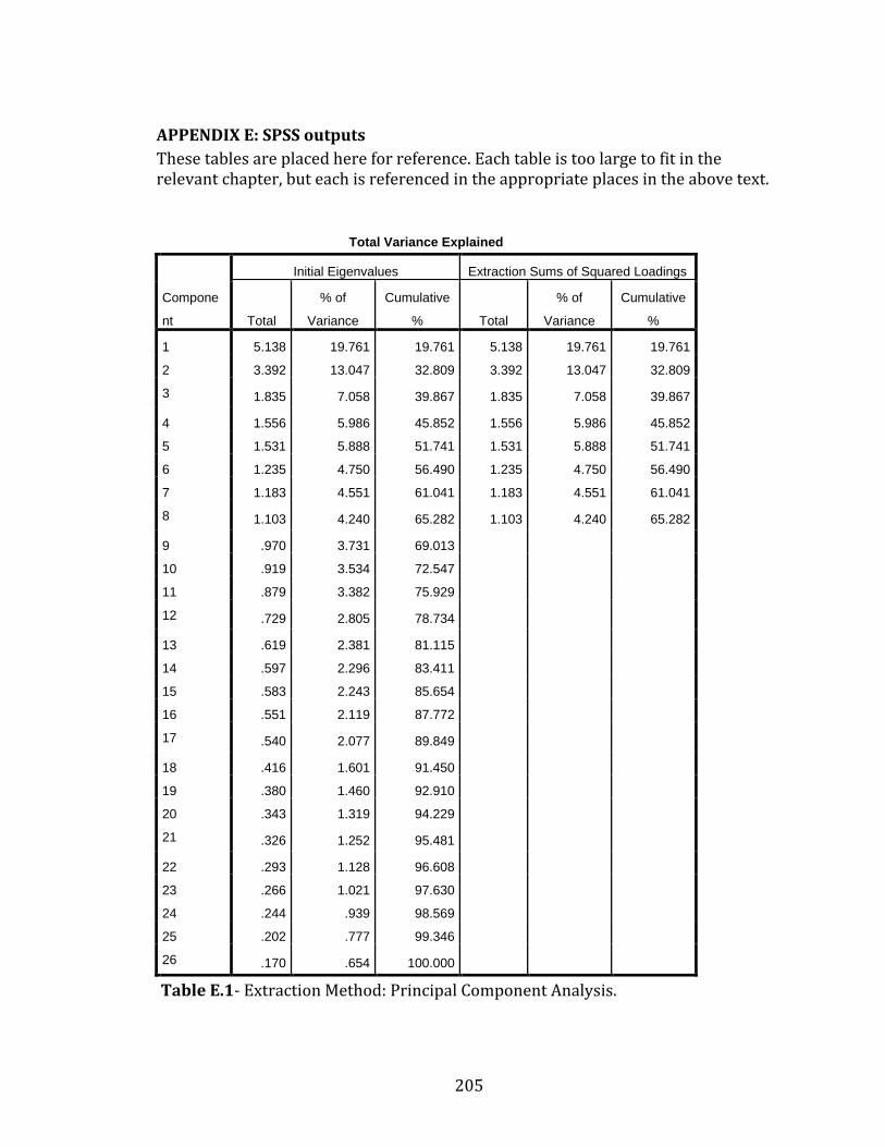
205
APPENDIX E: SPSS outputs
These tables are placed here for reference. Each table is too large to fit in the relevant chapter, but each is referenced in the appropriate places in the above text.
Total Variance Explained
Compone
nt
Initial Eigenvalues Extraction Sums of Squared Loadings
Total
% of
Variance
Cumulative
% Total
% of
Variance
Cumulative
%
1 5.138 19.761 19.761 5.138 19.761 19.761
2 3.392 13.047 32.809 3.392 13.047 32.809
3 1.835 7.058 39.867 1.835 7.058 39.867
4 1.556 5.986 45.852 1.556 5.986 45.852
5 1.531 5.888 51.741 1.531 5.888 51.741
6 1.235 4.750 56.490 1.235 4.750 56.490
7 1.183 4.551 61.041 1.183 4.551 61.041
8 1.103 4.240 65.282 1.103 4.240 65.282
9 .970 3.731 69.013
10 .919 3.534 72.547
11 .879 3.382 75.929
12 .729 2.805 78.734
13 .619 2.381 81.115
14 .597 2.296 83.411
15 .583 2.243 85.654
16 .551 2.119 87.772
17 .540 2.077 89.849
18 .416 1.601 91.450
19 .380 1.460 92.910
20 .343 1.319 94.229
21 .326 1.252 95.481
22 .293 1.128 96.608
23 .266 1.021 97.630
24 .244 .939 98.569
25 .202 .777 99.346
26 .170 .654 100.000
Table E.1- Extraction Method: Principal Component Analysis.
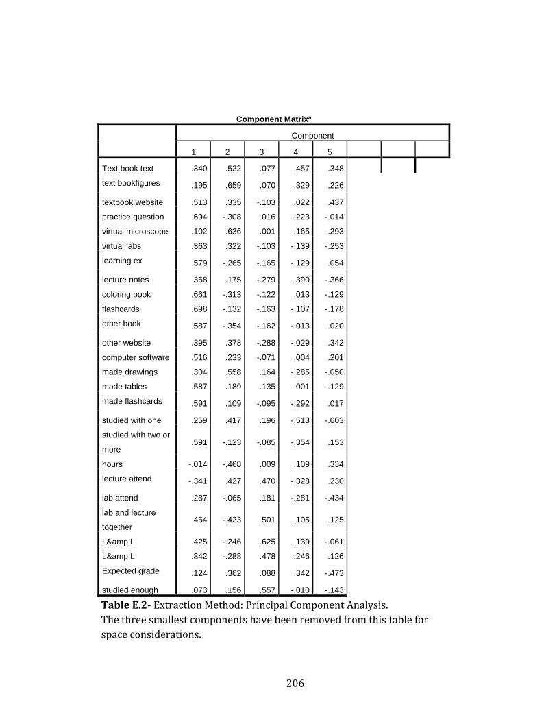
206
Component Matrixa
Component
1 2 3 4 5
Text book text .340 .522 .077 .457 .348
text bookfigures .195 .659 .070 .329 .226
textbook website .513 .335 -.103 .022 .437
practice question .694 -.308 .016 .223 -.014
virtual microscope .102 .636 .001 .165 -.293
virtual labs .363 .322 -.103 -.139 -.253
learning ex .579 -.265 -.165 -.129 .054
lecture notes .368 .175 -.279 .390 -.366
coloring book .661 -.313 -.122 .013 -.129
flashcards .698 -.132 -.163 -.107 -.178
other book .587 -.354 -.162 -.013 .020
other website .395 .378 -.288 -.029 .342
computer software .516 .233 -.071 .004 .201
made drawings .304 .558 .164 -.285 -.050
made tables .587 .189 .135 .001 -.129
made flashcards .591 .109 -.095 -.292 .017
studied with one .259 .417 .196 -.513 -.003
studied with two or
more .591 -.123 -.085 -.354 .153
hours -.014 -.468 .009 .109 .334
lecture attend -.341 .427 .470 -.328 .230
lab attend .287 -.065 .181 -.281 -.434
lab and lecture
together .464 -.423 .501 .105 .125
L&L .425 -.246 .625 .139 -.061
L&L .342 -.288 .478 .246 .126
Expected grade .124 .362 .088 .342 -.473
studied enough .073 .156 .557 -.010 -.143
Table E.2- Extraction Method: Principal Component Analysis.
The three smallest components have been removed from this table for
space considerations.
207
References
Achtman, R., Green, C., & Bavelier, D. (2008). Video games as a tool to train visual skills. Restorative neurology and neuroscience, 26(4, 5), 435-446.
Ainsworth, S., Prain, V., & Tytler, R. (2011). Drawing to Learn in Science. Science, 333(6046), 1096-1097. doi: 10.1126/science.1204153
Aleven, V., & Koedinger, K. R. (2000). Limitations of student control: Do students know when they need help? Paper presented at the Intelligent tutoring systems.
Alias, M., Black, T. R., & Gray, D. E. (2002). Effect of instruction on spatial visualization ability in civil engineering students. International Education Journal, 3(1).
Allen, D. (1994). Teaching Visual Literacy‐Some Reflections on the Term. Journal of Art & Design Education, 13(2), 133-143.
Allen, L. K., Bhattacharyya, S., & Wilson, T. D. (2014). Development of an interactive anatomical three-dimensional eye model. Anatomical Sciences Education, n/a-n/a. doi: 10.1002/ase.1487
Anderson, L. W., Krathwohl, D. R., & Bloom, B. S. (2001). A taxonomy for learning, teaching, and assessing: A revision of Bloom's taxonomy of educational objectives: Allyn & Bacon.
Anderson, N., & Dietrich, M. R. (2012). The educated eye: visual culture and pedagogy in the life sciences: UPNE.
Anderson, T. R. (2007). Bridging the educational research-teaching practice gap. Biochemistry and Molecular Biology Education, 35(6), 471-477. doi: 10.1002/bmb.20135
Atlas, M. C. (2001). Ethics and access to teaching materials in the medical library: the case of the Pernkopf atlas. BULLETIN-MEDICAL LIBRARY ASSOCIATION, 89(1), 51-58.
Avgerinou, M., & Ericson, J. (1997). A review of the concept of Visual Literacy. British Journal of Educational Technology, 28(4), 280-291. doi: 10.1111/1467-8535.00035
Azer, S. A. (2013). The place of surface anatomy in the medical literature and undergraduate anatomy textbooks. Anatomical Sciences Education, 6(6), 415-432. doi: 10.1002/ase.1368
Azer, S. A., & Azer, S. (2016). 3D Anatomy Models and Impact on Learning: A Review of the Quality of the Literature. Health Professions Education.
Backhouse, M., Fitzpatrick, M., Hutchinson, J., Thandi, C. S., & Keenan, I. D. (2016). Improvements in anatomy knowledge when utilizing a novel cyclical “Observe-Reflect-Draw-Edit-Repeat” learning process. Anatomical Sciences Education, n/a-n/a. doi: 10.1002/ase.1616
Bardes, C. L., Gillers, D., & Herman, A. E. (2001). Learning to look: developing clinical observational skills at an art museum. Medical Education, 35(12), 1157-1161. doi: 10.1046/j.1365-2923.2001.01088.x
Bareither, M. L., Arbel, V., Growe, M., Muszczynski, E., Rudd, A., & Marone, J. R. (2013). Clay modeling versus written modules as effective interventions in
208
understanding human anatomy. Anatomical Sciences Education, 6(3), 170-176. doi: 10.1002/ase.1321
Barger, B. (2016). Teaching Students to See: Visual Learning Instruction for Two Different Undergraduate Anatomy Courses. The FASEB Journal, 30(1 Supplement), 570.510-570.510.
Barger, B., & Fisch, A. (2015). A Survey of Student Use of a Novel Software Tool for Teaching Neuroanatomy, Draw It to Know It. The FASEB Journal, 29(1 Supplement), 550.559.
Barger, J. B. (2012). How do undergraduate students study for anatomy, and does it matter? The FASEB Journal, 26(1_MeetingAbstracts), 528.522.
Barger, J. B. (2014). Using in-class drawing exercises to teach anatomy and visual skills (534.10). The FASEB Journal, 28(1 Supplement), 534.510.
Barger, J. B., & Husmann, P. (2013). A comparison of medical student and undergrad study skills in anatomy lab courses. The FASEB Journal, 27(1_MeetingAbstracts), 956.954.
Barilan, Y. M. (2005). The story of the body and the story of the person: Towards an ethics of representing human bodies and body-parts. Medicine, Health Care and Philosophy, 8(2), 193-205.
Barilan, Y. M. (2006). Bodyworlds and the ethics of using human remains: A preliminary discussion. Bioethics, 20(5), 233-247.
Barke, H.-D. (1993). Chemical education and spatial ability. Journal of Chemical Education, 70(12), 968.
Barnett-Cowan, M., Dyde, R. T., Thompson, C., & Harris, L. R. (2010). Multisensory determinants of orientation perception: task-specific sex differences. European Journal of Neuroscience, 31(10), 1899-1907. doi: 10.1111/j.1460-9568.2010.07199.x
Beck, M. M., Taft, T. B., Jr., & Zimmer, S. E. (1978). Predicting clinical performance: a two-step approach. Journal of Dental Education, 42(9), 524-527.
Bell, L. T. O., & Evans, D. J. R. (2014). Art, anatomy, and medicine: Is there a place for art in medical education? Anatomical Sciences Education, 7(5), 370-378. doi: 10.1002/ase.1435
Berliner, D. C. (1986). In pursuit of the expert pedagogue. Educational researcher, 15(7), 5-13.
Berliner, D. C. (1988). Implications of studies on expertise in pedagogy for teacher education and evaluation. New directions for teacher assessment, 39-68.
Berk, R.A. An introduction to sample selection bias in sociological data. American Sociological Review (1983): 386-398.
Biggs, J. (1988). The role of metacognition in enhancing learning. Australian Journal of Education, 32(2), 127-138.
Biggs, J., Kember, D., & Leung, D. Y. (2001). The revised two‐factor study process questionnaire: R‐SPQ‐2F. British Journal of Educational Psychology, 71(1), 133-149.
Billett, S. (2001). Learning through work: workplace affordances and individual engagement. Journal of workplace learning, 13(5), 209-214.
Bjork, R. A., Dunlosky, J., & Kornell, N. (2013). Self-regulated learning: Beliefs, techniques, and illusions. Annual review of psychology, 64, 417-444.
209
Blanch-Hartigan, D. (2011). Medical students' self-assessment of performance: Results from three meta-analyses. Patient Education and Counseling, 84(1), 3-9. doi: 10.1016/j.pec.2010.06.037
Bodner, G. M., & Guay, R. B. (1997). The Purdue visualization of rotations test. The Chemical Educator, 2(4), 1-17.
Boudreau, J. D., Cassell, E. J., & Fuks, A. (2008). Preparing medical students to become skilled at clinical observation. Medical Teacher, 30(9-10), 857-862. doi: 10.1080/01421590802331446
Boz, N., & Boz, Y. (2008). A qualitative case study of prospective chemistry teachers’ knowledge about instructional strategies: Introducing particulate theory. Journal of Science Teacher Education, 19(2), 135-156.
Bransford, J. D., Brown, A. L., & Cocking, R. R. (2000). How people learn: Washington, DC: National Academy Press.
Braun, M. W., & Kearns, K. D. (2008). Improved Learning Efficiency and Increased Student Collaboration Through Use of Virtual Microscopy in the Teaching of Human Pathology. Anatomical Sciences Education, 1(6), 240-246. doi: 10.1002/ase.53
Bruch, L. A., De Young, B. R., Kreiter, C. D., Haugen, T. H., Leaven, T. C., & Dee, F. R. (2009). Competency assessment of residents in surgical pathology using virtual microscopy. Human Pathology, 40(8), 1122-1128. doi: 10.1016/j.humpath.2009.04.009
Canty, D. J., Hayes, J. A., Story, D. A., & Royse, C. F. (2015). Ultrasound simulator-assisted teaching of cardiac anatomy to preclinical anatomy students: A pilot randomized trial of a three-hour learning exposure. Anatomical Sciences Education, 8(1), 21-30. doi: 10.1002/ase.1452
Carpenter, G. A., & Grossberg, S. (1988). The ART of adaptive pattern recognition by a self-organizing neural network. Computer, 21(3), 77-88.
Carter, C. S., LaRussa, M. A., & Bodner, G. M. (1987). A study of two measures of spatial ability as predictors of success in different levels of general chemistry. Journal of Research in Science Teaching, 24(7), 645-657.
Cassidy, M. F., & Knowlton, J. Q. (1983). Visual literacy: A failed metaphor? ECTJ, 31(2), 67-90.
Champney, T. H. (2016). The business of bodies: Ethical perspectives on for‐profit body donation companies. Clinical Anatomy, 29(1), 25-29.
Chatterjee, A. K., & Walker, J. J. (2011). The importance of spatial ability and mental models in learning anatomy. Faseb Journal, 25.
Codd, A. M., & Choudhury, B. (2011). Virtual Reality Anatomy: Is it Comparable with Traditional Methods in the Teaching of Human Forearm Musculoskeletal Anatomy? Anatomical Sciences Education, 4(3), 119-125. doi: 10.1002/ase.214
Cohen, C. A., & Hegarty, M. (2007). Individual differences in use of external visualisations to perform an internal visualisation task. Applied Cognitive Psychology, 21(6), 701-711. doi: 10.1002/acp.1344
Coleman, S. L., & Gotch, A. J. (1998). Spatial perception skills of chemistry students. Journal of Chemical Education, 75(2), 206.
Collier, L., Dunham, S., Braun, M. W., & O'Loughlin, V. D. (2012). Optical versus
210
virtual: Teaching assistant perceptions of the use of virtual microscopy in an undergraduate human anatomy course. Anatomical Sciences Education, 5(1), 10-19. doi: 10.1002/ase.262
Cornwall, J. (2016). The ethics of 3D printing copies of bodies donated for medical education and research: What is there to worry about? The Australasian medical journal, 9(1), 8.
Cornwall, J., Callahan, D., & Wee, R. (2016). Ethical issues surrounding the use of images from donated cadavers in the anatomical sciences. Clinical Anatomy, 29(1), 30-36.
Credé, M., & Kuncel, N. R. (2008). Study habits, skills, and attitudes: The third pillar supporting collegiate academic performance. Perspectives on Psychological Science, 3(6), 425-453.
Crider, A. (2016). Astronomy, Visual Literacy, and Liberal Arts Education. Paper presented at the American Astronomical Society Meeting Abstracts.
Culver, C. M., & Dunham, F. (1969). Birth order and spatial-perceptual ability: negative note. Perceptual and Motor Skills, 28(1), 301-302.
Czarnolewski, M. Y., & Eliot, J. (2012). Exploratory study of the relations between spatial ability and drawing from memory (vol 114, pg 627, 2012). Perceptual and Motor Skills, 115(2), 676-676. doi: 10.2466/04.10.23.pms.115.5.676
Dabbs, J. M., Chang, E.-L., Strong, R. A., & Milun, R. (1998). Spatial ability, navigation strategy, and geographic knowledge among men and women. Evolution and Human Behavior, 19(2), 89-98.
de Faria, J. W. V., Teixeira, M. J., de Moura Sousa Júnior, L., Otoch, J. P., & Figueiredo, E. G. (2016). Virtual and stereoscopic anatomy: when virtual reality meets medical education. Journal of neurosurgery, 1-7.
Dempsey, B., & Betz, B. J. (2001). Biological drawing: a scientific tool for learning. American Biology Teacher, 63(4), 271-279.
Dondis, D. A. (1974). A primer of visual literacy: Mit Press. Drapkin, Z. A., Lindgren, K. A., Lopez, M. J., & Stabio, M. E. (2015). Development and
assessment of a new 3D neuroanatomy teaching tool for MRI training. Anatomical Sciences Education, n/a-n/a. doi: 10.1002/ase.1509
Draw it to Know It, LLC. (2016) Draw it to know it (website) https://www.drawittoknowit.com/
Edward, T. (1990). Envisioning information. Cheshire (Conn.). Estevez, M. E., Lindgren, K. A., & Bergethon, P. R. (2010). A Novel Three-Dimensional
Tool for Teaching Human Neuroanatomy. Anatomical Sciences Education, 3(6), 309-317. doi: 10.1002/ase.186
Evans, D. J. R. (2011). Using embryology screencasts: A useful addition to the student learning experience? Anatomical Sciences Education, 4(2), 57-63. doi: 10.1002/ase.209
Farkas, G. J., Mazurek, E., & Marone, J. R. (2015). Learning style versus time spent studying and career choice: Which is associated with success in a combined undergraduate anatomy and physiology course?. Anatomical sciences education.9(2), 121-131, DOI: 10.1002/ase.1563
Faurie, C., & Khadra, M. (2012). Technical competence in surgeons. Anz Journal of Surgery, 82(10), 682-690. doi: 10.1111/j.1445-2197.2012.06239.x
211
Feng, J., Spence, I., & Pratt, J. (2007). Playing an action video game reduces gender differences in spatial cognition. Psychological science, 18(10), 850-855.
Ferk, V., & Yrtacnik, M. (2003). Students' understanding of molecular structure representations. International Journal of Science Education, 25(10), 1227-1245. doi: 10.1080/0950069022000038231
Fernandez, R., Dror, I. E., & Smith, C. (2011). Spatial Abilities of Expert Clinical Anatomists: Comparison of Abilities Between Novices, Intermediates, and Experts in Anatomy. Anatomical Sciences Education, 4(1), 1-8. doi: 10.1002/ase.196
Fleming, N., & Baume, D. (2006). Learning Styles Again: VARKing up the right tree! Educational Developments, 7(4), 4.
Flavell, J. H. (1979). Metacognition and cognitive monitoring: A new area of cognitive–developmental inquiry. American psychologist, 34(10), 906.
Fonseca, G. R., & Finn, G. M. (2016). The Impact of Body Worlds on Adult Visitors' Knowledge on Human Anatomy: A Preliminary Study. Clinical Anatomy.
Foo, J.-L., Martinez-Escobar, M., Juhnke, B., Cassidy, K., Hisley, K., Lobe, T., & Winer, E. (2013). Evaluating Mental Workload of Two-Dimensional and Three-Dimensional Visualization for Anatomical Structure Localization. Journal of Laparoendoscopic & Advanced Surgical Techniques, 23(1), 65-70. doi: 10.1089/lap.2012.0150
Frailich, M., Kesner, M., & Hofstein, A. (2009). Enhancing students' understanding of the concept of chemical bonding by using activities provided on an interactive website. Journal of Research in Science Teaching, 46(3), 289-310.
Francey, M. F., Madigan, B. C., & Becherer, J. P. (1996). Instructor's Resource Manual with Tests: Art History, Volume I, Marilyn Stokstad: Prentice Hall.
Freeman, S., Eddy, S. L., McDonough, M., Smith, M. K., Okoroafor, N., Jordt, H., & Wenderoth, M. P. (2014). Active learning increases student performance in science, engineering, and mathematics. Proceedings of the National Academy of Sciences, 111(23), 8410-8415.
Fuks, A., Boudreau, J. D., & Cassell, E. J. (2009). Teaching clinical thinking to first-year medical students. Medical Teacher, 31(2), 105-111. doi: 10.1080/01421590802512979
Furnham, A. (1986). Response bias, social desirability and dissimulation.Personality and individual differences, 7(3), 385-400.
Ganesh, A. (2014). Alignment of Teaching Style to Learning Preferences: Impact on Student Learning. Training & Development Journal, 5(2), 119-131.
Garg, A., Norman, G., Spero, L., & Taylor, I. (1999). Learning anatomy: do new computer models improve spatial understanding? Medical Teacher, 21(5), 519-522.
Garg, A., Norman, G. R., Spero, L., & Maheshwari, P. (1999). Do virtual computer models hinder anatomy learning? Academic Medicine, 74(10), S87-S89. doi: 10.1097/00001888-199910000-00049
Garg, A. X., Norman, G., & Sperotable, L. (2001). How medical students learn spatial anatomy. Lancet, 357(9253), 363-364. doi: 10.1016/s0140-6736(00)03649-7
Glasgow, J. N. (1994). Teaching visual literacy for the 21st century. Journal of
212
reading, 494-500. Gombrich, E. H., Gombrich, E. H., Gombrich, E. H., & Gombrich, E. H. (2000). Art and
illusion: A study in the psychology of pictorial representation (Vol. 5): Phaidon London.
Gradl-Dietsch, G., Korden, T., Modabber, A., Sönmez, T. T., Stromps, J.-P., Ganse, B., . . . Knobe, M. (2016). Multidimensional approach to teaching anatomy—Do gender and learning style matter? Annals of Anatomy-Anatomischer Anzeiger.
Grady, J. (2001). Becoming a visual sociologist. Sociological Imagination, 38(1/2), 83-119.
Griffith, J. R. (2015). Active learning strategies and vocabulary achievement. UNIVERSITY OF MISSOURI-SAINT LOUIS.
Gurung, R. A., & McCann, L. I. (2011). How should students study? Tips, advice, and pitfalls: Observer.
Hacker, D. J., Dunlosky, J., & Graesser, A. C. (1998). Metacognition in educational theory and practice: Routledge.
Harackiewicz, J. M., Barron, K. E., Tauer, J. M., & Elliot, A. J. (2002). Predicting success in college: A longitudinal study of achievement goals and ability measures as predictors of interest and performance from freshman year through graduation. Journal of Educational Psychology, 94(3), 562.
Harle, M., & Towns, M. (2010). A review of spatial ability literature, its connection to chemistry, and implications for instruction. Journal of Chemical Education, 88(3), 351-360.
Hartwig, M. K., & Dunlosky, J. (2012). Study strategies of college students: Are self-testing and scheduling related to achievement? Psychonomic Bulletin & Review, 19(1), 126-134. doi: 10.3758/s13423-011-0181-y
Hattwig, D., Bussert, K., Medaille, A., & Burgess, J. (2013). Visual literacy standards in higher education: New opportunities for libraries and student learning. portal: Libraries and the Academy, 13(1), 61-89.
Hausmann, M., Slabbekoorn, D., Van Goozen, S. H., Cohen-Kettenis, P. T., & Güntürkün, O. (2000). Sex hormones affect spatial abilities during the menstrual cycle. Behavioral neuroscience, 114(6), 1245.
Hawk, T. F., & Shah, A. J. (2007). Using learning style instruments to enhance student learning. Decision Sciences Journal of Innovative Education, 5(1), 1-19.
Hegarty, M., Keehner, M., Khooshabeh, P., & Montello, D. R. (2009). How spatial abilities enhance, and are enhanced by, dental education. Learning and Individual Differences, 19(1), 61-70. doi: 10.1016/j.lindif.2008.04.006
Heuschele, A. (1999). "It all looks the same to me" - An exercise in critical observation. American Biology Teacher, 61(6), 434-437.
Hildebrandt, S. (2016). The Anatomy of Murder: Ethical Transgressions and Anatomical Science during the Third Reich: Berghahn Books.
Hmelo-Silver, C. E. (2004). Problem-based learning: What and how do students learn? Educational Psychology Review, 16(3), 235-266.
Hoyek, N., Collet, C., Di Rienzo, F., De Almeida, M., & Guillot, A. (2014). Effectiveness of three-dimensional digital animation in teaching human anatomy in an authentic classroom context. Anatomical Sciences Education, 7(6), 430-437. doi: 10.1002/ase.1446
213
Husmann, P. R., & Barger, J. B. (2011). Medical Student Study Skills in Anatomy and Physiology: Is There a Difference? The FASEB Journal, 25(1_MeetingAbstracts), 493.491.
Husmann, P. R., Barger, J. B., & Schutte, A. F. (2016). Study skills in anatomy and physiology: Is there a difference? Anatomical Sciences Education, 9(1), 18-27.
Husmann, P. R., O'Loughlin, V. D., & Braun, M. W. (2009). Quantitative and Qualitative Changes in Teaching Histology by Means of Virtual Microscopy in an Introductory Course in Human Anatomy. Anatomical Sciences Education, 2(5), 218-226. doi: 10.1002/ase.105
Hyde, J. S. (2016). Sex and cognition: gender and cognitive functions. Current opinion in neurobiology, 38, 53-56.
Jones, D. G. (2016). Searching for good practice recommendations on body donation across diverse cultures. Clinical Anatomy, 29(1), 55-59.
Jones, D. G., Gear, R., & Galvin, K. (2003). Stored human tissue: an ethical perspective on the fate of anonymous, archival material. Journal of medical ethics, 29(6), 343-347.
Kali, Y., & Orion, N. (1996). Spatial abilities of high-school students in the perception of geologic structures. Journal of Research in Science Teaching, 33(4), 369-391.
Kamp, M. T., Admiraal, W., Drie, J., & Rijlaarsdam, G. (2015). Enhancing divergent thinking in visual arts education: Effects of explicit instruction of meta‐cognition. British Journal of Educational Psychology, 85(1), 47-58.
Karpicke, J. D., Butler, A. C., & Roediger III, H. L. (2009). Metacognitive strategies in student learning: do students practise retrieval when they study on their own? Memory, 17(4), 471-479.
Keehner, M. (2011). Spatial Cognition Through the Keyhole: How Studying a Real-World Domain Can Inform Basic Science-and Vice Versa. Topics in Cognitive Science, 3(4), 632-647. doi: 10.1111/j.1756-8765.2011.01154.x
Keehner, M., Hegarty, M., Cohen, C., Khooshabeh, P., & Montello, D. R. (2008). Spatial Reasoning With External Visualizations: What Matters Is What You See, Not Whether You Interact. Cognitive Science, 32(7), 1099-1132. doi: 10.1080/03640210801898177
Keehner, M., Lippa, Y., Montello, D. R., Tendick, F., & Hegarty, M. (2006). Learning a spatial skill for surgery: How the contributions of abilities change with practice. Applied Cognitive Psychology, 20(4), 487-503. doi: 10.1002/acp.1198
Kelly, R. M. (2016). ConfChem Conference on Interactive Visualizations for Chemistry Teaching and Learning: Insights into Molecular Visualization Design. Journal of Chemical Education.
Kemp, M. (2006). Intimations and intuitions. New scientist, 191(2568), 48-49. Kennedy, K. M., & Raz, N. (2005). Age, sex and regional brain volumes predict
perceptual-motor skill acquisition. Cortex, 41(4), 560-569. doi: 10.1016/s0010-9452(08)70196-5
Khot, Z., Quinlan, K., Norman, G. R., & Wainman, B. (2013). The relative effectiveness of computer-based and traditional resources for education in anatomy. Anatomical Sciences Education, 6(4), 211-215. doi: 10.1002/ase.1355
214
Knobe, M., Carow, J. B., Ruesseler, M., Leu, B. M., Simon, M., Beckers, S. K., . . . Pape, H.-C. (2012). Arthroscopy or ultrasound in undergraduate anatomy education: a randomized cross-over controlled trial. Bmc Medical Education, 12. doi: 10.1186/1472-6920-12-85
Knowles, E. S., & Nathan, K. T. (1997). Acquiescent responding in self-reports: Cognitive style or social concern?. Journal of Research in Personality, 31(2), 293-301.
Kooloos, J. G. M., Schepens-Franke, A. N., Bergman, E. M., Donders, R. A. R. T., & Vorstenbosch, M. A. T. M. (2014). Anatomical knowledge gain through a clay-modeling exercise compared to live and video observations. Anatomical Sciences Education, 7(6), 420-429. doi: 10.1002/ase.1443
Kornell, N., & Bjork, R. A. (2007). The promise and perils of self-regulated study. Psychonomic Bulletin & Review, 14(2), 219-224. doi: 10.3758/bf03194055
Kotzé, S. H., & Mole, C. G. (2015). Making large class basic histology lectures more interactive: The use of draw‐along mapping techniques and associated educational activities. Anatomical Sciences Education, 8(5), 463-470.
Kozhevnikov, M., & Hegarty, M. (2001). A dissociation between object manipulation spatial ability and spatial orientation ability. Memory & Cognition, 29(5), 745-756. doi: 10.3758/bf03200477
Kozhevnikov, M., Motes, M. A., & Hegarty, M. (2007). Spatial visualization in physics problem solving. Cognitive Science, 31(4), 549-579.
Kozhevnikov, M., & Thornton, R. (2006). Real-Time Data Display, Spatial Visualization Ability, and Learning Force and Motion Concepts. Journal of Science Education and Technology, 15(1), 111-132. doi: 10.1007/s10956-006-0361-0
Kruger, J., & Dunning, D. (1999). Unskilled and unaware of it: how difficulties in recognizing one's own incompetence lead to inflated self-assessments. Journal of personality and social psychology, 77(6), 1121.
Langlois, J., Wells, G. A., Lecourtois, M., Bergeron, G., Yetisir, E., & Martin, M. (2013). Sex differences in spatial abilities of medical graduates entering residency programs. Anatomical Sciences Education, 6(6), 368-375. doi: 10.1002/ase.1360
Langlois, J., Wells, G. A., Lecourtois, M., Bergeron, G., Yetisir, E., & Martin, M. (2014). Spatial abilities of medical graduates and choice of residency programs. Anatomical Sciences Education, n/a-n/a. doi: 10.1002/ase.1453
Lawton, C. A. (1994). Gender differences in way-finding strategies: Relationship to spatial ability and spatial anxiety. Sex roles, 30(11-12), 765-779.
Leite, W. L., Svinicki, M., & Shi, Y. (2010). Attempted validation of the scores of the VARK: Learning styles inventory with multitrait–multimethod confirmatory factor analysis models. Educational and psychological measurement, 70(2), 323-339.
Linenberger, K. J., & Bretz, S. L. (2015). Biochemistry students' ideas about how an enzyme interacts with a substrate. Biochemistry and Molecular Biology Education, 43(4), 213-222.
Linn, M. C., & Petersen, A. C. (1985). Emergence and characterization of sex differences in spatial ability: A meta-analysis. Child development, 1479-1498.

215
Linn, M. C., & Petersen, A. C. (1986). A meta-analysis of gender differences in spatial ability: Implications for mathematics and science achievement. The psychology of gender: Advances through meta-analysis, 67-101.
Lohman, D. F. (1979). Spatial Ability: A Review and Reanalysis of the Correlational Literature: DTIC Document.
Lord, T. R. (1985). ENHANCING THE VISUO-SPATIAL APTITUDE OF STUDENTS. Journal of Research in Science Teaching, 22(5), 395-405. doi: 10.1002/tea.3660220503
Lufler, R. S. (2012). Predictors of Performance in Anatomical Sciences Courses: What Really Matters? Faseb Journal, 26.
Lufler, R. S., Zumwalt, A. C., Romney, C. A., & Hoagland, T. M. (2012). Effect of visual-spatial ability on medical students' performance in a gross anatomy course. Anatomical Sciences Education, 5(1), 3-9. doi: 10.1002/ase.264
Luursema, J.-M., Verwey, W. B., Kommers, P. A. M., & Annema, J.-H. (2008). The role of stereopsis in virtual anatomical learning. Interacting with Computers, 20(4-5), 455-460. doi: 10.1016/j.intcom.2008.04.003
Lyon, P., Letschka, P., Ainsworth, T., & Haq, I. (2013). An exploratory study of the potential learning benefits for medical students in collaborative drawing: creativity, reflection and 'critical looking'. Bmc Medical Education, 13. doi: 10.1186/1472-6920-13-86
Maan, Z. N., Maan, I. N., Darzi, A. W., & Aggarwal, R. (2012). Systematic review of predictors of surgical performance. British Journal of Surgery, 99(12), 1610-1621. doi: 10.1002/bjs.8893
Manolis, C., Burns, D. J., Assudani, R., & Chinta, R. (2013). Assessing experiential learning styles: A methodological reconstruction and validation of the Kolb Learning Style Inventory. Learning and Individual Differences, 23, 44-52.
Marcaida, J. R. (2016). Pictures and Conversation: How to Study the Visual Cultures of Science. Isis, 107(1), 134-139.
Martınez-Trinidad, J. F., & Guzmán-Arenas, A. (2001). The logical combinatorial approach to pattern recognition, an overview through selected works. Pattern Recognition, 34(4), 741-751.
Martín-Gutiérrez, J., Saorín, J. L., Contero, M., Alcañiz, M., Pérez-López, D. C., & Ortega, M. (2010). Design and validation of an augmented book for spatial abilities development in engineering students. Computers & Graphics, 34(1), 77-91.
Marton, F., & Säljö, R. (1976). On qualitative differences in learning: I—Outcome and process*. British Journal of Educational Psychology, 46(1), 4-11.
Masters, M. S., & Sanders, B. (1993). Is the gender difference in mental rotation disappearing? Behavior genetics, 23(4), 337-341.
Mayer, J. (2014). Visual Literacy across the Disciplines. In Research within the Disciplines, 2nd ed., 277.
McGee, M. G. (1979). Human spatial abilities: psychometric studies and environmental, genetic, hormonal, and neurological influences. Psychological bulletin, 86(5), 889.
McGuire, S. Y. (2015). Teach Students How to Learn: Strategies You Can Incorporate Into Any Course to Improve Student Metacognition, Study Skills, and Motivation: Stylus Publishing, LLC.
216
McGrath, M. B., & Brown, J. R. (2005). Visual learning for science and engineering. Computer graphics and applications, IEEE, 25(5), 56-63.
McMenamin, P. G., Quayle, M. R., McHenry, C. R., & Adams, J. W. (2014). The production of anatomical teaching resources using three-dimensional (3D) printing technology. Anatomical Sciences Education, 7(6), 479-486. doi: 10.1002/ase.1475
Meneghetti, C., Borella, E., & Pazzaglia, F. (2016). Mental rotation training: transfer and maintenance effects on spatial abilities. Psychological research, 80(1), 113-127.
Meneghetti, C., Gyselinck, V., Pazzaglia, F., & De Beni, R. (2009). Individual differences in spatial text processing: High spatial ability can compensate for spatial working memory interference. Learning and Individual Differences, 19(4), 577-589.
Meneghetti, C., Pazzaglia, F., & De Beni, R. (2011). Spatial mental representations derived from survey and route descriptions: When individuals prefer extrinsic frame of reference. Learning and Individual Differences, 21(2), 150-157. doi: 10.1016/j.lindif.2010.12.003
Metcalfe, J. (2008). Evolution of metacognition. Handbook of metamemory and memory, 29-46.
Meyers, C., & Jones, T. B. (1993). Promoting Active Learning. Strategies for the College Classroom: ERIC.
Mione, S., Valcke, M., & Cornelissen, M. (2013). Evaluation of virtual microscopy in medical histology teaching. Anatomical Sciences Education, 6(5), 307-315. doi: 10.1002/ase.1353
Moè, A. (2016). Does experience with spatial school subjects favour girls' mental rotation performance? Learning and Individual Differences, 47, 11-16.
Moffat, S. D., Hampson, E., & Hatzipantelis, M. (1998). Navigation in a “virtual” maze: Sex differences and correlation with psychometric measures of spatial ability in humans. Evolution and Human Behavior, 19(2), 73-87.
Mohler, J. L. (2006). Examining the spatial ability phenomenon from the student’s perspective. Purdue University West Lafayette.
Morehead, K., Rhodes, M. G., & DeLozier, S. (2016). Instructor and student knowledge of study strategies. Memory, 24(2), 257-271.
Morozov, M., Tanakov, A., Gerasimov, A., Bystrov, D., & Cvirco, E. (2004). Virtual chemistry laboratory for school education. Paper presented at the Advanced Learning Technologies, 2004. Proceedings. IEEE International Conference on.
Moscova, M., Bryce, D. A., Sindhusake, D., & Young, N. (2014). Integration of medical imaging including ultrasound into a new clinical anatomy curriculum. Anatomical Sciences Education, 8.3 (2015): 205-220: 10.1002/ase.1481
Murphy, K. P., Crush, L., O'Malley, E., Daly, F. E., Twomey, M., O'Tuathaigh, C. M. P., . . . O'Connor, O. J. (2014). Medical student perceptions of radiology use in anatomy teaching. Anatomical Sciences Education, 8.6 (2015): 510-517. doi: 10.1002/ase.1502
Naghshineh, S., Hafler, J. P., Miller, A. R., Blanco, M. A., Lipsitz, S. R., Dubroff, R. P., . . . Katz, J. T. (2008). Formal art observation training improves medical students’ visual diagnostic skills. Journal of General Internal Medicine, 23(7), 991-997.
217
Nelson, N. (2004). Visual Literacy and Library Instruction: A Critical Analysis. Education Libraries, 27(1), 5-10.
Netter, F. H., & Colacino, S. (1989). Atlas of human anatomy (Vol. 11). Summit, NJ: Ciba-Geigy.
Newcombe, N. S., & Stieff, M. (2012). Six myths about spatial thinking. International Journal of Science Education, 34(6), 955-971.
Ngan, N., Mulla, A., Nelson, A. J., & Wilson, T. D. (2010). Problem solving strategies and the relationship between visualization ability and spatial anatomy task performance. Faseb Journal, 24.
Ngan, N., Nelson, A. J., & Wilson, T. D. (2012). Computer visualizations: Factors that influence spatial anatomy comprehension. Anatomical Sciences Education, 5(2), 98-108. doi: 10.1002/ase.1258
Nguyen, N., Mulla, A., Nelson, A. J., & Wilson, T. D. (2014). Visuospatial anatomy comprehension: The role of spatial visualization ability and problem-solving strategies. Anatomical Sciences Education, 7(4), 280-288. doi: 10.1002/ase.1415
Nguyen, N., Nelson, A., & Wilson, T. D. (2011). Computer visualizations: Internal and external factors that influence comprehension of visuospatial information. Society for Neuroscience Abstract Viewer and Itinerary Planner, 41.
Nguyen, N., & Wilson, T. D. (2009). A Head in Virtual Reality: Development of A Dynamic Head and Neck Model. Anatomical Sciences Education, 2(6), 294-301. doi: 10.1002/ase.115
Noel, G. P. J. C. (2013). A novel patchwork model used in lecture and laboratory to teach the three-dimensional organization of mesenteries. Anatomical Sciences Education, 6(1), 67-71. doi: 10.1002/ase.1309
Nugent, E., Hseino, H., Boyle, E., Mehigan, B., Ryan, K., Traynor, O., & Neary, P. (2012). Assessment of the role of aptitude in the acquisition of advanced laparoscopic surgical skill sets Results from a virtual reality-based laparoscopic colectomy training programme. International Journal of Colorectal Disease, 27(9), 1207-1214. doi: 10.1007/s00384-012-1458-y
Nugent, E., Joyce, C., Perez-Abadia, G., Frank, J., Sauerbier, M., Neary, P., . . . Carroll, S. (2012). Factors influencing microsurgical skill acquisition during a dedicated training course. Microsurgery, 32(8), 649-656. doi: 10.1002/micr.22047
Olshausen, B. A., Anderson, C. H., & Van Essen, D. C. (1993). A neurobiological model of visual attention and invariant pattern recognition based on dynamic routing of information. The Journal of Neuroscience, 13(11), 4700-4719.
Orion, N., Ben-Chaim, D., & Kali, Y. (1997). Relationship between earth-science education and spatial visualization. Journal of Geoscience Education, 45, 129-132.
Pahuta, M. A., Schemitsch, E. H., Backstein, D., Papp, S., & Gofton, W. (2012). Virtual fracture carving improves understanding of a complex fracture: a randomized controlled study. The Journal of bone and joint surgery. American volume, 94(24), e1821-1827. doi: 10.2106/jbjs.k.00996
Pallrand, G. J., & Seeber, F. (1984). Spatial ability and achievement in introductory physics. Journal of Research in Science Teaching, 21(5), 507-516. doi: 10.1002/tea.3660210508
218
Pandey, P., & Zimitat, C. (2007). Medical students' learning of anatomy: memorisation, understanding and visualisation. Medical Education, 41(1), 7-14. doi: 10.1111/j.1365-2929.2006.02643.x
Panush, R. S. (1996). Nazi origins of an anatomy text: the Pernkopf atlas. JAMA, 276(20), 1633-1634.
Pauwels, L. (2006). Visual cultures of science: rethinking representational practices in knowledge building and science communication: UPNE.
Perkins, D. N. (1994). The intelligent eye: Learning to think by looking at art (Vol. 4): Getty Publications.
Phinney, J. S., Dennis, J. M., & Chuateco, L. I. (2005). The role of motivation, parental support, and peer support in the academic success of ethnic minority first-generation college students. Journal of College Student Development, 46(3), 223-236.
Piburn, M. D., Reynolds, S. J., McAuliffe, C., Leedy, D. E., Birk, J. P., & Johnson, J. K. (2005). The role of visualization in learning from computer‐based images. International Journal of Science Education, 27(5), 513-527.
Pickering, J. D. (2014). Anatomy drawing screencasts: Enabling flexible learning for medical students. Anatomical Sciences Education, 8.3 (2015): 249-257.
Potter, C., Kaufman, W., Delacour, J., Mokone, M., Van der Merwe, E., & Fridjhon, P. (2009). Three dimensional spatial perception and academic performance in engineering graphics: A longitudinal investigation. South African Journal of Psychology, 39(1), 109-121.
Preece, D., Williams, S. B., Lam, R., & Weller, R. (2013). “Let's Get Physical”: Advantages of a physical model over 3D computer models and textbooks in learning imaging anatomy. Anatomical Sciences Education, 6(4), 216-224. doi: 10.1002/ase.1345
Pribyl, J. R., & Bodner, G. M. (1987). Spatial ability and its role in organic chemistry: A study of four organic courses. Journal of Research in Science Teaching, 24(3), 229-240.
Prieto, G., & Velasco, A. D. (2010). Does spatial visualization ability improve after studying technical drawing? Quality & Quantity, 44(5), 1015-1024. doi: 10.1007/s11135-009-9252-9
Prince, M. (2004). Does active learning work? A review of the research. Journal of engineering education, 93(3), 223-231.
Provo, J., Lamar, C., & Newby, T. (2002). Using a cross section to train veterinary students to visualize anatomical structures in three dimensions. Journal of Research in Science Teaching, 39(1), 10-34.
Quaiser‐Pohl, C., Jansen, P., Lehmann, J., & Kudielka, B. M. (2016). Is there a relationship between the performance in a chronometric mental‐rotations test and salivary testosterone and estradiol levels in children aged 9–14 years? Developmental psychobiology, 58(1), 120-128.
Ramberg, R., & Karlgren, K. (1998). Fostering superficial learning. Journal of Computer Assisted Learning, 14(2), 120-129.
Raney, K. (1999). Visual literacy and the art curriculum. Journal of Art & Design Education, 18(1), 41-47.
Reilly, J. M., Ring, J., & Duke, L. (2005). Visual thinking strategies: a new role for art
219
in medical education. Fam Med, 37(4), 250-252. Richardson-Hatcher, A., Hazzard, M., & Ramirez-Yanez, G. (2014). The cranial nerve
skywalk: A 3D tutorial of cranial nerves in a virtual platform. Anatomical Sciences Education, 7(6), 469-478. doi: 10.1002/ase.1445
Riener, C., & Willingham, D. (2010). The myth of learning styles. Change: The magazine of higher learning, 42(5), 32-35.
Riederer, B. M. (2016). Body donations today and tomorrow: What is best practice and why? Clinical Anatomy, 29(1), 11-18.
Roach, V., Fraser, G., Kryklywy, J., Mitchell, D., & Wilson, T. D. (2016). Guiding Low Spatial Ability Individuals Through Visual Salience Cueing: The Dual Importance Of Where And When To Look. The FASEB Journal, 30(1 Supplement), 570.579-570.579.
Roach, V. A., Brandt, M. G., Moore, C. C., & Wilson, T. D. (2012). Is three-dimensional videography the cutting edge of surgical skill acquisition? Anatomical Sciences Education, 5(3), 138-145. doi: 10.1002/ase.1262
Roach, V. A., Mistry, M. R., & Wilson, T. D. (2014). Spatial visualization ability and laparoscopic skills in novice learners: Evaluating stereoscopic versus monoscopic visualizations. Anatomical Sciences Education, 7(4), 295-301. doi: 10.1002/ase.1412
Robbins, S. B., Lauver, K., Le, H., Davis, D., Langley, R., & Carlstrom, A. (2004). Do psychosocial and study skill factors predict college outcomes? A meta-analysis. Psychological bulletin, 130(2), 261.
Rourke, A. J., & O'Connor, Z. (2010). Visual Literacy Levels and Predominant Learning Modality among First Year Design Students: The Influence of Teaching Intervention. Design Principles & Practice: An International Journal, 4(1).
Rubio-Valdehita, S., Maria Diaz-Ramiro, E., Lopez-Higes, R., & Martin-Garcia, J. (2012). Effects of task load and cognitive abilities on performance and subjective mental workload in a tracking task. Anales De Psicologia, 28(3), 986-995. doi: 10.6018/analesps.28.3.156201
Ruisoto, P., Antonio Juanes, J., Contador, I., Mayoral, P., & Prats-Galino, A. (2012). Experimental evidence for improved neuroimaging interpretation using three-dimensional graphic models. Anatomical Sciences Education, 5(3), 132-137. doi: 10.1002/ase.1275
Ruiz, J. G., Cook, D. A., & Levinson, A. J. (2009). Computer animations in medical education: a critical literature review. Medical Education, 43(9), 838-846. doi: 10.1111/j.1365-2923.2009.03429.x
Ruiz, J. G., Mintzer, M. J., & Issenberg, S. B. (2006). Learning objects in medical education. Medical Teacher, 28(7), 599-605. doi: 10.1080/01421590601039893
Saltarelli, A. J., Roseth, C. J., & Saltarelli, W. A. (2014). Human cadavers Vs. multimedia simulation: A study of student learning in anatomy. Anatomical Sciences Education, 7(5), 331-339. doi: 10.1002/ase.1429
Schlickum, M., Hedman, L., & Felländer-Tsai, L. (2016). Visual-spatial ability is more important than motivation for novices in surgical simulator training: a preliminary study. International journal of medical education, 7, 56.
220
Schmitt, B., Wacker, C., Ikemoto, L., Meyers, F. J., & Pomeroy, C. (2014). A Transparent Oversight Policy for Human Anatomical Specimen Management: The University of California, Davis Experience. Academic Medicine, 89(3), 410-414. doi: 10.1097/acm.0000000000000135
Schonborn, K. J., & Anderson, T. R. (2006). The importance of visual literacy in the education of biochemists. Biochemistry and Molecular Biology Education, 34(2), 94-102. doi: 10.1002/bmb.2006.49403402094
Schonborn, K. J., & Anderson, T. R. (2010). Bridging the educational research-teaching practice gap: foundations for assessing and developing biochemistry students' visual literacy. Biochemistry and Molecular Biology Education, 38(5), 347-354. doi: 10.1002/bmb.20436
Schutte, A. F. (2013). Remediation trends in an undergraduate anatomy course and assessment of an anatomy supplemental study skills course. faculty of the University Graduate School in partial fulfillment of the requirements for the degree Doctor of Philosophy in the Department of Anatomy & Cell Biology, Indiana University.
Schwartz, B. M., & Gurung, R. A. (2012). Evidence-based teaching for higher education: American Psychological Association.
Schwibbe, A., Kothe, C., Hampe, W., & Konradt, U. (2016). Acquisition of dental skills in preclinical technique courses: influence of spatial and manual abilities. Advances in Health Sciences Education, 1-17.
Seels, B. A. (1994). Visual literacy: The definition problem. Visual literacy: A spectrum of visual learning, 97-112.
Serpente, N. (2016). Justifying molecular images in cell biology textbooks: From constructions to primary data. Studies in History and Philosophy of Science Part C: Studies in History and Philosophy of Biological and Biomedical Sciences, 55, 105-116.
Silberman, M. (1996). Active Learning: 101 Strategies To Teach Any Subject: ERIC. Sless, D. (1984). Visual literacy: A failed opportunity. Educational Technology
Research and Development, 32(4), 224-228. Small, M. Y., & Morton, M. E. (1983). Research in College Science Teaching: Spatial
Visualization Training Improves Performance in Organic Chemistry. Journal of College Science Teaching, 13(1), 41-43.
Smith, C. (2015). Spatial ability and learning anatomy. The FASEB Journal, 29(1 Supplement), 348.344.
Smith, C., Mahmoud, Y., Thanesan, J., & Bradshaw, C. (2015). Tracking Academic Achievement: Is Medical Student Visuo-spatial Ability Impacted by Undergraduate Anatomy Education? The FASEB Journal, 29(1 Supplement), 689.682.
Stieff, M., Ryu, M., & Dixon, B. (2010). Students' use of multiple strategies for spatial problem solving. Paper presented at the Proceedings of the 9th International Conference of the Learning Sciences-Volume 1.
Stieff, M., & Uttal, D. (2015). How Much Can Spatial Training Improve STEM Achievement? Educational Psychology Review, 27(4), 607-615.
Suhor, C., & Little, D. (1988). Visual literacy and print literacy‐theoretical considerations and points of contact. Reading Psychology: An International

221
Quarterly, 9(4), 469-481. Sweeney, K., Hayes, J. A., & Chiavaroli, N. (2014). Does spatial ability help the
learning of anatomy in a biomedical science course? Anatomical Sciences Education, 7(4), 289-294. doi: 10.1002/ase.1418
Sweetman, G. M., Crawford, G., Hird, K., & Fear, M. W. (2013). The benefits and limitations of using ultrasonography to supplement anatomical understanding. Anatomical Sciences Education, 6(3), 141-148. doi: 10.1002/ase.1327
Tabachnick, B. G., Fidell, L. S., & Osterlind, S. J. (2001). Using multivariate statistics. Tam, M. D. B. S. (2010). Building virtual models by postprocessing radiology images:
A guide for anatomy faculty. Anatomical Sciences Education, 3(5), 261-266. doi: 10.1002/ase.175
Tasker, R. (2016). ConfChem Conference on Interactive Visualizations for Chemistry Teaching and Learning: Research into PrWorld for a Deep Understanding of Chemistry. Journal of Chemical Education.
Titus, S., & Horsman, E. (2009). Characterizing and improving spatial visualization skills. Journal of Geoscience Education, 57(4), 242-254.
Topping, D. B. (2014). Gross anatomy videos: Student satisfaction, usage, and effect on student performance in a condensed curriculum. Anatomical Sciences Education, 7(4), 273-279. doi: 10.1002/ase.1405
Trelease, R. B., & Nieder, G. L. (2013). Transforming clinical imaging and 3D data for virtual reality learning objects: HTML5 and mobile devices implementation. Anatomical Sciences Education, 6(4), 263-270. doi: 10.1002/ase.1330
Trindade, J., Fiolhais, C., & Almeida, L. (2002). Science learning in virtual environments: a descriptive study. British Journal of Educational Technology, 33(4), 471-488.
Trumbo, J. (1999). Visual literacy and science communication. Science communication, 20(4), 409-425.
Uttal, D. H., Meadow, N. G., Tipton, E., Hand, L. L., Alden, A. R., Warren, C., & Newcombe, N. S. (2013). The malleability of spatial skills: a meta-analysis of training studies. Psychological bulletin, 139(2), 352.
Vadhan, V., & Stander, P. (1994). Metacognitive ability and test-performance among college-students. Journal of Psychology, 128(3), 307-309.
Van Meter, P., & Garner, J. (2005). The promise and practice of learner-generated drawing: Literature review and synthesis. Educational Psychology Review, 17(4), 285-325.
Vasan, N. S., DeFouw, D. O., & Compton, S. (2009). A survey of student perceptions of team‐based learning in anatomy curriculum: Favorable views unrelated to grades. Anatomical Sciences Education, 2(4), 150-155.
Vorstenbosch, M. A. T. M., Klaassen, T. P. F. M., Donders, A. R. T., Kooloos, J. G. M., Bolhuis, S. M., & Laan, R. F. J. M. (2013). Learning anatomy enhances spatial ability. Anatomical Sciences Education, 6(4), 257-262. doi: 10.1002/ase.1346
Vorstenbosch, M. A. T. M., Klaassen, T. P. F. M., Kooloos, J. G. M., Bolhuis, S. M., & Laan, R. F. J. M. (2013). Do images influence assessment in anatomy? Exploring the effect of images on item difficulty and item discrimination. Anatomical Sciences Education, 6(1), 29-41. doi: 10.1002/ase.1290
222
Voyer, D., Voyer, S., & Bryden, M. P. (1995). Magnitude of sex differences in spatial abilities: a meta-analysis and consideration of critical variables. Psychological bulletin, 117(2), 250.
Wai, J., Lubinski, D., & Benbow, C. P. (2009). Spatial ability for STEM domains: Aligning over 50 years of cumulative psychological knowledge solidifies its importance. Journal of Educational Psychology, 101(4), 817.
Wanzel, K. R., Hamstra, S. J., Anastakis, D. J., Matsumoto, E. D., & Cusimano, M. D. (2002). Effect of visual-spatial ability on learning of spatially-complex surgical skills. Lancet, 359(9302), 230-231. doi: 10.1016/s0140-6736(02)07441-x
Wiles, A. M. (2016). Figure analysis: A teaching technique to promote visual literacy and active Learning. Biochemistry and Molecular Biology Education.
Williams, C. L., & Meck, W. H. (1991). The organizational effects of gonadal steroids on sexually dimorphic spatial ability. Psychoneuroendocrinology, 16(1), 155-176.
Willis, A., Edmondson, A., & Martin, C. (2016). The Influence of Spatial Ability on Medical Student Performance in the Basic Sciences. The FASEB Journal, 30(1 Supplement), 570.575-570.575.
Winkelmann, A., Heinze, A. K., & Hendrix, S. (2016). Acknowledging tissue donation: Human cadaveric specimens in musculoskeletal research. Clinical Anatomy, 29(1), 65-69.
Wolfflin, H. (1950). Principles of Art. History (Dover edition), trans. MD. Xiong, J., Edmondson, A. C., & Martin, C. (2016). The Influence of Spatial Ability On
Anatomy Examination Questions in an Integrated Medical Curriculum. The FASEB Journal, 30(1 Supplement), 570.574-570.574.
Yammine, K., & Violato, C. (2014). A meta-analysis of the educational effectiveness of three-dimensional visualization technologies in teaching anatomy. Anatomical Sciences Education, n/a-n/a. doi: 10.1002/ase.1510
Yang, E.-M., Andre, T., Greenbowe, T. J., & Tibell, L. (2003). Spatial ability and the impact of visualization/animation on learning electrochemistry. International Journal of Science Education, 25(3), 329-349.
Yenawine, P. (1997). Thoughts on visual literacy. Young, M. R. (2005). The motivational effects of the classroom environment in
facilitating self-regulated learning. Journal of Marketing Education, 27(1), 25-40.
Zohar, A., & Barzilai, S. (2013). A review of research on metacognition in science education: Current and future directions. Studies in Science Education, 49(2), 121-169.
Zumwalt, A. C., Iyer, A., Ghebremichael, A., Frustace, B. S., & Flannery, S. (2014). Gaze patterns of gross anatomy students change with classroom learning. Anatomical Sciences Education, n/a-n/a. doi: 10.1002/ase.1485
Curriculum Vitae
J. Bradley Barger
Education-
Indiana University (IU)
PhD in Anatomy and Cell Biology
Completed: July 2016
Primary Advisor: Valerie O’Loughlin
Dissertation: Visual Learning in Anatomy
University of Kansas (KU), Lawrence, KS
MA - Entomology, August 2006
BA - Organismal Biology, May 2003
Publications-
Husmann, P. R., Barger, J. B. and Schutte, A. F. (2015). “Study skills in anatomy and physiology: Is there a difference?”. Anat Sci Ed. doi: 10.1002/ase.1522.
Barger, J. B. (2010). “Teaching from the Trenches: How my experiences as a teacher and as an anatomy education PhD student have changed the way I teach”. HAPS-Educator, 15(1), 22-25.
Published Abstracts-
Barger, J. B. (2014). “Using in-class drawing exercises to teach anatomy and visual skills” (534.10). The FASEB Journal, 28.
Barger, J. B., & Husmann, P. (2013). “A comparison of medical student and undergrad study skills in anatomy lab courses”. The FASEB Journal, 27.
Barger, J. B. (2012). “How do undergraduate students study for anatomy, and does it matter?” The FASEB Journal, 26.
Husmann, P. R., & Barger, J. B. (2011). “Medical Student Study Skills in Anatomy and Physiology: Is There a Difference?” The FASEB Journal, 25.
Invited Lectures-
HAPS Synapse 2015 (May 30, 2015 San Antonio, TX)- “Visual Literacy in Anatomy”
AAA Education Platform 2014 (April 28, 2014 San Diego, CA)- “In-Class Drawing Exercises to Teach Visual Skills”
Other presentations-
I have been an active member of the Human Anatomy and Physiology Society (HAPS) throughout my graduate career. I have presented at least one workshop at each annual meeting since 2011. These workshops have focused on the application of in-class drawing as a way of helping students learn visual literacy.
Teaching Experience-
Indiana University
Human Gross Anatomy; ANAT-A550 (Summer 2010-Present)
- Position: Associate Instructor
- Duties: run lab sessions; create and grade lab exams; perform cadaver prosection
Basic Human Anatomy; ANAT-A215 (Fall 2009-Present)
- Position: Associate Instructor
- Duties: deliver lab lectures; guide student lab work
Improving Study Skills in Anatomy: MSCI-M100 (Fall 2014-Spring 2015)
- Position: Instructor of Record
- Duties: develop original course design to teach visual skills to undergraduates enrolled in basic
human anatomy; create weekly assignments and assessments aligned with the topics covered by the anatomy course
Medical Neuroscience: MSCI-M555 (Spring 2012)
- Position: Associate Instructor
- Duties: deliver lab lectures; guide student lab work; create and grade lab exams
Histology: ANAT-A560 (Fall 2011)
- Position: Associate Instructor
- Duties: deliver lab lectures; guide student lab work including optical and virtual microscopy; create and grade lab exams
Ivy Tech Community College (position for all: Instructor of Record)
Anatomy and Physiology; APHY 101 (Fall 2008-Spring 2013)
Anatomy and Physiology; APHY 101 Hybrid/Online course (Spring 2009-Spring 2013)
- Special duties: develop original content including course syllabus, online presentations and learning exercises, and a series of narrated videos of the course’s anatomical models
Johnson County Community College (position for all: Instructor of Record)
Principles of Biology Lecture (Spring 2007-Summer 2007)
Principles of Biology Lab (Fall 2006)
Human Anatomy (Fall 2006)
Anatomy and Physiology (Spring 2007)
University of Kansas (Graduate Teaching 2003-2005, Undergraduate Teaching Assistant 2002-2003)
Human anatomy laboratory course (Spring 2004-Fall 2005)
Introductory biology laboratory course (Fall 2002-Fall 2003)
Research Experience-
Indiana University (Summer 2010- Present):
I have developed a survey instrument that examines student study habits outside the undergraduate anatomy classroom and the use of visual study skills by the student. I have also collected mental rotation scores and course grades from students (IRB Approved). In some courses, pedagogical interventions were used (drawing lessons, new software), and changes in student learning and visual literacy metrics were collected. These data were then compared to physiology students, medical anatomy students at two different institutions, and across semesters.
University of Kansas (Fall 2001-Summer 2006):
- Dissection of human cadavers for anatomy instruction course.
- Collection and lab experimentation on two species of hard ticks: (family Ixodidae) Amblyomma americanum and Dermacentor variabils.
- Designed and conducted biomechanical experiments on larval antlions (Myrmeleon crudelis), and their pitfall traps.
Awards-
2015 Outstanding Associate Instructor, Indiana University School of Medicine, Medical Sciences
2014 Lippincott Williams Wilkins/AAA Education Research Scholarship
2012 HAPS graduate student travel award
Professional service-
HAPS (2011-present)
I have been an active member of the Human Anatomy and Physiology Society for over four years, serving on several committees. Most recently I have served as a reviewer of submissions to the HAPS Educator publication.