Virus-Receptor Mediated Transduction of Dendritic Cells by Lentiviruses Enveloped with Glycoproteins...
10
Virus-Receptor Mediated Transduction of Dendritic Cells by Lentiviruses Enveloped with Glycoproteins Derived from Semliki Forest Virus Steven Froelich 1 , April Tai 1 , Katie Kennedy 1 , Adnan Zubair 1 , Pin Wang 1,2,3 * 1 Mork Family Department of Chemical Engineering and Materials Science, University of Southern California, Los Angeles, California, United States of America, 2 Department of Biomedical Engineering, University of Southern California, Los Angeles, California, United States of America, 3 Department of Pharmacology and Pharmaceutical Sciences, University of Southern California, Los Angeles, California, United States of America Abstract Lentiviruses have recently attracted considerable interest for their potential as a genetic modification tool for dendritic cells (DCs). In this study, we explore the ability of lentiviruses enveloped with alphaviral envelope glycoproteins derived from Semliki Forest virus (SFV) to mediate transduction of DCs. We found that SFV glycoprotein (SFV-G)-pseudotyped lentiviruses use C-type lectins (DC-SIGN and L-SIGN) as attachment factors for transduction of DCs. Importantly, SFV-G pseudotypes appear to have enhanced transduction towards C-type lectin-expressing cells when produced under conditions limiting glycosylation to simple high-mannose, N-linked glycans. These results, in addition to the natural DC tropism of SFV-G, offer evidence to support the use of SFV-G-bearing lentiviruses to genetically modify DCs for the study of DC biology and DC- based immunotherapy. Citation: Froelich S, Tai A, Kennedy K, Zubair A, Wang P (2011) Virus-Receptor Mediated Transduction of Dendritic Cells by Lentiviruses Enveloped with Glycoproteins Derived from Semliki Forest Virus. PLoS ONE 6(6): e21491. doi:10.1371/journal.pone.0021491 Editor: Lisa Ng Fong Poh, Agency for Science, Technology and Research - Singapore Immunology Network, Singapore Received April 13, 2011; Accepted May 30, 2011; Published June 27, 2011 Copyright: ß 2011 Froelich et al. This is an open-access article distributed under the terms of the Creative Commons Attribution License, which permits unrestricted use, distribution, and reproduction in any medium, provided the original author and source are credited. Funding: This work was supported by grants from the U.S. National Institute of Health (R01AI68978 and P01CA132681) and California HIV/AIDS Research Program (ID09-USC-027). The funders had no role in study design, data collection and analysis, decision to publish, or preparation of the manuscript. Competing Interests: The authors have declared that no competing interests exist. * E-mail: [email protected] Introduction The fundamental rationale behind gene-based immunotherapy lies in the ability to modify immune cells to achieve a therapeutic benefit. We and others have developed methods to target gene delivery to specific immune cell types [1]. Recent realization that antigen presenting cells (APCs) are powerful tools for the manipulation of the immune system, has led to new targets for gene-based immunotherapy. The application of gene delivery for immunization relies on a new strategy in which dendritic cells (DCs), the most powerful APCs which can initiate and maintain immune responses by stimulating both T and B cells, are genetically modified to express antigens or produce immunosti- mulatory molecules to create a therapeutic advantage [2]. Efficient gene-based immunization has been achieved using a variety of viral vectors, each of which have specific advantages and drawbacks [3]. Lentiviruses have emerged as a particularly efficient and promising tool for gene transfer into a wide range of immune cell types. They have been shown to be very effective in delivering genes into DCs [3,4,5,6,7,8]. DCs that are transduced by antigen-encoding lentiviruses are able to efficiently present the antigens and stimulate antigen-specific responses either in vitro [9,10,11,12], or after in vivo transplantation [9,13,14,15], and even through direct injection of the vector in vivo [9,16,17,18]. One strategy for directing the cellular transduction is through pseudotyping lentiviruses with glycoproteins from other enveloped viruses which have a natural tropism for APCs [19]. Recently, the mosquito-produced Sindbis alphavirus [20] and lentiviruses with engineered Sindbis glycoproteins [21] were shown to use C-type lectins as attachment receptors leading to productive transduction of DCs in vivo. The C-type lectins DC-SIGN and L-SIGN (together known as DC-SIGN(R) [22]) are important targets because they are promiscuous receptors capable of capturing viruses on APCs located throughout the body [23,24]. Both lectins are tetrameric type II transmembrane proteins composed of a carbohydrate recognition domain (CRD), which binds to high- mannose oligosaccharides in a calcium-dependent manner, and a short cytoplasmic tail responsible for signaling and internalization that can activate APCs leading to an immune response to the viral vectors or their transgene products [25,26,27,28]. Pseudotyped recombinant lentiviruses using the Semiliki Forest (SFV) envelope glycoproteins have recently been reported [29,30]. These pseudotypes are particularly promising because they are able to be concentrated to obtain high-titer viral preparations, resistant to inactivation by components in human sera, and stable packaging cell lines have previously been generated that could be scaled up to meet the larger volumes for clinical demand [30]. In this study, we generated recombinant lentiviral particles bearing envelope glycoproteins of SFV (SFV-G) and investigated the ability SFV-G to facilitate transduction of DCs. In light of previous findings with other alphaviral envelope glycoproteins, we investi- gated the role of C-type lectins to act as attachment factors for SFV-G. Our current studies identified effects of DC-SIGN and L- SIGN on binding and transduction for SFV-G-pseudotyped lentiviruses. Our data evaluating these pseudotyped viral particles suggests that SFV-G facilitates binding and transduction through PLoS ONE | www.plosone.org 1 June 2011 | Volume 6 | Issue 6 | e21491
Transcript of Virus-Receptor Mediated Transduction of Dendritic Cells by Lentiviruses Enveloped with Glycoproteins...

Virus-Receptor Mediated Transduction of Dendritic Cellsby Lentiviruses Enveloped with Glycoproteins Derivedfrom Semliki Forest VirusSteven Froelich1, April Tai1, Katie Kennedy1, Adnan Zubair1, Pin Wang1,2,3*
1 Mork Family Department of Chemical Engineering and Materials Science, University of Southern California, Los Angeles, California, United States of America,
2 Department of Biomedical Engineering, University of Southern California, Los Angeles, California, United States of America, 3 Department of Pharmacology and
Pharmaceutical Sciences, University of Southern California, Los Angeles, California, United States of America
Abstract
Lentiviruses have recently attracted considerable interest for their potential as a genetic modification tool for dendritic cells(DCs). In this study, we explore the ability of lentiviruses enveloped with alphaviral envelope glycoproteins derived fromSemliki Forest virus (SFV) to mediate transduction of DCs. We found that SFV glycoprotein (SFV-G)-pseudotyped lentivirusesuse C-type lectins (DC-SIGN and L-SIGN) as attachment factors for transduction of DCs. Importantly, SFV-G pseudotypesappear to have enhanced transduction towards C-type lectin-expressing cells when produced under conditions limitingglycosylation to simple high-mannose, N-linked glycans. These results, in addition to the natural DC tropism of SFV-G, offerevidence to support the use of SFV-G-bearing lentiviruses to genetically modify DCs for the study of DC biology and DC-based immunotherapy.
Citation: Froelich S, Tai A, Kennedy K, Zubair A, Wang P (2011) Virus-Receptor Mediated Transduction of Dendritic Cells by Lentiviruses Enveloped withGlycoproteins Derived from Semliki Forest Virus. PLoS ONE 6(6): e21491. doi:10.1371/journal.pone.0021491
Editor: Lisa Ng Fong Poh, Agency for Science, Technology and Research - Singapore Immunology Network, Singapore
Received April 13, 2011; Accepted May 30, 2011; Published June 27, 2011
Copyright: � 2011 Froelich et al. This is an open-access article distributed under the terms of the Creative Commons Attribution License, which permitsunrestricted use, distribution, and reproduction in any medium, provided the original author and source are credited.
Funding: This work was supported by grants from the U.S. National Institute of Health (R01AI68978 and P01CA132681) and California HIV/AIDS ResearchProgram (ID09-USC-027). The funders had no role in study design, data collection and analysis, decision to publish, or preparation of the manuscript.
Competing Interests: The authors have declared that no competing interests exist.
* E-mail: [email protected]
Introduction
The fundamental rationale behind gene-based immunotherapy
lies in the ability to modify immune cells to achieve a therapeutic
benefit. We and others have developed methods to target gene
delivery to specific immune cell types [1]. Recent realization that
antigen presenting cells (APCs) are powerful tools for the
manipulation of the immune system, has led to new targets for
gene-based immunotherapy. The application of gene delivery for
immunization relies on a new strategy in which dendritic cells
(DCs), the most powerful APCs which can initiate and maintain
immune responses by stimulating both T and B cells, are
genetically modified to express antigens or produce immunosti-
mulatory molecules to create a therapeutic advantage [2].
Efficient gene-based immunization has been achieved using a
variety of viral vectors, each of which have specific advantages and
drawbacks [3]. Lentiviruses have emerged as a particularly
efficient and promising tool for gene transfer into a wide range
of immune cell types. They have been shown to be very effective in
delivering genes into DCs [3,4,5,6,7,8]. DCs that are transduced
by antigen-encoding lentiviruses are able to efficiently present the
antigens and stimulate antigen-specific responses either in vitro
[9,10,11,12], or after in vivo transplantation [9,13,14,15], and even
through direct injection of the vector in vivo [9,16,17,18].
One strategy for directing the cellular transduction is through
pseudotyping lentiviruses with glycoproteins from other enveloped
viruses which have a natural tropism for APCs [19]. Recently, the
mosquito-produced Sindbis alphavirus [20] and lentiviruses with
engineered Sindbis glycoproteins [21] were shown to use C-type
lectins as attachment receptors leading to productive transduction
of DCs in vivo. The C-type lectins DC-SIGN and L-SIGN
(together known as DC-SIGN(R) [22]) are important targets
because they are promiscuous receptors capable of capturing
viruses on APCs located throughout the body [23,24]. Both lectins
are tetrameric type II transmembrane proteins composed of a
carbohydrate recognition domain (CRD), which binds to high-
mannose oligosaccharides in a calcium-dependent manner, and a
short cytoplasmic tail responsible for signaling and internalization
that can activate APCs leading to an immune response to the viral
vectors or their transgene products [25,26,27,28].
Pseudotyped recombinant lentiviruses using the Semiliki Forest
(SFV) envelope glycoproteins have recently been reported [29,30].
These pseudotypes are particularly promising because they are
able to be concentrated to obtain high-titer viral preparations,
resistant to inactivation by components in human sera, and stable
packaging cell lines have previously been generated that could be
scaled up to meet the larger volumes for clinical demand [30]. In
this study, we generated recombinant lentiviral particles bearing
envelope glycoproteins of SFV (SFV-G) and investigated the
ability SFV-G to facilitate transduction of DCs. In light of previous
findings with other alphaviral envelope glycoproteins, we investi-
gated the role of C-type lectins to act as attachment factors for
SFV-G. Our current studies identified effects of DC-SIGN and L-
SIGN on binding and transduction for SFV-G-pseudotyped
lentiviruses. Our data evaluating these pseudotyped viral particles
suggests that SFV-G facilitates binding and transduction through
PLoS ONE | www.plosone.org 1 June 2011 | Volume 6 | Issue 6 | e21491
C-type lectins which is enhanced when produced under high
mannose N-glycan glycosylation conditions.
Results
DC-SIGN correlates with increased infection of lentiviruspseudotyped with SFV-G
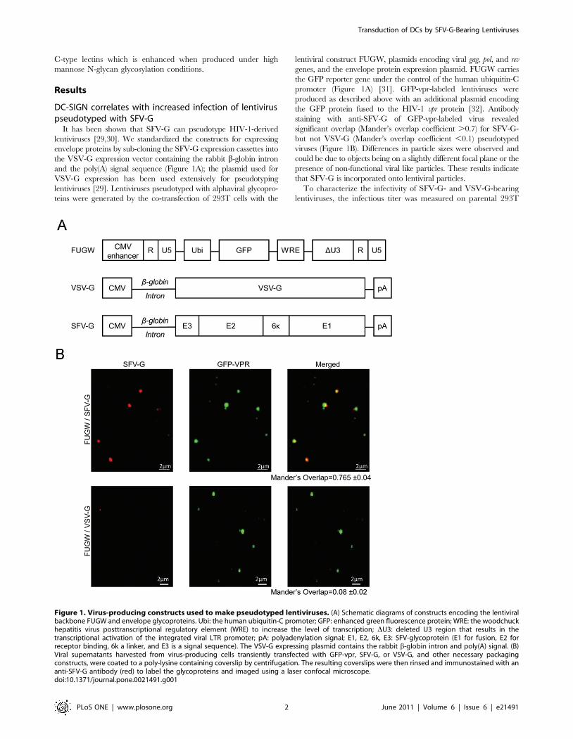
It has been shown that SFV-G can pseudotype HIV-1-derived
lentiviruses [29,30]. We standardized the constructs for expressing
envelope proteins by sub-cloning the SFV-G expression cassettes into
the VSV-G expression vector containing the rabbit b-globin intron
and the poly(A) signal sequence (Figure 1A); the plasmid used for
VSV-G expression has been used extensively for pseudotyping
lentiviruses [29]. Lentiviruses pseudotyped with alphaviral glycopro-
teins were generated by the co-transfection of 293T cells with the
lentiviral construct FUGW, plasmids encoding viral gag, pol, and rev
genes, and the envelope protein expression plasmid. FUGW carries
the GFP reporter gene under the control of the human ubiquitin-C
promoter (Figure 1A) [31]. GFP-vpr-labeled lentiviruses were
produced as described above with an additional plasmid encoding
the GFP protein fused to the HIV-1 vpr protein [32]. Antibody
staining with anti-SFV-G of GFP-vpr-labeled virus revealed
significant overlap (Mander’s overlap coefficient .0.7) for SFV-G-
but not VSV-G (Mander’s overlap coefficient ,0.1) pseudotyped
viruses (Figure 1B). Differences in particle sizes were observed and
could be due to objects being on a slightly different focal plane or the
presence of non-functional viral like particles. These results indicate
that SFV-G is incorporated onto lentiviral particles.
To characterize the infectivity of SFV-G- and VSV-G-bearing
lentiviruses, the infectious titer was measured on parental 293T
Figure 1. Virus-producing constructs used to make pseudotyped lentiviruses. (A) Schematic diagrams of constructs encoding the lentiviralbackbone FUGW and envelope glycoproteins. Ubi: the human ubiquitin-C promoter; GFP: enhanced green fluorescence protein; WRE: the woodchuckhepatitis virus posttranscriptional regulatory element (WRE) to increase the level of transcription; DU3: deleted U3 region that results in thetranscriptional activation of the integrated viral LTR promoter; pA: polyadenylation signal; E1, E2, 6k, E3: SFV-glycoprotein (E1 for fusion, E2 forreceptor binding, 6k a linker, and E3 is a signal sequence). The VSV-G expressing plasmid contains the rabbit b-globin intron and poly(A) signal. (B)Viral supernatants harvested from virus-producing cells transiently transfected with GFP-vpr, SFV-G, or VSV-G, and other necessary packagingconstructs, were coated to a poly-lysine containing coverslip by centrifugation. The resulting coverslips were then rinsed and immunostained with ananti-SFV-G antibody (red) to label the glycoproteins and imaged using a laser confocal microscope.doi:10.1371/journal.pone.0021491.g001
Transduction of DCs by SFV-G-Bearing Lentiviruses
PLoS ONE | www.plosone.org 2 June 2011 | Volume 6 | Issue 6 | e21491
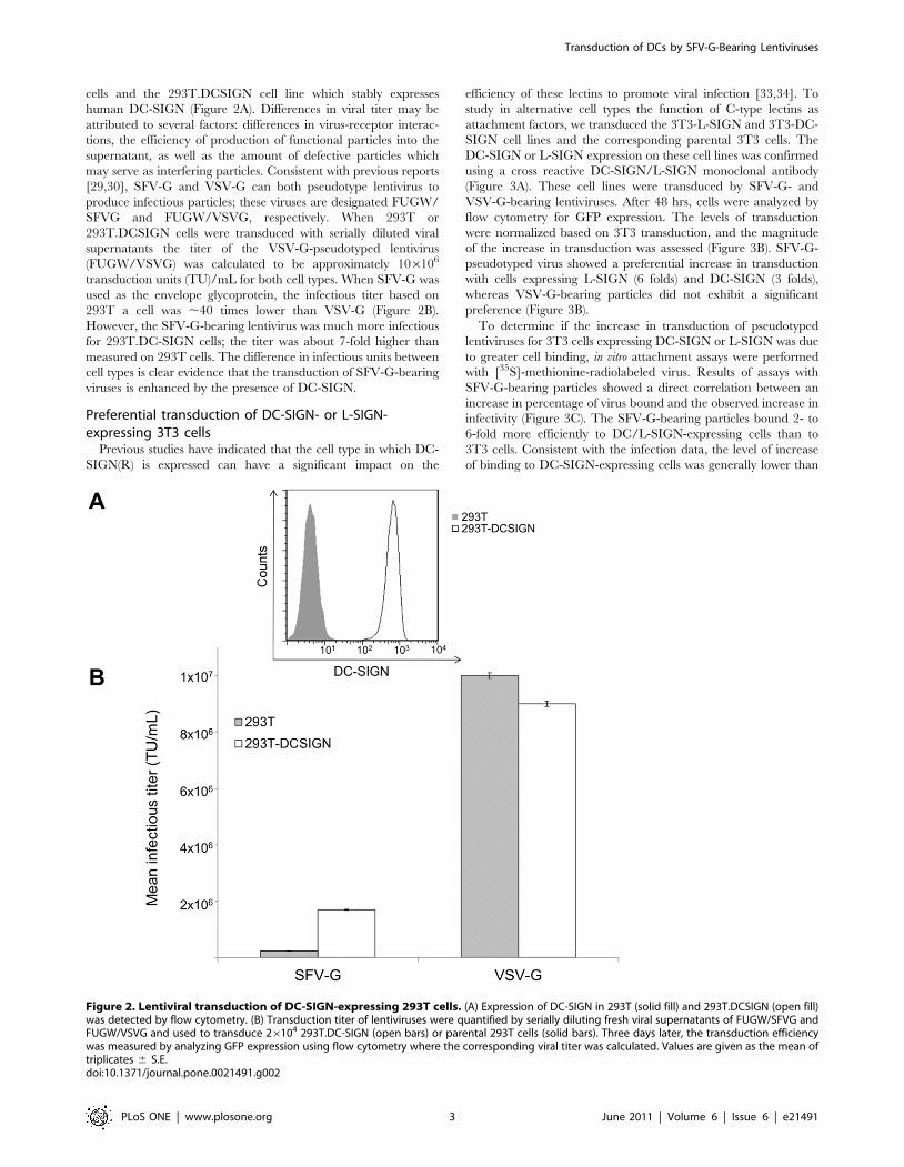
cells and the 293T.DCSIGN cell line which stably expresses
human DC-SIGN (Figure 2A). Differences in viral titer may be
attributed to several factors: differences in virus-receptor interac-
tions, the efficiency of production of functional particles into the
supernatant, as well as the amount of defective particles which
may serve as interfering particles. Consistent with previous reports
[29,30], SFV-G and VSV-G can both pseudotype lentivirus to
produce infectious particles; these viruses are designated FUGW/
SFVG and FUGW/VSVG, respectively. When 293T or
293T.DCSIGN cells were transduced with serially diluted viral
supernatants the titer of the VSV-G-pseudotyped lentivirus
(FUGW/VSVG) was calculated to be approximately 106106
transduction units (TU)/mL for both cell types. When SFV-G was
used as the envelope glycoprotein, the infectious titer based on
293T a cell was ,40 times lower than VSV-G (Figure 2B).
However, the SFV-G-bearing lentivirus was much more infectious
for 293T.DC-SIGN cells; the titer was about 7-fold higher than
measured on 293T cells. The difference in infectious units between
cell types is clear evidence that the transduction of SFV-G-bearing
viruses is enhanced by the presence of DC-SIGN.
Preferential transduction of DC-SIGN- or L-SIGN-expressing 3T3 cells
Previous studies have indicated that the cell type in which DC-
SIGN(R) is expressed can have a significant impact on the
efficiency of these lectins to promote viral infection [33,34]. To
study in alternative cell types the function of C-type lectins as
attachment factors, we transduced the 3T3-L-SIGN and 3T3-DC-
SIGN cell lines and the corresponding parental 3T3 cells. The
DC-SIGN or L-SIGN expression on these cell lines was confirmed
using a cross reactive DC-SIGN/L-SIGN monoclonal antibody
(Figure 3A). These cell lines were transduced by SFV-G- and
VSV-G-bearing lentiviruses. After 48 hrs, cells were analyzed by
flow cytometry for GFP expression. The levels of transduction
were normalized based on 3T3 transduction, and the magnitude
of the increase in transduction was assessed (Figure 3B). SFV-G-
pseudotyped virus showed a preferential increase in transduction
with cells expressing L-SIGN (6 folds) and DC-SIGN (3 folds),
whereas VSV-G-bearing particles did not exhibit a significant
preference (Figure 3B).
To determine if the increase in transduction of pseudotyped
lentiviruses for 3T3 cells expressing DC-SIGN or L-SIGN was due
to greater cell binding, in vitro attachment assays were performed
with [35S]-methionine-radiolabeled virus. Results of assays with
SFV-G-bearing particles showed a direct correlation between an
increase in percentage of virus bound and the observed increase in
infectivity (Figure 3C). The SFV-G-bearing particles bound 2- to
6-fold more efficiently to DC/L-SIGN-expressing cells than to
3T3 cells. Consistent with the infection data, the level of increase
of binding to DC-SIGN-expressing cells was generally lower than
Figure 2. Lentiviral transduction of DC-SIGN-expressing 293T cells. (A) Expression of DC-SIGN in 293T (solid fill) and 293T.DCSIGN (open fill)was detected by flow cytometry. (B) Transduction titer of lentiviruses were quantified by serially diluting fresh viral supernatants of FUGW/SFVG andFUGW/VSVG and used to transduce 26104 293T.DC-SIGN (open bars) or parental 293T cells (solid bars). Three days later, the transduction efficiencywas measured by analyzing GFP expression using flow cytometry where the corresponding viral titer was calculated. Values are given as the mean oftriplicates 6 S.E.doi:10.1371/journal.pone.0021491.g002
Transduction of DCs by SFV-G-Bearing Lentiviruses
PLoS ONE | www.plosone.org 3 June 2011 | Volume 6 | Issue 6 | e21491

that to L-SIGN-expressing cells. In the absence of DC-SIGN
expression, SFV-G pseudotypes bound poorly to 3T3 cells with
only 2,289 CPM binding to cells out of the 63,701 CPM that were
incubated with the cells. VSV-G-pseudotyped virus exhibited
similar levels of attachment to all cell types (Figure 3C) with
approximately 25% of the total CPM of virus bound to all cell
types. As shown in Figure 3B and 3C, the attachment and
transduction of FUGW/VSVG was approximately similar among
cells regardless of DC-SIGN(R) expression. This is consistent with
the lack of increased infectivity observed with FUGW/VSVG on
DC-SIGN expressing cells. Our data suggest that SFV-G-bearing
lentiviruses utilize DC-SIGN and L-SIGN as attachment receptors
to facilitate binding and transduction.
SFV-G- and VSV-G-mediated transduction requiresacidification
Internalization of SFV-G and VSV-G lentiviruses is known to
involve the endocytic pathway, in which the acidic environment of
endosomal vesicles is required. Treatment with the pH-interfering
drugs ammonium chloride caused a dose-dependent reduction of
infectivity in 293T.DCSIGN cells for both viruses (Figure 4B).
This suggests that both DC-SIGN-mediated infection of FUGW/
SFVG and DC-SIGN-independent infection by FUGW/VSVG
are pH-dependent processes, presumably due to the acidification
requirement of the virus-endosome fusion process [32]. Trans-
duction was inhibited by endosomal neutralization using bafilo-
mycin A1 (Figure 4C), verifying pH-mediated transduction with
both SFV- and VSV-G bearing particles.
SFV-G-mediated infectivity can be blocked withinhibitors of DC-SIGN
We observed that the infection efficiency of SFV-G-bearing
lentiviruses correlates with the expression of DC-SIGN on target
cells. To further examine the specificity of virus interaction with
these molecules, we performed infectivity experiments in the
presence of increasing concentrations of yeast mannan, or
Figure 3. Effects of DC-SIGN or L-SIGN expression. (A) Expression of L-SIGN and DC-SIGN in 3T3 (solid fill), 3T3.DCSIGN (open fill) and 3T3.LSIGN(gray fill), respectively, was detected using cross reactive DC-SIGN/L-SIGN antibody and quantified by flow cytometry. (B) Effects of DC-SIGN or L-SIGNexpression on the infectivity of pseudotyped lentiviruses. SFV-G- and VSV-G-pseudotyped lentiviruses were normalized by p24 and spin-inoculatedwith LSIGN- or DCSIGN-expressing 3T3 cells; the parental 3T3 cells were included as controls. Three days later, the transduction efficiency wasmeasured by analyzing GFP expression. Fold increase in percentage of GFP-positive cells is shown based on 3T3 cells where FUGW/SFVG transduced6.660.7% and FUGW/VSVG transduced 72.261.0%. (C) Specificity of binding to DC-SIGN. [35S]-methionine-labeled FUGW/SFVG or FUGW/VSVG wereincubated with 3T3 or DC-SIGN/L-SIGN-expressing cells at 4uC. Cells were washed and 35S radioactivity of the resuspended cells was quantitated witha liquid scintillation counter. Fold increase in [35S] bound viral particles is shown based on 3T3 cells with 3.5960.27% and 25.2361.25% of the totalCPM of virus bound for FUGW/SFVG and FUGW/VSVG, respectively, where values are given as the mean of triplicates 6 S.E.doi:10.1371/journal.pone.0021491.g003
Transduction of DCs by SFV-G-Bearing Lentiviruses
PLoS ONE | www.plosone.org 4 June 2011 | Volume 6 | Issue 6 | e21491

ethylenediaminetetraacetic acid (EDTA), soluble recombinant
DC-SIGN, anti-DC-SIGN monoclonal antibodies (mAbs), or an
isotype-matched control (Figure 4A and 4C). These treatments
disrupt interactions with DC-SIGN molecules [20,22,26]. Incu-
bation with mannan, a carbohydrate that competitively inhibits
virus binding with DC-SIGN [35], during viral inoculation of
FUGW/SFVG with 293T.DCSIGN cells resulted in a dose-
dependent reduction in the amount of GFP-positive cells
(Figure 4B). However, mannan was not as effective at inhibition
of VSV-G-bearing lentivirus, suggesting a different receptor
interaction between VSV-G and 293T.DCSIGN cells. The
principal characteristic of C-type lectins is that they interact with
mannose residues of viral glycoproteins in a calcium-dependent
manner via their C-terminal carbohydrate recognition domain
(CRD). EDTA is a calcium chelator and treatment during
infection with FUGW/SFVG resulted in a .70% reduction in
GFP-positive cells (Figure 4C). In contrast, FUGW/VSVG virus
was not dependent on calcium for infection as revealed by the
EDTA inhibition experiment. Incubation with anti-DC-SIGN
mAbs during infection also resulted in a reduction in the numbers
of GFP-positive cells with FUGW/SFVG infection, whereas the
isotype control antibody had little inhibitory effect (Figure 4C).
Again, the FUGW/VSVG virus did not exhibit significant
inhibition in the presence of DC-SIGN antibodies (Figure 4C).
Furthermore, pre-incubation of FUGW/SFVG lentivirus with
concavalin A (ConA), which binds to N-linked high-mannose
structures reduced infectivity by ,90% (Figure 4C). The
inhibition by ConA supports the theory that mannose carbohy-
drate residues present on SFV-G participate in the attachment to
DC-SIGN. Finally, infection of SFV-G- but not VSV-G-bearing
virus was blocked by pre-incubation of the cells with soluble DC-
SIGN (Figure 4C). The results of these experiments indicate that
the infectivity of SFV-G-pseudotyped lentivirus is dependent on
DC-SIGN expression for transduction, whereas FUGW/VSVG
transduction is not dependent upon DC-SIGN.
We further evaluated the ability of inhibitors of DC-SIGN to
block infection of human monocyte derived DCs (MoDCs).
MoDCs were prepared from the peripheral blood mononuclear
cells (PBMC) of healthy human donors and cultured with GM-
CSF and IL-4 to generate DC-SIGN+ DCs [36]. More than 80%
of the cultured DCs were positive for DC-SIGN expression before
infection (Figure 5A). MoDCs were challenged with the same
MOI of SFV-G- and VSV-G-pseudotyped lentiviruses incubated
in the presence of mannan or anti-DC-SIGN(R) antibody.
Transduction of MoDCs by FUGW/SFVG decreased from
,28% to ,8% with anti-DC-SIGN antibody and to ,4% in
the presence of yeast mannan (Figure 5B). The transduction
efficiency of FUGW/VSVG was lower than that of the SFV-G
bearing lentivirus (,13% GFP+). In contrast to FUGW/SFVG,
there was not a significant decrease when FUGW/VSVG was
incubated with MoDCs in the presence of either mannan or anti-
DC-SIGN(R) antibody (Figure 5B). These results suggest that DC-
Figure 4. Specific inhibitors prevent DC-SIGN-mediated infection. In dose-response experiments, 293T.DCSIGN cells were treated with SFV-G(solid circles) or VSV-G (open triangles) lentiviruses in the presence of increasing concentrations of mannan (A), or NH4Cl (B). (C) 293T.DCSIGN cellswere incubated with antibodies at a concentration of 5 mg/ml, 15 nM Bafilomycin A1, 5 mM EDTA, or 10 mg/ml soluble DC-SIGN at 37uC for 30 min,and then inoculated with SFV-G (filled bars) or VSV-G (open bars) lentiviruses at an MOI,0.8 for 8 hrs or lentiviruses incubated with 25 mg/mL ConA(1 h at 37uC). Subsequently, the supernatants were replaced and incubated with fresh medium for two days before being analyzed for GFPexpression. The relative transduction was determined based on non-treated controls and values are given as the mean of triplicates 6 S.E.doi:10.1371/journal.pone.0021491.g004
Transduction of DCs by SFV-G-Bearing Lentiviruses
PLoS ONE | www.plosone.org 5 June 2011 | Volume 6 | Issue 6 | e21491

SIGN(R) function as a SFV-G binding molecule that is required
for the productive infection of MoDCs.
Effects of viral glycosylationNext, we evaluated the ability mannose carbohydrate structures
on the SFV-G lentiviral particles to promote infection through a
mechanism of increase interactions with DC-SIGN(R). To test this,
we generated FUGW/SFVG particles containing only high-
mannose glycan content on their envelope glycoproteins by treating
virus-producing cells with 1-deoxymannojirimycin (DMJ). DMJ
is an inhibitor of Golgi mannosidase I and can arrest glycan
maturation primarily at the Man8GlcNAc2 stage [37]. Production
of pseudotyped lentiviruses in the presence of DMJ altered their
ability to transduce DC-SIGN-expressing 3T3 cells (Figure 6, DMJ
(+)). A two fold increase in transduction efficiency of 3T3-DCSIGN
cells was observed for DMJ-treated SFV-G-bearing virus, whereas
no significant transduction increase in parental 3T3 was observed.
We further tested how efficiently SFV-G pseudotyped lentiviruses
transduce primary immune cell targets, MoDCs, and the effect of
DMJ treatment on transduction efficiency. Transduction of MoDCs
by DMJ-treated FUGW/SFVG was approximately twice as
efficient as non-treated FUGW/SFVG, transducing ,20% versus
,10% MoDCs (Figure 6). Similar to the 3T3-DCSIGN cell line,
transduction by SFV-G pseudotyped lentiviruses produced in the
presence of DMJ increased their ability to transduce MoDCs
(Figure 6). Together, these results reveal that the 293T-produced
SFV-G-bearing particles are able to innately bind to DC-SIGN(R)-
receptors but DMJ-treatment can enhance transduction of DC-
SIGN-expressing cells.
Discussion
We have demonstrated that SFV-G-bearing lentiviruses can
utilize DC-SIGN and L-SIGN as attachment receptors, resulting
in productive infections of cell lines bearing these molecules and
human MoDCs. Preferential binding to DC-SIGN(R) by SFV-G
has not previously been reported, indicating that there is an
unappreciated naive trophism of SFV-G pseudotyped lentivirus
for APCs. Our results suggest that by utilizing the affinity of SFV-
G to DC-SIGN(R), lentiviruses can be engineered to preferentially
transduce antigen-presenting DCs for gene-based immunotherapy.
We found that FUGW/SFVG but not FUGW/VSVG
produced in 293T cells bind to DC-SIGN(R) receptors. When
produced in 293T cells, VSV-G-pseudotyped lentiviruses exhib-
ited similar binding and transduction of cells expressing DC-
SIGN, L-SIGN or parental cell lines. For SFV-G-bearing viruses,
cells expressing DC-SIGN or L-SIGN were more permissive than
non-expressing parental cell lines. The increased transduction was
Figure 5. Transduction of MoDCs by lentiviruses is inhibited with anti-DCSIGN antibody and mannan. Human monocyte-derived DCs(MoDCs) were generated by culturing respective precursor cells in the presence of GM-CSF and IL-4. (A) The adherent cells (16106) were cultured for2 days and then DC-SIGN expression was detected by flow cytometry. (B) Human MoDCs (16106) were incubated for 1 hour with mannan (200 mg/mL), anti-DC-SIGN(R) antibody (20 mg/mL) or without any reagents. The cells were then infected with FUGW/SFVG or FUGW/VSVG (MOI = 10) for8 hours in the presence of blocking reagents. GFP expression was assayed by flow cytometry five days post-transduction where one representativefigure is shown with values given as the mean of triplicates 6 S.E.doi:10.1371/journal.pone.0021491.g005
Transduction of DCs by SFV-G-Bearing Lentiviruses
PLoS ONE | www.plosone.org 6 June 2011 | Volume 6 | Issue 6 | e21491

well-correlated with an increase in binding to the cells as measured
by radiolabeled virus binding assays. Specific interaction between
DC-SIGN and SFV-G was demonstrated by blocking the
transduction of the DC-SIGN-expressing cells with inhibitors
such as ConA, EDTA, soluble DC-SIGN protein, yeast mannan,
or DC-SIGN-specific mAbs in both 293T.DCSIGN cells and
human MoDCs.
SFV-G-bearing lentiviruses have an enhanced ability to trans-
duce DC-SIGN-expressing cells when produced under conditions
arresting the viral glycan maturation primarily at the high mannose
stage. Several reports indicate that the presence of high-mannose-
content N-linked glycans on Sindbis virus, Ebola virus and West
Nile virus enhance the infection of mouse-derived DCs due to
interactions with the mannose binding C-type lectin receptors
[20,22,38]. In addition to DC-SIGN present on DCs, there are
other C-type lectin molecules on macrophages, endothelial cells,
and other APCs, that might play a role in FUGW/SFV-G
transduction [23,25]. High mannose content of viral envelope
glycoproteins directly influences the efficiency of viral capture by
DC-SIGN and L-SIGN [20,39,40]. Similar to findings reported
previously for Sindbis virus [41], when SFV-G-bearing lentiviruses
were generated in mammalian cells treated with the mannosidase I
inhibitor DMJ, the resulting particles exhibit an increased capacity
to utilize DC-SIGN for infection. The increase in interaction with
DC-SIGN by FUGW/SFVG is presumably mediated by increased
binding by the CRD to the mannose structures of these
glycoproteins. The SFV-G has four sites for N-linked glycosylation
(E1-141, E2-200, E2-262 and E3-14) [42]. The E3 protein is
cleaved from the mature SFV particles, but remains associated with
the virion of SFV [43]. When virus-producing cells are treated with
DMJ, transduction by SFV-G-bearing virus was increased towards
MoDCs as well as DC-SIGN-expressing 3T3 cells but not the
parental 3T3 cell line. The observation that FUGW/SFVG
produced by DMJ-treated cells have an enhanced ability to
preferentially transduce DC-SIGN-expressing cells suggests that
modification of N-linked glycans of SFV-G can be used to enhance
the transduction of DCs.
We observed that FUGW/VSVG lentiviruses exhibited no
increase in binding or transduction with cells expressing DC-SIGN
or L-SIGN. VSV-G-pseudotyped viruses do not target through
DC-SIGN(R) [44,45] unless produced with in the presence of
DMJ [46] but they also efficiently transduce a wide range of cell
types, likely through a ubiquitous membrane lipid [47]. Although
previous studies have found that VSV-G pseudotypes of HIV-1
infect bone marrow-derived premature DCs [30], we found that
FUGW/VSVG exhibited no preferential transduction towards
cells expressing the human C-type lectins DC-SIGN or L-SIGN.
FUGW/VSVG lentiviruses have a broad tropism and can
transduce multiple cell types. Therefore, they are undesirable for
delivering genes in vivo to APCs. Our results suggest that the
transduction by FUGW/VSVG is largely DC-SIGN(R)-indepen-
dent and less efficient than FUGW/SFVG at transducing DCs
when normalized by infectious particles.
In this study we assessed the relationship of DC-SIGN and L-
SIGN interactions with SFV glycoproteins to mediate the
transduction of DCs. DCs are potent APCs and play a major
role in the activation of both memory and naı̈ve T cells.
Genetically modified DCs have been used to elicit antigen-specific,
major histocompatibility complex-restricted cytotoxic T lympho-
cyte (CTL) responses [21,48]. The development of DC differen-
tiation protocols for PBMC has facilitated the study of DC biology
and the subsequent implementation of clinical DC-based vacci-
nation studies. Transduction of human MoDC by FUGW/SFVG
was more than twice as efficient as that by FUGW/VSVG when
normalized by MOI. Furthermore, the transduction by FUGW/
SFVG was DC-SIGN(R)-specific and could be inhibited by both
Figure 6. Transduction by SFV-G lentiviruses produced in DMJ treated cells. 3T3 (26104), 3T3-DCSIGN (26104) and MoDCs (16106) werespin-infected with FUGW/SFVG produced in 293T cells without DMJ(2) or with DMJ(+) treatment. GFP expression was assayed by flow cytometrythree days post-transduction where values are given as the mean of triplicates 6 S.E.doi:10.1371/journal.pone.0021491.g006
Transduction of DCs by SFV-G-Bearing Lentiviruses
PLoS ONE | www.plosone.org 7 June 2011 | Volume 6 | Issue 6 | e21491
yeast mannan and anti-DC-SIGN antibodies. SFV-G pseudotypes
preferentially transduced the DC-SIGN-positive cells, consistent
with the theory that DC-SIGN mediates transduction in DCs. The
preferential transduction of DCs can be further enhanced by
production under untrimmed (DMJ-treated) high mannose
conditions. Further studies to assess the maturation of DCs
transduced by FUGW/SFVG vectors by measuring maturation
markers such as HLA-DR, CD11b, and CD83 and the effect on
type I interferon production in DCs are ongoing. FUGW/SFVG
has previously been shown to have rather broad tropism and be
able to have low transduction efficiency for a wide range of cell
types [30], but we have found that FUGW/SFVG has a DC-
SIGN(R) tropism, which can be utilized for directing the cellular
transduction of lentiviruses to APCs.
The results described herein have relevance to the design and
production of viral vectors used for gene delivery to APCs.
Targeting of lentiviruses to C-type lectin-expressing cells such as
DCs can be increased by pseudotyping with the Semliki Forest
virus envelope glycoprotein and further enhanced by production
under conditions that limit host cell processing of viral carbohy-
drate modifications to contain mannose structures. When
lentiviruses were produced in DMJ-treated cells, they generated
a similar amount of physical viral particles as those produced in
normal conditions (unpublished data). Enhanced delivery of
antigen to immature DCs may provide an opportunity for
improvement of gene-based vaccination approaches. Future
studies are warranted to investigate whether the wild type Semliki
Forest virus has a similar tropism for DC-SIGN(R) expressing
cells. Our results show that SFV-G pseudotyped lentivectors
strongly bind to C-type lectins. The affinity of FUGW/SFVG with
DC-SIGN(R) represents a new strategy to genetically modify DCs.
Materials and Methods
Cell lines293T.DCSIGN were derived as previously described [21] and
stained (anti-DC-SIGN antibody from BD Biosciences) to confirm
expression of DC-SIGN. Mouse fibroblasts NIH 3T3 cells were
obtained from the American Tissue Culture Collection (ATCC,
Manassas, VA). 3T3-L-SIGN and 3T3-DC-SIGN were obtained
from the NIH AIDS Research and Reference Reagent Program,
Division of AIDS, NIAID. These cell lines were maintained in
DMEM medium (Invitrogen, Carlsbad, CA) supplemented with 10%
fetal calf serum (Sigma-Aldrich, St. Louis, MO), 2 mM L-glutamine,
and 100 U/mL of penicillin and 100 mg/mL of streptomycin.
Plasmid constructionThe glycoprotein expression plasmids were constructed similarly
to previously reported [29]. The cDNA of SFV-G was amplified
from the pSFV helper expression vector (a gift from Dr. Robert
Chow, University of Southern California). The amplified fragments
for the glycoprotein were subcloned into the vesicular stomatitis
virus glycoprotein (VSV-G) expression plasmid pVSV-G (Cell
Genesys, Foster City, CA). The resulting plasmid was designated
pSFV-G (Figure 1). The lentiviral backbone plasmid (FUGW and its
derivatives) used in this study have been previously described [31].
Production of pseudotyped viral particlesRecombinant lentiviruses were prepared by transient transfec-
tion of 293T cells using a standard calcium phosphate precipita-
tion protocol [49]. The viral supernatants were harvested 48 and
72 hrs post-transfection and filtered through a 0.45-mm filter. To
prepare concentrated viruses, the viral supernatants were ultra-
centrifugated (Optima L-80K preparative ultracentrifuge, Beck-
man Coulter) at 50,0006g for 90 min. Mammalian cell-derived
viral stocks with high-mannose glycans were generated by
transient transfection of 293T cells, which were subsequently
cultured in 1 mM 1-deoxymannojirimycin (DMJ, Sigma-Aldrich).
Confocal imaging of GFP-vpr labeled virionsGFP-vpr-labeled lentiviral particles were produced as previously
described [32]. Fresh viral supernatant was overlaid on polylysine-
coated coverslips in a 6-well culture dish and centrifuged at
3,7006g at 4uC for 2 hrs using a RT Legend centrifuge. The
coverslips were washed with cold PBS twice and incubated with
diluted rabbit polyclonal anti-SFV E1/E2 antibody (1:2000; a gift
from Margaret Kielian, Albert Einstein College of Medicine) for
40 min at 4uC. Coverslips were washed with PBS and incubated
for 40 min at 4uC with 1:500 dilutions of secondary antibodies
consisting of species-specific Cy5-conjugated anti-immunoglobulin
G (Santa Cruz Biotechnology, Santa Cruz, CA). Fluorescent
images were acquired by a Zeiss LSM 510 laser scanning confocal
microscope with a plan-apochromat oil immersion (636/1.4)
objective.
Virus attachment assaysProduction of [35S]-methionine-labeled viruses were produced
by transfection of 293T cells as described above. Cells were then
depleted of methionine and at 8 hrs post-transfection, [35S]-
methionine was added to a final concentration of 20 mCi/mL and
cells were incubated at 37uC for an additional 12 hrs. [35S]-
radiolabelled virus was purified from cell supernatants by using a
discontinuous sucrose gradient (20%/60% [wt/wt] in TNE buffer
[50 mM Tris-HCl, 100 mM NaCl, 1 mM EDTA]), followed by
pelleting through 20% sucrose in TNE buffer. Radiolabeled virus
particles were resuspended in PBS. Approximately 105 CPM of
each radiolabeled virus diluted in PBS was mixed with 106 cells in
1.5 mL microcentrifuge tubes and this mixture was incubated at
4uC for 1 hr with gentle agitation. Cells were washed and 35S
radioactivity was quantitated with a liquid scintillation counter.
Determination of titersTo determine viral titer, 26104 293T or 293T.DCSIGN cells
were transduced with 100 ml of serially diluted viral supernatants
with 8 mg/mL of polybrene (Sigma-Aldrich) for 1.5 hrs by spin-
inoculation at 2,500 rpm and 25uC using a RT Legend centrifuge.
Following the spin-infection, the supernatants were replaced with
fresh culture medium and incubated for an additional 48 hrs at
37uC with 5% CO2. The GFP expression was measured by flow
cytometry. The transduction titer was calculated based on dilution
ranges that exhibited a linear response of eGFP expression with
viral serial dilution concentration.
Lentivirus-mediated transduction of cell lines in vitroThe 3T3, 3T3-LSIGN and 3T3-DCSIGN cell lines were
stained with cross reactive anti-DCSIGN(R) antibody 14E3G7.
Target cells (3T3-LSIGN, 3T3-DCSIGN, or 3T3 cells; 0.26105
per well) were seeded in 96-well culture dishes and spin-infected
with viral supernatants (150 mL per well of p24-normalized virus)
at 2,500 rpm and 25uC for 90 min using a RT Legend centrifuge.
Subsequently, the supernatants were replaced with fresh culture
medium and incubated for 48 hrs at 37uC with 5% CO2.
Assays to inhibit pseudotyped virus-mediated infectionIn dose-response experiments, 293T.DCSIGN (0.26105 per well)
were incubated with 0.2 to 200 mg/mL of yeast mannan (Sigma-
Aldrich) at 37uC for 30 min. SFV-G- or VSV-G-bearing lentiviruses
Transduction of DCs by SFV-G-Bearing Lentiviruses
PLoS ONE | www.plosone.org 8 June 2011 | Volume 6 | Issue 6 | e21491
(MOI = 0.8) were incubated for 8 hrs, and then supernatant was
replaced with fresh medium. For NH4Cl inhibitions, pseudotyped
viral particles were spin-inoculated in the presence of increasing
concentrations of NH4Cl (Sigma-Aldrich) for 90 min at 25uC.
293T.DCSIGN cells were incubated with 5 mg/mL of anti-DCSIGN
antibodies (14E3G7, 19F7, DC-28, and isotype control antibody,
Santa Cruz Biotechnology), 5 mM EDTA, 15 nM Bafilomycin A1,
or 10 mg/ml sDC-SIGN, the soluble, tetrameric ectodomain of DC-
SIGN produced as previously described [50] at 37uC for 30 min, and
then inoculated SFV-G- or VSV-G-bearing lentiviruses at an MOI
,0.8 for 8 hrs. Similarly, virus was incubated with 25 mg/mL
concavalin A (Sigma-Aldrich) for 1 hr at 37C then incubated with
293T.DCSIGN cells for 8 hrs before changing to fresh D10 medium.
Transduction of human PBMC-derived DCsPeripheral blood mononuclear cells (PBMC) from two healthy
human donors were purchased from AllCells (Emeryville, CA).
PBMC were differentiated into DCs as described previously [36].
After two days of culture, DCs were identified by examining the
surface markers (CD11C+, DC-SIGN+) using flow cytometry analysis
on cells stained with anti-CD11c antibody and anti-DCSIGN
antibody (BD Biosciences). Monocyte-derived DCs (MoDC) on day
2 were exposed to virus at the required MOI based on 293T cells. For
inhibition of DC-SIGN-mediated transduction, DCs were incubated
with 20 mg/mL of anti-DC-SIGN(R) antibody (14E2G7, Santa Cruz
Biotechnology) or 200 mg/mL yeast mannan (Sigma-Aldrich) at
37uC for 30 min and then inoculated with 293T-produced FUGW/
SFVG or FUGW/VSVG lentiviruses at an MOI = 10 for 8 hrs
before the media was changed.
Acknowledgments
We thank Dr. Margaret Kielian for providing antibodies against the SFV
envelope glycoprotein, Dr. Robert Chow for the pSFV expression vector,
Xiao Liang for providing soluble DC-SIGN proteins, and Kye-Il Joo for
assistance with confocal imaging.
Author Contributions
Conceived and designed the experiments: SF AT PW. Performed the
experiments: SF AT KK AZ. Analyzed the data: SF PW. Wrote the paper:
SF PW.
References
1. Froelich S, Tai A, Wang P (2010) Lentiviral vectors for immune cells targeting.
Immunopharmacol Immunotoxicol 32: 208–218.
2. Ribas A, Butterfield LH, Glaspy JA, Economou JS (2002) Cancer immuno-
therapy using gene-modified dendritic cells. Curr Gene Ther 2: 57–78.
3. Dullaers M, Thielemans K (2006) From pathogen to medicine: HIV-1-derived
lentiviral vectors as vehicles for dendritic cell based cancer immunotherapy.
J Gene Med 8: 3–17.
4. Lizee G, Gonzales MI, Topalian SL (2004) Lentivirus vector-mediated
expression of tumor-associated epitopes by human antigen presenting cells.
Hum Gene Ther 15: 393–404.
5. Sumimoto H, Tsuji T, Miyoshi H, Hagihara M, Takada-Yamazaki R, et al.
(2002) Rapid and efficient generation of lentivirally gene-modified dendritic cells
from DC progenitors with bone marrow stromal cells. J Immunol Methods 271:
153–165.
6. Oki M, Ando K, Hagihara M, Miyatake H, Shimizu T, et al. (2001) Efficient
lentiviral transduction of human cord blood CD34(+) cells followed by their
expansion and differentiation into dendritic cells. Exp Hematol 29: 1210–1217.
7. Schroder AR, Shinn P, Chen H, Berry C, Ecker JR, et al. (2002) HIV-1
integration in the human genome favors active genes and local hotspots. Cell
110: 521–529.
8. Unutmaz D, KewalRamani VN, Marmon S, Littman DR (1999) Cytokine
signals are sufficient for HIV-1 infection of resting human T lymphocytes. J Exp
Med 189: 1735–1746.
9. Firat H, Zennou V, Garcia-Pons F, Ginhoux F, Cochet M, et al. (2002) Use of a
lentiviral flap vector for induction of CTL immunity against melanoma.
Perspectives for immunotherapy. J Gene Med 4: 38–45.
10. Gruber A, Kan-Mitchell J, Kuhen KL, Mukai T, Wong-Staal F (2000) Dendritic
cells transduced by multiply deleted HIV-1 vectors exhibit normal phenotypes
and functions and elicit an HIV-specific cytotoxic T-lymphocyte response in
vitro. Blood 96: 1327–1333.
11. Dyall J, Latouche JB, Schnell S, Sadelain M (2001) Lentivirus-transduced
human monocyte-derived dendritic cells efficiently stimulate antigen-specific
cytotoxic T lymphocytes. Blood 97: 114–121.
12. Zarei S, Leuba F, Arrighi JF, Hauser C, Piguet V (2002) Transduction of
dendritic cells by antigen-encoding lentiviral vectors permits antigen processing
and MHC class I-dependent presentation. J Allergy Clin Immunol 109:
988–994.
13. Breckpot K, Dullaers M, Bonehill A, van Meirvenne S, Heirman C, et al. (2003)
Lentivirally transduced dendritic cells as a tool for cancer immunotherapy.
J Gene Med 5: 654–667.
14. Metharom P, Ellem KA, Schmidt C, Wei MQ (2001) Lentiviral vector-mediated
tyrosinase-related protein 2 gene transfer to dendritic cells for the therapy of
melanoma. Hum Gene Ther 12: 2203–2213.
15. Zarei S, Abraham S, Arrighi JF, Haller O, Calzascia T, et al. (2004) Lentiviral
transduction of dendritic cells confers protective antiviral immunity in vivo.
J Virol 78: 7843–7845.
16. Esslinger C, Chapatte L, Finke D, Miconnet I, Guillaume P, et al. (2003) In vivo
administration of a lentiviral vaccine targets DCs and induces efficient CD8(+) T
cell responses. J Clin Invest 111: 1673–1681.
17. Palmowski MJ, Lopes L, Ikeda Y, Salio M, Cerundolo V, et al. (2004)
Intravenous injection of a lentiviral vector encoding NY-ESO-1 induces an
effective CTL response. J Immunol 172: 1582–1587.
18. Hu B, Tai A, Wang P (2011) Immunization delivered by lentiviral vectors forcancer and infectious diseases. Immunol Rev 239: 45–61.
19. Cronin J, Zhang XY, Reiser J (2005) Altering the tropism of lentiviral vectors
through pseudotyping. Curr Gene Ther 5: 387–398.
20. Klimstra WB, Nangle EM, Smith MS, Yurochko AD, Ryman KD (2003) DC-SIGN and L-SIGN can act as attachment receptors for alphaviruses and
distinguish between mosquito cell- and mammalian cell-derived viruses. J Virol
77: 12022–12032.
21. Yang L, Yang H, Rideout K, Cho T, Joo KI, et al. (2008) Engineered lentivectortargeting of dendritic cells for in vivo immunization. Nat Biotechnol 26:
326–334.
22. Davis CW, Nguyen HY, Hanna SL, Sanchez MD, Doms RW, et al. (2006) West
Nile virus discriminates between DC-SIGN and DC-SIGNR for cellularattachment and infection. J Virol 80: 1290–1301.
23. Bashirova AA, Geijtenbeek TB, van Duijnhoven GC, van Vliet SJ, Eilering JB,
et al. (2001) A dendritic cell-specific intercellular adhesion molecule 3-grabbingnonintegrin (DC-SIGN)-related protein is highly expressed on human liver
sinusoidal endothelial cells and promotes HIV-1 infection. J Exp Med 193:
671–678.
24. Braet F, Wisse E (2002) Structural and functional aspects of liver sinusoidalendothelial cell fenestrae: a review. Comp Hepatol 1: 1.
25. Figdor CG, van Kooyk Y, Adema GJ (2002) C-type lectin receptors on dendritic
cells and Langerhans cells. Nat Rev Immunol 2: 77–84.
26. Lozach PY, Burleigh L, Staropoli I, Amara A (2007) The C type lectins DC-
SIGN and L-SIGN: receptors for viral glycoproteins. Methods Mol Biol 379:51–68.
27. Mitchell DA, Fadden AJ, Drickamer K (2001) A novel mechanism of carbohydrate
recognition by the C-type lectins DC-SIGN and DC-SIGNR. Subunit organizationand binding to multivalent ligands. J Biol Chem 276: 28939–28945.
28. Soilleux EJ (2003) DC-SIGN (dendritic cell-specific ICAM-grabbing non-
integrin) and DC-SIGN-related (DC-SIGNR): friend or foe? Clin Sci (Lond)
104: 437–446.
29. Kahl CA, Marsh J, Fyffe J, Sanders DA, Cornetta K (2004) Humanimmunodeficiency virus type 1-derived lentivirus vectors pseudotyped with
envelope glycoproteins derived from Ross River virus and Semliki Forest virus.J Virol 78: 1421–1430.
30. Strang BL, Takeuchi Y, Relander T, Richter J, Bailey R, et al. (2005) Humanimmunodeficiency virus type 1 vectors with alphavirus envelope glycoproteins
produced from stable packaging cells. J Virol 79: 1765–1771.
31. Lois C, Hong EJ, Pease S, Brown EJ, Baltimore D (2002) Germline transmissionand tissue-specific expression of transgenes delivered by lentiviral vectors.
Science 295: 868–872.
32. Joo KI, Wang P (2008) Visualization of targeted transduction by engineered
lentiviral vectors. Gene Ther 15: 1384–1396.
33. Trumpfheller C, Park CG, Finke J, Steinman RM, Granelli-Piperno A (2003)Cell type-dependent retention and transmission of HIV-1 by DC-SIGN. Int
Immunol 15: 289–298.
34. Wu L, Martin TD, Carrington M, KewalRamani VN (2004) Raji B cells,
misidentified as THP-1 cells, stimulate DC-SIGN-mediated HIV transmission.Virology 318: 17–23.
35. Baribaud F, Pohlmann S, Leslie G, Mortari F, Doms RW (2002) Quantitative
expression and virus transmission analysis of DC-SIGN on monocyte-deriveddendritic cells. J Virol 76: 9135–9142.
Transduction of DCs by SFV-G-Bearing Lentiviruses
PLoS ONE | www.plosone.org 9 June 2011 | Volume 6 | Issue 6 | e21491

36. Obermaier B, Dauer M, Herten J, Schad K, Endres S, et al. (2003) Development
of a new protocol for 2-day generation of mature dendritic cells from human
monocytes. Biol Proced Online 5: 197–203.
37. Fuhrmann U, Bause E, Legler G, Ploegh H (1984) Novel mannosidase inhibitor
blocking conversion of high mannose to complex oligosaccharides. Nature 307:
755–758.
38. Marzi A, Moller P, Hanna SL, Harrer T, Eisemann J, et al. (2007) Analysis of
the interaction of Ebola virus glycoprotein with DC-SIGN (dendritic cell-specific
intercellular adhesion molecule 3-grabbing nonintegrin) and its homologue DC-
SIGNR. J Infect Dis 196 Suppl 2: S237–246.
39. Lozach PY, Burleigh L, Staropoli I, Navarro-Sanchez E, Harriague J, et al.
(2005) Dendritic cell-specific intercellular adhesion molecule 3-grabbing non-
integrin (DC-SIGN)-mediated enhancement of dengue virus infection is
independent of DC-SIGN internalization signals. J Biol Chem 280:
23698–23708.
40. Lozach PY, Lortat-Jacob H, de Lacroix de Lavalette A, Staropoli I, Foung S,
et al. (2003) DC-SIGN and L-SIGN are high affinity binding receptors for
hepatitis C virus glycoprotein E2. J Biol Chem 278: 20358–20366.
41. Morizono K, Ku A, Xie Y, Harui A, Kung SK, et al. (2010) Redirecting
lentiviral vectors pseudotyped with Sindbis virus-derived envelope proteins to
DC-SIGN by modification of N-linked glycans of envelope proteins. J Virol 84:
6923–6934.
42. Strauss JH, Strauss EG (1994) The alphaviruses: gene expression, replication,
and evolution. Microbiol Rev 58: 491–562.
43. Mancini EJ, Clarke M, Gowen BE, Rutten T, Fuller SD (2000) Cryo-electron
microscopy reveals the functional organization of an enveloped virus, SemlikiForest virus. Mol Cell 5: 255–266.
44. Kwon DS, Gregorio G, Bitton N, Hendrickson WA, Littman DR (2002) DC-
SIGN-mediated internalization of HIV is required for trans-enhancement of Tcell infection. Immunity 16: 135–144.
45. Simmons G, Wool-Lewis RJ, Baribaud F, Netter RC, Bates P (2002) Ebola virusglycoproteins induce global surface protein down-modulation and loss of cell
adherence. J Virol 76: 2518–2528.
46. Lin G, Simmons G, Pohlmann S, Baribaud F, Ni H, et al. (2003) Differential N-linked glycosylation of human immunodeficiency virus and Ebola virus envelope
glycoproteins modulates interactions with DC-SIGN and DC-SIGNR. J Virol77: 1337–1346.
47. Schlegel R, Tralka TS, Willingham MC, Pastan I (1983) Inhibition of VSVbinding and infectivity by phosphatidylserine: is phosphatidylserine a VSV-
binding site? Cell 32: 639–646.
48. Song W, Kong HL, Carpenter H, Torii H, Granstein R, et al. (1997) Dendriticcells genetically modified with an adenovirus vector encoding the cDNA for a
model antigen induce protective and therapeutic antitumor immunity. J ExpMed 186: 1247–1256.
49. Pear WS, Nolan GP, Scott ML, Baltimore D (1993) Production of high-titer
helper-free retroviruses by transient transfection. Proc Natl Acad Sci U S A 90:8392–8396.
50. Halary F, Amara A, Lortat-Jacob H, Messerle M, Delaunay T, et al. (2002)Human cytomegalovirus binding to DC-SIGN is required for dendritic cell
infection and target cell trans-infection. Immunity 17: 653–664.
Transduction of DCs by SFV-G-Bearing Lentiviruses
PLoS ONE | www.plosone.org 10 June 2011 | Volume 6 | Issue 6 | e21491